Human CD133+ Progenitor Cells Promote the Healing of Diabetic Ischemic Ulcers by Paracrine...
46
ISSN: 1524-4571 Copyright © 2009 American Heart Association. All rights reserved. Print ISSN: 0009-7330. Online TX 72514 Circulation Research is published by the American Heart Association. 7272 Greenville Avenue, Dallas, DOI: 10.1161/CIRCRESAHA.108.192138 published online Apr 2, 2009; Circ. Res. Alessandri, Costanza Emanueli and Paolo Madeddu Andreas Simm, Paola Campagnolo, Giuseppe Mangialardi, Lara Stevanato, Giulio Rajesh Katare, Mauro Siragusa, Marco Meloni, Ilaria Campesi, Manuela Monica, Lucíola S. Barcelos, Cécile Duplaa, Nicolle Kränkel, Gallia Graiani, Gloria Invernici, Signaling Ulcers by Paracrine Stimulation of Angiogenesis and Activation of Wnt Progenitor Cells Promote the Healing of Diabetic Ischemic + Human CD133 http://circres.ahajournals.org/cgi/content/full/CIRCRESAHA.108.192138/DC1 Data Supplement (unedited) at: http://circres.ahajournals.org located on the World Wide Web at: The online version of this article, along with updated information and services, is http://www.lww.com/reprints Reprints: Information about reprints can be found online at [email protected] 410-528-8550. E-mail: Fax: Kluwer Health, 351 West Camden Street, Baltimore, MD 21202-2436. Phone: 410-528-4050. Permissions: Permissions & Rights Desk, Lippincott Williams & Wilkins, a division of Wolters http://circres.ahajournals.org/subscriptions/ Subscriptions: Information about subscribing to Circulation Research is online at by on May 16, 2011 circres.ahajournals.org Downloaded from
Transcript of Human CD133+ Progenitor Cells Promote the Healing of Diabetic Ischemic Ulcers by Paracrine...
ISSN: 1524-4571 Copyright © 2009 American Heart Association. All rights reserved. Print ISSN: 0009-7330. Online
TX 72514Circulation Research is published by the American Heart Association. 7272 Greenville Avenue, Dallas,
DOI: 10.1161/CIRCRESAHA.108.192138 published online Apr 2, 2009; Circ. Res.
Alessandri, Costanza Emanueli and Paolo Madeddu Andreas Simm, Paola Campagnolo, Giuseppe Mangialardi, Lara Stevanato, GiulioRajesh Katare, Mauro Siragusa, Marco Meloni, Ilaria Campesi, Manuela Monica,
Lucíola S. Barcelos, Cécile Duplaa, Nicolle Kränkel, Gallia Graiani, Gloria Invernici,
SignalingUlcers by Paracrine Stimulation of Angiogenesis and Activation of Wnt Progenitor Cells Promote the Healing of Diabetic Ischemic+Human CD133
http://circres.ahajournals.org/cgi/content/full/CIRCRESAHA.108.192138/DC1Data Supplement (unedited) at:
http://circres.ahajournals.org
located on the World Wide Web at: The online version of this article, along with updated information and services, is
http://www.lww.com/reprintsReprints: Information about reprints can be found online at
[email protected]. E-mail:
Fax:Kluwer Health, 351 West Camden Street, Baltimore, MD 21202-2436. Phone: 410-528-4050. Permissions: Permissions & Rights Desk, Lippincott Williams & Wilkins, a division of Wolters
http://circres.ahajournals.org/subscriptions/Subscriptions: Information about subscribing to Circulation Research is online at
by on May 16, 2011 circres.ahajournals.orgDownloaded from
Human CD133� Progenitor Cells Promote the Healing ofDiabetic Ischemic Ulcers by Paracrine Stimulation of
Angiogenesis and Activation of Wnt SignalingLucíola S. Barcelos,* Cecile Duplaa,* Nicolle Krankel,* Gallia Graiani,* Gloria Invernici,
Rajesh Katare, Mauro Siragusa, Marco Meloni, Ilaria Campesi, Manuela Monica, Andreas Simm,Paola Campagnolo, Giuseppe Mangialardi, Lara Stevanato, Giulio Alessandri,
Costanza Emanueli, Paolo Madeddu
Abstract—We evaluated the healing potential of human fetal aorta–derived CD133� progenitor cells and their conditionedmedium (CD133� CCM) in a new model of ischemic diabetic ulcer. Streptozotocin-induced diabetic mice underwentbilateral limb ischemia and wounding. One wound was covered with collagen containing 2�104 CD133� or CD133�
cells or vehicle. The contralateral wound, covered with only collagen, served as control. Fetal CD133� cells expressedhigh levels of wingless (Wnt) genes, which were downregulated following differentiation into CD133� cells along withupregulation of Wnt antagonists secreted frizzled-related protein (sFRP)-1, -3, and -4. CD133� cells accelerated woundclosure as compared with CD133� or vehicle and promoted angiogenesis through stimulation of endothelial cellproliferation, migration, and survival by paracrine effects. CD133� cells secreted high levels of vascular endothelialgrowth factor (VEGF)-A and interleukin (IL)-8. Consistently, CD133� CCM accelerated wound closure and reparativeangiogenesis, with this action abrogated by coadministering the Wnt antagonist sFRP-1 or neutralizing antibodiesagainst VEGF-A or IL-8. In vitro, these effects were recapitulated following exposure of high-glucose-primed humanumbilical vein endothelial cells to CD133� CCM, resulting in stimulation of migration, angiogenesis-like networkformation and induction of Wnt expression. The promigratory and proangiogenic effect of CD133� CCM was bluntedby sFRP-1, as well as antibodies against VEGF-A or IL-8. CD133� cells stimulate wound healing by paracrinemechanisms that activate Wnt signaling pathway in recipients. These preclinical findings open new perspectives for thecure of diabetic ulcers. (Circ Res. 2009;104:00-00.)
Key Words: ischemia � wound healing � diabetes � stem cells � angiogenesis
Chronic wounds represent a relevant clinical and socio-economic burden, with diabetic foot ulcers alone causing
costs of 300 million pounds per annum to the UnitedKingdom National Health System.1 Diabetic patients withfoot ulcers associated with peripheral vascular disease man-ifest the worst outcome, with higher amputation and mortalityrates than patients carrying nonischemic ulcers.2,3 Althoughthe efficacy of a topical gel formulation of recombinanthuman platelet-derived growth factor-BB was recently dem-onstrated in patients with nonischemic neuropathic ulcers,4
most ischemic ulcers are refractory to conventional treatmentand growth factor (GF) therapy.5 Therefore, new strategiesfor the cure of life-threatening ischemic ulcers are urgentlyawaited.
Preliminary evidence supports the potential of adult or fetalstem/progenitor cells for the healing of skin ulcers.6–8 How-ever, because of the lack of an appropriate preclinical model,no information is available regarding the effectiveness of celltherapy on ischemic diabetic foot ulcers. The healing activityof stem cells is credited to their ability to transdifferentiateinto the vascular and nonvascular components of injuredtissue, as well as to secretion of GFs, which may activateendogenous modulators of angiogenesis in the recipient.9–11
Notably, fetal stem cells show significant advantages overtheir adult counterparts in terms of proliferative capacity,engraftment kinetics, and differentiation plasticity. Fetal stemcells abundantly express CD133, an antigenic marker associ-ated with high clonogenic potential and asymmetrical divi-
Original received May 22, 2008; resubmission received December 4, 2008; revised resubmission received March 12, 2009; accepted March 24, 2009.From the Bristol Heart Institute (L.S.B., N.K., R.K., M.S., M. Meloni, P.C., G.M., C.E., P.M.), Department of Clinical Science at South Bristol,
University of Bristol, United Kingdom; Institut National de la Sante et de la Recherche Medicale U828 (C.D.), University Victor Segalen–Bordeaux II,France; Department of Pathology (G.G., M. Monica), University of Parma, Italy; Neurobiology and Neuroregenerative Therapies Unit (G.I., G.A.), CarloBesta Neurological Institute, Milan, Italy; Experimental Medicine and Gene Therapy Section (I.C.), National Institute of Biostructures and Biosystems(INBB), Osilo (Sassari), Italy; Department of Cardio-Thoracic Surgery (A.S.), Martin-Luther University of Halle-Wittenberg, Halle, Germany; andDepartment Stem Cell Discovery (L.S.), ReNeuron Ltd, Guildford, United Kingdom.
*These authors contributed equally to this work.Correspondence to Paolo Madeddu, MD, CS, FAHA, Bristol Heart Institute, University of Bristol, Upper Maudlin St, Bristol, BS2 8HW, United
Kingdom. E-mail [email protected]© 2009 American Heart Association, Inc.
Circulation Research is available at http://circres.ahajournals.org DOI: 10.1161/CIRCRESAHA.108.192138
1 by on May 16, 2011 circres.ahajournals.orgDownloaded from
sion; both of these are typical “stemness” features.12 In anondiabetic murine ischemic hindlimb model, we recentlyreported that transplantation of a low number of CD133�
human fetal aorta–derived vascular progenitor cells promotesreparative neovascularization and skeletal myocyte regener-ation, thereby supporting limb salvage.13 We also showed thatfetal CD133� cells release large amounts of vascular endo-thelial growth factor (VEGF)-A.13 VEGF-A is a potentstimulator of the phosphatidylinositol 3-kinase–protein ki-nase B (Akt) pathway, which exerts proangiogenic andprosurvival effects through, among others, phosphorylation/activation of endothelial nitric oxide synthase and phosphor-ylation/inactivation of glycogen-synthase kinase 3� and fork-head box O (FOXO) transcription factors.14,15 Expanding ourpresent knowledge of stem cell action, we analyzed the roleof Wingless (Wnt) gene products, which previously havebeen implicated in stem cell self-renewal.16 Wnt proteins ofthe canonical pathway bind to Frizzled (Fz) receptors, whichthen form a complex with the coreceptor LRP (LDL recep-tor–related protein). Through several cytoplasmic relay com-ponents, the signal is transduced to �-catenin, which entersthe nucleus to modulate the expression of target genes.17 Thenoncanonical pathway is independent of LRP/�-catenin andencompasses the Wnt/Ca2� and Wnt/planar cell polaritypathways.18 Wnts play a key role in embryonic vasculogen-esis, by modulating the expansion of primitive VEGF recep-tor 2–positive vascular progenitor cells,16,19 as well as inpostnatal angiogenesis.20,21 In addition, Wnt/�-catenin signal-ing is implicated in physiological and pathological woundcicatrisation.22,23
Here, we used a newly developed mouse model of diabeticischemic foot ulcer to study the therapeutic activity of fetalCD133� cells and their conditioned medium. We furthermoreelucidated mechanistic aspects of CD133� cell action in thesetting of diabetic wound healing.
Materials and MethodsHuman Fetal CellsAortas from 11- to 12-week-old human fetuses (n�15) were ob-tained according to the ethical guidelines of the Network forEuropean CNS Transplantation and Restoration (NECTAR) asdescribed before.13 The experimental protocol was approved by theethics committees of the National Neurological Institute “CarloBesta” (Milan, Italy). CD133� cells were generated from CD133�
cells by serum-induced differentiation, as previously reported.13,24
Cell-conditioned medium (CCM) was obtained from cultures of2�105 cells/mL after 48 hours of incubation.
Animal ProceduresAll procedures complied with the standards stated in the Guide forthe Care and Use of Laboratory Animals (Institute of LaboratoryAnimal Resources, National Academy of Sciences, Bethesda, Md,1996) and were covered by ethical approvals from the ItalianMinistry of Health and the United Kingdom Home Office. In 6- to7-week-old male CD1 mice (Charles River Laboratories, Milan,Italy, and Morgate, UK) diabetes was induced by streptozotocin(Sigma), as described.25 Persistence of glycosuria of �10 g/L waschecked over the duration of the experiments.
Four weeks after diabetes induction, bilateral hindlimb ischemiawas induced by ligature of the proximal end of femoral arteries.13 Atthe same occasion, full-thickness wounds were created in the thighdorsal skin of both legs using a sterile 5-mm-wide biopsy punch.26
The wounds were covered with type I collagen (Sigma) alone orcollagen containing 2�104 CD133� or CD133� cells. In separateexperiments, wounds were covered with Extracel-HP hydrogel(Tebu-Bio, Le Perray en Yvelines, France),27 containing undilutedCD133� or CD133� CCM with or without the Wnt inhibitorsecreted frizzled-related protein (sFRP)-1, or CD133� CCM wasapplied together with neutralizing antibodies against VEGF, inter-leukin (IL)-6, or IL-8. Contralateral wounds were covered withhydrogel containing nonconditioned culture medium (NCCM). Aftersurgery, animals were maintained in individual cages with food andwater ad libitum and in a temperature and humidity-controlledenvironment. Clinical outcome was established by determining therate of wound closure.26
An expanded Materials and Methods section is available in theonline data supplement at http://circres.ahajournals.org.
ResultsCharacterization of CD133� and CD133� CellsProgenitor cells derived from human fetal aorta, expressedCD133, CD117 (c-Kit), and CXCR4 but only low levels ofmature endothelial markers, such as CD31 and CD146, aspreviously reported.13 Exposure to serum-induced differenti-ation associated with the loss of CD133 and CD117 and theacquisition of endothelial antigens (Online Figure I, A and B,in the online data supplement, available at http://circres.ahajournals.org). Gene expression analysis verified down-regulation of several stem cell–associated genes duringserum-induced maturation (Online Table I).
Furthermore, differentiation of CD133� into CD133� cellswas associated with downregulation of Wnt4, Wnt5A, Wnt7A,Wnt7B, and Wnt10A and upregulation of sFRPs (OnlineFigure I, C). Inversely, culture of CD133� cells in thepresence of sFRP-1 caused a 25% reduction in CD133expression (Online Figure I, D), without altering the abun-dance of CD31 (data not shown).
CD133� Cells Accelerate Wound Closurein DiabetesTo assess the added effect of ischemia on diabetic woundhealing, limb wounds were produced bilaterally together withunilateral femoral artery occlusion in streptozotocin-induceddiabetic mice. Laser Doppler flowmetry confirmed an initialreduction of limb blood flow on the side of artery occlusionby 82%. Blood flow was still reduced to 52% as compared tothe contralateral side on day 7. Wound closure was signifi-cantly delayed in ischemic as compared to nonischemicwounds (Online Figure II).
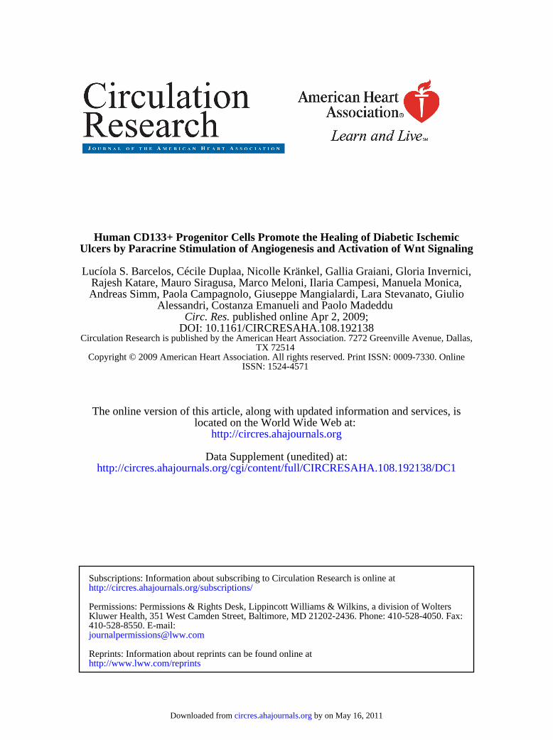
Next, to study the impact of cell or medium administrationon wound healing, we produced wounds and limb ischemiabilaterally. The right side was covered with plain collagen gelalone and served as internal control, whereas gel on the leftside wound contained either 2�104 CD133� or CD133� cellsor the vehicle. Transplantation of CD133� cells acceleratedthe rate of wound closure in streptozotocin-induced diabeticmice, whereas no effect was observed in groups givenCD133� cells or collagen as compared with the contralateralside (Figure 1A and Online Figure III). Two-way ANOVAdetected a treatment effect among groups (P�0.05), with nointeraction between treatment and time. In addition, Bonfer-roni post test analysis revealed an improved clinical outcomein the CD133� treatment group as compared with collagen or
2 Circulation Research May 8, 2009
by on May 16, 2011 circres.ahajournals.orgDownloaded from
CD133� cells. Neither cell therapy nor collagen acceleratedhindlimb hemodynamic recovery (data not shown).
Capillarization was increased at days 3 and 7 in woundstransplanted with CD133� cells as compared with CD133�
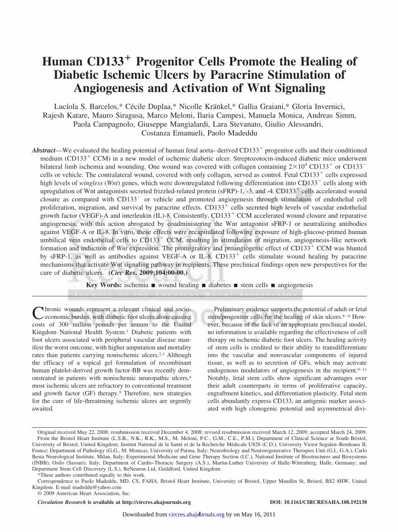
cell– or collagen-treated ulcers (Online Figure IV and Figure1B) but returned to levels comparable to CD133� cell orcollagen treatment at day 14 (Figure 1C). Furthermore, inulcers collected at 3 days from CD133� cell transplantation,a higher number of endothelial cells (ECs) stained positivefor the proliferation marker MCM-2, and a lower numbershowed apoptosis-associated TUNEL positivity, as comparedwith CD133� cell– or collagen-treated wounds (Figure 2Aand 2B). An increase in EC proliferation was still evidentin CD133� cell-transplanted ulcers collected a 7 days(0.81�0.04 versus 0.37�0.02 MCM-2 positive nuclei pervessel in CD133� cell–treated ulcers, P�0.05; Online FigureV, A and B). Immunohistochemistry studies also demon-strated an extremely thin endothelial lining of the formingblood vessels in the diabetic wounds, which is fragmental andexhibits gaps of all sizes with bleeding into the surroundingtissue, as indicated by the existence of numerous erythrocytesin the perivascular tissue. CD133� cell treatment improvesthis condition without completely omitting the gaps and theextreme stretching of many vessel walls.
CD133� Cells Mediate Wound Healing byParacrine MechanismsHuman cells derived from transplanted CD133� or CD133�
cells were rarely recognized in the wound granulation tissueharvested 3 days after transplantation (5.4�1.7 and 4.3�2.4cells/mm2, respectively, P�NS). Fluorescence-activated cell-
sorting (FACS) analysis, as well as quantitative PCR ofexcised wounds, confirmed that only low numbers of cellsremained in the wounds after 3 days, with no differencebetween cell groups (CD133� donor cells/wound: 34�9[FACS], 43�7 [quantitative PCR]; CD133� donor cells perwound: 36�11 [FACS], 18�6 [quantitative PCR]; P�NS).Immunohistochemistry indicated a drastic reduction in thenumber of human nuclear antigen-positive (hNA�) cells indiabetic wounds collected at later stages, namely at 7 daysposttransplantation (0.3�0.1 cells/mm2, P�0.05 versus day3), with no human cells being detectable at 14 days (OnlineFigure V, C and D). Furthermore, the rare hNA� cells werelocated at 2- to 5-cell-diameter distance from microvessels,indicating that these residual elements were not integrated inthe wound vasculature (Online Figure V, C). This led us tosuspect a paracrine mechanism underlying the supportiveeffect on wound healing and capillarization described above.
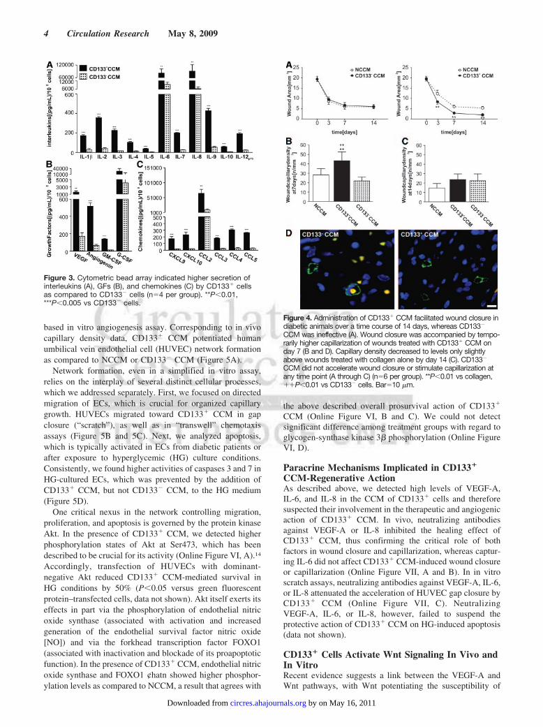
We have previously shown that CD133� cells producelarge amounts of VEGF-A.13 By cytokine bead array, weverified this and identified additional proangiogenic factors,secreted at higher levels by CD133� as compared to CD133�
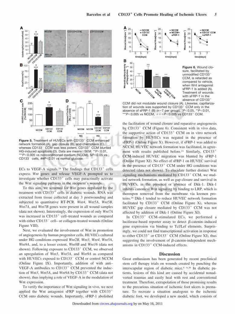
cells. Highest levels were detected for IL-6 and IL-8 amonginterleukins, VEGF-A and granulocyte-colony stimulatingfactor (G-CSF) among GF, and monocyte chemoattractantprotein-1 (CCL2) among chemokines (Figure 3). In accor-dance with those findings, administration of CD133� CCMinstead of cells supported wound closure, whereas CD133�
CCM was ineffective (Figure 4A). The healing action ofCD133� CCM was associated with increased wound vascu-larization at 7 days as compared with wounds given CD133�
CCM or NCCM (Figure 4B and 4D). However, no differencein capillary density was detected among groups at 14 days(Figure 4C).
CD133� CCM Promotes Endothelial CellMigration and Survival In VitroTo gain insight into the mechanisms underlying CD133�
CCM-induced capillarization, we first performed a Matrigel-
Figure 1. Accelerated wound closure in the presence ofCD133� cells (A, middle) is associated with higher temporarywound capillarization at day 7 (B). Capillary density declines tolevels of wounds treated with CD133� cells or collagen gelalone by day 14 (C) (n�12 mice per group). *P�0.05 vs colla-gen, �P�0.05 vs CD133� cells. Bar�20 �m.
Figure 2. Endothelial cell proliferation is stimulated in diabeticwounds by CD133� cells (A), whereas apoptosis is reduced (B).CD133� cells did not influence endothelial apoptosis or prolifer-ation (n�5 per group). *P�0.05 vs collagen, �P�0.05 vsCD133� cells. Bar�10 �m.
Barcelos et al CD133� Cells Promote Healing of Ischemic Ulcers 3
by on May 16, 2011 circres.ahajournals.orgDownloaded from
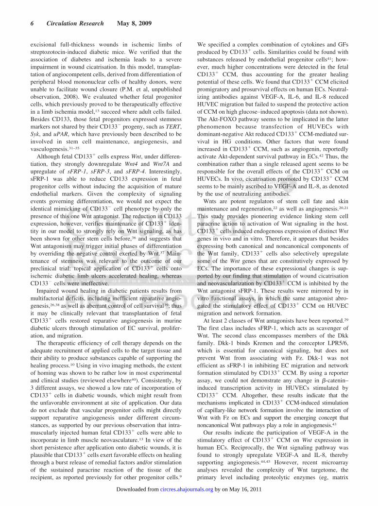
based in vitro angiogenesis assay. Corresponding to in vivocapillary density data, CD133� CCM potentiated humanumbilical vein endothelial cell (HUVEC) network formationas compared to NCCM or CD133� CCM (Figure 5A).
Network formation, even in a simplified in vitro assay,relies on the interplay of several distinct cellular processes,which we addressed separately. First, we focused on directedmigration of ECs, which is crucial for organized capillarygrowth. HUVECs migrated toward CD133� CCM in gapclosure (“scratch”), as well as in “transwell” chemotaxisassays (Figure 5B and 5C). Next, we analyzed apoptosis,which is typically activated in ECs from diabetic patients orafter exposure to hyperglycemic (HG) culture conditions.Consistently, we found higher activities of caspases 3 and 7 inHG-cultured ECs, which was prevented by the addition ofCD133� CCM, but not CD133� CCM, to the HG medium(Figure 5D).
One critical nexus in the network controlling migration,proliferation, and apoptosis is governed by the protein kinaseAkt. In the presence of CD133� CCM, we detected higherphosphorylation states of Akt at Ser473, which has beendescribed to be crucial for its activity (Online Figure VI, A).14
Accordingly, transfection of HUVECs with dominant-negative Akt reduced CD133� CCM-mediated survival inHG conditions by 50% (P�0.05 versus green fluorescentprotein–transfected cells, data not shown). Akt itself exerts itseffects in part via the phosphorylation of endothelial nitricoxide synthase (associated with activation and increasedgeneration of the endothelial survival factor nitric oxide[NO]) and via the forkhead transcription factor FOXO1(associated with inactivation and blockade of its proapoptoticfunction). In the presence of CD133� CCM, endothelial nitricoxide synthase and FOXO1 ¢hatn showed higher phosphor-ylation levels as compared to NCCM, a result that agrees with
the above described overall prosurvival action of CD133�
CCM (Online Figure VI, B and C). We could not detectsignificant difference among treatment groups with regard toglycogen-synthase kinase 3� phosphorylation (Online FigureVI, D).
Paracrine Mechanisms Implicated in CD133�
CCM-Regenerative ActionAs described above, we detected high levels of VEGF-A,IL-6, and IL-8 in the CCM of CD133� cells and thereforesuspected their involvement in the therapeutic and angiogenicaction of CD133� CCM. In vivo, neutralizing antibodiesagainst VEGF-A or IL-8 inhibited the healing effect ofCD133� CCM, thus confirming the critical role of bothfactors in wound closure and capillarization, whereas captur-ing IL-6 did not affect CD133� CCM-induced wound closureor capillarization (Online Figure VII, A and B). In in vitroscratch assays, neutralizing antibodies against VEGF-A, IL-6,or IL-8 attenuated the acceleration of HUVEC gap closure byCD133� CCM (Online Figure VII, C). NeutralizingVEGF-A, IL-6, or IL-8, however, failed to suspend theprotective action of CD133� CCM on HG-induced apoptosis(data not shown).
CD133� Cells Activate Wnt Signaling In Vivo andIn VitroRecent evidence suggests a link between the VEGF-A andWnt pathways, with Wnt potentiating the susceptibility of
Figure 4. Administration of CD133� CCM facilitated wound closure indiabetic animals over a time course of 14 days, whereas CD133�
CCM was ineffective (A). Wound closure was accompanied by tempo-rarily higher capillarization of wounds treated with CD133� CCM onday 7 (B and D). Capillary density decreased to levels only slightlyabove wounds treated with collagen alone by day 14 (C). CD133�
CCM did not accelerate wound closure or stimulate capillarization atany time point (A through C) (n�6 per group). **P�0.01 vs collagen,��P�0.01 vs CD133� cells. Bar�10 �m.
Figure 3. Cytometric bead array indicated higher secretion ofinterleukins (A), GFs (B), and chemokines (C) by CD133� cellsas compared to CD133� cells (n�4 per group). **P�0.01,***P�0.005 vs CD133� cells.
4 Circulation Research May 8, 2009
by on May 16, 2011 circres.ahajournals.orgDownloaded from
ECs to VEGF-A signals.28 The findings that CD133� cellsexpress Wnt genes and release VEGF-A prompted us toinvestigate whether CD133� cells may paracrinally activatethe Wnt signaling pathway in the recipient’s wounds.
To this aim, we screened for Wnt genes regulated by thetreatment with CD133� cells in diabetic wounds. RNA wasextracted from tissue collected at day 3 postwounding andsubjected to quantitative RT-PCR. Wnt4, Wnt5A, Wnt5B,Wnt7A, and Wnt7B genes were present in all wound samples(data not shown). Interestingly, the expression of only Wnt7Awas increased in CD133� cell–treated wounds as comparedwith either CD133� cell– or collagen-treated wounds (OnlineFigure VIII).
Next, we evaluated the involvement of Wnt in promotionof angiogenesis by human progenitor cells. HUVECs culturedunder HG conditions expressed Wnt2B, Wnt3, Wnt4, Wnt5A,Wnt9A, and, to a lesser extent, Wnt8B and Wnt16 (data notshown). Following exposure to CD133� CCM, we observedan upregulation of Wnt3, Wnt5A, and Wnt9A as comparedwith HUVECs exposed to CD133� CCM or control NCCM(Online Figure IX). Importantly, addition of with anti–VEGF-A antibodies to CD133� CCM prevented the induc-tion of Wnt3, Wnt5A, and Wnt9A by CD133� CCM (data notshown), thus implying a role of VEGF-A in the modulation ofWnt expression.
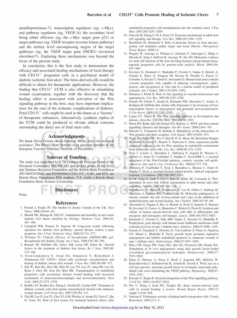
To verify the importance of Wnt signaling in vivo, we nextapplied the Wnt antagonist sFRP together with CD133�
CCM onto diabetic wounds. Importantly, sFRP-1 abolished
the facilitation of wound closure and reparative angiogenesisby CD133� CCM (Figure 6). Consistent with in vivo data,the supportive action of CD133� CCM on in vitro networkformation by HUVECs was negated in the presence ofsFRP-1 (Online Figure X). However, if sFRP-1 was added toNCCM, HUVEC network formation was facilitated, in agree-ment with results published before.21 Similarly, CD133�
CCM-induced HUVEC migration was blunted by sFRP-1(Online Figure XI). No effect of sFRP-1 on HUVEC survivalin the presence of CD133� CCM under HG conditions wasdetected (data not shown). To elucidate further distinct Wntsignaling mechanisms mediated by CD133� CCM, we stud-ied network formation, as well as gap closure and survival inHUVECs, in the presence or absence of Dkk-1. Dkk-1inhibits canonical Wnt signaling by binding to LRP, which isthereupon removed from the membrane via kremen pro-teins.29 Dkk-1 tended to reduce HUVEC network formationfacilitated by CD133� CCM (Online Figure X), whereasHUVEC gap closure mediated by CD133� CCM was notaffected by addition of Dkk-1 (Online Figure XI).
In CD133� CCM-stimulated ECs, we performed aluciferase-based reporter assay to detect �-catenin–inducedgene expression via binding to Tcf/Lef elements. Surpris-ingly, we could not find transcriptional activation in responseto either CD133� or CD133� CCM (Online Figure XI), thussuggesting the involvement of �-catenin-independent mech-anisms in CD133� CCM-induced effects.
DiscussionGreat enthusiasm has been generated by recent preclinicalstem cell therapy trials on wounds created by punching theinterscapular region of diabetic mice.6–8,30 In diabetic pa-tients, lesions of this kind are caused by accidental nonad-verted traumas and easily heal with rest and conventionaltreatment. Therefore, extrapolation of those promising resultsto the precarious situation of ischemic foot ulcers is prema-ture. To recreate a situation analogous to the ischemicdiabetic foot, we developed a new model, which consists of
Figure 5. Treatment of HUVECs with CD133� CCM enhancednetwork formation (A), gap closure (B), and chemotaxis (C),whereas CD133� CCM was less potent. CD133� CCM bluntedHG-induced apoptosis (D). Data are means�SEM. **P�0.01,***P�0.005 vs nonconditioned medium (NCCM), $P�0.05 vsCD133� cells, ##P�0.01 vs normal glucose.
Figure 6. Wound clo-sure, facilitated byunmodified CD133�
CCM, is retarded ascompared to vehicle,when Wnt antagonistsFRP-1 is added (A).Treatment of woundswith sFRP-1 in theabsence of CD133�
CCM did not modulate wound closure (A). Likewise, capillariza-tion of wounds was supported by CD133� CCM only in theabsence of sFRP-1 (B) (n�7 per group). *P�0.05, **P�0.01,***P�0.005 vs NCCM, ���P�0.005 vs CD133� CCM.
Barcelos et al CD133� Cells Promote Healing of Ischemic Ulcers 5
by on May 16, 2011 circres.ahajournals.orgDownloaded from
excisional full-thickness wounds in ischemic limbs ofstreptozotocin-induced diabetic mice. We verified that theassociation of diabetes and ischemia leads to a severeimpairment in wound cicatrisation. In this model, transplan-tation of angiocompetent cells, derived from differentiation ofperipheral blood mononuclear cells of healthy donors, wereunable to facilitate wound closure (P.M. et al, unpublishedobservation, 2008). We evaluated whether fetal progenitorcells, which previously proved to be therapeutically effectivein a limb ischemia model,13 succeed where adult cells failed.Besides CD133, those fetal progenitors expressed stemnessmarkers not shared by their CD133� progeny, such as TERT,Syk, and uPAR, which have previously been described to beinvolved in stem cell maintenance, angiogenesis, andvasculogenesis.31–35
Although fetal CD133� cells express Wnt, under differen-tiation, they strongly downregulate Wnt4 and Wnt7A andupregulate of sFRP-1, sFRP-3, and sFRP-4. Interestingly,sFRP-1 was able to reduce CD133 expression in fetalprogenitor cells without inducing the acquisition of matureendothelial markers. Given the complexity of signalingevents governing differentiation, we would not expect theidentical mimicking of CD133� cell phenotype by only thepresence of this one Wnt antagonist. The reduction in CD133expression, however, verifies maintenance of CD133� iden-tity in our model to strongly rely on Wnt signaling, as hasbeen shown for other stem cells before,36 and suggests thatWnt antagonism may trigger initial phases of differentiationby overriding the negative control exerted by Wnt.37 Main-tenance of stemness was relevant to the outcome of ourpreclinical trial: topical application of CD133� cells ontoischemic diabetic limb ulcers accelerated healing, whereasCD133� cells were ineffective.
Impaired wound healing in diabetic patients results frommultifactorial deficits, including inefficient reparative angio-genesis,26,38 as well as aberrant control of cell survival39; thusit may be clinically relevant that transplantation of fetalCD133� cells restored reparative angiogenesis in murinediabetic ulcers through stimulation of EC survival, prolifer-ation, and migration.
The therapeutic efficiency of cell therapy depends on theadequate recruitment of applied cells to the target tissue andtheir ability to produce substances capable of supporting thehealing process.10 Using in vivo imaging methods, the extentof homing was shown to be rather low in most experimentaland clinical studies (reviewed elsewhere40). Consistently, by3 different assays, we showed a low rate of incorporation ofCD133� cells in diabetic wounds, which might result fromthe unfavorable environment at site of application. Our datado not exclude that vascular progenitor cells might directlysupport reparative angiogenesis under different circum-stances, as supported by our previous observation that intra-muscularly injected human fetal CD133� cells were able toincorporate in limb muscle neovasculature.13 In view of theshort persistence after application onto diabetic wounds, it isplausible that CD133� cells exert favorable effects on healingthrough a burst release of remedial factors and/or stimulationof the sustained paracrine reaction of the tissue of therecipient, as reported previously for other progenitor cells.9
We specified a complex combination of cytokines and GFsproduced by CD133� cells. Similarities could be found withsubstances released by endothelial progenitor cells41; how-ever, much higher concentrations were detected in the fetalCD133� CCM, thus accounting for the greater healingpotential of these cells. We found that CD133� CCM elicitedpromigratory and prosurvival effects on human ECs. Neutral-izing antibodies against VEGF-A, IL-6, and IL-8 reducedHUVEC migration but failed to suspend the protective actionof CCM on high glucose–induced apoptosis (data not shown).The Akt-FOXO pathway seems to be implicated in the latterphenomenon because transfection of HUVECs withdominant-negative Akt reduced CD133� CCM-mediated sur-vival in HG conditions. Other factors that were foundincreased in CD133� CCM, such as angiogenin, reportedlyactivate Akt-dependent survival pathway in ECs.42 Thus, thecombination rather than a single released agent seems to beresponsible for the overall effects of the CD133� CCM onHUVECs. In vivo, cicatrisation promoted by CD133� CCMseems to be mainly ascribed to VEGF-A and IL-8, as denotedby the use of neutralizing antibodies.
Wnts are potent regulators of stem cell fate and skinmaintenance and regeneration,22 as well as angiogenesis.20,21
This study provides pioneering evidence linking stem cellparacrine action to activation of Wnt signaling in the host.CD133� cells induced endogenous expression of distinct Wntgenes in vivo and in vitro. Therefore, it appears that besidesexpressing both canonical and noncanonical components ofthe Wnt family, CD133� cells also selectively upregulatesome of the Wnt genes that are constitutively expressed byECs. The importance of these expressional changes is sup-ported by our finding that stimulation of wound cicatrisationand neovascularization by CD133� CCM is inhibited by theWnt antagonist sFRP-1. These results were mirrored by invitro functional assays, in which the same antagonist abro-gated the stimulatory effect of CD133� CCM on HUVECmigration and network formation.
At least 2 classes of Wnt antagonists have been reported.29
The first class includes sFRP-1, which acts as scavenger ofWnt. The second class encompasses members of the Dkkfamily. Dkk-1 binds Kremen and the coreceptor LPR5/6,which is essential for canonical signaling, but does notprevent Wnt from associating with Fz. Dkk-1 was notefficient as sFRP-1 in inhibiting EC migration and networkformation stimulated by CD133� CCM. By using a reporterassay, we could not demonstrate any change in �-catenin–induced transcription activity in HUVECs stimulated byCD133� CCM. Altogether, these results indicate that themechanisms implicated in CD133� CCM-induced stimulationof capillary-like network formation involve the interaction ofWnt with Fz on ECs and support the emerging concept thatnoncanonical Wnt pathways play a role in angiogenesis.43
Our results indicate the participation of VEGF-A in thestimulatory effect of CD133� CCM on Wnt expression inhuman ECs. Reciprocally, the Wnt signaling pathway wasfound to strongly upregulate VEGF-A and IL-8, therebysupporting angiogenesis.44,45 However, recent microarrayanalyses revealed the complexity of Wnt targetome, theprimary level including proteolytic enzymes (eg, matrix
6 Circulation Research May 8, 2009
by on May 16, 2011 circres.ahajournals.orgDownloaded from
metalloproteinase-7), transcription regulators (eg, c-Myc),and pathway regulators (eg, VEGF-A), the secondary levelbeing either effectors (eg, the c-Myc target gene p21) ortarget pathways (eg, VEGF receptor tyrosine kinase pathway)and the tertiary level encompassing targets of the targetpathways (eg, the VEGF target gene DSCR1) (reviewedelsewhere46). Exploring these mechanisms was beyond thefocus of the present study.
In conclusion, this is the first study to demonstrate theefficacy and associated healing mechanisms of local therapywith CD133� progenitor cells in a preclinical model ofdiabetic ischemic foot ulcer. The fetus-derived cells would bedifficult to obtain for therapeutic applications. However, thefinding that CD133� CCM is also effective in stimulatingwound cicatrisation, together with the discovery that thehealing effect is associated with activation of the Wntsignaling pathway in the host, may have important implica-tions for the cure of the ischemic complications of diabetes.Fetal CD133� cells might be used in the future as a “factory”of therapeutic substances. Alternatively, synthetic replica ofthe CCM could be produced to obviate ethical concernssurrounding the direct use of fetal stem cells.
AcknowledgmentsWe thank Silvia Cristini (Besta Institute, Milan, Italy) for technicalassistance. The Bristol Heart Institute is an associate member of theEuropean Vascular Genomic Network of Excellence.
Sources of FundingThe study was supported by a 7th Framework Program Grant of theEuropean Community (RESOLVE) and project grants of the Juve-nile Diabetes Research Foundation and the British Heart Foundation(PG 06/035/20641 and PG/06/096/21325). P.C., G.M., and M.S. areBritish Heart Foundation PhD students. C.E. holds a British HeartFoundation Basic Science Lectureship.
DisclosuresNone.
References1. Posnett J, Franks PJ. The burden of chronic wounds in the UK. Nurs
Times. 2008;104:44–45.2. Moulik PK, Mtonga R, Gill GV. Amputation and mortality in new-onset
diabetic foot ulcers stratified by etiology. Diabetes Care. 2003;26:491–494.
3. Campbell WB, Ponette D, Sugiono M. Long-term results followingoperation for diabetic foot problems: arterial disease confers a poorprognosis. Eur J Vasc Endovasc Surg. 2000;19:174–177.
4. Wieman TJ. Clinical efficacy of becaplermin (rhPDGF-BB) gel.Becaplermin Gel Studies Group. Am J Surg. 1998;176:74S–79S.
5. Bennett SP, Griffiths GD, Schor AM, Leese GP, Schor SL. Growthfactors in the treatment of diabetic foot ulcers. Br J Surg. 2003;90:133–146.
6. Sivan-Loukianova E, Awad OA, Stepanovic V, Bickenbach J,Schatteman GC. CD34� blood cells accelerate vascularization andhealing of diabetic mouse skin wounds. J Vasc Res. 2003;40:368–377.
7. Suh W, Kim KL, Kim JM, Shin IS, Lee YS, Lee JY, Jang HS, Lee JS,Byun J, Choi JH, Jeon ES, Kim DK. Transplantation of endothelialprogenitor cells accelerates dermal wound healing with increasedrecruitment of monocytes/macrophages and neovascularization. StemCells. 2005;23:1571–1578.
8. Badillo AT, Redden RA, Zhang L, Doolin EJ, Liechty KW. Treatment ofdiabetic wounds with fetal murine mesenchymal stromal cells enhanceswound closure. Cell Tissue Res. 2007;329:301–311.
9. Cho HJ, Lee N, Lee JY, Choi YJ, Ii M, Wecker A, Jeong JO, Curry C, QinG, Yoon YS. Role of host tissues for sustained humoral effects after
endothelial progenitor cell transplantation into the ischemic heart. J ExpMed. 2007;204:3257–3269.
10. Gnecchi M, Zhang Z, Ni A, Dzau VJ. Paracrine mechanisms in adult stemcell signaling and therapy. Circ Res. 2008;103:1204–1219.
11. Burchfield JS, Dimmeler S. Role of paracrine factors in stem and pro-genitor cell mediated cardiac repair and tissue fibrosis. FibrogenesisTissue Repair. 2008;1:4.
12. Wagner W, Ansorge A, Wirkner U, Eckstein V, Schwager C, Blake J,Miesala K, Selig J, Saffrich R, Ansorge W, Ho AD. Molecular evidencefor stem cell function of the slow-dividing fraction among human hema-topoietic progenitor cells by genome-wide analysis. Blood. 2004;104:675–686.
13. Invernici G, Emanueli C, Madeddu P, Cristini S, Gadau S, Benetti A,Ciusani E, Stassi G, Siragusa M, Nicosia R, Peschle C, Fascio U,Colombo A, Rizzuti T, Parati E, Alessandri G. Human fetal aorta containsvascular progenitor cells capable of inducing vasculogenesis, angio-genesis, and myogenesis in vitro and in a murine model of peripheralischemia. Am J Pathol. 2007;170:1879–1892.
14. Shiojima I, Walsh K. Role of Akt signaling in vascular homeostasis andangiogenesis. Circ Res. 2002;90:1243–1250.
15. Potente M, Urbich C, Sasaki K, Hofmann WK, Heeschen C, Aicher A,Kollipara R, DePinho RA, Zeiher AM, Dimmeler S. Involvement of Foxotranscription factors in angiogenesis and postnatal neovascularization.J Clin Invest. 2005;115:2382–2392.
16. Logan CY, Nusse R. The Wnt signaling pathway in development anddisease. Annu Rev Cell Dev Biol. 2004;20:781–810.
17. Moon RT, Kohn AD, De Ferrari GV, Kaykas A. WNT and beta-cateninsignalling: diseases and therapies. Nat Rev Genet. 2004;5:691–701.
18. Kikuchi A, Yamamoto H, Kishida S. Multiplicity of the interactions ofWnt proteins and their receptors. Cell Signal. 2007;19:659–671.
19. Wang H, Charles PC, Wu Y, Ren R, Pi X, Moser M, Barshishat-KupperM, Rubin JS, Perou C, Bautch V, Patterson C. Gene expression profilesignatures indicate a role for Wnt signaling in endothelial commitmentfrom embryonic stem cells. Circ Res. 2006;98:1331–1339.
20. Ezan J, Leroux L, Barandon L, Dufourcq P, Jaspard B, Moreau C,Allieres C, Daret D, Couffinhal T, Duplaa C. FrzA/sFRP-1, a secretedantagonist of the Wnt-Frizzled pathway, controls vascular cell prolif-eration in vitro and in vivo. Cardiovasc Res. 2004;63:731–738.
21. Dufourcq P, Couffinhal T, Ezan J, Barandon L, Moreau C, Daret D,Duplaa C. FrzA, a secreted frizzled related protein, induced angiogenicresponse. Circulation. 2002;106:3097–3103.
22. Ito M, Yang Z, Andl T, Cui C, Kim N, Millar SE, Cotsarelis G. Wnt-dependent de novo hair follicle regeneration in adult mouse skin afterwounding. Nature. 2007;447:316–320.
23. Stojadinovic O, Brem H, Vouthounis C, Lee B, Fallon J, Stallcup M,Merchant A, Galiano RD, Tomic-Canic M. Molecular pathogenesis ofchronic wounds: the role of beta-catenin and c-myc in the inhibition ofepithelialization and wound healing. Am J Pathol. 2005;167:59–69.
24. Alessandri G, Pagano S, Bez A, Benetti A, Pozzi S, Iannolo G, BaronioM, Invernici G, Caruso A, Muneretto C, Bisleri G, Parati E. Isolation andculture of human muscle-derived stem cells able to differentiate intomyogenic and neurogenic cell lineages. Lancet. 2004;364:1872–1883.
25. Emanueli C, Graiani G, Salis MB, Gadau S, Desortes E, Madeddu P.Prophylactic gene therapy with human tissue kallikrein ameliorates limbischemia recovery in type 1 diabetic mice. Diabetes. 2004;53:1096–1103.
26. Graiani G, Emanueli C, Desortes E, Van Linthout S, Pinna A, FigueroaCD, Manni L, Madeddu P. Nerve growth factor promotes reparativeangiogenesis and inhibits endothelial apoptosis in cutaneous wounds oftype 1 diabetic mice. Diabetologia. 2004;47:1047–1054.
27. Riley CM, Fuegy PW, Firpo MA, Shu XZ, Prestwich GD, Peattie RA.Stimulation of in vivo angiogenesis using dual growth factor-loadedcrosslinked glycosaminoglycan hydrogels. Biomaterials. 2006;27:5935–5943.
28. Klein D, Demory A, Peyre F, Kroll J, Augustin HG, Helfrich W,Kzhyshkowska J, Schledzewski K, Arnold B, Goerdt S. Wnt2 acts as acell type-specific, autocrine growth factor in rat hepatic sinusoidal endo-thelial cells cross-stimulating the VEGF pathway. Hepatology. 2008;47:1018–1031.
29. Kawano Y, Kypta R. Secreted antagonists of the Wnt signalling pathway.J Cell Sci. 2003;116:2627–2634.
30. Wu Y, Wang J, Scott PG, Tredget EE. Bone marrow-derived stemcells in wound healing: a review. Wound Repair Regen. 2007;15(suppl 1):S18–S26.
31. Natesan S. Telomerase extends a helping hand to progenitor cells. TrendsBiotechnol. 2005;23:1–3.
Barcelos et al CD133� Cells Promote Healing of Ischemic Ulcers 7
by on May 16, 2011 circres.ahajournals.orgDownloaded from
32. Yanagi S, Kurosaki T, Yamamura H. The structure and function ofnonreceptor tyrosine kinase p72syk expressed in hematopoietic cells. CellSignal. 1995;7:185–193.
33. Furuhata S, Ando K, Oki M, Aoki K, Ohnishi S, Aoyagi K, Sasaki H,Sakamoto H, Yoshida T, Ohnami S. Gene expression profiles of endo-thelial progenitor cells by oligonucleotide microarray analysis. Mol CellBiochem. 2007;298:125–138.
34. Lijnen HR. Plasmin and matrix metalloproteinases in vascularremodeling. Thromb Haemost. 2001;86:324–333.
35. Lacroix R, Sabatier F, Mialhe A, Basire A, Pannell R, Borghi H, RobertS, Lamy E, Plawinski L, Camoin-Jau L, Gurewich V, Angles-Cano E,Dignat-George F. Activation of plasminogen into plasmin at the surfaceof endothelial microparticles: a mechanism that modulates angiogenicproperties of endothelial progenitor cells in vitro. Blood. 2007;110:2432–2439.
36. Sato N, Meijer L, Skaltsounis L, Greengard P, Brivanlou AH. Mainte-nance of pluripotency in human and mouse embryonic stem cells throughactivation of Wnt signaling by a pharmacological GSK-3-specific inhib-itor. Nat Med. 2004;10:55–63.
37. Jacobsen SE. Defining ‘stemness’: Notch and Wnt join forces? NatImmunol. 2005;6:234–236.
38. Romano Di Peppe S, Mangoni A, Zambruno G, Spinetti G, Melillo G,Napolitano M, Capogrossi MC. Adenovirus-mediated VEGF(165) genetransfer enhances wound healing by promoting angiogenesis in CD1diabetic mice. Gene Ther. 2002;9:1271–1277.
39. Brem H, Tomic-Canic M. Cellular and molecular basis of wound healingin diabetes. J Clin Invest. 2007;117:1219–1222.
40. Chavakis E, Urbich C, Dimmeler S. Homing and engraftment of pro-genitor cells: a prerequisite for cell therapy. J Mol Cell Cardiol. 2008;45:514–522.
41. Urbich C, Aicher A, Heeschen C, Dernbach E, Hofmann WK, ZeiherAM, Dimmeler S. Soluble factors released by endothelial progenitor cellspromote migration of endothelial cells and cardiac resident progenitorcells. J Mol Cell Cardiol. 2005;39:733–742.
42. Kim HM, Kang DK, Kim HY, Kang SS, Chang SI. Angiogenin-inducedprotein kinase B/Akt activation is necessary for angiogenesis but isindependent of nuclear translocation of angiogenin in HUVE cells.Biochem Biophys Res Commun. 2007;352:509–513.
43. Masckauchan TN, Agalliu D, Vorontchikhina M, Ahn A, Parmalee NL,Li CM, Khoo A, Tycko B, Brown AM, Kitajewski J. Wnt5a signalinginduces proliferation and survival of endothelial cells in vitro andexpression of MMP-1 and Tie-2. Mol Biol Cell. 2006;17:5163–5172.
44. Masckauchan TN, Shawber CJ, Funahashi Y, Li CM, Kitajewski J.Wnt/beta-catenin signaling induces proliferation, survival andinterleukin-8 in human endothelial cells. Angiogenesis. 2005;8:43–51.
45. Zhang X, Gaspard JP, Chung DC. Regulation of vascular endothelialgrowth factor by the Wnt and K-ras pathways in colonic neoplasia.Cancer Res. 2001;61:6050–6054.
46. Vlad A, Rohrs S, Klein-Hitpass L, Muller O. The first five years of theWnt targetome. Cell Signal. 2008;20:795–802.
8 Circulation Research May 8, 2009
by on May 16, 2011 circres.ahajournals.orgDownloaded from
Supplemental Material Barcelos LS et al.
1
Online Supplementary Material
Human fetal cells
Aortas from 11- to 12-week-old human fetuses (n=15) were obtained according to the
ethical guidelines of the Network for European CNS Transplantation and Restoration
(NECTAR) as described before.1 The experimental protocol was approved by the ethics
committees of the National Neurological Institute "Carlo Besta" (Milan, Italy). CD133-
cells were generated from CD133+ cells by addition of 2% FBS (Gibco, N. Y., USA) to
the culture medium, as previously reported.1,2 Paired samples of CD133- and CD133+
were derived from the same fetal aorta preparation. Cell-conditioned medium (CCM) was
obtained from cultures of 200,000 cells/mL after 48 hours of incubation and kept frozen
in small aliquots at -80°C until use.
Animal procedures
All procedures complied with the standards stated in the Guide for the Care and Use of
Laboratory Animals (Institute of Laboratory Animal Resources, National Academy of
Sciences, Bethesda, Md, 1996) and were covered by ethical approvals from the Italian
Ministry of Health and the UK Home Office. Six to seven week old male CD1 mice
(Charles River Laboratories, Milan, Italy and Morgate, UK) were made diabetic by
streptozotocin (STZ, Sigma), as described.3 Persistence of glycosuria ≥10 g/L was
checked over the duration of the experiments.
Four weeks after diabetes induction, mice were depilated in their hindlimb area and
underwent bilateral hindlimb ischemia by ligature and electro-coagulation of the
by on May 16, 2011 circres.ahajournals.orgDownloaded from
Supplemental Material Barcelos LS et al.
2
proximal end of femoral arteries under anesthesia (Avertin, 880 mmol/kg, i.p., Sigma).1
At the same occasion, full thickness wounds were created in the thigh dorsal skin of both
legs using a sterile 5-mm-wide biopsy punch.4 The wounds were covered with type I
collagen (20 µl, Sigma) alone or collagen containing 2x10⁴ CD133+ or CD133- cells. In
separate experiments, wounds were covered with Extracel-HP hydrogel (Tebu-Bio, Le
Perray en Yvelines, France), which allows for controlled growth factor delivery,5
containing undiluted CD133- or CD133+ CCM (10µL) with or without the Wnt inhibitor
sFRP1 (5 µL, 1 µmol/L). In separate experiments, CD133+ CCM was applied onto ulcers
together with neutralizing antibodies against VEGF (5 µL, 1 µg/mL, R&D), interleukin 6
(IL-6) (5µL, 100ng/mL, R&D), or interleukin 8 (IL-8) (5µL, 10µg/mL, R&D).
Contralateral wounds were covered with hydrogel containing non-conditioned culture
medium (NCCM). After surgery, animals were maintained in individual cages with food
and water ad libitum and in a temperature and humidity-controlled environment.
Clinical outcome was established by determining the rate of wound closure.4 Laser
Doppler perfusion image analysis (Perimed, Stockholm, Sweden) was performed to
confirm and monitor limb ischemia.3
Flow cytometry
Antigenic characterization of fetal aorta-derived cells was carried out using a
FACS Calibur flow cytometer and the CellQuest software (BD Pharmingen). Cells were
incubated with phycoerytrin (PE)-conjugated anti-CD133 (Miltenyi), Allophycocyanin
(APC)-conjugated anti-CD117 (BD Pharmingen), APC-conjugated anti-CXCR4, or PE-
conjugated anti-CD146 or anti-CD31 monoclonal antibodies (R&D systems).
by on May 16, 2011 circres.ahajournals.orgDownloaded from

Supplemental Material Barcelos LS et al.
3
Fluorescence-conjugated, isotype-matched IgG with the same dye were used (BD
Pharmingen) was used as control.
Serum and sFRP-1 induced differentiation
Fetal aorta derived CD133+ were plated in triplicate in a 12 well plate (30,000
cells per well) with NeuroCult (Stem Cell Technologies, proliferation kit enriched with
EGF 20 ng/ml and FGF 10 ng/ml). After 24 hours, either sFRP-1 (6 µg/mL) or serum
(2% as a positive control for differentiation) or nothing was added to the medium. After 4
days of culture, pictures were taken in bright field microscope, cells were then detached
using TryPLE (Invitrogen) and stained for analysis of CD133 and CD31 expression using
a FACS Calibur flow cytometer.
Quantitative RT-PCR analysis for differential gene expression between CD133+ and
CD133- cells
Total RNA from undifferentiated and serum-differentiated fetal cells was isolated
with TRIzol reagent (Invitrogen, Carlsbad, CA, USA) according to the manufacturer’s
protocol. RNA from each sample was reverse transcribed with random hexamer primers
and Superscript III reverse transcriptase (Invitrogen) following the manufacturer’s
instructions.
The Human Angiogenesis RT² Profiler™ PCR Array was performed in triplicate
for each sample using a GeneAmp 5700 Sequence Detection System (Applied
Biosystems, Foster City, CA, USA). Data analysis is based on the ∆∆Ct method with
normalization of the raw data to housekeeping genes included in the array.
by on May 16, 2011 circres.ahajournals.orgDownloaded from
Supplemental Material Barcelos LS et al.
4
Multiplex cytokine analysis
The following cytokines were assayed by the use of the multiplex cytometric bead
array CBA flex, BD Biosciences, Heidelberg, Germany): angiogenin, interleukin-10 (IL-
10), macrophage inhibitory protein-1α (MIP-1 α/CCL3), interleukin-4 (IL-4), interleukin-
7 (IL-7), interleukin-1β (IL-1β), monocyte chemoattractant protein-1 (MCP-1/CCL2),
regulated upon activation normal T cell-expressed and -secreted cytokine
(RANTES/CCL5), interleukin-3 (IL-3), interferon gamma-induced protein 10 (IP-
10/CXCL10), interleukin-2 (IL-2), granulocyte colony-stimulating factor (G-CSF),
vascular endothelial growth factor-A (VEGF-A), interleukin-5 (IL-5), granulocyte-
macrophage colony-stimulating factor (GM-CSF), interleukin-8 (IL-8/CXCL8),
interleukin-12p70 (IL-12p70), monokine induced by interferon-gamma (MIG/CXCL9),
interleukin-6 (IL-6), macrophage inhibitory protein-1α (MIP-1 β/CCL4), and interleukin-
9 (IL-9). The test was performed and analyzed according to the manufacturer's
instructions. In brief, 50 µL of premixed capture beads were mixed with 50 µL PE-
detection reagent. After addition of 50 µL of the provided standards or sample (diluted
1:4) the mixture was incubated in the dark for 3 h at room temperature. After incubation
the mixture was washed, centrifuged (at 200x g for 5 min) and the pellet was resuspended
in 300 µL of wash buffer. The BD FACSCalibur flow cytometer was calibrated with
setup beads and 8,500 events were acquired for each sample. Individual analyte
concentrations were indicated by their fluorescence intensities (FL-2) and were computed
by using the respective standard reference curve and BD CBA software 1.1.
by on May 16, 2011 circres.ahajournals.orgDownloaded from
Supplemental Material Barcelos LS et al.
5
Histological assessment
At animal sacrifice by an overdose of anesthetic, wounds and surrounding skin
were removed and perpendicularly cut into two halves. One half was immediately frozen
for molecular biology studies while the other was fixed in 4% buffered formalin solution
and further processed for histology or immunohistochemical analyses.
Capillary profiles were recognized by immunohistochemical staining using rabbit
von Willebrand Factor (vWF, 1: 100, over night; Dako) followed by FITC- conjugated
donkey anti-rabbit IgG (1:20; Jackson Immunoresearch). Capillary density was
determined in a blind manner as described.6
Proliferating capillary endothelial cells were detected by co-staining for rabbit
MCM-2 (1:50; Santa Cruz), followed by TRITC- conjugated goat anti-rabbit IgG (1:50;
Sigma) and vWF, as described above. Detection of DNA strand breaks as a measure of
apoptosis was performed by using the TUNEL method and Biotin-16-dUTP followed by
streptavidin-TRITC secondary antibody together with staining for vWF, as described
above.
Detection of donor CD133+ and CD133
- cells in wounds
In paraffin-sections of wounds, human cell engraftment was identified using
mouse mAb against human nuclear antigen (hNA; 1:50; Chemicon) followed by FITC-
conjugated goat anti–mouse IgG (1:20, Sigma) staining.
Cultured human CD133+ and CD133- cells were stained with the red fluorescent
membrane tracer DiI and washed thoroughly immediately before transplantation. Two
days after transplantation, wounds were excised, cut into small pieces, washed in HBSS,
by on May 16, 2011 circres.ahajournals.orgDownloaded from

Supplemental Material Barcelos LS et al.
6
digested for 30min under agitation at 30°C in DMEM containing 0.2% collagenase II,
washed in PBS/5mM EDTA and digested for another 10min in PBS/2mMEDTA/ 0.25%
trypsin.7 The reaction was stopped by adding in excess PBS/10% FBS and cells spun
down at 300g for 10min. Cells were then resuspended in PBS/1%BSA and passed
through a 100µm filter. After another spin at 300g for 10min, single cells were fixed in
100µL PBS/0.5% paraformaldehyde. Before FACS data acquisition, 100µL of counting
beads were added to each sample according to the manufacturer’s instructions (Caltag).
Approximately 180µL of each sample were acquired in a FACS Canto II (BD). For data
analysis, counting beads were roughly identified by their small size in the FSC/SSC and
the 2 separate bead types (A- and B-beads) further defined by their different FL2-
fluorescence and side scatter (Online Figure XIII-A & B). Cells were first defined by
their FSC/SSC characteristics. DiI+ cells were then identified by their high FL2
fluorescence (Online Figure XIII-B). Using the known number of beads per total sample
(and therefore per wound), the number of DiI+ cells was calculated for each wound
(Online Figure XIII-C). Right side wounds which had not received any cells served as
negative controls.
Results of cell incorporation were confirmed by quantitative PCR for human genomic
DNA within the mouse wounds. Briefly, genomic DNA and total RNA were isolated
sequentially from excised murine wounds using the AllPrep DNA/RNA mini kit (Qiagen)
according to the manufacturer’s instructions. The amount of human gDNA within each
murine sample was detected by qPCR using specific primers against the most conserved
region of the human Alu sequence (Forward: 5’-TGA GGC AGG CGA ATC GCT TGA A-3’,
Reverse: 5’-GAC GGA GTT TCG CTC TTG TTG-3’). 45 cycles of 95°C (15s), 58°C (30s),
by on May 16, 2011 circres.ahajournals.orgDownloaded from

Supplemental Material Barcelos LS et al.
7
and 72°C(15s) were run in a LightCycler 480 (Roche) with fluorescence acquisition after
each cycle. Quantification of human DNA in mouse tissue was based on a standard curve
using serial dilution of human CD133- cell gDNA equivalents, spiked with mouse gDNA,
calculated according to assumption that one human cell contains 6.6 pg of gDNA. Using
serial dilution, the method was tested to evaluate sensitivity up to one cell.
Cell culture
Human umbilical vein endothelial cells (HUVEC) were grown in EGM-2,
containing 2% FBS (all Cambrex) and used between passages 3 and 6.
Functional assays on cultured HUVEC
Apoptosis assay. Subconfluent HUVEC (Cambrex) were harvested with
trypsin/EDTA, seeded into 6-well collagen-coated plates at 4x10⁵ cells / well and
incubated overnight to allow adhesion. Adherent cells were then incubated under high
glucose concentration (30mM) in EBM-2 (Cambrex) for 72 h. HUVEC grown under
normal glucose concentration (5.55 mM) were used as basal control. The effect of
NCCM, CD133+ or CD133- CCM (diluted 1:3) on high glucose-induced endothelial cell
apoptosis was examined by the Caspase-Glo® 3/7 assay (Promega).
Migration assays. HUVEC migration towards CCM of CD133+ or CD133- or
NCCM was assayed with a modified 96-well Boyden chamber (Neuro Probe) using an 8-
µm pore-size polyvinylpyrrolidone-free polycarbonate membrane (Neuro Probe). The
membrane of a Boyden chamber was precoated with type I collagen (10 µg/mL in PBS,
Sigma) at room temperature for 1 h or with fibronectin (100µg/mL in PBS, Sigma) at 4°C
overnight, respectively, and then washed with PBS. Subconfluent HUVEC were starved
by on May 16, 2011 circres.ahajournals.orgDownloaded from
Supplemental Material Barcelos LS et al.
8
in EBM-2 (Cambrex), respectively, under high glucose (30mM) for 24 h. HUVEC were
detached with 0.02% PBS/EDTA, resuspended in EBM-2 (Cambrex), respectively, and
then placed in the upper chamber at 5,000 cells/well. The stimuli (NCCM, CD133+ or
CD133- CCM (diluted 1:5 in EBM-2) were added to the lower chamber. During all the
starvation and experimental period, HUVEC were incubated in EBM-2 containing 0.1%
FBS. The chambers were maintained at 37°C and 5% CO2 for 4 h. The upper surface
adherent cells were removed by scrapping and the lower adherent cells were considered
to be migrated. The membranes were fixed with 100% methanol at room temperature for
5 minutes and then mounted in glass slides with Vectashield® mounting medium with
DAPI (Vector Laboratories). The numbers of stained nuclei were counted in five high-
power fields per each well under a fluorescence microscope.
In addition, the ability of CCM to stimulate HUVEC migration was evaluated in
the scratch assay. HUVEC grown to a confluent monolayer in 100µg/mL fibronectin /
0.5% gelatin-coated 8-well slide chambers were starved in EBM-2 containing 0.1% FBS
(Cambrex) under high glucose concentration (30mM) for 24 h. A central scratch was
created by scraping cells away with a p1000 pipette tip. After removal of debris by
washing the wells with DPBS, cells were incubated with EBM-2 containing 2mM of
hydroxyurea (Sigma) to induce growth arrest in the presence of 1:5 diluted NCCM,
CD133+ or CD133- CCM. After 24h of incubation, cells were washed, fixed with
methanol and subjected to hematoxylin/eosin staining. Scratches were photographed at
4x magnification at 25%, 50% and 75% of the scratch length and distance between
migrating fronts was measured using the Image Pro Plus software (Media Cybernetics).
Scratches fixed and stained immediately after scratching served to calculate initial gap
by on May 16, 2011 circres.ahajournals.orgDownloaded from

Supplemental Material Barcelos LS et al.
9
width (0% closure). Briefly, length was calibrated using a 1mm size standard
photographed in the same camera-microscope-setting. Migrating fronts were traced and
average distances were calculated by the software (Online Figure XIV). Each condition
was run in quadruplicate and the assay was repeated three times.
In vitro angiogenesis assay. Subconfluent HUVEC were incubated overnight with
EGM-2 (Cambrex) plus 2% FBS containing NCCM, CD133+ or CD133- CCM diluted
1:5 and then detached with trypsin/EDTA and resuspended in EBM-2 (Cambrex) plus
0.1% FBS containing NCCM or CCM diluted 1:5. The formation of network structures
was assessed using the growth factor reduced Matrigel™ (BD Biosciences) thick gel
method according to the manufacturer’s instructions. HUVEC were seeded at 3x10⁴ cells
/ well in 8-well slide chambers containing 1:5 diluted NCCM, CD133+ or CD133- CCM
in 100µL of Matrigel. The chambers were incubated at 37°C and 5% CO2 overnight. The
wells were then photographed under a phase-contrast inverted microscope at 4x and 10x
magnification. For each condition, network extension was measured using the Image Pro-
Plus software (Media Cybernetics) as recently described.8 Each condition was run in
quadruplicate and the assay was repeated two times.
Inhibition of signaling pathways. The involvement of Wnt in the effects exerted
by CD133+ CCM in HUVEC functional assays was evaluated by the use of the Wnt
inhibitors sFRP-1 (recombinant bovine, 10 nmol/L; from Cecile Duplaa) or Dkk-1
(recombinant human, 250ng/mL; R&D Systems). NCCM, CD133+ or CD133- CCM
(diluted 1:5 in EBM-2) was mixed with sFRP-1 or vehicle 30min prior to the addition of
the mixture to stimulate HUVEC in tube formation, migration and apoptosis assays. In
by on May 16, 2011 circres.ahajournals.orgDownloaded from

Supplemental Material Barcelos LS et al.
10
separate experiments, HUVEC were pre-incubated with Dkk1 for 1h before adding
NCCM, CD133+ or CD133- CCM (diluted 1:5) as a stimulus.
In separate experiments of apoptosis and migration, NCCM, CD133+ or CD133-
CCM (diluted 1:5 in EBM-2) was mixed with anti-VEGF-A (500ng/mL), anti-IL-6
(20µg/mL) or anti-IL-8 (15µg/mL) neutralizing antibodies (R&D System) 30min prior to
stimulation of HUVEC. Antibody final concentrations were 8- to 25-fold greater than the
concentrations for 100% inhibition of the highest values of those cytokines in CCM.
In order to determine the involvement of Akt in the pro-survival action of CD133+
CCM, HUVEC were infected with Ad.DN-Akt before stimulation. In brief, HUVEC
were seeded into white-walled 96-well culture plates at 3.5x10³ cells / well and, after
overnight incubation to allow adhesion, cells were infected with either Ad.Lac-Z or
Ad.DN-Akt at MOI of 250. After 6 hours, virus was removed and stimuli (NCCM,
CD133+ or CD133-CCM) were added in the presence of high glucose (30mM) in EBM-2
containing 0.1% FBS (Cambrex) for 72 h. HUVEC grown under normal glucose
concentration (5.55 mM) were used as basal control.
Western Blot analysis
Subconfluent HUVEC were harvested with trypsin/EDTA, seeded into 6-well
culture plates at 5x10⁵ cells/mL and incubated overnight to allow adhesion. Adherent
cells were then starved in EBM-2 (Cambrex) under high glucose concentration (30mM)
for 24 h and then stimulated with NCCM, CD133+ or CD133- CCM diluted 1:5 in EBM-2
as indicated. Cells were washed with PBS and immediately resuspended in lysis buffer
(50 mM Hepes pH 7.5, 150 mM NaCl, 1 mM EDTA, 1 mM EGTA, 25 mM NaF, 5mM
by on May 16, 2011 circres.ahajournals.orgDownloaded from
Supplemental Material Barcelos LS et al.
11
NaPPi, 1% Triton X-100, 1% NP-40, 0.25% sodium deoxycholate, protease and
phosphatase inhibitors) on ice. Protein whole extracts (50µg) were separated by SDS-
PAGE and then transferred to PVDF membrane (Bio-Rad Laboratories). Membranes
were probed with primary antibodies for phospho-Akt, total Akt, phospho-GSK3β, total
GSK3β, phospho-eNOS, phospho-FOXO1, tubulin (all from Cell Signaling Technology),
total eNOS (Santa Cruz Biotechnology) and total FOXO1 (Upstate), followed by
secondary antibody horseradish peroxidase conjugated-anti-rabbit IgG (Amersham
Bioscience; 1:5000).
Quantitative RT-PCR for Wnt gene expression.
Total RNAs was isolated using Tri Reagent (Euromedex) and DNase (Promega)
treatment according to manufacturer’s instructions. 500 ng of total RNA were reverse-
transcribed using M-MLV Reverse Transcriptase (Promega) as previously described.9
PCR was done using IQ SYBR Green supermix (Bio-Rad). An MJ Research Opticon and
the following parameters were used for real time PCR: 95°C for 5 min followed by 35
cycles of 95°C for 15 s, 60°C for 20 s and 72°C for 15 s. Negative controls without RT
were prepared in parallel for each RNA sample. All experiments were done in triplicate
and target mRNA levels were normalized to expression of β actin in each sample and
expressed as fold of indicated control samples (∆∆Ct method). Primers used were:
Human primers:
h Wnt2B F 5’- GCCTCTCAACTCAAAAGCAC -3’
R 5’- TCTCCAGAGCGGGAAATCAG -3’
h Wnt3 F 5’- AACTTTTGTGAGCCCAACC -3’
by on May 16, 2011 circres.ahajournals.orgDownloaded from

Supplemental Material Barcelos LS et al.
12
R 5’- CGTAGATGCGAATACACTCC -3’
h Wnt4 F 5’- AGGATGCTCTGACAACATCG -3’
R 5’- TTACCTCACAGGAGCCTGAC -3’
h Wnt5A F 5’- AGCATCAGTCCACAAACAC -3’
R 5’- TCACCATTCCACAGAGAGAG -3’
h Wnt 7A F 5’- GCAGTGCAACTGTAAGTTCC -3’
R 5’- CCTCAGCAGAAAAGACAAGC -3’
h Wnt 7B F 5’- CCTGGATCATGCACAGAAAC -3’
R 5’- CCTCCCCAATCACAATGATG -3’
h Wnt8B F 5’- TCTCTTCTGCACAGCTCCTC -3’
R 5’- TGGTGTTCAAGTTAGCACTCC -3’
h Wnt9A F 5’- ACAGCAGCAAGTTCGTCAAG -3’
R 5’- TTGCCCACCTCATGGAAAGG -3’
h Wnt10A F 5’- CACACCCTAAAACAAGCCTC -3’
R 5’- GAATGATGAAGGGAATGGTGG -3’
h Wnt16 F 5’- TGGAGAGGTGTGAGTGTAAG -3’
R 5’- ATTCCACTGCAAGAGTCAAC -3’
h sFRP1 F 5’- TGCCCCTGCTCAACAAGAAC -3’
R 5’- AAGCCGAAGAACTGCATGAC -3’
h sFRP3 F 5’- AGCAGTGAACGCTGTAAATG -3’
R 5’- AATCTCCTTCACCTCCACTAC -3’
h sFRP4 F 5’- GTAATCCCCCCAAACCAAAG -3’
by on May 16, 2011 circres.ahajournals.orgDownloaded from
Supplemental Material Barcelos LS et al.
13
R 5’- TGTAAGGAAGTCGGAAGTCTC -3’
h cyclinD1 F 5’- CACACACACACACACACAC -3’
R 5’- GCCAAACAGGCTGAATCAATG -3’
Murine primers:
m Wnt4 F 5’- TTCACAACAACGAGGCTGGCAG -3’
R 5’- CACCGTCAAACTTCTCCTTTAGCG -3’
m wnt5A F 5’- GGCATCAAGGAATGCCAGTA -3’
R 5’- GTACGTGAAGGCCGTCTCTC -3’
m Wnt7A F 5’- GCTCCTATCCTTTTGCCCTTTACAG -3’
R 5’- GCCTCTTATCCAGTGGTTCACG -3’
m Wnt7B F 5’- AAGATTTACTGGAGACCCCACGGC -3’
R 5’- CAGAAGAAGGACAAAACCCAAGG -3’
β actin F 5’- GTTCCGATGCCCGAGGCTCT -3’
R 5’- GCATTTGCGGTGCACGATGGA -3’
Luciferase reporter assay
For TOPFLASH reporter assays, MS1 cells were transfected in 24-well plates at
2x10⁵ cells/cm2 density with Super8XTOPFLASH luciferase reporter plasmid (375
ng/well) containing LEF/TCF consensus binding sites (Randal T. Moon laboratory).
pRL-CMV (Promega) was cotransfected (90 ng/well) to normalize samples for
transfection efficiency (Dual luciferase kit, Promega). Transfections were performed with
Lipofectamine 2000 (Invitrogen) reagents according to the manufacturers'
recommendations. After 24 hours, the medium was replaced with non conditioned
by on May 16, 2011 circres.ahajournals.orgDownloaded from

Supplemental Material Barcelos LS et al.
14
medium or with conditioned media obtained either from CD133+ or CD133- cells diluted
1:5 with medium containing 1% FCS and 0.5% BSA. Cells were harvested 24 h later, and
luciferase activity was determined with a Turner Designs luminometer. The data are
presented as the means ± SD of triplicate well measurements for one representative
experiment.
Statistical analysis
Results are presented as the mean ± SEM. Wound closure data were analyzed
using two-way ANOVA and the interaction between treatment and time was considered,
followed by Bonferroni posttest analysis. For other data sets, comparisons were carried
out using one-way analysis of variance (ANOVA) and differences between groups
assessed using the Newman-Keuls post-test. A p-value less than 0.05 was considered
significant.
by on May 16, 2011 circres.ahajournals.orgDownloaded from
Supplemental Material Barcelos LS et al.
15
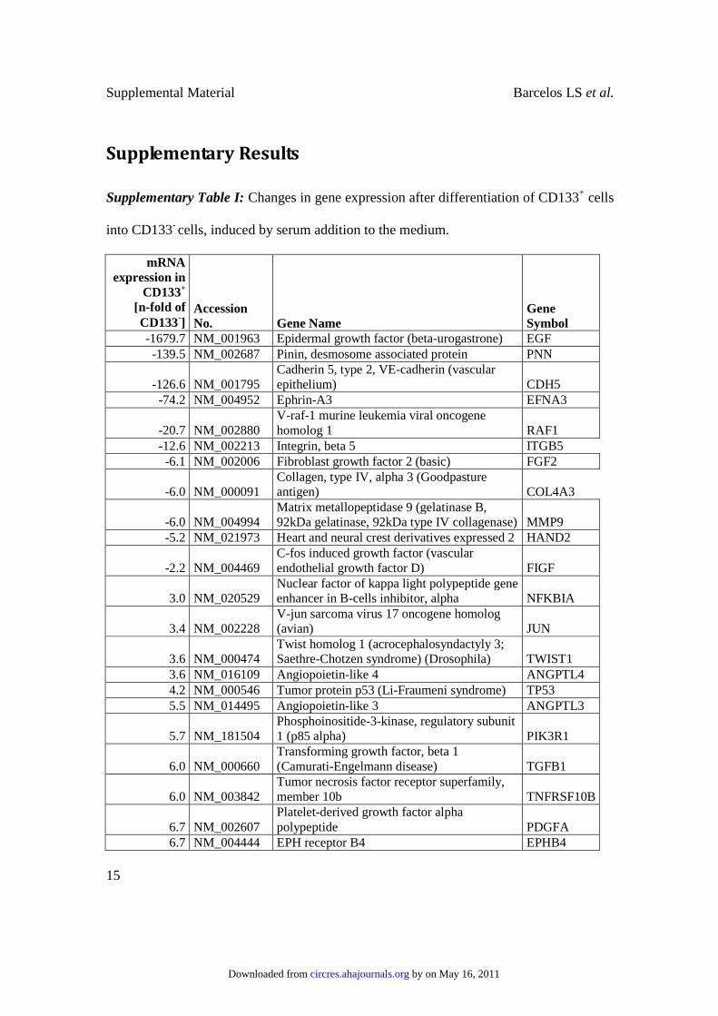
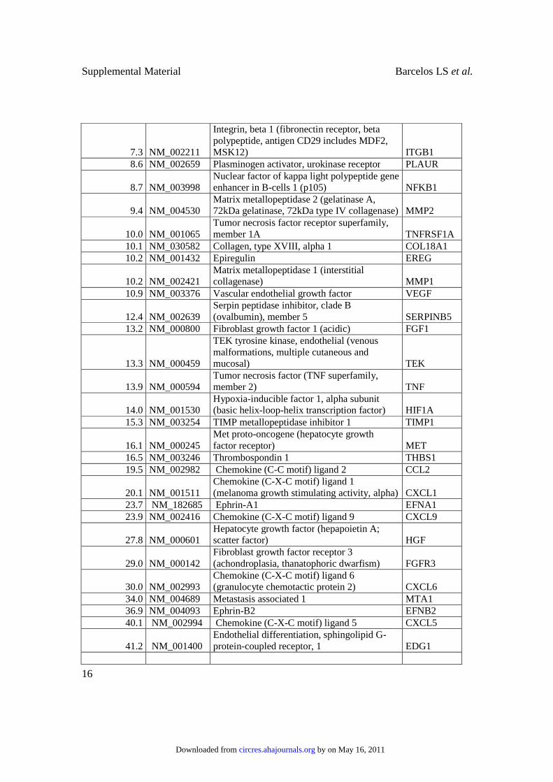
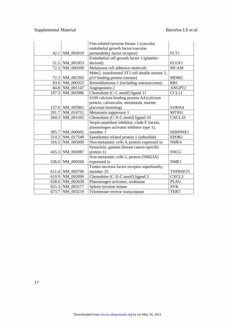
Supplementary Results
Supplementary Table I: Changes in gene expression after differentiation of CD133+ cells
into CD133- cells, induced by serum addition to the medium.
mRNA
expression in
CD133+
[n-fold of
CD133-]
Accession
No. Gene Name
Gene
Symbol
-1679.7 NM_001963 Epidermal growth factor (beta-urogastrone) EGF
-139.5 NM_002687 Pinin, desmosome associated protein PNN
-126.6 NM_001795 Cadherin 5, type 2, VE-cadherin (vascular epithelium) CDH5
-74.2 NM_004952 Ephrin-A3 EFNA3
-20.7 NM_002880 V-raf-1 murine leukemia viral oncogene homolog 1 RAF1
-12.6 NM_002213 Integrin, beta 5 ITGB5
-6.1 NM_002006 Fibroblast growth factor 2 (basic) FGF2
-6.0 NM_000091 Collagen, type IV, alpha 3 (Goodpasture antigen) COL4A3
-6.0 NM_004994 Matrix metallopeptidase 9 (gelatinase B, 92kDa gelatinase, 92kDa type IV collagenase) MMP9
-5.2 NM_021973 Heart and neural crest derivatives expressed 2 HAND2
-2.2 NM_004469 C-fos induced growth factor (vascular endothelial growth factor D) FIGF
3.0 NM_020529 Nuclear factor of kappa light polypeptide gene enhancer in B-cells inhibitor, alpha NFKBIA
3.4 NM_002228 V-jun sarcoma virus 17 oncogene homolog (avian) JUN
3.6 NM_000474 Twist homolog 1 (acrocephalosyndactyly 3; Saethre-Chotzen syndrome) (Drosophila) TWIST1
3.6 NM_016109 Angiopoietin-like 4 ANGPTL4
4.2 NM_000546 Tumor protein p53 (Li-Fraumeni syndrome) TP53
5.5 NM_014495 Angiopoietin-like 3 ANGPTL3
5.7 NM_181504 Phosphoinositide-3-kinase, regulatory subunit 1 (p85 alpha) PIK3R1
6.0 NM_000660 Transforming growth factor, beta 1 (Camurati-Engelmann disease) TGFB1
6.0 NM_003842 Tumor necrosis factor receptor superfamily, member 10b TNFRSF10B
6.7 NM_002607 Platelet-derived growth factor alpha polypeptide PDGFA
6.7 NM_004444 EPH receptor B4 EPHB4
by on May 16, 2011 circres.ahajournals.orgDownloaded from
Supplemental Material Barcelos LS et al.
16
7.3 NM_002211
Integrin, beta 1 (fibronectin receptor, beta polypeptide, antigen CD29 includes MDF2, MSK12) ITGB1
8.6 NM_002659 Plasminogen activator, urokinase receptor PLAUR
8.7 NM_003998 Nuclear factor of kappa light polypeptide gene enhancer in B-cells 1 (p105) NFKB1
9.4 NM_004530 Matrix metallopeptidase 2 (gelatinase A, 72kDa gelatinase, 72kDa type IV collagenase) MMP2
10.0 NM_001065 Tumor necrosis factor receptor superfamily, member 1A TNFRSF1A
10.1 NM_030582 Collagen, type XVIII, alpha 1 COL18A1
10.2 NM_001432 Epiregulin EREG
10.2 NM_002421 Matrix metallopeptidase 1 (interstitial collagenase) MMP1
10.9 NM_003376 Vascular endothelial growth factor VEGF
12.4 NM_002639 Serpin peptidase inhibitor, clade B (ovalbumin), member 5 SERPINB5
13.2 NM_000800 Fibroblast growth factor 1 (acidic) FGF1
13.3 NM_000459
TEK tyrosine kinase, endothelial (venous malformations, multiple cutaneous and mucosal) TEK
13.9 NM_000594 Tumor necrosis factor (TNF superfamily, member 2) TNF
14.0 NM_001530 Hypoxia-inducible factor 1, alpha subunit (basic helix-loop-helix transcription factor) HIF1A
15.3 NM_003254 TIMP metallopeptidase inhibitor 1 TIMP1
16.1 NM_000245 Met proto-oncogene (hepatocyte growth factor receptor) MET
16.5 NM_003246 Thrombospondin 1 THBS1
19.5 NM_002982 Chemokine (C-C motif) ligand 2 CCL2
20.1 NM_001511 Chemokine (C-X-C motif) ligand 1 (melanoma growth stimulating activity, alpha) CXCL1
23.7 NM_182685 Ephrin-A1 EFNA1
23.9 NM_002416 Chemokine (C-X-C motif) ligand 9 CXCL9
27.8 NM_000601 Hepatocyte growth factor (hepapoietin A; scatter factor) HGF
29.0 NM_000142 Fibroblast growth factor receptor 3 (achondroplasia, thanatophoric dwarfism) FGFR3
30.0 NM_002993 Chemokine (C-X-C motif) ligand 6 (granulocyte chemotactic protein 2) CXCL6
34.0 NM_004689 Metastasis associated 1 MTA1
36.9 NM_004093 Ephrin-B2 EFNB2
40.1 NM_002994 Chemokine (C-X-C motif) ligand 5 CXCL5
41.2 NM_001400 Endothelial differentiation, sphingolipid G-protein-coupled receptor, 1 EDG1
by on May 16, 2011 circres.ahajournals.orgDownloaded from
Supplemental Material Barcelos LS et al.
17
42.1 NM_002019
Fms-related tyrosine kinase 1 (vascular endothelial growth factor/vascular permeability factor receptor) FLT1
51.5 NM_001953 Endothelial cell growth factor 1 (platelet-derived) ECGF1
72.3 NM_006500 Melanoma cell adhesion molecule MCAM
72.3 NM_002392 Mdm2, transformed 3T3 cell double minute 2, p53 binding protein (mouse) MDM2
83.6 NM_000321 Retinoblastoma 1 (including osteosarcoma) RB1
84.8 NM_001147 Angiopoietin 2 ANGPT2
107.3 NM_002986 Chemokine (C-C motif) ligand 11 CCL11
127.6 NM_002961
S100 calcium binding protein A4 (calcium protein, calvasculin, metastasin, murine placental homolog) S100A4
201.7 NM_014751 Metastasis suppressor 1 MTSS1
264.3 NM_001565 Chemokine (C-X-C motif) ligand 10 CXCL10
305.7 NM_000602
Serpin peptidase inhibitor, clade E (nexin, plasminogen activator inhibitor type 1), member 1 SERPINE1
314.3 NM_017549 Ependymin related protein 1 (zebrafish) EPDR1
316.5 NM_005009 Non-metastatic cells 4, protein expressed in NME4
435.3 NM_003087 Synuclein, gamma (breast cancer-specific protein 1) SNCG
536.0 NM_000269 Non-metastatic cells 1, protein (NM23A) expressed in NME1
611.4 NM_003790 Tumor necrosis factor receptor superfamily, member 25 TNFRSF25
619.9 NM_002090 Chemokine (C-X-C motif) ligand 3 CXCL3
628.6 NM_002658 Plasminogen activator, urokinase PLAU
655.3 NM_003177 Spleen tyrosine kinase SYK
673.7 NM_003219 Telomerase reverse transcriptase TERT
by on May 16, 2011 circres.ahajournals.orgDownloaded from
Supplemental Material Barcelos LS et al.
18
Online Figure Legends
Online Figure I: Characterization of studied cell populations (CD133+ and CD133- cells)
verified loss of stem/progenitor associated markers (CD133, CD117, CXCR4) during
serum-induced differentiation of CD133+ cells into CD133- cells (A & B). Furthermore
RT-PCR revealed that Wnt genes, highly expressed in CD133+ cells, were downregulated
in CD133- cells, while expression of Wnt antagonists sFRPs was induced (C). Addition
of sFRP-1 to CD133+ culture medium resulted in the reduction of CD133 expression (D).
Data are expressed as mean±SEM, n=3 per group; * P<0.05 vs. CD133+ cells. Grey areas
in B and D mark the negative range determined by isotype control staining.
Online Figure II: Ischemia impedes wound closure in diabetic mice. Data are expressed
as mean±SEM, n=10 per group; ***P<0.005 vs. non-ischemic.
Online Figure III: Photographic documentation of wounds covered by collagen gel
alone, or collagen gel containing CD133+ or CD133- cells at days 0, 7 and 14.
Online Figure IV: CD133+ cells improve neovascularization in diabetic ischemic ulcers
collected at day 3 post-transplantation. Representative immunohistochemical images
showing higher abundance of von Willebrand factor (vWF) positive endothelial cells in
wounds treated with CD133+ cells as compared with CD133- cells or collagen. The bar
length corresponds to 20 µm.
Online Figure V: Representative micro-photographs showing the effect of CD133+ (A)
and CD133- cells (B) on endothelial cell proliferation, as assessed by co-staining for
MCM-2 (red fluorescence) and vWF (green fluorescence), in wounds harvested at 7 days
from cell transplantation. Human cells identified by staining for human nuclear antigen
by on May 16, 2011 circres.ahajournals.orgDownloaded from
Supplemental Material Barcelos LS et al.
19
(purple fluorescence) was rarely documented at 7 (C) but not at 14 days (D) post-
transplantation of CD133+ cells. Vessels were surrounded by erythrocytes (yellow
fluorescence). Bar corresponds to 10µm.
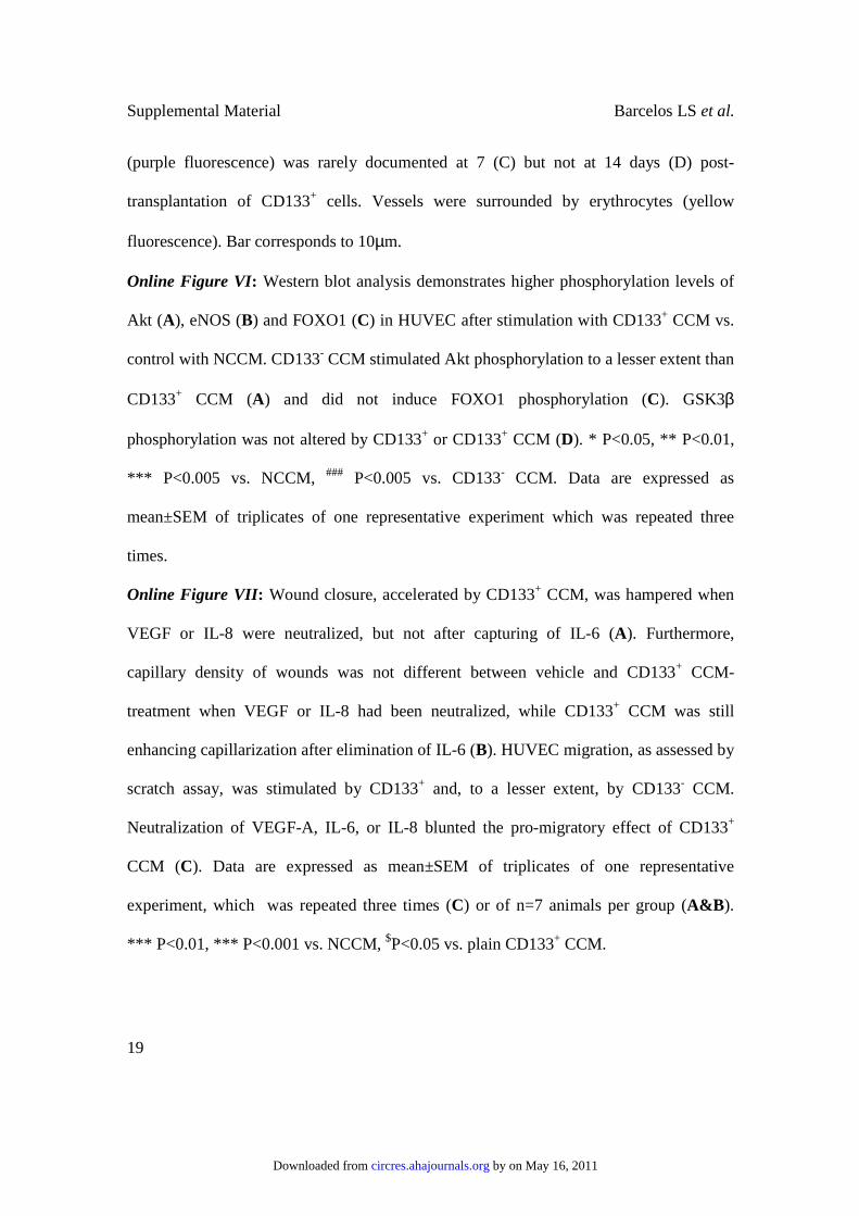
Online Figure VI: Western blot analysis demonstrates higher phosphorylation levels of
Akt (A), eNOS (B) and FOXO1 (C) in HUVEC after stimulation with CD133+ CCM vs.
control with NCCM. CD133- CCM stimulated Akt phosphorylation to a lesser extent than
CD133+ CCM (A) and did not induce FOXO1 phosphorylation (C). GSK3β
phosphorylation was not altered by CD133+ or CD133+ CCM (D). * P<0.05, ** P<0.01,
*** P<0.005 vs. NCCM, ### P<0.005 vs. CD133- CCM. Data are expressed as
mean±SEM of triplicates of one representative experiment which was repeated three
times.
Online Figure VII: Wound closure, accelerated by CD133+ CCM, was hampered when
VEGF or IL-8 were neutralized, but not after capturing of IL-6 (A). Furthermore,
capillary density of wounds was not different between vehicle and CD133+ CCM-
treatment when VEGF or IL-8 had been neutralized, while CD133+ CCM was still
enhancing capillarization after elimination of IL-6 (B). HUVEC migration, as assessed by
scratch assay, was stimulated by CD133+ and, to a lesser extent, by CD133- CCM.
Neutralization of VEGF-A, IL-6, or IL-8 blunted the pro-migratory effect of CD133+
CCM (C). Data are expressed as mean±SEM of triplicates of one representative
experiment, which was repeated three times (C) or of n=7 animals per group (A&B).
*** P<0.01, *** P<0.001 vs. NCCM, $P<0.05 vs. plain CD133+ CCM.
by on May 16, 2011 circres.ahajournals.orgDownloaded from
Supplemental Material Barcelos LS et al.
20
Online Figure VIII: Quantitative RT-PCR revealed increased Wnt7A mRNA levels in
wounds treated with CD133+ CCM. Data are expressed as mean±SEM, n=5 per group.*
P<0.05 vs. collagen, $ P<0.05 vs. CD133+ cells.
Online Figure IX: Culture of HUVEC in the presence of CD133+ CCM increased
expression of Wnt3, Wnt5A and Wnt9A, while CD133- CCM did not influence Wnt
expression as compared to NCCM. Non-quantitative (A) and quantitative PCR results (B)
and are shown. Data are expressed as mean±SEM of triplicates of one representative
experiment which was repeated two times. **P<0.01 vs. NCCM-treated HUVEC,
$ $P<0.01 vs. CD133- CCM-treated HUVEC.
Online Figure X: CD133+ CCM, and to a lesser extent also CD133- CCM, facilitate
HUVEC capacity to form network structures. While CD133- CCM effect was
independent of Wnt signalling, addition of Wnt antagonist sFRP1 to CD133+ CCM
reduced its capacity to stimulate HUVEC network formation. Representative bright-field
photographs are displayed in the lower panel. Data are expressed as mean±SEM of
quadruplicates of one representative experiment which was repeated twice. *P<0.05,
***P<0.001 vs. NCCM without antagonists; #P<0.05, ##P<0.01 vs. CD133+ CCM without
antagonists.
Online Figure XI: CD133+ CCM, and to a lesser extent also CD133- CCM, facilitate
HUVEC gap closure, the effect of CD133+ CCM being reduced by sFRP-1.
Representative bright-field photographs of gaps after hematoxylin/eosin staining are
displayed in the lower panel. Data are expressed as mean±SEM of quadruplicates of one
representative experiment, which was repeated twice. *P<0.05, ***P<0.001 vs. NCCM
without antagonists; #P<0.05 and ##P<0.01 vs. CD133+ CCM without antagonists.
by on May 16, 2011 circres.ahajournals.orgDownloaded from
Supplemental Material Barcelos LS et al.
21
Online Figure XII: CD133+ and CD133- CCM did not activate β-catenin as assessed by
luciferase reporter assay using a plasmid containing LEF-1/TCF binding sites. Lithium
was used as positive control. ***P<0.001 versus NCCM.
Online Figure XIII: The number of DiI-labelled donor cells remaining in the wounds
was assessed in digests by flow cytometry. For data analysis, first, type A and B counting
beads were identified by their size in FSC/SSC (A) and FL2-fluorescence intensity/SSC
(B). Cells were defined by their FSC/SSC characteristics and DiI+ cells were
subsequently identified by their high FL2 fluorescence (B). Using the known number of
beads per total sample (and therefore per wound), the number of DiI+ cells was calculated
for each wound (C). Right side wounds which had not received any cells served as
negative controls.
Online Figure XIV: Gap closure was calculated on images taken from
hematoxylin/eosin-stained gaps 24h after scratching. Using Image Pro Plus (Media
Cybernetics Inc.), migrating fronts were traced and average distances were calculated by
the software.
by on May 16, 2011 circres.ahajournals.orgDownloaded from
Supplemental Material Barcelos LS et al.
22
References
1. Invernici G, Emanueli C, Madeddu P, Cristini S, Gadau S, Benetti A, Ciusani E, Stassi G, Siragusa M, Nicosia R, Peschle C, Fascio U, Colombo A, Rizzuti T, Parati E, Alessandri G. Human fetal aorta contains vascular progenitor cells capable of inducing vasculogenesis, angiogenesis, and myogenesis in vitro and in a murine
model of peripheral ischemia. Am J Pathol. 2007;170:1879-92.
2. Alessandri G, Pagano S, Bez A, Benetti A, Pozzi S, Iannolo G, Baronio M, Invernici G, Caruso A, Muneretto C, Bisleri G, Parati E. Isolation and culture of human muscle-derived stem cells able to differentiate into myogenic and neurogenic cell
lineages. Lancet. 2004;364:1872-83.
3. Emanueli C, Graiani G, Salis MB, Gadau S, Desortes E, Madeddu P. Prophylactic gene therapy with human tissue kallikrein ameliorates limb ischemia recovery in
type 1 diabetic mice. Diabetes. 2004;53:1096-103.
4. Graiani G, Emanueli C, Desortes E, Van Linthout S, Pinna A, Figueroa CD, Manni L, Madeddu P. Nerve growth factor promotes reparative angiogenesis and inhibits
endothelial apoptosis in cutaneous wounds of Type 1 diabetic mice. Diabetologia. 2004;47:1047-54.
5. Riley CM, Fuegy PW, Firpo MA, Shu XZ, Prestwich GD, Peattie RA. Stimulation of in vivo angiogenesis using dual growth factor-loaded crosslinked glycosaminoglycan
hydrogels. Biomaterials. 2006;27:5935-43.
6. Madeddu P, Emanueli C, Pelosi E, Salis MB, Cerio AM, Bonanno G, Patti M, Stassi G, Condorelli G, Peschle C. Transplantation of low dose CD34+KDR+ cells
promotes vascular and muscular regeneration in ischemic limbs. Faseb J. 2004;18:1737-9.
7. McKinney-Freeman SL, Jackson KA, Camargo FD, Ferrari G, Mavilio F, Goodell MA. Muscle-derived hematopoietic stem cells are hematopoietic in origin. Proc Natl
Acad Sci U S A. 2002;99(3):1341-6.
8. Kränkel N, Katare RG, Siragusa M, Barcelos LS, Campagnolo P, Mangialardi G, Fortunato O, Spinetti G, Tran N, Zacharowski K, Wojakowski W, Mroz I, Herman A, Manning Fox JE, MacDonald PE, Schanstra JP, Bascands JL, Ascione R, Angelini G, Emanueli C, Madeddu P. Role of kinin B2 receptor signaling in the
recruitment of circulating progenitor cells with neovascularization potential. Circ Res. 2008;103:1335-43.
9. Barandon L, Dufourcq P, Costet P, Moreau C, Allières C, Daret D, Dos Santos P, Daniel Lamazière JM, Couffinhal T, Duplàa C. Involvement of FrzA/sFRP-1 and the
Wnt/frizzled pathway in ischemic preconditioning. Circ Res. 2005;96:1299-306.
by on May 16, 2011 circres.ahajournals.orgDownloaded from
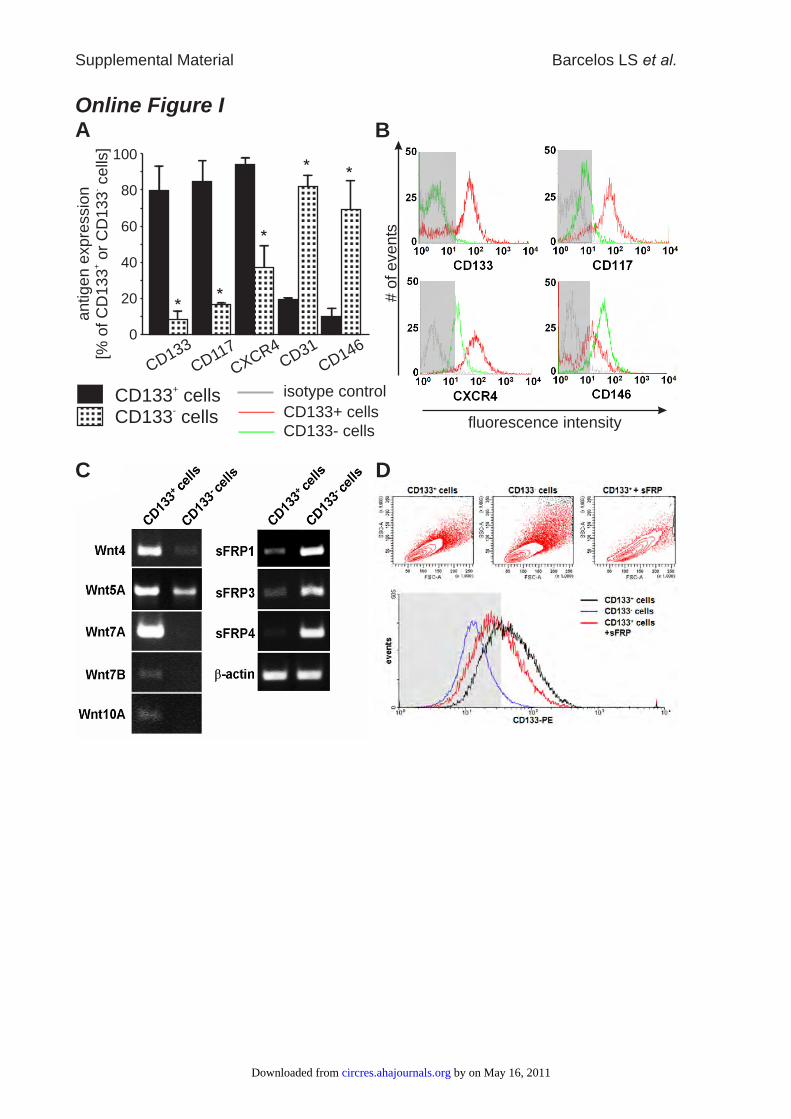
Online Figure I
Supplemental Material Barcelos LS et al.
A
CD133 cells+
CD133 cells-
# o
f events
fluorescence intensity
B
CD133+ cells
CD133- cells
isotype control
C D
CD31
CD146
CD133
CD117
CXCR40
20
40
60
80
100
antigen e
xpre
ssio
n[%
of C
D133
or
CD
133
cells
]+
-
by on May 16, 2011 circres.ahajournals.orgDownloaded from
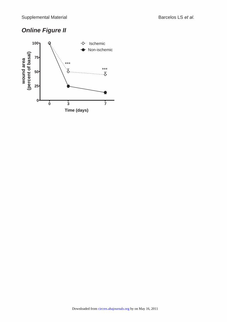
Online Figure II
0 3 70
25
50
75
100
***
***
Ischemic
Non-ischemic
Time (days)
wo
un
dare
a(p
erc
en
to
fb
asal)
Supplemental Material Barcelos LS et al.
by on May 16, 2011 circres.ahajournals.orgDownloaded from
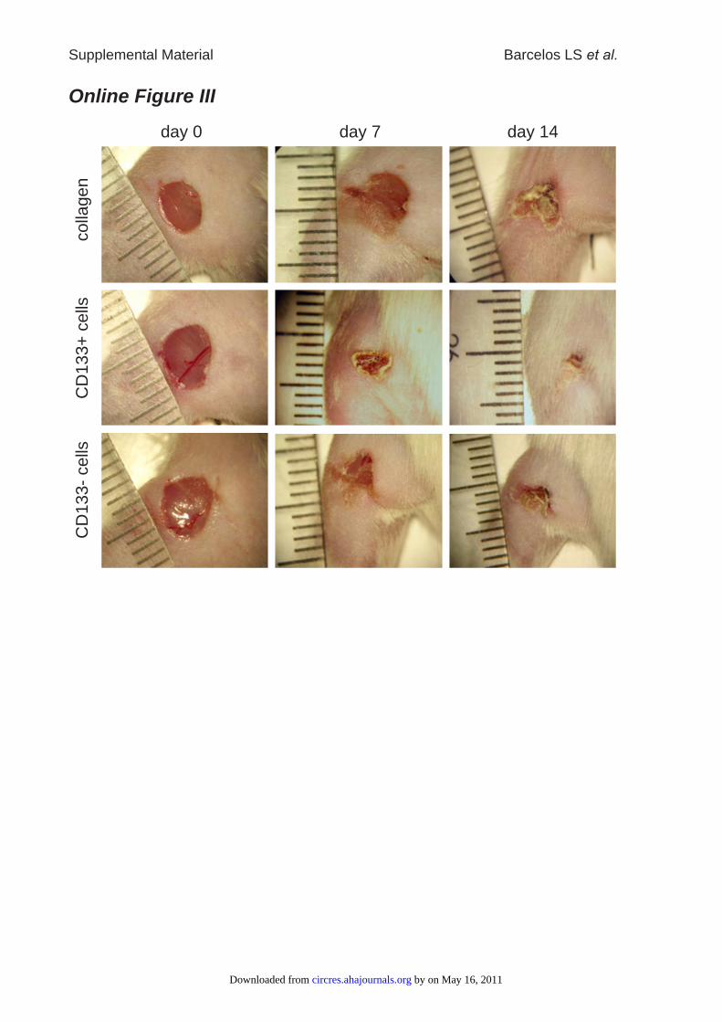
Online Figure IIIcolla
gen
CD
133+
cells
CD
133-
cells
day 0 day 14day 7
Supplemental Material Barcelos LS et al.
by on May 16, 2011 circres.ahajournals.orgDownloaded from
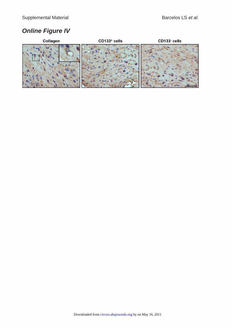
Online Figure IV
Supplemental Material Barcelos LS et al.
by on May 16, 2011 circres.ahajournals.orgDownloaded from
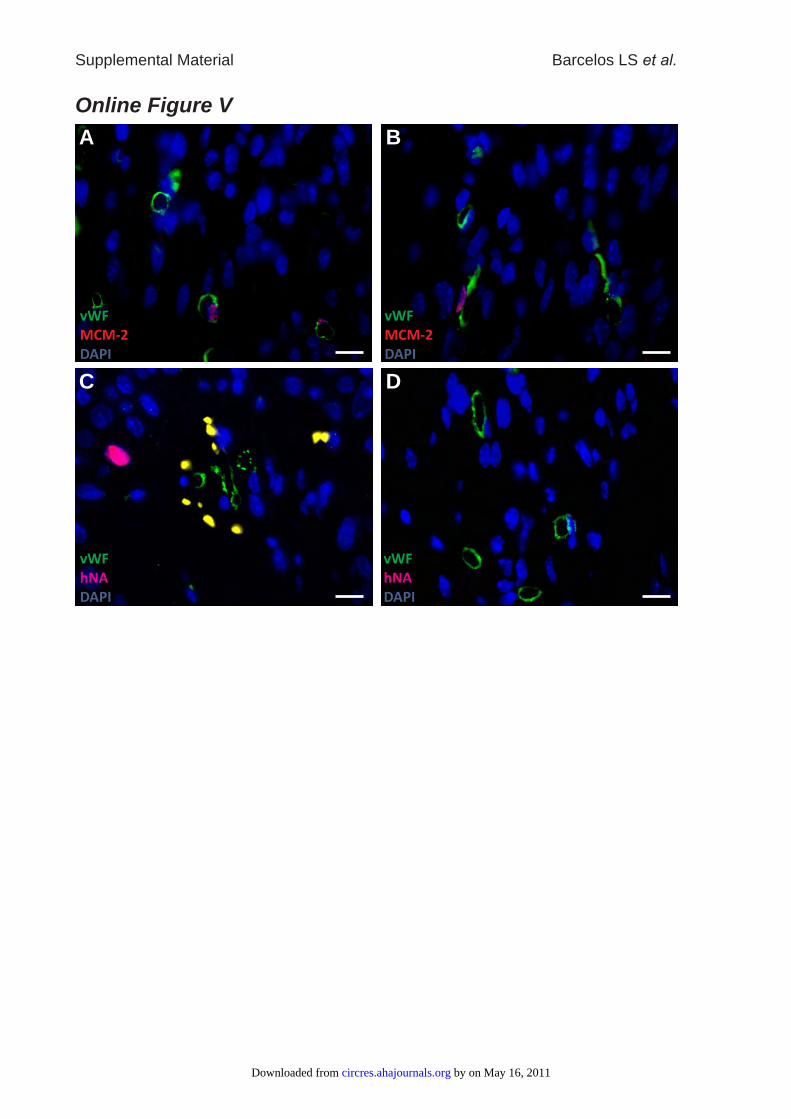
Online Figure V
Supplemental Material Barcelos LS et al.
vWF
MCM-2
DAPI
vWF
MCM-2
DAPI
A B
vWF
hNA
DAPI
vWF
hNA
DAPI
C D
by on May 16, 2011 circres.ahajournals.orgDownloaded from
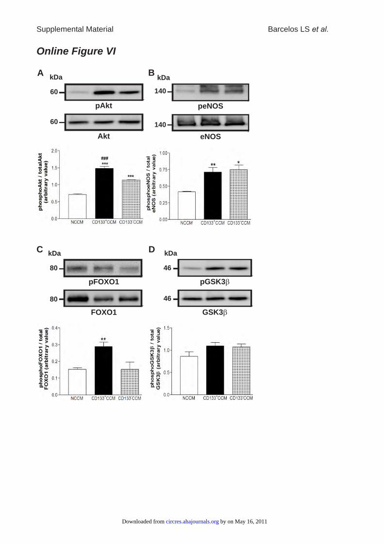
pGSK3b
GSK3b
46
46
kDaD
pAkt
Akt
60
60
kDaA
kDa
140
140
peNOS
eNOS
B
pFOXO1
FOXO1
kDa
80
80
C
Online Figure VI
Supplemental Material Barcelos LS et al.
by on May 16, 2011 circres.ahajournals.orgDownloaded from

Online Figure VII
A
*
*
*
**
*
**
B
0
10
20
30
40
wound c
apill
ary
density
[n/m
m]
2
NCCM CD133 CCM+
anti-IL8
anti-IL-6
anti-VEGF-A
0
5
10
15
20
Wound
Are
a [m
m]
2
time [days]0 3 7
0
5
10
15
20
Wound
Are
a [m
m]
2
time [days]0 3 7
CD133 CCM+ anti-VEGF-A
+
NCCM
CD133 CCM+
NCCM
0
5
10
15
20
Wound
Are
a [m
m]
2
time [days]0 3 7
CD133 CCM+ anti-IL6
+
NCCM
0
5
10
15
20W
ound
Are
a [m
m]
2
time [days]0 3 7
CD133 CCM+ anti-IL8
+
NCCM
Supplemental Material Barcelos LS et al.
by on May 16, 2011 circres.ahajournals.orgDownloaded from
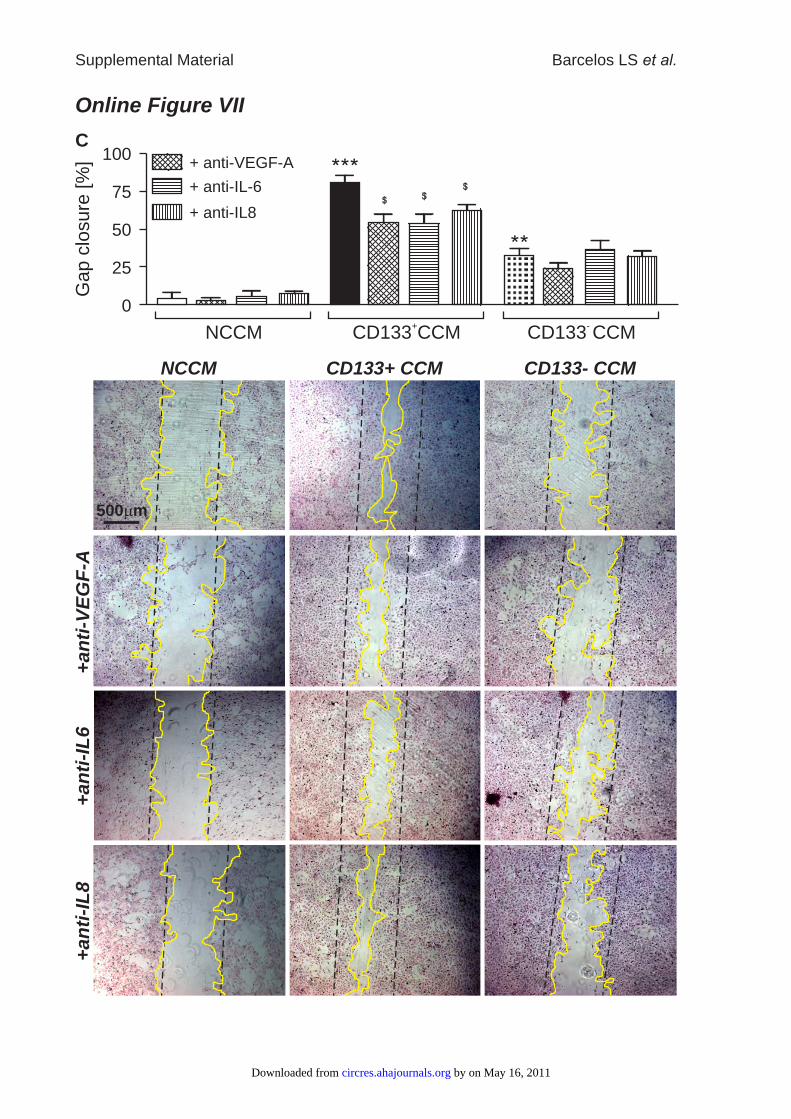
Online Figure VII
C
500 mm
NCCM CD133+ CCM CD133- CCM
+a
nti
-VE
GF
-A+
an
ti-I
L6
+a
nti
-IL
8
0
25
50
75
100
Gap c
losure
[%
]
NCCM CD133 CCM+
CD133 CCM-
+ anti-IL8
+ anti-IL-6
+ anti-VEGF-A
Supplemental Material Barcelos LS et al.
by on May 16, 2011 circres.ahajournals.orgDownloaded from
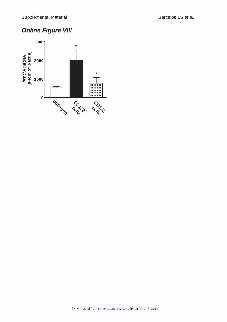
Online Figure VIII
0
1000
2000
3000
CD133
cells+
collagen
CD133
cells-
Wn
t7A
mR
NA
[n-f
old
of
-ac
tin
]b
Supplemental Material Barcelos LS et al.
by on May 16, 2011 circres.ahajournals.orgDownloaded from
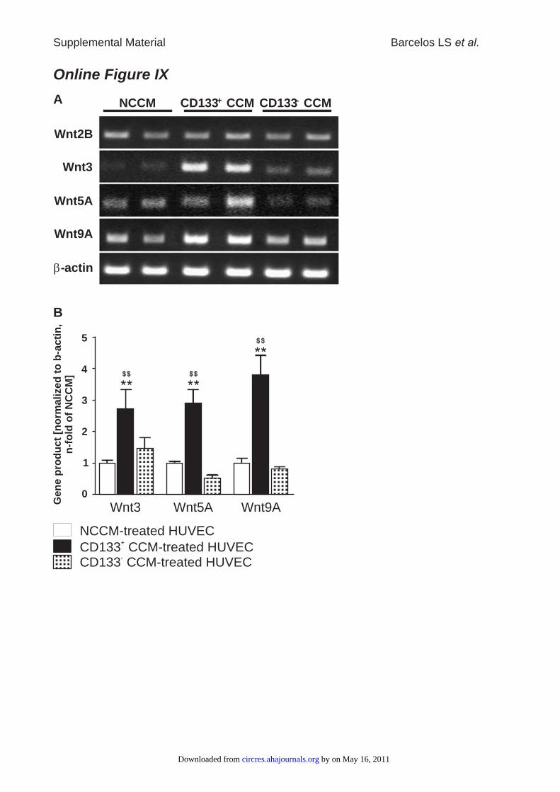
Online Figure IX
Wnt2B
Wnt3
Wnt5A
Wnt9A
b-actin
NCCM CD133+ CCM CD133- CCMA
0
1
2
3
4
5
Gen
e p
rod
uct
[no
rmali
zed
to
b-a
cti
n,
n-f
old
of
NC
CM
]
CD133 CCM-treated HUVEC+
CD133 CCM-treated HUVEC-
NCCM-treated HUVEC
Wnt3 Wnt5A Wnt9A
B
Supplemental Material Barcelos LS et al.
by on May 16, 2011 circres.ahajournals.orgDownloaded from
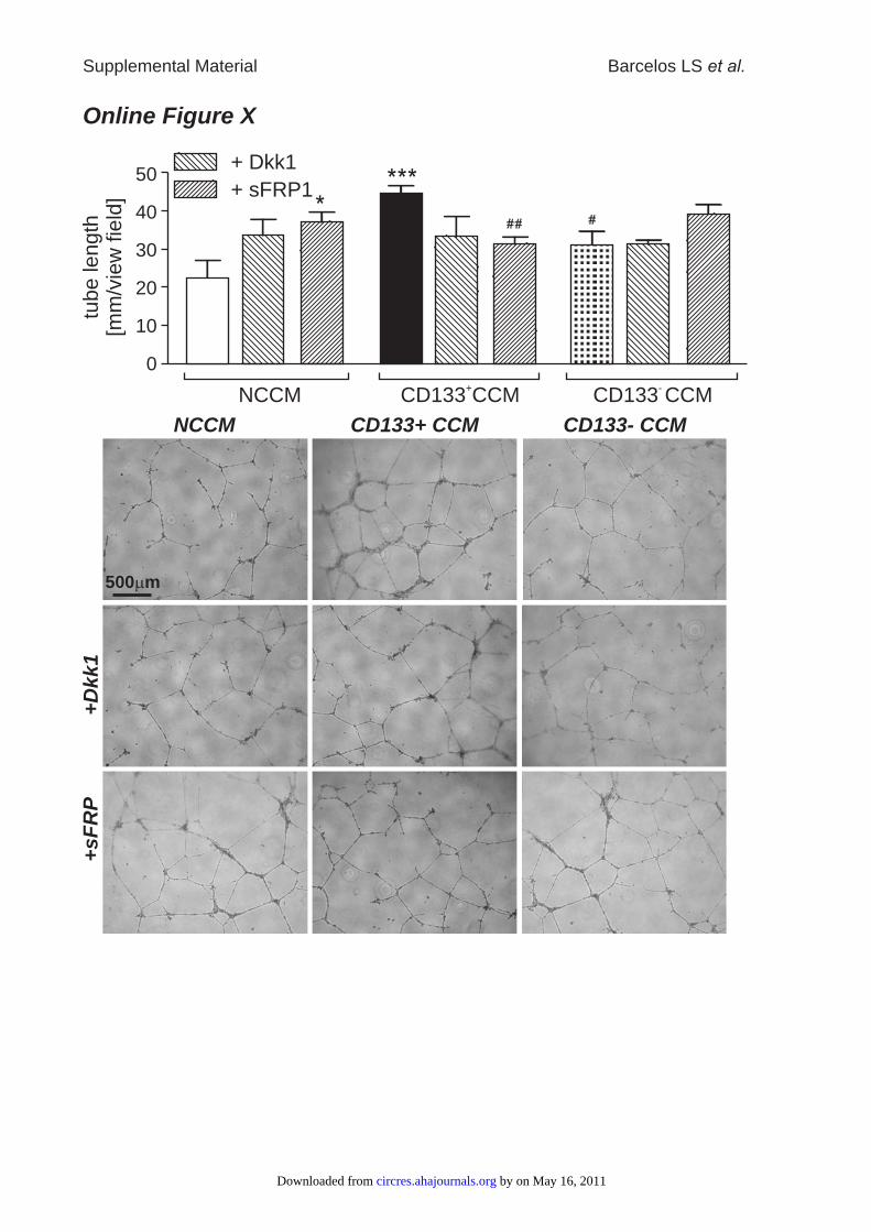
Online Figure Xtu
be length
[mm
/vie
w fie
ld]
50
10
20
40
0
30
NCCM CD133 CCM+
CD133 CCM-
+ Dkk1
+ sFRP1
NCCM CD133+ CCM CD133- CCM
500 mm
+D
kk1
+sF
RP
Supplemental Material Barcelos LS et al.
by on May 16, 2011 circres.ahajournals.orgDownloaded from
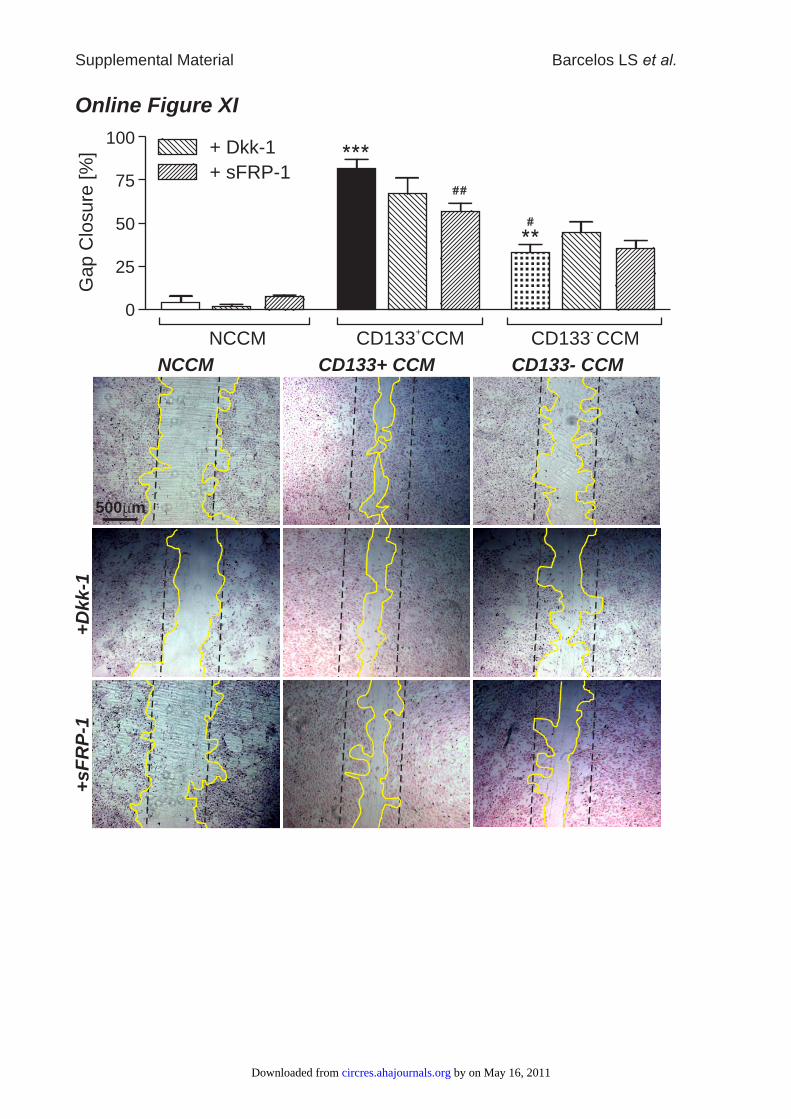
NCCM CD133+ CCM CD133- CCM
500 mm
+D
kk-1
+sF
RP
-1
Online Figure XIG
ap C
losure
[%
]
100
25
75
0
50
NCCM CD133 CCM+
CD133 CCM-
+ Dkk-1
+ sFRP-1
Supplemental Material Barcelos LS et al.
by on May 16, 2011 circres.ahajournals.orgDownloaded from
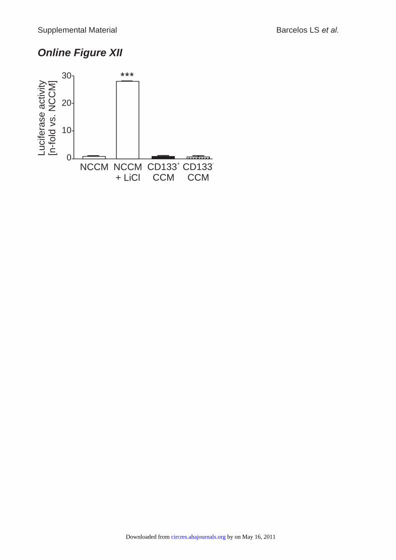
Online Figure XII
0
10
20
30
CD133CCM
+NCCM CD133
CCM
-
Lucifera
se a
ctivity
[n-f
old
vs. N
CC
M]
NCCM+ LiCl
Supplemental Material Barcelos LS et al.
by on May 16, 2011 circres.ahajournals.orgDownloaded from
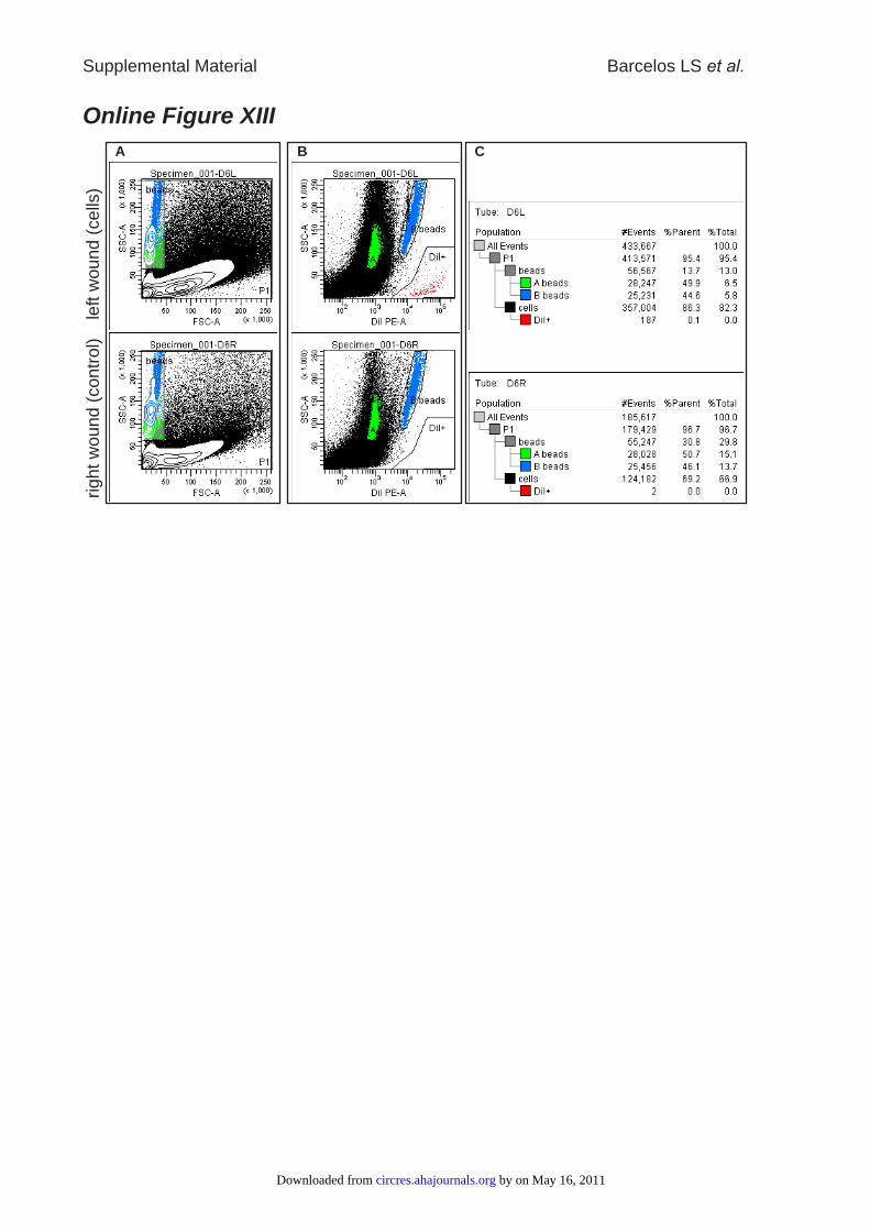
Online Figure XIII
A B C
left w
ound (
cells
)rig
ht w
ound (
contr
ol)
Supplemental Material Barcelos LS et al.
by on May 16, 2011 circres.ahajournals.orgDownloaded from
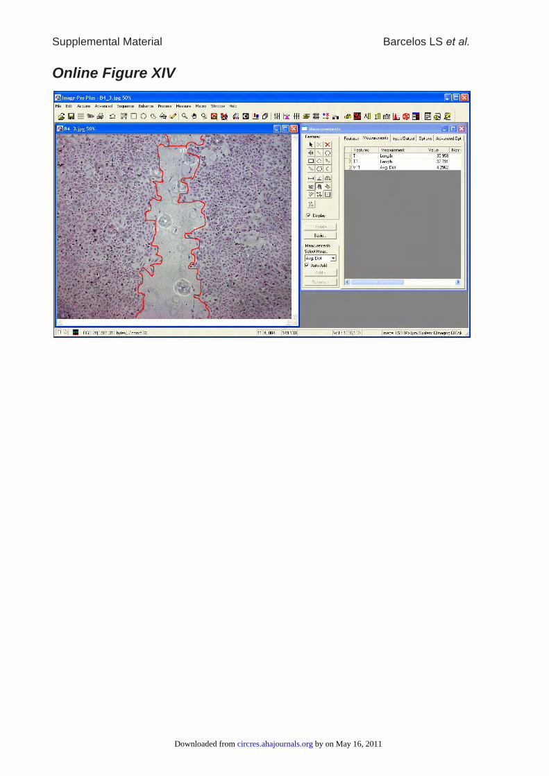
Online Figure XIV
Supplemental Material Barcelos LS et al.
by on May 16, 2011 circres.ahajournals.orgDownloaded from