Consumers' Willingness to Pay for Irradiated Poultry Products
Molecular Biological Studies to Evaluate
the Treatment Role of Irradiated Scaffolds
in Ulcers and Wounds in Rat Skin
A Thesis
Submitted In Partial Fulfillment of the Requirements for
the Master Degree of Science in
(BIOCHEMISTRY)
Presented by
Amir Mohammed Mohammed Ali Abdo B.Sc
Chemistry and Biochemistry
2009
Helwan University
Faculty of Sciences
Chemistry Department
2014
Molecular Biological Studies to Evaluate the Treatment Role
of Irradiated Scaffolds in Ulcers and Wounds in Rat Skin
By
Amir Mohammed Mohammed Ali Abdo
B.Sc in Chemistry and Biochemistry, 2009
In the partial fulfillment of the requirement of the
Master Degree in Science (BIOCHEMISTRY)
Under the supervision of
Prof. Dr. Elsayed Mahdy Prof.Dr. Eglal Eldegheidy
Professor of Biochemistry& Emeritus professor of Biochemistry
Dean of Faculty of Sciences Radiobiology department
Helwan University National Center for Radiation Research
and Technology (NCRRT)
Egyptian Atomic Energy Authority
Prof.Dr. Tarek Khaled Dr. Hatem Abdel Monem El-
Elmaghraby mezayen
Professor of Molecular Biology Assistant professor of Biochemistry
Radiobiology department Faculty of sciences
National Center for Radiation Helwan University
Research and Technology
Egyptian Atomic Energy Authority
Chemistry Department
Faculty of Science-Helwan University
2014

I hear you say "Why?" Always "Why?" You see things; and you say "Why?"
But I dream things that never were; and I say "Why not?"
(George Bernard Shaw)

DEDICATION

Acknowledgments
i
ACKNOWLEDGMENTS
With the pen in hand, I am proud to think that no words do justice to express my thanks to
ALMIGHTY ALLAH (The Omnipotent, The Omniscient, The Most Merciful and The Most Powerful) who is the entire source of all knowledge and wisdom to mankind and everything
is submitted to his will.
This work would not be performed without the support of the Academy of Scientific Research
and Technology grant (Scientists for Next Generation (SNG)). I wish to thank each one
working with this great project.
I would like to heartily thank my supervisor Professor Tarek Almaghraby, the
head of the molecular biology lab/ Radiobiology department/ National Center for Radiation
Research and Technology (NCRRT)/ The Egyptian Atomic energy Authority (EAEA), without
whom, this study would not have been possibly completed. Especially, recognition must be
given for offering me with guidance during the research and providing me with the scientific
expertise to complete my thesis.
I would like to express my sincere gratitude to Professor Eglal Eldegheidy, the
emeritus professor of biochemistry/Radiobiology department/ NCRRT/ EAEA for her ever-
inspiring guidance and constructive suggestions throughout the course of this effort.
To professor Elsayed Mahdy, the Dean of Faculty of Science/ Helwan university. I
am very proud to have a supervisor like you. No words can express my feelings and respect
for you.
I wish also to thank Dr. Hatem Abdel-Moneim El-mezayen, the assistant
professor of Biochemistry/ Faculty of Science/ Helwan university for kindly supervising the
present work, reading and scholarly criticizing the manuscript.
I wouldn`t have been able to complete this research without the generosity of Professor
Waleed Nazmy, the head of the Innovation Development Unit at VACSERA, Egypt. He
has provided me with extraordinary mentorship during my work in his lab. His meticulous
concern and patience were the ways to complete this research.

Acknowledgments
ii
The work presented in this study was impossible to be accomplished without the sympathetic
attitude and utmost care of my teacher, Dr. Sanaa Abdel-Hamid.
I wish to express my deepest feeling of gratitude to Dr. Mohammed Abdel-Baseer,
Dr. Saad Attiya and Dr.Wael Abul-Noor. Without their observant pursuit,
cheering perspective and the enlightened supervision, this work would not be completed.
Thanks to Prof. Renee Georgy, NCRRT, prof. Kawkab Abdel-aziz, Cairo Univ. and
Mr. Moussa Hussein, the National Cancer Institute (NCI) for conducting the
hisological staining and examinations.
Great thanks to Prof. Doaa Mekawy and Dr. Wael Hossam, the National
Research Center (NRC) for their help during my study.
I am grateful to prof. Hisham Attiya, prof. Ahmed Shafik, prof. Magdy Senna
,Dr. Mohammed Almohmady and Dr. Ahmed Elbarbary, NCRRT for their kind
advice and support during the work.
I can`t forget the kind support from Dr. Mohammed Hamdy, so I wish him to receive
my great thanks.
Words are lacking to express my humble obligation to my late father and my loving mother
who always longed for my successful and happy life. Their endless efforts and best wishes
sustained me at all stages of my life and their hands always remain raised in prayer for my
success. It’s my ever pray that may Allah bless my mother with long, happy and healthy life.
If the pearls were words and flowers feelings, it would be easier to express my deepest heart
gratitude and indebtedness to the all the peoples and friends who have helped me during this
research. May Allah Almighty infuses me with the energy to fulfill their noble inspiration and
expectations and further modify my competence. May Allah bless us all with long, happy and
peaceful lives.
Amier Mohammed
iii
List of Contents Section Contents Page
Acknowledgments i
List of Contents Iii
List of Figures v
List of Tables and Appendices vii
List of Abbreviations viii
Abstract xi
1 Introduction 1
Aim of Work 3
2 Review of literature 4
Chapter (1) Skin and Wound Healing 4
2.1.1 Skin Structure 4
2.1.2 Skin Anatomy 4
2.1.3 Functions of Skin 10
2.1.4 Skin Wounds 12
2.1.5 Skin Ulcers 13
2.1.6 Wound Healing 14
2.1.7 Classification of Wound Dressing Products 17
Chapter (2) Review on Alginates 27
2.2.1 Chemical Structure of Alginate 27
2.2.2 Sources of Alginates 28
2.2.3 Properties of Alginate 29
2.2.4 Alginate Gelation 30
2.2.5 Modification of Alginate 35
2.2.6 Purification of Alginate 43
Chapter (3) Review on Chitosans 46
2.3.1 Chemical Structure of Chitosan 46
2.3.2 Sources of Chitosans 47
2.3.3 Properties of Chitosan 49
2.3.4 Production of Chitosan from Chitin of Shrimp Shells 52
Chapter (4) Review on PolyElectrolyte Complexes (PECs) 53
2.4.1 Properties of the Alginate/Chitosan PECs 53
2.4.2 Principle of Formation of Alginate/Chitosan PECs 55
Chapter (5) Biological effects of alginates & chitosans-based dressings 58
2.5.1 Alginates as Wound Dressings 58
2.5.2 Chitosans as Wound Dressings 62
2.5.3 Alginate/ Chitosan-Based Wound Dressings 63
2.5.4 Angiogenesis and Angiogenesis-Controlling Genes 66
2.5.4.1 Vascular Endothelial Growth Factor (VEGF) 64
2.5.4.2 von Willebrand Factor (vWF) 72
3 Materials and Methods 80
3.1 Materials 80
3.2 Preparation of reagents 80
iv
Section Contents Page
3.3 Modification of Alginate 81
3.3.1 Irradiation of Sodium Alginate 81
3.3.2 Oxidation of Sodium Alginate 82
3.3.3 Characterization of the Different Modified Alginates 83
3.4 Purification Protocol of Sodium Alginate 86
3.4.1 Acid-washing of Activated Charcoal 86
3.4.2 Method of Alginate Purification 86
3.4.3 Testing the Effects of Purification in Alginate 87
3.5 Preparation of Chitosan 90
3.5.1 The Extraction and Deacetylation Steps 93
3.5.2 Characterization of the Prepared Chitosans Products 93
3.6 Method of Preparing the Alginate-Chitosan PECs hydrogels 93
3.7 Major Steps for Choosing the Best Type of Hydrogels 94
3.7.1 In vitro Swelling of Hydrogels in Simulated Wound Fluids 94
3.7.2 Stability Characterization Studies 94
3.7.3 Blood Compatibility Tests 95
3.7.4 Rate of Evaporation of Water from Gel 96
3.7.5 In vitro Degradation of the Prepared Hydrogel Films 96
3.7.6 Primary Skin Irritation Test for the Hydrogels 96
3.7.7 Testing the Optimum Composition of Hydrogel 97
3.7.7.1 Detection of the Best Concentration for the used(CaCl2) 97
3.7.7.2 Characterizing the Effects of (γ-irradiation) on (F-20) 97
3.7.7.3 Choosing the Best Working Film Structure 99
3.8 Statistical Analyses 100
3.9 Wounding and Wound Healing Assessment 102
3.9.1 Animals 102
3.9.2 Wounding Procedures 102
3.9.3 Wound and Skin Assessment 103
3.9.3.1 Monitoring the Visible Changes in Wounds During Healing 103
3.9.3.2 Measurement of Residual Wound Area 103
3.9.3.3 Histological Studies 104-105
3.9.3.4 Quantification of RNA corresponding to(VEGF and vWF) 105-110
3.9.3.5 Screening of Kidney Functions 110
4 Results 113
5 Discussion 142
Recommendations for future work 188
Summary and Conclusion 189
References 191
Appendices 235
v
List of figures No. Title Page
1 Cross-sections of Skin and the Epidermis layer 5
2 Summary for phases of Wound Healing 15
3 Summary for the overlapping periods of healing stages 15
4 Schematically drawn alginate block structure with a segment
showing structure of the molecules 27
5 The binding of a divalent cation to contiguous dimers of
guluronate residues 32
6 Proposed mechanism for Alginate degradation in the solid state 39
7 Suggested reaction scheme describing periodate oxidation of a
mannuronan residue within the alginate chain 41
8 The chemical structures of Chitin and Chitosan 46
9 Schematic diagram of counter-ions release upon PEC formation 56
10 Schematic Interpretation of the (Alginate-Chitosan physical
complex and Semi-IPN complex) 57
11 Genomic location of human VEGFA Gene on chromosome (6) 66
12 Exon structure and function of rat VEGFA 67
13 Different VEGF Receptors&the corresponding binding cytokines 68
14 Genomic location of human vWF gene on chromosome (12) 73
15 Structure of vWF Protein 74
16 FT-IR spectra for the irradiated and oxidized sodium alginates 114
17 FT-IR Spectra showing the changes within the activated charcoal
after washing with different acids 115
18 Quantitative evaluation of the major Alginate contaminants
before /after purification
116&
117
19 FT-IR spectra for (Na-Alginate) before and after purification 117
20 (FT-IR) spectra for the chitosans (1, 2) 119
21 Swelling kinetics for different formulae immersed in PBS
medium (pH 7.4) for 24 hours 121
22 Time dependence of water loss from the 3 formulae (F-20,18,5) 124
23 Rate of degradation for the different formulae (F-20,18,5) 125
24 The influence of cross-linking agent (CaCl2) concentration on the
swelling degree for the formulae (F-18 and 20) 127
25 Comparison of the swelling kinetics for the unirradiated and
irradiated formulae (F-20 and 5) 127
26 FTIR spectra for physical mixture of alginates, alginate with
chitosan, the unirradiated and irradiated PECs (F-20) 128
27 Scanning Electron Micrographs for the surfaces of different
formulae based on (Alginate/Chitosan PECs). 129
vi
No. Title Page
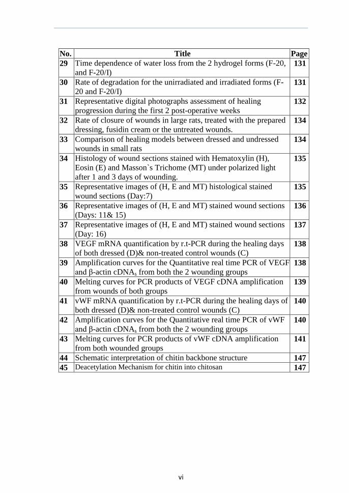
29 Time dependence of water loss from the 2 hydrogel forms (F-20,
and F-20/I) 131
30 Rate of degradation for the unirradiated and irradiated forms (F-
20 and F-20/I) 131
31 Representative digital photographs assessment of healing
progression during the first 2 post-operative weeks 132
32 Rate of closure of wounds in large rats, treated with the prepared
dressing, fusidin cream or the untreated wounds. 134
33 Comparison of healing models between dressed and undressed
wounds in small rats 134
34 Histology of wound sections stained with Hematoxylin (H),
Eosin (E) and Masson`s Trichome (MT) under polarized light
after 1 and 3 days of wounding.
135
35 Representative images of (H, E and MT) histological stained
wound sections (Day:7) 135
36 Representative images of (H, E and MT) stained wound sections
(Days: 11& 15) 136
37 Representative images of (H, E and MT) stained wound sections
(Day: 16) 137
38 VEGF mRNA quantification by r.t-PCR during the healing days
of both dressed (D)& non-treated control wounds (C) 138
39 Amplification curves for the Quantitative real time PCR of VEGF
and β-actin cDNAs from both the 2 wounding groups 138
40 Melting curves for PCR products of VEGF cDNA amplification
from wounds of both groups 139
41 vWF mRNA quantification by r.t-PCR during the healing days of
both dressed (D)& non-treated control wounds (C) 140
42 Amplification curves for the Quantitative real time PCR of vWF
and β-actin cDNAs from both the 2 wounding groups 140
43 Melting curves for PCR products of vWF cDNA amplification from both wounded groups
141
44 Schematic interpretation of chitin backbone structure 147
45 Deacetylation Mechanism for chitin into chitosan 147
vii
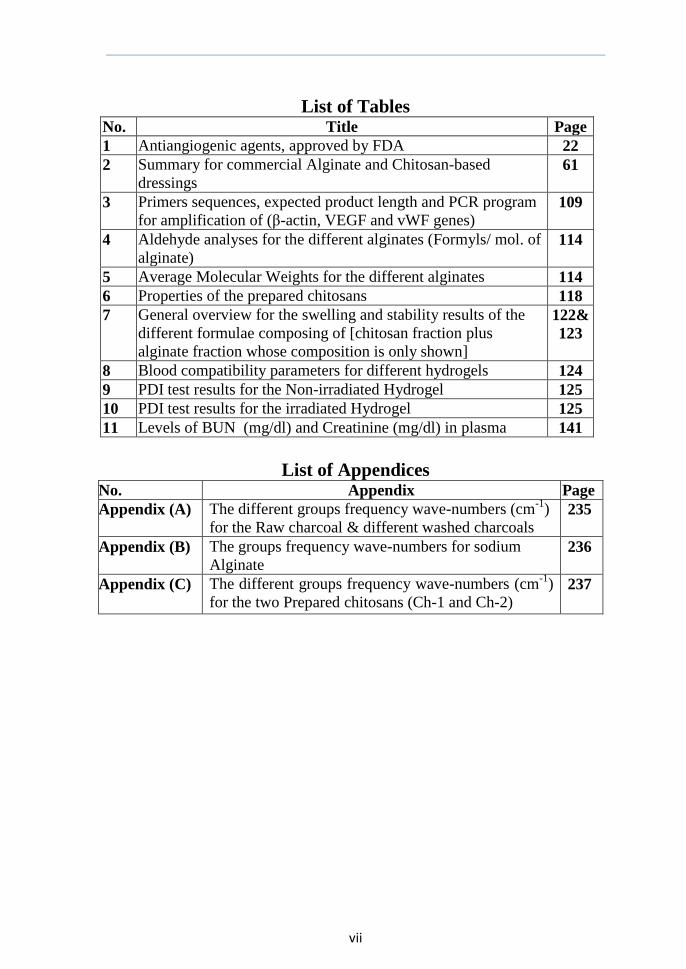
List of Tables No. Title Page
1 Antiangiogenic agents, approved by FDA 22
2 Summary for commercial Alginate and Chitosan-based
dressings 61
3 Primers sequences, expected product length and PCR program
for amplification of (β-actin, VEGF and vWF genes) 109
4 Aldehyde analyses for the different alginates (Formyls/ mol. of
alginate) 114
5 Average Molecular Weights for the different alginates 114
6 Properties of the prepared chitosans 118
7 General overview for the swelling and stability results of the
different formulae composing of [chitosan fraction plus
alginate fraction whose composition is only shown]
122&
123
8 Blood compatibility parameters for different hydrogels 124
9 PDI test results for the Non-irradiated Hydrogel 125
10 PDI test results for the irradiated Hydrogel 125
11 Levels of BUN (mg/dl) and Creatinine (mg/dl) in plasma 141
List of Appendices No. Appendix Page
Appendix (A) The different groups frequency wave-numbers (cm-1
)
for the Raw charcoal & different washed charcoals 235
Appendix (B) The groups frequency wave-numbers for sodium
Alginate 236
Appendix (C) The different groups frequency wave-numbers (cm-1
)
for the two Prepared chitosans (Ch-1 and Ch-2) 237
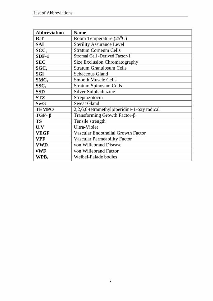
List of Abbreviations
viii
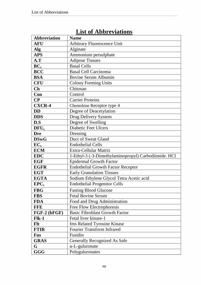
List of Abbreviations Abbreviation Name
AFU Arbitrary Fluorescence Unit
Alg Alginate
APS Ammonium persulphate
A.T Adipose Tissues
BCs Basal Cells
BCC Basal Cell Carcinoma
BSA Bovine Serum Albumin
CFU Colony Forming Units
Ch Chitosan
Con Control
CP Carrier Proteins
CXCR-4 Chemokine Receptor type 4
DD Degree of Deacetylation
DDS Drug Delivery System
D.S Degree of Swelling
DFUs Diabetic Feet Ulcers
Dre Dressing
DSwG Duct of Sweat Gland
ECs Endothelial Cells
ECM Extra-Cellular Matrix
EDC 1-Ethyl-3 (-3-Dimethylaminopropyl) Carbodiimide. HCl
EGF Epidermal Growth Factor
EGFR Endothelial Growth Factor Receptor
EGT Early Granulation Tissues
EGTA Sodium Ethylene Glycol Tetra Acetic acid
EPCs Endothelial Progenitor Cells
FBG Fasting Blood Glucose
FBS Fetal Bovine Serum
FDA Food and Drug Administration
FFE Free Flow Electrophoresis
FGF-2 (bFGF) Basic Fibroblast Growth Factor
Flk-1 Fetal liver kinase-1
Flt fms Related Tyrosine Kinase
FTIR Fourier Transform Infrared
Fus Fusidin
GRAS Generally Recognized As Safe
G α-L-guluronate
GGG Polyguluronates
List of Abbreviations
ix
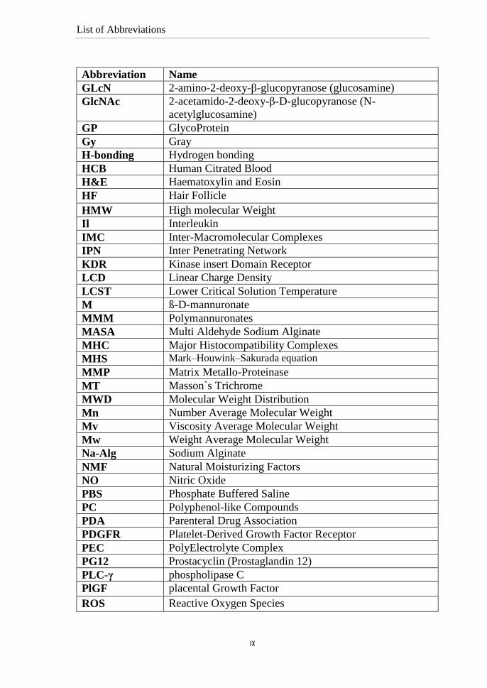
Abbreviation Name
GLcN 2-amino-2-deoxy-β-glucopyranose (glucosamine)
GlcNAc 2-acetamido-2-deoxy-β-D-glucopyranose (N-
acetylglucosamine)
GP GlycoProtein
Gy Gray
H-bonding Hydrogen bonding
HCB Human Citrated Blood
H&E Haematoxylin and Eosin
HF Hair Follicle
HMW High molecular Weight
Il Interleukin
IMC Inter-Macromolecular Complexes
IPN Inter Penetrating Network
KDR Kinase insert Domain Receptor
LCD Linear Charge Density
LCST Lower Critical Solution Temperature
M ß-D-mannuronate
MMM Polymannuronates
MASA Multi Aldehyde Sodium Alginate
MHC Major Histocompatibility Complexes
MHS Mark–Houwink–Sakurada equation
MMP Matrix Metallo-Proteinase
MT Masson`s Trichrome
MWD Molecular Weight Distribution
Mn Number Average Molecular Weight
Mv Viscosity Average Molecular Weight
Mw Weight Average Molecular Weight
Na-Alg Sodium Alginate
NMF Natural Moisturizing Factors
NO Nitric Oxide
PBS Phosphate Buffered Saline
PC Polyphenol-like Compounds
PDA Parenteral Drug Association
PDGFR Platelet-Derived Growth Factor Receptor
PEC PolyElectrolyte Complex
PG12 Prostacyclin (Prostaglandin 12)
PLC-γ phospholipase C
PlGF placental Growth Factor
ROS Reactive Oxygen Species
List of Abbreviations
x
Abbreviation Name
R.T Room Temperature (25oC)
SAL Sterility Assurance Level
SCCs Stratum Corneum Cells
SDF-1 Stromal Cell -Derived Factor-1
SEC Size Exclusion Chromatography
SGCs Stratum Granulosum Cells
SGl Sebaceous Gland
SMCs Smooth Muscle Cells
SSCs Stratum Spinosum Cells
SSD Silver Sulphadiazine
STZ Streptozotocin
SwG Sweat Gland
TEMPO 2,2,6,6-tetramethylpiperidine-1-oxy radical
TGF- β Transforming Growth Factor-β
TS Tensile strength
U.V Ultra-Violet
VEGF Vascular Endothelial Growth Factor
VPF Vascular Permeability Factor
VWD von Willebrand Disease
vWF von Willebrand Factor
WPBs Weibel-Palade bodies
Abstract
xi
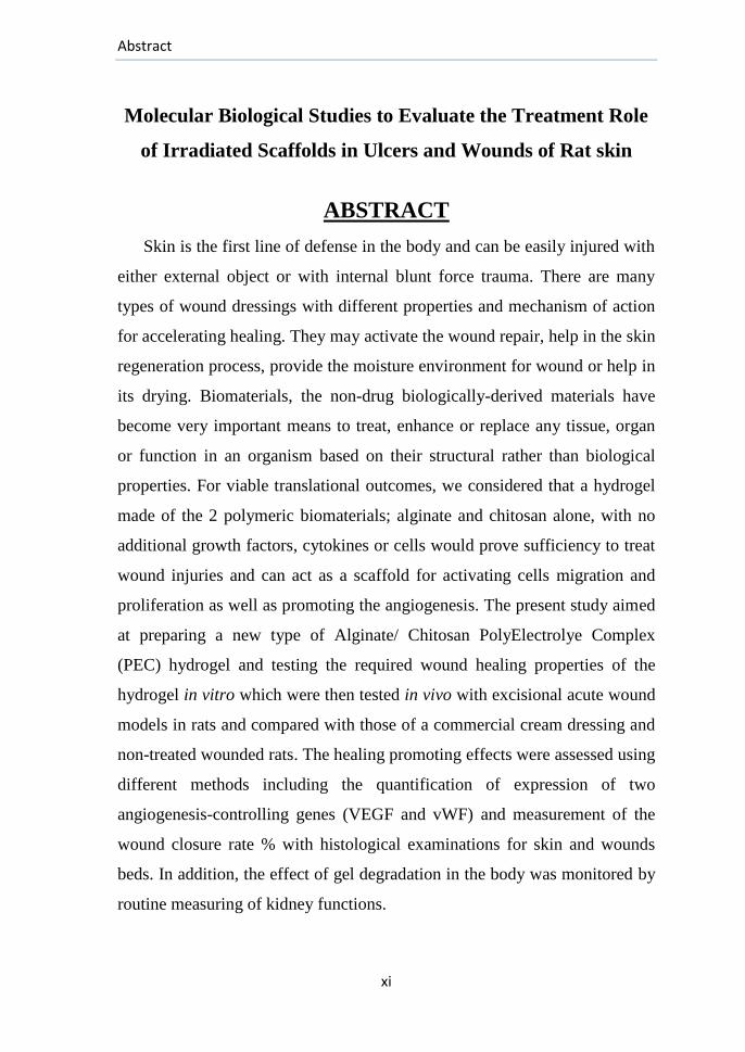
Molecular Biological Studies to Evaluate the Treatment Role
of Irradiated Scaffolds in Ulcers and Wounds of Rat skin
ABSTRACT
Skin is the first line of defense in the body and can be easily injured with
either external object or with internal blunt force trauma. There are many
types of wound dressings with different properties and mechanism of action
for accelerating healing. They may activate the wound repair, help in the skin
regeneration process, provide the moisture environment for wound or help in
its drying. Biomaterials, the non-drug biologically-derived materials have
become very important means to treat, enhance or replace any tissue, organ
or function in an organism based on their structural rather than biological
properties. For viable translational outcomes, we considered that a hydrogel
made of the 2 polymeric biomaterials; alginate and chitosan alone, with no
additional growth factors, cytokines or cells would prove sufficiency to treat
wound injuries and can act as a scaffold for activating cells migration and
proliferation as well as promoting the angiogenesis. The present study aimed
at preparing a new type of Alginate/ Chitosan PolyElectrolye Complex
(PEC) hydrogel and testing the required wound healing properties of the
hydrogel in vitro which were then tested in vivo with excisional acute wound
models in rats and compared with those of a commercial cream dressing and
non-treated wounded rats. The healing promoting effects were assessed using
different methods including the quantification of expression of two
angiogenesis-controlling genes (VEGF and vWF) and measurement of the
wound closure rate % with histological examinations for skin and wounds
beds. In addition, the effect of gel degradation in the body was monitored by
routine measuring of kidney functions.
Abstract
xii
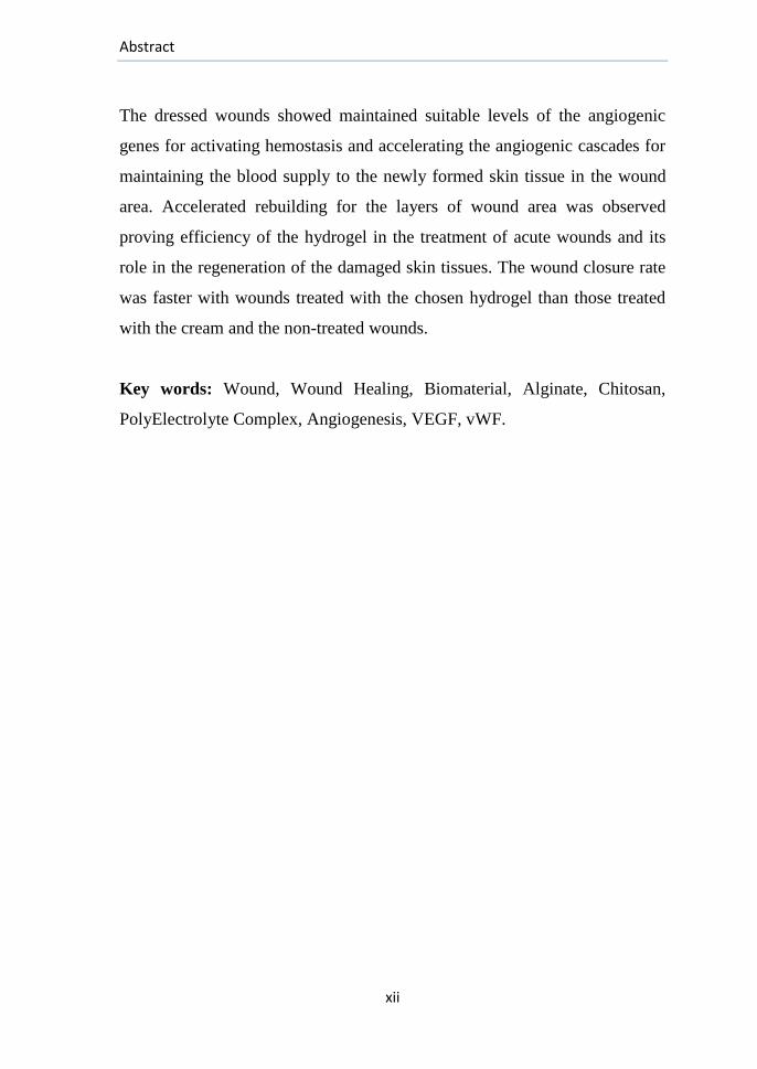
The dressed wounds showed maintained suitable levels of the angiogenic
genes for activating hemostasis and accelerating the angiogenic cascades for
maintaining the blood supply to the newly formed skin tissue in the wound
area. Accelerated rebuilding for the layers of wound area was observed
proving efficiency of the hydrogel in the treatment of acute wounds and its
role in the regeneration of the damaged skin tissues. The wound closure rate
was faster with wounds treated with the chosen hydrogel than those treated
with the cream and the non-treated wounds.
Key words: Wound, Wound Healing, Biomaterial, Alginate, Chitosan,
PolyElectrolyte Complex, Angiogenesis, VEGF, vWF.
Introduction and Aim of the work
1
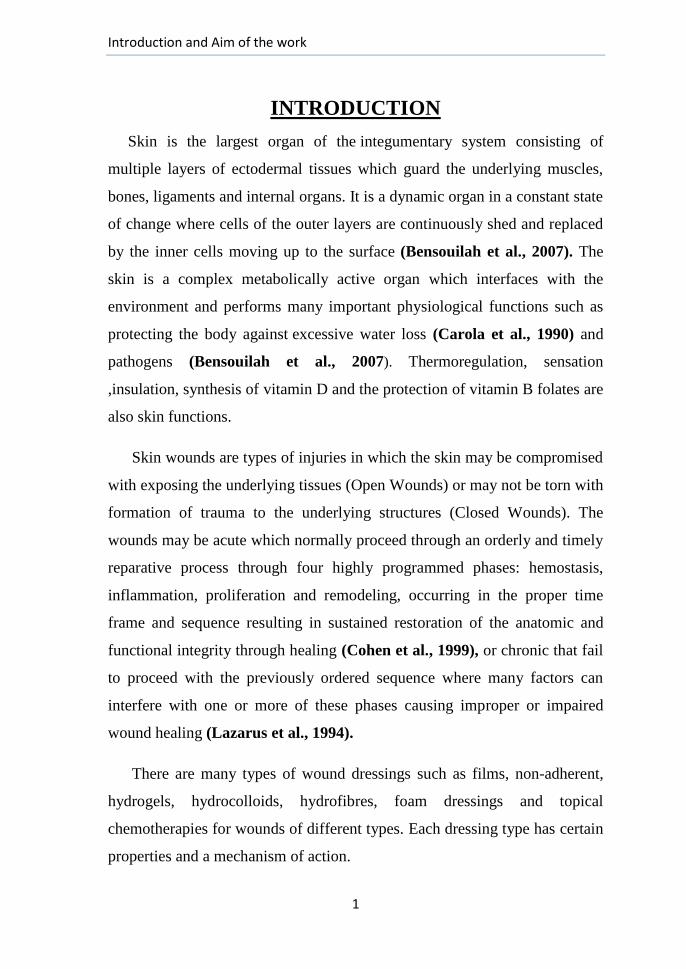
INTRODUCTION
Skin is the largest organ of the integumentary system consisting of
multiple layers of ectodermal tissues which guard the underlying muscles,
bones, ligaments and internal organs. It is a dynamic organ in a constant state
of change where cells of the outer layers are continuously shed and replaced
by the inner cells moving up to the surface (Bensouilah et al., 2007). The
skin is a complex metabolically active organ which interfaces with the
environment and performs many important physiological functions such as
protecting the body against excessive water loss (Carola et al., 1990) and
pathogens (Bensouilah et al., 2007). Thermoregulation, sensation
,insulation, synthesis of vitamin D and the protection of vitamin B folates are
also skin functions.
Skin wounds are types of injuries in which the skin may be compromised
with exposing the underlying tissues (Open Wounds) or may not be torn with
formation of trauma to the underlying structures (Closed Wounds). The
wounds may be acute which normally proceed through an orderly and timely
reparative process through four highly programmed phases: hemostasis,
inflammation, proliferation and remodeling, occurring in the proper time
frame and sequence resulting in sustained restoration of the anatomic and
functional integrity through healing (Cohen et al., 1999), or chronic that fail
to proceed with the previously ordered sequence where many factors can
interfere with one or more of these phases causing improper or impaired
wound healing (Lazarus et al., 1994).
There are many types of wound dressings such as films, non-adherent,
hydrogels, hydrocolloids, hydrofibres, foam dressings and topical
chemotherapies for wounds of different types. Each dressing type has certain
properties and a mechanism of action.
Introduction and Aim of the work
2
Dressings made of the biomaterials, chitosan and/or alginate have got
attention due to their peculiar properties, hemostatic, biodegradability,
bioactivity and remodeling properties (Otterlei et al., 1991; Azad et al.,
2004; Lin et al., 2006), so many types of dressings of each one alone, a
combination of them or with other materials as well have been synthesized
and their efficacies have been proved.
Bioengineering is considered one of the most innovative approaches
tackling many diseases and body parts that need to be replaced. This term
applies to the efforts that span interdisciplinary boundaries and connects the
engineering and physical sciences to the biological sciences and medicine in
a multidisciplinary setting to develop or apply new treatment technologies as
well as performing specific biochemical functions with a major dependence
on cells within artificially-created support system, called scaffold (Zhao et
al., report) whose properties depend primarily on the nature and properties
of the used materials. Novel free form fabrication methods for engineering
polymeric scaffolds have gained interests due to their repeatability and
capability of usage with high accuracy in the fabrication resolution at the
macro and micro scales. For example, ionically cross-linked alginates have
great potential as scaffolds where they can form highly hydrated hydrogels
representing hospitable environment for the transplanted cells and cellular
infiltration. An ideal wound dressing should control evaporative water loss,
prevent dehydration, protect the wound from bacterial infection, allow
diffusion of oxygen and carbon dioxide, absorb wound exudate and enhance
its healing (Kirker et al., 2002).
Wound assessment is essential for effective wound management and for
investigating the effect of certain dressing on the healing cascade (NHS,
2008) with monitoring the wound closure rate and any changes to it.
Introduction and Aim of the work
3
Histological examinations for wounds beds are also essential for
assessing the skin maturity and testing the influence of the dressing in the
histo-architectural organization of the wound area. Angiogenesis and
neovascularization are critical determinants of wound healing outcomes
where the newly formed blood vessels participate in the healing process with
providing nutrition and oxygen to the growing tissues. Accordingly; to better
determine the functionality of the developing vasculature, the angiogenic
response is studied by the quantitative measurement of expression of the
angiogenesis-controlling genes using the molecular biology technique,
Polymerase Chain Reaction (PCR). Nowadays, Molecular biology plays
important roles in understanding structures, actions and regulations of
various cellular compartments and can be used efficiently for targeting
new drugs, diagnosis of diseases and studying the physiology of cells.
Aim of Work
This study aims at: (1) preparing a hydrogel made of a new extracted
chitosan and chemically modified alginates with irradiation and oxidation in
the form of alginate-chitosan coacervates under controlled conditions for
casting into homogeneous films utilizing a new method.
(2)The designing of a general scheme for choosing the best suitable hydrogel
that can act as a scaffold for engineering dermal and epidermal tissues and as
a controlled release system for drugs to the skin aiming to accelerating the
wound healing.
(3) Its biological effects for treatment of rat skin wound models will be
investigated using histological and molecular biological methods with
measuring the expression of certain angiogenic genes (VEGF and vWF) for
assessing the potential effect of the chosen hydrogel on the skin wound and
its promotion for the corresponding angiogenic responses.
Review of Literature
4
2. REVIEW OF LITERATURE
(1): Skin and Wound Healing
2.1.1 Skin Structure:
The skin is a physiologically and anatomically specialized boundary lamina
essential to life and has several functions such as forming a physical barrier to
environment to allow and limit the inward and outward passage of water,
electrolytes and various substances with providing protection against toxic agents,
microorganisms, Ultra-Violet radiation (U.V) and mechanical insults. It occupies
almost 1.8 m2 of the surface area in average adults, accounting for 16% of the body
mass making it its largest organ (Bensouilah et al., 2007).
Skin can be classified according to its thickness that varies with age of the
individual and the anatomical part of the body where it is found. It may be thin,
hairy (hirsute), constituting the majority of the body‘s surface (e.g., Skin on the
eyelids is less than 0.5 mm thick), or may be thick, hairless (glabrous) skin such as
skin covering the palms, soles and flexor surfaces of the digits and skin on the
middle of the upper back which is more than 5mm thick (Gray, 1987; Carola et
al., 1990).
2.1.2. Skin Anatomy:
Skin is a structurally complex and highly specialized organ, consisting of two
intimately associated main layers called: (1) The epidermis, the outermost layer of
skin, and (2) The dermis (corium), a thicker layer beneath the epidermis. Certain
appendages such as hair follicles and sweat glands span both the 2 layers and
penetrate into the subcutaneous adipose tissue beneath the dermis (Alberts et al.,
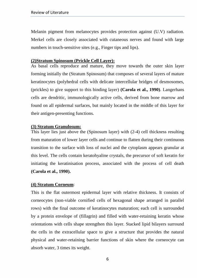
2002; Carola et al., 1990). Fig. (1) illustrates the general architecture of the skin
and the epidermal layers (Studyblue site).
Review of Literature
5
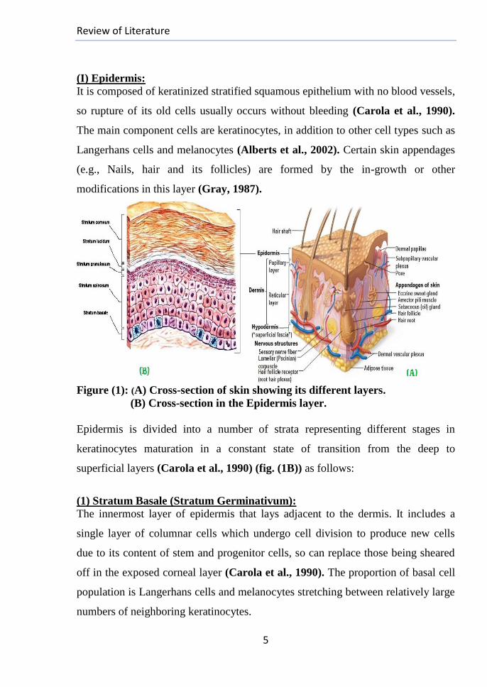
(I) Epidermis:
It is composed of keratinized stratified squamous epithelium with no blood vessels,
so rupture of its old cells usually occurs without bleeding (Carola et al., 1990).
The main component cells are keratinocytes, in addition to other cell types such as
Langerhans cells and melanocytes (Alberts et al., 2002). Certain skin appendages
(e.g., Nails, hair and its follicles) are formed by the in-growth or other
modifications in this layer (Gray, 1987).
Figure (1): (A) Cross-section of skin showing its different layers.
(B) Cross-section in the Epidermis layer.
Epidermis is divided into a number of strata representing different stages in
keratinocytes maturation in a constant state of transition from the deep to
superficial layers (Carola et al., 1990) (fig. (1B)) as follows:
(1) Stratum Basale (Stratum Germinativum):
The innermost layer of epidermis that lays adjacent to the dermis. It includes a
single layer of columnar cells which undergo cell division to produce new cells
due to its content of stem and progenitor cells, so can replace those being sheared
off in the exposed corneal layer (Carola et al., 1990). The proportion of basal cell
population is Langerhans cells and melanocytes stretching between relatively large
numbers of neighboring keratinocytes.
Review of Literature
6
Melanin pigment from melanocytes provides protection against (U.V) radiation.
Merkel cells are closely associated with cutaneous nerves and found with large
numbers in touch-sensitive sites (e.g., Finger tips and lips).
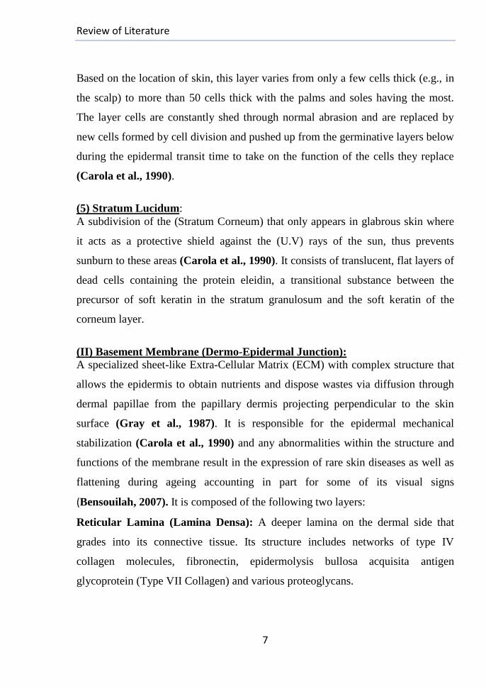
(2)Stratum Spinosum (Prickle Cell Layer):
As basal cells reproduce and mature, they move towards the outer skin layer
forming initially the (Stratum Spinosum) that composes of several layers of mature
keratinocytes (polyhedral cells with delicate intercellular bridges of desmosomes,
(prickles) to give support to this binding layer) (Carola et al., 1990). Langerhans
cells are dendritic, immunologically active cells, derived from bone marrow and
found on all epidermal surfaces, but mainly located in the middle of this layer for
their antigen-presenting functions.
(3) Stratum Granulosum:
This layer lies just above the (Spinosum layer) with (2-4) cell thickness resulting
from maturation of lower layer cells and continue to flatten during their continuous
transition to the surface with loss of nuclei and the cytoplasm appears granular at
this level. The cells contain keratohyaline crystals, the precursor of soft keratin for
initiating the keratinisation process, associated with the process of cell death
(Carola et al., 1990).
(4) Stratum Corneum:
This is the flat outermost epidermal layer with relative thickness. It consists of
corneocytes (non-viable cornified cells of hexagonal shape arranged in parallel
rows) with the final outcome of keratinocytes maturation; each cell is surrounded
by a protein envelope of (fillagrin) and filled with water-retaining keratin whose
orientations with cells shape strengthen this layer. Stacked lipid bilayers surround
the cells in the extracellular space to give a structure that provides the natural
physical and water-retaining barrier functions of skin where the corneocyte can
absorb water, 3 times its weight.
Review of Literature
7
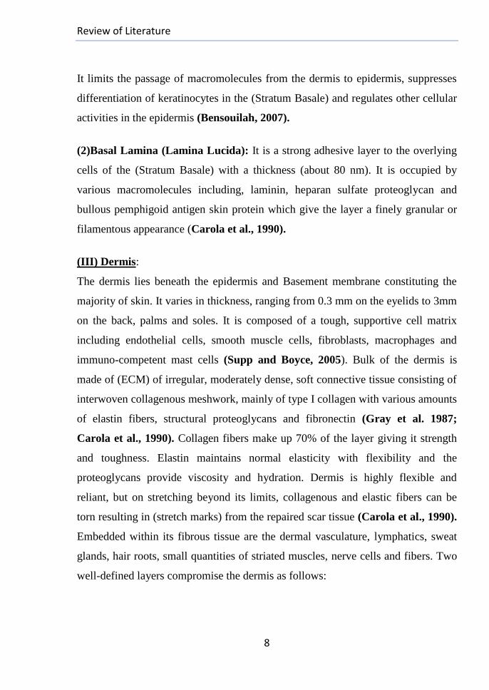
Based on the location of skin, this layer varies from only a few cells thick (e.g., in
the scalp) to more than 50 cells thick with the palms and soles having the most.
The layer cells are constantly shed through normal abrasion and are replaced by
new cells formed by cell division and pushed up from the germinative layers below
during the epidermal transit time to take on the function of the cells they replace
(Carola et al., 1990).
(5) Stratum Lucidum:
A subdivision of the (Stratum Corneum) that only appears in glabrous skin where
it acts as a protective shield against the (U.V) rays of the sun, thus prevents
sunburn to these areas (Carola et al., 1990). It consists of translucent, flat layers of
dead cells containing the protein eleidin, a transitional substance between the
precursor of soft keratin in the stratum granulosum and the soft keratin of the
corneum layer.
(II) Basement Membrane (Dermo-Epidermal Junction):
A specialized sheet-like Extra-Cellular Matrix (ECM) with complex structure that
allows the epidermis to obtain nutrients and dispose wastes via diffusion through
dermal papillae from the papillary dermis projecting perpendicular to the skin
surface (Gray et al., 1987). It is responsible for the epidermal mechanical
stabilization (Carola et al., 1990) and any abnormalities within the structure and
functions of the membrane result in the expression of rare skin diseases as well as
flattening during ageing accounting in part for some of its visual signs
(Bensouilah, 2007). It is composed of the following two layers:
Reticular Lamina (Lamina Densa): A deeper lamina on the dermal side that
grades into its connective tissue. Its structure includes networks of type IV
collagen molecules, fibronectin, epidermolysis bullosa acquisita antigen
glycoprotein (Type VII Collagen) and various proteoglycans.
Review of Literature
8
It limits the passage of macromolecules from the dermis to epidermis, suppresses
differentiation of keratinocytes in the (Stratum Basale) and regulates other cellular
activities in the epidermis (Bensouilah, 2007).
(2)Basal Lamina (Lamina Lucida): It is a strong adhesive layer to the overlying
cells of the (Stratum Basale) with a thickness (about 80 nm). It is occupied by
various macromolecules including, laminin, heparan sulfate proteoglycan and
bullous pemphigoid antigen skin protein which give the layer a finely granular or
filamentous appearance (Carola et al., 1990).
(III) Dermis:
The dermis lies beneath the epidermis and Basement membrane constituting the
majority of skin. It varies in thickness, ranging from 0.3 mm on the eyelids to 3mm
on the back, palms and soles. It is composed of a tough, supportive cell matrix
including endothelial cells, smooth muscle cells, fibroblasts, macrophages and
immuno-competent mast cells (Supp and Boyce, 2005). Bulk of the dermis is
made of (ECM) of irregular, moderately dense, soft connective tissue consisting of
interwoven collagenous meshwork, mainly of type I collagen with various amounts
of elastin fibers, structural proteoglycans and fibronectin (Gray et al. 1987;
Carola et al., 1990). Collagen fibers make up 70% of the layer giving it strength
and toughness. Elastin maintains normal elasticity with flexibility and the
proteoglycans provide viscosity and hydration. Dermis is highly flexible and
reliant, but on stretching beyond its limits, collagenous and elastic fibers can be
torn resulting in (stretch marks) from the repaired scar tissue (Carola et al., 1990).
Embedded within its fibrous tissue are the dermal vasculature, lymphatics, sweat
glands, hair roots, small quantities of striated muscles, nerve cells and fibers. Two
well-defined layers compromise the dermis as follows:
Review of Literature
9
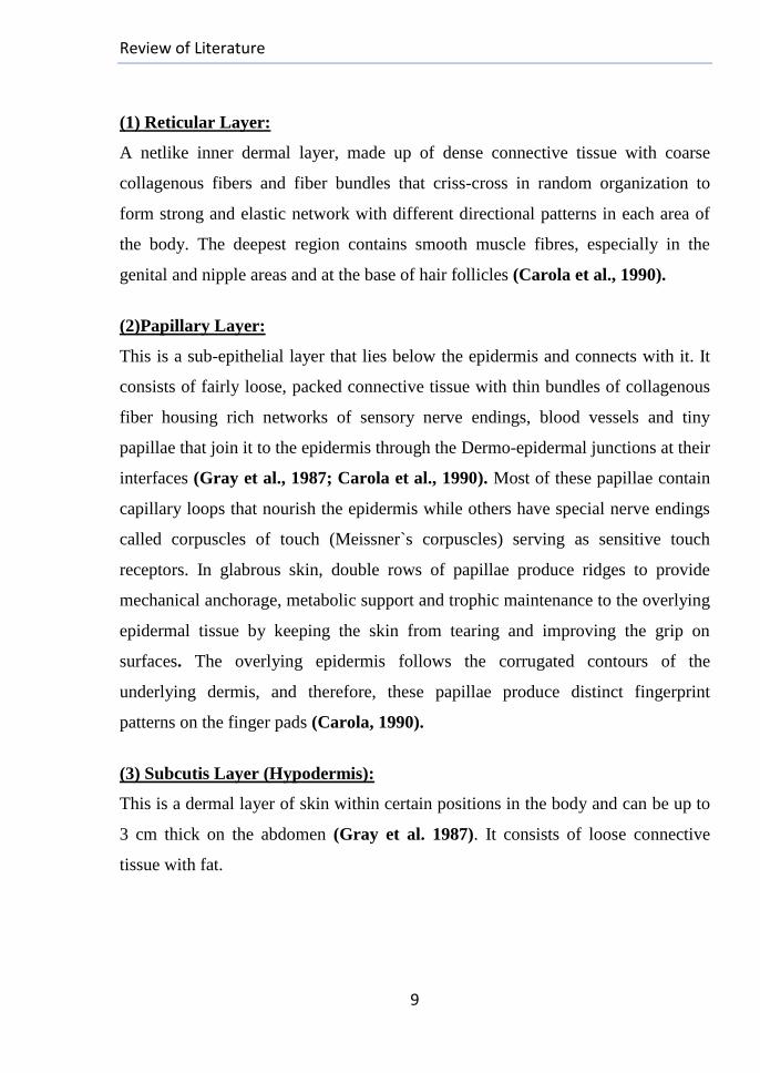
(1) Reticular Layer:
A netlike inner dermal layer, made up of dense connective tissue with coarse
collagenous fibers and fiber bundles that criss-cross in random organization to
form strong and elastic network with different directional patterns in each area of
the body. The deepest region contains smooth muscle fibres, especially in the
genital and nipple areas and at the base of hair follicles (Carola et al., 1990).
(2)Papillary Layer:
This is a sub-epithelial layer that lies below the epidermis and connects with it. It
consists of fairly loose, packed connective tissue with thin bundles of collagenous
fiber housing rich networks of sensory nerve endings, blood vessels and tiny
papillae that join it to the epidermis through the Dermo-epidermal junctions at their
interfaces (Gray et al., 1987; Carola et al., 1990). Most of these papillae contain
capillary loops that nourish the epidermis while others have special nerve endings
called corpuscles of touch (Meissner`s corpuscles) serving as sensitive touch
receptors. In glabrous skin, double rows of papillae produce ridges to provide
mechanical anchorage, metabolic support and trophic maintenance to the overlying
epidermal tissue by keeping the skin from tearing and improving the grip on
surfaces. The overlying epidermis follows the corrugated contours of the
underlying dermis, and therefore, these papillae produce distinct fingerprint
patterns on the finger pads (Carola, 1990).
(3) Subcutis Layer (Hypodermis):
This is a dermal layer of skin within certain positions in the body and can be up to
3 cm thick on the abdomen (Gray et al. 1987). It consists of loose connective
tissue with fat.
Review of Literature
10
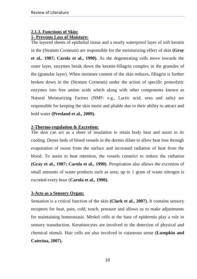
2.1.3. Functions of Skin:
1- Prevents Loss of Moisture:
The layered sheets of epithelial tissue and a nearly waterproof layer of soft keratin
in the (Stratum Corneum) are responsible for the moisturizing effect of skin (Gray
et al., 1987; Carola et al., 1990). As the degenerating cells move towards the
outer layer, enzymes break down the keratin-fillagrin complex in the granules of
the (granular layer). When moisture content of the skin reduces, fillagrin is further
broken down in the (Stratum Corneum) under the action of specific proteolytic
enzymes into free amino acids which along with other components known as
Natural Moisturizing Factors (NMF: e.g., Lactic acid, urea and salts) are
responsible for keeping the skin moist and pliable due to their ability to attract and
hold water (Presland et al., 2009).
2-Thermo-regulation & Excretion:
The skin can act as a sheet of insulation to retain body heat and assist in its
cooling. Dense beds of blood vessels in the dermis dilate to allow heat loss through
evaporation of sweat from the surface and increased radiation of heat from the
blood. To assist in heat retention, the vessels constrict to reduce the radiation
(Gray et al., 1987; Carola et al., 1990). Perspiration also allows the excretion of
small amounts of waste products such as urea; up to 1 gram of waste nitrogen is
excreted every hour (Carola et al., 1990).
3-Acts as a Sensory Organ:
Sensation is a critical function of the skin (Clark et al., 2007). It contains sensory
receptors for heat, pain, cold, touch, pressure and allows us to make adjustments
for maintaining homeostasis. Merkel cells at the base of epidermis play a role in
sensory transduction. Keratinocytes are involved in the detection of physical and
chemical stimuli. Hair cells are also involved in cutaneous sense (Lumpkin and
Caterina, 2007).
Review of Literature
11
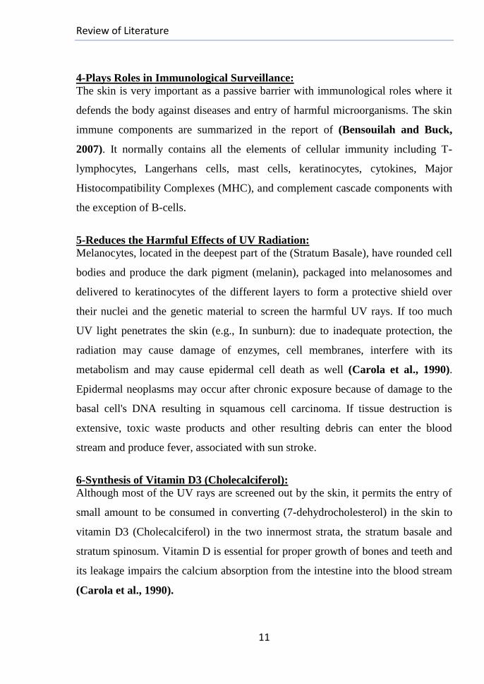
4-Plays Roles in Immunological Surveillance:
The skin is very important as a passive barrier with immunological roles where it
defends the body against diseases and entry of harmful microorganisms. The skin
immune components are summarized in the report of (Bensouilah and Buck,
2007). It normally contains all the elements of cellular immunity including T-
lymphocytes, Langerhans cells, mast cells, keratinocytes, cytokines, Major
Histocompatibility Complexes (MHC), and complement cascade components with
the exception of B-cells.
5-Reduces the Harmful Effects of UV Radiation:
Melanocytes, located in the deepest part of the (Stratum Basale), have rounded cell
bodies and produce the dark pigment (melanin), packaged into melanosomes and
delivered to keratinocytes of the different layers to form a protective shield over
their nuclei and the genetic material to screen the harmful UV rays. If too much
UV light penetrates the skin (e.g., In sunburn): due to inadequate protection, the
radiation may cause damage of enzymes, cell membranes, interfere with its
metabolism and may cause epidermal cell death as well (Carola et al., 1990).
Epidermal neoplasms may occur after chronic exposure because of damage to the
basal cell's DNA resulting in squamous cell carcinoma. If tissue destruction is
extensive, toxic waste products and other resulting debris can enter the blood
stream and produce fever, associated with sun stroke.
6-Synthesis of Vitamin D3 (Cholecalciferol):
Although most of the UV rays are screened out by the skin, it permits the entry of
small amount to be consumed in converting (7-dehydrocholesterol) in the skin to
vitamin D3 (Cholecalciferol) in the two innermost strata, the stratum basale and
stratum spinosum. Vitamin D is essential for proper growth of bones and teeth and
its leakage impairs the calcium absorption from the intestine into the blood stream
(Carola et al., 1990).

Review of Literature
12
7-It provides a protective barrier against mechanical, thermal, physical injury and
noxious agents.
8-Skin has also importance in the cosmetic, social and sexual associations.
2.1.4. Skin Wounds
2.1.4.1. Definition:
When the integrity of any tissue is compromised (e.g., Skin breaks, muscle tears,
burns, or bone fractures), a wound occurs. Skin wounds may be result of a fall,
surgical procedures; an infectious disease or by an underlying condition.
2.1.4.2. Description:
Types and causes of skin wounds are wide ranging with different ways of
classification. They may be acute wounds which normally proceed through an
orderly and timely reparative process resulting in sustained restoration of anatomic
and functional integrity through healing (Cohen et al., 1999). The other type is the
chronic wound that has failed to proceed through an orderly and timely process to
produce the required integrity due to compromised wound physiology (Lazarus et
al., 1994); examples include skin ulcers caused by diabetes, venous stasis or
prolonged local pressure.
2.1.4.3. Classification of Wounds:
(1) Open Wounds: Wounds in which the skin has been compromised and the
underlying tissues were exposed. The acute open wounds can be categorized
according to the relevant mechanism of injury into:
I-Abrasions (Scrapes): Superficial wounds in which the topmost layer of skin is
scraped off and rubbed away by friction against a rough surface.
II-Avulsions: Occur when an entire structure or part of it is forcibly pulled away
(e.g., Loss of a permanent tooth or an ear lobe, also with animal bites).
Review of Literature
13
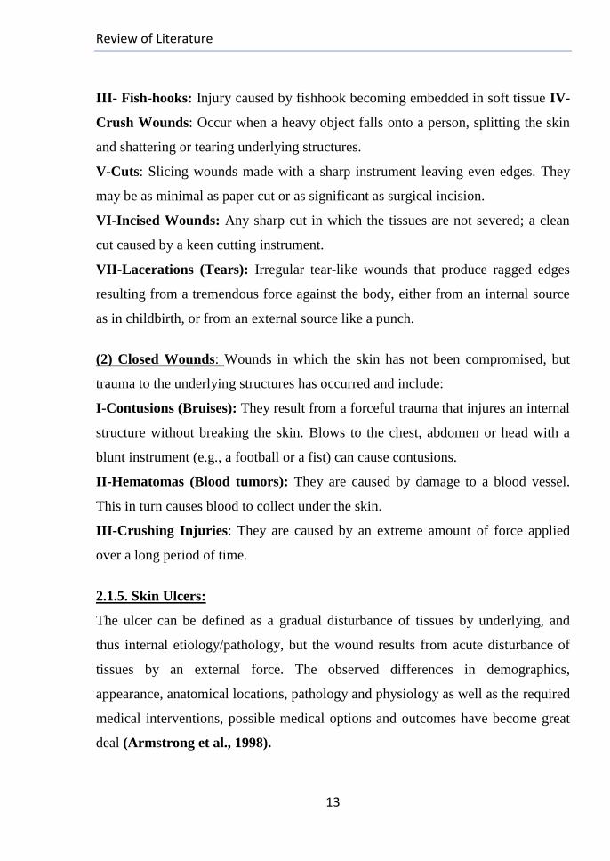
III- Fish-hooks: Injury caused by fishhook becoming embedded in soft tissue IV-
Crush Wounds: Occur when a heavy object falls onto a person, splitting the skin
and shattering or tearing underlying structures.
V-Cuts: Slicing wounds made with a sharp instrument leaving even edges. They
may be as minimal as paper cut or as significant as surgical incision.
VI-Incised Wounds: Any sharp cut in which the tissues are not severed; a clean
cut caused by a keen cutting instrument.
VII-Lacerations (Tears): Irregular tear-like wounds that produce ragged edges
resulting from a tremendous force against the body, either from an internal source
as in childbirth, or from an external source like a punch.
(2) Closed Wounds: Wounds in which the skin has not been compromised, but
trauma to the underlying structures has occurred and include:
I-Contusions (Bruises): They result from a forceful trauma that injures an internal
structure without breaking the skin. Blows to the chest, abdomen or head with a
blunt instrument (e.g., a football or a fist) can cause contusions.
II-Hematomas (Blood tumors): They are caused by damage to a blood vessel.
This in turn causes blood to collect under the skin.
III-Crushing Injuries: They are caused by an extreme amount of force applied
over a long period of time.
2.1.5. Skin Ulcers:
The ulcer can be defined as a gradual disturbance of tissues by underlying, and
thus internal etiology/pathology, but the wound results from acute disturbance of
tissues by an external force. The observed differences in demographics,
appearance, anatomical locations, pathology and physiology as well as the required
medical interventions, possible medical options and outcomes have become great
deal (Armstrong et al., 1998).
Review of Literature
14
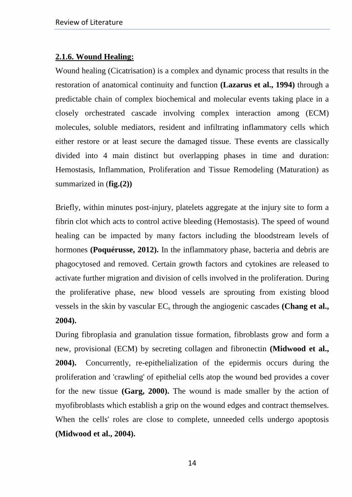
2.1.6. Wound Healing:
Wound healing (Cicatrisation) is a complex and dynamic process that results in the
restoration of anatomical continuity and function (Lazarus et al., 1994) through a
predictable chain of complex biochemical and molecular events taking place in a
closely orchestrated cascade involving complex interaction among (ECM)
molecules, soluble mediators, resident and infiltrating inflammatory cells which
either restore or at least secure the damaged tissue. These events are classically
divided into 4 main distinct but overlapping phases in time and duration:
Hemostasis, Inflammation, Proliferation and Tissue Remodeling (Maturation) as
summarized in (fig.(2))
Briefly, within minutes post-injury, platelets aggregate at the injury site to form a
fibrin clot which acts to control active bleeding (Hemostasis). The speed of wound
healing can be impacted by many factors including the bloodstream levels of
hormones (Poquérusse, 2012). In the inflammatory phase, bacteria and debris are
phagocytosed and removed. Certain growth factors and cytokines are released to
activate further migration and division of cells involved in the proliferation. During
the proliferative phase, new blood vessels are sprouting from existing blood
vessels in the skin by vascular ECs through the angiogenic cascades (Chang et al.,
2004).
During fibroplasia and granulation tissue formation, fibroblasts grow and form a
new, provisional (ECM) by secreting collagen and fibronectin (Midwood et al.,
2004).
Concurrently, re-epithelialization of the epidermis occurs during the
proliferation and 'crawling' of epithelial cells atop the wound bed provides a cover
for the new tissue (Garg, 2000). The wound is made smaller by the action of
myofibroblasts which establish a grip on the wound edges and contract themselves.
When the cells' roles are close to complete, unneeded cells undergo apoptosis
(Midwood et al., 2004).
Review of Literature
15
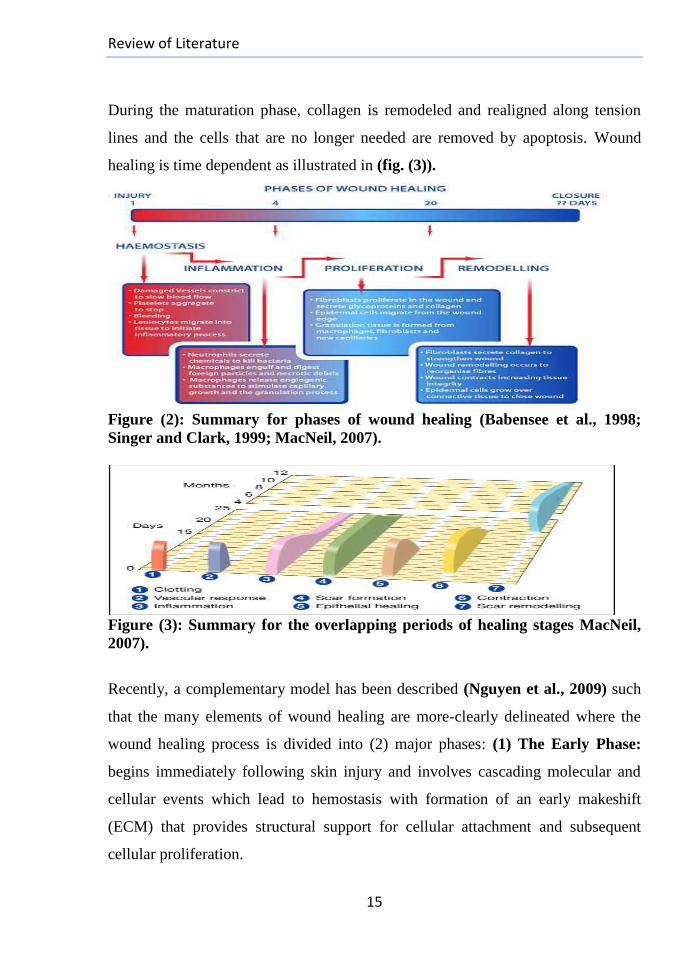
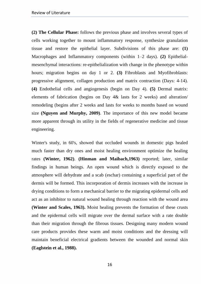
During the maturation phase, collagen is remodeled and realigned along tension
lines and the cells that are no longer needed are removed by apoptosis. Wound
healing is time dependent as illustrated in (fig. (3)).
Figure (2): Summary for phases of wound healing (Babensee et al., 1998;
Singer and Clark, 1999; MacNeil, 2007).
Figure (3): Summary for the overlapping periods of healing stages MacNeil,
2007).
Recently, a complementary model has been described (Nguyen et al., 2009) such
that the many elements of wound healing are more-clearly delineated where the
wound healing process is divided into (2) major phases: (1) The Early Phase:
begins immediately following skin injury and involves cascading molecular and
cellular events which lead to hemostasis with formation of an early makeshift
(ECM) that provides structural support for cellular attachment and subsequent
cellular proliferation.
Review of Literature
16
(2) The Cellular Phase: follows the previous phase and involves several types of
cells working together to mount inflammatory response, synthesize granulation
tissue and restore the epithelial layer. Subdivisions of this phase are: (1)
Macrophages and Inflammatory components (within 1–2 days). (2) Epithelial-
mesenchymal interactions: re-epithelialization with change in the phenotype within
hours; migration begins on day 1 or 2. (3) Fibroblasts and Myofibroblasts:
progressive alignment, collagen production and matrix contraction (Days: 4-14).
(4) Endothelial cells and angiogenesis (begin on Day 4). (5) Dermal matrix:
elements of fabrication (begins on Day 4& lasts for 2 weeks) and alteration/
remodeling (begins after 2 weeks and lasts for weeks to months based on wound
size (Nguyen and Murphy, 2009). The importance of this new model became
more apparent through its utility in the fields of regenerative medicine and tissue
engineering.
Winter's study, in 60's, showed that occluded wounds in domestic pigs healed
much faster than dry ones and moist healing environment optimize the healing
rates (Winter, 1962). (Hinman and Maibach,1963) reported; later, similar
findings in human beings. An open wound which is directly exposed to the
atmosphere will dehydrate and a scab (eschar) containing a superficial part of the
dermis will be formed. This incorporation of dermis increases with the increase in
drying conditions to form a mechanical barrier to the migrating epidermal cells and
act as an inhibitor to natural wound healing through reaction with the wound area
(Winter and Scales, 1963). Moist healing prevents the formation of these crusts
and the epidermal cells will migrate over the dermal surface with a rate double
than their migration through the fibrous tissues. Designing many modern wound
care products provides these warm and moist conditions and the dressing will
maintain beneficial electrical gradients between the wounded and normal skin
(Eaglstein et al., 1988).
Review of Literature
17
2.1.7. Classification of Wound Dressing Products:
2.1.7.1. Major Classifications of Wound Dressing Products:
Today, there is a wide variety of products to choose from that can lead to
confusion and, sometimes, choosing the wrong type for treating a particular
wound. Knowing the available types of dressings, their uses and the limits of usage
for certain wounds may be a difficult decision in the management of wound care.
Although there are hundreds of them to choose from, the dressings fall into the
following few categories from a clinical point of view.
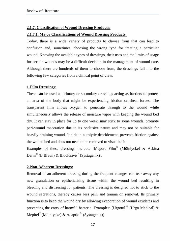
1-Film Dressings:
These can be used as primary or secondary dressings acting as barriers to protect
an area of the body that might be experiencing friction or shear forces. The
transparent film allows oxygen to penetrate through to the wound while
simultaneously allows the release of moisture vapor with keeping the wound bed
dry. It can stay in place for up to one week, may stick to some wounds, promote
peri-wound maceration due to its occlusive nature and may not be suitable for
heavily draining wound. It aids in autolytic debridement, prevents friction against
the wound bed and does not need to be removed to visualize it.
Examples of these dressings include: [Mepore Film® (Mölnlycke) & Askina
Derm® (B Braun) & Bioclusive
™ (Systagenix)].
2-Non-Adherent Dressings:
Removal of an adherent dressing during the frequent changes can tear away any
new granulation or epithelialising tissue within the wound bed resulting in
bleeding and distressing for patients. The dressing is designed not to stick to the
wound secretions, thereby causes less pain and trauma on removal. Its primary
function is to keep the wound dry by allowing evaporation of wound exudates and
preventing the entry of harmful bacteria. Examples: [Urgotul ®
(Urgo Medical) &
Mepitel®
(Mölnlycke) & Adaptic ™
(Systagenix)].
Review of Literature
18
Paraffin gauze dressings and synthetic bandages belong to this category, but they
are no longer recommended for use on open wounds (NICE, 2008), though they
are readily available and cheaper than others.
3-Simple Island Dressings:
Examples include dressings with central pad of cellulose material to be used over a
suture line of wounds closed by primary intention to absorb any oozing during the
first post-surgery 24 hours. Other examples include [Alldress® (Mölnlycke) &
Primapore® (Smith and Nephew) & Medipore
™ Pad (3M
™)].
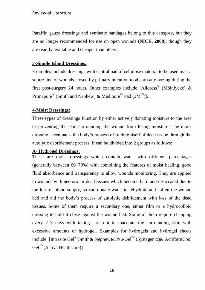
4-Moist Dressings:
These types of dressings function by either actively donating moisture to the area
or preventing the skin surrounding the wound from losing moisture. The moist
dressing accentuates the body’s process of ridding itself of dead tissue through the
autolytic debridement process. It can be divided into 2 groups as follows:
A- Hydrogel Dressings:
These are moist dressings which contain water with different percentages
(generally between 60–70%) with combining the features of moist healing, good
fluid absorbance and transparency to allow wounds monitoring. They are applied
to wounds with necrotic or dead tissues which become hard and desiccated due to
the loss of blood supply, so can donate water to rehydrate and soften the wound
bed and aid the body’s process of autolytic debridement with loss of the dead
tissues. Some of them require a secondary one, either film or a hydrocolloid
dressing to hold it close against the wound bed. Some of them require changing
every 2–3 days with taking care not to macerate the surrounding skin with
excessive amounts of hydrogel. Examples for hydrogels and hydrogel sheets
include: [Intrasite Gel®(Smith& Nephew)& Nu-Gel
™ (Systagenix)& ActiformCool
Gel ™
(Activa Healthcare)]
Review of Literature
19
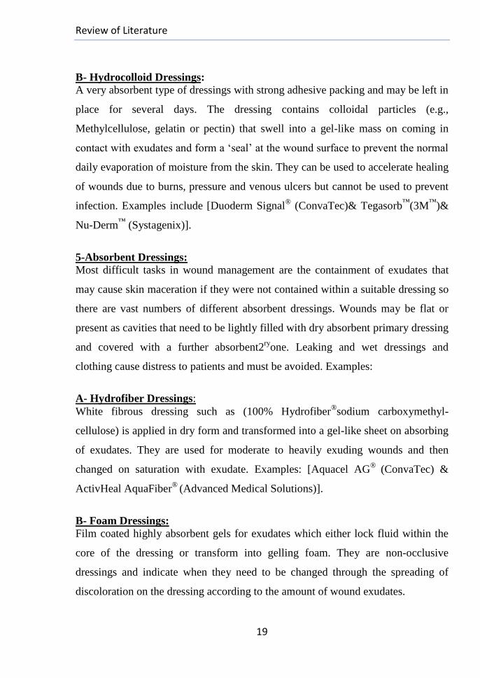
B- Hydrocolloid Dressings:
A very absorbent type of dressings with strong adhesive packing and may be left in
place for several days. The dressing contains colloidal particles (e.g.,
Methylcellulose, gelatin or pectin) that swell into a gel-like mass on coming in
contact with exudates and form a ‘seal’ at the wound surface to prevent the normal
daily evaporation of moisture from the skin. They can be used to accelerate healing
of wounds due to burns, pressure and venous ulcers but cannot be used to prevent
infection. Examples include [Duoderm Signal® (ConvaTec)& Tegasorb
™(3M
™)&
Nu-Derm™
(Systagenix)].
5-Absorbent Dressings:
Most difficult tasks in wound management are the containment of exudates that
may cause skin maceration if they were not contained within a suitable dressing so
there are vast numbers of different absorbent dressings. Wounds may be flat or
present as cavities that need to be lightly filled with dry absorbent primary dressing
and covered with a further absorbent2ry
one. Leaking and wet dressings and
clothing cause distress to patients and must be avoided. Examples:
A- Hydrofiber Dressings:
White fibrous dressing such as (100% Hydrofiber®sodium carboxymethyl-
cellulose) is applied in dry form and transformed into a gel-like sheet on absorbing
of exudates. They are used for moderate to heavily exuding wounds and then
changed on saturation with exudate. Examples: [Aquacel AG®
(ConvaTec) &
ActivHeal AquaFiber®
(Advanced Medical Solutions)].
B- Foam Dressings:
Film coated highly absorbent gels for exudates which either lock fluid within the
core of the dressing or transform into gelling foam. They are non-occlusive
dressings and indicate when they need to be changed through the spreading of
discoloration on the dressing according to the amount of wound exudates.
Review of Literature
20
If not changed often enough, this may promote peri-wound maceration. Some
foam may not be suitable for certain wounds, such as those that are infected or
tunneling. Examples include: [Allevyn AG® (Smith and Nephew) &Mepilex
Border® (Mölnlycke)].
C-Alginate Dressings:
They absorb exudates to form gel-like covering over the wound and the way of
absorption is dependent on the alginate makeup. They have many different
available non-adherent types which encourage the autolytic debridement. Some
alginate dressings retain their integrity and can be removed in one piece; others
disintegrate and need to be irrigated away from the wound bed. Alginate dressing
may be used for venous ulcers, infected wounds and those with tunneling or heavy
exudates. It can be used to lightly fill a cavity but needs to be covered by a
secondary one.
6-Composite Dressings (Composites):
This category involves a combination of types of dressings that may be used for a
variety of wounds either as primary or secondary dressings. These types are merely
of moisture retentive properties, in addition to using gauze dressing. Despite their
wide availability and usage simplicity, they may be more expensive and difficult to
store than other types with less choice/flexibility in use indications
Wound dressings may be also classified based on their nature of action as:
A-Passive Products: Include the traditional dressings which account for the
largest market product level (e.g., Gauze and tulle dressings) with a minimal role
in the healing process (Yannas and Burke, 1980).
B-Interactive Products: Include dressings in polymeric forms that are
recommended for low exuding wounds. These films are generally transparent,
permeable to water vapor and oxygen but not to bacteria.
Review of Literature
21
C-Bioactive Products: They deliver active substances to wounds during healing,
may be bioactive compounds or the dressing itself is constructed from materials
having endogenous activities. These materials include proteoglycans, collagen,
non-collagenous proteins, alginates and chitosan. Properties and different types of
alginate as well as chitosan-based wound dressings are summarized in the review
of (Paul and Sharma, 2004).
2.1.7.2. Topical Chemotherapy for Wounds:
Several studies have been performed to identify fundamental substances of
angiogenic activities and direct action in promoting the repair process with
improving the survival of wounded patients. The following are examples:
1-Some enzyme-based ointments (e.g., DNAses and collagenases) act to promote
wound debridement and assist in the restoration of tissue (Hebda et al., 1990).
2-Some growth factors are among the substances, used in topical chemotherapies
where they demonstrate good abilities to accelerate tissue repair on topical
application to the wounds in experimental animals (Pierce and Tarpley, 1994)
(e.g., Recombinant human Platelet-Derived Growth Factor (PDGF)-based drugs
were found to directly interfere with the healing steps to favor the repair process
with showing good results in the healing of diabetic ulcers) (Steed, 1998). Some
angiogenic growth factors and inhibitors are listed in (Table: 1); they have begun
to receive U.S. Food and Drug Administration (FDA) approval by 2003.
3-Silver is reemerging as a viable treatment option for infections encountered in
burns, open wounds and chronic ulcers. It may be in the form of Silver salts (e.g.,
AgNO3), Silver compounds (e.g., Silver sulfadiazine (SSD)), Silver proteins,
electrically charged colloidal silver solutions and sustained silver releasing systems
such as Nano-crystalline silver (Carneiro et al., 2002; Carsin et al., 2004).
Review of Literature
22
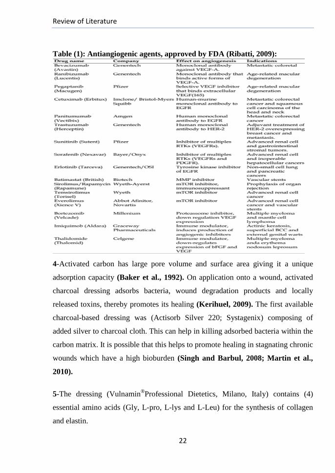
Table (1): Antiangiogenic agents, approved by FDA (Ribatti, 2009):
4-Activated carbon has large pore volume and surface area giving it a unique
adsorption capacity (Baker et al., 1992). On application onto a wound, activated
charcoal dressing adsorbs bacteria, wound degradation products and locally
released toxins, thereby promotes its healing (Kerihuel, 2009). The first available
charcoal-based dressing was (Actisorb Silver 220; Systagenix) composing of
added silver to charcoal cloth. This can help in killing adsorbed bacteria within the
carbon matrix. It is possible that this helps to promote healing in stagnating chronic
wounds which have a high bioburden (Singh and Barbul, 2008; Martin et al.,
2010).
5-The dressing (Vulnamin®Professional Dietetics, Milano, Italy) contains (4)
essential amino acids (Gly, L-pro, L-lys and L-Leu) for the synthesis of collagen
and elastin.
Review of Literature
23
It can modulate the inflammatory response with a reduction in the number of
inflammatory cells, an increase in fibroblast distribution density and it aids in the
synthesis of thin collagen fibers resulting in reduction in the healing time (Corsetti
et al., 2010).
6-The polysaccharides, chitosan and alginates in particular, are ideal materials for
the construction of dressings suitable for wound healing during its various phases
due to their specific biological properties including hemostasis, granulation and
epithelisation (Muzzarelli, 1993) as will be explained in section (5) of the review.
2.1.7.3. Bioengineering and Hydrogels in Wound Healing:
2.1.7.3.1. Bioengineering and Scaffolds System:
Bioengineering is defined as the science that puts efforts in designing and
manufacturing of spare parts for functional restoration of the impaired organs and
replacement of lost parts due to disease, trauma or tumors (Reddi, 1998), so it
rapidly became one of the most promising treatment options for patients suffering
from tissue failure. It is a multidisciplinary field incorporating the principles of
developmental biology, physiological modeling, chemistry, physics
,morphogenesis, kinetics, microfluidics and cell targeting gearing toward creating
biological substitutes of native tissues to replace, repair or augment diseased
tissues and it concerns itself more with the biological questions. Biomaterials,
Tissue engineering, Biomedical Engineering, Drug delivery and Biomechanics are
considered Bioengineering fields because of their strong dependence on the basic
science with more translational/medical applications. (Biomaterials) is a term used
for both: (1) The engineering of materials for and from biology; and (2) The study
of the interaction of materials with biology.
Tissue Engineering refers, generally, to the process of engineering or directing the
repair of tissues, but can also be applied to technologies outside of the body such
as to the building of tissues constructs for in vitro experimentation.
Review of Literature
24
Regenerative medicine is often used synonymously with tissue engineering,
although those involved in regenerative medicine place more emphasis on the use
of stem cells to produce tissues. There are four fundamental technologies in
bioengineering: (1) The scaffolding for cell proliferation and differentiation, (2)
The isolation and culturing of cells, (3) The drug delivery system (DDS) of bio-
growth factor and (4) The maintenance of space to induce tissue regeneration.
The cells can be seeded on biodegradable polymer which serves several purposes:
It functions as a cell-delivery system that enables the transplantation of many cells
into an organism and creates a three-dimensional (3D) space for cells growth
serving as a template which can provide structural cues to direct tissue
development. The matrix temporarily provides the necessary biomechanical
support in the construct while the cells lay down their own ECM which ultimately
provides the structural integrity and biomechanical profile of the engineered tissue
(Terada et al., 2000). One of the essential properties of the used tissue guiding
scaffold is to be biodegradable while providing therapeutic functions on degrading
during replacement of the artificial matrix with a physiological one of the cellular
system. If the polymer is completely absorbed into the body, the long term foreign
body reaction can be eliminated with leaving only the natural regenerated matrix.
Nature of the material has been a subject of extensive studies including different
types of both natural and synthetic origins; the issue of optimal guidance for the
ECM is crucial one (Zhao et al., report).
For successful regeneration therapy of tissues and organs, it is important and
indispensable to develop the technology and methodology of tissue engineering
with molecular designing of a biomaterial acting as an intact scaffold for cells as
well as the DDS technologies of bio-signaling molecules for creating a local
environment which enhances the proliferation of cells and induces cell-based tissue
regeneration.
Review of Literature
25
Growth factors are often required to promote tissue regeneration; they can induce
angiogenesis to promote sufficient supply of oxygen and nutrients for maintaining
the biological functions of cells transplanted for effective organ substitution.
2.1.7.3.2. Hydrogels as Wound Dressings:
Hydrogels are polymeric three-dimensional networks imbibing a large fraction of
aqueous medium and yet remain intact even given infinite time period without
dissolving. The hydrophilic polymer chains ensemble in the hydrogel, representing
the skeleton of gel, is somehow interacting with each other either by virtue of
covalent bonds or by interacting physically in cross-linking points as a network or
single mass (Kim et al., 1992) so as to keep the individual chains from diffusing
away into the aqueous milieu. The liquid in gel prevents its network from
collapsing into a compact mass and the network prevents its flowing away
(Tanaka et al., 1981). The network strands can be surrounded with the solvent
molecules, thereby push neighbor chains away and swell with occupying larger
volume. Thus, the hydrogel can be considered as intermediate matter state between
solid and liquid with maintaining its shape under the stress of its own weight.
Many extracellular structures which embed cells in the body can be considered as
(Hydrogels). The (ECM) of soft tissues and cartilage, for example, exists as a
network of glycoproteins and proteoglycans that both interact with each other
biophysically. Hydrogels of both natural and synthetic origin have been proposed
also as ECM analogues (Fonseca et al., 2011) due to their structural similarities to
the body macro molecular-based components so they met numerous applications.
Examples include: drugs delivery, medical prosthetic materials, antistatic coatings,
encapsulation materials for immunoisolation-based cell therapeutics, wound
dressings (Stile et al., 1999 ;Lee et al., 2001), as well as in soft contact lenses, gel
electrophoresis, anti-adhesion materials, environmental and chemical detectors
(Silva et al., 2006).
Review of Literature
26
These hydrogels are also used as tissue engineering scaffolds, structures for filling
the irregularly shaped defects. In addition, they are used in general
macromolecular research with easy means of delivery for the bioactive molecules
into the body in a minimally invasive manner (Lee et al., 2001).They can be
designed to provide instructive environments for the 3D assembly of vascular
networks.
Hydrogels made from natural polymers such as alginate, chitosan, collagen,
hyaluronate (Denuzière et al., 2000; Chen and Cheng, 2009) or dextran (Kikuchi
et al., 1997) are frequently used as scaffolding materials in tissue regeneration
strategies as they are either components of or have similar macromolecular
structure to constituents of the natural tissues (ECM). Many studies of hydrogel-
based scaffolds have focused on their applications in the healing of wounds
(Balakrishnan et al., 2005b; Boucard et al., 2007; Kim et al., 2009; Shepherd
et al., 2011). They can also deliver growth factors (Kiyozumi et al., 2006), cells
(Liu et al., 2009) and antibiotics (Shepherd et al., 2011) to allow complete skin
regeneration.

Review of Literature
27
(2): Review on Alginates
2.2.1. Chemical Structure of Alginate:
Based on description of the British chemist E. E. C. Stanford in 1881, alginate is a
random unbranched heteropolysaccharide with repeated two kinds of (1→4)
covalently linked monomers [ß-D-mannuronate (M) and its C5 epimer α-L-
guluronate (G)] in different sequences of varying proportions. They appear in
homopolymeric blocks fashion of consecutive G-residues (Polyguluronates; GGG-
blocks), consecutive M-residues (Poly mannuronates; MMM-blocks) and
heteropolymeric blocks of alternating randomly organized uronates (MGM-
blocks) (Sutherland et al., 1991).
-As shown in (fig. (4)), the monomers in the polymer chain have a tendency to
stay in their most energetically favorable structure. For M-M, this is the 4C1 chair
form, linked by β-(1, 4) glycosidic bond, but it is the 1C4 chair form for G-G,
linked by α-(1, 4) glycosidic bond (Yang et al., 2006). (G) and (M) residues adopt
axial and equatorial configurations, respectively; the M blocks have extended
ribbon form, G blocks are rigid and buckled and the MG-regions are of
intermediate rigidity (Grant et al., 1973).
Figure (4): Schematically drawn alginate block structure with a segment
showing structure of the molecules (Smidsrød et al., 1995).
Review of Literature
28
Alginate solubility is affected by primary structure of the polymer, ionic strength
and pH (d’Ayala et al., 2008). Due to its functional groups (-COO-and OH
-),
alginate can react readily with amino and amino derivative groups of other
polymers via electro-static interactions or with formation of Schiff bases or
amides.
2.2.2. Sources of Alginates:
Polysaccharides of algal origins are gaining particular attention due to their
peculiar chemical composition, renewability and abundance. For example, agar
and carrageenan that are extracted from red seaweeds (Hopkins et al., 2009) and
alginate from the brown seaweeds. Alginates are mainly alkaline extracted from
brown algae (phaeophyta, classe des Phaeophyceae), including the giant kelp
Macrocystis pyrifera, Ascophyllum nodosum and various species of Laminaria
with alginate contents (20-40 % of the dry weight) (Black, 1950). Amount and
properties of alginate vary based on the organism species, its reproductive cycle,
growing conditions and the tissue it is isolated from (Haug, 1964; Moe et al.,
1995). Alginate is located in the intercellular matrix and cell wall in a gel form
containing Ca+2
, Mg+2
and other multivalent cations (Haug and Smidsrød, 1967)
with mainly skeletal functions by conferring both mechanical strength and
flexibility to the algal tissue for growth so plants growing in rough waters provide
alginate richer in (G-residues) compared to plants of the same species from calmer
waters (Ertesvåg et al., 1996).
Alginate-like polymers are synthesized by number of bacterial strains as
exocellular secretions. The gram-negative bacterium, Pseudomonas aeruginosa,
and the soil bacterium, Azotobacter vinelandii that can fix nitrogen under aerobic
growth conditions are examples for these genera (Johnson et al., 1997).
Review of Literature
29
2.2.3. Properties of Alginate:
1-Alginates, unlike other natural polysaccharides, look very promising due to their
unique biocompatibility with both host and enclosed cells, low mitogenic activity
and toxicity (Wang et al., 2011), abundance and renewability (Matsumoto et al.,
2003). They are amenable to sterilization and storage with ease of chemical
modification through simple chemistries (Briand and Tang, 2007). Under normal
physiological conditions, alginate is bioerodible with non-inflammatory
degradation products and has easy solubility without any harsh reaction
conditions.
2-Alginates could be candidates in many biomedical applications for preparing
many artificial matrices aiming to the regeneration of damaged tissues including
cartilage (Bouhadir et al., 2001), bone (Alsberg et al., 2001), liver (Chung et al.,
2002), cardiac tissue remodeling (Dar et al., 2002), dermatology and regeneration
of skin (Hashimoto et al., 2004).
3-Because they can mild gelate over wide range of temperatures with the ability to
retain water (d’Ayala et al., 2008), alginates have been successfully used as
matrices for the entrapment and/or delivery of biological agents (e.g., Drugs and
growth factors) without loss of the biological activity of these mitogenic molecules
and also as artificial matrices with scaffolding action for cells (Chinen et al.,
2003).
4- Alginate has a recognized GRAS status (Generally Recognized As Safe) with
constantly ensured quality (Ghidoni et al., 2008), so it has been widely used over
the last few years in food industries (e.g., Juices, stabilizer in ice cream) and many
other industrial interests (e.g., Salad dressings, cosmetics, slimming aids, paper
and textile scaffold manufacturing, waterproofing and fireproofing fabrics
(Bartels et al., 2011).
Review of Literature
30
5-With its different biomedical and pharmaceutical applications, alginate can be
used alone, in composites with other materials as well as in blends with certain
modifications, especially due to its limited interaction with the majority of
mammalian cells due to its hydrophilic character (Wang et al., 1995) that
promotes limited protein adsorption (Lee and Mooney, 2001). Examples of these
reacting positively charged materials include: Ethyl cellulose (Bodmeier and
Wang, 1993), Eudragit (Gürsoy et al., 1998), Pectin (Liu and Krishnan, 1999)
and Chitosan (Sezer and Akbuga, 1999). This improves the deficiencies within
the alginate structure, helps solve the problems with drug leaching during
preparation and imports it innovative properties (d’Ayala et al., 2008). Thus,
alginate can compete with the synthetic biodegradable excipients available in the
market with opening more and more new perspectives and potential applications in
the future.
2.2.4. Alginate Gelation:
In several applications of alginate, strong thermo-stable gels can be prepared prior
to use or spontaneously formed in situ in physiological fluids. Alginate gelation
can be achieved by one of the following methods:
1-Photo-Polymerization of alginate monomers allows creating a hydrogel
independent of the divalent cation levels to control the gelation timing and kinetics
(Jeon et al., 2009; Rouillard et al., 2011).
2-Enzymatic Cross-linking: (Martinsen et al., 1991).
3-Chemical gelation: It can be achieved by one of the 2 following methods:
A-Lowering the pH of Alginate Solution: Induces the formation of acid gel
(Alginic acid) by physical hydrogen bonding.
B-Chemical Cross-linking: This method involves the covalent and ionic cross-
linking via crosslinker ions. The covalent cross-linked alginate gels show higher
stability than those cross-linked ionically (Eiselt et al., 1999).
Review of Literature
31
I-Covalent Cross-linking:
Carried out via cross-linking agents (e.g., Carbodiimide (Rees and Welsh,
1977), glutaraldehyde or adipic dihydrazide (Maiti et al., 2009) where the (-
COO-) groups on the alginate chains are left unperturbed.
II-Ionic Cross-linking via metal ions (Gelling Salt) [Egg-Box Model]:
As a hydrophilic polyelectrolyte, alginate can be cross-linked with exchange of
monovalent ions from guluronates with multivalent counter ions at certain
stoichiometric ratios (Martinsen et al., 1989). The diaxially-linked G-residues
spontaneously form electronegative cavities functioning as binding sites for some
di and polyvalent cations (e.g., Ca+2
, Sr+2
, Ba+2
, Fe+3
, Al+3
) (Patil et al., 2010)
when the polyguluronate segment exceeds the critical length (Stokke et al., 1991)
with small distances between the junctions and of the same order of magnitude as
the Kuhn statistical segment length (Smidsrød et al., 1974) for cancelling the
negative charges by these ions. Alternatively, other multivalent cations (e.g.,
Mg+2
) form soluble polymers on binding to the G-residues (Smidsrød et al.,
1970).
(Calcium alginate gels) are produced in calcium setting bath by (2) cooperative
inter-chains binding mechanisms responsible for the formation of the junction
zones (Smidsrød et al., 1972):
1-Calcium ions in the solution make ionic bridges for two carboxyl group moieties
on the adjacent polymer chains (Coviello et al., 2007).
2-The other energetically favorable mechanism is the crosslinking via (-COO-
groups) by (primary valences) and via the electronegative oxygen atoms of the
[OH- groups: O (5) and O (4) in one unit and O (2) and O (3) in the preceding unit]
by (secondary valences) (Smidsrød et al., 1972; Angyal et al., 1973) making an
insoluble polymeric network described as the so called” Egg-Box model”,
illustrated in (fig.(5)) (Grant et al., 1973).
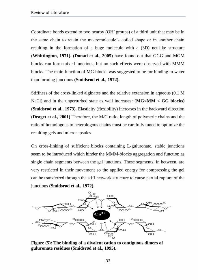
Review of Literature
32
Coordinate bonds extend to two nearby (OH- groups) of a third unit that may be in
the same chain to retain the macromolecule’s coiled shape or in another chain
resulting in the formation of a huge molecule with a (3D) net-like structure
(Whittington, 1971). (Donati et al., 2005) have found out that GGG and MGM
blocks can form mixed junctions, but no such effects were observed with MMM
blocks. The main function of MG blocks was suggested to be for binding to water
than forming junctions (Smidsrød et al., 1972).
Stiffness of the cross-linked alginates and the relative extension in aqueous (0.1 M
NaCl) and in the unperturbed state as well increases: (MG<MM < GG blocks)
(Smidsrød et al., 1973). Elasticity (flexibility) increases in the backward direction
(Draget et al., 2001) Therefore, the M/G ratio, length of polymeric chains and the
ratio of homologous to heterologous chains must be carefully tuned to optimize the
resulting gels and microcapsules.
On cross-linking of sufficient blocks containing L-guluronate, stable junctions
seem to be introduced which hinder the MMM-blocks aggregation and function as
single chain segments between the gel junctions. These segments, in between, are
very restricted in their movement so the applied energy for compressing the gel
can be transferred through the stiff network structure to cause partial rupture of the
junctions (Smidsrød et al., 1972).
Figure (5): The binding of a divalent cation to contiguous dimers of
guluronate residues (Smidsrød et al., 1995).
Review of Literature
33
The majorities of cross-links in the alginate gels are not permanent but move or
break when they are sheared (Mancini et al., 1999). While calcium levels are a
convenient means for controlling the properties of these gels (Brandl et al., 2007),
their physiological roles are important in many systems (Allgrove et al., 2009).
They can be either administered separately or added as part of the formulation
within the pharmaceutical preparation.
The solution viscosity, overall molecular weights, the block-wise structure of
alginate (Morris et al., 1980), Ca+2
ions concentration during gelation (Dumitriu,
1988), degree of cross-linking (Mitchell, 1980), method of gelation (Nunamaker
et al., 2007), number of monomers in a strand (N), the fraction of overall
guluronate residues in the polymer (FG value), number-average of guluronate units
in G-blocks (NG) (de Gennes, 1979), the sequential order of these residues
(Dumitriu, 1998), functionality of the cross-linking point (number of strands
connected to one crosslink, F), the average weight of strands between two
neighboring crosslinks (Mc), and sometimes, the presence of excipients in the
gelation bath (e.g., Na-hexametaphosphate (Van Wazer, 1958) and Glucono-δ-
lactone (Nussinovitch et al., 1990)) are all fundamentals to determine the
physicochemical properties of alginate, physiological and gelling properties,
mechanical strength, porosity, swelling, biocompatibility (Thu et al.,1996),
effectiveness in a given application and uniformity of the resultant gels (Klock et
al., 1994). Rate of diffusion of the reactants is considered the rate limiting step in
the gelation process (Martinsen et al., 1989).
In addition, (Amsden et al., 1999) reported that the greater the (G-content) of gel,
the higher affinity for cross-linkers and the greater is the restriction to solute
transport. Accordingly, alginates of high (G-content) can create transparent fibers
of more porous cross-linked gels with good stability towards competing Na+
ions.

Review of Literature
34
It will have also maintained mechanical integrity and rigidity for long periods
(Martinsen et al., 1989) and low degree of swelling. Conversely, alginate rich in
mannuronates can develop extra turbid elastic softer aggregates (Smidsrød et al.,
1972) with a high degree of swelling on calcium cross-linking (Grant et al., 1973)
and less proneness to syneresis (Nussinovitch, 1997). Alginates with high content
of the alternating sequence are characterized by low modulus, high volume and
flexibility.
The equilibrium of a freely swelling gel is determined by the interactions between
its network and the solvent. Generally in vitro, as Ca+2
ions are removed by
outward fluxing into the surrounding medium, the crosslinking in gel decreases
and becomes destabilized with loss of the mechanical stiffness over time (LeRoux
et al., 1999) due to the increased electrostatic repulsion between the (-COO-
anions) of alginate with increased swelling/ erosion (Kikuchi et al.,1997). The gel
is dissolved into dissociated individual chains with leakage of any entrapped
materials (Shoichet et al., 1996). These interactions are highly sensitive to
external conditions such as temperature, pH, presence of ions and external fields
(e.g., Magnetic, electric or pressure fields) (Vervoort, 2006).
Similar mechanism takes place in vivo where no hydrolytic or enzymatic chain
breakages occur within the alginate chains, but only softening of gel takes place
under physiological conditions forming absorbable alginate. This causes limited
quantities be safely left in situ accompanied with gradual disappearance of the
hydrogel and evacuation of the dissociated chains to be excreted by the kidneys,
especially on using alginates of modified molecular weights (Alshamkhani and
Duncan, 1995).
Review of Literature
35
2.2.5. Modification of Alginate:
It is widely assumed that the critical parameter in the different approaches of
Bioengineering and designing drug delivery vehicles is the ability of the used
material to degrade over time in body in concert with new tissue formation to
provide new space for matrix deposition and allow formation of the desired tissue
around each cell or coalescence of cell clusters into one interconnected tissue
structure with increased mechanical functions (Nerem and Sambanis, 1996).
This is why the polymer that biodegrades too rapidly may not serve as a space-
filling scaffold for supporting the development of new tissue.
Controlling of both the degradation and adhesion characteristics of the prepared
scaffold (Example: Ca-Alginate gels) is considered a powerful tool in regulating
the regeneration processes of a broad range of tissues. Unfortunately, these gels;
on reaching maximum swelling, begin to dissolve in an uncontrollable manner
with releasing high molecular weight strands which may have difficulty to be
cleared from the body where clearing occurs slowly under physiological
conditions (Shoichet et al., 1996), in addition to the absence of hydrolytic and
enzymatic chain breakages within the alginate chains (Alshamkhani and
Duncan, 1995).
Mechanical stiffness of the ionically cross-linked alginate hydrogel and its
degradation can be controlled by adjusting the M/G ratio (Stokke et al., 1991;
Wang et al., 2003), alginate molecular weight (King, 1994) and/or concentrations
of the binding cations (Mancini et al., 1999). It is believed; however, that
controlling alginate concentration and the Molecular Weight Distribution (MWD)
of the properly tailored polymer chains are the most straightforward effective
factors irrespective of the method of cross-linking (Kong et al., 2002; Kong et
al., 2004).
Review of Literature
36
Increasing concentration of the High Molecular Weight-Alginate (HMW-Alg)
typically used to form hydrogel increases viscosity of the pre-gelled solution
greatly resulting in non-uniform mixing with calcium slurry to make a gel with a
slow degradation rate and this may significantly limit this approach (Alsberg et
al., 2003). Alternatively, preparing a hydrogel with high Low Molecular Weight-
Alginate (LMW-Alg) concentration may limit this increase in viscosity while
enhancing stiffness of the hydrogel due to increased solids concentration. This
approach may not be ideal due to the potential brittleness of the resulting gel and
the high strains imposed on the material in the body which predicts its failure in
many applications. Additionally, the resulting device will biodegrade rapidly and
may not be able to serve as a space-filling scaffold capable of supporting new
tissue development (IAEA, 2009). At very low intrinsic viscosity, it is impossible
to make gels with low alginate concentrations (Martinsen et al., 1989).
Alginate properties can be regulated in a refined manner utilizing a bimodal
MWD system including a mixture of (HMW-polymer) and a polymer tailored to
have a lower MW but still able to participate in gel formation, so can decouple the
dependence of properties of the two fractions from the overall concentrations
(Kong et al., 2002) and alter the degradation rate of gels over a broad range
(Kong et al., 2004). Flexible (HMW-Alg) chains are more liable to form
intramolecular cross-links along a single molecule; the fraction of these cross-
links can be reduced with the incorporation of stiffer (LMW-Alg) chains of more
stretched conformation with improving the formation of intermolecular cross-
links between (HMW, LMW-alginates chains and the cross-linking ions). This
improves the capability of gel to transfer the deformation energy throughout its
entire (Kong et al., 2002). Several techniques have been reported to promote the
reaction rate of depolymerization process and reduce the MW of alginate
including:
Review of Literature
37
1-Treatment with enzymes from some microorganisms:(Shimokawa et al.,1996).
2- Acid hydrolysis: Using HCl (Bouhadir et al., 2000), H2SO4 (Muramatsu et
al., 1993), formic acid (Sherbrock et al., 1984) or oxalic acid. Although the
chemical procedures are convenient, their common disadvantage is the low
recovery of oligosaccharides.
3- Heating (Thermal degradation or homolysis): (Ren, 2008).
4- Irradiation: (Kume et al., 1983; Nagasawa et al., 2000).
5- Oxidation: (Bouhadir et al., 2001).
Several reports indicated that certain radiation intensities and degrees of
oxidation do not damage the gel-forming ability of alginates while decrease
length of the polymer chains, so with partial oxidation or degradation of alginate
and using combination of polymers with distinct MWDs to form gels,
controllable degradation kinetics within a desirable time-frame for tissue repair
can be provided (Kong et al., 2004) with allowing to control the release kinetics
of the incorporating factors (Hao et al., 2007).
2.2.5.1. Irradiation of Alginate:
Radiation induced degradation technology is a new and promising application of
ionizing radiation to develop pulp, viscose, paper, natural bioactive agents,
pharmaceutical products and food preservatives. Polysaccharides and their
derivatives, exposed to the ionizing radiation have been recognized as degradable
polymers based on the reduction of their M.Ws (Potthast et al., 2006; El-Sawy
et al., 2010; Hassan et al., 2011). In spite of its disastrous effect on both
solutions and dry powder of alginate, gamma (γ)-irradiation is widely utilized in
multiple studies due to several reasons, for instances:
* The degradation process can be performed at room temperature.
*The degraded polysaccharides can be used without further purification.
*The simplicity to control the whole process.

Review of Literature
38
*Economic competitiveness to the other alternative chain scissoring methods as
it offers a clean one step method for the formation of low molecular weight
polysaccharides in both the solid state and aqueous solutions even at high
concentrations.
Irradiating alginate up to a dose (50 KGy) does not affect the length of the GGG-
blocks or MMM-blocks (Kong et al., 2002) where chain scission, up to this
dose, occurs mainly in the bonds between (M& G) residues with preservation of
both overall G-content and G-block length that maintain the gel-forming ability
of the polymer. In contrast, the irradiated alginates at higher doses demonstrate a
decrease in the G-block length, along with the decreased molecular weight and
form extremely soft, weak gels.
For preparation of oligosaccharides with different molecular weights suitable for
using in the Bioengineering fields, higher degrading irradiation doses are
required when the polymer exists in solid form; however, such technology is not
economic. It was found out that the molecular weight of (Na-Alg) decreases with
using (γ-radiation) or oxidizing agent (initiator) alone (Li et al., 2010).
Meanwhile, combining both agents can accelerate the degradation rate and
decrease its (M.W) dramatically (Abdel-Rehim et al., 2011) and this is
considered a more economical way to produce alginate oligosaccharide units.
There are many types of initiators that can be combined with radiation, such as
ammonium persulphate (APS) and hydrogen peroxide (H2O2), but (H2O2) is
preferred in our study because of the following properties:
1-It is an effective and environmentally friendly oxidant that has been used to
oxidize many chain-scissoring polysaccharides (e.g., Starch (Poutanen et al.,
1995) & Cellulose (Zeronian and Inglesby, 1995) & Dextran (Ahrgren and de
Belder, 1975) and Chitosan (Kabal’nova et al., 2001; Qin et al., 2002).
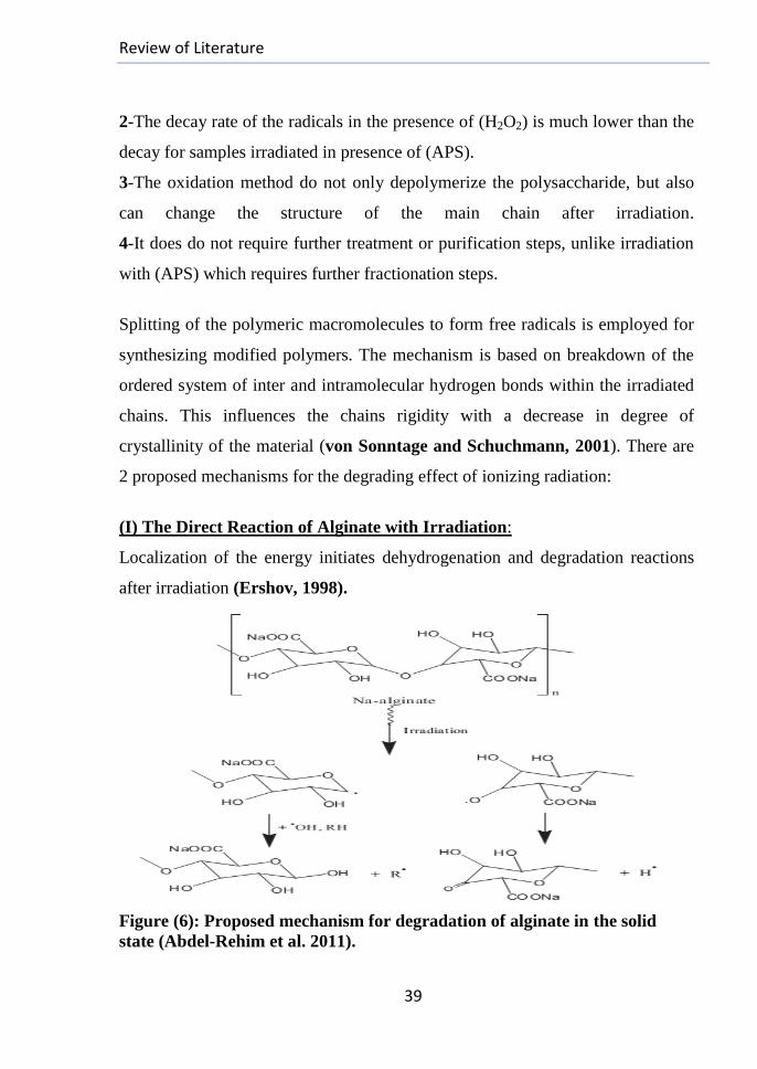
Review of Literature
39
2-The decay rate of the radicals in the presence of (H2O2) is much lower than the
decay for samples irradiated in presence of (APS).
3-The oxidation method do not only depolymerize the polysaccharide, but also
can change the structure of the main chain after irradiation.
4-It does do not require further treatment or purification steps, unlike irradiation
with (APS) which requires further fractionation steps.
Splitting of the polymeric macromolecules to form free radicals is employed for
synthesizing modified polymers. The mechanism is based on breakdown of the
ordered system of inter and intramolecular hydrogen bonds within the irradiated
chains. This influences the chains rigidity with a decrease in degree of
crystallinity of the material (von Sonntage and Schuchmann, 2001). There are
2 proposed mechanisms for the degrading effect of ionizing radiation:
(I) The Direct Reaction of Alginate with Irradiation:
Localization of the energy initiates dehydrogenation and degradation reactions
after irradiation (Ershov, 1998).
Figure (6): Proposed mechanism for degradation of alginate in the solid
state (Abdel-Rehim et al. 2011).
Review of Literature
40
Alginate undergoes ionization on exposing to high-energy radiation in dry state,
then most of the kicked out electrons are thermalized and eventually recombined
with their parent ions to produce excited fragments of the polymer.
These fragments decompose with cleavage of the chemical links, mostly splitting
of carbon-bonded hydrogen leading to the formation of free radicals on polymer
chains, especially the substituted side chains and hydrogen atoms. A proposed
mechanism is illustrated in (fig. (6)).
(II) Irradiating Alginate in Solutions or with Oxidizing Agents :
The degradation follows indirect way where the interaction of radiation with
water causes ionization and excitation effects to produce water radiolysis
products including fast electrons and short-lived H2O+ radical-cations with
electronically-excited water molecules (H2O*). These molecules are unstable and
decompose within 10-13
s to form OH• and H
• radicals (IAEA, 2010) which can
create alginate macro radicals by abstracting Hydrogen atoms from the polymer
chain. Hence, (humidity) enhances the yield of the degraded alginates.
Irradiation (IR) + H2OH2O+ + H2O
*
H2O++H2O H3O
++OH
•
H2O*H
•+OH
•
2.2.5.2. Oxidation of Alginate: Diols commonly found in carbohydrate groups may be oxidized by the natural
ageing in the presence of oxygen and light, enzymatically (Kristiansen, 2009),
or with chemical processing deliberately or un-deliberately (e.g., With periodate
or (2,2,6,6-tetramethylpiperidine-1-Oxy radical (TEMPO)) (Saito, 2006).
Periodate oxidation is commonly utilized as a ‘tool’ to control gel strength due to
the following reasons:
1-Although TEMPO oxidation introduces one carbonyl group at the C6 position
in the monosaccharide unit (Potthast et al., 2006), the product activity is lower
than that of periodate oxidation product.
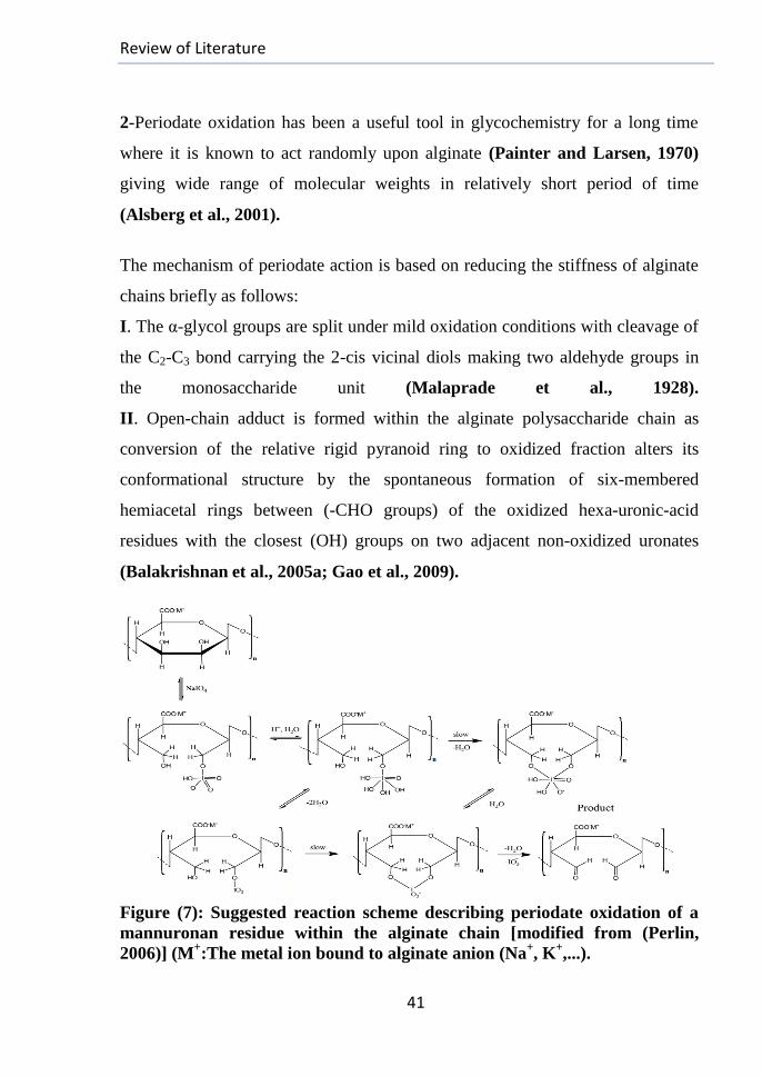
Review of Literature
41
2-Periodate oxidation has been a useful tool in glycochemistry for a long time
where it is known to act randomly upon alginate (Painter and Larsen, 1970)
giving wide range of molecular weights in relatively short period of time
(Alsberg et al., 2001).
The mechanism of periodate action is based on reducing the stiffness of alginate
chains briefly as follows:
I. The α-glycol groups are split under mild oxidation conditions with cleavage of
the C2-C3 bond carrying the 2-cis vicinal diols making two aldehyde groups in
the monosaccharide unit (Malaprade et al., 1928).
II. Open-chain adduct is formed within the alginate polysaccharide chain as
conversion of the relative rigid pyranoid ring to oxidized fraction alters its
conformational structure by the spontaneous formation of six-membered
hemiacetal rings between (-CHO groups) of the oxidized hexa-uronic-acid
residues with the closest (OH) groups on two adjacent non-oxidized uronates
(Balakrishnan et al., 2005a; Gao et al., 2009).
Figure (7): Suggested reaction scheme describing periodate oxidation of a
mannuronan residue within the alginate chain [modified from (Perlin,
2006)] (M+:The metal ion bound to alginate anion (Na
+, K
+,...).

Review of Literature
42
The formed adduct reduces the steric hindrance of the main chain and allows free
rotation of the β-glycosidic linkages to make it behave like an acetal group with
reduced stability to hydrolysis (Bruneel and Schacht, 1993) (Fig.(7)).
III. The resulting chains have the inability to form ionic bridges with the ionic
cross-linkers at the adduct sites where their formation requires an average of 20
adjacent guluronate groups and the breakage of one unit is expected to weaken
these ionic junctions (Bouhadir et al., 2001).
Although the periodate oxidation offers an interesting way for changing the
chemical structure of alginate and makes it more reactive, it leads to some
depolymerisation even when carried out in dark (Laurienzo et al., 2005) due to
the following reasons:
A-The involved degradation is presumably through a free radical mediated
mechanism, may be due to the oxidation of impurities present. The degradation
seems to be unavoidable even in the presence of free radicals scavengers
(Balakrishnan et al., 2005a).
B-The total MW of alginates decreases in proportion to the molar ratio of the
added NaIO4 reagent to the reaction (Kong et al., 2004).
C-Chemically, the oxidized residues can be degraded hydrolytically much faster
than the glycosidic linkages between the intact G and M residues. This can offer
a way to control degradation under mild acidic conditions to make the resulting
oxidized polymer suitable for various drug delivery and tissue engineering
approaches (Kristiansen, 2009).
Periodate oxidation is considered a selected approach to activate polysaccharides
where the new added (-CHO) groups are more reactive than the (OH) and(-COO-
) groups initially within the alginate structure (d’Ayala et al., 2008) and they
offer new sites for binding new materials and drugs for introducing to the body.
Review of Literature
43
2.2.6. Purification of Alginate
A major hurdle to the successful medical applications of any biomaterial is its
immunogenicity and the lack of reproducible biocompatibility (Orive et al.,
2004). Alginate, as a natural polymer is limited by its tendency to contain various
fractions of impurities which exhibit mitogenic activity in in vitro tests and could
favor the overgrowth of macrophages and fibroblasts in experimental small and
large animals causing graft failure (De Vos et al., 2002; Van Hoogmoed et al.,
2003). Cellular reactions surrounding the implanted biomaterial could also lead
to the production of toxic cytokines (Cole et al., 1992) or depletion of oxygen
and nutrients (Colton, 1995).
Alginate immunogenicity is affected by number of variables as follows:
1-The Starting Material: The availability of freshly harvested algae for alginate
extraction was depicted to increase its quality (Jork et al., 2000).
2-The Industrial Extraction Process of alginate perhaps introduces additional
contaminants into the extracted raw material (Qi et al., 2009).
3-The Guluronic/Mannuronic Acid Ratio: The chosen alginate content in this
study is (61% M and 39 % G). High M % alginate has been reported to be less
biocompatible than a high G %-alginate due to the mitogenic properties of
mannuronic acid component (Otterlei et al., 1991). In spite of that, certain
protocols proved that the both alginates have the same biocompatibility (Klöck
et al., 1994; Duvivier-Kali et al., 2001) under the same main control of
purification degree.
4-The Molecular Weight: (King et al., 2000).
5-The Nature and Quantity of Residual Contaminants, introduced during
the extraction steps: There are three common contaminant types detected in
alginates, and used also as contamination indicators:
Review of Literature
44
A-Proteins: These are the main contaminants in the alginate extract from algae
representing about 40% of the macro-components distribution of the different
seaweeds (Surialink et al., 2001). (Kanagaraja et al., 1999; Godek et al.,
2004) have found out that these impurities are responsible for provoking the host
immune reactions, so their removal is of paramount importance for enhancing the
biocompatibility of the used biomaterial, alginate. Proteins removal is more
difficult than that of the other 2 main contaminant types, polyphenols and
endotoxins.
B-Polyphenols and Polyphenol-like Compounds (PC): These are aromatic
compounds responsible for the chemical defense against herbivores in the brown
seaweeds (Pereira et al., 1999), so they are normally extracted with alginate.
These impurities are biorecalcitrants and can possibly accumulate in the body
(Skjak-Braek et al., 1989; W.H.O, 1994), so can be dangerous for humans. It
was proved that they can be mostly removed by simple chemical treatment steps.
C-Endotoxins: These are chemical compounds belonging to the pyrogen family
(Dusseault et al., 2006) and comprise the integral part of the outer cell
membrane of Gram-Negative Bacteria (Raetz, 1990) with organization and
stability responsibility (Vaara and Nikaido, 1984). In spite of that, they are
continuously liberated into the surrounding media during the cells growth,
division and after death, so found everywhere and their high concentrations are
found where bacteria accumulate specially during the bioprocesssing. These
molecules are very stable and their biologically active part survives extremes of
temperature and pH (Sharma et al., 1986), so their removal from alginates
requires routine temperatures within the range (180-250oC) with acids or alkalis
of at least 0.1M.
Review of Literature
45
6-The Method of Purification: Achieving a suitable biocompatibility level
requires highly purified alginates (Orive et al., 2002) and since the reporting of
immunogenicity of alginate in the early 90`s, several research protocols have
been described and many in-house methods have been developed including the
following methods:
1-Free Flow electrophoresis (FFE) method: (Zimmermann et al.,1992).
2-Klock method (Klock procedures and Saline Dialysis (K+SD), Pur. K):
Briefly, it involves (3) chloroform extraction repeats for alginate which then
treated with acid-washed as well as neutral activated charcoal. BaCl2 is a
jellifying reagent; the prepared beads are immersed then in acetic acid ,sodium
citrate and ethanol to remove the impurities (Klock et al., 1994).
3-Prokop-Wang method (Pur. P): It has the same procedures of (pur. K
method) without the chemical extractions on the alginate beads (Prokop and
Wang, 1997).
4-De Vos method (Pur. D): It uses Sodium Ethylene GlycolTetraAcetic Acid
(EGTA) solution of alginate with adjusted pH and involves continuous washing
with (HCl+ NaCl) solution, followed with several repeats of extraction with
Sevag Reagent, filtrations with several washings, and then ethanol precipitation
(De Vos et al., 1997).
5-Vidal-Serp D.S method (Pur. V): It depends mainly on acetone as a
purification reagent with several filtration and continuous washing steps with
(Vidal-Serp and Wandery, 2005).
6-Purification Preparative method: It uses the same technique of Size
Exclusion Chromatography (SEC); the eluent is KCl to reduce electrostatic
interactions among the proteins and alginate molecules (Ménard et al., 2010). It
is expensive method, needs special columns for carbohydrates purification and
better standardization for pure alginate preparation for clinical applications, so
considered a restricted method to laboratories.
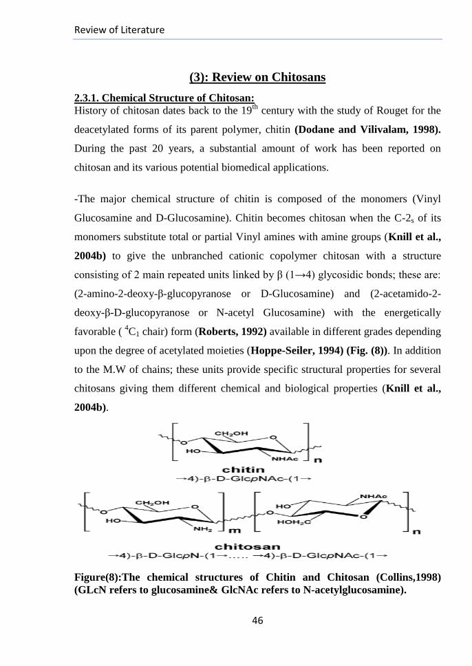
Review of Literature
46
(3): Review on Chitosans
2.3.1. Chemical Structure of Chitosan:
History of chitosan dates back to the 19th
century with the study of Rouget for the
deacetylated forms of its parent polymer, chitin (Dodane and Vilivalam, 1998).
During the past 20 years, a substantial amount of work has been reported on
chitosan and its various potential biomedical applications.
-The major chemical structure of chitin is composed of the monomers (Vinyl
Glucosamine and D-Glucosamine). Chitin becomes chitosan when the C-2s of its
monomers substitute total or partial Vinyl amines with amine groups (Knill et al.,
2004b) to give the unbranched cationic copolymer chitosan with a structure
consisting of 2 main repeated units linked by β (1→4) glycosidic bonds; these are:
(2-amino-2-deoxy-β-glucopyranose or D-Glucosamine) and (2-acetamido-2-
deoxy-β-D-glucopyranose or N-acetyl Glucosamine) with the energetically
favorable ( 4C1 chair) form (Roberts, 1992) available in different grades depending
upon the degree of acetylated moieties (Hoppe-Seiler, 1994) (Fig. (8)). In addition
to the M.W of chains; these units provide specific structural properties for several
chitosans giving them different chemical and biological properties (Knill et al.,
2004b).
Figure(8):The chemical structures of Chitin and Chitosan (Collins,1998)
(GLcN refers to glucosamine& GlcNAc refers to N-acetylglucosamine).
Review of Literature
47
2.3.1.1. Degree of Deacetylation (DD):
It refers to the percentage of average number of D-Glucosamine units present in
the polymer chains when the distribution of the 2 constitutive residues is random.
The term (Chitin) usually refers to a copolymer with (DD less than 70 %), while
(Chitosan) represents a series of copolymers of (DD <70 %) (Domard and
Domard, 2002) with lactic-average M.W in the range (10-103 KDa) (Roberts,
1992). Chitin is generally insoluble in standard polar and non-polar solvents, but
chitosan dissolves in diluted acidic solutions (Hayes et al., 1977) below pH 6.0,
suitable for quaternisation of the amine groups with macro intrinsic pKa value of
6.3 (Meng et al., 2010) giving it a high positive charge density to make water-
soluble cationic polyelectrolyte (New et al., 2008) able to form salts with inorganic
and organic acids (e.g., Glutamic and acetic acids) (Illum, 1998). Different
deacetylation conditions can be influenced by changing the inter and intra
molecular repulsion forces (Sashiwa et al., 1991).
Biodegradability in living organisms is (DD)-dependent where it increases as (DD)
decreases (Kurita et al., 2000; Yang et al., 2007), so lower DD chitosans induce
acute inflammatory responses, but those with high DDs produce a minimal
response due to their lower degradation rate. Chitosan is known to degrade
predominantly in vertebrates by lysozyme and by certain bacterial enzymes in the
colon (Kean and Thanou, 2009). (8) Human chitinases in the glycoside hydrolase
family have been identified and (3) of them have shown enzymatic activity
(Funkhouser and Aronson, 2007).
2.3.2. Sources of Chitosans:
Chitin is found primarily as supporting material in many marine organisms (e.g.,
Shrimp shells, lobster, krill, crab, cuttlefish and bone plates of squid).
Review of Literature
48
As the second most abundant natural polymer after cellulose (Rinaudo, 2006),
chitin is a principal component in the exoskeletons of silkworms, many insects
(e.g., Drosophila melanogaster, Extatosoma tiaratum, Sipyloidea sipylus,
mosquitoes, cockroaches and honey bees) (Jeuniaux, 1996), some terrestrial
crustaceans (e.g., Porcellio scaber and Armadillidium vulgare), nematodes, some
algae (Nwe et al., 2010 and its cited references: Anantaraman and
Ravindranath, 1976; Florida and Carlberg, 1982; Veronico et al., 2001;
Pochanavanich and Suntornsuk, 2002; Nemtsev et al., 2004; Moussian et
al.,2005; Tauber, 2005; Paulino et al., 2006; Mau, 2007; Hild et al., 2008;
Mario et al., 2008), but it is fully absent in mammals. Chitosan (Ch) is also a
structural component in certain fungi such as (Mucor rouxii, yeasts, mushrooms
(e.g., Auricularia auriculajudae, Pleurotus sp., Agaricus bisporus, Lentinula
edodes, Trametes versicolor and Armillaria mellea) (Roberts, 1992). Production
of chitosan from fungi using fermentation methods is gaining much interest in
recent years (Nwe et al., 2002).
From economic and technological points of view, these byproducts represent
abundant, cheap, ecologically-friendly, renewable resources for the extraction of
chitin and its derivatives where several million tons of chitin are harvested
annually in the world (Roberts, 1992). For example, the crude shrimp head and
skin have low economical values and are treated as biowaste or sold to animal feed
manufacturers (Suchiva et al., 2002); these biowaste in the topical region contain
[(10-20 %) Calcium + (30-65%) Protein content + (8-10%) Chitin) on a dry basis
(Rao et al., 1996), so they are used as raw materials for the isolation of chitin
through inexpensive simple chemistries.
Chitosan; as a semi-synthetic polysaccharide, can be prepared by the controlled
alkaline deacetylation of chitin (Furda, 1983) with different physicochemical
characteristics, viscosities and (DD) (Rao et al., 1996).
Review of Literature
49
There are three forms of chitin known as (α, β and γ) based on the origin of
polymer and its treatment during the extraction process (Wuolijoki et al., 1999).
The α-chitosan is the most commonly obtained one from the crustacean chitin of
crab and shrimp shell wastes (Roberts, 1992) accounting for approximately 70%
of the organic part.
2.3.3. Properties of Chitosan:
1-Chitosan is a pseudonatural, renewable cationic polysaccharide of superior
biodegradability (Hirano et al., 1990), ease of availability, biocompatibility
(Thanou et al., 2001), non-antigenicity (Berger et al., 2004a), low-toxicity (Kean
and Thanou, 2009) with bioabsorbable and easy chelating power. It is an
interesting natural glycosaminoglycan possessing rare bioactivity (Domard and
Domard, 2002) and the 4th
edition of the European Pharmacopoeia (2002)
included a monograph relating to (Chitosan hydrochloride). Chitosan has a gel-
forming capability either of the polymer itself (Ladet et al., 2008) or with other
compounds (Il’ina and Varlamov, 2005). It has the ability to be injected into the
body as a liquid below the Lower Critical Solution Temperature (LCST) and then
forms a gel in situ at body temperature (above the LCST) (Jeong et al., 2002).
2-Due to chitin’s poor solubility in aqueous solutions and organic solvents, it does
not find practical applications; whereas its artificial variants; chitosan oligomers,
with their interesting multidimensional properties have been examined and found
to have a wide range of applications.
Chitosans` applications are biomedical (Paul, 2000) as well as applications in
biotechnology, aquaculture, environmental engineering and several industries such
as in agrochemicals, cosmetics, pharmaceutics and food industries. In addition,
they are pervasively used as drug delivery carriers of the polymer alone (Lubben
et al., 2001).
Review of Literature
50
3-Combination of chitosan with other polymers can provide the required
physicochemical properties for designing of new drug delivery systems (Berger et
al., 2004b), synthesis of surgical threads (Lou, 2008), repairing of hyaline
cartilage (Montembault et al., 2006), preparing media for pancreatic islets
culturing (Cui et al., 2001), manufacturing of bone healing materials in different
forms such as sponges (Lee et al., 2000), synthesis of new preparations of
antitumor activities (Suzuki et al., 1986) and for using in gene transferring studies
(Leong et al., 1998).
4-Many synthetic chemicals such as the phenolic compounds are found to be
strong radical scavengers; however, the use of synthetic antioxidants is under strict
regulation due to their potential health hazards (Je et al., 2004). Therefore, the
usage of natural antioxidants as alternatives is of great importance. Recently, the
antioxidant activity of chitosan and its derivatives has attracted an increased
attention (Chiang et al., 2000).
5-The unique properties of chitosan are on account of its amino groups that carry
positive charges at pH values below 6 making cationic polyelectrolyte structures
(Sandford, 1989) that have the activity to bind to negatively charged materials
(e.g., Nucleic acids and enzymes) and interact with the negatively charged
glycosaminoglycans of the ECM and cells surfaces (e.g., Cells of skin) (Sandford,
1992).
6-As a cationic polymer, chitosan was studied as a partner of several Inter-
Macromolecular Complexes (IMC) with natural or synthetic anionic
polyelectrolytes, such as Chondroitin sulfate (Denuziere et al., 1998), Poly
(acrylic acid) (Wang et al., 1997), Poly (galacturonic acid) (Martins, 2007)
,Alginate (Knill et al., 2004a), K-carageenan (Zhang and Zhang, 2012), Xanthan
(Chellat et al.,2000), Carboxymethyldextran (Chen , 2003) and
Carboxymethylcellulose (Fukuda, 1980; Roscaa et al., 2005).
Review of Literature
51
7-Chitosan is the most useful derivative of chitin (Meyers et al., 1989). The
available primary (-NH2 and OH- groups) on the molecule are active sites that can
be used to chemically alter its properties through the formation of number of
linkages including amide and ester bonds as well as Schiff base formation
employing mild reaction conditions (Bergeret al., 2004; Hoare and Kohane,
2008). For example: Chitosan characteristics such as solubility, hemostatic and
cationic properties can be reversed by sulfating the amine groups to make anionic
and water-soluble molecules with new introduced anticoagulant properties (Suh
and Matthew, 2000). This facile derivatization makes chitosan an ideal candidate
for bio-fabrication with ample biomedical applications (Denuzière et al., 2000; Yi
et al., 2005).
8-Due to its cationic nature, chitosan has been claimed to entrap lipids in intestine
so utilized in treatment of some hyperlipidemia cases (Kanauchi et al., 1995;
Wuolijoki et al., 1999). It can enhance the paracellular route of absorption through
interaction with cell membrane resulting in structural reorganization of tight
junction-associated proteins. This mechanism is important in the transporting of
hydrophilic compounds (e.g., Therapeutic peptides and antisense oligonucleotides)
across the membrane.
9-Due to the several biological activities and excellent bioadhesive properties of
chitosan oligomers, they have become important biomaterials for wound
management (Azad et al., 2004; Lin et al., 2006). They were found to have anti-
acid and anti-ulcer activities that prevent or weaken drug irritation in the stomach
(Onal and Zihnioglu, 2002). The antimicrobial activities of chitosan and its
derivatives were reported where its (HMW- molecules) were suggested to stack on
the surface of bacterial cell wall to prevent the supply of nutrients (Tokura et al.,
1997).
Review of Literature
52
2.3.4. Production of Chitosan from Chitin of Shrimp Shells:
Shrimp industries generate large quantities of shrimp biowaste during processing;
approximately 45-55% of the weight of raw shrimp which can be used to produce
value-added products because of its richness in protein, carotenoids and chitin.
Many methods have been adopted for the extraction of chitin derivatives from
shells waste, maybe with the aid of some enzymes (Win et al., 2000; Win and
Stevens, 2001), by fermentation with certain microorganisms (Rao et al., 2000) or
with direct isolation using refluxing method (Sehol et al., 2002). The chemical
method of extraction is widely used to obtain chitin with a high quality by
removing protein, pigments, lipids and inorganic materials (mainly CaCO3). In
many chitin producing industries, the deproteination step is carried out prior to the
demineralization (Decalcification) step (Roberts, 1992) where deproteination is
achieved by treatment with NaOH and HCl is used in the demineralization step.
Review of Literature
53
(4): Review on PolyElectrolyete Complexes (PECs)
Polyelectrolyte complexes (PECs) represent a special class of polymeric
compounds (Michaels, 1965) consisting of oppositely charged polyions which
interact together spontaneously to yield a complex in different forms that
essentially contains stoichiometric equivalents of the component polyions. These
complexes have been studied since the early 60’s and the nature and extent of such
reactions were proven to be sensitive to the molecular properties of the reacting
polymers including molecular weights and structures, as well as to the
complexation conditions including temperature, period, pH, moisture content,
mixing ratio, ionic strength of the system, cross-linking agents and density of the
formed complex (Dumitriu et al., 1994; Dumitriu and Chornet, 1997).
2.4.1. Properties of the Alginate/Chitosan PECs:
With preparing a hydrogel matrix of (alginate-chitosan) in the form of PECs, the
resulting structure will have new unique composite properties over the properties
of each used one alone, but with retaining some of their properties. These overall
properties will suit various pharmaceutical and biomedical interests (Ulbricht,
2006) and can be summarized in the following points:
A-For ideal absorptive properties of the hydrogel, it must be rich moisture
resistant. For hydrogel films of single polymer, hydration is problematic due to
limited inter-chain interactions as water-polymer interactions compete effectively
with those between the polymer chains. Based on the forces driving intermolecular
interactions, polymer-polymer interactions can be strengthened by combining
polymers of different structures and predominantly introducing charge interactions
rather than (H-bonding) (Hoare and Kohane, 2008).
B-No auxiliary molecules (e.g., Catalysts or initiators) are needed for the
complexation reactions which are performed generally in aqueous solution; this
represents the main advantage over covalently crosslinked networks.

Review of Literature
54
This favors biocompatibility and avoids purification before administration. This is
very important for the drugs to be entrapped (Lee et al., 1999) and more effective
in limiting the release of encapsulated materials compared to either polymer alone
(Filipović-Grčić, 1995; Alexakis et al., 1995).
C-Both alginate and its membranes are non-adhesive so their structure must be
modified for enhancing their biocompatibility and bioadhesion properties on using
as dressing materials. There are several methods for mitigating that including the
usage of Schiff reaction for coupling (-COO- groups) with (-NH2) groups,
carbodiimide chemistry for binding sequence of amino acids (e.g., The crosslinker
1-ethyl-3 (-3-dimethylaminopropyl) carbodiimide. HCl (EDC) is commonly
employed with alginate as a tissue fixative where it binds to a certain sequence of
amino acids) (Hunt et al., 2009), or through the coupling of alginate chains with
positively charged polymers (Eiselt et al., 1999; Meng et al., 2010). D-It was
discovered that hydrogels of high alginate composition have high water uptake
attributed to its hydrophilicity that may cause the release of HMW chains on
degradation of the prepared scaffold (Tolba et al., 2010). Accordingly, the
possibility to change the cross-linking density within alginate hydrogels with
adjustment of their swellability through binding with other polymers may make
them potential for wound dressing applications.
E-Chitosan has low flexibility so it is majorly used with modification through
several methods such as cross-linking, grafting and blending (da Silva et al., 2008;
Hong et al., 2009). Blending is an effective and convenient method to improve the
performance of the polymers.
F-Hydrogels composed of chitosan alone are limited by the lack of an efficient
control for drug delivery, their poor Tensile Strength (TS) and elasticity (Berger et
al., 2004a; Berger et al.,2004b), so the addition of other polymers is necessary to
form PEC films with improved mechanical strength and elasticity while
maintaining the properties of the interacting polymers.
Review of Literature
55
Several research papers concerning the preparation of Inter Penetrating Networks
(IPNs)-based on chitosan and its derivatives with other polyanionic
macromolecules as well as physical gels have been published (Al-Kahtani et al.,
2009; Zhou et al., 2009).
In view of the limitations encountered in pure alginate, chitosan beads and
hydrogel systems, the concept of Alginate/Chitosan PECs (Alg/Ch-PECs) gained
acceptance (Murata et al., 1993; Huguet and Dellacherie, 1996) as the formed
complex has new properties involving those of the individual polymers with
circumventing their problems issues. For example, (Alg/Ch-PECs) were found to
still biocompatible and mechanically stronger at low pH values at which chitosan
dissolves (Hein et al., 2008), but also can degrade partially through means of
hydrolysis that can be regulated by change of the components ratios, so (PECs)
have high potential in bioengineering as scaffolds and supporting materials (Li et
al., 2009). The coacervation rate between chitosan and high-G alginates was found
to be higher than that with the high M-alginates (Gaserod et al., 1998).
2.4.2. Principle of Formation of Alginate/ Chitosan PECs:
The general PEC formation conception and its main driving force is the gain in
entropy caused by the release of low-molecular-weight counter-ions (fig.(9)). The
Hydrogen bonding and hydrophobic interactions can contribute to the process of
complexation. The structure formation is mainly determined by the fast kinetics of
the process which depend on the electrolytes with low molecular mass and the
polyelectrolytes concentrations, followed by a slower stage in which the chains
redistribute to a PEC conformation closer to equilibrium (Bakeev et al. 1992).
Other factors affecting the structure formation are the mixing procedures, medium
conditions and the macromolecular characteristics of the reacting polyelectrolytes
such as molecular weight, chain flexibility and linear charge density (LCD).
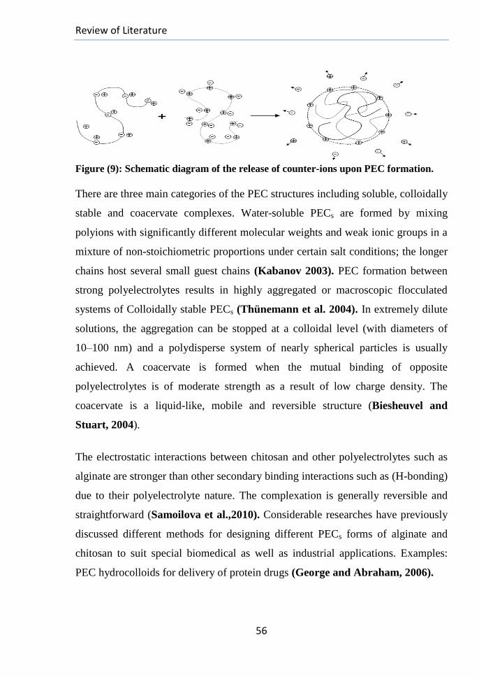
Review of Literature
56
Figure (9): Schematic diagram of the release of counter-ions upon PEC formation.
There are three main categories of the PEC structures including soluble, colloidally
stable and coacervate complexes. Water-soluble PECs are formed by mixing
polyions with significantly different molecular weights and weak ionic groups in a
mixture of non-stoichiometric proportions under certain salt conditions; the longer
chains host several small guest chains (Kabanov 2003). PEC formation between
strong polyelectrolytes results in highly aggregated or macroscopic flocculated
systems of Colloidally stable PECs (Thünemann et al. 2004). In extremely dilute
solutions, the aggregation can be stopped at a colloidal level (with diameters of
10–100 nm) and a polydisperse system of nearly spherical particles is usually
achieved. A coacervate is formed when the mutual binding of opposite
polyelectrolytes is of moderate strength as a result of low charge density. The
coacervate is a liquid-like, mobile and reversible structure (Biesheuvel and
Stuart, 2004).
The electrostatic interactions between chitosan and other polyelectrolytes such as
alginate are stronger than other secondary binding interactions such as (H-bonding)
due to their polyelectrolyte nature. The complexation is generally reversible and
straightforward (Samoilova et al.,2010). Considerable researches have previously
discussed different methods for designing different PECs forms of alginate and
chitosan to suit special biomedical as well as industrial applications. Examples:
PEC hydrocolloids for delivery of protein drugs (George and Abraham, 2006).

Review of Literature
57
(Alg/Ch) Beads for the biological molecules encapsulation (e,g., Hemoglobin
(Hucuet et al., 1994) and DNA microcapsules (Alexakis et al., 1995)), drugs
delivery as in the report of (Liu et al., 1997) and in the form of dry-coated tablets
(Takeuchi et al., 2000).
Based on the principles of gel network engineering, polymer chemistry and
physics; a ‘permanent interpenetrating polyion-complex hydrogel’ can be prepared
from these poly-pyranoid saccharides composing of a primary entangled network
of Ca+2
-crosslinked alginate chains (the chemical gel part) that connects and
encases physical hydrogel regions in which blends of alginate and chitosan
networks interact with each other. These interactions are initially via electrostatic
mechanisms neutralizing their charges (e.g.,ionic binding among Chitosan (NH3+)
and alginate (-COO-) groups at certain pH values. Hydrogen bonding between
chains, Coulomb forces, Vander Waals forces and transfer forces share in the
complexation process (Takahashi et al., 1990; Hoffman, 2002) as well with the
association of hydrophobic contributions of the polymers chains (Abe et al., 1978).
These complexes are supposed to be as in (fig. (10)).
Figure (10): Schematic interpretation of the (Alginate-Chitosan physical complex and
Semi-IPN complex) (with modification from (Knill et al., 2004a).
Review of Literature
58
(5): The Biological Effects of Alginates and Chitosans -based
Dressings on Wound Healing
2.5.1. Alginates as Wound Dressings:
Surgical applications for alginates were suggested many years ago with the first
published report on its hemostatic activity dating from 1947 (Blaine, 1947), then
met many development stages that were slow at first, but the manufacturing of
alginate fibers and quality of the resulting textiles were then improved with the
development of wet spinning and new textile technologies (Groves and
Lawrence, 1986). Among the various fibrous and hydrogel products, alginate-
based products are currently one of the most popular types used in wound
management. They have different physical and chemical properties based on the
proportion and arrangement of mannuronate and guluronate residues and their
(Ca+2
& Na+ ions-content) and they offer many advantages over conventional
dressings such as cotton gauze (Qin and Gilding, 1996) as follows:
1-Alginates are gel-forming materials with hemostatic properties (Blaine, 1947)
and enough strength for usage as an aid to retract and grip the tissues well.
2-In contact with body fluids, alginates are dissolved and absorbed where the
wound exudate converts the calcium to sodium salt facilitating removal of the
dressing by dissolution. Any residual fibers remaining within the wound are
degradable based on its composition (Burrow and Welch,1983).
3-Although a greater inflammatory response in the form of an increase in the
number of leukocytes was seen in wounds treated with the biodegradable (Ca-
Alginate) compared to the control wounds, this does not detract from its efficacy
(Barnett and Varley, 1987).
Review of Literature
59
4-If it is the main polymer in the medical product dressing; on absorption of wound
exudate, alginate can be rinsed away with saline irrigation, so the removal of
dressing does not disturb the healing tissues (Choi et al., 1999).
5-The value of treating wounds with alginates is based on their effects in
maintaining a physiologically optimal moist microenvironment that promotes
healing with formation of granulation tissue (Motta, 1989).
6-(Ca-Alginate) has been shown to be (4) times as absorbent per unit weight as
gauze. This reduces the number of swabs used; the time spent in mopping up blood
or other wound fluids, avoids excessive use of diathermy (Blair et al., 1990) and
makes the dressing usage be very useful for moderately-to-heavily exuding
wounds.
7-Topical alginate treatment exerts its bioactivity by augmenting the natural wound
healing with concomitant induction of cytokines production by human monocytes
(Otterlei et al., 1991) and inhibition of the cytokines associated with fibrosis
resulting in increased epithelial proliferation and decreased wound size (Lee et al.,
2009).
Many previous studies described alginate based wound-dressings as follows:
I. (Ca-Alginate), on contact with blood, releases Ca+2
ions in exchange for Na+
ions and activates coagulation by stimulating platelets and the clotting factors VII,
IX, X and VIII-vWF (Born and Cross, 1964) based on its Ca+2
-content (Jarvis et
al., 1987). Calcium alginate (BritCair Ltd., Aldershot, UK) was first introduced as
a wound dressing in the study of (Blair, 1990) and evaluated as a possible surgical
hemostat; especially in major surgery, particularly where the excessive blood loss
leads to the need for transfusion.
II. (Thomas and Tucker, 1989) prepared an alginate-based dressing and proved
its efficacy in the treatment of richly exuding wounds (e.g., Chronic leg ulcers and
pressure sores).
Review of Literature
60
In laboratory tests, alginate dressings have been found out to have an increased
capacity in absorbing blood compared to cotton gauze with promoting hemostasis
(Groves and Lawrence, 1986; Blair, 1990).
III. Through the active portion of alginate, the mannuronate residues have been
shown to stimulate human monocytes to produce elevated levels of IL-1b, IL-6 and
TNF-α (Gensheimer, 1993). This provides a convenient marker for macrophages
activation in the wound environment (Chensue et al.,1990) with exerting a
transient pro-inflammatory effect to recruit fresh leukocytes from blood (Groves et
al., 1995) and initiate the cascade of healing events.
IV. Alginate dressing was found to suppress the expression of fibronectin, TGF-β1
and VEGF effectively and blocks the accumulation of angiogenesis and ECM in
the wound but activates collagen-1 expression (Lee et al.,2009).
V. A hydrogel dressing made of cross-linked oxidized alginate with gelatin in the
presence of borax was prepared. This biodegradable, non-toxic composite matrix
has the hemostatic effects of gelatin, the wound healing-promoting features of
alginate and the antiseptic property of borax to make it a potential wound dressing
material (Balakrishnan et al., 2005b).
VI. The ability of alginate dressings to accelerate epithelialization has been
demonstrated in many experimental and clinical investigations (Agren, 1996).
Kaltostat and Sorbsan alginate dressings have been reported recently to exert
cytotoxic effects on fibroblasts. Numerous patents detailed the production of
alginate fibers and dressings (Cole and Nelson, 1993; Thompson, 1996; Griffiths
and Mahoney, 1997; Fenton et al., 1998; Mahoney et al., 1999; Qin and
Gilding, 2000; Horsler, 2000). Commercial alginate-based dressings are
summarized in (Table: 2).

Review of Literature
61
Table (2): Summary for commercial Alginate and Chitosan Dressings
Drug name Composition Company Action
Algisite®M Non-woven fibers of
(Calcium alginate)
Smith and
Nephew
-Induce inflammation by
the soluble endotoxins
-The Activity is
associated with the
insoluble fibers and
macrophages activation.
Algosteril® Ca-Alginate Beiersdorf
Kaltocarb® Ca-Alginate fiber ConvaTec
Kaltogel® Ca/Na-Alginate gelling fiber ConvaTec
Kaltostat®
hemostat
Guluronic-rich Ca-Alginate
fibers in non-woven pads,
ConvaTec
Melgisorb® Ca/Na-Alginate gelling fibre Molnlycke -Significant haemostatic
properties on utilizing to
control the blood loss
from skin graft donor
sites (BMA, 2001;
Kennedy et al., 2001).
Seasorb ® Ca/Na-Alginate gelling fibre Coloplast
Sorbalgon® Ca-Alginate Hartman
Sorbsan ® Ca-Alginate fibers in non-
woven pads (Groves and
Lawrence, 1986).
Maersk
Chitopack S® Chitin in sponge form
Eisai Co
Chitopack P® Non-woven dressing made
of chitin-modified PET
Chitopack C® Non-woven cotton-chitosan
Non-woven dressing of chitosan fibers
(Muzzarelli, 2003)
Concern
Nikita Co
Tegasorb® Chitosan gel American
firm 3M
Designed for the healing
of wide internal wounds. Tegaderm® Chitosan hydrocolloid form
HemCon
Patch PRO
Chitosan-based hemostatic
dressings in the sponge form.
HemCon
Medical
Syvek Patch -Chitosan-modified cellulose Marine
pol. Tech.
(Fischer et al., 2004;
Alam et al., 2005) -Chitin sponge RDH
Review of Literature
62
2.5.2. Chitosans as Wound Dressings:
The enhancement in chitosan purification and textile processing of fibers enhanced
the quality and yield of chitosan-based products in powder, film, bead, fiber and
fabric forms. In recent years, it has become important biomaterial for wound
management and the synthesis of dressings as one of its main uses (Azad et al.,
2004; Lin et al., 2006) where:
1-Chitosan was observed to have hemostatic properties independent of the normal
clotting cascade (Howling et al., 2001) and allow the promotion of normal
histoarchitectural organization (William and Herbert, 1985).
2-It can accelerate tensile strength of wounds as well as the infiltration of
inflammatory cells (Huang et al, link; Hiroshi et al., 1999) with activating the
migration and proliferation of fibroblasts (Hiroshi and Haruo, 1999; Wang et al.,
2003) as well as keratinocytes (Chatelet et al., 2001) in the wound area through
the induction of interleukin release.
3-Different chitosans-based dressings with bacteriostatic and fungistatic activities
were prepared (Tomihata and Ikada, 1997) due to the antimicrobial activities of
chitosan (Tokura et al., 1997).
4-Many Antibiotics can be included with many chitosan dressings (Mi et al.,
2002).
5-The presence of chitin/chitosan in a dressing was proven to speed the fibroblastic
synthesis of collagen in the first few days of healing (Chung et al., 1994) and
affect macrophages function to help in faster healing (Mattioli-Belmonte et al.,
1997; Muzzarelli et al., 1999).
6-A sponge constituted of (bovine type I collagen /chondroitin-4,6-sulfate/
chitosan) for skin reconstruction was developed and achieved a permanent skin
tissue regeneration with good functional and aesthetic characteristics (Li et
al.,1997). Some Chitosan-based dressings are summarized in (Table: 2).

Review of Literature
63
2.5.3. Alginate/ Chitosan-Based Wound Dressings:
One of the most common uses for composites made of (alginate/ chitosan) is the
synthesis of wound dressings that may be in form of (Alginate /Chitosan
/Polylactic Acid/ Cotton /Tencel composite) wound dressings (Huang et al., link),
Ca-Alginate filaments coated with Chitosan (Tamura et al., 2002), membrane
sheets (Wang et al., 2002) or sponge dressings (Lai, 2003).
Flexible transparent membranes of (Chitosan/Alginate (Ch/Alg) -PECs), cast from
aqueous suspensions of (Ch-Alg coacervates) with CaCl2 were evaluated as
potential wound-dressing materials that can accelerate healing of incision wounds
in rat models, comparing to conventional gauze dressing. They can facilitate
remodeling of scar tissue by increasing the rate of collagen synthesis, compaction
of its organized fibers into thicker bundles with alignment into parallel bundles,
epidermal architecture maturation with keratinized surfaces of normal thickness
and a subsided inflammation in the dermis with fibroblasts maturation (Wang et
al., 2002).
2.5.4. Angiogenesis and Angiogenesis-Controlling Genes:
Diverse processes such as the embryonic development, tumor growth and tissue
repair are linked by the absolute requirements for a vascular bed to deliver oxygen
and nutrients (e.g., Glucose and amino acids) to the metabolically active cells and
help the removal of debris (Colton, 1995). Under normal conditions, a tissue or
tumor cannot grow beyond 1 to 2 mm in diameter without neovascularization; this
distance is defined by the limits in the diffusion of oxygen and metabolites
(Folkman, 1971). A healthy human /animal being is bleeding permanently in
small arterioles and veins without even getting aware of, so wound healing is a
vitally important adjustment of nature to prevent continuous bleeding into the
tissue after damage of blood vessels.
Review of Literature
64
Achieving instantaneous repair and closure in wounds require sealing of numerous
blood clotting factors to vessel lesions for stopping the excessive blood loss,
provision of adequate oxygen, protein delivery and other nutritional factors to the
tissue, maintaining moist environment, appropriate inflammatory milieu,
debridement, appropriate management of infection and correction of the
contributing medical diagnoses.
Angiogenesis, the formation of new blood vessels from pre-existing vessels, is a
crucial process in wound tissue repairing in which various growth factors,
cytokines, and adhesion molecules are involved (Battegay, 1995; Martin, 1997).
Most blood vessels are formed during fetal development, but adult tissues can
induce angiogenesis in response to injury. This capability is governed by both
angiogenic agonists and antagonists that have been identified throughout the body
at various times during the repair (DiPietro et al., 1996) suggesting changes for
the net angiogenic stimuli as the balance of these factors alternatively favors either
vessel growth or regression (Martin, 1997).
2.5.4.1. Vascular Endothelial Growth Factor (VEGF):
2.5.4.1.1. Functions of VEGF:
VEGF was initially recognized as the best-characterized endothelial-specific
growth factor (Nissen et al., 1998), though other growth factors serve redundant
and overlapping functions(e.g., Angiogenin, angiotropin, angiopoetin-1, Epidermal
Growth Factor (EGF), Transforming Growth Factor -β (TGF- β) and Fibroblast
Growth Factors (FGFs)) (Folkman and D'Amore, 1996). Its biology is gaining in
complexity due to its different regulatory biological functions:
1- VEGF plays critical roles in the formation and development of blood vessels
networks, modulating thrombogenicity (Miyagami and Katayama, 2005) and
enhancing the vascular permeability with increasing the hydraulic conductivity
(Bates and Curry, 1997) and fenestration (Esser et al., 1998).

Review of Literature
65
It has three main mechanisms of action: (1) It may increase the vessel
permeability to water, small solutes and macromolecules (Nagy and Benjamin,
2008), (2) It can increase the blood flow to tissues by acting as a potent vasodilator
(Bates and Harper, 2002), and (3) It may reduce the distance of tissue cells from
the nearest blood vessel by stimulating angiogenesis.
2-VEGF has been implicated in the wound healing process (Hayashi et al., 2004).
Its administration improves healing in nondiabetic ischemic wounds (Corral et al.,
1999). VEGF Blocking with neutralizing antibodies impedes tissue repair
(Howdieshell et al., 2001). An increase in the total levels of VEGF in fetal skin
correlates with the transition from scarless to fibrotic healing and exposure of
wounds that normally heal in a scarless manner to exogenous VEGF can lead to
scar formation (Wilgus et al., 2008).
3-VEGF can interact with many body cells, some of them are as follows:
*It could act as Endothelial cells (ECs)-specific mitogen (De Vries et al., 1992).
This potently induces their migration and proliferation in vitro and angiogenesis in
vivo (Ferrara et al., 1992; Dvorak et al., 1995).
*It plays a role in the chemotaxis of monocytes/macrophages that are crucial in the
inflammatory reactions and in wound-repair processes by inducing monocytes
activation and migration (Clauss et al., 1990) via the (VEGF receptor-1; VEGFR-
1) (Barleon et al., 1996).
*There is a possibility that VEGF could indirectly modulate the contraction and
growth of smooth muscle cells (SMCs) (Doi et al., 1996).
4- It may play a role in tumor growth (Senger et al., 1993), age-related macular
degeneration, numerous pathological situations, rheumatoid arthritis, proliferative
retinopathy and collateral formation in the ischemic tissues (Dvorak et al., 1995;
Ferrara and Bunting, 1996).
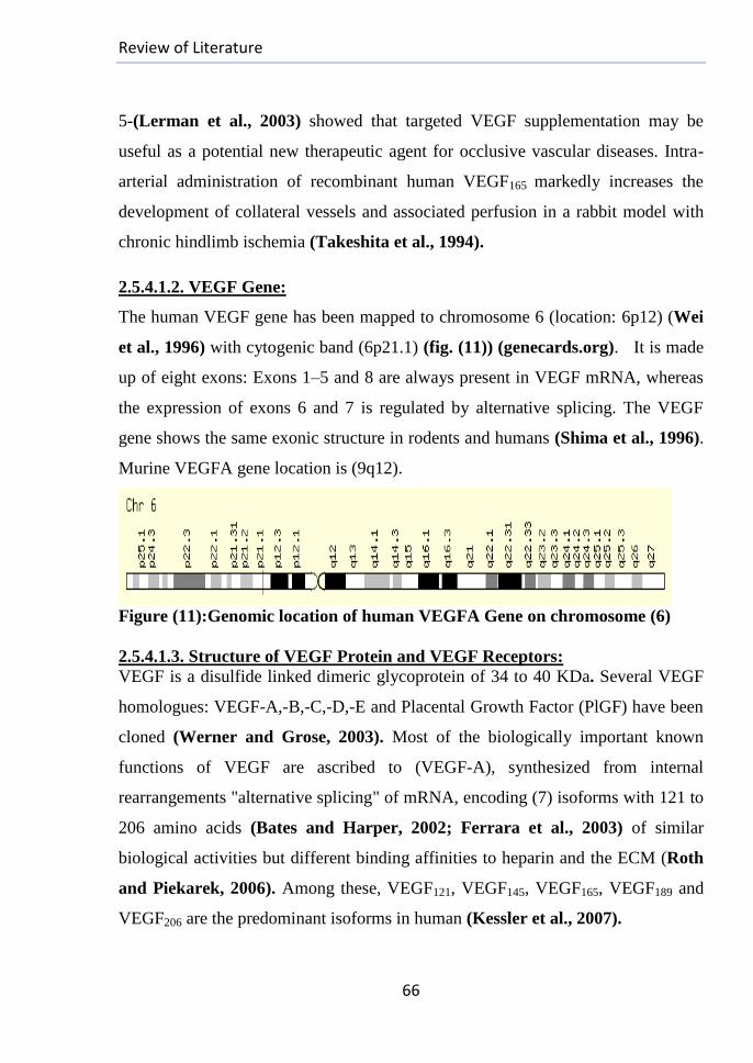
Review of Literature
66
5-(Lerman et al., 2003) showed that targeted VEGF supplementation may be
useful as a potential new therapeutic agent for occlusive vascular diseases. Intra-
arterial administration of recombinant human VEGF165 markedly increases the
development of collateral vessels and associated perfusion in a rabbit model with
chronic hindlimb ischemia (Takeshita et al., 1994).
2.5.4.1.2. VEGF Gene:
The human VEGF gene has been mapped to chromosome 6 (location: 6p12) (Wei
et al., 1996) with cytogenic band (6p21.1) (fig. (11)) (genecards.org). It is made
up of eight exons: Exons 1–5 and 8 are always present in VEGF mRNA, whereas
the expression of exons 6 and 7 is regulated by alternative splicing. The VEGF
gene shows the same exonic structure in rodents and humans (Shima et al., 1996).
Murine VEGFA gene location is (9q12).
Figure (11):Genomic location of human VEGFA Gene on chromosome (6)
2.5.4.1.3. Structure of VEGF Protein and VEGF Receptors:
VEGF is a disulfide linked dimeric glycoprotein of 34 to 40 KDa. Several VEGF
homologues: VEGF-A,-B,-C,-D,-E and Placental Growth Factor (PlGF) have been
cloned (Werner and Grose, 2003). Most of the biologically important known
functions of VEGF are ascribed to (VEGF-A), synthesized from internal
rearrangements "alternative splicing" of mRNA, encoding (7) isoforms with 121 to
206 amino acids (Bates and Harper, 2002; Ferrara et al., 2003) of similar
biological activities but different binding affinities to heparin and the ECM (Roth
and Piekarek, 2006). Among these, VEGF121, VEGF145, VEGF165, VEGF189 and
VEGF206 are the predominant isoforms in human (Kessler et al., 2007).
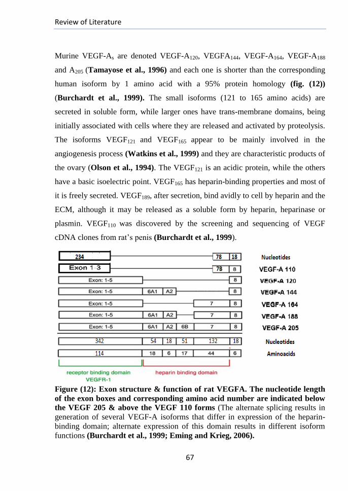
Review of Literature
67
Murine VEGF-As are denoted VEGF-A120, VEGFA144, VEGF-A164, VEGF-A188
and A205 (Tamayose et al., 1996) and each one is shorter than the corresponding
human isoform by 1 amino acid with a 95% protein homology (fig. (12))
(Burchardt et al., 1999). The small isoforms (121 to 165 amino acids) are
secreted in soluble form, while larger ones have trans-membrane domains, being
initially associated with cells where they are released and activated by proteolysis.
The isoforms VEGF121 and VEGF165 appear to be mainly involved in the
angiogenesis process (Watkins et al., 1999) and they are characteristic products of
the ovary (Olson et al., 1994). The VEGF121 is an acidic protein, while the others
have a basic isoelectric point. VEGF165 has heparin-binding properties and most of
it is freely secreted. VEGF189, after secretion, bind avidly to cell by heparin and the
ECM, although it may be released as a soluble form by heparin, heparinase or
plasmin. VEGF110 was discovered by the screening and sequencing of VEGF
cDNA clones from rat’s penis (Burchardt et al., 1999).
Figure (12): Exon structure & function of rat VEGFA. The nucleotide length
of the exon boxes and corresponding amino acid number are indicated below
the VEGF 205 & above the VEGF 110 forms (The alternate splicing results in
generation of several VEGF-A isoforms that differ in expression of the heparin-
binding domain; alternate expression of this domain results in different isoform
functions (Burchardt et al., 1999; Eming and Krieg, 2006).
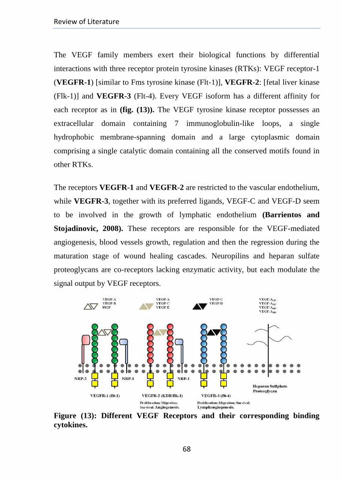
Review of Literature
68
The VEGF family members exert their biological functions by differential
interactions with three receptor protein tyrosine kinases (RTKs): VEGF receptor-1
(VEGFR-1) [similar to Fms tyrosine kinase (Flt-1)], VEGFR-2: [fetal liver kinase
(Flk-1)] and VEGFR-3 (Flt-4). Every VEGF isoform has a different affinity for
each receptor as in (fig. (13)). The VEGF tyrosine kinase receptor possesses an
extracellular domain containing 7 immunoglobulin-like loops, a single
hydrophobic membrane-spanning domain and a large cytoplasmic domain
comprising a single catalytic domain containing all the conserved motifs found in
other RTKs.
The receptors VEGFR-1 and VEGFR-2 are restricted to the vascular endothelium,
while VEGFR-3, together with its preferred ligands, VEGF-C and VEGF-D seem
to be involved in the growth of lymphatic endothelium (Barrientos and
Stojadinovic, 2008). These receptors are responsible for the VEGF-mediated
angiogenesis, blood vessels growth, regulation and then the regression during the
maturation stage of wound healing cascades. Neuropilins and heparan sulfate
proteoglycans are co-receptors lacking enzymatic activity, but each modulate the
signal output by VEGF receptors.
Figure (13): Different VEGF Receptors and their corresponding binding
cytokines.
Review of Literature
69
2.5.4.1.4. Sources of VEGF:
Vascular endothelial growth factor (VEGF) (or vascular permeability factor
(VPF)) was first described in 1983 (Senger et al., 1983) and proved to be involved
in angiogenesis after its isolation for the first time from bovine pituitary follicular
cells in 1989 (Ferrara and Henzel, 1989) where it was identified as an
endothelial-specific growth factor. VEGF was proven to be produced by malignant
tumor cells of several types as well as macrophages (Berse et al., 1992), cultured
Smooth Muscle cells (SMCs) (Williams et al., 1995), ECs (Namiki et al., 1995)
and platelets (Bao et al., 2009). Fibroblasts (Ben-Av et al., 1995; Ferrara et al.,
2003), skin epithelial cells (Takamiya et al., 2002) as well as glioma cells,
hypertrophic chondrocytes (Miyagami and Katayama, 2005) and osteoblasts
(Gerber et al., 1999) demonstrate VEGF immuno-reactivity also. (Brown et al,
1992) identified macrophage-like cells as sources of VEGF in mouse skin wounds.
The study of (Nicholas et al.,1998) demonstrated that the fibroblasts and
macrophages are primary sources of surgical wound VEGF. Up-regulation of
VEGF production was found in keratinocytes in wounds of rats and guinea pigs as
well (Brown et al., 1992).
2.5.4.1.5. VEGF and Wound Healing:
I-VEGF directly recruits bone marrow-derived progenitor cells through a
chemotactic gradient via VEGF receptors expressed in these cells. It may signals
the Endothelial Progenitor Cells (EPCs) releasing from the marrow via its soluble
circulating factors and may act as a local signal mediating the recruitment to injury
sites (via its heparin-binding non-soluble isoforms) (Moore, 2002). This
recruitment may be also by altering the expression of other stem cell trafficking
molecules in the dermis and blood vessels during the cutaneous repair (Fedyk et
al., 2001). Examples include Chemokine Receptor type 4 (CXCR-4) and the
Stromal Cell-Derived Factor-1 (SDF-1) (Jo et al., 2000; Shen et al., 2001; Petit et
al., 2002).

Review of Literature
70
II-In wound sites, Reactive Oxygen Species are generated by a NADPH oxidase-
dependent respiratory burst mechanism in both phagocytic (Babior et al., 1973)
and non-phagocytic cells (Suh et al., 1999). They may promote wound
angiogenesis by inducing VEGF expression in wound-related cells such as
keratinocytes and macrophages (Khanna et al., 2001; Sen et al., 2002).
III-VEGF is most likely to act through receptors in the endothelium to increase the
production of nitric oxide (NO) and prostacyclin (PGI2) and augment intracellular
endothelial cell survival signaling.
IV-VEGF-A controls wound repair by inducing vascular permeability; may be by
inducing vascular leakage (Senger et al., 1983; Dvorak et al., 1995).
V-There is an increasing evidence that VEGF contributes to inflammation
(Detmar et al.,1998) as the endothelial activation is important for (VEGF-induced
inflammation) (Yano et al., 2006). VEGFR-2 plays a main role in this induction
through both phospholipase C (PLC-γ)-dependent (in large part) and PKA-
dependent (to a lesser extent) signaling pathways (Xiong et al., 2009). VEGR-1
plays a role in inflammation by promoting the migration and secretion of
monocytes/ macrophages in which only VEGFR-1 is expressed (Pickkers et al.,
2005).
VI-Effect of Hypoxia in VEGF Secretion:
While hypoxia can initiate neovascularization by inducing angiogenic factors
expression, it cannot sustain it as a threshold level of oxygenation is required to
support the metabolic needs of tissue remodeling. Chronic hypoxia impairs wound
angiogenesis with causing dysfunction and death of tissue (Allen et al., 1997), but
acute hypoxia facilitates the angiogenic process (Semenza, 2000). On one hand,
hypoxia is a potent trigger of the inducible expression of VEGF as well as its
receptors (Berra et al., 2000).

Review of Literature
71
VEGF production and VEGF-mediated angiogenic activity would rise in the early
hypoxic wounds and then fall when the neovascularization is completed under the
action of anti-angiogenic mediators with restoring wound perfusion and capillary
regression (Shweiki et al., 1992; Detmar et al., 1997). On the other hand,
hyperoxia was proven to induce VEGF secretion (Maniscalco et al., 1995;
Darrington et al., 1997; Sheikh et al., 2000).
2.5.4.1.6. Expression of VEGF Gene:
1-The neutrophiles, through their characteristic “respiratory burst” activity,
produce O2-., a very well-known critical reactive oxygen species for defense
against bacteria and other pathogens (Babior et al., 1973). It may be rapidly
converted to a variety of species such as H2O2, .OH or ONOO
- depending on the
local conditions. It has been previously reported that H2O2 induces the VEGF
expression in human keratinocytes and the .OH radical is a stronger oxidizing
agent than H2O2 (Khanna et al., 2001).
2-The granulation tissues formation with their content of fibroblasts, macrophages
and ECs are all essential to maintain the VEGF secretion.
3-The VEGF gene expression is up-regulated by hypoxia as well as by TGF- β,
angiotensin II, (FGF-2) and Interleukin-1 (Il-1) (Williams et al., 1995; Ferrara
and Bunting, 1996; Hayashi et al., 2004; Nagata et al., 2005; Jin et al., 2006).
(Nogami et al., 2007) reported that the VEGF mRNA levels significantly increase
24 h after the incisional wounding of skin compared with the 3rd
and 7th
days, but
fibroblasts, ECs and CD68-positive mononuclear cells sustain VEGF expression 7
days after the wounding.
4-(Nogami et al., 2007) reported also that the presence of VEGF in the early
stages of wound healing indicates the possibility of its involvement in the
vascularization during healing. (Hayashi et al., 2004) observed VEGF expression
in a relatively late stage of skin injury.
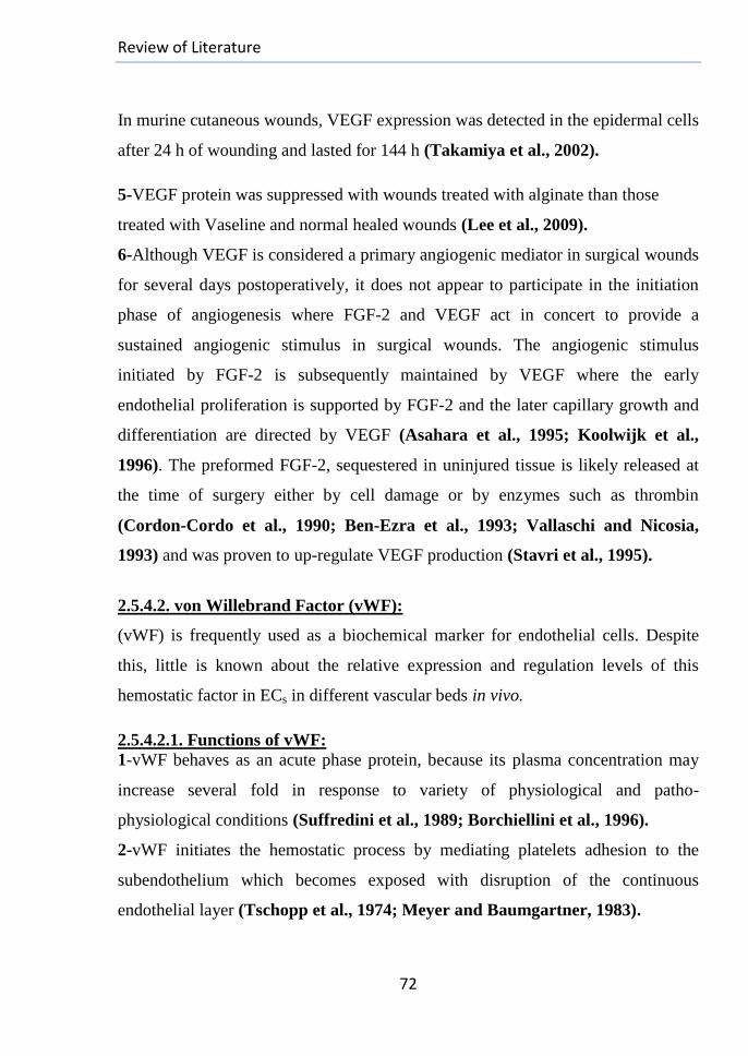
Review of Literature
72
In murine cutaneous wounds, VEGF expression was detected in the epidermal cells
after 24 h of wounding and lasted for 144 h (Takamiya et al., 2002).
5-VEGF protein was suppressed with wounds treated with alginate than those
treated with Vaseline and normal healed wounds (Lee et al., 2009).
6-Although VEGF is considered a primary angiogenic mediator in surgical wounds
for several days postoperatively, it does not appear to participate in the initiation
phase of angiogenesis where FGF-2 and VEGF act in concert to provide a
sustained angiogenic stimulus in surgical wounds. The angiogenic stimulus
initiated by FGF-2 is subsequently maintained by VEGF where the early
endothelial proliferation is supported by FGF-2 and the later capillary growth and
differentiation are directed by VEGF (Asahara et al., 1995; Koolwijk et al.,
1996). The preformed FGF-2, sequestered in uninjured tissue is likely released at
the time of surgery either by cell damage or by enzymes such as thrombin
(Cordon-Cordo et al., 1990; Ben-Ezra et al., 1993; Vallaschi and Nicosia,
1993) and was proven to up-regulate VEGF production (Stavri et al., 1995).
2.5.4.2. von Willebrand Factor (vWF):
(vWF) is frequently used as a biochemical marker for endothelial cells. Despite
this, little is known about the relative expression and regulation levels of this
hemostatic factor in ECs in different vascular beds in vivo.
2.5.4.2.1. Functions of vWF:
1-vWF behaves as an acute phase protein, because its plasma concentration may
increase several fold in response to variety of physiological and patho-
physiological conditions (Suffredini et al., 1989; Borchiellini et al., 1996).
2-vWF initiates the hemostatic process by mediating platelets adhesion to the
subendothelium which becomes exposed with disruption of the continuous
endothelial layer (Tschopp et al., 1974; Meyer and Baumgartner, 1983).
Review of Literature
73
3-With the help of collagen, vWF plays a key role in promoting platelet adhesion
to the damaged vessel walls, subendothelial matrix and endothelial surfaces
(Sakariassen et al., 1979; Girma et al., 1980; Baumgartner et al., 1980; Huang
et al., 2009) and in platelets activation (Farndale, 2006).
4-vWF acts as a genuine matrix protein that promotes the organization of
microfilaments and adhesion plaques via a trans-membrane mechanism with
keeping the anatomical integrity of the internal vessel lining (Dejana et al., 1989).
In addition, it promotes EC adhesion and supports their cytoskeletal organization
(Cheresh, 1987; Charo et al., 1987).
5-(factor VIII-related antigen): It acts as a stabilizer for the coagulation factor VIII
against proteolytic degradation inside the blood stream (Wagner, 1990) and its
carrier to the destination point in the circulation (Huang et al., 2009).
6-vWF plays essential roles in hemostasis: its deficiency or dysfunction results in
increased angiogenesis (Starke et al., 2011) with causing von Willebrand Diseases
(VWD); the most common congenital bleeding disorder in humans (Sadler, 2005).
Its increased levels are clinical markers of risk associated with atherosclerosis and
coronary thrombosis (Spiel et al., 2008).
7-Endothelial vWF is also involved in the regulation of inflammation by
modulating leukocytes adhesion through direct and indirect mechanisms (Denis et
al., 2001; Pendu et al., 2006).
2.5.4.2.2. vWF Gene:
-Human vWF gene is located on the short arm of chromosome 12 (Location:
12p13) with the cytogenetic band (12p13.31) (fig. (14)) (NCBI)) and consists of
178.000 bps. Murine vWF gene location is (4q42) (http://rgd).
Figure (14): Genomic location of human vWF gene on chromosome (12).
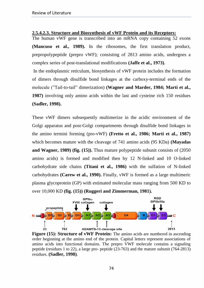
Review of Literature
74
2.5.4.2.3. Structure and Biosynthesis of vWF Protein and its Receptors:
The human vWF gene is transcribed into an mRNA copy containing 52 exons
(Mancuso et al., 1989). In the ribosomes, the first translation product,
prepropolypeptide (prepro vWF); consisting of 2813 amino acids, undergoes a
complex series of post-translational modifications (Jaffe et al., 1973).
In the endoplasmic reticulum, biosynthesis of vWF protein includes the formation
of dimers through disulfide bond linkages at the carboxy-terminal ends of the
molecule (”Tail-to-tail” dimerization) (Wagner and Marder, 1984; Marti et al.,
1987) involving only amino acids within the last and cysteine rich 150 residues
(Sadler, 1998).
These vWF dimers subsequently multimerize in the acidic environment of the
Golgi apparatus and post-Golgi compartments through disulfide bond linkages in
the amino termini forming (pro-vWF) (Fretto et al., 1986; Marti et al., 1987)
which becomes mature with the cleavage of 741 amino acids (95 KDa) (Mayadas
and Wagner, 1989) (fig. (15)). Thus mature polypeptide subunit consists of (2050
amino acids) is formed and modified then by 12 N-linked and 10 O-linked
carbohydrate side chains (Titani et al., 1986) with the sulfation of N-linked
carbohydrates (Carew et al., 1990). Finally, vWF is formed as a large multimeric
plasma glycoprotein (GP) with estimated molecular mass ranging from 500 KD to
over 10,000 KD (fig. (15)) (Ruggeri and Zimmerman, 1981).
Figure (15): Structure of vWF Protein: The amino acids are numbered in ascending
order beginning at the amino end of the protein. Capital letters represent associations of
amino acids into functional domains. The prepro VWF molecule contains a signaling
peptide (residues 1 to 22), a large pro- peptide (23-763) and the mature subunit (764-2813)
residues. (Sadler, 1998).
Review of Literature
75
vWF may be stored in special compartments, called Weibel-Palade Bodies
(WPBs), or released into the blood stream (Wagner and Marder, 1984) under the
induction of a variety of chemical stimuli (e.g., VEGF stimulators) that may be
induced by injuries of vessels walls or increased levels of adrenaline in the
surrounding blood (Sporn et al.,1986; Wagner et al., 1991). The deposited vWF
in the endothelial ECM consists of higher molecular weight multimers than
plasmatic vWF (Tannenbaum et al., 1989; Baruch et al., 1991) with increased
capacity to bind to the platelets (Moake et al., 1986). The largest vWF multimers
were found to bind preferentially to a fibroblast ECM (Sporn et al., 1987).
The two human platelet vWF glycoprotein receptors (GpIb and GpIIb-IIIa) belong
to the superfamily of cell adhesion receptors "integrins" (Hynes, 1987). This
complex has several structural and functional homologies and recognizes numbers
of ECM components via a common Arg-Gly-Asp (RGD) sequence (Ruoslahti and
Pierschbacher,1986) (Fig. (15)). They are structurally unrelated and recognize
different specific binding domains on the vWF molecule (Girma et al., 1987;
Nurden,1987) resulting in the rolling of platelets over the endothelium with their
firm adhesion and activation (Ruggeri and Mendolicchio, 2007).
2.5.4.2.4. Sources of vWF:
Besides being a plasma protein, vWF is also a constitutive component of the
subendothelial matrix (Giddings, 1986) where megakaryocytes and (ECs) are its
only synthesizing cells (Jaffe et al., 1973; Wagner, 1990; Zanetta et al., 2000).
They can release it in the blood stream with organization in close association with
other matrix proteins within the subendothelial basement lamina (Giddings, 1986).
The synthesized vWF by ECs can be stored within the WPBs with other angiogenic
regulators (e.g., Angiopoietin (Ang-2) (Starke et al., 2011)) that is associated with
hemostasis and vascular tone (Valentijn et al., 2011; Metcalf et al.,2008) and
with unidentified proteins.
Review of Literature
76
The normal biology of the WPBs is disrupted with deletion of vWF gene (Denis et
al., 2001). vWF and other molecules are released simultaneously and a temporal
association of vWF and Ang-2 release has been observed (Fiedler et al., 2004;
Starke et al., 2011). Rat endothelial cells store vWF in the endoplasmic reticulum
(Reidy et al., 1989). The synthesized protein may be secreted from the bovine ECs
as that observed in the work of (Lynch et al, 1983) without any significant net
intracellular accumulation of the precursor product. (Hormia et al., 1984; Warhol
and Sweet, 1984; Cramer et al., 1985) have visualized vWF in the Golgi
apparatus. The α-granules are the most abundant granules in platelets and contain a
multifunctional array of proteins (e.g., vWF, thrombospondin, P-selectin and
fibrinogen) (Sabrkhany et al., 2011). The physical structure of these organelles
with the observed longitudinal tubules seem to be due to the multimeric vWf
molecule itself indicating its capability for directing the formation of its own
storage granules (Wagner et al., 1991; Wagner, 1993; Voorberg et al., 1993).
2.5.4.2.5. vWF and Wound Healing:
I-vWF and Hemostasis:
Both the circulating and secreted vWF from the WPBs contribute to wound healing
and blood clotting. Under normal healing conditions, a stable blood clot seals an
injured blood vessel in a complex process involving more than a dozen of blood
coagulation factors, accompanied by alteration of local streaming conditions,
exposure of various connective tissues and the contraction of the injured blood
vessels to limit bleeding which affect strongly the streaming properties of the
flowing blood (Girma et al., 1987). The vWF network represents the basis for
wound healing and the aggregation of platelets to this network is the starting point
for the plug growing which finally seals the vessel damage. The primary
hemostasis process is the initial step to stop blood loss by the formation of (vWF-
platelet plugs) and is based on conformational changes within the protein from an
inactive globular into an elongated activated state.
Review of Literature
77
Plasma vWF does not bind to the circulating platelets, but on immobilizing on a
collagen surface, high shear stress with binding of vWF to the subendothelium
induces changes in the vWF conformation to allow platelets adhesion
(Sakariassen et al., 1984). At the vascular damage sites, activated vWF comprises
specific receptors in its A1 and A3 domain to the counterpart in the collagen
matrix.
On binding to the surface, vWF molecules interconnect to form macroscopic
networks on the exposed layers of collagenous components of the subendothelium
and on additional non-collagenous site(s) for vWF (Wagner and Marder, 1984;
De Groot et al., 1988). Platelets are enriched near the vessel wall with drastic
increase on their binding probability (Fåhræus–Lindqvist effect) (Perkkioe et al.,
1987), mediated by two distinct platelet receptors, GPIb and GP IIb/IIIa (integrin
αIIbβ3) (Girma et al., 1987).
A weak bond with a high dissociation constant is formed between VWF’s A1
domain and the platelet receptor GPIbα, followed with a stronger and irreversible
bond involving VWF’s C1 domain and the platelet integrin αIIbβ3 with a low
dissociation rate enough to form (fig. (15)) (Ruggeri, 1997). These interactions
lead to subsequent platelets spreading and aggregates formation on their membrane
comprising even more binding sites for VWF while reducing the opposing drag
forces at the same time (Weiss et al., 1986; Sakariassen et al., 1986; Fressinaud
et al., 1988). As a result, vWF can function as a bridge between platelets and
collagen (Fredrickson et al., 1998) and serve as glue between multiple platelets
layers. This platelet plaque grows till further loss of blood is prevented (Steppich
et al., 2009), especially if the rent in the vessel wall is small.
Under some circumstances, such as after thrombin activation, vWF also binds to
the platelet membrane glycoprotein complex (GpIIb-IIIa).
Review of Literature
78
This promotes the platelets aggregation (Fujimoto et al., 1982; Ruggeri et al.,
1982; Ruggeri et al., 1983). During secondary hemostasis: Larger damage
requires further stabilization of the platelets aggregates so water-soluble fibrinogen
is transformed to water insoluble fibrin forming a mesh-work for embedding both
erythrocytes and platelets (Guyton and Hall, 2000). The production of fibrin
involves a cascade of multiple blood clotting factors activating each other and the
dysfunction or deficiency of one of these factors can decelerate or even stop blood
clotting (Steppich et al., 2009).
II-vWF and Inflammation:
vWF is stored with P-selectin in the endothelial WPBs and secreted in response to
the same signals (e.g., Vascular injury or inflammatory mediators) which
externalize P-selectin to the plasma membrane and this suggests the possibility of
overlapping functions between the two proteins. The inflammatory role of vWF
could be largely secondary, with the adherent activated platelets providing a
surface for leukocyte recruitment so its deficiency in mice was found to lessen the
inflammatory response in wound healing (Methia et al., 2001) due largely to the
mispackaging and defective secretion of P-selectin associated with VWF
deficiency (Lo´pez, 2006). vWF could recruit leukocytes when conditions favor
the persistence of its hyper-adhesive forms on the endothelial surfaces as occurs
under the influence of inflammatory cytokines which stimulate VWF secretion and
inhibit its processing by ADAMTS13 (Bernardo et al., 2004). Its levels are
elevated in both chronic and acute inflammation (Vischer, 2006). WPBs were
reported also to play roles in inflammation through the stored IL-8 in the
microvascular ECs (Utgaard et al., 1988; Wolff et al., 1998).

Review of Literature
79
III-vWF and Angiogenesis (Relation between vWF and VEGF):
vWF is required to control the formation of the vascular network (Starke et al.,
2011). Its leakage results in increased vascularization, suggesting that VEGF-
dependent angiogenesis is enhanced in VWF-deficient cases. Increased VEGF
expression and signaling have been linked to vascular malformations (Thurston et
al.,1999; Ozawa et al.,2004). vWF has an inhibitory activity in the constitutive
VEGFR-2–dependent pathway(s) that promote EC migration; however, there is
partial effect for the extracellular VWF on this process (Starke et al., 2011).
vWF regulates Ang-2 release from ECs. Decreased/ dysfunctional vWF can cause
decrease or loss in production of WPB and cause defective packaging of these
proteins as a consequence resulting in constitutive release of its components (e.g.,
Ang-2) that destabilizes the blood vessels and acts synergistically with VEGF-A to
promote angiogenesis (Yu and Stamenkovic, 2001; Lobov, 2002; Hashizume et
al., 2010). The over-expression of Ang-2 in in vivo studies results in the formation
of unstable, dysplastic vessels (Feng et al., 2007). An increase in plasma levels of
(Ang-2) in patients with VWD might contribute to promoting blood vessel
plasticity and thus exacerbate angiodysplasia.
2.5.4.2.6. Expression of vWF Gene:
Variety of perturbations (e.g., Renal failure (Warrell et al., 1979), irradiation
(Sporn et al., 1984) and cerebral thrombosis (Zheng,1983) cause increase in the
circulating levels of vWF that is thought to be an indicative of endothelial injury.
vWF is heterogeneously distributed through the vascular tree and is associated
with regional variations in the mRNA levels (Rand et al., 1987; Wu et al., 1987;
Bahnak et al., 1989; Coffin et al., 1991; Page et al., 1992; Aird et al., 1995;
Smith et al., 1996). Low levels of vWF are present within the sinusoidal ECs of
liver and spleen. In en face preparations of the rat aorta, expression of vWF
appeared to vary from one EC to another (Senis et al., 1996).
Materials and Methods
80
3. MATERIALS and METHODS
3.1. Materials:
*Sodium alginate (Na-Alginate) (A2033, medium viscosity, M/G ratio (1.56)) and
Potassium Bromide: Sigma-Aldrich, USA.
*Coomassie Brilliant Blue G-250 & Ethylene glycol (ANALAR) & Diethyl ether
and Fehling solutions: Fisher scientific, USA.
*Bovine Serum Albumin (BSA) (Biotechnology Grade): AMResco, Canada.
*Haematoxylin, Eosin and MT: Surgipath Europe Ltd, UK.
*Sodium chloride: Riedel-de Haën, Germany.
*Acetic acid: FEINchemie Schwebda, Germany.
*Streptozotocin (STZ): Merck Chemical, Germany.
*Sodium (meta) periodate: LOBA-Chemie, India.
*Calcium chloride, Ferrous chloride, Ammonium thiocyanate, Charcoal, Sodium
hydroxide (Analytical grade), Hydroxylamine hydrochloride, absolute Ethanol,
Butanol, Methanol, Sulphoric, Hydrochloric, Nitric, Phosphoric acids (Analytical
grades), Formaldehyde and Chloroform: Elnasr Pharmaceutical Company
(ADWIC, Egypt).
*Hydrogen peroxide (Analytical grade): Piochem, Egypt.
*α-tocopherol: CID pharmaceutical company, Egypt.
3.2. Preparation of reagents:
3.2.1.Hydroxylamine.HCl (NH2OH.HCl) reagent (0.25 M)
According to (Huiru and Heindel, 1991), 17 gm dried (NH2OH.HCl) powder
were dissolved in 150 ml distilled water with heating at 65oC till complete
dissolving. The solution was cooled; then 6 ml (Methyl Orange reagent, 0.05 %)
were added and completed to 1 L with adjusted pH at 4.
3.2.2. Sevag Reagent:
Chloroform and n-butanol solutions were mixed together at a ratio (4:1).
Materials and Methods
81
3.2.3. Phosphate buffered saline (PBS):
I.Potassium dihydrogen phosphate (0.1 M): (6.81 gm KH2PO4 (from (ADWIC-
Egypt)) were dissolved in distilled water to 500 ml.
II.Disodium hydrogen phosphate (0.1 M): (8.91 gm Na2HPO4.2H2O (from
(ADWIC-Egypt)) were dissolved in distilled water to 500 ml.
III.Preparation of Phosphate Buffered Saline (PBS, 0.1 M, pH 7.4): 80.8 ml
(Na2HPO4, 0.1M) were mixed with 19.2 ml of (KH2PO4, 0.1 M).
3.2.4. Sodium Citrate buffer (0.1 M, pH 4.5):
44.5 ml (Citric acid. H2O, 0.1M) were mixed with 55.5ml (Trisodium citrate.
2H2O, 0.1M).
3.2.5. Reagents for the RNA Extraction based on (Fermentus, Thermo Fisher
Scientific Inc, UK) extraction kit:
*The working Lysis Buffer: 24 µl β-mercaptoethanol were added to each 1 ml of
the kit Lysis Buffer to prepare the required working buffer.
*TE-Buffer: Consisted of (10 mM Tris-Cl (pH 7.5), 1 mM EDTA (pH 8)).
*Proteinase-K: Diluted enzyme was prepared by adding 10 µl Proteinase K to 590
µl of the prepared TE-Buffer.
*Preparation of Wash Buffer (1): 10 ml of absolute ethanol were added to 40 ml
concentrated wash buffer (1) to reach a total volume (50 ml).
*Preparation of Wash Buffer (2): 39 ml of absolute ethanol were added to 23 ml
concentrated wash buffer (2) to reach a total volume (62 ml).
3.3. Modification of Alginate:
3.3.1. Irradiation of Sodium Alginate:
Degraded Na-Alginates were prepared by the following methods:
1-(LMW-Alginate) was obtained through γ-irradiation of sodium alginate from
(Cobalt-60 (Gamma-Cell 220), Atomic energy of Canada Limited, installed at
Radioisotope Department of the Egyptian AEA, Cairo-Egypt) at the 2 doses (50
and 100 KGy) at R.T on air with dose rate (5.6 KGy/ hour).
Materials and Methods
82
2-Sharp decreasing for the molecular weight was obtained by mixing Na-alginate
(powder) with 10 (wt. %) oxidizing agent (H2O2) for its obtaining in the paste form
that was then packaged tightly in polyethylene bags and subjected to γ-irradiation
at the doses (50 and 100 KGy) under the same previous conditions. The resulting
alginate was denoted (VLMW: Very Low Molecular Weight–Alg).
3.3.2. Oxidation of Sodium Alginate:
This was carried out according to the modified protocol of (Bouhadir et al., 2001).
8 Erlenmeyer flasks; each one contained 1 % homogeneous alginate solution in
100 ml NaCl solution (10 mM), were wrapped with aluminum foil, and divided
into (2) groups; each one of 4 flasks and denoted as follows:
-Group I (Multi Aldehyde Sodium Alginate-I (MASA-I)): each flask was charged
with 1.0 gm un-irradiated sodium alginate (HMW-Alg). -Group II
(Multi Aldehyde Sodium Alginate-II (MASA-II): each flask was charged with 1.0
gm irradiated sodium alginate (LMW-Alg).
4 different concentrations of Na-periodate solutions (0.25, 0.5, 0.75, and 1.0 M)
were prepared and 20 ml from each one were added to one flask of each group
with stirring the mixtures for 24 h at R.T. Ethylene glycol (0.5 ml) was added to
the mixture of each flask to quench the reactions by reducing any unreacted
periodate (Lee et al., 2002) and then stirred for additional 0.5 h at R.T. 2.5 gm
NaCl were then added to the solution of each flask, followed by precipitation with
excess of absolute ethanol (200 ml for each solution). The precipitates, collected
by centrifuging the different alginate products were redissolved in distilled water
and re-precipitated with ethanol. After their concentrating, the precipitates were
dried to yield a white product (about 0.7-0.8 gm).
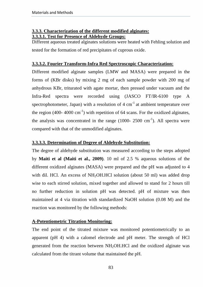
Materials and Methods
83
3.3.3. Characterization of the different modified alginates:
3.3.3.1. Test for Presence of Aldehyde Groups:
Different aqueous treated alginates solutions were heated with Fehling solution and
tested for the formation of red precipitates of cuprous oxide.
3.3.3.2. Fourier Transform-Infra Red Spectroscopic Characterization:
Different modified alginate samples (LMW and MASA) were prepared in the
forms of (KBr disks) by mixing 2 mg of each sample powder with 200 mg of
anhydrous KBr, triturated with agate mortar, then pressed under vacuum and the
Infra-Red spectra were recorded using (JASCO FT/IR-6100 type A
spectrophotometer, Japan) with a resolution of 4 cm-1
at ambient temperature over
the region (400- 4000 cm-1
) with repetition of 64 scans. For the oxidized alginates,
the analysis was concentrated in the range (1000- 2500 cm-1
). All spectra were
compared with that of the unmodified alginates.
3.3.3.3. Determination of Degree of Aldehyde Substitution:
The degree of aldehyde substitution was measured according to the steps adopted
by Maiti et al (Maiti et al., 2009). 10 ml of 2.5 % aqueous solutions of the
different oxidized alginates (MASA) were prepared and the pH was adjusted to 4
with dil. HCl. An excess of NH2OH.HCl solution (about 50 ml) was added drop
wise to each stirred solution, mixed together and allowed to stand for 2 hours till
no further reduction in solution pH was detected. pH of mixture was then
maintained at 4 via titration with standardized NaOH solution (0.08 M) and the
reaction was monitored by the following methods:
A-Potentiometric Titration Monitoring:
The end point of the titrated mixture was monitored potentiometrically to an
apparent (pH 4) with a calomel electrode and pH meter. The strength of HCl
generated from the reaction between NH2OH.HCl and the oxidized alginate was
calculated from the titrant volume that maintained the pH.
Materials and Methods
84
The degree of aldehyde substitution was calculated based on the report of Zhao
and Heindel (Zhao and Heindel, 1991) from the following equation:
B-The titration continued until the red (of the acidic liberated HCl-rich solution) -
to yellow (The neutral solution) end point was achieved by matching the color of
the samples solutions with that of a blank one.
3.3.3.4. Determination of the Average Molecular Weights:
3.3.3.4.1. The Viscometric Method:
Dynamic viscosities for the raw and modified alginates solutions (in 10 mM NaCl
solvent) were measured using (Synchro-Lectric rotational Viscometer: Model DV-
IV+ Pro, Brookfield Engineering Labs Inc., Middleboro, MA, USA), operated at
25°C using the spindle (No. CPE 41) rotating at 25 RPM. The spindle was
immersed at first on the solvent, then on each alginate type in ascending order of
concentrations. For the raw alginate, measurements were performed with the
concentrations (C: 0.1, 0.2, 0.4, 0.6, 0.8, and 1 %), but they were (0.5, 1, 2, 3, 4,
and 5%) for the degraded forms. The viscosity measurements of each sample were
performed in triplicates to obtain accurate readings for calculating the molecular
weights as follows:
(I) The obtained measurements are the coefficients of viscosity as follows:
-solvent): denotes the solvent NaCl (10 mM) viscosity.
- (sol`n): denotes the prepared alginate solutions viscosities.
(II) To a very good approximation, the Relative Viscosity (rel) is calculated from
the ratio between (stated sol`n and solvent) at the same temperature.
(III) The solution Specific Viscosity: (solvent
solventnsolsp
' =rel-1)
(IV) Reduced Viscosity (dl/gm): (redsp/ C).
(V) Inherent Viscosity (dl/gm): (inh=lnrel/ C).
Materials and Methods
85
With plotting inh and red versus the concentration and extrapolating to C=0, the
intrinsic viscosity (int) was determined by averaging the values of the intercept
(Russo et al., 1986). The average Molecular Weight was calculated on the basis of
the following Mark–Houwink–Sakurada equation (MHS) (Tanford, 1961):
Where: *int: the intrinsic viscosity.
*M or (Mv): viscosity average Molecular Weight of the polymer.
*The equation constants: (K= 4.85×10⁻³
ml/gm and a= 0.97) at which Mw ≈
Mv for alginate (Stokke et al., 2000).
3.3.3.4.2. Gel Permeation Chromatography (GPC) method:
The number-average Molecular Weight (Mn) and weight-average Molecular
Weight (Mw) for the unirradiated, irradiated and oxidized samples were measured
by ((GPC) 1100 Agilant instrument) equipped with organic and aqueous GPC-SEC
start up kits with a flow rate of 2 ml/min, maximum pressure 150 bar, minimum
pressure 5 bar, injection volume 50 µl and column temperature thermostat 25°C).
The eluent was monitored by a refractive index detector of optical unit temperature
25°C and peak width 0.1 min. The polymer concentration was 0.1 (wt. %) and the
M.Ws were determined from a calibration curve using polyethylene oxide
standards for aqueous systems. The used method for determining (Mn) and (Mw)
followed (Liu et al., 2006; Li and Liang 2007).
Materials and Methods
86
3.4. Purification Protocol of Sodium Alginate:
3.4.1. Acid-washing of Activated Charcoal:
This was accomplished based on (Asada et al., 2006) method with 3 trial
batches. Each 1 gm of pre-dried charcoal in each patch was completed to 100 ml
with 1 M H2SO4, HNO3 or HCl, then mixed together at R.T for 5 hours, filtrated
and washed several times with distilled water. The resulting charcoals were then
dried at 100oC overnight and tested as well as the raw type in the pellet form with
(JASCO FT/IR-6100-type A spectrophotometer) according to the procedures,
stated in section (3.3.3.2). The spectral range of analysis was (400-4000 cm-1
)
with 100 scans and a resolution of 4 cm-1
. The same washing steps were repeated
then in patches system with the chosen washing acid after the FT-IR testing.
3.4.2. Method of Alginate Purification:
At first, the alginates to be purified were separated into batches. All steps of
preparation and testing were carried out using double distilled water, tested for its
pH (7 ± 0.15) and calcium concentration (less than 3 ppm). All the used glass
flaks and containers were sterilized with saturated water steam before the
purification steps which were performed according to the protocol of (Qi et al.,
2009) with some modifications as follows:
A-Precipitation of Proteins from Alginate Solutions:
For all steps, the alginate concentration was expressed according to the weight
percent (wt. %). For each patch, (1% homogeneous solution, pH 7) was prepared
in 10 mM NaCl solution at R.T with continuous shaking. For each chilled 100 ml
solution, 25 ml Sevag reagent were added (Staub et al., 1965), shaken vigorously
for 30 min and centrifuged at 3000 RPM leaving the solution divided into 3 layers:
top organic layer, a middle layer containing the precipitated proteins; these two
layers were aspirated and only the bottom aqueous one containing the alginate
polymer remained. This step was repeated for each patch.
Materials and Methods
87
B-Precipitation of the (PC) from Alginate Solutions:
Equivalent alginate weight of acid-washed activated charcoal was added to each
solution, mixed well, then filtrated to remove the adsorbent charcoal with several
washings with dist. water till its complete removal.
C-Precipitation of Alginate:
200 ml ethanol were added to each 100 ml of alginate solution, mixed
continuously, allowed to stand for 30 min and filtered for separating the alginate
that was redissolved in water and re-precipitated then with ethanol. After
concentrating, the precipitates were frozen at -70oC over-night, lyophilized for 2
days and then the following tests were applied for testing the effects of
purification on the treated alginates.
3.4.3. Testing the Effects of Purification in Alginate:
3.4.3.1. Viscosity Measurements for the Raw and Purified Alginates:
Dynamic viscosities of the different alginate solutions (0.5 % w/v) before and
after purification were checked using a rotational Viscometer (Synchro-Lectric
Brookfield rotational Viscometer) operated at 20oC and the spindle (No. CPE 41)
was selected by rotating at 25 RPM.
3.4.3.2. Quantification of Impurities Concentrations:
3.4.3.2.1. Proteins Content Quantification:
I-Preparation of Reagents:
*Dye Stock: According to (Bradford, 1976), 100 mg Coomassie Brilliant Blue
G-250 were dissolved in 50 ml methanol (95%), 100 ml phosphoric acid (85%)
were then added. After complete dissolving of the dye, the solution was diluted
to 200 ml with dist. water to give a dark red solution with pH 0.08.
Concentrations of the final reagent components were (0.5 mg /ml Coomassie
Blue G, 25% methanol and 42.5% phosphoric acid).
Materials and Methods
88
*Assay Reagent: Prepared by diluting 1 vol. of the dye stock with 4 volumes of
dist. water to give brown colored solution, stable in dark at 4oC.
*Blank Solution: 0.15M NaCl.
*Protein Standards: Series of Bovine Serum Albumin (BSA) standards were
utilized for building a convenient standard curve and prepared by dilution with
(150 mM NaCl) with the following concentrations:
-0, 250, 500, 750, 1000, 1500 and 2000 µg/ml: for the standard assay.
-0, 1.5, 7.5, 10, 30, 40 and 50 µg/ml: for the micro-assay.
*Alginates Samples: For increasing the accuracy of measurements, unpurified
and purified (HMW and irradiated alginates) were prepared in different
concentrations using the same solvent (150 mM NaCl).
II-Method of Quantification (The Microassay):
1ml of the prepared assay reagent was added to 100 µl of each concentration
from both the standards and alginate samples and incubated at R.T for 10 min.
The absorbance of each resulted solution as well as the dye only (zero protein)
were measured using blanked spectrophotometer at 590 nm. Plastic cuvettes were
used and all measurements were in ascending order. For presentation of the
results, the concentrations were converted to the equivalent mg of proteins per
gm of dry alginate.
3.4.3.2.2. Polyphenolic-like Compounds (PC) Quantification:
The fluorescence spectra of unirradiated and irradiated alginate solutions (1%
w/v in dil. NaCl) were obtained using a spectrofluorimeter (JASCO FP-6500,
Japan, source: Xenon arc lamp 150 Watt, both the slit band width of excitation
monochromator and emission monochromator were 5 nm and PMT gain was
medium). An excitation wavelength of 366 nm and emission wavelength of 445
nm were applied.
Materials and Methods
89
The relative quantities of the Polyphenol-like compounds were detected by the
appearance of a characteristic absorbance peak at 445 nm (Skjak-Braek et al.,
1989) and were measured and expressed in terms of Arbitrary Fluorescence Units
(AFU) with correlation to Raman bands.
3.4.3.3. Characterization of the Purified Alginates with (FT-IR): Fourier
Transform-Infra Red (FT-IR) spectroscopic analysis was utilized to investigate
the effect of purification on the structural properties of the alginate chains and the
binding behavior of their functional groups (C-OH and COONa). Structures of
the purified products were confirmed through (FTIR) applied to (KBr-Alginate
pellets), prepared as previously described in section (3.3.3.2). The bands
corresponding to the functional groups for both the raw and purified alginates
within I.R charts were compared.
Materials and Methods
90
3.5. Preparation of Chitosan:
3.5.1. The Extraction and Deacetylation Steps:
These were carried out according to (Gopalakannan et al., 2000) with some
modifications. Frozen dried shrimp shells waste was cut into pieces, washed with
tap water and deproteinized by boiling in 1 N NaOH (1:5 w/v) for 30 min. The
alkali was drained, replaced with another amount and the process was repeated for
the removal of residual proteins. Colors of the solutions were noticed till becoming
clear and the pieces were washed with tap water. The shells were demineralised by
soaking in 1 N HCl (1:5 w/v) at 40°C for 30 min. The acid was then drained off
and the pieces were washed thoroughly with tap water till reaching the neutrality
followed with dist. water washing. The chitin product was dried at R.T and stored
under dry conditions till the further deacetylation procedures.
The deacetylation steps were carried out according to (Roberts, 1992); the chitin
was soaked in 50 % aqueous NaOH (1:20 w/v) and divided into two different
patches which were boiled at 100°C for 2 hours divided into 4 equal times with the
changing of NaOH. The first patch was then washed with water and nominated as
(Ch-1). The second patch, (Ch-2) was stored till cooled, then reboiled in 50 %
aqueous NaOH for additional 2 hours, re-cooled and reboiled for additional 1 hour.
After deacetylation of the (2) chitosan patches with draining off the alkali and
washing by tap water, they were washed by dist. water and dried at R.T for several
days.
3.5.2. Characterization of the Prepared Chitosan products:
3.5.2.1. Solubility Test:
Solubility of each patch product as well as the parent compound, chitin was tested
by formation of soluble polymer on preparation of (0.5 % solution in 1 % Acetic
acid solution (HAc)) with continuous heating and shaking.
Materials and Methods
91
3.5.2.2. (FT-IR) Spectroscopic Characterization:
Chitosans samples were prepared in the form of KBr pellets based on the method
of (Sabnis and Block, 1997) where the pellets (powdered chitosan: KBr=1: 3)
were dried at 80°C in hot air oven for 24 hours before analysis, then characterized
by infrared spectrophotometer in the range of (800-4000 cm-1
). The % N-
deacetylation values were determined using the equation proposed by (Terayama,
1952; Domszy and Roberts, 1985) but with using the baseline, proposed by
(Baxter et al., 1992) for comparing the intensity of the (amide I) band in chitosan
with that of the (-OH groups). The following equation was applied:
%DD=100- [(A1655/A3450) *100/1.33]
Where: A1655: The absorbance at 1655 cm-1
for the amide (N-H) band as a measure
of the N-acetyl group content.
A3450: Absorbance at 3450 cm-1
for the (OH) band as (internal standard) to correct
for film thickness or differences in chitosan powder concentration. 1.33:
Correction factor in case of (A1655/A3450) for fully N-acetylated chitosan (it reaches
zero for the fully deacetylated chitosans).
3.5.2.3. Measurement of the Protein Content:
After complete drying, the protein content % of chitin as well as each patch
product was measured according to the standard method, mentioned in section
(3.4.3.2.1) in triplicate measurements for each patch. The standard curve was built
using high BSA concentrations (1, 2, 4, 6, 8 and 10 mg/ml)
3.5.2.4. Measurement of the Ash Content (Purity):
For each chitosan patch, ash content was measured after heating the samples (in
triplicates for each patch) at 100°C for 24 h.
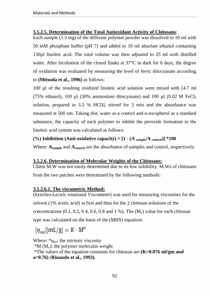
Materials and Methods
92
3.5.2.5. Determination of the Total Antioxidant Activity of Chitosans:
Each sample (1.3 mg) of the different polymer powder was dissolved to 10 ml with
50 mM phosphate buffer (pH 7) and added to 10 ml absolute ethanol containing
130µl linoleic acid. The total volume was then adjusted to 25 ml with distilled
water. After incubation of the closed flasks at 37°C in dark for 6 days, the degree
of oxidation was evaluated by measuring the level of ferric thiocyanate according
to (Mitsuda et al., 1996) as follows:
100 µl of the resulting oxidized linoleic acid solution were mixed with [4.7 ml
(75% ethanol), 100 µl (30% ammonium thiocyanate) and 100 µl (0.02 M FeCl2
solution, prepared in 3.5 % HCl)], stirred for 3 min and the absorbance was
measured at 500 nm. Taking dist. water as a control and α-tocopherol as a standard
substance, the capacity of each polymer to inhibit the peroxide formation in the
linoleic acid system was calculated as follows:
(%) Inhibition (Anti-oxidative capacity) = [1 - (A sample/A control)] *100
Where: Asample and Acontrol are the absorbance of samples and control, respectively.
3.5.2.6. Determination of Molecular Weights of the Chitosans:
Chitin M.W was not easily determined due to its low solubility. M.Ws of chitosans
from the two patches were determined by the following methods:
3.5.2.6.1. The viscometric Method:
(Synchro-Lectric rotational Viscometer) was used for measuring viscosities for the
solvent (1% acetic acid) at first and then for the 2 chitosan solutions of the
concentrations (0.1, 0.2, 0.4, 0.6, 0.8 and 1 %). The (Mv) value for each chitosan
type was calculated on the basis of the (MHS) equation:
Where: *ƞint: the intrinsic viscosity
*M (Mv): the polymer molecular weight
*The values of the equation constants for chitosan are (K=0.076 ml/gm and
a=0.76) (Rinaudo et al., 1993).
Materials and Methods
93
3.5.2.6.2. Gel Permeation Chromatography (GPC) Method:
The Mn and Mw values of both chitosans were measured by GPC instrument
(1100 Agilant) equipped with GPC-SEC start up kits with a flow rate of 2 ml/min,
maximum pressure 150 bar, minimum pressure 5 bar, injection volume 50µL and
column temperature thermostat 25°C. The eluent was monitored by a refractive
index detector of optical unit temperature 25°C and peak width 0.1 min. The
molecular weights were determined from calibration curve with polyethylene
oxide standards and the method used for determination followed (Liu et al., 2006;
Li and Liang, 2007).
3.6. Method of Preparing the (Alginate-Chitosan PECs Hydrogels):
For future references, Alginate will be referred to as (Alg) and Chitosan as (Ch).
Homogeneous chitosan solution (Ch-1, 0.4 %, pH 5) was prepared in 0.5 % acetic
acid. Alginate solutions (of different ratios of alginates, 0.4 %) were prepared in 10
mM NaCl with adjusted pH at 6.84.
For preparation of PECs Films: All polymers solutions were fresh to avoid any
intrinsic changes in them with time and they were autoclaved before using. The
used volume of alginate solution was 25 ml and chitosan solution was added
dropwise with continuous gentle shaking to the resulting mixture till ending the
reaction of coacervation with formation of PECs. For preparation of the (semi-
IPNs), CaCl2 solution (700 µl) was added dropwise with continuous shaking. After
ending the reaction, selected viscous suspensions were kept aside for some time till
the removal of air bubbles then casted into glass Petri dishes and allowed to dry at
40oC for 48 hours. The dried gels were then stored in polyethylene bags under
nitrogen atmosphere at R.T and sterilized by (γ-radiation) from (Co60
irradiation
source) at (25 KGy) with a dose rate (5.2 KGy/hour).
Materials and Methods
94
3.7. Major Steps for Choosing the Best Type of Hydrogels:
3.7.1. In Vitro Swelling of Hydrogels in Simulated Wound Fluids:
The kinetics of swelling for the different formulations was characterized in the
terms of % weight gain/loss at different time intervals using conventional
gravimetric procedures (Berens and Hopfenberg, 1978; Park et al., 2001). After
drying to a constant weight in a desiccator, each gel formulation was divided into
weighed pieces (1cm×1cm); all weights were within the range (0.06-0.08 gm) and
each one was placed into a well of a multi-wells plate containing (3 ml of the
prepared PBS, pH 7.4) representing a simulated wound fluid, covered to minimize
water loss by evaporation and incubated at 37oC. After 30 min, they were
withdrawn, soaked with tissue paper, kept in desiccator till getting a constant
weight and each one was weighed separately. The same steps were repeated with
periodic monitoring of pieces weights at the time intervals (1,2 ,4, 8, 12 and 24 hrs)
with the continuous medium changing. Three individual experiments were
performed at each time point with each gel with gaining the average values. The
fold swelling of the different formulations was calculated from the following
equation according to (Yeole et al., 2006):
Degree of Swelling % = [(Ms/Md)-1] x 100
Where: Ms = Mass of swollen film at time t.
Md = Mass of dried film at (t=0).
3.7.2. Stability Characterization Studies:
As a rapid test for strength of the prepared gels, a part of each prepared
formulation gel was tested for its physical stability by weak shaking of a small
piece (2 cm ×2 cm) from each for 60 seconds with visual establishing of the gels
integrity.
3.7.3. Blood Compatibility Tests:
The chosen hydrogels were tested for their hemocompatibility as follows:

Materials and Methods
95
3.7.3.1. Thrombus Formation Test:
This was judged by the blood clot formation test (Mishra and Chaudhar, 2010).
Each dried hydrogel film (2 gm) was equilibrated with 2 ml saline water and
incubated in a water bath (37oC) for 24 hrs. To each swollen hydrogel, 500 µl
Human Citrated Blood (HCB) were added, followed by 30 µl CaCl2 solution (4 M)
to start the thrombus formation with incubation at 37oC for 5 min. The reaction
was terminated by adding 4 ml dist. water to each sample and soaking at R.T for
10 min. The clots were fixed with 2 ml formaldehyde solution (37 %) for another
10 min., placed then in water for more 10 min and the weights corresponding to
the different gels and glass surface were recorded after drying.
3.7.3.2. Hemolysis Assay:
According to (Mishra and Chaudhar, 2010), 2 ml (0.9 % w/v) saline water were
added for each dry hydrogel film (2 gm) and incubated in water bath (37oC) for 24
hrs. To each swollen hydrogel, 250 µl (HCB) were added, mixed well and
incubated at 37oC for 20 min. 2 ml saline were added to each film for reaction
termination with incubation at 37oC for 60 min. Positive and negative controls
were obtained by adding 250 µl of (HCB) and saline solution, respectively to 2 ml
distilled water. The incubated samples were centrifuged for 45 min and absorbance
of the supernatants was recorded at 545 nm using a spectrophotometer. Each
absorbance data point was obtained by measuring three samples with determining
the deviations of the three tests. The percentage of hemolysis was calculated from
the following equation (Thomas, 1996):
Hemolysis (%) = [(Atest sample – A (-) control)/ (A (+) control – A (-) control)] x 100 where:
A is the absorbance.
3.7.4. Rate of Evaporation of Water from Gel:
The chosen dried hydrogels were kept at 37oC and 35 % relative humidity with
measuring their weights at regular intervals of time (Balakrishnan et al., 2005b).
Materials and Methods
96
Three specimens were used at each time point for each gel film. The following
equation was used to find out the weight remaining (%):
Hydrogel weight remaining (%) = (Mt/Mo) × 100
Where: Mo and Mt are initial mass and mass after time‘t’, respectively.
3.7.5. In vitro Degradation of the Prepared Hydrogel Films:
The chosen dried hydrogel samples were weighed and incubated under sterilized
conditions in 50 ml PBS at 37oC with changing the medium each 2 days. Gel
degradation behavior was monitored by measuring the dry weight loss of the gels
at predetermined time points after keeping in a desiccator for 1 day (Hian, 2009).
Three Specimens were used with each weight at each time point for each film to
obtain the degradation curve. The mass loss % was calculated from the following
equation:
Mass loss %= (Mi -Md) / Mi ×100
Where:(Mi& Md) are the initial mass and mass of degraded gel, respectively.
3.7.6. Primary Skin Irritation Test for the Hydrogels:
This is a characterizing test for properties of the prepared hydrogel and its irritation
effects were compared with gels before irradiation for only confirming the test
results. According to the (NIH Publication No. 85-23, 1996), six male rats
weighing (180-200 gm) were lightly anesthetized with diethyl ether; the dorsal
surface skin was completely shaved and cleaned with alcohol (70 %). The rats
were then returned to their cages; each one in individual cage for 24 hours to allow
any edema caused by shaving to recede. Application fields were outlined on the
right dorsal side of each rat with marking pen and single full thickness wound of
area (1 cm2) was created by circular excising in skin of the dorsum with forceps
and scissors. The wounds in 3 of the rats were covered with cut pieces of the
irradiated hydrogel membrane but the other 3 with unirradiated pieces and fixed
with plasters.
Materials and Methods
97
The shaved skin on the left side remained intact but also covered with the
corresponding type of gel on each rat. Rats were returned to their cages and the
plaster covers with the hydrogel remnants were removed from all wounds after 4
hours and examined for the irritation signs, edema and swelling after 1, 24, 48 and
72 hours. Effects of the tested hydrogels were scored according to the attached
grading system as reported in (Procter and Gamble, 1977) for calculating the
Primary Dermal Irritation Index (PDII) corresponding to each hydrogel from the
following equation:
PDII= (sum. of averages (1, 24, 48, 72 hrs))
(Number of observation periods times (4))
3.7.7. Testing the Optimum Composition of Hydrogel:
The following tests were carried out to reach the optimum composition of the
prepared hydrogel and its optimum preparation conditions:
3.7.7.1 Detection of the best concn. for the used cross-linker (CaCl2):
This test was applied to the two formulae (F-18, 20) using the same swelling
procedures, stated in section (3.7.1). Three different concentrations of CaCl2 (1%,
2%, and 2.5%) were used with each one and the swelling degree was calculated
after 4 hours of immersing in PBS (pH 7.4) at 37oC from the following equation
taking (n=3) for each concentration of each film.
Q %=(Ms/Md) ×100.
Where: Ms = Mass of swollen film at time t.
Md = Mass of dried film at (t=0).
3.7.7.2. Characterizing the Effects of (γ-irradiation) on Hydrogel (F-20):
3.7.7.2.1. Testing the Bacterial Growth with the Irradiated Hydrogels:
3 gel pieces (1cm×1cm) were taken from unirradiated gel samples and randomly
chosen irradiated hydrogels, placed aseptically in 24 multi-well plates; each in a
well with adding sufficient FBS nutrient medium amount. The plate was shaken to
achieve complete immersion of the pieces and better contact with the medium.
Materials and Methods
98
All wells were covered with para -film and the plate was covered, incubated at
37oC for 2 weeks and observed then with a light microscope. Interpretation of the
results was done then by turbidimetric analysis for the immersed gels; the cloudy
medium indicated the proliferation of bacteria and the clear one indicated the
growth absence.
3.7.7.2.2. Hemocompatibility Assays:
Both the anti-thrombogenic potentials of the hydrogel surfaces and hemolysis %
were measured for the irradiated and un-irradiated hydrogels of (F-20 and 5) as
previously described in the section (3.7.3).
3.7.7.2.3. Swelling Study for the Irradiated and Unirradiated gels:
The irradiated formulae (F-20 and 5) were tested using the same previous swelling
steps as in section (3.7.1). Their swelling behaviors were compared with the
corresponding unirradiated gels (F-20(un)& 5(un)). The CaCl2 concentration was
(2 %) and the (D.S) was recorded after immersing in PBS (pH 7.4) at 37oC for 24
hours (n=3 for each time point of the gel). (F-5) was used to confirm results of the
test.
3.7.7.2.4. (FT-IR) Spectroscopic Characterization:
Experimental spectra for the prepared irradiated and unirradiated (F-20) films were
obtained by mixing the dried film with KBr. Pellets of the alginates powder as well
as physical mixture of chitosan and the used alginates (1:1) were also obtained on
mixing 2 mg with 98 mg KBr, pressed with hydraulic pressure at 13 tons and the
disks were examined with (JASCO FT/IR 6100 type A spectrophotometer). The
spectra were recorded as (% T) at 32 scans and resolution of 4 cm-1
in the range
(500-4000 cm-1
).
Materials and Methods
99
3.7.7.2.5. Scanning Electron Microscopy (SEM):
Representative air dried samples from unirradiated and irradiated PEC sheets were
taken and analyzed by (SEM). The films were sectioned with a blade, mounted on
an aluminum sample support by means of a conductive and double-sided adhesive
tape before sputter-coating with an ultra thin gold layer (300 Ao) under vacuum in
a Polaron E-coating apparatus. The Morphologies of films' surfaces were then
analyzed using a (JEOL-JSM-5400 Scanning Electron Microscope, Japan) at
accelerating voltage (30 KV).
3.7.7.3. Choosing the Best Working Film Structure:
3 different forms of the formula (F-20) were prepared with changing the ratio
between (A& B and D chains) according to the following composition:
(F-20/I): [A+B+D (2:1:1) + (CaCl2)] + (Ch).
(F-20/II): [A+B+D (1:2:1) + (CaCl2)] + (Ch).
(F-20/III): [A+B+D (1:1:2) + (CaCl2)] + (Ch).
Their properties were compared to reach the structure of the best working film by
application of the following tests:
3.7.7.3.1. Swelling Studies:
With the same previous swelling steps in section (3.7.1) using CaCl2 (2%) as a
cross-linker, the swelling degrees for these forms were recorded after immersing in
PBS (pH 7.4) at 37oC for 24 hours (n=3).
3.7.7.3.2. Rate of Evaporation of Water from Gel:
The chosen hydrogels (F-20/I & F-20) were kept at 37oC and 35% relative
humidity. Their weights were measured at regular intervals of time and the weight
remaining % was calculated from the equation stated in section (3.7.4) (n=3).
Materials and Methods
100
3.7.7.3.3. In vitro Degradation of the Prepared Hydrogel Films:
The dried hydrogel samples were weighed and incubated under sterilized
conditions in 50 ml PBS at 37oC with changing the medium each 2 days. The gel
degradation behavior was calculated by the equation stated in section (3.7.5) (n=3).
3.7.7.3.4. Capability of drugs loading to the hydrogel forms (F-20, 20/I):
Two Hydrogel films of each form (F-20 and F-20/I) were prepared. One of each
contained a model protein, (BSA) representing a high molecular weight drug and
the other 2 preparations contained (Curcumin) representing a low molecular weight
drug:
3.7.7.3.4.1. Encapsulation of Albumin into the Hydrogel Network:
10 ml BSA (0.5 %) were mixed with alginate solution and the same steps of
preparing the PEC hydrogels were performed as detailed in section (3.6).
3.7.7.3.4.2. Encapsulation of Curcumin into the Hydrogel Network:
10 ml Curcumin (0.5%) were mixed with chitosan solution with repeating the same
steps of PEC hydrogels preparation in section (3.6).
3.7.7.3.4.3. Scanning electron microscopy for the prepared loaded films
To indemnify the ability of drug loading, representative air dried samples from
irradiated and un-irradiated PEC sheets loaded with Curcumin or BSA were
investigated through analyzing morphologies of the films surfaces with (SEM)
according to the procedures stated in section (3.7.7.2.5).
3.8. Statistical Analyses:
In all assays concerning the quantification of impurities, modification of alginate,
characterization of the prepared chitosans and the steps of characterizing the
hydrogels properties, triplicate treatments were tested individually with each.
Materials and Methods
101
The SPSS Computer program (Version: 14) aided in statistical analysis of the
results. Data were analyzed using one way Analysis Of Variance (ANOVA)
followed by post Hoc LSD test. The data were expressed as (Mean ± Standard
Error). Differences were considered Statistically Significant at (p≤ 0.05) and
Highly Significant at (p≤ 0.01).
Materials and Methods
102
3.9. Wounding and Wound Healing Assessment:
3.9.1. Animals:
The care and use of laboratory animals were cooperated in accordance with the
Ethics Committee of the National Research Center, conformed to the “Guide for
the Care and Use of Laboratory Animals” published by the U.S National Institute
of Health (NIH Publication No. 85-23, 1996). (160) adult male albino rats (Rattus
norvigicus); purchased from the animal farm of the Egyptian Holding Company
for Biological Products and Vaccines (VACSERA), Giza, Egypt, were used in the
present investigation. Their body weights ranged from (120-150 gm) and were
acclimatized to laboratory conditions for one week under well-ventilation
conditions in the animal house building of the National Center for Radiation
Research and Technology (NCRRT) with a 12:12-hour light dark cycle and had
free access to food and water prior to initiation of the study.
3.9.2. Wounding Procedures:
Before any step, approximate fasting glucose for blood samples from tail veins of
all rats was determined with (glucometer& reagent strip, BIONINE GM-100,
China) to make sure all rats; under the study, are not diabetic. An excision wound
model was created according to (Morton and Malone, 1972) method. At first, the
rats were lightly anesthetized with diethyl-ether; the dorsal surface skin was then
digitally shaved completely and cleaned with alcohol (70 %). The rats were
returned to their cages; each one in individual cage for 24 hours to allow any
edema, caused by shaving to recede. Application fields were outlined then with a
marking pen and single right full thickness circular wound of area (1 cm2) was
created by circular excising in the dorsal skin and the underlying (panniculus
carnosus) using toothed forceps and scissors. The day of skin excision was defined
as (D 0) and the animals were randomly assigned into the following three groups:
Materials and Methods
103
Group (1: Dressing/ Dre-group): Composed of 75 rats. Immediately on excision,
the wound was covered with the chosen hydrogel cut pieces. The PEC film
adhered well to the moist wound, but additional adhesive material was used to
prevent its removal by the rat itself. The wounds were monitored daily with
removal of the old hydrogel residues and redressing it approximately each 2 days.
Group (2:Fusidin/Fus-group):Composed of 10 rats. The wound was treated with
topical application of a commercial cream; fusidin, 2 times per day.
Group (3: Control/ Con-group): Composed of 75 rats serving as untreated
controls. The wounds were only disinfected with ethanol and kept opened.
3.9.3. Wound and Skin Assessment:
The involved methods for assessment of the potential wound healing activity of the
chosen hydrogel and in the other groups were as follows:
3.9.3.1. Monitoring the Visible Changes in Wounds during Healing:
Daily, the wounds of all groups were grossly monitored for infection and rats with
its signs were excluded. Photographing of the wounds was performed during the
post wounding days from a standard height to monitor any changes during healing
using a digital camera (Samsung, South Korea).
3.9.3.2. Measurement of Residual Wound Area:
By using a previously standardized tracing paper and thin marker method for
tracing wound healing (Thomas and Wysocki, 1990), all wounds margins were
drawn daily beginning with (D 0), then the paper pieces were weighed and surface
areas of the wounds at their continuous healing days were calculated. The %
reduction in area with respect to that of (D 0) and the original wound dimensions
was calculated according to the equation:
% Reduction= (SAi-SAc) ×100/ SAi Where: SAi: The wound Surface Area at (zero day) (D 0).
SAc: The wound Surface Area, currently.
Materials and Methods
104
3.9.3.3. Histological Studies:
I-Histological Stains:
The most commonly used stains are (Haematoxylin (H) and Eosin (E)).
Haematoxylin stains cell nuclei and other negatively charged structures(e.g.,
Alginate) blue-black while Eosin stains cytoplasm and the connective tissue fibers
in varying shades of pink, orange and red (Bancroft and Gamble, 2007).
Generally the (H& E staining) was used to assess the skin maturity by
demonstrating the inflammatory response at the wound area, dermal closure,
remodeling and general tissues architectures while advanced granulation tissues
formation as well as tissues remodeling were assessed via Masson`s-Trichrome
(MT) staining. Collagens are the most abundant component of ECM in most
tissues including the dermal layer and they are strongly stained with the acid dyes
due to the affinity of cationic groups of these proteins for the anionic reactive
groups of these dyes. Collagen fibers of different thicknesses are colored
differently.
II- Samples Preparations:
The wound tissues including the scab and complete epithelial margin for the
control (G:3) as well as the dressed wounds with hydrogel remnants (G:1) were
rapidly extirpated during different post-wounding days with surrounding skin
tissue at the edges and a supporting layer of body wall musculature as a mass. 10%
(v/v) formal-saline was prepared by dissolving 10 ml formaldehyde (37 %) in 90
ml saline solution (0.9%) and used to fix 3D samples for 24h at R.T. Acetone was
used then to fix cell monolayers for 30 min at R.T and 3D samples were
transferred to Cell Safe biopsy inserts (CellPath Ltd, UK) that were placed in
cassettes, then processed by progressive dehydration in graded ethanol and
immersed in xylene using a Citadel 1000 Tissue Processor.
Materials and Methods
105
The samples were then transferred to liquid paraffin (Tissue-Tek III Embedding
Wax, Sakura, UK) and embedded in paraffin using Tissue-Tek TEC Tissue
Embedding Console System. They were heated to 60°C (4.5 min), then immersed
in xylene (1 min), IMS 99 (40 sec), 95% IMS 99 (40sec), rinsed in tap water (20
sec), immersed in Haematoxylin (2.5min), rinsed in water (20 sec), immersed in
0.3% w/v acetic acid in dist. water (20 sec), 0.3% HCl in 70% IMS 99, rinsed in
water (20 sec), immersed in tap water substitute (40 sec), washed in water (20 sec),
immersed in Eosin diluted 1:1 with 70% IMS 99 (20 sec), rinsed in water (20 sec),
immersed in IMS 99 (1 min) and xylene (50 sec). Following this, the samples were
held in xylene for up to 4h. Coverslips were then mounted on the slides and viewed
with a light microscope. As part of the histological evaluation, all slides were
examined by a pathologist without knowledge of the group with treatment.
3.9.3.4. Quantification of RNA Corresponding to (VEGF and vWF):
In rats of the 1st and 3
rd groups, the expression of VEGF and vWF genes has been
quantified by the real-time PCR method (Isaksson and Nilsson, 2006, Benoy et
al., 2006; Graham et al., 2006). Specificity of the amplicons was verified by
building the melting curves for the PCR products. The extraction kit (Fermentus,
Thermo Fisher Scientific Inc, UK) was used to extract RNA from tissue and blood
samples according to the manufacturer’s protocol based on the (Chomczynski and
Sacchi, 1987; Boom et al., 1990) methods.
I. RNA Extraction from Wound Tissues:
Step (A): Cell Lysate Preparation
1-At the post-wounding days 1, 2, 3, 5, 7, 11, 15, 21 and 28, about 30 mg of fresh
tissue were excised from the subcutaneous area of both control and dressed
wounds. The harvested intact pieces were disrupted by grinding in liquid nitrogen
thoroughly with mortar and pestle till becoming powder.
Materials and Methods
106
2-The tissue powder was transferred immediately into a microcentrifuge tube (1.5
ml) containing 300 µl of the working buffer, and then mixed thoroughly with
vortexing for 10 sec.
3-600 µl diluted Proteinase K were added, vortexed to mix thoroughly and
incubated at 15-25°C for 10 min, centrifuged for 5 min. at 12000 x g and the
supernatant was transferred into new RNase-free microcentrifuge tube.
4-450 µl of absolute ethanol were finally added and mixed by pipetting.
Step (B): Binding RNA to Column:
5-Up to 700 µl of lysate were transferred to the RNA Purification Column inserted
in a 2 ml collection tube and centrifuged for 1 min at ≥ 12000 x g.
6-The flow-through was discarded and the purification column was placed back
into the collection tube with repeating step (5) till transferring all the lysate into the
Column. The collection tube was discarded with the flow-through solution and the
Column was placed into a new collection tube.
Step (C): Column Washing:
7-700 µl of (Wash Buffer 1) were added to the RNA Purification Column and
centrifuged for 1 min at ≥ 12000 x g with discarding the flow-through and the
Column was placed back into the collection tube.
8-600 µl of (Wash Buffer 2) were added to the RNA Purification Column and
centrifuged for 1 min at ≥ 12000 x g with discarding the flow-through and the
Column was placed back into the collection tube.
9-Washing with the same buffer (250 µl) was repeated and the Purification
Column was transferred to a sterile 1.5 ml RNase-free microcentrifuge tube after
discarding the collection tube containing the flow-through solution.
Materials and Methods
107
Step (D): RNA Elution:
10- 40 µl of nuclease-free water were added to the center of the RNA Purification
Column membrane and centrifuged for 1 min at ≥ 12000 x g to elute the RNA. The
purification column was discarded then and the purified RNA was used for the
synthesis of (cDNA).
II. RNA Extraction from Blood:
1-Blood cells were collected by centrifugation of 0.5 ml of fresh whole blood
(containing EDTA), collected from the studied rats of groups (1 and 3) at 400 x g
for 5 min at 4ºC to generate a pellet of about 60-70% of the total sample volume
after removal of the clear supernatant (plasma) with a pipette. The pellet was
resuspended in 600 µl of the working Lysis Buffer and pipetted to mix thoroughly.
2-450 µl of absolute ethanol were added and mixed by pipetting. The same RNA-
extraction steps from tissue were repeated then beginning with the step of binding
RNA to the column till getting RNA ready for cDNA synthesis.
III. Synthesis of (cDNA) via Reverse Transcription:
cDNA was synthesized from RNA extracted from both tissue and blood samples
after quantification of their concentrations with nano-dropper according to the
instructions of cDNA synthesis kit (Fermentus, Thermo Fisher Scientific Inc, UK)
(Wiame et al., 2000) as follows:
Step (A): The following components were mixed together in a chilled thin-walled
reaction tube which then incubated at 70oC for 5 min:
Sterile H2O…………………………………………….............................(6 µl).
Random Hexamer (400 ng/ µl)…………………………..........................(1 µl).
RNA (50 ng/µl).…........................................................………………..(5 µl).
Drop of oil………………………………………………………………(40 µl).
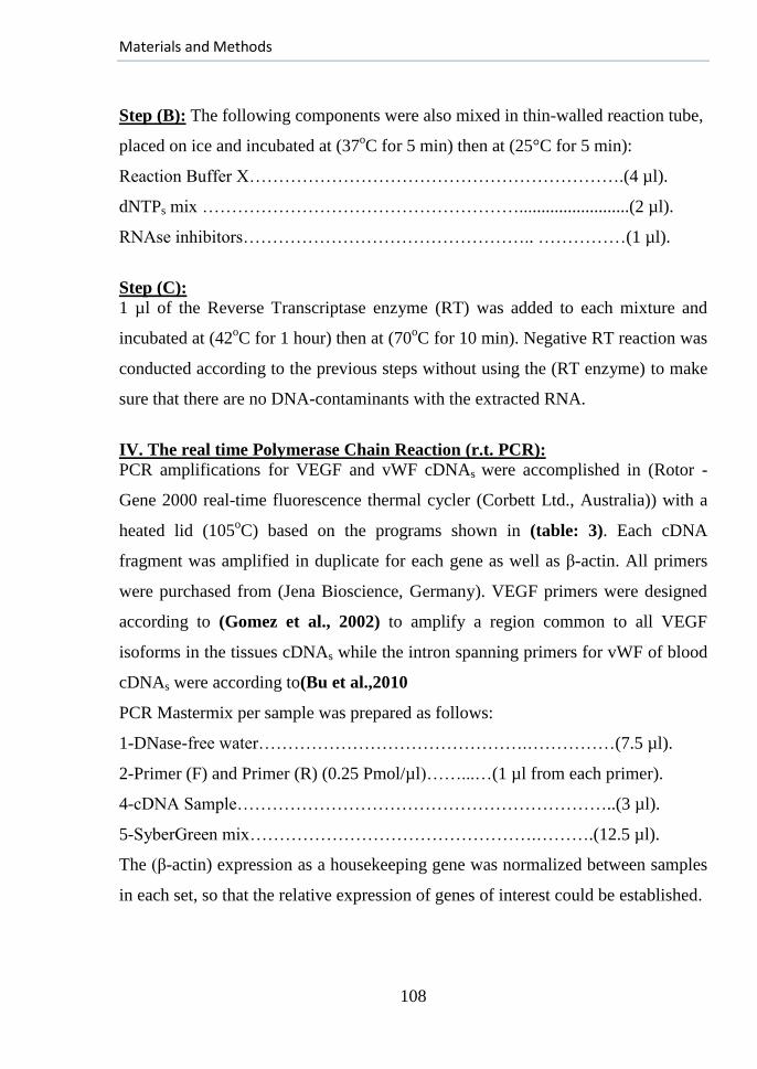
Materials and Methods
108
Step (B): The following components were also mixed in thin-walled reaction tube,
placed on ice and incubated at (37oC for 5 min) then at (25°C for 5 min):
Reaction Buffer X……………………………………………………….(4 µl).
dNTPs mix ……………………………………………….........................(2 µl).
RNAse inhibitors………………………………………….. ……………(1 µl).
Step (C):
1 µl of the Reverse Transcriptase enzyme (RT) was added to each mixture and
incubated at (42oC for 1 hour) then at (70
oC for 10 min). Negative RT reaction was
conducted according to the previous steps without using the (RT enzyme) to make
sure that there are no DNA-contaminants with the extracted RNA.
IV. The real time Polymerase Chain Reaction (r.t. PCR):
PCR amplifications for VEGF and vWF cDNAs were accomplished in (Rotor -
Gene 2000 real-time fluorescence thermal cycler (Corbett Ltd., Australia)) with a
heated lid (105oC) based on the programs shown in (table: 3). Each cDNA
fragment was amplified in duplicate for each gene as well as β-actin. All primers
were purchased from (Jena Bioscience, Germany). VEGF primers were designed
according to (Gomez et al., 2002) to amplify a region common to all VEGF
isoforms in the tissues cDNAs while the intron spanning primers for vWF of blood
cDNAs were according to(Bu et al.,2010
PCR Mastermix per sample was prepared as follows:
1-DNase-free water……………………………………….……………(7.5 µl).
2-Primer (F) and Primer (R) (0.25 Pmol/µl)……...…(1 µl from each primer).
4-cDNA Sample………………………………………………………..(3 µl).
5-SyberGreen mix………………………………………….……….(12.5 µl).
The (β-actin) expression as a housekeeping gene was normalized between samples
in each set, so that the relative expression of genes of interest could be established.
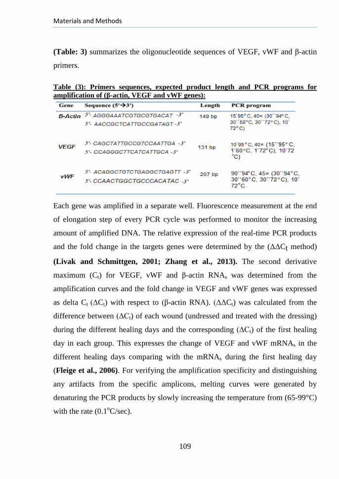
Materials and Methods
109
(Table: 3) summarizes the oligonucleotide sequences of VEGF, vWF and β-actin
primers.
Table (3): Primers sequences, expected product length and PCR programs for
amplification of (β-actin, VEGF and vWF genes):
Each gene was amplified in a separate well. Fluorescence measurement at the end
of elongation step of every PCR cycle was performed to monitor the increasing
amount of amplified DNA. The relative expression of the real-time PCR products
and the fold change in the targets genes were determined by the (ΔΔCt method)
(Livak and Schmittgen, 2001; Zhang et al., 2013). The second derivative
maximum (Ct) for VEGF, vWF and β-actin RNAs was determined from the
amplification curves and the fold change in VEGF and vWF genes was expressed
as delta Ct (ΔCt) with respect to (β-actin RNA). (ΔΔCt) was calculated from the
difference between (ΔCt) of each wound (undressed and treated with the dressing)
during the different healing days and the corresponding (ΔCt) of the first healing
day in each group. This expresses the change of VEGF and vWF mRNAs in the
different healing days comparing with the mRNAs during the first healing day
(Fleige et al., 2006). For verifying the amplification specificity and distinguishing
any artifacts from the specific amplicons, melting curves were generated by
denaturing the PCR products by slowly increasing the temperature from (65-99°C)
with the rate (0.1oC/sec).

Materials and Methods
110
Optical data were collected throughout the duration of temperature drop with
dramatic increase in fluorescence seen when the denatured strands on melting
reannealed. For the statistical analysis of results, every test was replicated three
times for the quantification of each gene corresponding to each time point of group
and the exponential Ct values were averaged before calculating the amplification
and the results were statistically analyzed as mentioned in section (3.8). The results
of each day were compared with the previous one in the same group as well as the
corresponding day in the other group.
3.9.3.5. Screening of kidney Functions:
Blood samples from both (Dre (1) and Con (2)-groups) of wounded rats were
collected in EDTA during the different post wounding-days, centrifuged
immediately and the plasma was stored at -20°C till further analysis. Later, the
samples were analyzed for renal function tests. A triplet sample of each group per
day was analyzed for optimal screening and the results were statistically analyzed
as mentioned in section (3.8).
3.9.3.5.1. Quantitative Estimation of (Blood Urea Nitrogen) in Plasma:
The enzymatic colorimetric method of (Tabacco et al., 1979) was the base for the
quantitative estimation of (Blood Urea Nitrogen (BUN)) in plasma using a
diagnostic kit supplied by (Diamond company-Egypt).
Principle:
The reaction involved in the assay system was as follows:
1-Sample urea is hydrolyzed enzymatically with (Urease) into (NH4+
+ CO2).
2-The formed (NH4+) ions react with salicylate and hypochlorite (NaClO) in the
presence of (catalyst-nitroprusside) to form green indophenol whose intensity is
proportional to the urea concentration in the sample.
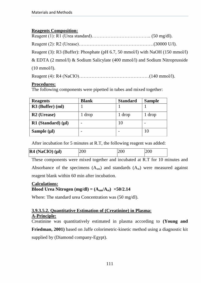
Materials and Methods
111
Reagents Composition:
Reagent (1): R1 (Urea standard)……………………………….. (50 mg/dl).
Reagent (2): R2 (Urease)…………………………………………(30000 U/l).
Reagent (3): R3 (Buffer): Phosphate (pH 6.7, 50 mmol/l) with NaOH (150 mmol/l)
& EDTA (2 mmol/l) & Sodium Salicylate (400 mmol/l) and Sodium Nitroprusside
(10 mmol/l).
Reagent (4): R4 (NaClO)………………………………………(140 mmol/l).
Procedures: The following components were pipetted in tubes and mixed together:
After incubation for 5 minutes at R.T, the following reagent was added:
R4 (NaClO) (µl) 200 200 200
These components were mixed together and incubated at R.T for 10 minutes and
Absorbance of the specimens (Asm) and standards (Ast) were measured against
reagent blank within 60 min after incubation.
Calculations:
Blood Urea Nitrogen (mg/dl) = (Asm/Ast) × 50/2.14
Where: The standard urea Concentration was (50 mg/dl).
3.9.3.5.2. Quantitative Estimation of (Creatinine) in Plasma:
A-Principle:
Creatinine was quantitatively estimated in plasma according to (Young and
Friedman, 2001) based on Jaffe colorimetric-kinetic method using a diagnostic kit
supplied by (Diamond company-Egypt).
Reagents Blank Standard Sample
R3 (Buffer) (ml) 1 1 1
R2 (Urease) 1 drop 1 drop 1 drop
R1 (Standard) (µl) - 10 -
Sample (µl) - - 10
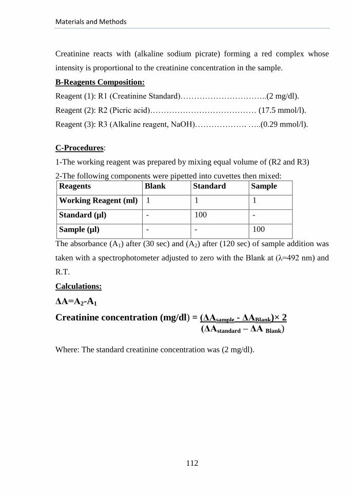
Materials and Methods
112
Creatinine reacts with (alkaline sodium picrate) forming a red complex whose
intensity is proportional to the creatinine concentration in the sample.
B-Reagents Composition:
Reagent (1): R1 (Creatinine Standard)…………………………..(2 mg/dl).
Reagent (2): R2 (Picric acid)………………………………… (17.5 mmol/l).
Reagent (3): R3 (Alkaline reagent, NaOH)………………. …..(0.29 mmol/l).
C-Procedures:
1-The working reagent was prepared by mixing equal volume of (R2 and R3)
2-The following components were pipetted into cuvettes then mixed:
Reagents Blank Standard Sample
Working Reagent (ml) 1 1 1
Standard (µl) - 100 -
Sample (µl) - - 100
The absorbance (A1) after (30 sec) and (A2) after (120 sec) of sample addition was
taken with a spectrophotometer adjusted to zero with the Blank at (λ=492 nm) and
R.T.
Calculations:
ΔA=A2-A1
Creatinine concentration (mg/dl) = (ΔAsample - ΔABlank)× 2
(ΔAstandard – ΔA Blank)
Where: The standard creatinine concentration was (2 mg/dl).
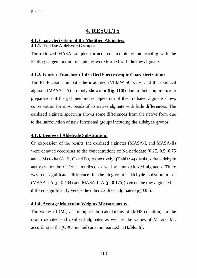
Results
113
4. RESULTS
4.1. Characterization of the Modified Alginates:
4.1.1. Test for Aldehyde Groups:
The oxidized MASA samples formed red precipitates on reacting with the
Fehling reagent but no precipitates were formed with the raw alginate.
4.1.2. Fourier Transform-Infra Red Spectroscopic Characterization:
The FTIR charts for both the irradiated (VLMW-50 KGy) and the oxidized
alginate (MASA-I A) are only shown in (fig. (16)) due to their importance in
preparation of the gel membranes. Spectrum of the irradiated alginate shows
conservation for most bands of its native alginate with little differences. The
oxidized alginate spectrum shows some differences from the native form due
to the introduction of new functional groups including the aldehyde groups.
4.1.3. Degree of Aldehyde Substitution:
On expression of the results, the oxidized alginates (MASA-I, and MASA-II)
were denoted according to the concentrations of Na-periodate (0.25, 0.5, 0.75
and 1 M) to be (A, B, C and D), respectively. (Table: 4) displays the aldehyde
analyses for the different oxidized as well as non oxidized alginates. There
was no significant difference in the degree of aldehyde substitution of
(MASA-I A (p=0.434) and MASA-II A (p=0.175)) versus the raw alginate but
differed significantly versus the other oxidized alginates (p≤0.05).
4.1.4. Average Molecular Weights Measurements:
The values of (Mv) according to the calculations of (MHS-equation) for the
raw, irradiated and oxidized alginates as well as the values of Mn and Mw
according to the (GPC-method) are summarized in (table: 5).
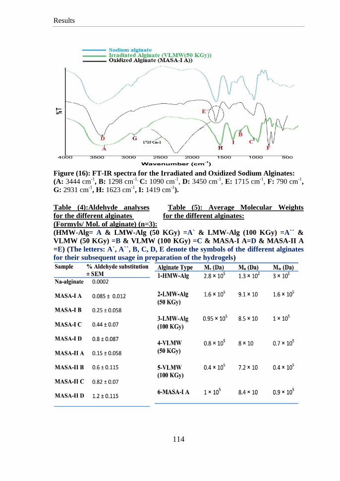
Results
114
Figure (16): FT-IR spectra for the Irradiated and Oxidized Sodium Alginates:
(A: 3444 cm-1
, B: 1298 cm-1,
C: 1090 cm-1
, D: 3450 cm-1
, E: 1715 cm-1
, F: 790 cm-1
,
G: 2931 cm-1
, H: 1623 cm-1
, I: 1419 cm-1
).
Table (4):Aldehyde analyses Table (5): Average Molecular Weights
for the different alginates for the different alginates:
(Formyls/ Mol. of alginate) (n=3):
(HMW-Alg= A & LMW-Alg (50 KGy) =A` & LMW-Alg (100 KGy) =A`` &
VLMW (50 KGy) =B & VLMW (100 KGy) =C & MASA-I A=D & MASA-II A
=E) (The letters: A`, A``, B, C, D, E denote the symbols of the different alginates
for their subsequent usage in preparation of the hydrogels)
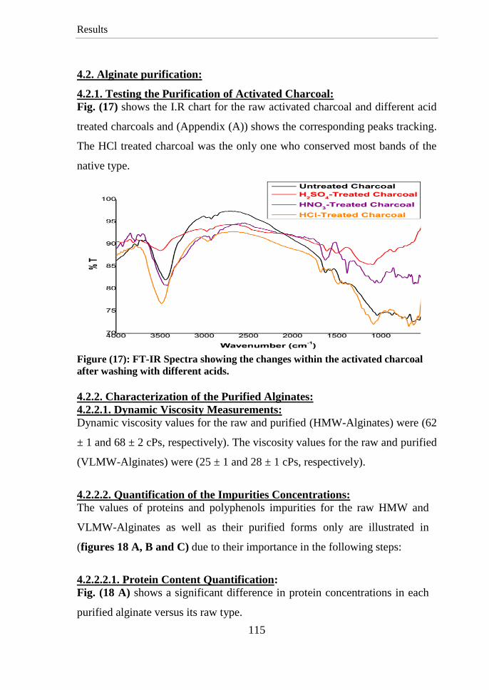
Results
115
4.2. Alginate purification:
4.2.1. Testing the Purification of Activated Charcoal:
Fig. (17) shows the I.R chart for the raw activated charcoal and different acid
treated charcoals and (Appendix (A)) shows the corresponding peaks tracking.
The HCl treated charcoal was the only one who conserved most bands of the
native type.
Figure (17): FT-IR Spectra showing the changes within the activated charcoal
after washing with different acids.
4.2.2. Characterization of the Purified Alginates:
4.2.2.1. Dynamic Viscosity Measurements:
Dynamic viscosity values for the raw and purified (HMW-Alginates) were (62
± 1 and 68 ± 2 cPs, respectively). The viscosity values for the raw and purified
(VLMW-Alginates) were (25 ± 1 and 28 ± 1 cPs, respectively).
4.2.2.2. Quantification of the Impurities Concentrations:
The values of proteins and polyphenols impurities for the raw HMW and
VLMW-Alginates as well as their purified forms only are illustrated in
(figures 18 A, B and C) due to their importance in the following steps:
4.2.2.2.1. Protein Content Quantification:
Fig. (18 A) shows a significant difference in protein concentrations in each
purified alginate versus its raw type.
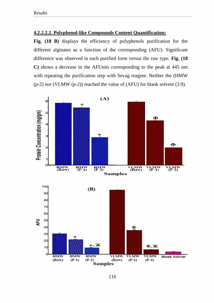
Results
116
4.2.2.2.2. Polyphenol-like Compounds Content Quantification:
Fig. (18 B) displays the efficiency of polyphenols purification for the
different alginates as a function of the corresponding (AFU). Significant
difference was observed in each purified form versus the raw type. Fig. (18
C) shows a decrease in the AFUnits corresponding to the peak at 445 nm
with repeating the purification step with Sevag reagent. Neither the (HMW
(p-2) nor (VLMW (p-2)) reached the value of (AFU) for blank solvent (3.9).
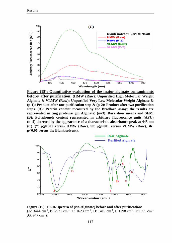
Results
117
Figure (18): Quantitative evaluation of the major alginate contaminants
before/ after purification: (HMW (Raw): Unpurified High Molecular Weight
Alginate & VLMW (Raw): Unpurified Very Low Molecular Weight Alginate &
(p-1): Product after one purification step & (p-2): Product after two purification
steps. (A): Protein content measured by the Bradford assay; the results are
represented in (mg proteins/ gm Alginate) (n=3). Bars show means and SEM.
(B): Polyphenols content represented in arbitrary fluorescence units (AFU)
(n=2) detected by the appearance of a characteristic absorbance peak at 445 nm
(C). (*: p≤0.001 versus HMW (Raw), Φ: p≤0.001 versus VLMW (Raw), Ж:
p≤0.05 versus the Blank solvent).
Figure (19): FT-IR spectra of (Na-Alginate) before and after purification:
(A: 3444 cm-1
, B: 2931 cm-1
, C: 1623 cm-1
, D: 1419 cm-1
, E:1298 cm-1
, F:1095 cm-1
,G: 947 cm-1
).
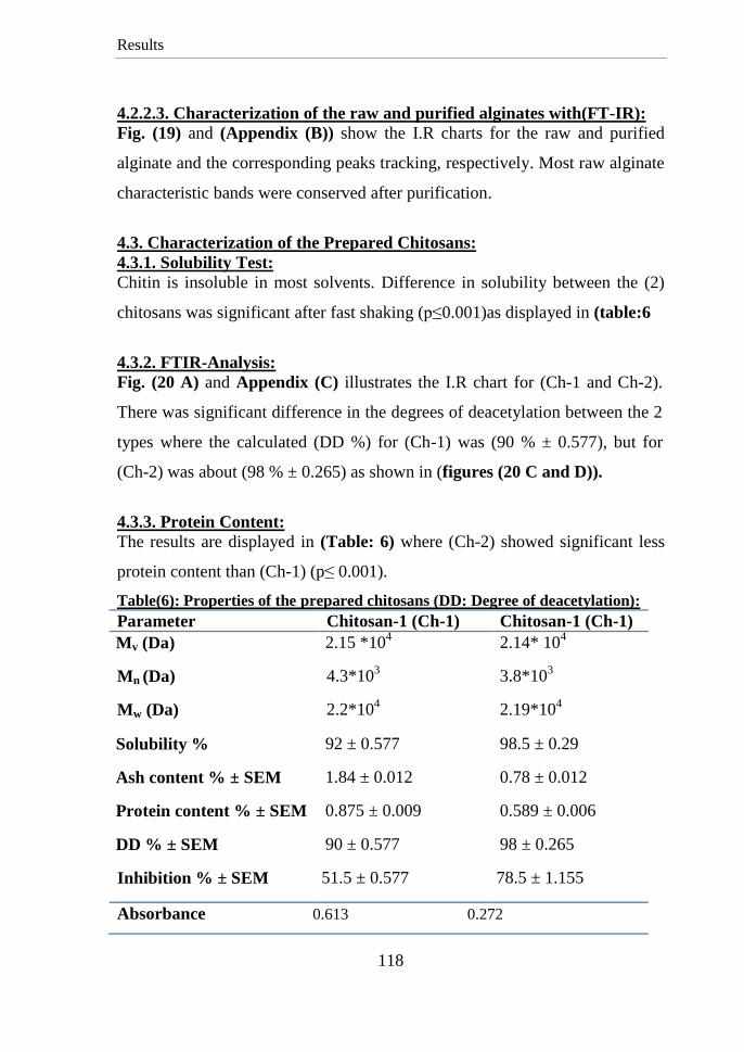
Results
118
4.2.2.3. Characterization of the raw and purified alginates with(FT-IR):
Fig. (19) and (Appendix (B)) show the I.R charts for the raw and purified
alginate and the corresponding peaks tracking, respectively. Most raw alginate
characteristic bands were conserved after purification.
4.3. Characterization of the Prepared Chitosans:
4.3.1. Solubility Test:
Chitin is insoluble in most solvents. Difference in solubility between the (2)
chitosans was significant after fast shaking (p≤0.001)as displayed in (table:6
4.3.2. FTIR-Analysis:
Fig. (20 A) and Appendix (C) illustrates the I.R chart for (Ch-1 and Ch-2).
There was significant difference in the degrees of deacetylation between the 2
types where the calculated (DD %) for (Ch-1) was (90 % ± 0.577), but for
(Ch-2) was about (98 % ± 0.265) as shown in (figures (20 C and D)).
4.3.3. Protein Content:
The results are displayed in (Table: 6) where (Ch-2) showed significant less
protein content than (Ch-1) (p≤ 0.001).
Table(6): Properties of the prepared chitosans (DD: Degree of deacetylation):
Parameter Chitosan-1 (Ch-1) Chitosan-1 (Ch-1)
Mv (Da) 2.15 *104 2.14* 10
4
Mn (Da) 4.3*103
3.8*103
Mw (Da) 2.2*104 2.19*10
4
Solubility % 92 ± 0.577 98.5 ± 0.29
Ash content % ± SEM 1.84 ± 0.012 0.78 ± 0.012
Protein content % ± SEM 0.875 ± 0.009 0.589 ± 0.006
DD % ± SEM 90 ± 0.577 98 ± 0.265
Inhibition % ± SEM 51.5 ± 0.577 78.5 ± 1.155
Absorbance 0.613 0.272

Results
119
Figure (20): (FT-IR) spectra for the Chitosans (1, 2). (A) I.R spectra within
range (500-4000 cm-1
). (B) I.R spectra within range (1200-1800 cm-1
). (C) and (D)
show the base lines for calculating the DD for (Chitosan-2) and (Chitosan-1),
respectively and values of the corresponding (A1655 and A3450):
(A: 3450 cm-1
, B: 2900 cm-1
, C: 1620 cm-1
, D: 1545 cm-1
.
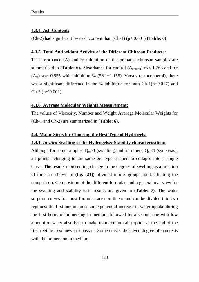
Results
120
4.3.4. Ash Content:
(Ch-2) had significant less ash content than (Ch-1) (p≤ 0.001) (Table: 6).
4.3.5. Total Antioxidant Activity of the Different Chitosan Products:
The absorbance (A) and % inhibition of the prepared chitosan samples are
summarized in (Table: 6). Absorbance for control (Acontrol) was 1.263 and for
(Ast) was 0.555 with inhibition % (56.1±1.155). Versus (α-tocopherol), there
was a significant difference in the % inhibition for both Ch-1(p=0.017) and
Ch-2 (p≥ 0.001).
4.3.6. Average Molecular Weights Measurement:
The values of Viscosity, Number and Weight Average Molecular Weights for
(Ch-1 and Ch-2) are summarized in (Table: 6).
4.4. Major Steps for Choosing the Best Type of Hydrogels:
4.4.1. In vitro Swelling of the Hydrogels& Stability characterization:
Although for some samples, Qm>1 (swelling) and for others, Qm<1 (syneresis),
all points belonging to the same gel type seemed to collapse into a single
curve. The results representing change in the degrees of swelling as a function
of time are shown in (fig. (21)); divided into 3 groups for facilitating the
comparison. Composition of the different formulae and a general overview for
the swelling and stability tests results are given in (Table: 7). The water
sorption curves for most formulae are non-linear and can be divided into two
regimes: the first one includes an exponential increase in water uptake during
the first hours of immersing in medium followed by a second one with low
amount of water absorbed to make its maximum absorption at the end of the
first regime to somewhat constant. Some curves displayed degree of syneresis
with the immersion in medium.
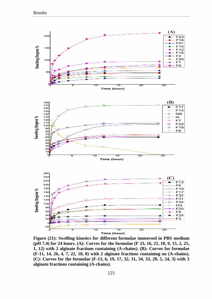
Results
121
Figure (21): Swelling kinetics for different formulae immersed in PBS medium
(pH 7.4) for 24 hours. (A): Curves for the formulae (F 23, 16, 21, 10, 9, 15, 2, 25,
1, 12) with 2 alginate fractions containing (A-chains). (B): Curves for formulae
(F-11, 14, 26, 4, 7, 22, 18, 8) with 2 alginate fractions containing no (A-chains).
(C): Curves for the formulae (F-13, 6, 19, 17, 32, 31, 34, 33, 20, 5, 24, 3) with 3
alginate fractions containing (A-chains).
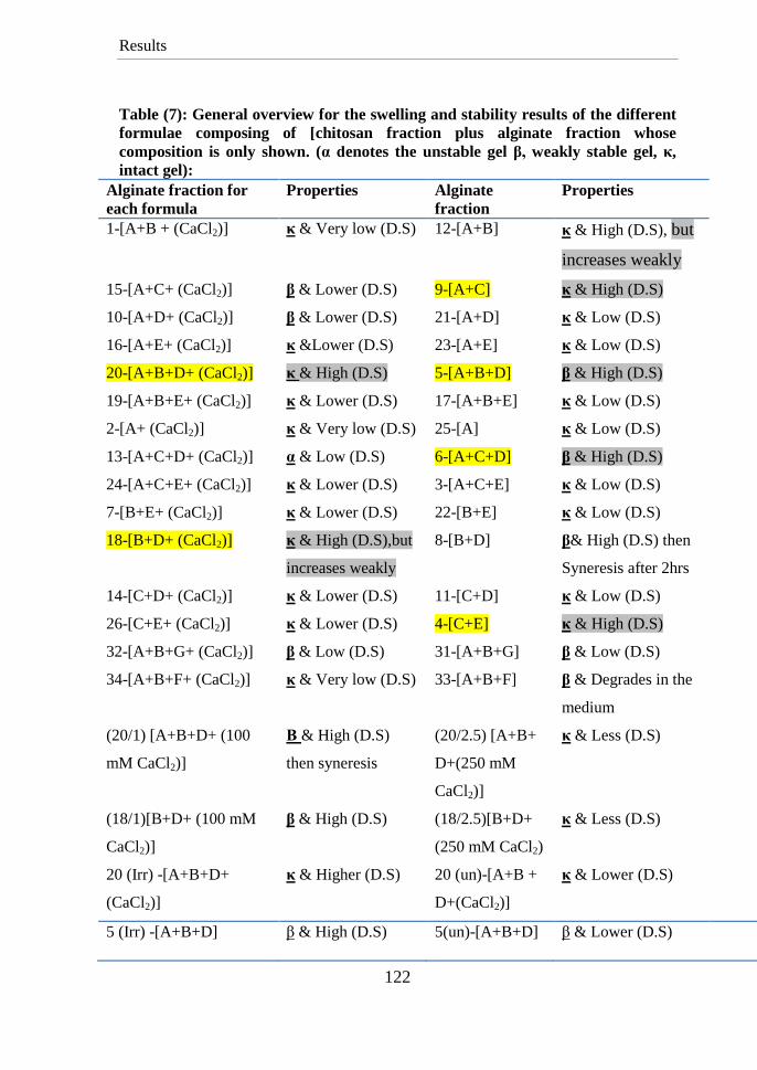
Results
122
Table (7): General overview for the swelling and stability results of the different
formulae composing of [chitosan fraction plus alginate fraction whose
composition is only shown. (α denotes the unstable gel β, weakly stable gel, κ,
intact gel):
Alginate fraction for
each formula
Properties Alginate
fraction
Properties
1-[A+B + (CaCl2)] κ & Very low (D.S) 12-[A+B] κ & High (D.S), but
increases weakly
15-[A+C+ (CaCl2)] β & Lower (D.S) 9-[A+C] κ & High (D.S)
10-[A+D+ (CaCl2)] β & Lower (D.S) 21-[A+D] κ & Low (D.S)
16-[A+E+ (CaCl2)] κ &Lower (D.S) 23-[A+E] κ & Low (D.S)
20-[A+B+D+ (CaCl2)] κ & High (D.S) 5-[A+B+D] β & High (D.S)
19-[A+B+E+ (CaCl2)] κ & Lower (D.S) 17-[A+B+E] κ & Low (D.S)
2-[A+ (CaCl2)] κ & Very low (D.S) 25-[A] κ & Low (D.S)
13-[A+C+D+ (CaCl2)] α & Low (D.S) 6-[A+C+D] β & High (D.S)
24-[A+C+E+ (CaCl2)] κ & Lower (D.S) 3-[A+C+E] κ & Low (D.S)
7-[B+E+ (CaCl2)] κ & Lower (D.S) 22-[B+E] κ & Low (D.S)
18-[B+D+ (CaCl2)] κ & High (D.S),but
increases weakly
8-[B+D] β& High (D.S) then
Syneresis after 2hrs
14-[C+D+ (CaCl2)] κ & Lower (D.S) 11-[C+D] κ & Low (D.S)
26-[C+E+ (CaCl2)] κ & Lower (D.S) 4-[C+E] κ & High (D.S)
32-[A+B+G+ (CaCl2)] β & Low (D.S) 31-[A+B+G] β & Low (D.S)
34-[A+B+F+ (CaCl2)] κ & Very low (D.S) 33-[A+B+F] β & Degrades in the
medium
(20/1) [A+B+D+ (100
mM CaCl2)]
Β & High (D.S)
then syneresis
(20/2.5) [A+B+
D+(250 mM
CaCl2)]
κ & Less (D.S)
(18/1)[B+D+ (100 mM
CaCl2)]
β & High (D.S) (18/2.5)[B+D+
(250 mM CaCl2)
κ & Less (D.S)
20 (Irr) -[A+B+D+
(CaCl2)]
κ & Higher (D.S) 20 (un)-[A+B +
D+(CaCl2)]
κ & Lower (D.S)
5 (Irr) -[A+B+D] β & High (D.S) 5(un)-[A+B+D] β & Lower (D.S)
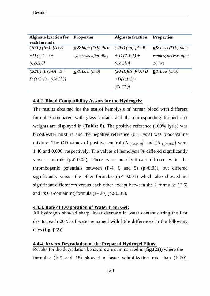
Results
123
Alginate fraction for
each formula
Properties Alginate fraction Properties
(20/I ) (Irr) -[A+B
+D (2:1:1) +
(CaCl2)]
κ & high (D.S) then
syneresis after 4hrs
(20/I) (un)-[A+B
+ D (2:1:1) +
(CaCl2)]
κ& Less (D.S) then
weak syneresis after
10 hrs
(20/II) (Irr)-[A+B +
D (1:2:1)+ (CaCl2)]
κ & Low (D.S) (20/III)(Irr)-[A+B
+D(1:1:2)+
(CaCl2)]
β& Low (D.S)
4.4.2. Blood Compatibility Assays for the Hydrogels:
The results obtained for the test of hemolysis of human blood with different
formulae compared with glass surface and the corresponding formed clot
weights are displayed in (Table: 8). The positive reference (100% lysis) was
blood/water mixture and the negative reference (0% lysis) was blood/saline
mixture. The OD values of positive control (A (+)control) and (A (-)control) were
1.46 and 0.008, respectively. The values of hemolysis % differed significantly
versus controls (p≥ 0.05). There were no significant differences in the
thrombogenic potentials between (F-4, 6 and 9) (p<0.05), but differed
significantly versus the other formulae (p ≤ 0.001) which also showed no
significant differences versus each other except between the 2 formulae (F-5)
and its Ca-containing formula (F- 20) (p≥ 0.05).
4.4.3. Rate of Evaporation of Water from Gel:
All hydrogels showed sharp linear decrease in water content during the first
day to reach 20 % of water remained with little differences in the following
days (fig. (22)).
4.4.4. In vitro Degradation of the Prepared Hydrogel Films:
Results for the degradation behaviors are summarized in (fig.(23)) where the
formulae (F-5 and 18) showed a faster solubilization rate than (F-20).
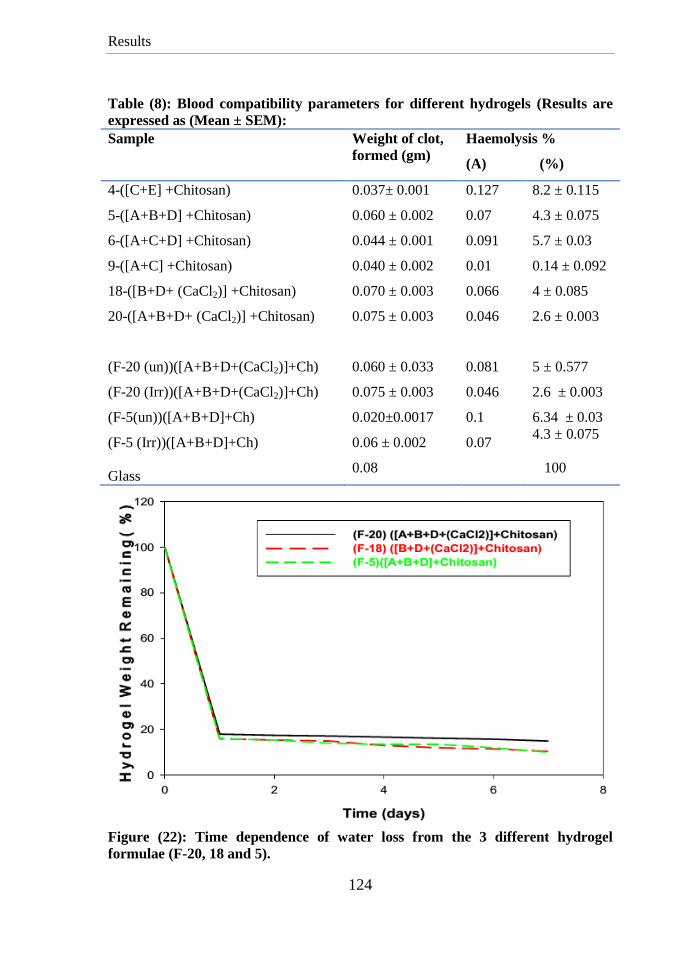
Results
124
Table (8): Blood compatibility parameters for different hydrogels (Results are
expressed as (Mean ± SEM):
Sample Weight of clot,
formed (gm)
Haemolysis %
(A) (%)
4-([C+E] +Chitosan)
5-([A+B+D] +Chitosan)
6-([A+C+D] +Chitosan)
9-([A+C] +Chitosan)
18-([B+D+ (CaCl2)] +Chitosan)
20-([A+B+D+ (CaCl2)] +Chitosan)
(F-20 (un))([A+B+D+(CaCl2)]+Ch)
(F-20 (Irr))([A+B+D+(CaCl2)]+Ch)
(F-5(un))([A+B+D]+Ch)
(F-5 (Irr))([A+B+D]+Ch)
Glass
0.037± 0.001
0.060 ± 0.002
0.044 ± 0.001
0.040 ± 0.002
0.070 ± 0.003
0.075 ± 0.003
0.060 ± 0.033
0.075 ± 0.003
0.020±0.0017
0.06 ± 0.002
0.08
0.127
0.07
0.091
0.01
0.066
0.046
0.081
0.046
0.1
0.07
8.2 ± 0.115
4.3 ± 0.075
5.7 ± 0.03
0.14 ± 0.092
4 ± 0.085
2.6 ± 0.003
5 ± 0.577
2.6 ± 0.003
6.34 ± 0.03
4.3 ± 0.075
100
Figure (22): Time dependence of water loss from the 3 different hydrogel
formulae (F-20, 18 and 5).
Results
125
Figure (23): Rate of degradation for the formulae (F-20, 18 and 5).
Table (9): PDI test results for the Non-irradiated Hydrogel
PDII= (5.6/4) = 1.4
Table (10): PDI test results for the irradiated Hydrogel
PDII= (1.5/4) =0. 375
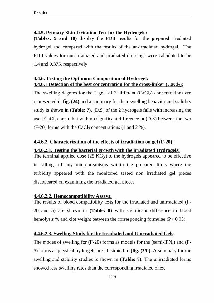
Results
126
4.4.5. Primary Skin Irritation Test for the Hydrogels:
(Tables: 9 and 10) display the PDII results for the prepared irradiated
hydrogel and compared with the results of the un-irradiated hydrogel. The
PDII values for non-irradiated and irradiated dressings were calculated to be
1.4 and 0.375, respectively
4.4.6. Testing the Optimum Composition of Hydrogel:
4.4.6.1 Detection of the best concentration for the cross-linker (CaCl2):
The swelling degrees for the 2 gels of 3 different (CaCl2) concentrations are
represented in fig. (24) and a summary for their swelling behavior and stability
study is shown in (Table: 7). (D.S) of the 2 hydrogels falls with increasing the
used CaCl2 concn. but with no significant difference in (D.S) between the two
(F-20) forms with the CaCl2 concentrations (1 and 2 %).
4.4.6.2. Characterization of the effects of irradiation on gel (F-20):
4.4.6.2.1. Testing the bacterial growth with the irradiated Hydrogels:
The terminal applied dose (25 KGy) to the hydrogels appeared to be effective
in killing off any microorganisms within the prepared films where the
turbidity appeared with the monitored tested non irradiated gel pieces
disappeared on examining the irradiated gel pieces.
4.4.6.2.2. Hemocompatibility Assays:
The results of blood compatibility tests for the irradiated and unirradiated (F-
20 and 5) are shown in (Table: 8) with significant difference in blood
hemolysis % and clot weight between the corresponding formulae (P≥ 0.05).
4.4.6.2.3. Swelling Study for the Irradiated and Unirradiated Gels:
The modes of swelling for (F-20) forms as models for the (semi-IPNs) and (F-
5) forms as physical hydrogels are illustrated in (fig. (25)). A summary for the
swelling and stability studies is shown in (Table: 7). The unirradiated forms
showed less swelling rates than the corresponding irradiated ones.
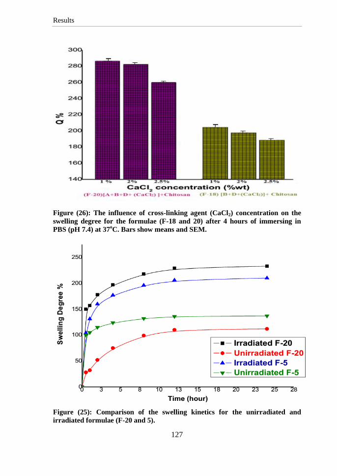
Results
127
Figure (26): The influence of cross-linking agent (CaCl2) concentration on the
swelling degree for the formulae (F-18 and 20) after 4 hours of immersing in
PBS (pH 7.4) at 37oC. Bars show means and SEM.
Figure (25): Comparison of the swelling kinetics for the unirradiated and
irradiated formulae (F-20 and 5).
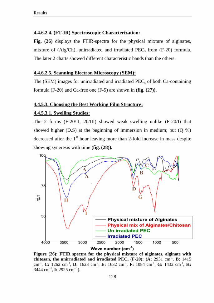
Results
128
4.4.6.2.4. (FT-IR) Spectroscopic Characterization:
Fig. (26) displays the FTIR-spectra for the physical mixture of alginates,
mixture of (Alg/Ch), unirradiated and irradiated PECs from (F-20) formula.
The later 2 charts showed different characteristic bands than the others.
4.4.6.2.5. Scanning Electron Microscopy (SEM):
The (SEM) images for unirradiated and irradiated PECs of both Ca-containing
formula (F-20) and Ca-free one (F-5) are shown in (fig. (27)).
4.4.5.3. Choosing the Best Working Film Structure:
4.4.5.3.1. Swelling Studies:
The 2 forms (F-20/II, 20/III) showed weak swelling unlike (F-20/I) that
showed higher (D.S) at the beginning of immersion in medium; but (Q %)
decreased after the 1st hour leaving more than 2-fold increase in mass despite
showing syneresis with time (fig. (28)).
Figure (26): FTIR spectra for the physical mixture of alginates, alginate with
chitosan, the unirradiated and irradiated PECs (F-20): (A: 2931 cm-1
, B: 1415
cm-1
, C: 1262 cm-1
, D: 1623 cm-1
, E: 1632 cm-1
, F: 1084 cm-1
, G: 1432 cm-1
, H:
3444 cm-1
, I: 2925 cm- 1
).
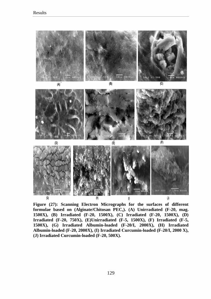
Results
129
Figure (27): Scanning Electron Micrographs for the surfaces of different
formulae based on (Alginate/Chitosan PECs). (A) Unirradiated (F-20, mag.
1500X), (B) Irradiated (F-20, 1500X), (C) Irradiated (F-20, 1500X), (D)
Irradiated (F-20, 750X), (E)Unirradiated (F-5, 1500X), (F) Irradiated (F-5,
1500X), (G) Irradiated Albumin-loaded (F-20/I, 2000X), (H) Irradiated
Albumin-loaded (F-20, 2000X), (I) Irradiated Curcumin-loaded (F-20/I, 2000 X),
(J) Irradiated Curcumin-loaded (F-20, 500X).
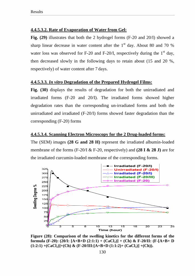
Results
130
4.4.5.3.2. Rate of Evaporation of Water from Gel:
Fig. (29) illustrates that both the 2 hydrogel forms (F-20 and 20/I) showed a
sharp linear decrease in water content after the 1st day. About 80 and 70 %
water loss was observed for F-20 and F-20/I, respectively during the 1st day,
then decreased slowly in the following days to retain about (15 and 20 %,
respectively) of water content after 7 days.
4.4.5.3.3. In vitro Degradation of the Prepared Hydrogel Films:
Fig. (30) displays the results of degradation for both the unirradiated and
irradiated forms (F-20 and 20/I). The irradiated forms showed higher
degradation rates than the corresponding un-irradiated forms and both the
unirradiated and irradiated (F-20/I) forms showed faster degradation than the
corresponding (F-20) forms
4.4.5.3.4. Scanning Electron Microscopy for the 2 Drug-loaded forms:
The (SEM) images (28 G and 28 H) represent the irradiated albumin-loaded
membrane of the forms (F-20/I & F-20, respectively) and (28 I & 28 J) are for
the irradiated curcumin-loaded membrane of the corresponding forms.
Figure (28): Comparison of the swelling kinetics for the different forms of the
formula (F-20): (20/I: [A+B+D (2:1:1) + (CaCl2)] + (Ch) & F-20/II: (F-[A+B+ D
(1:2:1) +(CaCl2)]+(Ch) & (F-20/III:[A+B+D (1:1:2)+ (CaCl2)] +(Ch)).
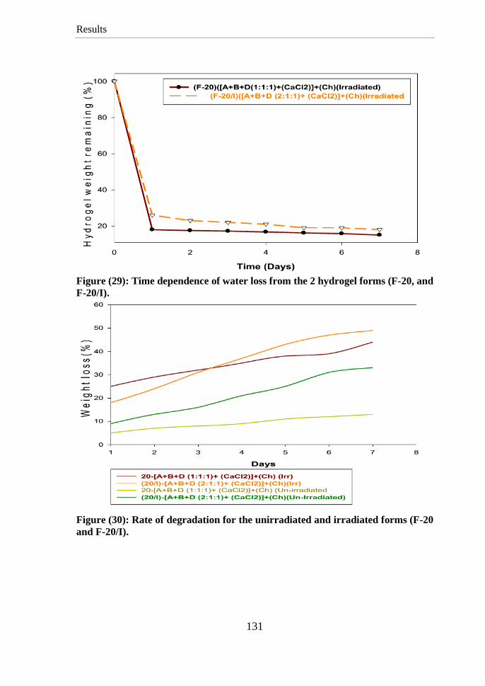
Results
131
Figure (29): Time dependence of water loss from the 2 hydrogel forms (F-20, and
F-20/I).
Figure (30): Rate of degradation for the unirradiated and irradiated forms (F-20
and F-20/I).
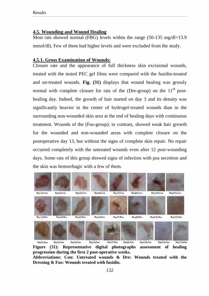
Results
132
4.5. Wounding and Wound Healing
Most rats showed normal (FBG) levels within the range (50-135 mg/dl=13.9
mmol/dl). Few of them had higher levels and were excluded from the study.
4.5.1. Gross Examination of Wounds:
Closure rate and the appearance of full thickness skin excisional wounds,
treated with the tested PEC gel films were compared with the fusidin-treated
and un-treated wounds. Fig. (31) displays that wound healing was grossly
normal with complete closure for rats of the (Dre-group) on the 11th
post-
healing day. Indeed, the growth of hair started on day 3 and its density was
significantly heavier in the center of hydrogel-treated wounds than in the
surrounding non-wounded skin area at the end of healing days with continuous
treatment. Wounds of the (Fus-group); in contrast, showed weak hair growth
for the wounded and non-wounded areas with complete closure on the
postoperative day 13, but without the signs of complete skin repair. No repair
occurred completely with the untreated wounds even after 12 post-wounding
days. Some rats of this group showed signs of infection with pus secretion and
the skin was hemorrhagic with a few of them.
Figure (31): Representative digital photographs assessment of healing
progression during the first 2 post-operative weeks.
Abbreviations: Con: Untreated wounds & Dre: Wounds treated with the
Dressing & Fus: Wounds treated with fusidin.

Results
133
4.5.2. Residual Wound Area Measurements:
The healing curves for wounds of large rats treated with the new hydrogel or
fusidin cream as well as the control wounds can fit into number of curves
representing a change in wound closure as a function of the post-wounding
days. No difference was observed in all wounds sizes during the first 2 post-
wounding days, but the significant reduction % for wounds treated with the
(Dre) was about 50 % in day 3 to reach 90 % after 6 days and about 95 % after
10 days (fig.(32)). The % reduction was only 30 % in case of wounds treated
with (Fus) and (Con) groups with no significant difference between them till
day 3 to reach about 55% after 6 days and 95 % after 13 days for the treated
wounds with significant difference than the Control wounds that showed weak
reduction in size to reach its maximum (95 %) after 2 weeks.
Fig.(33) displays 2 model curves for the reduction in area of wounds of small
rats treated with the (Dre) and a model curve for normal healing of wound.
Wounds treated with the (Dre) showed no significant change in size during the
first 2 days but rapid significant contraction was observed then till the 4th
day,
followed by a slight decrease with slight differences in wound area in the
following days to reach max wound size reduction in the 11th
or 13th
day. The
normal wound closure took place slowly with, to somewhat, equal rate from
each day to the following one reaching maximum level after 16 days.
4.5.3. Histological Examinations for Wounds Tissues:
Fig. (34) shows the histological images for the control and dressed wounds
tissues during the post wounding days (1 and 3) and stained with (H, E and
MT stains). The changes occurred during the post-wounding day (7) and days
(11& 15) are illustrated in (figures (35& 36)), respectively. Fig. (37) displays
the last healing day for the stained dressed wound (Dre-group) with (H, E and
MT).
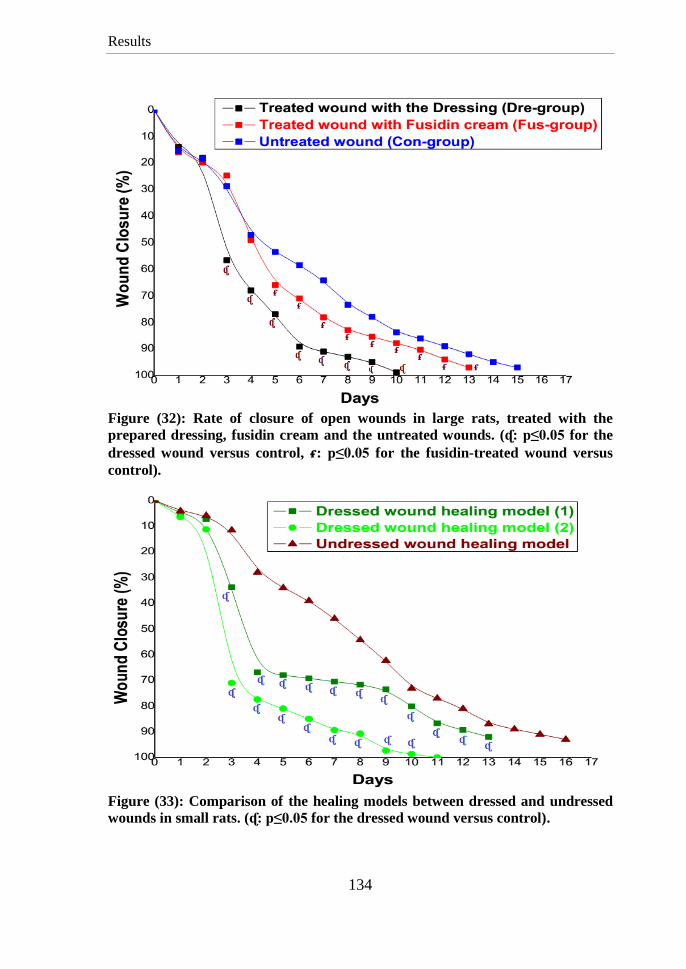
Results
134
Figure (32): Rate of closure of open wounds in large rats, treated with the
prepared dressing, fusidin cream and the untreated wounds. (ᶑ: p≤0.05 for the
dressed wound versus control, ᵳ: p≤0.05 for the fusidin-treated wound versus
control).
Figure (33): Comparison of the healing models between dressed and undressed
wounds in small rats. (ᶑ: p≤0.05 for the dressed wound versus control).
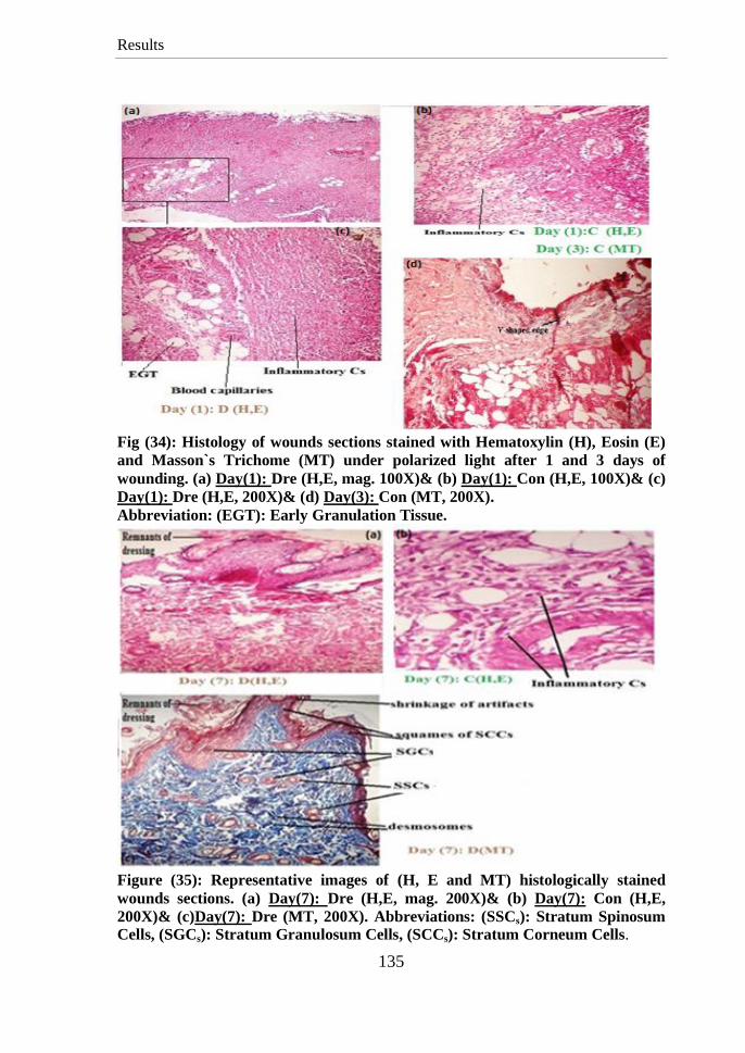
Results
135
Fig (34): Histology of wounds sections stained with Hematoxylin (H), Eosin (E)
and Masson`s Trichome (MT) under polarized light after 1 and 3 days of
wounding. (a) Day(1): Dre (H,E, mag. 100X)& (b) Day(1): Con (H,E, 100X)& (c)
Day(1): Dre (H,E, 200X)& (d) Day(3): Con (MT, 200X).
Abbreviation: (EGT): Early Granulation Tissue.
Figure (35): Representative images of (H, E and MT) histologically stained
wounds sections. (a) Day(7): Dre (H,E, mag. 200X)& (b) Day(7): Con (H,E,
200X)& (c)Day(7): Dre (MT, 200X). Abbreviations: (SSCs): Stratum Spinosum
Cells, (SGCs): Stratum Granulosum Cells, (SCCs): Stratum Corneum Cells.
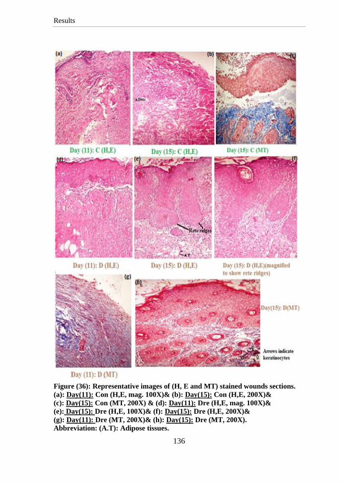
Results
136
Figure (36): Representative images of (H, E and MT) stained wounds sections.
(a): Day(11): Con (H,E, mag. 100X)& (b): Day(15): Con (H,E, 200X)&
(c): Day(15): Con (MT, 200X) & (d): Day(11): Dre (H,E, mag. 100X)&
(e): Day(15): Dre (H,E, 100X)& (f): Day(15): Dre (H,E, 200X)&
(g): Day(11): Dre (MT, 200X)& (h): Day(15): Dre (MT, 200X).
Abbreviation: (A.T): Adipose tissues.
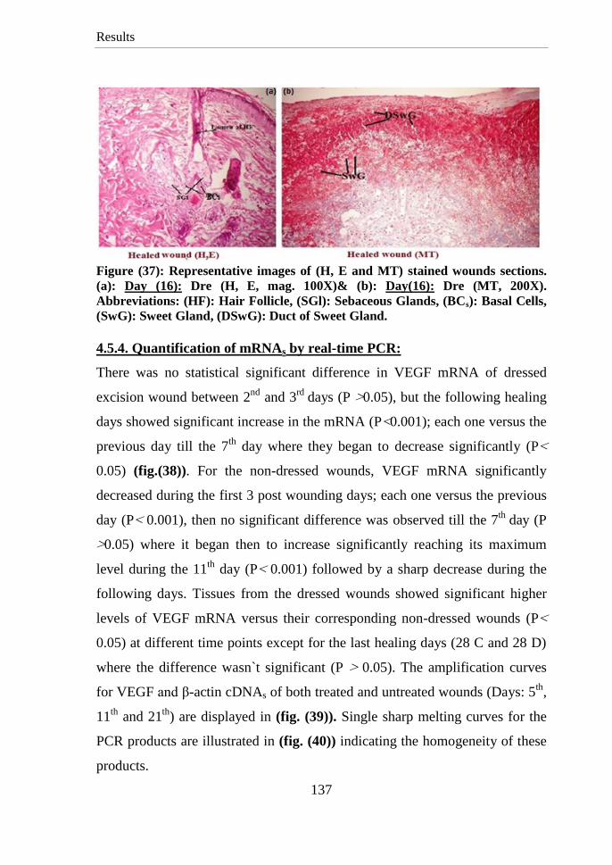
Results
137
Figure (37): Representative images of (H, E and MT) stained wounds sections.
(a): Day (16): Dre (H, E, mag. 100X)& (b): Day(16): Dre (MT, 200X).
Abbreviations: (HF): Hair Follicle, (SGl): Sebaceous Glands, (BCs): Basal Cells,
(SwG): Sweet Gland, (DSwG): Duct of Sweet Gland.
4.5.4. Quantification of mRNAs by real-time PCR:
There was no statistical significant difference in VEGF mRNA of dressed
excision wound between 2nd
and 3rd
days (P0.05>), but the following healing
days showed significant increase in the mRNA (P>0.001); each one versus the
previous day till the 7th
day where they began to decrease significantly (P>
0.05) (fig.(38)). For the non-dressed wounds, VEGF mRNA significantly
decreased during the first 3 post wounding days; each one versus the previous
day (P> 0.001), then no significant difference was observed till the 7th
day (P
<0.05) where it began then to increase significantly reaching its maximum
level during the 11th
day (P> 0.001) followed by a sharp decrease during the
following days. Tissues from the dressed wounds showed significant higher
levels of VEGF mRNA versus their corresponding non-dressed wounds (P>
0.05) at different time points except for the last healing days (28 C and 28 D)
where the difference wasn`t significant (P < 0.05). The amplification curves
for VEGF and β-actin cDNAs of both treated and untreated wounds (Days: 5th
,
11th
and 21th
) are displayed in (fig. (39)). Single sharp melting curves for the
PCR products are illustrated in (fig. (40)) indicating the homogeneity of these
products.
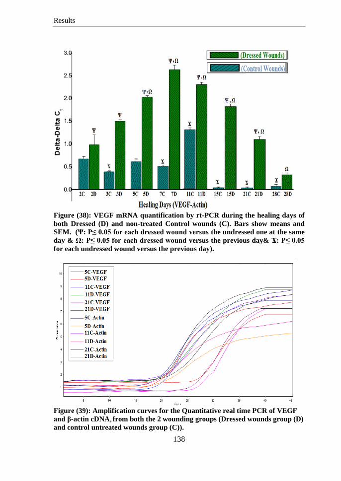
Results
138
Figure (38): VEGF mRNA quantification by rt-PCR during the healing days of
both Dressed (D) and non-treated Control wounds (C). Bars show means and
SEM. (Ψ: P≤ 0.05 for each dressed wound versus the undressed one at the same
day & Ω: P≤ 0.05 for each dressed wound versus the previous day& Ϫ: P≤ 0.05
for each undressed wound versus the previous day).
Figure (39): Amplification curves for the Quantitative real time PCR of VEGF
and β-actin cDNAs from both the 2 wounding groups (Dressed wounds group (D)
and control untreated wounds group (C)).
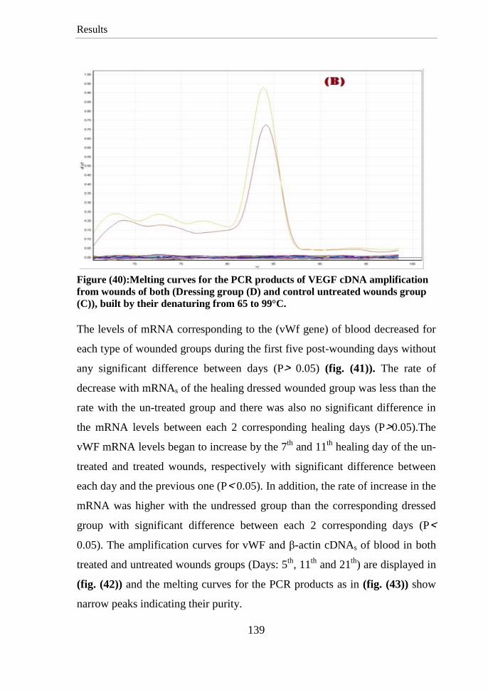
Results
139
Figure (40):Melting curves for the PCR products of VEGF cDNA amplification
from wounds of both (Dressing group (D) and control untreated wounds group
(C)), built by their denaturing from 65 to 99°C.
The levels of mRNA corresponding to the (vWf gene) of blood decreased for
each type of wounded groups during the first five post-wounding days without
any significant difference between days (P< 0.05) (fig. (41)). The rate of
decrease with mRNAs of the healing dressed wounded group was less than the
rate with the un-treated group and there was also no significant difference in
the mRNA levels between each 2 corresponding healing days (P<0.05).The
vWF mRNA levels began to increase by the 7th
and 11th
healing day of the un-
treated and treated wounds, respectively with significant difference between
each day and the previous one (P> 0.05). In addition, the rate of increase in the
mRNA was higher with the undressed group than the corresponding dressed
group with significant difference between each 2 corresponding days (P>
0.05). The amplification curves for vWF and β-actin cDNAs of blood in both
treated and untreated wounds groups (Days: 5th
, 11th
and 21th
) are displayed in
(fig. (42)) and the melting curves for the PCR products as in (fig. (43)) show
narrow peaks indicating their purity.
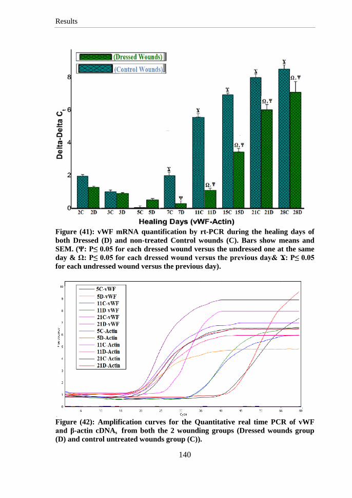
Results
140
Figure (41): vWF mRNA quantification by rt-PCR during the healing days of
both Dressed (D) and non-treated Control wounds (C). Bars show means and
SEM. (Ψ: P≤ 0.05 for each dressed wound versus the undressed one at the same
day & Ω: P≤ 0.05 for each dressed wound versus the previous day& Ϫ: P≤ 0.05
for each undressed wound versus the previous day).
Figure (42): Amplification curves for the Quantitative real time PCR of vWF
and β-actin cDNAs from both the 2 wounding groups (Dressed wounds group
(D) and control untreated wounds group (C)).

Results
141
Figure (43): Melting curves of the PCR products of vWF cDNA amplification
from wounds of both (Dressing group (D) and control untreated wounds group
(C)), built by their denaturing from 65°C to 99°C.
4.5.5. Screening of kidney Functions:
(Table: 11) displays the mean values for BUN and Creatinine in the blood of
both Control and Dressed wounds groups. Values of both are in the normal
range (for Creatinine= 0.3-0.6 mg/dl & for BUN=12-20mg/dl (Johnson-
Delaney, 1996; Quesenberry and Carpenter, 2004) with no significant
differences neither among their values with the different days within the same
experimental group nor versus the corresponding days of the other group.
Table (11): Levels of (BUN (mg/dl) and Creatinine (mg/dl) ± SEM) in plasma:
Discussion
142
5. DISCUSSION 5.1.Characterization of the prepared Oxidized and Irradiated Alginates:
The Fehling reagent gave red cuprous oxide precipitates with the oxidized
products indicating the presence of aldehyde groups within their structures and
its importance as a rapid test for the oxidation of organic compounds as stated in
the report of (Wang et al., 2011).
Spectrum of the irradiated Na-Alg: showed conservation for most bands of its
native alginate with little differences. The band at (3444 cm–1
) for the (O-H
groups) stretching mode and (1090 cm–1
) for (C–O stretching) became broader
(fig. (16)); broadening increased with the increase of radiation dose (not
shown). The peak of skeletal vibrations appeared weaker at(1298 cm–1
).
Spectrum of the oxidized alginate: showed some differences from its native
structure. Some new bands appeared proving its oxidation including:(1) The
characteristic peak of (C=O) stretching for the aldehyde groups at (1715 cm–1
)
in addition to the characteristic vibrational peak of (COO−) of Na-alginate at
(1623 cm–1
) proving alginate oxidation although it is weak. (2) The FT-IR band
of mannuronate residues at (~ 790 cm–1
) with absence of guluronates peaks;
may be due to oxidation of most of its units with ring opening as concluded by
(García-Ríos et al., 2012) (fig. (16)).
Sodium alginate was oxidized with periodate; increasing its concentration lead
to the formation of alginates with higher aldehyde contents. Four different
concentrations were chosen and the results of gelation tests proved higher
concentrations made oxidized forms with weak ability to gelate. The results
revealed also that the forms (MASA-I C, D and MASA-II B, C, D) had weak
gelation properties. (Maiti et al., 2009) found out that higher aldehyde content
than 1 mol. % makes oxidized alginates with no ability to make gels.

Discussion
143
Both irradiation and oxidation caused long chains breakages with decrease in
the M.W and its severity increased with the increase in dose (Table: 5).
Oxidation, in addition, introduced new functional groups to the chains. Only the
molecular weight of (MASA-I A-oxidized alginate) is displayed in (Table :5)
because it, unlike other oxidized forms, was found to have strong effects on
characteristics of the prepared membranes with good gelation properties.
5.2. Alginate Purification:
The used method for purification of alginate in this study utilized simple steps
but were proven to be effective according to (Qi et al., 2009) but with some
modifications for better biocompatibility and simplicity. The used Sevag
reagent was proven to be efficient in protein purification from chilled alginate
solutions. The activated charcoal is carbonaceous non-toxic adsorbent of high
efficiency, porosity and convenience with a large internal area that is effective
in adsorption of contaminants and removal of the organic compounds such as
polyphenols from aqueous solution. It was essential to characterize the used
distilled water to deviate any side reactions during the preparation and
purification steps where Na-alginate precipitates at pH>3.5 and may degrade
(Jain et al., 2011). The high concentrations of polyvalent ions, especially
calcium ions lead to gelation of the alginate solution, so they must be at the
suitable lowest ppm.
5.2.1. Characterization of the Washed Activated Charcoal:
The ash content of activated carbon is a measure of the mineral content left in
during the manufacturing and activation processes, so it must be cleaned at first
with acid followed with water to remove any contaminants (Strand, 2001) for
usage in the alginate purification. The FT-IR spectra were studied in many
previous assignments as a rapid and accurate method to check the structure and
any changes with the charcoal after the different treatments.
Discussion
144
The references in (Table: 1, cited in article of (Terzyk et al., 2001)) are
examples of the papers which involve bands covering the same ranges of wave-
numbers as observed with our investigated carbons and were chosen with the
assignment to certain functional groups with referring after each wave-number.
Interpretation of the I.R charts (fig. (17) and Appendix (A)) could prove that
spectrum of the washed Charcoal with (HCl); unlike the Spectrum of Charcoal,
washed with (HNO3) and that washed with (H2SO4), showed similar bands to
those of the initial carbon, so (HCl) was chosen as the washing acid for charcoal
where the other acids introduced new groups to the charcoal making it
unsuitable for the purification purposes.
5.2.2. Characterization of the Purified Alginate:
The rise in alginate viscosity after purification may be due to the following
reasons: (1) The used activated charcoal and Sevag reagent helped in removal of
the contaminants interfering with the intermolecular interactions among the
alginate chains in addition to proteins that interfered with its hydrophilicity
(Ménard et al.,2010). (2) Low molecular weight oligomers were absorbed and
removed during the filtration steps and this essentially increased the average
MW as well as the solution viscosity (Dusseault et al., 2006). (3) The applied
method was absolutely physical with slight chemical effects in the alginate
chains so no noticeable degradation could be observed
One purification step was not sufficient to deproteinize the different alginates,
so repeating the extraction with Sevag reagent was essential to reach the
lowest protein levels that guaranteed their biocompatibility (fig. (18A)). No
purification steps were carried out on the oxidized alginates. Alternatively, its
(HMW-Alg precursor) was only purified to deviate any side reactions between
the purification reagents and the oxidized fractions.
Discussion
145
Purification of the viscous (HMW-Alg) took place slowly due to the strong
electrostatic interactions among its chains and the contaminants molecules,
but was faster with the less viscous (VLMW-Alg) giving higher yields of the
purified alginate. Both the (HMW and VLMW-Alginates) required 2 repeats
of (PC) extraction for efficient purification. Due to its high viscosity, removal
of (PC) from (HMW-Alg) was slow and the minimum (AFU) for the finally
purified HMW was (9). In contrast, removal of (PC) from the (VLMW-Alg)
was faster due to its lower viscosity to reach a value of (7) for the final
purified polymer, despite having initial AFU of (93.8).
The Purified Alginate Spectrum: exhibited slight differences from most of
the native alginate characteristic peaks (fig. (19) and appendix (B)). The peak
at (1623 cm–1
) was taken as the reference peak for comparing the native
polymer with the purified type due to the fact that carboxyl groups do not
change after degradation. The peak corresponding to the free (O-H groups)
appeared stronger with the purified alginate than the peak of the raw polymer.
The peak for (C-C stretching) appeared with broadening at (1095 cm-1
) with
disappearance of the other characteristic peaks. The peak at (947 cm–1
) is
assumed to be due to (C-O) stretching of the uronate residues that may have
been formed due to slight degradation of the polymer during purification
(Matsuhiro et al, 2008; Xiaoxia et al., 2010).
5.3. Preparation of Chitosan:
On beginning with the deproteination step during the extraction, a higher
reduction in the ash content is achieved than on beginning with the
demineralization as the high levels of retained proteins increase the amount of
minerals that remain bound to the chitin solid fraction (Stevens, 2001). The 1st
step deproteination followed with 2nd
step demineralization have hydrolyzing
effects on the resulting polymer (Stevens, 2001).
Discussion
146
In spite of that, such degrading effects can be controlled with controlling the
demineralization period to get the required chitosan molecular weight for the
subsequent applications. NaOH concentration, reaction temperature, period and
the interaction between them play dominant roles in influencing the DD
(Lertsutthiwong et al., 2002, Sehol et al., 2002; Toan, 2009).
The schematic interpretation of chitin backbone structure is illustrated in (fig.
(44)). It shows a sandwich of thin layers of chitin between two thicker layers of
proteins, called carrier proteins (CP), surrounded by mineralized proteinous
matrix (MM) which is embedded in layers of CaCO3. The linkages between
chitin part and protein moieties are through amide formation between the free (-
NH2) groups in chitin and the (-COOH) groups in the side chains of proteins
(Roberts, 1992). NaOH, with heating have hydrolyzing effects on these bonds
(Lertsutthiwong et al.,2002) to overcome the resistance of such groups
imposed by the trans-arrangement of the C2-C3 substituents on the sugar ring
(Horton and Lineback, 1965) for removal of acetyl groups in the form of (Na-
acetate) through a mechanism illustrated in (fig. (45)).
(Roberts, 1997; Chinadit et al., 1998) reported that a multi-step process is
required to obtain high degrees of deacetylation at low temperatures. However,
high DD at high temperature can be reached in one step as well. The DD is an
important characteristic influencing the performance of polymer in many of its
applications. The interactions between chitosan and the cells increase as it
increases due to the increase in the free amino groups-content. Other biological
properties (e.g., Analgesic, antitumor, hypocholesterolemic, hemostatic,
antimicrobial and antioxidant properties) are also affected by the chemical and
physical properties of chitosan (Koide, 1998; Kumar, 2000; Kumar et al.,
2004).
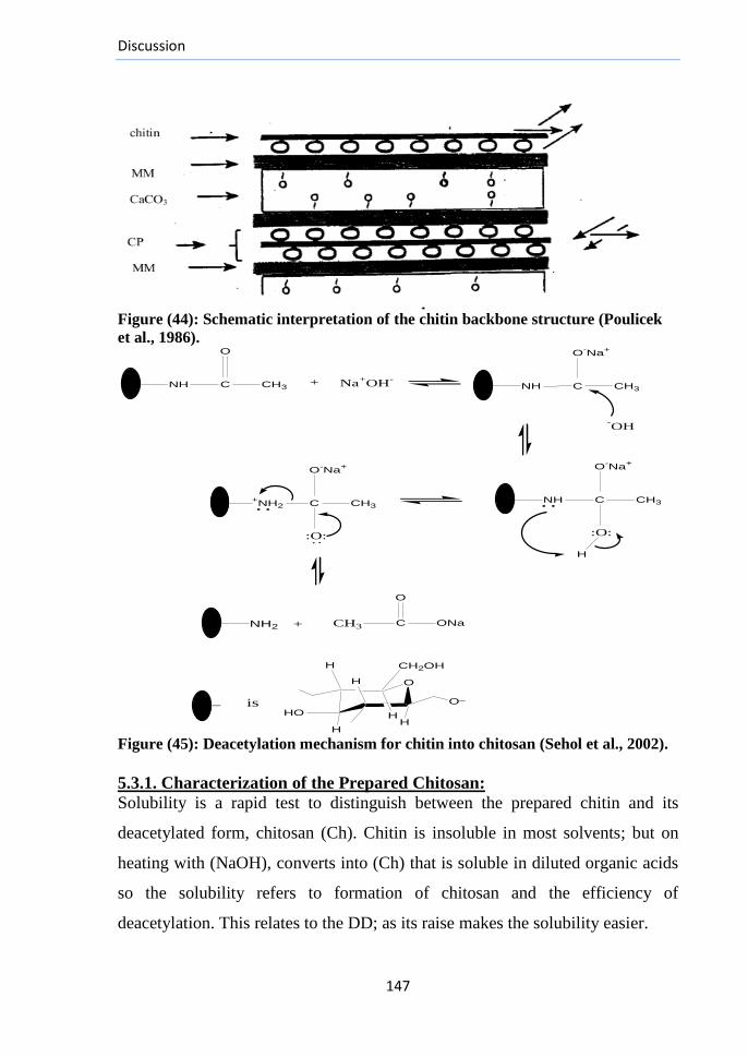
Discussion
147
Figure (44): Schematic interpretation of the chitin backbone structure (Poulicek
et al., 1986).
-OH
O-Na
+
O-Na
+
NH C CH3
NH C CH3
:O:
H
CH3 C ONa
O
HO
O
H
H
H
CH2OH
H
H
O_
Na+OH
-
O-Na
+
:O:
+NH2 C CH3
NH2
O
NH C CH3+
::
:
+
is_
Figure (45): Deacetylation mechanism for chitin into chitosan (Sehol et al., 2002).
5.3.1. Characterization of the Prepared Chitosan:
Solubility is a rapid test to distinguish between the prepared chitin and its
deacetylated form, chitosan (Ch). Chitin is insoluble in most solvents; but on
heating with (NaOH), converts into (Ch) that is soluble in diluted organic acids
so the solubility refers to formation of chitosan and the efficiency of
deacetylation. This relates to the DD; as its raise makes the solubility easier.
Discussion
148
Many methods were proposed to measure the D.D (e.g., Dye adsorption
measurement, residual salicylaldehyde determination and hydrobromide salt
titration (Baxter et al., 1992), but the I.R-spectroscopy provides a rapid,
accurate technique with a high level of precision to detect the different DDs.
(Fig. (20 A) and Appendix (C)). Due to the overlapping between the bands in
the spectral region (1100-1000 cm–1
) for both chitosans, it was difficult to
identify the peaks corresponding to the stretching of (O-H) groups of carbon
atoms (C3 and C6) as well as the C-O stretching vibration and bridge oxygen
stretching bands.
There were significant differences in protein and ash contents between the 2
prepared chitosans (Table: 6). This refers that the prolonged deacetylation steps
on heating with (NaOH) share in more deproteination as well as more removal
for the retained CaCO3 within the chitosan products. These results were also
observed by (Lertsutthiwong et al., 2002). The increases in DD as well lead to
increase in the inhibition ability and antioxidant activity of chitosan, so (Ch-2)
as well as (α-tocopherol) had significant higher antioxidant activities than (Ch-
1) (Table: 6). In addition, the results of (Park et al., 2004) showed that the
chitosans with high DDs exhibit also higher scavenging activity than those with
lower DDs.
The current study shows the ability to prepare 2 chitosan polymers of different
DDs with different periods of heating with NaOH, but they had similar M.Ws,
lower than those of many commercial chitosans (Table: 6). This is very
important result as preparing chitosans of high deacetylation degrees is known
to associate with their degradation and decrease in bulk density (Sehol et al.,
2002; Trung et al., 2006). The low M.W-chitosan is preferred to allow chitosan
penetration into the alginate chains to reinforce preparation of the desired Poly
Electrolyte complex (PEC).

Discussion
149
This would enhance tensile properties of the produced sheet structure (Knill et
al., 2004a). The very high M.W-chitosan; even at low concentrations,
precipitates in the presence of Ca+2
ions resulting in very low levels of chitosan
incorporated into the resulting PEC (Tamura et al., 2002).
5.4. Preparation and Choosing the Best Hydrogel Structure:
An aim of this study focused on preparing different (Alg/Ch) hydrogels and
evaluating their properties. For these purposes, different alginate and chitosan
components derivatives with different ratios as well as the optimal crosslinking
conditions were investigated aiming to producing biocompatible and highly
hydrophilic hydrogels with suitable structure/properties supposed to accelerate
the wound healing with preventing its dehydration and scab formation.
Accordingly, the designed preparation method was as follows:
A-The Preparation Method:
I. (Chitosan-1, DD=90 %) was chosen to prepare the desired PEC hydrogel with
alginate rather than (Chitosan-2, DD=98%) for the following reasons:
(1)With certain levels of (H3C-CO-NH-), the complete complexation between (-
COO-) of alginate and (-NH3
+) of chitosan can be deviated, so can control the
dissolution and degradation rates of the resulting (PEC) for its supposed
scaffolding action for cells. The amino groups in (Ch-2) chains would bind
stronger than (Ch-1) to the alginate chains forming more stable hydrogel.
(2) Many drugs can be loaded on the PEC hydrogel through binding with the (-
COO-) groups, so very high levels of (-NH3
+) groups were not needed.
(3) The suitability of (-NH3+ groups) to be modified for drugs loading is less
than that of (H3C-CO-NH-) groups, so certain levels of (H3C-CO-NH-) groups
were needed for further applications of the prepared PEC gels.
(4) Preparation of (Ch-2) as well as the conversion of (Ch-1) to (Ch-2) for
enhancing the DD requires more time, energy and effort.
Discussion
150
II. Concentrations of the Polymers:
Polyelectrolyte titration method was used to prepare the PEC from (alginate/
chitosan). Concentration of the polyelectrolyte in the beaker; alginate, into
which the chitosan is titrated, changes during titration. First, the solvent of the
titrant dilutes the solution and, second, the alginate in the beaker is consumed
by the ongoing complexation process. To counteract this dilution, the first
polyelectrolyte in beaker sometimes has a 2–10-times higher concentration than
the other one (Gärdlund et al. 2003). To circumvent that during the study, the
reaction was stopped directly after the observation of coacervates formation, so
the used volumes of chitosan and alginate were not equal. Special reactants
concentrations were used to prevent the precipitation of either one out of the
formed complex. Under certain dilution conditions, the prepared complex
disperses homogeneously as small particles in solution which finally grow to
fine fibrous structures on allowing standing for few days at certain temperatures
range (Tsuchida et al., 1980).
III. pH of the Polymers Solutions:
pH is the most pivotal factor affecting the strength and directing the formation
of (alginate/chitosan physical hydrogels). As a conclusion from our
experimental trials, mixing alginate solution (pH 6.84) and chitosan (pH 5) at
equimolar compositions may lead to reaching a suspending solution (pH 5.28)
at which: the chitosan protonated amino groups spontaneously associate with
the negatively charged (-COO-) groups of alginate to form the PEC in solution
which then mutually precipitates from it.
It was essential to control the pH of reactants for promoting the ionic
interactions between these two counter-charged biopolymers at which:
(1) The stoichiometric composition of strong charged polyelectrolyte (i.e.,
Strong Polyacid-Polybase complex) is retained (Tsuchida et al., 1980).
Discussion
151
(2) There is charge balance thus limiting repulsion.
(3)The interactions of polymers with water are greatly minimized with
maximization for the yielding dense of the (polymer-polymer interactions).
(4) The net charge does not reach zero to allow surplus electrical charging of the
resulting coacervates which is necessary for the solubilization of particles
(Gilsenan et al., 2003).
(5) For Alginate: Generally, at pH<3.5, the carboxylate groups become less
ionized with protonation to form alginic acid physical gel clusters through
(inter-chain interactions). The increase in pH makes free (-COO-) on the
polyanion chains. At pH>8, the high concentration of Na+
ions restrains the
extension of the tangled molecular chain on the hydrogel, therefore its swelling
may be inhibited (Liu et al., 2002).
(6) Chitosan precipitates above its pKa value of 6.3. Charge density of the
chitosan molecules is reduced with increasing the pH of solution and the
polymer chains become less extended (Ikeda et al., 1995). As pH increases
from 4.5 to 6.2, the ionization degree of chitosan decreases from 1.0 to 0.5
(Ikeda et al., 1995), the amino groups become less charged with reduction in
the charge density by almost 50% resulting in expected fewer ionic linkages
between any negatively charged polymer and chitosan.
In contrast, the chitosan chains become less extended with these changes at pH
6.2 with smaller radius of gyration and enhanced diffusion coefficient into the
(Alg/ Ch network) causing stronger combination with (Alg-chains) during a
specified reaction time than the linkages of the extended chains at pH 4.5.
Accordingly, PECs formed of chitosan at pH 6.2 have lower D.S than the
complex formed at 4.5. Raising the pH of chitosan solution from 4.5 to 5.5 with
a corresponding decrease in its total charge was observed to have no significant
effects on D.S of the complex gel (Argin-Soysal et al., 2009).
Discussion
152
IV. The Coacervation Reaction:
(1) Rapid coacervation for the reacting polymers at definite pH values induces
the formation of dense interphasic PEC membranes that separate the polymers
solutions and prevent further reaction and this is the common employed
technology to prepare microspheres of chitosan–alginate PECs (Tay et al.,
1993; Alexakis et al., 1995; Liu et al., 1997; Silva et al., 2006). This
technology was not used in the present study as it was unsuitable to produce
coacervates ready for casting into films. The extensive development of these
intermolecular interactions during hydrogel films preparation would generate
viscosities too high to distribute or matrices too solidified to flow. To prepare
coacervates for casting into homogeneous films, the rate of reaction between the
two polymers must be sufficiently slow to prevent the formation of such
membranes, so that the reaction can complete.
(2) Based on previous reports, (Wang et al., 2002; Ca´rdenas et al., 2003;
Meng et al., 2010), addition of water miscible organic solvent (e.g., Acetone ,
ethanol or PEG200) into the chitosan solution was proven effective to slow the
rate of coacervation. They have lower polarity so the chitosan chains will
assume less extended conformation in and this will restrict the fast reaction
between the two polyelectrolytes and so perfect complexation can occur.
(3) An aim of the current study was using the least number of chemicals, which
may cause skin irritation, inflammation or ulceration on application of the
prepared hydrogel film to the wound with a main dependence on the physical
activity for mixing the 2 polymers to produce a high quality of PECs through the
coacervation process. Thus, controlling the reactions speed through controlling
the rate of mixing of chitosan and alginate solutions together is undoubtedly the
pivotal point for success.
Discussion
153
(4) The high shaking rates with the titrant polymer solution addition promote
foam formation with opaque sheets having impaired barrier properties, more
permeability to gases, in addition to forming some suspensions of soluble
complexes with no ability to coacervate. The weak speeds lead to fast
coacervation with no ability to form gels. Accordingly, the moderate shaking
and mixing speeds (about 50 RPM) with controlled addition rate (about 300 µl/
min) were perfect to prepare ideal PECs suspensions of (alginate/ chitosan)
despite taking some time. The resulting hydrogel films were transparent that
allowed effective observation of changes in the wounds during skin repair.
(5) Multifaceted structure may have been formed when a chitosan drop of
certain concentration fell into alginate solution at the desired pH values.
Complexation may have occurred between the two oppositely charged
polyelectrolytes through the electrostatic interactions. By this way,
complexation between the common free (-COO-) groups of alginate chains and
the chitosan protonated amino groups (NH3+) occurred and chitosan may have
acted as the insolubilising cation for production of (alginate-chitosan Physical
complex) (Cole and Nelson, 1993; Pandit, 1998). With adding (CaCl2) into the
mixture during complexation, it may have diffused into the alginate core more
rapidly than chitosan because of its lower molecular weight to form a gel core.
Calcium cross-linking for alginate helped in its stabilization with forming (a
Semi Interpenetrating Network (Semi-IPN))of pH sensitivity and reversible
properties that were proved later through the characterization tests.
V. Preparation of the film dressings:
The stand-alone films from macromolecules are generally produced by
extrusion, deposition, or casting/solvent evaporation (Farris et al., 2009). For
(PEC-film) formation, all the involved physical changes must occur in situ
during the preparation in order to ensure formation of the desired complex
structure in place within the final film.
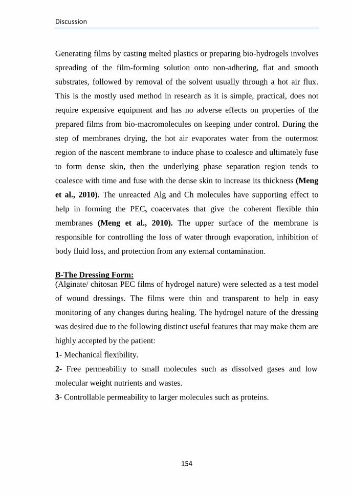
Discussion
154
Generating films by casting melted plastics or preparing bio-hydrogels involves
spreading of the film-forming solution onto non-adhering, flat and smooth
substrates, followed by removal of the solvent usually through a hot air flux.
This is the mostly used method in research as it is simple, practical, does not
require expensive equipment and has no adverse effects on properties of the
prepared films from bio-macromolecules on keeping under control. During the
step of membranes drying, the hot air evaporates water from the outermost
region of the nascent membrane to induce phase to coalesce and ultimately fuse
to form dense skin, then the underlying phase separation region tends to
coalesce with time and fuse with the dense skin to increase its thickness (Meng
et al., 2010). The unreacted Alg and Ch molecules have supporting effect to
help in forming the PECs coacervates that give the coherent flexible thin
membranes (Meng et al., 2010). The upper surface of the membrane is
responsible for controlling the loss of water through evaporation, inhibition of
body fluid loss, and protection from any external contamination.
B-The Dressing Form:
(Alginate/ chitosan PEC films of hydrogel nature) were selected as a test model
of wound dressings. The films were thin and transparent to help in easy
monitoring of any changes during healing. The hydrogel nature of the dressing
was desired due to the following distinct useful features that may make them are
highly accepted by the patient:
1- Mechanical flexibility.
2- Free permeability to small molecules such as dissolved gases and low
molecular weight nutrients and wastes.
3- Controllable permeability to larger molecules such as proteins.
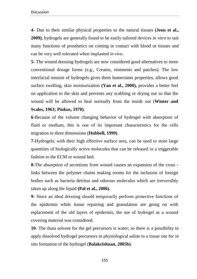
Discussion
155
4- Due to their similar physical properties to the natural tissues (Jeon et al.,
2009), hydrogels are generally found to be easily tailored devices in vitro to suit
many functions of prosthetics on coming in contact with blood or tissues and
can be very well tolerated when implanted in vivo.
5- The wound dressing hydrogels are now considered good alternatives to more
conventional dosage forms (e.g., Creams, ointments and patches). The low
interfacial tension of hydrogels gives them humectants properties, allows good
surface swelling, skin moisturization (Yan et al., 2000), provides a better feel
on application to the skin and prevents any scabbing or drying out so that the
wound will be allowed to heal normally from the inside out (Winter and
Scales, 1963; Pinkus, 1970).
6-Because of the volume changing behavior of hydrogel with absorption of
fluid or medium, this is one of its important characteristics for the cells
migration in three dimensions (Hubbell, 1999).
7-Hydrogels; with their high effective surface area, can be used to store large
quantities of biologically active molecules that can be released in a triggerable
fashion to the ECM or wound bed.
8-The absorption of secretions from wound causes an expansion of the cross -
links between the polymer chains making rooms for the inclusion of foreign
bodies such as bacteria detritus and odorous molecules which are irreversibly
taken up along the liquid (Pal et al., 2006).
9- Since an ideal dressing should temporarily perform protective functions of
the epidermis while tissue repairing and granulation are going on with
replacement of the old layers of epidermis, the use of hydrogel as a wound
covering material was considered.
10- The thata solvent for the gel precursors is water; so there is a possibility to
apply dissolved hydrogel precursors in physiological saline to a tissue site for in
situ formation of the hydrogel (Balakrishnan, 2005b).
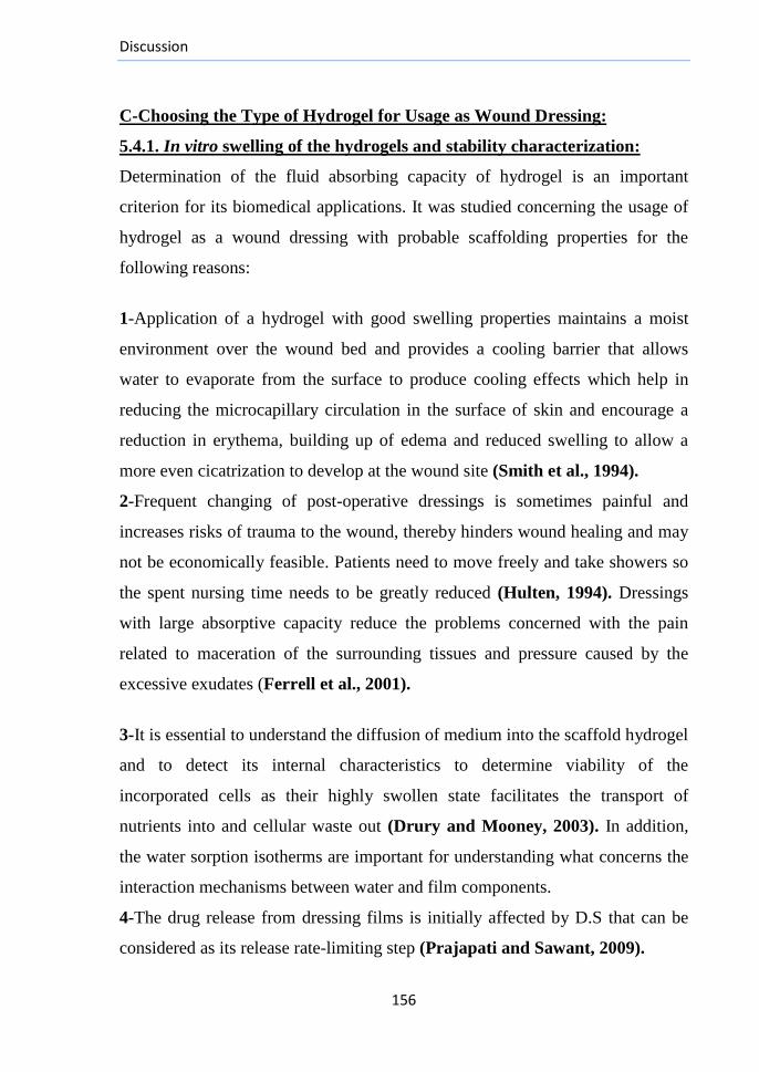
Discussion
156
C-Choosing the Type of Hydrogel for Usage as Wound Dressing:
5.4.1. In vitro swelling of the hydrogels and stability characterization:
Determination of the fluid absorbing capacity of hydrogel is an important
criterion for its biomedical applications. It was studied concerning the usage of
hydrogel as a wound dressing with probable scaffolding properties for the
following reasons:
1-Application of a hydrogel with good swelling properties maintains a moist
environment over the wound bed and provides a cooling barrier that allows
water to evaporate from the surface to produce cooling effects which help in
reducing the microcapillary circulation in the surface of skin and encourage a
reduction in erythema, building up of edema and reduced swelling to allow a
more even cicatrization to develop at the wound site (Smith et al., 1994).
2-Frequent changing of post-operative dressings is sometimes painful and
increases risks of trauma to the wound, thereby hinders wound healing and may
not be economically feasible. Patients need to move freely and take showers so
the spent nursing time needs to be greatly reduced (Hulten, 1994). Dressings
with large absorptive capacity reduce the problems concerned with the pain
related to maceration of the surrounding tissues and pressure caused by the
excessive exudates (Ferrell et al., 2001).
3-It is essential to understand the diffusion of medium into the scaffold hydrogel
and to detect its internal characteristics to determine viability of the
incorporated cells as their highly swollen state facilitates the transport of
nutrients into and cellular waste out (Drury and Mooney, 2003). In addition,
the water sorption isotherms are important for understanding what concerns the
interaction mechanisms between water and film components.
4-The drug release from dressing films is initially affected by D.S that can be
considered as its release rate-limiting step (Prajapati and Sawant, 2009).
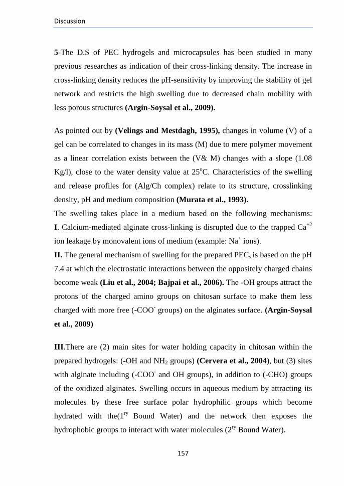
Discussion
157
5-The D.S of PEC hydrogels and microcapsules has been studied in many
previous researches as indication of their cross-linking density. The increase in
cross-linking density reduces the pH-sensitivity by improving the stability of gel
network and restricts the high swelling due to decreased chain mobility with
less porous structures (Argin-Soysal et al., 2009).
As pointed out by (Velings and Mestdagh, 1995), changes in volume (V) of a
gel can be correlated to changes in its mass (M) due to mere polymer movement
as a linear correlation exists between the (V& M) changes with a slope (1.08
Kg/l), close to the water density value at 25oC. Characteristics of the swelling
and release profiles for (Alg/Ch complex) relate to its structure, crosslinking
density, pH and medium composition (Murata et al., 1993).
The swelling takes place in a medium based on the following mechanisms:
I. Calcium-mediated alginate cross-linking is disrupted due to the trapped Ca+2
ion leakage by monovalent ions of medium (example: Na+ ions).
II. The general mechanism of swelling for the prepared PECs is based on the pH
7.4 at which the electrostatic interactions between the oppositely charged chains
become weak (Liu et al., 2004; Bajpai et al., 2006). The -OH groups attract the
protons of the charged amino groups on chitosan surface to make them less
charged with more free (-COO- groups) on the alginates surface. (Argin-Soysal
et al., 2009)
III.There are (2) main sites for water holding capacity in chitosan within the
prepared hydrogels: (-OH and NH2 groups) (Cervera et al., 2004), but (3) sites
with alginate including (-COO- and OH groups), in addition to (-CHO) groups
of the oxidized alginates. Swelling occurs in aqueous medium by attracting its
molecules by these free surface polar hydrophilic groups which become
hydrated with the(1ry
Bound Water) and the network then exposes the
hydrophobic groups to interact with water molecules (2ry
Bound Water).
Discussion
158
IV. The network then imbibes additional water based on the osmotic driving
force of the network chains towards infinite dilution due to the difference in
concentrations of the ionic solutes in gel and the surrounding solution with
flexibility of the polymeric chains (Berger et al., 2004a; Argin-Soysal et al.,
2009).The additional swelling is assumed to fill the space among the network
chains and/or the centers of macropores and voids till reaching equilibrium
swelling level on equilibration of the osmotic pressure in the gel and medium
with a balance between repulsion and contractile forces within the network.
Greater flexibility of the polymer chains would allow larger expansion of free
spaces, thereby increasing the capacity to retain the fluid (Hian, 2009).
V. This behavior is opposed by the covalent or physical crosslinks leading to
retraction forces within the elastic network. The maximum fluid uptake capacity
(equilibrium swelling level) would be reached on balancing between the
swelling pressure and elastic resistance of the polymer network (Remuñán-
López and Bodmeier, 1997). On reaching a critical value of the ion-exchange
with the cross-linked degradable hydrogel, the whole crosslinked network will
be disjointed and the gel will begin to disintegrate and dissolve at a rate
depending on its composition (Hoffman, 2002).
The Formula (F-25) ([A] +Chitosan): includes components with the highest
M.Ws (Ch+HMW-Alg). Considering the reaction stoichiometry, the largest
amounts of coacervate complexes may have been generated (Li and Pelton,
1992) with the same previous swelling mechanism.
The Formula (F-2) ([A+ (CaCl2)]+Ch): Swelling occurred due to the same
reasons as (F-25) but the affinity of gel to swell H2O molecules is less. Though
its affinity to retain water is expected to be higher than (F-25) by the formed
net-like structure with the cross-linking ions, the firm coacervation between (F-
25) polymers chains helps in the stabilization of this network.
Discussion
159
D.S of this formula is the least among the corresponding (Ca-containing
hydrogels) of different constituents (fig. (21A)) due to the stronger cross-linking
of Ca+2
with the repeated longest (G-blocks).
The Formulae (F-12) ([A+B] +Ch) & (F-9) ([A+C] +Ch): The total bound
H2O is the first reason as in the case of (F-25), but short chains of the irradiated
alginates (B and C) are movable that may have increased the tendency of the
free (COO- groups) to imbibe more H2O molecules to make them have a higher
swelling degrees than (F-25).
The Formulae (F-1)([A+B+(CaCl2)]+Ch)& (F-15) ([A+C+(CaCl2)] +Ch):
They had less D.S than (F-12 and F-9), respectively where the (A-chains) seem
to be the controllable factor of swelling in these 4 formulae as in case of (F-25
and 2). The difference in D.S between (F-12 and 1) was not great, unlike that
between (F-9 and 15).
The Formulae (F-21) ([A+D] +Ch) & (F-23) ([A+E] +Ch): They had chains
of low M.Ws due to the hydrolysis on oxidation to make them more movable
with higher affinity to H2O than (F-25) but with less affinity than those of (F-
12, 9) due to the less polar aldehyde groups than (COO- groups).
The Formulae (F-10) ([A+D+ (CaCl2)] +Ch) & (F-16) ([A+E+ (CaCl2)]
+Ch: The affinity of (F-10) to (H2O) molecules was slightly less than (F-21)
with weak effects of the formed network by Ca+2
ions due to the presence of
oxidized alginate fractions. The (A-chains) maintained the gel properties. No
difference was observed in D.S between (F-16 and 23).
The Formula (F-5) ([A+B+D] +Ch): The polar groups are distributed on (A, B
and D chains). The increase in the number of small chains (B and D) than in (F-
25) may have been the reason for the increased affinity to (H2O).

Discussion
160
The free and total bound water are responsible for this increased affinity.
Though they displayed higher diffusion ability to coacervate with chitosan
chains, the resulting hydrogel had slight dispersion which may relate to the
decreased ratio of the long (A-chains).
The Formula (F-20) ([A+B+D+ (CaCl2)] +Ch): Generally, addition of (Ca+2
)
ions during the hydrogel preparation enhances its mechanical strength due to the
strengthened forces by which these ions bind (Alg-chains) and the cross-linking
between (G-units) and these ions generally requires a critical length of (G-
blocks). Accordingly, hydrolysis of the small chains takes place on immersing
in the aqueous medium at first due to their weaker cross-linkages which cause
increasing in the hydrogel affinity to bind H2O molecules causing its swelling.
The D.S was higher than that of (F-5); they may have the same swelling
mechanism but the cross-linking ions help to stabilize the gel and the formed
net store the medium molecules with a higher degree than (F-5) causing the
retaining of medium molecules with increasing the binding of the hydrogel
chains to water.
The Formulae (F-17) ([A+B+E] +Ch) & (F-3) ([A+C+E] +Ch): They showed
similar swellability with initial fast imbibing of water, but lower than (F-5)
which may relate to faster release of the small chains to medium; especially the
higher oxidized alginate chains. In spite of that, the swelling curves of these 2
formulae reach early plateau phases on equilibrium between the swellability of
the remaining chains and their hydrolysis.
The formulae (F-19) ([A+B+E+(CaCl2)]+Ch)& (F-24)([A+C+E+(CaCl2)]
+Ch) : have lower affinity to (H2O molecules) than (F-20); may relate to fast
solubilization of the small chains accompanied with swelling and weak cross -
linking (Yan et al., 2000) making weak nets to store medium molecules.
Discussion
161
The formulae (F-6) ([A+C+D] +Ch) & (F-13) ([A+C+D+ (CaCl2)]+Ch):
Alginate long chains are the main factors responsible for stability of the formed
PEC with chitosan. The new added short irradiated and oxidized chains with the
decrease in (A-chains %) may lead to increasing the binding affinity between
the 2 polymers and the formed gel becomes dispersed. The weak networks,
formed by Ca+2
ions with (F-13) store small amounts of medium molecules
accompanied with diffusion of the small chains.
The formulae (F-31) ([A+B+G] +Ch) & (F-33) ([A+B+F] +Ch) & (F-32)
([A+B+G+ (CaCl2)] +Ch) & (F-34) ([A+B+F+ (CaCl2)]+Ch): Increasing the
ratio of alginate short chains lead to disturbing stability of the formed PECs.
Both the oxidized alginates (F&G) have lower M.Ws than (D&E) with more
aldehyde groups to generate unstable (PECs) of faster diffusion for the weakly
bound chains to medium. The short (G-blocks) weaken the stabilizing action of
Ca+2
ions and facilitate the short chains diffusion.
The formulae (F-8) ([B+D] +Ch & (F-18) [B+D+ (CaCl2)] +Ch:
The short alginate chains seem to have higher affinity to (H2O molecules) than
the HMW-Alginate (A-Alg) with higher swellability. Syneresis of (F-8) in
medium began after one hour of strong swelling (fig. (21C)). This may relate to
the rapid diffusion of the short chains to medium with beginning of film matrix
solubilization with weight loss (Yan et al., 2000). This may be a good
indication of the importance of alginate long chains in stabilization of the
hydrogel structure. The cross-linking of the chains with Ca+2
ions helps to
stabilize the formula (F-18) hydrogel than (F-8). The formed network
maintained the storage of medium molecules but the swelling curve appeared
weak after 1 hour of immersing in medium due to the fast movement of the
short chains to medium against the action of the cross-linking Ca+2
ions.
Discussion
162
The formulae (F-22) ([B+E] +Ch) &(F-7) ([B+E+(CaCl2)]+Ch)& (F-11)
([C+D]+Ch) & (F-14) ([C+D+(CaCl2)]+Ch): With similar composition as (F-
8, 18) but less in swellability. The D.S was higher than (F-25), but slowly
increased with time to make them unsuitable for use as intended dressings.
The formulae (F-4) ([C+E] +Ch) & (F-26) ([C+E+(CaCl2)]+Ch): They
displayed normal increasing swelling curves where the swelling was initially
rapid, slowed down to reach a plateau after 6 hours in case of (F-4) and after 8
hours for (F-26 ). The behavior of (F-4) in medium is questionable where it
differed from (F-22 and 11) as good D.S was observed though these small
chains were expected to diffuse into the medium or to express small level of
swelling. The behavior of (F-26) was expected due to the stabilizing action of
Ca+2
ions for the hydrogel structure.
The formulae (F-27)(D+Ch)& (F-28)(E+Ch)&(F-29) (B+Ch)& (F-30)(C+
Ch): According to (Vanerek and van deVen, 2006), chitosan chains with high
M.W act as host molecules for the other much smaller alginate chains
(Irradiated alginates (B& C) and oxidized alginates (D& E)). Only soluble PECs
may have been formed without forming any colloidal or coacervate complexes,
so no gels were formed on casting.
(According to these results, the formulae (F-4, 5, 6, 9, 18 and 20) were
considered as the best prepared hydrogels in their stability and water uptake
abilities where the gels do not exude the fluids and retain the absorbed amounts
present. In clinical practice, these findings would translate to a greater wound
fluid absorption and longer wear time.)
5.4.2. Hemocompatibility Assays of Hydrogels:
Biocompatibility, especially blood compatibility, is the most important property
with regard to the biomedical materials.
Discussion
163
When the polymeric device comes in contact with blood, it must not induce
thrombosis, severe antigenic responses, thromboembolisms or destruction of
blood constituents or plasma proteins (Sevastianov, 1991). Hence, the
hemocompatibility tests have been conducted with respect to the different
formulae. When the RBCs come into contact with water, blood is hemolyzed.
(Dhandayuthapani et al., 2012) had reported that a value of up to (5%)
hemolysis is permissible for the biomaterial and the device (dressing) will be
incompatible with human blood on having hemolytic tendencies (Pharm Labs,
2008). Accordingly, hemolysis % for the formulae (F-5, 9, 18 and 20) located
in this permissible limit with considerable biocompatibility. The fast release of
small chains from the other 2 formulae in spite of their high D.S may be
responsible for their high hemolytic tendencies.
The different clot weights relate to the different abilities of hydrogels to activate
thrombus formation. The anti-thrombogenic potential of surfaces for the
different formulae films was judged by the blood clot formation test where there
were similar weak clot formation abilities for the formulae (F-4, 6 and 9)
without any significant differences between them (p< 0.05). These values
differed significantly versus each of the (Ca-containing formulae) (p>0.001,
Table: 8). The abilities of (F-5, 18 and 20) to activate clotting were similar
with no significant differences between the (Ca-containing formulae), but the
difference was between (F-5) and its (Ca-containing formula; F-20) (p>0.05)
confirming the supposed action of (Ca+2
) within the structure of (F-18 and 20).
The release of (Ca+2
ions) on swelling during the ion exchange with the soluble
monovalent ions of the used test mediums helps to activate the clotting reactions
in the in-vitro tests. This is similar to the hydrogel behavior on application to
the wound where the blood and wound fluid enable the film to rehydrate, swell
and turn gel.

Discussion
164
This helps in the moisturization of wound bed and platelets activation, in
addition to exchange of the Ca+2
ions of gel with the monovalent ions of these
fluids to activate their actions on skin injury. In addition, Calcium ion is the
(clotting factor: IV) responsible for promoting homeostasis. (Vernenkar, 2009)
The formula (F-9) displayed good hemolytic activity, but weak clotting ability
as well.
(According to these results, the formulae (F-5, 18 and 20) may be the best
candidates for using as dressing agents due to their better blood clotting and
homeostatic abilities, in addition to their good hydration/swelling properties)
5.4.3. The Extent of Evaporation/ Loss of water from the hydrogel: was
evaluated to examine its behavior on using as a dressing over a dry wound. The
physical interaction between the gel network and incorporated water makes
jelly-like structure containing firmly bound water. This property is essential for
materials to be used in wound covering, so it was essential to determine the
water loss from the prepared gels at R.T. When it covers a wound, the water
loss enables the gel to take up exudate and edema fluid from the wound by an
active upward-directed process and this will avoid dehydration (fig. (22). The
three prepared hydrogels (F-5, 18 and 20) have, to somewhat, equal water
evaporation rates. Accordingly, they will be more beneficial to wounds with
moderate exudates than dry ones.
5.4.4. The In vitro Degradation of the Hydrogel Films test: measured the
solubilization rate of the hydrogel chains without further studying of their in
vivo biochemical degradation sequence. It was determined as the mass loss (%)
over time. Fig. (23) shows that the degradation rates for (F-18 & F-5) were
faster than that of (F-20) despite of its higher (D.S). Diffusion of the (F-18)
small chains to the test medium had a faster rate than that of (F-20). containing
the long (A-chains) to initiate faster solubilization from the gel.
Discussion
165
This causes an increase in the degradation of gel-network. The greater extent of
degradation would reduce the ability of gel matrix to retain the absorbed fluid.
The scaffold degradation must be under moderate control to make rooms for the
growth of new cells and then substituted by new skin tissue. Unlike (F-18),
application of the hydrogel (F-20) is not supposed to require continuous
changing. Drug release from gel would be faster to the wound bed with
uncontrolled diffusion on its encapsulation within (F-18).
Different extents of degradation with medium fluid uptake are attributed to the
rigidity and cross-linking effects of Ca+2
in the hydrogel. Generally, a high
extent of cross-linking and uniform distribution of cross-linkages across the
matrix would favor a lower degree of degradation. On the contrary, the
progression of degradation would result in the release of cross-linked cations
from the network junctions. Unlike the other formulae, (F-5) lacks the
stabilizers Ca+2
ions, so expressed a faster rate of solubilization than (F-20) but
less than (F-18) where the long (A-chains) are shown to play important role in
the stabilization of the coacervate structure in gel. This behavior, in addition to
the weaker blood-clotting properties than (F-20 & 18) makes the usage of (F-5)
as a wound dressing material impractical with a final conclusion that (F-20)
may be the best one as a dressing formula with probable scaffolding properties
for healing, better swelling and hemocompatibility properties with more
practical usage.
5.4.5. The results of Primary Skin Irritation Test for the Hydrogels, displayed in
(Tables: 9 and 10) showed that the irritation which accompanied the application
of the un-irradiated hydrogel to the wound was minimized with its terminal
irradiation which has effects in killing any microorganisms within the hydrogel.
In spite of that, the sterilization effect of radiation should be studied with
comparing its effect with the effect of other methods.
Discussion
166
5.4.6. Testing the Optimum Composition of Hydrogel:
The formula (F-18) was used to confirm the results of this study due to its good
swelling properties. The fall in (D.S) of the 2 hydrogels with increasing the used
CaCl2 concentration is related to the formation of gels with higher network
crosslinking density and enhanced mechanical strength. This reduces the ability
of hydrogel to uptake water with significant reduction in the D.S. On the other
hand, the low concentration is linked to higher swelling due to its weaker
crosslinks which cause also poor mechanical strength, limited stability of the
hydrogel with continuous immersing in medium and weak hemostatic effects.
These results are in accordance with the results of (Draget et al., 1990). Based
on the presented results, the (2%) concentration of (CaCl2) was preferred in
both types of hydrogels. Intact gel was formed with this concentration having
good swelling properties and better hemostatic properties.
Characterizing the Effects of Irradiation on the Hydrogel (F-20):
During the pharmaceutical products manufacturing, sterilization is essential for
destroying all the microbial life including microorganisms and bacterial
endopores. It includes certain physical or chemical procedures, applied either
before mixing the filling components (i.e., Alginate, Chitosan, CaCl2 and their
solvents) (Aseptic manufacturing process) or with the (Terminal sterilization
of the final product in the final container) for delivery to the end user
(Yaman, 2001) and that was the applied method in the present study. The major
applied methods for sterilizing the medical devices include:
1-Gaseous Sterilization: A Chemical method that utilizes a sterilizing gas (e.g.,
Ethylene oxide, formaldehyde, glutaraldehyde, or propylene oxide) under
special conditions of pressure, temperature, application time and humidity.

Discussion
167
One of its disadvantages is the presence of toxic residues in the product
hazardous to the patient and can lead to adverse biological reactions such as
anaphylaxis and immune responses (Silver, 1994) which limits its usage as a
mean for sterilization (Rao and Sharma, 1995).
2-Saturated Water Steam Sterilization: The bacterial Death by moist heat
relates to the denaturation and coagulation of essential protein molecules
(enzymes) and cell constituents. With the saturated water steam, the chitosan -
based membranes become water insoluble and lose about 80% of their original
tensile strength with retaining only 28% of the initial strain at break, so it was
not applied in the present study (Boucard et al., 2007).
3-Dry Heat Sterilization: The killing of microorganisms by heat is a function
of the time-temperature combination used. With increasing temperature, the
time required for killing all the bacteria will then decrease (Sultana, 2007). The
Surgical dressings cannot be sterilized by dry heat as they will get charred at the
high temperatures (4my4141, 2013).
4-Microwave Sterilization: This method is based on heating the product by
microwave radiation energy. It is more preferred during the aseptic
manufacturing of the products (Yaman, 2001).
5-Pulse Light Sterilization: It utilizes radiation in the U.V-region, pulsed
through the product with producing an internal minimal temperature increase
(Furukawa et al., 1999). The effective range of wavelengths to kill
microorganisms is narrow (220 to 280 nm), especially those close to 253.7 nm
at which maximum biological efficiency exists. This is also the absorption peak
of isolated DNA, the target for UV-induced lethal events (4my4141, 2013) with
the formation of a dimer of (2) bonded adjacent pyrimidines.
Discussion
168
The photo-reactivation for some microorganisms can be actually caused by a
light activated enzyme that recognizes thiamine dimers in the DNA and cleaves
them so that its normal structure can be restored (Cook and Mcgrath, 1967).
The bactericidal UV-light may cause eye problems and skin erythema on
exposing for a prolonged period of time. Its usage is not practical for the
photodegradable products and its sterilization efficacy is less than the following
two methods (Fleck and Nielsen, 2004).
6-Electron Beam Sterilization: It is based on particles radiation resulting from
the artificially accelerated electrons to high energies to improve their ability to
penetrate a target to a good depth. It has similar sterilization conditions to (γ-
irradiation) (Komender et al., 1981; Li et al., 1997), but cannot be used for
sterilizing the prepared gel films in the dishes because the size of sterilization
chamber is limited and subsequently the number of pallets that can be processed
per the sterilization load is limited as well.
7-Gamma Irradiation: The 2 radionuclide isotopes (Cobalt(Co60
)& Cesium
(Cs137
)) are typically used as γ-rays sources without application of humidity,
heat, pressure, or vacuum with 2 possible mechanisms for their sterilizing
effects: (I)The γ-rays attack the microorganism and can cause irreversible
damage to its critical biomolecules, specially the DNA. (II) They cause the
ionization of water molecules within its body to produce free radicals that
oxidize the (OH.) free radicals and reduce the H
. radicals (Boyd et al., 1981)
The following points summarize the advantages of using γ-irradiation for
sterilization of objects according to (Mukherjee, IAEA):
(a) After pre-packing of the object in hermetically sealed packages impermeable
to microorganisms, the γ-rays can reach easily to all of its parts due to their high
penetrating ability without retardation by most materials making the sterile
shelf-life of the supplies practically indefinite.
Discussion
169
(b) At the applied sterilization dose, radiation causes no significant rise in
temperature. Being a 'cold' process, it permits the sterilization of heat-sensitive
materials. Radiation is certainly the best and often the only method for
sterilizing the biological tissues and preparations of biological origin.
(c) γ-irradiation leaves no residual radioactivity, the possible disadvantages of
its chemical reactivity are relatively low compared with the often highly
sterilizing reactive gases and doesn`t require quarantine for the out-gassing.
(d) The sterilizing effect of irradiation is instantaneous with permitting the
stopping of its effect at the desired moment.
(e) Radiation sterilization is suitable for a continuous, fully automated process
with a single controllable parameter, the time of exposure. Steam and chemical
sterilization are batch processes requiring more controls.
(f) This method can be used to sterilize biomolecules (polymers, proteins and
peptides) and the sterilized product may be parametrically released without
performing sterility testing (Yaman et al., 2001).
(g) As a constant and predictable sterilization method, gamma irradiation
provides benefits in safety, time and cost.
Over time and dependent on the dose, the absorption of radiation is cumulative
and some polymers that may be qualified for use in gamma-irradiated single-use
bioprocess systems may not be capable of withstanding more than 50 KGy
(AAMI TIR17, 1997) where γ-rays may cause main chain scissions with
changes to the physical or chemical properties of many polymers (Lim et al.,
1998; Hemmerich, 2000). The doses of γ-irradiation (≥8 KGy) are generally
adequate to eliminate low bioburden levels (Table 9 in ANSI, 2006). In the
cases with elevated bioburden levels (>1,000 colony forming units (cfu/unit)) as
may occur with very large single-use systems, higher doses may be required to
achieve sterility.
Discussion
170
The accepted overkill dose established by the Association for the Advancement
of Medical Instrumentation (AAMI) is 25 KGy with a sterility assurance level
(SAL) of 10–6
(PDA, 1988). Even with elevated bioburden levels, its reduction
can be achieved with lower probabilities of sterility (e.g., SAL of 10–5
or 10–4
).
The Products irradiated to such SALs are still sterile but have higher
probabilities of non-sterility and may not meet the standards for validated sterile
claims as specified in the industry standards for sterilization of health care
products. (Rosiak et al., 1992) had proved that the biocompatibility of chitosan
is not affected by a sterilizing dose of (25 KGy) and (Lim et al., 1999)
concluded that γ-irradiation was the best sterilization mean for the chitosan-
based products at this dose. Gamma irradiation at 25 KGy under anoxic
conditions was believed to be suitable for sterilization of chitin and chitosan
products (Rosiak et al., 1992; Khor and Lim, 2003; Lim et al., 2010). This
dose was also used for sterilization of the alginate-based as well as the alginate/
chitosan based wound dressings (Wang et al., 2002; Kucharska et al., 2008).
The breakage of chemical bonds within the microorganisms (e.g., Bacterium)
has the same mechanism by which the irradiation attacks the bioactive
macromolecule, so its effect on properties of the prepared films was studied.
The irradiation of hydrogel lead to decreasing in its hemolytic effect and
increasing the blood clotting abilities as illustrated in (Table:8). The change in
integrity of gel with calcium content after irradiation and immersion in the used
medium or in contact with the wound fluid facilitates a faster release of (Ca+2
)
ions to cause faster clotting of blood than the Ca-free gel.
The sorption capacity increased substantially after sterilization with γ-
irradiation that probably disturbed the integrity of the formed complex between
the polymers chains.
Discussion
171
The applied irradiation dose may have caused partial breakages for chitosan
chains leading to making more cavities or pores within the hydrogel network to
cause more hydration with 2-fold increase in water absorption for (F-20(Irr))
than(F-20(Un)) after 30 min of swelling and this fold remained to somewhat
constant over time (fig. (25)). For the (Ca-free formulae): Although the D.S was
similar in both after 30 min. of swelling, its folding increased for the irradiated
formula after 1 hour of immersion. The unirradiated one showed a small
increase with time.
The FTIR spectroscopy has been used to test and explain the interactions
between functional groups of the oppositely charged polyions. (Stuart, 2004)
demonstrated that when two immiscible polymers are brought together, the
resulting I.R spectrum will be expected to be the sum of spectra for the
individual compounds as they will have the same environment of their pure
states. When they are; by contrary, miscible, intermolecular interactions may
occur among their chains and this will be reflected in changes with the I.R
spectra of the mixture such as wave-number shifts, broadening of bands, the
disappearance or appearance of absorption bands.
The absorption bands for (N-H) stretching of amide I, margined at(1326 cm–1
)
and amide III at (1367 cm–1
) in chitosan disappeared with complexation. Some
characteristic peaks of the free (Na-Alg) appeared with both (PECs) charts (e.g.,
the asymmetric stretching mode of (CH2) group at (2931cm–1
) (Zhu et al.,
2012)). Shifting occurred for the band (1432 cm-1
) with the physical mixture of
polymers to (1415 cm-1
) with the charts of both PECs indicating the binding of
alginate with NH3 +
groups of chitosan via hydrogen bonding (Meng et al.,
2010) (fig. (26)). In addition, irradiation may have activated the formation of
free radicals which induced the binding of (-OH groups) together and with the
(NH3+
groups) of chitosan.
Discussion
172
This caused shifting for the stretching peak of (O-H groups) to appear at (3444
cm–1
). The 2 bands (1415 & 1262 cm–1
) with the Unirradiated PECs chart
indicate that most of alginate was deprotonated (Lawrie et al., 2007; Li et al.,
2009), but the band (1262 cm–1
) was absent with chart of the irradiated PEC.
The peaks for C–O stretching vibrations of (Irrad. PEC) appeared at (1084 and
1039 cm–1
) and at (1084 and 1047 cm–1
) for the (unirrad. PEC). These results
show that only slight breakages may have occurred for the bonds between the 2
polymers chains after irradiation.
Scanning electron microscopy was followed to evaluate the internal and
external structures for the different prepared films before/ after irradiation and
spot the changes in their morphological appearance. Fig. (27A) illustrates the
micrographs of the formula (F-20 ([A+B+D + (CaCl2)] +Ch)) prior to
irradiation. The shot magnification (1500) didn’t show detailed structure for the
film and no textured surface was observed which may imply that complete
complexation didn’t actually take place. On the other hand, (fig.(27B)) shows
the irradiated sample of the former membrane; changes in the surface were
captured, may be due to the existence of more favorable complex formation
conditions under the irradiation with γ-rays (mag. 1500).
The dose of 25 KGy was applied beneficially for presence of the natural
chitosan in the structure. It might degrade under high doses with breakages
formation for some of its chains that may induce modifying their interaction
behaviors with alginate chains in addition to the cross-linking with Ca+2
ions
and rough porous web structures formation (fig.(27B)) which may indicate
compatibility of the resulting membrane and prove it can withstand the
mechanical action for dressing purposes. The scanned surface with the taken
shot (mag. 1500, fig (27 C)) may be due to rupture of the strong bonds among
the polymers chains during preparing the films for the SEM analysis.
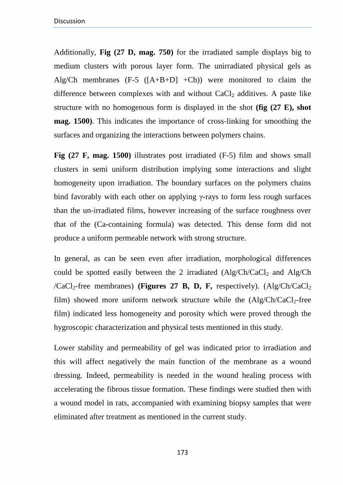
Discussion
173
Additionally, Fig (27 D, mag. 750) for the irradiated sample displays big to
medium clusters with porous layer form. The unirradiated physical gels as
Alg/Ch membranes (F-5 ([A+B+D] +Ch)) were monitored to claim the
difference between complexes with and without CaCl2 additives. A paste like
structure with no homogenous form is displayed in the shot (fig (27 E), shot
mag. 1500). This indicates the importance of cross-linking for smoothing the
surfaces and organizing the interactions between polymers chains.
Fig (27 F, mag. 1500) illustrates post irradiated (F-5) film and shows small
clusters in semi uniform distribution implying some interactions and slight
homogeneity upon irradiation. The boundary surfaces on the polymers chains
bind favorably with each other on applying γ-rays to form less rough surfaces
than the un-irradiated films, however increasing of the surface roughness over
that of the (Ca-containing formula) was detected. This dense form did not
produce a uniform permeable network with strong structure.
In general, as can be seen even after irradiation, morphological differences
could be spotted easily between the 2 irradiated (Alg/Ch/CaCl2 and Alg/Ch
/CaCl2-free membranes) (Figures 27 B, D, F, respectively). (Alg/Ch/CaCl2
film) showed more uniform network structure while the (Alg/Ch/CaCl2-free
film) indicated less homogeneity and porosity which were proved through the
hygroscopic characterization and physical tests mentioned in this study.
Lower stability and permeability of gel was indicated prior to irradiation and
this will affect negatively the main function of the membrane as a wound
dressing. Indeed, permeability is needed in the wound healing process with
accelerating the fibrous tissue formation. These findings were studied then with
a wound model in rats, accompanied with examining biopsy samples that were
eliminated after treatment as mentioned in the current study.

Discussion
174
The used ratio between the different incorporated alginates was (1:1:1), but
different ratios of alginates were supposed to have different effects on
properties of the previously chosen hydrogel, (F-20), so new three forms of the
same formula with the same components were prepared as follows: (F-20/I):
[A+B+D (2:1:1) + (CaCl2)] + (Ch)]& (F-20/II): [A+B+D (1:2:1) + (CaCl2)] +
(Ch) and (F-20/III): [A+B+D (1:1:2) + (CaCl2)] + (Ch). Their properties were
compared to reach the structure of the best working film with assessing their
different properties.
The alginate chains of low M.Ws seemed to interfere with the coacervation of
chitosan with the (HMW-Alg (Alg-A)) due to their easier diffusion to the
chitosan chains for a high degree of complexation. During swelling, diffusion of
the small chains to medium is limited due to the cross-linking with cations. As
can be concluded generally for (F-20) from (fig. (28)), maintaining constant
equal ratio between (A: B: D) gave the resulted gel high swellability for
medium molecules. The increase in ratio of (A-fraction) seemed to enhance the
stability of the resulted gel with stronger crosslinking.
With the increased initial imbibing ability for medium by the weak cross-linked
small and long chains, cleavage of these weak cross-linkages would be
accelerated. On swelling, the small chains begin to move faster at first to the
medium causing theoretical decrease in swelling, but the gel mass and
swellability were maintained by the coacervated alginate long chains. This is
shown with the swelling curve of the form (F-20/I) which after the early fast
swelling, the D.S began to decrease, but still higher than the other forms. The
irradiated (F-20/I) was compared with the corresponding unirradiated form. The
(F-20/I/Un) showed good D.S, but weaker than the irradiated one. These results
confirm the effects of irradiation on the complexation state within the gel
structure.
Discussion
175
Further studying for properties of the forms (F-20/II and F-20/III) was
cancelled due to their weak swelling abilities that may relate to the fast release
of the weakly cross-linked small chains to medium.
The extent of water loss from the hydrogels on exposure to air was evaluated to
examine their behavior on using as dressings over dry wounds. This water loss
can enable the gels (F-20 and 20/I) to take up the exudates from the wound by
an active upward-directed process to help in its moisturization for further
accelerating of healing (fig. (29)).
The irradiated (F-20/I) gel expressed higher rate of degradation in vitro than (F-
20) after (3) days, may be due to the initiation of gel syneresis after short
swelling period on immersing in medium during the first 24 hours, and it
continued in the following days. This degradation behaviour was compared with
that of the corresponding unirradiated formulae which displayed slower rates of
degradation (fig. (30)). The change in integrity of the gels after irradiation
accompanied with the faster swelling may have initiated this faster degradation.
(According to the previous results, the PEC hydrogel of the form (F-20) was
used as the model of dressings and its efficacy was tested in vivo with the
healing of wounds models).
For comparing the efficacy of drugs loading onto PEC hydrogel from the
formula (F-20), Albumin and Curcumin-loaded samples on the forms (F-20
&F-20/I) were prepared under optimum conditions and tested as drug loaded-
models. This highlights the ability of the prepared membranes to act as wound
dressing as well as drugs and antibiotics carriers for further in vitro and in vivo
release systems studies.
Discussion
176
During the preparation of loaded membranes, the used protein model was water
soluble and Curcumin was soluble in organic solvents. Unlike the SEM images
of their virgin membranes, the (irradiated Albumin loaded samples) showed
smooth yet cracked surface layer with more uniform distributed lamination over
the matrix of the prepared membrane in case of the film (F-20/I, fig. (27G))
than that of the loaded film (F-20, fig. (27 H)).
Curcumin loaded samples formed big non-uniform clusters with fusion form
and observed crystallization of the drug in case of (F-20-loaded film, fig. (27
J)) and (F-20/I-loaded film, fig. (27 I)) as well.
These results might indicate a homogeneity and well distribution of drugs in
case of (Albumin) over the prepared membranes in case of (Curcumin) samples
and this of course will be reflected in the texture, success percent, hygroscopic
characterization and performance of the complex during the healing process
cascades. Further studies for the mechanism of entrapment of each drug, the
changes within the hydrogel structure after entrapment, optimizing the
conditions of preparing these new devices and the release rate of the two drugs
from both (F-20 forms) are required to be studied as well.

Discussion
177
3.6. Wounding and Wound Healing:
Rodents are the indispensible tool of choice for most researchers as models for
human diseases where they require little space, easy to handle, inexpensive and
have accelerated healing compared with humans, thereby yield faster results
(Scherer et al., 2008). Murine male skin is 40% stronger than female skin due
to a much thicker dermis, while female skin exhibits a thicker epidermis and
hypodermis. (Azzi et al., 2005). Accordingly, using male rats in the study
would provide easier method for comparing effects of the hydrogel between the
treated and un-treated wounds.
Numerous wound healing components including hemostasis, inflammation,
angiogenesis and matrix deposition are impaired in wide variety of tissues
healing including myocardium, skeletal muscles, and nerves, so it was essential
to measure blood glucose level for the studied rats and exclude the diabetic
members.
Both the wound models, excisions and incisions, are valid in studying the
healing with well recognized advantages and limitations for both. These two
models have met some criticism because the main mechanism of healing in
rodents is contraction due to the presence of the panniculus carnosus muscle in
the subcutaneous tissue but human wounds; in contrast heal more through re-
epithelialization (Falanga, 2005). In spite of that along with observing the
healing of large open acute wounds in rodents, the human display a significant
amount of contraction with the excisional models but this is of less
consideration with the incisional ones that more closely represent the clinical
situation of acute surgical wounds. All excisional healing models are based on
the same principles (Scherer et al., 2008): (1) Robust
inflammation/angiogenesis are hallmarks of this model. (2) Granulation and
epithelialization can be examined with more accuracy (Wong et al., 2011).
Discussion
178
(3) This model supports the evaluation of new topical pharmacological
interventions as medications that can be applied directly to the wound bed
(Tsuboi and Rifkin, 1990; Toker et al., 2009). (4) The excision may be a
better model to evaluate cosmesis, despite the smaller scale of the injury
response (Wilgus and DiPietro, 2012).
A single standard outcome for wound healing does not exist, but some objective
and subjective measures are widely used by the researchers. Within health care
organizations, the % reduction in surface area is often used as an indicator for
the wound response to the treatment. No wound repair occurred completely for
the non-dressed wounds during the study period (Fig (31)) where the formation
of a scab loosely united the wound edges and contributed to a dry environment
that may also have retarded the healing (Hinman and Maibach, 1963). The
faster contraction in size of wounds treated with the hydrogel (Dre) than the
other groups may be due to the physical forces, initiated by the adsorption of
various protein molecules from the wound surface into the dressing
components, especially the chitosan fraction (figures (32, 33)).
3.6.1. The histological assessment of the healing progression:
A quantitative assessment strategy for wound healing cascades was established
based on histological criteria. They identified the different healing phases
including inflammatory response, re-epithelialization, cells migration, regulated
proliferation as well as the epidermal differentiation. Dermal closure, matrix
distribution and skin remodeling along with observing dissolution of the
hydrogel components during the timeline of healing progression occur in a
parallel and sequential manner and they were also identified with the
histological characterization as follows:
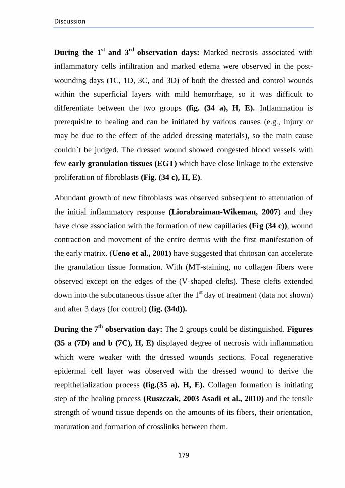
Discussion
179
During the 1st
and 3rd
observation days: Marked necrosis associated with
inflammatory cells infiltration and marked edema were observed in the post-
wounding days (1C, 1D, 3C, and 3D) of both the dressed and control wounds
within the superficial layers with mild hemorrhage, so it was difficult to
differentiate between the two groups (fig. (34 a), H, E). Inflammation is
prerequisite to healing and can be initiated by various causes (e.g., Injury or
may be due to the effect of the added dressing materials), so the main cause
couldn`t be judged. The dressed wound showed congested blood vessels with
few early granulation tissues (EGT) which have close linkage to the extensive
proliferation of fibroblasts (Fig. (34 c), H, E).
Abundant growth of new fibroblasts was observed subsequent to attenuation of
the initial inflammatory response (Liorabraiman-Wikeman, 2007) and they
have close association with the formation of new capillaries (Fig (34 c)), wound
contraction and movement of the entire dermis with the first manifestation of
the early matrix. (Ueno et al., 2001) have suggested that chitosan can accelerate
the granulation tissue formation. With (MT-staining, no collagen fibers were
observed except on the edges of the (V-shaped clefts). These clefts extended
down into the subcutaneous tissue after the 1st
day of treatment (data not shown)
and after 3 days (for control) (fig. (34d)).
During the 7th
observation day: The 2 groups could be distinguished. Figures
(35 a (7D) and b (7C), H, E) displayed degree of necrosis with inflammation
which were weaker with the dressed wounds sections. Focal regenerative
epidermal cell layer was observed with the dressed wound to derive the
reepithelialization process (fig.(35 a), H, E). Collagen formation is initiating
step of the healing process (Ruszczak, 2003 Asadi et al., 2010) and the tensile
strength of wound tissue depends on the amounts of its fibers, their orientation,
maturation and formation of crosslinks between them.
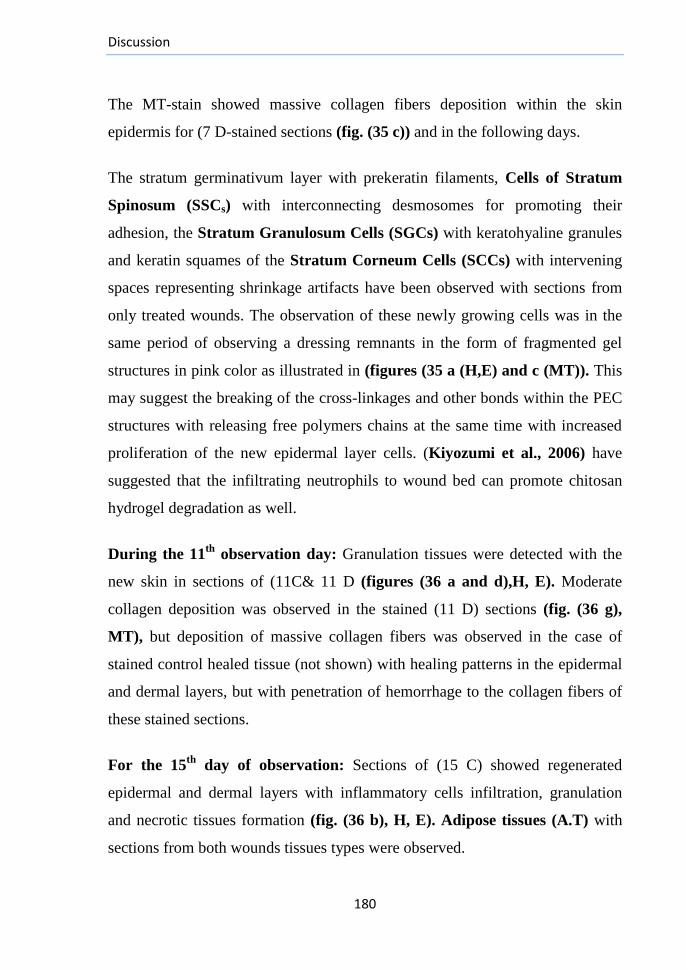
Discussion
180
The MT-stain showed massive collagen fibers deposition within the skin
epidermis for (7 D-stained sections (fig. (35 c)) and in the following days.
The stratum germinativum layer with prekeratin filaments, Cells of Stratum
Spinosum (SSCs) with interconnecting desmosomes for promoting their
adhesion, the Stratum Granulosum Cells (SGCs) with keratohyaline granules
and keratin squames of the Stratum Corneum Cells (SCCs) with intervening
spaces representing shrinkage artifacts have been observed with sections from
only treated wounds. The observation of these newly growing cells was in the
same period of observing a dressing remnants in the form of fragmented gel
structures in pink color as illustrated in (figures (35 a (H,E) and c (MT)). This
may suggest the breaking of the cross-linkages and other bonds within the PEC
structures with releasing free polymers chains at the same time with increased
proliferation of the new epidermal layer cells. (Kiyozumi et al., 2006) have
suggested that the infiltrating neutrophils to wound bed can promote chitosan
hydrogel degradation as well.
During the 11th
observation day: Granulation tissues were detected with the
new skin in sections of (11C& 11 D (figures (36 a and d),H, E). Moderate
collagen deposition was observed in the stained (11 D) sections (fig. (36 g),
MT), but deposition of massive collagen fibers was observed in the case of
stained control healed tissue (not shown) with healing patterns in the epidermal
and dermal layers, but with penetration of hemorrhage to the collagen fibers of
these stained sections.
For the 15th
day of observation: Sections of (15 C) showed regenerated
epidermal and dermal layers with inflammatory cells infiltration, granulation
and necrotic tissues formation (fig. (36 b), H, E). Adipose tissues (A.T) with
sections from both wounds tissues types were observed.
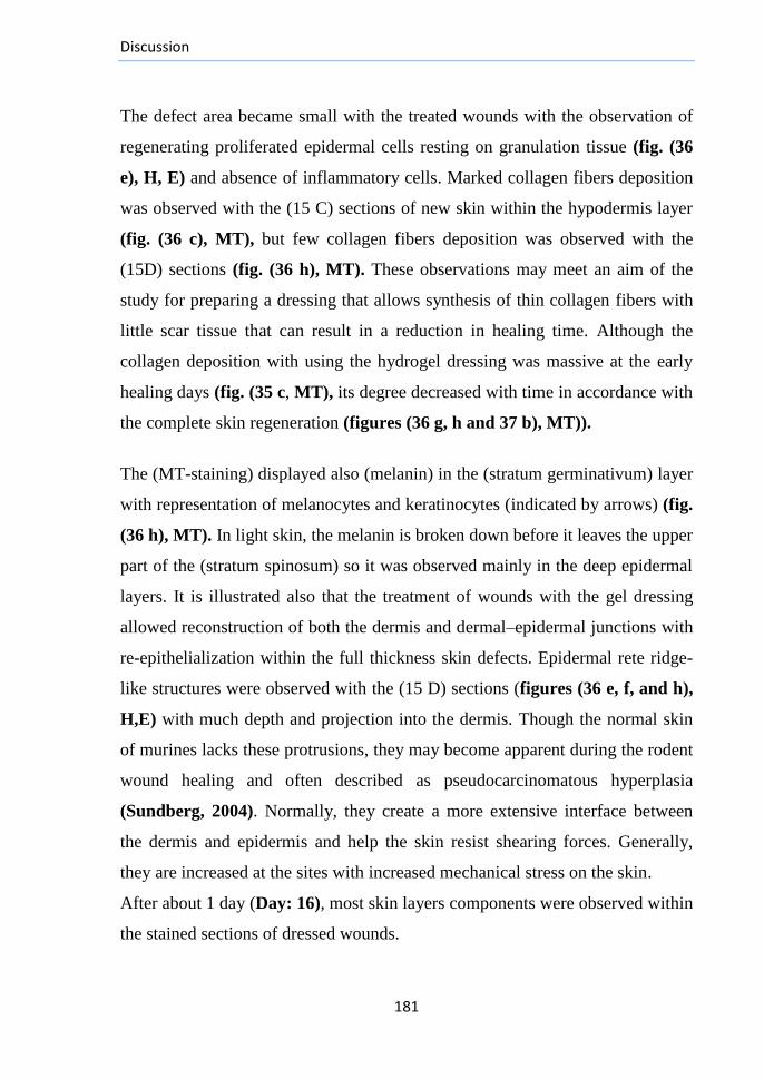
Discussion
181
The defect area became small with the treated wounds with the observation of
regenerating proliferated epidermal cells resting on granulation tissue (fig. (36
e), H, E) and absence of inflammatory cells. Marked collagen fibers deposition
was observed with the (15 C) sections of new skin within the hypodermis layer
(fig. (36 c), MT), but few collagen fibers deposition was observed with the
(15D) sections (fig. (36 h), MT). These observations may meet an aim of the
study for preparing a dressing that allows synthesis of thin collagen fibers with
little scar tissue that can result in a reduction in healing time. Although the
collagen deposition with using the hydrogel dressing was massive at the early
healing days (fig. (35 c, MT), its degree decreased with time in accordance with
the complete skin regeneration (figures (36 g, h and 37 b), MT)).
The (MT-staining) displayed also (melanin) in the (stratum germinativum) layer
with representation of melanocytes and keratinocytes (indicated by arrows) (fig.
(36 h), MT). In light skin, the melanin is broken down before it leaves the upper
part of the (stratum spinosum) so it was observed mainly in the deep epidermal
layers. It is illustrated also that the treatment of wounds with the gel dressing
allowed reconstruction of both the dermis and dermal–epidermal junctions with
re-epithelialization within the full thickness skin defects. Epidermal rete ridge-
like structures were observed with the (15 D) sections (figures (36 e, f, and h),
H,E) with much depth and projection into the dermis. Though the normal skin
of murines lacks these protrusions, they may become apparent during the rodent
wound healing and often described as pseudocarcinomatous hyperplasia
(Sundberg, 2004). Normally, they create a more extensive interface between
the dermis and epidermis and help the skin resist shearing forces. Generally,
they are increased at the sites with increased mechanical stress on the skin.
After about 1 day (Day: 16), most skin layers components were observed within
the stained sections of dressed wounds.
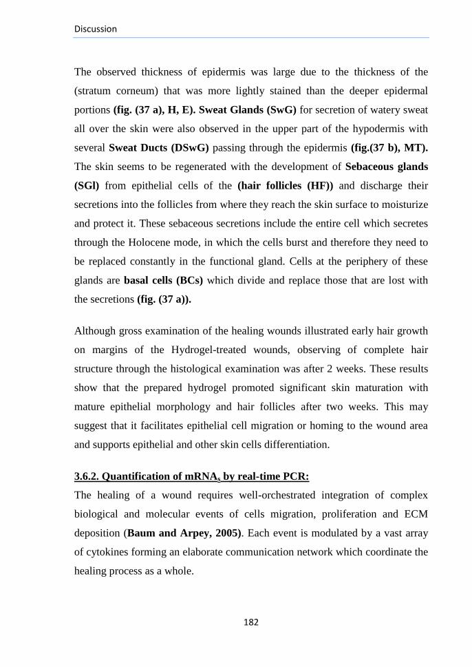
Discussion
182
The observed thickness of epidermis was large due to the thickness of the
(stratum corneum) that was more lightly stained than the deeper epidermal
portions (fig. (37 a), H, E). Sweat Glands (SwG) for secretion of watery sweat
all over the skin were also observed in the upper part of the hypodermis with
several Sweat Ducts (DSwG) passing through the epidermis (fig.(37 b), MT).
The skin seems to be regenerated with the development of Sebaceous glands
(SGl) from epithelial cells of the (hair follicles (HF)) and discharge their
secretions into the follicles from where they reach the skin surface to moisturize
and protect it. These sebaceous secretions include the entire cell which secretes
through the Holocene mode, in which the cells burst and therefore they need to
be replaced constantly in the functional gland. Cells at the periphery of these
glands are basal cells (BCs) which divide and replace those that are lost with
the secretions (fig. (37 a)).
Although gross examination of the healing wounds illustrated early hair growth
on margins of the Hydrogel-treated wounds, observing of complete hair
structure through the histological examination was after 2 weeks. These results
show that the prepared hydrogel promoted significant skin maturation with
mature epithelial morphology and hair follicles after two weeks. This may
suggest that it facilitates epithelial cell migration or homing to the wound area
and supports epithelial and other skin cells differentiation.
3.6.2. Quantification of mRNAs by real-time PCR:
The healing of a wound requires well-orchestrated integration of complex
biological and molecular events of cells migration, proliferation and ECM
deposition (Baum and Arpey, 2005). Each event is modulated by a vast array
of cytokines forming an elaborate communication network which coordinate the
healing process as a whole.
Discussion
183
New understandings in its complexities; particularly roles of the cytokines,
enable clinicians to manage superficial wounds (e.g., Skin flaps and even the
most difficult-to-heal types more effectively) (Brem and Tomic-Canic,2007).
To confirm the production and roles of VEGF during wound healing, we
quantified its gene expression by the sensitive and reliable real-time
fluorescence PCR technique. In the first phase of wound healing, VEGF
stimulates the coagulation factors in the ECs; therefore, the thrombocytes
accumulation and adhesion occur (Banks et al., 1998; Nogami et al., 2007). It
has been previously reported by neutralization experiments that Basic Fibroblast
Growth Factor (bFGF) accounts for a large part of the immediate angiogenic
stimulus in the wound area (Nissen et al., 1996; Takamiya et al., 2002)
without any participation of VEGF in the initiation of angiogenesis. The
neutrophils; recruited to wound area directly after wounding as a part of the
inflammatory response, have been described as (VEGF-negative secretory cells)
(Hayashi et al., 2004). These previous observations may explain why the
VEGF gene expression in our experiments was weaker at the first wounding day
than the 2nd
one (figures (38 and 39)).
For blood vessels formation, immuno-localization of VEGF increases in the
inflammatory cells during the inflammation phase as well as in ECs, fibroblasts
and macrophages in the following phases (Banks et al., 1998; Miyagami and
Katayama, 2005; Nogami et al., 2007). This recruiting along with activation of
the respiratory burst (Babior et al., 1973; Khanna et al., 2001) may explain the
significant rise in VEGF mRNA at day 2 of normal healing to attract
metabolites and oxygen to the healing region (Hayashi et al., 2004) with
maintaining these levels in the following days. Our results showed increased
VEGF gene expression levels after the 1st week of normal healing.Maintaining
high certain levels of VEGF expression and its mRNA is essential to activate
the required angiogenic steps for accelerating the healing.
Discussion
184
CD68-positive mononuclear cells, ECs and fibroblasts can sustain the VEGF
expression 7 days after the wounding indicating the possibility of its
involvement in the chemotaxis (Nissen et al., 1998) and vascularization
(Hayashi et al., 2004). Previous studies by (Nissen et al., 1998, Altavilla et al.,
2001; Nogami et al., 2007) have observed that VEGF mRNA has significant
high levels after one day of normal healing compared with the 3rd
and 7th
days.
There is an early appearance of fibroblasts in wounds treated with the dressing
along with the EGT (fig. (34 c)). These observations along with the reports of
(Okamoto et al., 1995; Ueno et al., 1999; Ueno et al., 2001; Alemdaroglu et
al., 2006) who found out faster recruiting of leukocytes and macrophages to the
wound area treated with chitosan may explain the early high levels of VEGF
during healing of the treated wounds with our hydrogel than the untreated ones
(fig. (38)). In addition, (Ueno et al., 2001; Inan and Saraydın, 2013) have
suggested that chitosan might stimulate recruiting and proliferation of
inflammatory cells during the early inflammation phase.
The growth factors including VEGF along with other fibroblasts cytokines in
the wound granulation tissue during the early healing phases might also increase
and last for only a few days (Alemdaroglu et al., 2006). Results of (Ying,
2011) as well found out that the effects of alginate on wound healing may
involve important roles associated with the expression of the cytokines VEGF
and TGF-β1. In summary, these reports showed increased expression of VEGF
during the healing days, especially the 7th
day of wounds treated with alginate
dressing and over that of the untreated wound. These are the same results
obtained using our newly prepared hydrogel although it consists of a complex of
alginate with chitosan. This refers that both the two polyelectrolytes within the
complex maintained their effects on VEGF gene expression with more
enhancement after complexation.
Discussion
185
(Lee et al., 2009) have observed suppression for VEGF protein with wounds
treated with alginate than those treated with Vaseline and normal healed
wounds. The discrepancy between our results and these observations may relate
to the prepared complex structure which may overcome the individual alginate
effects making the behavior of the complex membrane differ from some of its
individual component's behaviors.
(Blann and Taberner, 1995; Constans and Conri,2006) have observed that
the elevation in vWF levels is perhaps the most known marker of endothelial
injury. The decrease in level of blood vWF mRNA may relate to the direction of
its secretory cells towards the wound bed for the initiation of blood clotting.
During hemostasis, two steps in platelets adherence have been identified that
have a requirement for divalent cations. One step is the platelets spreading
reaction on subendothelium which has a need for factor VIII-von Willebrand
factor (FVIII-vWF) and Ca2+
or Mg2+
(Sakariassen et al., 1979; Bolhuis et al.,
1981). Some (vWF-related proteins) are already present in the subendothelium
(Rand et al., 1980), but the (FVIII-vWF) of plasma is required for optimal
adherence where it binds to the subendothelium after deendothelialization and
prior to the platelets adherence (Weiss et al.,1978; Sakariassen et al.,1979;
Bolhuis et al., 1981).
The second step is the facilitation of platelets adherence by (FVIII-vWF) that
has a requirement for Ca2+
(Sakariassen et al.,1984). Sufficient Ca2+
may be
associated with (FVIII-vWF) to change the microenvironment at the
subendothelium surface. This causes a small increase in platelets adherence
(Mikaelsson et al., 1983). Solubilization of the used dressing with releasing of
Ca2+
ions may be a reason for more activation of (FVIII-vWF) in wound bed
causing faster clotting.

Discussion
186
This may explain the reason for the decrease in blood levels of vWF mRNA
during the first post-wounding week comparable to the undressed wound though
the differences were not significant. The higher blood vWF in the un-treated
rats may also reflect higher release of vWF antigen from ECs and/or platelets
than in the treated rats.
vWF is a major part in the WPBs where it is involved in their biogenesis and
used as a marker of them (Rondaij et al., 2006; Sadler et al., 1991) and the
decrease in its expression causes the release of WBPs components including
(Ang-2) which may act synergistically with VEGF to promote angiogenesis.
This WPB exocytosis gives rise to a rapid release of vWF and other mediators
(e.g., interleukin-8 (IL-8)) (Babich et al., 2008) with translocation of P-selectin
from within granules to the endothelial surfaces for triggering leukocytes rolling
are all critical early events in the endothelial activation and vascular
inflammation (Wagner, 1993).
It has been reported that VEGF regulates the vWF/WPB release(Matsushita et
al., 2005) where it rapidly induces a time and dose-dependent vWF release from
human venous endothelial cells. Accordingly; as concluded from our results, the
raise in levels of VEGF from day 2 to reach its greatest levels at day 7 may
explain the corresponding decrease in vWF mRNA in the blood secretory cells.
Alternatively, the decrease in VEGF levels after the 7th
day with the untreated
wounds may explain the high corresponding increase in the mRNA of blood
vWF. For the dressed wound, the gradual decrease in VEGF mRNA is also
responsible for the gradual increase in vWF mRNA levels (fig. (41)).
Due to economic reasons, the expression of vWF gene has been quantified in
blood cells but further experiments are suggested to measure its levels in the
wound bed tissue with concentrating the studies in the blood vessels tree for
better understanding of the effects of the degraded gel in the wound bed.
Discussion
187
The overall result from these tests with using the prepared hydrogel as a
dressing without the addition of growth factors or cytokines seems to be the
enhancement of angiogenesis through accelerating the recruitment of ECs to the
wound area. This neovascularization facilitates cells and nutrients transportation
as well as oxygen and wastes exchange and these are all critical conditions for
the optimum wound healing with accelerating ECM production for perfect skin
regeneration.
Biomaterials are defined as the materials that can be interfaced with biological
systems in order to evaluate, treat, augment, or replace any tissue, organ or
function of the body. Any material to be used as a part of a biomaterial device
has to be biocompatible and its clinical applications should not cause any
adverse reactions in the organism nor endanger the life of patients. BUN and
creatinine levels were in the range of normal values with no significant
difference between the untreated rats and those treated with the hydrogel
(Table: 9). This refers that solubilization of its polymers chains does not affect
the physiological functions in the body.

Recommendations
188
Recommendations for Future Work Preparation of New Drugs-Loaded Wound Dressings:
Loading of 2 drug models was tested at the end of the study with (SEM), but more tests are
required to get an overview of the mechanism of binding of each one to the hydrogels as well
as the release mechanisms and rates in vitro and in vivo. Loading of new drugs is also
essential for enhancing the accelerating effects of the virgin hydrogel dressings on wound
healing, especially for the treatment of ulcers. Examples of these drugs may involve loading
of silver, charcoal, antibiotics, enzymes or nano-materials whose efficacies can be tested
through both in vitro and in vivo studies. These studies may require modifications for the
structures of the composing polymers, blending of new materials, as well as preparing new
hydrogels forms; maybe in the hydrocolloid, sponge, multi-membrane forms, and so on. This
requires more understanding of the chemical and physical properties of these materials and
may open the way to synthesize new materials to produce new wound care products.
Further studies for the healing wounds: More examinations are required for the treated healing wounds for understanding and giving
more proofs of the supposed scaffolding action of the hydrogels. The angiogenic response in
wounds beds was only studied with histological examinations and measuring the expression
of 2 angiogenic genes, but more experiments are required to get a complete overview on the
mechanism of angiogenesis and understanding the effects of the hydrogel on the normal
angiogenic cascades. Immuno-histochemical analysis for the infiltrating cells, measuring the
expression of more angiogenic genes and Doppler imaging of the wounds beds may be
suitable methods. This study focused on the angiogenic effects of the treating hydrogels and
skin maturation, but more studies are required to assess the complete mechanism of wound
healing as well as the scarring response after treatment at the molecular and cellular levels
with including the different cells involved in the healing stages. The different involved
molecular and cellular processes such as signal transduction, proliferation, migration and
growth factors production after the treatment should also be studied. More wounds models
can also be created (e.g., Ischemic flaps, mechanical load model, parabiosis model and
pressure ulcer model) in the future studies as well to overcome the differences in wound
healing mechanisms between rats and human.
Summary
189
Summary
Skin is the first line of defense from external factors all over the place. It holds
all organs together, protects our bodies, helps keep them at just the right
temperature and allows us to have the sense of touch. Skin wounds often occur
as a result of accident or injury. Surgical incisions, sutures and stitches also
cause wounds so the wound management is essential to promote healing of the
underlying soft tissue and treat or reduce the risk of infection.
We first prepared a Polyelectrolyte complex through the binding of alginate
with chitosan incorporating certain functional groups and proved its
biocompatibility and integration with the host tissue. A new type of wound
dressings in hydrogel film form based on this complex was prepared where the
2 polymers were mixed together with a new preparation method. At first, the
alginate molecular weight and chemical structure were tailored with (γ-
irradiation and oxidation), followed by characterization of their properties
through measuring their M.Ws and the degrees of oxidation for the modified
oxidized structures as well as assessing the resulting structures with (FT-IR).
The alginates were then purified and the purification degree was confirmed.
Different chitosans were extracted from shrimp shells and were characterized
by (FT-IR). Their degrees of purity, M.Ws and antioxidant properties were
investigated as well.
A scheme for choosing the best structure from different prepared hydrogels for
using as a dressing was suggested involving different physical, chemical,
spectroscopic and biological approaches. The chosen hydrogel was then
applied to excisional wound model in rats to improve the characterization of
its biological effects through monitoring of its effects on healing with
measuring the wounds closure rates.
Summary
190
The histological examinations for skin and wounds beds and tests for detecting
the expression of the angiogenic genes (VEGF& vWF) with the routine
measuring of kidney functions for the wounded rats were performed also to
test the effects of solubilization and degradation of these biomaterials
The chosen formula was (F-20) with the structure consisting of (chitosan and
different alginates of the types (High M.W+ Very low M.W+ Multi aldehyde
Sodium Alginate (Type I-A)). The in vivo study of its effects on wound
healing included (3) groups of wounded rats; single right wound was created
on the dorsal skin of each rat:
Group (1) (G. (1)): The created wounds were treated with the dressing.
Group (2) (G. (2)): The created wounds were treated with Fusidin cream.
Group (3) (Control group (G. (3)): The wounds were left untreated.
With continuous monitoring for wounds of these groups during the following
days, analysis of the results revealed that:(1) The wound closure rate % was
about 98 % after 11 days of healing for the hydrogel-treated wounds (G.(1)),
but after 2 weeks for the fusidin-treated wounds (G. (2)) and only 95 % was
achieved for the non-treated wounds (G. (3)) during the same period.
(2) The histological examination for skin and wounds beds of both (G. (1 and
3)) proved the efficiency of the used hydrogel to promote healing with
accelerating the remodeling of wounds tissues and its regeneration as well as
promoting the angiogenic response compared to the non-treated wounds.
(3) The hydrogel dressing (G. (1)) maintained certain levels of VEGF mRNA
in most post-wounding days and after the 7th
day with vWF which guaranteed
the acceleration of suitable blood supply to the injured tissues in addition to
performing their other functions. These genes expression levels were
significantly lower with the non-treated wounds (G. (3)).
References
191
REFERENCES
AAMI TIR17. (1997). Radiation Sterilization -Material Qualification.
(http://webstore.ansi.org/ansidocstore/product. asp?sku=AAMI+TIR17%3A1997).
Abd El-Rehim HA, El-Sawy NM, Farag IA, Elbarbary AM. (2011). Synergistic effect of
combining ionizing radiation and oxidizing agents on controlling degradation of Na-alginate
for enhancing growth performance and increasing productivity of zea maize plants.
Carbohydr. Polym.; 86:1439-44.
Abe K, Ohno H, Nii A, Tsuchida E. (1978). Calorimetric study on the formation of polymer
complexes through electrostatic interaction and hydrogen bonding. Die Makromolekulare
Chemie; 179 (8): 2043-50.
Agren MS. (1996). Four alginate dressings in the treatment of partial thickness wounds: a
comparative experimental study. Br. J. Plast. Surg.; 49:129 - 34.
Ahrgren L, de Belder AN. (1975). The action of fenton’s reagent on dextran. Starch – Stärke;
27: 121–3.
Aird WC, Jahroudi N, Weiler-Guettler H, Rayburn HB, Rosenberg RD. (1995). Human von
Willebrand factor gene sequences target expression to a subpopulation of endothelial cells in
transgenic mice. Proc. Natl. Acad. SCI. USA.; 92: 4567-71.
Alam HB, Burris D, DaCorta JA. (2005). Hemorrhage control in the battlefield ‘role of new
hemostatic agents”. Military Medicine; 170: 1-63.
Alberts B, Johnson A, Lewis J, Raff M, Roberts K, Walter P.(2002). Molecular biology of the
cell (4th
edition). Garland Publications: New York.
Alemdaroglu C, Degim Z, Celebi N Zor F, Oztürk S, Erdoğan D. (2006). An investigation
on burn wound healing in rats with chitosan geL formulation containing epidermal growth
factor. Burns J.; 32: 319-27.
Alexakis T, Boadi DK, Quong D, Groboillot A, O’Neill I, Poncelet D, Neufeld RJ. (1995).
Microencapsulation of DNA within alginate microspheres and crosslinked chitosan membranes
for in vivo application. Appl. Biochem. Biotechnol.; 50:93–106.
Al-Kahtani A, Bhojya NH, Sherigara BS. (2009). Synthesis and characterization of chitosan-
based pH-sensitive semi-inter-penetrating network microspheres for controlled release of
diclofenac sodium. Carbohydr. Res.; 344: 699-706.
Allen DB, Maguire JJ, Mahdavian M, Wicke C, Marcocci L, Scheuenstuhl H, Chang M, Le
AX, Hopf HW, Hunt TK. (1997). Wound hypoxia and acidosis limit neutrophil bacterial killing
mechanisms. Arch. Surg.; 132:991–6.
Allgrove J. (2009). Physiology of calcium, phosphate and magnesium. Endocr.Dev.;16:8-31
Alsberg E, Anderson KW, Albeiruti A, Franceschi RT, Mooney DJ. (2001). Cell-interactive
alginate hydrogels for bone tissue engineering. J. Dent. Res.; 80(11): 2025-9.
References
192
Alsberg E, Kong HJ, Hirano Y, Smith MK, Albeiruti A, Mooney DJ. (2003). Regulating
bone formation via controlled scaffold degradation. J.Dent.Res.; 82(11):903-8.
Alshamkhani A, Duncan R. (1995). Radioiodination of alginate via covalently-bound
tyrosinamide allows for monitoring of its fate in vivo. J. Bioact. Compat. Polym.; 10:4–13
Altavilla D, Saitta A, Cucinotta D, Galeano M, Deodato B, Colonna M, Torre V, Russo
G, Sardella A, Urna G, Campo GM, Cavallari V, Squadrito G, Squadrito F. (2001). Inhibition of lipid peroxidation restores impaired vascular endothelial growth factor expression
and stimulates wound healing and angiogenesis. Diabet. J.; 50:667–70.
Amsden B.; Turner N. (1999). Diffusion characteristics of calcium alginate gels. Biotech.
Bioeng.; 65; 605–10.
Angyal SJ. (1973). Complex formation between sugars and metal ions. Pure Appl. Chem.; 35(2):
131-46.
ANSI/AAMI/. (2006). ISO 11137-2: Sterilization of health care products –radiation, Part 2:
Establishing the Sterilization Dose (www.iso.org).
Argin-Soysal S, Kofinas P, Lo YM. (2009). Effect of complexation conditions on xanthan –
chitosan polyelectrolyte complex gels. Food Hydrocolloids; 23: 202-9.
Armstrong DG, Lavery LA, Harkless LB. (1998). Validation of diabetic wound classification
system: The contribution of depth, infection and ischemia to risk of amputation. Diabet. Care; 21:
855- 91.
Asada T, Ohkubo T, Kawata K, Oikawa K. (2006). Ammonia adsorption on bamboo
charcoal with acid treatment. J. Health Sci.; 52:585-9.
Asadi MR, Torkaman G, Hedayati M. (2010). The effect of electrical stimulation intensity on
VEGF expression and biomechanical properties during wound. World Academy of Science,
Engineering and Technology; 69: 284-6.
Asahara T, Bauters C, Zheng LP, Takeshita S, Bunting S, Ferrara N, Symes JF, Isner JM.
(1995). Synergistic effect of vascular endothelial growth factor and basic fibroblast growth factor
on angiogenesis in vivo. Circulation J.; 92:11365-71.
Azad AK, Sermsintham N, Chandrkrachang S, Stevens WF. (2004). Chitosan membrane as a
wound-healing dressing: characterization and clinical application. J. Biomed. Mater. Res. Part B:
Appl. Biomater.; 69: 216-22.
Azzi L, El-Alfy M, Martel C, Labrie F. (2005). Gender differences in mouse skin morphology
and specific effects of sex steroids and dehydroepiandrosterone. J. Invest.
Dermatol.; 124(1): 22–7.
Babensee JE, Anderson JM, McIntire LV, Mikos AG. (1998). Host response to tissue
engineered devices. Adv. Drug Deliv. Rev.; 33(1-2):111-39.
Babich V, Meli A, Knipe L, Dempster JE, Skehel P, Hannah MJ, Carter, T. (2008). Blood J.;
References
193
111: 5282–90.
Babior BM, Kipnes RS, Curnutte JT. (1973). Biological defense mechanisms: The production
by leukocytes of superoxide, a potential bactericidal agent. J. Clin. Invest.; 52: 741-4.
Bahnak BR, Wu Q, Coulombel L, Assouline Z, Kerbiriou-Nabias D, Pietu G, Drouet L,
Caen JP, Meyer D. (1989). Expression of von Willebrand Factor in porcine vessels:
heterogeneity at the level of von Willebrand Factor mRNA. J. Cell. Physiol.; 138:305–10.
Bajpai SK, Tankhiwale R. (2006). Investigation of water uptake behavior and stability of
Calcium Alginate/Chitosan bi-polymeric beads. Part-I. React. Funct. Polym.; 66(6):645-58
Baker FS, Miller CE, Repik AJ, Tolles ED. (1992). Activated carbon. Kirk-Othmer
Encyclopedia of Chemical Technology; 4: 1015-37.
Bakeev KN, Izumrudov VA, Kuchanov SI, Zezin AB, Kabanov VA. (1992). Kinetics and
mechanism of interpolyelectrolyte exchange and addition reactions. Macromol. J. 25(17): 4249-
54.
Balakrishnan B, Lesieur S, Labarre D. Jayakrishnan A. (2005a). Periodate oxidation of
sodium alginate in water and in ethanol-water mixture: a comparative study. Carbohydr. Res.,
340, 1425-9.
Balakrishnan B, Mohanty M, Umashankar PR, Jayakrishnan A. (2005b). Evaluation of an in
situ forming hydrogel wound dressing based on oxidized alginate and gelatin. Biomater. J. 26:
6335-42.
Bancroft JD, Gamble M. (2007). Theory and practice of histological Techniques ,(6
th edition).
Churchill-Livingstone, Inc.: New York.
Banks R, Forbes M, Kinsey S, Stanley A, Ingham E, Walters C, Selby P.(1998)Release of
the angiogenic cytokine vascular endothelial growth factor (VEGF) from platelets: Significance
for VEGF measurements and cancer biology. Br. J. Cancer; 77: 956 - 60.
Bao P, Kodra A, Tomic-Canic A, Golinko MS, Ehrlich HP, Brem H. (2009). The role of
vascular endothelial growth factor in wound healing. J. Surg. Res.; 153 (2): 347-58.
Barleon B, Sozzani S, Zhou D, Weich HA, Mantovani A, Marme D. (1996). Migration of
human monocytes in response to vascular endothelial growth factor (VEGF) is mediated via the
VEGF receptor flt-1. Blood J.; 87:3336 –43.
Barnett SE, Varley S. (1987). The effects of calcium alginate on wound Healing. Annals
of the Royal College of Surgeons of England. 69 (4):153-5.
Barrientos S, Stojadinovic O, Golinko MS, Brem H, Tomic-Canic M. (2008). Growth
factors and cytokines in wound healing.Wound Rep.Regen.; 16(5):585-601
Bartels VT. (2011). Handbook of medical textiles (1st edition). Woodhead Publishing Limited:
Cambridge.
Baruch D, Denis C, Marteaux C, Schoevaert D, Coulombel L, Meyer D. (1991). Role of von

References
194
Willebrand factor associated to extracellular matrices in platelet adhesion. Blood J.; 77: 519-27.
Bates DO, Curry FE. (1997). Vascular endothelial growth factor increases microvascular
permeability via a Ca2+
-dependent pathway. Am. J. Physiol; 273: H687-94.
Bates DO, Harper SJ.(2002). Regulation of vascular permeability by vascular endothelial
growth factors. Vascul. Pharmacol.; 39(4-5): 225-37.
Battegay J. (1995). Angiogenesis: mechanistic insights, neovascular diseases, and therapeutic
prospects. J. Mol. Med.; 73:333-46.
Baum CL, Arpey J. (2005). Normal cutaneous wound healing: clinical correlation with cellular
and molecular events. Dermatol. Surg.; 31(6):674-86.
Baumgartner HR, Tschopp TB, Meyer D. (1980). Shear rate dependent inhibition of platelet
adhesion and aggregation on collageneous surfaces by antibodies to human factor VIII-von
Willebrand factor. Br. J. Haematol; 44:127-39.
Baxter A, Dillon M, Taylor KDA, Roberts GAF. (1992). Improved method for i.r.
determination of the degree of N-acetylation of chitosan. Int. J. Biol. Macromol.; 14:166-9
Ben-Av P, Crofford U, Wilder RL, Hla T. (1995). Induction of vascular endothelial growth
factor expression in synovial fibroblasts by prostaglandin E and interleukin-1: a potential
mechanism for inflammatory angiogenesis. FEBS Lett.; 372:83-7.
Ben-Ezra M, Vlodavsky I, Ishai-Michaeli R, Neufeld G, Bar-Shavit R. (1993). Thrombin-
induced release of active basic fibroblast growth factorheparan sulfate complexes from
subendothelial extracellular matrix. Blood J.; 81:3324-31.
Benoy IH, Elst H, VanDam P, Scharpe S, Van Marck E, Vermeulen PB, Dirix LY. (2006). Detection of circulating tumour cells in blood by quantitative real-time RT-PCR: effect of pre-
analytical time. Clin. Chem. Lab. Med.; 44:1082-7.
Bensouilah J, Buck P, Tisser, R, Avis A. (2007). Skin structure and function. In: Bensouilah J,
Buck P., editors. Aromadermatology: Aromatherapy in the treatment and care
of common skin conditions. Blackwell Publishers: New Jersy.
Berens AR, Hopfenberg H. (1978). Diffusion and relaxation in glassy polymer powders: 2.
Separation of diffusion and relaxation parameters. Polym. J.; 19: 489 -96.
Berger J, Reist M, Mayer JM, Felt O, Gurny R. (2004a). Structure and interactions in chitosan
hydrogels formed by complexation or aggregation for biomedical applications. Eur. J. Pharm.
Biopharm.; 57: 35-52.
Berger J, Reist M, Mayer JM, Felt O, Gurny R. (2004b). Structure and interactions in
covalently and ionically crosslinked chitosan hydrogels for biomedical applications, Eur. J.
Pharm. Biopharm.; 57:19-34.
References
195
Bernardo A, Ball C, Nolasco L, Moake JF, Dong JF. (2004). Effects of inflammatory
cytokines on the release and cleavage of the endothelial cell-derived ultralarge von Willebrand
factor
multimers under flow. Blood J.; 104:100-6.
Berra E, Pages G, Pouyssegur J. (2000). MAP kinases and hypoxia in the control of VEGF
expression. Cancer & Metastasis Rev.; 19:139–45.
Berse B, Brown LF, van de Water L, Dvorak HF, Senger DR. (1992).Vascular permeability
factor (vascular endothelial growth factor) gene is expressed differentially in normal tissues,
macrophages and tumors. Mol. Biol.Cell.; 3:211-20.
Biesheuvel PM, Stuart MA. (2004). Cylindrical cell model for the electrostatic free energy of
polyelectrolyte complexes. Langmuir; 20(11): 4764-70.
Black WAP. (1950). The seasonal variation in weight and chemical composition of the common
British Laminariaceae. J. Mar. Biol. Assoc. UK.; 29:45-72.
Blaine G. (1947). Experimental observations on absorbable alginate products in surgery. Ann.
Surg.; 125:102-7.
Blair SD, Jarvis P, Salmon M, and Mc Collum C. (1990). Clinical trial of calcium
alginate haemostatic swabs. Br. J. Surg.; 77: 568-70.
Blann AD, Taberner DA. (1995). A reliable marker of endothelial cell dysfunction: does it
exist? Br. J. Haematol.; 90(2):244-8.
Bodmeier R, Wang J. (1993). Microencapsulation of drugs with aqueous colloidal
polymer dispersions. J. Pharm. Sci.; 82:191–4.
Bolhuis PA, Sakariassen KS, Sander Hi, Bouma BN. Sixma JJ. (1981). Binding of factor
VIII-von Willebrand factor to human arterial subendothelium precedes increased platelet
adhesion and enhances platelet spreading. Lab. Clin. Med.; 97: 568-76.
Boom R, Sol CJA, Salimans MMM, Jansen CL, Dillen MEW, Noordaa JV. (1990). Rapid
and simple method for purification of nucleic acids. J. Clin. Microbiol.; 28: 495-503
Borchiellini A, Fijnvandraat K, ten Cate JW, Pajkrt D, Pasterkamp G, van Deventer SJH,
Meijer-Huizinga F, Zwart-Huinink L, Voorberg J, van Mourik JA. (1996). Quantitative
analysis of von Willebrand factor propeptide release in vivo: Effect of experimental endotoxemia
and administration of 1-deamino-8-D-arginine vasopressin in humans. Blood J.; 88:2951.
Born GV, Cross MJ. (1964). Effects of inorganic ions and of plasma proteins on the aggregation
of blood platelets by adenosine phosphate. J. Physiol; 17: 397-414
Boucard N, Viton C, Agay D, Mari E, Roger T, Chancerelle Y, Domard A. (2007). The use
of physical hydrogels of chitosan for skin regeneration following third-degree burns. Biomater.
J.; 28:3478–88.
Bouhadir KH, Kruger GM, Lee KY, Mooney DJ. (2000). Sustained and controlled release
References
196
of daunomycin from cross-linked poly (aldehydeguluronate) hydrogels. J. Pharm Sci.;
89:910-9.
Bouhadir KH, Lee KY, Alsberg E, Damm KL, Anderson KW, Mooney DJ. (2001). Degradation of partially oxidized alginate and its potential application for tissue engineering.
Biotechnol. Prog.; 17(5): 945-50.
Boyd RF, Hoerl BG. (1981). Sterilization and disinfection: Ionizing radiation. In: Basic Medical
Microbiology, Boyd RF, Hoerl BG., editors. Little, Brown and Company: Boston.
Bradford MM. (1976). A rapid and sensitive method for the quantitation of microgram
quantities of protein utilizing the principle of protein-dye binding. Analytical biochemistry; 72:
248-54.
Brandenburg K, Seydel U. (1996). Fourier Transform Infrared Spectroscopy of cell surface
polysacharides. Wiley-Liss: New York.
Brem H, Tomic-Canic M. (2007). Cellular and molecular basis of wound healing in diabetes. J.
Clin. Invest.; 117 (5):1219–22.
Briand V, Tang S. (2007). Alginamide synthesis and emulsion preparation (Senior Thesis),
Major Qualifying Project (MQP):Number (TAC-FR11), Nancy, France at École Nationale
Supérieure des Industries Chimiques (ENSIC).
British Medical Association (BMA) and the Royal Pharmaceutical Society of Great Britain.
(2001). British National Formulary: London. (Volume: 41).
Brown LF, Harrist TJ, Yeo KT, Ståhle-Backdahl M, Jackman RW, Berse B, Tognazzi K,
Dvorak HF, Detmar M. (1995). Increased expression of vascular permeability factor (vascular
endothelial growth factor) in bullous pemphigoid, dermatitis herpetiformis, and erythema
multiforme. J. Invest. Dermatol.; 104:744-9.
Brown LF, Yeo KT, Berse B, Yeo TK, Senger DR, Dvorak HF, van de Water L. (1992). Expression of vascular permeability factor (vascular endothelial growth factor) by epidermal
keratinocytes during wound healing. J. Exp. Med.; 176: 1375-9.
Bruneel D, Schacht E. (1993). Chemical modification of pullulan: 1. Periodate-oxidation.
Polym. J.; 34: 2628-32.
Budinova T, Ekinci E, Yardim F, Grimm A, Björnbom E, Minkova V, Goranova M.
(2006). Characterization and application of activated carbon produced by H3PO4 and water
vapor activation. Fuel Process. Technol.; 87 (10): 899–905.
Burchardt M, Burchardt T, Chen MW, Shabsigh A, de lataille A, Buttyan R, Shabsigh R.
(1999). Expression of messenger ribonucleic acid splice variants for the vascular endothelial
growth factor in the penis of adult rats and humans. Biol. Reprod.; 60: 398-404.
Burrow T, Welch MJ. (1983). The development and use of alginate fibres in nonwovens for
medical end-uses. In: Cusick GE, editor. Nonwoven Conference Papers: UMIST.
References
197
Bu Z, Yan Y, Zhang Z, Gu X, Wang M, Gong A, Sun X, Cui Y, Zeng Y. (2010). Properties of
extracellular matrix-like scaffolds for the growth and differentiation of endothelial progenitor
cells. J. Surg. Res.; 164:50–7.
Ca´rdenas A, Argu¨elles-Monal W, Goycoolea FM, Higuera-Ciapara I, Peniche C. (2003). Diffusion through membranes of the polyelectrolyte complex of chitosan and alginate.
Macromol. Biosci.; 3: 535 –9.
Carew JA, Browning PJ, Lynch DC. (1990). Sulfation of von Willebrand Factor. Blood J.;
76(12):2530-9.
Carola R, Harley JP, Noback CR. (1990). The Integumentary System (International
Edition). McGraw-Hill: New York.
Carsin H, Wassermann D, Pannier M, Dumas R, Bohbot S. (2004). A silver sulphadiazine-
impregnated lipidocolloid wound dressing to treat second-degree burns. J. Wound Care; 13: 145-
8.
Cervera F, Karjalainen M, Airaksinen S, Rantanen J, Krogars K, Heina-maki J, Colarte
AI, Yliruusi J.(2004). Physical stability and moisture sorption of aqueous chitosan -amylose
starch films plasticized with polyols. Eur. J. Pharm. Biopharm.; 58(1):69-76.
Chang HY, Sneddon JB, Alizadeh AA, Sood R, West RB, Montgomery K, Chatelet C,
Damour O, Domard A.(2001). Influence of the degree of acetylation on some biological
properties of chitosan films. Biomater. J.; 19: 261–8.
Chang HY, Sneddon JB, Alizadeh AA, Sood R, West RB, Montgomery K, Chi JT, van de
Rijn M, Botstein D, Brown PO. (2004). Gene expression signature of fibroblast serum response
predicts human cancer progression: Similarities between tumors and wounds. PLoS Biology; 2
(2): 206-14.
Charo IF, Lynne S, Bakeart S, Phillips DR. (1987). Platelet glycoprotein IIb-IIIa like proteins
mediate endothelial cell attachment to adhesive proteins and the extracellular matrix. J. Biol.
Chem.; 262:9935-8.
Chellat F, Tabrizian M, Dumitriu S, Chornet S, Rivard CH, Yahia L. (2000). Study of
biodegradation behavior of chitosan–xanthan microspheres in simulated physiological media. J.
Biomed. Mater. Res.; 53: 592–9.
Chen JP, Cheng TH. (2009). Preparation and evaluation of thermo-reversible copolymer
hydrogels containing chitosan and hyaluronic acid as injectable cell carriers. Polym. J.;
50:107–16.
Chensue SW, Shnyr-Forsch C, Weng A, Otterness IG, Kunkel SL. (1990). Gene regulation in
macrophage activation: differential regulation of genes encoding for tumor necrosis factor,
interleukin-1, JE, and KC by interferon-gamma and lipopolysaccharide. J. Leukocyte Biol.; 48:
412-9.
Chen Y, Mohanraj VJ, Parkin JE. (2003). Chitosan-dextran sulfate nanoparticles for delivery
of an anti-angiogenesis peptide. Lett. Pept. Sci.; 10: 621-9.
References
198
Cheresh D. (1987). Human endothelial cells synthesize and express an Arg-Gly- Asp-directed
adhesion receptor involved in attachment to fibrinogen and von Willebrand factor.
Proc. Natl. Acad. Sci. USA; 84: 6471-5.
Chiang MT, Yao HT, Chen HC. (2000). Effect of dietary chitosans with different viscosity on
plasma lipids and lipid peroxidation in rats fed on a diet enriched with cholesterol. Biosci.
Biotechnol. Biochem.; 5:965-71.
Chinadit U, Wanichpongpan P, Ng CH, Stevens W, Chandrkrachang S. (1998). Chemical
deacetylation of shrimp chitin in different conditions. In: Advances in Chitin
Science, (Vol. 3), Huei RC and Chen CH., editors; 165-8.
Chinen N, Tanihara M, Nakagawa M, Shinozaki K, Yamamoto E, Mizushima Y, Suzuki Y.
(2003). Action of microparticles of heparin and alginate cross-linked gel when used as injectable
artificial matrices to stabilize basic fibroblast growth factor and induce
angiogenesis by controlling its release. J. Biomed. Mater. Res. A.; 67(1):61-8.
Choi YS, Hong SR, Lee YM, Song KW, Park MH, Nam YS. (1999). Study on gelatin-
containing artificial skin: I. Preparation and characteristics of novel gelatin-alginate sponge.
Biomater. J.; 20: 409–17.
Chomczynski, P Sacchi N. (1987). Single-step method of RNA isolation by acid guanidinium
thiocyanate phenol-chloroform extraction. Anal. Biochem. 162:156-9
Cho M, Hunt TK, Hussain MZ. (2001). Hydrogen peroxide stimulates macrophage
vascular endothelial growth factor release. Am. J. Physiol. Heart Circ.; 280: H2357–H63.
Chung LY, Schmidt RJ, Hamlyn PF, Sagar BF, Andrews AM, Turner TD. (1994). Biocompatibility of potential wound management products: fungal mycelia as a source of
chitin/chitosan and their effect on the proliferation of human F1000 fibroblasts in culture. J.
Biomed. Mater. Res.; 28(4): 463-9.
Chung TW, Yang J, Alkaike T, Cho KY, Nah JW, Kim SI, Cho CS. (2002). Preparation of
alginate/galactosylated chitosan scaffold for hepatocyte attachment. Biomater. J.; 23(14):2827-
34.
Clark RAF, Ghosh K, Tonnesen MG. (2007). Tissue engineering for cutaneous wounds. J.
Invest. Dermatol.; 127(5):1018-29.
Clauss M, Gerlach M, Gerlach H, Brett J, Wang F, Familletti PC, , Pan YC, Olander JV,
Connolly DT, Stern D. (1990). Vascular permeability factor: tumor -derived polypeptide
that induces endothelial cell and monocyte procoagulant activity, and promotes monocyte
migration. J. Exp. MED.; 172:1535-45.
Coates J.(2000).Interpretation of Infrared Spectra, A Practical Approach. In:Encyclopedia of
Analytical Chemistry, Meyers RA, editor. John Wiley and Sons Ltd.: Chichester.
Coffin JD, Harrison J, Schwartz S, Heimark R. (1991). Angioblast differentiation and
morphogenesis of the vascular endothelium in the mouse embryo. Dev. Biol.; 148:51–62.

References
199
Cohen IK, Diegelman RF, Dome RY. (1999). Wound care and wound healing. In: Principles of
surgery (7th
edition), Schwartz SI, Shires GT, Spencer FC., editors.. McGraw-Hill: New York;
263–95.
Cole DR. (1992). Waterfall M, McIntyre M, Baird JD: Microencapsulated islet grafts in the
BB/E rat: a possible role for cytokines in graft failure. Diabetologia J.; 35:231–7.
Cole SM, Nelson DL.(1993). Alginate wound dressing of good integrity. US Patent; 0344913
B1.
Collins PM., editor (1998). Dictionary of carbohydrates (2nd
edition). Chapman and Hall:
London
Colton CK. (1995). Implantable biohybrid artificial organs. Cell Transplant; 4: 415-36.
Constans J, Conri C.(2006).Circulating markers of endothelial function in cardiovascular
disease. Clin. Chim. Acta.; 368(1-2):33-47.
Cook JS, Mcgrath JR. (1967). Photoreactivating-enzyme activity in metazoan. Proc. Natl. Acad.
Sci. U S A.; 58(4): 1359-65.
Cordon-Cordo C, Vlodavsky I, Haimovitz-Friedman A, Hicklin AD, Fuks Z. (1990). Expression of basic fibroblast growth factor in normal human tissues. Lab Invest.;63:832-40
Corral CJ, Siddiqui A, Wu L, Farrell CL, Lyons D, Mustoe TA. (1999). Vascular endothelial
growth factor is more important than basic fibroblastic growth factor during ischemic wound
healing. Arch. Surg.; 134: 200-5.
Corsetti G, D’Antona G, Dioguardi FS, Rezzani R. (2010). Topical application of dressing
with amino acids improves cutaneous wound healing in aged rats. Acta Histochemica; 112:
497—507.
Coviello T, Matricardi P, Marianecci C, Alhaique F. (2007). Polysaccharide hydrogels for
modified release formulations. J. Controlled Release; 119(1):5–24.
Cramer EM, Meyer D, Le Menn R, Breton-Gorius J. (1985). Eccentric localization of von
Willebrand factor in an internal structure of platelet a-granule resembling that of Weibel-Palade
bodies. Blood J.; 66:710-3.
Cui WX, Kim DH, Imamura M, Hyon SH, Inoue K. (2001). Tissue-engineered pancreatic
islets: culturing rat islets in the chitosan sponge. Cell Transplant.; 10: 499-502.
Dar A, Shachar M, Leor J, Cohen S. (2002). Optimization of cardiac cell seeding and
distribution in 3D porous alginate scaffolds. Biotechnol. Bioeng.; 80(3): 305-12.
Darrington RS, Godden DJ, Park MS, Ralston SH, Wallace HM. (1997). The effect of
hyperoxia on the expression of cytokine mRNA in endothelial cells. Biochem. Soc. Trans.;
25: 292S.
References
200
da Silva RM, López-Pérez PM, Elvira C, Mano JF, Román JS, Reis RL. (2008). Poly (N-
isopropylacrylamide) surface-grafted chitosan membranes as new substrate for cell sheet
engineering and manipulation. Biotechnol. Bioeng. 101(6): 1321-31.
d’Ayala GG, Malinconico M, Laurienzo P. (2008). Marine Derived Polysaccharides for
Biomedical Applications: Chemical Modification Approaches. Molecules J.;13: 2069-106.
de Gennes P. (1979). Scaling concepts in polymer physics (Chapter 5). Cornell University Press:
Ithaca and London.
De Groot PG, Ottenhof-Rovers M, Van Mourik JA, Sixma JJ. (1988). Evidence that the
primary binding site of von Willebrand Factor that mediates platelet adhesion on subendothelium
is not collagen. J. Clin. Invest.; 82:65-73.
Dejana E, Lampugnani MG, Giorgi M, Gaboli M, Federici AB, Ruggeri ZM, Marchisio PC.
(1989).Von Willebrand Factor promotes endothelial cell adhesion via an
Arg-Gly-Asp-dependent mechanism. J. Cell Biol.; 109: 367-75.
Denis CV, Andre P, Saffaripour S, Wagner DD. (2001). Defect in regulated secretion of P-
selectin affects leukocyte recruitment in von Willebrand factor deficient mice. Proc. Natl. Acad.
Sci. U S A.; 98(7):4072-7.
Denuzière A, Ferrier D, Domard A. (2000). Interactions between chitosan and
glycosaminoglycans (chondroitin sulfate and hyaluronic acid): physicochemical and biological
studies. Ann. Pharm. Fr. Jan.; 58(1):47-53.
Detmar M, Brown LF, Berse B, Jackman RW, Elicker BM, Dvorak HF, Claffey KP. (1997). Hypoxia regulates the expression of vascular permeability factor/vascular endothelial growth
factor (VPF/EGF) and its receptors in human skin. J. Invest. Dermatol.; 108:263-8.
Detmar M, Brown LF, Schön MP, Elicker BM, Velasco P, Fukumura D, Jain RK, Richard
L, Monsky W, Claffey KP, (1998). Increased microvascular density and enhanced leukocyte
rolling and adhesion in the skin of VEGF transgenic mice. J. Invest. Dermatol.; 111: 1–6.
De Vos P, De Haan BJ, Wolters GH, Strubbe JH,Van Schilfgaarde R. (1997). Improved
biocompatibility but limited graft survival after purification of alginate for microencapsulation
of pancreatic islets. Diabetologia; 40(3):262-70.
De Vos P, Van Hoogmoed C, De Haan BJ, Busscher HJ. (2002). Tissue responses against
immunoisolating alginate-PLL capsules in the immediate posttransplant period. J.
Biomed. Mater. Res.; 62: 430–7.
De Vries C, Escobedo J, Ueno H, Houck K, Ferrara N, Williams L.(1992). The fms-like
tyrosine kinase, a receptor for vascular endothelial growth factor. Sci. J.; 255:989-91.
Dhandayuthapani B, Varghese SH, Aswathy RG, Yoshida Y, Sakthikumar D, Maekawa T.
(2012). Evaluation of Antithrombogenicity and Hydrophilicity on Zein-SWCNT Electrospun
Fibrous Nanocomposite Scaffolds. Int. J. Biomater.; 2012: 1-10.
DiPietro LA, Nissen NN, Gamelli RL, Koch AE, Pyle JM, Polverini PJ.(1996). Thrombospondin 1 synthesis, and function in wound repair. Am. J. Pathol.; 148: 1861-69.
References
201
Dodane V, Vilivalam VD. (1998). Pharmaceutical applications of chitosan. Pharm. Sci.
Technol. Today; 1:246–53.
Doi K, Itoh H, Komatsu Y, Igaki T, Chun TH, Takaya K, Yamashita J,Inoue M, Yoshimasa
T, Nakao K. (1996). Vascular endothelial growth factor suppresses C-type natriuretic peptide
secretion. Hypertens. J.; 27:811– 5.
Domard A, Domard M. (2002). Chitosan: structure–properties relationship and biomedical
applications. In: Polymeric biomaterials (2nd
edition), Dumitriu S., editor. Marcel Dekker, Inc:
New York; 187-212.
Domszy JG, Roberts GAF. (1985). Evaluation of infrared spectroscopic techniques for
analysing chitosan. Makromol. Chern.; 186:1671 – 7.
Donati I ,Holtan S, Mørch YA, Borgogna M, Dentini M, Skjåk-Braek G. (2005). New
hypothesis on the role of alternatingsequences in calcium-alginate gels. Biomacromol. J.; 6:1031–
40.
Draget KI, Gaserod O, Aune I, Andersen PO, Storbakken B, Stokkeb BT, Smidsrod Ø
(2001). Effects of molecular weight and elastic segment flexibility on syneresis in Ca-alginate
gels. Food Hydrocolloids; 15:485–90.
Draget KI, Oestgaard K, Smidsroed O. (1990). Homogeneous alginate gels: A technical
approach. Carbohydr. Polym.; 14(2): 159–78.
Drury JL, Mooney DJ. (2003). Hydrogels for tissue engineering: scaffold design variables and
applications. Biomater. J.; 24(24):4337–51.
Duarte ML, Ferreira MC, Marva˜o MR, Rocha J (2001). Determination of the degree
of acetylation of chitin materials by C13
CP/MAS NMR spectroscopy. Int. J. Biol. Macromol.; 28:
359-63.
Dumitriu, S. (1998). Bioencapsulation of living cells by entrapment in polysaccharide gels.
In Polysaccharides: Structural Diversity and Functional Versatility. Marcel Dekker, Inc.: New
York; 755–7.
Dumitriu S, Chornet S. (1997). Immobilization of xylanase in chitosan–xanthan hydrogels.
Biotechnol. Prog.; 13: 539–45.
Dumitriu S, Magny P, Montane D, Vidal PF, Chornet E. (1994). Polyionic hydrogels obtained
by complexation between xanthan and chitosan: Their properties as supports for enzyme
immobilization. J. Bioact. Compat. Polym.; 9: 184–209.
Dusseault J, Tam SJ, Ménard M, Polizu S, Jourdan G, Yahia L, Hallé J. (2006). Evaluation of alginate purification methods: effect on polyphenol, endotoxin and protein
contamination. J Biomed. Mat. Res. A; 76(2): 243-51.

References
202
Duvivier-Kali VF, Omer A, Parent RJ, O'Neil JJ, Weir G. (2001). Complete protection of
islets against allorejection andautoimmunity by a simple Barium-Alginate membrane. Diabet. J.;
50 (8):1698-705.
Dvorak HF, Brown LF, Detmar M, Dvorak AM. (1995). Vascular permeability factor/
vascular endothelial growth factor, microvascular hyperpermeability, and angiogenesis. Am. J.
Pathol.; 146:1029-39.
Eaglstein WH, Davis SC, Mehle AL, Mertz PM. (1988). Optimal use of an occlusive dressing
to enhance healing. Effect of delayed application and early removal on wound healing. Arch.
Dermatol.; 124:392-5.
Eiselt P, Lee KY, Mooney DJ. (1999). Rigidity of two-component hydrogels prepared from
alginate and poly(ethylene glycol)-diamines. Macromol.; 32 (17): 5561–6.
El-Sawy NM, Abd El-Rehim HA, Elbarbary AM, Hegazy EA. (2010). Radiation-induced
degradation of chitosan for possible use as a growth promoter in agricultural purposes.
Carbohydr. Polym.; 79: 555–62.
Eming SA, Krieg T. (2006). Molecular Mechanisms of VEGF-A Action during Tissue Repair. J.
Invest. Dermatol. Symp. Proc.; 11: 79-86.
Ershov BG. (1998). Radiation-chemical degradation of cellulose and other polysaccharides.
Russ. Chem. Rev.; 67: 315-34.
Ertesvåg, H.; Valla S, Skjåk-Bræk G. (1996). Genetics and biosynthesis of alginates.
Carbohyd. Europe; 14: 14–8.
Esser SK, Wolburg K, Wolburg H, Breier G, Kurzchalia T, Risau W. (1998). Vascular
endothelial growth factor induces endothelial fenestrations in vitro. J. Cell Biol.; 140(4): 947-59.
Falanga V. (2004). The chronic wound: impaired wound healing and solutions in the context of
wound bed preparation. Blood Cells Mol. Dis.; 32 (1): 88-94.
Farndale RW. (2006). Collagen-induced platelet activation, Blood Cells Mol. Dis.; 36:162–5
Farris S, Karen M., Liu LS, Piergiovanni L, Yam KL. (2009). Development of polyion -
complex hydrogels as an alternative approach for the production of bio-based polymers for
food packaging applications: a review. Trends in Food Sci. Technol.; 20:316-32
Fedyk ER, Jones D, Critchley HO, Phipps RP, Blieden TM, Springer TA. (2001). Expression
of stromal-derived factor-1 is decreased by IL-1 and TNF and in dermal wound healing. J.
Immunol.; 166:5749–54.
Feng Y, Vom HF, Pfister F, Djokic S, Hoffmann S, Back W, Wagner P, Deutsch U, Lin J,
Hammes HP. (2007). Impaired pericytes recruitment and abnormal retinal angiogenesis as a
result of angiopoietin-2 overexpression. Thromb.Haemost.;97(1):99-108
Ferrara N, Bunting S. (1996). Vascular endothelial growth factor, a specific regulator of
angiogenesis. Curr. Opin. Nephrol. Hypertens.; 5:35– 44.
References
203
Ferrara N, Gerber HP, Le Couter J. (2003). The biology of VEGF and its receptors. Nat.
Med.; 9:669-76.
Ferrara N, Henzel WJ. (1989). Pituitary follicular cells secrete a novel heparin-binding growth
factor specific for vascular endothelial cells. Biochem. Biophys. Res. Commun.; 161:851-8.
Ferrara N, Houck K, Jakeman L, Leung D. (1992). Molecular and biological properties of the
vascular endothelial growth factor family of proteins. Endocr. Rev.; 13:18-32.
Ferrell BR, Coyle N. (2001). Oxford Textbook of Palliative Nursing (3rd
edition). Oxford
University Press: Oxford.
Fiedler U, Scharpfenecker M, Koidl S, Hegen A, Grunow V, Schmidt JM, Kriz W,
Thurston G, Augustin HG. (2004). The Tie-2 ligand angiopoietin-2 is stored in and rapidly
released upon stimulation from endothelial cell Weibel-Palade bodies. Blood J.; 103(11):4150-6.
Fischer TH, Connolly R, Thatte HS, Schwaitzberg SS. (2004). Comparison of structural and
hemostatic properties of the poly-n-acetylglucosamine syvek patch with products containing
chitosan. Microsc. Res. Tech.; 63(3): 168-74.
Fleck O, Nielsen O. (2004). DNA repair. J. Cell Sci.; 117:515-7.
Fleige S, Walf V, Huch S, Prgomet C, Sehm J, Pfaffl W (2006). Comparison of relative
mRNA quantification models and the impact of RNA integrity in quantitative real-time RT-
PCR. Biotechnol. Lett.; 28:1601–13.
Folkman J. (1971). Tumor angiogenesis: therapeutic implications. N. Engl. J. Med.; 285 (21):
1182-6.
Folkman J, D'Amore PA. (1996). Blood vessel formation: what is its molecular basis?.
Cell; 87(7): 1153-5.
Fonseca KB, Bidarra SJ, Oliveira MJ, Granja PL, Barrias CC. (2011). Molecularly designed
alginate hydrogels susceptible to local proteolysis as three-dimensional cellular
microenvironments. Acta Biomater.; 7: 1674–82.
Fredrickson BJ, Dong JF, McIntire LV, Lopez JA. (1998). Shear-dependent rolling on von
Willebrand factor of mammalian cells expressing the platelet glycoprotein Ib-IX-V complex.
Blood J.; 92: 3684–93.
Fressinaud E, Baruch D, Girma JP, Sakariassen KS, Baum-gartner HR, Meyer D. (1988). von Willebrand factor-mediated platelet adhesion to collagen involves platelet membrane
glycoprotein IIb/IIIa as well as glycoprotein Ib. J. Lab. Clin. Med.; 112:58-67.
Fretto LJ, Fowler WE, McCaslin DR, Erickson HP, McKee PA. (1986). Substructure of
human von Willebrand factor: Proteolysis by V8 and characterization of two functional
domains. J. Biol. Chem.; 261: 15679-89.
References
204
Fujimoto T, Ohara S, Hawiger J. (1982). Thrombin-induced exposure and prostacyclin
inhibition of the receptor for factor VllI/von Witlebrand factor on human platelets. J. Clin.
Invest.; 69:1212-22.
Fukuda H. (1980). Polyelectrolyte complexes of chitosan with sodium carboxy-methylcellulose.
Bull. Chem. Soc. Jpn.; 53: 837–42.
Funkhouser JD, Aronson JNN. (2007). Chitinase family GH18: evolutionary insights from the
genomic history of a diverse protein family. BMC. Evol. Biol.; 7: 96-112.
Furda I. (1983). Unconventional Sources of Dietary Fiber. ACS Symp. Ser.; 214:105-22.
Furukawa M, Enta N, Kawamata T. (1999). Brand new pulsed light sterilization technology
can sterilize both injectable solution and its 20 mL polyethylene container. PDA International
Congress Tokyo: Bridging the Centuries through Innovation and Technology: Tokyo, Japan.
Gao C, Liu M, Chen J, Zhang X. (2009). Preparation and controlled degradation of oxidized
sodium alginate hydrogel. Polym. Degrad. Stab.; 94: 1405-10.
García-Ríos V, Ríos-Leal E, Robledo D, Freile-Pelegrin Y. (2012). Polysaccharide
composition from tropical brown seaweeds. Phycol. Res.; 60:305-15
Gärdlund L, Wågberg L, Gernandt R. (2003). Polyelectrolyte complexes for surface
modification of wood fibres II. Influence of complexes on wet and dry strength of paper. Coll.
Surf.: A; 218: 137-49.
Garg HG. (2000). Scarless Wound Healing, Electronic book. Marcel Dekker Inc: New York
Gaserod O, Smidsrod O, Skjak-Braek G. (1998). Microcapsules of alginate-chitosan: A
quantitative study of the interaction between alginate and chitosan. Biomater.; 19:1815-25.
Gensheimer D (1993). A review of calcium alginates. Ostotomy Wound Manage.; 39:34-8, 42-3.
George M, Abraham TE. (2006). Polyionic hydrocolloids for the intestinal delivery of protein
drugs: Alginate and chitosan: a review. J. Controlled Rel; 114: 1–14.
Gerber HP, Vu TH, Ryan AM, Kowalski J, Werb Z, Ferrara N. (1999). VEGF couples
hypertrophic cartilage remodeling, ossification and angiogenesis during endochondral bone
formation. Nat. Med.; 5:623-8.
Ghidoni I, Chlapanidas T, Bucco M, Crovato F, Marazzi M, Vigo D, Torre ML, Faustini
M. (2008). Alginate cell encapsulation: new advances in reproduction and cartilage regenerative
medicine. Cytotechnol.; 58:49–56.
Giddings JC. (1986). Von Willebrand factor physiology. In: Vascular Endothelium in
Hemostasis and Thrombosis, Gimbrone MA, editor. Churchill-Livingstone, Inc.: New York;
142-68.
Gilsenan PM, Richardson RK, Morris ER. (2003). Associative and segregative interactions
between gelatin and low-methoxy pectin: part 1. Associative interactions in the absence of Ca+2
.
Food Hydrocolloids; 17: 723-37.
References
205
Girma JP, Meyer D, Verweij CL, Pannekoek H, Sixma JJ. (1987). Structure function
relationship of human von Willebrand factor. Blood J.; 70:605-11.
Godek ML, Duchsherer NL, McElwee Q, Grainger DW. (2004). Morphology and growth of
murine cell lines on model biomaterials. Biomed. Sci. Instrum.; 40: 7-12.
Gómez R, Simón C, Remohí J, Pellicer A. (2002). Vascular Endothelial Growth Factor
Receptor-2 activation induces vascular permeability in hyperstimulated rats, and this effect is
prevented by receptor blockade. Endocr. J. ; 143(11):4339 - 48.
Gopalakannan A, Indra G, Shanmugam SA, Sugumar G. (2000). Application of proteolytic
enzyme, papain for the production of chitin and chitosan from shrimp waste. J. Mar. Biol.
Assoc. Ind; 42:167-72.
Graham K, Li W, Williams BR, Fraizer G. (2006). Vascular endothelial growth factor
(VEGF) is suppressed in WT1-transfected LNCaP cells. Gene Expr.; 13:1-14.
Grant GT, Morris ER, Rees DA, Smith PJC, Thom D. (1973). Biological interactions between
polysaccharides and divalent cations: The egg-box model. FEBS Lett.; 32: 195-8.
Gray H, Crocco J. (1987). Gray's Anatomy. Random House Value Publishing: Philadelphia
Greenhalgh DG. (2003). Wound healing and diabetes mellitus. Clinics in Plast. Surg.; 30: 37-45.
Griffiths B, Mahoney PMJ. (1997).Sustained release alginate fibre and process for the
preparation thereof. US Patent; 5690955 A.
Groves AR, Lawrence JC. (1986). Alginate dressing as a donor site haemostat. Ann. R. Coll.
Surg.; 68: 27-8.
Groves RW, Allen MH, Ross EL, Barker JN, Macdonald DM. (1995). Tumour necrosis
factorais pro-inflammatory in normal human skin and modulates cutaneous adhesion molecule
expression. Br. J. Dermatol.; 132:345-52.
Guide for the Care and Use of Laboratory Animals, NIH Publication No. 85-23, revised 1996.
Guo Y, Bustin RM. (1998). FTIR spectroscopy and reflectance of modern charcoals and fungal
decayed woods: implications for studies of inertinite in coals. Int. J. Coal Geol.; 37: 29-53.
Guo Y, Rockstraw DA. (2007). Physicochemical properties of carbons prepared from
pecan shell by phosphoric acid activation. Bioresour. Technol.; 98: 1513-21.
Gürsoy A, Kalkan F, Okar I. (1998). Preparation and tableting of dipyridamole alginate–
eudragit microspheres, J. Microencapsul.; 15: 621–8.
Guyton AC, Hall JE. (2000). Textbook of medical physiology (11th
edition). Saunders WB.
Elsevier: St. Louis.
References
206
Hao X, Silva EA, Månsson-Broberg A, Grinnemo K, Siddiqui AJ, Dellgren G, Wärdell E,
Brodin LA, Mooney DJ, Sylvén C. (2007). Angiogenic effects of sequential release of
VEGF-A165 and PDGF-BB with alginate hydrogels after myocardial infarction. Cardiovasc.
Res.; 75: 178-85.
Harrison L, Johnson-Delaney C. (1996). Exotic Animal Companion Medicine Handbook for
Veterinarians (Lslf Rev edition). Zoological Education Network: West Palm, Florida.
Hashimoto T, Suzuki Y, Yamamoto E, Tanihara M, Kakimaru Y, Suzuki K. (2004).
Development of alginate wound dressings linked with hybrid peptides derived from laminin and
elastin. Biomater. J.; 25 (7-8):1407-14.
Hashizume H, Falcon BL, Kuroda T, Baluk P, Coxon A, Yu D, Bready JV, Oliner JD,
McDonald DM. (2010). Complementary actions of inhibitors of angiopoietin-2 and VEGF on
tumor angiogenesis and growth. Cancer Res.; 70(6): 2213-23.
Haug A, Smidsrød O. (1967). Strontium, calcium and magnesium in brown algae. Nat. J.; 215:
757.
Hayashi T, Ishida Y, Kimura A, Takayasu T, Eisenmenger W, Kondo T. (2004). Forensic
application of VEGF expression to skin wound age determination Int. J. Legal Med.; 118:320-5.
Hayes ER, Davies DH, Munroe VG. (1977). Organic solvent systems for chitosan. Proceedings
of 1st International Conference on Chitin and Chitosan. MIT Sea Grant Program: Massachusetts.
Hebda PA, Klingbeil CK, Abraham JA, Fiddes JC. (1990). Basic fibroblast growth factor
stimulation of epidermal wound healing in pigs. J. Invest. Dermatol.; 95(6):626-31.
Hein S, Ng CH, Stevens WF. (2003). Quantification and characterization of insoluble chitinous
materials in viscous chitosan solutions. Biotechnol. Lett.; 25: 863–8.
Hein S, Wang K, Stevens WF, Kjems J. (2008). Chitosan composites for biomedical
applications: status, challenges and perspectives. Mater. Sci. Technol.; 24: 1053-61.
Hemmerich KJ. (2000). Polymer Materials, Selection for Radiation-Sterilized Products. MDDI
(www.devicelink.com/mddi/archive/00/02/006.html)
Hian GC. (2009). A study of alginate Matrices as Anti-microbial Wound Dressing (PhD
Thesis), Department of pharmacy, National University of Singapore.
Hinman CD, Maibach H. (1963). Effect of air exposure and occlusion on experimental human
skin wounds. Nat. J.; 200:377-8.
Hirano S, Seino H, Akiyama Y, Nonaka I. (1990). Chitosan: a biocompatible material for oral
and intravenous administration. In: Progress in Biomedical Polymers, Gebelein CH, Dunn RL.,
editors. Plenum Press: New York; 283–90.
Hiroshi V, Haruo Y. (1999). Accelerating effects of chitosan for healing at early phase of
experimental open wound in dogs. Biomater. J.; 20:1407–14.
References
207
Hoare TR, Kohane DS. (2008). Hydrogels in drug delivery: progress and challenges. Polym. J.;
49:1993–2007.
Hoffman AS.(2002). Hydrogels for biomedical applications.Adv.Drug Deliv. Rev.;64:18-23
Hong H, Liu C, Wu W. (2009). Preparation and characterization of chitosan/PEG/gelatin
composites for tissue engineering. J. Appl. Polym. Sci.; 114(2): 1220-5.
Hong KN, Meyers SP, Lee KS. (1989). Isolation and characterization of chitin from crawfish
shell waste. J. Agric. Food. Chem.; 27: 575-9.
Hopkins R. (2009). Solutions for tissue engineering and methods of use. US Patent; WO/
152384; PCT/ 047115.
Hoppe-Seiler F. (1994). Chitin and chitosan. Ber. Dtsch. Chem. Ges.; 27:3329-31.
Hormia M, Lehto VP, Virtanen I. (1984). Intracellular localization of factor Vill-related
antigen and fibronectin in cultured human endothelial cells: Evidence for divergent routes
of intracellular translocation. Eur. J. Cell Biol., 33:217-28.
Horncastle J. (1995). Wound dressings: Past, present, and future. Curr. Med. Device
Technol.; 6(1):30-4, 36.
Horsler JA. (2000). Wound dressing and manufacture thereof. USA Patent; 0935473A.
Horton D, Lineback DR. (1965). N-deacetylation, chitodan from chitin. In: Methods in
Carbohydrate Chemistry, Whistler RL, Wolfson ML., editors. Academic Press:New York;403.
Howdieshell TR, Callaway D, Webb WL, Gaines MD, Procter CD, Sathyanarayana J,
Pollock JS, Brock TL, McNeil PL. (2001). Antibody neutralization of vascular endothelial
growth factor inhibits wound granulation tissue formation. J. Surg. Res.; 96:173–82.
Howling GI, Dettmar PW, Goddard PA, Hampson FC, Dornish M, Wood EJ. (2001). The
effect of chitin and chitosan on the proliferation of human skin fibroblasts and keratinocytes in
vitro. Biomater. J.; 22: 2959-66.
http://www.genecards.org/cgi-bin/carddisp.pl?gene=VEGFA
http://www.ncbi.nlm.nih.gov/gene?linkname=protein_gene&from_uid=160358789
http://rgd.mcw.edu/fgb2/gbrowse/rgd_904/?name=Gene:Vegfa;label=ARGD_curated_gene
s)
http://4my4141.blogspot.com/
Huang J, Roth R, Heuser JE, Sadler JE. (2009). Integrin alpha (v) beta (3) on human
endothelial cells binds von Willebrand factor strings under fluid shear stress. Blood J.;
113(7):1589-97.
Huang SY, Lou CW, Lu CT, Lu PC, Lin JH. The application of chitosan particles to
composite wound dressings.
References
208
(http://140.134.131.19/download.php?filename=1090_cd7271a0.pdf&dir=users_sharing/13&title
=30+Huang+Shin-Yu%20P.
Hubbell JA. (1999). Hydrogels in Biological Control During Graft Healing (Chapter: 51). In:
Tissue engineering of vascular prosthetic grafts, Zilla P, Greisler H, editor. Publisher: R.G.
Landes Company, Texas: USA.
Hucuet Ml, Groboillot A, Neufeld Rj, Poncelet D, Dellacherie E. (1994). Hemoglobin
encapsulation in chitosan/ calcium alginate beads. J. Appl. Polym. Sci.; 51: 1427-32.
Huguet ML, Dellacherie E. (1996). Calcium-alginate beads coated with chitosan: effect of the
structure of encapsulated materials on their release.Process. Biochem.; 31: 745–51.
Hulten L. (1994). Dressings for surgical wounds. Am. J. Surg.; 167: 42S-45S.
Hunt NC, Shelton RM, Grover L. (2009). An alginate hydrogel matrix for the localised
delivery of a fibroblast/ keratinocyte co-cultur to expedite wound healing.(PhD thesis in
Biotechnology), School of Chemical Engineering of the University of Birmingham.
Hynes RO. (1987). Integrins:a family of cell surface receptors. Cell J.; 48:549-54.
IAEA-TECDOC-1617. (2009). Controlling of degradation effects in radiation processing of
polymers.
IAEA. (ISSN 1018-55) (VIENNA, 2010). Radiation Biology: A Handbook for Teachers and
Students.
Ikeda S, Kumagai H, Sakiyama T, Chu CH, Nakamura K. (1995). Method for analyzing pH-
sensitive swelling of amphoteric hydrogels- Application to a polyelectrolyte complex gel
prepared from xanthan and chitosan. Biosci. Biotechnol. Biochem.; 59(8): 1422–7.
Il’ina AV, Varlamov VP. (2005). Chitosan-based polyelectrolyte complexes: a review. Appl.
Biochem. Microbiol.; 41:5–11.
Illum L. (1998). Chitosan and its use as a pharmaceutical excipients. Pharm. Res.; 15: 1326 -31.
Inan ZDS, Saraydın SU. (2013). Investigation of the wound healing effects of chitosan on
FGFR3 and VEGF ımmunlocalization in experimentally diabetic rats. Int. J. Biomed. Mater.
Res.; 1 (1): 1-8.
Isaksson HS, Nilsson TK. (2006). Preanalytical aspects of quantitative TaqMan real-time RT-
PCR: applications for TF and VEGF mRNA quantification. Clin. Biochem ; 39:373-7.
ISO 11737-2. (2009).Sterilization of medical devices-Microbiological methods-Part 2: Tests of
sterility performed in the definition, validation and maintenance of sterilization process.
Jaffe EA, Hoyer LW, Nachman RL. (1973). Synthesis of antihemophilic factor antigen
by cultured human endothelial cells. J. Clin. Invest.; 52 (11):2757-64.
References
209
Jain JP, Ayen WY, Domb AJ, Kumar N. (2011). Biodegradable polymers in clinical use and
clinical development (1st edition). Wiley& Sons, Inc: New Jersey.
Jarvis PM, Calvin DAJ, Blair SD, McCollum CN. (1987). How does calcium alginate achieve
haemostasis in surgery?. Thromb. Haemost. (Stultg); 58: 50.
Je JY, Park PJ, Kim SK. (2004). Radical scavenging activity of heterochito- oligosaccharides.
Europ. Food Res. Technol.; 219: 60-5.
Jeon O, Bouhadir KH, Mansour JM, Alsberg E. (2009). Photocrosslinked alginate
hydrogels with tunable biodegradation rates and mechanical properties. Biomater. J.; 30(14):
2724-34.
Jeong B, Kim SW, Bae YH. (2002). Thermosensitive sol-gel reversible hydrogels. Adv. Drug
Deliv. Rev.; 54:37–51.
Jeuniaux C. (1996). A brief survey of the early contribution of European scientists to chitin
knowledge. In: Advances in Chitin Sciences, Domard A, Jeuniaux C, Muzzarelli RA, Roberts
G., editors. Jacques André Publishers: Lyon, France; 1-9.
Jin YL, Enzan H, Kuroda N, Hayashi Y, Toi M, Miyazaki E, Hamauzu T, Hiroi M, Guo
LM, Shen ZS, Saibara T. (2006). Vascularization in tissue remodeling after rat hepatic necrosis
induced by dimethylnitrosamine. Med. Mol. Morphol.; 39:33–43.
Johnson FA, Craig DQM, Mercer AD. (1997). Characterization of the block structure and
molecular weight of sodium alginates. J. Pharm. Pharmacol.; 49: 639–43.
Jork, A. hürmer F, Cramer H, Zimmermann G, Gessner P, Hofmann G, Hämel K,
Kuttler B, Hahn HJ, Josimovic-Alasevic O, Zimmermann U. Fritsch KG, (2000). Biocompatible alginate from freshly collected Laminaria pallida for implantation. Appl.
Microbiol. Biotechnol.; 53(2): 224-9.
Kabal’nova NN, Murinov KY, Mullagaliev IR, Krasnogorskaya NN, Shereshovets VV,
Monakov YB. (2001). Oxidative destruction of chitosan under the effect of ozone and
hydrogen peroxide. J. Appl. Polym. Sci.; 81: 875-81.
Kabanov V. (2003). Fundamentals of polyelectrolyte complexes in solution and the bulk.
In: Decher G and Schlenoff JB, editors. Multilayer Thin Films. Wiley-VCH Verlag GmbH
:Weinheim, Germany: 47-86.
Kalyani S, Smitha B, Sridhar S, Krishnaiah A. (2008). Pervaporation separation of ethanol–
water mixtures throughsodium alginate membranes. Desalination J.; 229: 68–81.
Kanagaraja S, Alaeddine S, Eriksson C, Lausmaa J, Tengvall P, Nygren H, Wennerberg
A. (1999). Surface characterization, protein adsorption, and initial cell-surface reactions on
glutathione and 3-mercapto-1,2-propanediol immobilized to gold. J. Biomed. Mater. Res.;
46(4): 582-91.
Kean T, Thanou M. (2009). Chitin and chitosan: sources, production and medical applications.
In: Williams PA, Arshady R., editors. Desk reference of natural polymers, their

References
210
sources, chemistry and applications. Kentus Books: London; 327-61.
Kennedy JF, Knill CJ, Thorley M. (2001). Natural polymers for healing wounds. In: Kennedy
JF, Phillips GO, Williams PA, Hatakeyama H., editors. Recent advances in
environmentally compatible polymers. Woodhead Publishing limited: Cambridge; 97–104.
Kerihuel JC. (2009). Charcoal combined with silver for the treatment of chronic wounds.
Wounds UK; 5(3): 87-93.
Kessler T, Fehrmann F, Bieker R, Berdel WE, Mesters RM. (2007). Vascular endothelial
growth factor and its receptor as drug targets in hematological malignancies. Curr. Drug Targets;
8(2): 257-68.
Khanna S, Roy S, Bagchi D, Bagchi M, Sen CK. (2001). Upregulation of oxidant-induced
VEGF expression in cultured keratinocytes by a grape seed pro-anthocyanidin extract. Free
Radic. Biol. Med.; 31: 38–42.
Khor E, Lim Y. (2003). Implantable applications of chitin and chitosan. Biomaterials J.; 24:
2339–49.
Kikuchi A, Kawabuchi M, Sugihara M, Sakurai Y. (1997). Pulsed dextran release from
calcium-alginate gel beads. J. Controlled Rel.; 47(1):21–9.
Kim KL, Han DK, Park K, Song SH, Kim JY, Kim JM, Ki HY, Yie SW, Roh CR, Jeon ES,
Kim DK, Suh W. (2009). Enhanced dermal wound neovascularization by targeted delivery of
endothelial progenitor cells using an RGD-g-PLLA scaffold. Biomater. J.; 30: 3742-48.
Kim SW, Bae YH, Okano T. (1992). Hydrogels: Swelling, drug loading, and release. Pharm.
Res.; 9:283-90.
King A, Andersson A, Sandler S. (2000). Cytokine-induced functional suppression of
microencapsulated rat pancreatic islets in vitro. Transplant. J.; 70(2):380-3.
King K. (1994). Changes in the functional properties and molecular weight of sodium
alginate following ɣ-irradiation. Food Hydrocoll.; 8:83-96.
Kirsner RS, Falanga V, Eaglstein WH. (1993). The biology of skin grafts: skin graft as
pharmacology agents. Arch. Dermatol.; 129:481– 3.
Kiyozumi T, Kanatani Y, Ishihara M, Saitoh D, Shimizu J, Yura H, Suzuki S, Okada Y,
Kikuchi M. (2006). Medium (DMEM/F12)-containing chitosan hydrogel as adhesive dressing in
autologous skin grafts and accelerator in the healing process. J. Biomed. Mater. Res. B (Appl.
Biomater.); 78:129-36.
Klöck G, Frank H, Houben R, Zekorn T, Horcher A, Siebers U, Wohrle M, Federlin K
,Zimmermann U. (1994). Production of purified alginates suitable for use in immunoisolated
transplantation. Appl. Microbiol. Biotechnol.; 40: 638-43.
Knill CJ, Kennedy JF, Mistry J, Miraftab M, Smart G, Groocock MR, Williams HJ.
(2004a). Alginate fibres modified with unhydrolysed and hydrolysed chitosans for wound
References
211
dressings. Carbohydr. Polym.; 55:65–76.
Knill CJ, Kennedy JF, Mistry J, Miraftab M, Smart G, Groocock MR, Williams HJ.
(2004b). Metal complexation by chitosan and its derivatives: a review. Carbohydr. Polym.; 55:
77-93.
Koide SS. (1998). Chitin-chitosan: properties, benefits and risks. Nutr. Res.;18: 1091-101.
Komender J, Malczewska H, Bulhak Z, Golaszewska A, Lesiak-Cyganowska E. (1981).
Radiation-sterilization of frozen biostatic grafts by means of an electron accelerator. Nukleonika;
26:85-90.
Kong HJ, Kaigler D, Kim H, Mooney DJ. (2004). Controlling rigidity and degradation of
alginate hydrogels via molecular weight distribution. Biomacromol. J.; 5:1720-7.
Kong HJ, Lee KY, Mooney DJ. (2002). Decoupling the dependence of rheological/ mechanical
properties of hydrogels from solids concentration. Polym. J.; 43:6239-46.
Koolwijk P, van Erck MG, de Vree WJ, Vermeer MA, van Hinsbergh VW, Weich HA,
Hanemaaijer R. (1996). Cooperative effects of TNFa, bFGF, and VEGF on the formation of
tubular structures of human microvascular endothelial cells in a fibrin matrix: role of urokinase
activity. J. Cell Biol.; 132: 1177-88.
Kristiansen KA. (2009). Detection and significance of non-terminal carbonyl groups in water
soluble polysaccharides. (PhD thesis), Trondheim, Norwegian University of Science and
Technology, Faculty of Natural Sciences and Technology, Department of Biotechnology.
Kucharska M, Niekraszewicz A, Wiśniewska-Wrona M, Brzoza-Malczewska K. (2008). Dressing sponges made of chitosan and chitosan-alginate fibrids. FIBRES & TEXTILES in
Eastern Europe; 16( 3): 109-13.
Kumar MN. (2000). A review of chitin and chitosan applications. React. Funct. Polym.; 46:1–
27.
Kumar MN, Muzzarelli RA, Muzzarelli C, Sashiwa H, Domb AJ. (2004). Chitosan chemistry
and pharmaceutical perspectives. Chem. Rev.; 104(12): 6017-84.
Kume T, Takehisa M (1983). Effects of gamma-irradiation on sodium alginate and
carrageenan powder. Agric. Biol. Chem.; 47:889-90.
Kurita K, Kaji Y, Mori T, Nishiyama Y. (2000). Enzymatic degradation of [beta] chitin:
susceptibility and the influence of deacetylation. Carbohydr. Polym.; 42: 19-21.
Ladet S, David L, Domard A. (2008). Multi-membrane hydrogels. Nat. J.; 452:76-9.
Lai HL, Abu’Khalil A, Craig DM. (2003). The preparation and characterisation of drug-loaded
alginate and chitosan sponges. Int. J. Pharm.; 251: 175-81.
Laurienzo P, Malinconico M, Motta A, Vicinanza A. (2005). Synthesis and characterization
of a novel alginate-poly (ethylene glycol) graft copolymer. Carbohydr. Polym.; 62:274–82.
References
212
Lawrie G, Keen I, Drew B. (2007). Interactions between alginate and chitosan biopolymers
characterized using FTIR and XPS. Biomacromol. J.; 8: 2533–41.
Lazarus GS, Cooper DM, Knighton DR. (1994). Definitions and guidelines for assessment of
wounds and evaluation of healing. Arch. Dermatol.; 130(4):489–93.
Leal D, Matsuhiro B, Rossi M, Caruso F. (2008). FT-IR spectra of alginic acid block fractions
in three species of brown seaweeds. Carbohydr. Res.; 343: 308-16.
Lee JW, Kim SY, Kim SS, Lee YM, Lee KH, Kim SJ. (1999). Synthesis and characteristics of
interpenetrating polymer network hydrogel composed of chitosan and poly (acrylic acid). J. Appl.
Polym. Sci.; 73(1): 113-20.
Lee KY, Mooney DJ.(2001). Hydrogels for tissue engineering. Chem.Rev.;101(7):1869-79
Lee WR, Park JH, Kim KH, Kim S, Park D, Hyun M, Suh S, Jeong S, Park K. (2009). The
biological effects of topical alginate treatment in an animal model of skin wound healing. Wound.
Rep. Reg.; 17:505–10.
Lee YM, Park YL, lee SJ, Ku Y, Han SB, Choi SM, Klokkevold PR, Chung CP. (2000).
Tissue engineered bone formation using chitosan/tricalcium phosphate sponges. J. Peridontol.;
71: 410-7.
Leong KW, Mao HQ, Truong-Le. VL, Roy K, Walsh SM, August JT.(1998). DNA-
polycation nanospheres as non-viral gene delivery vehicles. J. Controlled Rel.; 53:183-93.
Lerman OZ, Galiano RD, Armour M, Levine JP, Gurtner GC. (2003).Cellular
dysfunction in the diabetic fibroblast impairment in migration,vascular endothelial growth factor
production, and response to hypoxia. Am. J. Pathol.; 162: 303–12.
LeRoux MA, Guilak F, Setton LA. (1999). Compressive and shear properties of alginate gel:
effects of sodium ions and alginate concentration. J. Biomed. Mater. Res.;47: 46–53.
Lertsutthiwong P, How NC, Chandrkrachang S, Stevens WF. (2002). Effect of chemical
treatment on the characteristics of shrimp chitosan. J. Met, Mater. Minerals; 12(1):11-8.
Li J, Du Y, Liang H. (2007). Influence of molecular parameters on the degradation of chitosan
by a commercial enzyme. Polym. Degrad. Stab.; 92:515-24.
Limam Z, Selmi S, Sadok S, El Abed A. (2011). Extraction and characterization of chitin and
chitosan from crustacean by-products: Biological and physico-chemical properties. Afr. J.
Biotechnol.; 10 (4): 640-7.
Lim CK, Halim AS. (2010). Biomedical-grade chitosan in wound management and its
biocompatibility in vitro (Chapter:2). In: Biopolymers, Elnasha M., editor. Publisher:Sciyo.
Lim LY, Khor E, Koo O. (1998). Gamma irradiation of chitosan. J. Biomed. Mater. Res.; 43:
282–90.
Lim LY, Khor E, Ling CE. (1999). Effects of dry heat and saturated steam on the physical
properties of chitosan. J. Biomed. Mater. Res.; 48:111–6.
References
213
Lin JH, Chang CH, Chen CL, Lou CW. (2006). Preparation and characteristics of wound
dressing with weft-knitted fabric coated with chitosan. J. Adv. Mater.; 38(4): 46-50.
Liorabraiman-Wiksman L, Solomonik I, Spira R, Tennenbaum T. (2007). Novel Insights
into Wound Healing Sequence of Events.Toxicol. Pathol.; 35:767-79.
Li P, Pelton R.(1992).Wood pulp washing I: Influence of polymer structure on displacement of
aqueous lignin from model beds and from cellulose fibers. Colloids Surf.; 64: 217-22.
Li S, Zamansky I, Orlov I, Tyle P, Roy SD. (1997). Preformulation studies for the development
of a parenteral liquid formulation of an antitumor agent AG337. J.Pharm. Sci. Technol.;51:181-6
Liu H, Bao J, Du Y, Zhou X, Kennedy JF. (2006). Effect of ultrasonic treatment on the
biochemphysical properties of chitosan. Carbohydr. Polym.; 64: 553-9.
Liu LS, Liu SQ, Ng SY, Froix M, Ohno T, Heller J. (1997). Controlled release of interleukin-2
for tumor immunotherapy using alginate/ chitosan porous microspheres. J.
Controlled Rel.; 43: 65–74.
Liu P, Krishnan TR. (1999). Alginate–pectin–poly-L-lysine particulate as a potential
controlled release formulation. J. Pharm. Pharmacol. 51: 141–9.
Liu P, Zhai M, Li J, Peng J, Wu J. (2002). Radiation preparation and swelling behavior of
sodium carboxymethyl cellulose hydrogels. Radiat. Phys. Chem.; 63: 525-8.
Liu SQ, Rachel Ee PL, Ke CY, Hedrick JL, Yang YY. (2009). Biodegradable poly (ethylene
glycol)-peptide hydrogels with well-defined structure and properties for cell delivery. Biomater.
J.; 30:1453-61.
Liu X, Xue W, Liu Q, Yu W, Fu Y, Xiong X, Ma X, Yuan Q. (2004). Swelling behaviour of
alginate–chitosan microcapsules prepared by external gelation or internal gelation technology.
Carbohydr. Polym.; 56(4):459–64.
Livak KJ, Schmittgen TD. (2001). Analysis of relative gene expression data using real time
quantitative pcr and the 2-ΔΔct
method. Methods J.; 25: 402-8.
Li X, Xie H, Lin J, Xie W, Ma X. (2009). Characterization and biodegradation of chitosan -
alginate polyelectrolyte complexes. Polym. Degrad. Stab.; 94: 1–6.
Li X, Xu A, Xie H, Yu W, Xie W, Ma X. (2010). Preparation of low molecular weight alginate
by hydrogen peroxide depolymerization for tissue engineering, Carbohydr. Polym.; 79: 660-4.
Lobov IB, Brooks PC, Lang RA. (2002). Angiopoietin-2 displays VEGF-dependent modulation
of capillary structure and endothelial cell survival in vivo. Proc Natl Acad Sci U S A; 99(17):
11205-10.
Lo´pez J.(2006). Sticky business: von Willebrand factor in inflammation. Blood J.; 108(12):
3672.
References
214
Lou C. (2008). Process Technology and Properties Evaluation of a Chitosan-coated Tencel/
cotton Nonwoven Fabric as a Wound Dressing Fibers and Polymers; 9(3): 286-92.
Lubben VM, Verhoef JC, Van Aelst AC, Borchard G, Junginger HE. (2001). Chitosan
microparticles for oral vaccination: preparation, characterization and preliminary in vivo uptake
studies in murine Peyer's patches. Biomater. J.; 22:687-94.
Lumpkin EA, Caterina MJ. (2007). Mechanisms of sensory transduction in the skin. Nat. J.;
445 (7130): 858-65.
Lynch DC, Williams R, Zimmerman T, Kirby E, Livingston DM. (1983). Biosynthesis of the
subunits of factor VIIIR by bovineaortic endothelial cells. Proc. Natl. Acad. Sci. USA; 80:2738-
42.
MacNeil S. (2007). Progress and opportunities for tissue-engineered skin. Nat. J.; 445 (7130):
874-80.
Mahoney PMJ, Howells AE. (1998). Alginate fibres, method of preparation and use. US Patent;
5820874 A.
Mahoney PMJ, Pritchard D, Howells AE, Griffiths B. (1999). Alginate fibre, process for the
preparation thereof and use. US Patent; 5820874 A.
Maiti S, Singha K, Ray S, Dey P, Sa B. (2009). Adipic acid dihydrazide treated partially
oxidized alginate beads for sustained oral delivery of flurbiprofen. Pharm. Dev. Technol.;
14(5): 461–70.
Malaprade L. (1928). Action des polyalcools sur l’acide periodique: Application analytique.
Bull. Soc. Chim.; 43(4):683-96.
Mancini M, Moresi M, Rancini R. (1999). Mechanical properties of alginate gels: empirical
characterization. Food Eng.; 39:369-78.
Mancuso DJ, Tuley EA, Westfield LA, Worrall NK, Shelton-Inloes BB, Sorace JM, Alevy
YG, Sadler JE. Structure of the gene for human von Willebrand factor. J. Biol. Chem.;
264:19514–27.
Maniscalco WM, Watkins RH, Finkelstein JN, Campbell MH. (1995). Vascular endothelial
growth factor mRNA increases in alveolar epithelial cells during recovery from oxygen injury.
Am. J. Respir. Cell Mol. Biol.; 13:377–86.
Martin JM, Zenilman JM, lazarus GS. (2010). Molecular microbiology: new dimensions for
cutaneous biology and wound healing. J. Invest. Dermatol.; 130:38-48.
Martin P. (1997). Wound healing: aiming for perfect skin regeneration. Sci. J.; 276:75-81.
Martinsen A, Skjåk-Bræk G, Smidsrød O. (1989). Alginate as immobilized material: I.
Correlation between chemical and physical properties of alginate gel beads. Biotechnol. Bioeng.;
33: 79-89.
References
215
Martinsen A, Skjåk-Braek G, Smidsrod O, Zanetti F, Paoletti S. (1991). Comparison of
different methods for determination of molecular weight and molecular weight distribution of
technology alginates. Carbohydr. Polym.; 15:171-93.
Martins S, Sarmento B, Souto EB, Ferreira DC. (2007). Insulin-loaded alginate microspheres
for oral delivery– Effect of polysaccharide reinforcement on physicochemical properties and
release profile. Carbohydr. Polym.; 69:725-31.
Marti T, Rosselet SJ, Titani, K, Walsh KA. (1987). Identification of disulfide-bridged
substructures within human von Willebrand factor. Biochem.; 26:8099-109
Matsumoto M, Yochouchi H, Suzuki N, Ohata H, Matsunaga T.(2003). Saccharification of
marine microalgae using marine bacteria for ethanol production. J. Appl. Phycol.; 105(1-3):247-
54.
Matsushita K, Yamakuchi M, Morrell CN, Ozaki M, O’Rourke B, Irani K, Lowenstein CJ.
(2005). Vascular endothelial growth factor regulation of Weibel–Palade-body exocytosis. Blood
J.; 105: 207–14.
Mattioli-Belmonte M, Muzzarelli B, Muzzarelli RA. (1997). Chitin and chitosan in wound
healing and other biomedical applications. Carbohydr. Eur.; 19: 30-6.
Mayadas TN, Wagner DD. (1989). In vitro multimerization of von Willebrand factor is
triggered by low pH. Importance of the propolypeptide and free sulfhydryls. J. Biol. Chem.;
264 (23):13497-503.
Ménard M, Dusseault J, Langlois G, Baille WE, Tam SK, Yahia L, Zhu XX, Hallé JP.
(2010). Role of protein contaminants in the immunogenicity of alginates. J. Biomed. Mater. Res.
Part B.; 93(2):333-40.
Meng X, Tian F, Yang J, He C, Xing N, Li F. (2010). Chitosan and alginate polyelectrolyte
complex membranes and their properties for wound dressing application. J. Mater. Sci.: Mater.
Med.; 21:1751-9.
Metcalf DJ, Nightingale TD, Zenner HL, Lui-Roberts WW, Cutler DF. (2008). Formation
and function of Weibel-Palade bodies. J. Cell Sci.; 121:19-27.
Methia N, Andre P, Denis CV, Economopoulos M, Wagner DD. (2001). Localized reduction
of atherosclerosis in von Willebrand factor-deficient mice. Blood J.; 98: 1424-8.
Meyer D, Baumgartner HR. (1983). Role of von Willebrand factor in platelet adhesion to the
subendothelium. Br. J. Haematol.; 54:1.
Michael E, Howard A, editors. (1966). Current Topics in Radiation Research Book. North-
Holland Publishing Co.: Amsterdam.
Michaels AS. (1965). Polyelectrolyte Complexes. Ind. Eng. Chem.; 57(10):32-40.
Midwood KS, Williams LV, Schwarzbauer JE. (2004). Tissue repair and the dynamics of the
extracellular matrix. Int. J. Biochem. Cell Biol.; 36 (6): 1031-37.
References
216
Mi FL, Wu YB, Shyu SS, Schoung JY, Huang YB, Tsai YH, Hao JY. (2002). Control of
wound infections using a bilayer chitosan wound dressing with sustainable antibiotic delivery.
J. Biomed. Mater. Res.; 59: 438-49.
Mikaelsson ME, Forsman N, Oswaldsson UM. (1983). Human Factor VIII: A calcium linked
protein complex. Thromb. Haemost.; 62: 1006-15.
Mishra A, Chaudhar N. (2010). Study of povidone iodine loaded hydrogels as wound dressing
material. Trends Biomater. Artif. Organs; 23(3):122-28.
Mitchell JR. (1980). The rheology of gels. J. Texture Stud.; 11(4): 315–37.
Mitsuda H, Yasumoto K, Iwami K. (1996). Antioxidative action of indole compounds during
the autoxidation of linoleic acid. Eiyoto Shokuryo; 19: 210-4.
Miyagami M, Katayama Y. (2005). Angiogenesis of glioma: evaluation of ultrastructural
characteristics of microvessels and tubular bodies (Weibel–Palade) in endothelial cells and
immunohistochemical findings with VEGF and p53 protein. Med. Mol. Morphol.;38: 36-42.
Moake JL, Turner NA, Stathopoulos NA, Nolasco LH, Hellums JD. (1986). Involvement of
large plasma von Willebrand factor (vWF) multimers and unusually large vWF forms derived
from endothelial cells in shear stress-induced platelet aggregation. J. Clin. Invest.; 78:1456-61.
Moe ST, Draget KI, Skjåk-Bræk G, Smidsrød O. (1995). Alginates. In: Food Polysaccharides
and their Applications, Stephen A., editor. Marcel Dekker: New York.
Montembault A, Tahiri K, Korwin-Zmijowska C, Corvol MT, Domard A. (2006). A chitosan
physical hydrogel as a decoy for cartilage tissue engineering. Biochimie J.; 88: 551-64.
Moore MA. (2002). Cytokine and chemokine networks influencing stem cell proliferation,
differentiation, and marrow homing. J. Cell. Biochem. Suppl.; 38:29-38.
Morris ER, Rees DA, Thom D. (1980). Characterization of alginate composition and block-
structure by circular dichroism. Carbohydr. Res.; 81:305-14.
Morton JJP, Malone MH. (1972). Evaluation of vulnerary activity by an open wound procedure
in rats. Arch. Int. Pharmacodyn.; 196: 117-26.
Motta GJ. (1989). Calcium alginate topical wound dressings: a new dimension in the cost-
effective treatment for exudating dermal wound and pressure sores. Ostomy Wound Manage.; 25:
52–6.
Mukherjee RN, IAEA. Radiation: A Means of Sterilization,.
(http://www.iaea.org/Publications/Magazines/Bulletin/Bull176/17605882837.pdf)
Muramatsu T, Yamada K, Date M, Yoshika S. (1993). Action of poly (β-D-mannuronate)
lyase from Turbo cornutus on oligomeric substrates. Biosci. Biotechnol. Biochem.; 57(12): 1990-
4.
References
217
Murata Y, Maeda T, Miyamoto E, Kawashima S. (1993). Preparation of chitosan-
reinforced alginate gel beads: effects of chitosan on gel matrix erosion. Int. J. Pharm.; 96: 139-45.
Muzzarelli RAA. (1993). Biochemical significance of exogenous chitins and chitosans in
animals and patients. Carbohydr. Polym.; 20: 7-16.
Muzzarelli RAA. (2003). Formulary of Wound Management Products; Euromed
Communications.
Muzzarelli RAA, Mattioli-Belmonte M, Pugnaloni A, Biagini G. (1999). Biochemistry,
histology and clinical uses of chitins and chitosans in wound healing. EXS.; 87: 251–64.
Naasser AA, Hendawy E. (2003). Influence of HNO3 oxidation on the structure & adsorptive
properties of corncob-based activated carbon. Carbon J.; 41(4):713-22.
Nagasawa N, Mitomo H, Yoshii F, Tamikazu K.(2000). Radiation-induced degradation of
sodium alginate. Polym. Degr. Stabil.; 69:279-85.
Nagata T, Kudo H, Nishino T, Doi Y, Itoh H, Fujimoto S. (2005). Increased
immunoreactivities against endothelin-converting enzyme-1 and monocyte chemotactic protein-1
in hepatic stellate cells of rat fibrous liver induced by thioacetamide. Med. Mol. Morphol.;
38:161-72.
Nagy JA, Benjamin L, Zeng H, Dvorak AM, Dvorak HF. (2008). Vascular permeability,
vascular hyperpermeability and angiogenesis. Angiogenesis J.; 11(2): 109-19
Nakamoto K. (1986). Infrared and Raman Spectra of Inorganic and Coordination
Compounds (4th
edition). Wiley: New York; 124.
Namiki A, Brogi E, Kearney M, Kim EA, Wu T, Couffinhal T, Varticovski L, Isner JM.
(1995). Hypoxia induces vascular endothelial growth factor in cultured human endothelial cells.
J. Biol. Chem.; 270: 31189 -95.
National Institute for Health and Clinical Excellence (NICE) (2008). Surgical Site Infection.
Prevention and treatment of surgical site infection. NICE Clinical Guideline (74): London.
(http://guidance.nice.org.uk/CG74/Guidance/pdf/English)
Nerem RM, Sambanis A. (1995). Tissue engineering: From biology to biological substitutes.
Tissue Eng.; 1:3-12.
Nguyen DT, Orgill DP, Murphy GF. (2009). The Pathophysiologic Basis for Wound Healing
and Cutaneous Regeneration, (Chapter: 4): In: Biomaterials For Treating Skin Loss. Woodhead
Publishing: UK/Europe & CRC Press: US; 25-57.
NHS. (2008). Wound Management Formulary. The Joint Wound Management Formulary
Group(http://www.formulary.cht.nhs.uk/pdf,_doc_files_etc/APC/Wound%20Formulary/LR2414
%20full%20doc%20web.pdf )
Nissen NN, Polverini PJ, Gamelli RL, DiPietro LA. (1996). Basic fibroblast growth factor
mediates angiogenic activity in early surgical wounds. Surgery J.; 119: 459-65.
References
218
Nissen NN, Polverini PJ, Koch AE, Volin MV, Gamelli RL, DiPietro LA. (1998). Vascular
endothelial growth factor mediates angiogenic activity during the proliferative phase of wound
healing. Am. J. Pathol.; 152:1445-52.
Nogami M, Hoshi T, Kinoshita M, Arai T, Takama M, Takahashi I. (2007). Vascular
endothelial growth factor expression in rat skin incision wound. Med.Mol.Morphol.;40:82-7
Nunamaker EA, Purcell EK, Kipke DR. (2007). In vivo stability and biocompatibility of
implanted calcium alginate disks. J. Biomed. Mater. Res. Part: A; 83(4):1128-37.
Nurden AT.(1987). Abnormalities of platelet glycoproteins in inherited disorders of platelet
function. In: Platelet in Biology and Pathology III, Mclntyre DE, Gordon JL., editors. Elsevier:
North Holland: Amsterdam; 37-8.
Nussinovitch A.(1997). Gum technology in the food and other industries. Blackie Academic
& Professional: London.
Nussinovitch A, Kopelman IJ, Mizrahi S. (1990). Effect of hydrocolloids and minerals content
on mechanical properties of gels. Food Hydrocolloids; 4:257-65.
Nwe N, Chandrkrachang S, Stevens W, Maw T, Tan T, Khor E, Wong S. (2002). Production
of fungal chitosan by solid state and submerged fermentation. Carbohydr. Polym.; 49: 235-7.
Nwe N, Furuike T, Tamura H. (2010). Chitin and chitosan from terrestrial organisms, In:
Chitin, Chitosan, Oligosaccharides and Their Derivatives: Biological Activities and Applications,
Kim SK., editor. Taylor & Francis Group LLC: London; 3-10.
Nwe N, Stevens WF, Tokura S, Tamura H. (2008). Characterization of chitin and chitosan-
glucan complex extracted from cell wall of fungus Gongronella butleri USDB 0201 by enzymatic
method. Enzyme Microb. Technol.; 42: 242-51.
Okamoto Y, Shibazaki K, Minami S, Matsuhashi A, Tanioka S, Shigemasa Y. (1995). Evaluation of chitin and chitosan on open wound healing in dogs. J. Vet. Med. Sci.; 57:
851-4.
Olson TA, Mohanraj D, Carson LF, Ramakrishnan S. (1994). VP factor gene expression in
normal and neoplastic human ovaries. Cancer Res.; 54:276-80.
Onal S, Zihnioglu F.(2002).Encapsulation of insulin in chitosan-coated alginate beads:oral
therapeutic peptide delivery. Artif. Cells. Blood Substit. Immobil. Biotechnol.; 30(3):229-37.
Orive G, Hernández RM, Gascón AR, Calafiore R, Chang TM, de Vos P, Hortelano G,
Hunkeler D, Lacík I, Pedraz JL. (2004). History, challenges and perspectives of cell
microencapsulation. Trends Biotechnol.; 22(2) :87-92.
Orive G, Ponce S, Hernandez RM, Gascon AR, Igartua M, Pedraz JL. (2002). Biocompatibility of microcapsules for cell immobilization elaborated with different type of
alginates. Biomater. J.; 23(18):3825-31.
References
219
Otterlei M, Ostgaard K, Skjak-Brñk G, Smidsrod Ø, Soon-Shiong P, Espevik T. (1991). Induction of cytokine production from human monocytes stimulated with alginate. J.
Immunother.; 10(4):286-91.
Ozawa CR, Banfi A, Glazer NL,Thurston G, Springer ML, McDonald DM, Kraft PE, Blau
HM. (2004). Microenvironmental VEGF concentration, not total dose, determines a threshold
between normal and aberrant angiogenesis. J. Clin. Invest.; 113(4): 516-27.
Özgül G, Adnan Ö, Safa ÖA, Ferdi GH. (2007). Preparation of activated carbon from a
renewable bio-plant of Euphorbia rigida by H2SO4 activation and its adsorption behavior in
aqueous solutions. Appl. Surf. Sci.; 253(11):4843-52.
Page C, Rose M, Yacoub M, Pigott R. (1992). Antigenic heterogeneity of vascular
endothelium. Am. J. Pathol.; 141:673–83.
Painter T, Larsen B.(1970). Formation of hemiacetals between neighbouring hexuronic acid
residues during periodate oxidation of alginate. Acta Chem. Scand.; 24: 813-33.
Painter M, Starsinic M, Coleman M.(1985). Determination of functional groups in coal by
fourier transform interferometry. In: Fourier Transform Infrared Spectroscopy, Applications to
Chemical Systems; (Vol. 4), Ferraro JR, Basile LJ., editors. Academic Press: Orlando; 169 -241.
Pal K, Banthia AK, Majumdar DK. (2006). Starch based hydrogel with potential biomedical
application as artificial skin. Afr. J. Bio. Res.; 9:23-9.
Pandit A. (1998). Hemostatic wound dressing. US Patent, 5836970 A.
Park D, Hempleman SC, Propper CR. (2001). Endosulfan exposure disrupts pheromonal
systems in the redspotted newt. Environ. Health Perspect.; 109:669-73.
Park PJ, Je JY, Kim SK. (2004). Free radical scavenging activities of differently deacetylated
chitosans using an ESR spectrometer. Carbohydr. Polym.; 55(1):17-22.
Patil JS, Kamalapur MV, Marapur SC, Kadam DV.(2010). Ionotropic gelation and
polyelectrolyte complexation: the novel technique to design hydrogel particulate sustained
modulated drug delivery system: a review. Digest J. Nanomater. Biostruct.; 5(1): 241-8.
Paul WSC, Sharma CP. (2004). Chitosan and Alginate Wound Dressings: A Short Review.
Trends Biomater. Artif. Organs; 18 (1):18-23.
Pawlak A, Mucha M. (2003). Thermogravimetric and FTIR studies of chitosan blends.
Thermochim. Acta; 396: 153-66.
PDA. (1988). Technical Report No. 11: Sterilization of parenterals by γ-radiation. J. Parent. Sci.
Technol.; 42:3S.
Pearson FG, Marchessault RH, Liang CY. (1960). Infrared spectra of crystalline
polysaccharides. V. Chitin. J. Polym. Sci.; 43: 101-16.
References
220
Pendu R, Terraube V, Christophe D, Gahmberg G, de Groot PG, Lenting PJ, Denis CV.
(2006). Pselectin glycoprotein ligand 1 and beta 2-integrins cooperate in the adhesion of
leukocytes to von Willebrand factor. Blood J.; 108(12): 3746-52.
Pereira R, Yoneshique-Valentin Y. (1999). The role of polyphenols from the tropical brown
alga Sagassum furcatum on the feeding by amphypod herbivores. Botan. Marina; 42(5):441-8.
Perkkioe J, Wurzinger LJ, Schmid-Schönbein H. (1987). Plasma and platelet skimming at T-
junctions. Thromb. Res.; 45(5):517–26.
Perlin A. (2006). Glycol-cleavage oxidation. Adv. Carbohydr. Chem.; 60:183-250.
Petit I, Szyper-Kravitz M, Nagler A, Lahav M, Peled A, Habler L, Fujii N, Taichman RS,
Ponomaryov T, Arenzana-Seisdedos F, Sandbank J, Zipori D, Lapidot T. (2002). G-CSF
induces stem cell mobilization by decreasing bone marrow SDF-1 and upregulating CXCR4. Nat.
Immunol.; 3:687-94.
Pharm Labs. (2008). Blood Compatibility – Haemolysis Test, Direct Contact Method (ISO
10993-4). Aspen Medical Europe Ltd.; Technical File (1& 2).
Pickkers P, Sprong T, Eijk L, Hoeven H, Smits P, Deuren M. (2005). Vascular endothelial
growth factor is increased during the first 48 hours of human septic shock and correlates with
vascular permeability. Shock J.; 24: 508-12.
Pierce GF, Tarpley JE, Allman RM, Goode PS, Serdar CM, Morris B, Mustoe TA, Berg
JV. (1994). Tissue repair processes in healing chronic pressure ulcers treated with recombinant
platelet-derived growth factor BB. Am. J. Pathol.; 145(6): 1399-410.
Pinkus H. (1970). The direction of growth of human epidermis. Br. J. Derm.; 83: 556-64.
Poquérusse J. (2012). The Neuroscience of Sharing (www.uniiverse.com/neuroscience).
Potthast A, Rosenau T, Kosma P. (2006). Analysis of oxidized functionalities in cellulose.
Adv. Polym. Sci.; 205:1-48.
Poulicek M, Foucart MF, Jeuniaux Ch. (1986). Chitinoproteic complexes and mineralization
in mollusk skeletal structures. Chitin in Nature and Technology. Plenum
Press: New York; 7-11.
Poutanen K. (1995). Oxidation of potato starch by hydrogen peroxide. Starch – Stärke;
47: 19–23.
Prajapati BG, Sawant KK. (2009). PolyElectrolyte Complex of chitosan alginate for local
drug delivery. Int. J. Chem. Tech. Res.; 1(3):643-8.
Presland RB. (2009). Function of filaggrin and caspase-14 in formation and maintenance of
the epithelial barrier. Dermatologica Sinica; 27(1):1-14.
Procter and Gamble Company. (1977). International Research and Development,
Corporation Primary Skin Irritation in Rabbits. Study No: 191-081.
References
221
Prokop A, Wang TG. (1997). Purification of polymers used for fabrication of an
immunoisolation barrier. Ann. N.Y. Acad. Sci.; 831:223-31.
Puziy AM, Poddubnaya OI, Martınez-Alonso A, Suarez-Garcıa F, Tascon JM.(2005). Surface chemistry of phosphorus-containing carbons of lignocellulosic origin. Carbon J.;
43(14): 2857-68.
Qin CQ, Du YM, Xiao L. (2002). Effect of hydrogen peroxide treatment on the molecular
weight and structure of chitosan. Polym. Degrad. Stab.; 76: 211-8.
Qin Y, Gilding DK. (1996). Alginate fibres and wound dressings. Med. Dev. Technol.;
7(9): 32–41.
Qin Y, Gilding DK. (2000). Fibres of cospun alginates. US Patent; 6080420 A.
Qi Y, Lu L, Zhou C, Luo B. (2009). Purification of alginate for tissue engineering.
Bioinformatics and Biomedical Engineering, 3rd
International Conference. (Date:11-13/6)
Quesenberry KE, Carpenter JW (2004). Ferrets, Rabbits and Rodents (2nd
edition). Saunders
WB. Elsevier: St. Louis.
Raetz CRH. (1990). Biochemistry of endotoxins. Ann. Rev. Biochem.; 59, 129-70.
Rand JH, Sussman II, Gordon RE, Chu SV, Solomon V. (1980). Localization of factor VIlI-
related antigen in human vascular subendothelium. Blood; 55:752-6
Rand JH, Badimon L, Gordon RE, Uson RR, Fuster V. (1987). Distribution of von
Willebrand factor in porcine intima varies with blood vessel type and location.
Arteriosclerosis J.; 7:287- 91.
Rao MS, Munoz J, Stevens WF. (2000). Critical factors in chitin production by fermentation of
shrimp biowaste. Appl. Microbiol. Biotechnol. 54: 808-13.
Rao MS, Yu P, Stevens WF, Chandrkrachang S, Kungsuwan A, Hall GM. (1996). The
Proceedings of the 2nd
Asia Pacific Chitin and Chitosan Symposium; Bangkok, Thailand.
Ravindra R, Krovvidi KR, Khan AA.(1998). Solubility parameter of chitin and chitosan.
Carbohydr. Polym.; 36: 121-7.
Reddi AH. (1998). Role of morphogenetic proteins in skeletal tissue engineering and
regeneration. Nat. Biotechnol.; 16: 247-52.
Rees DA, Welsh EJ. (1977). Secondary and tertiary structure of polysaccharides in solution and
gels. Angew. Chem. Int. Ed. Engl.; 16: 214-24.
Reidy MA, Chopek M, Chao S, McDonald T, Schwartz SM. (1989). injury induces increase of
von Willebrand Factor in rat endothelial cells. Am. J. Pathol.; 134(4):857-64.
Remuñán-López C, Bodmeier R. (1997). Mechanical, water uptake and permeability properties
of crosslinked chitosan glutamate and alginate films. J. Controlled Rel.; 44(17): 215–25.
References
222
Ren Lu (2008). Production of alginate beads. Master thesis in food technology, Massey
University: Auckland, New Zealand.
Ribatti, D. (2009). Endogenous inhibitors of angiogenesis: a historical review. Leuk Res; 33(5):
638-44.
Rinaudo M. (2006). Chitin and chitosan: Properties and applications. Prog. Polym. Sci., 31, 603-
32.
Rinaudo M, Milas M, Dung PL. (1993). Characterization of chitosan. Influenceof ionic strength
and degree of acetylation on chain expansion. Int. J. Biol. Macromol.; 15: 281–5.
Roberts GAF. (1992). Structure of chitin and chitosan. In: Chitin chemistry (Vol. 1), Roberts
GAF, editor. The Mac Millan Press: London; 1–53 and 274–315
Roberts GAF. (1997). Chitosan production routes and their role in determining the structure and
properties of the product. In: Advance in Chitin Science (Vol. 2), Domard, Roberts AF, Vårum
KM., editors, National Taiwan Ocean University: Taiwan; 22-31.
Rondaij MG, Bierings R, Kragt A, van Mourik JA, Voorberg J. (2006). Dynamics and
plasticity of Weibel-Palade bodies in endothelial cells. Arterioscler. Thromb. Vasc. Biol.; 26:
1002-7.
Roscaa C, Popaa MI, Lisaa G, Chitanub GC. (2005). Interaction of chitosan with natural or
synthetic anionic polyelectrolytes. 1: The chitosan–carboxymethyl cellulose complex.
Carbohydr. Polym.; 62 (1):35-41.
Rosiak J, Ulanski P, Kucharska M, Dutkiewicz J, Judkiewicz L. (1992). Radiation
sterilization of chitosan sealant for vascular prostheses. J. Radioanal. Nucl. Chem.;1:87–96
Ros TG, van Dillen AJ, Geus JW, Koningsberger DC. (2002). Surface Oxidation of Carbon
Nanofibres. Chem. Eur. J.; 8(5):1151-62.
Roth D, Piekarek M, Paulsson M, Christ H, Bloch W, Krieg T, Davidson JM, Eming SA.
(2006). Plasmin modulates vascular endothelial growth factorA-mediated angiogenesis during
wound repair. Am. J. Pathol.; 168(2): 670-84.
Rouillard AD, Berglund CM, Lee JY, Polacheck WJ, Tsui Y, Bonassar LJ, Kirby BJ.
(2011). Methods for photocrosslinking alginate hydrogel scaffolds with high cell viability.
Tissue Eng.: Part C. 17(2):173-9.
Ruggeri ZM. (1997). von willebrand factor. J. Clin. Invest.; 99(4):559–64.
Ruggeri ZM, Bader R, De Marco L. (1982). Glanzmann thrombastenia: deficient binding of
von Willebrand factor to thrombin-stimulated platelets. Proc. Natl. Acad. Sci. USA; 79: 6038-45.
Ruggeri ZM, de Marco L, Gatti L, Bader R, Montgomery R. (1983). Platelets have more than
one binding site for von Willebrand factor. J. Clin. Invest.; 72:1-12.
Ruggeri ZM, Mendolicchio GL. (2007).Adhesion mechanisms in platelet function. Circ. Res.;
100: 1673-85.
References
223
Ruggeri ZM, Zimmerman TS. (1981). The complex multimeric composition of factor VIII
/von Willebrand factor. Blood J.; 57 (6):1140-3.
Ruoslahti E, Pierschbacher MD. (1986). Arg-Gly-Asp: a versatile cell recognition signal. Cell
J.; 44:515-18.
Russo PS, Saunders MJ, Karasz FE. (1986). Observations of porous gel structure in poly-p-
phenylene-benzobisthiazole/ 97% H2SO4. Macromolec.; 19: 2856-9.
Ruszczak Z. (2003). Effect of collagen matrices on dermal wound healing. Adv. Drug Deliv.
Rev.; 55: 1595-611.
Sabnis S, Block LH. (1997). Improved infrared spectroscopic method for the analysis of
degree of N-deacetylation of chitosan. Polym. Bull.; 39:67-71.
Sabrkhany S, Griffioen AW, Egbrink MGA. (2011). The role of blood platelets in tumor
angiogenesis. Biochim. Biophys. Acta; 1815 (2): 189-96.
Sadler JE. (1998). Biochemistry and genetics of von willebrand factor. Annu. Rev. Biochem.;
67(1): 395–424.
Sadler JE. (2005). New concepts in von Willebrand disease. Annu Rev Med.; 56: 173-91.
Sadler JE, Mancuso DJ, Randi AM, Tuley EA, Westfield LA. (1991). Molecular biology of
von Willebrand factor Ann. N. Y. Acad. Sci.; 614: 114-24.
Saito T, Okita Y, Nge TT, Sugiyama J, Isogai A. (2006). TEMPO-mediated oxidation of
native cellulose: Microscopic analysis of fibrous fractions in the oxidized products. Carbohydr.
Polym.; 65: 435-40.
Sakariassen KS, Bolhuis PA, Sixma JJ. (1979) Human blood platelet adhesion to artery
subendothelium is mediated by factor VIII-von Willebrand factor bound to the subendothelium.
Nat. J.; 279:636-8
Sakariassen KS, Nievelstein PFEM, Coller BS, Sixma JJ. (1986). The role of platelet
membrane glycoproteins Ib and IIb-IIIa in platelet adherence to human artery
subendothelium. Br. J. Haematol.; 63 (4):681-91.
Sakariassen KS, Ottenhof-Rovers M, Sixma JJ. (1984). Factor VIII-von Willebrand factor
requires calcium for facilitation of platelet adherence. Blood J.; 63:996-1103.
Samoilova NA, Krayukhina MA, Yamskov IA. (2010). Interpolyelectrolyte complexes of
chitosan. Nova Publishers: Hauppauge, NewYork.
Sandford P. (1989). Chitosan: Commercial uses and potential applications. In: Chitin and
chitosan, Skjak-Braek G, Anthonsen T, Sandford P., editors. Elsevier: London; 51–69.
Sandford P. (1992). High purity chitosan and alginate:preparation, analysis and applications. In:
Frontiers in carbohydrate research, Chandrasekaran R., editor. Elsevier: London; 250-69.
References
224
Sankalia MG, Mashru RC, Sankalia JM, Sutariya VB. (2007) Reversed chitosan–alginate
polyelectrolyte complex for stability improvement of alpha-amylase: optimization
and physicochemical characterization. Eur. J. Pharm. Biopharm.; 65: 215-32.
Sartori C, Finch SD, Ralph B. (1997). Determination of the cation content of alginate thin films
by FTIR spectroscopy. Polym. J.; 38 (1): 43-51.
Sashiwa H, Saimoto H, Shigemasa Y, Ogawa R, Tokura S(1991).Distribution of the acetamide
group in partially deacetylated chitins. Carbohydr. Polym.; 16:291-6.
Scherer S, Pietramaggiori G, Mathews J, Chan R, Fiorina P, Orgrill D.(2008). Wound healing kinetics of the genetically diabetic mouse. Wounds; 20:18-28.
Sehol M, Narsito, Santosa SJ, Jumina. (2002). The enhancement of the degree deacetylation
(dd) on chitosan isolation from crab shells. J.Met.Mater. Miner.; 12(1):11-18
(http://www.docstoc.com/docs/85911824/THE-ENHANCEMENT-OF-THE-DEGREE-
DEACETYLATION-DD-ON-CHITOSAN).
Semenza GL. (2000). HIF-1: using two hands to flip the angiogenic switch. Cancer & Metastasis
Rev.; 2000; 19:59–65.
Sen CK, Khanna S, Venojarvi M, Trikha P, Ellison EC, Hunt TK, Roy S. (2002). Copper-
induced vascular endothelial growth factor expression and wound healing. Am. J. Physiol.; 282
(5):H1821–H7.
Senger DR, Galli SJ, Dvorak AM, Perruzzi CA, Harvey VS, Dvorak HF. (1983). Tumor cells
secrete a vascular permeability factor that promotes accumulation of ascites fluid. Sci. J.; 219:
983-5.
Senger DR, Van De Water L, Brown L, Nagy JA, Yeo KT, Yeo TK, Berse B, Jackman RW,
Dvorak AM, Dvorak HF. (1993). Vascular permeability factor (VPF, VEGF) in tumor biology.
Cancer Metastasis Rev.; 12:303-24.
Senis YA, Richardson M, Tinlin S, Maurice DH, Giles AR (1996). Changes in the pattern of
distribution of von Willebrand factor in rat aortic endothelial cells following thrombin generation
in vivo. Br. J. Haematol.; 93:195 – 220.
Sevastianov VI. (1991). Interrelation of protein adsorption and blood compatibility of
biomaterials. In: High Performance Biomaterials: A comprehensive Guide to Medical and
Pharmaceutical Applications, (Chapter 21), Szycher M, editor. Pennsylvania Technomic Publ.
Co. Inc: Lancaster, USA; 245-69.
Sezer AD, Akbuga J (1999). Release characteristics of chitosan treated alginate beads: I.
Sustained release of a macromolecular drug from chitosan treated alginate beads. J.
Microencapsul.; 16:195-203.
Shaffer MSP, Fan X, Windle AH. (1998). Dispersion and packing of carbon nanotubes. Carbon
J., 36(11):1603-12.
References
225
Sharma SK. (1986). Endotoxin detection and elimination in biotechnology. Biotechnol. Appl.
Biochem.; 8: 5 –22.
Sheikh AY, Gibson JJ, Rollins MD, Hopf HW, Hussain Z, Hunt TK. (2000). Effect of
hyperoxia on vascular endothelial growth factor levels in a wound model. Arch. Surg.; 135:1293–
7.
Shen H, Cheng T, Olszak I, Garcia-Zepeda E, Lu Z, Herrmann S, Fallon R, Luster AD,
Scadden DT. (2001). CXCR-4 desensitization is associated with tissue localization of
hemopoietic progenitor cells. J. Immunol.; 166:5027-33.
Shepherd J, Sarker P, Rimmer S, Swanson L, MacNeil S, Douglas I. (2011). Hyperbranched
poly(NIPAM) polymers modified with antibiotics for the reduction of bacterial burden in infected
human tissue engineered skin. Biomater. J; 32: 258-67.
Sherbrock-Cox V, Russell NJ, Gacesa P. (1984), The purification and chemical
characterisation of the alginate present in extracellular material produced by mucoid strains of
Pseudomonas aeruginosa. Carbohydr. Res.; 135: 147-54.
Shima DT, Kuroki M, Deutsch U, Ng YS, Adamis AP, Damore PA. (1996). The mouse gene
for vascular endothelial growth factor: genomic structure, definition of the transcriptional unit,
and characterization of transcriptional and post-transcriptional regulatory sequences. J. Biol.
Chem.; 271:3877-83.
Shimokawa T, Yoshida S, Takeuchi T, Murata K, Ishii T, Kushakabe A (1996). Preparation of two series of oligoguluronic acids from sodium alginate by acid hydrolysis and
enzymatic degradation. Biosci. Biotechnol. Biochem.; 60:1532-4.
Shoichet MS, Li RH, White ML, Winn SR. (1996). Stability of hydrogels used in cell
encapsulation:an in vitro comparison of alginate and agarose. Biotechnol.Bioeng.;50:374-81
Silva CL, Pereira JC, Ramalho A, Pais ACC, Sousa JS. (2008). Films based on chitosan
polyelectrolyte complexes for skin drug delivery: Development and characterization. J.
Membr. Sci.; 320:268-79.
Silva CM, Ribeiro AJ, Figueiredo M, Ferreira D, Veiga F. (2006). Microencapsulation of
hemoglobin in chitosan-coated alginate microspheres prepared by emulsification/ internal
gelation, The AAPS J.; 7(4):903-13.
Silver FH. (1994). Wound dressings and skin replacement. In: Biomaterials, medical devices and
tissue engineering: An integrated approach. Chapman & Hall: London; 46-91.
Singer AJ, Clark RAF. (1999). Mechanisms of disease & Cutaneous wound healing. New
England J. Med.; 341(10): 738-46.
Singh VA, Barbul A. (2008). Bacterial biofilms in wounds. Wound Repair Regen.;16 (1): 1.
Skjak-Braek G, Murano E, Paoletti S. (1989). Alginate as immobilization material:
Determination of polyphenol contaminant by fluorescence spectroscopy and evaluation of
methods for their removal. Biotechnol. Bioeng.; 33: 90-4.
References
226
Smidsrød O. (1970). Solution properties of alginate.Carbohyd. Res.;13(3):359-72
Smidsrød O. (1974). Molecular basis for some physical properties of alginates in the gel state.
Faraday Discuss. Chem. Soc.; 57: 263-74.
Smidsrød O, Haug A, Whittington SG. (1972). The molecular basis for some physical
properties of polyuronides. Acta Chem. Scand.; 26:2563- 6.
Smith DJ, Thomson PD, Garner WL, Rodriguez JL. (1994). Burn wounds: infection and
healing. Am. J. Surg.; 167 (1A): 46S-48S.
Smith JM, Meinkoth JH, Hochstatter T, Meyers KM. (1996). Differential distribution of von
Willebrand factor in canine vascular endothelium. Am. J. Vet. Res.; 57:750-5.
Soares JP, Santos JE, Chierice GO, Cavalheiro ETG (2004). Thermal behavior of alginic acid
and its sodium salt. Ecl. Quím., São Paulo; 29(2): 53-6.
Spiel AO, Gilbert JC, Jilma B. (2008). von Willebrand factor in cardiovascular disease: focus
on acute coronary syndromes. Circulation J.; 117(11): 1449-59.
Sporn LA, Marder VJ, Wagner DD. (1986). Inducible secretion of large, biologically potent
von Willebrand factor multimers.Cell J.; 46(2):185-90.
Sporn LA, Marder VJ, Wagner DD. (1987). von Willebrand factor released from Weibel -
Palade bodies binds more avidly to extracellular matrix than that secreted constitutively.
Blood J.; 69:1531-41.
Sporn LA, Sporn LA, Rubin P, Marder, UJ, Wagner DD. (1984.) Irradiation induces release
of von Willebrand protein from endothelial cells in culture. Blood J.; 64:567-70.
Starke RD, Ferraro F, Paschalaki KE, Dryden NH, McKinnon TAJ, Sutton RE, Payne EM,
Haskard DO, Hughes AD, Cutler DF, Laffan MA, Randi AM. (2011). Endothelial von
Willebrand factor regulates angiogenesis. Blood J.; 117: 1071-80.
Staub AM. (1965). Removal of proteins. In: Methods in carbohydrate chemistry (Vol. 5),
Whistler RL, BeMiller JN, Wolfrom ML., editors.. Academic Press: New York, London.
Stavri GT, Zachary IC, Baskerville PA, Martin JF, Erusalimsky JD. (1995). Basic fibroblast
growth factor upregulates the expression of vascular endothelial growth factor in vascular smooth
muscle cells: synergistic interaction with hypoxia. Circ. J.; 92: 11-4.
Steed DL. (1998). Modifying the wound healing response with exogenous growth factors. Clin.
Plast. Surg.; 25(3): 397-405.
Steppich DM. (2009). The Physics of Von Willebrand Factor (VWF), Dissertation
(http://opus.bibliothek.uniaugsburg.de/opus4/files/1268/Dissertation_Daniel_Steppich.pdf.
References
227
Stevens WF. (2001). Production of chitin and chitosan: Refinement and sustainability of
chemical and biological processing. In: Chitin and Chitosan in Life Science. Uragami T, Kurita
K, Fukamizo T, editors. Yamaguchi, Kodansha Scientific: Tokyo, Japan; 293-300.
Stile RA, Burghardt WR, Healy KE. (1999). Synthesis and characterization of Injectable
poly(n-isopropylacrylamide)-based hydrogels that support tissue formation in vitro. Macromol.
J.; 32 (22):7370-9.
Stokke BT, Draget KI, Smidsrød O, Yuguchi Y, Urakawa H, Kajiwara K. (2000). Small-
angle X-ray scattering and rheological characterization of alginate gels. 1. Calcium alginate
gels. Macromol. J.; 33:1853-63.
Stokke BT, Smidsrød O, Bruheim P, Skjåk-Bræk G. (1991). Distribution of uronate residues
in alginate chains in relation to alginate gelling properties.Macromol.J.;24:4637-45
Strand G. (2001). Activated carbon for purification of alcohol and some useful distillation tips.
Internet publishing: Gert Strand, Malmoe, Sweden. (http://homedistiller.org/activated_book1.pdf)
Stuart BH., editor (2004). Infrared Spectroscopy: Fundamentals and Applications, John Wiley
and Sons Ltd.: West Sussex, England.
Suchiva K, Chandrkrachang S, Methacanon P, Peter MG. (2002). Proceedings of the 5th
Asia Pacific Chitin and Chitosan Symposium & Exhibition: Bangkok, Thailand.
Suffredini AF, Harpel PC, Parrillo JE. (1989). Promotion and subsequent inhibition of
plasminogen activation after administration of intravenous endotoxin to normal subjects. N. Engl.
J. Med.; 320:1165-72.
Suh JK, Matthew HW. (2000). Application of chitosan-based polysaccharide biomaterials in
cartilage tissue engineering: a review. Biomater. J.; 21:2589-98.
Suh YA, Arnold RS, Lassegue B, Shi J, Xu X, Sorescu D, Chung AB, Griendling K K,
Lambeth JD (1999). Cell transformation by the superoxide-generating oxidase Mox1. Nat. J.;
401:79–82.
Sultana Y. (2007). Pharmaceutical microbiology and biotechnology; sterilization methods
and principles. Dept of pharmaceutics, Jamia Hamdard, New Delhi
(http://nsdl.niscair.res.in/bitstream/123456789/704/1/revised+sterilization+methods+and+Principl
es.pdf)
Sundberg P. (2004). Skin and adnexa of the laboratory mouse. In: The Laboratory Mouse,
Hedrichand H, Bullock G, editors. Elsevier Academic Press, San Diego, Calif, USA; 195-206.
Supp DM, Boyce ST. (2005). Engineered skin substitutes: practices and potentials. Clinics
Dermatol.; 23(4): 403-12.
Surialink. (2001). Overview of sea vegetable chemical composition. Bulletin #1-0202.
(http://surialink.seaplant.net/infomedia/Monographs/comp_veg.htm)
Sutherland IW (1991). Alginates. In: Biomaterials Novel materials from biological sources.
Bryon D, editor. Stockton press: New York; 309.
References
228
Suzuki K, Mikami T, Okawa Y, Tokoro A, Suzuki S, Suzuki M. (1986). Antitumor effect of
hexa-N-acetylchitohexaose and chitohexaose. Carbohydr. Res.; 151: 403-8.
Tabacco A, Meiattini F, Moda E, Tarli P. (1979). Simplified enzymic/ colorimetric serum urea
nitrogen determination. Clin. Chem.; 25(2):336-7.
Takahashi T, Takayama K, Machida Y, Nagai T. (1990). Characteristics of polyion complexes
of chitosan with sodium alginate and sodium polyacrylate. Int J.Pharm.;61:35-41
Takamiya M, Saigusa K, Aoki Y. (2002). Immunohistochemical study of basic fibroblast
growth factor and vascular endothelial growth factor expression for age determination of
cutaneous wounds. Am. J. Forensic Med. Pathol.; 23:264-7.
Takeshita S, Zheng LP, Brogi E, Kearney M, Pu LQ, Bunting S, Ferrara N, Symes JF, Isner
JM. (1994). Therapeutic angiogenesis: a single intra-arterial bolus of vascular endothelial growth
factor augments revascularization in a rabbit ischemic hind limb model. J. Clin. Invest.; 93: 662-
70.
Takeuchi H, Yasuji T, Yamamoto H, Kawashima Y.(2000). Spray-dried lactose composite
particles containing an ion complex of alginate-chitosan for designing a dry-coated tablet having
a time -controlled releasing function. Pharm.Res.; 17:94-9.
Tamayose K, Hirai Y, Shimada T (1996). A new strategy for large-scale preparation of high-
titer recombinant adeno-associated virus vectors by using packaging cell lines and sulfonated
cellulose column chromatography. Hum. Gene Ther.; 7:507-13.
Tamura H, Tsuruta Y, Tokura S. (2002). Preparation of chitosan-coated alginate filament.
Mater. Sci. Eng., A; 20:143-7,
Tanaka, T. (1981). Gels. Sci. Amer.; 244(1):110- 23.
Tanford C. (1961). Physical chemistry of macromolecules. Wiley: New York.
Tannenbaum SH, Rick ME, Shafer B, Gralnick HR. (1989). Subendothelium matrix of
cultured endothelial cells contains fully processed high molecular weight von Willebrand factor.
J. Lab. Clin. Med.; 113(3):372-8.
Tay LF, Khoh LK, Loh CS, Khor E (1993). Alginate-chitosan coacervation in production of
artificial seeds. Biotech. Bioeng.; 42: 449-54.
Terada S, Sato M, Sevy A, Vacanti JP. (2000). Tissue engineering in the twenty-first century.
Yonsei Med. J.; 41(6):685-91.
Terayama HJ. (1952). Method of colloid titration: a new titration between polymer ions. Polym.
Sci.; 8(2): 243-53.
Terzyk AP (2001). The influence of activated carbon surface chemical composition on the
adsorption of acetaminophen (paracetamol) in vitro Part II. TG, FTIR, and XPS analysis of
carbons and the temperature dependence of adsorption kinetics at the neutral pH. Colloids and
Surf. A: Physicochem. Eng. Aspects; 177:23 – 45.
References
229
Thanou M, Verhoef JC, Junginger HE. (2001). Oral drug absorption enhancement by chitosan
and its derivatives. Adv. Drug Deliv. Rev.; 52:117–126.
Thomas AC, Wysocki AB. (1990). The healing wound: a comparison of three clinically
useful methods of measurement. Decubitus J.; 3(1):18-20.
Thomas S. (1996). The effect of dressing on the production of exudates from venous leg ulcers.
Wounds J.; 8:145-9.
Thomas S, Tucker CA. (1989). Sorbsan in the management of leg ulcers. Pharmacol. J.;
243:706-9.
Thompson J. (1996). Alginate gels to the form of fibrous pastes useful as wound dressings. US
Patent; 5482932 A.
Thu B, Bruheim P, Espevik T, Soon-Shiong P, Smidsrod O, Skjak-Braek G. (1996). Alginate polycation microcapsules. I. Interaction between alginate and polycation. Biomater. J.;
17:1031- 40.
Thünemann AF, Müller M, Dautzenberg H, Joanny JF, Löwen H. (2004). Polyelectrolyte
complexes: Polyelectrolytes with defined molecular architecture II. M.
Schmidt, Springer-Verlag Berlin Heidelberg, Germany; 166: 113-71.
Thurston G, Suri C, Smith K, McClain J, Sato TN, Yancopoulos GD, McDonald DM.
(1999). Leakage resistant blood vessels in mice transgenically overexpressing angiopoietin-1.
Sci. J.; 286(5449):2511- 4.
Titani K, Kumar S, Takio K, Ericsson LH, Wade RD, Ashida K, Walsh KA, Chopek MW,
Sadler EJ, Fujikawa K. (1986). Amino acid sequence of human von Willebrand factor.
Biochem. J.; 25:3171- 84.
Toan NV. (2009). Production of Chitin and Chitosan from Partially Autolyzed Shrimp Shell
Materials. The Open Biomater. J.; 1:21- 4.
Toker S, Gulcan E, Cayc MK, Olgun EG, Erbilen E, Ozay Y. (2009). Topical atorvastatin in
the treatment of diabetic wounds. Am. J. Med. Sci.; 338:201- 4.
Tokura S, Ueno K, Miyazaki S, Nishi N. (1997). Molecular weight dependent antimicrobial
activity by chitosan. Macromol. Symp.; 120(1): 1-9.
Tolba E, Abd-Elhady B, Elkholy B, Elkady H, Eltonsi M. (2010). Biomimetic synthesis of
guided-tissue regeneration hydroxyapatite/polyvinl alcohol nanocomposite scaffolds: influence of
alignate on mechanical and biological properties. J. Am. Sci.; 6(7):239- 49.
Tomihata K, Ikada Y. (1997). In vitro and in vivo degradation of films of chitin and its
deacetylated derivatives. Biomater. J.; 18(7): 567-75
Trung TS, Thein-Han WW, Qui NT, Ng CH, Stevens WF. (2006). Functional characteristics
of shrimp chitosan and its membranes as affected by the degree of deacetylation. Bioresour.
Technol.; 97(4): 659-63.

References
230
Tschopp TB, Weiss HJ, Baumgartner HR. (1974). Decreased adhesion of platelets to
subendothelium in von Willebrand’s disease. J. Lab.Clin.Med.; 83:296.
Tsuboi R, Rifkin DB. (1990). Recombinant basic fibroblast growth factor stimulates wound
healing in healing-impaired db/db mice. J. Exp.Med.;172:245-51
Tsuchida E, Osada Y, Ohno H. (1980). Formation of interpolymer complexes. J. Macromol.
Sci. Phys.; B17 (4), 683-714.
Ueno H, Mori T, Fujinaga T. (2001). Topical formulations and wound healing applications of
chitosan. Adv. Drug. Del. Rev.; 52:105- 15.
Ueno H, Yamada H, Tanaka I, Kaba N, Matsuura M, Kadosawa T, Okumura M,
Fujinaga T. (1999). Accelerating effects of chitosan for healing at early phase of
experimental open wound in dogs. Biomater. J.; 20: 1407- 14.
Ulbricht M. (2006). Advanced functional polymer membranes. Polym. J.; 47 (7): 2217- 62.
Utgaard JO, Jahnsen FL, Bakka A, Brandtzaeg P, Haraldsen G. (1998). Rapid secretion of
prestored interleukin 8 from weibel-palade bodies of microvascular endothelial cells. J. Exp.
Med.; 188:1751- 6.
Vaara M, Nikaido H. (1984). Outer membrane organization. In: Handbook of Endotoxin (Vol.
1), Rietschel ET., editor. Elsevier: Amsterdam; 1 – 45.
Valentijn KM, Sadler JE, Valentijn JA, Voorberg J, Eikenboom J. (2011). Functional
architecture of Weibel-Palade bodies. Blood J.; 117(19):5033- 43.
Vallaschi S, Nicosia RF. (1993). Angiogenic role of endogenous basic fibroblast growth factor
relased by rat aorta after injury. Am. J. Pathol.; 143:181- 90.
Van Hoogmoed CG, Busscher HJ, De Vos P.(2003). Fourier transform infrared spectroscopy
studies of alginate-PLL capsules with varying compositions. J. Biomed. Mater. Res.; 67:172– 8
Van Wazer J. (1958). Phosphorus and its compounds. J. Am. Chem. Soc., 1959, 81 (11), pp
2916. Wiley Interscience: New York.
Velings NM, Mestdagh MM. (1995). Physico-chemical properties of alginate gel beads. Polym.
Gels Net.; 3(3): 311- 30.
Vernenkar V. (2009). Hemostasis & Thrombosis [Online report].
[www.stbarnabashospital.org/training/surgery/Lectures/Excellent%2520hemostasis.ppt.
Vervoort MS. (2006). Behaviour of hydrogels swollen in polymer solutions under mechanical
stress, (PhD Thesis), E Balakrishnan des mines de Paris.
Vidal-serp DS, Wandery C. (2005). Purification of natural anionic polymers. Minerva
Biotecnol.; 17: 215- 29.
References
231
Vischer UM. (2006). von Willebrand factor, endothelial dysfunction, and cardiovascular disease.
J. Thromb. Haemost.; 4: 1186- 93.
von Sonntag C, Schuchmann HP. (2001). Carbohydrates. In: Radiation Chemistry. Present
Status and Future Trends, Jonah CD, Rao BS., editors. Elsevier Science: Amsterdam; 481–511.
Voorberg J, Fontijn R, Calafat J, Janssen H, van Mourik JA, Pannekoek H. (1993).
Biogenesis of von Willebrand factor-containing organelles in heterologous transfected CV-1
cells. EMBO J.; 12:749- 58.
Wagner DD. (1990). Cell Biology of von Willebrand factor. Annu. Rev. Cell Biol.; 6:217-46
Wagner DD. (1993). The Weibel-Palade body: the storage granule for von Willebrand factor and
P-selectin. Thromb. Haemost.; 70:105–10.
Wagner DD, Marder VJ. (1984). Biosynthesis of von Willebrand protein by human endothelial
cells: Processing steps and their intracellular localization. J. Cell Biol. 99(6):
2123- 30.
Wagner DD, Saffaripour S, Bonfanti R, Sadler JE, Cramer EM, Chapman B, Mayadas TN. (1991). Induction of specific storage organelles by von Willebrand factor propolypeptide. Cell J.;
64(2):403- 13.
Wang H, Li W, Lu Y, Wang Z. (1997). Studies on chitosan and poly (acrylic acid) interpolymer
complex. I. Preparation, structure, pH-sensitivity, and salt sensitivity of complex-forming poly
(acrylic acid): Chitosan semi-interpenetrating polymer network. J. Appl. Polym. Sci.; 65(8):
1445- 50.
Wang L, Khor E, Wee A, Lim LY. (2002). Chitosan-Alginate PEC Membrane as a Wound
Dressing: Assessment of Incisional Wound Healing. Inc. J. Biomed. Mater. Res. (Appl.
Biomater.); 63: 610- 8.
Wang Q, He W, Huang J, Liu S, Wu G, Teng W, Wang Q, Dong Y. (2011). Synthesis of
water soluble, biodegradable, and electroactive polysaccharide crosslinker with aldehyde
and carboxylic groups for biomedical applications.Macromol.Biosci;11:362-72
Wang XH, Li DP, Wang WJ, Feng QL, Cui FZ, Xub YX. (2003). Crosslinked collagen/
chitosan matrix for artificial livers. Biomater. J.; 24: 3213- 20.
Wang ZY, White JW, Konno M, Saito S, Nozawa T. (1995). A small angle X-ray scattering
study of alginate solution and its sol-gel transition by adding of divalent cations. Biopolym. J.;
35:227-38.
Warhol MJ, Sweet JM. (1984). The ultrastructural localization of von Willebrand factor in
endothelial cells. Am. J. Pathol.; 117: 310- 15.
Warrell R, Hultin MB, Coller BS. (1979). Increased factor VIII/von Willebrand factor antigen
and von Willebrand factor activity in renal failure. Am. J. Med.; 66: 226 - 8.

References
232
Watkins RH, D’Angio CT, Ryan RM, Patel A, Maniscalco WM. (1999). Differential
expression of VEGF mRNA splice variants in newborn and adult hyperoxic lung injury. Am. J.
Physiol.; 276:858- 67.
Wei MH, Popescu NC, Lerman MI, Merrill MJ, Zimonjic DB. (1996). Localization of the
human vascular endothelial growth factor gene, VEGF, at chromosome 6p12. Hum. Genet.;
97:794- 7.
Weiss HJ, Baumgartner HR. Tschopp TB, Turitto VT, Cohen D. (1978). Correction by factor
VIII of the impaired platelet adhesion to subendothelium in von Willebrand’s disease. Blood J.;
51:267-79.
Weiss HJ, Tschopp TB, Baumgartner HR. (1986). Platelet adhesion and thrombus formation
on subendothelium in platelets deficient in glycoproteins IIb-IIIa, Ib and storage granules. Blood
J.; 67(2):322- 30.
Werner S, Grose R. (2003). Regulation of wound healing by growth factors and cytokines.
Physiol. Rev.; 83(3): 835- 70.
Whittington SG. (1971). Conformational energy calculations of alginic acid. I. Helix parameters
and flexibility of the homopolymers. Biopolym. J.; 10(9): 1481-9
W.H.O. (1994). Environmental health criteria:161-phenols. World Health Organization:
Geneva
Wiame I, Remy S, Swennen R, Sági L. (2000). Irreversible heat inactivation of DNaseI without
RNA degradation. BioTechniques J.; 29: 252- 6.
Wilgus TA, DiPietro LA. (2012). Complex Roles for VEGF in Dermal Wound Healing. J.
Invest. Dermatol.; 132: 493- 4.
Wilgus TA, Ferreira AM, Oberyszyn TM, Bergdall VK, Dipietro LA. (2008). Regulation of
scar formation by vascular endothelial growth factor. Lab. Invest.;88:579-90
William GM, Herbert JQ. (1985). Method of achieving hemostasis, inhibiting fibroplasia and
promoting tissue regeneration in a tissue wound. U.S. Patent; 4532134.
Williams B, Baker AQ, Gallacher B, Lodwick D. (1995). Angiotensin II increases vascular
permeability factor gene expression by human vascular smooth muscle cells. Hypertens. J.;
25:913-7.
Win NN, Pengju G, Stevens WF.(2000). Deacetylation of chitin by fungal enzymes. In :
Advance in Chitin Science (Vol 4),Peter MG, Domard A, Muzzarelli RAA, editors. National
Taiwan Ocean University: Taiwan; 55-62.
Win NN, Stevens WF. (2001). Shrimp chitin as substrate for fungal chitin deacetylase. Appl.
Microbiol. Biotechnol.; 57: 334-41.
Winter GD. (1962). Formation of the scab and the rate of epithelialization of superficial
wounds in the skin of the young domestic pig. Nat. J.; 193:293- 4.
References
233
Winter GD, Scales JT. (1963). Effect of air drying and dressings on the surface of a wound. Nat.
J.; 197: 91- 2.
Wolff B, Burns AR, Middleton J, Rot A. (1998). Endothelial Cell “Memory” of Inflammatory
Stimulation: Human Venular Endothelial Cells Store Interleukin 8 in Weibel-Palade Bodies. J.
Exp. Med.; 188: 1757- 62.
Wong VW, Sorkin M, Glotzbach JP, Longaker MT, Gurtner GC. (2011). Surgical
approaches to create murine models of human wound healing. J. Biomed. Biotechnol.; 2011: 1-8.
Wu K, Huan Y. (2008). Streptozotocin-induced diabetic models in mice and rats, Chapter (5),
Unit: 5.47. In: Current Protocols in Pharmacology. Wiley Online library.
Wuolijoki E, Hirvelä T, Ylitalo P. (1999). Decrease in serum LDL cholesterol with
microcrystalline chitosan. Methods Find. Exp. Clin. Pharmacol.; 21: 357-61.
Wu QY, Drouet L, Carrier JL, Rothschild C, Berard M, Rouault C, Caen JP, Meyer
D.(1987). Differential distribution of von Willebrand factor in endothelial cells. Comparison
between normal pigs and pigs with von Willebrand disease. Arterioscler. J.; 7: 47–54.
Xiaoxia Li, Hongguo Xie, Junzhang Lin, Weiyang Xie, Xiaojun Ma. (2009). Characterization
and biodegradation of chitosan/alginate polyelectrolyte complexes. Polym. Degrad. Stab.; 94: 1–
6.
Xiong Y, Huo Y, Chen C, Zeng H, Lu X, Wei C, Ruan C, Zhang C , Hu Z, Shibuya M, Luo
J.(2009). Signaling Controls VEGF-induced von Willebrand Factor Release from Endothelial
Cells via Phospholipase C-1- and Protein Kinase A-dependent Pathways. J. Boil. Chem.
284(35):23217- 24.
Yaman A. (2001). Alternative methods of terminal sterilization for biologically active
Macromolecules. Curr. Opin. Drug Discovery Dev.; 4(6):760-3.
Yang Y. (2006). Manufacture of cervical skin tissue engineering scaffold. Peop. Rep. China, CN;
1720876: 9.
Yang YM, Hu W, Wang XD, Gu XS. (2007). The controlling biodegradation of chitosan
fibers by N- acetylation in vitro and in vivo. J. Mater. Sci. Mater. Med.; 18: 2117- 21.
Yannas IV, Burke JF. (1980). Design of an artificial skin. I. Basic design principles. J. Biomed.
Mater. Res.; 14(1):65–81.
Yano K, Liaw PC, Mullington JM, Shih SC, Okada H, Bodyak N, Belikoff B, Kang PM,
Toltl L, Buras J, Simms BT, Mizgerd JP, Karumanchi SA, Aird WC. Carmeliet P, (2006).
Vascular endothelial growth factor is an important determinant of sepsis morbidity and mortality.
J. Exp. Med.; 203: 1447- 58.
Yan X, Khor E, Lim L.(2000). PEC Films Prepared from Chitosan-Alginate Coacervates.
Chem. Pharm. Bull.; 48(7) 941- 6.

References
234
Yeole PG, Galgatee UC, Babla IB Nakhat PD. (2006). Design and evaluation of xanthan gum
based sustained release matrix tablets of Diclofenac Na. Indian J. Pharm. Sci.;68:185-9
Yi H, Wu LQ, Bentley WE, Ghodssi R, Rubloff GW, Culver JN, Payne GF. (2005). Biofabrication with chitosan. Biomacromol. J.; 6:2881- 94.
Ying L. (2011). The Effect of Alginate on the Expression of VEGF,TGF-β1 in Sus Scrofa in the
Skin Grafts Wound. (Master thesis in Surgery), Kunming Medical College, China.
Young DS, Friedman RB., editors (2001). Effects of Disease on Clinical Laboratory Tests (4th
edition, Vol. 1 and 2). AACC Press: Washington, DC.
Yu Q, Stamenkovic I. (2001). Angiopoietin-2 is implicated in the regulation of tumor
angiogenesis. Am. J. Pathol.; 158(2):563-70.
Zanetta L, Marcus SG, Vasile J, Dobryansky M, Cohen H, Shamamian P, Eng K, Mignatti
P. (2000). Expression of Von Willebrand factor, an endothelial cell marker, is up -regulated by
angiogenesis factors: a potential method for objective assessment of tumor angiogenesis, Int. J.
Cancer; 85:281- 8.
Zeronian SH, Inglesby MK. (1995). Bleaching Peroxide of cellulose by hydrogen. Cellul. J.,
2: 265–72.
Zhang AJ, Qin QL, Zhang H, Wang HT, Li X, Miao L, Wu YJ. (2011). Preparation and
Characterisation of Food-Grade Chitosan from Housefly Larvae. Czech J. Food Sci.; 29(6): 616-
23.
Zhang JD, Ruschhaupt M, Biczok R. (2003). ΔΔCt method for qRT-PCR data analysis.
(http://www.bioconductor.org/packages/2.6/bioc/vignettes/ddCt/inst/doc/rtPCR.pdf.)
Zhang Y, Zhang Y. (2012). Preparation of kappa-carrageenan-chitosan polyelectrolyte gel
beads. Zhongguo Zhong Yao Za Zhi; 37(4):466- 70.
Zhao C, Xu RF, Jiang R. Tissue Engineering and stem cell therapy. trends in bio/
Pharmaceutical Industry (Report); 21-25.
(http://www.tbiweb.org/tbi/file_dir/TBI2010/Tissue%20Engineering.pdf)
Zhao H, Heindel ND. (1991). Determination of degree of substitution of formyl groups in
polyaldehyde dextran by the hydroxylamine hydrochloride method. Pharm. Res.;8 (3):400-2
Zheng L. (1983). Increased factor VIII related antigen in cerebral thrombosis. Thromb. Res.;
32:321- 4.
Zhou Y, Yang D, Gao X, Chen X, Xu Q, Lu, F., Nie J. (2009). Semi-inter penetrating polymer
network hydrogels based on water-soluble N-carboxyethyl chitosan and photopolymerized poly
(2-hydroxyethyl methacrylate). Carbohydr. Polym.; 75:293- 8.
Zhu Z, Su M, Tang S, Wang L, Liang X, Meng F, Hong Y, Xu Z. (2012). Synthesis of
thiolated chitosan and preparation nanoparticles with sodium alginate for ocular drug delivery.
Mol. Vision; 18:1973- 82.

References
235
Zimmermann U, Klock G, Federlin K, Hannig K, Kowalski M, Bretzel RG, Horcher A,
Entenmann H, Sieber U, Zekorn T. Production of mitogen-contamination free alginates with
variable ratios of mannuronic acid to guluronic acid by free flow electrophoresis.
Electrophoresis J.; 13(5):269-74.

Appendices
235
APPENDICES Appendix (A): The different groups frequency wave-numbers (cm
-1) for
the Raw charcoal and different washed charcoals:
Functional Groups Assignments Absorption Position (cm-1)
References
Raw Charcoal *isolated O–H groups 3765-3755 -Puziy et al., 2003;
Budinova et al.,2006
*O–H stretching mode of (-OH)
groups and adsorbed water 3440
-Puziy et al., 2005;
Özgül et al., 2007
*Aliphatic, asymmetric C-H
stretching vibration of (-CH3 groups) 2974
*Aliphatic, asymmetric C-H
stretching vibration of (-CH2 groups) 2921
*Symmetric C-H stretching of (-CH2) 2856 *Absorbed water molecules on KBr 1627 -Nakamoto, 1986;
Shaffer et al.,1998
*Ring vibrations in large aromatic
skeleton in carbonaceous materials 1590–1500 -Sun et al., 2001
*C-O stretching vibrations
in alcohols and ethers 1200-800 -Puziy et al.,2005; Guo
and Rockstraw, 2007 *(-CH2) bending vibrations 1454 -Painter and Starsinic,
1985; Ros et al., 2002 *Aromatic C-H out-of-plane
bending vibration 873 Painter and Starsinic,
1985 *Aromatic CH deformation 900-700 Guo and Bustin, 1998 *Out-of-plane (O-H) bending mode 706
Spectrum of Charcoal, washed with (HNO3) *Asymmetric stretching vibrations
of C=O 1644 Painter and Starsinic,
1985 *Increase in the already available
Oxygen functionalities indicating
stronger oxidation
1384 Naasser and
Hendawy, 2003
*(N–O) asymmetric stretching 1570-1500
*(N–O) symmetric stretching 1395 -1355
Spectrum of Charcoal, washed with (H2SO4) *Asymmetric stretching vibrations
of C=O 1644 -Painter and Starsinic,
1985
Spectrum of Charcoal, washed with (HCl) * sp
3 CH3 bending modes 1375
*alkane (C-C) stretching modes 822
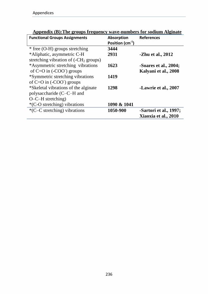
Appendices
236
Appendix (B):The groups frequency wave-numbers for sodium Alginate
Functional Groups Assignments Absorption Position (cm-1)
References
* free (O-H) groups stretching 3444 *Aliphatic, asymmetric C-H
stretching vibration of (-CH2 groups) 2931 -Zhu et al., 2012
*Asymmetric stretching vibrations
of C=O in (-COO-) groups
1623 -Soares et al., 2004;
Kalyani et al., 2008
*Symmetric stretching vibrations
of C=O in (-COO-) groups
1419
*Skeletal vibrations of the alginate
polysaccharide (C–C–H and
O–C–H stretching)
1298 -Lawrie et al., 2007
*(C-O stretching) vibrations 1090 & 1041
*(C–C stretching) vibrations 1050-900 -Sartori et al., 1997;
Xiaoxia et al., 2010
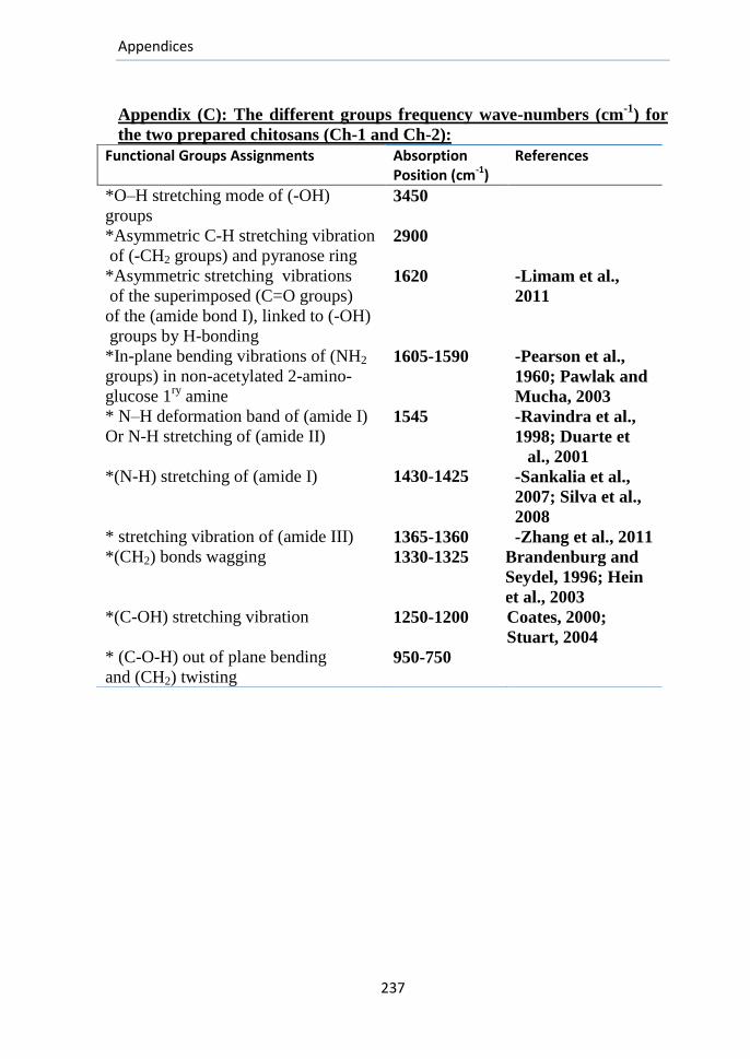
Appendices
237
Appendix (C): The different groups frequency wave-numbers (cm-1
) for
the two prepared chitosans (Ch-1 and Ch-2):
Functional Groups Assignments Absorption Position (cm-1)
References
*O–H stretching mode of (-OH)
groups 3450
*Asymmetric C-H stretching vibration
of (-CH2 groups) and pyranose ring 2900
*Asymmetric stretching vibrations
of the superimposed (C=O groups)
of the (amide bond I), linked to (-OH)
groups by H-bonding
1620 -Limam et al.,
2011
*In-plane bending vibrations of (NH2
groups) in non-acetylated 2-amino-
glucose 1ry
amine
1605-1590 -Pearson et al.,
1960; Pawlak and
Mucha, 2003 * N–H deformation band of (amide I)
Or N-H stretching of (amide II) 1545 -Ravindra et al.,
1998; Duarte et
al., 2001 *(N-H) stretching of (amide I) 1430-1425 -Sankalia et al.,
2007; Silva et al.,
2008 * stretching vibration of (amide III) 1365-1360 -Zhang et al., 2011 *(CH2) bonds wagging 1330-1325 Brandenburg and
Seydel, 1996; Hein
et al., 2003 *(C-OH) stretching vibration 1250-1200 Coates, 2000;
Stuart, 2004 * (C-O-H) out of plane bending
and (CH2) twisting 950-750
الملخص العربى
الملخص العربى
يعد الجلد أكبر أعضاء الجسم ويتكون من عدة طبقات التى تحمى العضالت والعظام واألعضاء الداخلية ولها عدة وظائف حسية وفسيولوجية مثل الحماية من فقد الماء المتزايد
قد الجلدية الجروح. بالجسم باإلضافة إلى التنظيم الحرارى للجسم والحماية من الطفيليات
أو ( الجروح المفتوحة)وتعريض الطبقات الداخلية للهواء الجلد طبقات من أى إزالة من تنتجالتئام الجروح يتم من خالل مجموعة (. الجروح المغلقة)نتيجة لصدمات داخلية بدون جرح
التجلط ومنع النزف، االلتهاب، تنشيط انقسام )من المراحل المتتابعة والمتداخلة وتشمل (.وأخيرا إعادة بناء التركيب العام للجلد المنزوع الخاليا،
تهدف هذه الدراسة إلى وضع طريقة جديدة لتحضير أفالم رقيقة من هالم مائى
الكيتوزان المستخلص من قشر ) صنوع من متضاعف اليكتروليتى مكون منم( هيدروجيل)واقتراح خطة من مجموعة من االختبارات (الجمبرى واأللجينات المحور اشعاعيا وبالتأكسد
التى تهدف الختيار أفضل هالم يمكن استخدامه فيما بعد كضمادة للجروح فى صورة دعامة للخاليا الجديدة إلعادة هيكلة كل من طبقة البشرة و األدمة فى الجلد المتمزق وتكون قادرة
الجى للهالم المختار على الجروح ثم يتم دراسة التأثير الع. على مدها بالتركيبات العالجيةالمفتوحة فى جلد الفئران من خالل مجموعة من الدراسات النسيجية والبيولوجية الجزيئية
عامل نمو )عبر قياس تعبير بعض الجينات المسئولة عن تكوين األوعية الدموية وتشمل (.بطانة األوعية الدموية وعامل فون ويلبراند
لجينات أوال من الشوائب وتعديل تركيبه الكيميائى باستخدام اإلشعاع وقد تم تنقية األ
الجامى واألكسدة المتعمدة باالضافة إلى استخالص الكيتوزان من قشر الجمبرى وتم الدمج بينهما بعدة صور بطريقة جديدة لتحضير عدة تركيبات مشععة تم المفاضلة بينها من خالل
اختبارات االنتفاخ، التشتت، التوافق مع الدم، معدل )مجموعة من االختبارات التى تشمل وكذلك تم دراسة تأثير االشعاع على مواصفات التركيبات المختارة ( تبخير الماء والتحلل
تم اختبارها على الجروح المفتوحة فى ثمواختيار أفضل تركيبة وبنسب معينة بين مكوناتها .جلد الفئران
المكونة من F) -02)المختلفة السابقة تم اختيارالهالم اعتمادا على االختبارات
ذات الوزن الجزيئى )المستخلص ومخلوط األلجينات ( Ch-1)الكيتوزان VLMW-50)ذات الوزن الجزيئى المنخفض جدا بواسطة االشعاع +(HMW)العالى
KGy)+ االلجينات المؤكسد(MASA-I A) (1:1:1)بالنسب).
هالم المختار على نوع من الجروح الحادة فى الفئران والتى تم من تم أوال تجربة ال :مجموعات( 3)خاللها تقسيم الفئران المجروحة إلى
تم معالجة الجرح الظهرى فى جلد كل فأر من هذه المجموعة بتركيبة : المجموعة االولى- .الهالم المختارة
الملخص العربى
فأر بكريم الفيوسيدين تم معالجة الجرح الظهرى فى جلد كل: المجموعة الثانية-لم يتم معالجة الجرح الظهرى (: Control-1)( )1)المجموعة الضابطة )المجموعة الثالثة -
.ألى من هذه الفئران وتركت لتلتئم بدون أى معالجات
ها بالمجموعتين األخرتين على جلد الفئران ومقارنت تأثير الهالميمكن تلخيص نتائج و :يلى كما
الثالثة حيث تم معدل إغالق الجرح فى المجموعة األولى أسرع من الثانية أسرع من ( 1)فى المجموعة بعد الجرحشر عحادى فى اليوم ال %( 89)بصورة شبة تامة اغالق الجرح
،أما فى المجموعة الثالثة فقد ( 13)األولى ،وفى المجموعة الثانية تم أقصى إغالق فى اليوم .%( 89)للجرح بعد أسبوعين من الجرح بمعدل وصل أقصى إغالق
تم مقارنة الجروح فى المجموعتين االولى والثالثة حيث : على المستوى النسيجى( 0)
أظهرت الدراسة بداية مرحلة االلتهاب فى كال المجموعتين مصحوبة بتكوين خاليا ليفية فى ة لطبقات الجلد المختلفة مع أنسجة الجروح المعالجة بالهالم فقط مع ظهور الخاليا المكون
البقايا المتفتتة من الهالم فى اليوم السابع فى حالة المجموعة األولى واكتمال تكوين طبقات أما فى حالة المجموعة . الجلد المختلفة بكامل مكوناتها وتنظيمها بعد أسبوعين من الجرح
.(19)الثالثة لم يحدث اكتمال للتكوين حتى اليوم ال عامل نمو بطانة )بمقارنة التعبير الجينى لكل من : بالنسبة لقياسات البيولوجيا الجزيئية(3)
فى المجموعتين األولى والثالثة أظهرت المجموعة ( األوعية الدموية وعامل فون ويلبراندعامل نمو بطانة األوعية )األولى تحسنا ملموسا أثناء حدوث االلتئام فى أغلب األيام فى حالة
مقارنة بالمجموعة الثالثة ولكن بدا التحسن الملموس من اليوم السابع فى حالة ( دمويةال .(عامل فون ويلبراند) بعد قياس وظائف الكلى للفئران من المجموعتين األولى والثالثة فى أيام االلتئام المختلفة (4)
جموعتين مما يدل لم يتم التوصل الى اختالف معنوى فى نسب الكرياتنين والبولينا بين المعلى ان تحلل مكونات الهالم فى جروح فئران المجموعة األولى لم تؤثر على النشاط
.الطبيعى الكلى وان التحلل ال يؤثر على الوظائف العامة للجسم

المستخلص العربى
المستخلص العربى
م مغلق، حاد أم ان الجرح، نوعه ماإذا كان مفتوح أنواع الجروح حسب المسبب، مكتختلف أ
مزمن، وكذلك فسيولوجية االلتئام وعليه يلزم اختيار نوع معين من الضمادات من بين عشرات
هذه الدراسة إلى تصميم نوع جديد من ضمادات هدفت. المتوفرة لكل نوع من الجروحاالنواع
وعمل االختبارات الالزمة على ( الكيتوزان و األلجينات)الجروح من نوعين من البوليمرات هما
المتراكب المتكون للتاكد من مواصفاته الفيزيائية والتركيبية والتحللية المفترضة ليقوم بتأثيره
اسة تأثيراته البيولوجية على الجروح الحادة فى الفئران من المساعد إلعادة بناء منطقة الجرح ثم در
النسيجى للجلد المنزوع ،وكذلك خالل قياس تأثيره على معدل اغالق الجروح ،وإعادة التركيب
قياس التعبير الجينى لبعض الجينات المسئولة عن تكوين األوعية الدموية خصوصا فى منطقة
.الجرح
الطيفية والفيزيائية والكيميائية تم اختيار نوع معين من المتراكبات والتى وبعد اجراء التحاليل
:أثبت التحليل االحصائى لتاثيره على الجرح الحاد النتائج التالية
سرع للجروح المعالجة بالهالم المختار عنه فى حالة الجروح المعالجة بالكريم عن معدل التئام أ-1
.الجروح الغير معالجة
تحسنا واضحا فى (المجموعة المعالجة بالهالم()1)أظهرت المجموعة : المستوى النسيجىعلى -2
(.الغير معالجة)االلتئام مقارنة بالمجموعة الضابطة
تحسنا ملموسا ( 1)اظهرت المجموعة : بالنسبة لقياسات البيولوجيا الجزيئية للجينات المختبرة-3
.الضابطة أثناء االلتئام فى اغلب األيام عن المجموعة
1)أثبتت قياسات وظائف الكلى تشابه النشاط الفسيولوجى وحيوية الكلى فى كال المجموعتين -4
(.والضابطة

دراسبت بيولوجية جزيئية لتقيين الدور العالجى للدعبهبت الوشععة على التقرحبت والجروح فى جلد الفئراى
رسبلة هقدهة هي الدارس
عبده أمير محمد محمد على بكبلوريوس علوم
(9002قسن الكيويبء والكيويبء الحيوية )
كمتطلب جزئى للحصول على
درجة الماجستير فى العلوم )تخصص كيمياء حيوية(
تحت إشراف
الدغيدى المنعم عبد اجالل/ د.أ ىمهد السيد محمد االسيد/ د.أ
المتفرغ الحيوية الكيمياء أستاذ الحيوية الكيمياء أستاذ االشعاعية البيولوجيا قسم حلوان جامعة-العلوم كلية عميدو
االشعاع وتكنولوجيا لبحوث القومى المركز
المصرية الذرية الطاقة هيئة
المزين محمد المنعم عبد حاتم/دأ.م. المغربى هللا عبد خالد طارق/ د.أ
الكيمياء الحيوية مساعدأستاذ أستاذ البيولوجيا الجزيئية
جامعة حلوان-كلية العلوم المركز القومى لبحوث وتكنولوجيا االشعاع
المصرية هيئة الطاقة الذرية
قسم الكيمياء
جامعة حلوان –كلية العلوم
2012

دراسبت بيولوجية جزيئية لتقيين الدور العالجى
الجروح للدعبهبت الوشععة على التقرحبت و
فى جلد الفئراى
رسبلة هقدهة هي الدارس
عبده أمير محمد محمد على
بكبلوريوس علوم
(9002قسن الكيويبء والكيويبء الحيوية )
جبهعة الونصورة
للحصول علىجزء متطلب ك
)تخصص كيمياء حيوية(درجة الماجستير فى العلوم
لىإ
جبهعة حلواى-كلية العلوم
قسن الكيويبء
9002
Copyright © 2022 FDOKUMEN