AB01 Silicon, Zener, LED Diode Characteristics Operating ...

Journal of Photochemistry and Photobiology B: Biology 118 (2013) 42–48
Contents lists available at SciVerse ScienceDirect
Journal of Photochemistry and Photobiology B: Biology
journal homepage: www.elsevier .com/locate / jphotobiol
Temperature measurement and Hsp47 immunoexpression in oral ulcersirradiated with defocused high-energy diode laser
Mayra Torres Vasques a, Marco Aurélio Verlangieri Alves a, Carolina Benetti b, Ana Cecília Correa Aranha c,Denise Maria Zezell b, Luciana Corrêa a,⇑a Department of Pathology, School of Dentistry, University of São Paulo, Av. Prof. Lineu Prestes, 2227, Cidade Universitária, 05508-000 São Paulo, SP, Brazilb Department of Biophotonics, Nuclear and Energy Research Institute, IPEN-CNEN/SP, Av. Prof. Lineu Prestes, 2242, Cidade Universitária, 05508-000 São Paulo, SP, Brazilc Department of Restorative Dentistry/Special Laboratory of Lasers in Dentistry (LELO), School of Dentistry, University of São Paulo, Av. Prof. Lineu Prestes, 2227,Cidade Universitária, 05508-000 São Paulo, SP, Brazil
a r t i c l e i n f o a b s t r a c t
Article history:Received 8 March 2012Received in revised form 16 October 2012Accepted 28 October 2012Available online 22 November 2012
Keywords:Low energy laserHeat shock protein 47Oral ulcerWound repair
1011-1344/$ - see front matter � 2012 Elsevier B.V. Ahttp://dx.doi.org/10.1016/j.jphotobiol.2012.10.014
⇑ Corresponding author. Tel.: +55 11 3091 7884.E-mail address: [email protected] (L. Corrêa).
Heat shock proteins (HSPs) are conservative protective proteins responsible for protein integrity duringtranscription in the cell under stress. Hsp47 is one of the most important chaperonins for collagen syn-thesis and release, and is up-regulated during wound repair. The aim of this study was to verify whetherdefocused high-energy diode laser (DDL) causes sufficient increase in local temperature to cause Hsp47up-regulation during repair of oral ulcers. Chemically-induced ulcers in the rat tongue, and non-ulceratedtongue mucosa were irradiated using a high energy diode laser (non-contact – 4 mm from surface,500 mW, 10 Hz for 40 s, energy density 80 J/cm2, fixed ulcer area of 0.25 cm2). Afterwards the specimenswere submitted to immunohistochemical test for Hsp47. Temperature oscillation during DDL irradiationwas also measured using a thermographic camera. Irradiated specimens exhibited transient mild increasein local temperature and significant up-regulation of Hsp47 in the mucosa from the superficial region(p = 0.035) to 1.7 mm deep (p = 0.049). In the deepest region of the mucosa Hsp47 was up-regulated onlyin ulcerated specimens mainly at 24 h (p = 0.049) and 72 h (p = 0.029) after ulcer induction. Conclusion:DDL increases local temperature and Hsp47 expression, which may contribute to wound repair byimprovement collagen synthesis and release.
� 2012 Elsevier B.V. All rights reserved.
1. Introduction
Heat shock proteins (HSPs) constitute a family of conservativeprotective proteins responsible for protein integrity during tran-scription, preventing aggregate formation in the cell under stressconditions. Induction of these molecules is started by a broad spec-trum of stressors, such as hyper/hypothermia, ischemia/reperfu-sion, hypoxia/hyperoxia, energy depletion, acidosis, viralinfection, and reactive oxygen species/reactive nitrogen species[1]. The protective effect of HSP seems to lead to improvement inwound healing, especially in the normal reparative process [2]. Inthe repair of oral and gastrointestinal ulcers, some HSPs, such asHsp27, Hsp47, and Hsp70 are up-regulated [3,4]. This up-regula-tion is associated with cell proliferating stimuli and protein syn-thesis, mainly collagen, and is restricted to specific cells, such askeratinocytes, endothelial cells, and fibroblasts [5]. Hsp47 is theHSP directly linked to collagen synthesis and is released by fibro-blasts, and is one of the most important constitutive and inducibleHsp for tissue repair [6].
ll rights reserved.
Thermal lasers used to stimulate tissue repair also seem to in-duce the expression of HSP during wound repair. Mild heat produc-tion (about 45–50 �C) associated with CO2 laser irradiation of theskin using controlled hyperthermia is correlated with increase inHsp70 expression, which in turn could play a role in the coordi-nated expression of growth factors, such as tumor growth factor b[7]. HSP, up-regulated during laser irradiation, has been consideredone of the main protein factors that contribute to wound repair.
High power diode laser can be used as low power laser when itis defocused. The advantages of this adaptation are to enable bothhigh and low power functions with same laser device. Defocuseddiode laser (DDL) is based on physical parameters that produceonly biomodulatory effects on the tissue without thermal damageor ablation. DDL has been applied to induce analgesia and acceler-ation of wound repair, mainly of minor aphthous stomatitis [8],oral mucositis [9], and labial herpes lesions [10]. It is not knownwhether DDL irradiation increases the inducible HSP and if thisinduction is present in a favorable scenario in tissue repair. It isalso not clear if this induction could be associated with increasein local temperature.
In this study we tested the hypothesis that DDL irradiationincreases local temperature and this thermal oscillation may be

M.T. Vasques et al. / Journal of Photochemistry and Photobiology B: Biology 118 (2013) 42–48 43
related to change in Hsp47 expression in ulcerated and non-ulcer-ated rat tongues. Therefore, the aim of this study was to measurethe in vitro DDL temperature oscillation in irradiated tongue ulcersand to analyze in vivo the wound repair process and Hsp47 expres-sion in different experimental periods.
2. Material and methods
The methodological steps described below were approved byEthical Committee in Animal Experimental Research of our institu-tion and are in accordance with International Ethical Committee ofAnimal Research.
2.1. Experimental groups for in vivo analysis
Fifty female rats (Rattus norvergicus, Wistar), with body weightof 200–250 g, 4 months of age, were randomly separated into fourgroups, as follows: (a) Laser treated, ulcerated group (LU) – 20 ani-mals with ulcers induced in the ventral tongue, and afterwardstreated with 810 nm diode laser; (b) Laser untreated, ulceratedgroup (UU) – 20 animals with ulcers induced in the ventral tonguebut without treatment afterwards; (c) Laser treated, non-ulceratedgroup (LG) – 5 animals with 810 nm laser application on the nor-mal surface (non-ulcerated) of ventral tongue mucosa; (d) Laseruntreated, non-ulcerated group (UG) – 5 control animals, withoutulceration and laser treatment.
Zero point of the experiment was 24 h after acid-induced ulcers.The animals with ulceration were euthanized at zero point and 24,48, 72, and 120 h (5 animals per experimental period). All the con-trol animals (LG and UG) were euthanized only at 120 h.
2.2. Ulcer induction
Ulcers were induced in the ventral surface of the tongue follow-ing a protocol adapted from Fujisawa et al. [11]. The animals wereanesthetized with an intraperitoneal injection of ketamine (Dopa-len�, Vetbrands, Paulinia, SP, Brazil) and xylazine (Anasedan�,Vetbrands, Paulinia, SP, Brazil) (0.1 ml/kg and 0.01 ml/kg, respec-tively). The tongue was pulled out of the mouth and a filter paperof 5 mm � 5 mm containing 20 ll of acetic acid 50% was applied tothe ventral surface for 60 s. The animals were kept in individualcages with commercial feed and water ad libitum. This acid appli-cation induces formation of blisters on the ventral epithelium thatlater rupture and ulcerate. Confirmation of the ulcer was per-formed after 24 h of the acid treatment, time considered as zeropoint of the experiment.
2.3. Diode laser irradiation for in vivo experiment
DDL irradiation (ZAP Lasers�, Pleasant Hill, CA, USA, 810 nm)was performed at zero point of the experiment, at a distance of4 mm from the ulcer surface (no contact) and perpendicular to it.The parameters used were adapted from Bello-Silva et al. [10] fororal herpetic lesions. Irradiation was restricted to the ulcer area,in continuous-wave mode, with 500 mW and 10 Hz pulse fre-quency for 40 s using horizontal scanning movements (energy den-sity 80 J/cm2, considering a fixed ulcer area of 0.25 cm2). For eachirradiation, the output power was measured with a power meter(Coherent Molectron�, Santa Clara, CA, USA). Irradiations were per-formed by a single operator. The animals were briefly maintainedunder sedation during irradiation, to facilitate access to the lesion.The same sedation was applied to non-laser groups.
2.4. Euthanasia and tongue processing
The animals were euthanized with a lethal dose of ketamine(Dopalen�, Vetbrands, Paulinia, SP, Brazil). Immediately after this,the tongue was extirpated and fixed in buffered formalin for24 h. After this a longitudinal cut was made through the largestdiameter of the ulcer, and the two fragments were submitted toroutine tissue processing and embedded in paraffin. Five 3 lm his-tological slices were obtained from each tongue fragment, totaling50 slices for each experimental period. Two slices were randomlychosen and stained with hematoxylin-eosin; the other three sliceswere stretched on glass slides, treated with 3-aminopropyltrietox-ysilane (3-APTS) and submitted to immunohistochemical tests.
2.5. Immunohistochemical tests
The streptavidin–biotin–peroxidase technique was used for thetests with polyclonal antibodies against Hsp47 (Abcam, Cam-bridge, MA, USA). The slides were dewaxed and rehydrated in aseries of descending grades of alcohol. Slides were then subjectedto endogenous tissue peroxidase blocking. Incubation was per-formed in primary antibody at a dilution of 1:1000. Afterwardsthe samples were incubated with a biotinylated swine-anti-rab-bit/goat antibody, as well as a streptavidin–biotin peroxidase con-jugate (LSAB System, Dako�, Carpenteria, CA, USA) for 30 min each.The reaction was then revealed by diaminobenzidine (Dako�, Car-penteria, CA, USA); the sections were stained with Mayer hematox-ylin, dehydrated in a series of increasing grades of alcohol,immersed in xylol, and mounted in resin for conventional lightmicroscopy. For the negative control, sections were incubated ina buffer without primary antibody.
2.6. Histological analysis and semi quantitative analysis
The repair process was analyzed by observation of the hematox-ylin-eosin stained ulcer slides. The intensity of the following tissueelements was considered: thermal damage, edema, hyperemia,inflammation, necrosis, young fibroblasts, angiogenesis, re-epithe-lialization and collagenization. The intensity was classified accord-ing the percentage of tissue element in the field: 0 = absent (0%);1 = mild (0.1–25%); 2 = mild to moderate (26–50%); 3 = moderateto intense (51–75%); and 4 = intense (76–100%). Immunohisto-chemical expression of Hsp47 was evaluated by means of theabove intensity classification observed in the following tissue re-gions: layers of preexistent (2 mm distant from the ulcer) and new-ly formed (migrating) epithelium; fibroblasts; vessel wall cells;inflammatory cells; extracellular matrix; skeletal muscle fibers.These two analyses were blinded and performed by two patholo-gists. The histological pattern corresponding to each graduationwas established beforehand by the two pathologists using a repre-sentative sample of histological slices.
2.7. Positive cell counts for Hsp47 immunolabeling
Positive cells for Hsp47 immunolabeling were counted in orderto achieve an objective index of Hsp expression. Hsp47 expressionwas correlated with the change in temperature data obtained in anex vivo experiment (see below). Three regions of the tongue wereanalyzed: A – at 0.5 mm from ulcer surface (in the lamina propriaof ventral tongue); B – at 1.5–1.7 mm from ulcer surface (in thesubmucosa of ventral tongue); C – at 2.7–3.0 mm from ulcer sur-face (in the lamina propria of dorsal tongue, i.e., immediately be-low the specialized epithelium of the dorsal tongue) (Fig. 1).Only the Hsp47 positive cells with fibroblast morphology werecounted by one (blinded) pathologist. Vessel wall cells and inflam-matory cells were not included in the count. Eight fields at �400

Fig. 1. Lateral view of a half tongue specimen under thermographic cameraanalysis. Temperature was measured in the points localized in the apical region(thin arrow) of the tongue irradiated with defocused diode laser. This image wasobtained immediately after the laser irradiation.
44 M.T. Vasques et al. / Journal of Photochemistry and Photobiology B: Biology 118 (2013) 42–48
original magnification of each region (area of 24,816 lm2) in threehistological slices of each animal were digitized and analyzed bymeans of morphometric software using a manual counting tool.
2.8. Ex vivo temperature measurement during laser irradiation
Additional ventral ulcers were induced in 6 animals for ex vivoanalysis of temperature oscillation during DDL irradiation. Afterthese animals were euthanized, the ulcerated tongues were extir-pated at zero point and then frozen at �20 �C. At the time of tem-perature measurement, the tongues were thawed in a water bathuntil the temperature rose to 22 �C, and then they were cutthrough the longitudinal axis in the middle of the ulcer. This cutwas made to obtain the temperature oscillation in the deep region.To confirm that the tongue was thawed, its initial temperature wasmeasured with a thermographic camera (ThermaCam FLIR SC3000Systems, Boston, MA, USA) with accuracy: 0.001 �C and responsetime: 0.02 s. The camera was set to 60 Hz capture frequency andtemperature range of �5 �C to +98 �C. Recording was done at21.5 �C room temperature and 50% relative humidity. Emittanceof the tongue was considered as 0.98, the same as for muscle tissue[12] since most of the tongue volume was composed of striatedmuscle. After the first temperature measurement, the specimenswere fixed in a plate and the ulcers were irradiated with the sameDDL parameters as those used in the in vivo experiment, and alsoperformed by the same operator. The laser power emission was
Fig. 2. Temperatures measured in ex vivo experiments, previous (0
previously checked with a Power Meter (Coherent Molectron�,Santa Clara, CA, USA). During irradiation, the temperature oscilla-tion was recorded by the thermographic camera. The specimenswere positioned at 10 cm from camera lens, in order to obtain fo-cus on the lateral surface of the tongue. Perpendicular points inrelation to irradiated area were analyzed using the thermographiccamera software (ThermaCam�, ThermaCam Research 2001, Bos-ton, MA, USA). First point was on the ulcer surface, and anothersix points were measured at a depth of 0.5 mm from this surface(Fig. 2). These points were coincident with those analyzed in thecell counts. One software operator calculated the temperaturevalues at the points in accordance with the irradiation time (30 sbefore, 40 s during, and 30 s after the laser irradiation).
The thermographic analysis described above was alsoperformed in vivo at the superficial point in order to verify if thetemperature variation was similar to the in vitro tests.
2.9. Statistical tests
Descriptive statistical analysis was performed for all the vari-ables using median, medium, standard-deviation, and minimum–maximum values. The Mann–Whitney test was used for numericaldata in semi quantitative analysis of the repair process analysis andof Hsp47 immunohistochemical labeling. For the cell counts, theKruskal–Wallis variance analysis was used to detect differencesconsidering all the groups together. The Mann–Whitney test wasapplied for the pairs of groups, and Friedman’s test was adoptedto identify differences between the experimental periods and his-tological regions analyzed (A, B, and C) for each group individually.Calculations were made using SPSS� software (IBM, NY, USA). Thelevel of Significance was 5%.
3. Results
3.1. Temperature variation during laser irradiation
There was variation in the tongue temperature during DDL irra-diation in all the points. Fig. 2 shows an example of a standardgraph of the temperature curve observed in all the specimens.The initial temperature (the first 30 s) was about 22 �C. The graphshows there was a temperature peak at 30 s (when irradiationstarted) from the ulcer surface to 1 mm deep. In the deepest re-gions, this oscillation was minimal at this time. From 40 s to 70 s,progressive temperature increase was observed in all the points,except the surface, which maintained the mean values presentedat 40 s. The temperature declined progressively from 70 s (when
–30 s), during (30–70 s), and after (70–100 s) laser irradiation.

Table 1Average (±standard error) of temperature variation according to the different depth ofmeasured points.
Depth of measured points Temperature variation (�C)
Surface 6.70 ± 0.500.5 mm 5.20 ± 0.601.0 mm 4.30 ± 0.501.5 mm 3.20 ± 0.302.0 mm 2.85 ± 0.102.5 mm 2.77 ± 0.23
M.T. Vasques et al. / Journal of Photochemistry and Photobiology B: Biology 118 (2013) 42–48 45
laser irradiation stopped) to the end of the experiment (100 s), butonly the surface point achieved the initial mean temperature val-ues. The other points showed about +1 �C temperature at 100 s.The temperature impairment was more pronounced in the surfacepoint. Table 1 shows the temperature variation means during laserirradiation in the analyzed points for the six tongues. The lowestvariation values were observed at deeper points (about 2.5 �C).The highest variation (about 7.20 �C considering the standard er-ror) was present in the surface point. This variation was similarto that observed in vivo test (8.12 �C), without statistically signifi-cant differences (p = 0.500). Considering the maximum tempera-ture value, there was significant difference between the surfacepoint and deepest point (p = 0.0455).
3.2. Analysis of the repair process
Table 2 contains the mean values of semi quantitative analysisfor each tissue element during ulcer repair with or without DDLirradiation. At 0 h there were no significant differences betweenthe groups. The tongue mucosa showed an ulcerated epitheliumcovered by necrotic tissue with intense inflammatory exudatecomposed mainly of neutrophils. Moderate to intense hyperemiaand edema were observed in the lamina propria. Discrete to mod-erate angiogenesis was noted mainly in the ulcer basis. At 24 hgranulation tissue, angiogenesis, and collagen deposition in ulcerstreated with laser were significantly more intense than in non-treated ulcers. However these differences were not maintained inthe other experimental periods, in which the tissue elements weresimilar (Fig. 3A and B). At 120 h, 97% of the ulcers in both groupshad undergone complete re-epithelialization, with a large number
Table 2Median (minimum–maximum) values in semiquantitative analysis for ulcer repair in the
Experimental period 0 h 24 h
Groups UU LU UU LU
Thermal damage 0 0 0 0(0–0) (0–0) (0–0) (0–0)
Edema 4 3 3 3(4–4) (2–4) (2–3) (2–3)
Hyperemia 3 3 3 3(3–4) (3–3) (2–3) (2–3)
Inflammatory infiltrate 4 4 3 3(4–4) (4–4) (2–3) (2–3)
Necrosis 3 3 3 3(3–3) (3–3) (3–3) (2–3)
Granulation tissue 0 0 1 2(0–0) (0–0) (1–1) (2–2)
Angiogenesis 1 2 2 3(1–2) (2–2) (2–2) (3–3)
Reepithelization 1 1 2 2(1–1) (1–1) (2–2) (2–2)
Collagen deposition 0 0 1 2(0–0) (0–0) (1–1) (2–2)
0 = absent (0%); 1 = mild (0.1–25%); 2 = mild to moderate (26–50%); 3 = moderate to intestatistical differences (Mann–Whitney test, p < 0.05).
of fibroblasts and mature granulation tissue in the lamina propria(Fig. 3C and D). No difference in speed of repair was observed be-tween the groups.
3.3. Analysis of Hsp47 immunolabeling
Table 3 contains the data of semiquantitative analysis for Hsp47immunolabeling in laser treated and non-treated ulcers. Inflamma-tory cells, muscle fibers, and epithelium >2 mm distant from theulcer were negative. In general the intensity of Hsp47 expressionincreased with time, mainly in migrating epithelium, fibroblasts,vessel wall, and extracellular matrix. The effects of laser irradiationon Hsp47 expression was early observed at 0 h, when the immu-nolabeling in the ulcers treated with laser was significantly stron-ger than that in the non-treated ulcers. At 24 h, the ulcers treatedwith laser exhibited moderate to intense Hsp47 expression forfibroblasts and vessel wall, which was significantly more intensein comparison with the non-treated ulcers (Fig. 3E and F). Thestrong positivity in fibroblasts was maintained in both groups until120 h without significant differences (Fig. 3G and H).
3.4. Hsp47 positive cell counts
Fig. 4 shows the average number of Hsp47 positive cells in non-ulcerated groups (LG and UG). The number of positive cells de-creased from region A (at 0.5 mm from epithelial surface) to regionC (at 2.7–3.0 mm from epithelial surface) with significant differ-ences between these regions only in the laser-irradiated group(p = 0.039). In the comparison of groups, the number of positivecells was significantly higher in irradiated than in non-irradiatedspecimens considering the regions A (p = 0.035) and B (p = 0.049).
Figs. 5–7 show the distribution of Hsp47 positive cells in rela-tion to experimental periods of ulcerated groups (LU and UU) foreach histological region (A, B, and C). For region A (at 0.5 mm fromepithelial surface) (Fig. 5), at 0 h, the number of positive cells wassignificantly higher in the laser-irradiated than in the non-irradi-ated group (p = 0.049). At 120 h this condition was inverted, withsignificantly higher frequency of positive cells in non-irradiatedgroup (p = 0.039). The average number of positive cells increasedfrom 0 h to 72 h with peak at 120 h in non-irradiated groups,whereas in irradiated group the peak was at 72 h. For region B(at 1.5–1.7 mm from epithelial surface) (Fig. 6) there was also a
laser treated- (LU) and laser untreated- (UU) ulcerated groups.
48 h 72 h 120 h
UU LU UU LU UU LU
0 0 0 0 0 0(0–0) (0–0) (0–0) (0–0) (0–0) (0–0)3 2 2 2 1 0(2–3) (2–3) (2–2) (2–2) (0–1) (0–1)2 3 2 2 2 2(1–3) (3–3) (1–2) (2–2) (2–2) (2–2)3 2 2 2 0 0(3–3) (2–3) (1–2) (2–2) (0–1) (0–0)2 2 1 1 0 0(2–2) (2–3) (1–2) (0–1) (0–1) (0–0)2 3 4 4 4 4(2–2) (2–3) (4–4) (4–4) (4–4) (4–4)3 3 3 3 3 3(3–3) (3–3) (3–3) (3–3) (3–3) (3–3)2 2 3 3 4 4(2–2) (2–2) (3–3) (3–3) (3–4) (3–4)2 3 3 3 4 4(2–2) (2–3) (3–3) (3–3) (3–4) (4–4)
nse (51–75%); and 4 = intense (76–100%). Pair of values in the bold had significant

Fig. 3. Histological sections of oral ulcers from laser-treated and laser-untreated groups. (A–D): Hematoxylin-eosin stain (�100). Intense collagen deposition andangiogenesis in the laser-treated group (B) in comparison with laser-untreated group (A) at 24 h. Similar reepithelization degree and connective tissue remodeling at 120 hfor both laser-untreated (C) and laser-treated (D) groups. (E–H): Streptoavidin–biotin stain (�100). Intense Hsp47 expression at ulcer basis 24 h after laser irradiation (E) inthe comparison with laser-untreated ulcer (F). Similar Hsp47 expression in the both laser-untreated (G) and laser-treated (H) at 120 h.
Table 3Median (minimum–maximum) values in semiquantitative analysis for Hsp47 immunolabeling in the laser treated- (LU) and laser untreated- (UU) ulcerated groups.
Experimental period 0 h 24 h 48 h 72 h 120 h
Groups UU LU UU LU UU LU UU LU UU LU
Preexistent epithelium 0 3 2 2 2 1 1 2 0 1(0–0) (0–3) (2–3) (1–3) (1–2) (1–1) (1–2) (2–2) (0–1) (0–2)
Migrating epithelium 1 2 2 3 3 3 3 3 2 2(1–1) (2–2) (2–2) (2–3) (2–3) (3–4) (3–3) (3–4) (1–2) (2–3)
Fibroblasts 2 3 3 4 4 4 3 4 3 4(1–2) (3–3) (3–3) (4–4) (4–4) (4–4) (3–4) (3–4) (3–4) (3–4)
Vessel wall 1 2 2 3 3 3 2 3 2 2(0–1) (1–2) (2–2) (3–3) (3–3) (3–3) (2–3) (3–4) (2–3) (2–3)
Extracellular matrix 0 1 2 2 3 3 2 3 1 1(0–0) (1–1) (0–2) (1–2) (3–3) (1–3) (2–3) (2–3) (1–1) (1–3)
0 = absent (0%); 1 = mild (0.1–25%); 2 = mild to moderate (26–50%); 3 = moderate to intense (51–75%); and 4 = intense (76–100%). Pair of values in the bold had significantstatistical differences (Mann–Whitney test, p < 0.05).
46 M.T. Vasques et al. / Journal of Photochemistry and Photobiology B: Biology 118 (2013) 42–48
higher average number of Hsp47 positive cells in the laser-irradi-ated than in the non-irradiated group at 0–72 h. The differenceswere statistically significant between the groups at 24–72 h
(p = 0.049). At 120 h, the number of the positive cells was similarin the groups. In region C (at 2.7–3.0 mm from epithelial surface)(Fig. 7), the average number of positive cells in laser-irradiated
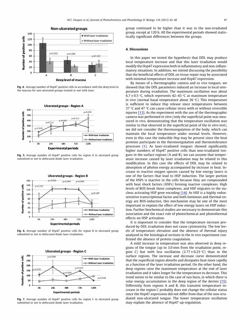
Fig. 4. Average number of Hsp47 positive cells in accordance with the deep level inthe mucosa for non-ulcerated groups treated or not with laser.
Fig. 6. Average number of Hsp47 positive cells for region B in ulcerated groupsubmitted or not to defocused diode laser irradiation.
Fig. 5. Average number of Hsp47 positive cells for region A in ulcerated groupsubmitted or not to defocused diode laser irradiation.
Fig. 7. Average number of Hsp47 positive cells for region C in ulcerated groupsubmitted or not to defocused diode laser irradiation.
M.T. Vasques et al. / Journal of Photochemistry and Photobiology B: Biology 118 (2013) 42–48 47
group continued to be higher than it was in the non-irradiatedgroup, except at 120 h. All the experimental periods showed statis-tically significant differences between the groups.
4. Discussions
In this paper we tested the hypothesis that DDL may producelocal temperature increase and that this laser irradiation wouldmodify the Hsp47 expression both in inflammatory and non-inflam-matory situations. In addition, we intend discussing the possibilitythat the beneficial effects of DDL on tissue repair may be associatedwith minimal temperature increase and Hsp47 expression.
By means of a thermographic camera and ex vivo tongues, weshowed that the DDL parameters induced an increase in local tem-perature during irradiation. The maximum oscillation was about6.7 ± 0.5 �C, which represents 42–43 �C as maximum temperaturein vivo (normal basal temperature about 36 �C). This temperatureis sufficient to induce Hsp release since temperatures between37 �C and 47 �C can cause cellular stress with or without reversibleinjuries [13]. As the experiment with the use of the thermographiccamera was performed in vitro (only the superficial point was mea-sured in vivo, demonstrating that the temperature oscillation wassimilar to that observed in the superficial point of the in vitro test),we did not consider the thermoregulation of the body, which canmaintain the local temperature under normal levels. However,even in this case the inducible Hsp may be present since the heatproteins participate in the thermoregulation and thermotoleranceprocesses [1]. As laser-irradiated tongues showed significantlyhigher numbers of Hsp47 positive cells than non-irradiated ton-gues in the surface regions (A and B), we can assume that temper-ature increase caused by laser irradiation may be related to thismodification. In this case the effects of DDL may be related toabsorption of photon energy accompanied by increase in heat. In-crease in reactive oxygen species caused by low energy lasers isone of the factors that lead to HSP induction. The larger portionof the HSPs is inactive in the cells because they are compoundedwith heat shock factors (HSFs) forming inactive complexes. Highlevels of ROS break these complexes, and HSF migrates to the nu-cleus activating HSP gene encoding [14]. As HSF is a highly redox-sensitive transcriptional factor and both luminous and thermal en-ergy are ROS-inductive, this mechanism may be one of the mostimportant to explain the effect of low energy lasers on HSP induc-tion. Further biochemical studies are necessary to demonstrate thisassociation and the exact role of photochemical and photothermaleffects on HSP activation.
It is important to consider that the temperature increase pro-duced by DDL irradiation does not cause cytotoxicity. The low lev-els of temperature elevation and the absence of thermal injuryanalyzed in the histological sections in the in vivo experiment con-firmed the absence of protein coagulation.
A mild increase in temperature was also observed in deep re-gions of the tongue (up to 3.0 mm from the irradiation point, re-gion C) but with less oscillation (2.77 ± 0.23 �C) than in thesurface regions. The increase and decrease curve demonstratedthat the superficial region absorbs and dissipates heat more rapidlyas a function of the laser irradiation period. On the other hand, thedeep regions raise the maximum temperature at the end of laserirradiation and it takes longer for the temperature to decrease. Thistrend seems to be similar to the case of sun burn, in which there issolar energy accumulation in the deep region of the dermis [15].Differently from regions A and B, this transient temperature in-crease in the region C probably does not change the cellular statussince the Hsp47 expression did not differ from that of the non-irra-diated non-ulcerated tongue. The lower temperature oscillationmay explain the absence of Hsp47 up-regulation.

48 M.T. Vasques et al. / Journal of Photochemistry and Photobiology B: Biology 118 (2013) 42–48
We also used an in vivo model with ulcerated tongues to ana-lyze whether the laser irradiation was superior to ulcer inflamma-tion in the induction of Hsp47. We observed that DDL producesstimulation of vessels, granulation tissue, and collagen depositionin the first periods of ulcer repair. The intense expression ofHsp47 in the fibroblasts, vessel wall, and extracellular matrix inthe same experimental periods contributes to explaining this stim-ulatory effect of DDL. Inducible Hsp47 has been correlated with tri-ple-helical formation of collagen with transient binding toprocollagen. Hsp47 is resident in the endoplasmatic reticulumand is the most important collagen-specific chaperone. It is consti-tutive in the oral mucosa only in the connective tissue, but duringoral mucosa repair some migrating keratinocytes can express thisprotein [5]. DDL induces the Hsp47 both in the connective tissueand in the migrating epithelium, and these expressions may com-pound the wide range of factors that explain the stimulatory ef-fects of low energy lasers.
Hsp47 expression was also analyzed in terms of tissue depth inthe in vivo analyses. Similarly to the ex vivo experiment, regions Aand B showed significantly more Hsp47 positive cells in the DDLgroup. However, there were differences regarding the chronologi-cal distribution of the positive cells. In region A, Hsp47 up-regula-tion was also brief and restricted to some minutes after laserirradiation, behavior that is coincident with the rapid heat dissipa-tion in this region. But in regions B and C, the number of Hsp47 po-sitive cells was significantly higher in the DDL group for most ofthe time (from 24 h to 48 h). This perpetuation of Hsp47 up-regu-lation in the comparison with non-irradiated ulcerated specimensmay be related both to the temperature oscillation curve discussedpreviously, and to the cascade of stress events promoted by laserirradiation. This cascade may promote an intense activation ofHsp induction, which is superior to ulcer inflammation alone.Therefore, we can say that the laser irradiation increases the natu-ral inflammation-evoked Hsp47 up-regulation during tissue repair.The results of Hsp47 expression in region C of the ulcerated spec-imens (about 3.0 mm from the surface) can confirm that Hsp47 up-regulation during tissue repair may be caused by laser irradiationin addition to the tissue inflammation. Considering the high pene-tration of the diode laser at 810 nm, the effects of irradiation arepotentially expected. Only in the ulcerated groups was the numberof Hsp47 positive cells higher in irradiated than non-irradiatedspecimens, which indicates that an additional input of for intenseHsp47 activation in the deeper regions of laser-irradiated biologi-cal tissue.
In conclusion, DDL within the described parameters can cause aslight increase in local temperature without the induction of ther-mal damage to the tissue. DDL irradiation can also increase theHsp47 expression both in non-ulcerated and ulcerated mucosa,which may contribute directly to ulcer repair by means of collagen
synthesis and release. The mild increase in local temperaturecaused by DDL could be associated with Hsp47 up-regulation.
5. Abbreviations
HSP
heat shock protein DDL defocused diode laserReferences
[1] K.C. Kregel, Heat shock proteins: Modifying factors in physiological stressresponses and acquired thermotolerance, J. Appl. Physiol. 92 (2002) 2177–2186.
[2] J.T. Kovalchin, R. Wang, M.S. Wagh, J. Azoulay, M. Sanders, R.Y. Chandawarkar,In vivo delivery of heat shock protein 70 accelerates wound healing by up-regulating macrophage-mediated phagocytosis, Wound Repair Regen. 14(2006) 129–137.
[3] J.S. Guo, C.H. Cho, J.Y. Wang, M.W. Koo, Expression and immunolocalization ofheat shock proteins in the healing of gastric ulcers in rats, Scand. J.Gastroenterol. 37 (2002) 17–22.
[4] M.P. Ebert, C. Schäfer, J. Chen, J. Hoffmann, P. Gu, C. Kubisch, et al., Protectiverole of heat shock protein 27 in gastric mucosal injury, J. Pathol. 207 (2005)177–184.
[5] M.T. Vasques, M.A. Alves, J.G. Cerqueira Luz, L. Corrêa, Immunolocalization ofheat shock proteins 27 and 47 during repair of induced oral ulcers, J. Oral Sci.52 (2010) 623–631.
[6] K. Nagata, N. Hosokawa, Regulation and function of collagen-specificmolecular chaperone, HSP47, Cell Struct. Funct. 21 (1996) 425–430.
[7] A. Capon, S. Mordon, Can thermal lasers promote skin wound healing?, Am J.Clin. Dermatol. 4 (2003) 1–12.
[8] N. Zand, L. Ataie-Fashtami, G.E. Djavid, M. Fateh, M.R. Alinaghizadeh, S.M.Fatemi, F. Arbabi-Kalati, Relieving pain in minor aphthous stomatitis by asingle session of non-thermal carbon dioxide laser irradiation, Lasers Med. Sci.24 (2009) 515–520.
[9] A. Simões, F.P. Eduardo, A.C. Luiz, L. Campos, P.H. Sá, M. Cristófaro, M.M.Marques, C.P. Eduardo, Laser phototherapy as topical prophylaxis against headand neck cancer radiotherapy-induced oral mucositis: Comparison betweenlow and high/low power lasers, Lasers Surg. Med. 41 (2009) 264–270.
[10] M.S. Bello-Silva, P.M. de Freitas, A.C. Aranha, J.L. Lage-Marques, A. Simões, C. dePaula Eduardo, Low- and high-intensity lasers in the treatment of herpessimplex virus 1 infection, Photomed. Laser Surg. 28 (2010) 135–139.
[11] K. Fujisawa, Y. Miyamoto, M. Nagayama, Basic fibroblast growth factor andepidermal growth factor reverse impaired ulcer healing of the rabbit oralmucosa, J. Oral Pathol. Med. 32 (2003) 358–366.
[12] T.C. Cetas, Practical thermometry with a thermographic camera-calibration,transmittance, and emittance measurements, Rev. Sci. Instrum. 49 (1978)245–254.
[13] P. Janda, R. Sroka, B. Mundweil, C.S. Betz, R. Baumgartner, A. Leunig,Comparison of thermal tissue effects induced by contact application of fiberguided laser systems, Lasers Surg. Med. 93 (2003) 93–101.
[14] H.G. Park, S.I. Han, S.Y. Oh, H.S. Kang, Cellular responses to mild heat stress,Cell Mol. Life Sci. 62 (2005) 10–23.
[15] F. Trautinger, I. Kindås-Mügge, R.M. Knobler, H. Hönigsmann, Stress proteins inthe cellular response to ultraviolet radiation, J. Photochem. Photobiol. B 35(1996) 141–148.
Copyright © 2022 FDOKUMEN