Characteristic spectroscopic properties of γ-irradiated rare-earth oxide-based ferrofluids
11
This article was downloaded by: [Tezpur University] On: 03 October 2012, At: 04:28 Publisher: Taylor & Francis Informa Ltd Registered in England and Wales Registered Number: 1072954 Registered office: Mortimer House, 37-41 Mortimer Street, London W1T 3JH, UK Journal of Experimental Nanoscience Publication details, including instructions for authors and subscription information: http://www.tandfonline.com/loi/tjen20 Characteristic spectroscopic properties of γ-irradiated rare-earth oxide-based ferrofluids Manasi Devi a , Nibedita Paul a , Dambarudhar Mohanta a & Abhijit Saha b a Nanoscience Laboratory, Department of Physics, Tezpur University, PO Napaam, Assam 784028, India b UGC-DAE Consortium for Scientific Research, Kolkata Centre, III/ LB-8 Bidhannagar, Kolkata 700098, India Version of record first published: 09 May 2012. To cite this article: Manasi Devi, Nibedita Paul, Dambarudhar Mohanta & Abhijit Saha (2012): Characteristic spectroscopic properties of γ-irradiated rare-earth oxide-based ferrofluids, Journal of Experimental Nanoscience, 7:5, 586-595 To link to this article: http://dx.doi.org/10.1080/17458080.2010.548408 PLEASE SCROLL DOWN FOR ARTICLE Full terms and conditions of use: http://www.tandfonline.com/page/terms-and- conditions This article may be used for research, teaching, and private study purposes. Any substantial or systematic reproduction, redistribution, reselling, loan, sub-licensing, systematic supply, or distribution in any form to anyone is expressly forbidden. The publisher does not give any warranty express or implied or make any representation that the contents will be complete or accurate or up to date. The accuracy of any instructions, formulae, and drug doses should be independently verified with primary sources. The publisher shall not be liable for any loss, actions, claims, proceedings, demand, or costs or damages whatsoever or howsoever caused arising directly or indirectly in connection with or arising out of the use of this material.
Transcript of Characteristic spectroscopic properties of γ-irradiated rare-earth oxide-based ferrofluids

This article was downloaded by: [Tezpur University]On: 03 October 2012, At: 04:28Publisher: Taylor & FrancisInforma Ltd Registered in England and Wales Registered Number: 1072954 Registeredoffice: Mortimer House, 37-41 Mortimer Street, London W1T 3JH, UK
Journal of Experimental NanosciencePublication details, including instructions for authors andsubscription information:http://www.tandfonline.com/loi/tjen20
Characteristic spectroscopic propertiesof γ-irradiated rare-earth oxide-basedferrofluidsManasi Devi a , Nibedita Paul a , Dambarudhar Mohanta a & AbhijitSaha ba Nanoscience Laboratory, Department of Physics, TezpurUniversity, PO Napaam, Assam 784028, Indiab UGC-DAE Consortium for Scientific Research, Kolkata Centre, III/LB-8 Bidhannagar, Kolkata 700098, India
Version of record first published: 09 May 2012.
To cite this article: Manasi Devi, Nibedita Paul, Dambarudhar Mohanta & Abhijit Saha (2012):Characteristic spectroscopic properties of γ-irradiated rare-earth oxide-based ferrofluids, Journalof Experimental Nanoscience, 7:5, 586-595
To link to this article: http://dx.doi.org/10.1080/17458080.2010.548408
PLEASE SCROLL DOWN FOR ARTICLE
Full terms and conditions of use: http://www.tandfonline.com/page/terms-and-conditions
This article may be used for research, teaching, and private study purposes. Anysubstantial or systematic reproduction, redistribution, reselling, loan, sub-licensing,systematic supply, or distribution in any form to anyone is expressly forbidden.
The publisher does not give any warranty express or implied or make any representationthat the contents will be complete or accurate or up to date. The accuracy of anyinstructions, formulae, and drug doses should be independently verified with primarysources. The publisher shall not be liable for any loss, actions, claims, proceedings,demand, or costs or damages whatsoever or howsoever caused arising directly orindirectly in connection with or arising out of the use of this material.

Journal of Experimental NanoscienceVol. 7, No. 5, September–October 2012, 586–595
Characteristic spectroscopic properties of c-irradiated rare-earth
oxide-based ferrofluids
Manasi Devia, Nibedita Paula, Dambarudhar Mohantaa* and Abhijit Sahab
aNanoscience Laboratory, Department of Physics, Tezpur University, PO Napaam, Assam 784028,India; bUGC-DAE Consortium for Scientific Research, Kolkata Centre, III/LB-8 Bidhannagar,Kolkata 700098, India
(Received 11 October 2010; final version received 12 December 2010)
Ferrofluids are being considered as potential candidates both in basic and appliedresearch owing to their novel optical and magneto-optical properties. We havesynthesised surfactant (N-Cetyl-N,N,N-trimethylammonium bromide, CTAB)-coated nanoscale gadolinium oxide (Gd2O3) based ferrofluids and then irradiatedby gamma (�-) rays with doses in the range of 32Gy–2.635 kGy. High-resolutiontransmission electron microscope (HRTEM) analysis shows that the particleshave developed intragranular defects owing to �-ray irradiation. Fouriertransform infrared (FT-IR) and photoluminescence (PL) studies also supportthe formation of defect ordering upon irradiation. Further, PL study indicatesabrupt change of the symmetry factor with increase in �-dose. By viewing thenature of variation between relative intensity of the defect-related emission anddose-dependent symmetry factor, one can predict the tunability of PL response. Aproper understanding of the PL response of the irradiated nanoscale rare-earthoxides would find new avenues for lasing and other optoelectronic/photonicdevices.
Keywords: ferrofluid; gamma irradiation; photoluminescence; defect
1. Introduction
The growing interest of low-dimensional materials with respect to their bulk counterpartsdwells on their unique electronic, magnetic, optical and mechanical properties. In therecent decades, nanoscience and nanotechnology have emerged as a thrilling trend both inthe field of basic research and industrial application [1,2]. Magnetic nanoparticle (MNP)systems have attracted a great deal of attention for their size-dependent functionality andapplicability in making integrated/hybrid structures. Ferrofluid, a colloidal dispersion ofMNPs, has several advantages as it displays tunable viscoelastic and optical propertiesin the presence of a static magnetic field (SMF) [3]. The most commonly used ferrofluidsgenerally comprise superparamagnetic magnetite (Fe3O4) or, maghemite (�-Fe2O3)particles which are dispersed in a suitable carrier fluid (oil, water, etc.). Ferrofluids havepotential in the area of drug targeting [4], sealing [5], shielding [6], optical limiting devices
*Corresponding author. Email: [email protected]
ISSN 1745–8080 print/ISSN 1745–8099 online
� 2012 Taylor & Francis
http://dx.doi.org/10.1080/17458080.2010.548408
http://www.tandfonline.com
Dow
nloa
ded
by [
Tez
pur
Uni
vers
ity]
at 0
4:28
03
Oct
ober
201
2

[7], etc. Also, ferrofluids are widely used in aircrafts and space shuttles. It has been foundthat the rare-earth (RE) oxide materials exhibit lower toxicity in comparison to severaltypes of quantum dots (Cd, Se, Tb, Hg, etc.) [8]. There exists ample scope for thedeployment of RE oxide-based ferrofluids as imperative fluorescent/drug targeting agents.Further, owing to better environmental stability and durability, RE oxide-basedferrofluids emerge as alternative candidate with immense technological relevance insealing and shielding applications.
Gamma (�) rays are energetic electromagnetic radiations like X-rays. They are themost energetic form of electromagnetic radiation. �-radiation is most common in outerspace environment. This type of radiation has mixed impact on different materials. Thereare reports on the effect of this type of radiation on the lasing performance of Nd, Cr:GSGG (Cr3þ-doped gadolinium scandium gallium garnet) crystals. It was found that thesecrystals retain high threshold for laser damage up to �-ray doses of 1MGy [9]. However,on exposure of such radiation, colour centres are induced which strengthen thefluorescence property [10]. Previously, Mak et al. [11] have observed shifting ofphotoluminescence (PL) peak after �-irradiation in ZnSe crystals. They correlated thepeak-shifting with the band-gap variation and ascribed it to radiation stimulated solid-state recrystallisation and accumulation of point defects. In RE-doped alkaline earthsulphates, the effect of �-irradiation was shown to influence their lattice parameters andluminescence patterns [12].
In this context, there is hardly any work that describes spectroscopic and light-emittingproperties of �-irradiated RE-based ferrofluids. In this report, we highlight the synthesisand impact of �-radiation on the molecular vibrational features and asymmetric emissionresponse of gadolinium oxide (Gd2O3)-based ferrofluids.
2. Experimental: materials and methods
The bulk Gd2O3 is a very stable RE compound against high-pressure, high-temperatureand environmental degeneration. It is very difficult to synthesise it in nanoscale formfollowing top-down approach. In order to prepare nano-sized Gd2O3 powders, we havefollowed a low-cost physico-chemical route as proposed by Chen et al. [13] with littlemodification. In this method, bulk Gd2O3 is converted first into a nitrate compoundfollowed by subsequent reduction to get hydroxide and oxide products. At first, 1mmol ofbulk Gd2O3 (99%, Otto) was added to 50mL of double-distilled water. Then, anappropriate amount of HNO3 (69%GR, Merck) was mixed to this solution undervigorous stirring until a clear solution of Gd(NO3)3 is obtained. The solution was dilutedto 100mL in a volumetric conical flask by adding more distilled water and then 3.3 g ofN-Cetyl-N,N,N-trimethylammonium bromide (CTAB) was subsequently added at 65�Cresulting in a yellow coloured precursor. After the yellowish solution was cooled down tothe room temperature, 10mL of freshly prepared 0.006M aqueous NaOH was transferredto it. As a result, a white precipitate of Gd(OH)3 is formed which is then followed bycontinuous stirring (30min), and centrifugation (30min). In order to obtain finest qualityprecipitate, the as-received product was subjected to repeated washing with hot distilledwater and centrifugation. The precipitate was dried in air and then heated at 800�C for 1 htill an off-white powder of Gd2O3 is received. For preparing a ferrofluid system,surfactant-coated particles were required to get dispersed in a carrier fluid. We have chosen
Journal of Experimental Nanoscience 587
Dow
nloa
ded
by [
Tez
pur
Uni
vers
ity]
at 0
4:28
03
Oct
ober
201
2
ethanol as a carrier medium as CTAB is chemically inert in ethanol. The CTAB-coatedGd2O3 nanoscale powder was dissolved in ethanol followed by stirring overnight whichhas resulted in a well-dispersed Gd2O3 ferrofluid. The prepared ferrofluid was then dividedinto six equal parts for carrying out irradiation experiment independently.
The as-prepared ferrofluid was irradiated with a �-source (60Co chamber) that iscapable of emitting photons with an average energy of 1.25MeV at a dose rate of 1.8Gy/s.Keeping in mind the amount of doses used by earlier workers, we have selected five dosesi.e. 32, 97, 292, 878Gy and 2.635 kGy. The crystal structure of the synthesised Gd2O3
powder was first investigated by an X-ray diffractometer (XRD, Rigaku Mini Flex 200).Microscopic characterisation of the pristine and irradiated ferrofluid was performed by ahigh-resolution transmission electron microscope (HRTEM, FEI, Tecnai S-twin) operat-ing at an accelerating voltage of 200 kV. For this purpose, the specimen was first subjectedto ultrasonication and then a microdrop was gently placed on a carbon-coated coppergrid (no. of meshes: 400 cm�2). The spectroscopic and luminescence properties of theferrofluids were characterised by different tools, namely Fourier transform infra-redspectroscopy (FT-IR, Nicolet Impact 410) and PL spectroscopy (Perkin Elmer LS 55).
3. Results and discussion
The structural and morphological aspects were revealed by HRTEM and XRD analyses.In contrast, the nature of molecular vibration and radiative light emission characteristicswere explored by FT-IR and PL measurements, respectively.
3.1. Microscopic and diffraction studies
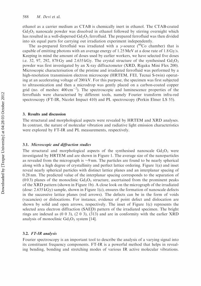
The structural and morphological aspects of the synthesised nanoscale Gd2O3 wereinvestigated by HRTEM and are shown in Figure 1. The average size of the nanoparticlesas revealed from the micrograph is �9 nm. The particles are found to be nearly sphericalalong with a high degree of crystallinity and perfect lattice ordering. Figure 1(a) and insetreveal nearly spherical particles with distinct lattice planes and an interplanar spacing of0.28 nm. The predicted value of the interplanar spacing corresponds to the separation of(0 0 3) planes of the monoclinic Gd2O3 structure, ascertained from the prominent peaksof the XRD pattern (shown in Figure 1b). A close look on the micrograph of the irradiated(dose: 2.635 kGy) sample, shown in Figure 1(c), ensures the formation of nanoscale defectsin the successive lattice planes (red arrows). The defects can be in the form of voids(vacancies) or dislocations. For instance, evidence of point defect and dislocation areshown by solid and open arrows, respectively. The inset of Figure 1(c) represents theselected area electron diffraction (SAED) pattern of the irradiated specimen. The brightrings are indexed as (0 0 3), (2 0 3), ð�313Þ and are in conformity with the earlier XRDanalysis of monoclinic Gd2O3 system [14].
3.2. FT-IR analysis
Fourier spectroscopy is an important tool to describe the analysis of a varying signal intoits constituent frequency components. FT-IR is a powerful method that helps in reveal-ing bending, bonding and stretching modes of various IR active molecular vibrations.
588 M. Devi et al.
Dow
nloa
ded
by [
Tez
pur
Uni
vers
ity]
at 0
4:28
03
Oct
ober
201
2
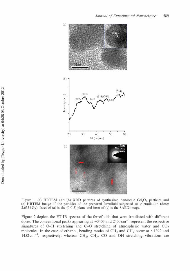
Figure 2 depicts the FT-IR spectra of the ferrofluids that were irradiated with different
doses. The conventional peaks appearing at �3403 and 2400 cm�1 represent the respective
signatures of O–H stretching and C–O stretching of atmospheric water and CO2
molecules. In the case of ethanol, bending modes of CH3 and CH2 occur at �1392 and
1452 cm�1, respectively; whereas CH2, CH3, CO and OH stretching vibrations are
20 30 40 50 60
(514)
(204)(313)(203)(202)
(003)
Inte
nsity
(a.
u.)
2θ (degree)
(b)
(a)
(c)
Figure 1. (a) HRTEM and (b) XRD patterns of synthesised nanoscale Gd2O3 particles and(c) HRTEM image of the particles of the prepared ferrofluid subjected to �-irradiation (dose:2.635 kGy). Inset of (a) is the (0 0 3) plane and inset of (c) is the SAED image.
Journal of Experimental Nanoscience 589
Dow
nloa
ded
by [
Tez
pur
Uni
vers
ity]
at 0
4:28
03
Oct
ober
201
2
observable at �2910, 2975, 1060 and 3679 cm�1 [15]. The weak band at 2980 cm�1 and the
sharp band at 1550 cm�1 indicate the presence of CTAB in the samples [16]. Different
peaks that are assigned to various components of Gd2O3-based ferrofluids are listed inTable 1. It was evident that the vibrational features corresponding to the Gd–O bonds
have been influenced by the irradiation effect. A close look on the spectra reveals that withincreasing dose of �-radiation, the peak becomes more prominent. But an anomaly in the
Gd–O vibration mode with discontinuities and less intensity profile is observed in sample
irradiated with a dose of 878Gy. Defects like oxygen vacancies are very common in oxidecompounds. Some Gd vacancies produced during synthesis may also exist in the system. In
fact, the creation of large number of free electrons by the energetic �-rays (1.25MeV) can
take part in defect formation. The observed variation in the metal–oxygen bond vibrationwith increased dose can be correlated with the increased defects up to a certain dose, where
the defects get saturated by distributing along the grain boundaries and the surface. We
speculate that the uneven distribution of defects and inhomogeneities have led to thediscontinuities in the vibrational patterns. It was reported that ethanol (the carrier fluid of
Figure 2. FT-IR spectra of the ferrofluids irradiated with different doses: (a) 32Gy, (b) 97Gy,(c) 292Gy (d) 878Gy and (e) 2.635 kGy.
Table 1. Assigned modes in the FT-IR spectra.
Peak position (cm�1) Assigned mode
2980 C–CH3 asymmetric stretching and N–CH3
symmetric stretching of CTAB1550 –CH2– and –CH3 stretching of CTAB1384–1392 CH3 vibration of ethanol1452–1469 CH2 vibration of ethanol1058 C–O vibration of ethanol536 In-plane Gd–O vibration
590 M. Devi et al.
Dow
nloa
ded
by [
Tez
pur
Uni
vers
ity]
at 0
4:28
03
Oct
ober
201
2

the ferrofluid) is less reactive in �-radiation [17]. But the nanoparticle-dispersed ethanol ischaracterised by an appreciable modification in the characteristic peaks observable at 1058and 1459–1469 cm�1.
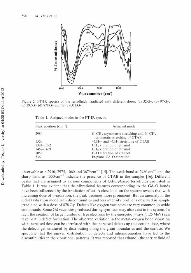
3.3. PL studies
The influence of light on the nature of radiative transition in material systems makesPL spectroscopy an important tool for material characterisation. Intra- and inter-bandtransitions can be visualised by PL studies. The room temperature PL spectra(�ex¼ 270 nm) of unirradiated and irradiated ferrofluids are shown in Figure 3(A).The unirradiated ferrofluid has recorded an asymmetrically broadened spectrum peakingat �355 nm. Earlier reports have suggested that this peak can be attributed to6P7/2$
8S7/2 transitions of Gd(III) [18]. Also the observed asymmetry in the PL response(pristine sample) is assigned to the existence of surface defects on the nanoscale Gd2O3
crystallites. In the pristine sample, the defect-related transition is very weak.Consequently, the overall PL spectrum of the pristine sample is dominated by theband-to-band transition. Upon �-irradiation, the above-mentioned peak is red-shifted to390 nm (we speculate that this is a superimposed peak due to band-to-band and defectemission). In this system, besides the already present vacancies, plentiful metastablesurface states (defect states) are created due to �-irradiation. Along with the shifting ofthe main peak towards higher wavelength, the abrupt change in symmetry signifies aremarkable improvement in the emission process via intermediate states, created by�-irradiation. Note that, during exposure of �-radiation, electrons are emitted due to theinteraction of high-energy photons with the medium through the Compton effect. Theseelectrons can be accommodated in the pre-existing oxygen vacancies. In oxide systems,an electron in oxygen vacancy results in Fþ centre [19]. The surface defect states arepositioned well below the lower end of the upper energy level. It is the transition viathese surface states that has resulted in the red-shifting of the emission peak, thusobstructing direct band-to-band transition significantly.
In order to correlate the amount of defect formation, asymmetry introduced and�-dose, we have performed deconvolution mechanism on each of the PL spectra(Figure 3B). Upon deconvolution, two distinct Gaussian peaks with variable intensities areobtained. The first peak positioned at �355 nm is band-to-band (6P7/2$
8S7/2) emission.The second peak is recognised as a defect-related peak and located at �410 nm, for all theirradiated ferrofluid samples. Irradiation-dependent variation in the peak intensity of thesetwo peaks would describe the nature of radiative transition undergoing in a givenspecimen. Emission as a result of formation/annihilation of additional defect centresmanifests the definite PL intensity in a selective way.
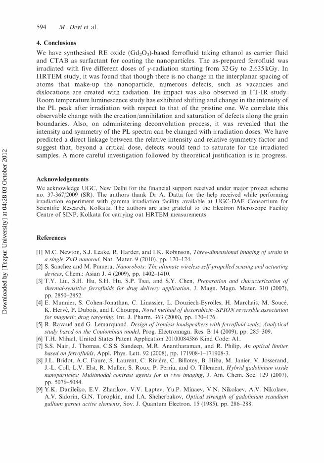
For the sake of clarity and better understanding, we have estimated symmetry factor(S) and correlated with the intensities of the deconvoluted peaks with respect to theoriginal peak. Note that right symmetry (SR) is related to the defect emission processwhereas left symmetry (SL) is associated with the band-to-band emission. Here, rightsymmetry factor is defined by (�M� �R)/D� with �M representing peak-wavelengthcorresponding to the main peak, �R is the wavelength on the right-hand side of the fullwidth at half maxima (FWHM) and D�¼ �R� �L, �L being the half-width wavelength onthe left-hand side of the FWHM. Figure 4(a) represents the variation of relative right
Journal of Experimental Nanoscience 591
Dow
nloa
ded
by [
Tez
pur
Uni
vers
ity]
at 0
4:28
03
Oct
ober
201
2

symmetry factor as well as relative intensity (I2/I, I2 being the intensity of the defect
emission after deconvolution and I is the intensity of the main peak) obtained from the
deconvolution. Similarly, the respective variations for the band-to-band emission can be
shown in Figure 4(b) with relative intensity as I1/I (I1 is the intensity of the deconvoluted
band-to-band emission) and relative symmetry factor SL/S (SL is the left symmetry factor
300
300 400 500 600
350 400 450 5000
100
200
300
400
500
600
700
0
100
200
300
400
500
600
700
3(A)
e
dc
b
a
Inte
nsity
(a.
u.)
Wavelength (nm)
3B(a)
Inte
nsity
(a.
u.)
Wavelength (nm)
300 350 400 450 500 5500
100
200
300
400
500
600
3B(b)
Inte
nsity
(a.
u.)
Wavelength (nm)
0
50
100
150
200
250
300
3503B(c)
Inte
nsity
(a.
u.)
Wavelength (nm)
0
50
100
150
200
250
300
3B(d)
Inte
nsity
(a.
u.)
Wavelength (nm)
300 350 400 450 500 550
300 350 400 450 500 550
300 350 400 450 500 5500
50
100
150
200
250
300
3503B(e)
Inte
nsity
(a.
u.)
Wavelength (nm)
Figure 3. Room temperature PL response of (A) unirradiated and �-irradiated Gd2O3 ferrofluidswith doses (a) 32Gy, (b) 97Gy, (c) 292Gy, (d) 878Gy and (e) 2.635 kGy. (B) (a)–(e) representingrespective deconvolution of individual PL peaks of the irradiated samples.
592 M. Devi et al.
Dow
nloa
ded
by [
Tez
pur
Uni
vers
ity]
at 0
4:28
03
Oct
ober
201
2

of the pristine sample). Interestingly, it was found that the relative intensity and symmetry
factor changes in a similar fashion with increasing dose, thus indicating direct relation
between these two measuring parameters. In PL spectroscopy, the intensity is a measure ofthe number of radiative transitions while symmetry signifies the nature of radiative centres
(energy spacing, order, etc.). The non-linear nature of the curve indicates the simultaneous
effect of defect formation/passivation in the irradiated system. By and large, it is expected
that the formation and annihilation processes compete each other until stable energy states(defect states) are created. First, with the increase in dose from 32 to 97Gy, we observe
drastic reduction in the relative PL intensity which may be ascribed to adequate
passivation of surface defects. Surprisingly, further increase in dose to 292 and 878Gy is
characterised by significant enhancement of I2/I ratio. At the highest dose (2.635 kGy), wenotice further reduction of I2/I. The relative symmetry factor (SR/S) also gives a similar
pattern. Reduction of defects makes the overall PL spectrum more symmetric with
suppressed right symmetry factor. The �-ray induced defect creation is not an indefinite
process and we speculate that the defects get saturated after a critical dose. The defectscould exist in the form of vacancies, interstitials, antisites, etc.
0.52
0.54
0.56
0.58
0.60
0.62
0.64
0.66
0.68
Dose (Gy)
Rel
ativ
e in
tens
ity (
I 2/I)
0.59
0.60
0.61
0.62
0.63
0.64
Rel
ativ
e sy
mm
etry
fact
or (
SR
/S)(a)
0 500 1000 1500 2000 2500 3000
0 500 1000 1500 2000 2500 3000
0.52
0.54
0.56
0.58
0.60
0.62
0.64
0.66
Dose (Gy)
Rel
ativ
e in
tens
ity (
I 1/I)
0.30
0.32
0.34
0.36
0.38
0.40
0.42
Rel
ativ
e sy
mm
etry
fact
or (
SL/
S)
(b)
Figure 4. Variation of relative intensity and relative symmetry factor vs. dose corresponding to(a) defect-related emission and (b) band-to-band emission.
Journal of Experimental Nanoscience 593
Dow
nloa
ded
by [
Tez
pur
Uni
vers
ity]
at 0
4:28
03
Oct
ober
201
2
4. Conclusions
We have synthesised RE oxide (Gd2O3)-based ferrofluid taking ethanol as carrier fluidand CTAB as surfactant for coating the nanoparticles. The as-prepared ferrofluid wasirradiated with five different doses of �-radiation starting from 32Gy to 2.635 kGy. InHRTEM study, it was found that though there is no change in the interplanar spacing ofatoms that make-up the nanoparticle, numerous defects, such as vacancies anddislocations are created with radiation. Its impact was also observed in FT-IR study.Room temperature luminescence study has exhibited shifting and change in the intensity ofthe PL peak after irradiation with respect to that of the pristine one. We correlate thisobservable change with the creation/annihilation and saturation of defects along the grainboundaries. Also, on administering deconvolution process, it was revealed that theintensity and symmetry of the PL spectra can be changed with irradiation doses. We havepredicted a direct linkage between the relative intensity and relative symmetry factor andsuggest that, beyond a critical dose, defects would tend to saturate for the irradiatedsamples. A more careful investigation followed by theoretical justification is in progress.
Acknowledgements
We acknowledge UGC, New Delhi for the financial support received under major project schemeno. 37-367/2009 (SR). The authors thank Dr A. Datta for the help received while performingirradiation experiment with gamma irradiation facility available at UGC-DAE Consortium forScientific Research, Kolkata. The authors are also grateful to the Electron Microscope FacilityCentre of SINP, Kolkata for carrying out HRTEM measurements.
References
[1] M.C. Newton, S.J. Leake, R. Harder, and I.K. Robinson, Three-dimensional imaging of strain ina single ZnO nanorod, Nat. Mater. 9 (2010), pp. 120–124.
[2] S. Sanchez and M. Pumera, Nanorobots: The ultimate wireless self-propelled sensing and actuating
devices, Chem.: Asian J. 4 (2009), pp. 1402–1410.[3] T.Y. Liu, S.H. Hu, S.H. Hu, S.P. Tsai, and S.Y. Chen, Preparation and characterization of
thermal-sensitive ferrofluids for drug delivery application, J. Magn. Magn. Mater. 310 (2007),
pp. 2850–2852.[4] E. Munnier, S. Cohen-Jonathan, C. Linassier, L. Douziech-Eyrolles, H. Marchais, M. Souce,
K. Herve, P. Dubois, and I. Chourpa, Novel method of doxorubicin–SPION reversible associationfor magnetic drug targeting, Int. J. Pharm. 363 (2008), pp. 170–176.
[5] R. Ravaud and G. Lemarquand, Design of ironless loudspeakers with ferrofluid seals: Analytical
study based on the Coulombian model, Prog. Electromagn. Res. B 14 (2009), pp. 285–309.[6] T.H. Mihail, United States Patent Application 20100084586 Kind Code: A1.[7] S.S. Nair, J. Thomas, C.S.S. Sandeep, M.R. Anantharaman, and R. Philip, An optical limiter
based on ferrofluids, Appl. Phys. Lett. 92 (2008), pp. 171908-1–171908-3.[8] J.L. Bridot, A.C. Faure, S. Laurent, C. Riviere, C. Billotey, B. Hiba, M. Janier, V. Josserand,
J.-L. Coll, L.V. Elst, R. Muller, S. Roux, P. Perria, and O. Tillement, Hybrid gadolinium oxide
nanoparticles: Multimodal contrast agents for in vivo imaging, J. Am. Chem. Soc. 129 (2007),pp. 5076–5084.
[9] Y.K. Danileiko, E.V. Zharikov, V.V. Laptev, Yu.P. Minaev, V.N. Nikolaev, A.V. Nikolaev,A.V. Sidorin, G.N. Toropkin, and I.A. Shcherbakov, Optical strength of gadolinium scandiumgallium garnet active elements, Sov. J. Quantum Electron. 15 (1985), pp. 286–288.
594 M. Devi et al.
Dow
nloa
ded
by [
Tez
pur
Uni
vers
ity]
at 0
4:28
03
Oct
ober
201
2

[10] D.L. Sun, J.Q. Luo, J.Z. Xiao, Q.L. Zhang, H.H. Jiang, S.T. Yin, Y.F. Wang, and X.W. Ge,
Effects of annealing treatment and gamma irradiation on the absorption and fluorescence spectra
of Cr:GSGG laser crystal, Appl. Phys. B 92 (2008), pp. 529–533.[11] V.T. Mak, V.S. Manzhara, V.I. Beizym, and V.I. Khivrich, The effect of gamma-irradiation on
the bandgap width of ZnSe, Tech. Phys. Lett. 28 (2002), pp. 757–758.[12] X. Gong, P. Wu, W.K. Chan, and W. Chen, Effect of �-ray irradiation on structures and
luminescent properties of nanocrystalline MSO4:xEu3+ (M¼Ca,Sr,Ba; x¼0.001–0.005), J. Phys.
Chem. Solids 61 (2000), pp. 115–121.[13] H. Chen, C. He, C. Gao, Y. Ma, J. Zhang, X. Wang, S. Gao, D. Li, S. Kan, and G. Zou, The
structural transition of Gd2O3 nanoparticles induced by high pressure, J. Phys.: Condens. Matter
19 (2007), pp. 425229-1–425229-5.
[14] S. Seo, H. Yang, and P.H. Holloway, Controlled shape growth of Eu- or Tb-doped luminescent
Gd2O3 colloidal nanocrystals, J. Colloid Interface Sci. 331 (2009), pp. 236–242.
[15] R.A. Nyquist, C.L. Putzig, and M.A. Leugers (eds.), The Handbook of Infrared and Raman
Spectra of Inorganic Compounds and Organic Salts, Vol. 1, Academic Press, San Diego, 1996.
[16] Y.D. Wang, S. Zhang, C.L. Ma, and H.D. Li, Synthesis and room temperature photolumines-
cence of ZnO/CTAB ordered layered nanocomposite with flake-like architecture, J. Lumin. 126
(1997), pp. 661–664.[17] C.B. Seymour, C. Mothersill, M.J. Moriarty, and F. Tipton, The effect of ethanol on the
radiation response of CHO-K1 cells, Br. J. Radiol. 60 (1987), pp. 577–581.[18] D.L. Rogow, C.H. Swanson, A.G. Oliver, and S.R.J. Oliver, Two related gadolinium aquo
carbonate 2-D and 3-D structures and their thermal, spectroscopic and paramagnetic properties,
Inorg. Chem. 48 (2009), pp. 1533–1541.[19] D.R. Vij (ed.), Hand Book of Applied Solid State Spectroscopy, Springer, Germany, 2006.
Journal of Experimental Nanoscience 595
Dow
nloa
ded
by [
Tez
pur
Uni
vers
ity]
at 0
4:28
03
Oct
ober
201
2