Leptin as an Extensive Modulator of Physiologic Systems - MDPI
Upload
khangminh22Category
view
0download
0


MEDICAL DEPARTMENT, UNITED STATES ARMY
SURGERY IN WORLD WAR 11
THE PHYSIOLOGIC EFFECTSOF WOUNDS
By
THE BOARD FOR THE STUDY OF THE SEVERELY WOUNDEDNorth African-Mediterranean Theater of Operations
OFFICE OF THE SURGEON GENERAL
DEPARTMENT OF THE ARMY
WASHINGTON, D. C., 7952
The volumes comprising the history it the Medical Department of the U. S. Armyin World War II are divided into two series: (i) The Administrative and Operationalseries which constitutes a part of the general series of the history of the U. S. Army inWorld War II, published under the direction of the Office of the Chief of Military History,and (2) the Professional, or clinical and technical series, published as The Medical De-partment of the United States Army under the direction of the Office of The SurgeonGeneral. Both series are being prepared by the Historical Unit, Army Medical Service.This volume is one of a number of surgical volumes to be published in the latter series.
Major General George E. Armstrong, The Surgeon General
Advisory Editorial Board
Brig. Gen. Albert G. Love, U. S. A. (Ret.), ChairmanMap Gen. Paul R. Hawley, U. S. A. (Ret.)Brig. Gen. Stanhope Bayne-Jones, A. U. S. (Ret.)Brig. Gen. Malcolm C. Grow, U. S. A. F. (Ret.)Brig. Gen. Raymond A. Kelser, U. S. A. (Ret.)Brig. Gen. Paul I. Robinson, U. S. A.Col. Joseph H. McNinch, MC, U. S. A.Michael E. DeBakey, M. D.Morris Fishbein, M. D.J. Ben Robinson, D. D. S.Richard H. Shryock, Ph. D.Lewis H. Weed, M. D.
Editors of Surgical Volumes
Fred W. Rankin, M. D.Michael E. DeBakey, M. D.
Colonel Calvin H. Goddard, MC, A.U.S., Editor in Chief
For sale by the Superintendent of Documents, U. S. Government Printing OfficeWashington 25, D. C. - Price $3.50 (Buckram)
THE MEDICAL DEPARTMENT OF THE UNITED STATES ARMY
The Physiologic Effects of Wounds
By
The Board for the Study of the Severely WoundedNorth African-Mediterranean Theater of Operations
Members
Henry K. Beecher, M. D.Formerly Lt. Colonel, MC, A. U. S.
Fiorindo A. Simeone, M, D.Formerly Colonel, MC, A. U. S.
Charles H. Burnett, M. D.Formerly Major, MC, A. U. S.
Louis D. Smith, Ph. D.Formerly Captain, SnC, A. U. S.
Seymour L. ShapiroFormerly Captain, SnC, A. U. S.
Eugene R. Sullivan, M. D.Formerly Lt. Colonel, MC, A. U. S.
Tracy B. Mallory, M. D.*Formerly Lt. Colonel, MC, A. U. S.
Chairman*Deceased
Report of the Board edited by Henry K. Beecher.
This volume was prepared by the Historical Division, Army Medical Library, under thedirection of Colonel Joseph H. McNinch, MC, U. S. A., Editor in Chief; Associate Editor,Sylvia Gottwerth.

THEBOARDFORTHESTUDYOF
THESEVERELY
WOUNDED.Leftto
right:Lt.Col.(laterColonel)
FiorindoA.
Simeone,Capt.(laterMajor)CharlesH.
Burnett,Capt.
LouisD.Smith,Capt.
SeymourL.Shapiro,Lt.Col.HenryK.Beecher,
Maj.(laterLt.Colonel)
EugeneR.Sullivan,Lt.Col.TracyB.
Mallory.Monghidora,Italy,
1944.

ForewordSevere surgical shock following trauma is not commonly encountered in
the medical practice of any group during peacetime. When it occurs it is theresult of unexpected accident, and adequate provision for careful study hasseldom been available. Consequently, although considerable noteworthy ex-perimental work had been done on shock in animals since World War I,little opportunity has existed to study this serious condition in man. Whenthe United States entered World War II, much information required forhandling battle casualties with serious injuries was not available. The needfor whole-blood replacement for resuscitation was incompletely understood.The significance of blood volumes, infection, continuing hemorrhage, theanatomic location of the injury, and the limitations of analgesic and anestheticagents was not fully appreciated although all these subjects had been under-stood in a seemingly adequate manner.
The brunt of this lack of information and training in U. S. Forces wascarried by the Medical Service of the North African and later the Mediter-ranean Theater. Until June 1944, when their forward medical service wasorganized to study the severely wounded, they were the only theaters withsizable numbers of ground troops steadily in contact with the enemy. Fornearly 2 years they experienced battle casualties at rates almost continuouslyabove 50 per 1,000 per annum, and for nearly 10 months of this periodtheir battle casualties were above 100 per 1,000 per annum. The ingenuity,resourcefulness, and thoroughness with which Medical Department personnelperformed under these trying circumstances are exemplified by the workof the individuals of the MTO Board for the Study of the Severely Woundedand the progressive steps taken by them to initiate their studies withoutspecial provision or support.
The Board’s studies on human casualties under battle conditions con-tributed much information of immediate practical value for the handlingof wounded as well as pointed the way for many basic studies required for

VI
the future. The data were accumulated under the rigors of field conditions,at times under fire, and the work was often begun in the shock tents a fewminutes after a wounded subject fell. This research was unique for AmericanForces during World War II and provided information on the resuscitationand treatment of the severely wounded that cannot be procured from anyother source. Aside from its significance as a contribution to medical knowl-edge, this report must stand as a tribute to the men who showed that itcould be done.
GEORGE E. ARMSTRONGMajor General, U. S. ArmyThe Surgeon General

Preface
On 3 September 1944, the Commanding General, North African Theaterof Operations, appointed a Medical Board to Study the Treatment of theSeverely Wounded, his action following by only two days a recommendationby the Surgeon of that theater that such a board be appointed. Appendix Aincludes the Surgeon’s communication and the order appointing the Board,together with data concerning its organization and operation.
Each of the authors of this volume was a member of the Board as formallyestablished. Before that time several of us had been interested individuallyin various aspects of the physiologic and pathologic changes in the severelywounded, and the chapters of this volume include our observations both asmembers of the Board and as individual investigators. The background againstwhich the study was undertaken is described fully in the Introduction.
At the time this study began, our attention was focused in considerablepart on anuria in the wounded. This interest continued, but it early becameapparent that to limit ourselves to the study of anuria was undesirable andactually impossible. Thus at the end of the work we have found, almostinevitably, that what we have is a kind of physiologic atlas of the severelywounded. In producing it we have followed the trend that medicine as awhole has taken: Probably there are few new things to be learned in studiesof gross morphology, but certainly much remains to be found in the domainof function. So too it may be with the wounds of battle. They have alreadybeen accurately described in terms of structural loss, but there are greatdeficiencies in our knowledge of the effects of wounds on the function oforgans. If this study has contributed in a useful way toward filling thesegaps, our effort will have been justified.
The several chapters of this volume have been written by the memberor members of the Board responsible for those particular phases of the inves-tigation. The aim has been to present factual observations, with a minimumof essential supplementary remarks to orient the reader. We have, there-
fore, in most instances deliberately avoided detailed discussion of the data.Many of the findings were provocative to us, and we hope they will be to
others; in any event the mass of data accumulated in this battlefront studyprovides a basis for contemplation and further work. From these data thereader who may differ with our views can construct his own and perhapsmore accurate interpretations and conclusions.
For those who have not had a similar experience, it may be difficult to
judge which things caused trouble during the course of the work and whichwere not important. Of real importance were the winds that blew sometents down, split others, upset stoves, knocked bottles off shelves, and stirredup endless clouds of dust that swirled around our hilltop at Monghidoroand invaded the laboratory. The rain and mud were unpleasant but not ofreal consequence until the dampness began to interfere with the spectro-photometer. When we were up in the Apennines, water froze and the coldat night caused solutions to alter and saturated solutions to crystallize out.Troublesome as these things were, they were overcome either by waitinguntil the environmental conditions had improved, or by the ingenuity of ourtechnicians.
Supplies and equipment needed for our studies were for the most partdelivered promptly and'in adequate quantity but occasionally delays in arrivalof items influenced the progress of our observations. In only two instanceswas absence of supplies of material consequence. We never succeeded inobtaining equipment to make desired oxygen determinations, and materialsfor renal clearance studies arrived so late that only a few cases could be studied.
Although the spectacular “artificial moonlight” produced by great flood-lights reflected from clouds lighted up the out-of-doors at night and wasmuch used in the months of the Northern Apennines Campaign, we neverescaped from the necessity of working in blacked-out tents, and in poorlylighted tents it was easy to overlook cyanosis or icterus.
The major consideration at all times was that our studies must not delayor otherwise interfere with the treatment of the wounded soldier. The neces-sity of not interfering with the therapy due the wounded man was important.The patient’s good came first; it was trying but necessary sometimes to letobservations go, knowing that an unfortunate shortcoming would be presentin the case under study. During periods of heavy action, when the casesbest suited for study were coming in, the man covering the shock tent neededall the maneuvering skill of a combat pilot to keep out of the way of those
IX
responsible for the care of the wounded man. At times he failed, all of usfailed, to get the observations needed for completeness.
Sometimes a patient would be accessible to us for only a few hours andthen would be evacuated to the rear, never to be seen again. Most of thepatients were followed into the postoperative period, but some were evacu-ated at, for our purposes, their most enlightening stage, or sometimes, becauseof the tactical situation, we were obliged to move forward or back andleave them.
We realize that those accustomed to working in the atmosphere of leisureand with the laboratory facilities of peacetime can find much to criticize here. Wehad to get what we could when we could, and record it “on the go.” Ourobservations are published as recorded at the time and without later attemptto identify and compare individual cases in all the various aspects of thestudy. It will be readily appreciated that identical data could not be obtainedon all cases. We present the results without apology. Moreover, we believethat what we were able to do indicates the soundness of our attempt to studythe wounded in the line of combat where they fell.
Finally, we are bound to acknowledge the extraordinary spirit of thenewly wounded, those with whom we dealt. They did all they could to helpus in this work.
HENRY K. BEECHER, M.D.(Formerly Consultant in Resusci-tation and Anesthesia, NorthAfrican-Mediterranean Theater ofOperations)
Boston, Massachusetts23 February 1952

ContentsPage
FOREWORD vPREFACE viiINTRODUCTION i
Chapter
I INTERNAL STATE OF SEVERELY WOUNDED MEN ON ENTRYTO THE MOST FORWARD HOSPITAL 21
II LIVER FUNCTION IN THE SEVERELY WOUNDED 75III RENAL FUNCTION IN THE SEVERELY WOUNDED 90IV DIAGNOSIS OF LOWER NEPHRON NEPHROSIS RESULTING
FROM TRAUMA AND SHOCK (“SHOCK KIDNEY”): CLINICO-PATHOLOGIC CORRELATION no
V CLINICAL, PHYSIOLOGIC, AND BIOCHEMIC CORRELATION INLOWER NEPHRON NEPHROSIS 121
VI TREATMENT OF LOWER NEPHRON NEPHROSIS 184VII EFFECT OF ALKALIS IN TREATMENT OF TRAUMATIC SHOCK 194
VIII PIGMENT MOBILIZATION IN SEVERELY WOUNDED MEN . . 201
IX PATHOLOGY OF THE KIDNEY IN TRAUMATIC SHOCK ... 224X SULFONAMIDES IN RELATION TO “SHOCK KIDNEY” .... 249
XI THE CRUSH SYNDROME IN BATTLE CASUALTIES 255XII GENERAL PATHOLOGY OF TRAUMATIC SHOCK 283
Appendixes
A ORGANIZATION AND OPERATION OF THE BOARD FOR THESTUDY OF THE SEVERELY WOUNDED 307
B THE FIELD LABORATORY 315C BIOCHEMIC METHODS 323D SURVEY OF CASE RECORDS 359
INDEX 369


The Physiologic Effects of Wounds

IntroductionA report is spread that there is, in some country or other, a giant as big as a
mountain; and men presently fall to hot disputing concerning the precise lengthof his nose, the breadth of his thumb, and other particulars, and anathematizeeach other for heterodoxy of belief concerning them. In the midst of all, if somebold sceptic ventures to hint a doubt as to the existence of this giant, all areready to join against him, and tear him to pieces. —Attributed to Voltaire
Shock —the BackgroundAlthough warriors have died of their wounds from the beginning of time,
the first scientific approach to an analysis of how and why they die wasmade during World War I. In that war the wound surgeon lifted his eyesfrom the shattered limb to inquire with some degree of precision about thenature of the processes that a wound may initiate in the body as a whole.Because, even if the limb were amputated and every organ of the body wassound, death was likely to occur as the terminal event of a profound dis-turbance known as wound shock.
World War I revealed wound shock as a complex problem. Its naturewas not solved nor were sufficient observational data accumulated to permitclear identification and subsequent analysis. Certain pre-existing hypotheses(vasomotor exhaustion, acapnia, adrenal exhaustion) were discredited, butother concepts inadequately supported by facts (traumatic toxemia, the dis-tinction between shock and hemorrhage) were substituted. These conceptscentered on wound shock as an entity not accounted for by hemorrhage,infection, brain injury, blast, asphyxia of cardiorespiratory origin, fat em-bolism, or any other clearly demonstrable lethal effect of trauma. World War Ithus recognized a problem of shock but left it wrapped in mystery.
At the end of World War I the so-called shock problem was transferredto the experimental laboratories of medical science. Attempts were made toresolve it by physiologic and chemical techniques under a wide variety ofexperimentally induced circumstances. As the methods of initiating experi-mental shock were multiplied, the term itself became broadened, so that it
THE PHYSIOLOGIC EFFECTS OF WOUNDS2
included a number of processes that appeared to have one feature in common—a reduced effective volume flow of blood with inadequacy of the peripheralcirculation and resulting tissue asphyxia. In the clinic as well as the labora-tory, shock became separated from wounds, and “medical shock,” “obstetricalshock,” “burn shock,” “shock due to infection,” and other types weredescribed as entities. So-called shock became synonymous with the process ofdying from almost any cause unless death was practically instantaneous or, asHenderson 1 stated, “unless one is burned alive.” The phrase, “the problems ofshocks,” was used by Mann2 to describe this confusion of definition.
In the welter of animal experimentation during and after World War Ithere were certain findings pertinent to the original problem. Bayliss andCannon 3 had imitated wound shock by crushing and lacerating the thighmuscles of anesthetized animals. This was supposed to produce a destructionof tissue but not a very great extravasation of blood. Failure to measurethe factor of blood and fluid loss in the local area of trauma (and, as shownlater, failure to recognize the superimposed clostridial infection) enabledCannon 4 and other experimenters to propose that toxic products of tissuedisintegration were absorbed into the general circulation, causing what wastermed traumatic toxemia. Parsons, 5 Parsons and Phemister, 6 and Blalock 7
measured the local blood and fluid loss in traumatized legs by precise methodsand showed that it was far greater than had been suspected and quitesufficient to account for the reduction in blood volume observed.
It was thus made clear that estimation of the amount of hemorrhagein an injured man must include the blood extravasated into the tissues aswell as that poured on the ground or caught by the dressings. It was alsofound necessary to consider the blood volume that remained in circulation
1 Henderson, Y.: Fundamentals of asphyxia. J.A.M.A. 101: 261-266, July 22, 1933.2 Mann, F. C.; In Bulletin on Shock, minutes of third meeting of Subcommittee on Shock, Division of
Medical Sciences, National Research Council, 18 May 1942, p. 117.3 Bayliss, W. M., and Cannon, W. B.; Sections IV, V, and VI in Report VIII, Traumatic Toxaemia as a
Factor in Shock, Special Report Series, No. 26, Medical Research Committee. London, H. M. StationeryOffice, 1919.
4 Cannon, W. B.: Traumatic Shock. New York, D. Appleton and Co., 1923.5 Parsons, E.: Experimental shock and hemorrhage. Tr. A. Resid. & ex-Resid. Physicians, Mayo Clin.
(1929) 10: 106-108, 1930.6 Parsons, E., and Phemister, D. B.: Haemorrhage and “shock” in traumatized limbs; experimental
study. Surg., Gynec. & Obst. 51: 196-207, Aug. 1930.7 Blalock, A.: Experimental shock; the cause of the low blood pressure produced by muscle injury.
Arch. Surg. 20: 959-996, June 1930.

INTRODUCTION 3
in terms of plasma and red cells, for the proportionate loss of these elementsvaried under different circumstances. The phenomena of hemoconcentrationand hemodilution were thus made understandable. Nevertheless certain in-vestigators insisted on using the physical state of the blood to define shockas an entity, and confusion was introduced by proponents of the thesis thatshock could not exist unless hemoconcentration was present.
In a critical review of the shock problem in 1942, Wiggers8 commentedthat contributions to the literature on shock appeared to be directed towardthe support of one or another favored theory. Experimental conditions, hestated, had not been carefully evaluated and conclusions, rather than facts,were emphasized. “Beneficial effects,” Wiggers said, “are claimed for variousforms of therapy in instances in which it was never shown that the subjectswere in a state of shock which would have proved fatal without treatment.”
Obvious loss of blood (hemorrhage), or plasma (burns), or water andelectrolytes (dehydration), or all elements (trauma) were generally acceptedas clearly recognized initiating factors in shock. Beyond these, however, ex-perimenters continued to search for other mechanisms by which the volumeof blood returned to the heart might be reduced. Belief in a generalizedincrease in capillary permeability, a concept introduced with “traumatictoxemia,” held sway for years, and many investigators insisted that this, andonly this, accounted for the “true” shock that had been witnessed in WorldWar I.
Analysis of the course of events in progressive circulatory failure waspressed in the attempt to identify and define a phase-line that ushered inwhat was called an “irreversible” state. It had been claimed in World War Ithat the seriously wounded could be resuscitated by appropriate measuresif their condition was attributable to hemorrhage alone, but that if profoundshock had been superimposed on hemorrhage or had appeared independentlyof hemorrhage, all measures, including blood transfusion, were futile. Theterm “irreversible” was used by some with specific reference to the functionof tissues or the closely linked processes of intermediary metabolism. It wasused in this sense by the Subcommittee on Shock9 of the National Research
8 Wiggers, C. J.: The present status of the shock problem. Physiol. Rev. 22; 74-123, Jan. 1942.9 Subcommittee on Shock, Committee on Surgery, Division of Medical Sciences, National Research
Council: Shock, p. xxi (Preface). In National Research Council, Division of Medical Sciences.Burns, Shock, Wound Healing and Vascular Injuries. Military Surgical Manuals, vol. V. Philadelphia,W. B. Saunders, 1943.
956507 0—52 2

THE PHYSIOLOGIC EFFECTS OF WOUNDS4
Council: “
. . . the process of shock brings about certain changes in thefunction of the tissues which, after a time, become irreversible.”
By others the term “irreversible” was applied to the dynamics of the circu-latory system itself. It was held that at some point in the deterioration ofthe organism in shock the circulatory failure became irreversible and thesubject displayed this change by becoming nonreactive to transfusion. “Ap-parently,” as Wiggers 10 wrote, “at a certain stage an adequate circulationcannot be restored by merely filling the system as one does an automobileradiator.” The same writer expressed his surprise at “how well tissues ororgans withstand a very low rate of blood flow before they cease to functionor are unable to revive.”
Thus the assumption was made that at one moment restoration of bloodvolume could stay the progress of death, but in the next moment it wouldbe unable to do so. It seemed reasonable to believe that if this phase-linecould be identified by experiment and an analysis be made of the physio-logic processes then in motion, corrective measures might suggest themselves.But even under precisely controlled laboratory experimentation, minor vari-ations in conditions may determine whether the animal lives or dies. Subtledifferences in environmental temperature, anesthetic agents, age, and pre-vious nutritional state of the subject, as well as other conditioning factorsmake it difficult to halt the cinema of life at a particular frame where onemay say: Up to this point continuing life is possible —beyond this, death isinevitable. If difficult in precise experimentation, identification of the onsetof irreversible shock becomes impossible when one is confronted by theresults of the random trauma sustained by soldiers under combat conditions.Although the diagnosis of “irreversible shock” appeared with some frequencyon the clinical records in World War II, it was merely a pretentious way ofindicating that the man had died of a lethal wound.
This, then, was the background of our knowledge of wound shock whenWorld War II began. It was entered with the concept that (i) plasma torestore the bulk of the blood in the intravascular space and (2) sodium chloridesolution for dehydration and electrolyte depletion of the interstitial space weretherapeutic measures adequate to the purpose of adjusting homeostasis in awounded man. As the war progressed, this concept changed. It soon becameclear that much precise information about the physiologic state of a wounded
10 See footnote 8.

INTRODUCTION 5
man was wanting, and efforts toward this end culminated in establishmentof the Board for the Study of the Severely Wounded in the MediterraneanTheater of Operations. The chronology of events leading to a better under-standing of the disturbed physiology of the severely wounded and their man-agement in World War II may be traced through several phases, beginningwith the preliminary one of planning for the treatment of shock.
iS/lanagement of Severely Wounded in World War IIPreliminary Planning for Treatment of Shock
The first problem encountered in the combat area was the need forwhole blood transfusion, for while plasma was in plentiful supply, it provedinadequate for the purposes envisioned. An extract from a “Report on theActivities of the Surgical Section of the 77th Evacuation Hospital,” dated10 December 1942, provides a baseline for a review of the planning for thetreatment of shock in World War II. This unit shipped from England withTorch Forces and entered on the landing at Oran.
The W.I.A. [wounded in action] had for the most part either succumbed to or
recovered from any existing shock before we saw them. However, later traumatic casescame to us in shock and some of the early cases were found to be in need of whole bloodtransfusions. There was plenty of reconstituted blood plasma available. However, somecases, particularly those with large blood loss, were in dire need of whole blood. We hadno transfusion sets, although such are readily available in the United States, no sodiumcitrate, no sterile distilled water, and no blood donors. Transfusion bottles were borrowedfrom the British, sodium citrate was purchased from a French pharmacy, a water stillappeared from some unexplained source, our enlisted men who had been working longhours volunteered as donors, and whole blood transfusions were given. It would seem thatthere is grave need of provision for whole blood at the locality and time of definitivetreatment.
The initial decision to rely on plasma rather than blood transfusion forthe resuscitation of the wounded appears to have been based in part on theview held in the Office of The Surgeon General of the Army, and in parton the opinion of the eminent civilian investigators summoned by the Na-tional Research Council to act as advisers to the Armed Forces. The Com-mittee on Transfusions first met on 31 May 1940. The Army representativemade the following statement: “If the theaters of operations are mostlyoutside the United States . . . the Army would likely discourage the use pf
6 THE PHYSIOLOGIC EFFECTS OF WOUNDS
blood banks. If war should come closer they might want to use blood thatcould be transported by airplane or specially devised refrigeration. In moredistant places where blood could not be collected locally, plasma, either plainor dried, would have to be used.” The representative of the Navy also favored“dried blood” (plasma).
The following is quoted from a report of the meeting submitted to theChairman of the Committee on Surgery, National Research Council, underdate of 24 July 1940:
The greater part of the day was devoted to a consideration of whole blood and bloodplasma and blood serum transfusions. The consensus of opinion was that the greatestemphasis should be placed on the use of blood plasma for the following reasons; (1) Mostinstances of shock are associated with hemoconcentration and a given quantity of plasmais more effective than an equal quantity of whole blood in treatment; (2) blood plasmais approximately as effective in the treatment of hemorrhage as is whole blood; (3) thedifficulties of preservability and transportability of plasma are considerably less than thoseof whole blood; and (4) matching and typing are not necessary when pooled plasma(suppression of iso-agglutinins) is used.
The last two reasons given may have been concessions to the positiontaken by the representatives of the Army and Navy; the first two, however,appear to reflect the prevailing concept of wound shock held by experts atthat time. The efficacy of blood in the treatment of hemorrhage had beenestablished in World War I. In small quantities it had been preserved andtransported considerable distances, even up to regimental aid posts. It hadbeen recorded that “in cases of profound shock accompanied by loss of blood,excellent results are obtained from direct blood transfusion.”11 Robertson 12
had cast doubt on the efficacy of various fluids used as “substitutes” for bloodin World War I (gum acacia, gelatin) and called attention to the fact thattheir beneficial effects were often slight. “The only means available of increas-ing the oxygen-carrying power of the blood is the addition of new red bloodcells,” he had said. “This constitutes the unique value of blood transfusion.”Whole blood transfusion also had become universally employed in surgeryin civil life.
It is of real interest, therefore, to inquire into the process of reasoning11 Fraser, J., and Cowell, E. M.: A clinical study of the blood pressure in wound conditions. Report
II, sec. i, Special Report Series, No. 25, Medical Research Committee. London, H. M. Stationery Office,1919, p. 49-71.
Robertson, O. H.: Memorandum on blood transfusion. Report IV, Special Report Series, No. 25,Medical Research Committee. London, H. M. Stationery Office, 1919, p. 143-180.

INTRODUCTION 7
that led the Committee on Transfusions of the National Research Councilto take the position that “most instances of shock are associated with hemo-concentration and a given quantity of plasma is more effective than an equalquantity of whole blood in treatment.” This concept can be traced back toobservations on the wounded made in World War I by Cannon, Fraser,and Hooper13 who reported that counts of red cells in blood taken fromthe capillary bed were high, particularly when compared with those of venousblood. This also was a keystone in the establishment of shock as an entitydistinct from hemorrhage and led to the widely accepted hypothesis of ageneralized increase in capillary permeability. “Hemoconcentration was foundto furnish a practical means for differentiating shock from hemorrhage, butthe enormous potential value of this sign was not comprehended by themembers of the Special Committee on Wound Shock nor has it been sensedby physicians during the 20 years since that time,” wrote Moon 14 in 1938.The manual on shock 1' 1 (1943), prepared under the auspices of the Com-mittee on Surgery of the Division of Medical Sciences of the National Re-search Council, also set forth this erroneous concept.
The other statement of the Committee on Transfusions, namely, that“blood plasma is approximately as effective in the treatment of hemorrhageas is whole blood,” appears to have found origin in conclusions drawn fromlaboratory experiments that were purposely designed so that the number ofvariables could be rigidly limited. Transference of these conclusions to asituation that introduced a number of additional variables was an error ofhuman reasoning. An example may be found in the widely quoted experi-ments of Rous and Wilson 10 (1918). These authors made a precise determina-tion of the limits within which plasma may replace the loss of whole bloodin acute hemorrhage induced in rabbits. In summarizing the results of theirexperiments these authors stated that “however desirable transfusion may be,it is not essential to recovery from even the severest acute hemorrhage, if
13 Cannon, W. B.; Fraser, J., and Hooper, A. N.: Some alterations in the distribution and characterof the blood. Report II, sec. 2, Special Report Series, No. 25, Medical Research Committee. London, H. M.Stationery Office, 1919, p. 72-84.
14 Moon, V. H.: Shock and Related Capillary Phenomena. New York, Oxford University Press, 1938.15 National Research Council, Division of Medical Sciences. Burns, Shock, Wound Healing and
Vascular Injuries, prepared under the auspices of the Committee on Surgery of the Division of MedicalSciences of the National Research Council. Military Surgical Manuals, vol. V. Philadelphia, W. B. SaundersCo., 1943.
14 Rous, P., and Wilson, G. W.: Fluid substitutes for transfusion after hemorrhage. J.A.M.A. 70:219-222, Jan. 26,1918.

8 THE PHYSIOLOGIC EFFECTS OF WOUNDS
only the blood bulk can be restored in other ways.” The conclusion drawnfrom this and subsequent observations by others 17 led to formulation of thestatement by the Committee on Transfusions. The brief description of arabbit in which up to three-fourths of the blood volume, as measured by thehemoglobin depletion, had been withdrawn and replaced by plasma con-tains one phrase that is significant; “The least exertion would cause theanimal to pant heavily.”
Presumably the rabbit had no semblance of a wound other than theneedle puncture. Substitute for the rabbit housed quietly in its cage a woundedsoldier picked up by litter bearers and transported by ambulance, who hasin addition an extensive and painful wound with continuing extravasationof blood and plasma into adjacent tissues. Then add sedation, roentgeno-graphic examination, anesthesia, and surgical operation with a further lossof blood. It is obvious that the introduction of these and other variables,purposely and of necessity excluded from the original experiments, maycompletely negate the conclusion. Both errors, the association of wound shockwith hemoconcentration and the estimation regarding the effectiveness ofblood plasma, are understandable in view of the paucity of observations madein World War I concerning the disturbed physiology of wounded men.
Restoration of the blood bulk in the intravascular space by infusion of acolloid solution that might be expected to stay within the confines of the semi-permeable membrane of the capillary walls was envisioned during WorldWar I. This was a projection of the Starling 18 concept elaborated by Scott 19
in 1916. Tests were made of the properties of soluble starch, dextrin, gelatin,and gum arabic, and preparations of the latter were given extensive field tests,particularly by the British, guided by the basic experiments of Bayliss. 20
The advent of human plasma, as a result of the development of methodsthat enabled it to be preserved and packaged in desiccated form, appearedto provide a final answer to the problem of the restoration of blood bulk byinfusion. The treatment of shock and hemorrhage was thus reduced to thesimple terms of the exchange of fluid between the intravascular space and
17 Bayliss, (see footnote 20) for example, had shown that “more than one-half of the blood in the catcan be replaced by gum solutions with satisfactory results.”
18 Starling, E. H.: On the absorption of fluids from the connective tissue spaces. J. Physiol. 19: 312-326,May 1896.
19 Scott, F. H.: The mechanism of fluid absorption from tissue spaces. J. Physiol. 50: 157-167, Feb. 1916.20 Bayliss, W. M.: Intravenous injections to replace blood. Report I, Special Report Series, No. 25,
Medical Research Committee. London, H. M. Stationery Office, 1919, p. 11-41.

INTRODUCTION 9
the interstitial space under clearly defined physicochemical laws. Extensionof this same reasoning led to the proposal that because the serum albuminfraction of the blood as prepared by Cohn21 packaged a high proportionof the total colloid osmotic activity of the serum in small liquid volume, itwas peculiarly appropriate to military needs. It was postulated that the inter-stitial fluid compartment would provide the necessary diluent unless thepatient were badly dehydrated.
This oversimplified physicochemical approach, which was an extensionof the World War I quest of Bayliss aided by the availability of refinedand human-derived preparations, not only failed to take into account thevariables described, but also placed undue emphasis on a single physico-chemical property of the blood; namely, the osmotic activity of the plasmaproteins. Not only was the important function of the red cells as oxygencarriers ignored, but also their contribution to the total blood mass underabnormal circumstances. Both the magnitude of the initial loss of wholeblood occasioned by wounding and the significance of a continuing seepageof blood and its fluid components into the tissue spaces were underestimated.And finally, an effort to restore and maintain blood bulk based on colloidpreparations, either derived from human proteins or otherwise, presupposesa space bounded by a semipermeable membrane—not one in which largeareas of the membrane may have been rendered freely permeable by the directeffects of trauma.
Evolution of Knowledge During World War II
The evolution of the management of the seriously wounded during WorldWar II may be divided into three phases for purposes of description, althoughit is apparent that as phases they are not pencilled with the clarity observedin phase-lines on a tactical map. A first phase may be recognized in whichefforts to identify the gross nature of the problems and devise immediatesolutions predominate. Cobwebs of theory and hypothesis were swept awayby simple observations and precise definitions. This was followed by a phaseduring which all efforts centered on the development and perfection of thepractical art of resuscitation. In the final phase systematic and precise meas-
21 Cohn, Edwin J. : Memorandum on the preparation of normal human serum albumin. Report No. i,Subcommittee on Blood Substitutes, Division of Medical Sciences, National Research Council (acting forCommittee on Medical Research, Office of Scientific Research and Development), n February 1942.
THE PHYSIOLOGIC EFFECTS OF WOUNDS10
urements were made that for the first time described the actual physiologicstate of the wounded man as it was observed on the field of battle.
A search among the records of World War II for novel and challenginghypotheses regarding the nature of shock is likely to prove disappointing.The very abundance of facts and experience discouraged “hot disputing” anddebate. And yet from this experience emerged certain concepts that, whenfully grasped, will be found no less significant because they appear simpleand direct.
First Phase: Identification of the Problem
Although in retrospect the North African Campaign was but a briefcurtain raiser for the sustained action that was to come later, it stands his-torically as a period in which the major problems of the management ofthe wounded were clearly identified. The campaign was over before manyneeds of the military organization could be met, but the foundation for futureaction was secured.
The Surgical Consultant, North African Theater of Operations (U. S. A.),reported for duty in Algiers on 7 March 1943. His first official report, sub-mitted under date of 24 March 1943, following a period of temporary dutyin II Corps on the southern Tunisian front, was a Memorandum on wholeblood transfusion. Further data were collected and a formal report on wholeblood transfusion was made to the Theater Surgeon, NATOUSA, on 16 April1943. The following conclusions and recommendations were made.
Conclusionsa. There is a need for whole blood transfusion in the treatment of a significant pro-
portion of the wounded. Plasma is not an adequate substitute in these cases.
b. Adequate and conventional safeguards that govern blood transfusion are difficultor impossible to attain in forward echelons.
c. The British Base Transfusion Unit has demonstrated the feasibility of supplyinglarge amounts of whole blood to the combat area.
RecommendationThat a central laboratory be established in NATOUSA to provide whole blood, intra-
venous solutions, distilled water, and plasma.
The Italian Campaign had progressed to the establishment of the AnzioBeachhead before the distribution of preserved blood from a central laboratory

INTRODUCTION
was realized. In the meantime, however, the evacuation hospitals and laterthe field hospitals employed for forward emergency surgery were encouragedto establish their own blood banks with supplies requisitioned for that purpose.
Following this decision on therapy, the next important question that facedthe Surgical Consultant in southern Tunisia in March and April 1943 waswhether casualties were dying of irreversible shock—in fact, whether woundshock, unassociated with hemorrhage and other clear results of trauma, existedas an important problem in World War II. It was obvious that a precise defi-nition was necessary if this question was to be answered, for, as was notedin the Report of the Surgical Consultant dated 2 July 1943:
In Field Medical Records, Case Reports, and Death Reports, as well as in verbaldiscussions among Medical Officers, the term “shock” is used with vague definition orquite commonly with no definition whatsoever. In the case reports of battle casualties dyingin the forward area, “shock” or “irreversible shock” is almost invariably recorded as asecondary cause of death. This is true whether the wounded man had a lethal craniocere-bral wound, an overwhelming peritonitis, fulminating gas gangrene infection, or simplydied of uncontrollable hemorrhage.
Circulatory failures from peritonitis, bacterial infection, intrathoracic in-jury, burns, and injury to the central nervous system were placed in separatecategories. All other forms of circulatory failure which arise within a fewhours as a result of wounding were considered as “wound shock.” 22 A studywas undertaken that covered the course of the evacuation of some 1,263casualties from the battalion areas through the evacuation hospitals. No recordcould be found of a death from wound shock under terms of the restricteddefinition, in which hemorrhage could be excluded as the important factor.The conclusions drawn from this study as well as from direct observation oflarge numbers of wounded were expressed as follows: 23
Under conditions that prevailed in the management of battle casualties between 20
and 25 March in the sampling area of II Corps, wound shock was not a cause of death.This does not imply that wound shock did not occur among the survivors, but if so it
appears that remedial treatment was adequate.22 The Battle of El Alamein (October and November 1942) was one of the first occasions on which blood
and blood substitutes were used on a large scale for the resuscitation of battle casualties in forward medicalunits. Report No. 1, Medical Research Section, GHQ, MEF, by Lt. Col. W. C. Wilson, RAMC, described thecondition of the wounded with special reference to wound shock and its treatment. The necessity for therestricted definition of wound shock was presented with great clarity.
23 Report of Surgical Consultant, Office of the Surgeon, Headquarters, North African Theater of Opera-tions, U.S.A., 2 July 1943. (Appendix B, 1;Par. B, 2.)

THE PHYSIOLOGIC EFFECTS OF WOUNDS12
“Irreversible” wound shock does not appear to be a problem of pressing significance.The problem of shock as observed in the Tunisian Campaign centered in the applica-
tion of accepted means of treatment, rather than in the need for additional methods ofmanagement.
Second Phase: Development of Resuscitation
The second phase in the advancement of understanding of the manage-ment of the seriously wounded was development and perfection of the prac-tical art of resuscitation. The many experienced surgeons of the Theatercontributed to and shared the responsibilities of a Theater-wide educationalprogram. Special acknowledgment is made of the contributions of ColonelHoward Snyder, Surgical Consultant to II Corps and subsequently to FifthArmy, and of Lt. Colonel Henry K. Beecher, assigned to AFHQ as Con-sultant in Resuscitation and Anesthesia and working on temporary duty inthe forward installations. Simple and direct observations made while actuallycaring for battle casualties confirmed the conclusions of the Tunisian Cam-paign and led to the complete discard of the confused theories of traumaticshock that had been elaborated from the experience of World War I.
A highly significant product of the development of the art of resuscitationwas merging of consideration of shock with consideration of the implicationsof the wound. Historically, wound surgery has been linked with the pre-vention and treatment of infection, and, as a matter of fact, in the lessseriously wounded this function of surgery still is predominant. In WorldWar II this concept was modified, as it was not applicable to the manydesperately wounded casualties that came under surgical management. It wasno longer valid to hold that a seriously wounded man could be resuscitatedsolely by measures directed toward restoring blood volume, and that whenthis was accomplished wound surgery could be undertaken, depending onlyon the time necessary for prevention of infection. Wound surgery under thesecircumstances assumed the new position of being in itself the climax ofresuscitation. General recognition of the principle that procedures commonlygrouped as “resuscitative” are but integral steps in the management of asituation that must be viewed as a whole, and that wound surgery may initself be the most potent act of resuscitation, stands as a basic achievementof military surgery in World War II.
This concept was glimpsed in the Tunisian Campaign and led to the

INTRODUCTION
following comment in the official report of the Surgical Consultant dated2 July 1943:
Resuscitation comes to be regarded as a sub-specialty of military surgery and as suchbecomes a goal in itself. One central fact must be kept in mind and, although it appearsobvious, it is often overlooked both in theory and practice. A wounded man is resuscitatednot only to save life but to prepare him for necessary surgery.
This divorce of surgeon from shock is a disquieting outgrowth of the war that cannotbe too strongly condemned. Resuscitation in every case being prepared for operation is anintegral part of the surgical management of trauma and must “remain so if optimal resultsare to be achieved.
The concept was more fully developed during the Italian Campaign byLt. Colonel Henry K. Beecher24 who presented the following broad defini-tion of resuscitation that includes operation as an essential component:
The enemy has produced the worst wound he could, and its consequences are cumu-lative—dehydration increased by unusual fluid loss in sweat and vomitus, continuinghemorrhage or plasma loss, pain making rest impossible, increasing emotional exhaustion,developing infection—these and other factors are set in operation by the initial wound.Their progress in the seriously wounded is to be checked in most cases only by surgeryor by death. Resuscitative measures give a temporary stay and make successful surgerypossible in the severely wounded; but in most cases true release from the consequences ofthe wound is effected only by surgery. Surgery is not only the goal but is itself a part ofresuscitation in the broad sense. Any other view is likely to lead to unfortunate separationbetween the activities of the “shock team” and those of the surgical team. Care of thewounded man must be continuous and supervision uninterrupted.
This concept now appears obvious, and in fact is a principle soon graspedby the practical worker in the field. It is likely to be overlooked when con-clusions are drawn from laboratory experiments purposely designed to isolateand test the efficacy of single therapeutic measures.
The establishment of wound surgery as inseparable from the managementof wound shock had many practical applications. It was a strong considerationin the placement of the surgical hospital for treatment of the severely woundedalongside the divisional clearing station, as was determined in Sicily, A shortlitter carry placed the casualty in the hands of a competent surgical teamequipped not only for resuscitation in the conventional sense but for themajor procedures of surgery. It led to the close observation of a woundedman’s response to blood replacement therapy. If the response was transient
24 Beecher, H. K.: Preparation of battle casualties for surgery. Ann. Surg. 121: 769-797, June 1945.

THE PHYSIOLOGIC EFFECTS OF WOUNDS
or unsatisfactory, it was not judged that his shock was “irreversible” or thathe displayed a “negative reaction” because of widespread capillary damage,or that it was futile to try “to repair the damage done by prolonged oxygenwant.” It was assumed that either continuing hemorrhage or spreading infec-tion was present, or that a dead limb required amputation or dead tissuecalled for excision; operation was immediately undertaken with continuingtransfusions to support the patient’s condition.
Another practical result was that resuscitative measures carried out in thefield, forward of a surgical hospital, came to be regarded as temporary anddesigned only to preserve life during transportation. They thus became bothqualitatively and quantitatively different from those combined with surgery.It was necessary to rely on plasma as the chief measure to support the patientduring transport, but plasma was used in minimal amounts without intentto restore the blood volume flow to a normal level. The dangers of theoveruse of plasma became apparent. Resuscitation within the hospital includeduse of additional plasma, whole blood, ancillary measures, such as bronchos-copy, oxygen therapy, and nerve block to relieve pain or to restore respiratoryeffectiveness if crippled by the wound, and initial wound surgery.
Third Phase: Documentation by Scientific Evidence
By the summer of 1944 it was evident that although nearly two years ofexperience had enabled the Theater to develop the procedures of resuscitationto a high peak of effectiveness, this was largely an accomplishment of thepractical art and remained to a considerable extent undocumented by scien-tific evidence. If left in this status at the end of World War II, it wouldtend to be forgotten, as are many other practical lessons that emerge fromthe experience of war. Even the validity of the experience would be opento question. Bayliss, 20 toward the end of World War I, had written: “Onthe whole it is remarkable that so little positive evidence is forthcoming asto the superiority of blood transfusions. Statements are made on the basis ofgeneral impressions, rather than on convincing proof. In the nature of thecase, such proof would be difficult to provide.” The question was often askedwhether the experience in Italy was really accepted at face value and whetherthe precepts that had been formulated would be transferred to the conflict inthe Pacific and to civilian needs.
25 See footnote 20

INTRODUCTION
EARLY STUDIES
Some data of a precise nature had been obtained, but they were of a frag-mentary nature, Lt. Colonel John D. Stewart, a member of the ConsultingSurgical Staff of the Surgeon, NATOUSA, while on temporary duty withthe Fifth Army in December 1943, made arrangements with the Command-ing Officer of the 2d Medical Laboratory, with the concurrence and supportof the Surgeon, Fifth Army, to conduct a clinical study of the freshlywounded. A small mobile laboratory was set up at the 3d Platoon of thenth Field Hospital on 20 January 1944. This platoon was situated near the36th Divisional Clearing Station, northeast of Mignano, about seven milesbehind the front. The objective was to study by formal biochemic methodscertain aspects of shock, hemorrhage, and dehydration,
A preliminary report was submitted under date of 17 March 1944. Obser-vations had been made on some 35 badly wounded patients immediately afteradmission, usually within 12 hours after wounding. A final report of thisstudy, extended to include 100 desperately wounded observed during the first6 months of 1944, was submitted on 2 January 1945.26 The data indicated(1) absence of hemoconcentration in shock, (2) reduction of blood volumein shock, (3) greater reduction of red-cell concentration than of plasma pro-tein concentration early after wounding, (4) lowering of both red-cell andplasma protein concentration later, and (5) frequency of later dehydration.
During approximately the same period (n February through 4 June 1944)the Consultant in Anesthesia and Resuscitation, NATOUSA, and CaptainCharles H. Burnett carried out an extensive study27 on the wounded at the94th Evacuation Hospital, observing 557 cases on the Cassino Front (Mig-nano) and 2,296 cases on the Anzio Beachhead. In the latter site the positionof the evacuation hospital bore the same relation to the front as a fieldhospital. While the greatest significance of this contribution lay in formu-lating procedure for the clinical management of resuscitation in the seriouslywounded, in 37 of the most severely wounded fairly extensive laboratory ob-servations were made. These confirmed the absence of hemoconcentration.
Starting in March 1944 in a field hospital platoon, Captain Joseph J, Lalich,26 Stewart, J. D.: Observations on the severely wounded in forward field hospitals of the Fifth Army,
with special reference to wound shock. Report to the Surgeon, Mediterranean Theater of Operations, U.S.A.,2 Jan. 1945. Also, J.A.M.A. 133: 216-219, Jan. 25, 1947.
27 Beecher, H. K., and Burnett, C. H.: Field experience in use of blood and blood substitutes (plasma,albumin) in seriously wounded men. M. Bull. North African Theat. Op. (no. 1) 2: 2-7, July 1944.

16 THE PHYSIOLOGIC EFFECTS OF WOUNDS
2d Auxiliary Surgical Group, carried out a series of hematocrit and plasmaprotein determinations by the copper sulfate method. His findings, like thoseof the other workers, were quickly made available to forward surgeons andwere submitted as a formal report on 12 November 1944. Attention was calledto the low hematocrit readings obtained from 3 to 5 days after initial surgerydespite the very liberal use of blood transfusions in resuscitation. This was aphenomenon that was exciting interest in the general hospitals in PeninsularBase Section. For the success of the vigorous program of reparative woundsurgery that was being formulated, it was found necessary to provide for theliberal use of whole blood transfusion at the base.
There was need, however, for a far more comprehensive study. In theopinion of the Medical Research Committee of the Theater there was littledoubt that the impetus of the tremendous program undertaken to provideso-called “substitutes” for blood in World War II would be projected intothe postwar period. It might be revived with any threat of a future war.It was essential, therefore, that the so-called impressions derived from experi-ence be documented by hard, cold facts about the condition of a freshly(wounded man. To this end, everything about a seriously wounded soldierthat could be observed and recorded by precise measurement should be ascer-tained and recorded. The collection of data needed to be extended to a suffi-cient number of casualties to make the findings conclusive.
ESTABLISHMENT OF BOARD FOR THE STUDY OF THE SEVERELY WOUNDED
The summer of 1944 in Italy was a period of readjustment to meet theover-all strategy of the war in Europe. Between mid-June and the end of Julymore than a division a week was withdrawn from the forces to train andstage for Operation Anvil, the attack in southern France executed on 15August. Pursuit of the enemy to the north had brought the Allied armiesup against the “Gothic Line,” an elaborate defense system in the northernApennines. Then on 10 September a general offensive was launched to breakthrough into the Po Valley. As it became apparent that the Medical Servicewas to face a renewed heavy flow of casualties, the Medical Research Com-mittee sponsored certain fact-finding tasks that required concentrated andcarefully organized effort for accomplishment. One of these was furtheranalysis of the state of the seriously wounded.
More information was urgently needed regarding the problem of anuria.
INTRODUCTION 17
Kidney damage associated with crushing injuries sustained in air raids hadbeen described as a component of the “crush syndrome” by Bywaters et al.28 earlyin the war. Identification of damaged kidney function as a component of in-jury in the soldier seriously wounded by flying missiles on the battlefield cameslowly, but experience had already suggested that it either was being over-looked or was subject to misinterpretation. Identification was slow because firstof all it requires the coordinated effort of a wide variety of expert skills in theforward area to rescue desperately wounded soldiers and keep them alive untilsuch time as suppression of kidney function manifests itself. This involves theactivity of the entire medical department from the company aid-man in thefield to the surgical team and nursing staff in a mobile hospital. When agravely wounded man dies within 48 hours of being hit, the chances are thatany suppression of kidney function will pass unrecognized.
In the N, R. C. Conference on Shock held on 1 December 1943, Dr. DonaldD. Van Slyke had presented a communication on the “Effect of Shock on theKidney.” The concept was developed that the peripheral vascular constrictionthat compensated for a deficit in the volume of circulating blood in shock maypractically stop the blood flow through the kidneys. Urinary excretion stops,and prolonged ischemia may be followed by permanent suppression of renalfunction. Although presented as a hypothesis, this concept. brought a freshpoint of view to a clinical problem that was beginning to be identified in thefield. Under date of 16 February 1944, a letter, from which the following ex-tract is quoted, was addressed to Dr. Van Slyke by the Surgical Consultant.
By excellent forward surgery and the liberal use of whole blood transfusion as wellas plasma, we are saving lives but also keeping certain men alive temporarily only todisplay the type of kidney damage you describe. This has been either complete anuria withdeath, or in one case a fall of urinary output to 200 cc. with ultimate recovery of kidneyfunction. As you suggest, this phenomenon is not unique to the “crush” syndrome butmay occur in any wounded man who experiences a long period of greatly reduced volumeflow.
Delay in the identification of the problem of anuria in battle casualties wasnot solely a matter of organization or preoccupation with more pressing prob-lems. Recognition of anuria depended on a close check of fluid intake andoutput, items that are difficult to secure even in well-run civilian hospitals.
28 Bywaters, E. G. L.; Delory, G. E.; Rimington, C., and Smiles, J.: Myohaemoglobin in urine of airraid casualties with crushing injury. Biochem. J. 35; 1164-1168, 1941.
THE PHYSIOLOGIC EFFECTS OF WOUNDS
Chemical tests for azotemia were not available in the mobile hospitals. Theterminal event of pulmonary edema from forcing fluids in order to correct
supposed dehydration was subject to misinterpretation as a manifestation ofblast injury or other result of direct trauma to the lungs.
Even when suppression of urinary excretion was recognized, other causesthan the specific effects of the injury required exclusion. In the earlier phasesof the war medical officers were alerted to the effects of sulfonamide adminis-tration on the kidney. Early in 1944 the widespread usage of sulfonamides wasstill making it difficult to clarify the problem of posttraumatic anuria. Thiswas referred to in the Annual Report of the Surgical Consultant (1943) asfollows: “Kidney damage is probably the most frequent and easily overlookedsequel of shock and is manifested by anuria or reduced urinary output. Infor-mation relative to renal damage produced by decreased volume flow of bloodis particularly desired because of a close linkage with policies on sulfonamidetherapy.”
Even more important, however, was the use of blood transfusion in resus-citation. The question arose again and again how often blood transfusion itselfmight be responsible for kidney damage. To interpret posttraumatic anuria,blood given in transfusion must meet rigid specifications. It must be compat-ible both in type and iso-agglutinin titer. It must be collected and stored in aclosed system to avoid contamination. When supplied in bulk in military op-eration, frequent checks must be made for free hemoglobin content both at thebank, in the forward hospital, and by examination of the recipients’ plasmaafter transfusion.
With the increased use of transfusion in the forward area and the distribu-tion of preserved whole blood from the central laboratory in Naples, the iden-tification of posttraumatic anuria became tangled with that of “transfusionkidney.” Informal requests came from Anzio Beachhead for distribution ofType A blood for massive transfusions in this type of recipient. The policy ofissuing only Type O blood in which the iso-agglutinins had been titered was ad-hered to. Blood with titer 1:64 or above was labeled “for O-Type recipientsonly”; that with weaker iso-agglutinin titer was considered suitable for uni-versal use. The problems of poorly preserved or contaminated blood encounteredelsewhere in the field during World War II were not encountered in the U. S.Army, Mediterranean Theater.
INTRODUCTION
The basic conditions outlined above had been established in Italy by latesummer in 1944. The medical department personnel were expert from longexperience; penicillin had replaced sulfonamides in the treatment of the seri-ously wounded; the Theater blood bank was issuing a liberal supply of wholeblood that met the required specifications. The total situation, both militaryand medical, was thus favorable for an intensive study of the seriously woundedsoldier. To this end, the Theater Surgeon recommended on 1 September 1944that a Board to Study the Treatment of the Severely Wounded be appointedby the Commanding General, NATOUSA. Such a board was established on
3 September 1944 and it is the report of this Board which is presented in thisvolume. In retrospect, it is doubtful that this particular effort would have beenfeasible at an earlier date; even if undertaken it probably would not have beenas productive, for reasons that have been presented.
Selection of the personnel of this Board was a matter of vital importance,and the recommendations of his Medical Research Committee were generouslyaccepted by the Theater Surgeon. It was essential that medical officers be se-lected who were skilled in the techniques of clinical investigation that can beutilized without harm or discomfort to seriously injured patients. Differentphases of the study required precise and critical observations in the laboratory,in the ward tents, and in the operating tent. It was essential that the membersof the Board be familiar with the subjects to be studied—seriously woundedsoldiers. Those finally selected had long experience in identification of thecomplex sequelae of wounds, and those in charge of the clinical aspects wereexperts in the practical art of resuscitation. Further, and most important, allhad become expert in the art of overcoming, rather than being frustrated by,the retarding element of “friction” ever present in a huge military undertaking.
It is of more than passing interest to note that the minutes of the first meet-ing of the Committee on Transfusions of the National Research Council, al-ready referred to, contain the suggestion “that a group of men be allowed towork in the Army, freed from any of the obligations of Army officers, whowould study cases of shock as investigators. This would give opportunity to
observe shock on a big scale, an opportunity to get an insight into the natureof shock.” This was on 31 May 1940. In May 1945, as the Germans in northernItaly capitulated and brought the task of the Board to a conclusion, this ob-jective had been accomplished—not precisely as visualized, but effectively. The
956507 0—52 3
THE PHYSIOLOGIC EFFECTS OF WOUNDS20
members of the Board were in no way “freed from any of the obligations ofArmy officers,” but were, on the contrary, selected because they were compe-tent to assume the highest privilege accorded officers—the freedom of individ-uarjudgment and action. They were not a group that merely worked “ in theArmy”; they were of the Army.
EDWARD D. CHURCHILL, M.D.(Formedy Colonel, MC, A.U.S., SurgicalConsultant, North African-MediterraneanTheater of Operations)
CHAPTER I
Internal State of SeverelyWounded Men on Entry to the Most
Forward HospitalThe effects on the human body of the destructive forces of warfare have
been described many times in terms of organic damage and tissue loss. Ourconcern was rather with the internal state of the severely wounded man. Grosstissue damage is obvious, or becomes obvious on surgical exploration, but ourpurpose during the first phase of this investigation was to describe the latentconsequences of the wound as revealed in impairment of organic function andin abnormalities of the blood and the urine. These initial studies were madeshortly after the patient entered the most forward field or evacuation hospital,before either vigorous resuscitative measures or operation had yet been under-taken. The physiologic studies were continued, whenever possible, throughoutthe patient’s course. Other aspects of the investigation as a whole relate to diag-nosis, treatment, and pathology of the severely wounded.
The very severely wounded (“nontransportable patients”) were those se-lected for study. They were the most critically wounded or injured battle casu-alties to reach a forward hospital alive. With few exceptions, chiefly cases ofinjury, 1 the casualties 2 studied were from the “wounded in action” 3 group. Thecases are listed in Appendix D.
1 AR 40—1025, Sec V, par 79a, 12 Dec 44, sub: Definition [of injury]. “The term ‘injury’ is usedhere in its broad sense to include such conditions as fractures, wounds, sprains, strains, dislocations, con-
cussions, and compressions, commonly thought of as ‘accidents’ ..
.”
2 ASF Manual M 807, 25 Oct 44, Glossary. “Casualty (Personnel). A soldier who is rendered unavailablefor service as a result of disease, injury, or enemy action . . .”
3 AR 40-1025, Sec II, par 26, 12 Dec 44, sub: [Definition of] WIA (wounded in action) cases.
“The term will include wounds or injuries incurred as a direct result of a hostile act of a military enemy.It will not include injuries accidentally incurred while in combat, or those incurred on purely trainingflights or missions.”

THE PHYSIOLOGIC EFFECTS OF WOUNDS22
Table 1.—Time from Wounding to Surgery, MediterraneanTheater of Operations
In all, 186 casualties were examined in the most forward hospitals by mem-bers of the Board. From previous studies made in the Theater, it was estimatedthat of 10,073 battle casualties in the area to reach forward hospitals alive dur-ing the period of the study, between 201 and 252 were seriously wounded.Hence the 186 studied here may be considered an adequate sample of the se-verely wounded in the Theater. One hundred and eight of these 186 casualtieswere seen at the time of admission and were studied rather completely (in-cluding blood chemistry and urine analyses) at that time. Account was takenof the nature and type of the wound, and also of the evacuation time, includ-ing the distance to be covered and the character of the terrain, since delayalong the evacuation trail, the reaction of the patient to his wound, and hisresponse to subsequent management all influence the factors under study andincrease the significance of the laboratory data.
In addition to the data obtained as background material, the initial studiesincluded determination of blood loss, of plasma protein and hemoglobin levels,analysis of other biochemic changes encountered, initial kidney function stud-ies, and a study of liver function in the newly wounded man.
It will be observed in the tables and charts of this and following chaptersthat different groups and varying numbers of patients have been drawn fromthe total for consideration in given instances. This has been done because itwas often found in comparing two or more factors that records were incom-plete for the specific comparison in question and had to be omitted. As a re-
Average Time in Hours
Source and Periodof
Collectioni
Numberof
WoundedWounding
toBattalion
Aid Station
BattalionAid Stationto Collecting
Company
CollectingCompany to
ClearingStation
North of Florence, Italy (Sep-tember 1944 to March 1945) 100 2.68 2.95 1.98
Loiano, Italy (April 1945). . 47 4.65 1.43 0.93
Via Reggio, Italy (March andApril 1945) 44 4.59 0.84 0.99
INTERNAL STATE ON ENTRY TO HOSPITAL 23
Table i.—Time from Wounding to Surgery, MediterraneanTheater of Operations—Continued
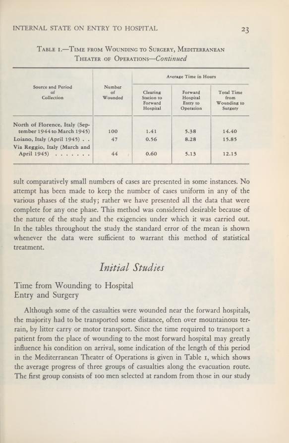
suit comparatively small numbers of cases are presented in some instances. Noattempt has been made to keep the number of cases uniform in any of thevarious phases of the study; rather we have presented all the data that werecomplete for any one phase. This method was considered desirable because ofthe nature of the study and the exigencies under which it was carried out.In the tables throughout the study the standard error of the mean is shownwhenever the data were sufficient to warrant this method of statisticaltreatment.
Initial StudiesTime from Wounding to HospitalEntry and Surgery
Although some of the casualties were wounded near the forward hospitals,the majority had to be transported some distance, often over mountainous ter-rain, by litter carry or motor transport. Since the time required to transport apatient from the place of wounding to the most forward hospital may greatlyinfluence his condition on arrival, some indication of the length of this periodin the Mediterranean Theater of Operations is given in Table 1, which showsthe average progress of three groups of casualties along the evacuation route.The first group consists of 100 men selected at random from those in our study
Average Time in Hours
Source and Periodof
Collection
Numberof
WoundedClearingStation to
ForwardHospital
ForwardHospitalEntry to
Operation
Total Timefrom
Wounding toSurgery
North of Florence, Italy (Sep-tember 1944to March 1945) 100 1.41 5.38 14.40
Loiano, Italy (April 1945). . 47 0.56 8.28 15.85
Via Reggio, Italy (March andApril 1945) 44 0.60 5.13 12.15
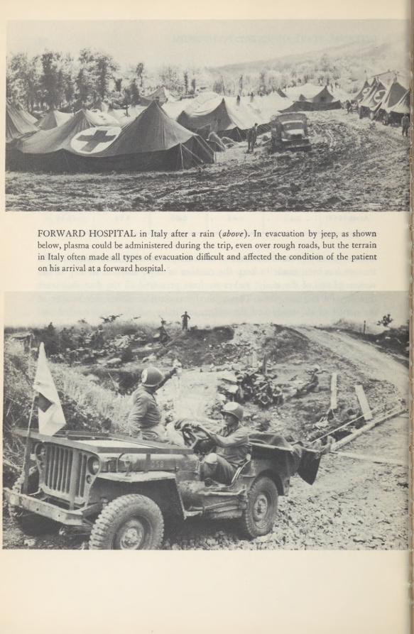
FORWARD HOSPITAL in Italy after a rain {above). In evacuation by jeep, as shownbelow, plasma could be administered during the trip, even over rough roads, but the terrainin Italy often made all types of evacuation difficult and affected the condition of the patienton his arrival at a forward hospital.
INTERNAL STATE ON ENTRY TO HOSPITAL 25
who were wounded during a relatively quiet period in the fall, winter, andspring of 1944-45. The other two groups represent men who were severelywounded during offensives in the spring of 1945. In the third locale cited, itwas contended by those concerned that, considering the circumstances, evacua-tion had been effected rapidly. The table also shows the average time from hos-pital admission to surgery and the total time from wounding to surgery in thethree groups.
Type and Location of WoundsFor various correlations throughout the study wounds are grouped accord-
ing to their type, or location, or both. Many patients incurred multiple wounds,some multiple major wounds. For certain purposes two broad classifications oftype were utilized: peripheral and nonperipheral, and this terminology willbe used whenever pertinent. Nonperipheral wounds were defined as those in-volving the major body cavities (the abdomen, the thorax, and the interior ofthe skull); all others were considered as peripheral. Crush cases are excludedin some of the correlations because they were studied separately.
In the following classification the severe wounds only are considered, sincethey were pertinent to the study. Thus in the patients with multiple severewounds some wounds were listed as the principal major ones; no attempt wasmade to record minor wounds, such as fracture of a phalanx, for example. Ingeneral, the types of wounds found in our patients were as follows:
Severe peripheral wounds were present in 116 patients, constitutinga major injury in 81 instances. Nearly all were wounds of the ex-tremities. Thirty-three patients had peripheral wounds without frac-ture, 16 of which were the patient’s major wound. Fifty-three of 70patients had major peripheral wounds with fracture, and 13 had trau-matic amputation of an extremity. In 10 of these 66, a major woundwas also listed in another category. Three patients among those withperipheral wounds had injury to the spinal cord.
Of the severe nonperipheral wounds, 34 patients had thoracicwounds (a major wound in 30 instances) and 56 patients had intra-abdominal wounds, a major wound in 50 instances. An additional 21
patients had combined thoraco-abdominal wounds and 2 patients hadseparate wounds of the chest and abdomen. Of the total abdominalwounds, there were 25 wounds of the liver, 20 wounds of the kidney
26 THE PHYSIOLOGIC EFFECTS OF WOUNDS
Table2.—Relationshipof
Painto
MajorWoundin215
Patients
(DataTakenfromStudy
onPaininMen
Woundedin
Battle*)
Further
AverageTimefromTotal
DoseLatestDoseTimesince
Pain(degree
PainRelief
TypeofWound
Numberof PatientsAge
WoundingofMorphineofMorphine
latest Morphineandnumber
ofpatientsin
Therapy Wanted
yrs.
hrs.,averagemg.,
average2
mg.,average
hrs.,average
eachgroup)
(numberof patients)
CompoundFracturesof
Long
Bones
50
24.8±0.9
12.5±1.327.0±1.5 (1
pt.none)
22.6
7.0±0.819
none12
slight7
moderate12severe
11yes
39no
ExtensiveSoft-tissueWounds
....
50
24.5±1.1
11.3±1.427.0±2.7 (11
pts.none)
19.5
7.2±0.619none
15slight
8moderate
8severe
9
yes41
no
PenetratingWoundsofThorax.
.
.50
24.5±0.8
9.8±1.025.0±1.8 (11
pts.none)
21.2
6.5±0.615none
18slight
11moderate
6severe
10yes
40no
PenetratingWoundsof
Abdomen
50
22.7±0.67.2±0.729-0±2.2
(5
pts.none)
25.0
4.8±0.77none
5
slight14
moderate24
severe27
yes23
no
PenetratingWoundsof
Cerebrum
15
25.1±1.4
7.9±1.419.8
±4.2(8pts.none)
19.8
6.2±1.59
none5
slight0
moderate1
severe1
yes14no
1
Seefootnote4,
text.
1Patients
whodidnot
receivemorphine
arenot
includedintheaverages.
INTERNAL STATE ON ENTRY TO HOSPITAL 27
(treated by nephrectomy in n instances), and in i case it was notknown whether a kidney or liver wound had been present. Woundsof the urinary tract involving the bladder or structures above it oc-curred in 9 patients. Ten patients with nonperipheral wounds hadmultiple major wounds.
Crush injuries were found in nine patients, and there was only onecase of head injury.
Clinical Condition of Patients on Arrivalat the Most Forward HospitalPain
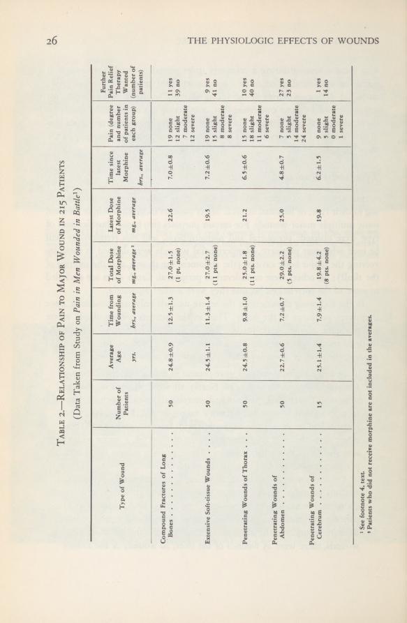
The frequency and severity of pain in different types of wounds had beenextensively studied under similar conditions and on the same types of patientsshortly before the Board was organized and the study was therefore not re-peated on these 186 patients. Part of the data obtained in the early study4 isshown in Table 2. The incidence of severe pain was surprisingly low. The datashowed that severe pain was not to be accounted for on the basis of the pa-tients’ having received less morphine or having received it earlier than patientswho reported little or no pain. It was also pointed out that three factors arechiefly important in the distress of the wounded: pain, mental distress, andthirst. In the severely wounded patient in good general condition, the firsttwo factors are important. In the man in shock, thirst is the main and oftenthe only cause of evident distress, but it may be extreme.
Shoc\Grading of Shoc\. —The view sometimes has been taken that shock is
either present or absent in a given case and that to try to distinguish betweendegrees of shock is futile. In this study, however, it was found instructive to sep-arate the patients arbitrarily into four categories; namely, those with “noshock,” “slight shock,” “moderate shock,” and “severe shock.” This was doneon the basis of the criteria listed in Table 3 which in turn were based on pre-liminary observation of large numbers of battle casualties by members of theBoard. A patient was assigned to a particular category if he exhibited the ma-
* Beecher, H. K.: Pain in men wounded in battle. Ann. Surg. 123: 96-105, January 1946; alsoBull. U. S. Army M. Dept. 5: 445-454, April 1946.

28 THE PHYSIOLOGIC EFFECTS OF WOUNDS
Table3.—Gradingof
Shock* Skin
DegreeofShock
BloodPressure (approx.)
PulseQuality
TemperatureColor
Circulation (responseto pressure, blanching)
Thirst
MentalState
None
Normal
NormalNormalNormalNormalNormal
Clearand
distressed.
Slight
Decreased20%
orless
NormalCool
Pale
Definite slowingNormalClear
anddistressed.
Moderate
Decreased20to
40%
Definite decreasein
volumeCool
Pale
Definite slowingDefinite
Clearand
someapa-
thyunlessstimu-
lated.
Severe
Decreased40%
tonon- recordable
Weakto
im- perceptibleCold
Ashento cyanotic (mot- tling)
Very sluggishSevereApathetictocoma-
tose;little
distressexcept
thirst.
*
Itwillbeobservedthatsweating,
nausea,andvomiting
arenotincluded,thanthey
areto
shock.Thepulse
ratecanbe
influencedby
toomany
unim-
althoughthese
criteriawere
frequentlyreferred
toinWorldWarI
reports.In
portantfactorsto
havevaluein
estimatingdegreeofshock;pulsequality,
how-
theBoard’s
experiencethey
werefoundtobe
uncommonandofno
valuein
ever,is
important.Thelastthree
columnsofthetableincludeitemsnot
estimatingthe
extentof
shock.They
areprobably
moreclosely
relatedto
ordinarilyconsideredin
evaluatinga
patient’scondition;sincewe
foundthem
psychologicfactors,to
thenature
ofthe
wound,orto
reactionto
morphineuseful,they
areincluded.

INTERNAL STATE ON ENTRY TO HOSPITAL 29
jority of criteria for that category as opposed to another. These signs were in-adequate, of course, for management of a case, for a comprehensive appraisalof the patient’s condition must include not only an accurate concept of hispresent state but also a shrewd estimate of his probable course in the imme-diate future.
On the basis of this arbitrary classification the 186 patients under studywere evaluated as to the degree of shock they had at the time of their admis-sion to the hospital. In three of them the degree of shock could not be ascer-tained. Those 78 patients who were not seen on admission by any member ofthe Board were classified by the Board on the basis of the available clinicaldata and on discussion with medical officers who had seen them on admission.In the 108 patients who had been observed on admission by some member ofthe Board the degree of shock was probably more uniformly classified. Table 4shows the clinical evaluation of shock and its distribution in these 108 patientsas well as in the entire series. It is apparent that the distribution remainedabout the same when the 108 were separated from the entire group. Since thepercentages were essentially unchanged when the magnitude of the cases wasroughly doubled, it was assumed that the size of the sample 108 cases wasadequate.
Table 4.—Classification and Distribution of Shock
Relationship to Time from Wounding.—Examination of the records of 167of the entire group of severely wounded men under study on whom these data
Degree of Shock
All PatientsPatients Seen on
Admission byMembers of Board
NumberPercent
ofTotal
NumberPercent
ofTotal
None 34 18.3 20 18.5Slight 37 20.0 27 25.0Moderate 55 29-5 34 31.5Severe 57 30.6 26 24.1Unclassified 3 1.6 1 0.9
Total 186 100.0 108 100.0

Table6.—WoundCompositionin
EachShock
Category—121
Patients
MajorWoundComposition
Degreeof
Shock
Numberof Patients
Pene- trated AbdomenPene- trated Chest
Lacerated Soft TissueTraumatic Amputation
ofExtremity1
CompoundFractureof
LongBones2
Miscellaneous Woundsand Injuries
Casesof
Concomitant Injuries
None Percentage....
24
7 29%
1 4%
5 21%
2 8%
■
1 4% *(8%)7
Crush 29%
]
Abdominalwoundandcompound,
com-
[
minutedfractureofhumerus—1
case.
Slight Percentage....
24
7 29%
3 13%
5 21%
2 8%
3 13%1
Crush1
Penetratedface
1
Compound,com-
minutedfractureof
sacrum1
Fracturedvertebra
17%
]
[•Abdominalwoundand
penetratingchest
|
wound—Icase.
Moderate Percentage....
36
5
4
Abdominalwoundand
penetratingchest
wound—3
cases.Abdominalwoundandcompound,
com-
minutedfractureof
bothbonesofleg
—1case. Abdominalwoundandcompound,
com-
minutedfractureofhumerus—1
case.
36%
22%
14%
17%
11%M9%)
Chestwoundandcompound,
commin-
utedfractureof
armbones—1
case.
Soft-tissuewoundandfractured
skull
—1
case. Traumaticamputationand
compound,
comminutedfractureof
bonesof
other
leg—1case.
Severe Percentage....
37
10 27%
5 14%
3 8%
9 24%
8 22% *(24%)1
Crush1
Fracturedpelvis
5%
Abdominalwoundand
penetratingchest
wound—2cases.Chestwound
andcompound,
commin-
utedfractureofhumerous—1case.
Chestwound
andcompound,
commin-
utedfractureofilium—1
case.
1
Includessomeca
*
Petcentagesmarsesin
whichcedwith
astersurgicalampu
iskincludeth
tationwas
undertakenearly.
e
compoundfractures
listedunder
concomitantinjuries.
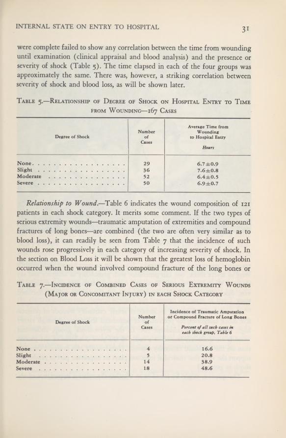
INTERNAL STATE ON ENTRY TO HOSPITAL 31
were complete failed to show any correlation between the time from woundinguntil examination (clinical appraisal and blood analysis) and the presence orseverity of shock (Table 5). The time elapsed in each of the four groups wasapproximately the same. There was, however, a striking correlation betweenseverity of shock and blood loss, as will be shown later.
Table 5.—Relationship of Degree of Shock on Hospital Entry to Timefrom Wounding—167 Cases
Relationship to Wound. —Table 6 indicates the wound composition of 121
patients in each shock category. It merits some comment. If the two types ofserious extremity wounds—traumatic amputation of extremities and compoundfractures of long bones—are combined (the two are often very similar as toblood loss), it can readily be seen from Table 7 that the incidence of suchwounds rose progressively in each category of increasing severity of shock. Inthe section on Blood Loss it will be shown that the greatest loss of hemoglobinoccurred when the wound involved compound fracture of the long bones or
Table 7,—Incidence of Combined Cases of Serious Extremity Wounds(Major or Concomitant Injury) in each Shock Category
Degree of ShockNumber
ofCases
Average Time fromWounding
to Hospital Entry
Hours
None 29 6.7 ±0.9Slight 36 7.6±0.8Moderate 52 6.4±0.5Severe 50 6.9±0.7
Degree of ShockNumber
ofCases
Incidence of Traumatic Amputationor Compound Fracture of Long Bones
Percent ofallsuch cases ineach shock group, Table 6
None 4 16.6Slight 5 20.8Moderate 14 38.9Severe 18 48.6
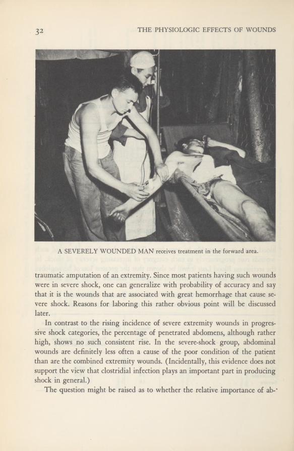
THE PHYSIOLOGIC EFFECTS OF WOUNDS32
A SEVERELY WOUNDED MAN receives treatment in the forward area.
traumatic amputation of an extremity. Since most patients having such woundswere in severe shock, one can generalize with probability of accuracy and saythat it is the wounds that are associated with great hemorrhage that cause se-vere shock. Reasons for laboring this rather obvious point will be discussedlater.
In contrast to the rising incidence of severe extremity wounds in progres-sive shock categories, the percentage of penetrated abdomens, although ratherhigh, shows no such consistent rise. In the severe-shock group, abdominalwounds are definitely less often a cause of the poor condition of the patientthan are the combined extremity wounds. (Incidentally, this evidence does notsupport the view that clostridial infection plays an important part in producingshock in general.)
The question might be raised as to whether the relative importance of ab-

INTERNAL STATE ON ENTRY TO HOSPITAL 33
dominal wounds as a cause of shock has heretofore been exaggerated. The poorprognosis often encountered in patients with abdominal wounds probably hasa great deal to do with the apprehension felt in the presence of such lesions.The concealed hemorrhage or concealed contamination often present in thesecases may lead to subsequent profound shock. So, while on the average ab-dominal wounds were not as often a cause of severe shock on hospital entryas were serious extremity wounds, the impossibility of accurate preoperativeappraisal of the abdominal wound makes it difficult to exaggerate its poten-tialities.
Shock will be discussed in each of the following sections of this chapter andan attempt will be made to correlate degree of shock with the data under dis-cussion whenever possible.
Cardiovascular SystemElectrocardiographic Observations.—Prior to organization of the Board, 58
electrocardiographic records were made on 30 patients in severe shock andafter recovery. 0 Since the observations were made on the same type of patientsas those studied by the Board and under similar circumstances, electrocardio-grams were not made on the Board’s cases. The results of that study are sum-marized here. In 10 patients (one-third of that series) the blood pressure couldnot be measured on hospital entry. In the other two-thirds the degree of circu-latory collapse was somewhat less severe, but even so the systolic blood pres-sures ranged from 60 to 70 millimeters of mercury and the diastolic from 20 to
40 millimeters.Definite abnormalities of the electrocardiograms were observed in 5 of the
30 patients. The most striking feature was the normal character of the findingsin the remaining twenty-five. In 2 of the 5 patients with abnormal findings,the electrocardiograms showed striking but transient inversion of the T wavein lead 1. In a patient with an intrathoracic injury there was a shift frommarked right-axis deviation back to normal following operation. The electro-cardiogram in the fourth patient showed bizarre QRS complexes of low volt-age, and in the fifth showed evidence of an unusual degree of temporary car-diac irritability with paroxysmal fibrillation and ventricular tachycardia.
5 Burnett, C. H.; Bland, E. F„ and Beecher, H. K.: Electrocardiograms in traumatic shock in manJ. Clin. Investigation 24; 687-690, September 1945.
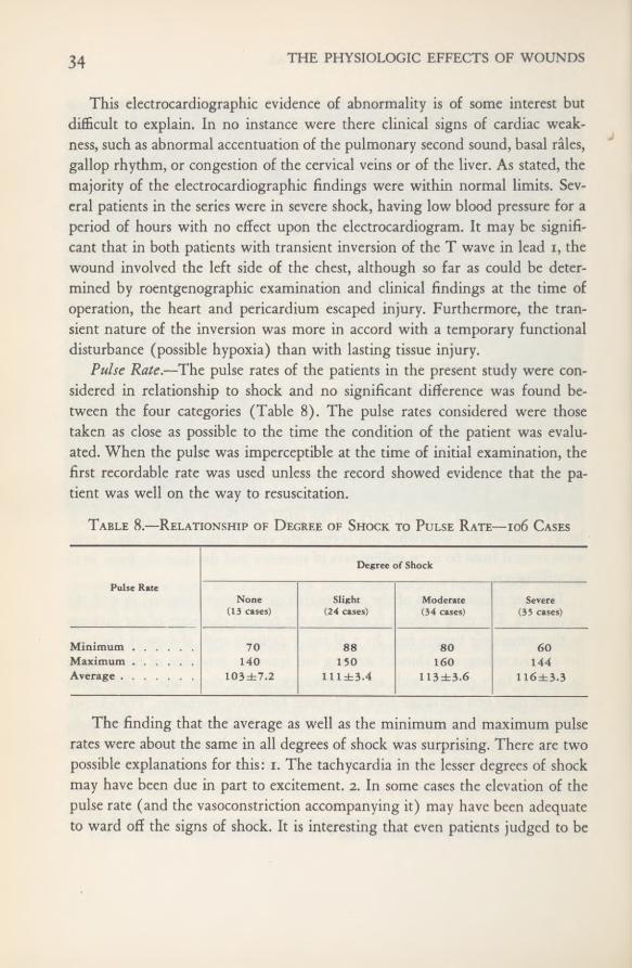
THE PHYSIOLOGIC EFFECTS OF WOUNDS34
This electrocardiographic evidence of abnormality is of some interest butdifficult to explain. In no instance were there clinical signs of cardiac weak-ness, such as abnormal accentuation of the pulmonary second sound, basal rales,gallop rhythm, or congestion of the cervical veins or of the liver. As stated, themajority of the electrocardiographic findings were within normal limits. Sev-eral patients in the series were in severe shock, having low blood pressure for aperiod of hours with no effect upon the electrocardiogram. It may be signifi-cant that in both patients with transient inversion of the T wave in lead 1, thewound involved the left side of the chest, although so far as could be deter-mined by roentgenographic examination and clinical findings at the time ofoperation, the heart and pericardium escaped injury. Furthermore, the tran-sient nature of the inversion was more in accord with a temporary functionaldisturbance (possible hypoxia) than with lasting tissue injury.
Pulse Rate.—The pulse rates of the patients in the present study were con-sidered in relationship to shock and no significant difference was found be-tween the four categories (Table 8). The pulse rates considered were thosetaken as close as possible to the time the condition of the patient was evalu-ated. When the pulse was imperceptible at the time of initial examination, thefirst recordable rate was used unless the record showed evidence that the pa-tient was well on the way to resuscitation.
Table 8.—Relationship of Degree of Shock to Pulse Rate —106 Cases
The finding that the average as well as the minimum and maximum pulserates were about the same in all degrees of shock was surprising. There are twopossible explanations for this: i. The tachycardia in the lesser degrees of shockmay have been due in part to excitement. 2. In some cases the elevation of thepulse rate (and the vasoconstriction accompanying it) may have been adequateto ward off the signs of shock. It is interesting that even patients judged to be
Degree of Shock
Pulse RateNone
(13 cases)Slight
(24 cases)Moderate(34 cases)
Severe(35 cases)
MinimumMaximumAverage
70140
103±7.2
88150
111 ±3.4
80160
113±3.6
60144
116±3.3

INTERNAL STATE ON ENTRY TO HOSPITAL 35
in severe shock can have a pulse rate as low as 60 beats per minute. Of greatersignificance than the actual rate of the pulse is its volume, which often wasdecreased so much in severe shock that the pulse could no longer be felt.
Blood Pressure.—Blood pressures were analyzed in only those 70 cases outof the 186 in which they had been recorded at the time the patient’s conditionwas evaluated. The volume of the circulating blood was also determined inthese 70 patients. There was no significant fall in the average systolic bloodpressure except in those in moderate or severe shock (Table 9). It will be shownthat these patients with considerable shock had lost on the average 33.6 per-cent of their calculated normal blood volume and nearly 50 percent of the totalcirculating hemoglobin. In those with severe shock, the systolic blood pressurefell rapidly, the average being 49 millimeters of mercury. This group had lostapproximately half the normal blood volume (see Table 22). There was, how-ever, a progressive drop in the average diastolic blood pressure with increasingdegrees of shock (Table 9). The average diastolic blood pressure of the pa-tients in severe shock was half that of the patients in moderate shock. As se-verity of shock increased, there was a significant and progressive decline in thepulse pressure (Table 9). This confirmed the clinical observation that the vol-ume of the pulse was closely correlated with the degree of shock.Table 9.—Relationship of Degree of Shock to Blood Pressure—70 Cases
“Irreversible” Cha?iges in the Cardiovascular System.—Everyone who hastreated many patients for shock has encountered some who fail to respond to
the transfusion of blood deemed adequate under ordinary circumstances, andthis is often attributed to “irreversible” changes that have presumably takenplace during prolonged hypotension, ischemia, and anoxia. This problem is
956507 0—52 4
Degree of Shock
Systolic Blood Pressure
mm. Hg
Diastolic Blood Pressure
mm. Hg
Pulse Pressure
mm. Hg
Lowest Highest Average Lowest Highest Average Lowest Highest Average
None (13 cases) 108 150 126±11.9 64 80 75 ±1.5 30 80 52 ±4.1Slight (20 cases) 90 140 109±3.0 40 86 66±2.7 28 80 44±2.7Moderate (21 cases) .... 30 136 95 ±4.9 20 90 58±3.5 10 56 36±2.8
Severe (16 cases) *0 80 49±7.6 *0 68 2 5 ±5.8 *0 58 24 ±4.7
* When the values were unmeasurable (2 cases) they were considered to be 0.
THE PHYSIOLOGIC EFFECTS OF WOUNDS
further discussed in the section on Blood Loss. In most instances, adequate ex-
planation can be found for the failure of patients in shock to respond to bloodtransfusion; some common examples are concealed and continuing hemor-rhage, hemothorax, irritant contamination of the peritoneum, peritonitis, clos-tridial myositis, and fat emboli. Four cases from our series are illustrative.
CASE REPORTS
Case 77. —A patient with a severe thoraco-abdominal wound was received at a forwardhospital in severe shock 814 hours after he was wounded. Resuscitative measures were
continued for nearly 9 hours. During that time he received only 1,500 cc. of whole blood.His condition failed to improve and he was operated upon but did not survive theoperation.
Necropsy showed massive collapse of the right lung with a plug of mucus in the rightmain bronchus. The lower lobe of the left lung was collapsed and about one-third of theleft upper lobe was atelectatic. There was gross dilatation of the right ventricle of theheart. On histologic examination minimal evidence of fat embolism in the pulmonaryvessels was found but considered of no clinical significance.
Comment.—There was adequate cause for this patient’s failure to respondto resuscitation. More aggressive measures should have been taken, includingbronchoscopy and the use of more blood in less time. There should have beenmore concern when no improvement occurred during the first 3 hours afterthe patient’s admission to the hospital.
Case 45.—A patient with a severe abdominal wound was admitted to a forward hos-pital in severe shock 8 hours after wounding. During the next 3 hours, 2 units (600 cc.total volume) of plasma and 1 liter of whole blood were transfused. The blood pressureduring that time changed from imperceptible to 90 millimeters of mercury systolic and70 diastolic. An additional unit of plasma was administered and an infusion of 500 cc.of 2-percent solution of sodium bicarbonate was given intravenously. Although this wasthe optimum time for surgery, operation was delayed and 5 hours later the blood pressurewas again unmeasurable. It was restored to 86 millimeters of mercury systolic and 60diastolic after transfusion of 1 liter of whole blood, and operation was performed whichlasted 4 hours.
At operation the abdominal cavity was found to be “full of blood.” The blood pressureand pulse rate were unmeasurable during much of the operation. The patient neverregained consciousness and died 3% hours after the end of the operation. Necropsy showedperforation of the inferior vena cava. There was histologic evidence of minimal fat embo-lism in the pulmonary vessels, probably of no clinical significance.
Comment.—The recurrent hypotension in this patient was probably due tocontinued extraperitoneal and intraperitoneal hemorrhage. Operation should

INTERNAL STATE ON ENTRY TO HOSPITAL 37
have been performed while he was responding well to resuscitative measuresduring the first 3 hours after admission to the hospital.
Case 100.—This patient had multiple wounds involving both arms, the left thigh, andthe face. There were compound fractures of the left humerus, radius, and ulna, and ofthe right ulna. There was also a transection of the right femoral artery with vascularinsufficiency in the leg. He was admitted in severe shock to a forward hospital 3% hoursafter wounding, and within 90 minutes he received 300 cc. (total volume) of plasma and2 liters of whole blood. He showed general improvement but his blood pressure was stillonly 80 millimeters of mercury systolic and 50 diastolic. His pulse rate was 144 beats perminute. Three hours after admission his blood pressure was 90 millimeters of mercurysystolic and 58 diastolic. Operation was delayed for 3 additional hours. At no time duringoperation did the recorded blood pressure fall below 85 millimeters of mercury systolic.The patient had received a total of 4,500 cc. of whole blood before, during, and immedi-ately after operation.
Ten hours after operation the patient’s blood pressure was low and he looked paleand “anemic.” A transfusion was started, but an hour later he suddenly died. Ten minutesearlier he had carried on an intelligent conversation. Pulmonary embolus was suspectedbut at necropsy no cause could be found for the sudden death. Microscopically, a moder-ately severe grade of fat embolism was found in the lungs.
Comment.—The question was raised whether the 5- or 6-hour period ofhypotension in this patient could have caused irreversible changes in thecardiovascular system so that it simply “gave out” when it did. This cannot beanswered with certainty. The fat embolism in retrospect appears to be the moreimportant consideration.
Case 120.—This patient had a simple penetrating wound of the thigh caused by a shellfragment. The femoral artery below the origin of the profunda femoris was severed.During evacuation the patient had received 4 units (1,200 cc. total volume) of plasma andwhen he reached the evacuation hospital, about 9 hours after wounding, he must haveappeared in good condition for no resuscitation was deemed necessary. At operation, per-formed 4V2 hours after admission, the femoral artery, vein, and nerve were found to becompletely transected. The vessels were ligated and the foreign body was removed.
At the conclusion of the operation, the systolic blood pressure was only 70 millimetersof mercury and remained between 70 and 60 throughout the day. Despite- this the patientappeared to have good color and his skin was not cold. Transfusion of 1 liter of wholeblood did not improve the blood pressure. The right leg looked as though it would notsurvive. Anuria developed and the patient died 48 hours after operation. Necropsy revealednothing to account for the postoperative hypotension. There was no concealed hemorrhageand no evidence of clostridial myositis in the involved extremity. There was no histologicevidence of fat embolism.
Comment. —This patient probably had lost more blood than was realized.
THE PHYSIOLOGIC EFFECTS OF WOUNDS
Resuscitation
Before admission of the patients in this study to a forward hospital, resus-citative efforts had been limited chiefly to control of pain and hemorrhageand to administration of blood plasma. Relatively little whole blood was given.The 108 patients seen by us on admission had received, on the average, 2 units6
of plasma before the first blood sample was taken. Plasma administration wasdistributed in this group as follows:
Thus 69 percent (74) of these patients had received two units (600 cc.) or lessof blood plasma before or shortly after arrival at the most forward hospital.Twenty-seven of the 108 patients received transfusions of whole blood prior towithdrawal of the first blood specimen for laboratory analysis. Three of these,or 3 percent, had received whole blood in an aid station before admission to aforward hospital. The blood transfusions were distributed as follows:
The following tabulation summarizes the average quantities of blood andof blood plasma used in resuscitating 157 of the very seriously wounded pa-tients in our series (the 108 referred to above and 49 others on whom we hadclinical notes):
Blood plasma preoperatively (average of 122 cases) 3.08 unitsBlood plasma during operation (average of 10 cases) 1.68 unitsWhole blood preoperatively (average of 127 cases) M50 cc -Whole blood during operation (average of 95 cases) 1,160 cc.
1 unit — 250 cc. of normal plasma diluted to 300 cubic centimeters.
Number ofPatients
22
Units ofPlasma
_ _ noneNumber of
PatientsI
Units ofPlasma
62K 1 I 717 2 2 813 2 X q
12 4 I 11
3 5
Number ofPatients
1310
Units of Blood
21
I
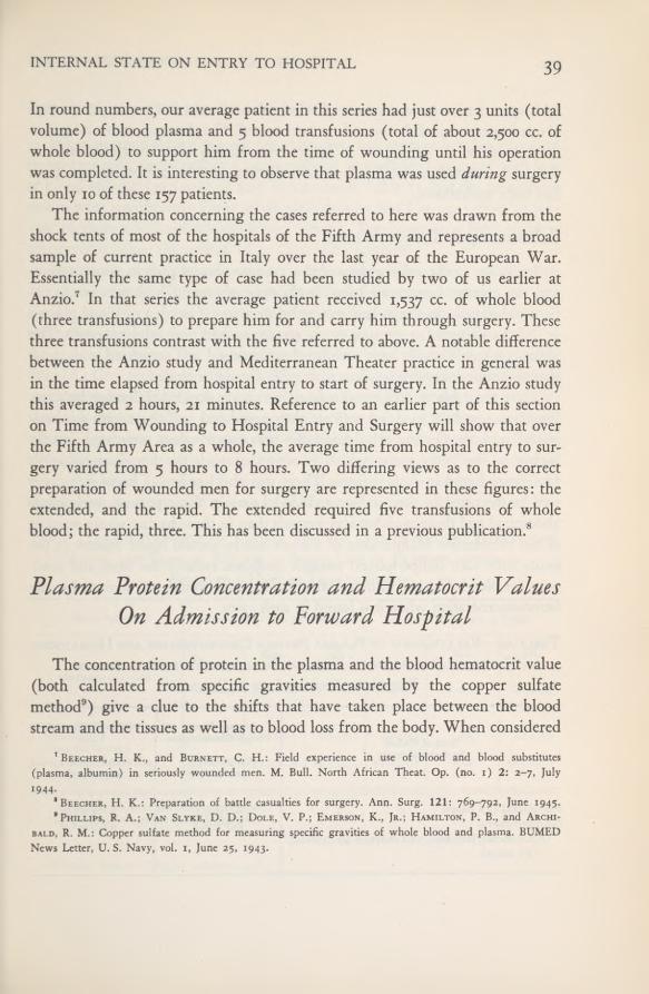
INTERNAL STATE ON ENTRY TO HOSPITAL 39
In round numbers, our average patient in this series had just over 3 units (totalvolume) of blood plasma and 5 blood transfusions (total of about 2,500 cc. ofwhole blood) to support him from the time of wounding until his operationwas completed. It is interesting to observe that plasma was used during surgeryin only 10 of these 157 patients.
The information concerning the cases referred to here was drawn from theshock tents of most of the hospitals of the Fifth Army and represents a broadsample of current practice in Italy over the last year of the European War.Essentially the same type of case had been studied by two of us earlier atAnzio. 7 In that series the average patient received 1,537 cc- whole blood(three transfusions) to prepare him for and carry him through surgery. Thesethree transfusions contrast with the five referred to above. A notable differencebetween the Anzio study and Mediterranean Theater practice in general wasin the time elapsed from hospital entry to start of surgery. In the Anzio studythis averaged 2 hours, 21 minutes. Reference to an earlier part of this sectionon Time from Wounding to Hospital Entry and Surgery will show that overthe Fifth Army Area as a whole, the average time from hospital entry to sur-gery varied from 5 hours to 8 hours. Two differing views as to the correctpreparation of wounded men for surgery are represented in these figures: theextended, and the rapid. The extended required five transfusions of wholeblood; the rapid, three. This has been discussed in a previous publication. 8
Plasma Protein Concentration and Hematocrit ValuesOn Admission to Forward Hospital
The concentration of protein in the plasma and the blood hematocrit value(both calculated from specific gravities measured by the copper sulfatemethod9 ) give a clue to the shifts that have taken place between the bloodstream and the tissues as well as to blood loss from the body. When considered
’Beecher, H. K., and Burnett, C. H.: Field experience in use of blood and blood substitutes(plasma, albumin) in seriously wounded men. M. Bull. North African Theat. Op. (no. i) 2; 2-7, July1944.
‘Beecher, H. K.: Preparation of battle casualties for surgery. Ann. Surg. 121: 769-792, June 1945.‘Phillips, R. A.; Van Slyke, D. D.; Dole, V. P.; Emerson, K., Jr.; Hamilton, P. B., and Archi-
bald, R. M.: Copper sulfate method for measuring specific gravities of whole blood and plasma. BUMEDNews Letter, U. S. Navy, vol. 1, June 25, 1943.
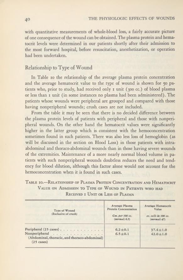
THE PHYSIOLOGIC EFFECTS OF WOUNDS40
with quantitative measurements of whole-blood loss, a fairly accurate pictureof one consequence of the wound can be obtained. The plasma protein and hema-tocrit levels were determined in our patients shortly after their admission to
the most forward hospital, before resuscitation, anesthetization, or operationhad been undertaken.
Relationship to Type of WoundIn Table 10 the relationship of the average plasma protein concentration
and the average hematocrit value to the type of wound is shown for 50 pa-tients who, prior to study, had received only 1 unit (300 cc.) of blood plasmaor less than 1 unit (in some instances no plasma had been administered). Thepatients whose wounds were peripheral are grouped and compared with thosehaving nonperipheral wounds; crush cases are not included.
From the table it may be seen that there is no decided difference betweenthe plasma protein levels of patients with peripheral and those with nonperi-pheral wounds. On the other hand the hematocrit values were significantlyhigher in the latter group which is consistent with the hemoconcentrationsometimes found in such patients. There was also less loss of hemoglobin (aswill be discussed in the section on Blood Loss) in those patients with intra-abdominal and thoraco-abdominal wounds than in those having severe woundsof the extremities. Maintenance of a more nearly normal blood volume in pa-tients with such nonperipheral wounds doubtless reduces the need and tend-ency for blood dilution, although this factor alone would not account for thehemoconcentration when it is found in such cases.
Table 10.—Relationship of Plasma Protein Concentration and HematocritValue on Admission to Type of Wound in Patients who had
Received i Unit or Less of Plasma
Type ofWound(Exclusive of crush)
Average PlasmaProtein Concentration
Gm.per 100 cc.{normal; 6.5)
Average HematocritValue
cc. cells in 100 cc.{normal; 47)
Peripheral (2 5 cases) 6.2 ±0.1 37.4±1.0Nonperipheral 6.5±0.1 42.0± 1.0
(Abdominal, thoracic, and thoraco-abdominal)(2 5 cases)
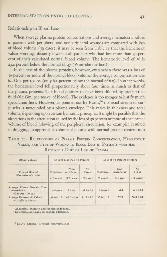
INTERNAL STATE ON ENTRY TO HOSPITAL 4 1
Relationship to Blood LossWhen average plasma protein concentrations and average hematocrit values
in patients with peripheral and nonperipheral wounds are compared with lossof blood volume (39 cases), it may be seen from Table 11 that the hematocritvalues were significantly lower in all patients who had lost more than 30 per-cent of their calculated normal blood volume. The hematocrit level of 36 is23.4 percent below the normal of 47 (Wintrobe method).
In the case of the plasma proteins, however, even when there was a loss of30 percent or more of the normal blood volume, the average concentration was6.1 Gm. per 100 cc. (only 6.1 percent below the normal of 6.5). In other words,the hematocrit level fell proportionately about four times as much as that ofthe plasma proteins. The blood appears to have been diluted by protein-richfluid (6.1 Gm. per 100 cc. of blood). The evidence is too meager to justify muchspeculation here. However, as pointed out by Evans, 10 the axial stream of cor-puscles is surrounded by a plasma envelope. This varies in thickness and totalvolume, depending upon certain hydraulic principles. It might be possible that thealterations in the circulation caused by the loss of 30 percent or more of the normalvolume of blood (slowing of the peripheral circulation, for example) resultedin dragging an appreciable volume of plasma with normal protein content into
Table ii.—Relationship of Plasma Protein Concentration, HematocritValue, and Type of Wound to Blood Loss in Patients who had
Received i Unit or Less of Plasma
10 Evans, Robley: Personal communication.
Blood Volume Loss of Less than 30 Percent Loss of 30 Percent or More
Non- All Non- AllType of Wound Peripheral peripheral 1 Types Peripheral peripheral 1 Types
(Exclusive ofcrush)(10 cases) (17 cases) (27 cases) (8 cases) (4 cases) (12 cases)
Average Plasma Protein Con-centration 2 6.4 ±0.1 6.5 ±0.1 6.5 ±0.1 6.0 ±0.1 6.4 6.1 ±0.1
(Gm. per 100 cc.)
Average Hematocrit Value 2.
.39.0±1,7 43.0±1.0 41.5 ±1.0 35.0±1.3 37.8 36.0±1.1
(cc. cells in 100 cc.)
1 Abdominal, thoracic, and thoraco-abdominal.2 Determinations made on hospital admission.

THE PHYSIOLOGIC EFFECTS OF WOUNDS42
the circulating blood. Or it might be possible that protein was brought into thecirculation from the liver.
Influence of Plasma Therapy
The influence of previous administration of blood plasma upon the con-centration of plasma protein and the hematocrit value was considered, and thefindings in different types of wounds are shown in Table 12 and Charts 1 and2. Only three of the patients had had blood transfusions; these will be ignored.It is clear from the table and charts that plasma therapy did not influence theplasma protein level, but it did have an important effect on the hematocritlevel.
The plasma protein concentration and hematocrit value were also analyzedin regard to shock, the data being broken down into two categories in which“no shock” and “slight shock” were grouped together, as were “moderateshock” and “severe shock.” No important differences were found between thetwo categories.
Table 12.—Effect of Plasma Therapy on Average Plasma Protein and
Hematocrit Admission Levels in 89 Patients with Various Types of Wounds
Plasma Time fromType of Wound Number Units of Protein Hematocrit Value Wounding
(Crush of Plasma Concentration to Determinationexcluded) Cases Received
Gm. per 100 cc. cc. cells in 100 cc. hours
[ 25 0 to 1 6.2 ±0.1 37.4±1.0 8.5 ±1.2Peripheral .... 17 2 to 3 5.9±0.1 3l.9±1.5 *8.1 ±1.5i 13 4 to 6 6.1 ±0.1 *28.6±1.9 9.8±1.2
Total . .
. 55 0 to 6 6.1 ±0.1 33.7 ±0.9 8.7 ±0.7
( 11 0 to 1 6.6 ±0.2 44.1 ±1.8 6.3 ±1.4Abdominal.... 5 2 to 4 6.7 ±0.3 40.2 ±4.3 7.8 ±3.1i 3 5 to 11 6.1 *32.2 *12.2
Total . . . 19 0 to 11 6.6 ±0.1 41.6±1.9 6.8±1.3
Thoracic( 8<
0 to 1 6.5 ±0.2 40.0±1.2 7.9±2.61 7 2 to 5 6.6 ±0.2 35.4±1.8 7.2 ±1.7
Total . . • 15 0 to 5 6.5 ±0.1 3 7.8 ±1.2 7.6±1.4
* No determination made in one case. (Determinations made on hospital admission.)

INTERNAL STATE ON ENTRY TO HOSPITAL 43
CHART i. Influence of plasma therapyon plasma protein and hematocrit levels in
peripheral wounds
CHART 2. Influence of plasma therapyon plasma protein and hematocrit levels in
abdominal wounds
Relationship to ShockPlasma protein and hematocrit levels were also studied in relation to the
clinical condition of about ioo badly wounded patients (crush cases excluded)on admission to the forward hospital. The fall in average plasma protein con-centration is probably significant as the cases are grouped in Table 13 andChart 3; the fall in hematocrit value is definitely significant. There was noevidence of hemoconcentration. When the patients were grouped according to
Table 13.—Plasma Protein and Hematocrit Levels on Admissionin Relation to Shock*
Degree of Shock None Slight Moderate Severe All Cases
Average Plasma ProteinConcentration
(Gm. per 100 cc.)Average Hematocrit Value
(cc. cells in 100 cc.)
6.6±0.1(15 cases)42.5 ± 1.7(15 cases)
6.4±0.1(26 cases)38.4±1.5(26 cases)
6.2 ±0.1(34 cases)34.6±1.0(33 cases)
6.0±0.1(2 5 cases)31.5± 1.5(24 cases)
6.3±0.1(100 cases)36.1±0.8(98 cases)
* All types of wounds, exclusive of crush cases.

THE PHYSIOLOGIC EFFECTS OF WOUNDS44
Table 14—PlasmaProtein and Hematocrit Levels on Admission in Relationto Shock in Patients with Abdominal Wounds
CHART 3. Plasma protein and hematocritlevels in relation to shock—all types of
wounds
location of the major wound, there was no significant fall in the concentrationof plasma protein in those with peripheral wounds in relation to the degree ofshock. The findings in patients with abdominal wounds showed up differently(Table 14). In the group with minimal shock, the plasma protein concentra-tion may possibly be accounted for by weeping of the irritated peritoneal sur-
faces, fluid being released which contained less protein than the plasma. Asshock became moderate or severe, probably due to greater blood loss, theplasma protein concentration fell to a figure like that for extremity wounds,with hemodilution overcoming the effects of exudation. These data indicatethat the plasma protein and hematocrit values can vary independently.
Degree of Shock None—Slight Moderate—Severe
Average Plasma Protein Concentration . . . 6.9±0.1 6.1 ±0.2(Gm, per 100 cc.) (10 cases) (9 cases)
Average Hematocrit Value 47.0± 1.6 34.9±1.8(cc. cells in 100 cc.) (10 cases) (8 cases)
Average Hours after Wounding 7.0±1.5 11.6(10 cases) (9 cases)

INTERNAL STATE ON ENTRY TO HOSPITAL 45
Blood LossVolume and Hemoglobin
The quantity of blood a wounded man can lose and yet recover has gener-ally been underestimated. One indication that this is so was the fact, wellshown in the prolonged campaigns of the Mediterranean Theater, that robustyoung soldiers tolerated surgery well, long before the blood volume or eventhe blood pressure had been restored to normal. Actually the concept of restor-ation of the patient in shock to normal prior to surgery is based upon a falsepremise. Full organic restoration probably requires days to achieve. A good re-sponse of a young wounded man to treatment is by no means admissible evi-dence that his circulatory system has been restored to normal; it is evidenceof the existence of safety factors in human physiology. These points have beendiscussed elsewhere. 11
In the belief that measurement of the blood loss that had been sustained bythese severely wounded men by the time of their arrival at a forward hospitalwould clarify the matter of the importance of whole blood for the wounded,such a study was carried out. The direct relationship between quantity of bloodlost and degree of shock had long been recognized, but further evidence of thisrelationship was desirable in view of the ever-recurring suggestions that thecause of shock is mysterious and to be explained by the presence of toxins inthe body or by the breakdown of some vague but vital force.
The blood volume and the hemoglobin concentration were determined in67 patients 12 shortly after their arrival at the most forward hospital (which inmost instances was a field hospital). The blood volume loss and the totalhemoglobin loss, expressed as percentages of a calculated normal for each pa-tient, were then determined on the basis of these findings. Normal blood vol-ume was considered to be 8.5 percent of the body weight, after Gregersen.(See Appendix C for the method used.) Peters 13 has commented on the lossof the dye T-1824 from the blood stream. Such loss of dye would of course
11 See footnote 8. Also see section on Resuscitation in volume on general surgery of the series: TheMedical Department of the United States Army. To be published.
15 The blood studies were actually made in yi cases (see Table 15), but one case was discardedbecause of a probable technical error and three others are excluded from the present discussion since theyrepresent types not common to the group; namely, two crush injuries (Cases 93 and 124) and a headinjury (Case A-7).
13 Peters, J. P.: Role of sodium in production of edema. New England J. Med. 239: 353-362, Sept. 2,
1948.

46 THE PHYSIOLOGIC EFFECTS OF WOUNDS
Table 15.—Initial Blood Changes in 71 Severely Wounded Men
' Case of crush injury,(0): Not measurable.
BloodBlood Volume Deviation
from Normal
CirculatingHemoglobin
Pressureat Time of
Blood
Degreeof
ShockBloodSugar
Timeafter
Wound-percent
Hemo-globinPresent
Hemo-globin
CaseNo.
VolumeDetermina-
tion
ing
FromObserved
Value
ByCorrection
Deviationfrom Nor-
mal by Correction B
mm. Hgmg. per100 cc. hours A B grams percent
53 80/40 Moderate 106 3 H -10.6 -22.9 -22.9 689.2 -37.883 130/70 Slight 53i + 13.1 -3.3 -0.8 941.1 -16.191 108/78 None 2H -26.0 -37.7 -37.7 464.3 -52.9
>93 (0) Severe 14'A -11.7 -34.0 -30.3 1,203.8 +2.6100 80/50
150/804 lA
3 H
-28.4
-21.3
-69.0
-36.9
-65.0 551.3
702.8
-76.6
102 None -29.1 -39.0107 80/50 Moderate 374 3 -20.7 -78.6 -68.9 419.2 -77.6108 58/0 Severe 372 3 XA -25.5 -38.9 -30.0 341.9 -64.1110 (0)
120/80y. -35.7
-2.7-37.6-7,1
-37.6-7.1
553.5851.8
-47.4111 Slight 157 14H -5.1
113 94/58 ... .do.. .. 149 3 A -14.9 -20.2 -20.2 573.1 -28.2
122 50/30 Severe 87 4 -34.4 -55.0 -48.2 632.3 -58.4> 124 120/64 None 10a + 15.3 -2.8 -2.8 1,012.5 -1.6
125 72/58 Severe 125 23 -21.3 -33.0 -29.1 562.2 -52.7126 100/60 Moderate 183 2h -30.9 -32.3 -32.3 666.6 -44.4
127 90/40 Slight 11 -39.8 -59.4 -44.2 331.2 -65.9128 110/80 ....do.... 184 6'A +6.2 -49.8 -32.6 837.8 -39.8129 120/64 Moderate 191 5A -21.7 -30.9 -21.7 485.6 -44.2130 60/20 Severe 272 5 -31.3 -51.6 -40.5 410.9 -62.0131 40/20 .
• ..do.... 177 IK -29.8 -43.6 -39.0 477.5 -54.3
134 120/76 None 15H -7.8 -11.3 -7.8 1,098.9 -3.8139 (0) Severe 9 -19.3 -75.7 -23.3 491.0 -54.5140 100/60 Slight 141 4A + 16.8 -1.1 -1.1 1,002.2 -5.8141 100/60 ....do.... 131 7 -20.2 -25.5 -25.5 648.5 -39.1142 110/70 Moderate 210 6% -25.7 -31.9 -27.0 521.3 -43.0
143 80/54 ....do.... 203 6 +3.4 -13.9 + 1.8 790.8 -24.0144 90/62 Slight 12A +8.8 -6.7 -1.5 542,8 —36.6145 100/50 ....do.... 9 -8.4 -15.7 -12.1 785.0 -30.7147 80/48 Severe 165 13H -6.5 -24.1 -14.3 510.4 -42.7148 74/20 Moderate 175 12 -5.2 -24.8 -14.9 493.2 -45.8149 48/? Severe 115 17H -2.3 -31.3 -9.8 693.3 -32.7
A-1 116/60 Slight 155 2H -9.3 -17.4 -14.0 967.2 -22.6A-2 114/62 ....do.... 170 4A -2.3 -2.3 -2.3 922.0 -9.6A-3 136/90 Moderate 185 4 + 5.4 +2.9 +2.9 896.6 -8.3A-4 132/72 None 93 4H -8.2 -8.2 -8.2 639.0 -31.1A-5 120/86 Slight 106 8 -25.3 -25.3 -25.3 604.4 -35.6A-6 118/64 Moderate 208 3 H -24.2 -29.8 -24.2 479.7 -38.4
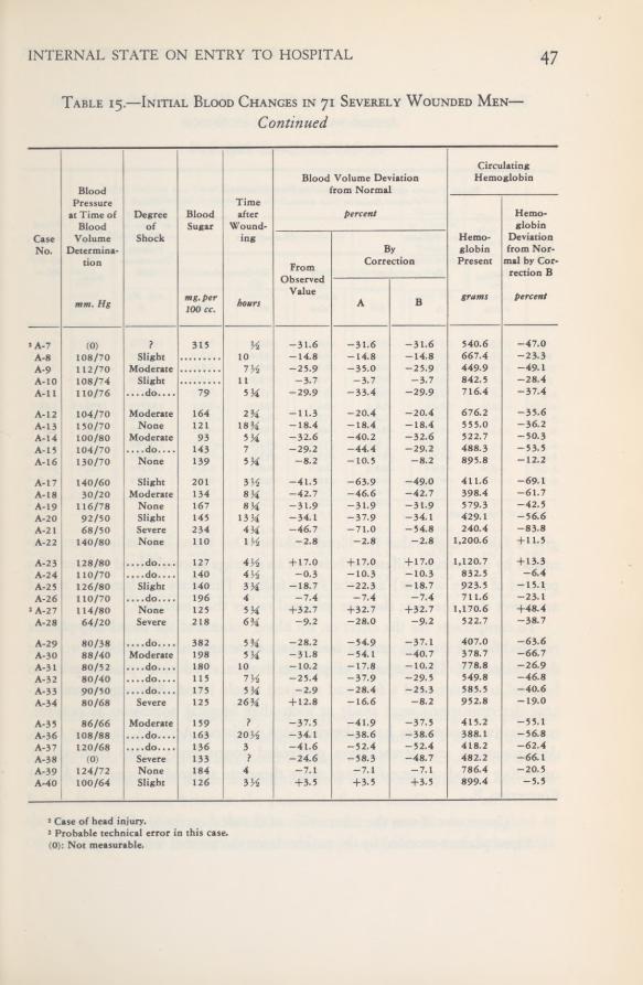
INTERNAL STATE ON ENTRY TO HOSPITAL 47
Table 15.—Initial Blood Changes in 71 Severely Wounded Men—Continued
2 Case of head injury.3 Probable technical error in this case.(0): Not measurable.
CaseNo.
BloodPressure
at Time ofBlood
VolumeDetermina-
tion
mm. Hg
Degreeof
Shock
BloodSugar
mg. per100 cc.
Timeafter
Wound-ing
hours
Blood Volume Deviationfrom Normal
percent
CircuHemo
Hemo-globin
Present
grams
latingglobin
Hemo-globin
Deviationfrom Nor-
mal by Cor-rection B
percent
FromObserved
Value
ByCorrection
A B
!A-7 (0) ? 315 A. -31.6 -31.6 -31.6 540.6 -47.0A-8 108/70 Slight 10 -14.8 -14.8 -14.8 667.4 -23.3A-9 112/70 Moderate 1V2 -25.9 -35.0 -25.9 449.9 -49.1A-10 108/74 Slight 11 -3.7 -3.7 -3.7 842.5 -28.4A-l I 110/76 ....do.... 79 5 H -29.9 -33.4 -29.9 716.4 -37.4
A-12 104/70 Moderate 164 2 H -11.3 -20.4 -20.4 676.2 -35.6A-13 150/70 None 121 18H -18.4 -18.4 -18.4 555.0 -36.2A-14 100/80 Moderate 93 5H -32.6 -40.2 -32.6 522.7 -50.3A-l 5 104/70 ....do.... 143 7 -29.2 -44.4 -29.2 488.3 -53.5A-16 130/70 None 139 -8.2 -10.5 -8.2 895.8 -12.2
A-l 7 140/60 Slight 201 3 A -41.5 -63.9 -49.0 411.6 -69.1A-18 30/20 Moderate 134 8H -42.7 -46.6 -42.7 398.4 -61.7A-19 116/78 None 167 8 H -31.9 -31.9 -31.9 579-3 -42.5A-20 92/50 Slight 145 13A -34.1 -37.9 -34.1 429-1 -56.6A-2 1 68/50 Severe 234 4H -46.7 -71.0 -54.8 240.4 -83.8A-22 140/80 None 110 IX -2.8 -2.8 -2.8 1,200.6 + 11.5
A-2 3 128/80 ....do.... 127 4A + 17.0 + 17.0 + 17.0 1,120.7 + 13.3A-24 110/70 ... .do.... 140 4'A -0.3 -10.3 -10.3 832.5 -6.4A-2 5 126/80 Slight 140 3 A -18.7 -22.3 -18.7 923.5 -15.1A-26 110/70 ....do.... 196 4 -7.4 -7.4 -7.4 711.6 -23.1
3 A-2 7 114/80 None 125 5H +32.7 +32.7 +32.7 1,170.6 +48.4A-28 64/20 Severe 218 6H -9.2 -28.0 -9.2 522.7 -38.7
A-29 80/38 ....do.... 382 5H -28.2 -54.9 -37.1 407.0 -63.6A-30 88/40 Moderate 198 5 H -31.8 -54.1 -40.7 378.7 -66.7A-31 80/52 ....do.... 180 10 -10.2 -17.8 -10.2 778.8 -26.9A-32 80/40 ....do.... 115 7 A -25.4 -37.9 -29.5 549.8 -46.8A-3 3 90/50 ....do.... 175 5H -2.9 -28.4 -25.3 585.5 -40.6A-34 80/68 Severe 125 26H + 12.8 -16.6 -8.2 952.8 -19.0
A-35 86/66 Moderate 159 ? -37.5 -41.9 -37.5 415.2 -55.1A-36 108/88 ....do.... 163 20 A -34.1 -38.6 -38.6 388.1 -56.8A-37 120/68 ....do.... 136 3 -41.6 -52.4 -52.4 418.2 -62.4A-38 (0) Severe 133 ? -24.6 -58.3 -48.7 482.2 -66.1A-39 124/72 None 184 4 -7.1 -7.1 -7.1 786.4 -20.5A-40 100/64 Slight 126 3 A +3.5 +3.5 +3.5 899.4 -5.5
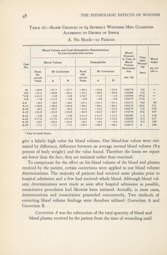
48 THE PHYSIOLOGIC EFFECTS OF WOUNDS
Table 16—Blood Changes in 69 Severely Wounded Men ClassifiedAccording to Degree of Shock
A. No Shock—12 Patients
give a falsely high value for blood volume. Our blood-loss values were esti-mated by difference, difference between an average normal blood volume (8.5percent of body weight) and the value found. Therefore the losses we reportare lower than the fact; they are minimal rather than maximal.
To compensate for the effect on his blood volume of the blood and plasmareceived by the patient, certain corrections were applied to our blood volumedeterminations. The majority of patients had received some plasma prior tohospital admission and a few had received whole blood. Although blood vol-ume determinations were made as soon after hospital admission as possible,resuscitative procedures had likewise been initiated. Actually, in most cases,determinations and resuscitation proceeded concurrently. Two methods ofcorrecting blood volume findings were therefore utilized: Correction A andCorrection B.
Correction A was the subtraction of the total quantity of blood andblood plasma received by the patient from the time of wounding until
Blood Volume and Total Hemoglobin DeterminationsPercent deviation from normal Blood
Pressure Timeafter
Wound-ing
at Time of Blood
CaseNo,
Blood Volume Hemoglobin BloodVolume
Sugar
Determina- mg. perFrom By Correction From By Correction tion hours 100 cc.Ob- Ob-
served served mm. HgValue A B Value A B
91 -26.0 -37.7 -37.7 -43.1 -52.9 -52.9 108/78 2H102 -21.3 -36.9 -29.1 -31.1 -39.0 -39.0 150/80 3H —
•124 + 15.3 -2.8 -2.8 + 14.7 -1.6 -1.6 120/64 10H —
134 -7.8 -11.3 -7.8 -3.8 -3.8 -3.8 120/76 15M —
A-4 -8.2 -8.2 -8.2 -31.1 -31.1 -31.1 132/72 4M 93A-13 -18.4 -18.4 -18.4 -36.2 -36.2 -36.2 150/70 ISH 121A-16 -8.2 -10.5 -8.2 -12,2 -12.2 -12.2 130/70 5 Ya. 139A-19 -31.9 -31.9 -31.9 -42.5 -42.5 -42.5 116/78 8H 167A-22 -2.8 -2.8 -2.8 + 11.5 + 11.5 + 11.5 140/80 l'A noA-23 + 17.0 + 17.0 + 17.0 + 13.3 + 13.3 + 13.3 128/80 4'A 127A-24 -0.3 -10.3 -10.3 +4.1 -6.4 -6.4 110/70 4A 140A-39 -7.1 -7.1 -7.1 -20.5 -20.5 -20.5 124/72 4 184
* Case of crush injury.

INTERNAL STATE ON ENTRY TO HOSPITAL 49
Table 16.—Blood Changes in 69 Severely Wounded Men ClassifiedAccording to Degree of Shock
B. Slight Shock—20 Patients
completion of the test.Correction B was the subtraction of only the quantity of blood and
plasma received from the time of the patient’s hospital admission untilcompletion of the test.
Correction A was applied for the purpose of correlating the patient’sclinical condition on arrival at the hospital with the amount of blood (the per-centage of his estimated normal blood volume) that he had actually lost due tohis wounds, regardless of the amount that may have been replaced. CorrectionB was for correlation between the patient’s clinical condition on arrival andthe blood deficit existing at that time. The same corrections were applied incalculating the total hemoglobin loss. However, in the case of hemoglobin
CaseNo.
Blood Volume and Total Hemoglobin DeterminationsPercent deviation from normal Blood
Pressureat Time of
BloodVolume
Determina-tion
mm. Hg
Timeafter
Wound-ing
hours
BloodSugar
mg. per100 cc.
Blood Volume Hemoglobin
FromOb-
servedValue
By Correction FromOb-
servedValue
By Correction
A B A B
83 + 13.1 -3.3 -0.8 -3.9 -16.1 -16.1 130/70111 -2.7 -7.1 -7.1 -5.1 -5.1 -5.1 120/80 14H 157113 -14.9 -20.2 -20.2 -23.4 -28.2 -28.2 94/58 3H 149127 -39.8 -59.4 -44.2 -63.1 -65.9 -65.9 90/40 11 —
128 +6.2 -49.8 -32.6 -9.7 -39.8 -39.8 110/80 6H 184140 + 16.8 -1.1 -1.1 + 11.7 -5.8 -5.8 100/60 4H 141141 -20.2 -25.5 -25.5 -34.7 -39.1 -39.1 100/60 7 131144 +8.8 -6.7 -1.5 -30.0 -36.6 -36.6 90/62 12X —
145 -8.4 -15.7 -12.1 -27.8 -30.7 -30.7 100/50 9 —
A-l -9.3 -17.4 -14.0 -17.8 -22.6 -22.6 116/60 2H 155A-2 -2.3 -2.3 -2.3 -9.6 -9.6 -9.6 114/62 4H 170A-5 -25.3 -25.3 -25.3 — 35.6 -35.6 -35.6 120/86 8 106A-8 -14.8 -14.8 -14.8 -23.3 -23.3 -23.3 108/70 10 —
A-10 -3.7 -3.7 -3.7 -28.4 -28.4 -28.4 108/74 11H —
A-l 1 -29-9 -33.4 -29.9 -37.4 -37.4 -37.4 110/76 5H 79A-17 -41.5 -63.9 -49.0 -61.6 -69.1 -69.1 140/60 3H 201A-20 -34.1 -37.9 -34.1 -56.6 -56.6 — 56.6 92/50 13X 145A-25 -18.7 -22.3 -18.7 -15.1 -15.1 -15.1 126/80 3H 140A-2 6 -7.4 -7.4 -7.4 -23.1 -23.1 -23.1 110/70 4 196A-40 +3.5 +3.5 +3.5 -5.5 -5.5 -5.5 100/64 3 Vi 126

THE PHYSIOLOGIC EFFECTS OF WOUNDS
Table 16.—Blood Changes in 69 Severely Wounded Men ClassifiedAccording to Degree of ShockC. Moderate Shock—21 Patients
there was essential agreement between calculation A and calculation B be-cause, with rare exceptions, whole blood had not been administered before thepatient reached the most forward hospital.
Tables 15 through 22 show blood and hemoglobin loss in relation to otherfindings for patients individually and by groups. The loss is given as the per-centage difference between a calculated normal and the observed value, or be-tween the calculated normal and the observed value modified by CorrectionsA and B. For blood all three determinations are shown. For hemoglobin onlythe estimated loss derived by Correction B is shown. It will be noted that afew entries (shown as plus values in the tables) seem to indicate that the bloodvolume was greater than normal despite loss of blood. Several factors, indi-
CaseNo.
Blood Volume and Total Hemoglobin DeterminationsPercent deviationfrom normal Blood
Pressureat Time of
BloodVolume
Determina-tion
mm. Hg
Timeafter
Wound-ing
hours
BloodSugar
mg. per100 cc.
Blood Volume Hemoglobin
FromOb-
servedValue
By Correction FromOb-
servedValue
By Correction
A B A B
53 -10.6 -22.9 -22.9 -29.6 -37.8 -37.8 80/40 3 a107 -20.7 -78.6 -68.9 -49.5 -77.6 -77.6 80/50 3 374126 -30.9 -32.3 -32.3 -43.0 -44.4 -44.4 100/60 2H 183129 -21.7 -30.9 -21.7 -44.2 -44.2 -44.2 120/64 5A 191142 -25.7 -31.9 -27.0 -41.9 -43.0 -43.0 110/70 6H 210143 +3.4 -13.9 + 1.8 -22.5 -24.0 -24.0 80/54 6 203148 -5.2 -24.8 -14.9 -39-6 -45.8 -45.8 74/20 12 175
A-3 +5.4 +2.9 +2.9 -5.8 -8.3 -8.3 136/90 4 185A-6 -24.2 -298 -24.2 -38.4 -38.4 -38.4 118/64 3 H 208A-9 -25.9 -35.0 -25.9 -49.1 -49.1 -49.1 112/70 7A —
A-12 -11.3 -20.4 -20.4 -23.5 -35.6 -35.6 104/70 2H 164A-14 -32.6 -40.2 -32.6 -50.3 -50.3 -50.3 100/80 5M 93A-15 -29.2 -44.4 -29.2 -53.5 -53.5 -53.5 104/70 7 143A-18 -42.7 -46.6 -42.7 -61.7 -61.7 -61.7 30/20 SH 134A-30 -31.8 -54.1 -40.7 -57.8 -66.7 -66.7 88/40 5M 198A-31 -10.2 -17.8 -10.2 -26.9 -26.9 -26.9 80/52 10 180A-32 -25.4 -37.9 -29.5 -42.6 -46.8 -46.8 80/40 7A 115A-3 3 -2.9 -28.4 -25.3 -21.7 -40.6 -40.6 90/50 5 H 175A-3 5 -37.5 -41.9 -37.5 -55.1 -55.1 -55.1 86/66 ? 159A-36 -34.1 -38.6 -38.6 -56.8 -56.8 -56.8 108/88 20A 163A-37 -41.6 -52.4 -52.4 -62.4 -62.4 -62.4 120/68 3 136

INTERNAL STATE ON ENTRY TO HOSPITAL 5 1
Table 16.—Blood Changes in 69 Severely Wounded Men ClassifiedAccording to Degree of ShockD. Severe Shock—16 Patients
vidually or collectively, might have brought about this apparent discrepancy:i. In the case of uncorrected entries, blood and plasma administered may havebeen in excess of blood loss. 2. Entries are from calculations based upon anaverage normal which may have been too low for some patients. 3. There mayhave been errors in technique in blood volume determination.
Relationship of Blood Lpss to Type of WoundFifty-nine of the patients listed in Tables 15 and 16 suffered primarily from
a single major wound (abdominal, chest, or peripheral) and could thereforebe more readily studied as to the relationship of blood loss to type of wound.
956507 0—52 5
Blood Volume and Total Hemoglobin DeterminationsPercent deviationfrom normal Blood
Pressure Timeafter
Wound-CaseNo.
Blood Volume Hemoglobinat Time of
BloodVolume
BloodSugar1
FromOb-
By Correction FromOb-
By CorrectionDetermina-
tioning
hoursmg. per100 cc.
servedValue A B
servedValue A B
mm. Hg
2 93 -11.7 -34.0 -30.3 + 12.0 +2.6 +2.6 (0) 14H100 -28.4 -69.0 -65.0 -44.1 -76.6 -76.6 80/50 4'A —
108 -25.5 -38.9 -30.0 -61.8 -64.1 -64.1 58/0 3 XA 372110 -35.7 -37.6 -37.6 -45.7 -47.4 -47.4 (0) H —
122 -34.4 -55.0 -48.2 -45.9 -58.4 -58.4 50/30 4 87125 -21.3 -33.0 -29.1 -44.9 -52.7 -52.7 72/58 23 125130 -31.3 -51.6 -40.5 -51.6 -62.0 -62.0 60/20 5 272131 -29.8 -43.6 -39.0 -45.1 -54.3 -54.3 40/20 177139 -19.3 -75.7 -23.3 -50.5 -70.7 -54.5 (0) 9 —
147 -6.5 -24.1 -14.3 -37.5 -42.7 -42.7 80/48 U3A 165149 -2.3 -31.3 -9.8 -26.1 -32.7 -32.7 48/? 17H 115
A-21 -46.7 -71.0 -54.8 -75.6 -83.8 -83.8 68/50 4Yx 234A-28 -9.2 -28.0 -9.2 -38.7 -38.7 -38.7 64/20 6H 218A-2 9 -28.2 -54.9 -37.1 -54.7 -63.6 —63.6 80/38 3H 382A-34 + 12.8 -16.6 -8.2 (0) -19.0 -19.0 80/68 26H 125A-38 -24.6 -58.3 -48.7 -42.0 -66.1 -66.1 (0) ? 133
1 Blood sugar was determined in another patient infound to be 216 mg. per 100 cc. of blood.
2 Case of crush injury.(0): Not measurable.
severe shock (Case 112) 11 % hours after wounding and
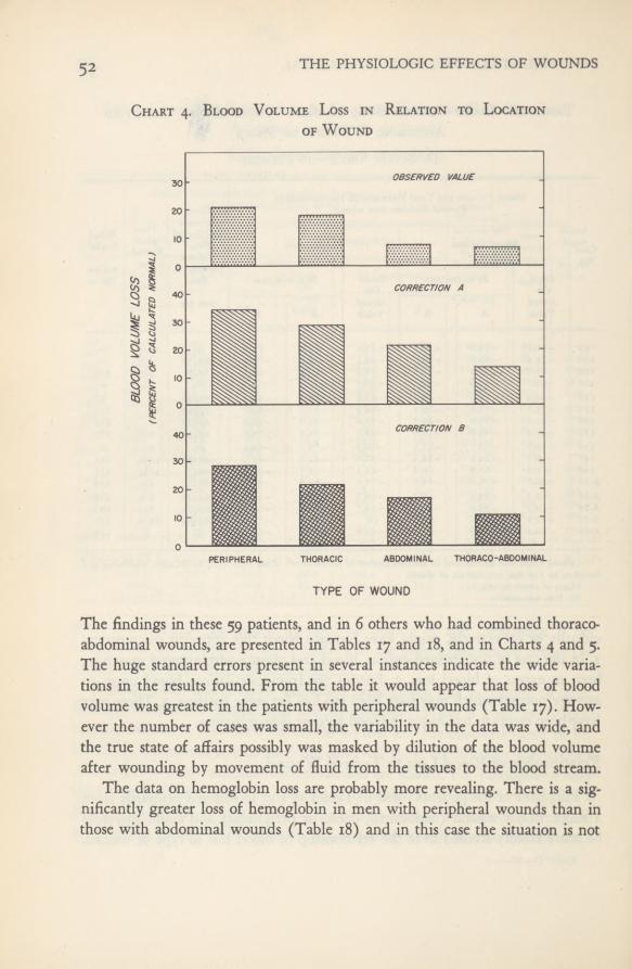
THE PHYSIOLOGIC EFFECTS OF WOUNDS52
Chart 4. Blood Volume Loss in Relation to Locationof Wound
The findings in these 59 patients, and in 6 others who had combined thoraco-abdominal wounds, are presented in Tables 17 and 18, and in Charts 4 and 5.The huge standard errors present in several instances indicate the wide varia-tions in the results found. From the table it would appear that loss of bloodvolume was greatest in the patients with peripheral wounds (Table 17). How-ever the number of cases was small, the variability in the data was wide, andthe true state of affairs possibly was masked by dilution of the blood volumeafter wounding by movement of fluid from the tissues to the blood stream.
The data on hemoglobin loss are probably more revealing. There is a sig-nificantly greater loss of hemoglobin in men with peripheral wounds than inthose with abdominal wounds (Table 18) and in this case the situation is not
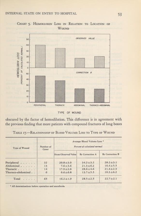
INTERNAL STATE ON ENTRY TO HOSPITAL 53
Chart 5. Hemoglobin Loss in Relation to Location of
Wound
obscured by the factor of hemodilution. This difference is in agreement withthe previous finding that more patients with compound fractures of long bones
Table 17.—Relationship of Blood Volume Loss to Type of Wound
Type of WoundNumber of
Cases
Average Blood Volume Loss *
Percent of calculated normal
From Observed Value By Correction A By Correction B
Peripheral 32 20.8±3.0 34.2 ±3.2 28.2 ±3.1Abdominal 13 7.6±3.6 21.3±6.2 16.4±5.5Thoracic 14 17.9±2.6 28.6±4.6 21.4±2.3Thoraco-abdominal .
. 6 6.6±6.8 13.7±5.5 10.3±6.2
Total 65 16.2±1.9 28.5 ±2.5 22.7 ±2.1
* All determinations before operation and anesthesia.
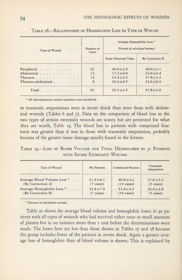
THE PHYSIOLOGIC EFFECTS OF WOUNDS54
Table 18.—Relationship of Hemoglobin Loss to Type of Wound
or traumatic amputations were in severe shock than were those with abdom-inal wounds (Tables 6 and 7). Data on the comparison of blood loss in thetwo types of serious extremity wounds are scanty but are presented for whatthey are worth, Table 19. The blood loss in patients with compound frac-tures was greater than it was in those with traumatic amputation, probablybecause of the greater tissue damage usually found in the former.
Table 19.—Loss of Blood Volume and Total Hemoglobin in 31 Patientswith Severe Extremity Wounds
Table 20 shows the average blood volume and hemoglobin losses in 40 pa-tients with all types of wounds who had received either none or small amountsof plasma but in no instance more than 1 unit before the determinations weremade. The losses here are less than those shown in Tables 17 and 18 becausethe group includes fewer of the patients in severe shock. Again a greater aver-age loss of hemoglobin than of blood volume is shown. This is explained by
Average Hemoglobin Loss *
Type of Wound Number ofCases
Percent of calculated normal
From Observed Value By Correction B
PeripheralAbdominalThoracicThoraco-abdominal
3213146
40.0±3.417.3±6.034.4±3.220.3 ±6.5
46.0±3.124.0±6.437.8±3.223.6±6.3
Total 65 32.5 ±2.5 37.8±2.6
* All determinations before operation and anesthesia.
Type of Wound No Fracture Compound FractureTraumaticAmputation
Average Blood Volume Loss *. . .
(By Correction A)Average Hemoglobin Loss * .
, ..
(By Correction B)
21.9±8.1(7 cases)32.8±7.0(7 cases)
40.8±4.2(19 cases)5I.6±4.2(19 cases)
27.0±3.2(5 cases)
43.0±3.9(5 cases)
* Percent of calculated normal.

INTERNAL STATE ON ENTRY TO HOSPITAL 55
hemodilution which normally takes place after blood loss. Red blood cells arenot replaced appreciably during the interval from wounding to arrival at aforward hospital except by transfusion.Table 20.—Loss of Blood Volume and Total Hemoglobin in 40 Patients
whohad Received i Unit or Less of PlasmaRelationship to Type of Wound
Relationship of Blood Loss to Time from WoundingThere was no important average increase in blood volume loss or in hemo-
globin loss with increased time elapsing between wounding and hospital entry(Table 21). However, those men who were suffering from continuing bloodloss were probably given priority of evacuation. Somewhat greater blood losseswere usually found in those who arrived at the hospital soon after woundingthan in those who were brought in later.Table 21.—Relationship of Blood Loss to Time from Wounding in 67 Cases
(All Types of Wounds)
Type of Wound Number ofCases
Average Blood VolumeLoss *
(By Correction A)
Percent of calculatednormal
Average HemoglobinLoss *
(By Correction B)
Percent of calculatednormal
Peripheral 18 23-9±3.9 38.0±4.3All Other 22 1 5.2 ±2.9 22.5 ±3.8
Total 40 19-1 ±2.4 29.5±3.1
* All determinations before operation and anesthesia.
Time from Wounding
HoursNumber of
Cases
Average Blood VolumeLoss
(By Correction A)
Percent of calculatednormal
Average HemoglobinLoss
(By Correction B)
Percent of calculatednormal
None to 6 41 29.0 37.57 to 12 17 31.9 42.313 and Over 9 24.7 35.8

56 THE PHYSIOLOGIC EFFECTS OF WOUNDS
Relationship of Blood Loss to Degree of Shock
It is well known that individuals do not respond alike to a given blood loss;even previously healthy, normal young soldiers vary greatly in their response.This fact, together with the inexactness inherent in any clinical appraisal of thedegree of shock plus the errors of the experimental method used, might havetended to obscure a real relationship between shock and blood loss. Howeverthis relationship was so striking that, even with the relatively small number ofcases, the positive correlation of blood loss to the severity of shock was sta-
tistically significant (Table 22 and Chart 6).
Table 22.—Relationship of Blood Loss to Degree of Shock in 67 Cases(All Types of Wounds)
The degree of wound shock as we saw it in men injured in battle pre-cisely paralleled the quantity of blood actually lost. Conversely, recovery fromshock resulted promptly from administration of whole blood. Although wemade intensive search at the bedside of thousands of wounded men throughoutthe shock tents in Italy, we never found a clear case of “irreversible shock,”mentioned so frequently in the literature. It is true that there were woundedmen in whom the loss of blood was so rapid and so great that it was impossibleto transfuse them with blood fast enough to save their lives. (For example, wewere unable, in the case of the soldier who had both thighs blown off by ashell burst just outside our door at Anzio to get blood into him fast enoughto save his life; he died in a very few minutes.) Nor were we able to resus-citate patients who had had inadequate circulation in the central nervous system
Number
Average Blood Loss
Percent of calculated normal
Degree of Shock ofCases From Observed Value By Correction A
Volume Hemoglobin Volume Hemoglobin
None 11 10.5db4.1 17.4±6.4 14.4±4.9 20.0±6.7Slight 20 11.2±6.5 25.0±4.4 20.7 ±4.1 29.7 ±4.2Moderate .... 21 21.7±3.1 4l.7±3.3 34.3±3.7 46.1 ±3.3Severe 15 22.0±3.9 44.3 ±4.3 45.9±4.6 55.5 ±4.5

INTERNAL STATE ON ENTRY TO HOSPITAL 57
Chart 6. Relationship of Blood Loss to Degree of Shock
so long that nearly all centers except the respiratory appeared to be dead; wewere not able to overcome death of organs or of nervous tissue by resuscitativeeffort. But we believe that application of the term “irreversible shock” to eithertype of case is to use a definition that has no place at the bedside, howeverinteresting it may be as a concept, and that may do real harm by providing anexcuse for limiting resuscitative effort.
In short, if “irreversible shock” in the accepted sense was present, we missedit. If toxins caused any of the shock we saw, with the exception of that due tooverwhelming and clinically apparent bacterial infections, we failed to recog-nize it. The shock we saw was caused by loss of blood (or of fractions of theblood). It was relieved by administration of whole blood.
From our data it may be said that, in general, when a third of the bloodvolume is lost, clinical shock of more than slight degree will result, and whenhalf is lost, severe shock will result. It was also shown that in individual casesas much as 75 percent of the blood could be lost and the patient survive.Cases A-17, A-21, A-29, A-37, A-38, 127, and 139 are examples of severeblood loss and survival. The greatest blood volume losses found were 78.6 per-cent in Case 107 and 75.7 percent in Case 139 (Table 16, C and D). Both de-

58 THE PHYSIOLOGIC EFFECTS OF WOUNDS
terminations were obtained by Correction A. Shock was moderate in the firstcase and severe in the second. The greatest loss of total hemoglobin found was
83.8 percent in Case A—21 (Table 16, D). This patient had serious extremitywounds and was in severe shock.
Our data also indicated that, on arrival at the hospital, generally the defi-ciency in total hemoglobin was greater than the deficiency in blood volume.This is understandable in view of the well-known mechanism for replenishingblood volume at the expense of tissue fluid. On the other hand, once the readilyavailable reserves of hemoglobin have been called into action, no mechanismis available to replace them rapidly; consequently it would be expected thatgreater deficiencies would be found in the total hemoglobin.
There was no correlation between passage of time and the degree of shockencountered, in that the elapsed time was the same in each of the four cate-gories of no shock, slight, moderate, and severe shock. This does not say thatcontinuing hemorrhage, for example, is not related to degree of shock; cer-tainly it is, but the important factor in the development of shock is the char-acter of the wound, particularly as it indicates the quantity of blood lost, notthe passage of time per se.
We have tried various ways of handling the data on shock. Some investi-gators have attempted to separate the effects of blood loss alone from the ef-fects supposedly accounted for by “sympathico-adrenal activity.” Practically,this is impossible. Moreover, the validity of any such separation even if possiblemust be questioned. For example there is the matter of increased glycogenolysisin shock. Following all the recent advances in knowledge of carbohydratemetabolism, it seems to be too great a simplification to hang the explanationon the rather shaky peg of “sympathico-adrenal activity,” so we take refuge inour purpose of merely stating our findings, with little conjecture, and leavingto future study the search for their significance.
One great consequence of blood loss is the intense vasoconstriction, theshrinkage of the capacity of the vascular bed to accommodate the decreasedblood volume. Contraction of the spleen probably plays a relatively small rolein compensating for the blood lost in battle. Other body adjustments to bloodloss do, however, take place, such as the entry of fluid into the blood vessels inan attempt at compensation. The greatest extravascular store of readily avail-able fluid in the body is that in the extracellular space. Dehydration and oli-gemia may make quite early demands, not only on this extracellular but alsoon the intracellular supply.
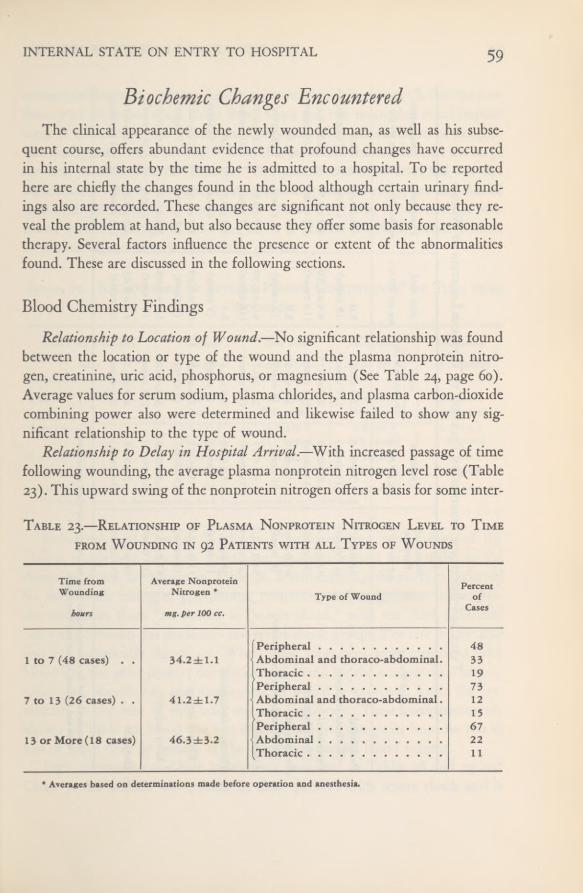
INTERNAL STATE ON ENTRY TO HOSPITAL 59
Biochemic Changes EncounteredThe clinical appearance of the newly wounded man, as well as his subse-
quent course, offers abundant evidence that profound changes have occurredin his internal state by the time he is admitted to a hospital. To be reportedhere are chiefly the changes found in the blood although certain urinary find-ings also are recorded. These changes are significant not only because they re-veal the problem at hand, but also because they offer some basis for reasonabletherapy. Several factors influence the presence or extent of the abnormalitiesfound. These are discussed in the following sections.
Blood Chemistry FindingsRelationship to Location of Wound. —No significant relationship was found
between the location or type of the wound and the plasma nonprotein nitro-gen, creatinine, uric acid, phosphorus, or magnesium (See Table 24, page 60).Average values for serum sodium, plasma chlorides, and plasma carbon-dioxidecombining power also were determined and likewise failed to show any sig-nificant relationship to the type of wound.
Relationship to Delay in Hospital Arrival.—With increased passage of timefollowing wounding, the average plasma nonprotein nitrogen level rose (Table23). This upward swing of the nonprotein nitrogen offers a basis for some inter-
Table 23.—Relationship of Plasma Nonprotein Nitrogen Level to Timefrom Wounding in 92 Patients with all Types of Wounds
Time fromWounding
hours
Average NonproteinNitrogen *
mg. per 100 cc.
Type of WoundPercent
ofCases
Peripheral 481 to 7 (48 cases) . . 34.2±1.1 Abdominal and thoraco-abdominal. 33
19Peripheral 73
7 to 13 (26 cases) . • 4l.2dbl.7 Abdominal and thoraco-abdominal. 12Thoracic 15Peripheral 67
13 or More (18 cases) 46.3±3.2 Abdominal 2211
* Averages based on determinations made before operation and anesthesia.

THE PHYSIOLOGIC EFFECTS OF WOUNDS
Table24.
—BiochemicFindings1
inRelation
toTypeand
Locationof
Wound
TypeofWound
Nonprotein Nitrogen
CreatinineNPN
UricAcid
PhosphorusMagnesium
mg.per100cc.
mg.per100cc.
Creatininex10
mg.per100cc.
mg.per100cc.
mg.per100cc.
Peripheral (Withoutfracture)
39.0±3.7 (11cases)
1.7db0.16
(11cases)
2.6±0.20 (11cases)
5.0 (5
cases)
4.5±0.65 (6cases)
2.3 (5
cases)
Peripheral (Withfracture)
4l.0±1.8 (32cases)
1.6±0.10(31cases)
2.7±0.13 (31cases)
4.4±0.50 (19cases)
4.9±0.50 (24cases)
2.3±0.13 (20cases)
Peripheral (Withtraumatic
amputation)47.0±4.3 (6
cases)
1.5±0.30
(6cases)
3.7±0.65 (6cases)
5.5 (2
cases)
4.9 (4cases)
2.0 (3cases)
Abdominal2
34.0±2.2 (19cases)
1.4±0.15
(19cases)
2.7±0.12 (19cases)
4.9±0.59 (16cases)
3.9±0.30 (17cases)
2.0±0.10 (16cases)
Thoraco-abdominal38.0±3.1 (7cases)
1.5±0.28(7cases)
3.0±0.37 (7
cases)
6.1 (5
cases)
4.5±0.55 (7cases)
2.0 (5
cases)
KidneyInjury
35.0±2.9 (9cases)
1.5±0.25(9cases)
2.6±0.29 (9
cases)
6.5±0.99 (6cases)
4.0±0.50 (9cases)
2.0±0.12 (7
cases)
LiverInjury
37.0±3.0 (10cases)
1.3±0.10(10cases)
3.0±0.23 (10cases)
5.8±0.98 (6
cases)
3.9±0.40 (9cases)
2.0±0.10 (6cases)
Thoracic
36.0±2.5 (14cases)
1.3±0.15(14cases)
2.9±0.26 (14cases)
4.1±0.63
(8cases)
4.2±0.28
(13cases)
1.9±0.10(11cases)
1
Averagesbased
ondeterminations
madebefore
operationand
anesthesia.
1Withandwithout
kidneyandliverinjury.
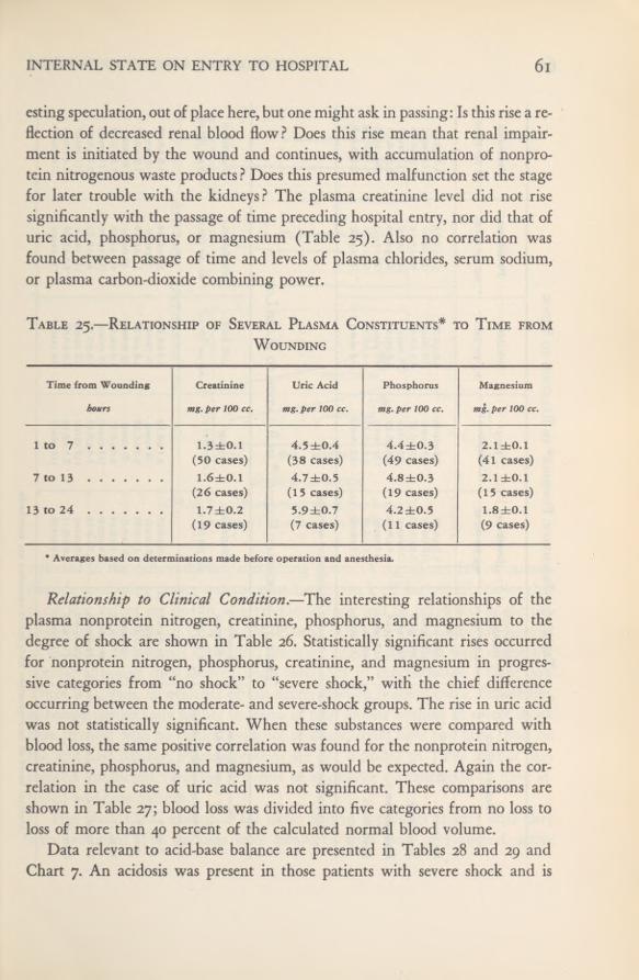
INTERNAL STATE ON ENTRY TO HOSPITAL 61
esting speculation, out of place here, but one might ask in passing: Is this rise a re-flection of decreased renal blood flow? Does this rise mean that renal impair-ment is initiated by the wound and continues, with accumulation of nonpro-tein nitrogenous waste products ? Does this presumed malfunction set the stagefor later trouble with the kidneys? The plasma creatinine level did not risesignificantly with the passage of time preceding hospital entry, nor did that ofuric acid, phosphorus, or magnesium (Table 25). Also no correlation wasfound between passage of time and levels of plasma chlorides, serum sodium,or plasma carbon-dioxide combining power.
Table 25.—Relationship of Several Plasma Constituents* to Time from
Wounding
Relationship to Clinical Condition.—The interesting relationships of theplasma nonprotein nitrogen, creatinine, phosphorus, and magnesium to thedegree of shock are shown in Table 26. Statistically significant rises occurredfor nonprotein nitrogen, phosphorus, creatinine, and magnesium in progres-sive categories from “no shock” to “severe shock,” with the chief differenceoccurring between the moderate- and severe-shock groups. The rise in uric acidwas not statistically significant. When these substances were compared withblood loss, the same positive correlation was found for the nonprotein nitrogen,creatinine, phosphorus, and magnesium, as would be expected. Again the cor-relation in the case of uric acid was not significant. These comparisons areshown in Table 27; blood loss was divided into five categories from no loss toloss of more than 40 percent of the calculated normal blood volume.
Data relevant to acid-base balance are presented in Tables 28 and 29 andChart 7. An acidosis was present in those patients with severe shock and is
Time from Wounding
hours
Creatinine
mg. per 100 cc.
Uric Acid
mg. per 100 cc.
Phosphorus
mg. per 100 cc.
Magnesium
mg. per 100 cc.
1 to 7 1.3±0.1 4.5 ±0.4 4.4±0.3 2.1 ±0.1(50 cases) (38 cases) (49 cases) (41 cases)
7 to 13 1.6±0.1 4.7 ±0.5 4.8±0.3 2.1 ±0.1(26 cases) (15 cases) (19 cases) (15 cases)
13 to 24 1.7 ±0.2 5.9±0.7 4.2 ±0.5 1.8±0.1(19 cases) (7 cases) (11 cases) (9 cases)
* Averages based on determinations made before operation and anesthesia.
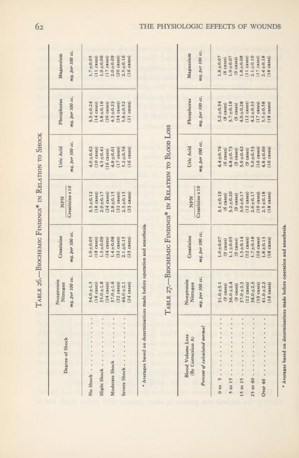
THE PHYSIOLOGIC EFFECTS OF WOUNDS
Table27.
—BiochemicFindings*
inRelation
toBloodLoss
Table26.—Biochemic
Findings*in
Relationto
Shock
DegreeofShock
Nonprotein Nitrogenmg.
per100cc.
Creatinine mg.per100cc.
NPN Creatininex10
UricAcid
mg.per100cc.
Phosphorusmg.per100cc.
Magnesium mg.per100cc.
NoShock Slight
Shock ModerateShock
SevereShock
34.0±1.5 (18cases) 35.0±1,8 (24
cases) 37.0±1.6 (32cases) 44,0±2.1 (24
cases)
l.0±0.05 (18cases)
1.3±0.09
(24cases) 1.4±0.08(32
cases)2.1±0.15
(23cases)
3.3±0,12
(18cases) 2.9±0.
17
(24cases)
2.8±0.15
(32cases)
2.3±0.15
(23cases)
4.0±0.62
(10cases)
4.5
±0.41(16cases)
4.8±0.61
(17cases)
5.2±0.56
(16cases)
3.3±0,24
(14cases)
3.8±0.18
(20cases)
4.3±0.23
(24cases) 5.8±0.52 (21
cases)
1.7±0,05(11cases)
1.9±0.06
(17cases)
2.0±0.09
(20cases)
2.5
±0.16(16cases)
•
Averagesbased
ondeterminations
madebefore
operationand
anesthesia.
BloodVolume
Loss
(ByCorrectionA)
Nonprotein Nitrogen
CreatinineNPN
UricAcid
PhosphorusMagnesium
Percentofcalculatednormal
mg.per100cc.
mg.per100cc.
Creatininex10
mg.per100cc.
mg.per100cc.
mg.per100cc.
0to5
5to15
15to25
25to
40Over40
31.0±3.1
(9cases) 36.0±2.4 (9
cases) 37.0±2,3 (12cases) 38.0±2.3 (19
cases) 41.0±2.2 (18cases)
1.0±0.07(9cases)1.1
±0.05(9cases)
1.3±0.14
(12cases) 1.7±0.
18
(19cases) 1.8±0.13(18
cases)
3.1±0.19
(6
cases)3.2±0.20
(9cases)
3.0±0.17
(12cases)
2.6±0.17
(19cases)
2.5±0,18
(18cases)
4.4±0.76
(6
cases)4.8±0.73
(9
cases)3.9±0.42
(9
cases) 5.0±0.53 (16cases)
4.8±0.60
(16cases)
3.2±0.34
(8
cases)3.7±0.18
(9
cases) 4.0±0.28 (12cases)
4.2±0.33
(17cases)
5.5±0.58
(18cases)
1.8±0.07
(8
cases) 1.9±0.07(9cases)1.8
±0.08(II
cases)2.1±0.10
(17cases) 2.4±0.
18
(18cases)
*
Averagesbased
ondeterminations
madebeforeoperationand
anesthesia.
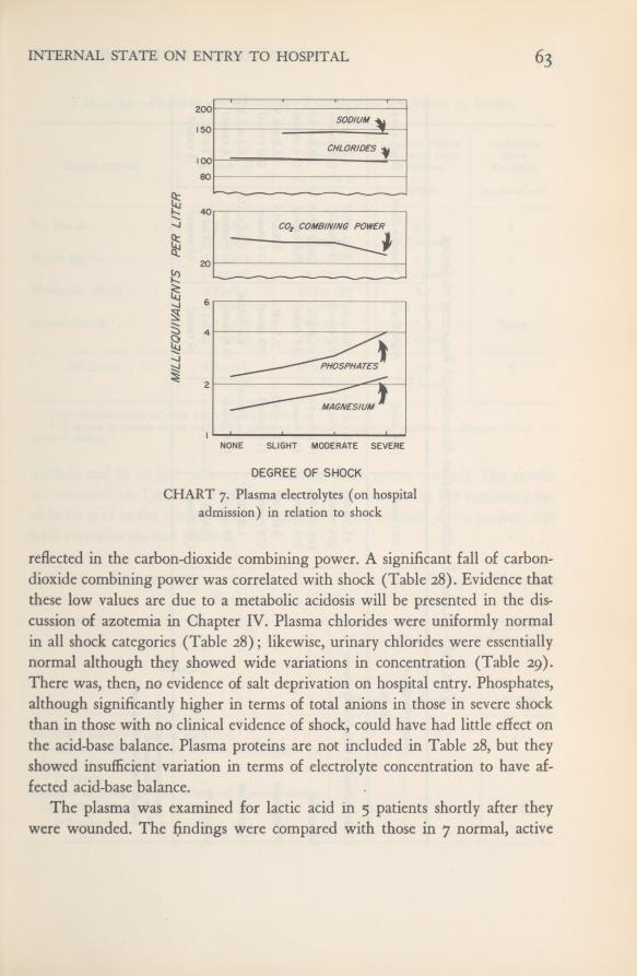
63INTERNAL STATE ON ENTRY TO HOSPITAL
CHART 7. Plasma electrolytes (on hospitaladmission) in relation to shock
reflected in the carbon-dioxide combining power. A significant fall of carbon-dioxide combining power was correlated with shock (Table 28). Evidence thatthese low values are due to a metabolic acidosis will be presented in the dis-cussion of azotemia in Chapter IV. Plasma chlorides were uniformly normalin all shock categories (Table 28); likewise, urinary chlorides were essentiallynormal although they showed wide variations in concentration (Table 29).There was, then, no evidence of salt deprivation on hospital entry. Phosphates,although significantly higher in terms of total anions in those in severe shockthan in those with no clinical evidence of shock, could have had little effect onthe acid-base balance. Plasma proteins are not included in Table 28, but theyshowed insufficient variation in terms of electrolyte concentration to have af-fected acid-base balance.
The plasma was examined for lactic acid in 5 patients shortly after theywere wounded. The findings were compared with those in 7 normal, active
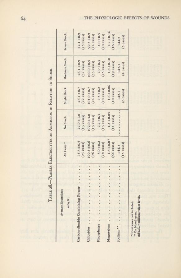
6 4 THE PHYSIOLOGIC EFFECTS OF WOUNDS
Table28.—Plasma
Electrolyteson
Admissionin
Relationto
Shock
AverageElectrolytes mEq./L.
AllCases*
NoShock
SlightShockModerateShockSevereShock
Carbon-dioxideCombining
Power
25.3±0.527.9±1.0
26.1±0.726.1±0.922.1±0.9
(93cases)
(13cases)
(22cases)
(3i
cases)
(25
cases)
Chlorides
100.5±0.6102.0±3.0101.0±0.7100.0±0.5
99.5±0.9
(96
cases)
(13cases)
(24cases)
(33cases)
(24cases)
Phosphates
3.0±0.22.2±0.3
2.5±0.22.9±0.3
4.0±0.5
(78cases)
(13cases)
(20cases)
(25
cases)
(20cases)
Magnesium
1.8±0.071.4±0.051.6±0.061.8±0.10
2.2±0.16
(64cases)
(11cases)
(18cases)
(19cases)
(16cases)
Sodium**
144.3(15cases)
142.1 (6cases)
147.1(4cases)
144.7 (5cases)
*
Crushcasesnot
included.**
Onblood
serum.mEq./L=milliequivalentlevels.

65INTERNAL STATE ON ENTRY TO HOSPITAL
Table 29.—Preoperative Urinary Findings in Relation to Shock
soldiers and in 10 bed patients convalescing from severe wounds. The resultsare recorded in Table 30. They show a twofold increase in the concentrationof lactic acid in the wounded when compared with normal, active soldiers andwith convalescent bed patients.
Table 30.—Concentration of Lactic Acid in Plasma in Severely WoundedSubjects and Controls
Average Average Sodium No SodiumChlorides Received since since
Degree of Shock Average pHmg. per 100 cc.
Wounding * Wounding
as NaCl grams number ofcases
No Shock 6.3±0.2 1,128± 185 1.0 3(7 cases) (5 cases) (4 cases)
Slight Shock 5.7=h0.1 945±l69 0.8 2(6 cases) (8 cases) (5 cases)
Moderate Shock .... 5.7 ±0.1 423±185 3.7 2(6 cases) (5 cases) (4 cases)
Severe Shock 5.7 ±0.2 704±24l 3.2 None(7 cases) (5 cases) (7 cases)
All Cases .... 5.9±0.1 819d-105 2.2 7(26 cases) (2 3 cases) (20 cases)
Note; Specific gravity of urine: 1.027 ±0.001 (26 cases).* 10 grams of sodium citrate furnish 2.3 grams of sodium. 10 grams of sodium bicarbonate furnish 2.7
grams ofsodium.
Subjects
Lactic Acid Concentration in the Plasma
mg. per 100 cc.
Minimum Maximum Average
Normal Controls (7 active soldiers)Wounded Examined 6 to 19 Hours (average 10
17 24 21
hours) after Wounding(5 cases)
Patients in third to fifth Week of Convalescence
29 46 38
from War Wounds(10 cases)
14 21 16
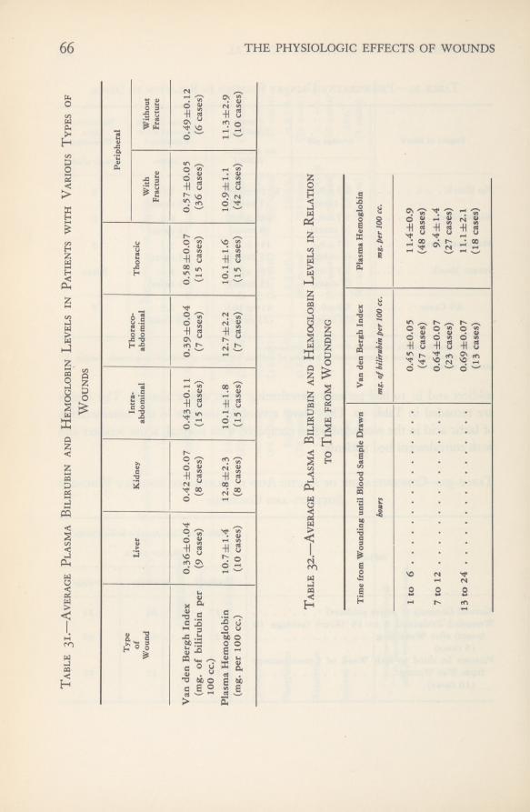
66 THE PHYSIOLOGIC EFFECTS OF WOUNDS
Table31.
—AveragePlasmaBilirubin
andHemoglobin
Levelsin
Patientswith
VariousTypesof
Table32.
—AveragePlasmaBilirubin
andHemoglobin
Levelsin
Relation
toTime
fromWounding
Wounds
Type
Intra- abdominalThoraco- abdominal
Peripheral
of Wound
Liver
Kidney
ThoracicWith FractureWithout Fracture
VandenBerghIndex
(mg.ofbilirubin
per
100cc.)
0.36±0.04 (9cases)
0.42±0.07
(8
cases)0.43±0.11
(15cases)
0.39±0.04 (7cases)0.58±0.07 (15
cases)0.57±0.05 (36
cases)0.49±0.12 (6cases)
PlasmaHemoglobin
(mg.per100
cc.)
10.7±1.4(10cases)
12.8±2.3(8cases)10.1
±1.8(15cases)
12.7±2.2
(7
cases)10.1
±1.6(15eases)
10.9±1.1(42cases)
11.3±2.9
(10cases)
Timefrom
Woundinguntil
BloodSample
Drawn
hours
Vanden
BerghIndex
mg.of
bilirubinper100cc.
PlasmaHemoglobin
mg.per100cc.
1to6
0.45±0.05
11.4±0.9
(47cases)
(48cases)
7to12
0.64±0.079.4±1.4
(23
cases)
(27cases)
13to24
0.69±0.0711.1
±2.1
(13cases)
(18cases)
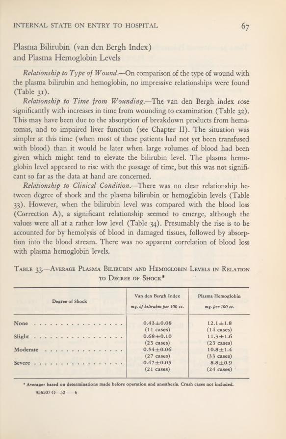
INTERNAL STATE ON ENTRY TO HOSPITAL
Plasma Bilirubin (van den Bergh Index)and Plasma Hemoglobin Levels
Relationship to Type of Wound. —On comparison of the type of wound withthe plasma bilirubin and hemoglobin, no impressive relationships were found(Table 31).
Relationship to Time from Wounding.—The van den Bergh index rosesignificantly with increases in time from wounding to examination (Table 32).This may have been due to the absorption of breakdown products from hema-tomas, and to impaired liver function (see Chapter II). The situation wassimpler at this time (when most of these patients had not yet been transfusedwith blood) than it would be later when large volumes of blood had beengiven which might tend to elevate the bilirubin level. The plasma hemo-globin level appeared to rise with the passage of time, but this was not signifi-cant so far as the data at hand are concerned.
Relationship to Clinical Condition.—There was no clear relationship be-tween degree of shock and the plasma bilirubin or hemoglobin levels (Table33). However, when the bilirubin level was compared with the blood loss(Correction A), a significant relationship seemed to emerge, although thevalues were all at a rather low level (Table 34). Presumably the rise is to beaccounted for by hemolysis of blood in damaged tissues, followed by absorp-tion into the blood stream. There was no apparent correlation of blood losswith plasma hemoglobin levels.
Table 33.—Average Plasma Bilirubin and Hemoglobin Levels in Relationto Degree of Shock*
* Averages based on determinations made before operation and anesthesia. Crush cases not included.956507 0—52 6
Degree of ShockVan den Bergh Index
mg. ofbilirubin per 100 cc.
Plasma Hemoglobin
mg. per 100 cc.
None 0.43 dbO.08 12.1 ±1.8(11 cases) (14 cases)
Slight 0.68±0.10 11.3±1.6(23 cases) (2 3 cases)
Moderate 0.54±0.06 10.8±1.4(27 cases) (33 cases)
Severe 0.47 ±0.05 8.8±0.9(21 cases) (24 cases)

68 THE PHYSIOLOGIC EFFECTS OF WOUNDS
Table 34,—Average Plasma Bilirubin and Hemoglobin Levels in Relationto Blood Loss*
Table 35.—Average Plasma Glucose Levels in Severely Wounded Men
A. Relationship to Degree of Shock
B. Relationship to Blood Loss
C. Relationship to Time from Wounding
Blood Loss(By Correction A)
Percent of calculatednormal
None to 5% 5 to 15% 15 to 25% 25 to 40% Over 40%
Plasma Bilirubin . .
(mg. per 100 cc.)Plasma Hemoglobin
(mg. per 100 cc.)
0.31 ±0.07(6 cases)15.5 ±2.6(7 cases)
0.56±0.10(8 cases)11.2±3.6(8 cases)
0.50±0.10(10 cases)9.8±2.0
(11 cases)
0.52 ±0.05(20 cases)14.3±1.6(20 cases)
0.64±0.11(18 cases)10.0±1.2(18 cases)
* Averages based on determinations made before operation and anesthesia. Crush cases not included.
Degree of Shock None Slight Moderate Severe
Glucose Level * 134±9 149±9 178±13 202 ±26(mg. per 100 cc.) (9 cases) (14 cases) (19 cases) (13 cases)
Blood Loss(By Correction A)
Percent of calculatednormal
None to 5 6 to 15 16 to 25 26 to 40 Over 40
Glucose Level *
(mg. per 100 cc.)143±12(6 cases)
159±15(7 cases)
153±7(9 cases)
177±18(17 cases)
194±23(15 cases)
Time from Wounding None to 6 Hours 7 to 12 Hours 13 to 24 Hours
Glucose Level *
(mg. per 100 cc.)182±13
(36 cases)161 ±10
(13 cases)144±7
(8 cases)
* Normal: from 80 to 90 mg. per 100 cc. of plasma.

INTERNAL STATE ON ENTRY TO HOSPITAL 69Chart 8. Plasma Glucose Levels
Blood SugarThe blood-sugar level was determined in 57 severely wounded men as they
arrived at the forward hospital and was found to be above normal in all ofthem (Table 35 A and Chart 8). Normal by the method used was consideredto be from 80 to 90 mg. per 100 cc. of plasma. Some of the men had received
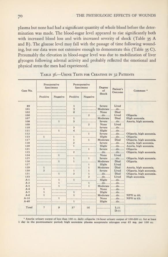
THE PHYSIOLOGIC EFFECTS OF WOUNDS70
plasma but none had had a significant quantity of whole blood before the deter-mination was made. The blood-sugar level appeared to rise significantly bothwith increased blood loss and with increased severity of shock (Table 35 Aand B). The glucose level may fall with the passage of time following wound-ing, but our data were not extensive enough to demonstrate this (Table 35 C).Presumably the elevation in blood-sugar level was due to mobilization of liverglycogen following adrenal activity and probably reflected the emotional andphysical stress the men had experienced.
Table 36.—Urine Tests for Creatine in 32 Patients
Case No.
PreoperativeSpecimens
PostoperativeSpecimens Degree
ofShock
Patient’sOutcome Comment *
Positive Negative Positive Negative
89 I101 3102 6106 2107 1108 1 3 Anuria, high azotemia.109 2110 3111 1 4112 I113 1 1117 1 I118 2120 i121 1123 2 Oliguria, high azotemia.124 1125 1 1126 1 I Oliguria.127 I129 1 1130 2 1 1 Severe Lived Oliguria, high azotemia.131 1 2 1 ....do.... Died Oliguria, high azotemia.133 2 High azotemia.A-l I 1A-2 1 1A-3 1 IA-4 IA-5 1 1A-6 1 I NPN tn 60.
A-16 I NPN to 63.A-40 1 Slight ....do....
Total 7 9 37 16 L:21D;ll
* Anuria; urinary output of less than 100 cc. daily; oliguria: 24-hour urinary output of 100-600 cc. for at least1 day in the posttraumatic period; high azotemia: plasma nonprotein nitrogen over 65 mg. per 100 cc.

INTERNAL STATE ON ENTRY TO HOSPITAL 7 1
Urinary FindingsAcid-Base Relationship.—Sodium in the urine was measured in too few
cases for reliable averages. Most of the values found were within the normalrange. Magnesium, like phosphates on the acid side, was increased but notsufficiently so to affect total acid-base equilibrium. Urinary pH and specificgravity indicate that these men had essentially normal renal function at thetime they were wounded, in that they could make both an acid and a concen-trated urine. The acid-base relationship will be discussed further in Chapter III.
Creatinuria.—The appearance of creatine in the urine of adult males isabnormal. On the assumption that there might be some abnormality in themetabolism of creatine and creatinine in shock, the urine was examined forcreatine in 32 patients (Table 36). In all, 69 urine specimens were tested;the results are summarized in Table 37. Creatine was found in the urinesof 26 of the 32 patients. It was present in 6 out of 15 of the patients ex-amined preoperatively, and in 20 out of 29 patients whose urines were ex-amined postoperatively. In 13 of the 32 patients urine tests yielded only posi-tive results, in 11 patients only negative, and in the remaining 9, different speci-mens tested yielded both positive and negative results.
With one exception (Case 121) all patients who died showed creatinuriaat one time or another. It was also present in approximately half the patientswho survived. In relation to shock, 14 of 18 patients who had been in moderateor severe shock showed creatinuria. This tendency for creatine to be excretedin the urine in cases of moderate and severe shock and in fatal cases mightindicate that it is the result of metabolic changes which accompany shock.
Table 37.—Summary of Results in 69 Urine Specimens Tested for Creatine
SourcePositive Negative
(44 specimens) (25 specimens)
Preoperative 7 9Postoperative 37 16
From Patients with No Shock or Slight Shock . . 18 11From Patients with Moderate or Severe Shock . . 26 14
From Patients Who subsequently Died 20 7From Patients Who Lived 24 18
THE PHYSIOLOGIC EFFECTS OF WOUNDS72
Liver Function in the Newly Wounded NlanAs will be discussed in Chapter II, the only direct laboratory test of liver
function carried out here was that of the bromsulfalein excretion. The van denBergh index and uric acid levels have also been considered as being in part atleast related to liver function.
Bromsulfalein Retention
On Arrival at the Most Forward Hospital.—In 59 severely wounded pa-tients the average bromsulfalein retention on forward hospital arrival was
12.4 ± 1.2 percent (standard error of the mean) 45 minutes after 5 mg. of dyeper Kg. of body weight had been injected intravenously. This is well above thenormal of 1.0 ± 0.1 percent (as established in a control group of 45 subjects),and above our arbitrarily chosen upper limit of normal of 3 percent (seeChapter II).
Relationship to Time from Wounding.—There was no difference betweenthe average bromsulfalein retention in 29 men examined within the first 6hours after wounding (14.4 ±1.8 percent) and in 19 men examined after thefirst 6 hours (13.1 ± 1.6 percent).
Relationship to Location of Wound.—In 22 patients with serious extremitywounds there was 13.3 ± 2.3-percent average retention, and in 18 patients withabdominal wounds there was an average of 14.7 ± 2.1-percent retention—nodifference. In n men with penetrating chest wounds, however, the average re-tention was only 7.0 ±1.8 percent, which is significantly lower than that foundin the other groups.
Relationship to Shoc\.—In 57 patients separated into the 4 shock categories(no shock, slight, moderate, and severe shock) no significant correlation withbromsulfalein excretion could be found; neither was there any correlation withblood volume or hemoglobin loss.
Relationship to Plasma Administration. —Curiously enough, there was agreat increase in average bromsulfalein retention percent) in 25men who had had one or two units of plasma, over the average retention(8.0±2.0 percent) in 15 men who had had none. Three or more units ofplasma did not increase the effect beyond that produced by one or two units.The effect was transient and disappeared between the first and second dayfollowing operation.
INTERNAL STATE ON ENTRY TO HOSPITAL 73
Plasma Bilirubin and Uric AcidThe bilirubin and uric acid concentrations in the blood have been discussed
earlier in this chapter in relation to other factors studied.
SUMMARY
In the past, battle wounds have been described chiefly in terms of organicdamage or tissue loss. The purpose of this chapter has been to describe, shortlyafter the soldier arrived at the forward hospital, the latent consequences of hiswounds as they influenced organic function and produced changes in bloodvolume and chemistry, and abnormalities in the urine. These matters werestudied, before vigorous resuscitative efforts had yet been made, in 108 patients.Altogether, 186 patients were studied in the course of the work carried out bythe Board for the Study of the Severely Wounded.
In considering patients who had received not more than one unit of plasma,or none at all, it was observed that the hematocrit level was higher in thosewith abdominal wounds than it was in those with peripheral wounds, but evenin the patients with abdominal wounds the average hematocrit values weresomewhat below normal. While severe hemoconcentration can occur in casesof burns, crush, and abdominal wounds, this was infrequent in our series andwas by no means a general characteristic of shock as we saw it.
When the patients who had received little or no plasma or blood therapyat the time of first examination were divided into two groups, depending uponwhether more or less than 30 percent of the blood volume had been lost, apuzzling situation was apparent: the average concentration of protein in theplasma in the more severely bled-out group was 6.1 grams per 100 cubic cen-timeters. This is 6.1 percent below normal. On the other hand, the averagehematocrit value in this same group had fallen to 36 from the normal of 47,a reduction of 23.4 percent. The hematocrit level thus fell about four times asmuch as that of the plasma proteins. One implication of this is that the bloodhad been diluted with protein-rich fluid. Its possible source is discussed.
Evidence is presented that the plasma protein level was not influenced byplasma therapy, although the hematocrit level was. There was a sharp fall inhematocrit value with increasing severity of shock. Other examples of theindependent variation of these two factors are given.
It is shown that men can lose about 75 percent of their blood and yet re-

74 THE PHYSIOLOGIC EFFECTS OF WOUNDS
cover—more than had been generally supposed. Blood loss in various types ofwounds is discussed. Data presented show that there was a quantitative rela-tionship between loss of blood volume or hemoglobin and the degree of shockmet clinically. This supports the view that the major cause of the shock weencountered was hemorrhage.
No important differences in blood volume or hemoglobin loss were en-countered with the passage of time from wounding to examination. This waspossibly to be accounted for by the high priority and consequent rapid evacua-tion given to patients with bleeding wounds.
The clinical condition of the newly wounded man offers abundant evi-dence that his internal state has been profoundly altered by the time he entersa forward hospital. In addition to the matters already mentioned, this wasstudied in terms of nitrogenous waste products, electrolytes, bilirubin, andblood sugar. In general, these substances were found not to be influenced bythe location of the wound. The plasma nonprotein nitrogen level rose ratherstrikingly with delay following wounding. The full significance of this is notclear, but it offers grounds for some interesting speculation. Examination of thefour shock categories in sequence from “no shock” to “severe shock” shows sig-nificant rises in nonprotein nitrogen, creatinine, phosphorus, and magnesium.
Acidosis was present in the patients in severe shock. They showed a con-siderable fall in carbon-dioxide combining power as compared with that ofpatients with no clinical evidence of shock. The acidosis appeared to be of the“metabolic” type.
No evidence of salt deprivation was found on hospital entry. Examinationof the admission urine specimen with regard to hydrogen ion concentrationand specific gravity indicated that the men studied had essentially normalrenal function at the time they were wounded.
The van den Bergh index rose significantly with increasing time fromwounding to examination. This is discussed briefly. No clear relationship ofshock to bilirubin or plasma hemoglobin levels was found. The blood-sugarlevel was found to be above normal. It was particularly high in the patientswith severe shock.
Definite depression of liver function, as measured by bromsulfalein reten-tion, was found on hospital arrival. We found no correlation between liverfunction and the degree of shock. The administration of one or two units ofplasma appeared to impair liver function still further. This was a transitoryeffect and was not increased by giving three or four units of plasma.
CHAPTER II
Liver Function in theSeverely Wounded
MethodsThe only specific laboratory test of liver function used in this study was
that of bromsulfalein excretion, although the van den Bergh index and uricacid levels were also considered in conjunction with liver activity. The test wasselected because of facilities available and on the basis of experience of thegroup studying infectious hepatitis in the Mediterranean Theater of Operations.
Procedures
The standard procedure for the determination of bromsulfalein dye reten-tion, using the Hynson, Westcott and Dunning Comparator Block, wasabandoned for the following reasons: Various degrees of hemolysis in the sam-ples of plasma would result in readings that indicated as much as 5-percentretention when no bromsulfalein was present. Therefore low-retention figurescould not be accurately determined, as results were often not reproducible towithin ± 5-percent retention. Furthermore the method was time-consuming.
The test was set up on the Coleman Junior Spectrophotometer Model No. 6,using a 1: 6 dilution of plasma, and reading the color of the alkalinized brom-sulfalein at 575 millimicrons. Five milligrams of bromsulfalein dye per kilo-gram of body weight were injected intravenously and 45 minutes later a bloodsample was drawn from a different vein. In brief, the procedure (fully de-scribed in Appendix C) was as follows:
1. To i cc. of plasma in a cuvette were added 5 cc. of a 0.9-percent solutionof sodium chloride. These ingredients were well mixed and the spectrophotometerwas set to read 100-percent transmission at 575 millimicrons.
2. Three drops of a 10-percent solution of sodium hydroxide were added tothe same tube; the tube was inverted once and was read in the spectrophotometer.

76 THE PHYSIOLOGIC EFFECTS OF WOUNDS
3. The transmission percentage was observed and the percentage of dyeretention recorded from the standard curve.
Standards of Normal and Abnormal Function (Controls)
In order to establish a standard of liver function that would be normal forour particular subjects who were all combat soldiers, 50 apparently healthymen on active duty in the combat zone were selected at random as a controlgroup. The men selected were hospital personnel, ranging in age from 19 to
45 years, the average age being twenty-nine. Forty-five minutes after injectionof bromsulfalein, 45, or 90 percent of the subjects, had less than 3-percent re-tention of the dye, with an average retention of i.o±o.i percent (standarderror of the mean) for the group.
Five subjects, or 10 percent of the group, had more than 3-percent reten-tion of the dye after 45 minutes, as shown in Table 38. It seems reasonablethat the first three (Subjects A, B, and C) and possibly all five of these menmay be considered to have had abnormal liver function of some degree, sincethe percentage of dye retention persisting after 45 minutes was “abnormally”high for this group. In any case, since 90 percent of men with apparentlynormal liver function had less than 3-percent bromsulfalein retention 45 min-
Table 38.—High Retention of Bromsulfalein in 5 Subjects* of ControlGroup
Subject
Percentage of Retentionafter 45 Minutes
Comments
First Test Second Test(1 month later)
A 16.0 4.0 Improvement indicates that impaired functionwas present at time of first test.
B 3.5 4.3 Subject had had jaundice 2 x/i years previously.Palpable liver, 3 fingers below costal mar-gin on inspiration. Liver disease present.
C 15.2 15.6 Persistent high retention. Liver definitelyabnormal.
D 7.2 3.9 Possibly abnormal function.E 5.8 7.6 Weight of subject 105 Kg. Possibly abnormal
liver function.
* None of these 5 subjects had any definite clinical symptoms of poor liver function.

LIVER FUNCTION IN THE WOUNDED 77
utes after 5 mg. per Kg. of body weight were injected, we arbitrarily chose3-percent dye retention as the upper limit of normal.
Liver Function in Severely Wounded Patientson Entry to the ISLost Forward Hospital
The average severely wounded battle casualty showed considerable impair-ment of liver function as measured by bromsulfalein retention on hospitalentry. The average dye retention in 59 severely wounded patients was 12.4 per-cent±i.2 percent, which is well above the arbitrary normal limit of 3 percent.This finding was considered in connection with several factors. As in the entirestudy, the number of patients on whom various combinations of findings areshown represent the maximum number available for that particular com-parison.
Relationship to Time from WoundingForty-eight patients were divided into two groups according to whether
liver function was measured (a) within the first 6 hours following woundingor (b) more than 6 hours after wounding. The average percentage of dye re-tention was found to be: for (a), 14.4=!=1.8 percent (29 patients); for (b),13. 1 ±i.6 percent (19 patients). It was therefore concluded that there was norelationship between time from wounding and bromsulfalein retention. •
Relationship to Location of Major WoundTwenty-two patients with severe extremity wounds had an average reten-
tion of i3,3±2.3 percent; 18 patients with abdominal wounds i4.7±2.i per-cent, and 11 patients with chest wounds 7.o±i.8 percent. If a conclusion maybe drawn from this small number of patients, those with chest wounds ap-peared to have significantly less dye retention than those with wounds of theextremities or of the abdomen. Patients with direct injury to the liver had asomewhat higher average retention (18.4 percent) than those with abdominalwounds without direct liver injury (14.7 percent). However there was such awide spread in the data, particularly in the patients with direct liver damage,that the difference between the two groups cannot be considered significant.
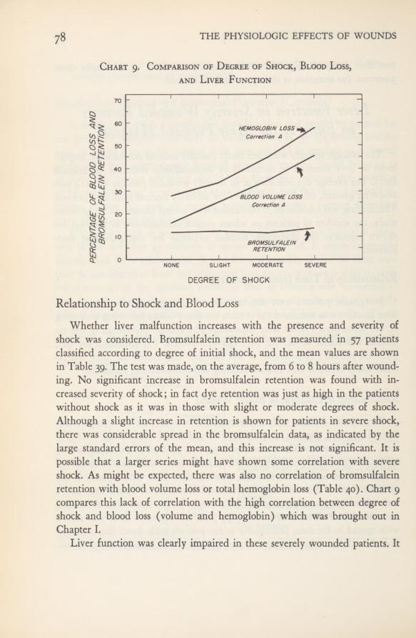
THE PHYSIOLOGIC EFFECTS OF WOUNDS
Chart 9. Comparison of Degree of Shock, Blood Loss,and Liver Function
Relationship to Shock and Blood Loss
Whether liver malfunction increases with the presence and severity ofshock was considered. Bromsulfalein retention was measured in 57 patientsclassified according to degree of initial shock, and the mean values are shownin Table 39. The test was made, on the average, from 6 to 8 hours after wound-ing. No significant increase in bromsulfalein retention was found with in-creased severity of shock; in fact dye retention was just as high in the patientswithout shock as it was in those with slight or moderate degrees of shock.Although a slight increase in retention is shown for patients in severe shock,there was considerable spread in the bromsulfalein data, as indicated by thelarge standard errors of the mean, and this increase is not significant. It ispossible that a larger series might have shown some correlation with severeshock. As might be expected, there was also no correlation of bromsulfaleinretention with blood volume loss or total hemoglobin loss (Table 40). Chart 9compares this lack of correlation with the high correlation between degree ofshock and blood loss (volume and hemoglobin) which was brought out inChapter I.
Liver function was clearly impaired in these severely wounded patients. It

LIVER FUNCTION IN THE WOUNDED 79
Table 39.—Bromsulfalein Retention and Shock
was just as much affected in those without shock as it was in those with severeshock. Shock therefore was evidently not the key to this condition. Thegreat deterioration of the circulation found with increasing degrees of shockdid not appear to strain liver function measurably insofar as can be judgedfrom these data. The main cause of the impaired function must be soughtelsewhere.
Before it is assumed from these findings, however, that there was no moreimpairment of liver function in those patients who had severe shock than inpatients without shock, the question should be raised as to whether the degreeof liver impairment could be masked by the given dose of bromsulfalein in apatient who, for example, had lost 50 percent of his normal blood volume. In thisinstance the given dose of dye would be abnormally concentrated, since itwould be less diluted in the abnormally small volume of blood present. Per-
Table 40.—Bromsulfalein Retention and Blood Loss
Time from Brom-Number Wounding to sulfalein
Degree of Shock of Test RetentionPatients
hours percent
None 10 5.9±1.5 11.6±2.3Slight 17 6.0± 1.8 11.9±2.2Moderate 17 6.5±0.6 11.1 ±2.7Severe 13 * 8.1 ±1.7 13.5±1.7
• Known in only 11 cases.
Numberof
Patients
Hemoglobin Loss(By Correction A*)
percent of calculated norfnal
AverageBromsulfalein
Retention
percent
Numberof
Patients
Blood Volume Loss(By Correction A*)
percent of calculated normal
AverageBromsulfalein
Retention
percent
II 0 to 2 5 12.2 ±3.2 22 0 to 25 I1.1 ±1.917 26 to 45 11.2 ±2.1 18 26 to 45 13.8±2.322 Over 45 14.3±2.1 12 Over 45 14.8±3.3
* Observed value less all blood ortermination.
plasma administered from time of wounding to completion of the de-
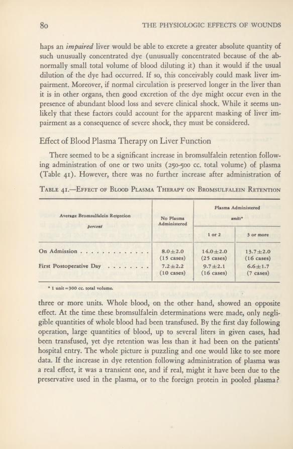
THE PHYSIOLOGIC EFFECTS OF WOUNDS
haps an impaired liver would be able to excrete a greater absolute quantity ofsuch unusually concentrated dye (unusually concentrated because of the ab-normally small total volume of blood diluting it) than it would if the usualdilution of the dye had occurred. If so, this conceivably could mask liver im-pairment. Moreover, if normal circulation is preserved longer in the liver thanit is in other organs, then good excretion of the dye might occur even in thepresence of abundant blood loss and severe clinical shock. While it seems un-likely that these factors could account for the apparent masking of liver im-pairment as a consequence of severe shock, they must be considered.
Effect of Blood Plasma Therapy on Liver FunctionThere seemed to be a significant increase in bromsulfalein retention follow-
ing administration of one or two units (250-500 cc. total volume) of plasma(Table 41). However, there was no further increase after administration ofTable 41.—Effect of Blood Plasma Therapy on Bromsulfalein Retention
three or more units. Whole blood, on the other hand, showed an oppositeeffect. At the time these bromsulfalein determinations were made, only negli-gible quantities of whole blood had been transfused. By the first day followingoperation, large quantities of blood, up to several liters in given cases, hadbeen transfused, yet dye retention was less than it had been on the patients’hospital entry. The whole picture is puzzling and one would like to see moredata. If the increase in dye retention following administration of plasma wasa real effect, it was a transient one, and if real, might it have been due to thepreservative used in the plasma, or to the foreign protein in pooled plasma?
Average Bromsulfalein Retentionpercent
No PlasmaAdministered
Plasma Administered
units*
I or 2 3 or more
On Admission 8.0±2.0(15 cases)
14.0±2.0(2 5 cases)
13.7±2.0(16 cases)
First Postoperative Day 7.2 ±2.2(10 cases)
9.7±2.1(16 cases)
6.6±1.7(7 cases)
• I unit =300 cc. total volume.

81LIVER FUNCTION IN THE WOUNDED
Chart io. Bromsulfalein Retention and Patients’Outcome
Postoperative Course and Liver FunctionIn Table 42 liver function is considered in relation to the patient’s out-
come. Average bromsulfalein retention, measured during the preoperativeperiod and the first 4 postoperative days, is shown for those who lived andthose who, either in the period covered or subsequently, died, as well as thosewho at this time or later developed “high azotemia.” 1 The first group, showingbromsulfalein retention in those who lived, is the only one in which the datacan be considered in any satisfactory quantitative sense (Chart 10). Not onlyare the cases relatively few in the other groups, but since death had not oc-curred nor had uremia or high azotemia appeared in many instances untilconsiderably later than the time interval covered by this table, the high brom-sulfalein retentions of those who died in the first four or five days are maskedby patients whose values were normal at this time but later became abnormal.The data shown in Table 42 are given, however, because they do show quali-tatively a typical trend.
1 In this study “high azotemia” was defined as a plasma nonprotein nitrogen level of 65 mg. per100 cc. or higher at any time in the posttraumatic period.
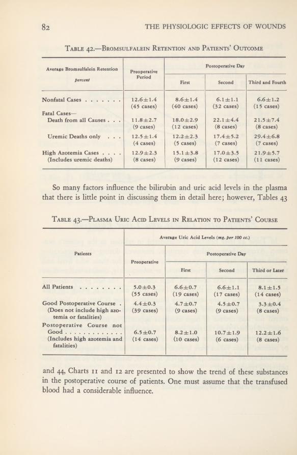
82 THE PHYSIOLOGIC EFFECTS OF WOUNDS
Table 42.—Bromsulfalein Retention and Patients’ Outcome
So many factors influence the bilirubin and uric acid levels in the plasmathat there is little point in discussing them in detail here; however, Tables 43
Table 43.—Plasma Uric Acid Levels in Relation to Patients’ Course
and 44, Charts n and 12 are presented to show the trend of these substancesin the postoperative course of patients. One must assume that the transfusedblood had a considerable influence.
Average Bromsulfalein Retention PreoperativePostoperative Day
percentPeriod
First Second Third and Fourth
Nonfatal Cases 12.6dhl .4 8.6±1.4 6.1±1.1 6.6± 1.2(45 cases) (40 cases) (32 cases) (15 cases)
Fatal Cases—
Death from all Causes . . . 11.8±2.7 18.0±2.9 22.1 ±4.4 2 1.5 ±7.4(9 cases) (12 cases) (8 cases) (8 cases)
Uremic Deaths only . . . 12.5±1.4 12.2±2.3 17.4±5.2 29.4±6.8(4 cases) (5 cases) (7 cases) (7 cases)
High Azotemia Cases .... 12.9±2.3 15.1 ±3.8 17.0±3.5 21.9±5.7(Includes uremic deaths) (8 cases) (9 cases) (12 cases) (11 cases)
Average Uric Acid Levels (mg. per 100 cc.)
Patients
Preoperative
Postoperative Day
First Second Third or Later
All Patients 5.0±0.3 6.6±0.7 6.6±1.1 8.1±1.5(55 cases) (19 cases) (17 cases) (14 cases)
Good Postoperative Course . 4.4±0.3 4.7 ±0.7 4.5 ±0.7 3.3±0.4(Does not include high azo-
temia or fatalities)Postoperative Course not
(39 cases) (9 cases) (9 cases) (8 cases)
Good 6.5±0.7 8.2±1.0 10.7±1.9 12.2±1.6(Includes high azotemia and
fatalities)(14 cases) (10 cases) (6 cases) (8 cases)

LIVER FUNCTION IN THE WOUNDED «3
Table44.
—PlasmaBilirubin
Levelsin
Relationto
Patients’Course
956507 0—52 7
AveragePlasma
BilirubinLevels
(ms.per100cc.)
Patients
Preoperative
PostoperativeDay
First
SecondThirdand
FourthFifth
orLater
AllPatients
0.59±0.041.63±0.33
1.55±0.261.69±0.35
2.55±0.81
(77cases)
(27cases)
(29cases)
(22cases)
(19cases)
GoodPostoperative
Course
0.61±0.06
1.36±0.271.31±0.270.91±0.15
0.84±0.24
(Doesnot
includehighazotemiaor
fatali-
ties)
(58cases)
(17cases)
(18cases)
(8cases)
(7
cases)
PostoperativeCoursenot
Good
0.43±0.042.04±0.62
1.98±0.622.50±0.662.98±1.0
(Includeshigh
azotemiaandfatalities)
(17cases)
(12cases)
(9cases)
(10cases)
(15cases) or
*2.22±0.7
(14cases)
•
Onevalueof
13.6omittedtoget
thisaverage.
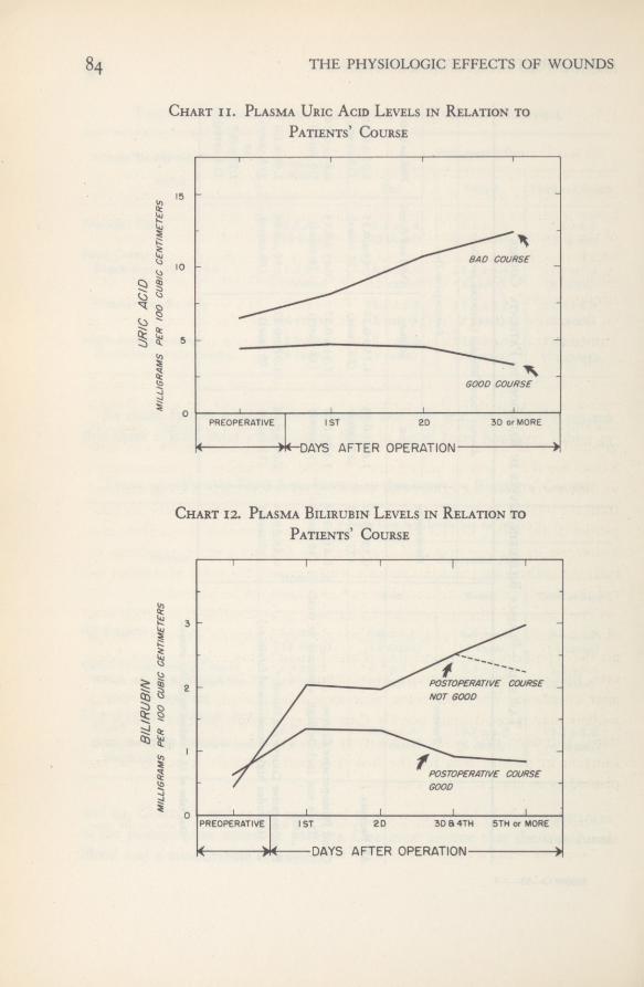
84 THE PHYSIOLOGIC EFFECTS OF WOUNDS
Chart ii. Plasma Uric Acid Levels in Relation to
Patients’ Course
Chart 12. Plasma Bilirubin Levels in Relation toPatients’ Course
LIVER FUNCTION IN THE WOUNDED 85
Effect of Ether Anesthesia on Liver Function
It has been repeatedly stated that liver function, as measured by bromsul-falein retention, is considerably impaired by ether anesthesia. Tentatively ac-cepting this statement, it was necessary to determine just how important thisfactor might be in causing bromsulfalein retention in our patients, for manyof the patients with oliguria2 or anuria3 were not seen until after operation atwhich time bromsulfalein retention was usually high. Accordingly data wereobtained on 42 patients prior to operation and again on the first day afteradministration of ether anesthesia. In most of the cases the test was repeatedon the second and even on the third postoperative day. The findings in the 42individual cases are presented in Table 45 and the averages are shown inTable 46.
It was observed that in the patients who were in grave condition followingoperation, as proved by their early deaths, the average percentage of dye re-tention rose sharply. There were 8 of these moribund patients; they aregrouped separately on Table 46. Presumably the failing liver function in these8 cases was due to anoxia, but whatever its cause, the moribund patients reactedso differently from the others that the findings when these 8 are eliminatedfrom the total of 42 are also shown. Surely this grouping gives a more accurate
picture of the effect, or lack of it, of ether on liver function. Since there wasa decrease in average bromsulfalein retention following ether anesthesia inboth the entire group and the patients who survived, the point of whether ornot the moribund patients should be included need not be labored.
The failure of ether to produce an increase in bromsulfalein retention wasof considerable surprise to us, and we are at a loss to explain why our findingsare the opposite of those reported by others. It must be taken into account, how-ever, that our first postoperative examination of liver function was 24 hoursafter anesthesia. The intervening time should be studied, as healthy youngsoldiers may respond more quickly than average civilian patients. It may bestated, therefore, that so far as this study is concerned, evidence of poor liverfunction in patients seen for the first time postoperatively is not to be ex-plained as an effect of ether anesthesia.
3 Oliguria was defined as a 24-hour urinary output of 100-600 cc. for at least 1 day in the post-traumatic period.
3 Anuria was defined as a urinary output of less than 100 cc. in 24 hours.

86 THE PHYSIOLOGIC EFFECTS OF WOUNDS
Table45.
—EtherAnesthesiain
Relationto
BromsulfaleinRetention
andPlasma
Nonprotein
NitrogenLevels
in42
Patients
Case No.Age yrs.
MajorWounds
Degree of Shock
OnAdmission
Duration ofAnes- thesia minutes
FirstPost- operative
Day
SecondPost- operative
Day
DyeRe- tention percentNPN mg.
per100cc.
DyeRe- tention percentNPN mg.
per100cc.
DyeRe- tention percentNPN mg.
per100
cc.
9127
Traumaticamputation,thigh
.
.
None
15
33
90
5
—
10621
Transection,femoral
artery.
.
.
do
8
30
180
7
24
1
—
12534
Perforating,chest;fracture,hu-
merus;gas
gangrene
Severe
15
64
150
26
89
6
136
13027
Compoundcomminutedfractures,
tibia,ilium(with
penetrating,chest)
3
27
240
3
49
18
60
13433
Penetrating,abdomen,chest.
.
None
10
35
50
17
12
46
13926
Penetrating,chest;fracture,femurSevere
15
53
150
8
36
—
14023
Penetrating,abdomen
Slight
10
37
175
8
11
41
14119
Penetrating,abdomen,chest.
.
do
15
36
135
7
29
4
—
14224
Penetrating,chest
Moderate5
26
75
6
29
6
—
14432
Perforating,facewith
compound
fractures
Slight
10
33
135
8
6
24
14541
Penetrating,arm,
thigh,chest.
.do
14
43
120
13
8
34
14825
Traumaticamputation,bothlower
Moderate6
46
60
40
—
A-l23
Penetrating,chest
Slight
6
26
210
1
30
—
A-221
Perforating,abdomen
do
7
29
225
2
26
3
—
A-3
25.
Penetrating,abdomen
Moderate7
27
160
22
46
6
60
A-5
17
Traumaticamputation,foot
.
.
.Slight
6
30
50
0
28
2
21
A-622
Penetrating,abdomen
Moderate31
38
180
34
45
30
46
A-l1
?
Penetrating,chest
Slight
14
27
70
6
29
2
31

87LIVER FUNCTION IN THE WOUNDED
A-1623
6
47
90
10
63
A-1721
Traumaticamputation,lowerarm;
penetrating,abdomen
Slight
30
31
180
16
34
9
26
A-1835
Traumaticamputation,leg(with
fracture,otherleg)
Moderate40
53
120
12
69
8
43
A-1926
Compoundfracture,tibia
....
None
23
42
90
17
53
4
36
A-2029Traumatic
amputation,foot
.
.
.
Slight
15
30
77
1
27
1
23
A-21
21Near
traumaticamputation,mid-
thigh
Severe
9
58
90
7
58
2
51
A-2221
Perforating,liver,
kidney....
None
23
31
140
10
29
12
—
A-27
25
Penetrating,abdomen
do
0
29
145
0
27
0
26
A-28
20
Compoundfractures,femur,hu-
merus
Severe
24
41
195
3
37
2
25
A-29
20Severedfemoralartery
do
22
44
70
4
34
2
29
A-31
20
Perforating,chest
Moderate11
48
180
6
49
6
25
A-3219
Penetrating,chest
do
8
37
100
4
46
5
—
A-3525
Neartraumatic
amputation,lower
34
31
60
5
34
A-37
19
Compoundfracture,humerus..do
4
36
85
2
29
2
28
A-3819
Compoundcomminutedfracture
withsevere
mangling,lowerleg
Severe
6
48
215
3
44
3
40
A-4025
Penetrating,back,
abdomen,chest
Slight
0
36
182
1
29
1
26
*12222
19
42
300
30
63
*12623
0
49
110
30
51
*12920
Penetratingwounds,lumbarre-
gion,compoundcomminuted
fractures
15
32
125
4
82
6
104
*13127
Penetrating,chest,
abdomen.
.
Severe
10
39
253
16
104
7
116
*14328
Perforating,chest,
abdomen.
.
Moderate26
38
120
28
66
27
62
*14723
Severe
10
47
163
20
105
*A-3024
Severedfemoralartery
Moderate12
39
120
30
61
39
90
*A-3621
Multiplesoft-tissue
wounds,both
1
53
210
42
77
*
Patientdied.
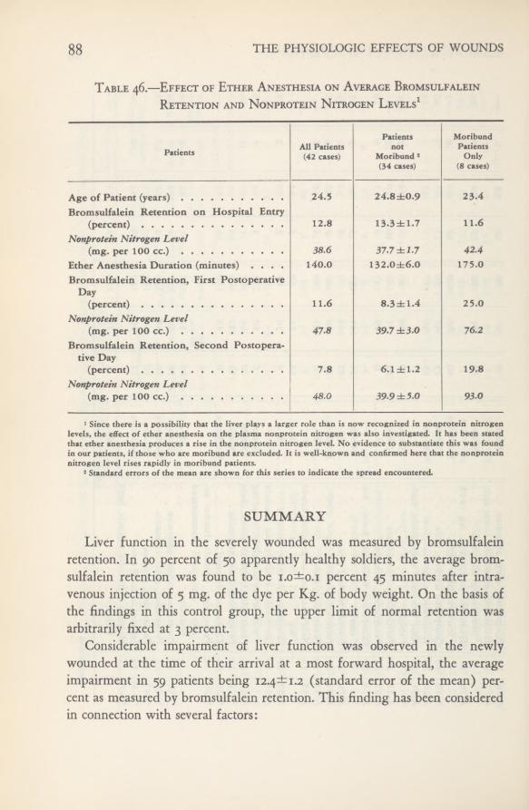
88 THE PHYSIOLOGIC EFFECTS OF WOUNDS
Table 46.—Effect of Ether Anesthesia on Average BromsulfaleinRetention and Nonprotein Nitrogen Levels 1
SUMMARY
Liver function in the severely wounded was measured by bromsulfaleinretention. In 90 percent of 50 apparently healthy soldiers, the average brom-sulfalein retention was found to be i.o±o,i percent 45 minutes after intra-venous injection of 5 mg. of the dye per Kg. of body weight. On the basis ofthe findings in this control group, the upper limit of normal retention wasarbitrarily fixed at 3 percent.
Considerable impairment of liver function was observed in the newlywounded at the time of their arrival at a most forward hospital, the averageimpairment in 59 patients being (standard error of the mean) per-cent as measured by bromsulfalein retention. This finding has been consideredin connection with several factors:
Patients All Patients(42 cases)
Patientsnot
Moribund J
(34 cases)
MoribundPatients
Only(8 cases)
Age of Patient (years)Bromsulfaiein Retention on Hospital Entry
24.5 24.8±0.9 23.4
(percent)Nonprotein Nitrogen Level
12.8 13-3±1.7 11.6
(mg. per 100 cc.) 38.6 37-7 ±1.7 42.4Ether Anesthesia Duration (minutes) ....
Bromsulfaiein Retention, First PostoperativeDay
140.0 132.0±6.0 175.0
(percent)Nonprotein Nitrogen Level
11.6 8.3dbl.4 25.0
(mg. per 100 cc.)Bromsulfaiein Retention, Second Postopera-
tive Day
47.8 39J ±3.0 76.2
(percent)Nonprotein Nitrogen Level
7.8 6.1 ±1.2 19.8
(mg. per 100 cc.) 48.0 39.9 ±5.0 93.0
1 Since there is a possibility that the liver plays a largerrole than is now recognized in nonprotein nitrogenlevels, the effect of ether anesthesia on the plasma nonprotein nitrogen was also investigated. It has been statedthat ether anesthesia produces a rise in the nonprotein nitrogen level. No evidence to substantiate this was foundin our patients, if those who are moribund are excluded. It is well-known and confirmed here that the nonproteinnitrogen level rises rapidly in moribund patients.
1 Standard errors of the mean are shown for this series to indicate the spread encountered.
LIVER FUNCTION IN THE WOUNDED 89Time from Wounding.—No relationship could be found between time
from wounding and bromsulfalein retention.Location of Wound. —Patients with extremity wounds had the same aver-
age retention (13.3—2.3 percent) as those with abdominal wounds (14.7*2.1percent). Curiously, those with chest wounds seemed to have less retention ofthe dye (7.0* 1.8 percent) than those in the previous categories. Patients withdirect injury to the liver showed 18.4-percent average retention. It is doubtfulif this is significantly higher than that found in other abdominal wounds, sincethere was considerable spread in the data.
Shoct{.—No correlation between presence or degree of shock and liver func-tion was shown, and, as might be expected, no correlation of bromsulfaleinretention with blood-volume loss or hemoglobin loss. The great deteriorationof the circulation found in patients in the more severe shock categories doesnot appear to strain liver function measurably insofar as can be judged fromthese data.
Plasma. —It was surprising that one of the great stresses placed upon thebody, such as that due to shock, had little if any effect on liver function asmeasured by bromsulfalein retention, yet the administration of one or twounits of blood plasma produced a considerable, although transient, increase inretention.
POSTOPERATIVE FINDINGS
Liver function during the postoperative course of the severely wounded, asdetermined by bromsulfalein retention and bilirubin and uric acid levels, isrecorded.
The statement has been made repeatedly in the past that liver function asmeasured by bromsulfalein retention is considerably impaired by ether anes-thesia, In order properly to assess patients seen for the first time postoper-atively, liver function studies were carried out in 42 patients before and afterprolonged ether anesthesia. Contrary to other reports, we found in these pa-tients at least a decrease in bromsulfalein retention postoperatively, except inthose who were moribund. There is generally a sharp increase in bromsul-falein retention in patients whose condition is deteriorating rapidly.

CHAPTER III
Renal Function in theSeverely Wounded
The desirability of measuring renal function as accurately as possible inthe severely wounded was clear at the outset of the study. In the majority ofcases this was done by determination of phenolsulfonphthalein excretory ca-pacity and by urine concentration tests, in conjunction with routine urine andblood chemistry analyses. Mannitol and sodium para-amino hippurate did not
become available for clearance measurements until the last weeks of the Italiancampaign. Results of the phenolsulfonphthalein, urine concentration, and thefew renal clearance tests performed will be described.
Phenolsulfonphthalein Excretory CapacityBecause of practical considerations, the phenolsulfonphthalein excretion test
was made postoperatively except on one patient who had no surgery, when itwas done after transference from the shock ward, but in all cases it was somehours after the patient had been adequately resuscitated. Fifty-seven patientswere tested, all of them within 72 hours after wounding, and some of themwere tested again after the third day in order to follow their rate of improve-ment. The latter group comprised 12 patients available for retesting out of 37in whom dye excretion had been found low in the first test and 3 patients onwhom the first tests had been made later than the third day after wounding,but in whom the quantity of dye excreted was still low (Cases 125, 104, and29 on Table 48).
Technique of Test.—The following technique of performing this test 1 wascommonly employed. Six milligrams of phenolsulfonphthalein were injected
1 See Appendix C for details of performing the test.

RENAL FUNCTION IN THE WOUNDED 91
intravenously and urine was collected 15, 30, 60, and 120 minutes later. It wasnecessary to use an indwelling catheter in almost every instance to insure accu-rate collection. Oral intake of fluid was restricted in most patients at the timethe test was performed; therefore, in order to promote urine flow, infusion of1 liter of 5-percent dextrose in isotonic saline solution, or 10-percent dextrose indistilled water, was frequently started about 30 minutes before the dye wasinjected.
Excretory Capacity in the First 5 Days after Wounding.—The average re-sults of the test made on 57 patients2 during this period, together with otherpertinent data, are shown in Table 47 and Chart 13.
The most striking finding was the difference in the average percentage ofdye excreted by the 20 patients who had not been in shock and that excretedby the 37 who had had slight, moderate, or severe shock. At all test periods theaverage excretion was normal for the group that had not been in shock. (Seealso section to follow on the effect of anesthesia on these patients.) During thefirst 30 minutes, the average excretion was low for the group that had beenin shock, and decreased progressively with increasing severity of initial shock.Although standard errors of the mean are rather large, and although there areno significant differences between the three groups with slight, moderate, orsevere initial shock, the qualitative variations are evident. After the first 30 min-utes, average dye excretion became normal for all patients except the 14 whohad been in severe shock at hospital admission. In them the average excretionwas significantly less than normal even after 2 hours.
The table shows that as phenolsulfonphthalein excretory capacity diminished,not only did the severity of initial shock increase but also, as would be expected,there was a rise in the number of patients who had had hypotension on admis-sion and the number who subsequently developed urinary suppression and /orhigh azotemia.
Effect of Anesthesia. —One might argue that the diminished ability to ex-crete phenolsulfonphthalein in these patients might have been largely or par-tially an effect of the anesthetics employed, especially ether. Our data indicatethat this is not true. Nineteen of the 20 patients shown in Table 47 and Chart 13,who had neither initial shock nor hypotension, received ether anesthesia, either
2 Twenty-one of these are not included in the total series of 186 patients. They were added merelyto enlarge the series of phenolsulfonphthalein tests without attempting to do complete studies. Seventeenof the 2i had no initial shock; i slight shock, and 3 moderate shock. Data on these additional cases wereobtained from the 8th Evacuation Hospital.
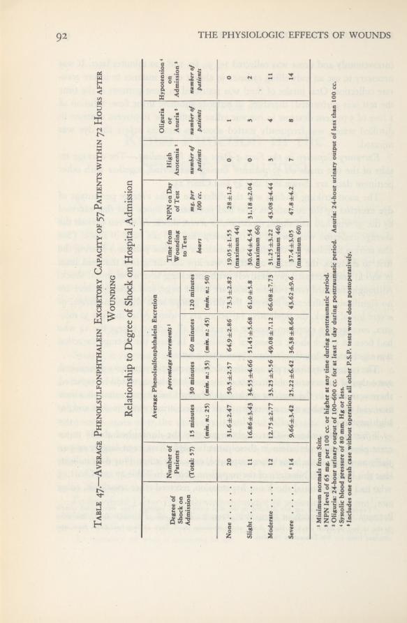
THE PHYSIOLOGIC EFFECTS OF WOUNDS92
Table47.
—AveragePhenolsulfonphthalein
ExcretoryCapacityof57
Patientswithin72
Hoursafter
Wounding
Relationshipto
DegreeofShock
onHospitalAdmission
Degreeof
Shockon Admission
Numberof Patients
AveragePhenolsulfonphthalein
Excretionpercentage
increments1
Timefrom WoundingNPNon
DayofTest
High Azotemia2
Oliguria or Anuria3
Hypotension on Admissions
(Total:57)
15minutes {min.
n.:25)
30minutes (min.
35)60minutes {min.
n.:45)
120minutes {min.
50)
toTest hours
mg.per
100cc.
numberof patients
numberof patients
numberof patients
None
20
31.6±2.4750.5±2.57
64.9±2.8673.3±2.82
19-05±1.55(maximum44)
28±1.2
0
1
0
Slight
11
16.86±3.4334.55
±4.6651.45
±5.686l.0±5.830.64
±4.54 (maximum66)
31.18±2.040
3
2
Moderate....
12
12.75±2.7733.25
±5.5649.08±7,12
66.08±7.73
31.25±3.22(maximum46)
43.08±4.443
4
11
Severe
‘
14
966±3.4225.22±6.4236.38
±8.6643.62±9.6
37.4±3.05 (maximum60)
47.8±4.27
8
14
1
MinimumnormalsfromStitt.
*
NPNlevelof65
mg.per100
cc.orhigheratanytime
duringposttraumaticperiod.
3
Oliguria:24-hoururinaryoutputof
100-600cc.
forat
least1
dayduring
posttraumaticperiod.Anuria:24-hoururinaryoutputoflessthan
100cc.
*
Systolicblood
pressureof80
mm.Hg
orless.
*
Includesonecrush
casewithoutoperation;allother
P.S.P.testsweredone
postoperatively.
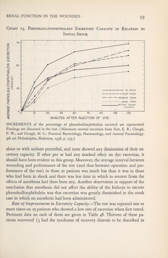
RENAL FUNCTION IN THE WOUNDED 93
Chart 13. Phenolsulfonphthalein Excretory Capacity in Relation to
Initial Shock
No ShockSlight Shock
• Moderate Shock• • • Severe Shock.Minima! Normal
Excretion ,i
INCREMENTS of the percentage of phenolsulfonphthalein excreted are represented.Findings are discussed in the text. (Minimum normal excretion from Stitt, E. R.; Clough,P. W., and Clough, M. C.: Practical Bacteriology, Haematology, and Animal Parasitology.9th ed. Philadelphia, Blakiston, 1938, p. 733.)
alone or with sodium pentothal, and none showed any diminution of their ex-cretory capacity. If ether per se had any marked effect on dye excretion, itshould have been evident in this group. Moreover, the average interval betweenwounding and performance of the test (and thus between operation and per-formance of the test) in these 20 patients was much less than it was in thosewho had been in shock and there was less time in which to recover from theeffects of anesthesia had there been any. Another observation in support of theconclusion that anesthesia did not affect the ability of the kidneys to excretephenolsulfonphthalein was that excretion was greatly diminished in the crushcase in which no anesthetic had been administered.
Rate of Improvement in Excretory Capacity.—The test was repeated one ormore times on 15 patients who showed a low rate of excretion when first tested.Pertinent data on each of them are given in Table 48. Thirteen of these pa-tients recovered (3 had the syndrome of recovery diuresis to be described in
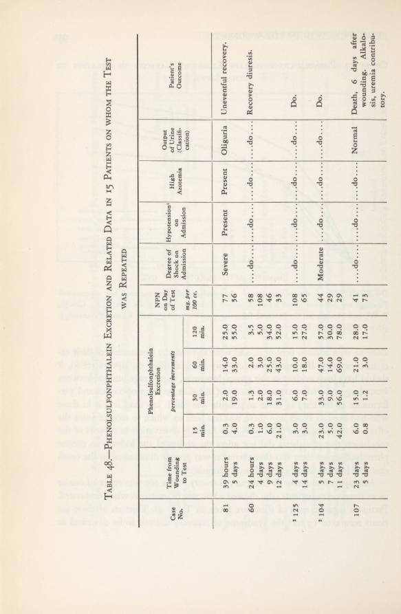
Table48.
—PhenolsulfonphthaleinExcretion
andRelatedData
in15
Patientsonwhomthe
Test
wasRepeated
Case No.
Timefrom Wounding to
Test
Phenolsulfonphthalein Excretion percentageincrements
NPN onDay
ofTest
Degreeof
Shockon Admission
Hypotension1
on AdmissionHigh Azotemia
OutputofUrine (Classifi-
Patient’s Outcome
15 min.
30 min.
60 min.
120min.
mg.per
100cc.
cation)
81
39hours
5
days
0.3 4.0
2.0 19.014.0 33.0
25.0 55.077 56
SeverePresent
PresentOliguria
Uneventfulrecovery.
60
24hours
4
days9
days12days
0,3 1.0 6.0 21.01.3 2.0 18.0 31.0
2.0 3.0 25.0 43.03.5 5.0 34.0 52.058 108 46 33
...
.do....
Recoverydiuresis.
2
125
4
days14days
3.0 3.0
6.0 7.0
10.0 18.015.0 27.0108 65
....do....
Do.
2
104
5
days7
days11
days
23.0 5.0 42.033.0 9.0 56.047.0 14.0 69.057.0 30.0 78.044
Moderate
...
.do....
Do.
29 29
10723days
5
days
6.0 0.8
15.0 1.2
21.0 3.0
28.0 17.041
...
.do.......
.do....
NormalDeath,6
daysafter
73
wounding.Alkalo-
sis,uremia
contribu-tory.
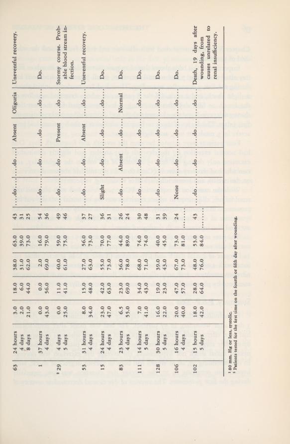
63
24hours
4
days8
days
3.0 2.0 21.018.0 6.0 44.0
38.0 31.0 62.063-0 39.0 73.043 31 25
....do....
....do
AbsentOliguria
Uneventfulrecovery.
1
37hours
4
days
0.0 43.00.0
2.0
16.0
54
...
.do....
....do........do
....
....do....Do.
56.069.079.0
36
2
29
4
days5
days
0.0 25.011.0
40.059.0 75.049 46
....do........do
....
Presentdo
Stormycourse.Prob-
ableblood
streamin-
fection.
41.061.0
53
31hours
4
days
8.0
15.027.0
56.037 27
...
.do........do....Absentdo....
Uneventfulrecovery.
34.048.0
63.073.0
15
24hours
4
days
23.0 47.042.055.0 73.070.036 31
Slight...
.do....
....do........do....Do.
63.0
77.0
83
23hours
4
days
6.5 55.023.0 69.036.0 78.044.0 89.026 24
do....Absent
....do
NormalDo.
111
7.0 41.034.0 43.068.074.030 48
...
.do........
do........do....
....do....Do.
5
days
71.074.0
12830
hours5
days
16.019.0 23.0
39.0 43.040.0
31 39
...
.do.......
.do....
...
.do........
do....Do.
22.0
45.0
106
20.047.0 57.0 28.067.073.0 81.024
None....
do........do
....
....do....Do.
4
days
40.0
73.0 48.0
102
18.0
53-0 84.043
...
.do.......
.do........do
....
...
.do....Death,
19days
after
wounding,from
causesunrelatedto
renalinsufficiency.
5
days
42.064.076.0
1
80mm.Hgor
less,systolic.
2
Patientstestedforthefirsttime
onthefourthorfifthday
afterwounding.
96 THE PHYSIOLOGIC EFFECTS OF WOUNDS
Chapter V). One patient died with alkalosis and renal failure, and the otherdied of peritonitis.
Inspection of this table shows that of 15 patients tested on the fourth or fifthday after wounding, 7 had normal excretion; in all 7 excretion had been lowduring the first 3 days after wounding. Three had had moderate initial shock,2 slight shock, and 2 none. Of the 8 patients in whom excretion was low on
the fourth or fifth day after wounding, 3 had been tested for the first timeduring this period, and 5 are known tohave had diminished excretion during thefirst 3 days. The degree of shock on hospital admission in the 8 patients had beensevere in 3, moderate in 3, and slight in two.
Four patients were retested between the seventh and fourteenth days. Onehad normal excretion by the eleventh day; another had slightly diminishedexcretion only during the first 15 minutes of the test on the eighth day, andtwo who had recovery diuresis (Cases 60 and 125) still had abnormal excretionon the twelfth and fourteenth days after wounding, respectively.
With the exception of one patient (Case 107) who died with alkalosis andrenal failure, all patients in whom excretion was low on initial tests showed im-provement in excretory capacity, reflected chiefly in ability to excrete increasedquantities of dye during the first 30 minutes, although the total quantitiesexcreted also increased with passage of time.
The time required for phenolsulfonphthalein excretory capacity to return tonormal was well correlated with urinary output, nonprotein nitrogen retention,and degree of initial shock and hypotension (Table 48). In general, those pa-tients with a normal output of urine or only slight suppression, with minimalnitrogen retention, and only slight initial shock regained renal function, asmeasured by phenolsulfonphthalein excretion, most rapidly.
Summary, P.S.P. TestPhenolsulfonphthalein excretory capacity was determined during the first
3 days after wounding in 57 patients. The test was made after resuscitationhad been effected and, with the exception of 1 patient who was not operatedupon, after surgery had been completed. In 20 of these patients who had hadneither initial shock nor hypotension, the average dye excretion was normal.In the remaining 37, all of whom had been in various degrees of shock, withhypotension occurring in 27, the average dye excretion was low, especiallyduring the first 30 minutes. The amount of dye cleared decreased as severity of
RENAL FUNCTION IN THE WOUNDED 97
previous shock increased. In patients who had had slight and moderate shock,average excretion was abnormal only in the first 30 minutes; in those who hadhad severe shock it was low throughout the 2-hour period.
The test was repeated one or more times between the fourth and fourteenthpostoperative days on 15 of 37 patients in whom initial excretion had been foundlow. In 14 of them dye excretion returned to normal during this time. In thosepatients in whom impairment was only mild, kidney function was apparentlynormal by the fourth or fifth day. In patients in whom it was more severe(e.g., in recovery diuresis) renal insufficiency, as gaged by phenolsulfonphthaleinexcretory capacity, persisted much longer.
Kenal Clearance StudiesThe majority of the severely wounded patients who died exhibited evidence
of renal insufficiency (see Chapter V). Histologic examination of the kidneysof these patients showed damage predominantly in the lower nephron andrelatively little evidence of glomerular damage (see Chapter IX). The anatomicfindings immediately raised the question of how closely functional impairmentcorresponded with anatomic alterations. One obvious approach to solution ofthis problem was utilization of clearance methods of measuring renal function.This was done in n patients—a small series but the largest we could obtainafter the materials necessary for performance of the tests became available.
Mannitol was used for measurement of glomerular filtration rate (Cm),sodium para-amino hippurate for effective renal plasma flow (Cpah) and max-imum tubular excretory capacity (Tiupah). Analysis of these substances is dis-cussed in Appendix C. Quantities given and rates of administration were essen-tially those suggested by Goldring and Chasis.3 Indwelling, multiple-eyedcatheters were routinely employed. The bladder was washed with 10 to 30 cc.of isotonic salt solution at the end of each collection period, followed by 10 to20 cc. of air to insure complete emptying.
Renal clearance findings together with other pertinent data on the 11 indi-vidual patients are shown in Table 49. Five patients were studied within thefirst 31 hours after wounding, but not until resuscitation had been effected andoperation completed. One of them had had no shock, 1 slight, 2 moderate, and
3 Goldring, W., and Chasis, H.: Hypertension and Hypertensive Disease. New York, The Common-wealth Fund, 1944.
98 THE PHYSIOLOGIC EFFECTS OF WOUNDS
1 severe initial shock. None of them had high azotemia (a plasma nonproteinnitrogen level of 65 mg. per 100 cc. or higher) while we observed them. Outputof urine was normal in 4 patients; one was listed as having had oliguria for1 day (Case 142), but it was slight and can be disregarded.
The remaining six patients were first studied between 3 and 30 days afterwounding. All of them had high azotemia at some time during their course.One had no initial shock, two moderate, and three severe shock. In three pa-tients clearances were first measured while renal failure was severe and repeatedafter recovery diuresis had taken place. A fourth had had severe renal failure,but clearance tests were not made until 30 days after wounding, by which timehe had recovery diuresis and his plasma nonprotein nitrogen was normal. Theremaining two patients died 16 hours and 5 days, respectively, after clearancestudies were done; in both uremia was present at the time of death but inneither was it an immediate or contributory cause of death.
ResultsCharts 14 through 16, in each of which all n cases are included, were con-
structed from mean normals406 and average values listed in Table 49. The5 patients who did not have high azotemia are represented by open symbols;the 6 who had renal failure by solid or semi-solid symbols. For Cases 138, 150,and 133—those with the lowest renal clearance values—only the initial observa-tion is shown. Chart 17 shows these initial values in addition to results whenthe tests were repeated on 1 of these 3 patients.
Glomerular Filtration Rate and Effective Plasma Flow (- ] —Table\CpAH /
49 and Chart 14.—These were definitely below normal in one patient (Case139) 20 hours after wounding; he was the only one of the five studied duringthe early postoperative period who had had severe initial shock. In two of thesefive glomerular filtration rate and effective plasma flow were in the low normalranges. Marked diminution of both components in the three patients (CasesT 33, 138, 150) with severe renal failure is evident. There were lesser degrees ofimpairment in the two in whom renal failure was only coincident in death
4 Ibid.5 Berger, E. Y.; Farber, S. J., and Earle, D. P., Jr.: Renal excretion of mannitol. Proc. Soc. Exper.
Biol. & Med. 66: 62-66, Oct. 1947.Chasis, H.; Redish, J.; Goldring, W.; Ranges, H. A., and Smith, H. W.: Use of sodium p-amino
hippurate for functional evaluation of human kidney. J. Clin. Investigation 24: 583-588, July 1945.
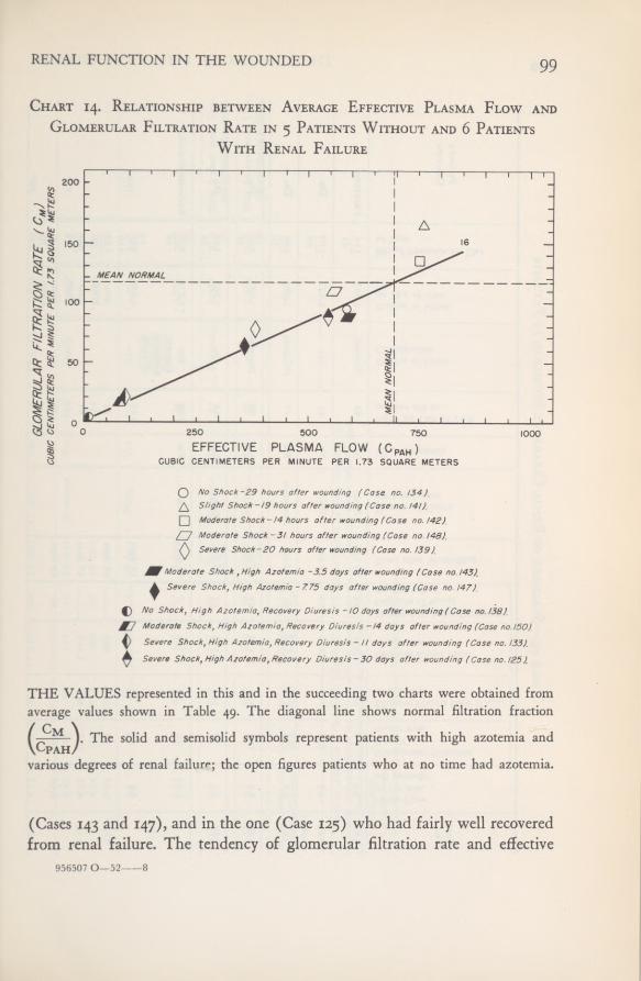
RENAL FUNCTION IN THE WOUNDED 99
Chart 14. Relationship between Average Effective Plasma Flow and
Glomerular Filtration Rate in 5 Patients Without and 6 PatientsWith Renal Failure
No Shock-29 hours after wounding (Case no. 134)Slight Shock ~/9 hours after wounding (Case no. !4t).
Moderate Shock-!4 hours after wounding (Case no. 142).
Moderate Shock-3/ hours after wounding (Case no 148).Severe Shock-20 hours after wounding (Case no. 139)
Moderate Shock ,High Azotemia -3.5 days after wounding (Cose no. /43)
Severe Shock, High Azotemia -7.75 days after wounding (Case no. 147).
No Shock, High Azotemia, Recovery Diuresis - tO days after wounding (Cose no. 138).
Moderate Shock, High Azotemia, Recovery Diuresis —!4 days after wounding (Case no. 150).
Severe Shock, High Azotemia, Recovery Diuresis - it days after wounding (Cose no. !33).
Severe Shock, High Azotemia, Recovery Diuresis-30 days after wounding (Case no. 125).
THE VALUES represented in this and in the succeeding two charts were obtained fromaverage values shown in Table 49. The diagonal line shows normal filtration fraction
(^Cm - ). The solid and semisolid symbols represent patients with high azotemia andCpAH/
various degrees of renal failure; the open figures patients who at no time had azotemia.
(Cases 143 and 147), and in the one (Case 125) who had fairly well recoveredfrom renal failure. The tendency of glomerular filtration rate and effective
956507 0—52 8

THE PHYSIOLOGIC EFFECTS OF WOUNDS100
Table49.
—Summaryof
RenalClearanceStudiesinii
Patients
zQVMvoaftIS0£
•ft
voV>»4kK)SA4lOiUrineFlowh->Cm4kOk)00boboOScc.permin.4^<-MK)
(allperiods)S
GlomerularFiltrationov-nSAoK)'vloOJosvo 00OSovoVOsa"Jsa
cc.permin.
OS00v»4kVOWM
00SAOs
'vl\j\os00EffectivePlasmaFlow(CPAh)
00K)VO■“-vj4LKJ00cc.permin.
r4ksyi00rbbEffectiveBloodFlow4KOS
VOVO
Osu>OSOSOSvoV>Jcc.permin.
MaximalTubularExcretoryVOo'vlvoOs
V>Jvoos004kCapacity(TmPAH)
mg.permin.4k*N)VO00OJoVOVOCLpi
CLpiaCL
PiCLPi3-STcr3*3-TimeafterWounding
(AP{/>
CLC/5ftCL
X0a
S220DegreeofShock0ft
hft
0*1P>ft
3*3ftonAdmission
Yes20NoNo20NoHighAzotemia
-NPNonDayofTestO4kOSK)00OSo"4*"*V>img.per100cc.
p•oJ_.
i20253'05cCl0CL0
»-lOutputofUrinecZ3
303C»n
0Bpi
ClassificationPi
ro o<bjbjK)CmK)
UiVi"v|
001,693K)4kos
OutputofUrineonDayofTest
o”o”o**OOK>OSACC.
IJ.,rtr11-,rooln114 65BloodPressureonDay
oloolOCslK)O1doo00I4koloolooloofTest
5C509ft
po«ao0ft3<5fpft
30c»
a.c*si »i2
'3ft
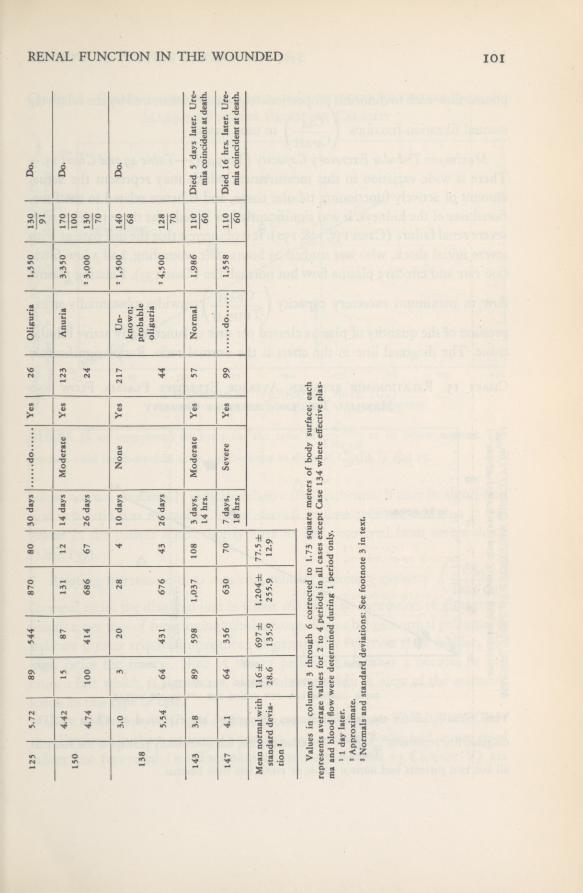
RENAL FUNCTION IN THE WOUNDED 101
125
5.72
89
544
870
80
30days
Yes
26
Oliguria1,550130 91
Do.
150
4.42 4.74
15 100
87 414
131 686
12 67
14days
26days
ModerateYes
123 24
Anuria3,350
2
3,000170 100 130 70
Do,
138
3.0 5.54
3 64
20 431
28 676
4 43
10days
26days
NoneYes217 44
Un- known: probable oliguria2
1,5002
4,500140 68 128 70
Do.
143
3.8
89
598
1.0371083
days,14hrs.
ModerateYes
57
Normal1,986
no 60
Died5
dayslater.Ure-
miacoincidentat
death.
147
4.1
64
356
630
70
7
days,18
hrs.
SevereYes
99
1,558110 60
Died16
hrs.later.Ure-
miacoincidentat
death.
Meannormalwith
standarddevia-
tion3
116± 28.6697± 135.9
1,204±255.977.5± 12.9
rsofbodysurface;each
134whereeffectiveplas-
Valuesincolumns3
through6
correctedto
1.73squaremett
representsaveragevalues
for2to4
periodsinall
casesexcept
Case
maandbloodflowweredetermined
duringI
periodonly.
1
1
daylater.
2
Approximate.3
Normalsandstandard
deviations:Seefootnote3in
text.

THE PHYSIOLOGIC EFFECTS OF WOUNDS102
plasma flow each to diminish proportionately was demonstrated by the relatively
normal filtration fractions ( M-~ ] in most cases.
\CpaH/Maximum Tubular Excretory Capacity (Thipah) —Table 49 and Chart 15.—
There is wide variation in this measurement which may represent the actualamount of actively functioning tubular tissue, and is hence related to the func-tional size of the kidneys. It was significantly low in only the three patients withsevere renal failure (Cases 133,138,150). It is of interest that the one patient withsevere initial shock, who was studied 20 hours after wounding, had a low filtra-tion rate and effective plasma flow but normal Tm (Case 139). Relating plasma
flow to maximum excretory capacity ( PAH-) provides substantially an ex-\ TmpAH /
pression of the quantity of plasma cleared per unit of functionally active tubulartissue. The diagonal line in the chart is the normal ratio. Ratios significantly
Chart 15. Relationship between Average Effective Plasma Flow and
Maximal Tubular Excretory Capacity
THE SYMBOLS for the individual cases represented are explained on Chart 14. The
diagonal line represents Variation is wide, but noteworthy perhaps is the fact thatTmpAH
all but two patients had normal ratios or ratios less than normal.
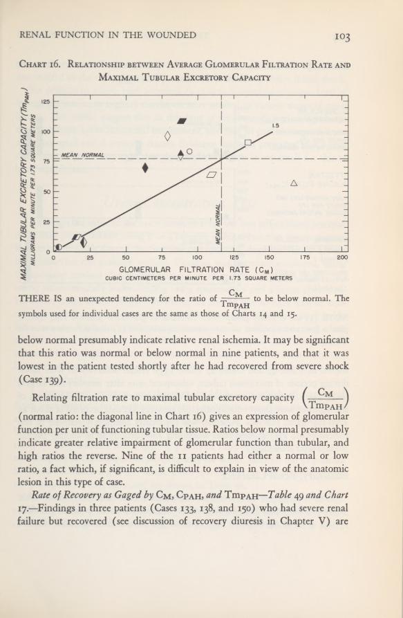
RENAL FUNCTION IN THE WOUNDED
Chart i6. Relationship between Average Glomerular Filtration Rate and
Maximal Tubular Excretory Capacity
THERE IS an unexpected tendency for the ratio of — to be below normal. TheTmp AHsymbols used for individual cases are the same as those of Charts 14 and 15.
below normal presumably indicate relative renal ischemia. It may be significantthat this ratio was normal or below normal in nine patients, and that it waslowest in the patient tested shortly after he had recovered from severe shock(Case 139).
Relating filtration rate to maximal tubular excretory capacity ( ——)VTmpAH/
(normal ratio: the diagonal line in Chart 16) gives an expression of glomerularfunction per unit of functioning tubular tissue. Ratios below normal presumablyindicate greater relative impairment of glomerular function than tubular, andhigh ratios the reverse. Nine of the n patients had either a normal or lowratio, a fact which, if significant, is difficult to explain in view of the anatomiclesion in this type of case.
Rate of Recovery as Gaged by Cm, Cpah, and Thipah—Table 49 and Chart17.—Findings in three patients (Cases 133, 138, and 150) who had severe renalfailure but recovered (see discussion of recovery diuresis in Chapter V) are

THE PHYSIOLOGIC EFFECTS OF WOUNDS
Chart 17. Severe Renal Failure and Subsequent Recovery Diuresis;Rate of Recovery (Case 133)
GLOMERULARFILTRATION RATE (Cm)CUBIC CENTIMETERS PERMINUTE PER 1.73SQUARE METERS (AVERAGE)
EFFECTIVEPLASMA FLOW (C PAH )
CUBIC CENTIMETERS PERMINUTE PER 1.73SQUARE METERS (AVERAGE)
MAXIMAL TUBULAREXCRETORY CAPACITY(Titipah)
milligrams per minutePER 1.73 SQUARE METERS(AVERAGE)
NOTE (i) the relatively proportionate reduction in glomerular filtration rate, effectiveplasma flow, and maximum tubular excretory capacity, and (2) similar recovery rates forall three.
shown in Table 49 and Chart 17 (Case 133). Initial observations were madeduring periods of maximum failure, subsequent ones after recovery had largelytaken place. In general, on the basis of clearance measurements, all portions ofthe nephron were about equally affected; recovery likewise occurred at about aneven rate in all portions. Function was apparently completely restored in onepatient (Case 133) 49 days after wounding and partially so in another patient26 days after wounding.
Summary, Renal ClearanceRenal clearance was measured in n patients in this study. Of 5 tested within
31 hours after wounding, findings were essentially normal in four. The fifthpatient showed significantly low rates of glomerular filtration and effective

RENAL FUNCTION IN THE WOUNDED
plasma flow, but normal maximum tubular excretory capacity. He was the onlyone studied in the early postoperative period who had had severe initial shock.
Six patients in whom high azotemia (a plasma nonprotein nitrogen level of65 mg. per 100 cc. or higher) developed were studied in various stages of renalfailure. The results suggest that in this type of renal insufficiency all portionsof the nephron suffer functional impairment, and that all portions regenerate atabout an equal rate if recovery ensues. Evidence has been presented that a rela-tive renal ischemia may exist, but the number of cases is too small for this to
be of significance.
Urine Concentration TestAlthough the value of the urine concentration test was self-evident, practical
difficulties prevented our using it as a measure of renal function in many cases.Restriction of fluids during the first few postoperative days was almost alwaysinadvisable for the patients’ welfare. Pituitrin was therefore employed, usingthe accepted method of administering 0.5 cc. of posterior pituitary extract (10units) subcutaneously and collecting urine specimens 1 and 2 hours afterward.However, many of the patients in whom we should like to have known con-centrating ability were dailyreceiving considerable quantities of sodium chlorideintravenously. Since the antidiuretic hormone may be ineffective during salineadministration, there was concern that in a large number of cases the pituitrintest would be complicated by recent administration of isotonic saline solution,but in most instances we did not feel justified in requesting that the saline solu-tion be withheld.
A compromise regimen was therefore established in which all parenteralfluids were withheld for 7 or 8 hours before the pituitrin was administered when-ever such restriction clearly would not be harmful in any way to the patient.Under these conditions a value for normal concentrating ability is difficult to
state; we have assumed that a specific gravity of 1.025 or higher represents nor-mal integrity of tubular reabsorptive capacity for water. Data on the 32 patientsin whom concentrating ability of the kidneys was tested in this manner duringthe postoperative days indicated are shown in Table 50.
Of the 17 patients in whom we assumed there was no impairment of con-centrating ability (that is, the specific gravity of one hourly specimen was 1.025or higher) during the first week after wounding, 8 had had moderate or severe

THE PHYSIOLOGIC EFFECTS OF WOUNDS
Table50.
—Resultsof
UrineConcentrationTests
EmployingPituitrin
—32
Cases
SpecificGravity
ofUrine1
Timefrom Wounding
OtherRelated
FactorsNumberof
Patients
Total Patients
to FirstTest
DegreeofInitialShock
HypotensionOliguria
High Azotemia Developed
Days
None
Slight
ModerateSevere
AdmissionAnuria
1.025or
Higher..
2to7
3
6
5
3
8
6
2
17
1.018to
1.025.
.
2to8
0
1
8
0
5
5
1
9
Under1.018.
.
.
1to17
1
1
1
3
4
2
5
5
6
Total.
.
.
4
8
14
6
17
16
8
32
1
Normalbythe
tneth
2Actualoutput
unkncod
used:1
hourlyspecimenof
1.025or
higher.
>wnin2
cases,butgood
presumptiveevidenceofoliguria.

RENAL FUNCTION IN THE WOUNDED 107
shock and hypotension on admission. In 2 of these 17, high azotemia subse-quently developed. Of the 9 patients with slightly decreased ability to reabsorbwater from the kidney tubules (specific gravity of urine 1.018 to 1.025), 8 hadbeen in moderate shock, 5 had hypotension on admission, in 5 oliguria or anuriadeveloped, and in 1 high azotemia. In the 6 patients with marked impairmentof concentrating ability, 4 had had moderate or severe shock, and 4 hypotension.Severe and prolonged azotemia developed in 5 of these 6; they are discussedin the section on recovery diuresis in Chapter V.
The urine concentration test was made more than once in eight patients. Intwo of them the specific gravity, which was 1.018 at the time of the first test,rose to 1.020 two and seven days later, respectively. In one it increased from 1.017to 1.022 in three days. One of these three patients also had high azotemia andoliguria, and two had normal output of urine and a plasma nonprotein nitrogenlevel under 65 mg. per 100 cubic centimeters. The remaining five patients hadrecovery diuresis, and after periods of from 14 to 40 days still were unable toproduce a concentrated urine.
Although the series was small, it suggests that the concentrating ability ofthe kidneys may follow somewhat the same pattern as ability to excrete phenol-sulfonphthalein, mannitol, and sodium para-amino hippurate. Ability to makea concentrated urine appeared to diminish within 2 days following shock, andimprove over a period of from 3 to 7 days, unless renal failure was severe (re-covery diuresis) in which case maximum tubular reabsorption of water remaineddiminished for many days or even weeks. In two patients (Cases 125 and 133)specific gravity remained fixed even after clearance of mannitol and sodiumpara-amino hippurate had returned to normal.
COMMENT
It has been demonstrated by Lauson, Bradley, and Cournand7 that duringshock glomerular filtration rate and effective plasma flow are reduced. Theseworkers have also advanced evidence that reduction in renal blood flow is par-tially explained by active vasoconstriction of the renal blood vessels as well asby reduction of arterial pressure.
7 Lauson, H. D.; Bradley, S. E., and Cournand, A.; Renal circulation in shock. J. Clin. Investigation23: 381-402, May 1944.

THE PHYSIOLOGIC EFFECTS OF WOUNDS
None of our studies were carried out while the patients were in shock. Thoseperformed in the first few days after trauma suggest that the impairment inkidney function may persist for some days after shock is relieved, even thoughthe usually accepted signs of renal failure (suppression of urinary output, nitro-gen retention) may be meager. These patients probably rapidly regain normalkidney function.
If the initial insult, whatever it might have been, was great, the resultingrenal insufficiency was much more severe and prolonged, and in a significantproportion of cases resulted in death in uremia. A few patients, however, didrecover, with gradually increasing function over a period of days to weeks, asindicated by their progress in restoration of ability to excrete phenolsulfon-phthalein, improvement in the glomerular filtration rate, and by increased effec-tive plasma flow, maximum tubular excretory capacity, and concentrating ca-pacity of the urine.
Although the histologic picture in fatal cases might suggest a selective func-tional impairment of the lower nephron, our studies indicated that all functionalcomponents of the kidneys were about equally impaired. Glomerular filtrationrate and effective plasma flow were reduced in essentially proportionate degreesin most of the patients we studied. That there may have been some relative renalischemia in these cases is suggested by the fact that in the few patients on whomclearance tests were done, the ratio of PAH tended to be low. One bit ofTmpAH
evidence in favor of greater relative insult in the lower nephron may be citedfrom the two patients in whom ability to concentrate urine was still much im-paired after clearances had returned to normal. Mannitol measures glomerularfiltration; sodium para-amino hippurate is believed to be excreted by the proxi-mal tubules. Urine concentration takes place primarily in the distal tubule. Thequestion arises whether this lag in recovery of concentrating capacity is a mani-festation of greater relative damage to the lower nephron. The discrepanciesbetween the physiologic and anatomic findings could perhaps be explained bythe production in these patients of renal vascular shunts, such as Trueta 8 andhis associates have recently demonstrated in animals. It is impossible to statefrom our data whether such a mechanism was operative.
8 Trueta, J.; Barclay, A. E.; Daniel, P. M.; Franklin, K. J., and Prichard, M. M. L.: Studies inthe Renal Circulation. Springfield, 111., Charles C Thomas, 1948.
RENAL FUNCTION IN THE WOUNDED 109
SUMMARY
Renal function in the wounded man was studied by measuring phenol-sulfonphthalein excretory capacity in 57 patients; glomerular filtration rate,effective plasma flow, and maximal tubular excretory capacity of para-aminohippuric acid in 11 patients, and urine concentration, employing pituitrin, in32 patients. All studies were made after resuscitation had been effected, and inall surgical cases after operation had been completed. The results were qualita-tively similar and indicated that functionally all portions of the nephron wereabout equally impaired for a period of a few to many days, depending on theseverity of the initial insult which in most patients paralleled the degree of shockon admission. Similarly, diminished renal function, as indicated by these tests,was associated with corresponding increases in the level of plasma nonproteinnitrogen and with decreases in urinary output. When recovery occurred, im-provement of most of these functions proceeded at about an equal rate.
These physiologic findings do not correspond with the histologic findings infatal cases, where the lesion was observed predominantly in the lower nephron.The fact that ability to produce a concentrated urine was impaired in a fewpatients long after phenolsulfonphthalein excretion, glomerular filtration rate,effective plasma flow, and maximum tubular excretion of PAH had returned tonormal might suggest greater relative damage to the lower nephron.
CASES OF SPECIAL INTEREST IN THIS CHAPTER
P.S.P. Re-testI 29 60 81 102 106 III
12815 53 63 83 104 107 125
Renal Clearance TestI25 134 139 i42 147
150x 33 138 141 143 148

CHAPTER IV
Diagnosis of Lower NephronNephrosis Resulting from Trauma
and Shock (“Shock Kidney”)As Derived from Clinicopathologic Correlation
The clinical syndrome of renal insufficiency following traumatic shock hadnot yet, to our knowledge at the time of this study, been defined in the medicalliterature. In the interval preceding publication Lucke 1 has described, from bothclinical and pathologic points of view, a group of similar cases under the term
“lower nephron nephrosis.” The present study, though now no longer novel,was done independently before his publication and therefore offers unbiasedconfirmation which seems worthy of record. Since civilian practice brings so fewapposite cases under the observation of any single group of investigators, one ofour clearest obligations was to attempt definition of this syndrome.
In exploration of a new syndrome the first necessity is establishment of a baseof departure; that is, an apparently new combination of symptoms, signs, labora-tory observations, or distinctive morphologic lesions. With widening experience,additions to and subtractions from this combination are made until the patternbecomes stabilized, the diagnostic importance of each feature or group of fea-tures being assessed by the frequency with which it occurs in the disease in ques-tion as compared with its infrequency in other conditions.
Since the case fatality rate of this condition was high, there is little risk ofdistorting the picture by using the fatal cases to establish the base of departure.In 38 of 60 cases in which necropsy material was available, pigment nephropathy,
1 Lucke, B.: Lower nephron nephrosis. Mil. Surg. 99: 371-396, November 1946.
DIAGNOSIS OF LOWER NEPHRON NEPHROSIS
the characteristic renal lesion of “shock kidney” which will be described in Chap-ter IX, was present. In the following discussion the symptomatology in thesecases will be presented, comparing the findings as we proceed with the controlgroup of 22 cases in which pigment nephropathy was proved absent at necropsy.To determine whether diagnosis of renal injury can be established beyond rea-sonable doubt, the fatal cases will be further compared with nonfatal cases inwhich there was evidence of kidney dysfunction.
Objective Evidence of Penal Insufficiency in Casesof Histologically Proved Nephropathy
Oliguria and Anuria
For purposes of this study, oliguria was defined as a 24-hour output of urinegreater than 100 cc. and less than 600 cc.; anuria as an output not exceeding100 cc. per day. Urine output, known in 35 of the above-mentioned 38 patientswith the characteristic renal lesion, had been subnormal for one or more daysin all, 15 patients remaining in the oliguric and 20 reaching the anuric level.Oliguria or anuria was present from the onset of shock in all but one case, andin this case the delay was apparent rather than real. The initial shock had beenmild, but secondary shock of greater severity occurred after operation and waspromptly followed by anuria. The majority of patients showed progression fromoliguric to anuric levels, but occasionally anuria was present from the start. In15 patients with clinical uremia and histologically proved nephropathy, the urineoutput never declined below the oliguric level. It is worth emphasis that theseindividuals, often casually referred to as “anuria cases,” may never in fact becomeanuric. Unless 24-hour outputs are carefully measured, significant oliguria mayeasily be missed.
Nine patients with oliguria (or anuria) failed to show a significant renallesion at necropsy. This is not surprising, considering the variety of conditionsfrom which these severely wounded men suffered which would tend to lowerurine output, such as deficient blood volume, prolonged hypotension, paralyticileus, infection, and exudation into traumatized areas and into infected serouscavities. In two of the nine oliguria or anuria was intermittent; in both the clin-ical picture was dominated by severe peritonitis.
THE PHYSIOLOGIC EFFECTS OF WOUNDS112
Azotemia
“High azotemia,” arbitrarily defined as a nonprotein nitrogen level in theplasma of 65 mg. per 100 cc. or higher, was found in 36 of the 38 proved ne-phrotic cases; in 1 case no determination was available, and in the remaining1, death occurred within 35 hours of injury. In this patient the nonprotein levelhad already climbed to 63 mg. per 100 cc., only 2 points below our arbitrary limit,and there can be no reasonable doubt that this limit would have been passedhad he lived a few hours longer. Like oliguria, nitrogen retention was evidentfrom the outset. The retention level climbed steadily, usually from 30 to 40points a day, the maximal level depending solely on the length of time elapsingbefore death ensued or, in those who survived, before onset of diuresis. Nitrogenretention, like oliguria, was also observed in the absence of a renal lesion (7 in-stances) and is explainable on similar grounds.
Hypertension
We defined hypertension for purposes of this study as an elevation of thesystolic blood pressure to or above 135 mm. Hg and of the diastolic to 90 mm.Hg or higher. By civilian standards these figures must seem low, but it is to beremembered that we were dealing primarily with a homogeneous group ofyoung men between 18 and 30 years of age who had recently suffered severetrauma usually associated with the loss of considerable amounts of blood. Bythis standard, 20 of 32 patients with the renal lesion whose blood pressures hadbeen recorded were judged to have significant elevations of systolic or diastolicpressure or both. Hypertension rarely appeared within the first 48 hours; itwas frequent by 72 hours, and was the rule by 96 hours.
Of the remaining 12 patients with the characteristic renal lesion who failedto show an elevation of blood pressure, 3 died within 2 days of injury and 6 onthe third day. In only 3 patients living beyond the 3-day period did hypertensionfail to develop. One died on the fourth, 1 on the fifth, and 1 on the seventh dayrespectively. The last 2 patients also had a severe progressive peritonitis. It seemsreasonable to conclude that hypertension is a characteristic if not constant symp-tom of the syndrome.
The importance of hypertension is increased when viewed from the conversepoint of view. No patient in this series whose kidneys failed to show pigment

DIAGNOSIS OF LOWER NEPHRON NEPHROSIS
nephropathy at necropsy had a sustained elevation of blood pressure, althoughan occasional determination might have been above the arbitrary limit. It mustbe remembered, however, that no cases of cerebral injury were included in ourseries. In such cases hypertension secondary to increased intracranial pressure isof course frequent.
Pigment Excretion in the UrineAs part of the routine urine examination of all patients included in this study,
the supernatant urine after centrifugation was tested with benzidine for thepresence of dissolved heme-containing pigments,2 which, as will be discussedfully in Chapter VIII, were largely hemoglobin and myoglobin.
All patients with morphologic evidence of renal damage excreted benzidine-positive material in the urine. In n patients (exclusive of those in whom therewas direct trauma to the urinary tract) enough pigment was excreted to makethe urine grossly red or brown. In some cases descriptions such as “dark amber”suggest a pigment element, but in the majority amber and yellow were the colorsrecorded, and only the benzidine test revealed the pigment element. Pigmentexcretion, furthermore, was in most instances transitory and usually disappearedon the third or fourth day. On the other hand, pigment excretion was observedin 6 patients whose kidneys did not show significant lesions at necropsy.
Other Urinary Findings
Other chapters of this volume contain complete discussions of the chemicalabnormalities found in the blood and urine and the results ofrenal function testsperformed during the course of the studies. Repetition would be pointless, espe-cially since these factors have little diagnostic value. However, three urinaryabnormalities, determination of which lies within the realm of routine clinicalpathology, are worthy of mention since, although of limited diagnostic value,they were constant features of the syndrome.
i. Proteinuria.—All patients with proved renal lesions showed proteinuria.However, since proteinuria was absent in only 14 of the entire series of casual-ties studied and was present in 51 patients without clinical or anatomic evidenceof nephropathy, its diagnostic import is negligible.
2 See Appendix C, section on routine urinalyses, for technique of performing the test.
THE PHYSIOLOGIC EFFECTS OF WOUNDS
2. Persistent Acidity.—The urine of all patients manifesting nephropathy at
necropsy was acid on initial examination. The range of pH was from 5.0 to 6.9with an average value of 5.8. The urines of the control group ranged from 5.0to 8.4 an average of 6.2, overlapping too much to give the figures any diag-nostic value. In succeeding specimens the urine from the nephropathic groupremained persistently acid with few exceptions, occasionally even despite a thera-peutically-induced alkalosis.
3. Fixation of Specific Gravity.—The specific gravities of the initial speci-mens varied from 1.009 to 1.032, though the majority of specimens fell withinthe range of 1.020 to 1.026. No significant difference was found in patients withand without nephropathy. Diversity in these initial specimens is not surprising,since it is probable that in many instances part of the urine may have been ex-creted by the kidney before the patient was wounded. With the passage of timethe specific gravity of subsequent specimens declined in the nephropathic group,despite oliguria, and a tendency to fixation between 1.010 and 1.015 became ap-parent inall the patients with proved renal lesions. Evidence of continued powerof urine concentration occasionally helped to distinguish cases of “extrarenal”oliguria and azotemia from the true nephropathies.
Summary: Diagnosis of Lower NephronNephrosis (“Shock Kidney”)
Seven phenomena have been described which are characteristic of the renallesion following traumatic shock. One of these, proteinuria, has no diagnosticvalue because of its frequency in non-nephropathic cases. Two others, persistentacidity of the urine and fixation of specific gravity at a low level despite oliguria,are of great interest in defining the physiologic abnormality, but proved of lim-ited diagnostic value since therapeutic emergencies frequently prevented verifi-cation by suitable test. A tetrad of findings—oliguria, azotemia, pigment excre-tion, and hypertension—were of practical diagnostic value.
Each of these may be present singly in patients with normal kidneys. So mayany pair. The combination of oliguria and azotemia, for instance, was found fivetimes; even the triad of oliguria, azotemia, and pigment excretion was foundtwice in patients with morphologically normal kidneys. The complete tetrad,however, was never found in this series in patients with normal kidneys. Barringthe complication of a head injury, it was concluded that this tetrad might beconsidered reliable evidence of renal damage.

DIAGNOSIS OF LOWER NEPHRON NEPHROSIS 115
Uremic SymptomatologySubjective symptoms of the type commonly to be expected in uremia were
conspicuous primarily by their absence. In the initial stages of the disorder minorsymptoms might well have been masked by the greater discomforts of recenttrauma, surgery, and anesthesia, or been suppressed by postoperative narcosis.As the days pass, these distractions cease to be of importance, yet again andagain statements such as “patient comfortable” and “no complaints” appearedin the records. A brief review of the usual symptoms ofrenal insufficiency, notingtheir frequency in this material, is in order.
Headache.—Headache was not mentioned in any case record. Minor com-plaints might not have been recorded, but even so headache could not have beena conspicuous feature.
Nausea and Vomiting.—Many of our patients had abdominal wounds withsubsequent gastric and intestinal surgery complicated by various grades of peri-toneal irritation. They were often treated by Wangensteen drainage, makinginterpretation of gastro-intestinal symptoms difficult. Nausea appeared in threeof the crush-syndrome cases and also in three others not complicated by abdom-inal wounds. In only two of these was it associated with vomiting, once on theseventh and once on the thirteenth day. Persistent hicupping was twicerecorded.
Vision.—No patient complained of visual difficulties. Eyeground examina-tions were recorded in nine cases. In six patients the eyes were found to be nor-mal when examined at intervals of from 3 to 10 days, and in three patientswere found to be abnormal. Two of the latter patients showed retinal hemor-rhages on the fifth and tenth days of disease, respectively, and one showed papil-ledema on the ninth day.
Consciousness. —Drowsiness was the most frequent cerebral symptom andwas noted in 17 instances. Its onset was recorded as early as the second day, aslate as the ninth. Very frequently the notation was accompanied by a statementsuch as “patient drowsy but when aroused answers questions clearly and intelli-gently.” One patient was frankly disoriented, and two were irrational. In n pa-tients drowsiness progressed to stupor; in 6 of these the condition was describedas coma. This stage seldom antedated death by many hours. Onset of coma wasrecorded as early as the third and as late as the tenth day. At the time of onset,the nonprotein nitrogen levels ranged from 89 to 305 mg. per 100 cc. of plasma.One patient regained consciousness after a lapse of 2 hours at a time when the
956507 0—52 9
THE PHYSIOLOGIC EFFECTS OF WOUNDS
nonprotein nitrogen level was 339 mg. per 100 cc. of plasma.Twitekings and Convulsions. —These were very uncommon phenomena.
Twitchings were recorded in two patients, convulsions in four; one of these fourrecovered. Neuromuscular irritability was noted in two other patients in whomChvostek’s sign developed. In both, the symptoms followed vigorous alkalitherapy.
Hyperpnea.—Dyspnea or hyperpnea was recorded on 12 occasions, with onsetranging from the fourth to the twelfth day. In six instances it appeared approxi-mately coincidentally with the onset of pulmonary edema and may well havebeen an effect of the latter. In only a few instances was respiration clearly of theKussmaul type.
Edema. —Evidence of edema was observed in 25 patients. It appeared as earlyas the third and as late as the eleventh day, largely depending on the extent ofintravenous fluid therapy. Clinical evidence of pulmonary edema was recordedin 12 instances. Either pulmonary or peripheral edema might appear first, andeach was observed in the absence of the other. Facial edema was noted only 8times.
Nonjatal CasesIn examining the clinical records of nonfatal cases in this series to see if diag-
nosis of renal injury could be established beyond reasonable doubt, we were ham-pered by incompleteness of observation and record. The findings in 44 patientswhose records were fairly complete and who manifested 1 or more of the 4criteria described above are summarized in the accompanying table.
Three patients presented the complete tetrad of characteristic findings. Twoshowed the triad of pigment excretion, azotemia, and hypertension, but in boththere was uncertainty regarding urine output during the first 48 hours afterinjury. Two presented the combination of pigment excretion, oliguria, and azo-temia, but blood pressure determinations at appropriate time intervals were notrecorded. In five others with the same triad, hypertension could be definitelyexcluded.
Among those with a combination of two principal signs, one patient showedoliguria and hypertension, but nonprotein nitrogen was not determined and theurine was not tested with benzidine. In one case hypertension of notable level(170/120) and azotemia were observed, but urine output was not recorded

DIAGNOSIS OF LOWER NEPHRON NEPHROSIS
Table 51.—Principal Findings in 44 Surviving Patients Suspected of Having
Renal Insufficiency
nor was the urine tested with benzidine during the first 5 days. In Case 130,oliguria and unimpressive azotemia of 69 mg. per 100 cc. were demonstrated,but again the urine was not tested and no postoperative blood pressures were re-corded. In another patient, azotemia and pigment excretion were present but noelevation of blood pressure occurred, and the urine excretion for the first 24 hourswas 750 cc., which was above our arbitrary limit but nevertheless in the lowerrange of normality.
Three patients presented the remaining pair of findings: pigment excretionand oliguria. Six patients manifested only transitory oliguria without supportingevidence of nephropathy, and 19 pigment excretion with similar lack of con-firmatory findings. In the light of the above findings, let us again evaluate ourdiagnostic criteria.
As in the fatal group, no patient with other evidence of renal insufficiencyfailed to show pigment excretion if the urine was tested with benzidine withinthe first 48 hours after injury. Nineteen patients out of the 44, however, who atno time showed any other evidence ofrenal damage, excreted benzidine-positivematerial.
Number of Patients
Findings
PigmentExcretion Oliguria
"HighAzotemia”
Hypertension(Above 135/90)
3 + + + +
2 +?
+ +
2 + + +?
1 p + ? +
1 p ? + +
1 ? + + ?
5 + + + 01 + 0 + 0
3 + + 0 06 0 + 0 0
19 + 0 0 0
+ =present.? =not recorded, or presence uncertain.0 =absent.
THE PHYSIOLOGIC EFFECTS OF WOUNDS
Oliguria was demonstrated in 15 patients with 1 or more other positive find-ings. In 3 patients its presence was uncertain and in 6 it was observed in theabsence of any other sign. In one questionable case, in which there was pigmentexcretion and azotemia, oliguria was absent. Oliguria in these patients whosurvived was, however, sometimes extremely transitory. Among the patientsmanifesting the complete tetrad, oliguria was of only a day’s duration in two
cases, and lasted only 2 days in one. The longest duration of oliguria did not
exceed 4 days. In none of the cases was output depressed to anuric levels; i.e.,below 100 cc. in 24 hours.
Azotemia was observed in 15 of the 44 patients. In no instance was it thesole finding. A plasma nonprotein nitrogen concentration of 88 mg. per 100 cc.accompanied only by pigment excretion occurred in one instance. In 7 patientsthe maximal level was below 100 mg. per 100 cc., and in 6 the range was be-tween 100 and 150 mg. per 100 cc. of plasma. Two desperately ill patients recov-ered despite nonprotein nitrogen levels of 247 and 217 mg. per 100 cc. respectively.In 3 patients the azotemia was of such low degree (74, 77, and 69 mg. per 100 cc.respectively) and so transitory (lasting only 1 and 2 days) as to be of doubtfulsignificance. In the other patients it lasted from 3 to 21 days.
Seven patients showed definite hypertension which ranged from 128/90 inCase 54 to 180/115 in Case 138. Blood pressure was not recorded for two patientswho presented the triad of pigment excretion, oliguria, and azotemia, but did notbecome elevated in five other patients presenting the same combination. The re-maining two patients in this group had, respectively, azotemia of more than 17days’ duration with a maximum nonprotein nitrogen level of 247 mg. per 100 cc.,and azotemia of over 7 days’ duration with a nonprotein nitrogen level of 140 mg.per 100 cubic centimeters. Hypertension was never observed without other evi-dence of renal insufficiency. Once hypertension developed it was very persistent,even after the nonprotein nitrogen had dropped to normal levels. The shortestduration of hypertension observed was 12 days, and in one patient it was stillpresent at 22 days.
In summary, pigment excretion appeared to be a constant phenomenon inthe shock kidney but was seen also in many patients who never showed evidenceof renal failure. Oliguria likewise was frequently seen in the absence of renalfailure. In positive cases it might be extremely transitory and the volume notgreatly depressed. Azotemia was a constant and, except in the presence of severecomplications or in the moribund state, a reliable sign of renal failure. Eleva-

DIAGNOSIS OF LOWER NEPHRON NEPHROSIS
tion of blood pressure in the absence of a head injury was inconstant. When pres-ent it constituted the most important confirmatory criterion.
On these grounds it is reasonable to conclude that all seven surviving patientsmanifesting hypertension did, in fact, have “shock kidney.” In six of these azo-temia was proved, and in the seventh the nonprotein nitrogen level was notrecorded. Eight other patients showed azotemia supported by one or more othercriteria of renal injury. In three patients azotemia was so slight and transitoryas to be of doubtful significance. In the remainder it was of sufficient severityor was bolstered by enough other confirmatory evidence to justify the diagnosisof renal injury.
We conclude, then, that a minimum of n and a possible maximum of 14of our patients developed and survived “shock kidney.” In comparison with 38fatal cases of histologically proved shock kidney, this indicates a case fatalityrate of approximately 75 percent.
SUMMARY AND CONCLUSIONS
The clinical records of 60 patients on whom necropsy was performed wereanalyzed from the standpoint of factors relating to renal function, and the find-ings in the group of 38 with histologically demonstrated pigment nephropathywere compared with those of the 22 in whom this lesion was not present. Itbecame apparent that the clinical syndrome ofrenal insufficiency which followsshock is remarkable chiefly for the scarcity and mildness of its symptoms. Littlethat the patient complained of served to call attention to the condition. Drowsi-ness slowly deepening into stupor was the most common symptom, but it mightbe absent almost until death. The only common sign was edema, either pul-monary or peripheral.
Laboratory and blood pressure findings provided more useful criteria. Sevenfindings were present with great constancy in the nephropathic cases that ap-peared only sporadically or never in the control group. These were in ascendingorder of diagnostic importance: proteinuria, persistent acidity of the urine, excre-tion of benzidine-positive material, oliguria, azotemia, fixation of specific gravityof the urine at a low level, and hypertension. It was found that the diagnosis ofnephropathy could only be made by the systematic recording of four factors inevery patient who was resuscitated from shock: (1) the total urinary output,(2) the presence of benzidine-positive material in the urine, (3) the nonprotein

120 THE PHYSIOLOGIC EFFECTS OF WOUNDS
nitrogen level in the blood plasma, and (4) the systolic blood pressure. Thesymptom complex of oliguria, pigment excretion, azotemia, and hypertensionestablished the diagnosis. It is probable that in some cases oliguria was too tran-sient to be recognizable, and certain that in some cases hypertension did notdevelop. If the patient survived beyond 4 days, fixation of specific gravity andconstant acidity of the urine afforded important confirmatory evidence.
These diagnostic criteria of nephropathy were then applied in a review of therecords of the nonfatal cases in the series. It was concluded that a minimum of11 and a possible maximum of 14 had had a pigment nephropathy. Using theformer figure, the case fatality rate is approximately 75 percent.
CASES OF SPECIAL INTEREST IN THIS CHAPTER
Nonfatal13 44 72 109 13327 54 81 112 13829 60 87 125 15037 7i 104 130
Fatal8 26 52 85 108 122
12 31 55 86 116 12322 41 65 88 117 12924 47 66 95 118 13125 49 74 97 I2° 136
80 98 A-30Crush Syndrome Fatalities
69 70 78 93 132
CHAPTER V
Clinical, Physiologic, and BiochemicCorrelation in Lower Nephron
NephrosisThe importance and frequent occurrence of renal failure in those battle casu-
alties who were severely wounded must be emphasized. In the preceding chap-ter the diagnosis of posttraumatic renal insufficiency was discussed from a clini-copathologic viewpoint. In this chapter an attempt will be made to present acomprehensive picture of the physiologic and biochemic features of the syn-drome and their correlation with the clinical findings.
In studying these patients in whom renal insufficiency developed followingtrauma we have dealt with a unique group of individuals. They were nearlyall young men, and so far as was known, physiologically sound prior to wound-ing. They had incurred severe wounds which almost immediately began to causechanges in their internal environment. Because of the effectiveness of resusci-tation and other early treatment, the wounds and the changes produced werenot severe enough to cause early death. These men withstood operation fairlywell, largely because they had been adequately treated preoperatively, but be-ginning with the first day or two after operation (or after trauma, if no surgerywas done) they began to show clinical and laboratory evidence of inadequaterenal function. The renal failure progressed rapidly and in most instances thepatients either died in uremia within 10 days or then began to show signs ofimprovement of renal function, such as diuresis and clearance of nonproteinnitrogen, and subsequently recovered.
In selecting patients presumed to have diminished renal function, two maincriteria were utilized: the nonprotein nitrogen level in the blood plasma, and thedegree of urinary suppression. Much of the data presented in this chapter willrelate primarily to the patients with high azotemia, since this was a constant andgenerally reliable indication of renal insufficiency. In many instances the data

THE PHYSIOLOGIC EFFECTS OF WOUNDS122
are also correlated with the degree of urinary suppression because a low urinaryoutput provides a simple and useful clinical means of recognizing many casesof impending renal failure. Our.definitions of “high azotemia” and of oliguriaand anuria have been given, and the diagnostic features of the syndrome werediscussed in Chapter IV.
The incidence of high azotemia, oliguria, and anuria in our series is shownin Table 52. In 5 of the 186patients no nonprotein nitrogen determinations weremade and in 50 the urinary output was unknown. Seventy-three of 181 patientswere found to have high azotemia. Thirty-three of 136 patients had anuria forat least 24 hours, 45 oliguria (they did not reach anuric level at any time), and58 had a normal output of urine. Of the patients with high azotemia, 27 alsohad anuria and 29 oliguria.
Table 52.—Plasma Nonprotein Nitrogen Level and Urinary Output in the
Severely Wounded
Clinical featuresCase Fatality
Sixty-five of the 186 patients in the total series failed to survive. Of the 65who died, 51, or 78 percent, were among those who had high azotemia or urinarysuppression or both. The serious implication of the onset of these conditions iswell illustrated by Table 53. Of the 73 patients with high azotemia, 50, or 68 per-cent, died. Of 33 with anuria, 30, or 91 percent, died. This table likewise shows
Nonprotein Nitrogen Level
Urinary Output (Number of Patients)
Anuria 1 Oliguria 2 NormalOutput
OutputUnknown Total
With High Azotemia 3
Without High Azotemia ....NPN Unknown
2715
2916
1048
743
73108
5
Total 33 45 58 50 186
1 Urinary output ofless than 100 cc. in 24 hours.2 Urinary output of 100-600 cc. in 24 hours.3 Plasma nonprotein nitrogen level of 65 mg. per 100 cc. or higher.
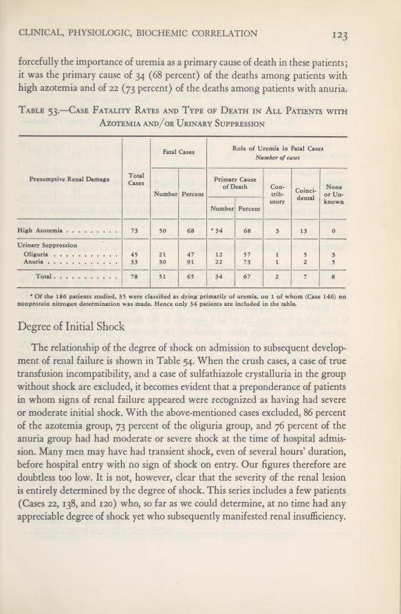
CLINICAL, PHYSIOLOGIC, BIOCHEMIC CORRELATION
forcefully the importance of uremia as a primary cause of death in these patients;it was the primary cause of 34 (68 percent) of the deaths among patients withhigh azotemia and of 22 (73 percent) of the deaths among patients with anuria.
Table 53.—Case Fatality Rates and Type of Death in All Patients with
Azotemia and/or Urinary Suppression
Degree of Initial ShockThe relationship of the degree of shock on admission to subsequent develop-
ment of renal failure is shown in Table 54. When the crush cases, a case of truetransfusion incompatibility, and a case of sulfathiazole crystalluria in the groupwithout shock are excluded, it becomes evident that a preponderance of patientsin whom signs of renal failure appeared were recognized as having had severeor moderate initial shock. With the above-mentioned cases excluded, 86 percentof the azotemia group, 73 percent of the oliguria group, and 76 percent of theanuria group had had moderate or severe shock at the time of hospital admis-sion. Many men may have had transient shock, even of several hours’ duration,before hospital entry with no sign of shock on entry. Our figures therefore aredoubtless too low. It is not, however, clear that the severity of the renal lesionis entirely determined by the degree of shock. This series includes a few patients(Cases 22, 138, and 120) who, so far as we could determine, at no time had anyappreciable degree of shock yet who subsequently manifested renal insufficiency.
Fatal Cases Role of Uremia in Fatal CasesNumber of cases
Presumptive Renal Damage TotalCases
Number Percent
Primary Causeof Death Con-
trib-Coinci-dental
Noneor Un-
Number Percentutory known
High Azotemia 73 50 68 * 34 68 3 13 0
Urinary SuppressionOliguria 45 21 47 12 57 1 5 3Anuria 33 30 91 22 73 1 2 5
Total 78 51 65 34 67 2 7 8
* Of the 186 patients studied, 35 were classified as dying primarily ofuremia, on 1 of whom (Case 146) nononprotein nitrogen determination was made. Hence only 34 patients are included in the table.

THE PHYSIOLOGIC EFFECTS OF WOUNDS124
Table 54.—Relationship of Initial Shock to Development of
Renal Insufficiency
Survival Period after WoundingThe time of death after wounding was considered in 51 patients who had
renal insufficiency (Table 55). Of these 51, uremia was the cause of death in35, a contributory cause in 3, and only coincidental in 13 patients. Of the 35patients in whom uremia was the primary cause of death, 15, or 43 percent, diedin the first 5 days and 17, or 48.5 percent, in the second 5 days—more than 91percent within 10 days after wounding. Of the entire group of 51 fatalities,94 percent (48 patients) died within the first 10 days. The significance of thistime factor will be illustrated in connection with the biochemic data and in the
Table 55.—Period of Survival after Wounding in 51 Patients with
Renal Insufficiency
Classification TotalDegree of Initial Shock
of Cases CasesNone Slight Moderate Severe Unknown
By NPN RetentionHigh Azotemia 73 1 6 6 27 33 1
By OutputOliguria 45 3 8 13 20 1Anuria 33 2 8 3 6 16 0
1 Includes 2 crush cases, 1 case of transfusion incompatibility, and I case of slight postoperative shock.s Includes 1 case of sulfathiazole crystalluria, I case of transfusion incompatibility, and 2 crush cases.
Time from Wounding to Death
Days
Role of Uremia in Cause ofDeath
Primary
Number of cases
Contributory
Number ofcases
Coincidental
Number of cases
1 to 5 15 2 96 to 10 17 1 41 1 to 15 3 0 0
Total 35 3 13
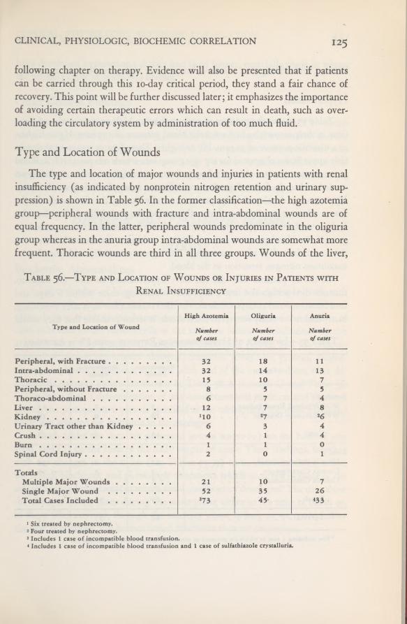
CLINICAL, PHYSIOLOGIC, BIOCHEMIC CORRELATION
following chapter on therapy. Evidence will also be presented that if patientscan be carried through this 10-day critical period, they stand a fair chance ofrecovery. This point will be further discussed later; it emphasizes the importanceof avoiding certain therapeutic errors which can result in death, such as over-loading the circulatory system by administration of too much fluid.
Type and Location of WoundsThe type and location of major wounds and injuries in patients with renal
insufficiency (as indicated by nonprotein nitrogen retention and urinary sup-pression) is shown in Table 56. In the former classification—the high azotemiagroup—peripheral wounds with fracture and intra-abdominal wounds are ofequal frequency. In the latter, peripheral wounds predominate in the oliguriagroup whereas in the anuria group intra-abdominal wounds are somewhat morefrequent. Thoracic wounds are third in all three groups. Wounds of the liver,
Table 56.—Type and Location of Wounds or Injuries in Patients withRenal Insufficiency
Type and Location of WoundHigh Azotemia
Numberofcases
Oliguria
Numberofcases
Anuria
Numberof cases
Peripheral, with Fracture 32 18 11Intra-abdominal 32 14 13Thoracic 15 10 7Peripheral, without Fracture 8 5 5Thoraco-abdominal 6 7 4Liver 12 7 8Kidney UO 2 7 2 6Urinary Tract other than Kidney 6 3 4Crush 4 1 4Burn 1 1 0Spinal Cord Injury 2 0 1
TotalsMultiple Major Wounds 21 10 7Single Major Wound 52 35 26Total Cases Included 3 73 45- 433
1 Six treated by nephrectomy.2 Four treated by nephrectomy.3 Includes 1 case of incompatible blood transfusion.4 Includes 1 case of incompatible blood transfusion and 1 case of sulfathiazole crystalluria.
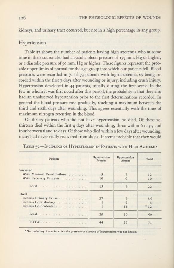
126 THE PHYSIOLOGIC EFFECTS OF WOUNDS
kidneys, and urinary tract occurred, but not in a high percentage in any group.
Hypertension
Table 57 shows the number of patients having high azotemia who at sometime in their course also had a systolic blood pressure of 135 mm. Hg or higher,or a diastolic pressure of 90 mm. Hg or higher. These figures represent the prob-able upper limits of normal for the age group into which our patients fell. Bloodpressures were recorded in 71 of 73 patients with high azotemia, 67 being re-corded within the first 7 days after wounding or injury, including crush injury.Hypertension developed in 44 patients, usually during the first week. In thefew in whom it was first noted after this period, the probability is that they alsohad an unobserved hypertension prior to the first determinations recorded. Ingeneral the blood pressure rose gradually, reaching a maximum between thethird and sixth days after wounding. This agrees essentially with the time ofmaximum nitrogen retention in the blood.
Of the 27 patients who did not have hypertension, 20 died. Of these 20,thirteen died within the first 4 days after wounding, three within 6 days, andfour between 6 and 10 days. Of those who died within a few days after wounding,many had never really recovered from shock. It seems probable that they would
Table 57.—Incidence of Hypertension in Patients with High Azotemia
Patients HypertensionPresent
HypertensionAbsent Total
SurvivedWith Minimal Renal Failure 5 7 12With Recovery Diuresis 10 0 10
Total 15 7 22
DiedUremia Primary Cause 27 7 34Uremia Contributory 1 2 3Uremia Coincidental 1 11 * 12
Total 29 20 49TOTAL 44 27 71
* Not including I case in which the presence or absence of hypertension was not known.

CLINICAL, PHYSIOLOGIC, BIOCHEMIC CORRELATION
have developed hypertension if they had lived longer, especially those in whomthe primary cause of death was uremia.
Other Clinical FindingsEdema was observed in 23 of the 73 patients with high azotemia. The edema
varied in degree, but it was usually generalized, involving all extremities and theface. It was present in 18 patients who died. Three patients had generalized con-vulsions. Eyegrounds were examined in 7 patients; 2 showed flame-shapedhemorrhages and 1 a small exudate. A pericardial friction rub was heard in 1 pa-tient who died of uremia, and pericarditis was found on necropsy.
Biochemic and Physiologic Features
BLOOD
Biochemic AbnormalitiesA large number of blood chemistry studies were made preoperatively and
postoperatively on the 73 patients with high azotemia. It was not possible tomake daily or even regular determinations on every patient, and the number ofcases indicated in the various cells of the tables to follow is conditioned by theavailable data. The plasma carbon-dioxide combining power, and the concen-trations of plasma nonprotein nitrogen, chlorides, phosphate, protein, magne-sium, phosphorus, creatinine, and uric acid, and of the serum sodium were de-termined. Average values are shown in Table 58 A through G and Table 59.When more than 1 determination of any constituent was made on any 1 patientduring the postoperative periods specified in the tables, only the most abnormalvalue was selected for inclusion in the averages.
In both tables the data are presented in two ways: (1) on the basis of sur-vival, and (2) on the basis of the daily output of urine. These findings, togetherwith the changes in the urine (see Tables 75-77) to be discussed later, reflectthe typical biochemic and physiologic alterations which occurred in those of theseverely wounded patients in whom renal insufficiency developed. The data showthe changes taking place during the acute phase either to the time of death or,in those who survived, through the recovery phase as long as we could followthe case. Variations will be mentioned whenever they are important.
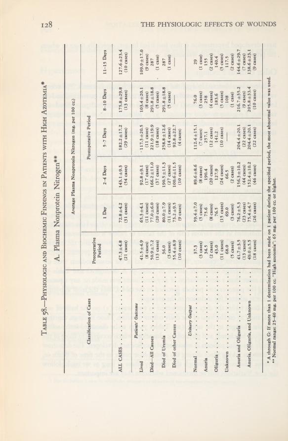
128 THE PHYSIOLOGIC EFFECTS OF WOUNDS
Table58.—Physiologicand
BiochemicFindings
inPatients
withHighAzotemia*
A.Plasma
NonproteinNitrogen**
AveragePlasma
NonproteinNitrogen(mg.
per100
cc.)
ClassificationofCases
Preoperative
PostoperativePeriod
Period
1
Day
2-4Days
5-7Days
8-10Days
11-15Days
ALLCASES
47.3±4.8
72.8±4.2
143.1±9.3
182.2±17.2175.8±29.8127.6±23.4
(21cases)
(31cases)
(54cases)
(29cases)
(13cases)
(10cases)
Patients’Outcome
Lived
41.5±4.9
65.3±4.1
92.8±9.1
'
117.3±20.5103.4±20.1
109.9±17.0
(8
cases)
(11cases)
(17cases)
(11cases)
(8
cases)
(9
cases)
Died—AllCauses
50.9±7.277.0±6.0
166.2±11.0
221.9±19.9291.8±18.8287
(13cases)
(20cases)
(37cases)
(18cases)
(5
cases)•
(1
case)
DiedofUremia
36.0
80.0±7.9190.5
±11.3
258.8±12.6291.8±18.8287
(3
cases)
(11
cases)
(27cases)
(14cases)
(5
cases)
(1
case)
DiedofotherCauses
55.4±8.9 (10cases)
73.2±9.5
(9cases)
100.6±11.5
(10cases)
92.8±22.7 (4cases)
UrinaryOutput
Normal
37.3
59.4±7.089.6±8.6
112.4±15.1
76.0
29
(3
cases)
(5
cases)
(8
cases)
(7cases)
(3
cases)
(1
case)
Anuria
34.5
75.6
190.4
257.1
258
155
(2
cases)
(8
cases)
(20cases)
(12cases)
(4cases)
(2
cases)
Oliguria
43.0
76.5
127.9
141.2
183.6
140.4
(11cases)
(15cases)
(24cases)
(10cases)
(5
cases)
(5
cases)
Unknown
68.0(5
cases)
69.0(3
cases)
66.5(2cases)
108(1
case)
117.5(2
cases)
AnuriaandOliguria
41.7±3.5
76.2±5.3156.3±10.2
204.4±20.1
216.7±35.2144.6±29.7
(13cases)
(23cases)
(44cases)
(22cases)
(9
cases)
(7
cases)
Anuria,Oliguria,andUnknown
49.0±5.575.4±4.7
152.4±10.2204.4±20.1
205.8±33.4138.6±23.
1
(18cases)
(26cases)
(46cases)
(22cases)
(10cases)
(9
cases)
*
A
throughG:If
morethanI
determinationhadbeenmade
on1
patientduringthespecified
period,the
mostabnormalvalue
wasused.
•*
Normalrange:25-40mg.per100
cc."Highazotemia”:65mg.per100
cc.or
higher.
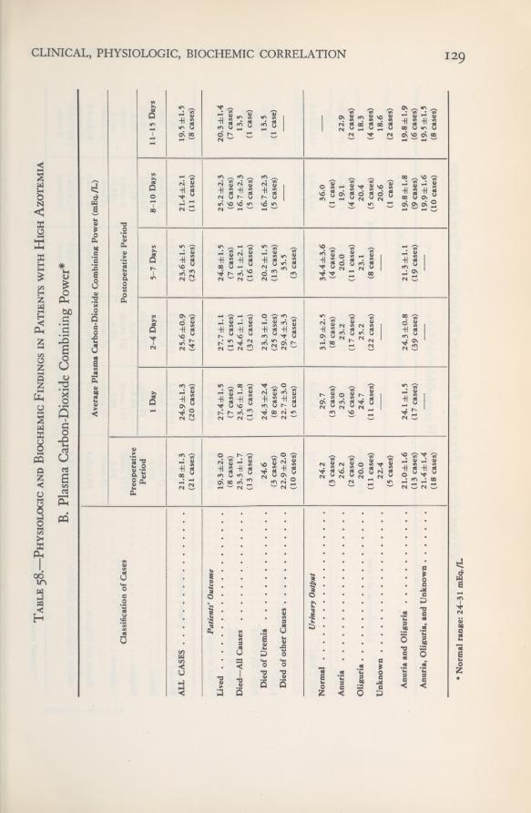
CLINICAL, PHYSIOLOGIC, BIOCHEMIC CORRELATION 129
Table58.
—Physiologicand
BiochemicFindings
inPatients
withHighAzotemia
B.Plasma
Carbon-DioxideCombining
Power*
ClassificationofCases
AveragePlasma
Carbon-DioxideCombining
Power(mEq./L)
Preoperative Period
PostoperativePeriod
1
Day
2-4Days
5-7Days
8-10Days
11-15Days
ALLCASES
21.8±1.3
24.9±1.325.6±0.923.6±1.5
21.4±2.1
19.5±1.5
(21cases)
(20cases)
(47cases)
(23cases)
(11cases)
(8cases)
Patients’Outcome
Lived
19.3±2.027.4±1.5
27.7±1.1
24.8±1.525.2±2.3
20.3±1.4
(8
cases)
(7
cases)
(15cases)
(7
cases)
(6cases)
(7
cases)
Died—AllCauses
23.3±1.723.6±1.824.6±1.1
23.1±2.1
16.7±2.3
13.5
(13cases)
(13cases)
(32cases)
(16cases)
(5
cases)
(1
case)
DiedofUremia
24.6
24.3±2.423.3±1.020.2±1.5
16.7±2.3
13.5
(3
cases)
(8
cases)
(25cases)
(13cases)
(5
cases)
(1
case)
DiedofotherCauses
22.9±2.022.7±3.0
29.4±3.335.5
—
—
(10cases)
(5
cases)
(7cases)
(3
cases)
UrinaryOutput
Normal
24.2
29.7
31.9±2.534.4±3.6
36.0
—
(3cases)
(3
cases)
(8cases)
(4
cases)
(1
case)
Anuria
26.2
23.0
23.2
20.0
19.1
22.9
(2
cases)
(6cases)
(17cases)
(11cases)
(4
cases)
(2
cases)
Oliguria
20.0
24.7
25.2
23.1
20.4
18.3
(11cases)
(11
cases)
(22cases)
(8
cases)
(5
cases)
(4cases)
Unknown
22.4
—
—
—
20.6
18.6
(5
cases)
(I
case)
(2
cases)
AnuriaandOliguria
21.0±1.624.1±1.524.3±0.8
21.3±1.119.8±1.819.8±1.9
(13cases)
(17cases)
(39cases)
(19cases)
(9cases)
(6cases)
Anuria,Oliguria,andUnknown
21.4±1.4—
—
—
19.9±1.619.5
±1.5
(18cases)
(10cases)
(8
cases)
*
Normalrange:24-31mEq./L

THE PHYSIOLOGIC EFFECTS OF WOUNDS130
Table58.
—Physiologicand
BiochemicFindings
inPatients
withHighAzotemia
C.Plasma
Chlorides* AveragePlasma
Chlorides(mEq./L.)
ClassificationofCases
Preoperative
PostoperativePeriod
Period
1
Day
2-4Days
5-7Days
8-10Days
11-15Days
ALLCASES
98.8±0.8
(21cases)
96.8±0.9
(31cases)
93.7±1.6 (54cases)
93.1±3.0 (28cases)
91.8±6.3 (13cases)
103.0±5.3(10cases)
Patients’Outcome
Lived Died—AllCauses
98.7±1.3
(8
cases) 98.9±l.l (13cases)
96.3±1.5 (11cases) 96.9±1.4 (19
cases)
96.7±2.3
(17cases)
92.7±2.1
(37cases)
94.1±3.3
(11cases)
92.2±4.3
(18cases)
102.7±7.9
(8cases) 74.4
±3.1(5
cases)
104.2±5.8
(9
cases) 91.4(I
case)
DiedofUremia
DiedofotherCauses
99.1(3
cases) 98.8±1.4 (10cases)
93.5±1.7/
(10cases) 100.7±1.3(9cases)
90.7±1.9 (27cases) 98.0±5.9 (10
cases)
89.3±3.2 (14cases) 102.2 (4cases)
74.4±3.1 (5
cases)
91.4(1
case)
UrinaryOutput
Normal Anuria Oliguria Unknown
102.1(3cases) 100.0(2cases) 98.9
(11
cases) 96.2(5
cases)
100.2±2.2
(5
cases) 94.4(8cases) 96.6 (15
cases) 98.9(3cases)
102.7±6.8(8cases) 88.9 (20cases) 94.3 (23
cases) 99.4(2
cases)
98.8±10.2 (7cases) 90.7 (11
cases) 91.7(10cases)
111.7(3cases) 76.7(4
cases) 83.5(5
cases) 133.7(Icase)
96.4(1
case) 92.0(2
cases) 99.8(5
cases) 125.0(2cases)
AnuriaandOliguria
Anuria,Oliguria,andUnknown
99.1±0.8
(13cases) 98.3±0.8 (18
cases)
95.8±1.2 (23cases)
96.2±1.1
(26cases)
91.8±1.5 (43cases) 92.2±1.5 (45
cases)
91.2±2.3 (21cases) 91.2±2.0 (21
cases)
80.5±3.4
(9cases)
85.8±6.1
(10cases)
97.6±3.9 (7cases) 103.7
±5.9(9cases)
*
Normalrange:97.5-104
mEq./L,
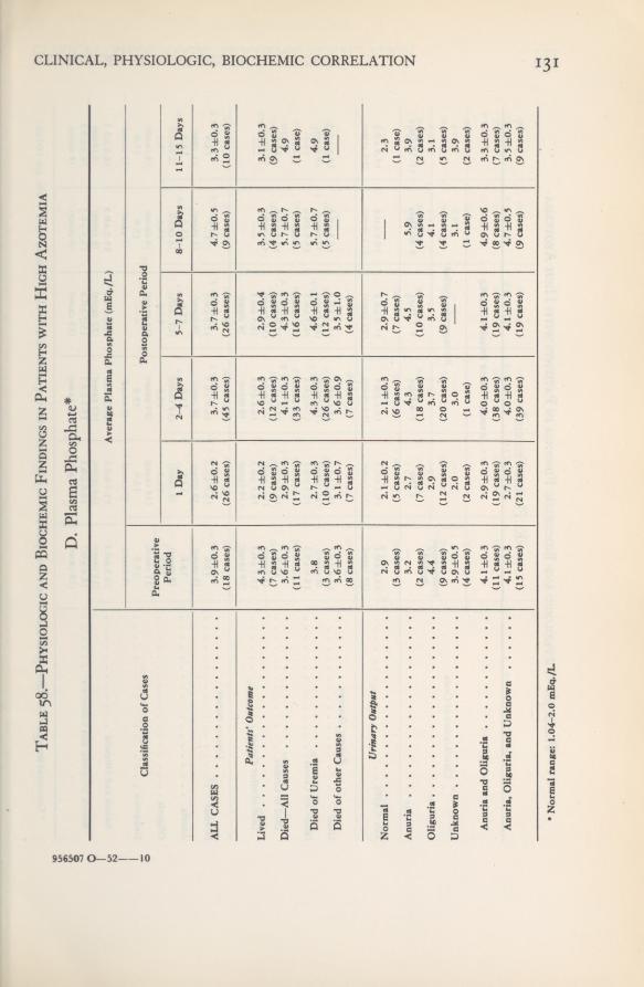
CLINICAL, PHYSIOLOGIC, BIOCHEMIC CORRELATION i3i
956507 0—52 10
Table58.—Physiologic
andBiochemicFindings
inPatients
withHighAzotemia
D.Plasma
Phosphate*
ClassificationofCases
AveragePlasmaPhosphate(mEq./L)
Preoperative Period
PostoperativePeriod
1
Day
2-4Days
5-7Days
8-10Days
11-15Days
ALLCASES
3.9±0.3
2.6±0.2
3.7±0.33.7±0.3
4.7±0.5
3.3±0.3
(18cases)
(26cases)
(45cases)
(26cases)
(9cases)
(10cases)
Patients’Outcome
Lived
4.3±0.3
2.2
±0.2
2.6±0.3
2.9±0.43.5±0.3
3.1±0.3
(7cases)
(9cases)
(12cases)
(10cases)
(4cases)
(9
cases)
Died—All
Causes
3.6±0.3
2.9±0.3
4.1±0.3
4.3±0.3
5.7±0.7
4.9
(11cases)
(17cases)
(33cases)
(16cases)
(5
cases)
(1
case)
DiedofUremia
.
3.8
2.7±0.3
4.3±0.3
4.6±0.1
5.7±0.7
4.9
(3
cases)
(10cases)
(26cases)
(12cases)
(5
cases)
(1
case)
DiedofotherCauses
3.6±0.33.1±0.7
3.6±0.93.5±1.0
—
—
(8
cases)
(7
cases)
(7cases)
(4cases)
UrinaryOutput
Normal
2.9
2.1±0.2
2.1±0.3
2.9±0.7
—
2.3
(3
cases)
(5
cases)
(6cases)
(7
cases)
(1
case)
Anuria
3.2
2.7
4.3
4.5
5.9
3.9
(2
cases)
(7cases)
(18cases)
(10cases)
(4cases)
(2
cases)
Oliguria
4.4
2.9
3.7
3.5
4.1
3.1
(9cases)
(12cases)
(20cases)
(9cases)
(4cases)
(5
cases)
Unknown
3.9±0.5
2.0
3.0
—
3.1
3.9
(4cases)
(2cases)
(1
case)
(1
case)
(2cases)
AnuriaandOliguria
4.1±0.3
2.9±0.3
4.0±0.3
4.1±0.3
4.9±0.63.3±0.3
(11cases)
(19cases)
(38cases)
(19cases)
(8
cases)
(7cases)
Anuria,Oliguria,andUnknown
4.1±0.3
2.7±0.3
4.0±0.3
4.1±0.3
4.7±0.5
3.5±0.3
(15cases)
(21cases)
(39cases)
(19cases)
(9cases)
(9
cases)
•
Normalrange:
1.04-2.0mEq./L

THE PHYSIOLOGIC EFFECTS OF WOUNDS
Table58.—Physiologic
andBiochemicFindings
inPatients
withHighAzotemia
E,PlasmaProtein*
ClassificationofCases
AveragePlasmaProtein(mEq./L.)
Preoperative Period
PostoperativePeriod
1
Day
2-4Days
5-7Days
8-10Days
11-15Days
ALLCASES
14.8±0.115.1
±0.1
15.6±0.115.8
±0.2
15.8±0.315.8±0.3
(21
cases)
(32cases)
(53cases)
(31cases)
(13cases)
(10cases)
Patients'Outcome
Lived
14.6±0.115.1
±0.2
15.1±0.2
15.1±0.2
16.3±0.3
16.5±0.3
(8cases)
(11
cases)
(17cases)
(13cases)
(7cases)
(8
cases)
Died—AllCauses
14.8±0.215.1±0.1
15.6±0.116.5
±0.2
15.6±0.614.1
(13cases)
(21
cases)
(36cases)
(18cases)
(5cases)
(1
case)
DiedofUremia
13.9
15.3±0.215.3
±0.1
15.8±0.215.6±0.6
14.1
(3
cases)
(11cases)
(27cases)
(14cases)
(5
cases)
(1
case)
DiedofotherCauses
15.1±0.1
14.8±0.2
16.8±0.4
18.5±0.3
—
—
(10cases)
(10cases)
(9cases)
(4cases)
UrinaryOutput
Normal
15.6
15.6±0.216.0±0.317.3±0.3
17.7
14.8
(3
cases)
(6cases)
(8
cases)
(8cases)
(3
cases)
(1
case)
Anuria
13.6
14.8
15.6
15.8
15.8
18.2
(2
cases)
(8
cases)
(19cases)
(12cases)
(4cases)
(2
cases)
Oliguria
14.6
15.3
15.3
14.8
15.1
15.3
(11cases)
(16cases)
(24cases)
(11cases)
(4cases)
(4
cases)
Unknown
15.1
12.6
15.6
—
15.8
17.0
(5
cases)
(2
cases)
(2
cases)
(1
case)
(2
cases)
AnuriaandOliguria
14.3±0.1
15.1±0.1
15.3±0.1
15.3±0.215.3±0.3
16.3±0.3
(13cases)
(24cases)
(43cases)
(23cases)
(8
cases)
(6
cases)
Anuria,Oliguria,andUnknown
14.6±0,1
14.8±0.1
15.3±0.1
15.3±0.2
15.3±0.316.3±0.3
(18cases)
(26cases)
(45cases)
(23cases)
(9cases)
(8
cases)
*
ConvertedfromGm.per100
cc.tomEq./L.for
purposesofcomparisonwithelectrolytes.Our
normalrangeof
plasmaprotein
was6-7Gm.
per100
cc.

CLINICAL, PHYSIOLOGIC, BIOCHEMIC CORRELATION 133
Table58.—Physiologic
andBiochemic
Findingsin
Patientswith
HighAzotemia
F.SerumSodium*
ClassificationofCases
AverageSerum
Sodium(mEq./L.)
Preoperative Period
PostoperativePeriod
1
Day
2-4Days
5-7Days
8-10Days
11-15Days
ALLCASES
146.3±3.7147.5±3.7
141.3±2.5
137.8±5.1140.7
±4.4
151.7±5.7
(5
cases)
(6cases)
(21
cases)
(8
cases)
(6cases)
(7
cases)
Patients'Outcome
Lived
137.9
148.0
139.9
124.0
147.4
153.7±6.3
(2
cases)
(2
cases)
(3cases)
(1
case)
(3cases)
(6
cases)
Died—AllCauses
151.9
147.3±5.1141.5
±2.8
139-7±5.5
134.0
140.0
(3
cases)
(4cases)
(18cases)
(7cases)
(3cases)
(1
case)
Diedof
Uremia
—
143.3
139.5±2.5
139.8±6.5
134.0
140.0
(2cases)
(16cases)
(6cases)
(3cases)
(I
case)
DiedofotherCauses
151.9
151.3
157.8
139.1
—
—
(3cases)
(2
cases)
(2
cases)
(1
case)
UrinaryOutput
Normal
150.8
160.4
169.6
139.1
—
—
(2
cases)
(1case)
(Icase)
(1
case)
Anuria
—
143.3
141.2
142.8
138.8
148.1
(2
cases)
(14cases)
(5
cases)
(3cases)
(2
cases)
Oliguria
137.9
148.0
137.6
124.5
136.5
148.0
(2
cases)
(2
cases)
(7cases)
(2
cases)
(2cases)
(3
cases)
Unknown
154.0
—
—
—
154.9
160.9
(1
case)
(1
case)
(2
cases)
Anuriaand
Oliguria
137.9
145.6±4.1140.0
±2.0
137.6±5.9137.9±4.2
148.0±4.3
(2
cases)
(4cases)
(21cases)
(7cases)
(5cases)
(5
cases)
Anuria,Oliguria,andUnknown
143.2
145.6±4.1140.0±2.0137.6±5.9
140.7±4.4
151.7±5.7
(3
cases)
(4cases)
(21cases)
(7cases)
(6
cases)
(7
cases)
*
Normalrange:139-149
mEq./L.

THE PHYSIOLOGIC EFFECTS OF WOUNDSi34
Table58.—Physiologic
andBiochemicFindings
inPatients
withHighAzotemia
G.Plasma
Magnesium*
ClassificationofCases
AveragePlasma
Magnesium(mEq./L)
Preoperative Period
PostoperativePeriod
1
Day
2-4Days
5-7Days
8-10Days
11-15Days
ALLCASES
2.0±0.1
1.4±0.1
2.4±0.3
2.8±0.6
3.3
2.2±0.3
(13cases)
(5
cases)
(13cases)
(9cases)
(3cases)
(5
cases)
Patients’Outcome
Lived
2.2±0.2
1.6
2.1±0.2
1.8
2.3
2.1±0.3
(4cases)
(1
case)
(4cases)
(2cases)
(2cases)
(4cases)
Died—All
Causes
2.0±0.2
1.4±0.2
2.6±0.43.2±0.7
5.2
2.6
(9cases)
(4cases)
(9cases)
(7cases)
(1
case)
(I
case)
2.1
1.4
2.3±0.4
3.3±1.0
5.2
2.6
(3
cases)
(2
cases)
(6cases)
(5
cases)
(I
case)
(1
case)
DiedofotherCauses
1.9±0.3
1.4
3.2
2.8
—
—
(6cases)
(2cases)
(3
cases)
(2cases)
UrinaryOutput
Normal
1.6
1.3
2.5
2.8
—
—
(3cases)
(1
case)
(2cases)
(2cases)
Anuria
1.8
1.7
2.3
2.4
1.8
1.8
(2
cases)
(1
case)
(5
cases)
(4cases)
(I
case)
(I
case)
Oliguria
2.3
1.4
2.5
3.6
5.2
2.3
(5
cases)
(3cases)
(5cases)
(3cases)
(1
case)
(2cases)
Unknown
2.3
—
—
—
2.8
2.3
(3cases)
(1case)
(2
cases)
AnuriaandOliguria
2.2±0.2
1.5±0.2
2.4±0.4
2.9±0.8
3.5
2.1
(7cases)
(4cases)
(10cases)
(7cases)
(2cases)
(3cases)
Anuria,Oliguria,andUnknown
2.2±0.1
1.5±0.2
2.4±0.4
2.9±0.8
3.3
2.2±0.3
(10cases)
(4cases)
(10cases)
(7cases)
(3
cases)
(5
cases)
*
Normalrange:0.8-2.5
mEq./L

CLINICAL, PHYSIOLOGIC, BIOCHEMIC CORRELATION 135
Nitrogenous Waste Products and Phosphorus
Nonprotein Nitrogen.—Averages of nonprotein nitrogen findings are shownin Tables 58A and 59. Although the number is small and the variation great insome periods, giving rise to large standard errors, the data are adequate to givea fairly representative picture. Generally nitrogen retention was already signifi-cant by the first postoperative day, increased rapidly during the first 10 days,and then began to decline. This is illustrated in Chart 18, which has been con-structed from averages shown in Table 58A, “All Cases.”
Chart 18. Plasma Nonprotein Nitrogen in Patients with High Azotemia
As previously discussed, most of the fatalities occurred in the first 10
days, so the fall in nitrogen retention in the tenth through fifteenth days chieflyrepresents patients who recovered. In general the nonprotein nitrogen level roseprogressively to the day of death in those patients dying primarily of uremia(Chart 19 and Table 60). However, two patients (Cases 66 and 93) who livedlonger than 10 days after wounding (14 and 13 days respectively) had begunto show some evidence of returning renal function. The importance of this factin relation to therapy cannot be too strongly emphasized: it is essential to avoid
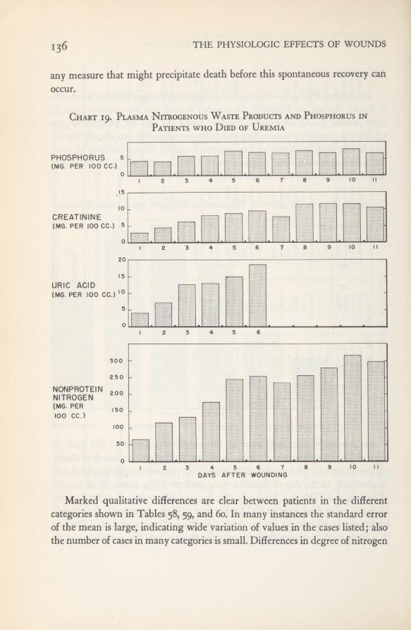
136 THE PHYSIOLOGIC EFFECTS OF WOUNDS
any measure that might precipitate death before this spontaneous recovery can
occur.
Chart 19. Plasma Nitrogenous Waste Products and Phosphorus inPatients who Died of Uremia
Marked qualitative differences are clear between patients in the differentcategories shown in Tables 58, 59, and 60. In many instances the standard errorof the mean is large, indicating wide variation of values in the cases listed; alsothe number of cases in many categories is small. Differences in degree of nitrogen
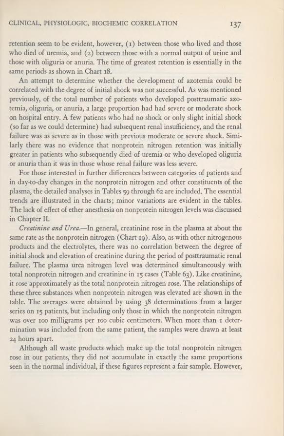
CLINICAL, PHYSIOLOGIC, BIOCHEMIC CORRELATION 137
retention seem to be evident, however, (i) between those who lived and thosewho died of uremia, and (2) between those with a normal output of urine andthose with oliguria or anuria. The time of greatest retention is essentially in thesame periods as shown in Chart 18.
An attempt to determine whether the development of azotemia could becorrelated with the degree of initial shock was not successful. As was mentionedpreviously, of the total number of patients who developed posttraumatic azo-temia, oliguria, or anuria, a large proportion had had severe or moderate shockon hospital entry. A few patients who had no shock or only slight initial shock(so far as we could determine) had subsequent renal insufficiency, and the renalfailure was as severe as in those with previous moderate or severe shock. Simi-larly there was no evidence that nonprotein nitrogen retention was initiallygreater in patients who subsequently died of uremia or who developed oliguriaor anuria than it was in those whose renal failure was less severe.
For those interested in further differences between categories of patients ancfin day-to-day changes in the nonprotein nitrogen and other constituents of theplasma, the detailed analyses in Tables 59 through 62 are included. The essentialtrends are illustrated in the charts; minor variations are evident in the tables.The lack of effect of ether anesthesia on nonprotein nitrogen levels was discussedin Chapter II.
Creatinine and Urea.—In general, creatinine rose in the plasma at about thesame rate as the nonprotein nitrogen (Chart 19). Also, as with other nitrogenousproducts and the electrolytes, there was no correlation between the degree ofinitial shock and elevation of creatinine during the period of posttraumatic renalfailure. The plasma urea nitrogen level was determined simultaneously withtotal nonprotein nitrogen and creatinine in 15 cases (Table 63). Like creatinine,it rose approximately as the total nonprotein nitrogen rose. The relationships ofthese three substances when nonprotein nitrogen was elevated are shown in thetable. The averages were obtained by using 38 determinations from a largerseries on 15 patients, but including only those in which the nonprotein nitrogenwas over 100 milligrams per 100 cubic centimeters. When more than 1 deter-mination was included from the same patient, the samples were drawn at least24 hours apart.
Although all waste products which make up the total nonprotein nitrogenrose in our patients, they did not accumulate in exactly the same proportionsseen in the normal individual, if these figures represent a fair sample. However,
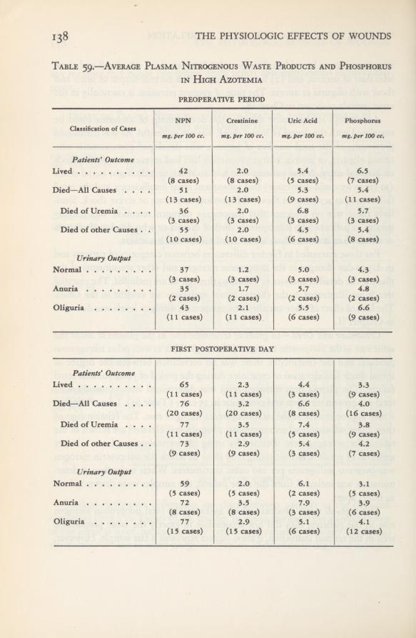
138 THE PHYSIOLOGIC EFFECTS OF WOUNDS
Table 59.—Average Plasma Nitrogenous Waste Products and Phosphorusin High Azotemia
PREOPERATIVE PERIOD
NPN Creatinine Uric Acid PhosphorusClassification ofCases
mg. per 100 cc. mg. per 100 cc. mg. per 100 cc. mg.per 100 cc.
Patients' OutcomeLived 42 2.0 5.4 6.5
(8 cases) (8 cases) (5 cases) (7 cases)Died—All Causes .... 51 2.0 5.3 5.4
(13 cases) (13 cases) (9 cases) (11 cases)Died of Uremia .... 36 2.0 6,8 5.7
(3 cases) (3 cases) (3 cases) (3 cases)Died of other Causes . . 55 2.0 4.5 5.4
(10 cases) (10 cases) (6 cases) (8 cases)
Urinary OutputNormal 37 1.2 5.0 4.3
(3 cases) (3 cases) (3 cases) (3 cases)Anuria 35 1.7 5.7 4.8
(2 cases) (2 cases) (2 cases) (2 cases)Oliguria 43 2.1 5.5 6.6
(11 cases) (11 cases) (6 cases) (9 cases)
FIRST POSTOPERATIVE DAY
Patients' OutcomeLived 65 2.3 4.4 3.3
(11 cases) (11 cases) (3 cases) (9 cases)Died—All Causes .... 76 3.2 6.6 4.0
(20 cases) (20 cases) (8 cases) (16 cases)Died of Uremia .... 77 3.5 7.4 3.8
(11 cases) (11 cases) (5 cases) (9 cases)Died of other Causes . . 73 2.9 5.4 4.2
(9 cases) (9 cases) (3 cases) (7 cases)
Urinary OutputNormal 59 2.0 6.1 3.1
(5 cases) (5 cases) (2 cases) (5 cases)Anuria 72 3.5 7.9 3.9
(8 cases) (8 cases) (3 cases) (6 cases)Oliguria 77 2.9 5.1 4.1
(15 cases) (15 cases) (6 cases) (12 cases)

CLINICAL, PHYSIOLOGIC, BIOCHEMIC CORRELATION
Table 59.—Average Plasma Nitrogenous Waste Products and Phosphorus
in High Azotemia—Continued
SECOND POSTOPERATIVE DAY
NPN Creatinine Uric Acid PhosphorusClassification of Cases
mg. per 100 cc. mg. per 100 cc. mg. per 100 cc. mg. per 100 cc.
Patients’ OutcomeLived 75 2.7 7.1 4.5
(11 cases) (10 cases) (4 cases) (6 cases)Died—All Causes .... 118 5.0 9.0 5.3
(27 cases) (26 cases) (8 cases) (23 cases)Died of Uremia .... 127 5.5 9.3 5.1
(20 cases) (20 cases) (7 cases) (18 cases)Died of other Causes •
. 91 3.4 6.7 5.9(7 cases) (6 cases) (1 case) (5 cases)
Urinary OutputNormal 68 2.2 6.7 3.5
(5 cases) (4 cases) (1 case) (3 cases)Anuria 125 5.6 9.2 5.0
(15 cases) (14 cases) (5 cases) (12 cases)Oliguria 104 4.0 8.0 5.7
(16 cases) (16 cases) (6 cases) (13 cases)
THIRD POSTOPERATIVE DAY
Patients' OutcomeLived 87 2.9 8.0 3.3
(11 cases) (10 cases) (1 case) (7 cases)Died—All Causes .... 154 7.1 13.3 5.4
(18 cases) (18 cases) (7 cases) (15 cases)Died of Uremia .... 159 7.4 14.4 5.6
(16 cases) (16 cases) (6 cases) (14 cases)Died of other Causes . . 115 4.2 6.8 2.9
\ (2 cases) (2 cases) (1 case) (1 case)
Urinary OutputNormal 80 1.8 6.8 2.3
(4 cases) (4 cases) (1 case) (3 cases)Anuria 166 7.7 10.3 5.3
(10 cases) (10 cases) (3 cases) (8 cases)Oliguria 118 5.1 15.8 5.1
(15 cases) (14 cases) (4 cases) (11 cases)
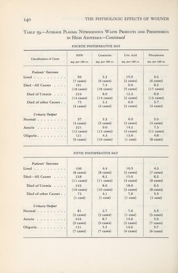
THE PHYSIOLOGIC EFFECTS OF WOUNDS
Table 59.—Average Plasma Nitrogenous Waste Products and Phosphorus
in High Azotemia—Continued
FOURTH POSTOPERATIVE DAY
NPN Creatinine Uric Acid PhosphorusClassification of Cases
mg. per 100 cc. mg. per 100 cc. mg. per 100 cc. mg. per 100 cc.
Patients' OutcomeLived 99 3.3 15.9 4.2
(7 cases) (9 cases) (2 cases) (6 cases)Died—All Causes .... 191 7.4 9.0 6.2
(18 cases) (18 cases) (5 cases) (17 cases)Died of Uremia .... 224 8.9 12.3 6.9
(14 cases) (14 cases) (2 cases) (13 cases)Died of other Causes . . 75 2.2 6.9 3.7
(4 cases) (4 cases) (3 cases) (4 cases)
Urinary OutputNormal 97 2.3 6.9 3.3
(4 cases) (5 cases) (3 cases) (4 cases)Anuria 221 9.0 14.2 7.3
(12 cases) (12 cases) (3 cases) (11 cases),Oliguria 121 4.3 13.6 4.6
(9 cases) (10 cases) (1 case) (8 cases)
FIFTH POSTOPERATIVE DAY
Patients' OutcomeLived 106 4.4 10.5 4.3
(8 cases) (8 cases) (3 cases) (7 cases)Died—All Causes .... 228 8.2 15.9 6.2
(11 cases) (11 cases) (4 cases) (9 cases)Died of Uremia .... 243 8.6 18.6 6.3
(10 cases) (10 cases) (3 cases) (8 cases)Died of other Causes . . 73 4.1 7.6 5.5
(1 case) (1 case) (1 case) (1 case)
Urinary OutputNormal 81 2.7 7.6 3.5
(3 cases) (3 cases) (1 case) (3 cases)Anuria 244 8.7 14.6 5.9
(9 cases) (9 cases) (2 cases) (7 cases)Oliguria 131 5.5 14.6 5.7
(7 cases) (7 cases) (4 cases) (6 cases)

CLINICAL, PHYSIOLOGIC, BIOCHEMIC CORRELATION
Table 59.—Average Plasma Nitrogenous Waste Products and Phosphorusin High Azotemia— Continued
SIXTH POSTOPERATIVE DAY
NPN Creatinine Uric Acid PhosphorusClassification of Cases
mg. per 100 cc. mg. per 100 cc. mg. per 100 cc. mg. per 100 cc.
Patients' OutcomeLived 91
(5 cases)187
3.0(5 cases)
7.6
3.8(4 cases)
6.4Died—All Causes .... 16.5(12 cases) (13 cases) (4 cases) (11 cases)
Died of Uremia .... 235 9.8 19.9 7.0(8 cases) (9 cases) (1 case) (7 cases)
Died of other Causes . . 92 2.7 15.3 5.2(4 cases) (4 cases) (3 cases) (4 cases)
Urinary OutputNormal 102 2.9 15.3 4.7
(6 cases) (6 cases) (3 cases) (6 cases)Anuria 245 11.1 7.1
Oliguria(5 cases)
144(6 cases)
5.1 19.9(4 cases)
5.7(6 cases) (6 cases) (1 case) (5 cases)
SEVENTH POSTOPERATIVE DAY
Patients' OutcomeLived 133 3.3 2.2 4.4
(5 cases) (5 cases) (1 case) (4 cases)Died—All Causes .... 212 8.6 15.5 6.1
(5 cases) (5 cases) (1 case) (5 cases)Died of Uremia .... 246 10.3 6.7
Died of other Causes . .
(4 cases)76
(4 cases)1.7 15.5
(4 cases)3.9
(1 case) (1 case) (1 case) (1 case)
Urinary OutputNormal 111 2.1 15.5 3.7
(3 cases) (3 cases) (1 case) (2 cases)239 9.4
(4 cases)6.5
(4 cases) (4 cases)Oliguria 145 5.1 2.2 4.9
(3 cases) (3 cases) (1 case) (3 cases)
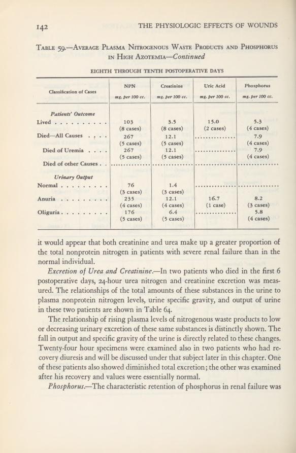
THE PHYSIOLOGIC EFFECTS OF WOUNDS142
Table 59.—Average Plasma Nitrogenous Waste Products and Phosphorus
in High Azotemia—Continued
it would appear that both creatinine and urea make up a greater proportion ofthe total nonprotein nitrogen in patients with severe renal failure than in thenormal individual.
Excretion of Urea and Creatinine.—In two patients who died in the first 6postoperative days, 24-hour urea nitrogen and creatinine excretion was meas-ured. The relationships of the total amounts of these substances in the urine toplasma nonprotein nitrogen levels, urine specific gravity, and output of urinein these two patients are shown in Table 64,
The relationship of rising plasma levels of nitrogenous waste products to lowor decreasing urinary excretion of these same substances is distinctly shown. Thefall in output and specific gravity of the urine is directly related to these changes.Twenty-four hour specimens were examined also in two patients who had re-covery diuresis and will be discussed under that subject later in this chapter. Oneof these patients also showed diminished total excretion; the other was examinedafter his recovery and values were essentially normal.
Phosphorus.—The characteristic retention of phosphorus in renal failure was
EIGHTH THROUGH TENTH POSTOPERATIVE DAYS
NPN Creatinine Uric Acid PhosphorusClassification of Cases
mg.per 100 cc. mg. per 100 cc. mg. per 100 cc. mg. per 100 cc.
Patients' OutcomeLived 103 3.5 15.0 5.3
(8 cases) (8 cases) (2 cases) (4 cases)Died—All Causes . r . . 267 12.1 7.9
(5 cases) (5 cases) (4 cases)Died of Uremia .... 267 12.1 7.9
(5 cases) (5 cases) (4 cases)
Urinary Output76 1.4
(3 cases) (3 cases)Anuria 235 12.1 16.7 8.2
(4 cases) (4 cases) (1 case) (3 cases)176 6.4 5.8
(5 cases) (5 cases)■
(4 cases)

CLINICAL, PHYSIOLOGIC, BIOCHEMIC CORRELATION 143
Table 60.—Average Plasma Nitrogenous Waste Products and Phosphorusin Patients who Died of Uremia
also observed in our cases, and in general paralleled the degree of nitrogen reten-tion (see Chart 19 and Tables 58A, 58D, 59, 60, 61, and 62). In these patientswith posttraumatic azotemia, phosphorus retention was primarily due to im-paired ability to excrete that substance; whereas the hyperphosphatemia seen inpatients in shock soon after wounding was possibly due to release of phosphatessecondary to muscle damage (Chapter I). Reference to Tables 58 through 62and to the individual case records shows that the patients with the most severerenal damage had the greatest phosphorus retention.
Relationship of Calcium and Phosphorus. —Calcium and phosphorus deter-minations were made on 12 patients who had high azotemia. The well-knownreciprocal relationship of calcium and phosphorus was present in the majorityof these cases (Table 65).
Days after WoundingNPN
mg. per 100 cc.
Creatinine
mg. per 100 cc.
Uric Acid
mg. per 100 cc.
Phosphorus
mg. per 100 cc.
First 69±10.3 3.0±0.24 4.1 4.2 ±0.47(9 cases) (9 cases) (3 cases) (8 cases)
Second 118±11.5 4.8±0.24 7.3±1.1 4.1 ±0.28(13 cases) (13 cases) (5 cases) (12 cases)
Third 132 ±6.2 6.3 ±0.26 12.8±2.4 5.8±0.42(16 cases) (16 cases) (8 cases) (14 cases)
Fourth 178±5.1 8.1 ±0.47 12.9 5.9±0.36(16 cases) (16 cases) (3 cases) (14 cases)
Fifth 245±18.9 8.9±0.53 15.0 7.1 ±0.96(13 cases) (13 cases) (3 cases) (12 cases)
Sixth 253±13.0 9.5 ±0.70 18.4 6.9±0.22(8 cases) (9 cases) (3 cases) (8 cases)
Seventh 234±26.3 7.8± 1.83 6.6(4 cases) (4 cases) (3 cases)
Eighth 255 11.3 7.1(2 cases) (2 cases) (2 cases)
Ninth 278±16.0 11.4±0.44 6.8(5 cases) (5 cases) (3 cases)
314 11.4 7.7(3 cases) (3 cases) (3 cases)
296 10.3 6.8(2 cases) (2 cases) (2 cases)
Note; Data are given on this table for days post-wounding rather than days postoperative as in the precedingtable.
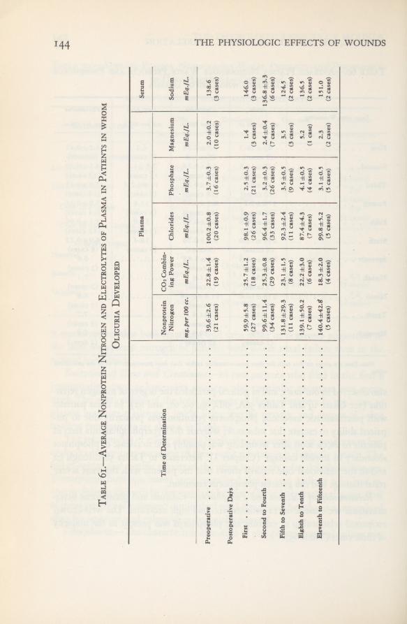
THE PHYSIOLOGIC EFFECTS OF WOUNDS
Table6i.—Average
NonproteinNitrogen
andElectrolytes
ofPlasma
inPatients
inwhom
OliguriaDeveloped
Plasma
Serum
Timeof
Determination
Nonprotein Nitrogenmg.
per100cc.
CO2Combin-
ingPower mEq./L.
Chlorides mEq./L.Phosphate mEq./L.
Magnesium mEq./L.Sodium mEq./L.
Preoperative
39.6±2.622.8±1.4
100.2±0.8
3.7±0.32.0±0.2
138.6
(21cases)
(19cases)
(20cases)
(16cases)
(10cases)
(3cases)
PostoperativeDays
First
59.9±5.825.7±1.298.1±0.92.5±0.3
1.4
146.0
(27cases)
(18cases)
(26cases)
(21cases)
(3
cases)
(3
cases)
Secondto
Fourth
99.6±11.4
25.3±0.896.4±1.7
3.2±0.32.4±0.4
136.8±3.3
(34cases)
(29cases)
(33cases)
(26cases)
(7
cases)
(6cases)
Fifthto
Seventh
131.8±29.323.1±1.5
92.3±2.43.5±0.5
3.5
124.5
(11
cases)
(8
cases)
(11cases)
(9
cases)
(3
cases)
(2cases)
Eighthto
Tenth
139.1±50.222.2±3.087.4±4.34.1±0,5
5.2
136.5
(7cases)
(6cases)
(7cases)
(4
cases)
(I
case)
(2
cases)
Eleventhto
Fifteenth
140.4±42.tf18.3±2.0
99.8±5.23.1±0.5
2.3
151.0
(5
cases)
(4cases)
(5
cases)
(5
cases)
(2
cases)
(2
cases)

CLINICAL, PHYSIOLOGIC, BIOCHEMIC CORRELATION
Table62.—Average
NonproteinNitrogen
andElectrolytes
ofPlasma
inPatients
inwhom
AnuriaDeveloped
Timeof
Determination
Plasma
Serum
Nonprotein Nitrogenmg.
per100cc.
CO2Combin-
ingPower mEq./L.
Chlorides mEq./L.Phosphate mEq./L.
Magnesium mEq./L.Sodium mEq./L.
Preoperative
34.7
23.9
98.7
2.8
1.8
___
(3
cases)
(3
cases)
(3cases)
(3cases)
(2
cases)
PostoperativeDays
First
75.6±4.7
23.0±3.095.7±2.42,7±0.3
1.7
143.3
(8
cases)
(6cases)
(8
cases)
(7cases)
(1
case)
(2
cases)
Secondto
Fourth
190.4±13.923.2±1.388.9±2.04.3±0.5
2.3±0,4
141.2±2.7
(20cases)
(17cases)
(20cases)
(18cases)
(5
cases)
(14cases)
Fifthto
Seventh
257.1±14.920.0±l,690.7±3.84,5±0.1
2,4±0,2142.8±6.9
(12cases)
(11cases)
(11cases)
(10cases)
(4cases)
(5
cases)
Eighthto
Tenth
258.0±21.8
19.1±2.3
76,7±4.25.9±0,9
1.8
138.8
(4cases)
(4cases)
(4
cases)
(4cases)
(1
case)
(3cases)
Eleventhto
Fifteenth
155.0
22.9
92.0
3.9
1.8
148.1
(2
cases)
(2cases)
(2
cases)
(2
cases)
(I
case)
(2
cases)
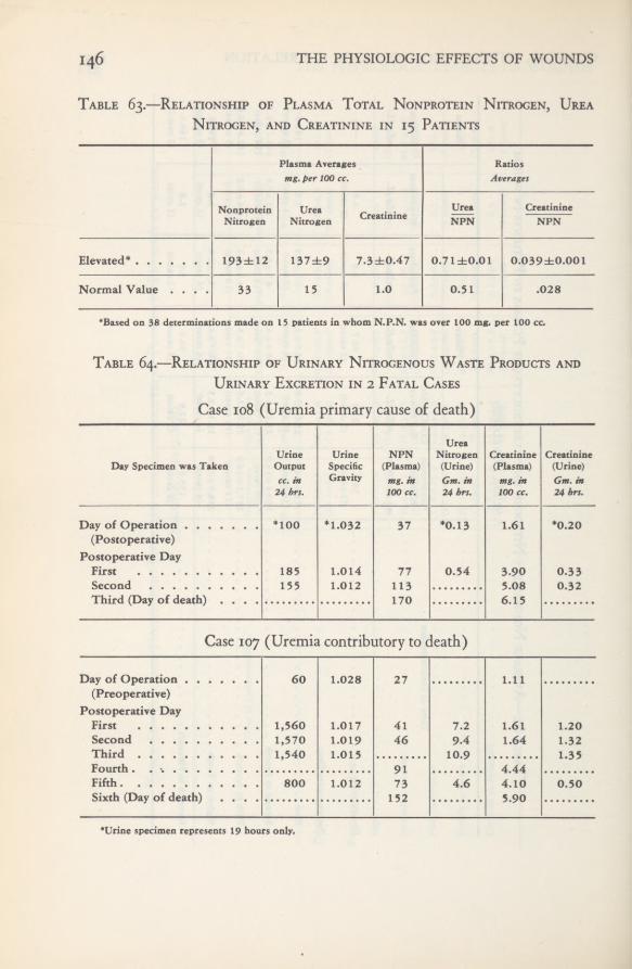
146 THE PHYSIOLOGIC EFFECTS OF WOUNDS
Table 63.—Relationship of Plasma Total Nonprotein Nitrogen, UreaNitrogen, and Creatinine in 15 Patients
Table 64.—Relationship of Urinary Nitrogenous Waste Products and
Urinary Excretion in 2 Fatal Cases
Plasma Averages Ratiosmg. per 100 cc. Averages
Nonprotein Urea CreatinineUrea Creatinine
Nitrogen Nitrogen NPN NPN
Elevated* 193±12 137±9 7.3±0.47 0.71±0.01 0.039±0.001
Normal Value .... 33 15 1.0 0.51 .028
‘Based on 38 determinations made on 15 patients in whom N.P.N. was over 100 mg. per 100 cc.
Case 108 (Uremia primary cause of death)
UreaUrine Urine NPN Nitrogen Creatinine Creatinine
Day Specimen was Taken Output Specific (Plasma) (Urine) (Plasma) (Urine)cc. in Gravity mg. in Gm. in mg. in Gm. in24 hrs. 100 cc. 24 hrs. 100 cc. 24 hrs.
Day of Operation *100 *1.032 37 *0.13 1.61 *0.20(Postoperative)
Postoperative DayFirst 185 1.014 77 0.54 3.90 0.33Second 155 1.012 113 5.08 0.32Third (Day of death) .... 170 6.15
Case 107 (Uremia contributory to death)
Day of Operation 60 1.028 27 1.11(Preoperative)
Postoperative DayFirst 1,560 1,017 41 7.2 1.61 1.20Second 1,570 1.019 46 9.4 1.64 1.32Third 1,540 1.01 5 10.9 1.35Fourth. , 91 4.44Fifth 800 1.012 73 4.6 4.10 0.50Sixth (Day of death)
.... 152 5.90
•Urine specimen represents 19 hours only.
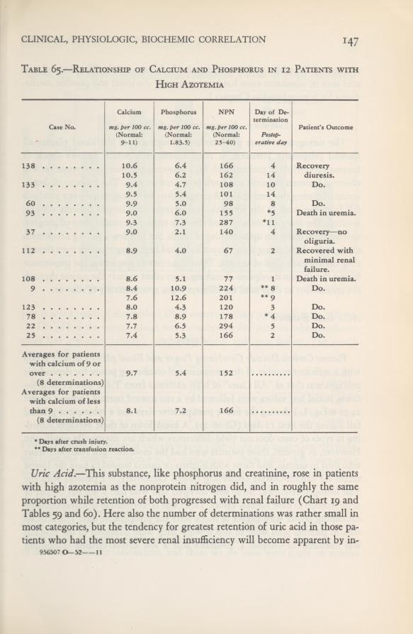
CLINICAL, PHYSIOLOGIC, BIOCHEMIC CORRELATION
Table 65.—Relationship of Calcium and Phosphorus in 12 Patients with
High Azotemia
Uric Acid.—This substance, like phosphorus and creatinine, rose in patientswith high azotemia as the nonprotein nitrogen did, and in roughly the sameproportion while retention of both progressed with renal failure (Chart 19 andTables 59 and 60). Here also the number of determinations was rather small inmost categories, but the tendency for greatest retention of uric acid in those pa-tients who had the most severe renal insufficiency will become apparent by in-
956307 0—52 11
Calcium Phosphorus NPN Day of De-termination
Case No. mg. per 100 cc. mg. per 100 cc. mg. per 100 cc. Patient’s Outcome(Normal: (Normal: (Normal: Postop-
9-11) 1.83.5) 25-40) erative day
138 10.6 6.4 166 4 Recovery10.5 6.2 162 14 diuresis.
133 9.4 4.7 108 10 Do.9.5 5.4 101 14
60 9.9 5.0 98 8 Do.93 9.0 6.0 155 *5 Death in uremia.
9.3 7.3 287 *1137 9.0 2.1 140 4 Recovery-—no
oliguria.112 8.9 4.0 67 2 Recovered with
minimal renalfailure.
108 8.6 5.1 77 1 Death in uremia.9 8.4 10.9 224 ** 8 Do.
7.6 12.6 201 ** 9123 8.0 4.3 120 3 Do.78 7.8 8.9 178 * 4 Do.22 7.7 6.5 294 5 Do.25 7.4 5.3 166 2 Do.
Averages for patientswith calcium of 9 or
9.7 5.4 152(8 determinations)
Averages for patientswith calcium of less
8.1 7.2 166(8 determinations)
*Days after crush injury.** Days after transfusionreaction.
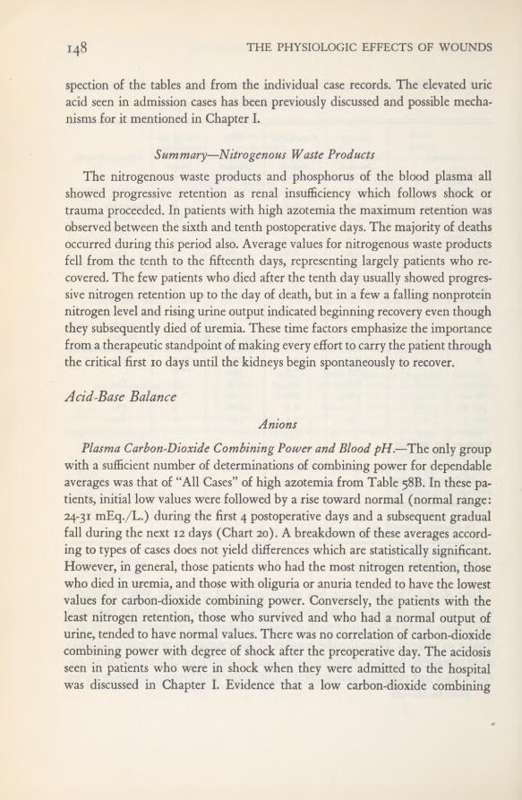
148 THE PHYSIOLOGIC EFFECTS OF WOUNDS
spection of the tables and from the individual case records. The elevated uricacid seen in admission cases has been previously discussed and possible mecha-nisms for it mentioned in Chapter I.
Summary—Nitrogenous Waste ProductsThe nitrogenous waste products and phosphorus of the blood plasma all
showed progressive retention as renal insufficiency which follows shock ortrauma proceeded. In patients with high azotemia the maximum retention wasobserved between the sixth and tenth postoperative days. The majority of deathsoccurred during this period also. Average values for nitrogenous waste productsfell from the tenth to the fifteenth days, representing largely patients who re-covered. The few patients who died after the tenth day usually showed progres-sive nitrogen retention up to the day of death, but in a few a falling nonproteinnitrogen level and rising urine output indicated beginning recovery even thoughthey subsequently died of uremia. These time factors emphasize the importancefrom a therapeutic standpoint of making every effort to carry the patient throughthe critical first 10 days until the kidneys begin spontaneously to recover.
Acid-Base BalanceAnions
Plasma Carbon-Dioxide Combining Power and Blood pH.—The only groupwith a sufficient number of determinations of combining power for dependableaverages was that of “All Cases” of high azotemia from Table 58B. In these pa-tients, initial low values were followed by a rise toward normal (normal range:24-31 mEq./L.) during the first 4 postoperative days and a subsequent gradualfall during the next 12 days (Chart 20). A breakdown of these averages accord-ing to types of cases does not yield differences which are statistically significant.However, in general, those patients who had the most nitrogen retention, thosewho died in uremia, and those with oliguria or anuria tended to have the lowestvalues for carbon-dioxide combining power. Conversely, the patients with theleast nitrogen retention, those who survived and who had a normal output ofurine, tended to have normal values. There was no correlation of carbon-dioxidecombining power with degree of shock after the preoperative day. The acidosisseen in patients who were in shock when they were admitted to the hospitalwas discussed in Chapter I. Evidence that a low carbon-dioxide combining

CLINICAL, PHYSIOLOGIC, BIOCHEMIC CORRELATION
Chart 20. Plasma Carbon-Dioxide Combining Power in High Azotemia
power was a result of diminished alkali reserve in these patients is only indirect.The pH of the venous blood was measured in five patients who simultane-
ously had low carbon-dioxide combining power and renal failure. The resultswere as follows:
From the limited number of cases in which a blood pH determination was done,and from the indirect evidence to be cited later, it seems likely that there was ametabolic acidosis in the majority of cases. If the low carbon-dioxide combiningpower had been due to respiratory alkalosis, one would expect to see clinicalevidence of hyperventilation and possibly an alkaline urine, depending on therenal function. None of our patients had either. Furthermore, in such cases theblood pH, although probably in the normal range (7.38-7.48), would be in theupper limits of normal. Obviously we do not have enough pH determinationsto draw any definite conclusions, and those we do have were made on venous
Case No.6990
Plasma CO tCombining
Power ( mEq./L.)23
Blood pH( venous)
7-397-37
93 x6 _ 7-3i112 _ .... - -.
-].20 ... _ 7.0/j
133 21 7.32

THE PHYSIOLOGIC EFFECTS OF WOUNDS150
Chart 21. Hydrogen-ion Concentration of Urine in High Azotemia
blood, but they support the view that most of these patients were suffering froma metabolic acidosis. This acidosis was not of the hyperchloremic type. Forfurther discussion, see the following sections on chlorides and sodium.
The range of the pH of the urine in patients with high azotemia gave at leasta partial explanation for the acidosis; after the first postoperative day none ofthe patients was able to produce a urine more acid than pH of 6.0 despite anincreasingly severe acidosis. In those in whom renal failure was most severe—
those who died of uremia—this abnormality was even more evident (Charts 20
and 21). The inference here is that those mechanisms responsible for acidifica-tion of the urine, such as tubular transfer of hydrogen ion, were impaired. Thissubject will be discussed further in the section on Urine; average urine pH isshown in Chart 21 and Table 76. Measurements of urinary ammonia were toofew to say whether the deficient formation of this base was also responsible forthe acidosis.
Plasma Chlorides.—The relationship of the plasma chloride level to theseverity of renal failure is shown in Chart 22 and Table 58C. The averages ofall patients with high azotemia show progressive hypochloremia through thefirst 10 postoperative days and normal values after this time (normal range;

CLINICAL, PHYSIOLOGIC, BIOCHEMIC CORRELATION
Chart 22. Plasma Chlorides in High Azotemia
97-5' io4)- Of the patients who died in uremia, the average values show that anextreme hypochloremia was reached by the tenth day; only one of these patientssurvived longer than that day. Average values for patients who lived were onlyslightly low during the period of greatest nitrogen retention. Analysis of indi-vidual case records shows that the lowest plasma chloride levels usually occurredin those patients who died before the tenth day, and the rise in the level fromthe eleventh through the fifteenth day occurred largely in those patients whohad a recovery diuresis or only minimal azotemia.
Relationship ofLow Plasma Chloride Levels to Intake of Sodium Chloride.—The mechanism of the hypochloremia in these patients is not clear. Most of thepatients with a low plasma chloride level were extremely ill and were takingpractically no food by mouth; hence their source of salt was almost entirely de-rived from that administered parenterally. In those patients who presumablyhad the most severe renal lesions (those who died in uremia), there is a possiblecorrelation between the quantity of parenteral sodium chloride given and theplasma chloride levels (Table 66). The table shows the influence of parenteralsodium chloride intake over a period of 3 days on the subsequent plasma chloridelevel in 32 patients who died and in 19 who survived. The patients were arbi-
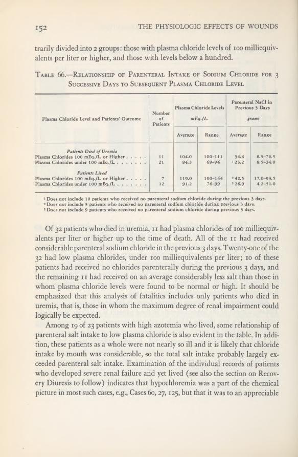
THE PHYSIOLOGIC EFFECTS OF WOUNDS152
trarily divided into 2 groups: those with plasma chloride levels of 100 milliequiv-alents per liter or higher, and those with levels below a hundred.
Table 66.—Relationship of Parenteral Intake of Sodium Chloride for 3Successive Days to Subsequent Plasma Chloride Level
Of 32 patients who died in uremia, 11 had plasma chlorides of 100 milliequiv-alents per liter or higher up to the time of death. All of the n had receivedconsiderable parenteral sodium chloride in the previous 3 days. Twenty-one of the32 had low plasma chlorides, under 100 milliequivalents per liter; 10 of thesepatients had received no chlorides parenterally during the previous 3 days, andthe remaining n had received on an average considerably less salt than those inwhom plasma chloride levels were found to be normal or high. It should beemphasized that this analysis of fatalities includes only patients who died inuremia, that is, those in whom the maximum degree of renal impairment couldlogically be expected.
Among 19 of 23 patients with high azotemia who lived, some relationship ofparenteral salt intake to low plasma chloride is also evident in the table. In addi-tion, these patients as a whole were not nearly so ill and it is likely that chlorideintake by mouth was considerable, so the total salt intake probably largely ex-ceeded parenteral salt intake. Examination of the individual records of patientswho developed severe renal failure and yet lived (see also the section on Recov-ery Diuresis to follow) indicates that hypochloremia was a part of the chemicalpicture in most such cases, e.g., Cases 60,27,125, but that it was to an appreciable
Plasma Chloride Level and Patients’ OutcomeNumber
ofPatients
Plasma Chloride Levels
mEq./L.
Parenteral NaCl inPrevious 3 Days
grams
Average Range Average Range
Patients Died of UremiaPlasma Chlorides 100 mEq./L. or HigherPlasma Chlorides under 100 mEq./L
1121
104.084.3
100-11169-94
34.4> 23.2
8.5-8.5-
Patients LivedPlasma Chlorides 100 mEq./L. or HigherPlasma Chloridesunder 100 mEq./L
712
119.091.2
100-14476-99
2 42.53 26.9
17.0-93.54.2-51.0
1 Does not include 10 patients who received no parenteral sodium chloride during the previous 3 days.2 Does not include 3 patients who received no parenteral sodium chloride during previous 3 days.3 Does not include 9 patients who received no parenteral sodium chloride during previous 3 days.

CLINICAL, PHYSIOLOGIC, BIOCHEMIC CORRELATION 153
degree associated with the sodium chloride intake. This relationship to intake isalso brought out in conjunction with the discussion of sodium to follow.
Several exceptions to these generalizations were evident in individual cases.With apparently adequate salt intake, the chloride level was sometimes low, evenin patients in whom there was no loss of chloride through Wangensteen drain-age or vomiting. In one patient with high azotemia and with all the other clinicalfeatures common to the syndrome of severe renal failure, the plasma chloridelevels were abnormally high (Case 133), but in this instance salt intake had beenexcessive.
It is possible that the hypochloremia might have been due in part to a simpledilution of the chloride ion, since practically all of these patients had an increasedplasma volume. This will be discussed further in the section on Plasma Volume.No such connection is apparent, however, in Table 67 which shows plasma vol-ume and plasma chloride determinations done simultaneously in 18 patientswho subsequently died of uremia. No correlation between degree of initialshock and plasma chloride level could be demonstrated.Table 67.—Plasma Volume and Plasma Chloride Determinations in 18
Patients who Died of Uremia
Case No. PostoperativeDays
Plasma Chloride Level
mEq./L.(normal: 97.5-104)
Plasma Volume(% ofcalculated normal)
Decrease Increase
9 6 72 21.022 5 90 126 3 106 2 1.847 3 82 8.852 2 93 33.555 7 80 37.769 8 76 15.880 4 86 5.586 6 76 25.593 6 82 6.195 3 92 1.098 5 111 32.7105 7 103 39.8108 3 91 3.8118 5 82 6.1123 3 93 18.6135 4 91 63.9136 3 104 14.5
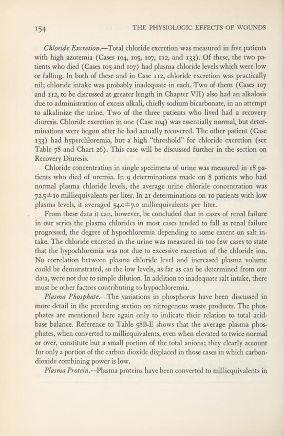
THE PHYSIOLOGIC EFFECTS OF WOUNDS
Chloride Excretion.—Total chloride excretion was measured in five patientswith high azotemia (Cases 104, 105, 107, 112, and 133). Of these, the two pa-tients who died (Cases 105 and 107) had plasma chloride levels which were lowor falling. In both of these and in Case 112, chloride excretion was practicallynil; chloride intake was probably inadequate in each. Two of them (Cases 107and 112, to be discussed at greater length in Chapter VII) also had an alkalosisdue to administration of excess alkali, chiefly sodium bicarbonate, in an attemptto alkalinize the urine. Two of the three patients who lived had a recoverydiuresis. Chloride excretion in one (Case 104) was essentially normal, but deter-minations were begun after he had actually recovered. The other patient (Case133) had hyperchloremia, but a high “threshold” for chloride excretion (seeTable 78 and Chart 26). This case will be discussed further in the section onRecovery Diuresis.
Chloride concentration in single specimens of urine was measured in 18 pa-tients who died of uremia. In 9 determinations made on 8 patients who hadnormal plasma chloride levels, the average urine chloride concentration was
milliequivalents per liter. In 21 determinations on 10 patients with lowplasma levels, it averaged 54.0±7.0 milliequivalents per liter.
From these data it can, however, be concluded that in cases of renal failurein our series the plasma chlorides in most cases tended to fall as renal failureprogressed, the degree of hypochloremia depending to some extent on salt in-take. The chloride excreted in the urine was measured in too few cases to statethat the hypochloremia was not due to excessive excretion of the chloride ion.No correlation between plasma chloride level and increased plasma volumecould be demonstrated, so the low levels, as far as can be determined from ourdata, were not due to simple dilution. In addition to inadequate salt intake, theremust be other factors contributing to hypochloremia.
Plasma Phosphate.—The variations in phosphorus have been discussed inmore detail in the preceding section on nitrogenous waste products. The phos-phates are mentioned here again only to indicate their relation to total acid-base balance. Reference to Table 58B-E shows that the average plasma phos-phates, when converted to milliequivalents, even when elevated to twice normalor over, constitute but a small portion of the total anions; they clearly accountfor only a portion of the carbon dioxide displaced in those cases in which carbon-dioxide combining power is low.
Plasma Protein.—Plasma proteins have been converted to milliequivalents in

CLINICAL, PHYSIOLOGIC, BIOCHEMIC CORRELATION 155
Table 58E. Inspection of these values in all categories shows a remarkable con-stancy with very small standard errors of the mean. Although the proteins rep-resent a significant proportion of the total anions present, their importance interms of change in acid-base balance is negligible.
Cations
Serum Sodium.—The number of sodium analyses (Table 58F) was smallin comparison with those of anions. However, by grouping all determinationsmade on patients with high azotemia between the second and tenth postoper-ative days (or days after trauma) some interesting facts emerge (Table 68).
Table 68.—Relationship of Serum Sodium, Plasma Chloride, and PlasmaCarbon-dioxide CombiningPower toParenteral Sodium and Chloride Intake
in High Azotemia— 32 Determinations in 26 Patients
Thirty-two determinations were made on 26 patients. In 4 instances, 2 de-terminations for the same patients on different days are included, and in oneinstance, 3 determinations on different days. Twenty-four of the determinationswere among patients dying in uremia, 2 were done on patients in whom uremiawas contributory to death, and 1 on a patient in whom uremia was coincidentwith death. Five determinations were made on patients who survived, 4 ofwhom had severe renal failure. Nine of the 14 determinations which were above139 milliequivalents per liter represent patients dying primarily of uremia; 2determinations, patients in whom uremia was contributory to death; 2 deter-
Average Levels Average Intake in Previous 3 Days *
mEq./L. Total milliequivalents
Classification by SodiumLevels
Sodium(Serum)
Chloride(Plasma)
CO 2Combin-
ing Power(Plasma)
ExtraSodium
TotalSodium Chloride
Total Determinations 137.6±1.7 87,1 ±2.1 22.9±1.3 221 ±39 561 ±78 339±7lLow Sodium
(Below 140—18 determinations)130.6±1.2 80.7 ±2.0 22.9±1.8 187 ±56 369±65 183±54
Normal Sodium(Above 139—14 determinations)
146.5±1.5 95.3±2.7 23.0±2.0 270±48 805 ±117 541 ±93
* Calculated from percentage ofsodium or chloride present in sodium citrate, sodium bicarbonatechloride. Extra sodium therefore represents that given as citrate or bicarbonate.
or sodium
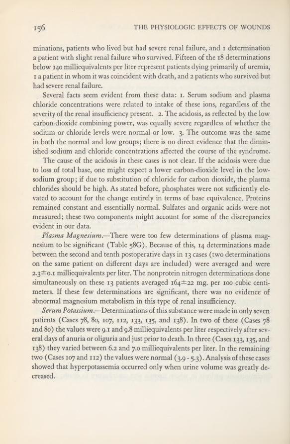
156 THE PHYSIOLOGIC EFFECTS OF WOUNDS
minations, patients who lived but had severe renal failure, and i determinationa patient with slight renal failure who survived. Fifteen of the 18 determinationsbelow 140 milliequivalents per liter represent patients dying primarily of uremia,1 a patient in whom it was coincident with death, and 2 patients who survived buthad severe renal failure.
Several facts seem evident from these data: 1. Serum sodium and plasmachloride concentrations were related to intake of these ions, regardless of theseverity of the renal insufficiency present. 2. The acidosis, as reflected by the lowcarbon-dioxide combining power, was equally severe regardless of whether thesodium or chloride levels were normal or low. 3. The outcome was the samein both the normal and low groups; there is no direct evidence that the dimin-ished sodium and chloride concentrations affected the course of the syndrome.
The cause of the acidosis in these cases is not clear. If the acidosis were dueto loss of total base, one might expect a lower carbon-dioxide level in the low-sodium group; if due to substitution of chloride for carbon dioxide, the plasmachlorides should be high. As stated before, phosphates were not sufficiently ele-vated to account for the change entirely in terms of base equivalence. Proteinsremained constant and essentially normal. Sulfates and organic acids were notmeasured; these two components might account for some of the discrepanciesevident in our data.
Plasma Magnesium.—There were too few determinations of plasma mag-nesium to be significant (Table 58G). Because of this, 14 determinations madebetween the second and tenth postoperative days in 13 cases (two determinationson the same patient on different days are included) were averaged and were2.3±o.i milliequivalents per liter. The nonprotein nitrogen determinations donesimultaneously on these 13 patients averaged 164±22 mg. per 100 cubic centi-meters. If these few determinations are significant, there was no evidence ofabnormal magnesium metabolism in this type of renal insufficiency.
Serum Potassium.—Determinations of this substance were made in only sevenpatients (Cases 78, 80, 107, 112, 133, 135, and 138). In two of these (Cases 78and 80) the values were 9.1 and 9.8 milliequivalents per liter respectively after sev-eral days of anuria or oliguria and justprior to death. In three (Cases 133,135,and138) they varied between 6.2 and 7.0 milliequivalents per liter. In the remainingtwo (Cases 107and 112) the values were normal (3.9 - 5.3).Analysis of these casesshowed that hyperpotassemia occurred only when urine volume was greatly de-creased.
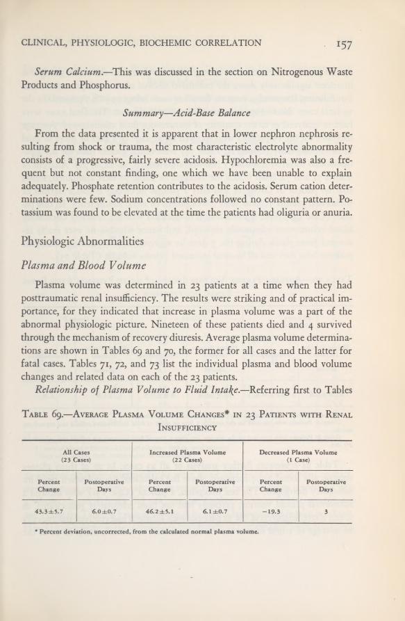
CLINICAL, PHYSIOLOGIC, BIOCHEMIC CORRELATION 157
Serum Calcium.—This was discussed in the section on Nitrogenous WasteProducts and Phosphorus.
Summary—Acid-Base Balance
From the data presented it is apparent that in lower nephron nephrosis re-sulting from shock or trauma, the most characteristic electrolyte abnormalityconsists of a progressive, fairly severe acidosis. Hypochloremia was also a fre-quent but not constant finding, one which we have been unable to explainadequately. Phosphate retention contributes to the acidosis. Serum cation deter-minations were few. Sodium concentrations followed no constant pattern. Po-tassium was found to be elevated at the time the patients had oliguria or anuria.
Physiologic Abnormalities
Plasma and Blood VolumePlasma volume was determined in 23 patients at a time when they had
posttraumatic renal insufficiency. The results were striking and of practical im-portance, for they indicated that increase in plasma volume was a part of theabnormal physiologic picture. Nineteen of these patients died and 4 survivedthrough the mechanism of recovery diuresis. Average plasma volume determina-tions are shown in Tables 69 and 70, the former for all cases and the latter forfatal cases. Tables 71, 72, and 73 list the individual plasma and blood volumechanges and related data on each of the 23 patients.
Relationship of Plasma Volume to Fluid Intake.—Referring first to Tables
Table 69.—Average Plasma Volume Changes* in 23 Patients with RenalInsufficiency
All Cases Increased Plasma Volume Decreased Plasma Volume(23 Cases) (22 Cases) (1 Case)
Percent Postoperative Percent Postoperative Percent PostoperativeChange Days Change Days Change Days
43.3±5.7 6.0 ±0.7 46.2 ±5.1 6.1 ±0.7 -19.3 3
* Percent deviation, uncorrected, from the calculated normal plasma volume.
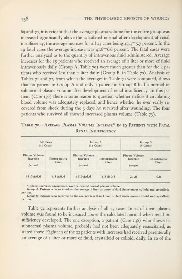
THE PHYSIOLOGIC EFFECTS OF WOUNDS
69 and 70, it is evident that the average plasma volume for the entire group wasincreased significantly above the calculated normal after development of renalinsufficiency, the average increase for all 23 cases being 43.3—57 percent. In the19 fatal cases the average increase was percent. The fatal cases werefurther analyzed as to the quantity of intravenous fluid administered. Averageincreases for the 15 patients who received an average of 1 liter or more of fluidintravenously daily (Group A, Table 70) were much greater than for the 4 pa-tients who received less than 1 liter daily (Group B, in Table 70). Analysis ofTables 71 and 72, from which the averages in Table 70 were computed, showsthat no patient in Group A and only 1 patient in Group B had a normal orsubnormal plasma volume after development of renal insufficiency. In this pa-tient (Case 136) there is some reason to question whether deficient circulatingblood volume was adequately replaced, and hence whether he ever really re-covered from shock during the 3 days he survived after wounding. The fourpatients who survived all showed increased plasma volume (Table 73).
Table 70. —Average Plasma Volume Increase* in 19 Patients with FatalRenal Insufficiency
Table 74 represents further analysis of all 23 cases. In 22 of them plasmavolume was found to be increased above the calculated normal when renal in-sufficiency developed. The one exception, a patient (Case 136) who showed asubnormal plasma volume, probably had not been adequately resuscitated, asstated above. Eighteen of the 22 patients with increases had received parenterallyan average of 1 liter or more of fluid, crystalloid or colloid, daily. In 10 of the
All Cases(19 Cases)
Group A(15 Cases)
Group B(4 Cases)
Plasma VolumeIncrease
percent
PostoperativeDays
Plasma VolumeIncrease
percent
PostoperativeDays
Plasma VolumeIncrease
percent
PostoperativeDays
4l.6±6.6 4.8±0.4 46.9±6.6 4.8±0.5 21.8 4.8
‘Percent increase, uncorrected, over calculated normal plasma volume.Group A; Patients who received on the average 1 liter or more of fluid (intravenous colloid and crystalloid)
per day.Group B: Patients who received on the average less than I liter of fluid (intravenous colloid and crystalloid)
per day.
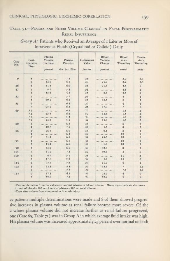
CLINICAL, PHYSIOLOGIC, BIOCHEMIC CORRELATION 159
Table 71.—Plasma and Blood Volume Changes 1 in Fatal PosttraumaticRenal Insuffiency
Group A: Patients who Received an Average of i Liter or More ofIntravenous Fluids (Crystalloid or Colloid) Daily
22 patients multiple determinations were made and 8 of them showed progres-sive increases in plasma volume as renal failure became more severe. Of the2 whose plasma volume did not increase further as renal failure progressed,one (Case 69, Table 71) was in Group A in which average fluid intake was high.His plasma volume was increased approximately 23 percent over normal on both
CaseNo.
Post-operative
Days
PlasmaVolumeIncrease
percent
PlasmaProteins
Gm. per 100 cc.
HematocritValue
percent
BloodVolumeChange
percent
Bloodsince
Wounding
«»//* 2
Plasmasince
Wounding
««/Vs 2
9 5 7.5 36 2.26 43.9 6.8 37 21.0 2.2 3.3
26 3 41.5 6.9 38 21.8 4.5 647 1 8.7 5.3 33 4.5 2
3 33.6 4.8 35 8.8 4.5 252 2 5.7 36 8 4
3 60.1 6.2 38 33.5 8 455 6 6.4 27 6 6
7 95.1 6.3 25 37.7 7 669 3 1 6.8 70 1,5 2
3 2 23.5 6.3 52 13.6 1.5 23 5 5.4 47 1.53 9 23.5 5.1 42 15.8 1.5 2
80 3 6.5 38 8 34 10.7 7.1 38 -5.5 8 3
86 2 26.5 6.0 33 -0.1 8 14 6.2 39 10 16 61.4 6.1 32 25.5 10 1
95 1 6.5 48 10 13 13.4 6.6 40 -1.0 10 5
98 5 33.9 6.6 47 32.7 8 5105 7 61.0 7.3 39 39.8 3 6108 1 6.7 5.1 28 11 3
3 17.7 5.6 40 3.8 13 3114 6 75,2 5.8 30 31.9 6 1123 3 52.3 5.8 32 18.6 7 1.3
6 5.8 29 7.5 1,3135 2 17.3 6.5 50 22.9 6 0
4 80.2 7.3 42 63.9 6 0
1 Percent deviation from the calculated normal plasma or blood volume. Minus signs indicate decreases.2 1 unit of blood =500 cc.; 1 unit ofplasma =300 cc. total volume.3 Days after release from compression in crush injury.

THE PHYSIOLOGIC EFFECTS OF WOUNDS
Table 72.—Plasma and Blood Volume Changes 1 in Fatal PosttraumaticRenal Insufficiency
Group B: Patients who Received an Average of Less than 1 Liter ofIntravenous Fluids (Crystalloid or Colloid) Daily
the second and ninth days after crushing injury. The other (Case 118, Table 72)was in Group B, those with restricted fluid intake. His increased plasma volumeactually diminished although he subsequently died in uremia.
Among those who had increased plasma volumes were the four patients whohad a recovery diuresis and survived. In three of them plasma volume first in-creased and then decreased as diuresis proceeded and nitrogenous waste productswere excreted (Table 73). The progress of the plasma volume changes in relationto the plasma nonprotein nitrogen in these cases is shown in Chart 27. The fourthpatient (Case 150) had his first determination after diuresis had begun althoughhe still had severe renal failure and an increased plasma volume.
Relationship ofPlasma Volume to Plasma Protein Concentration, HematocritLevel, and Total Blood Volume.—The relationships of plasma volume to plasmaprotein concentration, hematocrit value, and total blood volume are evident inTables 71, 72, and 73. There were considerable individual variations, but in gen-eral it can be stated that an expected coincident decrease in plasma protein con-centration as the plasma volume rose during renal insufficiency was not usuallydemonstrated. Thus of the eight patients in whom the already increased plasmavolume was known to rise, there were no significant changes in plasma proteinconcentration in five (three of whom received blood between measurements);
CaseNo.
Post-operative
Days
PlasmaVolumeChange
percent
PlasmaProteins
Gm. per 100 cc.
HematocritValue
percent
BloodVolumeChange
percent
Bloodsince
Wounding
units 2
Plasmasince
Wounding
units 2
22 5 67.9 7.1 21 13.0 5 3
93 3 1 -31.4 7.3 60 -5.7 0 23 3 6.2 44 0 23 6 22.2 6.2 39 6.1 0 2
118 2 34.4 6.7 41 20.7 9 25 16.3 6.6 42 6.1 9 2
136 3 -19.3 8.0 50 -14.5 9 4
1 Percent deviation from the calculated normal plasma or blood volume. Minus signs indicate decreases.2 1 unit of blood =500 cc; 1 unit of plasma =300 cc. total volume.3 Days after release from compression in crush injury. The first determination in this case was made before
renal insufficiency developed.

CLINICAL, PHYSIOLOGIC, BIOCHEMIC CORRELATION
Table 73.—Plasma and Blood Volume Changes 1 in 4 Patients with
Posttraumatic Renal Insufficiency and Subsequent Recovery Diuresis
Table 74,—Trend of Plasma Volume Changes during Posttraumatic RenalInsufficiency in 23 Patients
there was an increase in one, and a decrease in only two. Explanation for theabsence of a dilution phenomenon is not evident from our data. One can only
CaseNo.
Post-operative
Days
PlasmaVolumeIncrease
percent
PlasmaProteins
Gm. per100 cc.
Hema-tocritValue
percent
BloodVolumeChange
percent
I ntra-venousFluids
cc.
OralFluids
cc.
Bloodsince
Wounding
units 2
Plasmasince
Wounding
units *
60 0.75 30.3 6.3 25 -8 3.429 5.5 102.5 70.1 6.5 25 20 3 1,000 3 1.435 7.5 106.5 73.5 6.4 24 21 3 625 3 2,386 8.5 10
11.5 31.7 6.4 38.4 13 3 600 3 3,952 13.5 10
133 13 44.3 7.6 40 25 2,518 1,750 23 10.516 49.8 7.4 40 32 3 1,583 3 1,416 24 12.527 36.1 6.1 32 5 26 12.535 7.4 5.8 36 — 12 26 12.5
138 4 30.7 6.6 40 16 ? ? 2 25 29.7 6.0 40 15 0 3 1,525 2 2
10 55.6 6.6 32 20 1,650 3 1,160 2 614 11.1 8.0 40 -1.9 1,000 3 2,326 2 6
150 12.5 27.3 6.0 34 1.7 1,000 4 3,330 9.2 4
1 Percent deviation from the calculated normal plasmaor blood volume. Minus signs indicate decreases.1 1 unit of blood ==500 cc.; 1 unit of plasma =300 cc. total volume.3 Between determinations.3 Approximately.
1 Liter orPlasmaVolume
Results of RepeatedDetermination
Patients’ Outcome
Plasma VolumeTotal
Patientsmore of
I.V. FluidDaily
Deter-minationRepeated
IncreasedFurther
Did notIncrease Lived Died
Number ofpatients
Number ofpatients
Number ofpatients
Number ofpatients
Number ofpatients
Number ofpatients
Increased over calculatednormal * 22 18 10 8 2 4 18
Decreased below calcu-1 o o 0 1
* Four patients with recovery diuresis included. In them the plasma volume wasdiuresis.
increased until onset of

162 THE PHYSIOLOGIC EFFECTS OF WOUNDS
postulate that in such cases plasma protein was being mobilized from proteinsources elsewhere in the body.
The hematocrit level in the eight patients in whom plasma volume increasedprogressively (as shown by serial determinations) rose in one who receivedblood between measurements, was unchanged in three, two of whom receivedblood between measurements, and fell in four, one of whom received bloodbetween measurements. Although the total blood volumes were also increased,the increments clearly were a reflection of the increase in the plasma volume,and because of the low hematocrit level in most cases, they were not as strikinglyincreased as was the plasma volume.
These data indicate, then, that in posttraumatic renal failure, total circulatingplasma volume is increased. This must be due largely to the inability of thekidneys to excrete adequate water. However, because the plasma protein con-centration did not usually diminish as plasma volume increased, it is evidentthat the sole explanation is not simply that hydremia exists. There would seemto be also unexplained extrarenal factors interfering with maintenance of a nor-mal extracellular fluid volume. The practical importance of these observationsis self-evident. Administration of excessive quantities of fluids to these patientswho already have increased extracellular fluid volume can probably do nothingtoward stimulating the kidneys to excrete; it can cause fatal pulmonary edema.
Summary—Plasma and Blood Volume
Plasma volume was determined in 23 patients in whom posttraumatic renalinsufficiency developed, 19 of whom died. Averaging all 23 cases, there was anincrease over the calculated normal plasma volume of 43.3 percent, and of41.6 percent in the fatal cases. Considering the patients individually, there wasonly one in whom plasma volume was less than normal after signs of renalimpairment appeared, and there was good reason to believe that this patienthad never been adequately resuscitated from shock. Comparison of the plasmavolume with simultaneous plasma protein levels indicates that there was an in-crease in total circulating plasma and not simply an increase in proportion ofwater in the plasma; i.e., in general, plasma protein did not decrease with in-creasing plasma volume. In those patients in whom serial determinations weremade, including several who had a recovery diuresis, the time of maximumihcrease in plasma volume coincided with that of maximum azotemia, and inthose who recovered, plasma volume and plasma nonprotein nitrogen dimin-ished at parallel rates.
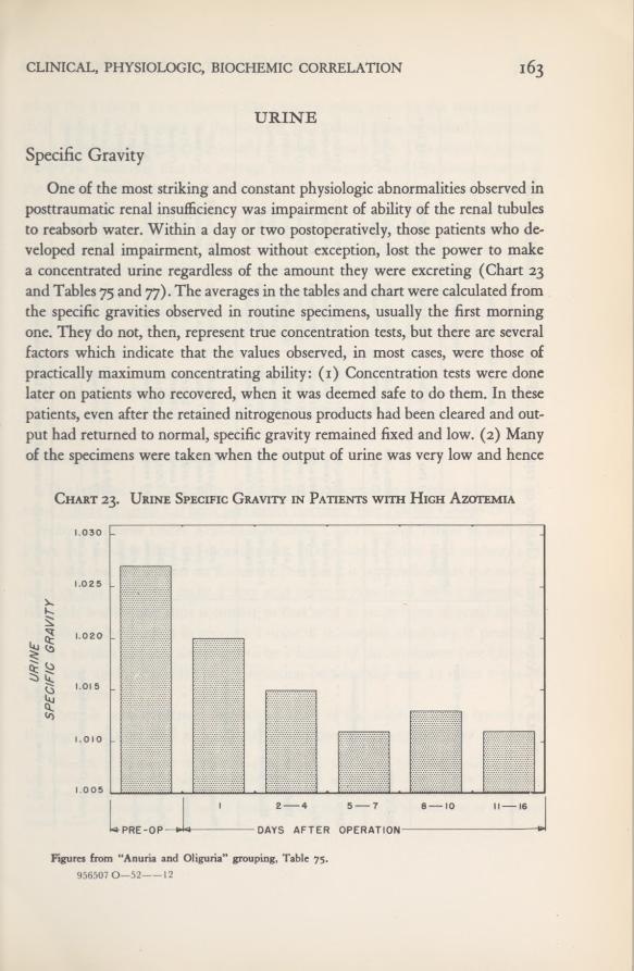
163CLINICAL, PHYSIOLOGIC, BIOCHEMIC CORRELATION
URINE
Specific GravityOne of the most striking and constant physiologic abnormalities observed in
posttraumatic renal insufficiency was impairment of ability of the renal tubulestoreabsorb water. Within a day or two postoperatively, those patients who de-veloped renal impairment, almost without exception, lost the power to makea concentrated urine regardless of the amount they were excreting (Chart 23and Tables 75 and 77). The averages in the tables and chart were calculated fromthe specific gravities observed in routine specimens, usually the first morningone. They do not, then, represent true concentration tests, but there are severalfactors which indicate that the values observed, in most cases, were those ofpractically maximum concentrating ability: (1) Concentration tests were donelater on patients who recovered, when it was deemed safe to do them. In thesepatients, even after the retained nitrogenous products had been cleared and out-put had returned to normal, specific gravity remained fixed and low. (2) Manyof the specimens were taken when the output of urine was very low and hence
Chart 23. Urine Specific Gravity in Patients with High Azotemia
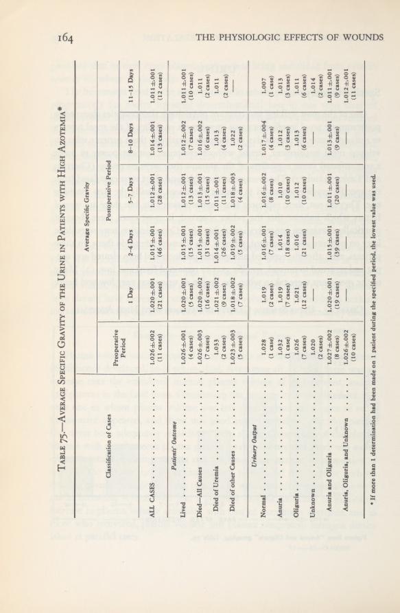
THE PHYSIOLOGIC EFFECTS OF WOUNDS
Table75.
—AverageSpecificGravityofthe
Urinein
Patientswith
HighAzotemia*
AverageSpecificGravity
ClassificationofCases
Preoperative
PostoperativePeriod
Period
1
Day
2-4Days
5-7Days
8-10Days
11-15Days
ALLCASES
1.026±.0021.020±.001
1.015±.001
1.012±.001
1.014±.0011.011±.001
(11cases)
(21cases)
(46cases)
(28cases)
(13cases)
(12cases)
Patients'Outcome
Lived
1.026±.0011.020±.001
1.015±.001
1.012±.0011.012±.0021.011±.001
(4
cases)
(5
cases)
(15cases)
(13cases)
(7
cases)
(10cases)
Died—All
Causes
,
1.026±.0031.020±.002
1.015±.001
1.013±.0011.016±.002
1.011
(7
cases)
(16cases)
(31cases)
(15cases)
(6
cases)
(2
cases)
DiedofUremia
1.033
1.021±.002
1.014±.0011.011±.001
1.013
1.011
(2
cases)
(9cases)
(26cases)
(11
cases)
(4
cases)
(2
cases)
DiedofotherCauses
1.023±.003
(5
cases)
1.018±.002(7
cases)
1.019±.002(5
cases)
1.018±.003(4cases)
1.022(2
cases)
UrinaryOutput
Normal
1.028
1.019
1.016±.0011.016±.002
1.017±.0041.007
(1
case)
(2
cases)
(7
cases)
(8
cases)
(4
cases)
(1
case)
Anuria
1.032
1.019
1.014
1.010
1.012
1.013
(1
case)
(7
cases)
(18cases)
(10cases)
(3
cases)
(3
cases)
Oliguria
1.026
1.021
1.016
1.012
1.013
1.011
(7cases)
(12cases)
(21cases)
(10cases)
(6
cases)
(6
cases)
Unknown
1.020(2
cases)
—
—
—
—
1.014 (2cases)
AnuriaandOliguria
1.027±.0021,020±.001
1.015±.001
1.011±.0011.013±.00l1.011±.001
(8
cases)
(19cases)
(39cases)
(20cases)
(9
cases)
(9
cases)
Anuria,Oliguria,andUnknown
1.026±.002(10cases)
1.012±.001(11cases)
*
IfmorethanI
determinationhadbeenmade
onI
patientduringthespecified
period,thelowestvalue
wasused.

CLINICAL, PHYSIOLOGIC, BIOCHEMIC CORRELATION 165when the kidneys were theoretically concentrating urine to the maximum oftheir ability. (3) In many of the patients, particularly those who died in uremia,fluids were sharply restricted, usually to about 1 liter a day. This could be furtherreason for assuming that the average urine specimen would be concentrated ifthe kidneys were capable of making it so; on the other hand, this argumentmay be rendered untenable by the fact that plasma volume was probably in-creased in all cases. (4) Twenty-four hour urine specimens were collected fromfive patients in whom renal failure developed. In these the specific gravity of thetotal specimens showed the same trend, even though plasma nonprotein nitrogenwas rising, and in three cases total output of urine was diminishing.
It will be shown in the section on Recovery Diuresis that in those patientswho did recover, the nitrogenous waste products were cleared because of anincreasing output of urine but the urine specific gravity remained fixed at a lowlevel. In summary, it appears from our data that in this syndrome one of theearliest functional derangements of the kidney to occur, and probably one of thelast to disappear when recovery takes place, is the ability to concentrate the urine.
Hydrogen-Ion Concentration
The tendency of the acidity of the urine to decrease as systemic acidosis andrenal failure progress has been previously discussed (see Plasma Carbon-DioxideCombining Power under Acid-Base Balance, Chart 21, and Tables 76 and 77).From our meager data on measurement of titratable acidity and ammonia ofthe urine (see the section on Recovery Diuresis), it is probable that the mecha-nism of this failure to make a very acid urine is associated with a decrease intitratable acidity and thus is similar to that seen in most types of renal failure.Inability of the kidneys to produce a urine of maximum alkalinity, if presentedwith a surplus of base, also seems to be a feature of the syndrome (see ChapterVII) and again is similar to the situation occasionally seen in other types ofkidney disease.
There is good evidence 1 that acidification of the urine by active transfer ofhydrogen ions, as well as ammonia production is accomplished by the distal
1 Pitts, R. F.; Lotspeich, W. D.; Schiess, W. A., and Ayer, J. L.; The renal regulation of acid-base balance in man. I. The nature of the mechanism for acidifying the urine. J. Clin. Investigation27: 48-56, January 1948. Schiess, W. A.; Ayer, J. L.; Lotspeich, W. D., and Pitts, R. F.: The renalregulation of acid-base balance in man. II. Factors affecting the excretion of titratable acid by the normalhuman subject. Ibid. 57-64.
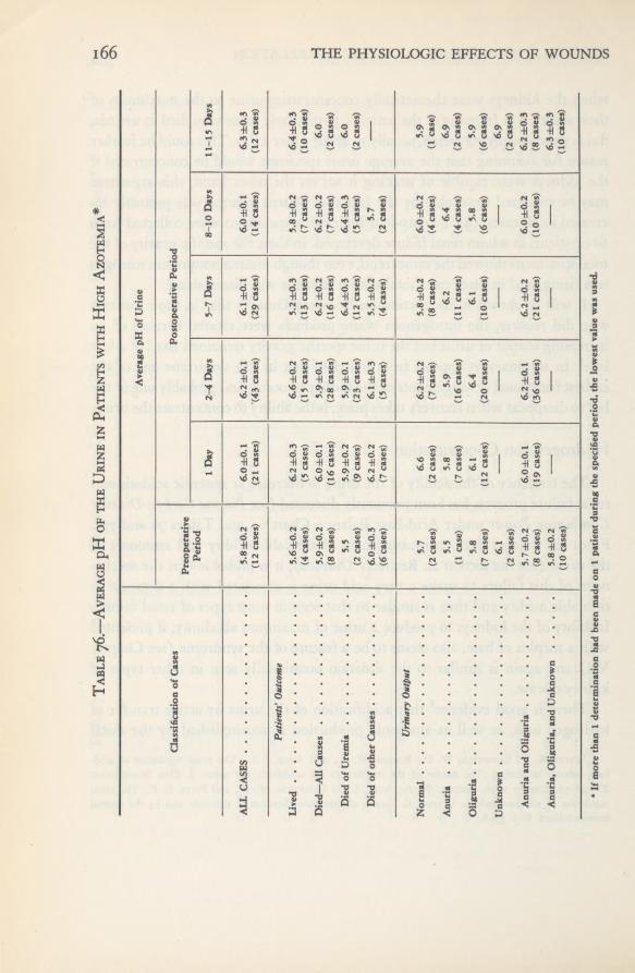
THE PHYSIOLOGIC EFFECTS OF WOUNDS
Table76.—AveragepH
ofthe
Urinein
Patientswith
HighAzotemia*
ClassificationofCases
AveragepHofUrine
Preoperative Period
PostoperativePeriod
1
Day
2-4Days
5-7Days
8-10Days
11-15Days
ALLCASES
5.8±0.2
6.0±0,1
6.2±0.1
6.1±0.1
6.0±0.1
6.3±0.3
(12cases)
(21cases)
(43cases)
(29cases)
(14cases)
(12cases)
Patients’Outcome
Lived
5.6±0.26.2±0.3
6.6±0.25.2±0.3
5.8±0.2
6.4±0.3
(4cases)
(5
cases)
(15cases)
(13cases)
(7cases)
(10cases)
Died—All
Causes
5.9±0.26.0±0.1
5.9±0.1
6.2±0.2
6.2±0,2
6.0
(8cases)
(16cases)
(28cases)
(16cases)
(7cases)
(2
cases)
DiedofUremia
5.5
5.9±0.2
5.9±0.1
6.4±0.3
6.4±0.3
6.0
(2
cases)
(9cases)
(23cases)
(12cases)
(5
cases)
(2
cases)
DiedofotherCauses
6.0±0.3
6.2±0.2
6.1±0.3
5.5±0.2
5.7
—
(6cases)
(7cases)
(5
cases)
(4cases)
(2
cases)
UrinaryOutput
Normal
5.7
6.6
6.2±0.2
5.8±0.2
6.0±0.2
5.9
(2
cases)
(2
cases)
(7cases)
(8cases)
(4cases)
(I
case)
Anuria
5.5
5.8
5.9
6.2
6.4
6.9
(1
case)
(7cases)
(16cases)
(11cases)
(4cases)
(2
cases)
Oliguria
5.8
6.1
6.4
6.1
5.8
5.9
(7cases)
(12cases)
(20cases)
(10cases)
(6
cases)
(6cases)
Unknown
6.1
—
—
:
—
6.9
(2
cases)
(2cases)
AnuriaandOliguria
5.7±0.26.0±0.1
6.2±0.1
6.2±0.2
6.0±0.26.2±0.3
(8
cases)
(19cases)
(36cases)
(21cases)
(10cases)
(8cases)
Anuria,Oliguria,andUnknown
5.8±0.2
—
—
—
—
6.3±0,3
(10cases)
(10cases)
*
Ifmorethan1
determinationhadbeenmadeonI
patientduringthespecified
period,thelowestvalue
wasused.

167CLINICAL, PHYSIOLOGIC, BIOCHEMIC CORRELATION
tubular cells. This provides further correlation, perhaps, of functional with ana-tomic findings in patients suffering from lower nephron nephrosis followingshock or trauma.
Table 77.—Average Specific Gravity and pH of Urine in Patients with
Oliguria or Anuria
Proteinuria and Pigment ExcretionAs was pointed out in Chapter IV, all patients with proved renal lesions had
proteinuria, and this finding was also a constant one in all patients with post-traumatic renal insufficiency. It was not, however, a specific finding, for it wasabsent in only 14 of the entire series of casualties studied. The relationship ofpigment excretion in the urine to posttraumatic renal insufficiency will be dis-cussed in Chapter VIII.
Summary—Changes in the Urine
The most striking change in the urine was the fixation of specific gravityat low levels within a day or two after the onset of renal failure following shockor trauma. This impairment of maximum reabsorptive capacity of water by therenal tubules persisted after other evidence of kidney damage disappeared inthose patients who recovered. Inability to manufacture a highly acid urine in
Time of DeterminationOliguria Anuria
pH Specific Gravity pH Specific Gravity
Preoperative 5.8±0.1 1.026±.002 5.5 1.032(10 cases) (10 cases) (1 case) (1 case)
Postoperative DaysFirst 6.2 ±0.1 1.023±.001 5.8±0.2 1.019±.003
(22 cases) (22 cases) (7 cases) (7 cases)Second to Fourth 6.4±0.2 1.0l6±.001 5.9=fc0.2 1.014±.001
(31 cases) (32 cases) (16 cases) (18 cases)Fifth to Seventh 6.4±0,2 1.012±.001 6.2±0.3 l.OlOdc.OOl
(17 cases) (17 cases) (11 cases) (10 cases)Eighth to Tenth 6.0±0.2 1.015±.001 6.4±0.2 1.012
(10 cases) (11 cases) (4 cases) (3 cases)Eleventh to Fifteenth 6.0±0.2 l.Olli.OOl 6.8 1.013
(6 cases) (6 cases) (3 cases) (3 cases)

168 THE PHYSIOLOGIC EFFECTS OF WOUNDS
the presence of metabolic acidosis was also a constant feature in the patientsstudied.
Recovery DiuresisThe arbitrary choice of 65 mg. per 100 cc. or higher concentration of non-
protein nitrogen in the plasma as an index of renal insufficiency in this series ofcases has been discussed. Of the 73 patients included in our “high azotemia”group, 23 (32 percent) lived. Those who survived may be further classifiedaccording to the degree of renal impairment they exhibited: Twelve had appar-ently only minimal interference with renal function, with a rapid return tonormal after transient nitrogen retention. We were unable to follow one patientwith a nonprotein nitrogen level greater than 65 mg. per 100 cc. who lived andso do not know what degree of renal insufficiency he ultimately had. Ten hadeven greater evidence of renal impairment and their course conformed with thesyndrome we have designated as recovery diuresis.
The characteristic features of this syndrome are:
1. A nonprotein nitrogen level greater than 100
milligrams per 100 cubic centimeters.2. Oliguria or anuria, followed by a substantial
diuresis resulting in clearance of nitrogenous waste prod-ucts and return to normal of the electrolyte pattern,
3. Impaired ability to concentrate the urine.4. Hypertension (systolic blood pressure above
135 millimeters of mercury; diastolic above 90).Of the 10 patients exhibiting this syndrome, 9 showed at least three and most
of them all four of these characteristics. The tenth patient (Case 44) was ob-served during a period when the laboratory was not functioning, so his bloodand urine could not be examined, but clinically he displayed the characteristicsof the syndrome.
This small group of cases is of great interest and practical importance. Onewould like to know why these patients recovered whereas the majority of thosewith renal insufficiency died, and whether in their course or treatment thereare any clues which might lead to more effective treatment than has been foundto the present. Detailed data on a few typical cases (see Charts 25 and 26),

CLINICAL, PHYSIOLOGIC, BIOCHEMIC CORRELATION 169together with pertinent clinical and physiologic findings in the group as a whole,are considered of value here. (The 10 cases were Numbers 27, 30, 43, 44, 60, 104,I25> !33> i38> and 150-)
Clinical Features
The degree of initial shock was essentially the same for these 10 patients asfor the entire group having renal failure. Three patients had had severe shock,5 moderate, i slight, and i no shock. As in other instances, it is entirely possiblethat there may have been considerable shock in all 10, but so far as we coulddetermine at the time of admission, they must be classified as above.
Five of the 10 patients had multiple major wounds. The major wounds inthe entire group were: 6 peripheral with fracture, 4 thoracic, 4 intra-abdominal,and one thoraco-abdominal. One patient had a contusion of the bladder. Therewere no wounds of the liver or kidney.
The time of onset of oliguria in relation to wounding and operation, and theduration of suppression of urinary output are of interest. Unfortunately the day-to-day records are not as accurate as one would like. Many of these patients wereseen in field hospitals during periods of great military activity, when the pressof work made it very difficult to make such observations. We have no recordof output of urine until the first postoperative day in any of the cases. Eight ofthe 10 patients are known to have had at least 1 day of oliguria or anuria betweenthe first and sixth postoperative days. Records of output of urine were not keptfor the remaining 2 patients at the time they probably had oliguria, but ques-tioning of ward personnel and of the patients suggested very strongly that theytoo had had oliguria during this period. The duration of oliguria ranged from1 to 4 days. There followed then a period of gradually increasing output of urine,reaching in some cases 5 or 6 liters a day. The plasma nonprotein nitrogen leveldid not, as a rule, begin to decrease until several days after diuresis had begun.
Because of the increase in plasma volume during the period of increasingazotemia, it might be expected that total fluid output would exceed intake dur-ing the diuresis period. This clearly occurred in two cases (Case 60 (Chart 24)and Case 150). In the remaining eight cases it was impossible to demonstratethis fact from the figures available, for the records kept gave only an approxi-mate estimate of total water balance; they did not account for water lost byperspiration, respiration, or with the stools. Since the plasma volume returned

170 THE PHYSIOLOGIC EFFECTS OF WOUNDS
to normal and the edema subsided as diuresis proceeded, it is logical to assumethat total fluid output did exceed intake until equilibrium was reestablished.
All these patients had hypertension, by our definition. In general the bloodpressure was highest at the time of the most severe nitrogen retention and the
Chart 24. Relationship of Fluid Intake and Output in a Patient with
Recovery Diuresis (Case 60)
Chart 25. Recovery diuresis occurred in this patient following an initial period ofrenal insufficiency due to severe traumatic shock and great blood loss. Note: (i) Initialperiod of oliguria and diminished renal function as evidenced by low phenolsulfonphthaleinexcretion with fixed urine specific gravity which persisted throughout the period of obser-vation. (2) The gradual increase in urinary output during the first week accompanied byrising blood pressure, rising plasma nonprotein nitrogen concentration, falling carbon-dioxide combining power and plasma chlorides. (3) The fall in nonprotein nitrogenoccurring only several days after adequate urinary output, accompanied by improvementof phenolsulfonphthalein excretion, rising carbon-dioxide combining power and plasmachloride levels, and return of the blood pressure toward normal. See page iji.
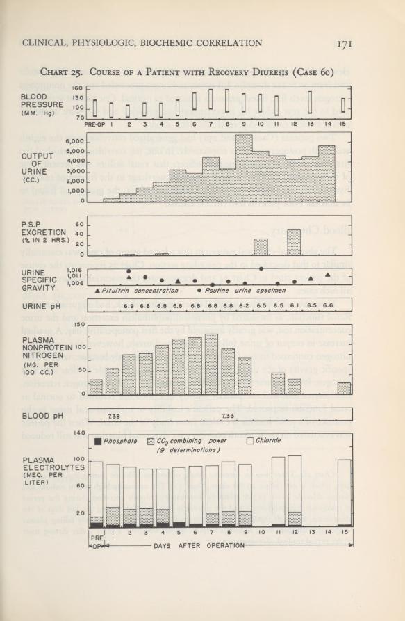
CLINICAL, PHYSIOLOGIC, BIOCHEMIC CORRELATION
Chart 25. Course of a Patient with Recovery Diuresis (Case 60)

THE PHYSIOLOGIC EFFECTS OF WOUNDS172
elevation was significant, with levels ranging from 150 to 170 mm. Hg systolicand from 90 to no diastolic. Likewise, as diuresis proceeded and the nonproteinnitrogen levels fell, blood pressures returned to normal. One patient was evacu-ated to the rear before his hypertension had subsided and we were unable toobtain subsequent blood-pressure determinations.
Two patients (Cases 44 and 150) had generalized convulsions on the eighthand ninth postoperative days respectively. In one, the convulsions furnished thefirst clue to the attending medical officers that renal failure was present. Oneof these patients also had a small retinal hemorrhage in the fundus of one eye.Eyegrounds were examined in three other patients in the group and found tobe normal. Four patients had clinical edema.
Blood ChemistryThe abnormal chemical pattern in this selected group of cases was essentially
similar to that described in the preceding section. Chart 25 represents the courseof the patient cited in Chart 24 and demonstrates the essential features seen inall such cases.
This man, who had had a period of severe initial shock, had oliguria for 1 day.Renal function, as measured by phenolsulfonphthalein excretion and the urineconcentration test, was greatly impaired by the first postoperative day. A gradualincrease in output of urine followed. Despite diuresis, however, the nonproteinnitrogen continued to rise for the first 7 days—apparently because, with a fixedspecific gravity of the urine, the kidneys were not at first able to clear sufficientnitrogen. Blood pressure rose and fell approximately with the nitrogen retention.Electrolytes followed the pattern already described but returned to normal asrenal function improved. The patient’s inability to make an acid urine in thepresence of a mild acidosis is evident from study of the chart. When the patientwas evacuated on the fifteenth postoperative day,renal function was still reduced
Chart 26. There was an excessive intake of sodium chloride during the previous 10
days, which is not shown on the chart. Note: (1) The unusually high serum sodium andplasma chloride levels. (2) A relatively low urinary chloride excretion during the periodof maximum hyperchloremia and the extremely low excretion during the last days of theobservation, although significant hyperchloremia persisted. (3) The slowly falling plasmanonprotein nitrogen level, although urinary output was over 2 liters a day during mostof the period studied. See page 773.

CLINICAL, PHYSIOLOGIC, BIOCHEMIC CORRELATION 173
Chart 26. Course of a Patient with Recovery Diuresis (Case 133)
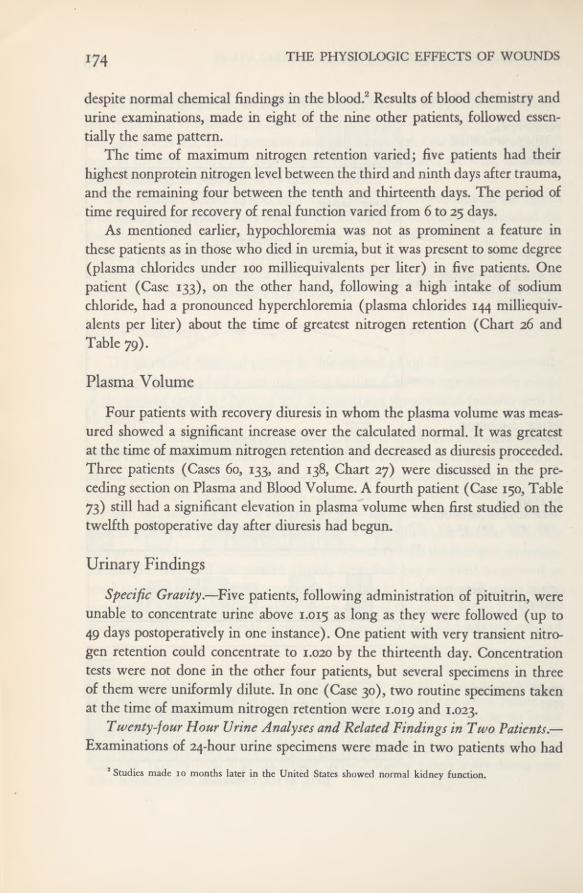
THE PHYSIOLOGIC EFFECTS OF WOUNDS
despite normal chemical findings in the blood.2 Results of blood chemistry andurine examinations, made in eight of the nine other patients, followed essen-tially the same pattern.
The time of maximum nitrogen retention varied; five patients had theirhighest nonprotein nitrogen level between the third and ninth days after trauma,and the remaining four between the tenth and thirteenth days. The period oftime required for recovery of renal function varied from 6 to 25 days.
As mentioned earlier, hypochloremia was not as prominent a feature inthese patients as in those who died in uremia, but it was present to some degree(plasma chlorides under 100 milliequivalents per liter) in five patients. Onepatient (Case 133), on the other hand, following a high intake of sodiumchloride, had a pronounced hyperchloremia (plasma chlorides 144 milliequiv-alents per liter) about the time of greatest nitrogen retention (Chart 26 andTable 79).
Plasma Volume
Four patients with recovery diuresis in whom the plasma volume was meas-ured showed a significant increase over the calculated normal. It was greatestat the time of maximum nitrogen retention and decreased as diuresis proceeded.Three patients (Cases 60, 133, and 138, Chart 27) were discussed in the pre-ceding section on Plasma and Blood Volume. A fourth patient (Case 150, Table73) still had a significant elevation in plasma volume when first studied on thetwelfth postoperative day after diuresis had begun.
Urinary FindingsSpecific Gravity.—Five patients, following administration of pituitrin, were
unable to concentrate urine above 1.015 as long as they were followed (up to
49 days postoperatively in one instance). One patient with very transient nitro-gen retention could concentrate to 1.020 by the thirteenth day. Concentrationtests were not done in the other four patients, but several specimens in threeof them were uniformly dilute. In one (Case 30), two routine specimens takenat the time of maximum nitrogen retention were 1.019 and 1.023.
Twenty-four Hour Urine Analyses and Related Findings in Two Patients.—Examinations of 24-hour urine specimens were made in two patients who had
2 Studies made 10 months later in the United States showed normal kidney function.

CLINICAL, PHYSIOLOGIC, BIOCHEMIC CORRELATION
Chart 27. Plasma Volume and Plasma Nonprotein Nitrogen
Concentration in 3 Patients with Recovery Diuresis(Cases 60,133, and 138)
recovery diuresis. In Case 133 the collections were started just at the end of therecovery period. Table 78 lists the findings in one of these patients (Case 104)along with plasma determinations made on the same days. From these dataa few conclusions could be drawn regarding this one patient: (1) Titratableacidity values were rather low, considering low plasma carbon-dioxide com-bining power during the first 3 days of collection. (2) Ammonia productionwas normal. (3) Chloride excretion and sodium chloride intake were normal.Plasma chloride values were normal. (4) Excretion of urea was high the first

176 THE PHYSIOLOGIC EFFECTS OF WOUNDS
day and accompanied a fall in plasma nonprotein nitrogen to normal.Table 79 and Chart 26 show comparable findings on the other patient (Case
133). Ammonia and titratable acidity determinations of the urine in this casewere not numerous enough and varied too widely to draw any conclusionsregarding acid-base regulation. Excretion of urea and creatinine was about whatwould be expected in a normal individual, but not sufficient to clear the retainednitrogen rapidly. Toward the end of the 10-day period during which 24-hoururine collections were made, plasma levels of urea nitrogen and creatinine be-
Table 78.—Relationship of 24-HOUR Urinalyses and Plasma BiochemicFindings in a Patient with Recovery Diuresis (Case 104)
PostoperativeDays
FluidIntake
cc.
ParenteralNaClIntake
Gm.
UrineOutput
cc.
PlasmaCO2 Com-
biningPower
rnEq./L.
UrinepH
UrineTitratableAcidity
cc. 0.1 N
UrineAmmonia
cc. 0.1 N
UrineAmmoniaTitratableAcidity
Third .... 1,060 900 +
Fourth . . . 3,705 1.6 2,335 25 5.9 257 428 1.7Fifth .... 2,100 1,590 23 5.6 199 652 3.3
33*50 1,950 5.6 302 688 2.2Seventh . . . 3,480 ! 1,780 23 5.2 444 656 1.4
4,850 2,480 5.4 509 779 1.53,650 2,050 6.9 145 4 14 2.8
Tenth .... 2,790 2,100 26 6.4 69 497 7.13,2 50 2,890 6.5 93 594 (5.4
2,550 2,200 6.6 154 544 3.4Thirteenth. . 2,500 2,730 6.8 137 475 3.5
Plasma PlasmaPlasma Urea Urine Great- Urine
Urine Plasma NPN Nitrogen Urea inine Great-PostoperativeDays Chloride Chloride
mg. per mg. perNitrogen
mg. perinine Specific
GravityGm. NaCl mEq./L. 100 cc. 100 cc. Gm. 100 cc. Gm.
Third .... Ill4.7 102 70 22.7 1.08 1.42
Fifth 3.2 101 44 27 15.4 0.80 1.306.9 19.2 1.80
Seventh . . . 4.7 102 29 12.2 1.39 1.25 1.0129.1 26.1 1.724.7 13.5 1.20 1.01 6
Tenth .... 5.9 94 29 19.9 0.94 1.48 1.0128.1 13.5 1.33 1.0108.4 14.5 1.25
Thirteenth. . 11.7 15.4 0.80
CLINICAL, PHYSIOLOGIC, BIOCHEMIC CORRELATION
gan to fall. Clearance rates of these substances, however, were diminished duringthe entire period of observation.
The relationship of serum sodium, plasma carbon-dioxide combining power,fluid and sodium chloride intake, urinary output, and urinary excretion ofchloride in this same patient are also shown. After a high intake of sodiumchloride, sodium and chloride retention developed. Plasma carbon-dioxide com-bining power fell, in this instance partially as a result of hyperchloremia. Chlor-ide excretion was relatively low considering the high plasma chloride level.As the plasma chloride level fell, chloride excretion decreased to negligibleamounts during the last 4 days, even though the plasma chloride level was stillunusually high. High serum sodium accompanied the hyperchloremia. Thereis in this case evidence of a high renal “threshold” for both sodium and chlorideexcretion.
The course and essential features in this patient were characteristic of recov-ery diuresis, but he exhibited most unusual electrolyte abnormalities. Althoughhe had an increased plasma volume (Chart 27), there were also marked hyper-natremia and hyperchloremia. An elevated serum sodium is most unusual ex-cept in the presence of dehydration, which, if the measurements of plasmavolume can be accepted, was not present in this patient. Further evidence ofinability of the renal tubules to reabsorb maximum amounts of water is seenhere in the dilute urines of total specimens in both Cases 104 and 133 duringperiods of high nitrogen retention.
Renal Clearance Tests and Phenolsulfonphthalein Excretion
The results of renal clearance and phenolsulfonphthalein excretion tests inpatients with recovery diuresis were discussed in Chapter III. Briefly, in threesuch patients in whom clearance measurements were made, all functional com-ponents of the kidney were diminished when first observed and gradually re-turned toward normal over a period of several days or weeks. Similar evidenceof functional impairment and subsequent improvement was seen in three addi-tional patients with recovery diuresis on'whom phenolsulfonphthalein excretorycapacity tests, were made.
Summary—Recovery DiuresisOf the 23 patients with posttraumatic renal insufficiency who lived, 10 ex-
hibited certain features which conform to the syndrome designated as recovery

17% THE PHYSIOLOGIC EFFECTS OF WOUNDS
Table79.
—Relationshipof24-HOUR
Urinalysesand
PlasmaBiochemicFindings
ina
Patientwith
RecoveryDiuresis(Case
133)
Postoperative Days
Fluid Intake cc.
Par- enteral NaCl Intake Gm.
Urine Output cc.
Blood pH (venous)Plasma C0
2
Com- bining Power mEq./L.Urine pH
Urine Titrat- able Acidity cc.0.1N
Urine Ammonia cc.0.1N
Urine Ammonia Titratable Acidity
Fourth
2,500 4,870 7,000 4.400 3.900 4,395 3,150 2,810 3,570 3,110 2,540 3,700 3.350 4.350 4.400 5,300 5.90017 25.5 25.5 8.5 8.5 8.5 8.5 8.5 8.5 0
? 1,600 1,450 2,000 2,950 3,000 1,270 2,650 2,380 2,030 2,150 2,220 2,300 2,375 2,610 2,550 3,500
Fifth Sixth Seventh Eighth Ninth Tenth
7.32
21
Eleventh
5.7
375
m
1.0
Twelfth
20 21 20 20 21
Thirteenth
5.6
383
244
0.7
Fourteenth Fifteenth
6.1 6.8 8.4 8.4 8.4
461 242
528 306
1.1 1.3
Sixteenth Seventeenth....
Eighteenth
20
Nineteenth Twentieth
16
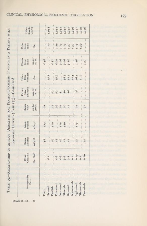
CLINICAL, PHYSIOLOGIC, BIOCHEMIC CORRELATION i79
956507 0—52 13
Table79.
—Relationshipof24-HOUR
Urinalysesand
PlasmaBiochemic
Findingsina
Patientwith
RecoveryDiuresis(Case133)—
Continued
Postoperative Days
Urine Chloride Gm.NaCl
Plasma Chloride mEq./L.Serum Sodium mEq./L.
Plasma NPN mg.per
100cc.
Plasma Urea Nitrogen mg.per
100cc.
Urine Urea Nitrogen Gm.
Plasma Creat- inine mg.per
100cc.
Urine Creat- inine Gm.
Urine Specific Gravity
Tenth
134
155
108
4.34
Eleventh
6.7
19.8
1.75
1.014
Twelfth
140
170
112
92
4.87
Thirteenth
4.6
144
106
92
19.2
4.08
1.58
1.01S
Fourteenth
4.6
140
178
101
81
3.96
1.73
1.01S
Fifteenth
3.6
142
180
100
80
14.5
3.96
1.73
1.015
Sixteenth
5.4
141
111
86
18.1
3.84
1.32
1.015
Seventeenth....
0.12
18.3
1.32
1.01G
Eighteenth
0.13
129
173
102
76
16.5
2.86
1.73
1.017
Nineteenth
0.51
11.9
1.39
1
01ft
Twentieth
0.70
119
97
2.37
1.016

THE PHYSIOLOGIC EFFECTS OF WOUNDS
diuresis; namely, (i) nonprotein nitrogen greater than 100 mg. per 100 cc.,(2) oliguria or anuria followed by diuresis and fall in nonprotein nitrogen,(3) low and fixed urinary specific gravity, and (4) hypertension. Their clinicalcourse and physiologic and biochemic changes were entirely similar to thosedemonstrated in patients who died and were found at necropsy to have hadlower nephron nephrosis. This group of patients demonstrates, then, that re-covery from this type of renal disease can occur spontaneously if the patientsurvives the first critical 10 days after onset of renal failure.
SUMMARY
In earlier chapters the changes that began to occur in the internal environ-ment soon after a man had been wounded were described. The effects of theseearly changes upon the kidney have been reported in this and the precedingchapter. Following severe trauma, accompanied usually by shock, a man couldbe adequately resuscitated and successfully operated upon. The latent renal in-competency which might develop in this man, who had normal kidneys at thetime he was wounded, usually did not become manifest until two or three dayslater. At this time there appeared signs of failure on the part of the kidneys towithstand the initial insult, the effects of which his body had thus far resistedwith fair success.
The first clinical sign of impending renal failure usually was suppression ofurinary output. Of 73 patients with “high azotemia” (a plasma nonproteinnitrogen concentration of 65 mg. per 100 cc. or higher at some time duringtheir course), 27 had anuria (urinary output of less than 100 cc. in any 24-hourperiod) and 29 had oliguria (output of from 100 to 600 cc. in a 24-hour period).
The case fatality rate was high.Fifty patients (69 percent) of the 73 with highazotemia died. Twenty-one (47 percent) of 45 who had oliguria, and 30 (91percent) of 33 with anuria had a fatal outcome.
Initial shock—if special types of cases such as crush injuries, reaction toincompatible blood transfusion, and sulfathiazole crystalluria are excluded—-was observed in a large proportion of the cases. Eighty-two percent of thosewho had high azotemia, and, in another category, 73 percent of those who hadoliguria and 69 percent of those who had anuria had been in moderate or severeinitial shock. These figures are undoubtedly too low, for many men probablyhad been in shock before we saw them.
181CLINICAL, PHYSIOLOGIC, BIOCHEMIC CORRELATION
Death occurred within 10 days after wounding in 48 (94 percent) of 51patients who died of posttraumatic renal insufficiency. Apparently if thewounded man can withstand this critical 10-day period, recovery of renal func-tion begins and he may survive. In a few patients there was evidence of return-ing renal function toward the end of their course, even though they died inuremia. The importance of this fact cannot be overemphasized, for a thera-peutic error (such as overloading the circulatory system by fluid administration)during this critical period may cause a fatal outcome before natural recoverycan take place.
Hypertension (at least 135 mm. Hg systolic and at least 90 mm. Hg diastolic)occurred in 62 percent of the patients with high azotemia, and in 79 percentof those in this group who died in uremia. In many of the fatal cases in whichhypertension did not develop, death occurred within 4 days after wounding.Had these patients survived longer, probably they too would have had hyper-tension.
The important biochemic and physiologic abnormalities in the blood re-sulting from posttraumatic renal insufficiency were found to be nitrogen andphosphorus retention, acidosis, hypochloremia, and increase in the plasma vol-ume. These blood and plasma changes reflect rapidly diminishing renal func-tion, as indicated by: (1) inability to concentrate the urine; (2) frequent failureto make a highly acid or alkaline urine in the presence of metabolic acidosisor alkalosis; (3) diminished glomerular filtration rate and renal blood flow,and (4) decreased phenolsulfonphthalein and maximum tubular excretorycapacity of para-amino hippuric acid.
Nonprotein nitrogen, urea nitrogen, creatinine, uric acid, and phosphoruslevels in the plasma rose as renal failure progressed during the first 10 daysafter wounding. Most observations after this period were on patients who re-covered. The level of these waste products fell between the tenth and fifteenthdays.
A progressive, fairly severe acidosis was characteristic, manifested by fallingplasma carbon-dioxide combining power as renal failure advanced. Loss ofability to make a highly acid urine in most cases suggests that the acidosis waspartially to be explained by impairment of the mechanism which produces anacid urine. A few observations also indicated, as expected, an impairment ofammonia production by the renal tubules. There was also good evidence thatremoval of sodium by excretion as sodium bicarbonate was poorly accomplished

182 THE PHYSIOLOGIC EFFECTS OF WOUNDS
in those cases in which alkalosis resulted from administration of large quantitiesof sodium.
Hypochloremia was severe and progressive, if all fatal cases are averaged.Correlation of the plasma chloride level and sodium chloride intake, however,demonstrated that the low chlorides were to some degree a result of inadequatesalt intake. Serum sodium levels showed similar correlation. There was no dif-ference in case fatality between the group with hypochloremia and those withnormal plasma chloride levels. One patient with renal failure had severe sodiumand chloride retention following a high salt intake. Intake, however, does not
entirely explain variations in plasma chloride levels. No correlation with degreeof plasma volume increase was apparent, but it is suggested that some inter-ference with water and sodium chloride equilibrium was present in additionto the demonstrated relation to intake.
Phosphates in terms of acid equivalence, contributed toward but did notentirely account for the acidosis. Plasma proteins, if converted to milliequiv-alents, were normal and constant. Sulfates and organic acids were not meas-ured and possibly account for discrepancies in our anion determinations.
Cation determinations were few. It has already been stated that sodiumlevels were partially correlated with salt intake. Magnesium was not signifi-cantly elevated in most cases. Potassium was determined in only a few cases,but the results suggest that hyperpotassemia was probably a feature of the syn-drome. Calcium followed a reciprocal relationship with rising phosphorus levelsin the few cases in which such determinations were made.
Total plasma volume was significantly elevated in 22 of 23 patients withposttraumatic renal insufficiency. Nineteen of these 23 died. Three patientswith severe renal failure in whom a recovery diuresis developed had plasmavolume increases that reached a maximum at the time of greatest nitrogenretention and decreased after diuresis. The degree of increase of the plasmavolume was clearly related to fluid intake. The water retention appeared to belargely a result of administration of more fluid than the impaired kidneyscould excrete. Comparison of plasma volume with total blood volume indicatedthat it was the plasma which was increased, rather than all elements of theblood. The expected dilution of plasma proteins was not demonstrated in mostcases. The practical importance of this increase in plasma volume has beenmentioned.

CLINICAL, PHYSIOLOGIC, BIOCHEMIC CORRELATION 183Ability to concentrate the urine diminished rapidly as the syndrome of
posttraumatic renal insufficiency progressed, and specific gravity became fixedin all patients with a severe degree of renal failure. In patients who recoveredit was the last of the kidney functions of those measured to return to normal.Since urine concentration is accomplished by the distal tubular cells, whereasmannitol, para-amino hippuric acid, and phenolsulfonphthalein reflect glo-merular and proximal tubular function, the lag in recovery of water-reabsorp-tive capacity may point to greater relative functional impairment of the lowernephron.
Ten patients of the 73 who had high azotemia exhibited all of the featuresof severe renal failure and subsequently a diuresis developed and they recovered.The course of these patients followed a pattern which has been termed the“syndrome of recovery diuresis.” This small group re-emphasizes the impor-tance of avoiding early fatal therapeutic errors (such as overloading the circu-latory system with fluid) thereby affording the kidneys an opportunity to re-cover spontaneously.

CHAPTER VI
Treatment of Lower NephronNephrosis
It should be stated at once that treatment of posttraumatic renal insufficiencyafter the syndrome has become established is disappointing although not hope-less. Evidence has been presented that shock is one factor important in itsetiology. As soon as the patient becomes available for treatment, the shock statecan usually be corrected fairly rapidly. Primary treatment of the woundedshould therefore be directed toward prompt and adequate resuscitation of everyman in shock in order to prevent, if possible, development of serious renal in-sufficiency.
In presenting our experience in the management of patients with posttrau-matic renal insufficiency, certain therapeutic errors to be avoided will be stressedand the use of various drugs and procedures to stimulate renal function (mostof which are of questionable value) will be discussed.
Fluid IntakeEarly in the Italian campaign fatal cases of posttraumatic renal insufficiency
began to appear. Therapy almost universally consisted of intravenous adminis-tration of large quantities of various crystalloids. Practically every knowndiuretic was also employed, and attempts were frequently made to alkalinizethe urine by administering large quantities of available base by mouth or vein.In the absence of adequate output of urine, the renal insufficiency soon becamecomplicated by cardiac failure as a result of overloading the circulatory system.The majority of patients so treated died rather promptly of pulmonary edemabefore they had an opportunity either to die in uremia or to regain adequaterenal function.
TREATMENT OF LOWER NEPHRON NEPHROSIS 185Early in the course of our study it was found that those patients with renal
insufficiency almost without exception had increased plasma volumes, as wasshown in Chapter V, Here then was an additional explanation for the fre-quency of pulmonary edema; we were dealing with patients who, as a result ofrenal failure, already had a circulating plasma volume larger than normal. Ifadditional fluids were added, especially those containing sodium, water reten-tion became more severe. The frequency of pulmonary edema and cardiac fail-ure is therefore not surprising. The therapeutic implications of these physiologicabnormalities are clear. Patients with this type of renal insufficiency alreadyhave too much extracellular fluid and too large a plasma volume, apparentlyprimarily as a result of the kidney’s being unable to excrete this surplus water.Treatment should therefore include (i) any judicious measures which mightpossibly encourage the kidneys to excrete more urine, and (2) avoidance ofany measures which would further increase plasma volume and secondarilycause cardiac embarrassment.
As was shown in the previous chapter, the critical period in the majority ofpatients with renal insufficiency appeared to be the first 10 days after wounding,when 94 percent of the total deaths occurred. Evidence has been presented thatrenal function had begun to improve after this time in a number of patients,even though they subsequently died in uremia. Thus the importance of prevent-ing an early fatal outcome resulting from too enthusiastic fluid administrationcannot be overemphasized. Unfortunately most of these patients eventually diein uremia regardless of treatment, but by judicious use of fluids they are atleast allowed an opportunity to recover renal function spontaneously and theirchance of survival is increased.
In cases in which fluids were restricted, the incidence of pulmonary edemawas, in our considered judgment, materially lessened, even in those patientswho subsequently died primarily of renal failure. A few who recovered (Cases43,44, and 138) might have died of heart failure if quantities of fluid comparableto those previously employed had been given.
Our recommendation on fluid intake for the average patient with oliguriaor anuria is that ordinarily not more than from 500 to 1,000 cubic centimetersin excess of urinary output be given daily. The patients in this study were seenin the fall, winter, and spring months when the temperature in Northern Italywas such that extrarenal fluid losses were probably minimal. In hot weather orwhen sweating is excessive this allowance should probably be increased. If
186 THE PHYSIOLOGIC EFFECTS OF WOUNDS
urinary output increases to more than i liter daily, it is suggested that intake offluids parallel or slightly exceed output of urine until retained nitrogen is cleared.Oral fluids, whenever their use is feasible, are preferred to parenteral ones inorder to avoid sudden augmentation of the already increased plasma volume.Such parenteral fluids as are used should be administered slowly, and the patientshould be carefully observed for signs of pulmonary edema during infusion.For reasons to be mentioned later under Hypertonic Solutions, the preferredparenteral fluid is an isotonic solution of dextrose. 1
We have too little data on sodium to compare the level of this cation in theserum with the sodium intake. Hence it cannot be stated whether hyponatremiaaccompanied the hypochloremia, and therefore whether serious sodium deple-tion ever occurred. It now appears that as a consequence of the practice of givingalmost no salt parenterally, hypochloremia resulted in some patients, althoughthe outcome in those with decreased plasma chlorides apparently was no differ-ent from that in patients with normal chlorides. In our opinion, once posttrau-matic renal insufficiency has become established, the danger of producing furtheredema and circulatory failure exceeds the questionable benefit to be derivedfrom continued attempts to replenish deficient sodium by administration ofsodium salt. We would therefore now recommend that during the oliguric andanuric phase salt be given only in quantities sufficient to replace extrarenal saltlosses and the small quantity lost in the urine. These combined losses can only beestimated; they probably would seldom exceed one or two grams of salt daily.
jProcedures and Drugs Used toStimulate Kidney function
Our experience revealed no positive and sure measures capable of re-estab-lishing kidney function after renal insufficiency had developed. The aim shouldalways be to avoid measures that might be harmful during the period before
1 Because of the water-retaining property of the cation in sodium chloride, we suggested that thelargest portion of the parenteral fluids given consist of 5- or 10-percent dextrose in distilled water. How-ever, only the 10-percent prepared solution was available during most of the period when these studieswere being made. Although 5-percent dextrose can be made up by adding dextrose solution to distilledwater, under field conditions and the pressure of work during combat this is troublesome and was notoften done.

1 87TREATMENT OF LOWER NEPHRON NEPHROSIS
the kidneys spontaneously begin to clear retained waste products. Well-knownmethods of stimulating urine flow that were tried will be discussed briefly. It isevident from the results that they were of no value, or at best of questionablevalue.
Hypertonic SolutionsThe advisability of giving hypertonic solutions parenterally in this syndrome
is highly debatable. They exert, at least in the presence of normal renal func-tion, a diuretic effect by (i) temporarily increasing plasma volume, and (2) bylimiting tubular reabsorption of water due to the osmotic effect of increasedconcentration of solute in the distal tubules. It is clear that the first of theseactions is undesirable, since the circulating plasma volume is already increased;the second is desirable if tubular reabsorption can be altered in the damagedkidney.
Hypertonic saline (10-percent) solution was used in only one patient—the only one in our series with a true transfusion incompatability (Case 9). Therewas no observed effect on urinary output. It might be argued that early trialof such a solution would be of value in this type of renal insufficiency. If thereis no response to 500 cc. of 3-percent saline solution, further attempts at diuresisby this method surely are contra-indicated because of the undesirable increasein plasma volume which must result from excessive salt administration.
Either a hypertonic dextrose solution or 5-percent dextrose in isotonic salinesolution was the parenteral fluid most often given to the patients we observed.When solutions rendered hypertonic by sugar are employed, the osmotic effecton the plasma volume must be transient, lasting only until the dextrose is me-tabolized. The dextrose aids in nourishing patients who, as a rule, are eatingpoorly or not at all. The concentration of dextrose employed in hypertonicsolution varied from 10 to 50 percent, but the 10-percent solution was the onemost often used. There was little if any evidence from extensive use of theseforms of hypertonic solutions that they influenced the output of urine.
In summary, since hypertonic solutions may dangerously increase plasmavolume, and moreover since our data indicate they are ineffective in promotingurinary flow, isotonic solutions would seem to be preferable. If hypertonic solu-tions are employed, as they occasionally are in oliguria, dextrose is usually theone of choice; limited trial of hypertonic saline solution might be indicated insome cases.
i88 THE PHYSIOLOGIC EFFECTS OF WOUNDS
AlcoholThe known diuretic effect of alcohol, and the suggestion that its administra-
tion might increase renal blood flow2 prompted a rather extensive trial of thisagent. It was given to 22 patients, 20 of whom subsequently died of renal insuf-ficiency and 2 of whom survived through the mechanism of recovery diuresis.Usually it was given intravenously, slowly, as a 5-percent solution, the totaldaily dose being from 50 to 100 cc. of 95-percent ethyl alcohol. As an alternative,if the patient could tolerate it, from 120 to 180 cc. of whiskey were given bymouth daily. Of the 20 patients who died, 7 received approximately these dosesfor 1 day only, 9 for 3 to 4 days, and 4 for 5 to 8 days. Records are not avail-able of the total quantities received by the 2 patients who recovered, but it isknown that they received similar daily doses. No symptoms due to the alcohol,other than occasional mild euphoria and drowsiness, were observed in anypatient.
In only 2 of the 20 patients who died (Cases 41 and 52) was there a signifi-cant increase in urinary output following the use of alcohol. The 2 patients whorecovered had marked diuresis. Other patients showed no significant effects,although occasional transitory increases of as much as 300 cc. per day in urinaryoutput were noted. Similar increases, however, occurred in patients with renalinsufficiency who had not been given alcohol.
Mercurial DiureticsMercupurin, in doses of 1 or 2 cc., was given to three patients who subse-
quently died ofrenal failure. No diuretic effect was demonstrable.
AminophyllineNo demonstrable diuresis resulted from intravenous administration of 0.24
to 0.48 Gm. of this drug to four patients who later died of renal failure.
Inorganic IonsThe use of sodium chloride has been mentioned under “Fluid Intake” and
Hypertonic Solutions.2 Lauson, H. D.; Bradley, S. E. and Cournand, A.: Renal circulation in shock. J. Clin. Investigation
23: 381-402, May 1944.
TREATMENT OF LOWER NEPHRON NEPHROSIS 189Sodium sulfate in isotonic (3.2 percent) or hypertonic (4.2 percent) solution
was given to two patients in total doses of 800 cc. and 1,000 cc. respectively.Neither any beneficial nor harmful effects were observed. Both patients subse-quently died in renal failure.
Magnesium sulfate and potassium chloride were employed on the theorythat these solutions might be effective in reducing edema of renal tubular cellsand hence perhaps promoting urinary flow. Magnesium sulfate was used in fourpatients (Cases 69, 78, 93, and 95). From 3 to 8 Gm. were given daily (as50-percent solution intravenously and intramuscularly) for periods of from 2 to
5 days. Plasma magnesium levels, determined in two of these patients, were
7.0 milliequivalents per liter after 24 Gm. of magnesium sulfate had been ad-ministered in 4 days in one (Case 93), and 3.8 milliequivalents per liter after8 Gm. had been given in 2 days in the other (Case 95). This second patientreceived in addition 10 Gm. of potassium chloride (as 0.5-percent solution intra-venously) at about the same time. No increase in output of urine was demon-strated in these patients and all four died of typical renal failure.
Although no definite symptoms of toxicity due to these cations were demon-strable in these four patients, we believe that the use of magnesium and potas-sium in such cases is dangerous. In the presence of renal failure, administrationof magnesium results in a rapid rise in the plasma concentration of this ion.The danger of reaching toxic levels, it is believed, outweighs any possible bene-ficial effects. We have no data on potassium levels reached in the one patientwho received potassium chloride. However, because this ion likewise exertstoxic effects, in our opinion it should not be used in such cases unless serumpotassium levels are known to be normal or low. There is some evidence inthe literature to support the contention that retained potassium may play a lethalrole in uremia.3
AlkalisThe effect of alkalis in wounded patients will be discussed in detail in Chap-
ter VII. Alkalis were given, usually in small doses, to many of the patients afterrenal insufficiency had developed. Eleven patients who later died of uremiawere given between 10 and 20 Gm. of sodium bicarbonate per day for 2 or moredays, yet in only two of these patients was an alkaline urine observed. The
3 Hoff, H. E.; Smith, P. K., and Winkler, A. W.: Cause of death in experimental anuria. J. Clin.Investigation 20: 607-624, November 1941.
190 THE PHYSIOLOGIC EFFECTS OF WOUNDS
dangers of adding sodium to the extracellular fluid have been discussed. Further,the main reason for giving it in renal insufficiency is to produce an alkalineurine, the therapeutic value of which is debatable. We found no evidence thatthe use of alkalis has any place in the therapy of established posttraumatic renalinsufficiency; in fact it may cause further harm.
Spinal AnesthesiaFor many years, whenever anuria developed, it had been customary to sug-
gest that spinal anesthesia be induced in the belief that the kidney failure mightperhaps be accounted for, at least in part, by reflex vascular spasm and that thiscould be interrupted by spinal anesthesia. The results on the whole have been dis-couraging. Recent information suggests that part of the innervation of the kidneymay come from higher origins than was earlier supposed. For this reason, anes-thesia to the level of the clavicle was planned. With sensory anesthesia to thislevel, it is probable that vasomotor anesthesia was higher than this, since it isproduced by a lower concentration of procaine hydrochloride than is requiredfor sensory anesthesia. Pertinent data from four cases 4 in this study in whichthis procedure was tried are summarized briefly as follows.
Case Summaries
1. (Case 4j) —In 22 hours preceding high spinal anesthesia this patient had voided30 cc. of urine. The bladder was empty at the time anesthesia was induced.
o8oo hours—Blood pressure 128/90.0820 “
—175 mg. procaine hydrochloride in 4 cc. spinal fluid were injectedinto the third lumbar interspace. Height achieved by barbotage.
0830 “ —Blood pressure 135/88.0835 “ —Blood pressure 118/80.0843 “ —Blood pressure 120/80.0830 “ —Sensory anesthesia (pinprick) was present to the top of the second
rib at the sternum.
0850 “ —Blood pressure 112/80.0915
“ —Blood pressure 118/80. Sensory anesthesia persisted at the samelevel as at 0830,
0925 “ —Blood pressure 118/80.0945 “ —Sensory anesthesia was beginning to wear off. There was no respira-
tory impairment at any time.4 Capt. Gerald Shortz of the 2d Auxiliary Surgical Group carried out the spinal anesthesias on three
of these patients.

TREATMENT OF LOWER NEPHRON NEPHROSIS I 9 I
The patient was catheterized 5 /i hours after the spinal anesthesia had been induced.The bladder was still empty. Conclusion: The anesthesia had no diuretic effect.
2. (Case 95) —The details concerning this case are similar to those of Case 47, exceptthat in this one anesthesia was obtained to about the nipple line, where it persisted for70 minutes. The blood pressure did not fall during the period of anesthesia. Sixty-five cc.of urine were present in the bladder just before induction of anesthesia. In the 24 hoursfollowing induction of anesthesia there was no formation of urine; this was checked bycatheter.
3, (Case 133)—In 8 hours preceding spinal anesthesia 65 cc. of urine had formedand were obtained by catheter just before anesthetization.
1410 hours—180 mg. procaine hydrochloride in 10 cc. of spinal fluid were in-jected into the third lumbar interspace.
1415 “ —Anesthesia was present to the eighth cervical vertebra.1430 “ —Anesthesia was present to the fourth thoracic vertebra.1500
“ —Anesthesia was present to the seventh thoracic vertebra.1515 “ —Anesthesia was present to the ninth thoracic vertebra.1535
“ —Anesthesia was present to the second lumbar vertebra.1600 “ —Anesthesia of the saddle area and legs only.1630 “ —Full recovery.2015
“ —85 cc. of urine were obtained by catheterization.There was no fall of blood pressure during this test. Since 65 cc. of urine had been
formed in 8 hours preceding anesthesia, and 85 cc. in 6 hours following, no definiteeffect could be attributed to the anesthesia.
4. (Case 138)—In 11 hours preceding spinal anesthesia, 40 cc, of urine had beenformed. The bladder was emptied of this just before anesthesia was induced. The bloodpressure remained at 120/72 throughout the test.
1500 hours—200 mg. procaine hydrochloride were dissolved in 12 cc. of spinalfluid and injected into the third lumbar interspace.
1505 “*—Anesthesia was present to the eighth cervical vertebra.
1515 “ —Anesthesia was present to the fifth thoracic vertebra.1535
“ —Anesthesia was present to the second lumbar vertebra.1600 “ —Saddle and leg anesthesia only.1650 “ —Complete recovery.2100
“ —60 cc. of urine were obtained by catheterization.
Sixty cc. of urine were excreted in 6 hours following induction of anesthesiaand 40 cc, had been formed in n hours preceding anesthesia; hence no definiteeffect of the spinal anesthesia was evident. The patient’s low point in output was2 days preceding the anesthesia; in other words, a steady increase in urinaryoutput had already begun. This patient was the only one of this group who

THE PHYSIOLOGIC EFFECTS OF WOUNDS192
recovered. One cannot say that the spinal anesthesia either did or did not play a
part in his recovery.No definite evidence was obtained from the four patients studied that spinal
anesthesia influenced the output of urine. We had planned to try this proce-dure earlier, that is, soon after the onset of progressive oliguria. These planswere interrupted by the end of the war in Italy.
Kidney Decapsulation, SympathectomyThis was carried out in one patient (Case 129) 54 hours after initial opera-
tion, during which time the patient had voided 150cc. of urine. The right kidneywas decapsulated and a periarterial sympathectomy performed. There was noeffect on urinary output, and the patient died in uremia 48 hours later, havingexcreted 180cc. of urine since the second operation.
Correction of AnemiaIn instances in which whole blood transfusions were indicated for the cor-
rection of severe anemia, these were given. Relatively fresh bank blood wasused to avoid possible pigment insult secondary to intravascular hemolysis ofaged cells. No benefit or detriment to the already failing kidney was observed.
NutritionIdeally, nutritive requirements during the acute phase of the syndrome
should be met by an adequate caloric intake, but with such foods that there willbe minimal destruction of body protein and production of urea nitrogen fromexogenous proteins. These demands would best be served by a carbohydrate andfat intake sufficient to furnish from 1,500 to 2,000 calories per day. If the pa-tient is unable to take food by mouth, it is practically impossible to administerenough dextrose intravenously to furnish such a caloric intake and still adhereto the more important rules of restricting fluids and prohibiting hypertonic so-lutions. During the acute phase of renal insufficiency one must be content withmerely furnishing the small requirement of sodium chloride as isotonic salinesolution, and with an attempt to meet caloric requirements by supplying dextrose
TREATMENT OF LOWER NEPHRON NEPHROSIS min a 5-percent solution in water in the additional small fluid allowance remain-ing—that is, a total daily intake of500 to 1,000 cc. in excess of total urinary output.
SUMMARY
Various procedures were tried in the treatment of lower nephron nephrosiswhich occurred as a complication in severely wounded men. Among these werecontrol of fluid intake, the use of relatively fresh blood for transfusions to com-bat anemia, attempts to make the urine alkaline, spinal anesthesia, kidney de-capsulation, and the administration of a number of drugs commonly believedto stimulate the excretion of urine. Except for evidence that avoiding excess fluidadministration reduced the incidence of early death from pulmonary edema, allwere essentially disappointing, leading to the conclusion that “the best treat-ment of posttraumatic renal insufficiency is its prevention.” Since shock is knownto be a major factor in its development, prompt and adequate resuscitation ofevery man in shock is most important.
Once renal insufficiency has developed, avoidance of the therapeutic errorof administering too much fluid and hence accentuating an already increasedplasma volume becomes of first importance. Types and quantities of fluids tobe administered during the period when renal failure is most severe (usuallythe first 10 days after trauma) have been recommended. Although we employedvarious drugs or procedures directed toward promoting urine flow or improvingkidney function, we met with little success in their use. These measures includedthe use of hypertonic solutions, alcohol, mercurial and xanthine diuretics, andsolutions of various inorganic ions, the induction of spinal anesthesia, and de-capsulation of a kidney. The essential requirement appears to be that of tidingthe patient over the critical period until natural recovery of renal function be-gins. Dialysis procedures, which attempt to remove waste products by routesother than the kidneys until the kidneys resume their function, offer possibilitiesthat were not tried in this study.
CASES OF SPECIAL INTEREST IN THIS CHAPTER
9 43 47 69 93 g4 1 44 52 78 95 135 3

CHAPTER VII
Effect of Alkalis in Treatmentof Traumatic Shock
The use of alkalis has been recommended by some as an adjunct in thetreatment of the severely wounded for two reasons. First, it was proposed as ameans of combatting the acidosis known to exist in shock; second, it was sug-gested that production of an alkaline urine might make more soluble any bloodor muscle pigments or sulfonamide crystals in the urine. Evidence will be pre-sented here that with judicious use of alkalis it is possible to relieve metabolicacidosis, but that in the presence of shock and the accompanying decrease inrenal function, it may be very difficult and even dangerous to attempt to pro-duce an alkaline urine.
Any patient who receives whole blood or blood substitutes of necessity alsogets sodium citrate which is employed as an anticoagulant when the blood iscollected from the donor. Although the quantity varies slightly, we have assumedthat each unit* of blood or plasma contained 2 Gm. of U.S.P. sodium citrate.The quantity of alkali received by a patient transfused with several liters ofblood or plasma was therefore considerable. In the patients we observed, anyadditional alkali given was usually in the form of a 2-percent solution of sodiumbicarbonate. This was prepared by adding sodium bicarbonate to distilled watershortly after the water was removed from the autoclave. Although some sodiumcarbonate undoubtedly resulted from this procedure, no untoward reactionswere encountered in a large series of patients to whom this solution was givenintravenously. Sodium citrate, 4 or 2.5 percent, in sterile ampules, was also em-ployed in a few instances. The sodium administered in excess of that given assodium chloride was calculated from the quantity present in sodium citrate orsodium bicarbonate. This figure furnished a convenient index of the total alkali
* 1 unit of blood = 500 cc.; 1 unit of plasma = 300 cc. total volume.
ALKALIS IN TRAUMATIC SHOCK J 95
received, since both sodium citrate and sodium bicarbonate were frequently ad-ministered.
In Table 80 the effect of increasing quantities of alkali in relation to thedegree of initial shock is shown. Those patients who received between i and5 Gm. of sodium (approximately from 5 to 20 Gm. of sodium bicarbonate orsodium citrate) still had an acid urine after 20 to 30 hours, and they showed noremarkable rise in plasma carbon-dioxide combining power. In those who re-ceived between 5.1 and 10 Gm. of sodium the urine became alkaline only ifthey had had little or no initial shock. There was a significant change in pre-operative and postoperative plasma carbon-dioxide combining power only inthose who had had moderate or severe shock, but their urines remained acid.
Inspection of the data on individual patients (Table 80) who received be-tween 10.1 and 20 Gm. of sodium shows that of six who had been in moderateor severe shock, only one was producing an alkaline urine 24 hours later andthe remaining five still had acid urines 26 to 34 hours after wounding. The twopatients with no shock and slight initial shock respectively both had highlyalkaline urines. The plasma carbon-dioxide combining power rose significantlyin five of seven patients on whom postoperative determinations were made.
These observations show rather clearly that in patients who have had severeor moderate shock, an alkaline urine is not usually produced even after admin-istration of large quantities of alkali, although the metabolic acidosis may berelieved. The dangers of producing severe alkalosis in such patients, and a par-tial explanation of their inability to form an alkaline urine become apparentin Tables 81, 82, and 83, and Chart 28 which show the sequence of events inthree patients with moderate or severe initial shock who received large quan-tities of alkali. The cases are summarized as follows:
Cases of AlkalosisCase 108 (Table 81).—This patient, who had severe shock on hospital entry, received
34 Gm. of sodium bicarbonate and 24 Gm. of sodium citrate within the first 24 hoursafter admission. The plasma carbon-dioxide combining power responded to this excess
alkali by rapidly rising to 34 milliequivalents per liter, but despite this relative alkalosis theurine did not become alkaline until the third postoperative day. The plasma chloride levelfell although scarcely any chloride was excreted in the urine. Coincident with these changesin acid-base metabolism, the output of urine was very small, the specific gravity of theurine fell, the plasma nonprotein level rose, and the urea nitrogen excretion was minimal.The patient died in uremia on the third postoperative day.
956507 0—52 14
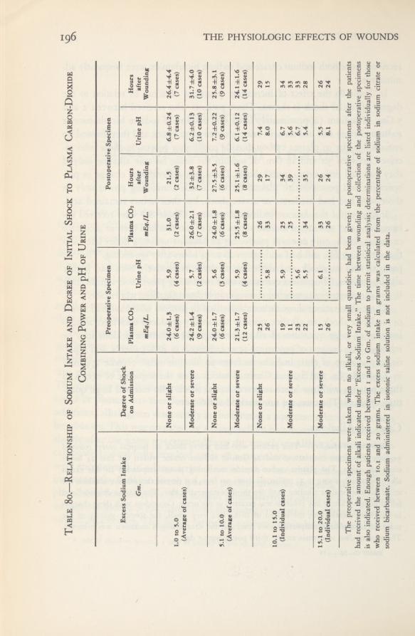
196 THE PHYSIOLOGIC EFFECTS OF WOUNDS
Thepreoperative
specimensweretaken
whenno
alkali,orvery
smallquantities,
hadbeengiven;the
postoperativespecimens
afterthe
patients
hadreceivedthe
amountof
alkaliindicatedunder“Excess
SodiumIntake.”Thetime
betweenwoundingandcollectionofthe
postoperativespecimens
isalsoindicated.
Enoughpatients
receivedbetweeni
and10
Gm.ofsodiumto
permitstatistical
analysis;determinationsare
listedindividuallyforthose
whoreceivedbetween
10.1and
20
grams.The
excesssodiumintakein
gramswascalculatedfromthepercentageofsodiuminsodiumcitrateor
sodiumbicarbonate.
Sodiumadministeredinisotonicsalinesolutionisnotincludedinthedata.
Table8o.—Relationshipof
SodiumIntake
andDegree
ofInitialShockto
PlasmaCarbon-Dioxide
CombiningPower
andpH
ofUrine
PreoperativeSpecimen
PostoperativeSpecimen
ExcessSodiumIntake
DegreeofShock
onAdmission
PlasmaCO2
PlasmaCO2
Hours
Hours
Gm.
UrinepH
after
UrinepH
after
mEq./L.
mEq./L.Wounding
Wounding
Noneor
slight
24.0±1.35.9
31.0
21.5
6.8±0.2426.4±4.4
1.0to
5.0
(6cases)
(4cases)
(2
cases)
(2
cases)
(7cases)
(7
cases)
(Averageofcases)
Moderateorsevere
24.2±1.45.7
26.0±2.132±3.8
6.2±0.13
31.7±4.0
(9cases)
(2
cas'es)
(7
cases)
(7cases)
(10cases)(10cases)
Noneor
slight
24.0±1.75.6
24.0±1.827.5±3.57.2
±0.2225.8±3.1
5.1to10.0
(6cases)
(3
cases)
(6cases)
(6cases)
(9cases)
(9cases)
(Averageofcases)
Moderateorsevere
21.3±1.75.9
25.5±1.825.1±1.6
6.1±0.1224.1
±1.6
(12cases)
(4cases)
(8cases)
(8
cases)
(14cases)(14cases)
Noneor
slight
25
26
29
7,4
29
26
5.8
33
17
8.0
15
10.1to1
S.O (Individualcases)
19
5.9
25
34
6.7
34
Moderateorsevere11
25
39
5.6
33
23
5.6
6.7
33
22
5.5
34
35
5.4
28
15.1to20.0
Moderateorsevere15
6.1
33
26
5.5
26
(Individualcases)
26
26
24
8.1
24

ALKALIS IN TRAUMATIC SHOCK 197
Table 81.—Changes in Acid-Base Metabolism and Coincident Findingsin a Patient Who Received Excess Alkali
Severe Initial Shock and Subsequent Death from Posttraumatic Renal Insufficiency
Case 112 (Table 82).—Twenty Gm. of sodium bicarbonate and 16 Gm. of sodiumcitrate were administered within 6 hours after this patient entered the hospital in severeshock. The pH of the venous blood and the plasma carbon-dioxide combining powerpromptly rose to levels indicative of moderate alkalosis, but the urine did not becomealkaline until nearly 24 hours after the alkali had been administered. Although urinaryoutput was normal after the first day, the plasma nonprotein nitrogen level had risen to
67 mg. per 100 cc. by the second postoperative day, when the patient was evacuated.Plasma chloride levels fell and chloride excretion was low. Subsequent follow-up revealedthat nitrogen retention persisted for 6 days; the status of the acid-base metabolism couldnot be followed.
Case loy (Table 83 and Chart 28).—This patient, admitted in moderate shock, received35 Gm. of sodium bicarbonate and 28 Gm. of sodium citrate on the day of operation, and15 additional grams of sodium bicarbonate early on the first postoperative day. The result-
ing severe and prolonged alkalosis is evident in the high plasma carbon-dioxide combining
Case 108
Parenteral IntakePlasma Plasma Urine Urine
Period CO* Cl NaClNa Cl pH
mEq. mEq. mEq./L. mEq./L. Gm.
Preoperative 811 300 22 100 5.5 0.04(18-hr. period)
PostoperativeDayFirst 417 300 34 97 5.4 0.26Second 115 75 30 96 5.4 ——
Third 23 91 7.6 —
Parenteral Intake UrinePlasma Urea Urine
PeriodNa Cl
NPN Nitrogen Gravity Output
mEq. mEq. mg.perlOOcc. Gm. cc.
Preoperative 811 300 37 0.13 1.032 100(18-hr. period)
Postoperative DayFirst 417 300 77 0.54 1.014 185Second 115 75 113 1.012 155Third 170 —

198 THE PHYSIOLOGIC EFFECTS OF WOUNDS
Table 82.—Changes in Acid-Base Metabolism and Coincident Findings in aPatient who Received Excess Alkali
Severe Initial Shock, Subsequent Nitrogen Retention, and Recovery
Table 83.—Changes in Acid-Base Metabolism and Coincident Findings in aPatient who Received Excess Alkali
Postoperative Alkalosis and Azotemia, Resulting in Death
power and blood pH. Considerable ammonium chloride was given on the fourth and fifthpostoperative days in an unsuccessful attempt to relieve the alkalosis. In this case also therewas a marked lag in the production of an alkaline urine after metabolic alkalosis appeared.By the fourth postoperative day, although severe alkalosis persisted, the patient was nolonger able to excrete an alkaline urine. Plasma chlorides fell to phenomenally low levels
Case 112
Parenteral Blood Plasma Plasma Urine Urine Plasma Urine UrineIntake pH co 2 Cl pH NaCl NPN Specific Output
Period (venous) GravityNa Cl mEqJL. mEq./L. Gm. mg. per CC,
mEq. mEq. 100 cc.
Preoperative . . . 400 0 7.04 to 20 to 99 5.4 to 40 1.025 530(21.5-hr. period) 7.45 30 6.5
Postoperative DayFirst 0 0 7.45 32 86 7.1 0.43 53 1.010 2,180Second .... 0 0 36 87 7.2 1.4 67 1.010 1,945
5734
Case ioy
ParenteralIntake Blood Serum Plasma Plasma Urine Urine Plasma Urine
Period pH Na CO2 Cl pH NaCl NPN Output(venous)
Na Cl rnEq./L. mEq./L. mEq.IL. (n:7.38- Gm. mg. per CC,tnEq. mEq. (n:l42) (n:27) (n:103) 7.48) 100 cc.
681 0 15 103 6.1 27 60(18-hr. period)
Postoperative Day324 146 160 30.2 too 5.5 to 0.3 41 1,560
7.3Second .... 0 0 7.44 170 41 91 8.2 0.23 46 1,570
146 146 7.7 0.80 1,540332 482 7.48 160 47 73 6.8 91 875
Fifth 0 245 7.52 140 43 65 5.8 0.0 73 80060 0 37 56 7.3 152 200(?)
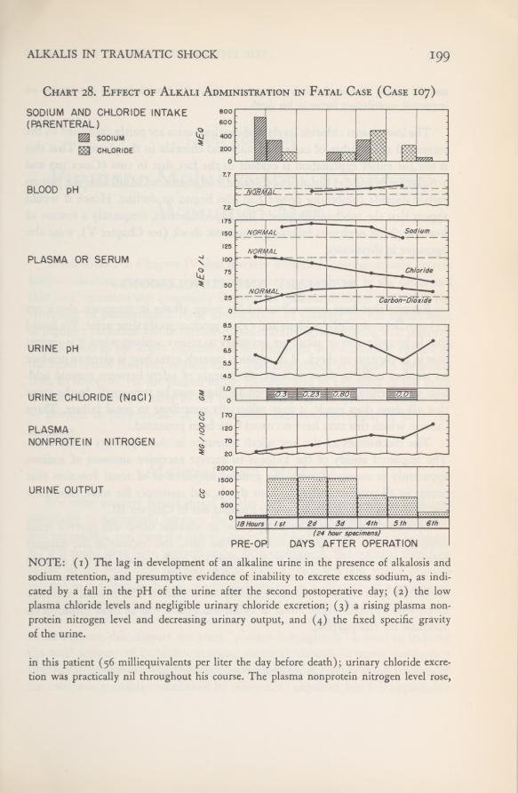
ALKALIS IN TRAUMATIC SHOCK 199
Chart 28. Effect of Alkali Administration in Fatal Case (Case 107)SODIUM AND CHLORIDE INTAKE(PARENTERAL)
SODIUMCHLORIDE
BLOOD pH
PLASMA OR SERUM
URINE pH
URINE CHLORIDE (NaCI)
PLASMANONPROTEIN NITROGEN
URINE OUTPUT
NOTE: (1) The lag in development of an alkaline urine in the presence of alkalosis andsodium retention, and presumptive evidence of inability to excrete excess sodium, as indi-cated by a fall in the pH of the urine after the second postoperative day; (2) the lowplasma chloride levels and negligible urinary chloride excretion; (3) a rising plasma non-protein nitrogen level and decreasing urinary output, and (4) the fixed specific gravityof the urine.
in this patient (56 milliequivalents per liter the day before death); urinary chloride excre-tion was practically nil throughout his course. The plasma nonprotein nitrogen level rose,
THE PHYSIOLOGIC EFFECTS OF WOUNDS200
and the patient died on the sixth postoperative day, with renal failure in our opinion animportant contributory factor in his death.
The low plasma chloride levels in these three cases are partly explained by thereciprocal relationship of carbon dioxide and chloride in the plasma. That thisis not the entire explanation is evident in the fact that in two (Cases 107 and108, Tables 81 and 83) the plasma chloride level continued to fall after the plasma
carbon-dioxide combining power had also begun to decline. Hence it wouldappear that the mechanisms producing hypochloremia, frequently a feature oflower nephron nephrosis following traumatic shock (see Chapter V), were alsooperative in these cases.
SUMMARY AND CONCLUSIONS
Reasons that have been advanced for using alkalis in traumatic shock are(i) to relieve metabolic acidosis and (2) to produce an alkaline urine. We foundthat large quantities of alkali are necessary to relieve acidosis when it is severe;that is, in patients in shock. If in addition enough extra base is given to producean alkaline urine in these patients, the margin of safety between normal acid-base equilibrium and uncompensated alkalosis may be very small. In the eventthat alkalosis does result, it may materially contribute to renal failure. Threecases in which this may have occurred have been presented.
The mechanism of the low alkali tolerance in these patients is not clear.The impaired ability of the kidneys to excrete excessive amounts of sodiumapparently is associated with the general impairment of renal function thatoccurs in all patients suffering from shock, and continues for some time afterthe shock has been relieved, as was brought out in Chapter III.
Because the evidence is so meager that an alkaline urine will prevent renalcomplications in the type of patient studied here, and because of the dangersinherent in trying to produce such a urine, the use of alkalis for this purposeis not recommended. Small quantities of alkali sufficient to relieve metabolicacidosis, if judiciously employed, probably are advisable. The amount routinelygiven with citrated whole blood or blood substitutes will in most instances beadequate for this purpose.

CHAPTER VIII
Pigment Mobilization in SeverelyWounded Men
It was shown in Chapter IV that severely wounded men commonly excreteheme-containing pigments, either hemoglobin or myoglobin, in the urine andthat such excretion was constantly observed in patients destined to develop thesyndrome of posttraumatic renal insufficiency. In Chapter IX the significanceof pigment excretion will be further emphasized by demonstrating that pigmentprecipitation in the lower segments of the nephrons was a constant feature ofthe renal lesion observed in fatal cases. The importance of pigment mobiliza-tion, its transport via the plasma to the kidneys, and its excretion in the urinewas therefore obvious early in our investigations, and the present chapter willbe devoted to presentation of the available data and discussion of their sig-nificance.
Materials and MethodsWe were severely handicapped by the lack of a spectroscope of sufficient
sensitivity to distinguish between the closely similar spectrums of hemoglobinand myoglobin, and even if a suitable instrument had been available it is doubt-ful if it could have been utilized under field conditions. We resorted thereforeto use of the benzidine method, which gave a color reaction strong enoughto read in a Coleman Junior spectrophotometer in a dilution of i milligramof benzidine-positive material per 100 cubic centimeters. The method did not,however, permit distinction between myoglobin and hemoglobin in plasma,so throughout this chapter the term “plasma hemoglobin” is used to indicatethe total amount of benzidine-reacting material, though in many instances asignificant proportion was undoubtedly myoglobin. A further small fraction ofthe total was probably contributed by nonspecific oxydases, but it is improbable
THE PHYSIOLOGIC EFFECTS OF WOUNDS202
that this was large enough to affect comparative results. Under these conditions,results of the test were always positive for “hemoglobin,” ranging from 6 to 12mg, per 100 cc. in the majority of initial samples and also in samples procuredduring convalescence. We therefore considered values up to 12 mg. per 100 cc.as being within normal range.
In the urine it did prove possible to distinguish between the two pigmentswithin certain limits by taking advantage of the greater stability of myoglobinin alkaline solutions. By cautiously alkalinizing the urine, hemoglobin couldbe converted to alkaline hematin and precipitated while the greater portion ofthe myoglobin remained in solution. Tests with known mixtures of the two
pigments showed that the separation was approximately 80-percent effective,and analyses of normal and decolorized muscles from patients who died of thecrush syndrome provided further confirmation of the validity of the method.When enough pigment for quantitative determination was present in a urinespecimen, this method was applied. Values of “myoglobin” that were less than20 percent of the hemoglobin present were considered negative; findings of20 percent or more were recorded as myoglobin. Details of the methods usedwill be found in Appendix C.
Determinations of plasma “hemoglobin” and of urine hemoglobin and myo-globin were made routinely upon all patients in the series from whom suitablespecimens could be obtained. In some instances high plasma values were un-doubtedly due to hemolysis incurred in taking samples of blood. Great care wasused in cleaning and drying needles and syringes and in the technique of vene-puncture, but needles and syringes were not paraffinized. In certain patientsinjury to the kidney or other portion of the urinary tract resulted in gross hema-turia, and urine hemoglobin levels in these cases were considered valueless; how-ever, if myoglobin was discovered in such specimens, it was recorded.
Bank BloodSince hemolysis is inevitable in stored blood, many samples of bank blood,
taken at the time the blood was given out for transfusion to our patients, wereanalyzed. The plasma hemoglobin and the blood sugar levels were determined,the latter in order to check upon the effectiveness of added dextrose as a pre-servative, Confirmation of the serologic blood group of the bank blood was notattempted because the triple check used in the theater blood bank was as ac-

PIGMENT MOBILIZATION 203
curate as any method we could have tried. We did, however, take the precau-tion of checking the blood group of the recipient whenever possible since the“dog tags” were known to be approximately 8-percent erroneous, and since thegrouping was often too difficult for hospital technicians because of the largequantities of group O blood that some patients had already received before thefirst test-specimen was obtained.
Few Rh factor determinations were made because very little typing serumwas available. All patients, however, who were conscious at the time of obser-vation were carefully questioned regarding previous transfusion and answerswere uniformly negative. Previous sensitization was therefore improbable, andthe short duration of the period of transfusion therapy—from i to 3 days—-made active sensitization of no importance in our problem.
Specifications of Ban\ Blood.—Blood given to patients in this study wasderived from the theater blood bank in all but two instances. 1 All bank bloodwas group O and the plasma agglutinins against A and B cells had been de-termined. If these were below 1:64, the blood was labeled Universal Donor; ifabove this level, O-Blood, for Use in O-Recipients Only. The blood had beendrawn into sodium citrate, and dextrose had been added to give a concentrationapproximating 0.3 percent. Blood was stored at all times, including periods oftransportation, in refrigerators at approximately 8° Centigrade. It was neverused after the tenth day.
Analyses of Ban\ Blood. —Since all blood samples contain cells of varyingmaturity, progressive hemolysis is inevitable in all stored specimens. This isapparent in Chart 29 and Table 84 which show average plasma hemoglobinlevels at the time of transfusion in 213 samples of blood from 2 to 10 days old.The rate compares favorably with Gibson’s2 figures based on storage in A-C-Dsolution. In occasional samples, usually those from 6 to 8 days old, considerablehemolysis was evidenced by plasma hemoglobin levels from 100 to 300 mg. per
1 Case 9 was an example of a transfusion accident—A blood to an O recipient. The patient was seenin consultation at a neighboring hospital and was studied for comparison only. The second patient (Case 22)belonged to group A. He received three transfusions of bank blood in a forward hospital and subsequentlywas given 2 units of group A blood at a general hospital. He had already given evidence of renal insufficiencybefore the type-specific blood was administered.
2 Gibson, J. G., 2nd; Evans, R. D.; Aub, J. C.; Sack, T., and Peacock, W. C.: The post-transfusionsurvival of preserved human erythrocytes stored as whole blood or in resuspension, after removal ofplasma, by means of two isotopes of radioactive iron. J. Clin. Investigation 26: 715-738, July 1947.
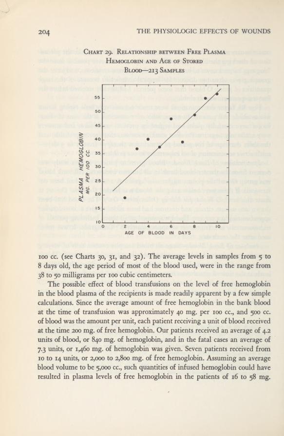
THE PHYSIOLOGIC EFFECTS OF WOUNDS204
Chart 29. Relationship between Free PlasmaHemoglobin and Age of Stored
Blood—213 Samples
ioo cc. (see Charts 30, 31, and 32). The average levels in samples from 5 to8 days old, the age period of most of the blood used, were in the range from38 to 50 milligrams per 100 cubic centimeters.
The possible effect of blood transfusions on the level of free hemoglobinin the blood plasma of the recipients is made readily apparent by a few simplecalculations. Since the average amount of free hemoglobin in the bank bloodat the time of transfusion was approximately 40 mg. per 100 cc., and 500 cc.of blood was the amount per unit, each patient receiving a unit of blood receivedat the time 200 mg. of free hemoglobin. Our patients received an average of 4.2units of blood, or 840 mg. of hemoglobin, and in the fatal cases an average of7.3 units, or 1,460 mg. of hemoglobin was given. Seven patients received from10 to 14 units, or 2,000 to 2,800 mg. of free hemoglobin. Assuming an averageblood volume to be 5,000 cc., such quantities of infused hemoglobin could haveresulted in plasma levels of free hemoglobin in the patients of 16 to 58 mg.
PIGMENT MOBILIZATION 205
per 100 cubic centimeters. It must not be forgotten, however, that these approxi-mations are based on the assumption that all infused free hemoglobin wouldremain in the circulating plasma. This would certainly not be true, since inmany patients significant quantities would be excreted in the urine and in allinstances some would escape with the plasma into traumatized tissues.
Table 84.—Relationship of Average Plasma Hemoglobin Level to Age of
Stored Blood—213 Analyses
Charts 30 through 33 show the relationship between hemolysis and sugarlevels in the stored blood. It is evident that most instances of extreme hemolysisoccurred in blood samples with low sugar levels. Table 85 and Chart 33 showaverage sugar levels in relationship to age of the blood. The chart shows thatthe sugar level was well maintained until the seventh day and dropped sharplythereafter.
Table 85.—Relationship of Average Blood Sugar to Age of Stored Blood—-223 Samples
Age of Stored Blood
days
Number ofAnalyses
Mean PlasmaHemoglobin
mg. per 100 cc.
Age ofStored Blood
days
Number ofAnalyses
Mean PlasmaHemoglobin
mg. per 100 cc.
2 2 19 7 56 39.6±5.83 15 36.7 ±6.6 8 39 49.5±104 10 40±6.5 9 6 555 30 38±8 10 3 57
6 52 47.4±8 Total 213 Av.: 43.4
Number of Mean Age of Number of MeanAge of Stored Blood Analyses Blood Sugar Stored Blood Analyses Blood Sugar
days mg. per 100 cc. days mg. per 100 cc.
2 2 256 7 60 289±173 15 295±33 8 40 266±254 11 276± 59 9 6 2115 31 326±34 10 3 160
6 55 290±21 Total 223 Av.: 286

206 THE PHYSIOLOGIC EFFECTS OF WOUNDS
CHART 30. Relationship between PlasmaHemoglobin and Blood Sugar in Blood
Stored 6 Days—52 Samples
CHART 31. Relationship between PlasmaHemoglobin and Blood Sugar in Blood
Stored 7 Days
CHART 32. Relationship between PlasmaHemoglobin and Blood Sugar in Blood
Stored 8 Days
CHART 33. Relationship between BloodSugar and Age of Stored Blood—223
Samples

PIGMENT MOBILIZATION 207
Plasma Hemoglobin” Levels in Wounded M.en
The pattern of plasma “hemoglobin” (benzidine-reacting material) levels inseverely traumatized men is shown in Charts 34 through 37. In Chart 34, basedupon findings in 133 patients, the mean plasma hemoglobin level for each dayafter injury is plotted. From an initial figure of 10 mg. per 100 cc., it climbs in72 hours to 17.3 mg., then slowly falls in the course of the next 13 days to alevel of 3 mg. per 100 cc. of plasma. Chart 35 shows daily mean levels of 64patients who had high azotemia, over half of whom died. The curve is essen-tially similar, though the peak is higher and is delayed 24 hours. In Chart 36only the fatal cases with histologically proved pigment nephropathy are shown,exclusive of crush cases. In this group the peak is sharp and high, occurring at
24 hours and attaining a maximum of 36.8 mg. per 100 cubic centimeters. Thisfigure is still far below the generally accepted threshold level of 135 mg. per100 cc. at which a normal kidney begins to excrete hemoglobin.3 4 5
The plasma bilirubin level as determined by the van den Bergh diazo testhas also been plotted on these charts, omitting cases of direct liver trauma. Aswould be expected, the bilirubin curve roughly paralleled that of hemoglobinbut its peak occurred from 3 to 4 days later. Strict parallelism could not be ex-pected since the bilirubin level is also affected by efficiency of liver function;evidence of liver injury in these patients is presented in Chapters II and XII.
In considering the significance of the plasma hemoglobin levels, two possiblerelationships at once come to mind: does the level depend upon the severity ofshock? or upon the quantity of blood the patient was given? The answer isnot obvious since the patients with the most severe shock usually received themost blood. Available evidence compiled from the first specimen of blood ob-tained for analysis is presented in Table 86. The following cases have beenexcluded: all crush cases, in which benzidine-reacting pigment in the plasma
3 Gilligan, D. R., and Blumgart, H. L.: March hemoglobinuria; studies of clinical characteristics,blood metabolism and mechanism, with observations on three new cases, and review of the literature.Medicine 20: 341-395, September 1941.
4 Gilligan, D. R.; Altschule, M. D., and Katersky, E. M.: Studies of hemoglobinemia and hemo-globinuria produced in man by intravenous injection of hemoglobin solutions. J. Clin. Investigation 20:177-187, March 1941.
5 Ottenberg, R., and Fox, C. L., Jr.: Rate of removal of hemoglobin from circulation and its renalthreshold in human beings. Am. J. Physiol. 123: 516-525, August 1938.
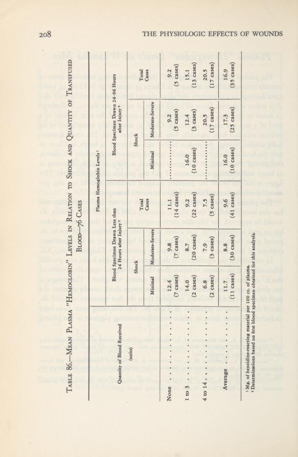
208 THE PHYSIOLOGIC EFFECTS OF WOUNDS
Table86.—Mean
Plasma“Hemoglobin”
Levelsin
Relationto
Shockand
Quantityof
Transfused
Blood—
76Cases Plasma
HemoglobinLevels1
Quantityof
BloodReceived
BloodSpecimen
DrawnLessthan
24HoursafterInjury2
BloodSpecimen
Drawn24-96Hours
afterInjury2
(units)
Shock
Total
Shock
Total
MinimalModerate-Severe
Cases
MinimalModerate-Severe
Cases
None
12.4
9.8(7cases)
11.1 (14cases)
9.2(5cases)
9.2(5cases)
(7cases)
1to3
14.0(2cases)
8.7(20cases)
9.2(22cases)
16.0 (10cases)
12.4(3cases)
15.1 (13cases)
4to14
6.8
7.9(3cases)
7.5(5cases)
20.5
20.5 (17cases)
(2
cases)
(17cases)
Average
11.7(11cases)
8.8(30cases)
9.6(41cases)
16.0 (10cases)
17.3 (25cases)
16.9(35cases)
1
Mg.of
benzidine-reactingmaterial
per100
cc.of
plasma.
*
Determinationsbased
onfirstbloodspecimenobtainedfor
thisanalysis.
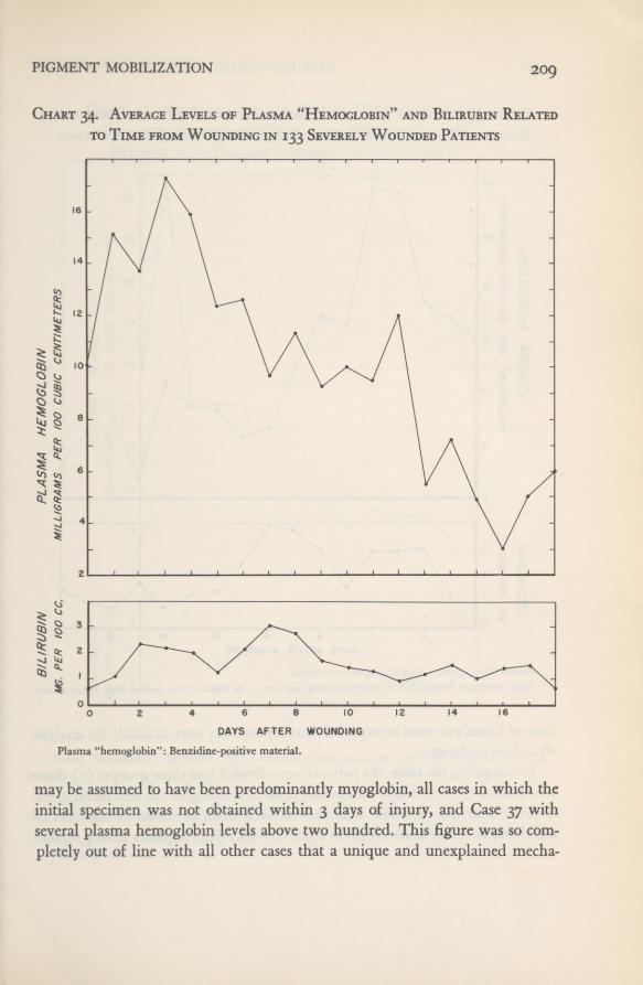
PIGMENT MOBILIZATION 209
Chart 34. Average Levels of Plasma “Hemoglobin” and Bilirubin Relatedto Time from Wounding in 133 Severely Wounded Patients
Plasma “hemoglobin”: Benzidine-positive material
may be assumed to have been predominantly myoglobin, all cases in which theinitial specimen was not obtained within 3 days of injury, and Case 37 withseveral plasma hemoglobin levels above two hundred. This figure was so com-pletely out of line with all other cases that a unique and unexplained mecha-
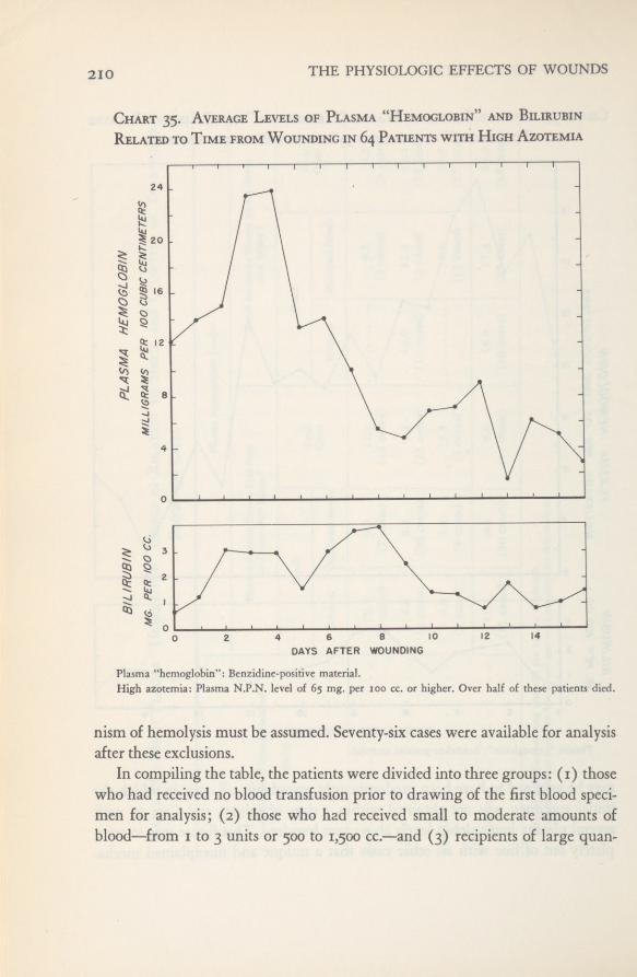
THE PHYSIOLOGIC EFFECTS OF WOUNDS210
Chart 35. Average Levels of Plasma “Hemoglobin” and BilirubinRelated to Time from Wounding in 64 Patients with High Azotemia
Plasma “hemoglobin”; Benzidine-positive material.High azotemia: Plasma N.P.N. level of 65 mg. per too cc. or higher. Over half of these patients died.
nism of hemolysis must be assumed. Seventy-six cases were available for analysisafter these exclusions.
In compiling the table, the patients were divided into three groups: (i) thosewho had received no blood transfusion prior to drawing of the first blood speci-men for analysis; (2) those who had received small to moderate amounts ofblood—from 1 to 3 units or 500 to 1,500 cc.—and (3) recipients of large quan-

PIGMENT MOBILIZATION 211
Chart 36. Average Levels of Plasma “Hemoglobin” and
Bilirubin Related to Time from Wounding in 33Patients with Pigment Nephropathy at Necropsy
Plasma “hemoglobin”: Benzidine-positive material. Cases 9, 41, 85, 115, 116, 132, and A-30 areomitted.
titles of blood—from 4 to 14 units or 2 to 7 liters. Shock had been estimatedin three grades of severity, and there were a few patients without shock. The
956507 0—52 15
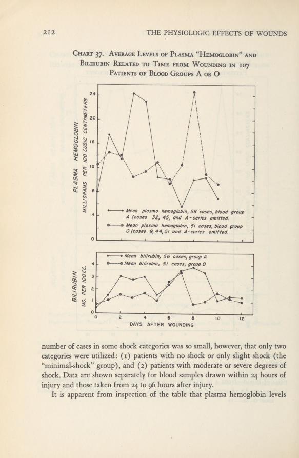
212 THE PHYSIOLOGIC EFFECTS OF WOUNDS
Chart 37. Average Levels of Plasma “Hemoglobin” andBilirubin Related to Time from Wounding in 107
Patients of Blood Groups A or O
VMean plasma hemoglobin, 56 cases, blood groupA (cases 32, 45, and A-series omitted.Mean plasma hemoglobin, 5/ cases, blood groupO (cases 9,44,5! and A-series omitted.
• Mean bilirubin, 56 cases, group A® Mean bilirubin, 5/ cases, group O
number of cases in some shock categories was so small, however, that only twocategories were utilized: (i) patients with no shock or only slight shock (the“minimal-shock” group), and (2) patients with moderate or severe degrees ofshock. Data are shown separately for blood samples drawn within 24 hours ofinjury and those taken from 24 to 96 hours after injury.
It is apparent from inspection of the table that plasma hemoglobin levels
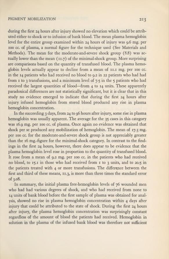
PIGMENT MOBILIZATION 2I 3
during the first 24 hours after injury showed no elevation which could be attrib-uted either to shock or to infusion of bank blood. The mean plasma hemoglobinlevel for the entire group examined within 24 hours of injury was 9.6 mg. per100 cc. of plasma, a normal figure for the technique used (See Materials andMethods). The mean for the moderate-and-severe shock group (8.8) was ac-tually lower than the mean (11.7) of the minimal-shock group. More surprisingare comparisons based on the quantity of transfused blood. The plasma hemo-globin levels actually appear to decline from a mean of 11.1 mg. per 100 cc.in the 14 patients who had received no blood to 9.2 in 22 patients who had hadfrom 1 to 3 transfusions, and a minimum level of 7.5 in the 5 patients who hadreceived the largest quantities of blood—from 4 to 14 units. These apparentlyparadoxical differences are not statistically significant, but it is clear that in thisstudy no evidence emerged to indicate that during the first 24 hours afterinjury infused hemoglobin from stored blood produced any rise in plasmahemoglobin concentration.
In the succeeding 3 days, from 24 to 96 hours after injury, some rise in plasmahemoglobin was usually apparent. The average for the 35 cases in this categorywas 16.9 mg. per 100 cc. of plasma. Once again no evidence was obtained thatshock per se produced any mobilization of hemoglobin. The mean of 17.3 mg.per 100 cc. for the moderate-and-severe shock group is not appreciably greaterthan the 16 mg.-figure for the minimal-shock category. In contrast to the find-ings in the first 24 hours, however, there does appear to be evidence that theplasma hemoglobin level rose in proportion to the quantity of transfused blood.It rose from a mean of 9.2 mg. per 100 cc. in the patients who had receivedno blood, to 15. 1 in those who had received from 1 to 3 units, and to 20.5 inthe patients treated with 4 or more transfusions. The difference between thefirst and third of these means, 11.3, is more than three times the standard errorof 3.08.
In summary, the initial plasma free-hemoglobin levels of 76 wounded menwho had had various degrees of shock, and who had received from none to
14units of bank blood before the first sample of plasma was obtained for anal-ysis, showed no rise in plasma hemoglobin concentration within 4 days afterinjury that could be attributed to the state of shock. During the first 24 hoursafter injury, the plasma hemoglobin concentration was surprisingly constantregardless of the amount of blood the patients had received. Hemoglobin insolution in the plasma of the infused bank blood was therefore not sufficient
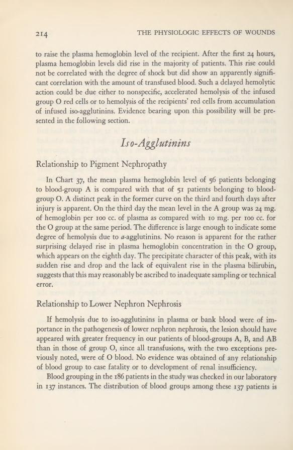
THE PHYSIOLOGIC EFFECTS OF WOUNDS214
to raise the plasma hemoglobin level of the recipient. After the first 24 hours,plasma hemoglobin levels did rise in the majority of patients. This rise couldnot be correlated with the degree of shock but did show an apparently signifi-cant correlation with the amount of transfused blood. Such a delayed hemolyticaction could be due either to nonspecific, accelerated hemolysis of the infusedgroup O red cells or to hemolysis of the recipients’ red cells from accumulationof infused iso-agglutinins. Evidence bearing upon this possibility will be pre-sented in the following section.
Is o-AgglutininsRelationship to Pigment Nephropathy
In Chart 37, the mean plasma hemoglobin level of 56 patients belongingto blood-group A is compared with that of 51 patients belonging to blood-group O. A distinct peak in the former curve on the third and fourth days afterinjury is apparent. On the third day the mean level in the A group was 24 mg.of hemoglobin per 100 cc. of plasma as compared with 10 mg. per 100 cc. forthe O group at the same period. The difference is large enough to indicate somedegree of hemolysis due to No reason is apparent for the rathersurprising delayed rise in plasma hemoglobin concentration in the O group,which appears on the eighth day. The precipitate character of this peak, with itssudden rise and drop and the lack of equivalent rise in the plasma bilirubin,suggests that this may reasonably be ascribed to inadequate sampling or technicalerror.
Relationship to Lower Nephron NephrosisIf hemolysis due to iso-agglutinins in plasma or bank blood were of im-
portance in the pathogenesis of lower nephron nephrosis, the lesion should haveappeared with greater frequency in our patients of blood-groups A, B, and ABthan in those of group O, since all transfusions, with the two exceptions pre-viously noted, were of O blood. No evidence was obtained of any relationshipof blood group to case fatality or to development of renal insufficiency.
Blood grouping in the 186patients in the study was checked in our laboratoryin 137 instances. The distribution of blood groups among these 137 patients is
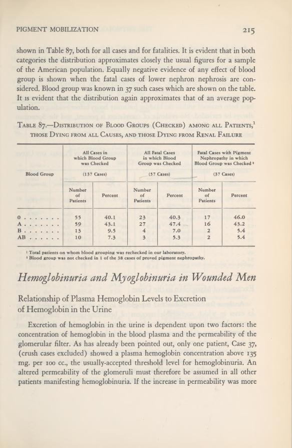
PIGMENT MOBILIZATION 215
shown in Table 87, both for all cases and for fatalities. It is evident that in bothcategories the distribution approximates closely the usual figures for a sampleof the American population. Equally negative evidence of any effect of bloodgroup is shown when the fatal cases of lower nephron nephrosis are con-sidered. Blood group was known in 37 such cases which are shown on the table.It is evident that the distribution again approximates that of an average pop-ulation.
Table 87.—Distribution of Blood Groups (Checked) among all Patients, 1
those Dying from all Causes, and those Dying from Renal Failure
Hemoglobinuria and Myoglobinuria in Wounded Men
Relationship of Plasma Hemoglobin Levels to Excretionof Hemoglobin in the Urine
Excretion of hemoglobin in the urine is dependent upon two factors: theconcentration of hemoglobin in the blood plasma and the permeability of theglomerular filter. As has already been pointed out, only one patient, Case 37,(crush cases excluded) showed a plasma hemoglobin concentration above 135mg. per 100 cc., the usually-accepted threshold level for hemoglobinuria. Analtered permeability of the glomeruli must therefore be assumed in all otherpatients manifesting hemoglobinuria. If the increase in permeability was more
All Cases inwhich Blood Group
was Checked
All Fatal Casesin which Blood
Group was Checked
Fatal Cases with PigmentNephropathy in which
Blood Group was Checked 2
Blood Group (137 Cases) (57 Cases) (37 Cases)
Numberof
PatientsPercent
Numberof
PatientsPercent
Numberof
PatientsPercent
0 55 40.1 23 40.3 17 46.0A 59 43.1 27 47.4 16 43.2B 13 9.5 4 7.0 2 5.4AB 10 7.3 3 5.3 2 5.4
1 Total patients on whom blood grouping was rechecked in our laboratory.2 Blood group was not checked in 1 of the 38 cases of proved pigment nephropathy.
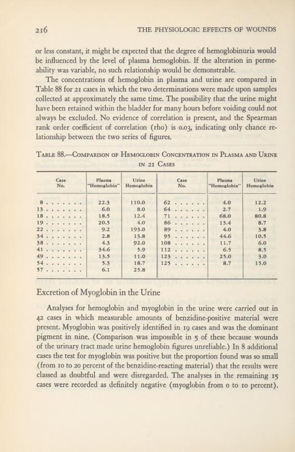
216 THE PHYSIOLOGIC EFFECTS OF WOUNDS
or less constant, it might be expected that the degree of hemoglobinuria wouldbe influenced by the level of plasma hemoglobin. If the alteration in perme-ability was variable, no such relationship would be demonstrable.
The concentrations of hemoglobin in plasma and urine are compared inTable 88 for 21 cases in which the two determinations were made upon samplescollected at approximately the same time. The possibility that the urine mighthave been retained within the bladder for many hours before voiding could notalways be excluded. No evidence of correlation is present, and the Spearmanrank order coefficient of correlation (rho) is 0.03, indicating only chance re-lationship between the two series of figures.
Table 88.—Comparison of Hemoglobin Concentration in Plasma and Urinein 21 Cases
Excretion of Myoglobin in the Urine
Analyses for hemoglobin and myoglobin in the urine were carried out in42 cases in which measurable amounts of benzidine-positive material werepresent. Myoglobin was positively identified in 19 cases and was the dominantpigment in nine. (Comparison was impossible in 5 of these because woundsof the urinary tract made urine hemoglobin figures unreliable.) In 8 additionalcases the test for myoglobin was positive but the proportion found was so small(from 10 to 20 percent of the benzidine-reacting material) that the results wereclassed as doubtful and were disregarded. The analyses in the remaining 15cases were recorded as definitely negative (myoglobin from 0 to 10 percent).
CaseNo.
Plasma"Hemoglobin”
UrineHemoglobin
CaseNo.
Plasma"Hemoglobin”
UrineHemoglobin
8 22.3 110.0 62 4.0 12.213 6.0 8.0 64 2.7 1.918 18.5 12.4 71 68.0 80.819 20.5 4.0 86 13.4 8.722 9.2 193.0 89 4.0 3.834 2.8 13.8 95 44.6 10.538 4.3 92.0 108 11.7 6.041 34.6 5.9 112 6.5 8.549 13.5 11.0 123 25.0 3.054 5.3 18.7 125 8.7 15.057 6.1 25.8
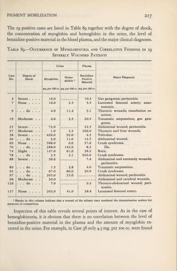
PIGMENT MOBILIZATION 217
The 19 positive cases are listed in Table 89 together with the degree of shock,the concentration of myoglobin and hemoglobin in the urine, the level ofbenzidine-positive material in the blood plasma, and the major clinical diagnoses.
Table 89.—Occurrence of Myoglobinuria and Correlative Findings in 19Severely Wounded Patients
Inspection of this table reveals several points of interest. As in the case ofhemoglobinuria, it is obvious that there is no correlation between the level ofbenzidine-positive material in the plasma and the amount of myoglobin ex-creted in the urine. For example, in Case 38 only 4.3 mg. per 100 cc. were found
Urine Plasma
CaseNo.
Degree ofShock Myoglobin Hemo-
globin *
Benzidine-PositiveMaterial
Major Diagnosis
mg.per 100 cc. mg.per 100 cc. mg. per 100 cc.
4 Severe .... 16.0 10.3 Gas gangrene; peritonitis.7 None .... 16.0 2.5 5.5 Lacerated femoral artery; anas-
tomosis.9 . . . do . . . 4.0 11.4 3.1 Thoracic wounds; transfusion re-
action.19 Moderate . . 4.0 3.5 20.5 Traumatic amputation; gas gan-
grene.25 Severe .... 72.0 24.5 Abdominal wound; peritonitis.37 Moderate . . 1.0 2.5 266.0 Thoracic and liver wounds.38 Severe .... 420.0 92.0 4.3 Volvulus.49 . . . do . . . 3.0 11.0 13-5 Abdominal wound.69 None .... 588.0 0.0 37.6 Crush syndrome.70 . . . do . . . 288.0 142.0 8.2 Do.74 Slight .... 147.0 61.0 38.2 Burn.78 . . . do . . . 2.5 2.1 920.0 Crush syndrome.88 Severe .... 36.6 7.4 Abdominal and extremity wounds;
peritonitis.89 . . . do . . . 1.5 3.8 4.0 Traumatic amputation.93 . . . do . . . 67.0 80.0 30.0 Crush syndrome.97 . . . do . . . 205.0 2 5.0 Abdominal wound; peritonitis.98
12630.0
7.0Abdominal and cerebral wounds.
. . . do . . . 9.3 Thoraco-abdominal wound; peri-tonitis.
137 None .... 203.0 41.0 38.4 Lacerated femoral artery.
• Blanks in this column indicate that a wound of the urinary tract rendered the determination useless forpurposes ofcomparison.
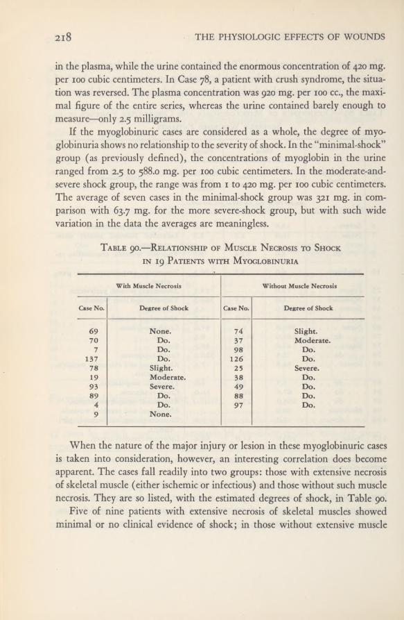
2l8 THE PHYSIOLOGIC EFFECTS OF WOUNDS
in the plasma, while the urine contained the enormous concentration of 420 mg.per 100 cubic centimeters. In Case 78, a patient with crush syndrome, the situa-tion was reversed. The plasma concentration was 920 mg. per 100 cc., the maxi-mal figure of the entire series, whereas the urine contained barely enough tomeasure—only 2.5 milligrams.
If the myoglobinuric cases are considered as a whole, the degree of myo-globinuria shows no relationship to the severity of shock. In the “minimal-shock”group (as previously defined), the concentrations of myoglobin in the urineranged from 2.5 to 588.0 mg. per 100 cubic centimeters. In the moderate-and-severe shock group, the range was from 1 to 420 mg. per 100 cubic centimeters.The average of seven cases in the minimal-shock group was 321 mg. in com-parison with 63.7 mg. for the more severe-shock group, but with such widevariation in the data the averages are meaningless.
Table 90.—Relationship of Muscle Necrosis to Shockin 19 Patients with Myoglobinuriawith Myoglobinuria
When the nature of the major injury or lesion in these myoglobinuric casesis taken into consideration, however, an interesting correlation does becomeapparent. The cases fall readily into two groups: those with extensive necrosisof skeletal muscle (either ischemic or infectious) and those without such musclenecrosis. They are so listed, with the estimated degrees of shock, in Table 90.
Five of nine patients with extensive necrosis of skeletal muscles showedminimal or no clinical evidence of shock; in those without extensive muscle
With Muscle Necrosis Without Muscle Necrosis
Case No. Degree ofShock Case No. Degree of Shock
69 None. 74 Slight.70 Do. 37 Moderate.
7 Do. 98 Do.137 Do. 126 Do.78 Slight. 25 Severe.19 Moderate. 38 Do.93 Severe. 49 Do.89 Do. 88 Do.
4 Do. 97 Do.9 None.

PIGMENT MOBILIZATION 219
necrosis, in contrast, eight were in the moderate-and-severe shock group andonly two in the minimal-shock group. One of the latter, Case 74, with extensiveburns showed sufficient hemoconcentration to suggest a more severe grade ofshock than was clinically apparent. These findings strongly suggest, on the onehand, that myoglobin is rarely liberated in the absence of shock unless there hasbeen extensive necrosis of skeletal muscle, and on the other, that in the presenceof severe shock, there is some mechanism for its mobilization other than musclenecrosis.
Relationship of Hemoglobinuria to MyoglobinuriaAttempts to correlate the variety of pigment excreted in the urine with
the type of injury or lesion provided extremely puzzling results, as may be seenfrom the tabulation to follow in which cases have been classified by the pre-dominant pigment, though many of them showed simultaneous excretion ofboth pigments. Because of the frequency of multiple injuries, the same case hasoften been included under more than one heading. Cases of trauma to theurinary tract have been excluded from the hemoglobinuric but not from themyoglobinuric category.
It is at once evident that any type of injury may be associated with eitherhemoglobinuria or myoglobinuria and that in most types, except the crush syn-drome and in clostridial myositis, the former is by far the more common. Itis noteworthy that in two typical crush cases no evidence of myoglobin excre-tion was obtained (see Chapter XI). Neither massive trauma to the extremitiesnor interruption of a major vessel, with consequent ischemic necrosis of muscle,
Type of injury or complicationNumber
predominantlyNumber
predominantly
Crush syndromehemoglobin
2myoglobin
3Wounds of extremity - I 9 5Major vascular interruption II 3Clostridial myositis I 2
Urinary tract injury — 2Liver injury 4 1Abdominal wound II 3Peritonitis 4 3Burn 0 1
Volvulus 0 1

THE PHYSIOLOGIC EFFECTS OF WOUNDS220
usually resulted in predominant myoglobinuria, and in two of the three pa-tients in whom it did appear, a successful arterial anastomosis had re-establishedcirculation of the leg before myoglobinuria was observed. In three patients (Case70, extensive but superficial burns; Case 38, volvulus, and Case 9, mismatchedtransfusion) injury of voluntary muscle can be absolutely excluded. In consid-ering the findings, however, it must be remembered first that our test for myo-globinuria was crude and results were recorded as positive only when consid-erable quantities of myoglobin were present, and second that myoglobin israpidly excreted in the absence of renal insufficiency and the loss of a singleurine specimen might cause a falsely negative result.
Table 91.—Greatest Concentrations 1 of Hemoglobin and Myoglobin
Observed in the Urine
One final comparison is of interest. In Table 91 the 12 patients showing thehighest concentrations of hemoglobin and the 12 with the highest myoglobinconcentrations are listed. As before, cases of trauma to the urinary tract havebeen eliminated from the hemoglobinuric category. It is apparent from thesefigures that myoglobinuria was frequently massive whereas hemoglobinuria wasrarely so when cases of direct trauma to the urinary tract are eliminated. Itis also noteworthy that four cases (Cases 38, 70, 74, and 93) appear in bothlists, suggesting that the conditions for the liberation of myoglobin and of hemo-globin may not be unrelated.
Case No.Hemoglobinuria
mg. per 100 cc.Case No.
Myoglobinuria
mg. per 100 cc.
22 193 69 588702 142 38 2 420
109 123 702 2888 110 97 205
56 110 137 203382 92 7 4 2 14771 81 25 7293 2 80 93 2 677 4 2 61 88 3772 58 98 30
118 38 7 16128 34 4 . 16
1 Highest of a number of determinations in each case.2 Cases that appear in both columns.
PIGMENT MOBILIZATION 221
SUMMARY
The concentration of benzidine-reacting heme pigment in the blood plasma(recorded as plasma hemoglobin) and the individual concentrations of hemo-globin and myoglobin in the urine were measured in our patients to determinethe extent of pigment mobilization. These data, together with determinationsof plasma hemoglobin in the bank blood administered to the patients, wereanalyzed in an attempt to determine the mechanism of pigment mobilizationin the body and of pigment excretion by the kidney.
It was found that the bank blood used in therapy was in a satisfactory stateof preservation, with an average of only 43 mg. of free hemoglobin per 100 cc.of plasma at the moment of utilization. Even in patients who received as manyas 10 to 14 transfusions, the plasma “hemoglobin” levels of the recipients showedin the first 24 hours no elevation which could be attributed to free hemoglobinin the transfused blood. Comparison of the plasma hemoglobin concentrationswith the degree of shock in 76 patients showed no evidence that shock itselfinduced any immediate increase in plasma hemoglobin concentration.
Twenty-four hours after injury, however, a progressive rise in plasma hemo-globin began to be apparent which reached a peak between 48 and 72 hoursand then slowly dropped to normal over a period of 2 weeks. This peak wasapproximately twice as high (36.8 mg. per 100 cc.) in 33 cases of fatal nephro-pathy as in the series as a whole (17.3 mg. per 100 cc.) and occurred only 24hours after wounding. No evidence was obtained that it was higher in patientswith severe shock than in those without shock or with only minimal shock. Itwas, however, significantly higher in patients who had received multiple trans-fusions than in those who had received no blood.
Evidence that this delayed rise in plasma hemoglobin was largely due toiso-agglutinins was afforded by comparison of group A and group O recipients.In 56 of the former, the mean plasma hemoglobin on the third day was 24 mg.per 100 cc., whereas in a sample of 51 group O recipients the correspondinglevel was only 10.2 milligrams. This difference was of no significance, how-ever, in the development of pigment nephropathy as shown by the percentagedistribution of blood groups among the nephropathies, which was essentiallyidentical with the distribution of blood groups in the entire series of casesstudied by the Board and with an average sampling of the American population.
In 21 cases in which plasma hemoglobin levels were obtained at approxi-
THE PHYSIOLOGIC EFFECTS OF WOUNDS222
mately the same time that the first urine specimen was voided, the concentra-tions of pigment in the two fluids were compared. No evidence of correlationwas found. It was concluded that the threshold of hemoglobin excretion inwounded men must vary over a wide range.
Myoglobin was positively identified in the urine of 19 patients in the seriesand these cases were subjected to special scrutiny. As in the case of hemoglobin,no correlation was found between the concentrations of benzidine-reacting pig-ment in the plasma and of myoglobin in the urine.
With the exception of clear-cut cases of the crush syndrome, excretion ofmyoglobin could not be predicted from the nature of the patient’s injury or itscomplications. It was rarely seen in wounds of the extremities, even those associ-ated with extensive necrosis of muscle, unless they were complicated by clos-tridial myositis, or, as in two cases, circulation was re-established after a periodof ischemia by a successful arterial anastomosis. It was sometimes very severe inpatients with little or no muscle damage. When muscle damage was extensive,the mobilization and excretion of myoglobin appeared to be independent of thedevelopment of shock. When there was insignificant or no muscle injury, myo-globinuria was rarely found in the absence of moderate or severe shock.
Two further observations are noteworthy though their significance is notapparent. Myoglobinuria was frequently massive, hemoglobinuria rarely so inthe absence of injury to the urinary tract. Myoglobinuria and hemoglobinuriaof significant degree frequently occurred in the same patient, suggesting a com-mon but undiscovered mechanism.
CONCLUSIONS
1. Neither the development of shock nor the therapeutic use of multipletransfusions of group O bank blood produced immediate elevation of theplasma “hemoglobin” levels in severely wounded men.
2. The delayed rise in mean plasma “hemoglobin” for the series as a wholein the period from 24 to 96 hours was largely attributable to iso-agglutinins inthe O bank blood, since it was absent in a sample of 51 O recipients.
3. Mean plasma “hemoglobin” concentrations were higher in the patients inwhom pigment nephrosis developed than in other wounded men but were stillfar below the threshold level at which the normal kidney excretes hemoglobin.

PIGMENT MOBILIZATION 223
A depression of the threshold for hemoglobin excretion must be assumed.4. The lack of correlation between concentrations of benzidine-reacting pig-
ment in the plasma and of hemoglobin or myoglobin in the urine suggests thatthis alteration of threshold was variable.
5. The irregularity with which extensive muscle injury was followed bymyoglobinuria indicates that some factor other than necrosis of muscle cellsis involved. This factor is not shock and appears to be the maintenance orre-establishment of the circulation in the involved muscles.
6. In a small number of cases severe myoglobinuria developed in the ab-sence of demonstrable muscle injury. The almost constant presence of moderateor severe shock in such cases suggests the possibility of diffuse ischemic injuryof muscle which is not morphologically recognizable.
7. The fact that severe myoglobinuria and severe hemoglobinuria were oftenobserved in the same patient suggests the possibility of a common mechanism.

CHAPTER IX
Pathology of the Kidneyin Traumatic Shock
The kidney lesion associated with the clinical syndrome ofrenal insufficiencyfollowing resuscitation from shock is constant but not pathognomonic. Lucke, 1
in a study of material from the Army Institute of Pathology, has shown that asimilar lesion, which he has called “lower nephron nephrosis,” develops in avariety of conditions such as mismatched blood transfusion, infusion of humanhemoglobin, the crush syndrome, heat stroke, blackwater fever, thermal burns,carbon tetrachloride poisoning, mushroom poisoning (Amanita phalloides ) andsulfonamide sensitivity. The author, from his own experience, can add to thelist of etiologic agents chemical burns, anaphylactic shock following thera-peutic use of antipneumococcus rabbit serum, and hemolytic reactions followingtransurethral resections in which the operative field is irrigated with tap water.Many years ago F. B. Mallory 2 noted the lesion in an occasional patient withhemolytic streptococcus infection. There can be little doubt that with increasingbreadth of experience many other causal agents will be brought to light.
Descriptions of the lesion in various stages may be found in innumerablecase reports under such titles as transfusion kidney,3 hemoglobinuric nephrosis,4
interstitial nephritis, 5 hepatorenal syndrome, 6 and other terms, but surveys of
1 Luck£, B.: Lower nephron nephrosis (renal lesions of crush syndrome, of burns, transfusions, andother conditions affecting lower segments of nephrons). Mil. Surgeon 99: 371-396, November 1946.
2 Mallory, F. B.; Personal communication.3 DeNavasquez, S.; Excretion of haemoglobin, with special reference to “transfusion” kidney. J. Path.
& Bact. 51: 413-425, November 1940.4 Mallory, T. B.: Hemoglobinuric nephrosis in traumatic shock. Am. J. Clin. Path. 17: 427-443,
June 1947.s Kimmelstiel, P.: Acute hematogenous interstitial nephritis. Am. J. Path. 1-4: 737-761, November
1938."Helwig, F. C., and Orr, T. G.: Traumatic necrosis of liver with extensive retention of creatinine and
high grade nephrosis. Arch. Surg. 24: 136-144, January 1932.

PATHOLOGY OF THE KIDNEY 225
any considerable mass of material are few. The most notable contributions arethose of Lucke 7 and of Bywaters and Dible 8 in their studies of the crush syn-drome. The present study is based primarily on a review of material from 60out of 63 necropsies on patients studied by the Board, but is fortified by experi-ence gained from review of more than 150 similar nephropathies at the 15thMedical General Laboratory. The records and material from the 60 cases, corre-lated with the clinical and biochemic data, provided the means for surveyingthe lesion in all stages of development as manifested in wounded soldiers resus-citated from shock by human plasma or whole blood transfusions.
Microscopic PathologyThe characteristic features of the lesion emphasized to a greater or lesser
degree by different authors are seven: pigment casts; internal hydronephrosis;degeneration of epithelial elements, particularly in the lower segments of thenephrons; rupture of tubules with extrusion of their content into the stroma;interstitial inflammation; granulomatous inflammation, and thrombophlebitisof small intrarenal veins. Let us examine them successively.
Pigment Casts
Pigment casts, as in other forms of lower nephron nephrosis, are the mostconspicuous feature of the “shock kidney.” They were present in all cases ofproved renal insufficiency in the present series in which the survival periodwas 3 days or longer. Their absence in many of the more acute cases is an inter-esting feature which will be considered later.
The pigment casts were found in the distal convoluted and collecting tubulesand in the ducts of Bellini, their relative frequency in these respective locationsvarying considerably from case to case. The number of nephrons involved wasdifficult to estimate inroutine sections, since only portions of each nephron werevisualized. In most cases with clinically demonstrated renal insufficiency, theproportion was obviously high but the range was wide, probably from 20 to80 percent.
' See footnote i.
®BywATERs, E. G. L., and Bible, J. H.: Renal lesion in traumatic anuria. J. Path. & Bact. 54: 111-120,January 1942.
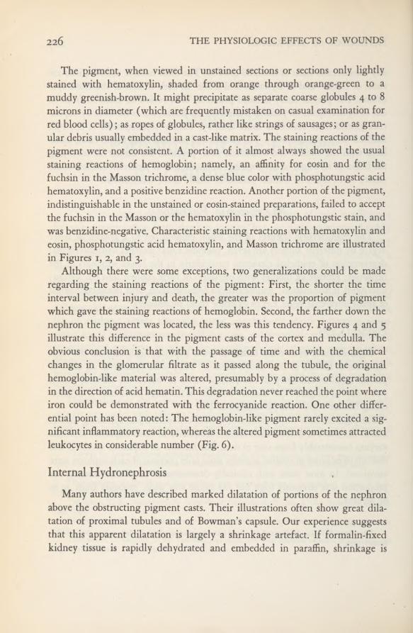
226 THE PHYSIOLOGIC EFFECTS OF WOUNDS
The pigment, when viewed in unstained sections or sections only lightlystained with hematoxylin, shaded from orange through orange-green to amuddy greenish-brown. It might precipitate as separate coarse globules 4 to 8microns in diameter (which are frequently mistaken on casual examination forred blood cells); as ropes of globules, rather like strings of sausages; or as gran-ular debris usually embedded in a cast-like matrix. The staining reactions of thepigment were not consistent. A portion of it almost always showed the usualstaining reactions of hemoglobin; namely, an affinity for eosin and for thefuchsin in the Masson trichrome, a dense blue color with phosphotungstic acidhematoxylin, and a positive benzidine reaction. Another portion of the pigment,indistinguishable in the unstained or eosin-stained preparations, failed to acceptthe fuchsin in the Masson or the hematoxylin in the phosphotungstic stain, andwas benzidine-negative. Characteristic staining reactions with hematoxylin andeosin, phosphotungstic acid hematoxylin, and Masson trichrome are illustratedin Figures 1, 2, and 3.
Although there were some exceptions, two generalizations could be maderegarding the staining reactions of the pigment: First, the shorter the timeinterval between injury and death, the greater was the proportion of pigmentwhich gave the staining reactions of hemoglobin. Second, the farther down thenephron the pigment was located, the less was this tendency. Figures 4 and 5illustrate this difference in the pigment casts of the cortex and medulla. Theobvious conclusion is that with the passage of time and with the chemicalchanges in the glomerular filtrate as it passed along the tubule, the originalhemoglobin-like material was altered, presumably by a process of degradationin the direction of acid hematin. This degradation never reached the point whereiron could be demonstrated with the ferrocyanide reaction. One other differ-ential point has been noted: The hemoglobin-like pigment rarely excited a sig-nificant inflammatory reaction, whereas the altered pigment sometimes attractedleukocytes in considerable number (Fig. 6).
Internal Hydronephrosis
Many authors have described marked dilatation of portions of the nephronabove the obstructing pigment casts. Their illustrations often show great dila-tation of proximal tubules and of Bowman’s capsule. Our experience suggeststhat this apparent dilatation is largely a shrinkage artefact. If formalin-fixedkidney tissue is rapidly dehydrated and embedded in paraffin, shrinkage is
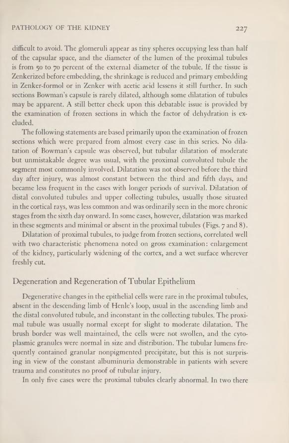
PATHOLOGY OF THE KIDNEY 227
difficult to avoid. The glomeruli appear as tiny spheres occupying less than halfof the capsular space, and the diameter of the lumen of the proximal tubulesis from 50 to 70 percent of the external diameter of the tubule. If the tissue isZenkerized before embedding, the shrinkage is reduced and primary embeddingin Zenker-formol or in Zenker with acetic acid lessens it still further. In suchsections Bowman's capsule is rarely dilated, although some dilatation of tubulesmay be apparent. A still better check upon this debatable issue is provided bythe examination of frozen sections in which the factor of dehydration is ex-cluded.
The following statements are based primarily upon the examination of frozensections which were prepared from almost every case in this series. No dila-tation of Bowman’s capsule was observed, but tubular dilatation of moderatebut unmistakable degree was usual, with the proximal convoluted tubule thesegment most commonly involved. Dilatation was not observed before the thirdday after injury, was almost constant between the third and fifth days, andbecame less frequent in the cases with longer periods of survival. Dilatation ofdistal convoluted tubules and upper collecting tubules, usually those situatedin the cortical rays, was less common and was ordinarily seen in the more chronicstages from the sixth day onward. In some cases, however, dilatation was markedin these segments and minimal or absent in the proximal tubules (Figs. 7 and 8).
Dilatation of proximal tubules, to judge from frozen sections, correlated wellwith two characteristic phenomena noted on gross examination: enlargementof the kidney, particularly widening of the cortex, and a wet surface whereverfreshly cut.
Degeneration and Regeneration of Tubular EpitheliumDegenerative changes in the epithelial cells were rare in the proximal tubules,
absent in the descending limb of Henle’s loop, usual in the ascending limb andthe distal convoluted tubule, and inconstant in the collecting tubules. The proxi-mal tubule was usually normal except for slight to moderate dilatation. Thebrush border was well maintained, the cells were not swollen, and the cyto-plasmic granules were normal in size and distribution. The tubular lumens fre-quently contained granular nonpigmented precipitate, but this is not surpris-ing in view of the constant albuminuria demonstrable in patients with severetrauma and constitutes no proof of tubular injury.
In only five cases were the proximal tubules clearly abnormal. In two there

Staining Reactions of Pigment Casts
FIGURE i. Stain: Hematoxylin and eosin.The glomerulus is normal. The distal con-
voluted tubules contain orange colored castsof precipitated hemoglobin. X 330
FIGURE 2. Stain: Masson trichrome. Thecasts stain deep red as do red cells with thisstain. X 330
FIGURE 3. Stain: Phosphotungstic acidhematoxylin. The deep blue staining of thepigment casts is characteristic of the reac-
tions of hemoglobin with this stain. Theproximal convoluted tubules are dilatedand contain precipitated albumin, but theepithelial cells show no degenerativechanges. X 330

Staining Reactions of Pigment Casts
FIGURE 4. Stain: Phosphotungstic acidhematoxylin. Renal cortex, low magnifica-tion, showing blue-staining casts in the dis-tal convoluted tubules. The proximal tu-bules are uniformly slightly dilated andcontain precipitated albumin but they areotherwise normal. X 80
FIGURE 5. Stain: Phosphotungstic acidhematoxylin. Section from the kidney pyra-mid of the case shown in Figure 4. Thispatient died 3 days after injury. A consider-able proportion of the pigment casts havelost theirability to stain blue with the phos-photungstic acid stain. X 80
FIGURE 6. Stain: Masson trichrome. Notethe acute exudation into the distal tubulesabout the masses of pigment which havebegun to lose their hemoglobin-like stain-ing reactions. X 200
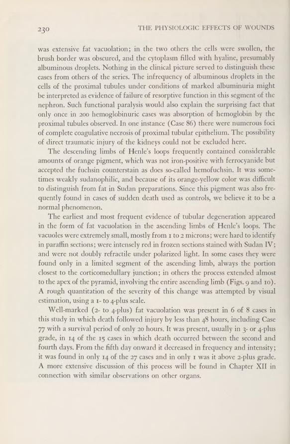
THE PHYSIOLOGIC EFFECTS OF WOUNDS23°
was extensive fat vacuolation; in the two others the cells were swollen, thebrush border was obscured, and the cytoplasm filled with hyaline, presumablyalbuminous droplets. Nothing in the clinical picture served to distinguish thesecases from others of the series. The infrequency of albuminous droplets in thecells of the proximal tubules under conditions of marked albuminuria mightbe interpreted as evidence of failure of resorptive function in this segment of thenephron. Such functional paralysis would also explain the surprising fact thatonly once in 200 hemoglobinuric cases was absorption of hemoglobin by theproximal tubules observed. In one instance (Case 86) there were numerous fociof complete coagulative necrosis of proximal tubular epithelium. The possibilityof direct traumatic injury of the kidneys could not be excluded here.
The descending limbs of Henle’s loops frequently contained considerableamounts of orange pigment, which was not iron-positive with ferrocyanide butaccepted the fuchsin counterstain as does so-called hemofuchsin. It was some-times weakly sudanophilic, and because of its orange-yellow color was difficultto distinguish from fat in Sudan preparations. Since this pigment was also fre-quently found in cases of sudden death used as controls, we believe it to be anormal phenomenon.
The earliest and most frequent evidence of tubular degeneration appearedin the form of fat vacuolation in the ascending limbs of Henle’s loops. Thevacuoles were extremely small, mostly from 1 to 2 microns; were hard to identifyin paraffin sections; were intensely red in frozen sections stained with Sudan IV;and were not doubly refractile under polarized light. In some cases they werefound only in a limited segment of the ascending limb, always the portionclosest to the corticomedullary junction; in others the process extended almostto the apex of the pyramid, involving the entire ascending limb (Figs. 9 and 10).A rough quantitation of the severity of this change was attempted by visualestimation, using a 1- to 4-plus scale.
Well-marked (2- to 4-plus) fat vacuolation was present in 6 of 8 cases inthis study in which death followed injury bv less than 48 hours, including Case77 with a survival period of only 20 hours. It was present, usually in 3- or 4-plusgrade, in 14 of the 15 cases in which death occurred between the second andfourth days. From the fifth day onward it decreased in frequency and intensity;it was found in only 14 of the 27 cases and in only 1 was it above 2-plus grade.A more extensive discussion of this process will be found in Chapter XII inconnection with similar observations on other organs.
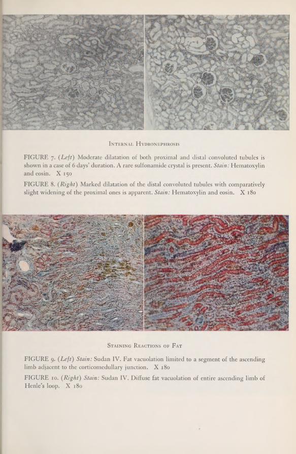
Internal Hydronephrosis
FIGURE 7. (Left) Moderate dilatation of both proximal and distal convoluted tubules isshown in a case of 6 days’ duration. A rare sulfonamidecrystal is present. Stain: Hematoxylinand eosin. X 150FIGURE 8. (Right) Marked dilatation of the distal convoluted tubules with comparativelyslight widening of the proximal ones is apparent. Stain: Hematoxylin and eosin. X 180
Staining Reactions of Fat
FIGURE 9. (Left) Stain: Sudan IV. Fat vacuolation limited to a segment of the ascendinglimb adjacent to the corticomedullary junction. X 180FIGURE 10. (Right) Stain: Sudan IV. Diffuse fat vacuolation of entire ascending limb ofHenle’s loop. X 180
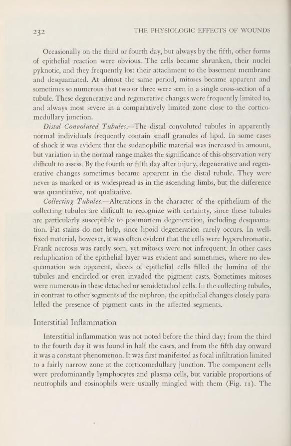
THE PHYSIOLOGIC EFFECTS OF WOUNDS232
Occasionally on the third or fourth day, but always by the fifth, other formsof epithelial reaction were obvious. The cells became shrunken, their nucleipyknotic, and they frequently lost their attachment to the basement membraneand desquamated. At almost the same period, mitoses became apparent andsometimes so numerous that two or three were seen in a single cross-section of atubule. These degenerative and regenerative changes were frequently limited to,and always most severe in a comparatively limited zone close to the cortico-medullary junction.
Distal Convoluted Tubules.—The distal convoluted tubules in apparentlynormal individuals frequently contain small granules of lipid. In some casesof shock it was evident that the sudanophilic material was increased in amount,but variation in the normal range makes the significance of this observation verydifficult to assess. By the fourth or fifth day after injury, degenerative and regen-erative changes sometimes became apparent in the distal tubule. They werenever as marked or as widespread as in the ascending limbs, but the differencewas quantitative, not qualitative.
Collecting Tubules.—Alterations in the character of the epithelium of thecollecting tubules are difficult to recognize with certainty, since these tubulesare particularly susceptible to postmortem degeneration, including desquama-tion. Fat stains do not help, since lipoid degeneration rarely occurs. In well-fixed material, however, it was often evident that the cells were hyperchromatic.Frank necrosis was rarely seen, yet mitoses were not infrequent. In other casesreduplication of the epithelial layer was evident and sometimes, where no des-quamation was apparent, sheets of epithelial cells filled the lumina of thetubules and encircled or even invaded the pigment casts. Sometimes mitoseswere numerous in these detached or semidetached cells. In the collecting tubules,in contrast to other segments of the nephron, the epithelial changes closely para-lelled the presence of pigment casts in the affected segments.
Interstitial InflammationInterstitial inflammation was not noted before the third day; from the third
to the fourth day it was found in half the cases, and from the fifth day onwardit was a constant phenomenon. It was first manifested as focal infiltration limitedto a fairly narrow zone at the corticomedullary junction. The component cellswere predominantly lymphocytes and plasma cells, but variable proportions ofneutrophils and eosinophils were usually mingled with them (Fig. n). The
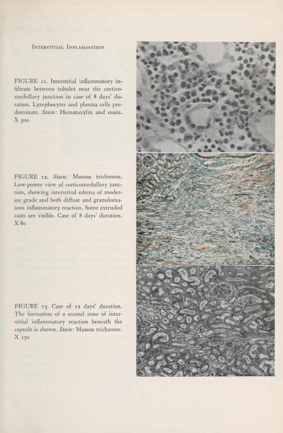
Interstitial Inflammation
FIGURE ii. Interstitial inflammatory in-filtrate between tubules near the cortico-medullary junction in case of 8 days’ du-ration. Lymphocytes and plasma cells pre-dominate. Stain: Hematoxylin and eosin.X 300
FIGURE 12. Stain: Masson trichrome.Low-power view of corticomedullary junc-tion, showing interstitial edema of moder-ate grade and both diffuse and granuloma-tous inflammatory reaction. Some extrudedcasts are visible. Case of 8 days’ duration.X80
FIGURE 13. Case of 12 days’ duration.The formation of a second zone of inter-stitial inflammatory reaction beneath thecapsule is shown. Stain: Masson trichrome.X 150

THE PHYSIOLOGIC EFFECTS OF WOUNDS234
Tubular Rupture
FIGURE 14. Stain: Masson trichrome. A nonpigmented cast found in the process of extru-sion from a ruptured tubule. The cells of the tubule show pyknosis and reduplication ofnuclei. X 250
infiltrate tended to be most marked in the neighborhood of the blood vesselsand also to run in strands between the straight tubules. Edema, though usuallypresent, was rarely a conspicuous feature (Fig. 12). As days passed, the zone ofinflammatory infiltration widened and new foci appeared both in the cortexand the medulla. In the former, they tended to be concentrated about the distalconvoluted tubules. In a few of the most severe lesions, a clearly-defined secondzone of inflammatory infiltration was noted in the outer millimeter of thecortex beneath the capsule (Fig. 13).
Tubular RuptureThe distinctive phenomenon of tubular rupture was generally apparent in
any well-developed lesion from the fifth day onward. It was usually associatedwith the presence of hyaline, non-pigmented casts in the affected segment,which was most commonly the ascending limb but sometimes the distal con-voluted tubule. These casts were chromophobic with hematoxylin and eosinor phosphotungstic acid hematoxylin, but stained a fairly bright green in theMasson trichrome. In the affected segments containing the casts, portions ofthe epithelial lining and of the underlying basement membrane disappeared

PATHOLOGY OF THE KIDNEY 235
without trace as if by solution. The casts at this point were frequently seen halfwithin the tubular remnant and half extruded into the stroma (Fig. 14).
Interstitial Granulomas
In the process of tubular rupture and cast extrusion, the advancing marginof the cast abutting on the stromal cells was quickly surrounded by histiocytes.With further extrusion or with complete destruction of the tubular segment,the cast became completely surrounded by histiocytes which occasionally butrarely fused to form giant cells (Figs. 15 and 16). The histiocytes soon invadedthe cast and appeared to break it up into comparatively small fragments. Itwas possible to observe all stages from the original encapsulation to completedisintegration, leaving a focal aggregate of histiocytes with no trace of theoriginal cast material (Fig. 17).
Although this process unquestionably accounts for many, perhaps for most,of the granulomatous lesions found in these kidneys, the evidence is inadequateto prove that it was the only mechanism for their formation. Granulomas werefrequently found in the walls of veins and occasionally in the capsule of thekidney, separated from the nearest tubule by apparently intact fibrous tissueof considerable density. Some of these lesions were quite similar in appearanceto the Aschoff bodies ofrheumatic fever.
Hyaline masses, indistinguishable from the intratubular casts by any stain-ing method tried, were also occasionally found in the walls of veins, evenjutting into the lumen to become the basis for thrombus formation (Fig. 18).The question whether these represent extruded casts which have wanderedsurprisingly from their original location or which have developed in situ insome entirely different manner cannot be answered at present. It is of interestthat on only one occasion in over 200 cases studied did the author note extrusionof a pigment cast and consequent granuloma formation.
Thrombophlebitis
In a considerable proportion of cases of 5 or more days’ duration, thrombiwere readily found in the large but thin-walled veins of the corticomedullaryjunction. These thrombi were attached to a portion of the vein wall and theyjutted into and narrowed but almost never occluded the lumen. Beneath somepoints of attachment a mass of hyaline material as described in the preceding
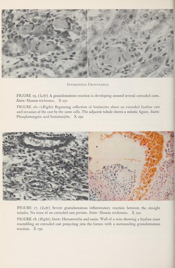
Interstitial Granulomas
FIGURE 15. (Left) A granulomatous reaction is developing around several extruded casts.Stain: Masson trichrome. X 250
FIGURE 16.—(Right) Beginning collection of histiocytes about an extruded hyaline castand invasion of the cast by the same cells. The adjacent tubule shows a mitotic figure. Stain:Phosphotungstic acid hematoxylin. X 250
FIGURE 17. (Left) Severe granulomatous inflammatory reaction between the straighttubules. No trace of an extruded cast persists. Stain: Masson trichrome. X 250FIGURE 18. (Right) Stain: Hematoxylin and eosin. Wall of a vein showing a hyaline massresembling an extruded cast projecting into the lumen with a surrounding granulomatousreaction. X 150
PATHOLOGY OF THE KIDNEY 237
paragraph was found; beneath others were well-developed granulomas withor without traces of hyaline material. The vein walls were frequently invadedby lymphocytes, polymorphonuclear leukocytes, or eosinophils, apparently ex-tending from focal concentrations of the interstitial infiltrate, such concentra-tions being characteristically clustered about these veins.
Summary of Histologic Findings in Chronologic SequenceThe earliest change noted in the “shock kidney” was lipoid degeneration
of the ascending limbs of the loops of Henle, which usually made its appear-ance about 18 hours after injury, progressed in severity until the third day,then tended to wane. Precipitation of the hemoglobin-like pigment in the distalconvoluted and collecting tubules was infrequently observed short of 24 hoursand was inconstant before 48 hours. The pigment at first stained like hemo-globin but later lost this characteristic. Moderate dilatation of the proximalconvoluted tubules was evident following the development of pigment casts.
By the third and fourth days, an interstitial inflammatory infiltrate and franknecrobiotic changes in the cells of the ascending limbs were occasionally seen,and from the fifth day onward both of these findings were constant. Rarelyon the third or fourth day, but usually by the fifth, tubular rupture with extru-sion of nonpigmented casts was apparent, and coincidentally stromal granu-lomas were found in moderate numbers. From the fifth day onward, throm-bophlebitis of the small veins was found in an increasing proportion of the cases.
Gross PathologyThe kidney might or might not show gross abnormalities (Figs. 19, 20, 21,
and 22). Enlargement was noted as early as the second day but was not usualbefore the fourth. It tended to become more frequent and more extensive withduration of the lesion; the largest pair of kidneys observed, 625 grams, wasfound in Case 93 with the unusual survival period of 13 days. In patients withfrom 7- to 10-day survival periods, kidneys weighing up to 500 grams wereusual (Figs. 20 and 21).
Enlarged kidneys were almost without exception pale, whereas kidneys ofnormal size, regardless of duration of the lesion, were usually congested. Onsection it became apparent that the enlargement was chiefly due to swelling
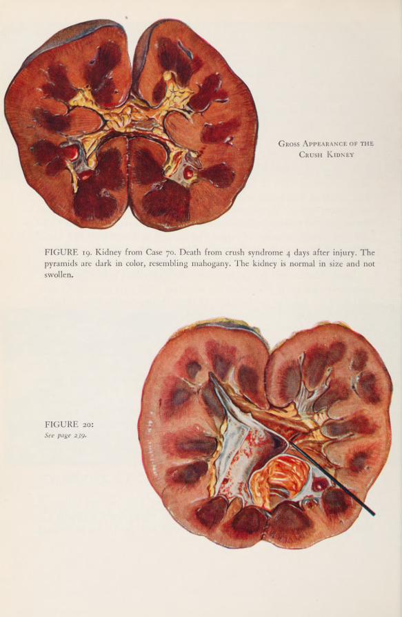
Gross Appearance of the
Crush Kidney
FIGURE 19. Kidney from Case 70. Death from crush syndrome 4 days after injury. Thepyramids are dark in color, resembling mahogany. The kidney is normal in size and notswollen.
FIGURE 20:
See page 239.

FIGURE 21. (Left) Kidney from Case 47 after brief fixation in Kaiserling’s fluid. Thepatient died of pigment nephropathy 8 days after wounding. The pallor of the organ isslightly exaggerated by the fixation. The cortex is swollen and the pyramids are almostchocolate in shade.
FIGURE 22. (Right) Left kidney from case of mismatched transfusion (Case 9) after similarfixation in Kaiserling’s fluid. Gross appearance is identical with that of Case 47 except forsize. The enlargement of this left kidney was partly due to a congenital hypoplasia of its mate.
of the cortex, which would measure from 9 to 12 microns in thickness. Thecorticomedullary junction was unusually sharp, since the cortex was pale andthe pyramids either unchanged or darker than normal. A zone of intense con-gestion sometimes further intensified the distinction. A constant finding wasthe unusual wetness of the freshly cut cortical surface.
Petechial hemorrhages beneath the pelvic mucosa were the only other grossmanifestation which was at all frequent (Fig. 20). They were commonest incases of long duration with high grades of azotemia and are reminiscent ofsimilar hemorrhagic changes in advanced “decompensated’' nephrosclerosis withrenal insufficiency. They may be merely a phenomenon of the uremic state.
FIGURE 20. Kidney from Case 69. Death from crush syndrome 10 days after injury. Theorgan is markedly swollen. The pallor of the cortex is in striking contrast to the still darkpyramids. Fine hemorrhages are apparent beneath the mucosa of the renal pelvis. Page 238

THE PHYSIOLOGIC EFFECTS OF WOUNDS240
PathogenesisShock
Two factors which are evidently of outstanding importance in the develop-ment of the renal lesion may appear in severely traumatized persons. One ispigment excretion in the urine and the other is the physiologic state known asshock. A survey of the first hundred cases of pigment nephropathy studied at
the 15th Medical General Laboratory provided interesting figures regardingthe relative frequency of the lesion in different types of Army hospitals. Inbase hospitals (station and general hospitals), nephropathy was found in only6 percent of the necropsies, in evacuation hospitals the percentage rose to 18,and in the field hospitals (to which only the most severely wounded, non-transportable cases were admitted) to 30 percent. Clearly pigment nephropathyoccurred in direct relation to severity of injury and, by implication, to the pro-fundity of shock.
The cases studied by the Board offer more direct evidence. Whenever pos-sible, a clinician who was a member of the Board made the examination andrecorded his impression regarding the presence and severity of shock. Whenshock was recognized, it was classified as slight, moderate, or severe. In 183cases so classified pigment nephrosis was proved at necropsy in thirty-seven.(There were actually 38 proved cases in the Board’s entire series, but 1 hadnot been classified as to degree of shock.) Their distribution in the variousshock categories is shown in Table 92.
The decreasing frequency of renal involvement from those with markedshock to those with lesser degrees of shock is apparent, but the six patients
Table 92.—Relationship of Proved Pigment Nephropathy to Degree of
Shock
Degree of Shock Numberof Cases
FatalNephropathies Percentage
Severe 57 20 35.1Moderate 55 7 12.7Slight 37 4 10.8None 34 6 17.6

PATHOLOGY OF THE KIDNEY 241
who were not considered to be in shock confuse the picture. This group, whichconsists of Cases 9, 22, 69, 70, 120, and 132, is therefore considered in detail.
“No Shod(' Group.—Case 9 is an example of a frank transfusion accident.Between 100 and 125 cc. of group A blood had been given in error to a groupO recipient before a characteristic reaction developed and the transfusion wasinterrupted. Hemoglobinemia and hemoglobinuria were demonstrated. Thiscase belongs in a different category and requires no further consideration.
The patient in Case 22 did not present evidence of shock at the time of entryto the hospital 3 hours after wounding, or at any subsequent period of observa-tion. He belonged to blood group A and received 2 units of blood, presumablygroup A, not from the theater blood bank and not checked by any memberof the Board. There was no clinical evidence of reaction to these transfusionswhich were given on the fourth day of disease, only 2 days before his death.On the following day the nonprotein nitrogen level in the plasma was 294 mg.per 100 cc., so the renal lesion must have antedated these transfusions. The threeearly transfusions on the day of wounding were of the usual theater bank bloodand were given without incident. The Rh reaction of the patient was not de-termined. No etiology for a hemolytic reaction was discovered, yet large amountsof free hemoglobin, 193 mg. per 100 cc., were found in the bladder urine atthe time of necropsy.
Cases 69, 70, and 132 are examples of the crush syndrome with severe gradesof myoglobinuria. Judged by the usual clinical criteria—blood pressure, pulse,pallor, coldness of extremities, anxiety—these patients did not impress the ex-aminer as being in shock. Each of them, however, clearly exhibited one phe-nomenon which is emphasized by many students of shock: hemoconcentration,as shown by hematocrit values of 71, 80, and 57, respectively. If shock itself wasnot present, there is no doubt that a closely allied physiologic abnormality was.
In Case 120 the patient was wounded in the right thigh by a shell fragmentwhich completely transected the femoral artery, vein, and nerve. Hemorrhagewas severe and he was given 1,000 cc. of plasma en route to the hospital. Onarrival he seemed in good condition and was operated upon without furtherpreparation, but 1,500 cc. of whole blood were given during the operation. Im-mediately after operation the blood pressure was no mm. Hg systolic and 62diastolic, but 7 hours later it dropped to 70/40 and throughout the day thesystolic level ranged between 60 and 70 millimeters of mercury. Neverthelessthe patient’s color was good and his skin remained warm. The pathologist has
THE PHYSIOLOGIC EFFECTS OF WOUNDS242
the temerity to suggest that in consideration of this case also shock can scarcelybe excluded.
Pigment Excretion
In Chapter IV it was shown that all patients with clinical evidence of renalinvolvement following traumatic injury excreted benzidine-positive pigment inthe urine, although the amounts varied widely. In excellent correlation with thisobservation is the histologic evidence of pigment precipitation in the lowernephron segments of all patients with fatal nephropathy. These related phenom-ena deserve further consideration.
The Nature of the PigmentNumerous clinical studies 010 11 have established the two pigments found in
the urine as hemoglobin and the closely related myoglobin. In histologic sectionsthey react identically with all methods employed (the benzidine reaction, hema-toxylin and eosin, Masson’s trichrome and Mallory’s phosphotungstic acidhematoxylin stains). It has generally been considered a safe inference that thepigment casts found in the tubules were precipitates of the same material spec-troscopically identified in its soluble form in the urine, though the possibilitythat they might be oxidized or otherwise chemically altered forms could notbe excluded. Harrison et al.] ~ provided spectroscopic proof that in the lesionsproduced in dogs by arsine hemolysis the casts consisted predominantly ofmethemoglobin. No pigments such as hemochromagen or hematin which wouldbe insoluble at the pH of urine were formed.
Efforts to identify myoglobin by chemical means in the kidneys of patientswho died of the crush syndrome were disappointingly unsuccessful. (See Ap-
9 Bywaters, E. G. L.; Delory, G. E.; Rimington, C., and Smiles, J.; Myohaemoglobin in urine of airraid casualties with crushing injury. Biochem. J. 35: 1164—1168, November 1941.
10 Maegraith, B. G.; Havard, R. E., and Parsons, D. S.: Renal syndrome of wide distribution inducedpossibly by renal anoxia. Lancet 2: 293-296, September 8, 1945.
“Minami, S.: fiber Nierenveriinderungen nach Verschiittung. Virchow’s Arch. f. path. Anat. 245:246-267, 1923.
12 Harrison, H. E.; Bunting, H.; Ordway, N. K., and Albrink, W. S.: The pathogenesis of renalinjury produced in the dog by hemoglobin or methemoglobin. j.Exp. Med. 86: 339—356, October 1947.

PATHOLOGY OF THE KIDNEY 243
pendix C for the method used.) The data are presented in the following tabu-lation.
It is obvious that the kidneys in the crush cases, which one might reasonablyexpect to be loaded with myoglobin, did not show significantly more than thosein cases of sudden death selected as controls; actually they contained consid-erably less myoglobin than did the kidneys in shock cases with or withoutnephropathy in which the urine never contained myoglobin. It seems probablethat this failure to recover myoglobin correlates with the fairly rapid altera-tion in the staining reactions of the pigment in the lower nephron segmentsalready described and may be attributed to a chemical degradation. Underfield conditions it was impractical to attempt further identification of theseproducts.
Acidity of the Urine and Precipitation ofPigment in the Tubules
One widely accepted explanation of the development of a pigment nephro-pathy is that the pigment is precipitated from solution when the glomerularfiltrate becomes acid in reaction upon reaching the distal convoluted tubule.It is of interest to compare the acidity of the initial pigment-containing urinesfrom patients in whom nephropathy was proved with those from patients withno evidence of a renal lesion other than pigment excretion. A control groupin which the urine was consistently free from benzidine-positive material isalso compared in Table 93 with the two pigment-containing groups.
956507 0—52 17
Extraction of Myoglobin from KidneysGm. of myoglobin
Type of case Case No. per Kg. of tissue
Control (sudden death) w>
■
i
i
i
i
i
i
i
i
i
i
i 0.400.50
Shock, no pigment casts l 977
f65 1
1
1
1
I
1
1
1
1
I
1
■
1
1
1
M
M
m
bo-fc*.
OO
OUl
Shock, pigment nephropathy _ 668o
f69
1.3°i-5°
0.45Crush syndrome i
1
1
1
1
1
1
1
1
1
1
1
OOO
0.600.93
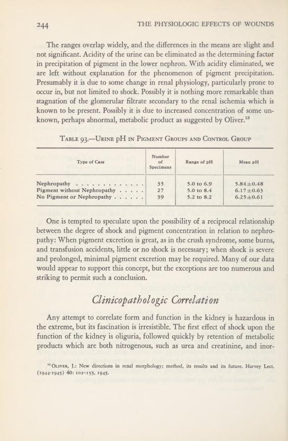
THE PHYSIOLOGIC EFFECTS OF WOUNDS244
The ranges overlap widely, and the differences in the means are slight andnot significant. Acidity of the urine can be eliminated as the determining factorin precipitation of pigment in the lower nephron. With acidity eliminated, weare left without explanation for the phenomenon of pigment precipitation.Presumably it is due to some change in renal physiology, particularly prone tooccur in, but not limited to shock. Possibly it is nothing more remarkable thanstagnation of the glomerular filtrate secondary to the renal ischemia which isknown to be present. Possibly it is due to increased concentration of some un-known, perhaps abnormal, metabolic product as suggested by Oliver. 13
Table 93.—Urine pH in Pigment Groups and Control Group
One is tempted to speculate upon the possibility of a reciprocal relationshipbetween the degree of shock and pigment concentration in relation to nephro-pathy: When pigment excretion is great, as in the crush syndrome, some burns,and transfusion accidents, little or no shock is necessary; when shock is severeand prolonged, minimal pigment excretion may be required. Many of our datawould appear to support this concept, but the exceptions are too numerous andstriking to permit such a conclusion.
Clinicopathologic CorrelationAny attempt to correlate form and function in the kidney is hazardous in
the extreme, but its fascination is irresistible. The first effect of shock upon thefunction of the kidney is oliguria, followed quickly by retention of metabolicproducts which are both nitrogenous, such as urea and creatinine, and inor-
13 Oliver, J.: New directions in renal morphology; method, its results and its future. Harvey Lect.(1944-1945) 40: 102-155, 1945.
Type of CaseNumber
ofSpecimens
Range of pH Mean pH
Nephropathy 35 5.0 to 6.9 5.84±0.48Pigment without Nephropathy 27 5.0 to 8.4 6.17±0.63No Pigment or Nephropathy 39 5.2 to 8.2 6.25±0.6I

PATHOLOGY OF THE KIDNEY 245
ganic, such as phosphates. Both of these effects were manifested within a fewhours of the onset of shock, whereas the first recognizable morphologic change,the appearance of lipoid vacuoles in the ascending loops, developed about 18hours after onset, and pigment casts appeared still later. The initial renal insuffi-ciency is, therefore, functional rather than structural in basis. The studies ofCournand and his associates 14 have established the early onset of renal ischemiain shock and adequately explain the initial functional changes.
Ischemia with consequent anoxemia also serves as a logical explanation ofthe appearance of demonstrable lipid in the parenchymal cells. As will be shownin Chapter XII, a similar phenomenon in shock was found in the liver, theheart, and other organs in which the relationship of fatty degeneration to anox-emia has received more attention. Further knowledge of renal circulation andof the relative susceptibility of various portions of the nephron to anoxemia isnecessary to an understanding of the localization of the changes. The hypothesisof Trueta et al. 10 that blood is shunted from the cortex into the vasa recta of thepyramids is not a satisfactory explanation, since the degenerative changes weremaximal in the very areas which, according to his theory, receive the largestquantities of blood.
The lipoid vacuolation became more intense on the second and third daysafter the onset of shock. This, however, cannot be interpreted as evidence thatthe initial causative factor necessarily persisted. A form of “chain reaction”which required time for its complete development may have been inauguratedby the initial insult.
An interesting feature of the development of the “shock kidney” is the ap-parent delay in the appearance of the pigmented casts. Pigment casts werepresent in significant numbers in only two of the nine Board cases in whichdeath occurred within 48 hours of injury. Even in Cases 32 and 34, in whichdeath occurred between the second and third days, only traces of pigment couldbe found. It is obvious from examination of the histories of this group thatnone of them, with the possible exception of one (Case 34), was ever adequatelyresuscitated from shock. In the case records of scores of other fatal shock cases
14 Cournand, A.; Riley, R. L.; Bradley, S. E.; Breed, E. S.; Noble, R. P.; Lauson, H. D.; Gregersen,M. I., and Richards, D. W.: Studies of the circulation in clinical shock. Surgery 13: 964-995, June 1943.
15 Trueta, J.; Barclay, A. E.; Daniel, P.; Franklin, K. J., and Prichard, M. M. L.: Renal pathologyin the light of recent neurovascular studies. [Preliminary communication.] Lancet 2: 237-238, August 17,1946.
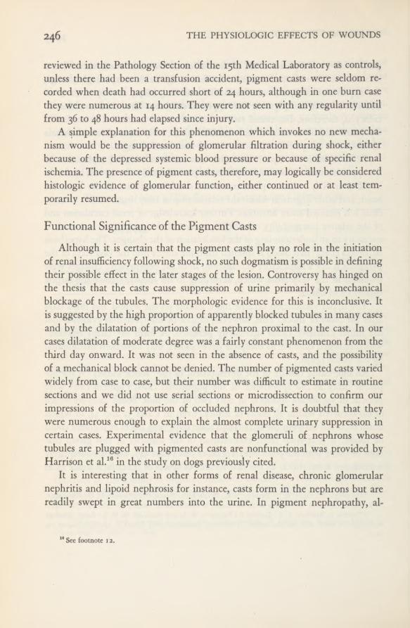
246 THE PHYSIOLOGIC EFFECTS OF WOUNDS
reviewed in the Pathology Section of the 15th Medical Laboratory as controls,unless there had been a transfusion accident, pigment casts were seldom re-corded when death had occurred short of 24 hours, although in one burn casethey were numerous at 14 hours. They were not seen with any regularity untilfrom 36 to 48 hours had elapsed since injury.
A simple explanation for this phenomenon which invokes no new mecha-nism would be the suppression of glomerular filtration during shock, eitherbecause of the depressed systemic blood pressure or because of specific renalischemia. The presence of pigment casts, therefore, may logically be consideredhistologic evidence of glomerular function, either continued or at least tem-porarily resumed.
Functional Significance of the Pigment Casts
Although it is certain that the pigment casts play no role in the initiationof renal insufficiency following shock, no such dogmatism is possible in definingtheir possible effect in the later stages of the lesion. Controversy has hinged onthe thesis that the casts cause suppression of urine primarily by mechanicalblockage of the tubules. The morphologic evidence for this is inconclusive. Itis suggested by the high proportion of apparently blocked tubules in many casesand by the dilatation of portions of the nephron proximal to the cast. In ourcases dilatation of moderate degree was a fairly constant phenomenon from thethird day onward. It was not seen in the absence of casts, and the possibilityof a mechanical block cannot be denied. The number of pigmented casts variedwidely from case to case, but their number was difficult to estimate in routinesections and we did not use serial sections or microdissection to confirm ourimpressions of the proportion of occluded nephrons. It is doubtful that theywere numerous enough to explain the almost complete urinary suppression incertain cases. Experimental evidence that the glomeruli of nephrons whosetubules are plugged with pigmented casts are nonfunctional was provided byHarrison et al, 16 in the study on dogs previously cited.
It is interesting that in other forms of renal disease, chronic glomerularnephritis and lipoid nephrosis for instance, casts form in the nephrons but arereadily swept in great numbers into the urine. In pigment nephropathy, al-
16 See footnote 12.
PATHOLOGY OF THE KIDNEY 247
though casts may regularly be found in the sediment, it is rare that largenumbers of them are passed.
Tubular DegenerationThe initial evidence of tubular degeneration has invariably been fat vacuoles
in the ascending limbs of Henle’s loops and less regularly in the distal convo-luted tubules. This change evidently antedated and was independent of pig-ment precipitation, since it was frequently seen in the absence of the latter.By the fourth and fifth days lipid tended to disappear from the tubules, butfrank necrosis and regeneration of the epithelium became evident and affectedthe same segment of the nephron. These phenomena were never observed inthe shock cases in the absence of pigment precipitation, although in sulfonamideinjury to the kidney they were occasionally seen without pigment. Althoughthe pigment deposit appears to be a sine qua non, the mechanism of its effectis obviously indirect, since the maximal degenerative change occurred at a higherlevel in the nephron than did the first pigment deposits. Degeneration was mostpronounced in the ascending limbs, whereas pigment deposits began in thedistal convoluted tubules.
The functional significance of this tubular degeneration is of interest. Inlipoid nephrosis extensive tubular degeneration is associated with oliguria. Ithas been suggested that the injured tubular cells permit rediffusion of theglomerular filtrate back into the vascular system. In pigment nephropathytubular degeneration was constant, and such a mechanism may be involvedin this syndrome as well. One point of difference between the two syndromesshould be noted, however, before accepting the hypothesis. In lipoid nephrosisthe gravity of the urine is well maintained or even concentrated above usuallimits. In pigment nephropathy fixation at a low level is almost constant. Thiscould be readily explained by the theory of mechanical blockage, since only afew unobstructed nephrons can still function.
The focal rupture of tubules, so frequently noted from the fifth day onward,has, in the author’s opinion, little functional significance. It is a late phenom-enon, whereas the entire syndrome—oliguria, azotemia, pigment excretion, andhypertension, together with fixation of gravity—is established before it appears.Furthermore, if actual extravasation of glomerular filtrate occurred throughthese ruptures, it seems inevitable that interstitial edema would be a more con-spicuous feature of the histologic process.

248 THE PHYSIOLOGIC EFFECTS OF WOUNDS
SUMMARY
A clinicopathologic survey of necropsy findings in 60 severely woundedcasualties provided 38 examples of lower nephron nephrosis in all stages of devel-opment. The initial change,-appearing from 18 to 24 hours after injury, wasfound to be lipoid degeneration of a segment of the nephron, particularly theascending limb of Henle’s loop, less markedly the distal convoluted tubule.The second stage was the precipitation of pigment, either hemoglobin or myo-globin, in the distal convoluted and collecting tubules, seldom occurring shortof 24 hours after injury, more commonly between 32 and 72 hours. Moderatedilatation of proximal and sometimes distal convoluted tubules followed pig-ment precipitation. Sometimes on the third, regularly on the fourth and fifthdays, necrosis and regeneration of epithelium in the ascending limbs and distaltubules became evident, and simultaneously lymphocytes appeared between thetubules and about the vessels. From five days onward, rupture of tubules withherniation of their contents into the stroma and consequent granuloma forma-tion became frequent. The last development in many cases was the formationof nonocclusive mural thrombi in many of the small, thin-walled veins.
In clinicopathologic correlation it was shown that two factors were constantin the nephropathy cases, inconstant in the remainder. These are (1) the ex-cretion of benzidine-positive pigment in the urine, and (2) (with one apparentexception) a state of shock or, at least, a related physiologic abnormality.
Renal insufficiency was found to antedate all structural change, but wasnever progressive in the absence of a demonstrable pigment nephropathy. Theeffects of myoglobin and of hemoglobin precipitation upon the kidney wereindistinguishable. Our evidence does not support the hypothesis that acidityof the urine is the factor responsible for pigment precipitation in the nephrons.
Although pigment precipitation is an essential factor in the development ofthe “shock kidney,” the mechanism of its effect upon the kidney remains un-explained. The possibility that it is in part mechanical cannot be disregarded,but the complex chain of degenerative and inflammatory phenomena demandsanother explanation. Controlled experimentation will be necessary before manyof these uncertainties can be resolved.
For Cases of Special Interest in this chapter, see page 282.

CHAPTER X
Sulfonamides in Relationto “Shock Kidney”
No discussion of renal failure in the wounded of World War II would becomplete without consideration of the possible role of sulfonamide injury of thekidneys. Two forms of this type of injury are generally recognized. One is thepurely mechanical process of crystalluria, with precipitation of the crystals inthe ureters, renal pelves, and perhaps in some instances in the collecting tubules.The second is a more complex process involving true parenchymal damage tothe kidney; it appears to be relatively independent of dosage and is believedby many to belong in the category of hypersensitive reactions along with otherrecognized sulfonamide lesions such as dermatitis, myocarditis, and pneumonitis.
The problem is difficult because this renal lesion is a characteristic lowernephron nephrosis, usually although not invariably associated with the pre-cipitation of hemoglobin-like products in the tubules and hence indistinguish-able from transfusion kidney, crush kidney, or shock kidney. With presentknowledge, distinction is impossible on the basis of renal morphology.
Sulfonamide Therapy in Battle CasualtiesDuring the period in which the cases studied by the Board were collected,
sulfonamides were still extensively used in forward installations (aid stations,collecting stations, and clearing stations) in the Fifth Army Area, although theiruse in hospitals had greatly diminished with the arrival of large supplies ofpenicillin. In the aid stations a sulfonamide dressing almost universally wasapplied. This might or might not be supplemented by indeterminate amounts ofsulfonamide powder dusted onto or into the wounds, and more of the drugby mouth, the most frequent total oral dose being 4 grams. In the field andevacuation hospitals, sulfonamide therapy usually was stopped and penicillin sub-

THE PHYSIOLOGIC EFFECTS OF WOUNDS250
stituted. Notable exceptions, however, were in some cases of abdominal woundswith incipient peritonitis. Many surgeons placed from 5 to 10 Gm. of sulfa-diazine in the abdominal cavity before closing it, and some continued sulfona-mide treatment by mouth for several days. There was no standard practicesince this was considered a matter for individual surgical judgment.
Blood Sulfonamide LevelsRelationship to Sulfonamide Intake
Blood sulfonamide levels were determined in 50 of our patients. The testswere made for free sulfonamide only, whereas the total of free and acetylatedblood sulfonamide would probably have been a more valuable index of sul-fonamide retention by the kidney. As would be expected, there seemed to bea general relationship between the blood levels of the drug and the quantityadministered, as shown in Table 94. However, the fluctuations within eachgroup are too great and the number of cases is too small to justify further anal-ysis. It is noteworthy that no patient tested failed to show at least a trace ofsulfonamide in the blood stream, and in two of six patients with no recordedchemotherapy, levels of 7.0 and 10.6 mg. per 100 cc. were observed. Further-more, characteristic sulfonamide crystals were found at necropsy in the renaltubules in several other patients with no history of exposure to sulfonamide
Table 94.—Blood Sulfonamide Levels in 50 Patients
* The maximum was actually 9.7 in one case, but this case is excluded from the average because the figure iscompletely out of line and it seems certain that the patient must have received oral sulfonamide medication whichwas not recorded.
Blood SulfonamideLevelmg. per 100 cc.
Mode of Therapy Number• of Cases
Minimum Maximum Mean
Dressing only 12 0.2 * 2.4 1.4Dressing and powder 7 0.2 5.0 1.9Dressing, powder, and 4 Gm. orally . . 12 0.2 9.7 2.5Intraperitoneal 5 3.8 27.0 12.2Continued oral 8 3.5 51.6 11.8No history of therapy 6 0.1 10.6 3.7
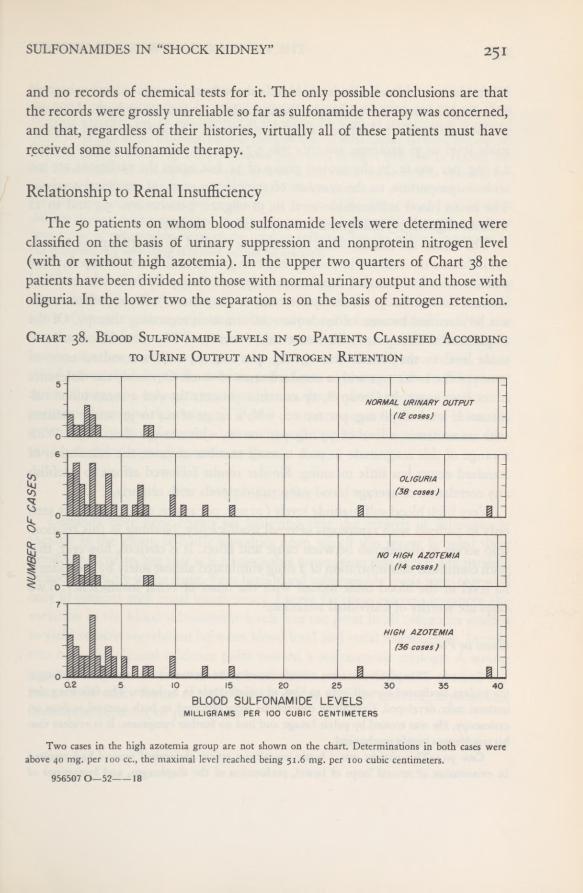
SULFONAMIDES IN “SHOCK KIDNEY” 251
and no records of chemical tests for it. The only possible conclusions are thatthe records were grossly unreliable so far as sulfonamide therapy was concerned,and that, regardless of their histories, virtually all of these patients must havereceived some sulfonamide therapy.
Relationship to Renal InsufficiencyThe 50 patients on whom blood sulfonamide levels were determined were
classified on the basis of urinary suppression and nonprotein nitrogen level(with or without high azotemia). In the upper two quarters of Chart 38 thepatients have been divided into those with normal urinary output and those witholiguria. In the lower two the separation is on the basis of nitrogen retention.
Chart 38. Blood Sulfonamide Levels in 50 Patients Classified According
to Urine Output and Nitrogen Retention
Two cases in the high azotemia group are not shown on the chart. Determinations in both cases wereabove 40 mg. per 100 cc., the maximal level reached being 51.6 mg. per too cubic centimeters.
956507 0—52 18

THE PHYSIOLOGIC EFFECTS OF WOUNDS252
It is evident that blood sulfonamide levels above 8 mg. per 100 cc. were observedonly in patients suffering from oliguria, or high azotemia, or both. Attemptsat closer analysis are disappointingly inconclusive. The average blood sulfona-mide level in 36 azotemic patients was 5.7 mg. per 100 cc. as compared with2.3 mg. per 100 cc. in the control group of 14, but again the variations are too
wide in proportion to the number of samples to warrant statistical analysis.The mean blood sulfonamide level in 38 oliguric patients was 5.4 and in 12
without oliguria, 2.5 milligrams per 100 cubic centimeters.The results are similar when the cases are divided, in an attempt to exclude
the influence of variation in dosage, into two groups as follows: Group A, thosepatients who had external medication only, and Group B, those who receivedinternal medication (oral, intraperitoneal, or intravenous). Eight patients couldnot be classified because of inadequate information regarding therapy. Of the18 patients in Group A, 12 were azotemic, 6 were not. The mean blood sulfona-mide level in the former was 1.7 mg. per 100 cc., with a standard error of±1.1; of the latter 1.32 with a standard error of ±0.8. Obviously the differenceis not significant. In Group B, 17 azotemic patients showed a mean blood sul-fonamide level of 7.8 mg. per 100 cc., with a range of 0.4 to 39; and 7 patientswith no azotemia, a level of 3.7 mg. per ioo cc., with a range of 0.2 to 7.8. Witha range of this magnitude in such a small number of cases, the calculation ofstandard errors has little meaning. Similar results followed efforts to establishany correlation of average blood sulfonamide levels with oliguria.
Very high blood sulfonamide levels (10 mg. per 100 cc. or higher) were seenonly in patients with symptoms of renal insufficiency. Nothing in this relation-ship serves to distinguish between cause and effect. It is obvious, however, thatwith continued administration of a drug eliminated almost solely by the kidney,its level in the blood must mount with the onset of renal insufficiency. Twocases are worthy of individual attention.
Cases in Point
Case 21.—This patient was not a battle casualty. As treatment for a urethral discharge,the patient medicated himself with 12 Gm. of sulfathiazole in 24 hours. The following dayureteral colic developed. Crystalline concretions were observed in both ureteral orifices oncystoscopy. He was treated by pelvic lavage and had no further symptoms. It is evident thathis trouble was purely mechanical.
Case 90.—This patient was a battle casualty; a severe abdominal wound had resultedin evisceration of several loops of bowel, perforation of the diaphragm, and lacerations of

SULFONAMIDES IN “SHOCK KIDNEY” 253
the liver and right kidney. The patient was in moderate shock on entry to the hospital. Forthe first week postoperatively he voided adequate quantities of urine and did not shownitrogen retention, but he was febrile. On the ninth day signs of consolidation developedin the lower lobe of the left lung. He was given 66 Gm. of sulfadiazine between the seventhand twelfth days. Within 24 hours the plasma nonprotein nitrogen level rose to 143 mg. per100 cc. and later to 166 mg. per 100 cc., and the blood sulfonamide level to 51.6 mg. per100 cc. (free sulfonamide 34.4, acetylated 17.2). Following cessation of sulfonamide therapy,the nonprotein nitrogen level fell to 41 mg. per 100 cc. in the course of 17 days. Oliguriadid not develop during the period of nitrogen retention. It is possible that this may havebeen a case of sulfonamide injury of the renal parenchyma, but the relative importance ofthe simultaneous spreading infection cannot be assessed.
One other line of argument must be explored before conclusion. As wasstated in the introduction, there are reasons for believing that the sulfonamideform of lower nephron nephrosis is dependent upon hypersensitivity. One rea-son for this belief is the frequent association of this lesion with inflammatorylesions of other tissues and organs; for example, dermatitis, myocarditis, inter-stitial pancreatitis, hepatitis, arteritis, and a form of pneumonitis. In the presentseries of cases, the only such associated lesion observed was pneumonitis. It is thepersonal opinion of the writer (T.B.M.) that pneumonitis can occur in uremiaindependently of sulfonamide treatment and that, therefore, its presence can-not be accorded great diagnostic weight.
SUMMARY
Grounds have been found for believing that the records of sulfonamidetherapy in the group of battle casualties upon which this report is based weregrossly inaccurate, but that it is a reasonable assumption that few if any patientsescaped exposure to the drug. Very high blood sulfonamide levels were observedonly in patients with renal insufficiency. Except for these extreme instances, thevariation in the blood sulfonamide levels was too great in all categories studiedto yield reliable correlation between blood level and renal insufficiency. In onlytwo cases did clinical evidence point toward a sulfonamide etiology. A strongargument against such a causative mechanism was the failure to observe anyinstance of other forms of sulfonamide injury such as arteritis, myocarditis,dermatitis, hepatitis, or pancreatitis.

AN EARLY CRUSH CASE. This fresco in the Basilica of San Miniato al Monte, Florence,Italy, by Spinello Aretino, 1387 A.D., depicting construction of the Abbey at Monte Cassino,is perhaps the earliest record of treatment of crush injury. The devil has pushed a wall onto
a monk who is then revived by St. Benedict, “Our ministrations were less effective.”
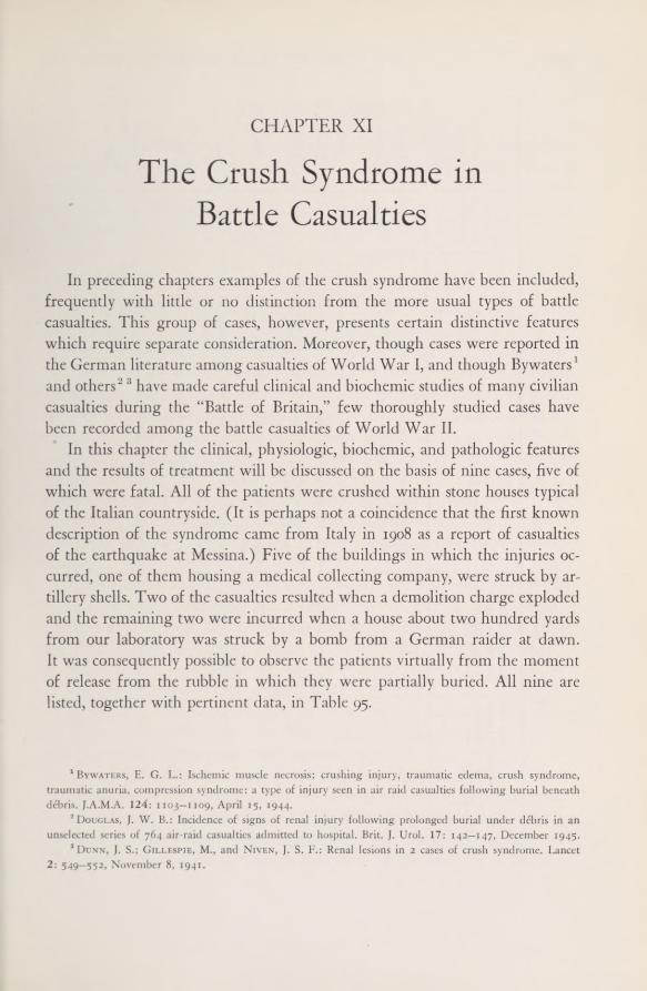
CHAPTER XI
The Crush Syndrome inBattle Casualties
In preceding chapters examples of the crush syndrome have been included,frequently with little or no distinction from the more usual types of battlecasualties. This group of cases, however, presents certain distinctive featureswhich require separate consideration. Moreover, though cases were reported inthe German literature among casualties of World War I, and though Bywaters 1and others" 1 have made careful clinical and biochemic studies of many civiliancasualties during the “Battle of Britain,” few thoroughly studied cases havebeen recorded among the battle casualties of World War II.
In this chapter the clinical, physiologic, biochemic, and pathologic featuresand the results of treatment will be discussed on the basis of nine cases, five ofwhich were fatal. All of the patients were crushed within stone houses typicalof the Italian countryside. (It is perhaps not a coincidence that the first knowndescription of the syndrome came from Italy in 1908 as a report of casualtiesof the earthquake at Messina.) Five of the buildings in which the injuries oc-curred, one of them housing a medical collecting company, were struck by ar-tillery shells. Two of the casualties resulted when a demolition charge explodedand the remaining two were incurred when a house about two hundred yardsfrom our laboratory was struck by a bomb from a German raider at dawn.It was consequently possible to observe the patients virtually from the momentof release from the rubble in which they were partially buried. All nine arelisted, together with pertinent data, in Table 95.
'Bywaters, E. G. L.: Ischemic muscle necrosis; crushing injury, traumatic edema, crush syndrome,traumatic anuria, compression syndrome: a type of injury seen in air raid casualties following burial beneathdebris. J.A.M.A. 124: 1103—1109, April 15, 1944.
2 Douglas, J. W. B.: Incidence of signs of renal injury following prolonged burial under debris in anunselected series of 764 air-raid casualties admitted to hospital. Brit. J. Urol. 17: 142—147, December 1945.
3 Dunn, J. S.; Gillespie, M., and Niven, J. S. F.: Renal lesions in 2 cases of crush syndrome. Lancet2: 549—552, November 8, 1941.
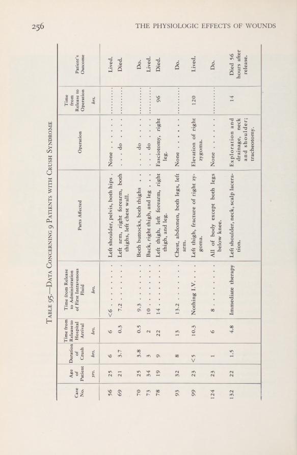
256 THE PHYSIOLOGIC EFFECTS OF WOUNDS
Table95.
—DataConcerning9
Patientswith
CrushSyndrome
Case No.
A*te °/PatientDuration of Crush
Timefrom Release
to Hospital ArrivalTimefrom
Releaseto
Administrationof
FirstIntravenous Fluid
PartsAffected
Operation
Time from Releaseto Operation
Patient’s Outcome
yrs.
hrs.
hrs.
hrs.
hrs.
56
25
6
6
<6
Leftshoulder,
pelvis,bothhips
.
None
Lived.
69
21
3.7
0.3
7.2
Leftarm,
rightforearm,both
thighs,leftchestwall.
...do
....
Died.
70
25
3.8
0.5
9.3
Bothbuttocks,boththighs
.
.
...do
....
Do.
73
34
3
2
10
Back,rightthigh,andleg
.
.
.
...do
....
Lived.
78
19
9
22
14
Leftthigh,leftforearm,
right
thigh,andleg.
Fasciotomy,right
leg.
96
Died.
93
32
8
13
13.2
Chest,abdomen,bothlegs,
left
arm.
None
Do.
99
23<5
10.3
NothingI.V.
.
.
.
Leftthigh,fractureof
rightzy-
goma.
Elevationofright
zygoma.
120
Lived.
12423
1
6
8
Allofbody
exceptbothlegs
belowknee.
None
Do.
132
22
1.5
4.8
Immediatetherapy
Leftshoulder,neck,scalplacera-
tion.
Explorationand
drainage,neck
andshoulder; tracheotomy.
14
Died56
hoursafter release.
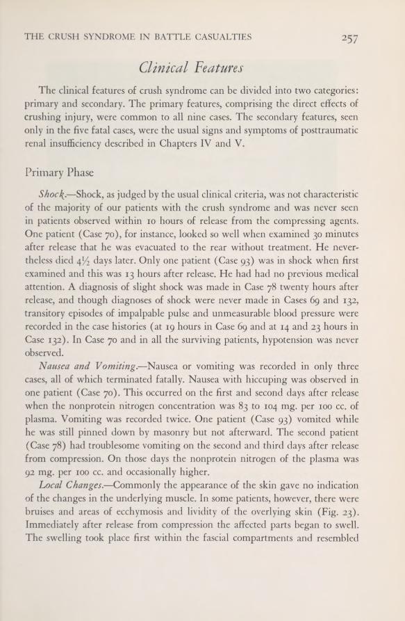
THE CRUSH SYNDROME IN BATTLE CASUALTIES 257
Clinical FeaturesThe clinical features of crush syndrome can be divided into two categories:
primary and secondary. The primary features, comprising the direct effects ofcrushing injury, were common to all nine cases. The secondary features, seenonly in the five fatal cases, were the usual signs and symptoms of posttraumaticrenal insufficiency described in Chapters IV and V.
Primary PhaseShod{.—Shock, as judged by the usual clinical criteria, was not characteristic
of the majority of our patients with the crush syndrome and was never seenin patients observed within 10 hours of release from the compressing agents.One patient (Case 70), for instance, looked so well when examined 30 minutesafter release that he was evacuated to the rear without treatment. He never-theless died 4 1/. days later. Only one patient (Case 93) was in shock when firstexamined and this was 13 hours after release. He had had no previous medicalattention. A diagnosis of slight shock was made in Case 78 twenty hours afterrelease, and though diagnoses of shock were never made in Cases 69 and 132,transitory episodes of impalpable pulse and unmeasurable blood pressure wererecorded in the case histories (at 19 hours in Case 69 and at 14 and 23 hours inCase 132). In Case 70 and in all the surviving patients, hypotension was neverobserved.
Nausea and Vomiting.—Nausea or vomiting was recorded in only threecases, all of which terminated fatally. Nausea with hiccuping was observed inone patient (Case 70). This occurred on the first anti second days after releasewhen the nonprotein nitrogen concentration was 83 to 104 mg. per 100 cc. ofplasma. Vomiting was recorded twice. One patient (Case 93) vomited whilehe was still pinned down by masonry but not afterward. The second patient(Case 78) had troublesome vomiting on the second and third days after releasefrom compression. On those days the nonprotein nitrogen of the plasma was
92 mg. per 100 cc, and occasionally higher.Local Changes.—Commonly the appearance of the skin gave no indication
of the changes in the underlying muscle. In some patients, however, there werebruises and areas of ecchymosis and lividity of the overlying skin (Fig. 23).Immediately after release from compression the affected parts began to swell.The swelling took place first within the fascial compartments and resembled
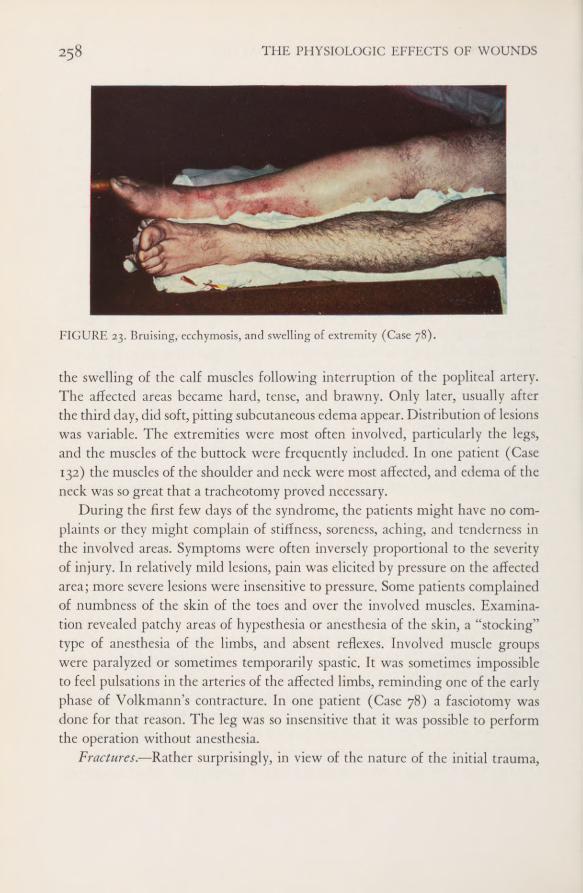
258 THE PHYSIOLOGIC EFFECTS OF WOUNDS
FIGURE 23. Bruising, ecchymosis, and swelling of extremity (Case 78).
the swelling of the calf muscles following interruption of the popliteal artery.The affected areas became hard, tense, and brawny. Only later, usually afterthe third day, did soft, pitting subcutaneous edema appear. Distribution of lesionswas variable. The extremities were most often involved, particularly the legs,and the muscles of the buttock were frequently included. In one patient (Case132) the muscles of the shoulder and neck were most affected, and edema of theneck was so great that a tracheotomy proved necessary.
During the first few days of the syndrome, the patients might have no com-plaints or they might complain of stiffness, soreness, aching, and tenderness inthe involved areas. Symptoms were often inversely proportional to the severityof injury. In relatively mild lesions, pain was elicited by pressure on the affectedarea; more severe lesions were insensitive to pressure. Some patients complainedof numbness of the skin of the toes and over the involved muscles. Examina-tion revealed patchy areas of hypesthesia or anesthesia of the skin, a “stocking”type of anesthesia of the limbs, and absent reflexes. Involved muscle groupswere paralyzed or sometimes temporarily spastic. It was sometimes impossibleto feel pulsations in the arteries of the affected limbs, reminding one of the earlyphase of Volkmann’s contracture. In one patient (Case 78) a fasciotomy wasdone for that reason. The leg was so insensitive that it was possible to performthe operation without anesthesia.
Fractures. —Rather surprisingly, in view of the nature of the initial trauma,
THE CRUSH SYNDROME IN BATTLE CASUALTIES 259
no major fractures were observed. The only fracture recorded (of the zygoma)could be considered only coincidental.
Secondary PhaseOne patient (Case 132) died 56 hours after release from compression; the
other four lived from 4 to 13 days after release. One died suddenly, apparentlyof acute cardiac failure, the others more slowly, with progressive lethargy anddrowsiness but no typical uremic coma. Drowsiness was particularly marked inone patient (Case 78) who had been treated with magnesium. None of the pa-tients in this group had convulsions at any time during their illness. The mecha-nism of death appeared to be pulmonary edema, except in one patient (Case 93)in whom lobar pneumonia developed on the thirteenth day shortly after theappearance of a diuresis had led to hope for his recovery.
All of the patients who died showed the usual signs and symptoms of alower nephron nephrosis previously described (Chapters IV and V). The initialurine specimens were deeply pigmented and reacted positively with the benzi-dine test; oliguria was marked and, except in Cases 69 and 93, progressive (seeTable 97). The urine, with one exception, was persistently acid and the specificgravity was constantly low from the third day onward. The nonprotein nitrogenlevel in the plasma rose progressively, and on the third or fourth day mildhypertension developed. Details of the chemical abnormalities of the blood andurine follow.
Physiologic and Biochemic Peattires
BloodBlood Volume.—The blood volume was determined immediately upon ar-
rival at the hospital in only two patients with crush injury. The first (Case 93)was the only patient of the group thought to be in severe shock. He had lostone-third of his total blood volume, apparently entirely in the form of plasma,for he showed no evidence of diminution of the total hemoglobin or red-cellmass. The hemoconcentration might have been partly responsible for the clini-cal picture of shock, as severe shock was generally associated with a loss of cir-culating blood greater than one-third of the normal volume. The second patient
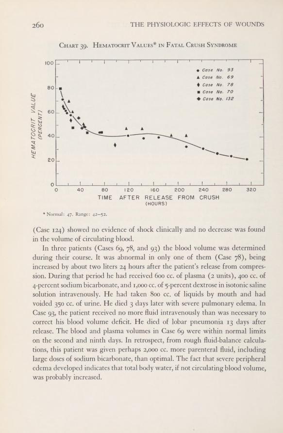
260 THE PHYSIOLOGIC EFFECTS OF WOUNDS
Chart 39. Hematocrit Values* in Fatal Crush Syndrome
Case No. 93Case No. 69Case No 78Case No. 70Cose No. 132
* Normal: 47. Range: 42—52.
(Case 124) showed no evidence of shock clinically and no decrease was foundin the volume of circulating blood.
In three patients (Cases 69, 78, and 93) the blood volume was determinedduring their course. It was abnormal in only one of them (Case 78), beingincreased by about two liters 24 hours after the patient’s release from compres-sion. During that period he had received 600 cc. of plasma (2 units), 400 cc. of4-percent sodium bicarbonate, and 1,000 cc. of 5-percent dextrose in isotonic salinesolution intravenously. He had taken 800 cc. of liquids by mouth and hadvoided 350 cc. of urine. He died 3 days later with severe pulmonary edema. InCase 93, the patient received no more fluid intravenously than was necessary tocorrect his blood volume deficit. He died of lobar pneumonia 13 days afterrelease. The blood and plasma volumes in Case 69 were within normal limitson the second and ninth days. In retrospect, from rough fluid-balance calcula-tions, this patient was given perhaps 2,000 cc. more parenteral fluid, includinglarge doses of sodium bicarbonate, than optimal. The fact that severe peripheraledema developed indicates that total body water, if not circulating blood volume,was probably increased.
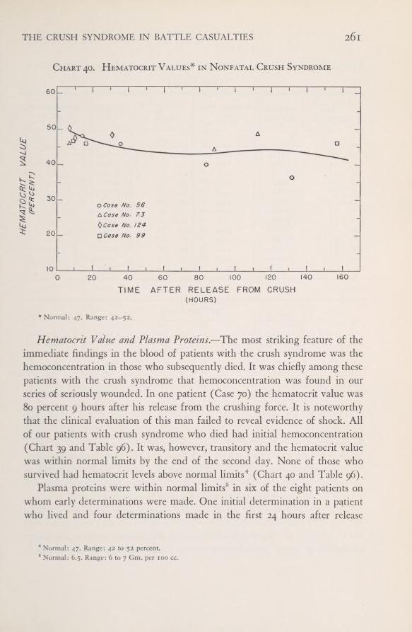
261THE CRUSH SYNDROME IN BATTLE CASUALTIES
Chart 40, Hematocrit Values* in Nonfatal Crush Syndrome
Case No. 56Case No- 73Case No. 124Case No. 99
* Normal: 47. Range: 42—52.
Hematocrit Value and Plasma Proteins.—The most striking feature of theimmediate findings in the blood of patients with the crush syndrome was thehemoconcentration in those who subsequently died. It was chiefly among thesepatients with the crush syndrome that hemoconcentration was found in ourseries of seriously wounded. In one patient (Case 70) the hematocrit value was80 percent 9 hours after his release from the crushing force. It is noteworthythat the clinical evaluation of this man failed to reveal evidence of shock. Allof our patients with crush syndrome who died had initial hemoconcentration(Chart 39 and Table 96). It was, however, transitory and the hematocrit valuewas within normal limits by the end of the second day. None of those whosurvived had hematocrit levels above normal limits 4 (Chart 40 and Table 96).
Plasma proteins were within normal limits 1’ in six of the eight patients onwhom early determinations were made. One initial determination in a patientwho lived and four determinations made in the first 24 hours after release
4 Normal: 47. Range: 42 to 52 percent.4 Normal: 6.5. Range: 6 to 7 Gm. per 100 cc.

262 THE PHYSIOLOGIC EFFECTS OF WOUNDS
Table 96.—Hematocrit Value in Relation to Patients’ Course
* Normal: 47. Range: 42-52.
from compression in a fatal case were moderately elevated (Table 97). Again,as was the case in the hematocrit values, the elevated plasma protein concen-tration soon returned to normal. Although our data are too few to permit ofinterpretation, it is probable that a consistently high plasma protein concentra-tion in the first 24 hours is indicative of a poor prognosis.
In these crush cases the initial concentration of red blood cells was rela-tively greater than the concentration of plasma protein, as may be seen in Chart41. These findings suggest that with release of compression and re-establishmentof circulation to the injured areas, there is prompt outpouring of plasma into
Fatal Cases
Hematocrit Value *
Case Hours after Case Hours after iNo. No. Releasecc. cells in 100 cc. cc. cells in 100 cc.
70 9.0 80.0 70 52.0 44.078 12.0 65.0 93 72.5 43.593 13.0 71.0 70 76.0 44.093 14.5 63.0 78 96.0 34.293 16.0 61.5 69 117.5 47.093 16.5 59.9 93 119.5 41.169 21.5 70.0 69 141.5 47.093 23.0 59.0 93 144.0 39.078 24.0 54.1 93 167.5 39.869 27.0 61.0 69 189.5 41.670 28.0 48.0 69 214.0 42.0
132 37.5 57.0 93 215.5 32.9132 45.0 47.0 93 240.5 30.569 46.0 51.7 93 265.0 26.578 47.0 49.0 93 287.5 24.593 48.0 48.0 93 311.5 21.9
Nonfatal Cases
73 7.0 46 56 35.5 45.7124 7.0 50 56 83.5 40.099 9.0 46.8 73 88.5 44.4
124 10.0 47.7 73 112.5 48.556 14.0 48.0 56 131.5 36.799 16.5 46.0 99 157.0 46.2
124 31.0 48.0 56 203.5 39.0
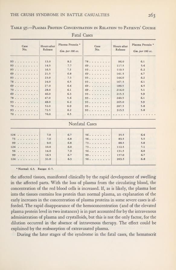
THE CRUSH SYNDROME IN BATTLE CASUALTIES 263Table 97.—Plasma Protein Concentration in Relation to Patients’ Course
Fatal Cases
Nonfatal Cases
* Normal: 6.5. Range: 6-7.
the affected tissues, manifested clinically by the rapid development of swellingin the affected parts. With the loss of plasma from the circulating blood, theconcentration of the red blood cells is increased. If, as is likely, the plasma lostinto the tissues contains less protein than normal plasma, an explanation of theearly increases in the concentration of plasma proteins in some severe cases is af-forded. The rapid disappearance of the hemoconcentration (and of the elevatedplasma protein level in two instances) is in part accounted for by the intravenousadministration of plasma and crystalloids, but this is not the only factor, for thedilution occurred in the absence of intravenous therapy. The effect could beexplained by the reabsorption of extravasated plasma.
During the later stages of the syndrome in the fatal cases, the hematocrit
CaseNo,
Hours afterRelease
Plasma Protein *
Gm. per 100 cc.
CaseNo.
Hours afterRelease
Plasma Protein *
Gm. per 100 cc.
93 13.0 8.2 78 96.0 6.193 14.5 7.7 69 117.5 5.493 16.5 7,3 93 119.5 6.269 21,5 6.8 69 141.5 4.793 23.0 7.3 93 144.0 6.278 24.0 6.5 93 167.5 6.169 27.0 6.4 69 189.5 4.570 28.0 6.1 69 214.0 5.169 46.0 6.3 93 215.5 5.978 47.0 5.6 93 240.5 6.193 . . . 48.0 6.2 93 265.0 5.970 52.0 6.9 93 287.5 5.893 72.5 6.2 93 315.5 5.870 76.0 6.5
124 7,0 6.7 56 35.5 6.473 7.0 6.8 56 83.5 5.599 9.0 6.8 73 88.5 5.8
124 .
. 10.0 6.0 73 112.5 7.156 14.0 7.9 56 131.5 6.099 16.5 6.7 99 157.0 6.7
124 31.0 6.3 56 203.5 6.8
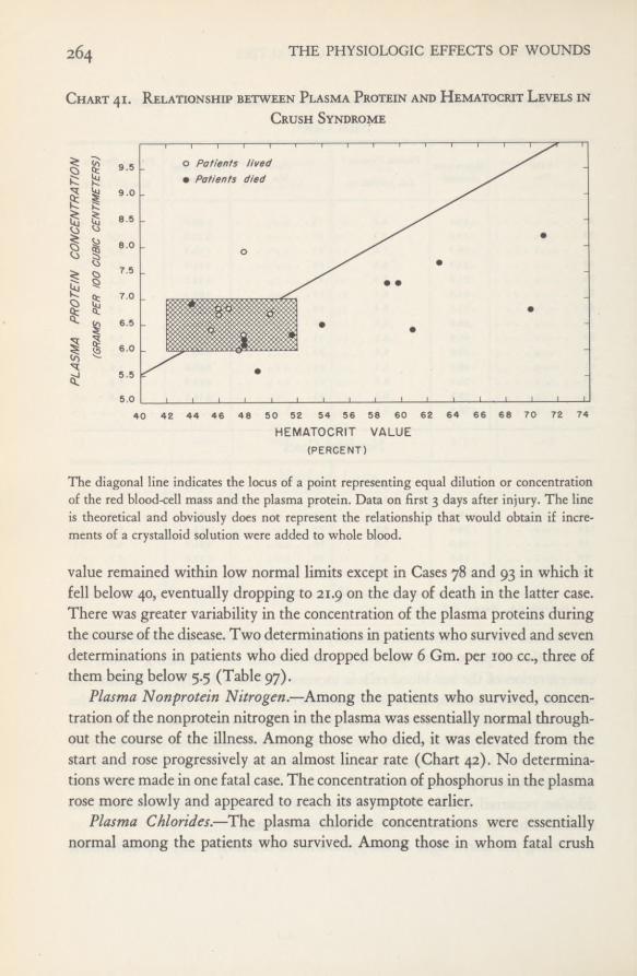
264 THE PHYSIOLOGIC EFFECTS OF WOUNDS
Chart 41. Relationship between Plasma Protein and Hematocrit Levels in
Crush Syndrome
The diagonal line indicates the locus of a point representing equal dilution or concentrationof the red blood-cell mass and the plasma protein. Data on first 3 days after injury. The lineis theoretical and obviously does not represent the relationship that would obtain if incre-ments of a crystalloid solution were added to whole blood.
value remained within low normal limits except in Cases 78 and 93 in which itfell below 40, eventually dropping to 21.9 on the day of death in the latter case.There was greater variability in the concentration of the plasma proteins duringthe course of the disease. Two determinations in patients who survived and sevendeterminations in patients who died dropped below 6 Gm. per 100 cc., three ofthem being below 5.5 (Table 97).
Plasma Nonprotein Nitrogen.—Among the patients who survived, concen-tration of the nonprotein nitrogen in the plasma was essentially normal through-out the course of the illness. Among those who died, it was elevated from thestart and rose progressively at an almost linear rate (Chart 42). No determina-tions were made in one fatal case. The concentration of phosphorus in the plasmarose more slowly and appeared to reach its asymptote earlier.
Plasma Chlorides.—The plasma chloride concentrations were essentiallynormal among the patients who survived. Among those in whom fatal crush
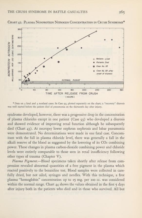
265THE CRUSH SYNDROME IN BATTLE CASUALTIES
Chart 42. Plasma Nonprotein Nitrogen Concentration in Crush Syndrome*
* Data on 4 fatal and 4 nonfatal cases. In Case 93, plotted separately on the chart, a “recovery” diuresiswas well started before the patient died of pneumonia on the thirteenth day after injury.
syndrome developed, however, there was a progressive drop in the concentrationof plasma chlorides except in one patient (Case 93) who developed a diuresisand showed evidence of improving renal function although he subsequentlydied (Chart 43). At necropsy lower nephron nephrosis and lobar pneumoniawere demonstrated. No determinations were made in one fatal case. Concom-itant with the fall in plasma chloride level, there was generally a fall in thealkali reserve of the blood as suggested by the lowering of its CO2 combiningpower. These changes in plasma carbon-dioxide combining power and chloridelevels were entirely comparable to those seen in renal insufficiency followingother types of trauma (Chapter V).
Plasma Pigment.—Blood specimens taken shortly after release from com-pression revealed abnormal quantities of a free pigment in the plasma whichreacted positively to the benzidine test. Blood samples were collected in care-fully dried, but not oiled, syringes and needles. With this technique, a freeplasma “hemoglobin” concentration up to 15 mg. per 100 cc. was consideredwithin the normal range. Chart 44 shows the values obtained in the first 5 daysafter injury both in the patients who died and in those who survived. All but

266 THE PHYSIOLOGIC EFFECTS OF WOUNDS
Chart 43. Plasma Chloride Concentration* in Crush Syndrome
* Normal: xoo. Range: 97.5—104. Data on 4 fatal and 4 nonfatal cases,
two of the determinations we were able to obtain during the first 24 hourswere above the limits accepted as normal; the exceptions were determinationsmade in Case 99, nine and sixteen hours after release. Only one of the patientsshowed a heme concentration of over 40 mg. per 100 cubic centimeters. Datawere available on eight cases. Of particular interest, from those data, is that, likethe return of the hematocrit level and the plasma protein concentration to nor-mal, the concentration of the free benzidine-positive substance in the plasmatended to be within normal limits by the end of the second day. As in the caseof the hemoconcentration, then, the presence of free benzidine-positive sub-stance in the plasma might have been missed if specimens had not been collectedsoon after the patients’ release from the crushing force.
The nature of this substance, free in the plasma and giving the reaction ofa heme, could not be settled. The quantities present were not large enough tomake spectroscopic analysis possible in the instrument available to us. Chemicalexamination of the urine of these patients (Chapter VIII) showed that bothhemoglobin and myoglobin were excreted in large amounts, the proportions ofeach varying widely from case to case. It is reasonable to assume, therefore,that both pigments were represented in the plasma as well.

THE CRUSH SYNDROME IN BATTLE CASUALTIES 267Chart 44. Free Benzidine-Positive Substance in Plasma in Crush Syndrome*
Patients Lived
Patients Died
•Data on 4 fatal and 4 nonfatal cases.
Urine
Gross abnormalities were found only in the urine specimens collected soonafter release from the compression force. Table 98 indicates the duration of someof the abnormalities in the urine. It will be observed that myoglobin was notfound in the urine more than a day and a half after release from the crushingforce. Abnormality in the color of the urine lasted somewhat longer (up to
3 days), and albuminuria to a significant degree was detected for as long as
9 days after release from the crushing influence.Output.—None of the patients who survived the injury had any diminution
in volume of urinary output below the limits of normal. In all of those whodied (Table 99) urinary output was diminished from the start. All had oliguria.In one instance (Case 70) anuria (an output of less than 100 cc.) was recordedduring the 24 hours beginning the day after release. Two other patients wereoliguric until the second 24-hour period, when the output dropped below 100 cc.a day. The remaining two patients (Cases 93 and 78) were oliguric but neverbecame anuric. In one (Case 69) diuresis had just begun to develop when hesuddenly died; in another (Case 93, Table 99) an excellent diuresis was estab-
956507 0—52 20
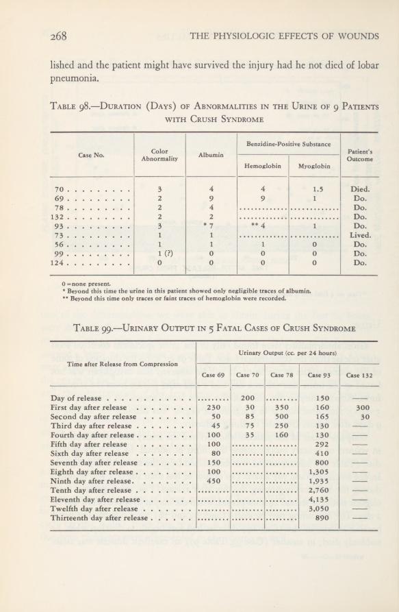
268 THE PHYSIOLOGIC EFFECTS OF WOUNDS
lished and the patient might have survived the injury had he not died of lobarpneumonia.
Table 98,—Duration (Days) of Abnormalities in the Urine of 9 Patientswith Crush Syndrome
Table 99.—Urinary Output in 5 Fatal Cases of Crush Syndrome
Case No. ColorAbnormality Albumin
Benzidine-Positive SubstancePatient’sOutcome
Hemoglobin Myoglobin
70 3 4 4 1.5 Died.69 2 9 9 1 Do.78 2 4 Do.
2 2 Do.93 3 * 7 *� 1 Do.73 1 156 1 1 1 0 Do.99 1 (?) 0 0 0 Do.
124 0 0 0 0 Do.
0 =none present.* Beyond this time the urine in this patient showed only negligible traces of albumin.** Beyond this time only traces or faint traces of hemoglobin were recorded.
Time after Release from Compression
Urinary Output (cc. per 24 hours)
Case 69 Case 70 Case 78 Case 93 Case 132
200 1 50First day after release 230 30 350 160 300Second day after release 50 85 500 165 30Third day after release 45 75 250 130 —
Fourth day after release 100 35 160 130 —
Fifth day after release 100 292 —
Sixth day after release 80 410 —
Seventh day after release 150 800 —
Eighth day after release 100 1,305 —
Ninth day after release 450 1,935 —
Tenth day after release 2,760 —
Eleventh day after release 4,135 —
Twelfth day after release 3,050 —
Thirteenth day after release 890 —
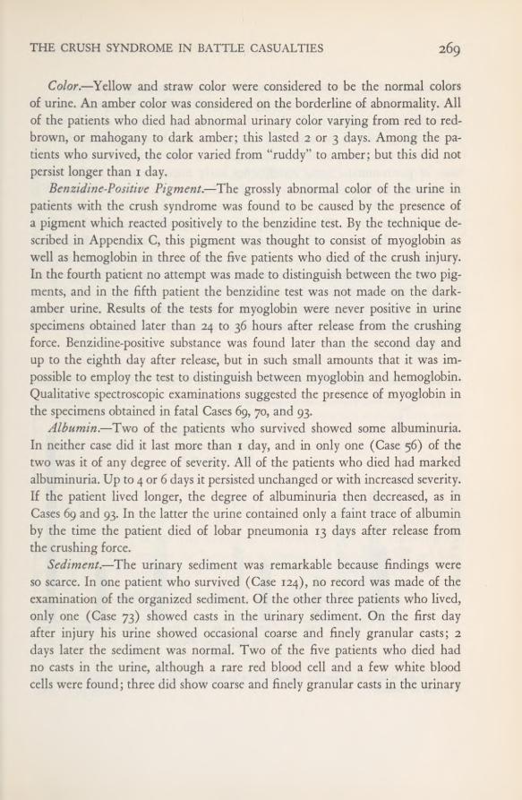
THE CRUSH SYNDROME IN BATTLE CASUALTIES 269Color.—Yellow and straw color were considered to be the normal colors
of urine. An amber color was considered on the borderline of abnormality. Allof the patients who died had abnormal urinary color varying from red to red-brown, or mahogany to dark amber; this lasted 2 or 3 days. Among the pa-tients who survived, the color varied from “ruddy” to amber; but this did notpersist longer than 1 day.
Benzidine-?oshive Figment.—The grossly abnormal color of the urine inpatients with the crush syndrome was found to be caused by the presence ofa pigment which reacted positively to the benzidine test. By the technique de-scribed in Appendix C, this pigment was thought to consist of myoglobin aswell as hemoglobin in three of the five patients who died of the crush injury.In the fourth patient no attempt was made to distinguish between the two pig-ments, and in the fifth patient the benzidine test was not made on the dark-amber urine. Results of the tests for myoglobin were never positive in urinespecimens obtained later than 24 to 36 hours after release from the crushingforce. Benzidine-positive substance was found later than the second day andup to the eighth day after release, but in such small amounts that it was im-possible to employ the test to distinguish between myoglobin and hemoglobin.Qualitative spectroscopic examinations suggested the presence of myoglobin inthe specimens obtained in fatal Cases 69, 70, and 93.
Albumin.—Two of the patients who survived showed some albuminuria.In neither case did it last more than 1 day, and in only one (Case 56) of thetwo was it of any degree of severity. All of the patients who died had markedalbuminuria. Up to 4 or 6 days it persisted unchanged or with increased severity.If the patient lived longer, the degree of albuminuria then decreased, as inCases 69 and 93. In the latter the urine contained only a faint trace of albuminby the time the patient died of lobar pneumonia 13 days after release fromthe crushing force.
Sediment.—The urinary sediment was remarkable because findings wereso scarce. In one patient who survived (Case 124), no record was made of theexamination of the organized sediment. Of the other three patients who lived,only one (Case 73) showed casts in the urinary sediment. On the first dayafter injury his urine showed occasional coarse and finely granular casts; 2
days later the sediment was normal. Two of the five patients who died hadno casts in the urine, although a rare red blood cell and a few white bloodcells were found; three did show coarse and finely granular casts in the urinary

270 THE PHYSIOLOGIC EFFECTS OF WOUNDS
sediment, but these casts were not abundant, even in centrifuged specimens.Specific Gravity.—For the first day or two of the illness, the specific gravity
of the urine was normal in all cases. After the second to fourth days, however,the specific gravity of the urine fell and appeared to become fixed at 1.012 to
1.013. Urine concentration tests were not done in these patients, but as in othercases of posttraumatic renal insufficiency early impairment of ability to re-absorb water by the renal tubules is surely a characteristic feature (Chapter V).
Reaction.—With one exception, the patients passed acid urine at all timesthroughout the illness. This one exception (Case 69) was a patient whose urinewas on the alkaline side of neutral (pH 7.2) on two occasions on the seventhand eighth days of the illness. At all other times the urine of this patient wasacid, despite administration of sodium bicarbonate in doses thought adequatefor alkalinization of the urine (16 to 36 Gm. per 24 hours). As had beenfound in the study of patients who had renal insufficiency from other causes,alkalinization of the urine by means of administration of sodium bicarbonatewas inconsistent with safety.
Chart 45. Bromsulfalein Excretion in Patients with Fatal Crush Syndrome
Cose No. 93Cose No. 69Case No. 70Case No. 78
One value in each of 2 cases was estimated for a 45-minute period, as the figures had been recordedat 30 and 60 minutes.

THE CRUSH SYNDROME IN BATTLE CASUALTIES 271
Chart 46. Plasma Bilirubin Concentration in Crush Syndrome(First 6 Days)
All values in the fatal cases were above normal though still below the level at which clinicaljaundice would appear (2 mg. per 100 cc.). Values for patients who survived were withinnormal limits with the exception of Case 56, plotted separately on the chart. Data on 4 fataland 4 nonfatal cases.
Liver FunctionThe function of the liver was tested in one of the four patients who sur-
vived and in four of the five patients who died. The examination consisted ofa test of the ability of the liver to excrete bromsulfalein injected intravenously.Dosage was calculated on the basis of 5 milligrams of bromsulfalein per kilo-gram of body weight (Chapter II). By this method, retention of the dye upto 3 percent of the injected dose after 45 minutes was considered the upper limitof normal. In the patient who survived the function of the liver was found tobe normal as tested by this method. In all of the patients who died liver func-tion was found to be impaired (Chart 45). The average dye retention in thefour fatal cases was 12.77 percent, exclusive of one determination made inCase 93, which showed 26-percent retention on the day the patient died oflobar pneumonia.

272 THE PHYSIOLOGIC EFFECTS OF WOUNDS
The concentration of bilirubin in the plasma was increased in those pa-tients in whom fatal crush syndrome developed and in one patient (Case 56) whosurvived (Chart 46). Data were obtained on eight cases. Sufficient informationwas not available to distinguish between increased production of bilirubin anddecrease in its elimination.
PathologyThe important findings at postmortem examination were confined to the
v
lungs, the liver, the skeletal muscles, and the kidneys.Lungs. —All of the five fatal cases showed slight to extensive pulmonary
edema, both grossly and microscopically. The weight of the two lungs togetherwas recorded in four of the five fatal cases. The weights varied from 1,075 to1,600 grams. Blood as well as edema fluid appeared on cut surfaces of the lungsin Case 132 and, microscopically, large numbers of alveoli were packed withred blood cells. This patient had had a tracheotomy and the blood may havebeen aspirated. Another patient (Case 78) was unusual in that even the visceralpleura was edematous. Fluid poured from the cut surfaces without the neces-sity of compression, and the tracheobronchial tree was filled with nonbloodyfluid. In Case 93 death was due primarily to lobar pneumonia involving the leftupper lobe. Microscopically, in addition to the typical pneumonia process, someareas showed edema with spreading pneumonic involvement.
Liver. —In view of the decreased liver function, as measured by ability toexcrete bromsulfalein and as indicated by the elevated plasma bilirubin andthe abnormally low ratio of urea nitrogen to total nonprotein nitrogen, somehistologic changes were expected in the liver. Surprisingly few were found. Theliver was microscopically normal in Case 70, and showed only scattered vacuolesin Case 132. In Case 69 occasional mitotic figures were observed in hepatic cells,and in Case 93, in which there had been terminally a rising bilirubin concen-tration in the blood and a terminal bromsulfalein retention of 26 percent, theonly histologic changes found were mild nuclear atypicality in the cells at thecenters of the lobules.
Skeletal Muscles.—The gross changes in the involved muscles were con-spicuous. In the involved areas the normal pigment had disappeared, leavingmuscle bundles with the appearance of fish or rabbit flesh. Sometimes an entiremuscle was decolorized, more commonly only segmental areas, particularly re-

THE CRUSH SYNDROME IN BATTLE CASUALTIES 273
FIGURE 24. Decolorization inthe gastrocnemius-soleus mus-cle group. From necropsy inCase 78 (see Fig. 23).
FIGURE 25. Decolorization in the left brachioradialis muscle in Case 93. Note the “fishflesh” appearance, the sharp line of demarcation, and the distinct white band at the junctionof the depigmented and normally pigmented muscle.
gions close to major bony structures (Fig. 24). In Figure 25 (Case 93) com-plete depigmentation of the distal half of the forearm muscles is apparent,whereas the proximal half is normally colored. The transition from normallycolored to depigmented areas was sharp, particularly in the cases of longersurvival, and in Case 93 (13 days’ survival) the junction was marked by anarrow white band 2 to 3 mm. broad (Fig. 25). In spite of the loss of color, the
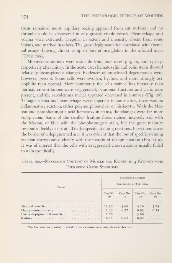
274 THE PHYSIOLOGIC EFFECTS OF WOUNDS
tissue remained moist, capillary oozing appeared from cut surfaces, and nothrombi could be discovered in any grossly visible vessels. Hemorrhage andedema were extremely irregular in extent and intensity, absent from somelesions, and marked in others. The gross depigmentation correlated with chemi-cal assays showing almost complete loss of myoglobin in the affected areas(Table 100).
Microscopic sections were available from four cases 4, 5, 10, and 13 daysrespectively after injury. In the acute cases hematoxylin and eosin stains showedrelatively inconspicuous changes. Evidences of muscle-cell degeneration were,however, present. Some cells were swollen, hyaline, and more strongly aci-dophilic than normal. More commonly the cells stained less intensely thannormal, cross-striations were exaggerated, occasional fractures and clefts werepresent, and the sarcolemma nuclei appeared decreased in number (Fig. 26).Though edema and hemorrhage were apparent in some areas, there was noinflammatory reaction, either polymorphonuclear or histiocytic. With the Mas-son and phosphotungstic acid hematoxylin stains, the changes were far moreconspicuous. Some of the swollen hyaline fibers stained intensely red withthe Masson, or blue with the phosphotungstic stain, but the great majorityresponded feebly or not at all to the specific staining reactions. In sections acrossthe border of a depigmented area it was evident that the loss of specific stainingreaction corresponded closely with the margin of depigmentation (Fig. 27 a).It was of interest that the cells with exaggerated cross-striations usually failedto stain specifically.
Table ioo.—Myoglobin Content of Muscle and Kidney in 4 Patients who
Died from Crush Syndrome
* The low value was probably caused b y the extensive interstitial edema in this case.
Tissue
Myoglobin Content
Gm. per Kg. of Wet Tissue
Case No.69
Case No.70
Case No.78
Case No.93
Normal muscleDepigmented musclePartly depigmented muscle
* 2.101.001.860.45
3.600.27
4.200.833.000.93
3.740.54
Kidney 0.60

THE CRUSH SYNDROME IN BATTLE CASUALTIES 275
FIGURE 26. Decolorized muscle from Case 70. The patient died 4 days after injury. Themuscle cells, which should stain intensely with the phosphotungstic acid hematoxylin, em-ployed stain weakly. Cross-striations are exaggerated and numerous transverse clefts haveappeared. An occasional cell has become hyalinized and lost all evidence of striation. Thereis much hemorrhage into the interstitial connective tissue.
In the cases of longer duration (to and 13 days), changes were conspicu-ous with all staining methods but particularly with hematoxylin and eosin.Many swollen, necrotic muscle cells had become intensely basophilic owing tothe precipitate of innumerable fine granules staining like calcium throughoutthe sarcoplasm (Fig. 27 c and d). In other cells the precipitate was intense inthe central portion but absent at the periphery (Fig. 28 a). Another commontype of degenerative change was a coarse hydropic vacuolation which frequentlyproduced an appearance suggestive of vegetable cells (Fig. 27 d). Other de-generating cells showed varying degrees of infiltration with histiocytes (Figs.27 b and c). The sarcolemma nuclei in many cells showed amitotic division,with chains of touching nuclei 5 to 10 elements in length (Figs. 27 b and 28 b).About many of these, viable cytoplasm was present in long, slender masses sug-gesting regeneration of muscle cells. The interstitial connective tissue wasslightly edematous and densely infiltrated with histiocytes (Fig. 28 b). Nodefinite evidence of fibroblastic proliferation could be made out. Blood vesselsstill remained free from thrombi or inflammatory reaction.
Kidneys.—In all but one patient (Case 70) the kidneys were enlarged atnecropsy. In three of the five fatal cases the combined weights of the two kidneyswere recorded; they were respectively 500, 550, and 625 grams. The capsulesstripped easily. In some there were ecchymoses beneath the capsule. Focal

FIGURE 27, a. Muscle from Case 70 (4 days’ survival). Phosphotungstic acid hematoxylinstain. Section taken from a partially depigmented area. In the more severely involved areasnecrotic muscle cells stain only a faint brown instead of an intense purplish-blue. Even thesurviving cells stain irregularly and striations are difficult to see. The interstitial tissue isedematous but shows little inflammatory infiltration.b. Case 70 (see also a and Fig. 26). Hematoxylin and eosin stain. At the top are threeswollen muscle cells which have lost striations. Beneath them are two necrotic cells whichhave been invaded by histiocytes. At upper right and lower left are fragments of survivingmuscle which show chains of touching nuclei. Two foci of calcification are present. Theinterstitial tissue shows fibrosis and inflammatory infiltration.c. Muscle from Case 69 (10 days’ survival). Hematoxylin and eosin stain. This low-powerview shows focal calcification and vascular degeneration of muscle cells as well as chronicinflammatory infiltration and fibrosis of tbe interstitial tissues.d. A higher magnification from the same section shown in c. Masson trichrome stain.Coarse vascular degeneration of muscle cells producing vegetable-cell appearance is evident.Several muscle cells no longer stain red in normal fashion and have been invaded byhistiocytes. Fibrosis and histiocytic infiltration of the interstitial tissue is apparent.

FIGURE 28, a. A focus of intense calcification involving almost every muscle cell. There ismarked increase in interstitial fibrous tissue with an occasional focus of lymphocytic infiltra-tion. Section from Case 93 (13 days’ survival).b. Section from the same case. Hematoxylin and eosin stain. A regenerating muscle cell showslong chains of touching sarcolemma nuclei. The fibrosis and the patency of the blood vesselsare noteworthy. Partial calcification of some muscle cells at the periphery of the field is evident.
hemorrhages and fibrin deposits were commonly found beneath and on theepithelium of the renal pelvis (see Fig. 20 in Chapter IX). When the kidneyswere cut, with one exception (Case 70), the renal parenchyma pouted throughthe cut, rolling outward and forming a rounded instead of a sharp edge.
Characteristically, the cut surface of the parenchyma showed pallor of thecortex and darkening of the pyramids to a dark brown or mahogany color(Figs. 19 to 22 in Chapter IX). The thickness of the cortex was normal or in-creased, never decreased.
The microscopic appearance of the kidneys from patients who died fromcrush syndrome have been described and illustrated in Chapter IX as a “lowernephron nephrosis,’' and it will be sufficient merely to list the salient featureshere.
Microscopic Features of the Kidney. —Most conspicuous was the presence ofdensely pigmented casts in the distal convoluted and the collecting tubules. Thesecould not be distinguished in stained or unstained sections from the pigmentedcasts in Case 9, a known transfusion reaction. Proximal to the pigmented casts,usually in the ascending limbs of Henle’s loops, hyaline nonpigmented casts werenumerous. In cases with survival periods of 4 days, these hyaline casts werefrequently extruded into the stroma and formed foci for granulomatous re-actions. Degenerative changes in the renal parenchyma were limited to thelower nephron segments, usually the ascending limbs, less often the distal con-voluted tubules.
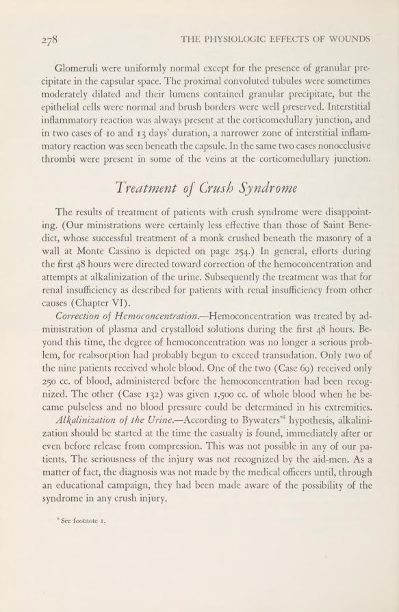
278 THE PHYSIOLOGIC EFFECTS OF WOUNDS
Glomeruli were uniformly normal except for the presence of granular pre-cipitate in the capsular space. The proximal convoluted tubules were sometimesmoderately dilated and their lumens contained granular precipitate, but theepithelial cells were normal and brush borders were well preserved. Interstitialinflammatory reaction was always present at the corticomedullary junction, andin two cases of 10 and 13 days’ duration, a narrower zone of interstitial inflam-matory reaction was seen beneath the capsule. In the same two cases nonocclusivethrombi were present in some of the veins at the corticomedullary junction.
Treatment of Crush SyndromeThe results of treatment of patients with crush syndrome were disappoint-
ing. (Our ministrations were certainly less effective than those of Saint Bene-dict, whose successful treatment of a monk crushed beneath the masonry of awall at Monte Cassino is depicted on page 254.) In general, efforts duringthe first 48 hours were directed toward correction of the hemoconcentration andattempts at alkalinization of the urine. Subsequently the treatment was that forrenal insufficiency as described for patients with renal insufficiency from othercauses (Chapter VI).
Correction of Hemoconcentration.—Hemoconcentration was treated by ad-ministration of plasma and crystalloid solutions during the first 48 hours. Be-yond this time, the degree of hemoconcentration was no longer a serious prob-lem, for reabsorption had probably begun to exceed transudation. Only two ofthe nine patients received whole blood. One of the two (Case 69) received only250 cc. of blood, administered before the hemoconcentration had been recog-nized. The other (Case 132) was given 1,500 cc. of whole blood when he be-came pulseless and no blood pressure could be determined in his extremities.
Alkalinization of the Urine.—According to By waters’*’ hypothesis, alkalini-zation should be started at the time the casualty is found, immediately after oreven before release from compression. This was not possible in any of our pa-tients. The seriousness of the injury was not recognized by the aid-men. As amatter of fact, the diagnosis was not made by the medical officers until, throughan educational campaign, they had been made aware of the possibility of thesyndrome in any crush injury.
° See footnote i.

THE CRUSH SYNDROME IN BATTLE CASUALTIES 279
Chart 47. Effect of Alkali Therapy in a Patient with Crush Syndrome
(Case 69)
pH OF URINE
PLASMA CHLORIDES(MILLIEQUIVALENTS PER LITER)
C0 2 COMBINING POWEROF PLASMA(MILLIEQUIVALENTS PER LITER)
ALKALI INTAKE(GRAMS NoHC03 PER DAY)
After admission to the hospital, however, all but two of the nine patientsreceived alkali therapy. Four of the five patients who died were treated withalkali, one starting promptly upon admission to the hospital, one starting dur-ing the first 24-hour period after release from the crushing force, and two dur-ing the second 24-hour period. No beneficial effects were observed that couldbe attributed to the alkali. As a matter of fact, the most striking feature wasthe failure, in the fatal cases, to succeed in alkalinizing the urine. A pH of 7.2was attained in only two specimens of urine in Case 69 (Chart 47). This pa-tient had received larger doses of sodium bicarbonate than the other patients.One patient in this group (Case 124) did have an alkaline urine (pH 8.4) 4hours after intravenous administration of sodium bicarbonate, and it was keptalkaline by the administration of 3 Gm. of the bicarbonate every 4 hours. Thispatient had a minimal renal lesion and survived. Administration of sodium bi-carbonate was done cautiously or abandoned altogether, because of unfortunateexperiences with vigorous attempts at alkalinization in patients with renal in-sufficiency from other causes (Chapter VII).
Diuretics. —Alcohol was used with the hope that it would promote blood
280 THE PHYSIOLOGIC EFFECTS OF WOUNDS
flow through the kidney and serve as a diuretic. It was used intravenously (totaldose; 50 to 100 cc. of 95-percent ethyl alcohol as a 5-percent solution in dextroseor isotonic saline solution), and as whiskey by mouth (60 to 180 cc. per day).No beneficial effects were detected that could be attributed directly to the alco-hol. Three of the patients were given 10-percent dextrose in distilled waterintravenously, and one received 50-percent dextrose intravenously as a diuretic,but no effect was noted in any of them that could be attributed to the medica-tion. The failure of both alcohol and hypertonic solutions of dextrose to promoteurine flow or improve kidney function confirmed the findings in patients withrenal insufficiency from other causes (Chapter VI). Not only did concentratedsolutions fail to induce a diuresis, but in three cases (Cases 70, 78, and 132) thehypertonic dextrose solution may have hastened the onset of pulmonary edema.Mercurial diuretics were not tried in patients with the crush syndrome.
Dehydrating Agents.—On the hypothesis that swelling and edema of thekidney may be in part responsible for failure of function, the use of potassiumand magnesium solutions was recommended by Barker 7 to reduce the tissueswelling and to promote diuresis. Potassium chloride was not available to usfor clinical use, but magnesium sulfate was used intravenously and intramuscu-larly. It was employed in three cases (Cases 69, 78, and 93) in doses of 4 to8 Gm. daily for 3 to 5 days, which was sufficient to elevate the concentration ofmagnesium in the blood as indicated in the two patients in whom such analyseswere made (Cases 78 and 93). In these three patients there was no definite effectupon urinary output, and it is impossible to attribute any beneficial effect tothe use of magnesium. Undue drowsiness in Case 78 could well have beencaused by the magnesium therapy, for his serum concentration of magnesiumwas 5.8 milliequivalents per liter at necropsy. The serum magnesium reacheda level of 6.8 milliequivalents per liter at one stage during the course of treat-ment in Case 93. For the same reasons stated in Chapter VI, it is our opinionthat neither magnesium nor potassium solutions should be used in the treat-ment of this syndrome.
Other Measures.—Study of these nine patients taught us little as to possiblemeans of preventing anuria or inducing diuresis. Generally accepted methodsfailed. On the hypothesis that injury to the kidney may be produced whenreabsorption of extravasated fluid takes place, pressure bandages were consideredworthy of trial and were applied in one case (Case 78) without apparent effect.
1 Barker, Marion H.: Personal communication.
THE CRUSH SYNDROME IN BATTLE CASUALTIES 281
In this case, however, serious extravasation into the tissues had already occurredbefore the patient reached the hospital. Cold applications might prevent rapidflooding of the kidney with large quantities of muscle-breakdown products,inasmuch as cold is known to inhibit the flow of lymph from the peripheryinto the general circulation. This therapy was not tried. Again, however, to beeffective it would have to be started immediately after release from compres-sion, Therapeutic amputation of the involved extremities was thought to beout of the question for the same reason.
What was learned from the patients with crush syndrome, as well as fromthose with renal insufficiency from other causes, was that it is very easy tobring about an increase in plasma volume in these patients by too vigorousattempts at inducing a diuresis with intravenous crystalloids. Once oliguria oranuria is established, it is best to withhold fluid, allowing, preferably by mouth,just enough fluid to compensate for loss from the gastrointestinal tract, andfor insensible loss through the skin, from the lungs, and from the sweat glands.Under ordinary circumstances, these requirements can be met by 1,000 cc. orless of fluid daily; more must be allowed if excessive extra-renal losses occur.In general, patients with renal insufficiency have been found to be over-hydratedrather than under-hydrated, and this accounts for the fact that pulmonary edemais a common cause of death.
SUMMARY AND CONCLUSIONS
A laboratory and clinical study was made of nine patients who receivedcrushing injuries; four of the patients survived, five died. It is emphasized thatthe crush syndrome develops insidiously in patients who appear well whenfirst seen. All patients who have suffered compression interfering with the cir-culation of muscle masses for periods of an hour, and perhaps even less, shouldbe observed carefully for development of features of the syndrome; namely,tense swelling of the affected parts, hemoconcentration, benzidine-positive pig-ment (presumably myoglobin) free in the plasma and urine, and oliguria oranuria. If examinations are not made within 24 or 48 hours, the hemoconcen-tration and the myoglobinuria may be missed. The volume of urinary outputshould be watched closely, for significant oliguria may easily be overlooked.
The principal pathologic findings are in the compressed muscles and in thekidneys. In the former the normal pigment, myoglobin, was rapidly mobilized
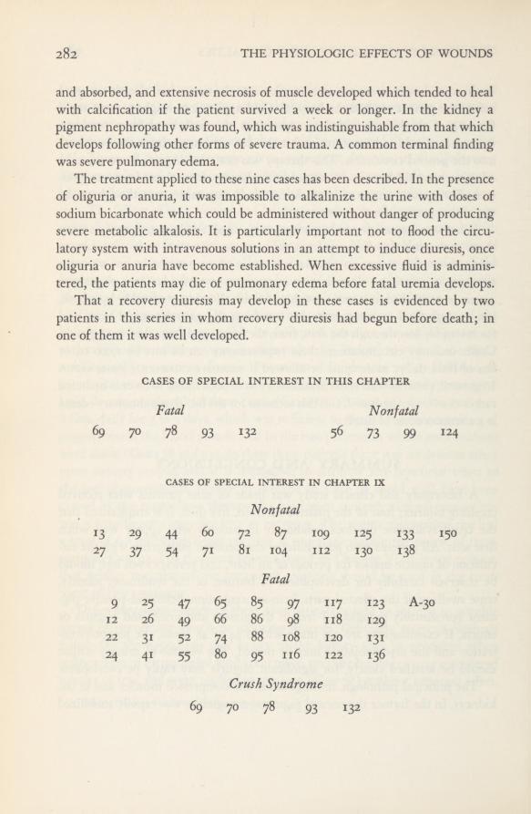
282 THE PHYSIOLOGIC EFFECTS OF WOUNDS
and absorbed, and extensive necrosis of muscle developed which tended to healwith calcification if the patient survived a week or longer. In the kidney apigment nephropathy was found, which was indistinguishable from that whichdevelops following other forms of severe trauma. A common terminal findingwas severe pulmonary edema.
The treatment applied to these nine cases has been described. In the presenceof oliguria or anuria, it was impossible to alkalinize the urine with doses ofsodium bicarbonate which could be administered without danger of producingsevere metabolic alkalosis. It is particularly important not to flood the circu-latory system with intravenous solutions in an attempt to induce diuresis, onceoliguria or anuria have become established. When excessive fluid is adminis-tered, the patients may die of pulmonary edema before fatal uremia develops.
That a recovery diuresis may develop in these cases is evidenced by twopatients in this series in whom recovery diuresis had begun before death; inone of them it was well developed.
CASES OF SPECIAL INTEREST IN THIS CHAPTER
Fatal69 70 78 93 132
Nonfatal56 73 99 124
CASES OF SPECIAL INTEREST IN CHAPTER IX
Nonfatal13 29 44 60 72 87 109 125 133 15027 37 54 71 81 104 112 130 138
Fatal9 25 47 65 85 97 117 123 A-30
12 26 49 66 86 98 118 12922 31 52 74 88 108 120 13124 41 55 80 95 116 122 136
Crush Syndrome
69 70 78 93 132
CHAPTER XII
General Pathology ofTraumatic Shock
To demonstrate a relationship between a parenchymal degenerative changeor pattern of changes and the clinical state of shock several conditions must
be satisfied. The lesions must first be sufficiently distinctive, so that there canbe no possibility of confusion with postmortem degeneration or the artefactsof poor fixation and faulty histologic technique. Second, they must be clearlydistinguishable from the group of agonal changes seen in the tissues of anyperson whose death has not been instantaneous or at least relatively rapid. Spe-cificity of individual lesion cannot reasonably be expected, but the changes shouldbe demonstrable in a high proportion of shock cases, absent in cases of suddendeath, and relatively infrequent in cardiac, metabolic, or cerebral deaths inwhich shock-like states are comparatively uncommon. There should be a de-monstrable time-relationship between the onset of shock and appearance ofthe lesions, and conversely between recovery from shock and their disappearance.So far as could be ascertained, no pattern of lesions which fulfills these con-ditions 1 in man has been described in the literature.
The anatomic changes associated with shock may be usefully divided into(i) those which occur concomitantly with the state of shock and thereforeare of interest in relation to its pathogenesis and (2) those which follow shockand therefore may be considered as its consequences. In studies of the pathologyof human shock attention has heretofore been concentrated primarily on theformer. In the present work attention is particularly devoted to the latter, buttwo brief sections dealing with pulmonary edema and with fat embolism havebeen included because of the extensive consideration given to them by previousinvestigators.
In our own preliminary investigations, the search for possible sequelae of1 Moon, V. H.: Analysis of shock. Brit. M. J. 1: 773-779, June 10, 1944.
956507 0—52 22

284 THE PHYSIOLOGIC EFFECTS OF WOUNDS
shock seemed fruitless for many months. The postmortem material of severalhundred battle casualties was vainly examined for any lesion or combination oflesions which would fulfill the imposed conditions. The organs of patients dyingafter 4, 8, or 12 hours of prolonged shock showed little evidence of histologicchange and none which could not readily be duplicated in a series of non-shockcases. When, however, the range of investigation was broadened to includematerial from casualties who lived more than 18 hours after injury, and whenfrozen sections stained for fat with Sudan IV were substituted for paraffin sec-tions, a fairly constant pattern was disclosed. Evidence was obtained that ap-proximately 18 hours after a shock-producing injury, fat vacuolation appearsin the parenchymal cells of the heart, liver, and kidneys. These changes in-creased in frequency and severity up to 96 hours after injury, then progressivelydeclined with longer periods of convalescence from the episode of shock, un-less complicating factors, such as fat embolism or severe infection, preventedresolution of the process.
The pancreas and adrenal were also studied in the same group of cases.In the pancreas no lesion was recognized which appeared to correlate withshock. In the adrenal gland a series of changes was readily demonstrable: swell-ing of the cortex, pseudotubular degeneration, and depletion of stainable lipid,particularly of doubly refractile lipid. Since these changes did not differ quali-tatively and were less marked quantitatively than those seen in cases with in-fection and in other types of control material, their significance is questionable.Only one factor, the depletion of doubly refractile lipid elements, showed atendency to reversibility with increasing time intervals following resuscitationfrom shock.
MaterialThe nucleus of material for this section of the study was provided by the
necropsy protocols and corresponding microscopic preparations from 60 casesthat had been studied clinically by members of the Board. As the work pro-gressed, the importance of sepsis as a complicating factor became apparent.Additional cases were therefore selected from the files of the 15th MedicalGeneral Laboratory in which the clinical data were sufficiently detailed to estab-lish the diagnosis of shock and in which major sepsis could be excluded. Nu-merous additional examples of acute shock were included in order to cover a
285GENERAL PATHOLOGY OF TRAUMATIC SHOCK
wider time-range than was provided by the Board’s cases. In the tables to
follow, these supplementary cases have been included unless the tables are des-ignated as “Board Cases.”
Control material was drawn both from the 15th Laboratory and from theArmy Institute of Pathology. This included several groups of cases: (1) instan-taneous or sudden deaths, such as homicides and motor vehicle accidents, (2)“medical deaths” unselected except for elimination, as far as possible, of “shock-like” states (the group included cerebral, cardiac, and infectious diseases), (3)deaths primarily due to starvation (from necropsies done in a German con-centration camp), and (4) deaths from aplastic anemia. Not all organs wereavailable for fat stains in all of these cases; consequently the number of con-trols varies somewhat from one section to another. The starvation and aplasticanemia groups were selected because of the known frequency with which fattyvacuolation of various organs occurs under these conditions.
The majority of necropsies on the combat soldier had been performed underfield conditions by an officer of a field or evacuation hospital whose experiencein pathology was often limited. (In the original 60 cases, one of the Boardmembers was usually present at the necropsy.) Deaths were most numerousat times of heightened military activity when hospital staffs were overworkedin caring for the living. Necropsies were sometimes conducted in hospital op-erating rooms but more often in tents without benefit of flooring, heat, or ade-quate illumination. Scales were rarely available. The examinations and recordswere inevitably incomplete. The central nervous system was rarely examined,the intestinal tract seldom opened, and wounds of the extremities were givenbut cursory attention to determine the presence or absence of gross infection,major vascular involvement, or extensive necrosis of muscle. Blocks of repre-sentative tissues were fixed in formalin, occasionally in Zenker’s solution, andtransmitted to the Pathology Section of the 15th Medical General Laboratorywhere they were sectioned and the histologic findings were reviewed by theauthor.
FindingsLungs
The Board’s cases include no examples of sudden death; hence the lungswere never normal. Congestion was described in every case; edema in approxi-
286 THE PHYSIOLOGIC EFFECTS OF WOUNDS
mately 85 percent; atelectasis in 70 percent; fat embolism in 65 percent; intra-alveolar hemorrhage in 55 percent; interstitial hemorrhage in 25 percent, andpneumonia in 30 percent. Because two of these lesions, pulmonary edema andfat embolism, have received considerable attention in relation to the mechanismof shock, their significance is worth examining in more detail.
Pulmonary Edema. —Estimation of the severity of pulmonary edema is diffi-cult from the available data in view of the large number of prosectors and thefew lung weights recorded. A rough classification into three grades has beenattempted for 47 cases in which adequate information was recorded in the ne-cropsy protocols: Grade I, little or no edema (classified as “slight”) with lungweights probably below 800 Gm.; Grade II, moderate edema with lung weightsbetween 800 and 1,200 Gm.; Grade III, severe edema with lung weights from1,200 to 1,800 grams. Fifteen cases were Grade I (slight), 14 Grade II (moder-ate), and 18 Grade III (severe).
Several factors might influence the development of pulmonary edema inthese patients. It has been suggested 2 that pulmonary edema is a constant andintegral part of the shock mechanism. It is certainly a common sequela of renalinsufficiency, from which a large proportion of the patients in this series suf-fered. It could be due to myocardial failure and evidence will be presented laterin this chapter that myocardial degeneration actually was present in manycases. It might be influenced by fat embolism and finally it could be broughtabout, or at least intensified, by over-enthusiastic intravenous fluid therapy.
In Table 101 the degree of pulmonary edema is compared with the periodof survival in 47 shock cases. The patients dying in less than 48 hours representby and large the group in which resuscitation was unsuccessful. One patient(Case 22) briefly appeared to be resuscitated only to develop two secondaryattacks of circulatory failure, in the last of which he died. These patients mayfairly be considered to have died in, perhaps of, severe and prolonged shock.Five had minimal, 2 moderate, and only 3 severe edema. All had received re-peated infusions of plasma and whole blood.
In the patients who survived into the third or fourth days, it is safe to assumethat shock had disappeared. In the majority of these, incipient renal insuffi-ciency was manifest, and only one patient failed to show a nephropathy at
2 Moon, V. H.: Shock, its mechanism and pathology. Arch. Path. 24; 642—663 (November) and 794—813 (December), 1937.
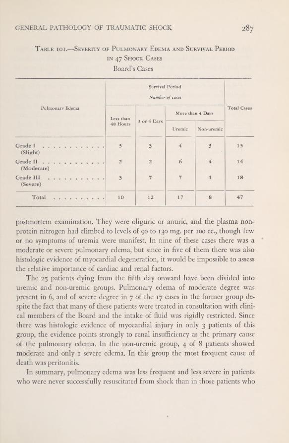
GENERAL PATHOLOGY OF TRAUMATIC SHOCK 287Table i01.—Severity of Pulmonary Edema and Survival Period
in 47 Shock CasesBoard’s Cases
postmortem examination. They were oliguric or anuric, and the plasma non-protein nitrogen had climbed to levels of 90 to 130 mg. per 100 cc., though fewor no symptoms of uremia were manifest. In nine of these cases there was amoderate or severe pulmonary edema, but since in five of them there was alsohistologic evidence of myocardial degeneration, it would be impossible to assessthe relative importance of cardiac and renal factors.
The 25 patients dying from the fifth day onward have been divided intouremic and non-uremic groups. Pulmonary edema of moderate degree waspresent in 6, and of severe degree in 7 of the 17 cases in the former group de-spite the fact that many of these patients were treated in consultation with clini-cal members of the Board and the intake of fluid was rigidly restricted. Sincethere was histologic evidence of myocardial injury in only 3 patients of thisgroup, the evidence points strongly to renal insufficiency as the primary causeof the pulmonary edema. In the non-uremic group, 4 of 8 patients showedmoderate and only 1 severe edema. In this group the most frequent cause ofdeath was peritonitis.
In summary, pulmonary edema was less frequent and less severe in patientswho were never successfully resuscitated from shock than in those patients who
Survival Period
Number of cases
Pulmonary Edema
Less than 3 or 4 Days
More than 4 DaysTotal Cases
48 HoursUremic Non-uremic
Grade I(Slight)
5 3 4 3 15
Grade II(Moderate)
2 2 6 4 14
Grade III(Severe)
3 7 7 1 18
Total 10 12 17 8 47
288 THE PHYSIOLOGIC EFFECTS OF WOUNDS
did recover from the acute stage of shock but subsequently displayed evidenceofrenal insufficiency. Ouranatomic data offer no confirmation for the hypothesisthat loss of fluid into the alveoli is an important factor in the development ofshock.
Pulmonary Fat Embolism. —Pulmonary fat embolism is a very frequentcomplication of battle casualties, as the 65-percent incidence in the present groupof cases shows. Its significance is extremely difficult to assess. In the early stagesof World War 1, much attention was given to it and it was considered by someinvestigators'5 to be an important, perhaps the most important cause of shock.Further experience failed entirely to substantiate the hypothesis, the theoriesof Bayliss and Cannon 4 became dominant, and fat embolism was forgotten.
Frozen sections were cut and stained for fat in all of the present series ofcases in which lung tissue was available (51 cases). An attempt was made toestimate the severity of the embolism in three grades as follows: In Grade Ionly an occasional droplet is seen. In Grade II droplets are fairly numerous; theyare found chiefly in large vessels or arterioles and seldom have passed into thecapillaries of the alveolar walls. In Grade Ilia considerable proportion, a quarterto half or more, of the arterioles are plugged, and in numerous areas capillariesare filled as if by an injection mass. Systemic fat embolism of significant gradewas never found in association with Grades I and II but was present in abouta fifth of the cases in Grade III. Judged by experience covering several hundredbattle casualties, the author considers it improbable that Grades I and II ofpulmonary fat embolism are functionally significant.
In Table 102 the grade of fat embolism is compared with the degree ofshock. It is apparent from inspection of the table that Grade III fat embolismwas present in only 8 of the 39 cases of moderately severe or severe shock. Con-versely, of 10 patients with significant fat embolism, 8 were estimated to havemoderate or severe shock. It is evident that fat embolism is not the cause ofshock in any considerable proportion of cases. Conversely, severe pulmonaryfat embolism was always associated with shock, usually of moderate or severe
3 Porter, W. T.: Fat embolism, a cause of shock. Boston M. & S. J. 176; 248, February 15, 1917.4 Great Britain. Medical Research Committee. Special Report Series, No. 26. Traumatic Toxaemia
as a Factor in Shock. Oxford, His Majesty’s Stationery Office, 1919. IV. Cannon, W. B., and Bayliss, W. M.:Note on muscle in relation to shock, p. 19—23. V. Bayliss, W. M.: Further observations on the results ofmuscle injury and their treatment, p. 23—26. VI. Cannon, W. B.; Some characteristics of shock induced bytissue injury, p. 27—32,
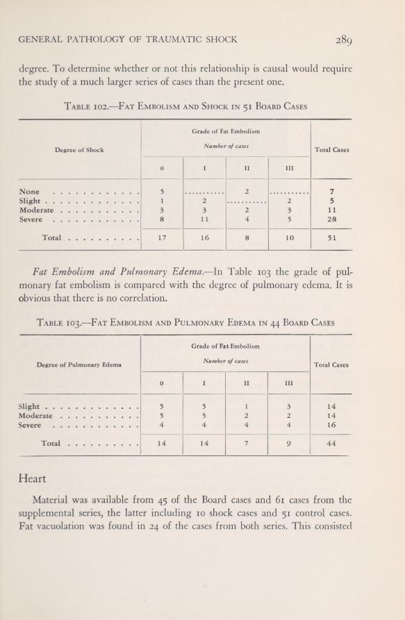
GENERAL PATHOLOGY OF TRAUMATIC SHOCK 289degree. To determine whether or not this relationship is causal would requirethe study of a much larger series of cases than the present one.
Table 102.—Fat Embolism and Shock in 51 Board Cases
Fat Embolism and Pulmonary Edema.—In Table 103 the grade of pul-monary fat embolism is compared with the degree of pulmonary edema. It isobvious that there is no correlation.
Table 103.—Fat Embolism and Pulmonary Edema in 44 Board Cases
Heart
Material was available from 45 of the Board cases and 61 cases from thesupplemental series, the latter including 10 shock cases and 51 control cases.Fat vacuolation was found in 24 of the cases from both series. This consisted
Degree of Shock
0
Grade of Fa
Number
1
t Embolism
of cases
II III
Total Cases
3 2 71 2 2 5
Moderate 3 3 2 3 11Severe 8 11 4 5 28
Total 17 16 8 10 51
Degree of Pulmonary Edema
Grade of Fat Embolism
Number of cases Total Cases
0 I II III
Slight 5 5 1 3 14Moderate 5 5 2 2 14Severe 4 4 4 4 16
Total 14 14 7 9 44

THE PHYSIOLOGIC EFFECTS OF WOUNDS290
FIGURE 29. Frozen section of myocardium stained with Sudan IV. The patient died 48hours after a shock-producing wound. Many cells are apparently filled with minute vacuoles,from 2 to 3 microns in diameter, which appear as bright red granules. The affected cells are
not swollen and the nuclei appear normal. X 70
of very minute vacuoles, seldom more than 2 microns in diameter, arrangedin parallel rows between the myofibrils (Fig. 29). When present in a cell, thevacuolation was apparent throughout the cell’s entire length and breadth,rarely segmentally or focally. It was entirely independent of the amount oflipochrome demonstrable at the nuclear poles. The affected cell often seemedslightly swollen in the frozen section but this change could not be recognizedwith certainty in the paraffin sections. Sometimes single cells, more oftengroups of from 10 to 50 adjacent fibers, were affected. Vacuolation was neverdiffuse throughout the myocardium. In appearance it was strongly reminis-cent of, though usually less severe than the patchy fat vacuolation seen in severeanemia. However, no gross changes suggestive of “tigering" were ever noted.
The severity of the change, estimated by the number and size of the vacuolesper cell and the proportion of cells involved, was roughly quantitated on a zeroto 3-plus scale. The degree of involvement is compared with the survival periodin Table 104. It is evident that fat vacuolation of cardiac muscle cells was notobserved in previously healthy young men who developed shock followingtrauma but failed to survive at least 18 hours. In patients manifesting shockwho lived from 18 to 96 hours, fat vacuolation of the myocardium was found

GENERAL PATHOLOGY OF TRAUMATIC SHOCK 291
in 75 percent. With longer periods of survival the incidence dropped sharplyto 17 percent, and there were no cases of grade 3-f- severity.
Table 104.—Myocardial Fat Vacuolation in 55 Shock Cases*
Focal fat vacuolation of cardiac muscle cells has been noted frequently bythe author in cases of systemic fat embolism with embolization of the myo-cardial capillaries. In only two of the group under consideration was this com-plication present. These two men died on the fifth and sixth days after injury.If they are excluded, the incidence of fat vacuolation in the patients survivingmore than 4 days falls to n percent.
In Table 105 the degree of shock and the severity of fat vacuolation arecompared in 14 patients among those dying within the 18- to 96-hour periodwho had been examined during life by a clinical member of the Board. Thenumber of cases is small, but increasing intensity of fat vacuolation is apparentas shock becomes more severe.
Table 105.—Degree of Shock and Fat Vacuolation of Myocardium in 1/
Board Patients who Survived from 18 to 96 Hours
Survival Period Number of Cases
0
Grade of Fat
Number
1 +
Vacuolatior
ofcases
2 + 3 +
PercentPositive
Less than 18 Hours 10 10 0 0 0 018 to 96 Hours . . 16 4 4 2 6 75More than 4 Days . 29 24 2 3 0 17
*45 Board cases and 10 from the supplemental series.
Degree of Shock
Grade of Fat Vacuolation
Number ofcases
0 1 + 2 + 3 +
Slight 0 0 1 0Moderate 1 2 1 1Severe 2 1 0 5
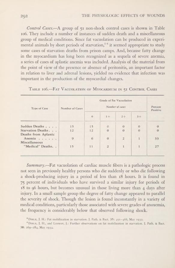
THE PHYSIOLOGIC EFFECTS OF WOUNDS292
Control Cases.—A group of 51 non-shock control cases is shown in Table106, They include a number of instances of sudden death and a miscellaneousgroup of medical conditions. Since fat vacuolation can be produced in experi-mental animals by short periods of starvation/' *’ it seemed appropriate to studysome cases of starvation deaths from prison camps. And, because fatty changein the myocardium has long been recognized as a sequela of severe anemia,a series of cases of aplastic anemia was included. Analysis of the material fromthe point of view of the presence or absence of peritonitis, an important factorin relation to liver and adrenal lesions, yielded no evidence that infection wasimportant in the production of the myocardial changes.
Table 106.—Fat Vacuolation of Myocardium in 51 Control Cases
Summary.—Fat vacuolation of cardiac muscle fibers is a pathologic processnot seen in previously healthy persons who die suddenly or who die followinga shock-producing injury in a period of less than 18 hours. It is found in75 percent of individuals who have survived a similar injury for periods of18 to 96 hours, but becomes unusual in those living more than 4 days afterinjury. In a small sample group the degree of fatty change appeared to parallelthe severity of shock. Though the lesion is found inconstantly in a variety ofmedical conditions, particularly those associated with severe grades of anoxemia,the frequency is considerably below that observed following shock.
6 Dible, J. H.: Fat mobilization in starvation. J. Path. & Bact. 35: 451—466, May 1932.6 Dible, J. H., and Libman, J.: Further observations on fat mobilization in starvation. J. Path. & Bact.
38: 269—284, May 1934.
Type of Case Number ofCases
Grade of Fat Vacuolation
Number of cases PercentPositive
0 1 + 2 + 3 +
Sudden Deaths .... 15 15 0 0 0 0Starvation Deaths . . . 12 12 0 0 0 0Deaths from Aplastic
Anemia ...... 9 6 0 2 1 33Miscellaneous
"Medical” Deaths. . 15 11 2 1 1 27

GENERAL PATHOLOGY OF TRAUMATIC SHOCK 293
FIGURE 30. Frozen section of liver stained with Sudan IV. The patient died 72 hours aftera shock-producing wound. The illustration shows the center of a lobule with some fine fatvacuolation of all visible liver cells. X 400
Liver
Fat vacuolation of the liver appears in such a variety of conditions and maybe present in so many apparently normal individuals that one is often temptedto regard it as not significant unless the amount is very great. The usual formobserved consists of vacuoles of considerable size, ranging from 5 to 20 micronsin diameter. Such vacuoles are readily visible in paraffin sections with lowmagnification (from 16- to 30-mm, objectives) inasmuch as they occupy thefull thickness of the section and appear as sharply outlined holes. Because oftheir large size there are never many vacuoles in a single cell.
Fat vacuolation of this type was rarely seen in shock cases, and when foundprobably existed before the shock-producing injury. Extensive fat vacuolationwas nevertheless readily demonstrable in shock if sufficient time had elapsedfor its development. This form of fat vacuolation consisted almost entirely ofvery fine droplets in the range from 2 to 4 microns in diameter (Fig. 30). Evenwhen the process was severe, though slightly larger droplets were found, therewas little tendency for them to fuse, and as many as 15 or 20 might be presentin a single cell. The vacuolation is readily seen in paraffin sections if the 4-mm.lens is used. Few of them extend through the thickness of the section, but twoor more layers can be seen by varying the depth of focus. Because of the small
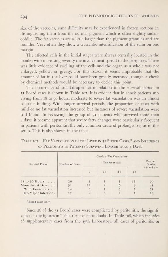
THE PHYSIOLOGIC EFFECTS OF WOUNDS294
size of the vacuoles, seme difficulty may be experienced in frozen sections indistinguishing them from the normal pigment which is often slightly sudan-ophilic. The fat vacuoles are a little larger than the pigment granules and arerounder. Very often they show a crescentic intensification of the stain on onemargin.
The affected cells in the initial stages were always centrally located in thelobule; with increasing severity the involvement spread to the periphery. Therewas little evidence of swelling of the cells and the organ as a whole was notenlarged, yellow, or greasy. For this reason it seems improbable that theamount of fat in the liver could have been greatly increased, though a checkby chemical methods would be necessary to decide this point.
The occurrence of small-droplet fat in relation to the survival period in51 Board cases is shown in Table 107, It is evident that in shock patients sur-viving from 18 to 96 hours, moderate to severe fat vacuolation was an almostconstant finding. With longer survival periods, the proportion of cases withmild or no fat vacuolation increased but instances of severe vacuolation werestill found. In reviewing the group of 31 patients who survived more than4 days, it became apparent that severe fatty changes were particularly frequentin patients with peritonitis, the only common cause of prolonged sepsis in thisseries. Th is is also shown in the table.
Table 107.—Fat Vacuolation in the Liver in 51 Shock Cases,* and Influenceof Peritonitis in Patients Surviving Longer than 4 Days
Since 26 of the 51 Board cases were complicated by peritonitis, the signifi-cance of the figures in Table 107 is open to doubt. In Table 108, which includes28 supplementary cases from the 15th Laboratory, all cases of peritonitis or
Survival Period Number ofCases
Grade of Fat Vacuolation
Number of cases PercentGrades
2 + and 3+
0 1 + 2 + 3 +
1 8 to 96 Hours. . . . 20 1 1 3 15 90More than 4 Days. . . 31 12 4 6 9 48
With Peritonitis ..
14 3 1 3 7 71No Major Infection . 17 9 3 4 1 29
*Board cases only.

GENERAL PATHOLOGY OF TRAUMATIC SHOCK 295
other major sepsis have been excluded. In this selected series, it is apparentthat fat vacuolation of more than minimal degree was seldom seen in shockpatients who failed to survive at least 18 hours. Of those who survived from18 to 96 hours, 87 percent showed the presence of fat, 59 percent in moderateor severe degree. With survival beyond 96 hours, the tendency to return tonormal is seen, the total dropping to 47 percent and that for the more severegrades to 29 percent.
Table 108.—Centrolobular Fat Vacuolation of Liver Cells in 53 ShockCases Uncomplicated by Major Infection*
Control Cases.—Since fat vacuolation of the liver is such a common phe-nomenon and can be produced by so many etiologic factors, numerous controlcases seemed necessary. Seventeen instances of sudden death were selected atrandom from the laboratory files. Fifteen cases of “medical” deaths (cardiac,cerebral, nephritic) without obvious shock-like states, 20 instances of deathfrom aplastic anemia, and 38 starvation deaths were included—a total of 90control cases. The incidence of centrolobular fat vacuolation in 36 shock casesand 90 controls is shown in Table 109.
The occurrence of moderate or severe fat vacuolation in 24 percent of thesudden-death group is worthy of comment. This group was compiled largelyfrom motor-vehicle accident and homicide cases in base-section troops. Theproportion of limited-service personnel and older men was higher in suchtroops than in combat organizations. Two of the men in the group studiedhad been intoxicated at the time of death and had enlarged, grossly fatty liverssuggestive of chronic alcoholism. In contrast, the acute-shock group, with sur-vival periods shorter than 18 hours, was made up almost entirely of battle
Survival Period Numberof Cases
Grade of Fat Vacuolation
Number of cases PercentPositive
PercentGrade
2+ or 3 +
0 1 + 2 + 3+
Less than 18 Hours .... 14 11 2 1 0 22 8.2
18 to 96 Hours 22 3 6 4 9 87 59
More than 4 Days 17 9 3 4 1 47 29
* Includes 25 Board cases and 28 supplementary shock cases.

296 THE PHYSIOLOGIC EFFECTS OF WOUNDS
casualties, all previously healthy and vigorous young men, and therefore repre-sents a better control group than the “sudden deaths.” The relatively highincidence (40 percent) in the miscellaneous medical deaths is not surprisingin view of the multiple etiology of fat deposit in the liver, but is still not ashigh as the frequency (59 percent) in the 18- to 96-hour shock group. Thelow incidence in the anemia group proved surprising in view of the knownfrequency of myocardial fat vacuolation in such conditions. The starvationcases provided a most interesting contrast to the shock material. Although fatwas present in the majority of these cases, it was always manifest at the periphery,never at the center of the lobule.
Table 109.—Centrolobular Fat Vacuolation of Liver in Shock Casesand Controls
Summary.—Eighteen hours after a shock-producing injury, fat vacuolationwas demonstrable in the liver cells at the center of the lobule in 87 percentof 53 cases uncomplicated by major infection and was of moderate or severegrade in 59 percent. After the fourth day it tended to disappear in cases un-complicated by peritonitis. The incidence in the 18- to 96-hour cases was some-what higher than in any of the control group studied.
Type of Case Number of Cases
Grade of Fat Vacuolation
Number ofcases PercentGrades
2+or 3+
0 l + 2 + 3 +
SHOCK CASESLess than 18 Hours’
Survival 14 11 2 1 0 8.218 to 96 Hours’
Survival 22 3 6 4 9 59
CONTROLSSudden Deaths . . . 17 8 5 2 2 24Miscellaneous
"Medical” Deaths. 15 3 6 3 3 40Deaths from Aplastic
Anemia 20 10 6 1 3 20Starvation Deaths . . 38 0 0 0 0 0
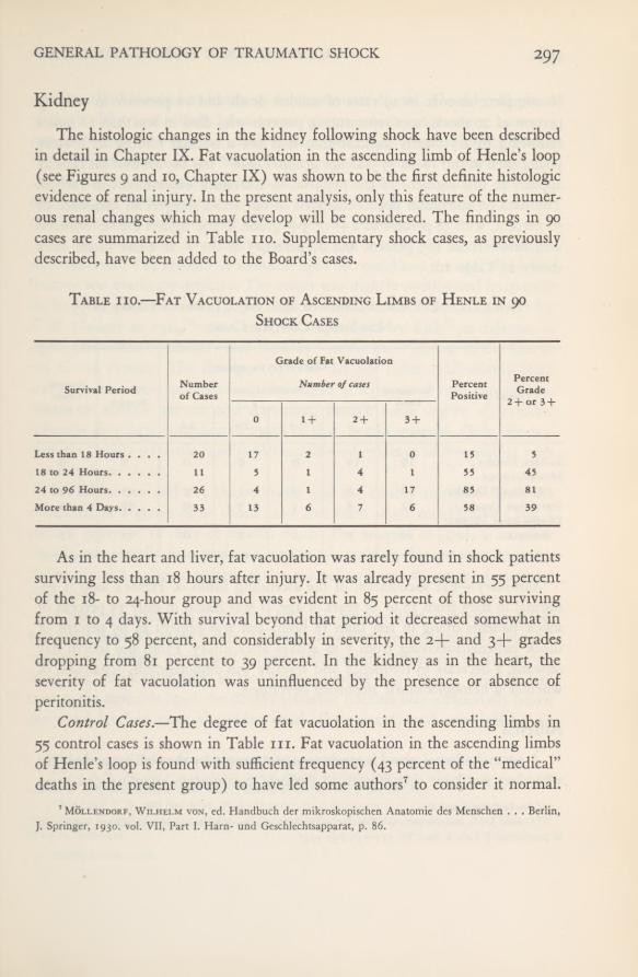
GENERAL PATHOLOGY OF TRAUMATIC SHOCK 297
KidneyThe histologic changes in the kidney following shock have been described
in detail in Chapter IX. Fat vacuolation in the ascending limb of Henle’s loop(see Figures 9 and 10, Chapter IX) was shown to be the first definite histologicevidence of renal injury. In the present analysis, only this feature of the numer-ous renal changes which may develop will be considered. The findings in 90cases are summarized in Table no. Supplementary shock cases, as previouslydescribed, have been added to the Board’s cases.
Table no.—Fat Vacuolation of Ascending Limbs of Henle in 90Shock Cases
As in the heart and liver, fat vacuolation was rarely found in shock patientssurviving less than 18 hours after injury. It was already present in 55 percentof the 18- to 24'hour group and was evident in 85 percent of those survivingfrom 1 to 4 days. With survival beyond that period it decreased somewhat infrequency to 58 percent, and considerably in severity, the 2-f- and 3+ gradesdropping from 81 percent to 39 percent. In the kidney as in the heart, theseverity of fat vacuolation was uninfluenced by the presence or absence ofperitonitis.
Control Cases.—The degree of fat vacuolation in the ascending limbs in55 control cases is shown in Table in. Fat vacuolation in the ascending limbsof Henle’s loop is found with sufficient frequency (43 percent of the “medical”deaths in the present group) to have led some authors 7 to consider it normal.
7 Mollendorf, Wilhelm von, ed. Handbuch der mikroskopischen Anatomic des Menschen . . . Berlin,J. Springer, 1930. vol. VII, Part I. Harn- und Geschlechtsapparat, p. 86.
Survival Period Numberof Cases
Grade of Fat Vacuolation
Number of cases PercentPositive
PercentGrade
2+ or 3 +
0 1 + 2 + 3 +
Less than 18 Hours .... 20 17 2 1 0 15 518 to 24 Hours 11 5 1 4 1 55 4524 to 96 Hours 26 4 I 4 17 85 81More than 4 Days 33 13 6 7 6 58 39

298 THE PHYSIOLOGIC EFFECTS OF WOUNDS
Its complete absence in 19 cases of sudden death and its presence in only 15percent of 20 shock cases representing patients who died in less than 18 hoursfollowing injury clearly indicate that it is not a normal finding in healthyyoung men in the 18- to 35-year age group. Dible 8 9 has shown that fat rapidlyappears in this segment of the nephron in starvation experiments in rabbits.The data on starvation deaths included in our series showed an incidence of50 percent, with 38 percent of 2-(- or 3-f- grade. These figures are neverthelesswell below those (85 percent and 81 percent) of the 1- to 4-day shock casesshown in Table no.
Table hi.—Fat Vacuolation of Ascending Limbs of Henle in 55Non-Shock Control Cases
Summary.—Fat vacuolation of the ascending limbs of Henle’s loop is ab-normal in men from 18 to 35 years of age. It was found in only 15 percent ofshock patients dying in less than 18 hours but was present in 85 percent ofshock patients surviving from 1 to 4 days after injury. From the fourth dayonward, a decrease in frequency and severity was demonstrable, regardless ofwhether a hemoglobinuric nephrosis developed.
AdrenalA normal adrenal gland is rarely seen at necropsy by the civilian pathologist
who does not have opportunity to perform postmortem examinations upon8 See footnotes 5 and 6.9 Dible, J. H., and Popjak, G.: Distribution of fatty change in kidneys and some factors influencing
its production. J. Path. & Bact. 53: 133—146, July 1941.
Type ofCase Number ofCases
Grade ofFat Vacuolation
Number ofcases PercentPositive
PercentGrade
2 + or 3 +
0 1 + 2 + 3+
Sudden Deaths . . . 19 19 0 0 0 0 0Miscellaneous
"Medical” Deaths 14 8 3 1 2 43 22Starvation Deaths. . 12 8 1 3 0 50 38Deaths from Aplastic
Anemia 10 8 1 1 0 25 13

GENERAL PATHOLOGY OF TRAUMATIC SHOCK 299
persons who have died suddenly without previous disease. The adrenal glandof the healthy young male has a narrow cortex which ranges from i.o to 1.3mm. in width. The cells are richly packed with lipid (stainable with SudanIV), predominantly in the fascicular layer, but numerous vacuoles are alsodemonstrable in the zona glomerulosa and reticularis. Limited to the zonareticularis are large quantities of doubly refractile lipid which serve to outlinethis layer sharply when viewed with crossed Nicol prisms.
In our shock cases no medullary changes but a variety of cortical changeswere observed. The amount of stainable lipid decreased and the optically activefraction was markedly depleted. The cortex was slightly swollen and frequentlyshowed the pseudoacinar type of degeneration described and illustrated byF. B. Mallory in 1914,10 more recently emphasized by Rich 11 in relation tosepsis, and noted by Mallory and Brickley 12 in the burn victims of the Cocoa-nut Grove disaster. The changes observed did not differ qualitatively fromand were less severe quantitatively than those of sepsis or of such control con-ditions as aplastic anemia and starvation, though some degree of terminalinfection may have complicated much of this control material. Only one fea-ture, the doubly refractile lipid depletion, showed evidence of reversibility withrecovery from shock.
In Tables 112 and 113 the stainable and the doubly refractile lipids arecompared in a group of 40 shock cases free from septic complications and acontrol group of 18 cases of sudden death. The amount of lipid, as in otherorgans, was visually estimated on a zero to 3-}- scale. In this instance, however,the maximal 3-f figure represents the normal and zero the stage of maximaldepletion.
A moderate depletion of the total stainable fat is evident in the shock groupsurviving from 18 to 96 hours. There is, however, no evidence of return tonormal with longer intervals of survival after recovery from shock; the processis, in fact, much intensified in the group surviving more than 4 days. In con-trast to the stainable lipid, the doubly refractile fraction was occasionally belowthe usual level in cases of sudden death (17 percent) and was depleted in
“Mallory, F. B.t Principles of Pathologic Histology. Philadelphia, W. B. Saunders, 19x4, p. 653.11 Rich, A. R.: Peculiar type of adrenal cortical damage associated with acute infections, and its possible
relation to circulatory collapse. Bull. Johns Hopkins Hosp. 74: 1—15, January 1944.12 Mallory, T. B., and Brickley, W. J.: Symposium on management of Cocoanut Grove burns at the
Massachusetts General Hospital; pathology, with special reference to pulmonary lesions. Ann. Surg. 117;
865—884, June 1943.
956507 0—52 24

THE PHYSIOLOGIC EFFECTS OF WOUNDS300
Table 112.—Stainable Lipids in Adrenal Cortex in 40 Shock Cases withoutInfection and 18 Cases of Sudden Death
Table 113.—Doubly Refractile Lipids in Adrenal Cortex in 40 Shock Caseswithout Infection and 18 Cases of Sudden Death
27 percent of shock patients dying in less than 18 hours, in one case severelydepleted. In the shock cases of 18 to 96 hours’ survival, this type of lipid wasdiminished in all but one instance (92 percent of the cases) and markedly so
Type of Case Number ofCases
Amount of Lipid
Number ofcases PercentDiminished
PercentGreatly
Diminished3 + 2 + 1 + 0
SHOCK CASESLess than 18
Hours’ Survival. 11 11 0 0 0 0 018 to 96 Hours’
Survival .... 13 8 3 2 0 38 15More than 4 Days’
Survival .... 16 4 8 4 0 75 25
CONTROLSSudden Death . . 18 18 0 0 0 0 0
Type of Case Number ofCases
Amount of Lipid
Number of cases PercentDiminished
PercentGreatly
Diminished3 + 2 + i + 0
SHOCK CASESLess than 18
Hours’ Survival. 11 8 2 1 0 27 918 to 96 Hours’
Survival .... 13 1 4 7 1 92 61More than 4 Days’
Survival .... 16 3 7 3 3 81 38
CONTROLSSudden Death . . 18 15 3 0 0 17 0

GENERAL PATHOLOGY OF TRAUMATIC SHOCK 301
in eight cases, or 61 percent. In those patients surviving more than 4 days,some evidence of a tendency to return to normal is shown by a drop in thepercentage of severe depletion from 61 to 38 percent.
Control Material.—It was difficult to know what kind of control materialwould be suitable inasmuch as most types of illness which lead to death pro-duce similar and even more marked adrenal changes. Four groups of controlcases are listed in Table 114. It is evident that a wide variety of factors affectthe storage of lipidic substances in the adrenal cortex, many of them moreprofoundly than does shock.
Table 114.—Adrenal Cortical Lipids in 44 Control Cases
Summary.—Although numerous changes occur in the adrenal cortex fol-lowing shock, they do not differ in character from, and are less severe thanthose seen in infection and in a wide variety of lethal disorders. The only factorwhich was found to change abruptly 18 hours after a shock-producing injurywas the amount of doubly refractile lipid in the zona reticularis. This wasdiminished in 92 percent of the cases in the 18- to 96-hour group (Table 114).This factor also was the only one in which any tendency could be establishedto return toward normal with increasing time intervals following recoveryfrom shock.
COMMENT
The possible roles of pulmonary edema and of fat embolism in the patho-genesis of shock in men wounded in battle have been discussed in the sectionsdevoted to these phenomena. The pattern of parenchymal degenerative changes
' Stainable Fat Doubly Refractile Lipids
Type of Case Number ofCases Percent
DiminishedPercentGreatly
DiminishedPercent
DiminishedPercentGreatly
Diminished
Miscellaneous "Medical” Deaths. 14 50 22 79 29Starvation Deaths 9 78 44 100 78
Deaths from Aplastic Anemia . . 11 82 18 91 91Fatal Cases with Infection .... 10 100 80 100 100
THE PHYSIOLOGIC EFFECTS OF WOUNDS3 02
described in the heart, liver, kidneys, and adrenals deserves further considera-tion as a probable consequence of shock. None of the changes described arespecific for the state of shock. Fat is known to make its appearance in one oranother of these organs in response to a variety of pathologic conditions suchas anoxemia, starvation, and chemical or bacterial toxic agents—and lipidsdisappear from the adrenal under an even greater variety of conditions. Criticalexamination of the data is therefore necessary before concluding that the ob-served changes were due to shock.
Errors of subjective interpretation in relying upon such vague changes as“parenchymatous degeneration” and “cloudy swelling,” which are readily con-fused with postmortem degeneration and the artefacts of faulty histologictechnique, have been avoided by the use of frozen sections and fat stains;general histologic experience indicates that there is no significant shift in theproportion of stainable fat up to 12 hours after death. Less is known of thebehavior of doubly refractile lipids, but in the material included in this studyno evidence was discovered that this fraction of the fatty substance was affectedby postmortem degeneration within the same time limit. The observed varia-tions from the normal are therefore vital phenomena.
Next in importance are the time relationships to shock. Table 115 showsthe percentage of all cases with abnormal fat vacuolation in the heart, liver,and kidney, and depletion of the doubly refractile lipid in the adrenal, foreach group and for the sudden-death control group. In each of the four organsstudied no significant difference in demonstrable fat is apparent between thesudden-death series and the shock cases in which death occurred in less than18 hours. Abnormal changes are evident, however, in these organs in from75 to 92 percent of shock patients who survived from 18 to 96 hours after injury.The almost synchronous appearance of the lesions 18 hours after injury in allfour organs suggests a single causative factor. The delay in appearance of themorphologic evidence of injury in many instances until shock has been relievedis not incompatible with a causal relationship. Days and even weeks may inter-vene between injury and reaction with such widely varying agents as diphtheriatoxin and x-rays.
Of equal importance in establishing an etiologic relationship between shockand the lesions which have been described is evidence of reversibility, of returnto or at least toward normal with increasing intervals after the episode of shock.As all the necropsy material in this series necessarily derived from individuals
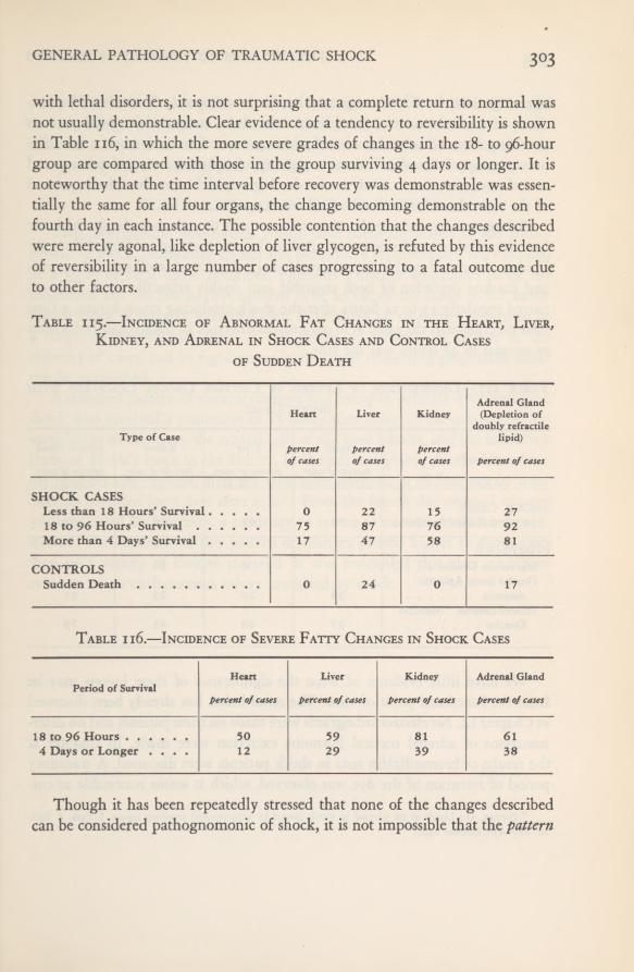
GENERAL PATHOLOGY OF TRAUMATIC SHOCK 3°3
with lethal disorders, it is not surprising that a complete return to normal wasnot usually demonstrable. Clear evidence of a tendency to reversibility is shownin Table 116, in which the more severe grades of changes in the 18- to 96-hourgroup are compared with those in the group surviving 4 days or longer. It isnoteworthy that the time interval before recovery was demonstrable was essen-tially the same for all four organs, the change becoming demonstrable on thefourth day in each instance. The possible contention that the changes describedwere merely agonal, like depletion of liver glycogen, is refuted by this evidenceof reversibility in a large number of cases progressing to a fatal outcome dueto other factors.
Table 115.—Incidence of Abnormal Fat Changes in the Heart, Liver,Kidney, and Adrenal in Shock Cases and Control Cases
of Sudden Death
Table ii6.—Incidence of Severe Fatty Changes in Shock Cases
Though it has been repeatedly stressed that none of the changes describedcan be considered pathognomonic of shock, it is not impossible that the pattern
Type of Case
Heart
percentofcases
Liver
percentofcases
Kidney
percentof cases
Adrenal Gland(Depletion of
doubly refractilelipid)
percent of cases
SHOCK CASESLess than 18 Hours’ Survival 0 22 15 2718 to 96 Hours’ Survival 75 87 76 92More than 4 Days’ Survival 17 47 58 81
CONTROLSSudden Death 0 24 0 17
Period of SurvivalHeart Liver Kidney Adrenal Gland
percent of cases percent of cases percent ofcases percent of cases
18 to 96 Hours 50 59 81 614 Days or Longer .... 12 29 39 38
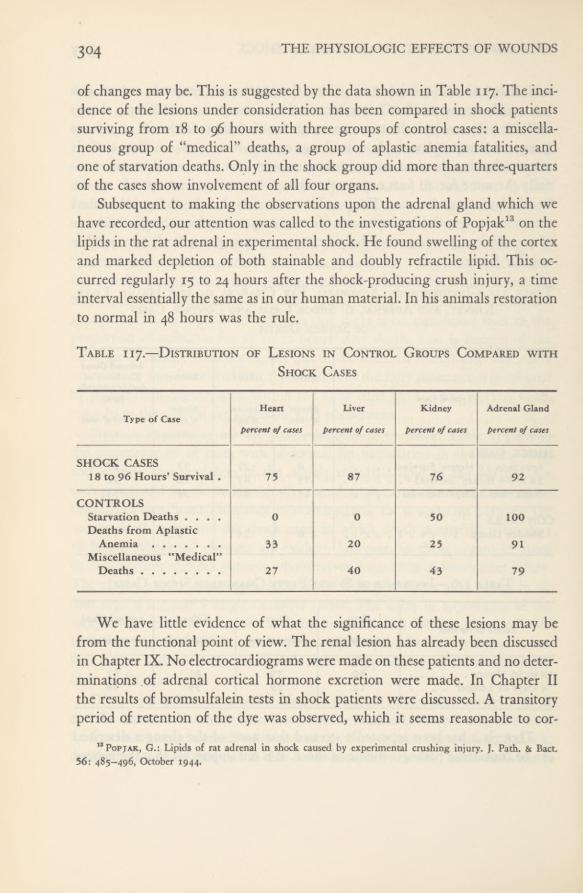
3°4 THE PHYSIOLOGIC EFFECTS OF WOUNDS
of changes may be. This is suggested by the data shown in Table 117. The inci-dence of the lesions under consideration has been compared in shock patientssurviving from 18 to 96 hours with three groups of control cases: a miscella-neous group of “medical” deaths, a group of aplastic anemia fatalities, andone of starvation deaths. Only in the shock group did more than three-quartersof the cases show involvement of all four organs.
Subsequent to making the observations upon the adrenal gland which wehave recorded, our attention was called to the investigations of Popjak 13 on thelipids in the rat adrenal in experimental shock. He found swelling of the cortexand marked depletion of both stainable and doubly refractile lipid. This oc-curred regularly 15 to 24 hours after the shock-producing crush injury, a timeinterval essentially the same as in our human material. In his animals restorationto normal in 48 hours was the rule.
Table i 17.—Distribution of Lesions in Control Groups Compared with
Shock Cases
We have little evidence of what the significance of these lesions may befrom the functional point of view. The renal lesion has already been discussedin Chapter IX. No electrocardiograms were made on these patients and no deter-minations of adrenal cortical hormone excretion were made. In Chapter IIthe results of bromsulfalein tests in shock patients were discussed. A transitoryperiod of retention of the dye was observed, which it seems reasonable to cor-
13 Popjak, G.: Lipids of rat adrenal in shock caused by experimental crushing injury. J. Path. & Bact.56: 485—496, October 1944.
Type of CaseHeart
percent of cases
Liver
percent of cases
Kidney
percent of cases
Adrenal Gland
percent ofcases
SHOCK CASES18 to 96 Hours’ Survival . 75 87 76 92
CONTROLSStarvation Deaths .... 0 0 50 100Deaths from Aplastic
Anemia 33 20 25 91Miscellaneous "Medical”
Deaths 27 40 43 79
GENERAL PATHOLOGY OF TRAUMATIC SHOCK 3°5
relate with the fatty changes in the liver which have been described. Althoughno morphologic evidence of bile stasis was observed in liver sections, the possi-bility of a hepatic as well as a hemolytic factor must be borne in mind ininterpreting the elevated plasma bilirubin often noted in these patients.
SUMMARY AND CONCLUSIONSTwo morphologic phenomena which have been implicated in the patho-
genesis of shock, pulmonary edema and pulmonary fat embolism, were studiedin a series of fatal battle casualties. Pulmonary edema was found to be tooinconstant and too late in development to be an important factor in the initia-tion of shock in severely wounded men. Pulmonary fat embolism proved to bea frequent coincidental lesion. It was absent or minimal in degree in themajority of cases, and its significance in the small remaining proportion couldnot be established.
A standard pattern of visceral changes was found in patients with traumaticshock who survived a minimum of 18 hours after injury. This consisted of fatvacuolation of the heart, the central cells of the liver lobules, and the ascendinglimbs of Henle’s loops in the kidney. In the adrenal gland, the doubly refrac-tile lipid became depleted after the same time interval. In all four organs thesechanges persisted for 3 days after injury. From the fourth day onward in casesuncomplicated by infection, a tendency to return to normal could be demon-strated. The incidence of this pattern of changes proved higher in shock casesthan in a variety of control material. It was concluded that they constituteevidence of parenchymatous injury produced by shock.
APPENDIX A
Organization and Operationof the Board for the Studyof the Severely Wounded
North African (later Mediterranean) Theater of Operations, U.S.A.
ORGANIZATION
The background of events and observations leading to recognition of theneed for an organized study of the severely wounded in an active theater ofwar has been described in the Introduction to this volume. The Chief Surgeon,North African Theater of Operations, Maj. General Morrison C. Stayer,U.S.A., discussed this problem with two members of his staff, Colonel EdwardD. Churchill, MC, Surgical Consultant, and Colonel William S. Stone, MC.On their recommendation he initiated action from which stemmed the Boardwhose work has been presented in this volume. His memorandum to theCommanding General follows.
HEADQUARTERSNORTH AFRICAN THEATER OF OPERATIONS
Office of the Surgeoni September 1944
SUBJECT: Appointment of Medical Board to Study the Treatment of the SeverelyWounded
TO: Quartermaster, Ordnance, G-i, G-4 and AG
1. Outstanding progress in the treatment of the wounded has been made bythe Medical Service in this theater. Many lives have been saved by this work andmany individuals that would have been hopeless wrecks, if they survived, have beenrehabilitated into normal or near normal individuals. Further progress in improv-ing the Medical Service will depend upon proper and timely employment of the
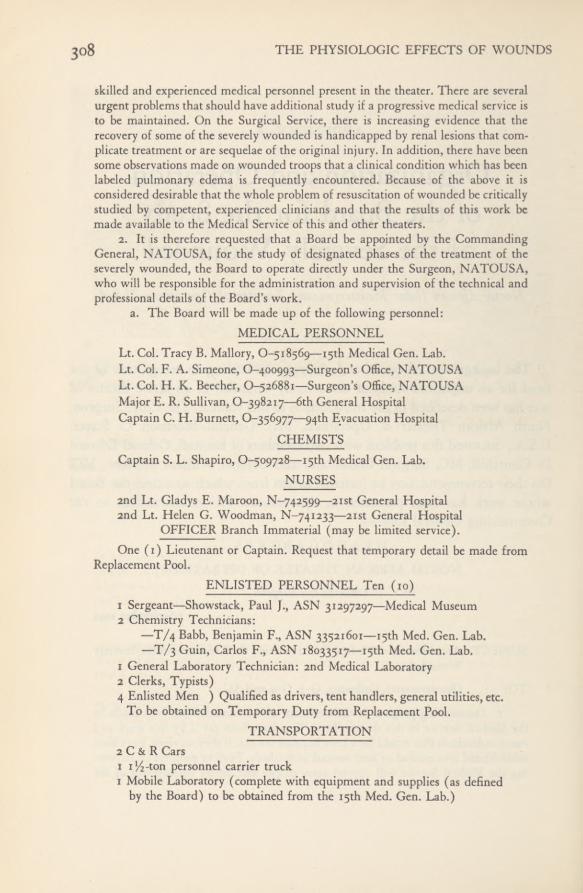
308 THE PHYSIOLOGIC EFFECTS OF WOUNDS
skilled and experienced medical personnel present in the theater. There are severalurgent problems that should have additional study if a progressive medical service isto be maintained. On the Surgical Service, there is increasing evidence that therecovery of some of the severely wounded is handicapped by renal lesions that com-plicate treatment or are sequelae of the original injury. In addition, there have beensome observations made on wounded troops that a clinical condition which has beenlabeled pulmonary edema is frequently encountered. Because of the above it isconsidered desirable that the whole problem of resuscitation of wounded be criticallystudied by competent, experienced clinicians and that the results of this work bemade available to the Medical Service of this and other theaters.
2. It is therefore requested that a Board be appointed by the CommandingGeneral, NATOUSA, for the study of designated phases of the treatment of theseverely wounded, the Board to operate directly under the Surgeon, NATOUSA,who will be responsible for the administration and supervision of the technical andprofessional details of the Board’s work.
a. The Board will be made up of the following personnel:MEDICAL PERSONNEL
Lt. Col. Tracy B. Mallory, O-518569—15th Medical Gen. Lab.Lt. Col. F. A. Simeone, O-400993—Surgeon’s Office, NATOUSALt. Col. H. K. Beecher, O-526881—Surgeon’s Office, NATOUSAMajor E. R, Sullivan, O-398217—6th General HospitalCaptain C. H. Burnett, O-356977—94th Evacuation Hospital
CHEMISTSCaptain S. L. Shapiro, O-509728—15th Medical Gen. Lab.
NURSES2nd Lt. Gladys E. Maroon, N-742599—21st General Hospital2nd Lt. Helen G. Woodman, N-741233—21st General Hospital
OFFICER Branch Immaterial (may be limited service).One (1) Lieutenant or Captain. Request that temporary detail be made from
Replacement Pool.ENLISTED PERSONNEL Ten (10)
1 Sergeant—Showstack, Paul J., ASN 31297297—Medical Museum2 Chemistry Technicians:
—T/4 Babb, Benjamin F., ASN 33521601—15th Med. Gen. Lab.—T/3 Guin, Carlos F., ASN 18033517—15th Med. Gen. Lab.
1 General Laboratory Technician: 2nd Medical Laboratory2 Clerks, Typists)4 Enlisted Men ) Qualified as drivers, tent handlers, general utilities, etc.
To be obtained on Temporary Duty from Replacement Pool.TRANSPORTATION
2 C & R Carsi personnel carrier trucki Mobile Laboratory (complete with equipment and supplies (as defined
by the Board) to be obtained from the 15th Med. Gen. Lab.)

APPENDIX A 3°9
VEHICLES carrying equipment used by the Board for the Study of the Severely Woundedpause beforea bomb-damaged building in Monghidoro.
TENTAGE Seven (7) Pyramidal Tents
3. Personnel involved will be on a temporary duty status while assigned tothe Board and will be relieved of all other duties while so assigned. Upon comple-tion of their work, theBoard will render a report of their findings and recommenda-tions to the Surgeon, NATOUSA. Progress reports will also be made when findingswould indicate that immediate advantage should be taken of the scientific dataobtained by the Board.
4. If requested by the Board, all Medical Installations coming under the juris-diction of this Headquarters will, in every way possible, aid the Board in its work.
5. Medical supplies and equipment not obtainable at units where the Boardis functioning, will be furnished, as requested by the Board, from Medical depotstocks on 90-day shipping ticket. All supplies, equipment and transportation to bereturned to proper organization or depot upon completion of temporary duty.
6. Verbal concurrence has been obtained from all other Headquarters andunits involved.
For the SURGEON:E. STANDLEE, Colonel, MC

THE PHYSIOLOGIC EFFECTS OF WOUNDS3 10
Lt. General Jacob B. Devers, Commanding General, North African Theaterof Operations, recognizing that lives might be saved by means of such a study,gave his immediate support to the project. Only two days after the date of theoriginal memorandum from his Chief Surgeon, he published the followingorder establishing the Medical Board to Study the Treatment of the SeverelyWounded. (This title was later commonly abbreviated to the Board for theStudy of the Severely Wounded.) The Board was made directly responsibleto the Chief Surgeon of the Theater.
HEADQUARTERSNORTH AFRICAN THEATER OF OPERATIONS
UNITED STATES ARMY
3 September 1944SUBJECT; Appointment of Medical Board to Study the Treatment of the Severely
Wounded
TO: All Concerned
1. A Board of Officers, consisting of the following officers:LT COL TRACY B. MALLORY O-518569 MC 15th Medical General
LaboratoryLT COL FIORINDO A. SIMEONE ©-400993 MC Medical Section, AFHQLT COL HENRY K. BEECHER 0-526881 MC Medical Section, AFHQMAJOR EUGENE R. SULLIVAN 0-398217 MC 6th Gen Hosp on T/D
Med Sec, AFHQCAPT CHARLES H. BURNETT O-356977 MC 94th Evac Hosp, Semi-
mobileCAPT S. L. SHAPIRO O-509728 SnC 15th Medical General
Laboratory2nd LT GLADYS E. MAROON N—742599 ANC 21st General Hospital2nd LT HELEN G. WOODMAN N-741233 ANC 21st General Hospitalis appointed for the study of designated phases of the treatment of the severelywounded. Officers involved will be on a temporary duty status while assigned tothe Board and will be relieved of all other duties while so assigned.
2. The Board will operate directly under the Surgeon, NATOUSA, who willbe responsible for the administration and supervision of the technical and profes-sional details of the work of the Board. Upon completion of work, the Board willrender a report of their findings and recommendations to the Surgeon, NATOUSA.
By command of Lieutenant General DEVERS:R. E. LINES, Captain, AGDAsst. Adj. Gen.
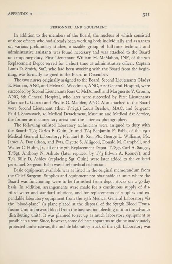
APPENDIX A 3 11
PERSONNEL AND EQUIPMENT
In addition to the members of the Board, the nucleus of which consistedof those officers who had already been working both individually and as a teamon various preliminary studies, a sizable group of full-time technical andadministrative assistants was found necessary and was attached to the Boardon temporary duty. First Lieutenant William H. McMahon, INF, of the ythReplacement Depot served for a short time as administrative officer. CaptainLouis D. Smith, SnC, who had been working with the Board from the begin-ning, was formally assigned to the Board in December.
The two nurses originally assigned to the Board, Second Lieutenants GladysE. Maroon, ANC, and Helen G. Woodman, ANC, 21st General Hospital, weresucceeded by Second Lieutenants Rose C. McDonnell and Marguerite V. Cronin,ANC, 6th General Hospital, who later were succeeded by First LieutenantsFlorence L. Giberti and Phyllis G. Madden, ANC. Also attached to the Boardwere Second Lieutenant (then T/Sgt.) Louis Breslow, MAC, and SergeantPaul J. Showstack, 3d Medical Detachment, Museum and Medical Art Service,the former as documentary artist and the latter as photographer.
The following enlisted laboratory technicians were assigned to duty withthe Board: T/3 Carlos F. Guin, Jr. and T/4 Benjamin F. Babb, of the 15thMedical General Laboratory; Pfc. Earl R. Zea, Pfc. George L. Williams, Pfc.James A. Donaldson, and Pvts. Clyette S. Alligood, Donald M. Campbell, andWalter C. Huhn, Jr., all of the 7th Replacement Depot. T/Sgt. Carl A. Sauget,T/Sgt. Anthony N. Ashute (later replaced by T/3 Edwin A, Rooney), andT/4 Billy D. Ashley (replacing Sgt. Guin) were later added to the enlistedpersonnel. Sergeant Babb was chief medical technician.
Basic equipment available was as listed in the original memorandum fromthe Chief Surgeon. Supplies and equipment not obtainable at units where theBoard was functioning were to be furnished from depot stocks on a 90-daybasis. In addition, arrangements were made for a continuous supply of dis-tilled water and standard solutions, and for replacements of supplies and ex-pendable laboratory equipment from the 15th Medical General Laboratory viathe “blood-plane” (a plane placed at the disposal of the 6713th Blood Trans-fusion Unit to forward blood from the base section bleeding unit to the advancedistributing unit). It was planned to set up as much laboratory equipment aspossible in a tent. Since, however, some delicate apparatus might be inadequatelyprotected under canvas, the mobile laboratory truck of the 15th Laboratory was

THE PHYSIOLOGIC EFFECTS OF WOUNDS3 12
placed at the Board’s disposal. Details of the organization and operation of thefield laboratory appear in Appendix B.
DEFINITION OF OBJECTIVESAfter discussion among Board members in consultation with Colonel
Churchill and Colonel Stone, it was agreed that the major problems for investi-gation should be: (i) Physiologic and metabolic studies of the severely wounded;(2) adequacy of shock resuscitation therapy; (3) possible hazards of trans-
fusion with type O blood, and (4) the mechanism of renal insufficiency fol-lowing resuscitation from shock. The investigative program of the Board, al-though developed along diversified and varied lines, remained focused on theseprimary broad objectives.
MANNER OF OPERATION
It was evident that the objectives of the study as defined could be approachedonly by selection of a base of operations which would meet all of several essen-tial requirements: i. Facilities for rationing, housing, and other administrativesupport of the Board. 2. Access to an adequate number of severely woundedmen. 3. Observation of the severely wounded at the earliest possible time afterwounding but under such circumstances that patients included in the studynormally would remain available for observation through the period of resus-citation (including surgery) and at least the immediate postoperative phaseof treatment. 4. Utilization of an installation in the established chain of evacua-tion, thereby avoiding establishment of special facilities. Obviously aid stations,collecting stations, and clearing stations met only the second of these require-ments. General hospitals failed to meet either the second or third. Forwardhospitals, i. e., either evacuation or field hospitals as employed in the combatzone in Italy, satisfied the general requirements. The size of the research unit,however, precluded attachment to a platoon of a field hospital. It was not toogreat for an evacuation hospital and selection of a base was thus determined.
On 19 September 1944,therefore, with equipment loaded on a 2V2-ton truck,two Command and Reconnaissance cars and the overload transported by the“blood plane,” the personnel of the Board reported to Fifth Army Headquartersand proceeded to the 94th Evacuation Hospital at Pratalino, a few miles northof Florence, Italy. During the greater part of the 8-month period of the study

APPENDIX A 3 T 3
(September 1944 to May 1945) the Board remained with this hospital. For a2-month period it was attached to the 24th General Hospital, and during thattime two members of the Board alternated at B platoon of the 32nd Field Hos-pital while another was making biochemic tests at the 8th Evacuation Hospital.This was at the time of the push of the 10th Mountain Division, and illustratesone method by which extensive data were obtained on casualties in several areasduring periods of maximum military activity.
A system of study gradually evolved as work poured in. The hospital pro-vided a special ward tent in which all patients under study were concentratedfollowing operation. The Board supplied the nursing service, but primary re-sponsibility for professional care remained in the hands of the hospital staff.The clinical members of the Board supervised the study ward and looked overnew admissions to the hospital shock ward to select cases suitable for study.Liaison was rapidly established with nearby field and evacuation hospitals whosestaffs kept the Board informed of cases appropriate for inclusion in the mainstudy. Daily trips were made by two or more members of the Board to manyof these hospitals to examine patients, record intakes and outputs, keep follow-upnotes, copy histories, pick up blood and urine specimens, and occasionally takepart in necropsies. Suitable clinical cases were abundant and the laboratory wasoften hard pressed to keep up with material.
The Board functioned in a close and unified manner, with all memberscontributing to the whole task. Individual members assumed responsibility forvarious phases of the work. Lt. Colonel Mallory, who acted as chairman of theBoard, was in charge of the pathologic aspects of the work and directed thelaboratory. He made numerous postmortem examinations, supervised others,and from time to time took the accumulated material back to the 15th GeneralLaboratory for histologic study. Lt, Colonel Simeone was primarily responsiblefor determinations of blood volume, as well as cardiovascular findings. Lt.Colonel Beecher was responsible for the liver function studies, and, with Lt.Colonel Simeone, for almost all the admission blood volume data. He was col-lator of the final report. Lt. Colonel Sullivan was in charge of hematologicaspects and, with Captain Smith, made the studies of blood groups and iso-agglutinins, and worked with Lt. Colonel Mallory on some of the pathologicstudies, particularly on pigment. Major Burnett carried out the kidney functionstudies and directed, with Lt. Colonel Simeone, the postoperative therapy andthe collection of biochemic data. Captain Smith handled bacteriologic prob-

THE PHYSIOLOGIC EFFECTS OF WOUNDS3M
lems. Captain Shapiro was in charge of laboratory work and collaborated inthe preparation of the biochemic data.
All medical installations within the Theater had been directed to aid theBoard in its work, and everywhere the Board met whole-hearted cooperation.Since it was not administratively self-contained, the Board and its ancillary per-sonnel were dependent on the various hospitals to which they were attachedfor quarters, rations, standard supplies, and other administrative essentials, in-cluding transportation beyond that immediately available. (As stated previ-ously, special supplies were obtained from the 15th General Laboratory.) Inaddition, a high degree of professional cooperation was essential in selectingcases for special study and making them available for such study without inter-ference with required treatment which remained the responsibility of the hos-pital staff.
DISSEMINATION OF PROGRESS REPORTS
It was recognized that observations and tentative conclusions based on theBoard’s work should be disseminated as promptly and widely as possible. Thispolicy would have the double advantage of making significant findings avail-able for immediate general application and of facilitating the Board’s work asthe result of understanding and interest on the part of the medical staffs fromwhom cooperative effort might be required. Therefore from time to time, find-ings were published in the Theater medical bulletin, discussed at medical meet-ings in the Theater, and circulated throughout other theaters and through TheSurgeon General’s Office by means of Essential Technical Medical Data reports.Personal contacts, however, were probably the most rapid and effective meansfor widespread dissemination of reports of the progress of the Board’s work.For example, the 2d Auxiliary Surgical Group, attached to a hospital wherethe Board was working, split up in the fall of 1944 and one-half went to thesupport of the Seventh Army invading Southern France. The group in theEuropean Theater kept in close contact with the Mediterranean group, throughwhich reports of the study were received and disseminated as various phaseswere developed.
APPENDIX B
The Field LaboratoryEstablishment of a field laboratory was manifestly essential to accomplish-
ment of the program mapped out by the Board, since collection of voluminousand complex biochemic data was fundamental to the plan of study. The labora-tory supporting the study had to be mobile and readily available wherever ob-servations were to be made on the recently and severely wounded; that is, nearthe front line.
The 15th Medical General Laboratory was to serve as base of operations forthe field laboratory and thus was available for the examination of such speci-mens as the Board might send to it. All histologic work in fact was done atthis laboratory. In theory many other procedures required by the Board couldhave been performed at the 15th Medical General Laboratory but this systemwould have been totally inadequate to the need for two principal reasons: Itwould not have provided immediate reports on laboratory procedures whichcould aid in guiding treatment and evaluation of each patient, nor would ithave revealed findings of particular interest which might suggest and directspecial follow-up analyses. Furthermore many types of analyses, such as thevan den Bergh diazo test, and determination of blood pH, phosphorus, chlorides,and sugar, must be performed very soon after the specimen is collected.
It was recognized, in addition, that the laboratory must not be a burden onthe unit to which it might be attached and therefore it had to be self-sufficient,not only in the performance of laboratory procedures but also with regard to itsutilities, such as electric power, heat, shelter, water supply, and disposal ofwastes. A means of supply must be established which would permit the labora-tory to carry only minimum essential equipment and chemicals for day-to-dayoperation and to be able to count on regular and dependable replacement ofsupplies and equipment from a base. Some of the practical difficulties encoun-tered in operating the laboratory have been mentioned in the Preface.
956507 0—52 25
3 l6 THE PHYSIOLOGIC EFFECTS OF WOUNDS
The joint considerations of mobility and self-sufficiency were reflected inthe transport and major equipment initially made available to the Board, Alarge mobile laboratory truck was obtained on loan from the 15th MedicalGeneral Laboratory. In addition to reconnaissance cars and jeeps there also wasavailable a 2 1
/4-ton cargo truck. On the latter were carried adequate glasswareto take care of one day’s run of samples, a size No. 1 International Centrifuge,two Coleman Junior Spectrophotometers, complete, a Klett colorimeter, a hotplate, a prescription balance with weights, a drying oven, and alcohol burners.A 2 1/z-kilowatt generator, five carboys of distilled water, a typewriter, and ninepyramidal tents for the laboratory and the personnel completed the equipment,which was loaded on a 4-by-q cargo truck.
In view of the anticipated working conditions, it was considered inadvisableto carry an analytical balance for preparation of standard solutions; therefore,as mentioned below, arrangements were made for obtaining a supply of thesesolutions as needed. The needs of the study did not necessitate carrying a refrig-erator. A vacuum pump and a field autoclave were available in the mobilelaboratory truck. A serviceable spectroscope was needed and was obtained laterfrom the University of Rome.
Assurance of a continuous supply of reagents and other laboratory suppliesnecessitated special arrangements. Certain common items could be obtainedfrom any medical unit to which the laboratory might be attached, but adequatesupplies of distilled water, standard solutions, and special reagents and apparatuscould not be obtained in this manner. Fortunately such items were readily ob-tainable from the 15th Medical General Laboratory which was the base ofoperations for the Board. They were delivered as needed to the field labora-tory by means of the carrier “blood-bank” plane which shuttled between theGeneral Laboratory and the forward distributing unit of the 6713th BloodTransfusion Unit.
Shortcomings in basic physical arrangements were soon evident. The mobilelaboratory truck proved too small to provide space for more than one technicianto perform chemical analyses. The pyramidal tents were not adaptable for useas a laboratory, so a ward tent was requisitioned from the 94th EvacuationHospital. A tent of this size permitted three technicians to work comfortablywithout interference and allowed a systematic distribution of reagents and equip-ment, thus eliminating the rehandling necessary in a confined space.
Tables, a working cabinet, and a glassware cabinet were added. A sink was

APPENDIX B 3 X 7
made from a truck gasoline tank, and water was piped from a 50-gallon drum.The sewage system was simple. All blood, plasma, urine, and solutions fromcompleted analyses were poured into a slop pail, which was emptied into aprepared soakage pit. Water used in washing glassware ran into channels andsoaked into the ground. The tables and equipment were distributed so that allthe analytic work, the washing and drying of glassware, the storage of surplussupplies, and the administrative and clerical work were performed in the sametent. Blood typing and agglutination studies were made in the laboratory truck.
For the collection and submission of specimens, boxes were prepared in trip-licate to contain approximately:
15 screw-cap, 22-ml. flat-bottom vials containing lithium oxalate to be used forcollection of blood specimens.
10 screw-cap, 8-ml. flat-bottom vials containing ammonium and potassium oxa-late for holding blood specimens on which determinations of hematocrit value andplasma protein concentration would be made.
5 corked test tubes for submission of clotted blood.4 screw-cap, 250-ml. urine bottles,12 sterile 10-ml. syringes.3 sterile 30-ml. syringes.2 sterile 2-ml. syringes.10 dry 10-ml. syringes.30 sterile 19-gage needles.Absorbent cotton, alcohol, and rubber tubing for tourniquets.Ampules of bromsulfalein, phenolsulfonphthalein, and a sterile solution con-
taining 100 mg. per 100 cc. of the dye T-1824 for determination of blood volume.
The specimens were required to be labelled with the patient’s name and thedate and hour the sample was taken.
AnalysesThe following analyses were undertaken on the Board’s material:Blood. —Volume, hemoglobin, hematocrit value, hydrogen ion, sugar, sul-
fonamides, methemoglobin, the bromsulfalein liver function test; plasma ureanitrogen, creatinine, lactic acid, bilirubin (van den Bergh reaction), uric acid,protein, free hemoglobin, carbon-dioxide combining power, magnesium, chlo-rides, phosphorus, and nonprotein nitrogen; serum sodium, calcium, and po-tassium.

THE TWO LABORATORY TENTS and the laboratory truck used by the Board for theStudy of the Severely Wounded, shown at Monghidoro, Italy, autumn of 1944. Later thelaboratory tents were winterized as shown.

ITALY PROVIDED VARIETY OF CLIMATE. Winter snows and spring rains miredthe laboratory truck and dampened equipment. Wherever possible the laboratory was
housed in a building, but more often the work was done in tents.
320 THE PHYSIOLOGIC EFFECTS OF WOUNDS
Urine.—Routine urinalysis, hydrogen ion, free hemoglobin, myoglobin, cre-atinine, creatine, ammonia, titratable acidity, chlorides, urea nitrogen, organicacids, magnesium, bile, urobilinogen.
Tissue.—Myoglobin.Renal Function Tests.—Phenolsulfonphthaleinexcretion, mannitol clearance,
para-amino hippuric acid clearance, and pituitrin concentration test.Effort was made to synchronize the submission of specimens so that several
of the same tests could be performed simultaneously; immediate analyses ofsingle specimens were, however, made when requested.
Each technician kept a notebook in which he recorded the details of theparticular determinations assigned to him. The calculations were checked, en-tered into the main laboratory book, and read back. After an adequate numberof cases had been completed, the laboratory data were transcribed onto fivecopies of laboratory sheets and each set was stored in a different place (onecopy being filed with the case record) in case storm, fire, or warfare wouldspare one or more sets. Even so, the laboratory sheets of some cases were lost intransit.
Many of the specimens submitted were irreplaceable. During the entirecourse of the study, no sample was sacrificed due to breakage upon centrifu-gation. The No. i International Centrifuge yielded results which far outweighedthe inconveniences attached to moving and servicing it. The more easily movedtable centrifuge had caused many tube breakages and was therefore not used.
The majority of the ultimate measurements made required colorimetricreadings. The Coleman Junior Spectrophotometer, powered by its own 6-voltbattery, contributed in several respects to improving the caliber of the work.It broadened the scope of procedures that could be carried out, it reproducedthe readings accurately, and in all cases it allowed us to standardize the obser-vations which, particularly in visual colorimetric reading of yellow, variedamong technicians. Most of the color tests, except sugar and phosphorus whichentailed comparison of easily-read blue colors, were set up for spectrophotometerreadings. The spectrophotometer sturdily resisted the stresses and strains ofrepeated packing, unpacking, and moving, and no replacement of parts wasnecessary during the entire study.
The initial move up to the combat line was made with emphasis on careand protection of our supplies and equipment. Each subsequent move requiredspeed. For this, boxes were prepared with individual cubicles for all the reagent

APPENDIX B 3 21
bottles. The glsssware was rolled up in blankets and these blanket rolls weresnugly wedged between the shelves of the glassware cabinet. The test tubesand blood-drawing equipment were kept during the working period in thesame boxes in which they were moved, and the pipets were bound in groups of10 and wedged into the working cabinet drawer. The laboratory could there-fore be dismantled, packed, and loaded within four or five hours.
The first move into the field indicated that certain standards of protectionin inclement weather were imperative. During the winter period the laboratorytent was winterized by completely boarding up the front and back as illus-trated. A close-fitting door was installed, the floor was gravelled, and an oilstove was kept burning day and night. The final set-up during the spring wasthe most convenient. No stove was required and the work tables were groupedabout the center of the tent. To eliminate the discomforts of heat and the roarof the ovens, the dishwashing, drying, and autoclaving were done in a pyramidaltent adjacent to the chemistry tent.
The need of conservation of electricity made it quite obvious that dryingcould not be effected by electric oven nor could hot plates be used. Alcoholburners and gasoline stoves served satisfactorily for this purpose. During oneparticularly dusty period it was necessary to operate the centrifuge and spec-trophotometer in the mobile laboratory vehicle, and it was often necessary toplug with cotton all the flasks and cylinders and to box the tubes and pipetsto keep them dust-free.
In the 8 months of the study, the laboratory moved seven times, and over12,000 biochemic analyses representing 56 different types of tests were made:8,269 on blood (31 types), 3,961 on urine (22 types), and 58 (3 types) on tissue.The scope of these chemical analyses is shown in Appendix C where the meth-ods used are described.
A detailed listing of the number of chemical analyses is given on page 322.All work was done in connection with the Board material except for a 3-dayhepatitis survey.
The field laboratory proved its value in this study. It was shown that amobile laboratory unit could be established and could function well close to theline of combat, and that it could do accurate and comprehensive biochemicanalyses in this situation.

322 THE PHYSIOLOGIC EFFECTS OF WOUNDS
Chemical Analyses
Blood ChemistryHemoglobin -- 636 Sulfonamides 108Hematocrit __ 636 Glucose 377Plasma protein
-- 639 Blood volume 207Plasma hemoglobin 892 Bromsulfalein
_ _ 45 1
Nonprotein nitrogen 605 Sulfate 2Creatinine 580 Methemoglobin _ _ _ _ 15Chlorides -- 59 1 Urea nitrogen 80Uric acid 195 Vitamin C - 8Carbon-dioxide combining power- -- 3Sl Cephalin flocculation test 119Van den Bergh 700 Mannitol clearance 75Phosphorus -- 44° Renal blood flow 40PH 28 Total mass—hippurate 29Sodium -- 474 Total mass—glucose 6Potassium 26 Lactic acid _ 27Calcium 42 Bilirubin (acetone) 2
Magnesium __ 158Total 8,269
Urine ChemistryRoutine urinalysis 670 Ammonia 130pH 774 Titratable acidity - - 13°Benzidine 662 Organic acids 91Myoglobin 144 Urobilinogen 7Chlorides . _ _ 289 Bromsulfalein 6Phenolsulfonphthalein 385 Urea nitrogen 96Concentration test 43 MagnesiumSulfadiazine
_ _ _ - 33 Methylene blue test— 119
Hippuric acid 1 Spectroscopic examination 2Bile 32 Titratable alkalinity _ _ . 2Creatinine 197Creatine 83 Total _
_ _ _ _ . — 3,961
Tissue ChemistryMyoglobin 4 1Sulfadiazine 6Magnesium 11
Total _ _ _ _ - 58Total Analyses 12,288

APPENDIX C
ContentsPage
Blood Analyses 324Normal values 324Preparation of precipitants 324Blood and plasma volume
Modification of Gregersen method 326Procedure for determination of blood volume 330
Blood ChemistryNonprotein nitrogen products 332
(Nonprotein nitrogen in plasma , . . creatinine in plasma . .. uric acid inplasma . . . urea nitrogen in plasma)
Acid-base analyses 337(Blood pH . . . serum sodium . . . serum potassium . . . serum calcium . . .
magnesium in plasma . . . phosphorus in plasma . . . C02 combining powerof plasma .. . chlorides in plasma ... lactic acid in plasma)
Glucose in plasma 343Sulfonamides in whole blood 343Determination of methemoglobin 343Plasma bilirubin 344Hemoglobin, hematocrit, and plasma protein 344Plasma hemoglobin determination 344
Liver Function Test 345Renal Function Tests . 346
(Mannitol . .. para-amino hippuric acid . . . phenosulfonphthalein test)Urinalyses 349
(Routine ... the benzidine test... titratable acidity .. . organic acids . ,. creatinineand creatine)
Determination of Myoglobin 354(Urine and tissue)
Biochemic MethodsThe biochemic procedures to be used in this study were selected on the
basis of their accuracy and their applicability to the anticipated conditions in acombat area. Many procedures were modified so that the critical quantitativereadings might be made on the spectrophotometer. Those described for thedetermination of blood volume; plasma magnesium, serum sodium; creatinineand creatine in urine; the bromsulfalein and phenolsulfonphthalein tests, andthe determination of myoglobin, for example, offer generally useful modifica-tions of standard procedures. Methods in which any changes in the standardprocedure were made are described in detail.
Blood Analyses
Normal Values in the Blood AnalysesThe ranges of normal values for analyses made on the blood as shown in
Table 118 are essentially those given in the literature. For convenience it wasoften necessary to assume a single figure as representing the standard normallevel for a particular analysis in order to contrast it with the average findingsin certain groupings of the cases in this study. The figure used for the standardnormal was selected as a result of our experience with each method.
Preparation of Blood and Plasma Protein PrecipitantsTungstic Acid Filtrate
Reagent.—Van Slyke and Hawkins tungstic acid. (Twenty-five ml. of 2/3 N sulfuricacid and 25 ml. of 10-percent sodium tungstate are combined and diluted to 225 ml. withwater.) This reagent is satisfactory for 1 week. Turbidity due to precipitation of tungsticacid does not interfere with preparation of the filtrate.
Procedure.—To 1 volume of blood or plasma in a flask are added 9 volumes

APPENDIX C 325
Table 118.—Normal Values Used in this Study
of the tungstic acid reagent, the mixture is allowed to stand for 10 minutes andis then filtered.
Trichloracetic Acid FiltratesReagents.—Trichloracetic acid, 10-percent solution, 15-percent solution, and 20-percent
solution. (Prepared by titration.)
Procedure.—Prepare filtrates (as required by the analysis to be performed)
Determination or Test Range of Normal
Range ofEquivalents
mEq./L.Standard Normal
BloodVolume 8-9% of body weight .
14-17.5 Gm./lOO ml. .
8.5% of body weight.16 Gm./lOO mi.Hemoglobin
Hematocrit value 42-52 ml. cells in 100ml 47 ml. cells in 100 ml.
PH 7.38-7.48
PlasmaProtein 6-7 Gm./lOO ml. ... 6.5 Gm./lOO ml."Hemoglobin”
Nonprotein nitrogen. . .
Less than 10 mg./100ml
25-40 mg./100 ml. . . 33 mg./lOO ml.Urea nitrogen 10-20 mg./100 ml. . . 15 mg./lOO ml.Creatinine 0.5-1.2 mg./lOO ml. . . 1.0 mg./lOO ml.Uric acid 1.5-3.5 mg./lOO ml. . . 3.0 mg./lOO ml.Phosphorus 1.8-3.5 mg./lOO ml. . . 1.04-2.0 3.0 mg./lOO ml.Magnesium 1.0-3.0 mg./lOO ml. . . 0.8-2.5 1.8 mg./lOO ml.Chlorides 570-620 mg./lOO ml. . 97.5-104 585 mg./lOO ml.; 100
CO2 combining power . . 53-70 vol. % 24-31mEq./L.
60 vol. %; 27 mEq./L.Bilirubin (van den Bergh) . 0.1-0.5 mg./lOO ml. . .
Bromsulfalein Less than 3%(5 mg./Kg. in 45 min.)
Lactic acid 10-20 mg./lOO ml. . . 15 mg./lOO ml.
SerumSodium 320-340 mg./lOO ml. . 139-149 330 mg./lOO ml.; 143
Potassium 15-21 mg./lOO ml. . . 3-9-5.3mEq./L.
Calcium 9-11 mg./lOO ml. . . ,
326 THE PHYSIOLOGIC EFFECTS OF WOUNDS
Determination of Blood and Plasma VolumeModification of Gregersen Method
Blood and plasma volume was determined in this study by modification ofthe method developed by Gregersen 12 primarily for use of the Armed Forces.The dye T-i824 was used and the basic principles of the method were followed.
The Gregersen method, in brief, consists in injecting a known quantity ofdye into the antecubital vein, waiting about twenty minutes for the dye to bedistributed through the circulating blood, and taking three or four samples ofblood at 20-minute intervals. The plasma is separated and its concentrationof dye is measured and plotted against time. The curve so obtained is extrapo-lated back to zero time to correct for dye lost during the mixing period. Thisvalue at zero time gives the theoretical concentration of dye that would haveoccurred in the plasma if uniform mixing had been effected at the instant ofinjection and none of the dye had been excreted. The total blood volume iscalculated from the plasma volume (measured by the dye) and the hematocritvalue.
Experience with the method revealed two serious objections, one practicaland one technical. The practical objection was that the method was too time-consuming for use with battle casualties before operation; therefore, a morerapid procedure was sought. This will be discussed later. The technical flawin the procedure was that the difference in turbidity between the blank, dye-free plasma and the dye-containing samples yielded spectrophotometric read-ings which had to be resolved as amount of dye plus or minus turbidity. Plottedpoints using such readings rarely indicated excretion of dye as an exponentialfunction, and very often, when considerable variations in turbidity existed,would lead to gross errors of reading at zero time. Corresponding errors resultedin the calculation of the blood volume.
The cause of this turbidity, which varies during the course of a determination,has not- been ascertained. The plasma, however, does not clear upon centrifu-gation. The following observations made the application of a turbidity-correction
1 Gregersen, M. I.; Gibson, J. G., and Stead, E. A.; Plasma volume determination with dyes: errorsin colorimetry; use of the blue dye T—1824. Am. J. Physiol. 113: 54—55, Sept. 1, 1935.
2 Gregersen, M. I.: A practical method for the determination of blood volume with the dye T—1824;survey of the present basis of the dye-method and its clinical applications. J. Lab. & Clin. Med. 29; 1266—1286, December 1944.
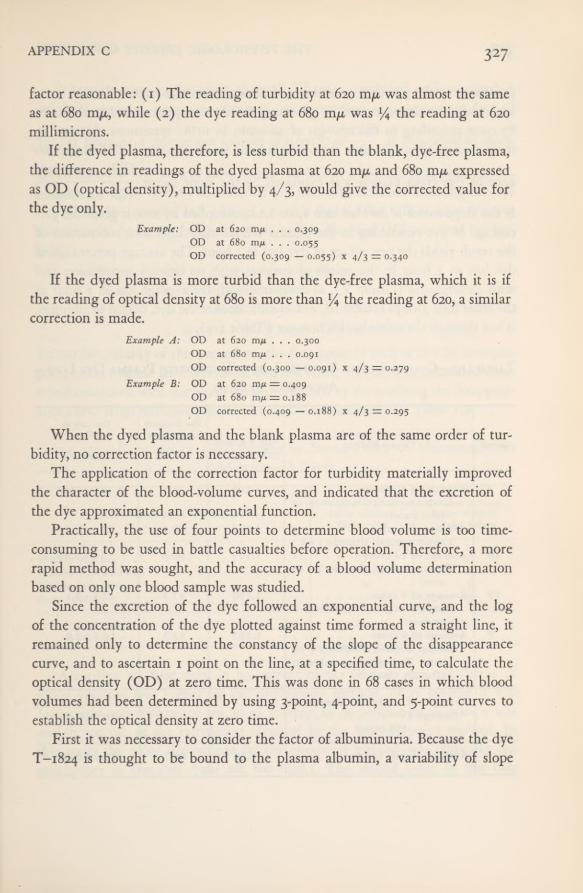
APPENDIX C 327
factor reasonable: (1) The reading of turbidity at 620 mfx was almost the sameas at 680 m/x, while (2) the dye reading at 680 m/x was % the reading at 620millimicrons.
If the dyed plasma, therefore, is less turbid than the blank, dye-free plasma,the difference in readings of the dyed plasma at 620 mjx and 680 m/x expressedas OD (optical density), multiplied by 4/3, would give the corrected value forthe dye only.
Example: OD at 620 m/u. . . . 0.309OD at 680 m/x . . . 0.055OD corrected (0.309 — 0.055) x 4/3 — 0.340
If the dyed plasma is more turbid than the dye-free plasma, which it is ifthe reading of optical density at 680 is more than % the reading at 620, a similarcorrection is made.
Example A: OD at 620 m/* .. . 0.300
OD at 680 mp . . . 0.091OD corrected (0.300 —0.091) x 4/32=0.279
Example B: OD at 620 mp = 0.409OD at 680 mp — 0.188OD corrected (0.409 —0.188) x 4/3 = 0.295
When the dyed plasma and the blank plasma are of the same order of tur-bidity, no correction factor is necessary.
The application of the correction factor for turbidity materially improvedthe character of the blood-volume curves, and indicated that the excretion ofthe dye approximated an exponential function.
Practically, the use of four points to determine blood volume is too time-consuming to be used in battle casualties before operation. Therefore, a morerapid method was sought, and the accuracy of a blood volume determinationbased on only one blood sample was studied.
Since the excretion of the dye followed an exponential curve, and the logof the concentration of the dye plotted against time formed a straight line, itremained only to determine the constancy of the slope of the disappearancecurve, and to ascertain i point on the line, at a specified time, to calculate theoptical density (OD) at zero time. This was done in 68 cases in which bloodvolumes had been determined by using 3-point, 4-point, and 5-point curves toestablish the optical density at zero time.
First it was necessary to consider the factor of albuminuria. Because the dyeT--1824 is thought to be bound to the plasma albumin, a variability of slope

328 THE PHYSIOLOGIC EFFECTS OF WOUNDS
according to the presence or absence of albuminuria was expected. Such a re-lationship was demonstrated by grouping the dye disappearance-rate slopes in67 cases according to the amount of albumin in urine specimens obtained inthese cases at about the time the blood volume was determined. The disappear-
OD o minutesance of dye from plasma was expressed by the slope factor .
OD 60 minutesIf the slope factor is divided into 1,000 and multiplied by 100, it gives the per-centage of dye remaining in the plasma 1 hour after injection; subtraction ofthe result yields the percentage of dye lost in 1 hour. The average percentage ofdye lost in 1 hour in the urines of patients with no urinary suppression andwith albuminuria (Group V), as compared with the average loss in 1 hour inthe other four groups combined, would indicate that the dye, bound to albumin,is lost through the urine in this manner (Table 119).
Table 119.—Comparison of Albumin Loss in Urine with Plasma Dye Loss—
Average of 67 Cases
Dye Retained Dye Lost inNumber of Cases and
OD 0 minutesafter 1 Hour 1 Hour
Groups Urinary FindingsOD 60 minutes (percent) (percent)
I Average of 36 cases(Output normal; albumin neg. or
faint trace)
1.105 90.5 9.5±0.58
II Average of 4 cases(Output normal; albumin 1 +)
1.148 87.2 12.8±2.4
III Average of 14 cases(Patients anuric; albumin 2 + or
over)
1.111 90.0 10±0.85
IV Average of 4 cases(Patients oliguric; albumin 2 +
or over)
1.150 87.3 12.7 ±2.1
V Average of 9 cases(Output normal; albumin 2 + or
over. Two patients polyuric;albumin, trace)
1.195 83.7 16.3±2.8
Average of cases in Groups Ithrough IV 1.112 90 10.0±0.46
(58 cases)
Average of total cases 1.128 88.7 11.3±0.62
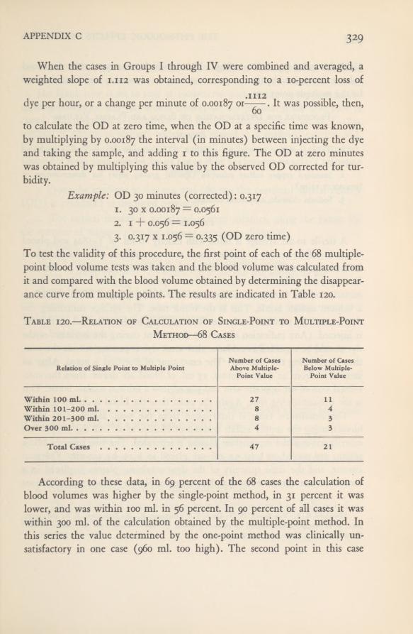
APPENDIX C 329
When the cases in Groups I through IV were combined and averaged, aweighted slope of 1.112 was obtained, corresponding to a 10-percent loss of
dye per hour, or a change per minute of 0.00187 or-——. It was possible, then,60to calculate the OD at zero time, when the OD at a specific time was known,by multiplying by 0.00187 the interval (in minutes) between injecting the dyeand taking the sample, and adding 1 to this figure. The OD at zero minuteswas obtained by multiplying this value by the observed OD corrected for tur-bidity.
Example: OD 30 minutes (corrected): 0.3171. 30 x 0.00187 “ 0.05612. 1 + 0.056 = 1.0563. 0.317 x 1.056 = 0.335 (OD zero time)
To test the validity of this procedure, the first point of each of the 68 multiple-point blood volume tests was taken and the blood volume was calculated fromit and compared with the blood volume obtained by determining the disappear-ance curve from multiple points. The results are indicated in Table 120.
Table 120.—Relation of Calculation of Single-Point to Multiple-PointMethod—68 Cases
According to these data, in 69 percent of the 68 cases the calculation ofblood volumes was higher by the single-point method, in 31 percent it waslower, and was within 100 ml. in 56 percent. In 90 percent of all cases it waswithin 300 ml. of the calculation obtained by the multiple-point method. Inthis series the value determined by the one-point method was clinically un-satisfactory in one case (960 ml. too high). The second point in this case
Relation ofSingle Point to Multiple PointNumber of CasesAbove Multiple-
Point Value
Number of CasesBelow Multiple-
Point Value
Within 100 ml 27 11Within 101-200 ml 8 4Within 201-300 ml 8 3Over 300 ml 4 3
Total Cases 47 21

THE PHYSIOLOGIC EFFECTS OF WOUNDS33°
yielded a value 330 ml. too high. This series demonstrated that the bloodvolume determined by the one-point test compares favorably with that obtainedby the multiple-point test.
Procedure for Determination of Blood and Plasma Volume
Reagents1. T-1824 solution, 1 mg. per milliliter. (The contents of ten 5-ml. ampules of
0.5-percent T-i824 are removed and made up to a 250-ml. solution, using sterile technique.)2. Standard copper sulfate solutions, (specific gravity series for determination of
hematocrit value).3. Sodium chloride, 0.9-percent solution.
Procedure
A sterile 10-ml. syringe is filled with exactly 10 ml. of T-1824 and placedwhere it will be readily available. About twenty milliliters of blood are drawnfrom the antecubital vein with a sterile syringe, leaving the needle in the vein.Exactly 5 ml. of blood are measured into a specially oxalated bottle for deter-mination of the hematocrit value, and the remaining blood is transferred toa lithium oxalate bottle. This is the blank tube. The syringe containing thedye is carefully attached to the needle remaining in the vein, and all the dyeis injected. (Any indication of pain by the patient during the injection of thedye should be regarded as evidence that some dye is passing outside the vein,thus invalidating the analysis.) The exact time of injection is noted. After aninterval of from 20 to 40 minutes, 15 ml, of blood are drawn from the ante-cubital vein of the opposite arm and placed into a lithium oxalate bottle. Thisis the dye-containing tube. Again the exact time the blood is drawn is noted.
The hematocrit value is determined from the 5-ml. sample (the blankblood), using the copper sulfate method of Phillips et al. 3 All the samples arecentrifuged and the supernatant plasma is separated. The two tubes of blankplasma are pooled, at least 5.5 ml. are placed in a 19- by 150-mm. Colemancuvette, and the same quantity of the dye-containing plasma is placed in asimilar cuvette, (In the event that the amount of plasma is inadequate, dilutions
‘Phillips, R. A.; Van Slyke, D. D.; Dole, V. P.; Emerson, K., Jr.; Hamilton, P. B., and Archi-bald, R. M.: Copper sulfate method for measuring specific gravities of whole blood and plasma. BUMEDNews Letter, U. S. Navy, June 25, 1943, p. 1—16.

APPENDIX C 331
are made with o.p-percent saline solution, treating both tubes in the samemanner.)
The blank tube is set to read at 100-percent transmission at 620 mjtt in thespectrophotometer and the optical density of the plasma in the dye tube isnoted. This procedure is repeated at 680 millimicrons. Exactly 1 ml. of T-1824solution is diluted to 500 ml. with water, placed in a matched cuvette, and,blanking out with distilled water, the optical density at 620 m/t is determined.
Calculation1. From the readings at 620 mji and 680 mjjl the corrected optical density
(OD) is calculated as discussed previously.2. The optical density is calculated at zero minutes, using the factor for
the number of minutes after injection as previously discussed.3. Plasma volume:OD0
— corrected optical density at zero minutes for dyed plasma.OD dye = optical density of (1:500) dilution of dye that was injected for test.
Plasma volume =dye x 500 x 10.
KJLJo(The figure of 500 represents the dilution factor of the dye as read in the instrument, andthe figure 10 is the number of milliliters injected.)
4. Blood volume:100 minus the hematocrit value = percentage of plasma ( % PI)
,. Plasma volume
Blood volume =~ m x 100% PI
Summary
1. The technique for determining the circulating blood volume by meansof the dye T-1824 and a spectrophotometer is described.
2. The correction for variable turbidity of samples is described.3. The disappearance of the dye in battle casualties was 10 percent of the
injected substance at the end of 1 hour.4. Albuminuria increased the disappearance rate.
5. The feasibility of blood volume determinations based on only one dye-containing sample is demonstrated.
956507 0—52 26
THE PHYSIOLOGIC EFFECTS OF WOUNDS332
Blood ChemistryNonprotein Nitrogen Products
Nonprotein Nitrogen in Plasma—Modifiedfrom Koch and McMeehjn 4
Reagents1. Sulfuric acid, 50-percent aqueous solution by volume.2. Potassium persulfate, saturated solution. (Seven Gm. in 100 ml. of water.)3. Potassium mercuric iodide for Nessler’s solution: (a) Using a i-liter Erlenmeyer
flask, 22.5 Gm. of iodine are dissolved in 20 ml. of water containing 30 Gm. of potassiumiodide, (b) After the solution is complete, 30 Gm. (about 2.5 ml.) of mercury are addedand the mixture is shaken vigorously in order to break the mercury into globules, coolingit from time to time by immersing the flask in cold water. The shaking is continued untilthe supernatant fluid has lost all color due to iodine, (c) The supernatant is decanted fromthe excess mercury and tested for free iodine by adding a drop or two of the solution toi ml. of a i-percent starch solution. If the result of the starch test for iodine is negative,a few drops of an iodine solution of the same concentration as described in (a) are addeduntil a faint excess of iodine can be detected by the starch test, (d) The double iodidesolution is diluted to 200 ml. with distilled water and mixed thoroughly.
4. Nessler’s reagent. (By titration a 10-percent sodium hydroxide solution is accuratelyprepared from a saturated solution of sodium hydroxide that has been allowed to standuntil all carbonates have settled. To 975 ml. of this solution is added the entire solution(200 ml.) of potassium mercuric iodide prepared as described, after which it is mixedthoroughly and allowed to settle.)
5. Ammonium sulfate standard solution. (Prepared by dissolving 4.716 Gm. of pure,dry ammonium sulfate in 0.2 N sulfuric acid and making up to a volume of 1,000 ml.with the 0.2 N sulfuric acid (1 ml. = 1 mg. of nitrogen).)
6. Dilute nitrogen standard solution. (Five ml. of stock standard solution is dilutedto 100 ml. with 0.2 N sulfuric acid (3 ml. = 0.15 mg. nitrogen).)
ProcedureTo 2.5 ml. of protein-free tungstic acid plasma filtrate in an ignition tube
graduated at 35 ml. is added 1 ml. of 50-percent sulfuric acid solution, togetherwith a glass bead to prevent bumping, and it is boiled vigorously until itbecomes brown and dense white fumes appear. A funnel is then placed in thetube and the solution is digested for about one minute, when the flame andfunnel are removed, 5 drops of potassium persulfate are added, and the mix-
4 In Hawk, P. B., and Bergeim, O. Practical Physiological Chemistry, xoth ed. Philadelphia, Blakiston,I93U P- 415-

APPENDIX C 333
ture is boiled gently for several minutes. If a brown color persists, another5 drops of potassium persulfate are added and the mixture is again boiledgently. (A greenish-yellow color at this point indicates an excess of tungsticacid. This color will clear up after the addition of the Nessler’s reagent.) It iscooled, diluted to the 35-ml. mark with water, and mixed by inversion.
Into an identical ignition tube are placed 3 ml. of working standard and1 ml. of 50-percent sulfuric acid solution; this is made up to the 35-ml. markwith water and mixed by inversion. Into a third ignition tube is placed 1 ml.of the sulfuric acid solution, water is added to the 35-ml. mark, and it is mixedby inversion. This is the blank. Next, 3.5 ml. each of the sample, the standard,and the blank are transferred to a small cuvette (12 by 75 mm.), 1.5 ml. ofNessler’s reagent is added, the contents are mixed by inversion, and read inthe Coleman Junior Spectrophotometer at 440 m/a on the optical density scale,blanking out with the blank solution.
If the nonprotein nitrogen value is high, 2 ml. of the solution and 1.5 ml.of the blank are nesslerized. In this case the final value is multiplied by seven-fourths. Other appropriate dilutions that bring the color intensity into thestandard range can be used.
CalculationThe working nitrogen standard is equivalent to 60 mg. of nitrogen per
100 ml. of plasma. (Rs = OD of the standard, Ru = OD of the sample.)60 x
Rs Ru60 x Ru , , . .
——= mg./100 ml. nitrogen m plasma sample.
Comment
The method as modified has the following advantages: (1) Nessler’s solu-tion is used sparingly; less than 10 percent of the quantity used in the standardprocedure is necessary. (2) The procedure is expedited by the digestion ofhalf the quantity of filtrate ordinarily used. (3) An objective reading of thenesslerized solutions increases the accuracy of reading the yellow color. (4)In the event the nonprotein nitrogen level is extremely high, a further dilutionof the initial digestion can be made, rather than repeating the entire analysis.(5) The use of saturated potassium persulfate in the oxidation provides a sub-stance which is nitrogen-free, stable, and unlike superoxol, is non-injurious.
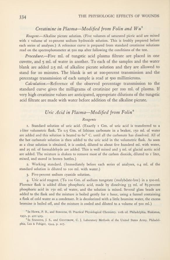
THE PHYSIOLOGIC EFFECTS OF WOUNDS334
Creatinine in Plasma—Modified from Folin and Wu5
Reagent.—Alkaline picrate solution. (Five volumes of saturated picric acid are mixedwith 1 volume of 10-percent sodium hydroxide solution. This is freshly prepared beforeeach series of analyses.) A reference curve is prepared from standard creatinine solutionsread on the spectrophotometer at 500 m[X after following the conditions of the test.
Procedure.—Five ml. of tungstic acid plasma filtrate are placed in onecuvette, and 5 ml. of water in another. To each of the samples and the waterblank are added 2.5 ml. of alkaline picrate solution and they are allowed tostand for 10 minutes. The blank is set at 100-percent transmission and thepercentage transmission of each sample is read at 500 millimicrons.
Calculation.—Reference of the observed percentage transmission to thestandard curve gives the milligrams of creatinine per 100 ml. of plasma. Ifvery high creatinine values are anticipated, appropriate dilutions of the tungsticacid filtrate are made with water before addition of the alkaline picrate.
Uric Acid in Plasma—Modified from Polin'Reagents
1. Standard solution of uric acid. (Exactly 1 Gm. of uric acid is transferred to ai-liter volumetric flask. To 0.5 Gm. of lithium carbonate in a beaker, 150 ml. of water
are added and this solution is heated to 6o° C. until all the carbonate has dissolved. All ofthe hot carbonate solution is then added to the uric acid in the volumetric flask. As soon
as a clear solution is obtained, it is cooled, diluted to about five hundred ml. with water,and 25 ml. of formaldehyde are added. This is well mixed and 3 ml. of glacial acetic acidare added. The mixture is shaken to remove most of the carbon dioxide, diluted to 1 liter,mixed, and stored in brown bottles.)
2. Working standard. (Immediately before each series of analyses, 0.4 ml. of thestandard solution is diluted to 100 ml. with water.)
3. Five-percent sodium cyanide solution.4. Uric acid reagent, (To 100 Gm. of sodium tungstate (molybdate-free) in a 500-ml.
Florence flask is added dilute phosphoric acid, made by dissolving 33 ml. of 85-percentphosphoric acid in 150 ml. of water, and the solution is mixed. Several glass beads areadded to the flask and the mixture is boiled gently for 1 hour, using a funnel containinga flask of cold water as a condenser. It is decolorized with a little bromine water, the excessbromine is boiled off, and the mixture is cooled and diluted to a volume of 500 ml.)
5 In Hawk, P. B., and Bergeim, O. Practical Physiological Chemistry. loth ed. Philadelphia, Blakiston,1931, p. 421-423.
6 In Simmons, J. S., and Gentzkow, C. J. Laboratory Methods of the United States Army. Philadel-phia, Lea & Febiger, 1944, p. 207.

APPENDIX C 335
Procedure
To i ml. of tungstic acid filtrate in a cuvette are added 3 ml. of water. Intoanother cuvette is pipeted 1 ml. of dilute standard, and 3 ml. of water areadded. This is the standard solution corresponding to 4 mg. of uric acid per100 ml. of plasma. Four ml. of water in a third cuvette constitutes the blanktube. To each of the samples, the uric acid standard, and the blank tube, 4 ml.of sodium cyanide are added, using an automatic pipet, and the solutions aremixed, after which 2 ml. of the uric acid reagent are added to each tube, andthe solutions are again mixed and are allowed to stand for 30 minutes. Theblank is set at 100-percent transmission on the spectrophotometer and theoptical density of the standard and each of the samples at 650 m/x is noted.
CalculationThe calculation of milligrams per 100 milliliters of plasma is obtained by
setting up a proportion comparing the optical density of the standard ofknown strength with the observed reading of the sample.
Example: OD of sample .. . . 0.150
OD of standard . . . 0.200Standard = 4 mg./100 ml.Sample (calculated) = 3 mg. of uric acid per 100 ml. of plasma.
CommentThe advantages of the method as modified are that it (1) yields a non-
clouding color development when sodium cyanide and phosphotungstic acidare used, (2) avoids the danger of color development varying with the tem-perature by the preparation of standard and blank at the same time and byreading the color on the optical density scale of the spectrophotometer, and(3) insures against decomposition of the working standard by preparation ofa fresh standard before each series of analyses.
Urea Nitrogen in Plasma—Modified from Karr 7
Reagents1. Urease solution. (A mixture of 0.3 Gm. of double-strength urease in 2.5 ml. of
water and 7.5 ml. of glycerin is centrifuged and decanted. The supernatant solution is theurease extract and lasts for at least 3 months.)
7 In Hawk, P. B., and Bergeim, O. Practical Physiological Chemistry. 10th ed. Philadelphia, Blakiston,1931, p. 418.

336 THE PHYSIOLOGIC EFFECTS OF WOUNDS
2. Acetate buffer solution. (To 15 Gm. of sodium acetate dissolved in 50 ml. of water
is added 1 ml. of glacial acetic acid, and the mixture is diluted to 100 ml. with water.)3. Dilute standard nitrogen solution. (Same as for nonprotein nitrogen.)4. Potassium mercuric iodide. (Same as for nonprotein nitrogen.)5. Urea Nessler’s reagent. (Thirty ml. of potassium mercuric iodide are mixed with
30 ml. of water and 140 ml. of 5-percent sodium hydroxide solution are added.)6. 2/3 N sulfuric acid,
7. Sodium tungstate, 10-percent solution.
Procedure
To 1 ml. of plasma in a tube are added 6.7 ml. of water, 2 drops of ureasesolution, and 4 drops of acetate buffer, and the tube is incubated at 40° to 50 0 C.for 15 minutes. One ml. each of the sulfuric acid and the sodium tungstatesolution are added, the solution is mixed, allowed to stand for several minutes,and filtered. Into another tube are placed 2 ml. of filtrate and 7 ml. of water.Into a tube graduated at 22.5 ml. are placed 1.5 ml. of the working nitrogenstandard, the solution is diluted to the mark, mixed, and 9 ml. of this dilutestandard is placed in a tube. To each of the samples and the dilute standardsolution is added 1 ml. of urea Nessler’s reagent, they are mixed and read inthe colorimeter.
CalculationThe unknown is set at 15 mm. and the reading of the standard will be
the milligrams of urea nitrogen per 100 ml. of plasma. In the event that ahigh urea nitrogen is expected, aliquots of the 2-ml. sample of filtrate are takenand the contents of the tube made up to 9 ml. with water. The observed levelis then multiplied by the proper factor.
Example: 0.5 ml. taken instead of 2.0 ml.; multiply answer by 4 for mg. of ureanitrogen per 100 ml. of plasma.
A urease blank should be determined about every 3 days. The urease blankusually averaged between 2 and 3 mg. expressed as urea nitrogen per 100 ml.of plasma and was subtracted from the observed values.
Comment
The method as modified has the following advantages: (1) It yields a non-turbid, stable solution upon direct nesslerization; (2) the reagents are stableat room temperature and are used sparingly, and (3) if the urea nitrogen level

APPENDIX C 337
is extremely high, a further dilution of the initial filtrate of the incubationmixture can be made, rather than repeating the entire analysis.
Acid-Base Analyses
Blood pH (from Haw\ins, Hastings, and Sendroys)
Preparation of the bicolor standards was taken from Peters and Van Slyke’sQuantitative Clinical Chemistry (cited below) p. 797, Table 65. The prepara-tion of the reagents (p. 796) and Hawkins’ modification (p. 801) for using asample of whole blood were also followed.
Procedure
The phenol red tube and the saline tube are prepared immediately beforethe analysis, using tubes similar to those for the preparation of standards.{Phenol red tube: After placing 5 ml. of freshly adjusted saline solution (whichcontains n ml. of 0.0075-percent phenol red solution per 100 ml.) in a tube,about one-quarter inch of liquid petrolatum is stratified above this solution.Saline tube: Five ml. of freshly prepared saline are put into a similar tube.)
Drawing blood samples: A glass bead is placed in the barrel of a 2-ml.syringe with needle attached, about 0.5 ml. of sterile liquid petrolatum anda small drop of saturated potassium oxalate are added, and the plunger isinserted in the syringe. The syringe is inverted so that the needle is pointingupward, the droplet of potassium oxalate is allowed to fall to the bottom ofthe solution, and all the air and excess petrolatum are slowly expressed untilthe movement of the plunger is arrested by the glass bead. The excess petro-latum is wiped off the needle, a blood sample of about 1.5 to 2 milliliters istaken, and the syringe is inverted several times to insure mixing with theoxalate. After waiting a moment for the petrolatum to stratify to the top ofthe syringe, the needle is placed under the petrolatum layer of the phenol redtube and exactly 0.5 ml. of blood is expressed into the tube. In a similar manner
0.5 ml. of blood is placed in the saline tube. The petrolatum is siphoned offfrom the phenol red tube, which is then sealed with % inch of paraffin, andthe contents of both tubes are mixed by inversion and centrifuged.
8 In Peters, J. P., and Van Slyke, D. D. Quantitative Clinical Chemistry, vol. II: Methods. Balti-more, Williams & Wilkins, 1932, p. 796—Sox.
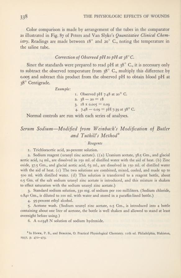
338 THE PHYSIOLOGIC EFFECTS OF WOUNDS
Color comparison is made by arrangement of the tubes in the comparatoras illustrated in Fig. 87 of Peters and Van Slyke’s Quantitative Clinical Chem-istry. Readings are made between 180 and 20 0 C, noting the temperature inthe saline tube.
Correction of Observed pH to pH at 38° C.Since the standards were prepared to read pH at 38° C., it is necessary only
to subtract the observed temperature from 38° C., multiply this difference by0.005 an d subtract this product from the observed pH to obtain blood pH at
38° Centigrade.Example:
1. Observed pH 7.48 at 20 0 C.2. 38 — 20 = 183. 18 x 0.005 = 0.094. 7.48 — 0.09 = pH 7.39 at 38° C.
Normal controls are run with each series of analyses.
Serum Sodium—Modified from Weinhach’s Modification of Butlerand Tuthill’s Method 9
Reagents1. Trichloracetic acid, 20-percent solution.2. Sodium reagent (uranyl zinc acetate), ((a) Uranium acetate, 38.5 Gm., and glacial
acetic acid, 14 ml., are dissolved in 150 ml. of distilled water with the aid of heat, (b) Zincoxide, 37.5 Gm., and glacial acetic acid, 63 ml., are dissolved in 150 ml. of distilled waterwith the aid of heat, (c) The two solutions are combined, mixed, cooled, and made up to
500 ml. with distilled water, (d) This solution is transferred to a reagent bottle, about0.5 Gm. of the salt sodium uranyl zinc acetate is introduced, and this mixture is shakento effect saturation with the sodium uranyl zinc acetate.)
3. Standard sodium solution, 330 mg. of sodium per 100 milliliters. (Sodium chloride,0.840 Gm., is diluted to 100 ml. with water and stored in a paraffin-lined bottle.)
4. 95-percent ethyl alcohol.5. Acetone wash. (Sodium uranyl zinc acetate, 0.5 Gm., is introduced into a bottle
containing about one liter of acetone, the bottle is well shaken and allowed to stand at leastovernight before using.)
6. A 0.0348 N solution of sodium hydroxide.
9 In Hawk, P. B., and Bergeim, O. Practical Physiological Chemistry, nth ed. Philadelphia, Blakiston,I 937> P- 472-473-
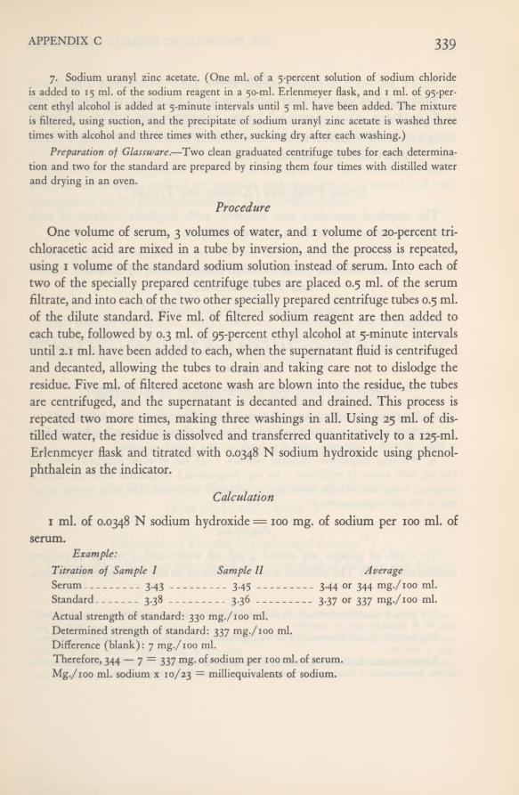
APPENDIX C 339
7. Sodium uranyl zinc acetate. (One ml. of a 5-percent solution of sodium chlorideis added to 15 ml. of the sodium reagent in a 50-ml. Erlenmeyer flask, and 1 ml. of 95-per-cent ethyl alcohol is added at 5-minute intervals until 5 ml. have been added. The mixtureis filtered, using suction, and the precipitate of sodium uranyl zinc acetate is washed threetimes with alcohol and three times with ether, sucking dry after each washing.)
Preparation of Glassware.—Two clean graduated centrifuge tubes for each determina-tion and two for the standard are prepared by rinsing them four times with distilled waterand drying in an oven.
Procedure
One volume of serum, 3 volumes of water, and 1 volume of 20-percent tri-chloracetic acid are mixed in a tube by inversion, and the process is repeated,using 1 volume of the standard sodium solution instead of serum. Into each oftwo of the specially prepared centrifuge tubes are placed 0,5 ml. of the serumfiltrate, and into each of the two other specially prepared centrifuge tubes 0.5 ml.of the dilute standard. Five ml. of filtered sodium reagent are then added toeach tube, followed by 0.3 ml. of 95-percent ethyl alcohol at 5-minute intervalsuntil 2.1 ml. have been added to each, when the supernatant fluid is centrifugedand decanted, allowing the tubes to drain and taking care not to dislodge theresidue. Five ml. of filtered acetone wash are blown into the residue, the tubesare centrifuged, and the supernatant is decanted and drained. This process isrepeated two more times, making three washings in all. Using 25 ml. of dis-tilled water, the residue is dissolved and transferred quantitatively to a 125-ml.Erlenmeyer flask and titrated with 0.0348 N sodium hydroxide using phenol-phthalein as the indicator.
Calculationi ml. of 0.0348 N sodium hydroxide = 100 mg. of sodium per 100 ml. of
serum.
Actual strength of standard: 330 mg./100 ml.Determined strength of standard: 337 mg./ioo ml.Difference (blank); 7 mg./ioo ml.Therefore, 344 — 7 = 337 mg. of sodium per 100 ml. of serum.Mg./ioo ml. sodium x 10/23 = milliequivalents of sodium.
Example:Titration of Sample I Sample II AverageSerum 3.43 3.45 3.44 or 344 mg./100 ml.Standard 3.38 3.36 3-37 or 337 mS-/ 100 ml-
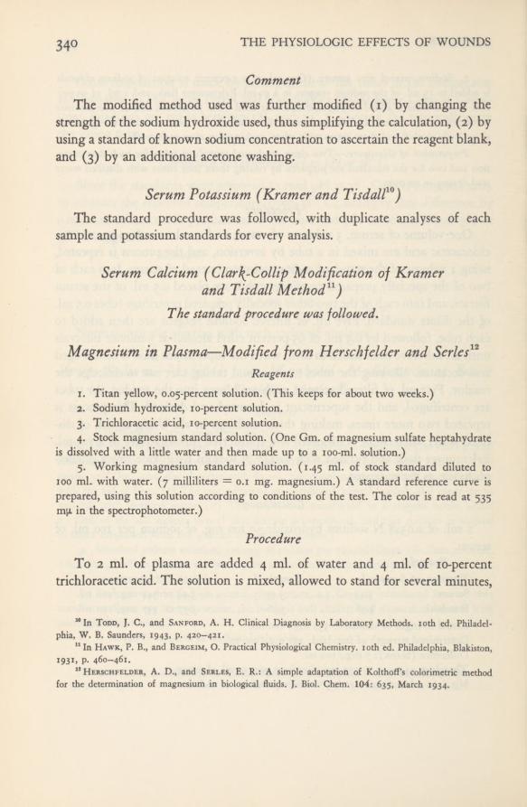
THE PHYSIOLOGIC EFFECTS OF WOUNDS340
Comment
The modified method used was further modified (1) by changing thestrength of the sodium hydroxide used, thus simplifying the calculation, (2) byusing a standard of known sodium concentration to ascertain the reagent blank,and (3) by an additional acetone washing.
Serum Potassium (Kramer and Tisdall 10 jThe standard procedure was followed, with duplicate analyses of each
sample and potassium standards for every analysis.
Serum Calcium (Clar\-Collip Modification of Kramerand Tisdall Method 11
)
The standard procedure was followed.
Magnesium in Plasma—Modified from Herschfelder and Series 12
Reagents1. Titan yellow, 0.05-percent solution. (This keeps for about two weeks.)2. Sodium hydroxide, 10-percent solution.3. Trichloracetic acid, 10-percent solution.4. Stock magnesium standard solution. (One Gm. of magnesium sulfate heptahydrate
is dissolved with a little water and then made up to a 100-ml. solution.)5. Working magnesium standard solution. (1.45 ml. of stock standard diluted to
100 ml. with water. (7 milliliters = 0.1 mg. magnesium.) A standard reference curve isprepared, using this solution according to conditions of the test. The color is read at 535m[x in the spectrophotometer.)
ProcedureTo 2 ml. of plasma are added 4 ml. of water and 4 ml. of 10-percent
trichloracetic acid. The solution is mixed, allowed to stand for several minutes,
10 In Todd, J. C., and Sanford, A. H. Clinical Diagnosis by Laboratory Methods. 10th ed. Philadel-phia, W. B. Saunders, 1943, p. 420—421.
11 In Hawk, P. B., and Bergeim, O. Practical Physiological Chemistry. 10th ed. Philadelphia, Blakiston,1931, p. 460—461.
12 Herschfelder, A. D., and Serles, E. R.: A simple adaptation of Kolthoff s colorimetric methodfor the determination of magnesium in biological fluids. J. Biol. Chem. 104: 635, March 1934.

APPENDIX C 34 1
centrifuged, and filtered. To 5 ml. of plasma filtrate in a cuvette, 1.6 ml. ofwater is added, and 7 ml. of water in another cuvette serves as the blank. Oneml. of titan yellow is then added to each of the samples and to the blank. Twoml. of the sodium hydroxide solution are added to the blank tube, the contentsare mixed, and the tube is set to read at 100-percent transmission on the spec-trophotometer at 535 millimicrons. Similarly, 2.4 ml. of the sodium hydroxidesolution are added to each of the samples, and the solution is mixed and readimmediately on the spectrophotometer at 535 millimicrons.
CalculationMilligrams of magnesium per 100 ml. of plasma are determined by refer-
ence of the observed percentage transmission to the standard curve.
Comment
This method is a simple, rapid procedure. Recoveries of added magnesiumand comparisons of normal range of plasma magnesium with that of othermethods (1.5-2.0 mg. per 100 ml.) indicate its accuracy.
Phosphorus in Plasma—Modified from Bodans\y 13
The method was modified by making up the standard in trichloracetic acid.This permitted of more exact color comparison and eliminated the need for thetwo molybdate solutions which the original method demands.
Carbon-Dioxide Combining Power of Plasma(Van Sly\e and Cullen 1
*)
Chlorides in Plasma (Schales and Schales 13)
Plasma chlorides were determined by the method of Schales and Schales.
13 In Todd, J. C., and Sanford, A. H. Clinical Diagnosis by Laboratory Methods, xoth ed. Philadel-phia, W. B. Saunders, 1943, p. 404—411.
14 The procedure was followed as described in Methods for Laboratory Technicians, War DepartmentTechnical Manual 8—227, October 1946, p. 123—129.
15 In Simmons, J. S., and Gentzkow, C. J. Laboratory Methods of the United States Army. Philadel-phia, Lea & Febiger, 1944, p. 209.

THE PHYSIOLOGIC EFFECTS OF WOUNDS34 2
Lactic Acid in Plasma—Modified fromMendel and Goldschieder16
Reagents1. Metaphosphoric acid, 5-percent solution, prepared daily.
2. Copper sulfate, 15 percent by weight of pentahydrate.3. Blank-free concentrated sulfuric acid. (The quality of the sulfuric acid should be
such that no yellow color develops on the addition of 0.1 ml. of o.i25-percent veratrole to
3 ml. of acid. A good reagent grade of sulfuric acid is satisfactory.)4. Lactic acid, 100 mg. per 100 ml., prepared from lithium lactate.5. Sodium fluoride, 0.1-percent solution.6. Veratrole, 0.125 percent in absolute alcohol.
ProcedureTo i ml. of plasma separated from freshly drawn blood are added 5 ml. of
water, 1 ml. of sodium fluoride, and 1 ml. of metaphosphoric acid. The wholeis shaken vigorously, allowed to stand for several minutes, and filtered. To4 ml. of the protein-free filtrate in a centrifuge tube are added 1 ml. of coppersulfate solution and 1 Gm. of calcium hydroxide, the mixture is allowed tostand about 30 minutes, shaking several times, and is then centrifuged. Thepellicle of copper hydroxide is broken with a glass rod and the supernatant isdecanted into another tube and centrifuged again, resulting in a clear, sugar-free filtrate. The standard lactic acid solution is diluted 1:10, corresponding to100 mg. of lactic acid per 100 ml. of plasma, according to the conditions of thetest. Weaker dilutions corresponding to 75, 50, 25, and 12.5 mg. of lactic acidper 100 ml. of plasma are also prepared.
To 0.5 ml. of the clear sugar-free filtrate (obtained by centrifugation only),to 0.5 ml. of each standard, and to 0.5 ml. of water in a carefully cleaned testtube, 3 ml. of concentrated sulfuric acid are slowly added while the tubes arecooled in cold water. The tubes are then placed in a boiling water bath forexactly 4 minutes, removed and immersed in cold water for 5 minutes. Toeach tube 0.1 ml. of the veratrole solution is added, and the solution is mixedand allowed to stand for 20 minutes, after which it is decanted into smallcuvettes (12 by 75 mm.) and read in the spectrophotometer at 545 m/x, blank-ing out with the water reagent blank and reading the optical density scale.
ia In Peters, J. P., and Van Slyke, D. D. Quantitative Clinical Chemistry, vol. II; Methods. Balti-more, Williams & Wilkins, 1932, p. 618—620.

APPENDIX C 343
Note: In the process of color development, a brown color may form in addi-tion to the pink of the test color. This is due to unclean glassware and con-taminated filtrate. In these instances the analysis is repeated.
CalculationA proportion is set up comparing the reading of the sample with that of the
nearest standard.
Comment
The test was set up on the spectrophotometer, and readings were made onthe optical density scale, using a series of standards with each run, since colordevelopment is not reproducible from day to day as required for using a per-manent chart. The color development does not satisfactorily follow Beer’s law,and the optical density reading of the closest standard was used in calculatingthe value of the sample.
The use of i ml. of sodium fluoride in the dilution of the plasma permitspreservation of the lactic acid level until a series of samples has accumulated.
Glucose in Plasma
The standard procedure of Folin and Wu17 was followed for the deter-mination of glucose in plasma.
Sulfonamides in Whole Blood
The method of Bratton and Marshall 18 was followed.
Determination of Methemoglobin
The method of Paul and Kemp 19 was followed.
1T In Simmons, J. S., and Gentzkow, C. J. Laboratory Methods of the United States Army. Philadel-phia, Lea & Febiger, 1944, p. 207-208.
18 In Simmons, J. S., and Gentzkow, C. J. Laboratory Methods of the United States Army. Philadel-phia, Lea & Febiger, 1944, p. 219-220.
18 Paul, W. D., and Kemp, C. R.: Methemoglobin; a normal constituent of blood. Proc. Soc. Exp,Biol. & Med. 56: 55-56, 1944.

THE PHYSIOLOGIC EFFECTS OF WOUNDS344
Plasma Bilirubin (van den Bergh)The method of Evelyn and Malloy 20 was followed.
Hemoglobin, Hematocrit, and Plasma Protein
The method of Phillips et al. 21 for the calculation of hemoglobin, hematocritvalue, and plasma proteins by means of copper sulfate was followed with theexception that, since a chemical balance was available, all the solutions werechecked and standardized so that they were correct to within ±2 units in thefourth decimal place, using a 25-ml, pyknometer and water at the same tem-perature as the standard solution. Each bottle was prepared in i-liter quantityand replacements were made from this stock. Bottles were prepared for eachunit difference in the third decimal place.
Plasma Hemoglobin Determination
Plasma Hemoglobin—Modified from Bing and Baker 22
Reagents1. Benzidine reagent. (Five Gm. of a good quality of benzidine are dissolved in
50 milliliters of glacial acetic acid, warming gently to effect solution. The mixture iscooled, diluted to 250 ml. with distilled water, about 2 Gm. of decolorizing charcoal areadded, and the solution is thoroughly mixed, allowed to stand for approximately 10 min-utes, and filtered into a brown bottle.)
2. Hydrogen peroxide, 3-percent solution.3. Hemoglobin standard solution. (A 20-mg. per 100 ml. solution of hemoglobin is
prepared in i-percent boric acid by diluting blood of known hemoglobin content.)4. Acetic acid, 20-percent solution.
Procedure
To 0.2 ml. of each sample and of the standard in separate tubes are added
20 In Simmons, J. S., and Gentzkow, C. J. Laboratory Methods of the United States Army. Philadel-phia, Lea & Febiger, 1944, p. 214—215.
11 Phillips, R. A.; Van Slyke, D. D.; Dole, V. P.; Emerson, K., Jr.; Hamilton, P. B., and Archi-bald, R. M.: Copper sulfate method for measuring specific gravities of whole blood and plasma. BUMEDNews Letter, U. S. Navy, June 25, 1943, p. 1-16.
“ Bing, F. C., and Baker, R. W.; The determination of hemoglobin in minute amounts of blood byWu’s method. J. Biol. Chem. 92: 589—600, August 1931.

APPENDIX C 345
2 ml. of the benzidine reagent, and each is thoroughly mixed. One ml. of the3-percent hydrogen peroxide is then added to each tube and they are allowedto stand for 30 minutes, at the end of which time the mixtures are diluted with6.8 ml. of the acetic acid and allowed to stand until all the tubes have developedthe red color (about 30 minutes), when they are compared in the colorimeter.
Calculation
The sample is set on 20 millimeters. The reading of the standard equalsmilligrams of benzidine-reacting substance per 100 ml. of plasma. As has beenexplained in Chapter VIII of the text, this entire benzidine-reacting substancewas termed “plasma hemoglobin” because a means for its further breakdownwas not available to us.
Liver Function Test
Bromsulfalein Test—Modified from Rosenthal 23
ReagentsBromsulfalein, 5-percent solution, in sterilized, sealed ampules.Sodium hydroxide, 10-percent solution.
3. Sodium chloride, 0.9-percent solution.
Procedure
The patient’s weight is recorded in kilograms and the amount of dye tobe injected is calculated on the basis of 5 mg. per Kg. of body weight. A simpleprocedure is to divide the body weight in kilograms by 10 to yield the exactamount in milliliters of bromsulfalein to be used for injection. The requiredamount of dye is drawn into a 10-ml. syringe and slowly injected into a veinin the patient’s arm, the exact time of injection being recorded. Forty-five min-utes after injection, a sample of blood from the opposite arm is collected in thestandard lithium oxalate bottle, the blood specimen is centrifuged, and theplasma is collected.
23 In Todd, J. C., and Sanford, A. H. Clinical Diagnosis by Laboratory Methods. 10th ed. Philadel-phia, W. B. Saunders, 1943, p. 428—429.
THE PHYSIOLOGIC EFFECTS OF WOUNDS
To i ml. of plasma placed in a cuvette 5 ml. of a 0.9-percent solutionof sodium chloride are added. The contents are well mixed and the spectro-photometer is set to read 100-percent transmission at 575 millimicrons withthis solution. Three drops of a 10-percent solution of sodium hydroxide arethen added to the same tube and it is inverted and read on the spectrophotometer.The transmission percentage is noted and the percentage of dye retention isobtained from the curve plotted from a standard bromsulfalein solution, fol-lowing the conditions of the test wherein 10 mg. per 100 ml. is considered100-percent retention.
Comment
The standard procedure for the determination of dye retention using theHynson, Westcott & Dunning Comparator Block was abandoned because vari-ous degrees of hemolysis in the samples would result in readings that indicatedas much as 5-percent retention when no bromsulfalein was present. Low reten-tion figures, therefore, could not be expressed with any degree of certainty, asresults were often not reproducible to within —5-percent retention. Further-more the method was time-consuming.
The test was set up on the Coleman Junior Spectrophotometer Model No. 6,using a 1:6 dilution of plasma and reading the color of the alkalinized brom-sulfalein at 575 millimicrons.
Renal Function TestsDetermination of Mannitol—Modified from Smith, H .
24
MEASUREMENT OF GLOMERULAR FILTRATION RATE
Reagents1. A 20-percent yeast suspension. (Bakers’ yeast, 25 Gm., is placed in about 100 ml.
of water, allowed to incubate overnight, and then washed by repeated centrifugation untila clear supernatant is obtained (about 6 washings are necessary). The resulting suspensionis diluted to contain 20 percent by volume of yeast. This is washed several times each day
24 In Goldring, W., and Chassis, H. Hypertension and Hypertensive Disease. New York, The Com-monwealth Fund, 1944. Appendix D.

APPENDIX C 347
before using and readjusted to the 20-percent volume.)2. 1.1 N sodium hydroxide.3. Zinc sulfate solution. (A solution of zinc sulfate is prepared and adjusted so that
6 ml. of zinc sulfate are equivalent to 2.2 ml. of 1.1 N sodium hydroxide.)4. 0.005 N sodium thiosulfate. (The normality is determined by using 10 ml. of
0.010 N potassium iodate. The factor for 0.005 N thiosulfate is; 1 ml. = 4.6 mg. of man-nitol. The factor for the determined normality is calculated daily rather than adjusting thestrength of the thiosulfate.)
5. Potassium periodate, 0.1-percent solution. (Prepared with the aid of heat.)6. Sulfuric acid, 5 percent by volume,
7. Acid potassium periodate. (A solution of three parts of 0.1-percent potassiumperiodate and two parts of 5-percent sulfuric acid is prepared daily.)
8. Potassium iodide, 50-percent solution. (Prepared just before the analysis is made.)9. Starch, i-percent solution. (Prepared just before the analysis is made.)
Procedure
Six ml. of the 20-percent yeast suspension are placed in each of as manytubes as there are specimens and in a blank tube.
For plasma analyses, 1 ml. of water is first added to each of the yeast tubesand then 1 ml. of each mannitol-containing plasma specimen is added. Similarly,to another yeast tube are added 1 ml. of water and 1 ml. of blank (mannitol-free) plasma.
For urine analyses, the urine samples are diluted to contain from 100 to
150 mg. of mannitol per 100 ml. of urine. Two ml. from each such dilutedsample are added to 6 ml. of yeast. Another tube containing 2 ml. of water and6 ml. of yeast serves as the urine blank.
The yeasted tubes are shaken at intervals, and allowed to stand for 20 minutes,then centrifuged, and the supernatant fluid is decanted. Four ml, of each super-natant are measured into 6 ml. of zinc sulfate solution, mixed, and 2 ml. of1.1 N sodium hydroxide are added. The tubes are shaken vigorously, and al-lowed to stand for 10 minutes, centrifuged, and filtered. Two ml. of each filtrateare placed in a 50-ml. Erlenmeyer flask, and exactly 5 ml. of acid potassiumperiodate are added with scrupulous care with an Ostwald pipet. A glass beadis placed in the flask and a glass stopper is placed in the mouth of the flask toeffect condensation. The contents are boiled gently for 5 minutes and whencooled 1 ml. of 50-percent potassium iodide is added. The liberated iodine istitrated with sodium thiosulfate, adding 0.5 ml. of starch just before the end-point and titrating until the blue color disappears.
956507 0—52 27
348 THE PHYSIOLOGIC EFFECTS OF WOUNDS
CalculationPlasmaml. of thiosulfate used for plasma blank — ml. of thiosulfate for sample = ml.
of thiosulfate for mannitol in plasma.mg. of mannitol per 100 ml. of plasma = ml. of thiosulfate used x 4.6 (thiosul-
fate factor) x 24 (dilution factor).Urineml. of thiosulfate for water blank — ml. of thiosulfate used for urine sample
= ml. of thiosulfate for mannitol in urine.mg. of mannitol per 100 ml. of urine = ml. of thiosulfate used x 4.6 x D x W x 12
(dilution factor).Where:
4.6 = thiosulfate factorD = urine dilutionW = washout dilution factor12 = dilution factor for analysis
Mannitol Clearance (rate of glomerular filtration):.
.V x Um
Cm (in ml./minute = —
rmWhere:
Cm = mannitol clearanceV = urine volume in ml. per minute
Um = mg. mannitol in i ml. of urinePm = mg. mannitol in i ml. of plasma
Para-Amino Hippuric AcidMEASUREMENT OF EFFECTIVE RENAL PLASMA FLOW
AND MAXIMUM TUBULAR EXCRETORY CAPACITY
The method of Homer Smith25 was followed.
Phenolsulfonphthalein Test—Modified—Analytical Procedure 26
Reagentsi. Sodium hydroxide, 2-percent solution.
“ Ibid.20 In Todd, J. C., and Sanford, A. H.: Clinical Diagnosis by Laboratory Methods. 10th ed. Philadel-
phia, W. B. Saunders, 1943, p. 163—167.

APPENDIX C 349
2. Acetic acid, 10-percent solution,A standard curve is prepared from the phenolsulfonphthalein standard solution ac-
cording to conditions of the test, blanking out with the acid color of the indicator at 540millimicrons.
Procedure
The volumes of four urine specimens (timed) are measured and aliquotsrepresenting one-hundredth of the volume of each specimen are each pipetedinto two cuvettes, one being designated as the acid tube and one the alkalinetube for the individual specimens. All the samples are diluted to 9.75 ml. withwater, and to the alkaline tubes 0.25 ml. of the sodium hydroxide solution isadded, and 0.25 ml. of the acetic acid to the acid tubes. Unless the solutionsare absolutely clear, they are filtered. The alkaline tube is read in the spectro-photometer at 540 mfx, blanking out with the corresponding acid tube. Thepercentage transmission is noted for each alkaline tube and the milligrams ofphenolsulfonphthalein content are recorded from the chart. In the event thatexcretion is markedly diminished, the procedure is repeated, using one-tenthaliquots of each specimen and dividing the chart readings by ten.
Comment
This method is much more accurate than methods depending on the usualcomparator block because color due to urinary pigments is entirely blanked out.
UrinalysesRoutine Urinalysis
Specific Gravity.—Specific gravity is measured with a urinometer. If thespecimen is not sufficient, the specific gravity is determined by weighing.
Appearance.—The color and appearance of the urine are noted.Albumin.—Approximately 8 ml. of the specimen are placed in a test tube,
the upper part of which is heated gently to the boiling point, any change inturbidity being carefully observed. Three drops of acetic acid in 20-percent solu-tion are added and the upper portion is again heated to boiling. The heatedportion is compared with the portion in the bottom of the tube, and dependingupon the density of the precipitate, is read as negative, trace, i-plus, 2-plus,3-plus, or 4-plus.

THE PHYSIOLOGIC EFFECTS OF WOUNDS
pH.—Indicators used: Phenol red, bromcresol purple, and methyl red.Procedure for determining hydrogen ion concentration: Three tubes of the
same size as the indicator tubes are used, each of two of them containing i ml.of urine and 4 ml. of water, and the third containing 5 ml. of water only. Toone of the tubes containing urine 0.25 ml. of bromcresol purple is added. Thecolor resulting is compared with the standard solutions, 27 arranging the tubesso the diluted urine to which the indicator has been added is backed by distilledwater and the standard tube is backed by the other diluted urine tube. If thepH is above 7.0, phenol red is used in a similar manner. If the pH is below 5.8,methyl red is used. Methyl red ranges from red to yellow between pH of 4.4and 6.0.
The Benzidine Test.—Reagents: (1) Benzidine solution. (Five Gm. of areagent grade of benzidine are dissolved in 50 ml. of glacial acetic acid withthe aid of gentle heat, cooled, and diluted to 250 ml. with distilled water, 1 Gm.of animal charcoal is added, and the mixture is shaken and filtered into a brownbottle. It should be kept cool.) (2) Hydrogen peroxide, 3-percent solution.(3) Hemoglobin standard, 20 mg. of hemoglobin per 100 ml. in boric acid,i-percent solution. (Lasts a week or 10 days.)
Procedure for performing the benzidine test: To 2 ml. of benzidine reagentin a clean tube are added 0,2 ml. of urine and 1 ml. of hydrogen peroxide. Thisis allowed to stand for from 30 to 60 minutes, the shorter time being for highertemperatures, and is then diluted to 10 ml. with 20-percent acetic acid solution.The red color may turn blue at this point, in which case the solution is allowedto stand in a warm place until the red color again develops. The standard isprepared exactly as if it were a sample. The solutions are filtered and com-pared in the colorimeter, with the unknown set on 20 millimeters. The readingof the standard yields the milligrams of hemoglobin per 100 ml. of urine.
Bile {Rosenbach’s Modification of Gmelins Test 28 ).—A portion of the urineis filtered and a drop of fuming nitric acid is introduced into the cone of thefilter paper. A succession of colors—green, blue, violet, and red in a ring pattern—is interpreted as a positive result.
The following urinalyses were performed by the standard methods indi-
21Preparation of the Clark and Lubs standard buffer mixture is described on p. 24 of Hawk, P. B.,and Bergeim, O. Practical Physiological Chemistry. 10th ed. Philadelphia, Blakiston, 1931.
28 Ibid. Page 762,

APPENDIX C 35 1
cated: Ammonia (Folin and Bell 29); urea nitrogen (Folin and Youngburg 30
);
titratable acidity (Henderson and Palmer31 ); chlorides (Schales and Schales32 ).
Organic Acids in Urine—Modified from Van Slyke and Palmer 33
Reagents1. Calcium hydroxide.2. Thymol blue indicator solution. (0.1 Gm. of thymol blue and 2.1 ml. of 0.1 N
sodium hydroxide are made up to too milliliters.)3. 0.50 N hydrochloric acid.
4. Phenolphthalein. (1 percent in 50-percent alcohol.)
ProcedureThe urine is tested for albumin. If the albumin is i-plus or higher, a noted
volume of urine (greater than 100 ml.) is acidified with 0.5 ml. of concentratedhydrochloric acid, boiled for several minutes to coagulate the protein, cooled,made up to the initial volume, and filtered. If the urine is protein-free, it isused directly. Next, 0.5 ml. of phenolphthalein is added to 100 ml. of urine (orthe protein-free urine filtrate) and powdered calcium hydroxide is added untilthe body of the solution turns pink (about 2 to 4 Gm. are required). The mix-ture is allowed to stand for 1 hour. It is then filtered and 50 ml. of the filtrateare measured into a 125-ml.Erlenmeyer flask, decanting the remaining filtrateinto an identical flask. To discharge the phenolphthalein color in both flasks0.5 N HC1 is added.
Two ml. of thymol blue are added to the 50-ml. sample of the urine, and2 ml. of thymol blue and 0.3 ml. of 0.5 N hydrochloric acid to 50 ml. of dis-tilled water, and the flasks are arranged for titration. (The 50-ml. urine flaskis placed in front of a flask of distilled water, and the flask containing the dilute
29 In Hawk, P. B., and Bergeim, O. Practical Physiological Chemistry. 10th ed. Philadelphia, Blakiston,1931, p. 829.
30 Ibid. Page 825.31 In Peters, J. P., and Van Slyke, D. D. Quantitative Clinical Chemistry, vol. II; Methods. Balti-
more, Williams & Wilkins, 1932, p. 827132 In Simmons, J. S., and Gentzkow, C. J. Laboratory Methods of the United States Army. Philadel-
phia, Lea & Fcbiger, 1944, p. 209.33 In Peters, J. P., and Van Slyke, D. D. Quantitative Clinical Chemistry, vol. II: Methods. Baltimore,
Williams & Wilkins, 1932, p. 647—651.
THE PHYSIOLOGIC EFFECTS OF WOUNDS352
hydrochloric acid and thymol blue is placed in front of the urine flask that doesnot contain thymol blue.) The 50-ml. urine specimen is titrated until its colormatches the other set of flasks.
Calculation. 24-hr. urine volume! fGm, creatinine in 24-hr. urine!
(ml required — 0.3 x —
100 J |_ 100 J= ml. of 1 N acid
Comment
The original method was modified by the use of thymol blue as the indicatorinstead of tropeolin 00, which was not available for the study, and by introduc-ing into the comparator-block system a sample of the phosphate-free urine to
compensate for urinary pigments in the titration flask.
Creatinine and Creatine in Urine—Modified from Folin and fromFolin-Benedict and Myers3i
Reagents1. Saturated picric acid.2. Sodium hydroxide, 10-percent solution.3. i N hydrochloric acid.
4. 0.25 N sodium hydroxide.5. Alkaline picrate. (Made by combining 5 volumes of saturated picric acid with
1 volume of 10-percent sodium hydroxide solution. It is freshly prepared before each seriesof analyses.)
Procedure for Determination of CreatinineOne-thousandth of the volume of a 24-hour urine specimen is made up to
10 milliliters. This is designated as the first tube. One milliliter of the contentsof this tube is then diluted to 10 ml. with water, and designated as the secondtube. Three ml. of the contents of the second tube are diluted to 10 milliliters,forming the third tube. Five ml. of the contents of this third tube are placedin a cuvette, and 2.5 ml. of alkaline picrate are added. The color is allowed to
34 In Hawk, P. B., and Bergeim, O. Practical Physiological Chemistry. 10th ed. Philadelphia, Blakis-ton, 1931, p. 835-837.
APPENDIX C 353
develop for 10 minutes and is then read in the spectrophotometer at 500 m/x,blanking out with a solution containing 5 ml. of water and 2.5 ml. of alkalinepicrate solution prepared at the same time as the sample. Grams of creatinineper 24-hour urine specimen are calculated by reference to the chart used forplasma creatinine and dividing by three.
Procedure for Determination of CreatineTo 2.5 ml. of the first creatinine tube, placed in a 25-ml. volumetric flask,
is added 1 ml. of 1 N hydrochloric acid and the flask is placed in an autoclaveat i20°C. for 15 minutes, cooled, and made up to volume with water. Aportion of the contents of the flask is titrated with 0.25 N sodium hydroxide,using phenolphthalein as an indicator to determine the amount required toneutralize 3 milliliters. A 3-ml. aliquot is neutralized and made up with waterto 10 ml., 5.0 ml. are transferred to a cuvette, 2.5 ml. of alkaline picrate areadded, and the color is read as for creatinine.
The turbidity correction factor is determined and subtracted in this manner:The autoclaved solution in the volumetric flask is placed in a cuvette, blankedout with the contents of the second creatinine tube, and its value in terms ofoptical density is determined. The value is muliplied by 0.3, changed to thetransmission percentage at the creatine reading, and added to that reading.
Example: Creatinine plus creatine reading; 50-percent T. (transmission)OD of flask contents: 0.0030.003 x 0.3 — 0.0009 = O.OOI
OD of 0.001 corresponds to 1.5% T. at 50% T., making the correctedcreatine reading 51.5-percent transmission.
The corrected creatine plus creatinine reading is converted to grams per24-hour urine specimen by reference to the chart and then dividing by three.
The creatine content is determined by subtracting creatinine from the com-bined creatinine and creatine.
Comment
The method as modified had these advantages: (1) The analysis was setup on the spectrophotometer, which yielded more accurate colorimeter read-ings, and equipment and reagents were used sparingly, and (2) a turbiditycorrection factor was introduced into the creatine analysis which compensatesfor turbidity formed during the autoclaving process.

THE PHYSIOLOGIC EFFECTS OF WOUNDS354
Determination of MyoglobinMyoglobin, the red coloring matter of muscle, is a conjugated protein con-
sisting of a protein part called globin and the iron pyrol compound knownas hematin; it has a molecular weight of 17,000.
As stated in Chapter VIII, a spectroscope sufficiently sensitive to distinguishbetween hemoglobin and myoglobin was not available to us, and it did not
prove possible to separate the two pigments in the plasma. The measurementof myoglobin in tissue and urine involved the following considerations: (1) Itwas necessary to find some property of myoglobin which would permit itsquantitative separation from hemoglobin; (2) a convenient procedure formeasuring the ultimately separated myoglobin, one that would be sufficientlysensitive to measure the small quantities anticipated in urine, was required,and (3) a rapid urine test was deemed essential since identification of myo-globin in the urine might influence the treatment of the patient.
Quantitative determination by means of the benzidine reagent was deter-mined as the most satisfactory method. This test, however, was not satisfac-tory in media containing large amounts of phosphates and sulfates whichcaused gross precipitation of the benzidine reagent. These ions were presentwhen it was attempted to separate myoglobin from hemoglobin by fractionalcrystallization procedures as described by Morgan30 and Rossi-Fanelli, 36 usinglead acetate in 3 M phosphate buffer solutions or in concentrated ammoniumsulfate. An efficient means of dialysis was not available at the time of the studyand this method of separation was not pursued.
The myoglobin for further experimentation was extracted from perfusednormal human skeletal muscle. A saline extract was made of finely mincedmuscle, eluting with constant shaking for 1 hour, using 2 ml. of saline foreach gram of muscle tissue and allowing it to stand overnight in a refrigerator.The suspension was centrifuged to eliminate any red cells and then filtered(the muscle was completely bleached).
The concentration of myoglobin in this extract was determined by thebenzidine test. The benzidine test measures the hematin part of the hemo-
85 Morgan, V. E.: The solubility of myoglobin in concentrated ammonium sulfate solutions. J. Biol.Chem. 112:557-563, January 1936.
36 Rossi-Fanelli, A.: Crystalline human myoglobin; some physicochemical properties and chemicalcomposition. Science 108: 15—16, July 2, 1948.

APPENDIX C 355
globin, or myoglobin molecule, and since the relationship of myoglobin (onehematin molecule, molecular weight 17,000) to hemoglobin (four hematinmolecules, molecular weight 68,000) indicates the same proportion of benzi-dine-positive substance per unit weight of protein, reference can be made tostandard hemoglobin solutions for quantitative measurement of myoglobin.
A portion of the stock myoglobin solution obtained as described above wasdiluted with normal urine to yield a level of 50 mg. per 100 ml., and a controlsolution containing 50 mg. of hemoglobin in 100 ml. of urine was prepared.On the addition of 0.5 ml. of 2.5 N sodium hydroxide to 10-ml. of each sample,a striking difference in the behavior of the two solutions was noted. The solu-tion containing the myoglobin retained its reddish tint, while that containingthe hemoglobin turned brown with the formation of alkali hematin. Whenacid, in equivalent amount to that of the alkali previously added, was addedto each of the samples, the one containing myoglobin precipitated as a brown-ish-red protein, while in the hemoglobin tube the precipitation yielded a whiteprotein material and the supernatant fluid remained brown due to the hematin.The contents of the tubes were then centrifuged.
The following chemical pattern indicates the different properties of thetwo proteins:
alkali acidify and centrifugeMyoglobin y myoglobin y
myoglobin (precipitate) + hematin-free supernatant[ benzidine-positive ] [benzidine-negative ]
alkali acidify and centrifugeHemoglobin > alkali hematin + globin y
globin (precipitate) + hematin supernatant[benzidine-negative ] [ benzidine-positive ]
The supernatant fluid of the hemoglobin tube reflects the original concen-tration of the hemoglobin as hematin. The precipitate in the myoglobin tubewas the myoglobin present. This dissolved readily in a little weak alkali andwas made up to the 10-ml. volume. Quantitative estimation of myoglobin couldthen be made by the benzidine test.
Determinations made with hemoglobin solutions in urine indicated thaton the average 90 percent of the hemoglobin could be recovered as hematinin the supernatant fluid while the precipitate would contain 10 percent ofbenzidine-positive material in the form of undecomposed hemoglobin. Deter-minations with myoglobin solutions in urine indicated that from 65 to 85 per-cent could be recovered in the precipitate (the myoglobin fraction of the reac-

356 THE PHYSIOLOGIC EFFECTS OF WOUNDS
tion) and that the remainder had been decomposed by the alkali to alkalihematin, yielding different proportions (15 to 35 percent) of myoglobin whichwould be measured as hemoglobin in the supernatant.
Recoveries from mixtures of myoglobin and hemoglobin indicated thatseparation could be effected between the two proteins, and that quantitativeestimation of myoglobin in urine as well as in tissue was feasible. The follow-ing standards were established for interpretation of the findings:
1. A urine sample reflecting an analysis as 90 percent of the total benzidine-positive substance as hemoglobin and the remainder as myoglobin was con-sidered to be all hemoglobin, while an analysis reflecting between 80 and 90percent as hemoglobin was considered doubtful for myoglobin, and was alsorecorded as myoglobin-negative. A sample of urine yielding a level of lessthan 4 mg. of myoglobin per 100 ml., with a higher level in the hemoglobinfraction, was considered doubtful for myoglobin and recorded as negative.
2. A sample was considered to contain myoglobin beyond a doubt if itturned red or retained its reddish tint on addition of alkali and if, after sepa-ration of the protein, the precipitate was 20 percent or more of the total benzi-dine-positive substance. Recoveries in the myoglobin tube were considered as
70 percent; the value found was divided by 0.7 and the result reported in termsof the recovery factor.
For the first month of the survey each day’s run of benzidine-positive urineswas accompanied by control recovery tubes of hemoglobin and myoglobin.Solutions of methemoglobin or hematin failed to show the analytical patterndescribed.DETERMINATION OF MYOGLOBIN IN URINE
Following are the details of performing the test on urine.Reagents
1. Benzidine solution. (As for plasma hemoglobin.)2. Hydrogen peroxide, 3-percent solution. (As for plasma hemoglobin.)3. Hemoglobin standard solution, 20 mg. per 100 cc. (As for plasma hemoglobin.)4. Sodium hydroxide, 10-percent solution.5. 2.5 N hydrochloric acid.6. 0.1 N sodium hydroxide.
ProcedureTo 10 ml. of urine in a test tube is added 0.5 ml. of the 10-percent sodium

APPENDIX C 357
hydroxide solution. (Reddening of the urine at this time serves as a qualita-tive test for the presence of myoglobin.) The solution is allowed to stand for5 minutes, 0.5 ml. of the 2.5 N hydrochloric acid is added dropwise, mixingbetween each drop, and the solution is allowed to stand for 10 minutes, afterwhich the tube is centrifuged for 10 minutes and the supernatant carefullydecanted into another tube to be called the hemoglobin tube. To the residuein the first tube are added 9 ml. of water and 1 ml. of the 0.1 N sodiumhydroxide, and this is mixed to effect complete solution of the protein precipi-tate. This is called the myoglobin tube. The “plasma hemoglobin” test is thenmade on the contents of both the hemoglobin and myoglobin tubes, using0.2 ml. of each.
Calculation
mg. of hemoglobin per 100 ml. found in the hemoglobin tube x n/io= mg. of hemoglobin in 100 ml. of urine,
mg. of myoglobin per 100 ml. in the myoglobin tube divided by0.7= mg. of myoglobin per 100 ml. of urine.
If the urine contains over 100 mg. of benzidine-positive pigments per 100ml., it is diluted to contain no more than from 50 to 75 mg.; the dilution isaccounted for before making the final calculation. If the concentration in themyoglobin tube is low, instead of making the contents of this tube up to10 ml., the precipitate is dissolved and made up to 2.5 ml. and the resultingcalculation is divided by four.DETERMINATION OF MYOGLOBIN IN TISSUE
The procedure followed for the determination of myoglobin in tissue isessentially the same as that for the determination of myoglobin in urine. Thewet tissues are minced, finely ground, and weighed. Two ml, of saline areadded for each gram of wet tissue and the mixture is shaken for 30 minutesand allowed to stand overnight. The contents of the tube are then centrifugedand filtered. Two ml. of the filtrate (corresponding to 1 Gm. of tissue) areplaced in a tube, 8 ml. of water are added, and from this point the sample ishandled as a urine sample.
Calculationmg. of myoglobin per 100 ml. of diluted extract
= Gm. of myoglobin per Kg. of wet tissue.0.7 x 10

APPENDIX D
Survey of Case RecordsCOMPOSITION OF GROUP
The 186 patients studied, the majority of them American infantrymen,were largely battle casualties classified under U. S. Army policy as “woundedin action” (WIA). Wounds were inflicted by gunshot, bullet, shell fragmenta-tion, grenade, mine explosion, and collapse of buildings. In addition, therewere two patients seriously injured in truck accidents, one in a plane crash,one in a tent fire, and three who had been accidentally shot or stabbed. Twomedical cases were included for comparison of renal lesions. Six patients werecivilians wounded during combat, and 13 were wounded German prisonersof war. The term “severely wounded” (nontransportable wounded or injuredpatients) has been used to designate the entire group because the distinctionsbetween those wounded by direct enemy action, by other agent, or injuredaccidentally are military ones, and because the causative agent was not asignificant factor in any phase of this report.
DEATH RATES
Of the 186 patients studied, 65 died, a case fatality rate of 35 percent. Withfew exceptions, these deaths all occurred in the forward hospitals. It was im-possible in most instances for the Board to follow a case once the patient hadleft the forward area, so the death rate applies only to the evacuation or fieldhospital level (field hospital as it operated in the Mediterranean Theater ofOperations). The death rate of these seriously wounded was approximatelydouble that of the field hospital average death rate, which included allwounded, at the time this study was being made.
Fifty, or nearly 70 percent, of the 73 patients with high azotemia includedin the study died, and of these, 73 patients, uremia appeared to be the majorcause of death in thirty-five. The importance of uremia as a cause of death

360 THE PHYSIOLOGIC EFFECTS OF WOUNDS
in the severely wounded is evident when the number dying of this complica-tion is compared with the total number of severely wounded. Of the 35 patientsdying of uremia, 32 were battle casualties. Since it was estimated that therewere, during the period of this study, between 201 and 252 severely woundedmen among the 10,073 battle casualties admitted to forward hospitals in thearea where the study was conducted, it is apparent that at least 14 to 17 per-cent of the severely wounded were dying of uremia.
CLASSIFICATION OF CASE RECORDS
A table in which the cases have been classified so that they may be readilylocated according to various aspects of the study follows. Cases 10, 46, 59, and94 were discarded because data were inadequate; hence the omission of thesenumbers between Cases 1 and 150.
The “A” series (A-i through A-40) represent a group in which attentionwas focused primarily on blood-volume data. For these data to have signifi-cance, a large number of determinations was needed. Therefore a temporarybranch of the Board’s activities was established in a field hospital during aperiod of heavy combat activity in the vicinity. All pertinent data possible wereobtained on these patients at the time of their entry to this forward hospitalbut, with the exception of a few cases and of outcome, no follow-up studieswere made by us, and no intake-output records were kept on these cases.Therefore the A series records are, for the most part, very brief. Two patientsof this group died.
Resumes of the case records on which the study is based are on file in the TreadwellLibrary, Massachusetts General Hospital, Boston, Massachusetts.

APPENDIX D 361Table 121.—Total Cases Classified according to Shock on Admission, Major
Wounds or Injuries, and Outcome
(B): Cases that were classified by the Board and that had admission laboratory data.
CaseNo. Age Shock on
Admission Major Wound or Injury Patient’sOutcome
1 21 Moderate(B)
Soft part with fracture Lived.
2 22 Moderate(B)
Soft part with fracture Lived.
3 29 None(B)
Soft part without fracture Lived.
4 23 Severe(B)
Intra-abdominal; bladder; soft part with fracture. Died.
5 27 Slight Soft part without fracture Lived.6 31 Slight
(B)Thoracic Lived.
7 20 None Soft part without fracture Lived.8 24 Severe Intra-abdominal; bladder Died.9 25 None Thoracic (transfusion reaction) Died.
11 19 Moderate(B)
Soft part with fracture Lived.
12 26 Severe Intra-abdominal; kidney (nephrectomy done) . Died.13 19 Moderate
(B)Soft part with fractures; intra-abdominal; liver . Lived.
14 22 Severe Thoraco-abdominal; kidney (nephrectomy done). Lived.15 22 Slight
(B)Soft part without fracture Lived.
16 ? Moderate(B)
Soft part with fractures Lived.
17 20 None Soft part with fracture Lived.18 22 Slight Soft part without fracture Lived.19 ? Moderate Traumatic amputation of extremity Died.20 20 None Soft part with fracture Lived.21 25 None Ureteral obstruction (disease) Lived.22 19 None Soft part with fractures Died.23 28 None
(B)Soft part with fracture Lived.
24 28 Moderate Intra-abdominal; liver Died.25 19 Severe Intra-abdominal; liver; kidney (nephrectomy
done).Died.
26 20 Unclassified Thoraco-abdominal; liver; kidney Died.27 25 Moderate Thoracic Lived.
(recoverydiuresis)
28 21 Moderate Spinal cord; soft part with fractures Lived.

362 THE PHYSIOLOGIC EFFECTS OF WOUNDS
Table 121.—Total Cases Classified according to Shock on Admission, MajorWounds or Injuries, and Outcome—Continued
CaseNo, Age
Shock onAdmission Major Wound or Injury Patient’s
Outcome
29 18 Moderate Intra-abdominal Lived.30 19 Moderate Intra-abdominal Lived.
(recoverydiuresis)
31 ? Moderate Thoraco-abdominal; spinal cord; liver; kidney(nephrectomy done).
Died.
32 p Severe Thoraco-abdominal; intra-abdominal; liver . . Died.33 25 Slight
(B)Soft part with fractures Lived.
34 30 Severe(B)
Soft part with fractures Died.
35 18 Moderate(B)
Soft part with fracture Lived.
36 ? Severe(B)
Soft part with fracture Lived.
37 ? Moderate Thoracic; liver Lived.38 22 Severe Intra-abdominal (volvulus) Lived.39 19 Moderate
(B)Soft part with fracture Lived.
40 40 Severe(B)
Traumatic amputation of extremity Lived.
41 33 Slight Thoracic Died.42 28 Severe Soft part with fractures Lived.43 25 Slight Intra-abdominal Lived.
(recoverydiuresis)
44 19 Moderate Thoracic Lived.(recoverydiuresis)
45 20 Severe Intra-abdominal Died.47 23 Slight Soft part with fracture Died.48 22 Moderate
(B)Soft part without fracture Died.
49 32 Severe Intra-abdominal; transection, ureter Died.50 23 Moderate
(B)Soft part with fracture Lived.
51 20 Severe Intra-abdominal Died.52 31 Severe Intra-abdominal Died.53 26 Moderate
(B)Thoracic Lived.
54 27 Moderate Soft part with fractures Lived.55 29 Severe Intra-abdominal; kidney (nephrectomy done) . Died.

APPENDIX D 36 3
Table 121.—Total Cases Classified according to Shock on Admission, MajorWounds or Injuries, and Outcome—Continued
956507 0—52 28
CaseNo. Age
Shock onAdmission Major Wound or Injury Patient’s
Outcome
56 25 None(B)
Crush Lived.
57 29 Slight Soft part with fracture Lived.58 28 Moderate
(B)Spinal cord Lived.
60 26 Severe(B)
Soft part with fracture Lived.(recoverydiuresis)
61 22 Slight Thoracic Lived.62 20 Slight
(B)Intra-abdominal Lived.
63 26 Moderate Intra-abdominal Lived.64 20 Moderate
(B)Intra-abdominal; bladder; soft part with fracture. Lived.
65 23 Severe Thoraco-abdominal; liver Died.66 20 Severe Intra-abdominal Died.67 26 Severe Intra-abdominal; urinary tract Died.68 36 None Kidney (disease) Lived.69 21 None
(B)Crush Died.
70 25 None Crush Died.71 23 Moderate Intra-abdominal Lived.72 16 Severe
(B)Intra-abdominal; kidney Lived.
73 34 None Crush Lived.74 25 Slight Burn Died.75 ? Slight Thoracic Lived.76 25 Moderate
(B)Soft part with fracture Lived.
77 27 Severe Thoraco-abdominal; liver Died.78 19 Slight
(B)Crush Died.
79 27 Slight(B)
Soft part with fractures Lived.
80 19 Severe Thoracic; liver Died.81 25 Severe
(B)Thoracic Lived.
82 21 Severe Thoraco-abdominal; liver; kidney (nephrectomydone).
Died.
83 28 Slight(B)
Thoraco-abdominal; liver Lived,
84 30 Slight(B)
Soft part with fractures Lived.

36 4 THE PHYSIOLOGIC EFFECTS OF WOUNDS
Table 121.—Total Cases Classified according to Shock on Admission, MajorWounds or Injuries, and Outcome—Continued
CaseNo. Age Shock on
Admission Major Wound or Injury Patient’sOutcome
85 38 Severe Thoracic Died.86 29 Severe Soft part with fractures Died.87 ? Severe
(B)Soft part with fractures Lived.
88 25 Severe Intra-abdominal; kidney (nephrectomy done) . Died.89 24 Severe
(B)Traumatic amputation of extremity Lived.
90 34 Moderate(B)
Thoraco-abdominai; liver; kidney (nephrectomydone).
Lived.
91 27 None(B)
Soft part with fracture Lived.
92 29 Slight Soft part with fracture Died.93 32 Severe
(B)Crush Died.
95 28 Severe Intra-abdominal Died.96 27 None Soft part with fracture Lived.97 27 Severe Intra-abdominal Died.98 20 Moderate Intra-abdominal; bladder Died.99 23 None
(B)Crush Lived.
100 24 Severe(B)
Soft part with fractures Died.
101 20 Moderate Intra-abdominal Lived.102 32 None
(B)Intra-abdominal Died.
103 35 Unclassified Soft part with fractures Lived.104 32 Moderate Intra-abdominal; bladder Lived.
(recoverydiuresis)
105 34 Severe Intra-abdominal Died.106 21 None
(B)Soft part without fracture Lived.
107 ? Moderate(B)
Intra-abdominal Died.
108 27 Severe(B)
Soft part with fracture' Died.
109 29 None(B)
Intra-abdominal; liver Lived.110 35 Severe
(B)Soft part with fracture Lived.
111 35 Slight(B)
Intra-abdominal Lived.
112 21 Severe(B)
Traumatic amputation of extremity Lived.
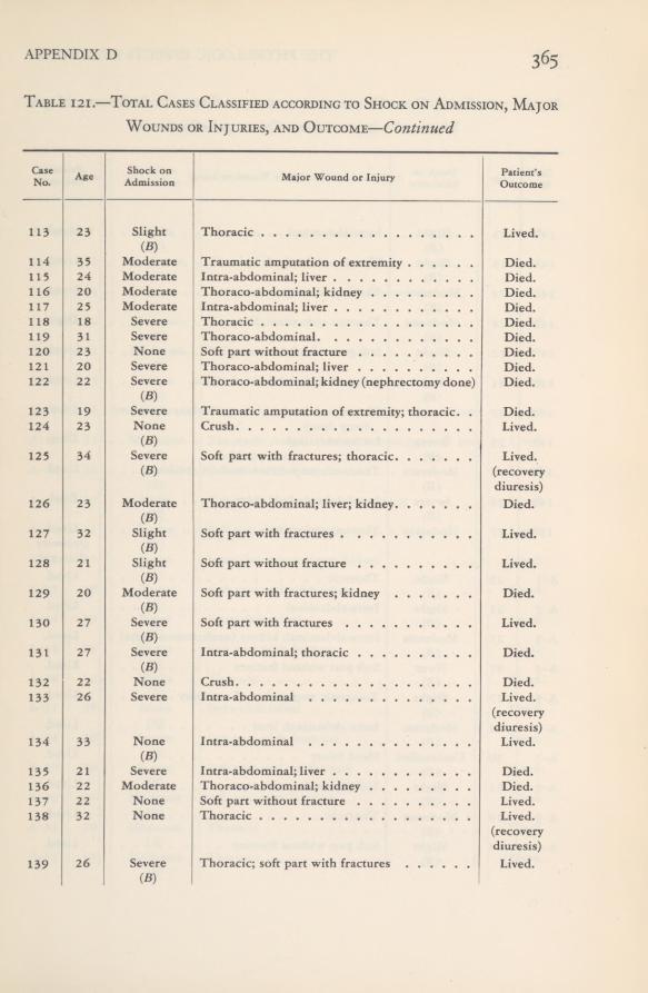
APPENDIX D 365Table 121.—Total Cases Classified according to Shock on Admission, Major
Wounds or Injuries, and Outcome—Continued
CaseNo. Age Shock on
Admission Major Wound or Injury Patient’sOutcome
113 23 Slight(B)
Thoracic Lived.
114 35 Moderate Traumatic amputation of extremity Died.115 24 Moderate Intra-abdominal; liver Died.116 20 Moderate Thoraco-abdominal; kidney Died.117 25 Moderate Intra-abdominal; liver Died.118 18 Severe Thoracic Died.119 31 Severe Thoraco-abdominal Died.120 23 None Soft part without fracture Died,121 20 Severe Thoraco-abdominal; liver Died.122 22 Severe
(B)Thoraco-abdominal; kidney (nephrectomy done) Died.
123 19 Severe Traumatic amputation of extremity; thoracic. . Died.124 23 None
(B)Crush Lived.
125 34 Severe(B)
Soft part with fractures; thoracic Lived.(recoverydiuresis)
126 23 Moderate(B)
Thoraco-abdominal; liver; kidney Died.
127 32 Slight(B)
Soft part with fractures Lived.
128 21 Slight(B)
Soft part without fracture Lived.
129 20 Moderate(B)
Soft part with fractures; kidney Died.
130 27 Severe(B)
Soft part with fractures Lived.
131 27 Severe(B)
Intra-abdominal; thoracic Died.
132 22 None Crush Died.133 26 Severe Intra-abdominal Lived.
(recoverydiuresis)
134 33 None(B)
Intra-abdominal Lived.
135 21 Severe Intra-abdominal; liver Died.136 22 Moderate Thoraco-abdominal; kidney Died.137 22 None Soft part without fracture Lived.138 32 None Thoracic Lived.
(recoverydiuresis)
139 26 Severe(B)
Thoracic; soft part with fractures Lived.
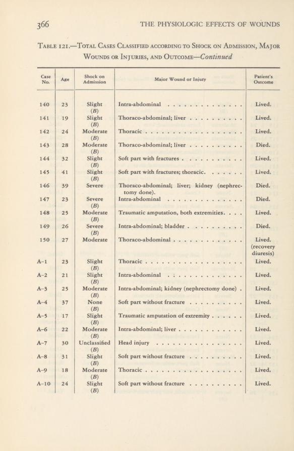
366 THE PHYSIOLOGIC EFFECTS OF WOUNDS
Table 121.—Total Cases Classified according to Shock on Admission, MajorWounds or Injuries, and Outcome—Continued
CaseNo. Age Shock on
Admission Major Wound or Injury Patient’sOutcome
140 23 Slight(B)
Intra-abdominal Lived.
141 19 Slight(B)
Thoraco-abdominal; liver Lived.
142 24 Moderate(B)
Thoracic Lived.
143 28 Moderate(B)
Thoraco-abdominal; liver Died.
144 32 Slight(B)
Soft part with fractures Lived.
145 41 Slight(B)
Soft part with fractures; thoracic Lived.
146 39 Severe Thoraco-abdominal; liver; kidney (nephrec-tomy done).
Died.
147 23 Severe(B)
Intra-abdominal Died.
148 25 Moderate(B)
Traumatic amputation, both extremities. . . . Lived.
149 26 Severe(B)
Intra-abdominal; bladder Died.
150 27 Moderate Thoraco-abdominal Lived.(recoverydiuresis)
A-l 23 Slight(B)
Thoracic Lived,
A-2 21 Slight(B)
Intra-abdominal . ; Lived,
A-3 25 Moderate(B)
Intra-abdominal; kidney (nephrectomy done) . Lived.
A-4 37 None(B)
Soft part without fracture Lived.
A-5 17 Slight(B)
Traumatic amputation of extremity Lived.
A-6 22 Moderate(B)
Intra-abdominal; liver Lived.
A-7 30 Unclassified(B)
Head injury Lived.
A-8 31 Slight(B)
Soft part without fracture Lived.
A-9 18 Moderate(B)
Thoracic Lived.
A-10 24 Slight(B)
Soft part without fracture Lived,
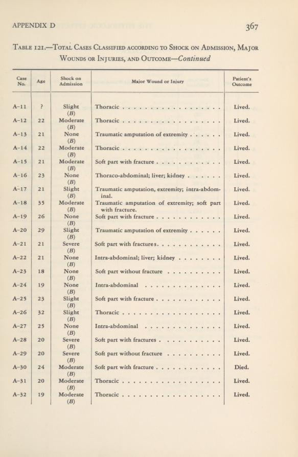
APPENDIX D 367Table 121.—Total Cases Classified according to Shock on Admission, Major
Wounds or Injuries, and Outcome—Continued
CaseNo. Age Shock on
Admission Major Wound or Injury Patient’sOutcome
A-l 1 ? Slight(B)
Thoracic Lived.
A-12 22 Moderate(B)
Thoracic Lived.
A-l 3 21 None(B)
Traumatic amputation of extremity Lived.
A-14 22 Moderate(B)
Thoracic Lived.
A-l 5 21 Moderate(B)
Soft part with fracture Lived.
A-16 23 None(B)
Thoraco-abdominal; liver; kidney Lived.
A-17 21 Slight(B)
Traumatic amputation, extremity; intra-abdom-inal.
Lived.
A-18 35 Moderate(B)
Traumatic amputation of extremity; soft partwith fracture.
Lived.
A-19 26 None(B)
Soft part with fracture Lived.
A-20 29 Slight(B)
Traumatic amputation of extremity Lived.
A-21 21 Severe(B)
Soft part with fracture s Lived.
A-22 21 None(B)
Intra-abdominal; liver; kidney Lived.
A-2 3 18 None(B)
Soft part without fracture Lived.
A-24 19 None(B)
Intra-abdominal Lived.
A-2 5 23 Slight(B)
Soft part with fracture Lived.
A-26 32 Slight(B)
Thoracic Lived.
A-2 7 25 None(B)
Intra-abdominal Lived.
A-2 8 20 Severe(B)
Soft part with fractures Lived.
A-2 9 20 Severe(B)
Soft part without fracture Lived.
A-30 24 Moderate(B)
Soft part with fracture Died.
A-31 20 Moderate(B)
Thoracic Lived.
A-32 19 Moderate(B)
Thoracic Lived.
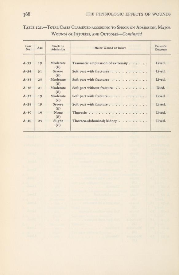
368 THE PHYSIOLOGIC EFFECTS OF WOUNDS
Table 121.—Total Cases Classified according to Shock on Admission, MajorWounds or Injuries, and Outcome—Continued
CaseNo. Age Shock on
Admission Major Wound or Injury Patient’sOutcome
A-33 19 Moderate(B)
Traumatic amputation of extremity Lived.
A-34 31 Severe(B)
Soft part with fractures Lived.
A-35 25 Moderate(■B)
Soft part with fractures Lived.
A-36 21 Moderate(B)
Soft part without fracture Died.
A-37 19 Moderate(B)
Soft part with fracture Lived.
A-38 19 Severe(B)
Soft part with fracture Lived,
A-39 19 None(B)
Thoracic Lived.
A-40 25 Slight(B)
Thoraco-abdominal; kidney Lived.
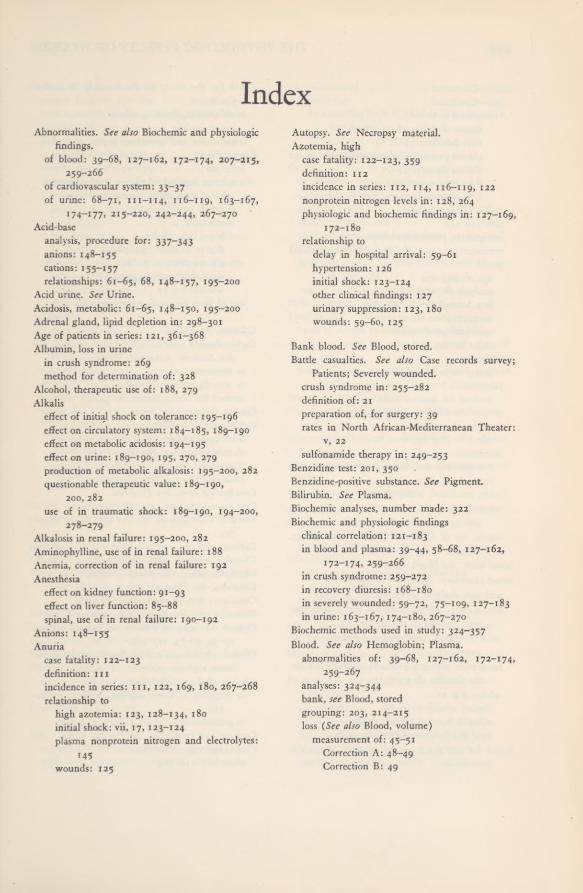
IndexAbnormalities. See also Biochemic and physiologic
findings.of blood: 39-68, 127-162, 172-174, 207-215,
259-266of cardiovascular system: 33-37of urine: 68-71, 111-114, 116—119, 163-167,
174-177, 215-220, 242-244, 267-270Acid-base
analysis, procedure for: 337-343anions: 148-155cations: 155-157relationships: 61-65, 68, 148-157, 195-200
Acid urine. See Urine.Acidosis, metabolic: 61-65, 148-150, 195-200Adrenal gland, lipid depletion in: 298-301Age of patients in series: 121, 361-368Albumin, loss in urine
in crush syndrome: 269method for determination of: 328
Alcohol, therapeutic use of: 188, 279Alkalis
effect of initial shock on tolerance: 195-196effect on circulatory system: 184-185, 189-190effect on metabolic acidosis: 194-195effect on urine: 189-190, 195, 270, 279production of metabolic alkalosis: 195-200, 282questionable therapeutic value: 189-190,
200,282use of in traumatic shock: 189-190, 194-200,
278~2 79Alkalosis in renal failure: 195-200, 282Aminophylline, use of in renal failure: 188Anemia, correction of in renal failure: 192Anesthesia
effect on kidney function: 91-93effect on liver function: 85-88spinal, use of in renal failure: 190-192
Anions: 148—155Anuria
case fatality: 122-123definition: mincidence in series: m, 122, 169, 180, 267-268relationship to
high azotemia: 123, 128-134, 180initial shock: vii, 17, 123-124plasma nonprotein nitrogen and electrolytes:
M5wounds: 125
Autopsy. See Necropsy material.Azotemia, high
case fatality: 122-123, 359definition: 112incidence in series: 112, 114, 116-119, 122nonprotein nitrogen levels in: 128, 264physiologic and biochemic findings in: 127-169,
172-180relationship to
delay in hospital arrival: 59-61hypertension: 126initial shock: 123-124other clinical findings: 127urinary suppression: 123, 180wounds: 59-60, 125
Bank blood. See Blood, stored.Battle casualties. See also Case records survey;
Patients; Severely wounded,crush syndrome in: 255-282definition of: 21preparation of, for surgery: 39rates in North African-Mediterranean Theater:
v, 22sulfonamide therapy in: 249—253
Benzidine test: 201, 350Benzidine-positive substance. See Pigment.Bilirubin. See Plasma.Biochemic analyses, number made: 322Biochemic and physiologic findings
clinical correlation: 121-183in blood and plasma: 39-44, 58-68, 127-162,
172-174, 259-266in crush syndrome: 259-272in recovery diuresis: 168-180in severely wounded: 59-72, 75-109, 127-183in urine: 163-167, 174-180, 267-270
Biochemic methods used in study: 324-357Blood. See also Hemoglobin; Plasma.
abnormalities of: 39-68, 127-162, 172-174,259-267
analyses: 324-344bank, see Blood, storedgrouping: 203, 214-215loss (See also Blood, volume)
measurement of: 45-51Correction A: 48-49Correction B: 49

THE PHYSIOLOGIC EFFECTS OF WOUNDS
Blood —Continuedloss—Continued
relationship todegree of shock: 48-51, 56—58liver function: 78-80plasmaprotein and hematocrit levels: 41-42plasma therapy: 54-55time after wounding: 55type of wound: 51-55
normal values in: 325pH: 148-149precipitants, preparation of: 324-325pressure: 35, 112, 118 (See also Hypertension)stored
age of: 203-204analyses of: 202-206free hemoglobin in: 203-205iso-agglutinins in: 214-215specifications of: 203sugar in: 205-206
substitutespolicy on in World War II: 5—n
sulfonamide levels in: 250-253method for determination of: 343
theater blood banks: 11, 18-19, 203therapy: 38-39, 192
effect on liver function: 80transfusion
National Research Council committee on: 7-9reaction: 18, 203
types, see Blood, groupingvolume, method for determination of: 45-51,
326-331 {See also Blood, loss)whole, use of in World War II: 5-11, 38-39,
56-57Blood bank. See Blood, stored.Blood chemistry. See also Plasma; Serum,
acid-base analyses, procedure for: 337-343hemoglobin, hematocrit values, and plasma pro-
tein, method of calculation: 344methemoglobin, method for determination of;
343nonprotein nitrogen products, methods for de-
termination of; 332-337relationship to
clinical condition: 6i-66delay in hospital arrival: 59-61type and location of wound: 59-60
Board for the Study of the Severely Wounded:Frontispiece
Board for the Study of the Severely Wounded:—Continuedestablishment of: vii, 5, 16—20
events leading to: 1-19organization and operation: 307-314
Bromsulfaleinretention
average hospital admission levels: 72-77effect of ether anesthesia on: 85-88in crush syndrome: 271relationship to
blood loss: 78-80degree of shock: 72, 78-79plasma therapy: 72, 80time after wounding: 72wound: 72, 77
test of liver function: 72, 75-77, 345-346
Calcium. See Serum.Carbon-dioxide combining power of plasma. See
also Acidosis, metabolic; Plasma,in azotemia: 127, 129in severely wounded: 63-64method for determination of: 341
Cardiac changes. See Heart.Cardiovascular findings: 33-36
blood pressure; 35, 112, 118electrocardiograms: 33-34“irreversible changes”: 35-36pulse rate and volume: 34-35
Case fatality rates. See Fatalities.Case records survey; 359-368Casualty, definition of: 21. See also Battle casual-
ties.Cations: 155-157Chemical methods used. See Biochemic methods.Chemistry, blood. See Blood chemistry.Chlorides. See Plasma.Circulatory system, overloading: 184-186Clearance tests. See Renal clearance tests; Urine.Clinical condition of patients on hospital arrival:
27-39, 59-73, 257-259Clinical, physiologic, and biochemic correlation in
lower nephron nephrosis: 121-183Clinicopathologic correlation
in diagnosis of lower nephron nephrosis: 110-
120
in pathogenesis of shock kidney: 244-247Creatine and creatinine. See Plasma; Urine.Crush syndrome: 255-282
albuminuria in: 269

INDEX 371
Crush syndrome—Continuedclinical features: 257-259hemoconcentration in: 261-264, 281initial shock: 257kidney damage: 259, 277-278
described early in war: 17muscle damage: 272-275myoglobin in kidney and muscle: 274, 281-282pathologic findings: 272-278
in kidneys: 275-278in liver; 272in lungs: 272in skeletal muscles: 272-275
physiologic and biochemic findings: 259-272in blood and plasma: 259-266in urine; 267-270liver function: 271
pigmentin muscle: 274in plasma: 265-266in urine: 269nephropathy: 277
pulmonary edema in: 281recovery diuresis in: 282treatment: 278-281
alkalinization of urine: 278-279correction of hemoconcentration: 278dehydrating agents: 280diuretics: 279-280other measures: 280—281
Deaths. See Fatalities.Decolorization of muscle in crush syndrome: 272-
274Diuresis. See Recovery diuresis.Diuretics, use of: 188, 279—280Drugs and procedures used to stimulate renal func-
tion: 186-192, 278-281
Edema. See Pulmonary edema.Electrocardiographic observations: 33-34Electrolytes. See Plasma.Embolism, fat. See Fat embolism.Ether anesthesia
effect on kidney function: 91-93effect on liver function: 85-88
Evacuation time: 23-25
Fatdepletion in adrenal cortex: 299-301, 304embolism, pulmonary: 288-289, 305
Fat—Continuedvacuolation
in ascending limb of Henle’s loop: 227-232,297-298
in liver: 293-296in myocardium: 289-292
Fatalitiesin azotemia: 122-123, 359in crush syndrome: 259in entire series: 122—124, 359-360, 361-368in hypertension: 126in oliguria and anuria: 122-123in “shock kidney”: 119incidence in first 10 days after wounding: 124,
126, 135, 185uremia as cause: 123-124, 359-360
Field laboratory: viii, 315-322Fluid intake
cause of plasma volume increase: 185, 281excess as cause of pulmonary edema: 184-186,
281restriction of in treatment of lower nephron
nephrosis: 186, 281Fractures. See Wounds.Free hemoglobin. See Plasma, “hemoglobin.”
Glomerular filtration, measurement of: 97-101,346-348
Glucose in plasma, method for determination of:343
Granulomas, interstitial: 235
Heartcardiovascular findings: 33—36pathologic findings: 289-292
Hematocrit valueand plasma proteins: 39-44, 160—162
in crush syndrome: 261-264determination of: 39, 344influence of plasma therapy on; 42-43
Heme pigment. See Myoglobin; Pigment, heme;Plasma, “hemoglobin.”
Hemoconcentration in crush syndrome: 261-264correction of: 278
Hemoglobin, See also Pigment; Plasma; Urine,pigment,
calculation: 344excretion in urine, see Urine, pigmentfree, in plasma, see Plasma, “hemoglobin”levels in wounded men: 46-51, 66—68loss of total (See also Blood)

372 THE PHYSIOLOGIC EFFECTS OF WOUNDS
Hemoglobin—Continuedloss of total—Continued
determination of: 45-51effect of blood therapy on: 54-55relationship to degree of shock: 56-58relationship to time after wounding: 55relationship to type of wound: 51-55
methemoglobin: 343Hemoglobinuria. See Pigment, excretion; Urine,
pigment.Henle’s loop: 227-232
fat vacuolation in: 230, 297-298High azotemia. See Azotemia, high.Histologic findings. See Kidney, pathology;
Pathology.Historical background of study: 1-20
Hydrogen ion concentration, determination of:337-338, 350. See also pH.
Hydronephrosis, internal: 226-227Hypertension
definition: 112
in high azotemia: 126in lower nephron nephrosis: 112, 116, 118, 126
Hypertonic solutions; 187
Injury, definition of: 21Interstitial inflammation: 232-234Ions. See Anions; Cations.“Irreversible” shock: 3-4, 11, 35-36, 56-57Iso-agglutinins: 214-215
Kidney. See also Lower nephron nephrosis,decapsulation: 192function: 17, 90-109, 111, 121-122
drugs and procedures to stimulate: 186-192effect of anesthesia: 91-93effect of shock: 17, 91-92effect of sulfonamides: 251-253recovery diuresis; 168-180significance of pigment casts: 246-247tests
phenolsulfonphthalein; 90-97, 348-349renal clearance: 97-105, 346-349urine concentration: 105-106
lesionpathogenesis of: 240-244
pathology: 224-248, 275-278, 297-298clinical correlation: 244-247gross: 237-239, 275-277in crush syndrome: 275-278microscopic: 225-237, 277-278
Kidney—Continuedpathology—Continued
pigment precipitation: 225-226, 242-244,274, 277
“shock kidney,” see Lower nephron nephrosis
Laboratory, field; 315-322Lactic acid in plasma; 63-65
method for determination of: 342-343Latent consequences of wounds: 21Lipid depletion in adrenal glands; 299-301Liver
fat vacuolation in; 293-296function: 75-89
bromsulfalein test for: 72, 75-77, 345-346effect of ether anesthesia on: 85-88effect of plasma therapy on; 80in crush cases: 271in newly wounded: 72, 75-89plasma bilirubin levels: 83-85relationship to
location of major wound: 77postoperative course: 81-88shock and blood loss: 78-80time from wounding: 77
histologic findings in, in crush syndrome: 272Loops of Henle. See Henle’s loop.Lower nephron nephrosis
abnormalities of blood: 127-162, 169, 172-174abnormalities of urine: 163-167, 169, 174-177clinical features; 115-119, 122-127, 240-242clinical, physiologic, and biochemic correlation:
I2i'-x83clinicopathologic correlation: 110-120, 244-247diagnosis; 1x0-120pathogenesis: 240-242pathology: 224-248, 275-278treatment: 184-193
Lungs, pathologic findings in: 272, 285-289. Seealso Pulmonary edema; Pulmonary fat em-bolism.
Magnesiumplasma: 134
method for determination of: 340-341sulfate, use in renal failure: 189
Mannitolmethod for determination of: 346-348use in renal clearance studies: 97-101
Metabolic acidosis in shock. See Acidosis,metabolic.

INDEX 373Methemoglobin, method for determination of: 343Muscles
decolorized, in crush syndrome: 272-275myoglobin in: 274, 281necrosis of, in crush syndrome: 275
Myocardium, fat vacuolation in: 289—292Myoglobin. See also Pigment,
in kidneys: 242, 274in muscle: 274, 281-282in plasma: 201, 266in urine: 216—220, 266, 269method for determination of: 201-202, 354-357
Myoglobinuriain wounded men: 215-220relation to hemoglobinuria: 219-220
Necropsy material: 240, 284-285Necrosis of skeletal muscles in crush cases; 218-219Nephrons, lower, precipitation of pigment in: 243-
244Nephropathy, pigment. See Pigment.Nephrosis. See Lower nephron nephrosis.Nitrogenous waste products: 135-148Nonprotein nitrogen. See Plasma.Nontransportable patient, definition of: 21, 359Nutrition in treatment of lower nephron nephrosis:
I 92_I 93
“O” group blood: 18, 203Oliguria. See also Urine, suppression,
case fatality: 123, 180definition: mincidence in series: m, xi8, 122, 169, 180,
267-268relationship to
high azotemia: 128-134initial shock: 123-124lower nephron nephrosis: 247plasma N.P.N. and electrolytes: 144renal tubule degeneration: 247wounds: 125
Organic acids in urine, procedure for determina-tion of; 351-352
Pain in wounded men: 27Para-amino hippuric acid: x02-105, 348Pathology of traumatic shock
crush syndrome; 272-278kidney: 275-278liver: 272lungs; 272skeletal muscles: 272-275
Pathology of traumatic shock—Continuedgeneral: 283-305
adrenal gland: 298-301, 304heart: 289-292kidney: 224-248, 297-298liver: 293-296lungs: 285-289pattern of pathognomonic changes: 301-305reversibility of lesions: 302-304
“shock kidney”clinical correlation: 110-120, 244-247gross changes: 237-239microscopic changes: 225-237pathogenesis of: 240-242
Patients. See also Battle casualties; Case recordssurvey.
age of, in series: 121, 361—368clinical condition on hospital arrival: 27-39nontransportable: 21
pHof blood: 148-149, 337-338of urine: 114, 150, 165-167, 350
Phenolsulfonphthaleinexcretory capacity: 90-97
effect of anesthesia on: 91-93in first 3 days after wounding: 91rate of improvement in: 93-97
test: 90-91, 177, 348-349Phosphorus. See Plasma, phosphorus.Physiologic findings. See Biochemic and physio-
logic findings; Urine, suppression.Pigment, heme. See also Myoglobin; Plasma,
“hemoglobin”; Urine,casts, staining reactions of: 225-230excretion in urine: 113, 116-119, 167, 202,
215-220, 269functional significance of: 246relationship to initial shock: 242
hemoglobin, see Plasma, “hemoglobin”identification of: 201—202, 242, 344-345, 350,
354-357in muscle and kidney, see Myoglobin; Pigment,
precipitationin pathogenesis of “shock kidney”; 240-244in plasma, see Plasma, “hemoglobin”mobilization in severely wounded men: 201-
223nature of: 242-243, 266nephropathy (See also Kidney, pathology)
incidence in severely wounded: no—in,225-226, 243, 277, 282

THE PHYSIOLOGIC EFFECTS OF WOUNDS374
Pigment, heme—Continuednephropathy—Continued
relationship to shock: 244renal insufficiency in: m-114
precipitation in renal tubules: 225-226, 243-244Plasma
bilirubin: 66-68, 73, 82-84method for determination of: 344
carbon-dioxide combining power: 63-65, 129,148-150
method for determination of: 341chlorides: 63, 130, 150-153, 264-265
method for determination of: 341-343creatinine: 61-62, 137-142
method for determination of: 334electrolytes in shock and renal failure: 63-65,
144-145glucose, method for determination of: 343“hemoglobin” (See also Myoglobin; Pigment)
determination of: 201-203, 344-345levels in crush syndrome: 265-266levels in stored blood: 203-206levels in wounded men: 207-214, 265-266relationship to hemoglobinuria: 215-216
lactic acid: 63-65method for determination of: 342-343
magnesium: 134, 156method for determination of: 340-341
myoglobin; 201, 266nonprotein nitrogen (See also Azotemia, high)
levelsin crush syndrome: 264in high azotemia: 112, 1x6-119, 128, 135-
137in recovery diuresis: 172-174in severely wounded: 59-62, 112, 118, 122,
x8xmethod for determination of: 332-334products, method for determination of: 332—
337normal values in: 325phosphorus: 131, 142-143, 154
method for determination of: 341relationship to calcium: 143—147
pigment: 201-223 (See also Myoglobin; Pig-ment; Plasma, “hemoglobin”)
method for determination of: 344-345precipitants, preparation of: 324-325proteins: 39-44, 132, 154-155
determination of: 39, 344in crush syndrome: 261-262
Plasma—Continuedtherapy, prior to hospital entry: 38-39urea nitrogen; 137-142
method for determination of: 335-337uric acid: 82-84, 147-148
method for determination of: 334-335volume
effect of fluid intake on: 157-160increased in renal insufficiency: 157-162method for determination of: 326-331relationship to plasma protein, hematocrit,
and blood volume: 160-162Posttraumatic renal insufficiency, syndrome of. See
Lower nephron nephrosis.Potassium, serum: 156, 340Potassium chloride: 189Proteins. See Plasma.Proteinuria: 113, 167P.S.P. test. See Phenolsulfonphthalein.Pulmonary edema: 281, 286-288
prevention of: 184-186, 305Pulmonary fat embolism: 288-289, 305Pulse rate and volume: 34-35
Recovery diuresis: 168-180blood chemistry: 172-174clinical features: 169-172in crush syndrome: 282plasma volume in: 174urinary findings: 174-179
Renal clearance tests: 97-105and phenolsulfonphthalein excretion in crush
cases: 177Renal failure. See also Kidney, function; Lower
nephron nephrosis,relationship to
hypertension: 126initial shock: 123-124other clinical findings: 127survival period: 124type of wound; 125
Renal function in severely wounded. See Kidney,function.
Renal lesions. See Kidney, pathology.Renal tubules. See Tubules, renal.Resuscitation. See Shock, traumatic, resuscitation
from.Retention. See Bromsulfalein; Urine, suppression.Reversibility of lesions in shock: 302-304

INDEX 375
Serumcalcium
method for determination of; 340relationship to phosphorus: 143, 147
normal values in: 325potassium: 156
method for determination of; 340sodium: 133, 144-145, 155-156
method for determination of: 338-340Severely wounded
Board for the Study of. See Board for the Studyof the Severely Wounded,
classification of cases: 361-368definition of: 21, 359initial blood changes: 46-51initial studies on: 23-39internal state on entry to forward hospital: 21-74management of in World War II: 5-20number of in Mediterranean Theater of Oper-
ations: 22pigment mobilization in: 201—223
Shock, traumaticbiochemic and physiologic findings: 61-65, 67,
70-71blood and plasma therapy: 38-39, 56-57, 194-
195blood loss: 48-51, 56-58blood pressure: 35degree of: 27-29, 361-368
in anuria and oliguria: m, 116-118in azotemia: 112, 116-119in crush syndrome: 257in lower nephron nephrosis: 110-120, 240—
242in relation to time after wounding: 29-31in relation to type of wound: 30-33in renal insufficiency: 123-124
early concepts: 1-4electrocardiograms in cases of: 33-34hemoglobin loss: 48-51, 56-58incidence: 29, 361-368“irreversible”; 3-4, 11, 35-36, 56-57kidney function in: 91-96liver function in: 72, 78-80management of in World War II: 5-20National Research Council subcommittee on: 3-4pathogenesis of: 305pathognomonic changes in; 303-304pathology of: 283-305pigment mobilization in: 207-220plasma protein and hematocrit levels in: 43-44
Shock, traumatic—Continuedpulse rate and volume in: 34-35renal failure in: 123-124resuscitation from: 12-15, 38-39, 56-57treatment: 184-200 (See also Alkalis)
preliminary planning for: 5-9“Shock kidney.” See Kidney; Lower nephron
nephrosis.Sodium. See also Serum.
bicarbonate, administration of: 189-190, 194-195
chloride, administration of; 151-153, 186-187citrate, in stored blood and plasma: 194—195
Spinal anesthesia. See Anesthesia.Staining reaction of pigment casts: 225-230Stored blood. See Blood, stored.Sulfonamides
in relation to “shock kidney”: 18, 249-253method for determination of in blood: 343
Sympathectomy in treatment of lower nephronnephrosis: 192
Testsbenzidine: 201, 350bromsulfalein: 72, 75-77, 345-346phenolsulfonphthalein: 90-97, 177, 348-349renal clearance: 97-105, 177, 346-349urine concentration: 105-106
Therapy. See Treatment.Thrombophlebitis in “shock kidney”: 235-237Trauma. See Wounds.Traumatic shock. See Shock, traumatic.Treatment
errors to avoid: 184-186of crush syndrome: 278-281of renal failure and lower nephron nephrosis:
184-193, 194-200of shock and blood loss: 38—39sulfonamide: 249-254
Tubules, renal. See also Henle’s loop; Kidney,pathology,
degeneration of: 227-232, 247excretory capacity of: 102-105fat vacuolation in: 227-232rupture of: 234-235
Urea nitrogen, in plasmaand creatinine: 137-142method for determination of: 335-337
Uremia as cause of death: 122-124, 359-360

376 THE PHYSIOLOGIC EFFECTS OF WOUNDS
Uremic symptomatology: 115-116Uric acid in plasma: 82-84
method for determination of: 334-335Urinalyses: 349-353. 35 6~357Urine
abnormalitiesin crush syndrome; 267-270in lower nephron nephrosis: 113-114, 163-
167, 174-177, 215-220, 242in recovery diuresis: 174-177in wounded men: 68-71, m, 114
acid: 68, 243-244acidity, persistent: 114, 150, 243-244alkalinization of: 189-190, 194-200, 278-279benzidine-positive substance in, see Urine, pig-
mentchlorides: 63-65clearance tests: 97-105, 177, 346-349concentrated: 68, 108, 163-165concentration test: 105-106creatine and creatinine: 69-71, 142
method for determination of: 352-353pH; 114, 150, 165-167, 350phenolsulfonphthalein excretion in: 90-97, 177pigment
excretion: 113, 116-119, 167, 201-202, 215-220, 269
method for determination of: 201—202, 356—357
relation of hemoglobinuria to myoglobinuria:219—220
relationship to hemoglobin in plasma: 215-216protein excretion: 113, 167specific gravity, fixed, in lower nephron nephro-
sis: 114, 163-165, 174, 270
Urine—Continuedsuppression (See also Anuria; Oliguria)
in crush syndrome: 267-268relationship of anuria, oliguria, and azotemia:
123, 128-134relationship to proved pigment nephropathy:
243-244time as factor in development of; 124
Vacuolation. See Fat.Van den Bergh test. See Plasma bilirubin.Vasoconstriction, result of blood loss: 58Visceral changes, pattern of: 304-305
Whole blood. See Blood.Wound shock. See Shock, traumatic.Wounded in action, definition of: 21Wounded men. See Battle casualties; Severely
wounded.Wounds
how incurred: 359latent consequences of: 21major, in entire series: 25-27, 361-368relationship to
blood chemistry findings: 59-60blood loss: 51-55development of renal failure: 125hemoglobin loss: 52-55liver function: 72, 77pain; 27plasma bilirubin and hemoglobin levels; 66-67plasma protein and hematocrit values: 40,
43-44shock: 30-33
type and location of major: 25-27, 361-368(See also Crush syndrome)
in renal insufficiency: 125in recovery diuresis; 169U. S. GOVERNMENT PRINTING OFFICE: 1952—956507


Copyright © 2022 FDOKUMEN