Chronic Inflammation in Non-Healing Skin Wounds ... - MDPI
31
Citation: Schilrreff, P.; Alexiev, U. Chronic Inflammation in Non-Healing Skin Wounds and Promising Natural Bioactive Compounds Treatment. Int. J. Mol. Sci. 2022, 23, 4928. https://doi.org/ 10.3390/ijms23094928 Academic Editor: Maria Vittoria Barone Received: 10 March 2022 Accepted: 26 April 2022 Published: 28 April 2022 Publisher’s Note: MDPI stays neutral with regard to jurisdictional claims in published maps and institutional affil- iations. Copyright: © 2022 by the authors. Licensee MDPI, Basel, Switzerland. This article is an open access article distributed under the terms and conditions of the Creative Commons Attribution (CC BY) license (https:// creativecommons.org/licenses/by/ 4.0/). International Journal of Molecular Sciences Review Chronic Inflammation in Non-Healing Skin Wounds and Promising Natural Bioactive Compounds Treatment Priscila Schilrreff and Ulrike Alexiev * Fachbereich Physik, Institut für Experimentalphysik, Freie Universität Berlin, Arnimallee 14, 14195 Berlin, Germany; [email protected] * Correspondence: [email protected]; Tel.: +49-30-838-55157 Abstract: Chronic inflammation is one of the hallmarks of chronic wounds and is tightly coupled to immune regulation. The dysregulation of the immune system leads to continuing inflammation and impaired wound healing and, subsequently, to chronic skin wounds. In this review, we discuss the role of the immune system, the involvement of inflammatory mediators and reactive oxygen species, the complication of bacterial infections in chronic wound healing, and the still-underexplored potential of natural bioactive compounds in wound treatment. We focus on natural compounds with antioxidant, anti-inflammatory, and antibacterial activities and their mechanisms of action, as well as on recent wound treatments and therapeutic advancements capitalizing on nanotechnology or new biomaterial platforms. Keywords: chronic wounds; immunity; inflammation; natural bioactive compounds 1. Introduction The skin is a complex organ that has numerous strategies to protect the body from external insults. It contains a highly specialized network of immune cells, crucial for the defense and repair, and also for the maintenance of tissue homeostasis. Following injury, the skin’s immune system plays a key role not only in preventing infections, but also in orchestrating the tissue-repair process. Broadly speaking, the normal wound-healing process involves four successive, but overlapping, phases that vary in time, including the homeostasis and the inflammation phase, the proliferation phase, and the tissue remodeling phase. Wound-healing processes tend to be strictly regulated by various growth factors and cytokines that are released at the wound site. The deregulation of immune responses often results in impaired healing and poor tissue restoration and function [1]. Clinically, chronic wounds are those that cannot be healed through the orderly phases of healing, but are detained in a self-perpetuating inflammatory stage and remain intractable despite adequate wound management (Figure 1). Numerous factors can delay wound healing, such as chronic disease, vascular insufficiency, diabetes, malnutrition, aging, or local factors such as pressure, infection, and edema [2]. Skin wounds have a huge negative impact on healthcare systems and economies worldwide. It is estimated that nearly one billion people worldwide suffer from acute and chronic conditions. Chronic wounds are becoming an increasing socioeconomic problem for aging societies due to the prevalence of obesity, diabetes, and cardiovascular diseases among elderly people. Increasing antibiotic resistance poses a further challenge in the treatment of bacterial infections in the context of chronic wounds. It persists as a silent epidemic affecting the quality of life of those suffering from it [3]. The costs associated with the treatment of wounds represent about 2–4% of the total healthcare expenditure in Europe [4,5]. As the population grows and people live longer, the costs and the number of patients are expected to increase, affecting around 1 / 4 of the elderly population by 2050 [6]. Advances in wound care over the last century have been very slow [7]. Deeper knowledge Int. J. Mol. Sci. 2022, 23, 4928. https://doi.org/10.3390/ijms23094928 https://www.mdpi.com/journal/ijms
-
Upload
khangminh22 -
Category
Documents
-
view
4 -
download
0
Transcript of Chronic Inflammation in Non-Healing Skin Wounds ... - MDPI

Citation: Schilrreff, P.; Alexiev, U.
Chronic Inflammation in
Non-Healing Skin Wounds and
Promising Natural Bioactive
Compounds Treatment. Int. J. Mol.
Sci. 2022, 23, 4928. https://doi.org/
10.3390/ijms23094928
Academic Editor: Maria Vittoria
Barone
Received: 10 March 2022
Accepted: 26 April 2022
Published: 28 April 2022
Publisher’s Note: MDPI stays neutral
with regard to jurisdictional claims in
published maps and institutional affil-
iations.
Copyright: © 2022 by the authors.
Licensee MDPI, Basel, Switzerland.
This article is an open access article
distributed under the terms and
conditions of the Creative Commons
Attribution (CC BY) license (https://
creativecommons.org/licenses/by/
4.0/).
International Journal of
Molecular Sciences
Review
Chronic Inflammation in Non-Healing Skin Wounds andPromising Natural Bioactive Compounds TreatmentPriscila Schilrreff and Ulrike Alexiev *
Fachbereich Physik, Institut für Experimentalphysik, Freie Universität Berlin, Arnimallee 14,14195 Berlin, Germany; [email protected]* Correspondence: [email protected]; Tel.: +49-30-838-55157
Abstract: Chronic inflammation is one of the hallmarks of chronic wounds and is tightly coupledto immune regulation. The dysregulation of the immune system leads to continuing inflammationand impaired wound healing and, subsequently, to chronic skin wounds. In this review, we discussthe role of the immune system, the involvement of inflammatory mediators and reactive oxygenspecies, the complication of bacterial infections in chronic wound healing, and the still-underexploredpotential of natural bioactive compounds in wound treatment. We focus on natural compounds withantioxidant, anti-inflammatory, and antibacterial activities and their mechanisms of action, as well ason recent wound treatments and therapeutic advancements capitalizing on nanotechnology or newbiomaterial platforms.
Keywords: chronic wounds; immunity; inflammation; natural bioactive compounds
1. Introduction
The skin is a complex organ that has numerous strategies to protect the body fromexternal insults. It contains a highly specialized network of immune cells, crucial for thedefense and repair, and also for the maintenance of tissue homeostasis. Following injury,the skin’s immune system plays a key role not only in preventing infections, but alsoin orchestrating the tissue-repair process. Broadly speaking, the normal wound-healingprocess involves four successive, but overlapping, phases that vary in time, including thehomeostasis and the inflammation phase, the proliferation phase, and the tissue remodelingphase. Wound-healing processes tend to be strictly regulated by various growth factorsand cytokines that are released at the wound site. The deregulation of immune responsesoften results in impaired healing and poor tissue restoration and function [1]. Clinically,chronic wounds are those that cannot be healed through the orderly phases of healing,but are detained in a self-perpetuating inflammatory stage and remain intractable despiteadequate wound management (Figure 1). Numerous factors can delay wound healing,such as chronic disease, vascular insufficiency, diabetes, malnutrition, aging, or local factorssuch as pressure, infection, and edema [2].
Skin wounds have a huge negative impact on healthcare systems and economiesworldwide. It is estimated that nearly one billion people worldwide suffer from acute andchronic conditions. Chronic wounds are becoming an increasing socioeconomic problemfor aging societies due to the prevalence of obesity, diabetes, and cardiovascular diseasesamong elderly people. Increasing antibiotic resistance poses a further challenge in thetreatment of bacterial infections in the context of chronic wounds. It persists as a silentepidemic affecting the quality of life of those suffering from it [3]. The costs associatedwith the treatment of wounds represent about 2–4% of the total healthcare expenditure inEurope [4,5]. As the population grows and people live longer, the costs and the number ofpatients are expected to increase, affecting around 1/4 of the elderly population by 2050 [6].Advances in wound care over the last century have been very slow [7]. Deeper knowledge
Int. J. Mol. Sci. 2022, 23, 4928. https://doi.org/10.3390/ijms23094928 https://www.mdpi.com/journal/ijms

Int. J. Mol. Sci. 2022, 23, 4928 2 of 31
of the anatomical structure and function of the skin during chronic wound healing isessential for chronic wound management. In addition, it is still a major challenge to find anew treatment for chronic wounds [8].
However, plants and microorganisms such as bacteria, fungi, microalgae, cyanobacte-ria, and archaea have proven to be an excellent source of bioactive compounds. In particular,plant-derived compounds have been used worldwide for thousands of years as traditionaltreatments for numerous diseases. Only a very small percentage of the thousands of knownspecies of plants in the world have been chemically analyzed, demonstrating great potentialfor the discovery of new drugs. Natural bioactive compounds with high levels of antioxi-dant, anti-inflammatory, and antimicrobial properties could be of great benefit for chronicwound healing. For example, anti-inflammatory drugs can quickly regulate the levels ofvarious inflammatory factors and normalize the inflammatory response of chronic woundswith severe inflammation. Several studies have documented the use of extracts from naturalorigin for the development of bioactive wound treatments, which provide opportunitiesfor eliminating the inflammatory response and accelerating wound healing [9].
Int. J. Mol. Sci. 2022, 23, 4928 2 of 35
knowledge of the anatomical structure and function of the skin during chronic wound healing is essential for chronic wound management. In addition, it is still a major challenge to find a new treatment for chronic wounds [8].
However, plants and microorganisms such as bacteria, fungi, microalgae, cyanobacteria, and archaea have proven to be an excellent source of bioactive compounds. In particular, plant-derived compounds have been used worldwide for thousands of years as traditional treatments for numerous diseases. Only a very small percentage of the thousands of known species of plants in the world have been chemically analyzed, demonstrating great potential for the discovery of new drugs. Natural bioactive compounds with high levels of antioxidant, anti-inflammatory, and antimicrobial properties could be of great benefit for chronic wound healing. For example, anti-inflammatory drugs can quickly regulate the levels of various inflammatory factors and normalize the inflammatory response of chronic wounds with severe inflammation. Several studies have documented the use of extracts from natural origin for the development of bioactive wound treatments, which provide opportunities for eliminating the inflammatory response and accelerating wound healing [9].
Factors that contribute to delayed healing in chronic wounds are key components of a comprehensive approach to wound care and present the primary challenges for their treatment. Therefore, a better understanding of the molecular and cellular aspects of inflammation involved in chronic wounds should improve our treatment approaches, leading to better healing rates, and facilitate the development of new and more effective therapies.
In this review, the relevant aspects of the inflammatory pathogenesis of chronic wounds and promising therapeutic natural bioactive compounds are reviewed in detail. The following sections are divided into: chronic wounds (Section 2); role of innate and adaptative immunity in chronic wounds (Section 3); role of bacterial infection in chronic wounds (Section 4); and natural bioactive compounds with antioxidant, antimicrobial, and anti-inflammatory activities as wound treatments (Section 5).
Figure 1. Pathologic abnormalities associated with delayed wound healing in chronic wounds. Persistent inflammation, as a hallmark of chronic wounds, is connected to the dysregulation of the immune response during wound healing by various factors and leads to excessive levels of pro-inflammatory signals, reactive oxygen species (ROS), changes in the proteolytic balance, and an increased amount of matrix metalloproteinases (MMPs) that eventually cause damage to the extracellular matrix (ECM) and impaired epithelialization and proliferation of keratinocytes. The molecular pathways and targets are summarized in Figure 2. The image of the chronic wound is reproduced from [10]. Reprinted with permission from AAAS, 2014.
Figure 1. Pathologic abnormalities associated with delayed wound healing in chronic wounds.Persistent inflammation, as a hallmark of chronic wounds, is connected to the dysregulation ofthe immune response during wound healing by various factors and leads to excessive levels ofpro-inflammatory signals, reactive oxygen species (ROS), changes in the proteolytic balance, andan increased amount of matrix metalloproteinases (MMPs) that eventually cause damage to theextracellular matrix (ECM) and impaired epithelialization and proliferation of keratinocytes. Themolecular pathways and targets are summarized in Figure 2. The image of the chronic wound isreproduced from [10]. Reprinted with permission from AAAS, 2014.
Factors that contribute to delayed healing in chronic wounds are key components ofa comprehensive approach to wound care and present the primary challenges for theirtreatment. Therefore, a better understanding of the molecular and cellular aspects of inflam-mation involved in chronic wounds should improve our treatment approaches, leading tobetter healing rates, and facilitate the development of new and more effective therapies.
In this review, the relevant aspects of the inflammatory pathogenesis of chronicwounds and promising therapeutic natural bioactive compounds are reviewed in de-tail. The following sections are divided into: chronic wounds (Section 2); role of innate andadaptative immunity in chronic wounds (Section 3); role of bacterial infection in chronicwounds (Section 4); and natural bioactive compounds with antioxidant, antimicrobial, andanti-inflammatory activities as wound treatments (Section 5).

Int. J. Mol. Sci. 2022, 23, 4928 3 of 31
2. Chronic Wounds
Chronic wounds are wounds that have not gone through the usual healing stages and,hence, are trapped at a state of pathologic inflammation, persistent infections, and necrosis,and are unable to carry out the follow-up repair process (Figure 1). Changes that interferewith time-regulated healing processes increase tissue damage and delay recovery [11]. Asa result, the injury does not heal in a physiologically adequate time frame. This delay inwound healing also aggravates scarring due to prolonged inflammation and predisposes toneoplastic progression [12].
During the initial phase of wound healing, inflammation is considered a critical periodessential for clearing contaminating bacteria and creating a favorable environment forregenerating and repairing tissue events [13]. Following injury, the extracellular matrix(ECM) orchestrates the recruitment of platelets, their adhesion and aggregation, and directsthe inflammatory cell response [14]. Damaged tissue and aggregated platelets trigger theextrinsic and intrinsic coagulation pathways, which work together to stabilize the fibrinand platelet clot. This forms a scaffold for the migration and proliferation of other cellsinvolved in wound healing to fill the wound space, as well as a reservoir for cytokines andgrowth factors [2]. Neutrophils, as immune cells, infiltrate the site and usually secrete anappropriate concentration of reactive oxygen species (ROS) and proteases to help eliminatebacteria and foreign pathogens. They also remove the breakdown products of the injuredcells and clots, and release various growth factors and cytokines [15]. In addition, otherinnate and adaptive immune cells, such as macrophages, mast cells, Langerhans cells, andT and B cells, were shown to participate in the process [16].
Dysregulations of the immune response during wound healing, such as an increase ofthe local necrotic tissue, poor local vascular conditions, excessive levels of pro-inflammatorycytokines, proteases, ROS, and other molecules, as well as infections caused by variouspathogens, lead to aberrations in immune cell recruitment, changes in proteolytic balance,and impaired blood vessel formation. The latter causes the wound to stagnate in theinflammatory reaction phase, resulting in delayed healing or chronic wounds [5]. Themolecular pathways and cellular targets involved in impaired wound healing are illustratedin Figure 2. Excessive neutrophil infiltration appears to be a critical factor in this cycleof chronic inflammation, and acts as a biomarker of chronic wounds [2]. An abundanceof neutrophils leads to the overproduction of ROS, causing direct damage to the ECM.Another factor that facilitates a delay in wound healing relates to matrix metalloproteinases(MMPs). MMPs are normally required in a small amount and are responsible for theproper epithelization and proliferation of keratinocytes. Local mediators such as cytokinesand growth factors involved in wound healing induce the secretion of MMPs from im-mune cells, fibroblasts, and keratinocytes. However, their dysregulation leads to impairedepithelialization and is strongly associated with hard-to-heal wounds [5]. In addition,wound-healing disorders were recently connected to genetic predispositions such as theoverexpression of the actin remodeling protein flightless I, which was shown to negativelyaffect cell proliferation and migration [17].
Macrophages are mainly responsible for eliminating apoptotic neutrophils and theyare involved in controlling the process of inflammation through phenotype transformation.However, macrophages found in chronic wounds have a limited capability to clear apop-totic neutrophils and display an abnormal differentiation. There are excessive numbers ofpro-inflammatory macrophages (M1 phenotype), whereas the number of macrophages withanti-inflammatory phenotypes is low (M2 phenotype) [18]. This leads to the establishmentof a highly inflammatory environment with an overabundance of inflammatory mediators.
Immune cells actively communicate by secreting signaling molecules with non-hematopoietic cells, such as keratinocytes, which contribute significantly to chronic woundformation. In particular, T cells are known to be involved in maintaining a pro-inflammatoryprofile in chronic wounds [5].
Int. J. Mol. Sci. 2022, 23, 4928 4 of 31
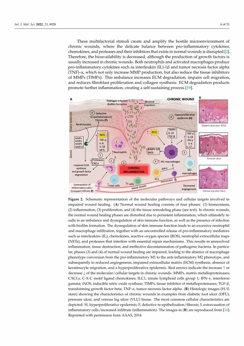
These multifactorial stimuli create and amplify the hostile microenvironment ofchronic wounds, where the delicate balance between pro-inflammatory cytokines,chemokines, and proteases and their inhibitors that exists in normal wounds is disrupted [2].Therefore, the bioavailability is decreased, although the production of growth factors isusually increased in chronic wounds. Both neutrophils and activated macrophages producepro-inflammatory cytokines such as interleukin (IL)-1β and tumor necrosis factor alpha(TNF)-α, which not only increase MMP production, but also reduce the tissue inhibitorsof MMPs (TIMPs). This imbalance increases ECM degradation, impairs cell migration,and reduces fibroblast proliferation and collagen synthesis. ECM degradation productspromote further inflammation, creating a self-sustaining process [19].
Int. J. Mol. Sci. 2022, 23, 4928 4 of 35
These multifactorial stimuli create and amplify the hostile microenvironment of chronic wounds, where the delicate balance between pro-inflammatory cytokines, chemokines, and proteases and their inhibitors that exists in normal wounds is disrupted [2]. Therefore, the bioavailability is decreased, although the production of growth factors is usually increased in chronic wounds. Both neutrophils and activated macrophages produce pro-inflammatory cytokines such as interleukin (IL)-1β and tumor necrosis factor alpha (TNF)-α, which not only increase MMP production, but also reduce the tissue inhibitors of MMPs (TIMPs). This imbalance increases ECM degradation, impairs cell migration, and reduces fibroblast proliferation and collagen synthesis. ECM degradation products promote further inflammation, creating a self-sustaining process [19].
Figure 2. Schematic representation of the molecular pathways and cellular targets involved in impaired wound healing. (A) Normal wound healing consists of four phases: (1) homeostasis, (2) inflammation, (3) proliferation, and (4) the tissue remodeling phase (see text). In chronic wounds, the normal wound healing phases are disturbed due to persistent inflammation, which ultimately results in an imbalance and dysregulation of skin immune function, as well as the presence of infection with biofilm formation. The dysregulation of skin immune function leads to an excessive neutrophil and macrophage infiltration, together with an uncontrolled release of pro-inflammatory mediators such as interleukins (IL), chemokines, reactive oxygen species (ROS), neutrophil extracellular traps (NETs), and proteases that interfere with essential repair mechanisms. This results in unresolved inflammation, tissue destruction, and ineffective decontamination of pathogenic bacteria. In particular, phases (3) and (4) of normal wound healing are impaired, leading to the absence of macrophage phenotype conversion from the pro-inflammatory M1 to the anti-inflammatory M2 phenotype, and subsequently to reduced angiogenesis, impaired extracellular matrix (ECM) synthesis, absence of keratinocyte migration, and a hyperproliferative epidermis. Red arrows indicate the increase ↑ or decrease ↓ of the molecular/cellular targets in chronic wounds. MMPs, matrix metalloproteinases; CXCLs, C-X-C motif ligand chemokines; ILC1, innate lymphoid cells group 1; IFN-γ, interferon gamma; iNOS, inducible nitric oxide synthase; TIMPs, tissue inhibitor of metalloproteinases; TGF-β, transforming growth factor beta; TNF-α, tumor necrosis factor alpha. (B) Histologic images (H/E stain) showing the characteristics of chronic wounds in examples from diabetic foot ulcer (DFU), pressure ulcer, and venous leg ulcer (VLU) tissue. The most common cellular characteristics are depicted: H, hyperproliferative epidermis; F, defective re-epithelization/fibrosis; I, extravasation of inflammatory cells/increased infiltrate (inflammation). The images in (B) are reproduced from [16]. Reprinted with permission from AAAS, 2014.
In addition to immune imbalances, low nitric oxide (NO) content and excessive ROS production are responsible for impeding the healing of chronic wounds [20,21]. NO is an
Figure 2. Schematic representation of the molecular pathways and cellular targets involved inimpaired wound healing. (A) Normal wound healing consists of four phases: (1) homeostasis,(2) inflammation, (3) proliferation, and (4) the tissue remodeling phase (see text). In chronic wounds,the normal wound healing phases are disturbed due to persistent inflammation, which ultimately re-sults in an imbalance and dysregulation of skin immune function, as well as the presence of infectionwith biofilm formation. The dysregulation of skin immune function leads to an excessive neutrophiland macrophage infiltration, together with an uncontrolled release of pro-inflammatory mediatorssuch as interleukins (IL), chemokines, reactive oxygen species (ROS), neutrophil extracellular traps(NETs), and proteases that interfere with essential repair mechanisms. This results in unresolvedinflammation, tissue destruction, and ineffective decontamination of pathogenic bacteria. In particu-lar, phases (3) and (4) of normal wound healing are impaired, leading to the absence of macrophagephenotype conversion from the pro-inflammatory M1 to the anti-inflammatory M2 phenotype, andsubsequently to reduced angiogenesis, impaired extracellular matrix (ECM) synthesis, absence ofkeratinocyte migration, and a hyperproliferative epidermis. Red arrows indicate the increase ↑ ordecrease ↓ of the molecular/cellular targets in chronic wounds. MMPs, matrix metalloproteinases;CXCLs, C-X-C motif ligand chemokines; ILC1, innate lymphoid cells group 1; IFN-γ, interferongamma; iNOS, inducible nitric oxide synthase; TIMPs, tissue inhibitor of metalloproteinases; TGF-β,transforming growth factor beta; TNF-α, tumor necrosis factor alpha. (B) Histologic images (H/Estain) showing the characteristics of chronic wounds in examples from diabetic foot ulcer (DFU),pressure ulcer, and venous leg ulcer (VLU) tissue. The most common cellular characteristics aredepicted: H, hyperproliferative epidermis; F, defective re-epithelization/fibrosis; I, extravasation ofinflammatory cells/increased infiltrate (inflammation). The images in (B) are reproduced from [16].Reprinted with permission from AAAS, 2014.

Int. J. Mol. Sci. 2022, 23, 4928 5 of 31
In addition to immune imbalances, low nitric oxide (NO) content and excessive ROSproduction are responsible for impeding the healing of chronic wounds [20,21]. NO isan endogenous neurotransmitter that plays a key role in inflammatory regulation, butNO levels in chronic wounds are much lower than normal [22]. Additionally, excess ROScan cause oxidative damage to the wound, neovascularization damage, and metabolicdamage, which can prolong inflammation [23]. Advanced glycation end products (AGEs)in diabetic wounds can induce excessive ROS production that leads to significant oxidativedamage and aging of the ECM and cell membrane, finally causing poor angiogenesis andre-epithelialization, insufficient production of growth factors, and a prolonged inflamma-tory response [21].
Another condition commonly found in chronic wounds is inadequate tissue oxygena-tion. Oxygen is necessary for healing wounds. It protects wounds from infection, improvesproliferation and migration of fibroblasts, causes angiogenesis, increases differentiationof keratinocytes and re-epithelialization, increases collagen synthesis, and facilitates thecontraction of wounds, which are all required for the restoration of tissue function andintegrity. Nevertheless, chronic wounds are hypoxic, with tissue oxygen concentrationsthree-fold lower than control tissue [23].
Chronic wound management remains an issue, as the ongoing inflammation in thesewounds is very difficult to control. Another reason for chronic inflammation is infection ofthe wound. If the infection is not controlled in a timely fashion, a biofilm can form. Biofilmformation results in the secretion of an exopolysaccharide matrix, which can protect bacteriafrom antibiotic treatment and host immune response. In the presence of bacteria andendotoxins, the level of inflammatory factors increases abnormally and makes the woundenter the vicious circle of the inflammatory response [20,24]. Biofilms interact with the hostimmune system by activating pro-inflammatory macrophages and neutrophils, resulting inthe accumulation of inflammatory cytokines such as TNF-α and IL-6, as well as MMPs, andpromoting ongoing inflammation [25]. In addition, biofilms are very difficult to eradicatetherapeutically due to the reduced penetration of antimicrobial agents into the biofilm, thepresence of multiple microbial species, and the rapid development of antibiotic resistanceby biofilm bacteria, among many other challenges [26]. In particular, the polymicrobialnature of biofilms in chronic wounds facilitates the genetic exchange between bacteria and,thus, the antibiotic resistance that emerges as an important public health issue. Severalstrategies that include bioactive molecules have been proposed to improve the healing ofchronic wounds and have shown positive results in preclinical studies [5].
As discussed above, the immune system is one of the key contributors to the persis-tence of chronic wounds. This justifies the use of immunomodulation to enhance chronicwound healing. To date, multiple immunomodulatory strategies have been proposed forcutaneous wound repair that include natural bioactive-based approaches. After describingthe role of the immune system and the role of microbial infections in chronic wound healing,at the end of this review, we focus on several bioactive compounds from natural sources aspromising therapeutic wound-healing strategies.
3. Role of Innate and Adaptative Immunity in Chronic Wounds
The innate immune system comprises a diverse range of defense systems that act toprovide primary protection against potentially harmful agents.
Innate immune cells produce pro-inflammatory cytokines that exacerbate host defensefunctions by inducing antimicrobial molecules, attracting leukocytes, and creating anenvironment to protect tissue from microbial infection. In the late stages of the inflammatoryphase of wound healing, macrophages are found to switch from a pro-inflammatory to ananti-inflammatory phenotype, as will be discussed below. The innate immune system isable to activate and deploy fast responses to offending agents and their products that rangefrom pathogen-associated molecular patterns (PAMPs) to damage-associated molecularpatterns (DAMPs). Toll-like receptors (TLR) and other pattern recognition receptors of theimmune system recognize these distinct patterns, thereby activating the cellular defense,

Int. J. Mol. Sci. 2022, 23, 4928 6 of 31
including pro-inflammatory cascades against endogenous or exogenous danger signals,foreign organisms such as viruses and bacteria, as well as particles (for a review of innateimmunity in chronic wounds, see [27]).
Overall, inflammation is an essential, nonspecific, innate immune response. The innateimmune system interacts with, directs, and instructs the adaptive immune system to elicitoptimal immune responses [28]. Moreover, the response of the innate immune system istightly coupled to the release of soluble mediators like interferons (IFNs), interleukins, andantimicrobial peptides and proteins (AMPs), as well as acute-phase proteins. Macrophages,neutrophils, mast cells, eosinophils, and innate lymphoid cells belong to the innate immunesystem. In the next subsections, we explain in more detail the function of these cells withregard to wound healing. Figure 3 shows the various immune cells involved and their roleand connections in wound healing.
Int. J. Mol. Sci. 2022, 23, 4928 6 of 35
or exogenous danger signals, foreign organisms such as viruses and bacteria, as well as particles (for a review of innate immunity in chronic wounds, see [27]).
Overall, inflammation is an essential, nonspecific, innate immune response. The innate immune system interacts with, directs, and instructs the adaptive immune system to elicit optimal immune responses [28]. Moreover, the response of the innate immune system is tightly coupled to the release of soluble mediators like interferons (IFNs), interleukins, and antimicrobial peptides and proteins (AMPs), as well as acute-phase proteins. Macrophages, neutrophils, mast cells, eosinophils, and innate lymphoid cells belong to the innate immune system. In the next subsections, we explain in more detail the function of these cells with regard to wound healing. Figure 3 shows the various immune cells involved and their role and connections in wound healing.
Figure 3. Schematic representation of the main players of innate and adaptative immunity in response to chronic wounds. Upon tissue injury, tissue-resident immune cells, including macrophages, sense tissue damage and trigger the mobilization of other immune cells. Damage-associated molecular patterns (PAMPs) and pathogen-associated molecular patterns (DAMPs) activate resident immune cells, such as mast cells, Langerhans cells, T cells, and macrophages, by binding pattern recognition receptors to elicit downstream inflammatory pathways. Platelet-derived growth factors (PDGFs), released by platelets and leukocytes, play an important role in initiating the chemotaxis of neutrophils and monocytes. Monocytes migrate into the wound and mature into macrophages. Neutrophils and activated macrophages secrete reactive oxygen species (ROS), antimicrobial peptides, proteases, and pro-inflammatory cytokines to amplify the inflammation and to help eliminate pathogens. However, the prolonged presence of pro-inflammatory macrophages (M1) in the wound environment and the overexpression of inflammatory mediators lead to the failure of the reparative (M2) phenotype polarization. In addition, adaptive immune cells, such as cytotoxic T cells (CD8+ cells), mast cells, T helper (Th) cells, and B cells, were shown to participate in the process. Arrows indicate pathway induction and numbers indicate the corresponding healing phases ((1) homeostasis, (2) inflammation, (3) proliferation). NETs, neutrophil extracellular traps; IFN-γ, interferon gamma; ILC, innate lymphoid cells; NK, natural killer cells.
Figure 3. Schematic representation of the main players of innate and adaptative immunity in responseto chronic wounds. Upon tissue injury, tissue-resident immune cells, including macrophages, sensetissue damage and trigger the mobilization of other immune cells. Damage-associated molecularpatterns (PAMPs) and pathogen-associated molecular patterns (DAMPs) activate resident immunecells, such as mast cells, Langerhans cells, T cells, and macrophages, by binding pattern recognitionreceptors to elicit downstream inflammatory pathways. Platelet-derived growth factors (PDGFs),released by platelets and leukocytes, play an important role in initiating the chemotaxis of neutrophilsand monocytes. Monocytes migrate into the wound and mature into macrophages. Neutrophils andactivated macrophages secrete reactive oxygen species (ROS), antimicrobial peptides, proteases, andpro-inflammatory cytokines to amplify the inflammation and to help eliminate pathogens. However,the prolonged presence of pro-inflammatory macrophages (M1) in the wound environment andthe overexpression of inflammatory mediators lead to the failure of the reparative (M2) phenotypepolarization. In addition, adaptive immune cells, such as cytotoxic T cells (CD8+ cells), mast cells,T helper (Th) cells, and B cells, were shown to participate in the process. Arrows indicate pathwayinduction and numbers indicate the corresponding healing phases ((1) homeostasis, (2) inflamma-tion, (3) proliferation). NETs, neutrophil extracellular traps; IFN-γ, interferon gamma; ILC, innatelymphoid cells; NK, natural killer cells.

Int. J. Mol. Sci. 2022, 23, 4928 7 of 31
3.1. Neutrophils
In regular wound healing, platelet-derived growth factors (PDGFs) released by plateletsand leukocytes play an important role in initiating the chemotaxis of neutrophils, mono-cytes, smooth muscle cells, and fibroblasts [2]. The initial leukocyte response is dominatedby neutrophils, which are usually the first cells to reach the tissue and can regulate theinflammatory responses for the first two to five days. Neutrophils’ functions are wounddebridement and phagocytosis. They also play an important role in killing microorgan-isms and controlling inflammation by secreting various antimicrobial substances, such asantimicrobial peptides, ROS, and antimicrobial proteases [29]. In turn, neutrophils cancast out neutrophil extracellular traps (NETs), which are decondensed DNA, granule, andhistone-based networks, to degrade virulence factors and kill microorganisms in wounds.However, the overexpression of NET components can destroy wound structures, includingcollagen, fibronectin, and cellular matrix, can impair angiogenesis, and can eventually leadto delayed wound healing [30]. Together with NETs, the generation of free radicals viathe myeloperoxidase pathway contributes to the killing of pathogens in wounds. Besidesremoving pathogens, neutrophils can upregulate and secrete various pro-inflammatorycytokines such as IL-1α, IL-1β, IL-6, and TNF-α to stimulate monocytes to differentiateinto M1 macrophages [31]. After completing their function between day three and fiveof wound healing, neutrophils usually undergo apoptosis, followed by macrophage up-take [20,32]. However, if the process is impaired to any degree, it may result in a prolongedpresence of neutrophils in the wound environment. Reduced neutrophil apoptosis andincreased levels of neutrophil-derived proteases with broad substrate specificity such asserine proteases, or with narrow specificity such as MMP-2 and -9, which are known todegrade the ECM, as well as neutrophil chemo-attractant C-X-C motif chemokine ligand 8(CXCL8), are associated with chronic wounds [5]. As such, an increased expression of MMP-9 by activated neutrophils is linked to the delayed repair of chronic wounds (also calledulcers) in diabetic patients [33]. Moreover, elevated glucose levels can stimulate MMP-9 overexpression through the activation of the extracellular-regulated kinase/activatorprotein-1 (ERK/AP1) signaling pathway [34]. Neutrophils also release MMP-8 and serineproteases such as elastase, which degrades important growth factors such as PDGF andtransforming growth factor (TGF)-β [2,35]. In addition, neutrophil depletion in a murinemodel of imiquimod-induced psoriatic lesions via injection of monoclonal antibody 1A8revealed significantly lower levels of infiltrating macrophages and CD4+ T-cells in tissuesamples and the reduced production of pro-inflammatory cytokines TNF-α, IFN-γ, andIL-1β, suggesting the active role of neutrophils during wound inflammation [36]. The levelof NETs released by neutrophils is associated with impaired wound healing in diabetes.NETs are increased in diabetic foot ulcer (DFU) patients compared to healthy patients [37].NETs such as citrullinated histone 3 were suggested as potential negative markers forwound healing [37]. In summary, the persistent presence of neutrophils at the wound sitedelays the healing process through the expression of pro-inflammatory factors, proteases,and NETs.
3.2. Macrophages
Following injury, macrophages take over from approximately day three of the woundhealing process. Monocytes migrate into the wound and mature into macrophages.Macrophages become the most abundant and important immune cells in the inflammatoryphase and are crucial during healing. Their main functions are to regulate the inflammatoryresponse and to act as phagocytotic cells.
Activated macrophages secrete chemokines, cytokines, and growth factors such asTGF-α, TGF-β, basic fibroblast growth factor (bFGF), PDGF, and vascular endothelialgrowth factor (VEGF) to amplify and eventually resolve inflammation. However, theprolonged presence of macrophages in the wound environment and the persistent in-flammation can lead to tissue damage and may result in chronic wounds. The failure ofmacrophages to polarize from a pro-inflammatory M1 towards a reparative M2 phenotype

Int. J. Mol. Sci. 2022, 23, 4928 8 of 31
may cause chronic wounds [5]. This failure is due to an overexpression of inflammatorymediators, such as IL-17, TNF-α, inducible nitric oxide synthase (iNOS), and ROS, as wellas impaired clearance of apoptotic neutrophils by macrophages, which negatively affectsthe wound microenvironment, thereby resulting in a large proportion of pro-inflammatorycytokines [38]. In addition, macrophages in chronic wounds release several MMPs, in-cluding MMP-2 and MMP-9, which degrade the ECM and prevent the initiation of theproliferative phase of healing.
A study comparing wound-derived macrophages from healthy individuals with dia-betic patients revealed a distinct expression of histone methyltransferase Setdb2, whoseproduction in wound macrophages is under the control of IFN-γ. In diabetic patients,the impairment of IFN-γ–Setdb2 interaction results in the failure of the M1 to M2 pheno-type switch, leading to the accumulation of pro-inflammatory macrophages in diabeticwounds [39]. Furthermore, M1 macrophages in the diabetic wound microenvironmentwere found to overexpress the small regulatory RNA microRNA-21, leading to the in-creased secretion of inflammatory mediators, such as IL-1β, TNF-α, iNOS, IL-6, and IL-8,and increased macrophage polarization toward the M1 phenotype [40]. M2 macrophagescontribute to scar formation by increasing ECM protein synthesis and secreting MMP-10and TGF-β1. However, excessive M2 macrophage activity during wound healing has beenassociated with hypertrophic scar formation [41,42].
Taken together, the regulation of M1–M2 polarization is crucial for proper woundhealing. Any change in this balance has consequences such as non-healing of wounds orincreased tissue fibrosis.
3.3. Innate Lymphoid Cells
Innate lymphoid cells (ILCs) are a recently identified population of immune cells oflymphoid origin and morphology, which do not possess antigen-specific receptors andmarkers associated with T-, B-, natural killer (NK)-, or myeloid cells [43]. These cells playimportant roles in the innate response, regulation of homeostasis and inflammation, andinteraction with adaptive immunity. Although relatively rare in the systemic circulationcompared to other hematopoietic cells, ILCs are enriched on epithelial barrier surfacesand act as regulators for chronic inflammation and tissue remodeling, bridging innate andadaptive immunity [44].
Innate lymphoid cells can be divided into three subsets designated as group 1, 2, and3, with distinct cytokine and transcriptional profiles, as well as effector functions. NKcells, which belong to the ILC group 1 (ILC1), produce IFN-γ. They are involved in theinflammatory phase of the wound-healing process, thus exerting mostly negative effectson tissue repair. IFN-γ polarize macrophages to the pro-inflammatory M1 phenotype andamplify immune cell infiltration to the wound site by macrophages expressing IL-1β, IL-6,IL-12, IL-23, and TNF-α [5].
ILC group 2 cells (ILC2) produce Th2-associated cytokines, such as IL-33. Skin in-jury promotes an IL-33-dependent ILC2 response [45]. It was recently shown that theabrogation of this response impairs re-epithelialization and efficient wound healing [45].ILC3 and ILC1 cells are implicated in inflammatory responses, whereas ILC2 may exertan anti-inflammatory effect through the enhancement of M2 macrophage polarization [46]and the expansion and localization of regulatory T cells (Tregs) [47]. However, the localaccumulation of ILC2 cells has been shown to play a pathological role in the context ofchronic dermal inflammation [48].
3.4. Adaptive Immunity
In contrast to innate immunity, the adaptive immune system provides a more de-layed and specific response. Adaptive immunity consists of humoral and cell-mediatedresponses, carried out by B- and T cells, respectively. The role of adaptive immunity inchronic wound repair has not been extensively investigated. However, it is known that inchronic wounds, fewer T cells are present, and those that are present exhibit a defective, un-

Int. J. Mol. Sci. 2022, 23, 4928 9 of 31
responsive, functionally impaired state [49]. As mentioned before, the prolonged presenceof neutrophils and M1 macrophages leads to a highly inflammatory wound profile. Theprocess is enhanced by mast cells and CD8+ T cell activity. The level of other inflammatoryT cell subtypes (Th1, Th17, and Th22) is increased in diabetic ulcers [50] (Figure 3). Theligand for CXCR3, an interferon-inducible chemokine receptor expressed in various celltypes, but preferentially on Th1 cells, is highly expressed in chronic inflammation [51,52].Together, all of these pathological processes promote inflammation, tissue fibrosis, andpoor vascularization [5,50].
Furthermore, there is evidence for direct cell–cell interactions of cluster of differentia-tion 40, CD40 ligand-expressing T lymphocytes with keratinocytes and their influence onthe healing response [53]. The prolonged and increased presence of T lymphocytes anda nonstandard CD4-CD8 ratio, reported in chronic venous and DFUs, may be related toimpaired epithelialization [28].
Impaired wound healing in ulcers associated with diabetic mellitus is characterized bya highly pro-inflammatory profile. This pro-inflammatory profile is caused by an excessiveexpression of inflammatory cytokines, such as TNF-α, and the reduced production ofpro-healing mediators, such as IL-10 and TGF-β. This leads to macrophage polarizationtowards the M1 phenotype and the activation and degranulation of CD8+ T cells, resultingin tissue necrosis [54].
Although Tregs play a balancing role in inflammation by suppressing the immuneresponse, some studies showed that an elevated number of Tregs at sites of chronic skininflammation was not only unable to resolve the lesion, but even contributed to the patho-genesis of the disease [5,55].
Regarding humoral responses and wound healing, B cells have been shown to play arole. It is suggested that CD19 regulates B cell contribution to wound healing by affectingTLR4 signaling, thereby altering cytokine production [28]. The exact roles of B cells inchronic wound healing remain to be elucidated. However, a recent publication emphasizedthe positive role of B cells for accelerated wound healing, significant mitigation of apoptosis,and enhanced fibroblast proliferation by using a topically-applied B cell treatment onmurine chronic wounds [56]. A better understanding of the contributions of T and Blymphocytes to the wound repair process could provide new clues about the regulation ofwound re-epithelialization.
4. Role of Bacterial Infection in Chronic Wounds
Microorganisms that are usually found on the skin surface could gain access to the un-derlying tissues after skin injury. The involvement of replicating organisms within a woundwith consequent damage to the host is known as invasive infection. This infection plays amajor role in delaying chronic wounds from healing (Figure 4). The high-level exudate inchronic wounds provides a moist and nutritious environment for bacterial colonizationand propagation. Moreover, the severity of the inflammatory response is further increasedby the presence of bacteria and endotoxins [57]. In an early phase, chronic wounds have apreponderance of Gram-positive bacteria, particularly Staphylococcus aureus, and in laterphases, Gram-negative bacteria such as Pseudomonas aeruginosa [58]. These bacteria pro-duce virulence factors and endotoxins and promote the expression of pro-inflammatorycytokines, which are a major cause of chronic inflammation. In addition, prolonged in-flammation can lead to a disordered metabolism (such as high MMP expression) anddelays to the normal healing process [59]. Although chronic skin wounds are exposed torelatively high levels of oxygenation, anaerobic bacteria are found in relative abundancein chronic wounds [60]. Interestingly, chronic wounds are characterized by the presenceof Corynebacterium, a traditional commensal bacterium [60]. Commensal bacteria havelong been shown to benefit the host organism by educating the host’s adaptive responseand inhibiting the growth of pathogenic bacteria. However, recent data have shown thatcoryneform bacteria can be pathogenic in wounds [61]. The polymicrobial nature of the

Int. J. Mol. Sci. 2022, 23, 4928 10 of 31
wound allows for microbial diversity and heterogeneity within the wound, which poses anadditional challenge to the wound-healing capacity.
Int. J. Mol. Sci. 2022, 23, 4928 10 of 35
bacteria. However, recent data have shown that coryneform bacteria can be pathogenic in wounds [61]. The polymicrobial nature of the wound allows for microbial diversity and heterogeneity within the wound, which poses an additional challenge to the wound-healing capacity.
Figure 4. Role of bacterial infection in chronic wounds during the four phases of wound healing. Excessive inflammation is the main cause of chronicity and disruption of normal healing phases ((1) homeostasis, (2) inflammation, (3) proliferation, and (4) tissue remodeling phase; see text in Section 2. Chronic wounds). This inflammation is maintained by chronic activation of the innate immune system, which is driven by its interactions with and responses to polymicrobial biofilms formed within the wound. The response of the innate immune system (see Figure 3) is tightly coupled to the release of soluble mediators like reactive oxygen species (ROS), pro-inflammatory cytokines, chemokines, matrix metalloproteinases (MMPs), and antimicrobial peptides and proteins (AMPs), as well as damage-associated molecular patterns (DAMPs), pathogen-associated molecular patterns (PAMPs), neutrophil extracellular traps (NET)-osis, and the tissue inhibitor of metalloproteinases (TIMPs). This tight interplay between bacterial infection and immune responses is the major causes of chronic inflammation that prevents the initiation of the proliferative phase of healing.
In addition to direct damage to the host, bacteria attract leukocytes. Leukocytes then lead to inflammation enhancement, facilitating the elimination of the bacterial infection. However, inflammation can be prolonged in the absence of successful microbial decontamination and the wound may reach a chronic state and fail to heal if the situation persists. Sustained inflammatory cascades with elevated levels of pro-inflammatory cytokines such as IL-1 and TNF-α, proteases, and ROS will contribute to levels of both bacteria and endotoxins, thus maintaining and elongating the inflammatory process. In addition, host- and bacterial-derived proteases such as MMPs and ROS degrade the ECM and growth factors, disrupting cell migration and inhibiting wound closure. There is a decrease in the level of natural protease inhibitors in combination with an increase in protease content. This change in protease balance may cause the rapid deterioration of growth factors that occurs in chronic wounds [9].
To enhance their antimicrobial resistance, free-floating (planktonic) bacteria can evolve to acquire the ability to form biofilms. Biofilms are surface-adherent and matrix-enveloped bacterial communities that form when bacterial cells attach to a surface and use quorum sensing to orchestrate and change gene expression, which ultimately creates a barrier consisting of exopolymers [62]. Biofilms are typically composed of 85% exopolymers, including polysaccharides, proteins, and nucleic acids, combined with 15% bacteria. By creating and incorporating into biofilms, bacterial cells create an optimal
Figure 4. Role of bacterial infection in chronic wounds during the four phases of wound healing.Excessive inflammation is the main cause of chronicity and disruption of normal healing phases((1) homeostasis, (2) inflammation, (3) proliferation, and (4) tissue remodeling phase; see text inSection 2. Chronic wounds). This inflammation is maintained by chronic activation of the innateimmune system, which is driven by its interactions with and responses to polymicrobial biofilmsformed within the wound. The response of the innate immune system (see Figure 3) is tightly coupledto the release of soluble mediators like reactive oxygen species (ROS), pro-inflammatory cytokines,chemokines, matrix metalloproteinases (MMPs), and antimicrobial peptides and proteins (AMPs), aswell as damage-associated molecular patterns (DAMPs), pathogen-associated molecular patterns(PAMPs), neutrophil extracellular traps (NET)-osis, and the tissue inhibitor of metalloproteinases(TIMPs). This tight interplay between bacterial infection and immune responses is the major causesof chronic inflammation that prevents the initiation of the proliferative phase of healing.
In addition to direct damage to the host, bacteria attract leukocytes. Leukocytes thenlead to inflammation enhancement, facilitating the elimination of the bacterial infection.However, inflammation can be prolonged in the absence of successful microbial decontami-nation and the wound may reach a chronic state and fail to heal if the situation persists.Sustained inflammatory cascades with elevated levels of pro-inflammatory cytokines suchas IL-1 and TNF-α, proteases, and ROS will contribute to levels of both bacteria and endo-toxins, thus maintaining and elongating the inflammatory process. In addition, host- andbacterial-derived proteases such as MMPs and ROS degrade the ECM and growth factors,disrupting cell migration and inhibiting wound closure. There is a decrease in the levelof natural protease inhibitors in combination with an increase in protease content. Thischange in protease balance may cause the rapid deterioration of growth factors that occursin chronic wounds [9].
To enhance their antimicrobial resistance, free-floating (planktonic) bacteria can evolveto acquire the ability to form biofilms. Biofilms are surface-adherent and matrix-envelopedbacterial communities that form when bacterial cells attach to a surface and use quorumsensing to orchestrate and change gene expression, which ultimately creates a barrierconsisting of exopolymers [62]. Biofilms are typically composed of 85% exopolymers,including polysaccharides, proteins, and nucleic acids, combined with 15% bacteria. Bycreating and incorporating into biofilms, bacterial cells create an optimal environment toevade the host immune response and antibiotic treatment. Bacteria within biofilms can

Int. J. Mol. Sci. 2022, 23, 4928 11 of 31
lower their metabolic activity, making antimicrobial agents that target metabolically activecells less effective against bacterial cells [63]. Other mechanisms of antibiotic resistance inbiofilms are associated with genetic changes, acquired either by mutating the endogenousgenes or by incorporating exogenous genes of resistance, adaptive stress responses andthe formation of phenotypic resistance (without any genetic alteration), which can lateralso lead to resistance in chronically treated infected wounds [64]. In addition, the biofilmexopolysaccharides, which resemble the mucus layer, function as a mechanical layer,thereby providing an additional barrier and level of protection against antibiotics andhost immune cells due to diffusion limitations [65,66]. Biofilms allow plasmid-mediatedantimicrobial resistance gene transfer between bacteria, which not only increases the cellsubpopulation heterogeneity in the wound, but also provides additional resistance. Biofilmsmay possess an additional evolutionary response to antimicrobial therapy by developingthicker mucoid-like phenotypes in response to some antimicrobial therapies [67].
Though some infectious biofilms are dominated by a single species, chronic woundmicrobiota are mainly organized in the form of a polymicrobial biofilm. Interactions be-tween different species in the polymicrobial environment have been shown to be dynamicand modify bacterial behavior, leading to increased virulence and delayed wound heal-ing [68]. Polymicrobial infections often require about 12+ months to clear, have recurrencefrequencies of 60 to 70%, and have elevated mortality rates compared to single-speciesinfections [69–71]. Microbial synergy within a biofilm gives cohabiting organisms a compet-itive advantage, but little is known about how this synergy may increase net pathogenicityin chronic wounds [72].
Leukocytes within the wound have difficulty penetrating the biofilm and have areduced ability to produce ROS [26]. This property also prevents phagocytosis of bacteriathrough normal wound-healing pathways. It has been suggested that the biofilm structuralexopolymer evades the host inflammatory response by further blocking complementactivation, suppressing the lymphoproliferative response, and impairing the ability ofbacterial wall opsonin’s to be detected by phagocytes [73,74].
Biofilms are present in almost 60% of chronic wounds, but only in 10% of acute wounds,and they obviously stimulate chronic inflammation in the chronic setting. Stimulationof the immune system when it is unable to effectively eradicate infection can lead to theworsening of chronic inflammation and can perpetuate the chronic wound cycle [75].
5. Natural Bioactive Compounds as Wound Treatments
Plants and marine organisms have a great ability to produce secondary metabolitesincluding polyphenols, carotenoids, terpenoids, alkaloids, and vitamins that have beendemonstrated to possess antioxidant, anti-inflammatory, angiogenic, and antimicrobialactivities, which are essential for wound healing.
In chronic wound treatment, it is imperative to reduce the excessive, uncontrolled,and persistent inflammation caused by oxidative stress [76]. As mentioned in Section 2, ox-idative stress arises when the production of ROS exceeds the intrinsic antioxidant defenses.Fortunately, when the cell’s endogenous enzymatic antioxidants are unable to overcomethe high rate of oxidative stress, natural exogenous antioxidants, anti-inflammatory agents,and antimicrobials can balance, reduce, or even eliminate the oxidative stress and improvewound healing [20,77,78].
Natural exogenous antioxidants are chemical compounds that affect excess ROS indifferent ways. They can inhibit ROS production, catalyze a complex cascade of reactions toconvert ROS into more stable molecules such as H2O and O2, stimulate various endogenousantioxidant enzyme systems, and accelerate the production of non-enzymatic antioxidantsin vivo. Thus, antioxidants can maintain non-toxic levels of ROS in the wound [77] andaccelerate wound healing [78]. Based on the mechanism of action, antioxidants are mainlyclassified as enzymatic and non-enzymatic compounds, as shown in Scheme 1. Enzymaticantioxidants are mainly endogenous molecules found in the cell and include superoxidedismutase, catalase, and glutathione peroxidases, among others [77].

Int. J. Mol. Sci. 2022, 23, 4928 12 of 31
Int. J. Mol. Sci. 2022, 23, 4928 12 of 32
mainly classified as enzymatic and non-enzymatic compounds, as shown in Scheme 1. En-zymatic antioxidants are mainly endogenous molecules found in the cell and include su-peroxide dismutase, catalase, and glutathione peroxidases, among others [77].
Natural exogenous antioxidants, such as carotenoids, terpenoids, polyphenols, alka-loids, and vitamins, have considerable potential for the development of antioxidant wound dressings [79].
Scheme 1. Overview of antioxidants and classification of non-enzymatic compounds based on their main groups: terpenoids, polyphenols, alkaloids, and vitamins. Selected compounds are given as examples.
Depending on the severity of the inflammation, the appropriate administration and dosage of anti-inflammatory drugs can normalize the prolonged and disordered inflam-matory response of chronic wounds. Various anti-inflammatory drugs and phyto-modu-lators are used to treat wound inflammation [20,80,81]. For example, polyphenols can act as anti-inflammatory agents by inhibiting pro-inflammatory mediators, neutralizing free radicals, and thereby inhibiting lipid peroxidation [82]. Oxidative stress-mediated lipid peroxidation has significant effects on the structure and dynamics of lipid membranes, including membrane water permeability, decreased lipid bilayer thickness, or alterations in the membrane lipid order and fluidity [83]. These alterations potentiate oxidative stress-induced damage and wound healing impairment via increased cell apoptosis and senescence [76].
As mentioned before, bacterial biofilms are present in almost 60% of chronic wounds. Antibiotics (such as tetracycline, gentamicin, and sulfadiazine) are still the main treat-ments for wound infections [84]. However, bacterial resistance and the biofilm formation itself significantly limit the therapeutic effect, and thus the development of new alterna-tive therapies is urgently needed. Antibacterial ingredients of natural origin are increas-ingly being used in wound dressings and have attracted considerable attention given their availability in nature and being Generally Recognized as Safe (GRAS) [79,85]. The design of dressings based on natural extracts may provide a suitable alternative to eliminate se-rious infection and prolonged inflammation in the wound with minimal adverse effects, easy application, greater effectiveness, and low-cost treatment [79].
The following subsections will give an overview of selected, well-characterized nat-ural antioxidants together with their anti-bacterial and anti-inflammatory properties. Fig-ure 5 shows selected natural compounds and their cellular/molecular targets in the differ-ent pathways connected to wound healing.
Scheme 1. Overview of antioxidants and classification of non-enzymatic compounds based on theirmain groups: terpenoids, polyphenols, alkaloids, and vitamins. Selected compounds are givenas examples.
Natural exogenous antioxidants, such as carotenoids, terpenoids, polyphenols, alka-loids, and vitamins, have considerable potential for the development of antioxidant wounddressings [79].
Depending on the severity of the inflammation, the appropriate administration anddosage of anti-inflammatory drugs can normalize the prolonged and disordered inflamma-tory response of chronic wounds. Various anti-inflammatory drugs and phyto-modulatorsare used to treat wound inflammation [20,80,81]. For example, polyphenols can act as anti-inflammatory agents by inhibiting pro-inflammatory mediators, neutralizing free radicals,and thereby inhibiting lipid peroxidation [82]. Oxidative stress-mediated lipid peroxidationhas significant effects on the structure and dynamics of lipid membranes, including mem-brane water permeability, decreased lipid bilayer thickness, or alterations in the membranelipid order and fluidity [83]. These alterations potentiate oxidative stress-induced damageand wound healing impairment via increased cell apoptosis and senescence [76].
As mentioned before, bacterial biofilms are present in almost 60% of chronic wounds.Antibiotics (such as tetracycline, gentamicin, and sulfadiazine) are still the main treatmentsfor wound infections [84]. However, bacterial resistance and the biofilm formation itselfsignificantly limit the therapeutic effect, and thus the development of new alternativetherapies is urgently needed. Antibacterial ingredients of natural origin are increasinglybeing used in wound dressings and have attracted considerable attention given theiravailability in nature and being Generally Recognized as Safe (GRAS) [79,85]. The design ofdressings based on natural extracts may provide a suitable alternative to eliminate seriousinfection and prolonged inflammation in the wound with minimal adverse effects, easyapplication, greater effectiveness, and low-cost treatment [79].
The following subsections will give an overview of selected, well-characterized naturalantioxidants together with their anti-bacterial and anti-inflammatory properties. Figure 5shows selected natural compounds and their cellular/molecular targets in the differentpathways connected to wound healing.

Int. J. Mol. Sci. 2022, 23, 4928 13 of 31Int. J. Mol. Sci. 2022, 23, 4928 13 of 35
Figure 5. Graphical representation of infected chronic wound treated with natural bioactive compounds. (A) Molecular pathways and main targets affected by the natural compounds described in the manuscript are indicated. The arrows indicate the effects on the targets upon treatment. The numbers refer to respective targets and the natural compounds that seem to be best for the treatment as shown in (B).
5.1. Polyphenols Polyphenols have been of great interest in wound treatment due to their
antimicrobial, regenerative, and antioxidant properties [86,87,88]. Polyphenols are usually extracted from plants and marine organisms. There are more than 8000 different polyphenols described in the literature. Polyphenols can be classified into three main groups, depending on the number of phenolic units and the structural elements that connect the phenol rings: (a) flavonoids (e.g., anthoxanthin and anthocyanins), (b) phenolic acids (e.g., hydroxycinnamic acids and hydrobenzoic acids), and (c) non-flavonoids (e.g., tannins, stilbenes, and lignans), as shown in Scheme 1. Polyphenols have been of great interest for medical and pharmaceutical applications, and in particular for wound healing due to their strong ROS-scavenging and potential antimicrobial activity [86,88]. In general, polyphenols are known to have high antioxidant activity, providing protection against ROS by neutralizing free radicals. Although the mechanism of antimicrobial action is not yet fully understood, it is believed that the effects could be associated with the phenolics’ action on cell membranes. In particular, membrane rigidification and the hydrophobicity of the phenolic compounds were positively correlated with the antimicrobial action [89]. The antimicrobial potential of certain polyphenols was reported against antibiotic-resistant strains, such as methicillin-resistant S. aureus [90]. Next, we will discuss the three main groups of polyphenols (flavonoids, phenolic acids, and non-flavonoids) in more detail.
Flavonoids are the most abundant and most-studied class of polyphenols found in plants. Chemically, flavonoids are hydroxylated phenolic substances with a basic structure of a 15-carbon skeleton that consists of two phenyl rings (A and B) linked by a three-carbon chain (a heterocyclic ring with an oxygen, the C ring). The structure is presented in Table 1. Flavonoids have gained enormous interest because of their beneficial health effects, such as anti-inflammatory, antioxidant, cardioprotective, and anticarcinogenic activities. The pharmacological effects of flavonoids are mainly due to their antioxidant and enzyme inhibition activities. One of the main factors contributing to these properties lies in their structure–activity relationship. The presence of hydroxyl groups in their chemical structure, especially when presented in positions 3, 5, 7, 3′, and 4′ (Table 1), are essential for their antibacterial, antifibrotic, antioxidant, and anti-inflammatory properties (for a review, see [91]). Flavonoids may be key for the treatment
Figure 5. Graphical representation of infected chronic wound treated with natural bioactive com-pounds. (A) Molecular pathways and main targets affected by the natural compounds describedin the manuscript are indicated. The arrows indicate the effects on the targets upon treatment. Thenumbers refer to respective targets and the natural compounds that seem to be best for the treatmentas shown in (B).
5.1. Polyphenols
Polyphenols have been of great interest in wound treatment due to their antimicrobial,regenerative, and antioxidant properties [86–88]. Polyphenols are usually extracted fromplants and marine organisms. There are more than 8000 different polyphenols describedin the literature. Polyphenols can be classified into three main groups, depending onthe number of phenolic units and the structural elements that connect the phenol rings:(a) flavonoids (e.g., anthoxanthin and anthocyanins), (b) phenolic acids (e.g., hydroxycin-namic acids and hydrobenzoic acids), and (c) non-flavonoids (e.g., tannins, stilbenes, andlignans), as shown in Scheme 1. Polyphenols have been of great interest for medical andpharmaceutical applications, and in particular for wound healing due to their strong ROS-scavenging and potential antimicrobial activity [86,88]. In general, polyphenols are knownto have high antioxidant activity, providing protection against ROS by neutralizing freeradicals. Although the mechanism of antimicrobial action is not yet fully understood, itis believed that the effects could be associated with the phenolics’ action on cell mem-branes. In particular, membrane rigidification and the hydrophobicity of the phenoliccompounds were positively correlated with the antimicrobial action [89]. The antimicrobialpotential of certain polyphenols was reported against antibiotic-resistant strains, suchas methicillin-resistant S. aureus [90]. Next, we will discuss the three main groups ofpolyphenols (flavonoids, phenolic acids, and non-flavonoids) in more detail.
Flavonoids are the most abundant and most-studied class of polyphenols found inplants. Chemically, flavonoids are hydroxylated phenolic substances with a basic structureof a 15-carbon skeleton that consists of two phenyl rings (A and B) linked by a three-carbon chain (a heterocyclic ring with an oxygen, the C ring). The structure is presentedin Table 1. Flavonoids have gained enormous interest because of their beneficial healtheffects, such as anti-inflammatory, antioxidant, cardioprotective, and anticarcinogenicactivities. The pharmacological effects of flavonoids are mainly due to their antioxidant andenzyme inhibition activities. One of the main factors contributing to these properties liesin their structure–activity relationship. The presence of hydroxyl groups in their chemicalstructure, especially when presented in positions 3, 5, 7, 3′, and 4′ (Table 1), are essentialfor their antibacterial, antifibrotic, antioxidant, and anti-inflammatory properties (for areview, see [91]). Flavonoids may be key for the treatment of several chronic diseasesthat cause cutaneous lesions, such as diabetes mellitus, due to the reported increase in

Int. J. Mol. Sci. 2022, 23, 4928 14 of 31
epithelialization rate, modulation of inflammatory cytokines, the decrease in the number ofmononuclear cells in the proliferative phase, accelerated wound contraction rate, promotionof vasculogenesis, and angiogenesis [92–94].
Polyphenols in honey, especially flavonoids and phenolic acids, have been reported tobe solely responsible for the antioxidant and other medicinal effects of honey. Honey candownregulate wound inflammation by inhibiting ROS formation, leukocyte infiltration, andcyclooxygenase-2 (COX-2), iNOS, and MMP-9 expression [95]. Several honey-impregnateddressings, such as MediHoney®, are currently on the market; however, their antibacterialproperties are easily affected by catalase [96].
Flavonoids are known to be synthesized in plants as a defense mechanism againstmicroorganisms, so it is not surprising that they exert an antimicrobial effect in vitro againsta wide range of microorganisms. Their antimicrobial properties are related to their abilityto form complexes with extracellular and soluble proteins as well as with the bacterialcell wall, altering cell membranes if they are sufficiently lipophilic. Cloves and rosemaryextracts have antibacterial activity against Bacillussubtilis. The antimicrobial defense systemof these plants is most likely due to the presence of phenolics, flavonoids, and terpenoidsin their extracts [97].
Table 1. Major classes of natural bioactive compounds, structure, main sources, and their therapeutictargets for wound-healing activity.
Bioactive Compounds—Major Classes ExampleCompounds Structure Main Natural
Source Wound Healing Activity References
Polyphenols
Flavonoids
Anthoxantins
Catechins
Int. J. Mol. Sci. 2022, 23, 4928 15 of 32
Table 1. Major classes of natural bioactive compounds, structure, main sources, and their therapeu-tic targets for wound-healing activity.
Bioactive Compounds—Major Classes
Example Compounds Structure Main Natural
Source Wound Healing
Activity References
Polyphenols
Flavonoids
Anthoxantins
Catechins
Quercetin
Green tea, cocoa, and
berries.
Antioxidant (free radical scavenger), anti-
inflammatory (down regulation of inflammatory
pathways), antiviral and antibacterial activities.
[98–105]
Kaempferol
Kale, spinach, dill, and Chinese cabbage.
Anti-inflammatory properties, positive
effect on VEGF-mediated cell migration
and wound healing effects.
[94]
Quercetin
Onions, dill, fennel leaves, oregano, and citrus fruits.
Strong antioxidant (free radical scavenger), anti-
inflammatory (macrophage modulation
polarization) properties and increase fibroblast
proliferation.
[106–111]
Anthocyanins Delphinidin
Blackcurrants hibiscus and
bilberry.
Antioxidant and anti-inflammatory effects and stimulate wound
healing rate. Modulates collagen, NF-kB
inflammatory signaling and oxidative stress.
[112,113]
Phenolic acids
Hydroxycinnamic acid
Curcumin
Curcuma longa
Potent antioxidant (free radicals scavenger,
ROS-generating enzymes inhibitor and
ROS-neutralizing enzymes activator), anti-
inflammatory (inhibitory effect on
expression of proinflammatory
cytokines and cyclin E) and antibacterial and
wound-healing activity. Cytoprotective effects against oxidative and
inflammatory stresses in several cell studies.
[114–119]
Hydrobenzoic acid
Gallic acid
Tea, red fruits, black radish, and onions.
Anti-inflammatory, anti-bacterial, anti-biofilm and wound
healing activity.
[120]
Quercetin
Green tea,cocoa, and
berries.
Antioxidant (free radicalscavenger),
anti-inflammatory (downregulation of
inflammatory pathways),antiviral and
antibacterial activities.
[98–105]
Kaempferol
Kale, spinach,dill, andChinesecabbage.
Anti-inflammatoryproperties, positive effecton VEGF-mediated cellmigration and wound
healing effects.
[94]
Quercetin
Onions, dill,fennel leaves,oregano, andcitrus fruits.
Strong antioxidant (freeradical scavenger),anti-inflammatory
(macrophage modulationpolarization) propertiesand increase fibroblast
proliferation.
[106–111]
Anthocyanins Delphinidin
Int. J. Mol. Sci. 2022, 23, 4928 15 of 32
Table 1. Major classes of natural bioactive compounds, structure, main sources, and their therapeu-tic targets for wound-healing activity.
Bioactive Compounds—Major Classes
Example Compounds Structure Main Natural
Source Wound Healing
Activity References
Polyphenols
Flavonoids
Anthoxantins
Catechins
Quercetin
Green tea, cocoa, and
berries.
Antioxidant (free radical scavenger), anti-
inflammatory (down regulation of inflammatory
pathways), antiviral and antibacterial activities.
[98–105]
Kaempferol
Kale, spinach, dill, and Chinese cabbage.
Anti-inflammatory properties, positive
effect on VEGF-mediated cell migration
and wound healing effects.
[94]
Quercetin
Onions, dill, fennel leaves, oregano, and citrus fruits.
Strong antioxidant (free radical scavenger), anti-
inflammatory (macrophage modulation
polarization) properties and increase fibroblast
proliferation.
[106–111]
Anthocyanins Delphinidin
Blackcurrants hibiscus and
bilberry.
Antioxidant and anti-inflammatory effects and stimulate wound
healing rate. Modulates collagen, NF-kB
inflammatory signaling and oxidative stress.
[112,113]
Phenolic acids
Hydroxycinnamic acid
Curcumin
Curcuma longa
Potent antioxidant (free radicals scavenger,
ROS-generating enzymes inhibitor and
ROS-neutralizing enzymes activator), anti-
inflammatory (inhibitory effect on
expression of proinflammatory
cytokines and cyclin E) and antibacterial and
wound-healing activity. Cytoprotective effects against oxidative and
inflammatory stresses in several cell studies.
[114–119]
Hydrobenzoic acid
Gallic acid
Tea, red fruits, black radish, and onions.
Anti-inflammatory, anti-bacterial, anti-biofilm and wound
healing activity.
[120]
Blackcurrantshibiscus and
bilberry.
Antioxidant andanti-inflammatory effects
and stimulate woundhealing rate. Modulates
collagen, NF-kBinflammatory signaling
and oxidative stress.
[112,113]
Phenolicacids
Hydroxycinnamicacid Curcumin
Int. J. Mol. Sci. 2022, 23, 4928 15 of 32
Table 1. Major classes of natural bioactive compounds, structure, main sources, and their therapeu-tic targets for wound-healing activity.
Bioactive Compounds—Major Classes
Example Compounds Structure Main Natural
Source Wound Healing
Activity References
Polyphenols
Flavonoids
Anthoxantins
Catechins
Quercetin
Green tea, cocoa, and
berries.
Antioxidant (free radical scavenger), anti-
inflammatory (down regulation of inflammatory
pathways), antiviral and antibacterial activities.
[98–105]
Kaempferol
Kale, spinach, dill, and Chinese cabbage.
Anti-inflammatory properties, positive
effect on VEGF-mediated cell migration
and wound healing effects.
[94]
Quercetin
Onions, dill, fennel leaves, oregano, and citrus fruits.
Strong antioxidant (free radical scavenger), anti-
inflammatory (macrophage modulation
polarization) properties and increase fibroblast
proliferation.
[106–111]
Anthocyanins Delphinidin
Blackcurrants hibiscus and
bilberry.
Antioxidant and anti-inflammatory effects and stimulate wound
healing rate. Modulates collagen, NF-kB
inflammatory signaling and oxidative stress.
[112,113]
Phenolic acids
Hydroxycinnamic acid
Curcumin
Curcuma longa
Potent antioxidant (free radicals scavenger,
ROS-generating enzymes inhibitor and
ROS-neutralizing enzymes activator), anti-
inflammatory (inhibitory effect on
expression of proinflammatory
cytokines and cyclin E) and antibacterial and
wound-healing activity. Cytoprotective effects against oxidative and
inflammatory stresses in several cell studies.
[114–119]
Hydrobenzoic acid
Gallic acid
Tea, red fruits, black radish, and onions.
Anti-inflammatory, anti-bacterial, anti-biofilm and wound
healing activity.
[120]
Curcuma longa
Potent antioxidant (freeradicals scavenger,
ROS-generating enzymesinhibitor and
ROS-neutralizingenzymes activator),anti-inflammatory
(inhibitory effect onexpression of
proinflammatorycytokines and cyclin E)and antibacterial and
wound-healing activity.Cytoprotective effectsagainst oxidative and
inflammatory stresses inseveral cell studies.
[114–119]
Int. J. Mol. Sci. 2022, 23, 4928 15 of 31
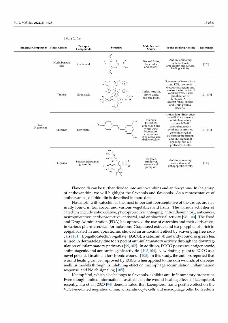
Table 1. Cont.
Bioactive Compounds—Major Classes ExampleCompounds Structure Main Natural
Source Wound Healing Activity References
Hydrobenzoicacid Gallic acid
Int. J. Mol. Sci. 2022, 23, 4928 15 of 32
Table 1. Major classes of natural bioactive compounds, structure, main sources, and their therapeu-tic targets for wound-healing activity.
Bioactive Compounds—Major Classes
Example Compounds Structure Main Natural
Source Wound Healing
Activity References
Polyphenols
Flavonoids
Anthoxantins
Catechins
Quercetin
Green tea, cocoa, and
berries.
Antioxidant (free radical scavenger), anti-
inflammatory (down regulation of inflammatory
pathways), antiviral and antibacterial activities.
[98–105]
Kaempferol
Kale, spinach, dill, and Chinese cabbage.
Anti-inflammatory properties, positive
effect on VEGF-mediated cell migration
and wound healing effects.
[94]
Quercetin
Onions, dill, fennel leaves, oregano, and citrus fruits.
Strong antioxidant (free radical scavenger), anti-
inflammatory (macrophage modulation
polarization) properties and increase fibroblast
proliferation.
[106–111]
Anthocyanins Delphinidin
Blackcurrants hibiscus and
bilberry.
Antioxidant and anti-inflammatory effects and stimulate wound
healing rate. Modulates collagen, NF-kB
inflammatory signaling and oxidative stress.
[112,113]
Phenolic acids
Hydroxycinnamic acid
Curcumin
Curcuma longa
Potent antioxidant (free radicals scavenger,
ROS-generating enzymes inhibitor and
ROS-neutralizing enzymes activator), anti-
inflammatory (inhibitory effect on
expression of proinflammatory
cytokines and cyclin E) and antibacterial and
wound-healing activity. Cytoprotective effects against oxidative and
inflammatory stresses in several cell studies.
[114–119]
Hydrobenzoic acid
Gallic acid
Tea, red fruits, black radish, and onions.
Anti-inflammatory, anti-bacterial, anti-biofilm and wound
healing activity.
[120] Tea, red fruits,black radish,and onions.
Anti-inflammatory,anti-bacterial,
anti-biofilm and woundhealing activity.
[120]
NonFlavonoids
Tannins Tannic acid
Int. J. Mol. Sci. 2022, 23, 4928 16 of 32
Non Flavonoi
ds
Tannins Tannic acid
Coffee, nutgalls, brown algae, and tara
pods.
Scavenger of free radicals and ROS, promotes wound contraction, and
increase the formation of capillary vessels and
proliferation of fibroblasts. Active
against fungal species and Gram positive
bacteria.
[121–130]
Stilbenes Resveratrol
Peanuts, pistachios,
grapes, red and white wine, blueberries, cranberries,
even cocoa, and dark chocolate.
Antioxidant (direct effect as radical scavenger), anti-
inflammatory (targets NF-kB, pro-
inflammatory cytokines expression, genes
involved in eicosanoid production and TLR
signaling) signaling, and cell protective effects.
[131–141]
Lignans Secoisolaricire
sinol diglucoside
Flaxseed, sunflower, sesame and pumpkin.
Anti-inflammatory, antioxidant and
antiapoptotic effects. [142]
Flavonols are flavonoids with a ketone group (see Table 1). Quercetin, a flavonol compound commonly found in vegetables and fruits, has strong antioxidant and anti-in-flammatory properties, as well as ROS-scavenging activity. Moreover, quercetin induces fibroblast proliferation, which advocates its possible application in wound healing. In ad-dition, quercetin was shown to inhibit the acute and chronic phases of inflammation [106,107]. Thus, quercetin regulates two factors that delay the healing process: oxidative stress and inflammation [108–110]. Recently, Jia Fu et al. [111] reported that quercetin in-hibits inflammatory reactions via the modulation of macrophage polarization switching from the M1 to M2 phenotype, thereby accelerating the diabetic wound repair.
Anthocyanidins are flavonoids with delphinidin as one of the main representatives. Delphinidin can be found in plants such as blackcurrants, hibiscus, and bilberries, and has potent antioxidant and anti-inflammatory activities. Delphinidin 3-sambubioside, a Hi-biscus anthocyanin, possess potential anti-inflammatory properties for wound healing, as demonstrated by the reduction of inflammatory mediators such as IL-6, monocyte chem-oattractant protein-1 (MCP-1), and TNF-α in vitro and in vivo [112]. Blackcurrant extracts rich in delphinidin modulate type I collagen expression reduce the nuclear factor kappa-light-chain-enhancer of activated B cell (NF-κB) inflammatory signaling, and modulates oxidative stress on skin fibroblast cells, which is suggested to help in the remodeling phase of wound healing [113].
Phenolic acids have a single benzene ring in their structure (Table 1). Phenolic acids include hydroxycinnamic acids (e.g., curcumin) and hydroxybenzoic acids (e.g., gallic acid). Hydroxybenzoic acids are present in edible plants, while hydroxycinnamic acids are common in food products, especially coffee, whole grains, and curcuma longa.
Curcumin possesses a wide spectrum of biological activities, including antioxidant activity and wound healing properties. In fact, the anti-infectious, anti-apoptotic, and anti-
Coffee, nutgalls,brown algae,
and tara pods.
Scavenger of free radicalsand ROS, promotes
wound contraction, andincrease the formation of
capillary vessels andproliferation of
fibroblasts. Activeagainst fungal species
and Gram positivebacteria.
[121–130]
Stilbenes Resveratrol
Int. J. Mol. Sci. 2022, 23, 4928 16 of 32
Non Flavonoi
ds
Tannins Tannic acid
Coffee, nutgalls, brown algae, and tara
pods.
Scavenger of free radicals and ROS, promotes wound contraction, and
increase the formation of capillary vessels and
proliferation of fibroblasts. Active
against fungal species and Gram positive
bacteria.
[121–130]
Stilbenes Resveratrol
Peanuts, pistachios,
grapes, red and white wine, blueberries, cranberries,
even cocoa, and dark chocolate.
Antioxidant (direct effect as radical scavenger), anti-
inflammatory (targets NF-kB, pro-
inflammatory cytokines expression, genes
involved in eicosanoid production and TLR
signaling) signaling, and cell protective effects.
[131–141]
Lignans Secoisolaricire
sinol diglucoside
Flaxseed, sunflower, sesame and pumpkin.
Anti-inflammatory, antioxidant and
antiapoptotic effects. [142]
Flavonols are flavonoids with a ketone group (see Table 1). Quercetin, a flavonol compound commonly found in vegetables and fruits, has strong antioxidant and anti-in-flammatory properties, as well as ROS-scavenging activity. Moreover, quercetin induces fibroblast proliferation, which advocates its possible application in wound healing. In ad-dition, quercetin was shown to inhibit the acute and chronic phases of inflammation [106,107]. Thus, quercetin regulates two factors that delay the healing process: oxidative stress and inflammation [108–110]. Recently, Jia Fu et al. [111] reported that quercetin in-hibits inflammatory reactions via the modulation of macrophage polarization switching from the M1 to M2 phenotype, thereby accelerating the diabetic wound repair.
Anthocyanidins are flavonoids with delphinidin as one of the main representatives. Delphinidin can be found in plants such as blackcurrants, hibiscus, and bilberries, and has potent antioxidant and anti-inflammatory activities. Delphinidin 3-sambubioside, a Hi-biscus anthocyanin, possess potential anti-inflammatory properties for wound healing, as demonstrated by the reduction of inflammatory mediators such as IL-6, monocyte chem-oattractant protein-1 (MCP-1), and TNF-α in vitro and in vivo [112]. Blackcurrant extracts rich in delphinidin modulate type I collagen expression reduce the nuclear factor kappa-light-chain-enhancer of activated B cell (NF-κB) inflammatory signaling, and modulates oxidative stress on skin fibroblast cells, which is suggested to help in the remodeling phase of wound healing [113].
Phenolic acids have a single benzene ring in their structure (Table 1). Phenolic acids include hydroxycinnamic acids (e.g., curcumin) and hydroxybenzoic acids (e.g., gallic acid). Hydroxybenzoic acids are present in edible plants, while hydroxycinnamic acids are common in food products, especially coffee, whole grains, and curcuma longa.
Curcumin possesses a wide spectrum of biological activities, including antioxidant activity and wound healing properties. In fact, the anti-infectious, anti-apoptotic, and anti-
Peanuts,pistachios,
grapes, red andwhite wine,blueberries,cranberries,
even cocoa, anddark chocolate.
Antioxidant (direct effectas radical scavenger),
anti-inflammatory(targets NF-kB,
pro-inflammatorycytokines expression,
genes involved ineicosanoid production
and TLR signaling)signaling, and cellprotective effects.
[131–141]
Lignans Secoisolariciresinoldiglucoside
Int. J. Mol. Sci. 2022, 23, 4928 16 of 32
Non Flavonoi
ds
Tannins Tannic acid
Coffee, nutgalls, brown algae, and tara
pods.
Scavenger of free radicals and ROS, promotes wound contraction, and
increase the formation of capillary vessels and
proliferation of fibroblasts. Active
against fungal species and Gram positive
bacteria.
[121–130]
Stilbenes Resveratrol
Peanuts, pistachios,
grapes, red and white wine, blueberries, cranberries,
even cocoa, and dark chocolate.
Antioxidant (direct effect as radical scavenger), anti-
inflammatory (targets NF-kB, pro-
inflammatory cytokines expression, genes
involved in eicosanoid production and TLR
signaling) signaling, and cell protective effects.
[131–141]
Lignans Secoisolaricire
sinol diglucoside
Flaxseed, sunflower, sesame and pumpkin.
Anti-inflammatory, antioxidant and
antiapoptotic effects. [142]
Flavonols are flavonoids with a ketone group (see Table 1). Quercetin, a flavonol compound commonly found in vegetables and fruits, has strong antioxidant and anti-in-flammatory properties, as well as ROS-scavenging activity. Moreover, quercetin induces fibroblast proliferation, which advocates its possible application in wound healing. In ad-dition, quercetin was shown to inhibit the acute and chronic phases of inflammation [106,107]. Thus, quercetin regulates two factors that delay the healing process: oxidative stress and inflammation [108–110]. Recently, Jia Fu et al. [111] reported that quercetin in-hibits inflammatory reactions via the modulation of macrophage polarization switching from the M1 to M2 phenotype, thereby accelerating the diabetic wound repair.
Anthocyanidins are flavonoids with delphinidin as one of the main representatives. Delphinidin can be found in plants such as blackcurrants, hibiscus, and bilberries, and has potent antioxidant and anti-inflammatory activities. Delphinidin 3-sambubioside, a Hi-biscus anthocyanin, possess potential anti-inflammatory properties for wound healing, as demonstrated by the reduction of inflammatory mediators such as IL-6, monocyte chem-oattractant protein-1 (MCP-1), and TNF-α in vitro and in vivo [112]. Blackcurrant extracts rich in delphinidin modulate type I collagen expression reduce the nuclear factor kappa-light-chain-enhancer of activated B cell (NF-κB) inflammatory signaling, and modulates oxidative stress on skin fibroblast cells, which is suggested to help in the remodeling phase of wound healing [113].
Phenolic acids have a single benzene ring in their structure (Table 1). Phenolic acids include hydroxycinnamic acids (e.g., curcumin) and hydroxybenzoic acids (e.g., gallic acid). Hydroxybenzoic acids are present in edible plants, while hydroxycinnamic acids are common in food products, especially coffee, whole grains, and curcuma longa.
Curcumin possesses a wide spectrum of biological activities, including antioxidant activity and wound healing properties. In fact, the anti-infectious, anti-apoptotic, and anti-
Flaxseed,sunflower,
sesame andpumpkin.
Anti-inflammatory,antioxidant and
antiapoptotic effects.[142]
Flavonoids can be further divided into anthoxanthins and anthocyanins. In the groupof anthoxanthin, we will highlight the flavanols and flavonols. As a representative ofanthocyanins, delphinidin is described in more detail.
Flavanols, with catechin as the most important representative of the group, are nat-urally found in tea, cocoa, and various vegetables and fruits. The various activities ofcatechins include antioxidative, photoprotective, antiaging, anti-inflammatory, anticancer,neuroprotective, cardioprotective, antiviral, and antibacterial activity [98–100]. The Foodand Drug Administration (FDA) has approved the use of catechins and their derivativesin various pharmaceutical formulations. Grape seed extract and tea polyphenols, rich inepigallocatechin and epicatechin, showed an antioxidant effect by scavenging free radi-cals [101]. Epigallocatechin 3-gallate (EGCG), a catechin abundantly found in green tea,is used in dermatology due to its potent anti-inflammatory activity through the downreg-ulation of inflammatory pathways [99,102]. In addition, EGCG possesses antigenotoxic,antimutagenic, and anticarcinogenic activities [103,104]. New findings point to EGCG as anovel potential treatment for chronic wounds [105]. In this study, the authors reported thatwound healing can be improved by EGCG when applied to the skin wounds of diabetesmellitus models through its inhibiting effect on macrophage accumulation, inflammationresponse, and Notch signaling [105].
Kaempferol, which also belongs to flavanols, exhibits anti-inflammatory properties.Even though limited information is available on the wound-healing effects of kaempferol,recently, Hu et al., 2020 [94] demonstrated that kaempferol has a positive effect on theVEGF-mediated migration of human keratinocyte cells and macrophage cells. Both effects

Int. J. Mol. Sci. 2022, 23, 4928 16 of 31
play critical roles in wound healing, including keratinocyte re-epithelialization and immunecell migration from circulation to injured tissues.
Flavonols are flavonoids with a ketone group (see Table 1). Quercetin, a flavonolcompound commonly found in vegetables and fruits, has strong antioxidant and anti-inflammatory properties, as well as ROS-scavenging activity. Moreover, quercetin inducesfibroblast proliferation, which advocates its possible application in wound healing. In addi-tion, quercetin was shown to inhibit the acute and chronic phases of inflammation [106,107].Thus, quercetin regulates two factors that delay the healing process: oxidative stress andinflammation [108–110]. Recently, Jia Fu et al. [111] reported that quercetin inhibits inflam-matory reactions via the modulation of macrophage polarization switching from the M1 toM2 phenotype, thereby accelerating the diabetic wound repair.
Anthocyanidins are flavonoids with delphinidin as one of the main representatives.Delphinidin can be found in plants such as blackcurrants, hibiscus, and bilberries, andhas potent antioxidant and anti-inflammatory activities. Delphinidin 3-sambubioside, aHibiscus anthocyanin, possess potential anti-inflammatory properties for wound heal-ing, as demonstrated by the reduction of inflammatory mediators such as IL-6, monocytechemoattractant protein-1 (MCP-1), and TNF-α in vitro and in vivo [112]. Blackcurrantextracts rich in delphinidin modulate type I collagen expression reduce the nuclear factorkappa-light-chain-enhancer of activated B cell (NF-κB) inflammatory signaling, and modu-lates oxidative stress on skin fibroblast cells, which is suggested to help in the remodelingphase of wound healing [113].
Phenolic acids have a single benzene ring in their structure (Table 1). Phenolic acidsinclude hydroxycinnamic acids (e.g., curcumin) and hydroxybenzoic acids (e.g., gallicacid). Hydroxybenzoic acids are present in edible plants, while hydroxycinnamic acids arecommon in food products, especially coffee, whole grains, and curcuma longa.
Curcumin possesses a wide spectrum of biological activities, including antioxidantactivity and wound healing properties. In fact, the anti-infectious, anti-apoptotic, and anti-inflammatory activities of curcumin are used in the treatment of chronic wounds in diabeticpatients (for a review, see [114]). The strong antioxidant activity to reduce ROS during theinflammatory phase is mediated by the ß-diketone moiety and two o-methoxyphenolicgroups in the curcumin structure [115]. Due to its strong antioxidant activity, curcumin isthe most-studied antioxidant in wound healing. It has been shown that curcumin stimulatesgrowth factors, in particular TGF-β1 expression, which promotes VEGF expression via theTGF-β pathway [116]. Further, curcumin can eliminate inflammation by affecting cytokinelevels, in particular by downregulating IL-1β, TNF-α, and MMP-9, or by upregulatingIL-10 [117,118]. Recently, Liu et al. [119] reported a novel hydrogel loaded with curcumin.This platform was shown to attenuate oxidative damage and accelerate wound healing indiabetic mice [119].
Gallic acid (GA) is a plant-derived compound and its potential for the treatment ofchronic wounds was shown [120]. In particular, the authors reported that GA is a potentialantioxidant that directly upregulates the expression of antioxidant genes (SOD2, CAT,and Gpx1) in human keratinocytes and fibroblasts, as investigated under normal andhyperglucidic conditions to mimic diabetes.
Non-flavonoids (Table 1) consist of the following subclasses: tannins, stilbenes, andlignans [86].
Tannins are polymeric phenolics and can be divided into two main groups: hy-drolysable and condensed tannins. Hydrolysable tannins are esters of gallic acids anda polyol, mainly D-glucose. Condensed tannins are more abundant and are a group offlavonoids derived from flavan-3-ols. Tannins have been shown to possess antioxidant andanti-inflammatory properties that could be correlated with oxidation and polymerizationpatterns [121]. Tannins show strong affinity to proteins and polysaccharides through co-valent and non-covalent interactions. Thus, tannic acid, a hydrolysable plant tannin, canstabilize collagen and elastin in the ECM by inhibiting collagenase and enhancing collagencross-linking [122]. Since chronic wounds are characterized by hyper-inflammation and

Int. J. Mol. Sci. 2022, 23, 4928 17 of 31
high collagenase activity, the effects of natural products with a high content of tannins areworthy of investigation. There is evidence that tannins extracted from Terminalia chebulapromote angiogenesis in wounds of rat models shown by the upregulation of VEGF [123].
In addition, tannins have high antimicrobial effects. Their antibacterial mode ofaction may be related to their ability to inactivate microbial adhesins, enzymes, and cellenvelope transport proteins (for a review, see [124]). There is also evidence of directinactivation of microorganisms, because even low concentrations of tannin (0.063 mg/mL)modify the morphology of the germ tubes of Crinipellis perniciosa [125]. In the case ofcondensed tannins, they have also been shown to be capable of binding cell walls of ruminalbacteria, inhibiting their growth and protease activity [126]. Additionally, the results froma pilot study suggest that the use of tannins may provide benefits in reducing S. aureuscolonization in partial-thickness burn wounds, with improved healing quality withoutincreasing toxicity [127]. Tannins from Conocarpus erectus L., a tropical and subtropicalevergreen tree, were found to be active against three fungal species: Saccharomyces cerevisiae,Aspergillus niger, and Penicillium chrysogenum and against Gram-positive bacteria, includingS. aureus and B. subtilis [128]. Methanolic and aqueous extracts of Zingiber officinale andCurcuma longa exhibited antimicrobial activity against S. pyogenes, S. aureus, Escherichiacoli, and P. aeruginosa. Phytochemical screening indicated the presence of tannins in theextracts of both plants (Zingiber officinale and Curcuma longa) [129]. The methanolic extractsof pomegranate (Punica granatum) contain high concentrations of hydrolysable tannins,ellagic acid, and gallic acid and exhibited activity against S. aureus, methicillin-resistant S.aureus, E. coli, and Salmonella typhimurium [130].
Stilbenes are chromophores that contain two benzene rings connected by a linker(Table 1) and can undergo photoisomerization around the linker bond. Among stilbenes,resveratrol (3,5,4′-trihydroxy-trans-stilbene), a natural stilbene found in grapes, is wellknown. The linker molecule in resveratrol is ethylene (Table 1). Resveratrol has mainly beenstudied for its antioxidant, anti-inflammatory, anti-obesity, and anti-cancer properties [131].Among these various biological effects of resveratrol, the antioxidant properties are themost prominent. Resveratrol has been shown to protect cells against hydrogen peroxide-induced oxidative stress and UV-mediated cell death, due to its direct effects as a radicalscavenger and its indirect effects by modulating cellular antioxidant pathways [131,132].Resveratrol is also known to interfere with inflammatory responses and has beneficial anti-inflammatory properties for chronic autoimmune and inflammatory conditions. It targetsNF-κB, pro-inflammatory cytokine expression (e.g., interleukin 6, IL6), and also genesinvolved in eicosanoid production [133]. This stilbene also targets TLR signaling [134–136].
Resveratrol shows promising potential for the treatment of diabetic ulcers [137] andmay also play a relevant role in wound healing. Several studies have shown that resveratrolinduces the expression of VEGF, one of the most prevalent, effective, and long-lasting sig-nals that stimulates angiogenesis in wounds [138,139]. Moreover, resveratrol is associatedwith antibacterial and antifungal properties at the wound site. Topical treatment withresveratrol showed significant antimicrobial efficacy against S. aureus, P. aeruginosa, andCandida albicans, which were previously discussed as important pathogens in the context ofnon-healing wounds [140,141].
Lignans are diphenolic compounds possessing a 2,3-dibenzylbutane structure (Table 1).Lignans are also referred to as an antioxidant phytoestrogen. The lignan secoisolariciresinoldiglucoside can be found in various seeds and wheat bran. Lignan metabolites functionas antioxidants and free radical scavengers. Because of lignans’ antioxidant and anti-inflammatory properties, they also have potential in wound healing. The antioxidanteffects of phenolic molecules are mainly due to the fact that they prevent oxidative stresswhile stimulating collagen synthesis. Phenolic compounds in flaxseed extracts possessvaluable antioxidant and healing properties. An experimental study of the healing effectsof topically administered flaxseed extract cream on Wistar rats’ open wounds have shownwound-healing efficacy [142].

Int. J. Mol. Sci. 2022, 23, 4928 18 of 31
5.2. Terpenoids
In the continuous search for new bioactive natural products against oxidation andinflammation, terpenes are emerging as a rich source. Terpenes are ubiquitous in nature andare mainly produced by plants. Studies have shown that both natural terpenes and theirsynthetic derivatives present diverse pharmacological properties, including antioxidant,antifungal, anti-inflammatory, antiviral, anticancer, and antibacterial activities, amongothers (for a review, see [143]).
Terpenes are biosynthetically derived from five-carbon isoprene units with the molec-ular formula of (C5H8)n, where n is the number of linked isoprene units. Terpenes aresimply hydrocarbons, while terpenoids contain additional oxidized functional groups.Monoterpenes contain two isoprene molecules (Table 2). The further classification includessesquiterpenes (three isoprene units), diterpenes (four isoprene units), sesterterpenes (fiveisoprene units), triterpenes (six isoprene units), sesquaterpenes (seven isoprene units), andtetraterpenes (eight isoprene units). Next, we will highlight the pharmacological propertiesof selected monoterpenes and tetraterpenes (carotenoids).
Monoterpenes/monoterpenoids are present in plant oils and are known as essentialoils (EOs). EOs are complex liquid and volatile lipophilic substances synthesized byaromatic plants through their secondary metabolism [144]. These natural products arepotent antioxidants and free radical scavengers. Preclinical studies have reported theanti-inflammatory effects of EOs [144]. Therefore, EOs are considered as potential sourcesfor the development of high-value pharmaceutical products [144–146].
Table 2. Major classes of natural bioactive compounds, structure, main sources, and their therapeutictargets for wound-healing activity.
Bioactive Compounds—Major Classes ExampleCompounds Structure Main Natural Source Wound Healing
Activity References
Terpenes/Terpenoids
Monoterpenes
Borneol
Int. J. Mol. Sci. 2022, 23, 4928 19 of 32
Table 2. Major classes of natural bioactive compounds, structure, main sources, and their therapeu-tic targets for wound-healing activity.
Bioactive Compounds—Major Classes
Example Compounds Structure Main Natural
Source Wound Healing
Activity References
Terpenes/ Terpenoi
ds
Monoterpenes
Borneol
Citrus peel oils (orange, lemon, lime), cinnamon leaf, cassia leaf,
ginger, coriander seed, laurel,
Ocimumbasillum, Thymus vulgaris,
and Curcuma.
Anti-inflammatory, antioxidant activity
with radical scavenging
properties, antibiotic activity, antifungal, and wound-healing
activity.
[147–153]
Thymol
Essential oils of thyme, oregano,
and carum.
Anti-inflammatory effect in human neutrophils and
antioxidant activity. It exhibits
antimicrobial activity and wound-healing
activity.
[154–163]
α-terpineol
Essential oils of pine and
petitgrain.
Anti-inflammatory, wound healing effect and down-regulation of pro-inflammatory cytokines, reduction of NO production,
antimicrobial activity, and antifungal
effects.
[164–168]
D-Limonele
Citrus essential oils.
Antioxidant, anti-inflammatory
activities and neo-vascularization
induction.
[169–171]
Tetraterpenes
(Carotenoids)
Carotenes
β-carotene
Carrots, spinach, lettuce, tomatoes, sweet potatoes,
broccoli, cantaloupe,
winter squash, marine animals, and microalgae.
Antioxidant activity, metalloproteinases
inhibition, cell proliferation, migration and angiogenesis
stimulation and inflammation control.
[172–174]
Lycopene
Tomato, watermelon, red
carrots, pink grapefruit, pink
guava, and papaya.
Antioxidant and anti-inflammatory activity
and antimicrobial efficacy
[175–178]
Xanthopylls Astaxanthin
Algae, yeast, salmon, trout,
krill, shrimp, and crayfish.
Strong anti-inflammatory and
antioxidant activity. It regulates collagen
through inhibition of MMP-1 and
production of
[179–184]
Citrus peel oils(orange, lemon, lime),cinnamon leaf, cassialeaf, ginger, coriander
seed, laurel,Ocimumbasillum,
Thymus vulgaris, andCurcuma.
Anti-inflammatory,antioxidant activity
with radicalscavenging
properties, antibioticactivity, antifungal,and wound-healing
activity.
[147–153]
Thymol
Int. J. Mol. Sci. 2022, 23, 4928 19 of 32
Table 2. Major classes of natural bioactive compounds, structure, main sources, and their therapeu-tic targets for wound-healing activity.
Bioactive Compounds—Major Classes
Example Compounds Structure Main Natural
Source Wound Healing
Activity References
Terpenes/ Terpenoi
ds
Monoterpenes
Borneol
Citrus peel oils (orange, lemon, lime), cinnamon leaf, cassia leaf,
ginger, coriander seed, laurel,
Ocimumbasillum, Thymus vulgaris,
and Curcuma.
Anti-inflammatory, antioxidant activity
with radical scavenging
properties, antibiotic activity, antifungal, and wound-healing
activity.
[147–153]
Thymol
Essential oils of thyme, oregano,
and carum.
Anti-inflammatory effect in human neutrophils and
antioxidant activity. It exhibits
antimicrobial activity and wound-healing
activity.
[154–163]
α-terpineol
Essential oils of pine and
petitgrain.
Anti-inflammatory, wound healing effect and down-regulation of pro-inflammatory cytokines, reduction of NO production,
antimicrobial activity, and antifungal
effects.
[164–168]
D-Limonele
Citrus essential oils.
Antioxidant, anti-inflammatory
activities and neo-vascularization
induction.
[169–171]
Tetraterpenes
(Carotenoids)
Carotenes
β-carotene
Carrots, spinach, lettuce, tomatoes, sweet potatoes,
broccoli, cantaloupe,
winter squash, marine animals, and microalgae.
Antioxidant activity, metalloproteinases
inhibition, cell proliferation, migration and angiogenesis
stimulation and inflammation control.
[172–174]
Lycopene
Tomato, watermelon, red
carrots, pink grapefruit, pink
guava, and papaya.
Antioxidant and anti-inflammatory activity
and antimicrobial efficacy
[175–178]
Xanthopylls Astaxanthin
Algae, yeast, salmon, trout,
krill, shrimp, and crayfish.
Strong anti-inflammatory and
antioxidant activity. It regulates collagen
through inhibition of MMP-1 and
production of
[179–184]
Essential oils of thyme,oregano, and carum.
Anti-inflammatoryeffect in humanneutrophils and
antioxidant activity. Itexhibits antimicrobial
activity andwound-healing
activity.
[154–163]
α-terpineol
Int. J. Mol. Sci. 2022, 23, 4928 19 of 32
Table 2. Major classes of natural bioactive compounds, structure, main sources, and their therapeu-tic targets for wound-healing activity.
Bioactive Compounds—Major Classes
Example Compounds Structure Main Natural
Source Wound Healing
Activity References
Terpenes/ Terpenoi
ds
Monoterpenes
Borneol
Citrus peel oils (orange, lemon, lime), cinnamon leaf, cassia leaf,
ginger, coriander seed, laurel,
Ocimumbasillum, Thymus vulgaris,
and Curcuma.
Anti-inflammatory, antioxidant activity
with radical scavenging
properties, antibiotic activity, antifungal, and wound-healing
activity.
[147–153]
Thymol
Essential oils of thyme, oregano,
and carum.
Anti-inflammatory effect in human neutrophils and
antioxidant activity. It exhibits
antimicrobial activity and wound-healing
activity.
[154–163]
α-terpineol
Essential oils of pine and
petitgrain.
Anti-inflammatory, wound healing effect and down-regulation of pro-inflammatory cytokines, reduction of NO production,
antimicrobial activity, and antifungal
effects.
[164–168]
D-Limonele
Citrus essential oils.
Antioxidant, anti-inflammatory
activities and neo-vascularization
induction.
[169–171]
Tetraterpenes
(Carotenoids)
Carotenes
β-carotene
Carrots, spinach, lettuce, tomatoes, sweet potatoes,
broccoli, cantaloupe,
winter squash, marine animals, and microalgae.
Antioxidant activity, metalloproteinases
inhibition, cell proliferation, migration and angiogenesis
stimulation and inflammation control.
[172–174]
Lycopene
Tomato, watermelon, red
carrots, pink grapefruit, pink
guava, and papaya.
Antioxidant and anti-inflammatory activity
and antimicrobial efficacy
[175–178]
Xanthopylls Astaxanthin
Algae, yeast, salmon, trout,
krill, shrimp, and crayfish.
Strong anti-inflammatory and
antioxidant activity. It regulates collagen
through inhibition of MMP-1 and
production of
[179–184]
Essential oils of pineand petitgrain.
Anti-inflammatory,wound healing effectand down-regulationof pro-inflammatorycytokines, reductionof NO production,
antimicrobial activity,and antifungal effects.
[164–168]
D-Limonele
Int. J. Mol. Sci. 2022, 23, 4928 19 of 32
Table 2. Major classes of natural bioactive compounds, structure, main sources, and their therapeu-tic targets for wound-healing activity.
Bioactive Compounds—Major Classes
Example Compounds Structure Main Natural
Source Wound Healing
Activity References
Terpenes/ Terpenoi
ds
Monoterpenes
Borneol
Citrus peel oils (orange, lemon, lime), cinnamon leaf, cassia leaf,
ginger, coriander seed, laurel,
Ocimumbasillum, Thymus vulgaris,
and Curcuma.
Anti-inflammatory, antioxidant activity
with radical scavenging
properties, antibiotic activity, antifungal, and wound-healing
activity.
[147–153]
Thymol
Essential oils of thyme, oregano,
and carum.
Anti-inflammatory effect in human neutrophils and
antioxidant activity. It exhibits
antimicrobial activity and wound-healing
activity.
[154–163]
α-terpineol
Essential oils of pine and
petitgrain.
Anti-inflammatory, wound healing effect and down-regulation of pro-inflammatory cytokines, reduction of NO production,
antimicrobial activity, and antifungal
effects.
[164–168]
D-Limonele
Citrus essential oils.
Antioxidant, anti-inflammatory
activities and neo-vascularization
induction.
[169–171]
Tetraterpenes
(Carotenoids)
Carotenes
β-carotene
Carrots, spinach, lettuce, tomatoes, sweet potatoes,
broccoli, cantaloupe,
winter squash, marine animals, and microalgae.
Antioxidant activity, metalloproteinases
inhibition, cell proliferation, migration and angiogenesis
stimulation and inflammation control.
[172–174]
Lycopene
Tomato, watermelon, red
carrots, pink grapefruit, pink
guava, and papaya.
Antioxidant and anti-inflammatory activity
and antimicrobial efficacy
[175–178]
Xanthopylls Astaxanthin
Algae, yeast, salmon, trout,
krill, shrimp, and crayfish.
Strong anti-inflammatory and
antioxidant activity. It regulates collagen
through inhibition of MMP-1 and
production of
[179–184]
Citrus essential oils.
Antioxidant,anti-inflammatory
activities andneo-vascularization
induction.
[169–171]
Tetraterpenes(Carotenoids)
Carotenes β-carotene
Int. J. Mol. Sci. 2022, 23, 4928 19 of 32
Table 2. Major classes of natural bioactive compounds, structure, main sources, and their therapeu-tic targets for wound-healing activity.
Bioactive Compounds—Major Classes
Example Compounds Structure Main Natural
Source Wound Healing
Activity References
Terpenes/ Terpenoi
ds
Monoterpenes
Borneol
Citrus peel oils (orange, lemon, lime), cinnamon leaf, cassia leaf,
ginger, coriander seed, laurel,
Ocimumbasillum, Thymus vulgaris,
and Curcuma.
Anti-inflammatory, antioxidant activity
with radical scavenging
properties, antibiotic activity, antifungal, and wound-healing
activity.
[147–153]
Thymol
Essential oils of thyme, oregano,
and carum.
Anti-inflammatory effect in human neutrophils and
antioxidant activity. It exhibits
antimicrobial activity and wound-healing
activity.
[154–163]
α-terpineol
Essential oils of pine and
petitgrain.
Anti-inflammatory, wound healing effect and down-regulation of pro-inflammatory cytokines, reduction of NO production,
antimicrobial activity, and antifungal
effects.
[164–168]
D-Limonele
Citrus essential oils.
Antioxidant, anti-inflammatory
activities and neo-vascularization
induction.
[169–171]
Tetraterpenes
(Carotenoids)
Carotenes
β-carotene
Carrots, spinach, lettuce, tomatoes, sweet potatoes,
broccoli, cantaloupe,
winter squash, marine animals, and microalgae.
Antioxidant activity, metalloproteinases
inhibition, cell proliferation, migration and angiogenesis
stimulation and inflammation control.
[172–174]
Lycopene
Tomato, watermelon, red
carrots, pink grapefruit, pink
guava, and papaya.
Antioxidant and anti-inflammatory activity
and antimicrobial efficacy
[175–178]
Xanthopylls Astaxanthin
Algae, yeast, salmon, trout,
krill, shrimp, and crayfish.
Strong anti-inflammatory and
antioxidant activity. It regulates collagen
through inhibition of MMP-1 and
production of
[179–184]
Carrots, spinach,lettuce, tomatoes,sweet potatoes,
broccoli, cantaloupe,winter squash, marine
animals, andmicroalgae.
Antioxidant activity,metalloproteinases
inhibition, cellproliferation,
migration andangiogenesis
stimulation andinflammation control.
[172–174]

Int. J. Mol. Sci. 2022, 23, 4928 19 of 31
Table 2. Cont.
Bioactive Compounds—Major Classes ExampleCompounds Structure Main Natural Source Wound Healing
Activity References
Lycopene
Int. J. Mol. Sci. 2022, 23, 4928 19 of 32
Table 2. Major classes of natural bioactive compounds, structure, main sources, and their therapeu-tic targets for wound-healing activity.
Bioactive Compounds—Major Classes
Example Compounds Structure Main Natural
Source Wound Healing
Activity References
Terpenes/ Terpenoi
ds
Monoterpenes
Borneol
Citrus peel oils (orange, lemon, lime), cinnamon leaf, cassia leaf,
ginger, coriander seed, laurel,
Ocimumbasillum, Thymus vulgaris,
and Curcuma.
Anti-inflammatory, antioxidant activity
with radical scavenging
properties, antibiotic activity, antifungal, and wound-healing
activity.
[147–153]
Thymol
Essential oils of thyme, oregano,
and carum.
Anti-inflammatory effect in human neutrophils and
antioxidant activity. It exhibits
antimicrobial activity and wound-healing
activity.
[154–163]
α-terpineol
Essential oils of pine and
petitgrain.
Anti-inflammatory, wound healing effect and down-regulation of pro-inflammatory cytokines, reduction of NO production,
antimicrobial activity, and antifungal
effects.
[164–168]
D-Limonele
Citrus essential oils.
Antioxidant, anti-inflammatory
activities and neo-vascularization
induction.
[169–171]
Tetraterpenes
(Carotenoids)
Carotenes
β-carotene
Carrots, spinach, lettuce, tomatoes, sweet potatoes,
broccoli, cantaloupe,
winter squash, marine animals, and microalgae.
Antioxidant activity, metalloproteinases
inhibition, cell proliferation, migration and angiogenesis
stimulation and inflammation control.
[172–174]
Lycopene
Tomato, watermelon, red
carrots, pink grapefruit, pink
guava, and papaya.
Antioxidant and anti-inflammatory activity
and antimicrobial efficacy
[175–178]
Xanthopylls Astaxanthin
Algae, yeast, salmon, trout,
krill, shrimp, and crayfish.
Strong anti-inflammatory and
antioxidant activity. It regulates collagen
through inhibition of MMP-1 and
production of
[179–184]
Tomato, watermelon,red carrots, pinkgrapefruit, pink
guava, and papaya.
Antioxidant andanti-inflammatory
activity andantimicrobial efficacy
[175–178]
Xanthopylls
Astaxanthin
Int. J. Mol. Sci. 2022, 23, 4928 19 of 32
Table 2. Major classes of natural bioactive compounds, structure, main sources, and their therapeu-tic targets for wound-healing activity.
Bioactive Compounds—Major Classes
Example Compounds Structure Main Natural
Source Wound Healing
Activity References
Terpenes/ Terpenoi
ds
Monoterpenes
Borneol
Citrus peel oils (orange, lemon, lime), cinnamon leaf, cassia leaf,
ginger, coriander seed, laurel,
Ocimumbasillum, Thymus vulgaris,
and Curcuma.
Anti-inflammatory, antioxidant activity
with radical scavenging
properties, antibiotic activity, antifungal, and wound-healing
activity.
[147–153]
Thymol
Essential oils of thyme, oregano,
and carum.
Anti-inflammatory effect in human neutrophils and
antioxidant activity. It exhibits
antimicrobial activity and wound-healing
activity.
[154–163]
α-terpineol
Essential oils of pine and
petitgrain.
Anti-inflammatory, wound healing effect and down-regulation of pro-inflammatory cytokines, reduction of NO production,
antimicrobial activity, and antifungal
effects.
[164–168]
D-Limonele
Citrus essential oils.
Antioxidant, anti-inflammatory
activities and neo-vascularization
induction.
[169–171]
Tetraterpenes
(Carotenoids)
Carotenes
β-carotene
Carrots, spinach, lettuce, tomatoes, sweet potatoes,
broccoli, cantaloupe,
winter squash, marine animals, and microalgae.
Antioxidant activity, metalloproteinases
inhibition, cell proliferation, migration and angiogenesis
stimulation and inflammation control.
[172–174]
Lycopene
Tomato, watermelon, red
carrots, pink grapefruit, pink
guava, and papaya.
Antioxidant and anti-inflammatory activity
and antimicrobial efficacy
[175–178]
Xanthopylls Astaxanthin
Algae, yeast, salmon, trout,
krill, shrimp, and crayfish.
Strong anti-inflammatory and
antioxidant activity. It regulates collagen
through inhibition of MMP-1 and
production of
[179–184] Algae, yeast, salmon,trout, krill, shrimp,
and crayfish.
Stronganti-inflammatory
and antioxidantactivity. It regulates
collagen throughinhibition of MMP-1and production ofcollagen, promotes
keratinocytesmigration and
angiogenesis andaccelerates
wound-healing.
[179–184]
Lutein
Int. J. Mol. Sci. 2022, 23, 4928 20 of 32
collagen, promotes keratinocytes migration and
angiogenesis and accelerates wound-
healing.
Lutein
Microalgae, spinach, broccoli,
lettuce, swiss chard, kale,
parsley, pistachios, green peas, egg yolks, sweet corn, and
red grapes.
Anti-oxidants and anti-Inflammatories
[185–190]
Zeaxanthin
Alkaloids Isoquinoline
Berberine
Goldenseal, barberry, Oregon
grape, and tree turmeric.
Accelerate wound healing and enhanced
ECM synthesis and inhibit oxidative
stress and apoptosis, promote cell
proliferation, down-regulation of MMP-9 and up-regulation of TGF-β1 and TIMP-1.
[191–196]
Palmatine
Phellodendron amurense, Rhizoma
coptidis/Coptis Chinensis, and
Corydalis yanhusuo.
Antibacterial activity, antioxidant property,
anti-inflammatory effect, accelerate
wound healing and inhibit hypertrophic
scar formation
[197–202]
Vitamins
Water-soluble Ascorbic acid(Vitamin C)
Blackcurrant, strawberry,
lemon, orange, lime, broccoli,
Brussels sprouts, cauliflower, and
cabbage.
Antioxidant, contributes to
neutrophils functions and towards
synthesis, maturation, secretion and degradation of
collagen
[203,204]
Lipid-soluble α-tocopherol (Vitamin E)
Soybean, olive sunflower, and
almond oils. It is also found in
peanuts, asparagus,
tomatoes, carrots, and some animal
fats.
Major free radical scavenging
antioxidant, anti-inflammatory and
platelet aggregation inhibitor
[205–218]
Among the monoterpenes, we selected four compounds that possess cellular activities related to wound healing, namely, borneol, thymol, α-terpineol, and D-limonene (Table 2).
Borneol is a bicyclic monoterpene alcohol present in the essential oils of plants, in-cluding the species of the genus Lavandula and Valerian. Borneol has shown analgesic, an-tioxidant, antibacterial [147], and anti-inflammatory activity by reducing leukocyte mi-gration and mRNA expression of pro-inflammatory cytokines (IL-1β and IL-6) [148]. It
Microalgae, spinach,broccoli, lettuce, swiss
chard, kale, parsley,pistachios, green peas,egg yolks, sweet corn,
and red grapes.
Anti-oxidants andanti-Inflammatories [185–190]
Zeaxanthin
Int. J. Mol. Sci. 2022, 23, 4928 20 of 32
collagen, promotes keratinocytes migration and
angiogenesis and accelerates wound-
healing.
Lutein
Microalgae, spinach, broccoli,
lettuce, swiss chard, kale,
parsley, pistachios, green peas, egg yolks, sweet corn, and
red grapes.
Anti-oxidants and anti-Inflammatories
[185–190]
Zeaxanthin
Alkaloids Isoquinoline
Berberine
Goldenseal, barberry, Oregon
grape, and tree turmeric.
Accelerate wound healing and enhanced
ECM synthesis and inhibit oxidative
stress and apoptosis, promote cell
proliferation, down-regulation of MMP-9 and up-regulation of TGF-β1 and TIMP-1.
[191–196]
Palmatine
Phellodendron amurense, Rhizoma
coptidis/Coptis Chinensis, and
Corydalis yanhusuo.
Antibacterial activity, antioxidant property,
anti-inflammatory effect, accelerate
wound healing and inhibit hypertrophic
scar formation
[197–202]
Vitamins
Water-soluble Ascorbic acid(Vitamin C)
Blackcurrant, strawberry,
lemon, orange, lime, broccoli,
Brussels sprouts, cauliflower, and
cabbage.
Antioxidant, contributes to
neutrophils functions and towards
synthesis, maturation, secretion and degradation of
collagen
[203,204]
Lipid-soluble α-tocopherol (Vitamin E)
Soybean, olive sunflower, and
almond oils. It is also found in
peanuts, asparagus,
tomatoes, carrots, and some animal
fats.
Major free radical scavenging
antioxidant, anti-inflammatory and
platelet aggregation inhibitor
[205–218]
Among the monoterpenes, we selected four compounds that possess cellular activities related to wound healing, namely, borneol, thymol, α-terpineol, and D-limonene (Table 2).
Borneol is a bicyclic monoterpene alcohol present in the essential oils of plants, in-cluding the species of the genus Lavandula and Valerian. Borneol has shown analgesic, an-tioxidant, antibacterial [147], and anti-inflammatory activity by reducing leukocyte mi-gration and mRNA expression of pro-inflammatory cytokines (IL-1β and IL-6) [148]. It
Alkaloids Isoquinoline
Berberine
Int. J. Mol. Sci. 2022, 23, 4928 20 of 32
collagen, promotes keratinocytes migration and
angiogenesis and accelerates wound-
healing.
Lutein
Microalgae, spinach, broccoli,
lettuce, swiss chard, kale,
parsley, pistachios, green peas, egg yolks, sweet corn, and
red grapes.
Anti-oxidants and anti-Inflammatories
[185–190]
Zeaxanthin
Alkaloids Isoquinoline
Berberine
Goldenseal, barberry, Oregon
grape, and tree turmeric.
Accelerate wound healing and enhanced
ECM synthesis and inhibit oxidative
stress and apoptosis, promote cell
proliferation, down-regulation of MMP-9 and up-regulation of TGF-β1 and TIMP-1.
[191–196]
Palmatine
Phellodendron amurense, Rhizoma
coptidis/Coptis Chinensis, and
Corydalis yanhusuo.
Antibacterial activity, antioxidant property,
anti-inflammatory effect, accelerate
wound healing and inhibit hypertrophic
scar formation
[197–202]
Vitamins
Water-soluble Ascorbic acid(Vitamin C)
Blackcurrant, strawberry,
lemon, orange, lime, broccoli,
Brussels sprouts, cauliflower, and
cabbage.
Antioxidant, contributes to
neutrophils functions and towards
synthesis, maturation, secretion and degradation of
collagen
[203,204]
Lipid-soluble α-tocopherol (Vitamin E)
Soybean, olive sunflower, and
almond oils. It is also found in
peanuts, asparagus,
tomatoes, carrots, and some animal
fats.
Major free radical scavenging
antioxidant, anti-inflammatory and
platelet aggregation inhibitor
[205–218]
Among the monoterpenes, we selected four compounds that possess cellular activities related to wound healing, namely, borneol, thymol, α-terpineol, and D-limonene (Table 2).
Borneol is a bicyclic monoterpene alcohol present in the essential oils of plants, in-cluding the species of the genus Lavandula and Valerian. Borneol has shown analgesic, an-tioxidant, antibacterial [147], and anti-inflammatory activity by reducing leukocyte mi-gration and mRNA expression of pro-inflammatory cytokines (IL-1β and IL-6) [148]. It
Goldenseal, barberry,Oregon grape, and
tree turmeric.
Accelerate woundhealing and enhanced
ECM synthesis andinhibit oxidative
stress and apoptosis,promote cellproliferation,
down-regulation ofMMP-9 and
up-regulation ofTGF-β1 and TIMP-1.
[191–196]
Palmatine
Int. J. Mol. Sci. 2022, 23, 4928 20 of 32
collagen, promotes keratinocytes migration and
angiogenesis and accelerates wound-
healing.
Lutein
Microalgae, spinach, broccoli,
lettuce, swiss chard, kale,
parsley, pistachios, green peas, egg yolks, sweet corn, and
red grapes.
Anti-oxidants and anti-Inflammatories
[185–190]
Zeaxanthin
Alkaloids Isoquinoline
Berberine
Goldenseal, barberry, Oregon
grape, and tree turmeric.
Accelerate wound healing and enhanced
ECM synthesis and inhibit oxidative
stress and apoptosis, promote cell
proliferation, down-regulation of MMP-9 and up-regulation of TGF-β1 and TIMP-1.
[191–196]
Palmatine
Phellodendron amurense, Rhizoma
coptidis/Coptis Chinensis, and
Corydalis yanhusuo.
Antibacterial activity, antioxidant property,
anti-inflammatory effect, accelerate
wound healing and inhibit hypertrophic
scar formation
[197–202]
Vitamins
Water-soluble Ascorbic acid(Vitamin C)
Blackcurrant, strawberry,
lemon, orange, lime, broccoli,
Brussels sprouts, cauliflower, and
cabbage.
Antioxidant, contributes to
neutrophils functions and towards
synthesis, maturation, secretion and degradation of
collagen
[203,204]
Lipid-soluble α-tocopherol (Vitamin E)
Soybean, olive sunflower, and
almond oils. It is also found in
peanuts, asparagus,
tomatoes, carrots, and some animal
fats.
Major free radical scavenging
antioxidant, anti-inflammatory and
platelet aggregation inhibitor
[205–218]
Among the monoterpenes, we selected four compounds that possess cellular activities related to wound healing, namely, borneol, thymol, α-terpineol, and D-limonene (Table 2).
Borneol is a bicyclic monoterpene alcohol present in the essential oils of plants, in-cluding the species of the genus Lavandula and Valerian. Borneol has shown analgesic, an-tioxidant, antibacterial [147], and anti-inflammatory activity by reducing leukocyte mi-gration and mRNA expression of pro-inflammatory cytokines (IL-1β and IL-6) [148]. It
Phellodendronamurense, Rhizoma
coptidis/CoptisChinensis, and
Corydalis yanhusuo.
Antibacterial activity,antioxidant property,
anti-inflammatoryeffect, accelerate
wound healing andinhibit hypertrophic
scar formation
[197–202]
Vitamins
Water-solubleAscorbic
acid(Vitamin C)
Int. J. Mol. Sci. 2022, 23, 4928 20 of 32
collagen, promotes keratinocytes migration and
angiogenesis and accelerates wound-
healing.
Lutein
Microalgae, spinach, broccoli,
lettuce, swiss chard, kale,
parsley, pistachios, green peas, egg yolks, sweet corn, and
red grapes.
Anti-oxidants and anti-Inflammatories
[185–190]
Zeaxanthin
Alkaloids Isoquinoline
Berberine
Goldenseal, barberry, Oregon
grape, and tree turmeric.
Accelerate wound healing and enhanced
ECM synthesis and inhibit oxidative
stress and apoptosis, promote cell
proliferation, down-regulation of MMP-9 and up-regulation of TGF-β1 and TIMP-1.
[191–196]
Palmatine
Phellodendron amurense, Rhizoma
coptidis/Coptis Chinensis, and
Corydalis yanhusuo.
Antibacterial activity, antioxidant property,
anti-inflammatory effect, accelerate
wound healing and inhibit hypertrophic
scar formation
[197–202]
Vitamins
Water-soluble Ascorbic acid(Vitamin C)
Blackcurrant, strawberry,
lemon, orange, lime, broccoli,
Brussels sprouts, cauliflower, and
cabbage.
Antioxidant, contributes to
neutrophils functions and towards
synthesis, maturation, secretion and degradation of
collagen
[203,204]
Lipid-soluble α-tocopherol (Vitamin E)
Soybean, olive sunflower, and
almond oils. It is also found in
peanuts, asparagus,
tomatoes, carrots, and some animal
fats.
Major free radical scavenging
antioxidant, anti-inflammatory and
platelet aggregation inhibitor
[205–218]
Among the monoterpenes, we selected four compounds that possess cellular activities related to wound healing, namely, borneol, thymol, α-terpineol, and D-limonene (Table 2).
Borneol is a bicyclic monoterpene alcohol present in the essential oils of plants, in-cluding the species of the genus Lavandula and Valerian. Borneol has shown analgesic, an-tioxidant, antibacterial [147], and anti-inflammatory activity by reducing leukocyte mi-gration and mRNA expression of pro-inflammatory cytokines (IL-1β and IL-6) [148]. It
Blackcurrant,strawberry, lemon,
orange, lime, broccoli,Brussels sprouts,cauliflower, and
cabbage.
Antioxidant,contributes to
neutrophils functionsand towards
synthesis, maturation,secretion and
degradation ofcollagen
[203,204]
Lipid-solubleα-
tocopherol(Vitamin E)
Int. J. Mol. Sci. 2022, 23, 4928 20 of 32
collagen, promotes keratinocytes migration and
angiogenesis and accelerates wound-
healing.
Lutein
Microalgae, spinach, broccoli,
lettuce, swiss chard, kale,
parsley, pistachios, green peas, egg yolks, sweet corn, and
red grapes.
Anti-oxidants and anti-Inflammatories
[185–190]
Zeaxanthin
Alkaloids Isoquinoline
Berberine
Goldenseal, barberry, Oregon
grape, and tree turmeric.
Accelerate wound healing and enhanced
ECM synthesis and inhibit oxidative
stress and apoptosis, promote cell
proliferation, down-regulation of MMP-9 and up-regulation of TGF-β1 and TIMP-1.
[191–196]
Palmatine
Phellodendron amurense, Rhizoma
coptidis/Coptis Chinensis, and
Corydalis yanhusuo.
Antibacterial activity, antioxidant property,
anti-inflammatory effect, accelerate
wound healing and inhibit hypertrophic
scar formation
[197–202]
Vitamins
Water-soluble Ascorbic acid(Vitamin C)
Blackcurrant, strawberry,
lemon, orange, lime, broccoli,
Brussels sprouts, cauliflower, and
cabbage.
Antioxidant, contributes to
neutrophils functions and towards
synthesis, maturation, secretion and degradation of
collagen
[203,204]
Lipid-soluble α-tocopherol (Vitamin E)
Soybean, olive sunflower, and
almond oils. It is also found in
peanuts, asparagus,
tomatoes, carrots, and some animal
fats.
Major free radical scavenging
antioxidant, anti-inflammatory and
platelet aggregation inhibitor
[205–218]
Among the monoterpenes, we selected four compounds that possess cellular activities related to wound healing, namely, borneol, thymol, α-terpineol, and D-limonene (Table 2).
Borneol is a bicyclic monoterpene alcohol present in the essential oils of plants, in-cluding the species of the genus Lavandula and Valerian. Borneol has shown analgesic, an-tioxidant, antibacterial [147], and anti-inflammatory activity by reducing leukocyte mi-gration and mRNA expression of pro-inflammatory cytokines (IL-1β and IL-6) [148]. It
Soybean, olivesunflower, and
almond oils. It is alsofound in peanuts,
asparagus, tomatoes,carrots, and some
animal fats.
Major free radicalscavengingantioxidant,
anti-inflammatoryand platelet
aggregation inhibitor
[205–218]
Among the monoterpenes, we selected four compounds that possess cellular activitiesrelated to wound healing, namely, borneol, thymol, α-terpineol, and D-limonene (Table 2).
Borneol is a bicyclic monoterpene alcohol present in the essential oils of plants, in-cluding the species of the genus Lavandula and Valerian. Borneol has shown analgesic,antioxidant, antibacterial [147], and anti-inflammatory activity by reducing leukocyte mi-gration and mRNA expression of pro-inflammatory cytokines (IL-1β and IL-6) [148]. Italso shows antifibrotic activities including decreased MMP-2 activity, as well as antifungaland antibacterial activities [149–151]. It was shown that borneols’ antibacterial activity isdirected on several Gram-positive and Gram-negative microorganisms [151,152]. Borneolreduces ROS and NO, and increases iNOS expression and enzymatic activity [153].

Int. J. Mol. Sci. 2022, 23, 4928 20 of 31
Thymol is a monoterpene phenol with multiple biological activities. Studies on humanneutrophils show that, besides antimicrobial [154–156] and antioxidant effects, thymolalso provides anti-inflammatory activity [157]. Thymol’s antioxidant effects are exertedby positively influencing docosahexaenoic acid (DHA) concentration in the brain [158].Thymol prevents lipid autoxidation [159] and the formation of toxic products by theaction of reactive nitrogen species [160]. The wound-healing activities of thymol havebeen reported [161–163]. Thymol enhances fibroblast growth in vitro [163], increasesmacrophage migration, and shows anti-inflammatory effects by reducing edema anddecreasing leukocyte influx [161].
α-terpineol is a relatively non-toxic monoterpene alcohol that is present in the essen-tial oils of several species [164]. This monoterpene has wound-healing effects and showsanti-inflammatory activity by inhibiting the COX enzyme and IL production [143,165].α-terpineol is also an inhibitor of NF-κB, promotes the downregulation of IL-1β expres-sion and IL-6 production, and reduces TNF-α and NO production, thereby affecting in-flammation [166]. Furthermore, α-terpineol has shown inhibition of neutrophile influxinto tissue [166]. The antimicrobial and antifungal effects of α-terpineol have been re-ported [167,168].
D-limonene demonstrates strong antioxidant, anti-inflammatory, and anticancer proper-ties [169,170]. EOs containing limonene were evaluated recently for free radical-scavengingactivity using the DPPH (2,2-diphenyl-1-picryl-hydrazyl-hydrate) assay, for pleural cellmigration, and tested in an anticancer assay against various human cancer cell lines (U251,UACC-62, MCF-7, NCI-ADR/RES, and OVCAR-3), respectively [171]. The skin repairproperties of D-limonene were shown in a murine model of dermal inflammation [170].This study reveals the anti-inflammatory and wound-healing properties of D-limonene andthe contribution of decreased cytokine production and inhibition of endothelial P-selectinexpression in these processes [170].
Tetraterpenes are also called carotenoids and consist of a linear C40 hydrocarbonbackbone with eight C5 isoprenoid units (Table 2). They are lipophilic organic pigmentsbiosynthesized by plants, algae, fungi, and bacteria. These pigments confer the yellow,orange, or red color to many fruits and vegetables, crustaceans, and some fish. Hydrocarboncarotenoids are known as carotenes, and the oxygenated derivatives are called xanthophylls.The major carotenes and xanthophylls investigated in terms of human health are: β-carotene, lycopene, lutein, astaxanthin, and zeaxanthin.
β-carotene, present in several vegetables and fruits, acts as a preventive elementfor photoaging and carcinogenesis through the inhibition of NF-κB signaling pathwaysin the hemostasis and inflammatory phase, as well as by the Mitogen-Activated ProteinKinase (MAPK) pathway in the proliferative phase. β-carotene has a long chain of doublebonds conjugated with two β-ionic rings [172] that help prevent photodamage, inhibit theproliferation and migration in epithelial cell carcinogenesis, and inhibit the degradationof MMPs in collagen deposition in the proliferative and remodeling phase of woundhealing [173,174].
Lycopene (C40H56) (Table 2) is the most abundant carotenoid in tomato fruit [175].It possesses antioxidant properties that acts as a quencher of oxygen free radicals, thusprotecting lipids, proteins, and nucleic acids from oxidative modification. Other biologicaleffects of lycopene observed in vitro have been anti-inflammatory, anticancer, and anti-platelet activities [176–178].
Astaxanthin has been shown to have properties similar to those of β-carotene. Astaxan-thin has a hydroxy group on a β-ionone ring at each end of the polyene chain (Table 2) [179].It has been reported to play a role in inhibiting photoaging, decreasing MMP-1 enzymeproduction and the inflammatory signaling pathway, and promoting keratinocyte migra-tion in the proliferative phase of wound healing [180–182]. Due to these characteristics,astaxanthin is a promising molecule for accelerating the wound-healing process throughmigration and collagen production [183,184].
Int. J. Mol. Sci. 2022, 23, 4928 21 of 31
Lutein and zeaxanthin isomers are important xanthophylls naturally occurring invarious vegetables such as spinach, kale, and carrots, as well as fruits, legumes, the skin ofseafoods, and eggs [185,186]. Lutein and zeaxanthin are isomers that differ by the locationof a single double bond (Table 2). These carotenoids are present in the macula of the retina.Lutein suppresses oxidative stress in eye tissues. Lutein and zeaxanthin are powerfulantioxidants and can filter out high-energy blue light. Therefore, xanthophylls may beprotective against photo-induced oxidative damage. Lutein is one of the main carotenoidspresent in skin cells [187]. Lutein and zeaxanthin isomers can inhibit membrane lipidperoxidation and protect the skin from high-energy sources [188,189]. In a systematicstudy using a validated in vitro model of human tissue, distinctive effects of lutein andzeaxanthin treatment on gene expression were found that provide a molecular basis for theprotective effects on lipid peroxidation and skin photodamage [190].
5.3. Alkaloids
Alkaloids are a class of basic nitrogen-containing organic compounds that originatemainly from plants, and rarely from fungi and animals. Alkaloids have been shown to haveantimicrobial, analgesic, anti-inflammatory, antioxidant, antitumor, and antibacterial activi-ties, providing abundant resources for drug discovery [219]. Many of these plant alkaloidshave not yet been identified, and scientists are currently directing their attention to find newantimicrobials within these compounds that can help fight multidrug-resistant bacteria.
From the main classes of alkaloids, we will focus on isoquinoline alkaloids (Table 2),such as berberine and palmatine.
Berberine hydrochloride, the most commonly available salt form of berberine, is aquaternary ammonium isoquinoline alkaloid [191]. Berberine was originally used as abroad-spectrum antibacterial drug in traditional medicine, as it seems to affect bacterial celldivision. A recent study revealed that the primary mechanism of berberine inhibition is viathe cell division protein FtsZ (filamenting temperature sensitive mutant Z) [192,193]. Exten-sive research has shown a wide range of pharmacological activities of berberine, includingantibacterial, anti-inflammatory, antihypertensive, hypolipidemic, and antidiarrheal effects.A recent review summarizes clinical trials dedicated to the use of berberine [194]. In pre-clinical research, the effect of berberine on diabetic wounds was investigated for instancein streptozotocin-induced diabetic rats and in a high glucose-induced cell model [195]. Forthe latter, HaCat cells, an immortalized human keratinocyte cell line, were induced by highglucose and treated with berberine, and showed accelerated wound healing and increasedECM synthesis [195]. Besides the significantly inhibited cell damage in this model, furtheranalyses indicated that berberine inhibits oxidative stress, apoptosis, promotes cell prolif-eration, the downregulation of MMP-9, and the upregulation of TGF-β1 and TIMP-1, allresulting in accelerated wound healing [195]. When berberine was loaded on nano-colloidshydrogels (BNH) and applied to a diabetic wound in a rat model, the authors found im-proved wound-healing effects [196]. Further investigations on the molecular mechanismrevealed that BNH could inhibit the expression of NF-κB, TNF-α, and IL-6 [196].
Palmatine is an isoquinoline alkaloid found in a number of medicinal plants suchas Coptis and Corydalis species [197,198]. It has been used in the clinic for abdominalpain, gastritis, and inflammation [199–201]. Palmatine-loaded poly(e-caprolactone)/gelatinnanofibrous scaffolds showed antibacterial and antioxidant activity in vitro and werefound to facilitate the cell adhesion, propagation, and proliferation of L929 fibroblasts [202].In vivo assays demonstrate accelerated wound healing and the inhibition of hypertrophicscarring, together with sustained palmatine release [202].
5.4. Vitamins
Vitamins are generally classified into water-soluble vitamins and lipid-soluble vitamins.Vitamin C, or ascorbic acid, is a water-soluble vitamin (Table 2) with an important
antioxidant activity involved in all phases of wound healing. In the inflammatory phase,vitamin C is necessary for apoptosis and neutrophil clearance. During the proliferative

Int. J. Mol. Sci. 2022, 23, 4928 22 of 31
phase, it contributes to the synthesis, maturation, secretion, and degradation of collagen.Vitamin C effectively removes ROS. Lee et al. [203] reported that an antioxidant hydrogelloaded with vitamin C reduced oxidative stress and accelerated diabetic wound healing bycontinuously removing ROS. These results confirm that reducing oxidative damage is aneffective way to reduce inflammation and accelerate the healing of chronic wounds [203].
Jujube is a common herb used in traditional Chinese medicine exhibiting anti-inflammatory and antioxidant effects. The main biologically active constituents are vitaminC, phenolics, flavonoids, triterpenic acids, and polysaccharides. Recently, Huang et al. [204]developed red jujube hybrid hydrogels with remarkable antioxidant activity to be used asa promising wound-dressing material.
Vitamin E is the major lipid-soluble vitamin (Table 2). It is the collective name forlipophilic compounds that include four tocopherols and four tocotrienols. Vitamin E wasdiscovered as a dietary factor essential for normal reproduction. The main natural sourcesinclude wheat germ oil, wheat, rice bran, barley, oats, coconut, palm, and annatto [205].Vitamin E is now accepted as a major free radical-scavenging antioxidant, as well asan anti-inflammatory and platelet aggregation inhibitor in humans to prevent oxidativereactions that can cause tissue damage [206,207]. Vitamin E also increases and restores thelevel of antioxidant enzymes in wound tissue, such as superoxide dismutase, glutathioneperoxidase, glutathione S-transferase, and catalase, which are responsible for detoxifyingROS [208,209]. Most preclinical studies reported promising wound healing properties forvitamin E when administered via topical dressings, or orally [210,211]. Beneficial effectsof vitamin E were detected both in the proliferative and remodeling phases of the healingprocess by stimulating angiogenesis. In addition, re-epithelialization, matrix deposition,tissue granulation, and collagen synthesis were observed [209–214]. Studies indicate thatvitamin E also regulates inflammation by reducing NF-κB, TNF-α, and IL-1β in diabeticskin wounds [211,212,214]. A hydrophilic synthetic analog of vitamin E, raxofelast, hasshown its promising wound healing properties by restoring wound healing to nearlynormal levels in diabetes-impaired wounds in mice [214].
Several human studies indicated evidence for the beneficial effects of vitamin E (ad-ministered either orally or topically) on wound healing. For instance, a double-blind,placebo-controlled pediatric pilot study demonstrated the positive effect of the oral ad-ministration of vitamin C, E, and zinc in the tissue repair of thermal skin trauma [215]. Inanother pediatric study, the pre- and post-surgery topical use of vitamin E in the form ofLipogel® improved wound healing, showing no infection and no development of keloidsin all patients involved [216]. Indeed, an active research area focuses on novel wound dress-ings involving vitamin E. A copper-doped borate bioactive glass/poly(lactic-co-glycolicacid) dressing is one example [213].
Vitamin E has also been shown to act synergistically with antibiotics such as tigecy-cline or daptomycin in a mouse model infected with methicillin-resistant S. aureus, thusinfluencing wound healing [217,218]. Vitamin E may be a potential therapeutic agent forwound healing by suppressing bacterial growth as well as excessive oxidative stress andinflammation. However, more research is needed to understand the complete mechanisms.
6. Conclusions
The immune system plays a central role in creating an environment for optimal woundhealing by generating an inflammatory response that facilitates the wound-healing pro-cess. However, the delicate immune balance that exists during normal wound healing isdisrupted in chronic wounds. The dysregulation of the immune response during woundhealing, such as excessive levels of pro-inflammatory cytokines, proteases, ROS, or in-fections caused by various pathogens, leads to aberrations in immune cell recruitment,changes in proteolytic balance, and elevated oxidative stress, resulting in severe tissuedamage. These multifactorial stimuli create and amplify the hostile microenvironment ofchronic wounds. However, current knowledge is insufficient to effectively control inflam-mation, oxidation balance, and specific immune cell response. Furthermore, much of the

Int. J. Mol. Sci. 2022, 23, 4928 23 of 31
data presented are extrapolated from in vitro and in vivo animal models. The immunity ofwounds, as well as the process of healing in humans, is different to wound pathogenesis inmice, which is the approach most often used in preclinical studies of skin injury research.Further studies are required to develop clinically relevant human cell-derived chronicwound models that fully reproduce the complexity of delayed healing and enhance theunderstanding of the biological processes of human wound healing in order to developnew therapeutic treatments and improve clinical outcomes.
This review highlighted important and promising secondary metabolites as naturalproducts that might be used in the treatment of chronic wounds. They are a diverse groupof phytonutrients found in plants (from the root to the leaves) or marine microorganisms(algae). Due to their great diversity of chemical structures, secondary metabolites stand outfor having important pharmacological properties for combating inflammation, oxidativestress, and infections in chronic wounds. Figure 5 summarizes which of these naturalcompounds are best suited for the treatment of chronic wounds depending on the molec-ular pathways and main targets. It is worth mentioning that some of the limitations ofnatural bioactive compounds as a promising treatment in wound healing lie in their lowsolubility in water. To overcome this low bioavailability, nano-based drug delivery systemsare being developed with the objective of increasing the stability and solubility of thesebiocompounds, making the therapeutic use of these substances safer and more efficient.
Overall, optimizing the clinical management of chronic wounds remains a challenge.The ideal treatment of chronic wounds should not only effectively remove the biofilmsthat have formed and interfere with the formation of new bacterial biofilms, but alsocreate a suitable micro-environment for wound healing, such as reducing oxidation andinflammation [28,77].
Author Contributions: Investigation and conceptualization, P.S. and U.A.; writing—original draftpreparation, P.S.; writing—review and editing, U.A. and P.S.; supervision, U.A.; project administration,U.A.; funding acquisition, U.A. and P.S. All authors have read and agreed to the published version ofthe manuscript.
Funding: This research was funded by H2020 Marie Skłodowska-Curie Actions (H2020-MSCA-IF-2019), grant ID 896904.
Institutional Review Board Statement: Not applicable.
Informed Consent Statement: Not applicable.
Data Availability Statement: Not applicable.
Acknowledgments: We thank Timm Günter Rafaluk-Mohr for his English proofreading of the manuscript.
Conflicts of Interest: The authors declare no conflict of interest. The funders had no role in the designof the study; in the collection, analyses, or interpretation of data; in the writing of the manuscript, orin the decision to publish the results.
References1. Nguyen, A.V.; Soulika, A.M. The Dynamics of the Skin’s Immune System. Int. J. Mol. Sci. 2019, 20, 1811. [CrossRef] [PubMed]2. Zhao, R.; Liang, H.; Clarke, E.; Jackson, C.; Xue, M. Inflammation in Chronic Wounds. Int. J. Mol. Sci. 2016, 17, 2085. [CrossRef]
[PubMed]3. Järbrink, K.; Ni, G.; Sönnergren, H.; Schmidtchen, A.; Pang, C.; Bajpai, R.; Car, J. Prevalence and incidence of chronic wounds and
related complications: A protocol for a systematic review. Syst. Rev. 2016, 5, 152. [CrossRef] [PubMed]4. Olsson, M.; Järbrink, K.; Divakar, U.; Bajpai, R.; Upton, Z.; Schmidtchen, A.; Car, J. The humanistic and economic burden of
chronic wounds: A systematic review. Wound Repair Regen. 2019, 27, 114–125. [CrossRef] [PubMed]5. Raziyeva, K.; Kim, Y.; Zharkinbekov, Z.; Kassymbek, K.; Jimi, S.; Saparov, A. Immunology of Acute and Chronic Wound Healing.
Biomolecules 2021, 11, 700. [CrossRef]6. Mustoe, T.A.; O’Shaughnessy, K.; Kloeters, O. Chronic wound pathogenesis and current treatment strategies: A unifying
hypothesis. Plast. Reconstr. Surg. 2006, 117, 35s–41s. [CrossRef]7. Harding, K. Innovation and wound healing. J. Wound Care 2015, 24, 7–13. [CrossRef]8. Ashtikar, M.; Wacker, M.G. Nanopharmaceuticals for wound healing-Lost in translation? Adv. Drug Deliv Rev. 2018, 129, 194–218.
[CrossRef]

Int. J. Mol. Sci. 2022, 23, 4928 24 of 31
9. Sharma, A.; Khanna, S.; Kaur, G.; Singh, I. Medicinal plants and their components for wound healing applications. Future J.Pharm. Sci. 2021, 7, 53. [CrossRef]
10. Kirsner, R.S.; Eaglstein, W.H. The wound healing process. Derm. Clin. 1993, 11, 629–640. [CrossRef]11. Van der Veer, W.M.; Bloemen, M.C.; Ulrich, M.M.; Molema, G.; van Zuijlen, P.P.; Middelkoop, E.; Niessen, F.B. Potential cellular
and molecular causes of hypertrophic scar formation. Burns 2009, 35, 15–29. [CrossRef] [PubMed]12. Rosique, R.G.; Rosique, M.J.; Farina Junior, J.A. Curbing Inflammation in Skin Wound Healing: A Review. Int. J. Inflam. 2015,
2015, 316235. [CrossRef] [PubMed]13. Schultz, G.S.; Davidson, J.M.; Kirsner, R.S.; Bornstein, P.; Herman, I.M. Dynamic reciprocity in the wound microenvironment.
Wound Repair Regen. 2011, 19, 134–148. [CrossRef] [PubMed]14. Wang, J. Neutrophils in tissue injury and repair. Cell Tissue Res. 2018, 371, 531–539. [CrossRef] [PubMed]15. Cañedo-Dorantes, L.; Cañedo-Ayala, M. Skin Acute Wound Healing: A Comprehensive Review. Int. J. Inflamm. 2019, 2019,
3706315. [CrossRef]16. Eming, S.A.; Martin, P.; Tomic-Canic, M. Wound repair and regeneration: Mechanisms, signaling, and translation. Sci. Transl.
Med. 2014, 6, sr6–sr265. [CrossRef]17. Cowin, A.; Adams, D.; Strudwick, X.; Chan, H.; Hooper, J.; Sander, G.; Rayner, T.; Matthaei, K.; Powell, B.; Campbell, H. Flightless
I deficiency enhances wound repair by increasing cell migration and proliferation. J. Pathol. 2007, 211, 572–581. [CrossRef]18. Julier, Z.; Park, A.J.; Briquez, P.S.; Martino, M.M. Promoting tissue regeneration by modulating the immune system. Acta Biomater.
2017, 53, 13–28. [CrossRef]19. Mast, B.A.; Schultz, G.S. Interactions of cytokines, growth factors, and proteases in acute and chronic wounds. Wound Repair
Regen. 1996, 4, 411–420. [CrossRef]20. Xu, Z.; Liang, B.; Tian, J.; Wu, J. Anti-inflammation biomaterial platforms for chronic wound healing. Biomater. Sci. 2021, 9,
4388–4409. [CrossRef]21. Champeau, M.; Póvoa, V.; Militão, L.; Cabrini, F.M.; Picheth, G.F.; Meneau, F.; Jara, C.P.; de Araujo, E.P.; de Oliveira, M.G.
Supramolecular poly(acrylic acid)/F127 hydrogel with hydration-controlled nitric oxide release for enhancing wound healing.Acta Biomater. 2018, 74, 312–325. [CrossRef] [PubMed]
22. Dunnill, C.; Patton, T.; Brennan, J.; Barrett, J.; Dryden, M.; Cooke, J.; Leaper, D.; Georgopoulos, N.T. Reactive oxygen species(ROS) and wound healing: The functional role of ROS and emerging ROS-modulating technologies for augmentation of thehealing process. Int. Wound J. 2017, 14, 89–96. [CrossRef] [PubMed]
23. Hong, W.X.; Hu, M.S.; Esquivel, M.; Liang, G.Y.; Rennert, R.C.; McArdle, A.; Paik, K.J.; Duscher, D.; Gurtner, G.C.; Lorenz, H.P.; et al.The Role of Hypoxia-Inducible Factor in Wound Healing. Adv. Wound Care 2014, 3, 390–399. [CrossRef] [PubMed]
24. Malone-Povolny, M.J.; Maloney, S.E.; Schoenfisch, M.H. Nitric Oxide Therapy for Diabetic Wound Healing. Adv. Healthc. Mater.2019, 8, e1801210. [CrossRef] [PubMed]
25. Wu, Y.K.; Cheng, N.C.; Cheng, C.M. Biofilms in Chronic Wounds: Pathogenesis and Diagnosis. Trends Biotechnol. 2019, 37, 505–517.[CrossRef] [PubMed]
26. Omar, A.; Wright, J.B.; Schultz, G.; Burrell, R.; Nadworny, P. Microbial Biofilms and Chronic Wounds. Microorganisms 2017, 5, 9.[CrossRef]
27. MacLeod, A.S.; Mansbridge, J.N. The Innate Immune System in Acute and Chronic Wounds. Adv. Wound Care 2016, 5, 65–78.[CrossRef]
28. Strbo, N.; Yin, N.; Stojadinovic, O. Innate and Adaptive Immune Responses in Wound Epithelialization. Adv. Wound Care 2014, 3,492–501. [CrossRef]
29. Wilgus, T.A.; Roy, S.; McDaniel, J.C. Neutrophils and Wound Repair: Positive Actions and Negative Reactions. Adv. Wound Care2013, 2, 379–388. [CrossRef]
30. Zhu, S.; Yu, Y.; Ren, Y.; Xu, L.; Wang, H.; Ling, X.; Jin, L.; Hu, Y.; Zhang, H.; Miao, C.; et al. The emerging roles of neutrophilextracellular traps in wound healing. Cell Death Dis. 2021, 12, 984. [CrossRef]
31. Ellis, S.; Lin, E.J.; Tartar, D. Immunology of Wound Healing. Curr. Dermatol. Rep. 2018, 7, 350–358. [CrossRef] [PubMed]32. Widgerow, A.D. Cellular resolution of inflammation-catabasis. Wound Repair Regen. 2012, 20, 2–7. [CrossRef] [PubMed]33. Nguyen, T.T.; Ding, D.; Wolter, W.R.; Pérez, R.L.; Champion, M.M.; Mahasenan, K.V.; Hesek, D.; Lee, M.; Schroeder, V.A.; Jones,
J.I.; et al. Validation of Matrix Metalloproteinase-9 (MMP-9) as a Novel Target for Treatment of Diabetic Foot Ulcers in Humansand Discovery of a Potent and Selective Small-Molecule MMP-9 Inhibitor That Accelerates Healing. J. Med. Chem. 2018, 61,8825–8837. [CrossRef] [PubMed]
34. Lang, J.; Yang, C.; Liu, L.; Li, L.; Wu, L.; Liu, Y.; Luo, H.; Yan, L.; Chen, S.; Ning, J.; et al. High glucose activates ERK1/2 tostabilize AP1 and increase MMP9 expression in diabetic foot ulcers. Exp. Cell Res. 2021, 403, 112550. [CrossRef]
35. Demidova-Rice, T.N.; Hamblin, M.R.; Herman, I.M. Acute and impaired wound healing: Pathophysiology and current methodsfor drug delivery, part 1: Normal and chronic wounds: Biology, causes, and approaches to care. Adv. Skin Wound Care 2012, 25,304–314. [CrossRef]
36. Han, G.; Havnaer, A.; Lee, H.H.; Carmichael, D.J.; Martinez, L.R. Biological depletion of neutrophils attenuates pro-inflammatorymarkers and the development of the psoriatic phenotype in a murine model of psoriasis. Clin. Immunol. 2020, 210, 108294.[CrossRef]

Int. J. Mol. Sci. 2022, 23, 4928 25 of 31
37. Yang, S.; Gu, Z.; Lu, C.; Zhang, T.; Guo, X.; Xue, G.; Zhang, L. Neutrophil Extracellular Traps Are Markers of Wound HealingImpairment in Patients with Diabetic Foot Ulcers Treated in a Multidisciplinary Setting. Adv. Wound Care 2020, 9, 16–27. [CrossRef]
38. Kohno, K.; Koya-Miyata, S.; Harashima, A.; Tsukuda, T.; Katakami, M.; Ariyasu, T.; Ushio, S.; Iwaki, K. Inflammatory M1-likemacrophages polarized by NK-4 undergo enhanced phenotypic switching to an anti-inflammatory M2-like phenotype uponco-culture with apoptotic cells. J. Inflamm. 2021, 18, 2. [CrossRef]
39. Kimball, A.S.; Davis, F.M.; denDekker, A.; Joshi, A.D.; Schaller, M.A.; Bermick, J.; Xing, X.; Burant, C.F.; Obi, A.T.; Nysz, D.; et al.The Histone Methyltransferase Setdb2 Modulates Macrophage Phenotype and Uric Acid Production in Diabetic Wound Repair.Immunity 2019, 51, 258–271.e5. [CrossRef]
40. Liechty, C.; Hu, J.; Zhang, L.; Liechty, K.W.; Xu, J. Role of microRNA-21 and Its Underlying Mechanisms in InflammatoryResponses in Diabetic Wounds. Int. J. Mol. Sci. 2020, 21, 3328. [CrossRef]
41. DiPietro, L.A.; Wilgus, T.A.; Koh, T.J. Macrophages in Healing Wounds: Paradoxes and Paradigms. Int. J. Mol. Sci. 2021, 22, 950.[CrossRef] [PubMed]
42. Feng, Y.; Sun, Z.L.; Liu, S.Y.; Wu, J.J.; Zhao, B.H.; Lv, G.Z.; Du, Y.; Yu, S.; Yang, M.L.; Yuan, F.L.; et al. Direct and Indirect Roles ofMacrophages in Hypertrophic Scar Formation. Front. Physiol. 2019, 10, 1101. [CrossRef] [PubMed]
43. Vivier, E.; van de Pavert, S.A.; Cooper, M.D.; Belz, G.T. The evolution of innate lymphoid cells. Nat. Immunol. 2016, 17, 790–794.[CrossRef] [PubMed]
44. Hams, E.; Bermingham, R.; Fallon, P.G. Macrophage and Innate Lymphoid Cell Interplay in the Genesis of Fibrosis. Front.Immunol. 2015, 6, 597. [CrossRef] [PubMed]
45. Rak, G.D.; Osborne, L.C.; Siracusa, M.C.; Kim, B.S.; Wang, K.; Bayat, A.; Artis, D.; Volk, S.W. IL-33-Dependent Group 2 InnateLymphoid Cells Promote Cutaneous Wound Healing. J. Investig. Dermatol. 2016, 136, 487–496. [CrossRef]
46. Yin, H.; Li, X.; Hu, S.; Liu, T.; Yuan, B.; Gu, H.; Ni, Q.; Zhang, X.; Zheng, F. IL-33 accelerates cutaneous wound healing involved inupregulation of alternatively activated macrophages. Mol. Immunol. 2013, 56, 347–353. [CrossRef]
47. Molofsky, A.B.; Van Gool, F.; Liang, H.-E.; Van Dyken, S.J.; Nussbaum, J.C.; Lee, J.; Bluestone, J.A.; Locksley, R.M. Interleukin-33and Interferon-γ Counter-Regulate Group 2 Innate Lymphoid Cell Activation during Immune Perturbation. Immunity 2015, 43,161–174. [CrossRef]
48. Imai, Y.; Yasuda, K.; Sakaguchi, Y.; Haneda, T.; Mizutani, H.; Yoshimoto, T.; Nakanishi, K.; Yamanishi, K. Skin-specific expressionof IL-33 activates group 2 innate lymphoid cells and elicits atopic dermatitis-like inflammation in mice. Proc. Natl. Acad. Sci. USA2013, 110, 13921–13926. [CrossRef]
49. Larouche, J.; Sheoran, S.; Maruyama, K.; Martino, M.M. Immune Regulation of Skin Wound Healing: Mechanisms and NovelTherapeutic Targets. Adv. Wound Care 2018, 7, 209–231. [CrossRef]
50. Strang, H.; Kaul, A.; Parikh, U.; Masri, L.; Saravanan, S.; Li, H.; Miao, Q.; Balaji, S. Chapter 11-Role of cytokines and chemokinesin wound healing. In Wound Healing, Tissue Repair, and Regeneration in Diabetes; Bagchi, D., Das, A., Roy, S., Eds.; Academic Press:Cambridge, MA, USA, 2020; pp. 197–235.
51. Berchiche, Y.A.; Sakmar, T.P. CXC Chemokine Receptor 3 Alternative Splice Variants Selectively Activate Different SignalingPathways. Mol. Pharmacol. 2016, 90, 483–495. [CrossRef]
52. Kuo, P.T.; Zeng, Z.; Salim, N.; Mattarollo, S.; Wells, J.W.; Leggatt, G.R. The Role of CXCR3 and Its Chemokine Ligands in SkinDisease and Cancer. Front. Med. 2018, 5, 271. [CrossRef]
53. Eming, S.A.; Hammerschmidt, M.; Krieg, T.; Roers, A. Interrelation of immunity and tissue repair or regeneration. Semin. CellDev. Biol. 2009, 20, 517–527. [CrossRef]
54. Seraphim, P.M.; Leal, E.C.; Moura, J.; Gonçalves, P.; Gonçalves, J.P.; Carvalho, E. Lack of lymphocytes impairs macrophagepolarization and angiogenesis in diabetic wound healing. Life Sci. 2020, 254, 117813. [CrossRef]
55. Kalekar, L.A.; Rosenblum, M.D. Regulatory T cells in inflammatory skin disease: From mice to humans. Int. Immunol. 2019, 31,457–463. [CrossRef]
56. Sîrbulescu, R.F.; Boehm, C.K.; Soon, E.; Wilks, M.Q.; Ilies, I.; Yuan, H.; Maxner, B.; Chronos, N.; Kaittanis, C.; Normandin, M.; et al.Mature B cells accelerate wound healing after acute and chronic diabetic skin lesions. Wound Repair Regen. 2017, 25, 774–791.[CrossRef]
57. Versey, Z.; da Cruz Nizer, W.S.; Russell, E.; Zigic, S.; DeZeeuw, K.G.; Marek, J.E.; Overhage, J.; Cassol, E. Biofilm-Innate ImmuneInterface: Contribution to Chronic Wound Formation. Front. Immunol. 2021, 12, 648554. [CrossRef]
58. Cardona, A.F.; Wilson, S.E. Skin and Soft-Tissue Infections: A Critical Review and the Role of Telavancin in Their Treatment. Clin.Infect. Dis. 2015, 61 (Suppl. 2), S69–S78. [CrossRef]
59. Caley, M.P.; Martins, V.L.C.; O’Toole, E.A. Metalloproteinases and Wound Healing. Adv. Wound Care 2015, 4, 225–234. [CrossRef]60. Wolcott, R.D.; Hanson, J.D.; Rees, E.J.; Koenig, L.D.; Phillips, C.D.; Wolcott, R.A.; Cox, S.B.; White, J.S. Analysis of the chronic
wound microbiota of 2,963 patients by 16S rDNA pyrosequencing. Wound Repair Regen. 2016, 24, 163–174. [CrossRef]61. Rudresh, S.M.; Ravi, G.S.; Alex, A.M.; Mamatha, K.R.; Sunitha, L.; Ramya, K.T. Non Diphtheritic Corynebacteria: An Emerging
Nosocomial Pathogen in Skin and Soft Tissue Infection. J. Clin. Diagn Res. 2015, 9, DC19–DC21. [CrossRef]62. Yin, W.; Wang, Y.; Liu, L.; He, J. Biofilms: The Microbial “Protective Clothing” in Extreme Environments. Int. J. Mol. Sci. 2019, 20,
3423. [CrossRef]

Int. J. Mol. Sci. 2022, 23, 4928 26 of 31
63. Awadh, A.A.; Le Gresley, A.; Forster-Wilkins, G.; Kelly, A.F.; Fielder, M.D. Determination of metabolic activity in planktonic andbiofilm cells of Mycoplasma fermentans and Mycoplasma pneumoniae by nuclear magnetic resonance. Sci. Rep. 2021, 11, 5650.[CrossRef]
64. Sauer, K. The genomics and proteomics of biofilm formation. Genome Biol. 2003, 4, 219. [CrossRef]65. Burmølle, M.; Thomsen, T.R.; Fazli, M.; Dige, I.; Christensen, L.; Homøe, P.; Tvede, M.; Nyvad, B.; Tolker-Nielsen, T.; Givskov, M.;
et al. Biofilms in chronic infections-a matter of opportunity-monospecies biofilms in multispecies infections. FEMS Immunol. Med.Microbiol. 2010, 59, 324–336. [CrossRef]
66. Bjarnsholt, T. The role of bacterial biofilms in chronic infections. APMIS Suppl. 2013, 121, 1–51. [CrossRef]67. Bakshani, C.R.; Morales-Garcia, A.L.; Althaus, M.; Wilcox, M.D.; Pearson, J.P.; Bythell, J.C.; Burgess, J.G. Evolutionary conservation
of the antimicrobial function of mucus: A first defence against infection. NPJ Biofilms Microbiomes 2018, 4, 14. [CrossRef]68. Tomic-Canic, M.; Burgess, J.L.; O’Neill, K.E.; Strbo, N.; Pastar, I. Skin Microbiota and its Interplay with Wound Healing. Am. J.
Clin. Derm. 2020, 21 (Suppl. 1), 36–43. [CrossRef]69. Richmond, N.A.; Maderal, A.D.; Vivas, A.C. Evidence-based management of common chronic lower extremity ulcers. Dermatol.
Ther. 2013, 26, 187–196. [CrossRef]70. Frykberg, R.G.; Banks, J. Challenges in the Treatment of Chronic Wounds. Adv. Wound Care 2015, 4, 560–582. [CrossRef]71. Phalak, P.; Henson, M.A. Metabolic modelling of chronic wound microbiota predicts mutualistic interactions that drive community
composition. J. Appl. Microbiol. 2019, 127, 1576–1593. [CrossRef]72. Percival, S.L.; Thomas, J.G.; Williams, D.W. Biofilms and bacterial imbalances in chronic wounds: Anti-Koch. Int. Wound J. 2010,
7, 169–175. [CrossRef] [PubMed]73. Wilson, M. Bacterial biofilms and human disease. Sci. Prog. 2001, 84 Pt 3, 235–254. [CrossRef] [PubMed]74. Donlan, R.M.; Costerton, J.W. Biofilms: Survival mechanisms of clinically relevant microorganisms. Clin. Microbiol. Rev. 2002, 15,
167–193. [CrossRef] [PubMed]75. Wolcott, R.D.; Rhoads, D.D.; Dowd, S.E. Biofilms and chronic wound inflammation. J. Wound Care 2008, 17, 333–341. [CrossRef]76. Cano Sanchez, M.; Lancel, S.; Boulanger, E.; Neviere, R. Targeting Oxidative Stress and Mitochondrial Dysfunction in the
Treatment of Impaired Wound Healing: A Systematic Review. Antioxidants 2018, 7, 98. [CrossRef]77. He, L.; He, T.; Farrar, S.; Ji, L.; Liu, T.; Ma, X. Antioxidants Maintain Cellular Redox Homeostasis by Elimination of Reactive
Oxygen Species. Cell Physiol. Biochem. 2017, 44, 532–553. [CrossRef]78. Comino-Sanz, I.M.; López-Franco, M.D.; Castro, B.; Pancorbo-Hidalgo, P.L. The Role of Antioxidants on Wound Healing: A
Review of the Current Evidence. J. Clin. Med. 2021, 10, 3558. [CrossRef]79. Artem Ataide, J.; Caramori Cefali, L.; Machado Croisfelt, F.; Arruda Martins Shimojo, A.; Oliveira-Nascimento, L.; Gava Mazzola, P.
Natural actives for wound healing: A review. Phytother. Res. 2018, 32, 1664–1674. [CrossRef]80. Thangapazham, R.L.; Sharad, S.; Maheshwari, R.K. Phytochemicals in Wound Healing. Adv. Wound Care 2016, 5, 230–241.
[CrossRef]81. Shukla, S.K.; Sharma, A.K.; Gupta, V.; Yashavarddhan, M.H. Pharmacological control of inflammation in wound healing. J. Tissue
Viability 2019, 28, 218–222. [CrossRef]82. Hussain, T.; Tan, B.; Yin, Y.; Blachier, F.; Tossou, M.C.; Rahu, N. Oxidative Stress and Inflammation: What Polyphenols Can Do for
Us? Oxid. Med. Cell. Longev. 2016, 2016, 7432797. [CrossRef] [PubMed]83. Yadav, D.K.; Kumar, S.; Choi, E.-H.; Chaudhary, S.; Kim, M.-H. Molecular dynamic simulations of oxidized skin lipid bilayer and
permeability of reactive oxygen species. Sci. Rep. 2019, 9, 4496. [CrossRef] [PubMed]84. Simões, D.; Miguel, S.P.; Ribeiro, M.P.; Coutinho, P.; Mendonça, A.G.; Correia, I.J. Recent advances on antimicrobial wound
dressing: A review. Eur. J. Pharm. Biopharm. 2018, 127, 130–141. [CrossRef] [PubMed]85. Polat Yemis, G.; Delaquis, P. Natural Compounds With Antibacterial Activity Against Cronobacter spp. in Powdered Infant
Formula: A Review. Front. Nutr. 2020, 7, 595964. [CrossRef]86. Działo, M.; Mierziak, J.; Korzun, U.; Preisner, M.; Szopa, J.; Kulma, A. The Potential of Plant Phenolics in Prevention and Therapy
of Skin Disorders. Int. J. Mol. Sci. 2016, 17, 160. [CrossRef]87. Ghuman, S.; Ncube, B.; Finnie, J.F.; McGaw, L.J.; Mfotie Njoya, E.; Coopoosamy, R.M.; Van Staden, J. Antioxidant, anti-
inflammatory and wound healing properties of medicinal plant extracts used to treat wounds and dermatological disorders. S.Afr. J. Bot. 2019, 126, 232–240. [CrossRef]
88. Guimarães, I.; Baptista-Silva, S.; Pintado, M.; Oliveira, A.L. Polyphenols: A Promising Avenue in Therapeutic Solutions forWound Care. Appl. Sci. 2021, 11, 1230. [CrossRef]
89. Wu, T.; He, M.; Zang, X.; Zhou, Y.; Qiu, T.; Pan, S.; Xu, X. A structure–activity relationship study of flavonoids as inhibitors of E.coli by membrane interaction effect. Biochim. Biophys. Acta Biomembr. 2013, 1828, 2751–2756. [CrossRef]
90. Su, Y.; Ma, L.; Wen, Y.; Wang, H.; Zhang, S. Studies of the in vitro antibacterial activities of several polyphenols against clinicalisolates of methicillin-resistant Staphylococcus aureus. Molecules 2014, 19, 12630–12639. [CrossRef]
91. Kumar, S.; Pandey, A.K. Chemistry and biological activities of flavonoids: An overview. Sci. World J. 2013, 2013, 162750. [CrossRef]92. Carvalho, M.T.B.; Araújo-Filho, H.G.; Barreto, A.S.; Quintans-Júnior, L.J.; Quintans, J.S.S.; Barreto, R.S.S. Wound healing properties
of flavonoids: A systematic review highlighting the mechanisms of action. Phytomedicine 2021, 90, 153636. [CrossRef] [PubMed]

Int. J. Mol. Sci. 2022, 23, 4928 27 of 31
93. Singh, W.R.; Devi, H.S.; Kumawat, S.; Sadam, A.; Appukuttan, A.V.; Patel, M.R.; Lingaraju, M.C.; Singh, T.U.; Kumar, D.Angiogenic and MMPs modulatory effects of icariin improved cutaneous wound healing in rats. Eur. J. Pharm. 2019, 858, 172466.[CrossRef] [PubMed]
94. Hu, W.-H.; Wang, H.-Y.; Xia, Y.-T.; Dai, D.K.; Xiong, Q.-P.; Dong, T.T.-X.; Duan, R.; Chan, G.K.-L.; Qin, Q.-W.; Tsim, K.W.-K.Kaempferol, a Major Flavonoid in Ginkgo Folium, Potentiates Angiogenic Functions in Cultured Endothelial Cells by Binding toVascular Endothelial Growth Factor. Front. Pharmacol. 2020, 11, 526. [CrossRef] [PubMed]
95. Ranneh, Y.; Akim, A.M.; Hamid, H.A.; Khazaai, H.; Fadel, A.; Zakaria, Z.A.; Albujja, M.; Bakar, M.F.A. Honey and its nutritionaland anti-inflammatory value. BMC Complement. Med. Ther. 2021, 21, 30. [CrossRef]
96. Robson, V.; Dodd, S.; Thomas, S. Standardized antibacterial honey (Medihoney) with standard therapy in wound care: Random-ized clinical trial. J. Adv. Nurs. 2009, 65, 565–575. [CrossRef]
97. Othman, L.; Sleiman, A.; Abdel-Massih, R.M. Antimicrobial Activity of Polyphenols and Alkaloids in Middle Eastern Plants.Front. Microbiol. 2019, 10, 911. [CrossRef]
98. Grzesik, M.; Bartosz, G.; Dziedzic, A.; Narog, D.; Namiesnik, J.; Sadowska-Bartosz, I. Antioxidant properties of ferrous flavanolmixtures. Food Chem. 2018, 268, 567–576. [CrossRef]
99. Nagle, D.G.; Ferreira, D.; Zhou, Y.-D. Epigallocatechin-3-gallate (EGCG): Chemical and biomedical perspectives. Phytochemistry2006, 67, 1849–1855. [CrossRef]
100. Lin, Y.H.; Tsai, M.J.; Fang, Y.P.; Fu, Y.S.; Huang, Y.B.; Wu, P.C. Microemulsion formulation design and evaluation for hydrophobiccompound: Catechin topical application. Colloids Surf. B Biointerfaces 2018, 161, 121–128. [CrossRef]
101. Grzesik, M.; Naparło, K.; Bartosz, G.; Sadowska-Bartosz, I. Antioxidant properties of catechins: Comparison with otherantioxidants. Food Chem. 2018, 241, 480–492. [CrossRef]
102. Kim, E.; Hwang, K.; Lee, J.; Han, S.Y.; Kim, E.M.; Park, J.; Cho, J.Y. Skin Protective Effect of Epigallocatechin Gallate. Int. J. Mol.Sci. 2018, 19, 173. [CrossRef]
103. Roy, M.; Chakrabarty, S.; Sinha, D.; Bhattacharya, R.K.; Siddiqi, M. Anticlastogenic, antigenotoxic and apoptotic activity ofepigallocatechin gallate: A green tea polyphenol. Mutat. Res. Fundam. Mol. Mech. Mutagenesis 2003, 523–524, 33–41. [CrossRef]
104. Kuroda, Y.; Hara, Y. Antimutagenic and anticarcinogenic activity of tea polyphenols. Mutat. Res. 1999, 436, 69–97. [CrossRef]105. Huang, Y.W.; Zhu, Q.Q.; Yang, X.Y.; Xu, H.H.; Sun, B.; Wang, X.J.; Sheng, J. Wound healing can be improved by (-)-epigallocatechin
gallate through targeting Notch in streptozotocin-induced diabetic mice. FASEB J. 2019, 33, 953–964. [CrossRef]106. Li, Y.; Yao, J.; Han, C.; Yang, J.; Chaudhry, M.T.; Wang, S.; Liu, H.; Yin, Y. Quercetin, Inflammation and Immunity. Nutrients 2016,
8, 167. [CrossRef]107. Aceituno-Medina, M.; Mendoza, S.; Rodríguez, B.A.; Lagaron, J.M.; López-Rubio, A. Improved antioxidant capacity of quercetin
and ferulic acid during in-vitro digestion through encapsulation within food-grade electrospun fibers. J. Funct. Foods 2015, 12,332–341. [CrossRef]
108. Tran, H.A.; Ly, K.L.; Fox, K.E.; Tran, P.A.; Nguyen, T.H. Immobilization of Antimicrobial Silver and Antioxidant Flavonoid as aCoating for Wound Dressing Materials. Int. J. Nanomed. 2019, 14, 9929–9939. [CrossRef]
109. Gomathi, K.; Gopinath, D.; Rafiuddin Ahmed, M.; Jayakumar, R. Quercetin incorporated collagen matrices for dermal woundhealing processes in rat. Biomaterials 2003, 24, 2767–2772. [CrossRef]
110. Yin, G.; Wang, Z.; Wang, Z.; Wang, X. Topical application of quercetin improves wound healing in pressure ulcer lesions. Exp.Derm. 2018, 27, 779–786. [CrossRef]
111. Fu, J.; Huang, J.; Lin, M.; Xie, T.; You, T. Quercetin Promotes Diabetic Wound Healing via Switching Macrophages from M1 to M2Polarization. J. Surg. Res. 2019, 246, 213–223. [CrossRef]
112. Sogo, T.; Terahara, N.; Hisanaga, A.; Kumamoto, T.; Yamashiro, T.; Wu, S.; Sakao, K.; Hou, D.X. Anti-inflammatory activity andmolecular mechanism of delphinidin 3-sambubioside, a Hibiscus anthocyanin. Biofactors 2015, 41, 58–65. [CrossRef] [PubMed]
113. Nanashima, N.; Horie, K.; Maeda, H.; Tomisawa, T.; Kitajima, M.; Nakamura, T. Blackcurrant Anthocyanins Increase the Levels ofCollagen, Elastin, and Hyaluronic Acid in Human Skin Fibroblasts and Ovariectomized Rats. Nutrients 2018, 10, 495. [CrossRef][PubMed]
114. Marton, L.T.; Pescinini, E.S.L.M.; Camargo, M.E.C.; Barbalho, S.M.; Haber, J.; Sinatora, R.V.; Detregiachi, C.R.P.; Girio, R.J.S.;Buchaim, D.V.; Cincotto Dos Santos Bueno, P. The Effects of Curcumin on Diabetes Mellitus: A Systematic Review. Front.Endocrinol. 2021, 12, 669448. [CrossRef] [PubMed]
115. Priyadarsini, K.I. The Chemistry of Curcumin: From Extraction to Therapeutic Agent. Molecules 2014, 19, 20091–20112. [CrossRef]116. Rujirachotiwat, A.; Suttamanatwong, S. Curcumin upregulates transforming growth factor-β1, its receptors, and vascular
endothelial growth factor expressions in an in vitro human gingival fibroblast wound healing model. BMC Oral Health 2021, 21,535. [CrossRef]
117. Shah, A.; Amini-Nik, S. The Role of Phytochemicals in the Inflammatory Phase of Wound Healing. Int. J. Mol. Sci. 2017, 18, 1068.[CrossRef]
118. Makuch, S.; Wiecek, K.; Wozniak, M. The Immunomodulatory and Anti-Inflammatory Effect of Curcumin on Immune CellPopulations, Cytokines, and In Vivo Models of Rheumatoid Arthritis. Pharmaceuticals 2021, 14, 309. [CrossRef]
119. Liu, J.; Chen, Z.; Wang, J.; Li, R.; Li, T.; Chang, M.; Yan, F.; Wang, Y. Encapsulation of Curcumin Nanoparticles with MMP9-Responsive and Thermos-Sensitive Hydrogel Improves Diabetic Wound Healing. ACS Appl. Mater. Interfaces 2018, 10, 16315–16326.[CrossRef]

Int. J. Mol. Sci. 2022, 23, 4928 28 of 31
120. Yang, D.J.; Moh, S.H.; Son, D.H.; You, S.; Kinyua, A.W.; Ko, C.M.; Song, M.; Yeo, J.; Choi, Y.H.; Kim, K.W. Gallic Acid PromotesWound Healing in Normal and Hyperglucidic Conditions. Molecules 2016, 21, 899. [CrossRef]
121. Coppo, E.; Marchese, A. Antibacterial activity of polyphenols. Curr. Pharm. Biotechnol. 2014, 15, 380–390. [CrossRef]122. Jackson, J.K.; Zhao, J.; Wong, W.; Burt, H.M. The inhibition of collagenase induced degradation of collagen by the galloyl-
containing polyphenols tannic acid, epigallocatechin gallate and epicatechin gallate. J. Mater. Sci. Mater. Med. 2010, 21, 1435–1443.[CrossRef] [PubMed]
123. Li, K.; Diao, Y.; Zhang, H.; Wang, S.; Zhang, Z.; Yu, B.; Huang, S.; Yang, H. Tannin extracts from immature fruits of Terminaliachebula Fructus Retz. promote cutaneous wound healing in rats. BMC Complement. Altern. Med. 2011, 11, 86. [CrossRef][PubMed]
124. Cowan, M.M. Plant products as antimicrobial agents. Clin. Microbiol. Rev. 1999, 12, 564–582. [CrossRef] [PubMed]125. Brownlee, H.; McEuen, A.B.; Hedger, J.N.; Scott, I.M. Anti-fungal effects of cocoa tannin on the witches’ broom pathogen
Crinipellis perniciosa. Physiol. Mol. Plant. Pathol. 1990, 36, 39–48. [CrossRef]126. Jones, G.A.; McAllister, T.A.; Muir, A.D.; Cheng, K.J. Effects of Sainfoin (Onobrychis viciifolia Scop.) Condensed Tannins on Growth
and Proteolysis by Four Strains of Ruminal Bacteria. Appl. Environ. Microbiol. 1994, 60, 1374–1378. [CrossRef]127. Chokotho, L.; van Hasselt, E. The use of tannins in the local treatment of burn wounds-a pilot study. Malawi Med. J. 2005, 17,
19–20. [CrossRef]128. Shohayeb, L.M.; Abdel-Hameed, E.-S.S.; Bazaid, S.A. Antimicrobial activity of tannins and extracts of different parts of conocarpus
erectus. Int. J. Pharm. Bio. Sci. 2013, 3, 544–553.129. Al-Daihan, S.; Al-Faham, M.; Al-shawi, N.; Almayman, R.; Brnawi, A.; Zargar, S.; Bhat, R.S. Antibacterial activity and phytochem-
ical screening of some medicinal plants commonly used in Saudi Arabia against selected pathogenic microorganisms. J. KingSaud Univ. Sci. 2013, 25, 115–120. [CrossRef]
130. Yassin, M.T.; Mostafa, A.A.; Al Askar, A.A. In Vitro Evaluation of Biological Activities and Phytochemical Analysis of DifferentSolvent Extracts of Punica granatum L. (Pomegranate) Peels. Plants 2021, 10, 2742. [CrossRef]
131. Marques, F.Z.; Markus, M.A.; Morris, B.J. Resveratrol: Cellular actions of a potent natural chemical that confers a diversity ofhealth benefits. Int. J. Biochem. Cell Biol. 2009, 41, 2125–2128. [CrossRef]
132. Konyalioglu, S.; Armagan, G.; Yalcin, A.; Atalayin, C.; Dagci, T. Effects of resveratrol on hydrogen peroxide-induced oxidativestress in embryonic neural stem cells. Neural Regen. Res. 2013, 8, 485–495. [PubMed]
133. Malaguarnera, L. Influence of Resveratrol on the Immune Response. Nutrients 2019, 11, 946. [CrossRef] [PubMed]134. Saqib, U.; Faisal, S.M.; Saluja, R.; Baig, M.S. Structural insights of resveratrol with its binding partners in the toll-like receptor 4
pathway. J. Cell Biochem. 2019, 120, 452–460. [CrossRef] [PubMed]135. Lai, C.Y.; Su, Y.W.; Lin, K.I.; Hsu, L.C.; Chuang, T.H. Natural Modulators of Endosomal Toll-like Receptor-Mediated Psoriatic
Skin Inflammation. J. Immunol. Res. 2017, 2017, 7807313. [CrossRef]136. Youn, H.S.; Lee, J.Y.; Fitzgerald, K.A.; Young, H.A.; Akira, S.; Hwang, D.H. Specific Inhibition of MyD88-Independent Signaling
Pathways of TLR3 and TLR4 by Resveratrol: Molecular Targets Are TBK1 and RIP1 in TRIF Complex. J. Immunol. 2005, 175,3339–3346. [CrossRef]
137. Bashmakov, Y.K.; Assaad-Khalil, S.H.; Abou Seif, M.; Udumyan, R.; Megallaa, M.; Rohoma, K.H.; Zeitoun, M.; Petyaev, I.M.Resveratrol promotes foot ulcer size reduction in type 2 diabetes patients. ISRN Endocrinol. 2014, 2014, 816307. [CrossRef]
138. Khanna, S.; Venojarvi, M.; Roy, S.; Sharma, N.; Trikha, P.; Bagchi, D.; Bagchi, M.; Sen, C.K. Dermal wound healing properties ofredox-active grape seed proanthocyanidins. Free Radic. Biol. Med. 2002, 33, 1089–1096. [CrossRef]
139. Lakshmanan, R.; Campbell, J.; Ukani, G.; O’Reilly Beringhs, A.; Selvaraju, V.; Thirunavukkarasu, M.; Lu, X.; Palesty, J.A.; Maulik,N. Evaluation of dermal tissue regeneration using resveratrol loaded fibrous matrix in a preclinical mouse model of full-thicknessischemic wound. Int. J. Pharm. 2019, 558, 177–186. [CrossRef]
140. Chan, M.M. Antimicrobial effect of resveratrol on dermatophytes and bacterial pathogens of the skin. Biochem. Pharm. 2002, 63,99–104. [CrossRef]
141. Shevelev, A.B.; La Porta, N.; Isakova, E.P.; Martens, S.; Biryukova, Y.K.; Belous, A.S.; Sivokhin, D.A.; Trubnikova, E.V.; Zylkova,M.V.; Belyakova, A.V.; et al. In Vivo Antimicrobial and Wound-Healing Activity of Resveratrol, Dihydroquercetin, and Dihy-dromyricetin against Staphylococcus aureus, Pseudomonas aeruginosa, and Candida albicans. Pathogens 2020, 9, 296. [CrossRef]
142. Draganescu, D.; Ibanescu, C.; Tamba, B.I.; Andritoiu, C.V.; Dodi, G.; Popa, M.I. Flaxseed lignan wound healing formulation:Characterization and in vivo therapeutic evaluation. Int. J. Biol. Macromol. 2015, 72, 614–623. [CrossRef] [PubMed]
143. De Cássia da Silveira e Sá, R.; Andrade, L.N.; de Sousa, D.P. A review on anti-inflammatory activity of monoterpenes. Molecules2013, 18, 1227–1254. [CrossRef] [PubMed]
144. Miguel, M.G. Antioxidant and anti-inflammatory activities of essential oils: A short review. Molecules 2010, 15, 9252–9287.[CrossRef] [PubMed]
145. Orchard, A.; van Vuuren, S. Commercial Essential Oils as Potential Antimicrobials to Treat Skin Diseases. Evid. Based Complemen-tary Alternat. Med. 2017, 2017, 4517971. [CrossRef]
146. Salas-Oropeza, J.; Jimenez-Estrada, M.; Perez-Torres, A.; Castell-Rodriguez, A.E.; Becerril-Millan, R.; Rodriguez-Monroy, M.A.;Jarquin-Yañez, K.; Canales-Martinez, M.M. Wound Healing Activity of α-Pinene and α-Phellandrene. Molecules 2021, 26, 2488.[CrossRef]
Int. J. Mol. Sci. 2022, 23, 4928 29 of 31
147. Unlü, M.; Daferera, D.; Dönmez, E.; Polissiou, M.; Tepe, B.; Sökmen, A. Compositions and the in vitro antimicrobial activities ofthe essential oils of Achillea setacea and Achillea teretifolia (Compositae). J. Ethnopharmacol. 2002, 83, 117–121. [CrossRef]
148. Almeida, J.R.G.d.S.; Souza, G.R.; Silva, J.C.; Saraiva, S.R.G.d.L.; Júnior, R.G.d.O.; Quintans, J.d.S.S.; Barreto, R.d.S.S.; Bonjardim,L.R.; Cavalcanti, S.C.d.H.; Junior, L.J.Q. Borneol, a Bicyclic Monoterpene Alcohol, Reduces Nociceptive Behavior and InflammatoryResponse in Mice. Sci. World J. 2013, 2013, 808460. [CrossRef]
149. Kordali, S.; Kotan, R.; Mavi, A.; Cakir, A.; Ala, A.; Yildirim, A. Determination of the chemical composition and antioxidant activityof the essential oil of Artemisia dracunculus and of the antifungal and antibacterial activities of Turkish Artemisia absinthium, A.dracunculus, Artemisia santonicum, and Artemisia spicigera essential oils. J. Agric. Food Chem. 2005, 53, 9452–9458.
150. Dai, J.P.; Chen, J.; Bei, Y.F.; Han, B.X.; Wang, S. Influence of borneol on primary mice oral fibroblasts: A penetration enhancer maybe used in oral submucous fibrosis. J. Oral Pathol. Med. 2009, 38, 276–281. [CrossRef]
151. Knobloch, K.; Pauli, A.; Iberl, B.; Weigand, H.; Weis, N. Antibacterial and Antifungal Properties of Essential Oil Components. J.Essent. Oil Res. 1989, 1, 119–128. [CrossRef]
152. Tabanca, N.; Kirimer, N.; Demirci, B.; Demirci, F.; Baser, K.H. Composition and antimicrobial activity of the essential oils ofMicromeria cristata subsp. phrygia and the enantiomeric distribution of borneol. J. Agric. Food Chem. 2001, 49, 4300–4303.[CrossRef] [PubMed]
153. Liu, R.; Zhang, L.; Lan, X.; Li, L.; Zhang, T.T.; Sun, J.H.; Du, G.H. Protection by borneol on cortical neurons against oxygen-glucosedeprivation/reperfusion: Involvement of anti-oxidation and anti-inflammation through nuclear transcription factor κappaBsignaling pathway. Neuroscience 2011, 176, 408–419. [CrossRef] [PubMed]
154. Memar, M.Y.; Raei, P.; Alizadeh, N.; Akbari Aghdam, M.; Kafil, H.S. Carvacrol and thymol: Strong antimicrobial agents againstresistant isolates. Rev. Med. Microbiol. 2017, 28, 63–68. [CrossRef]
155. Marino, M.; Bersani, C.; Comi, G. Antimicrobial activity of the essential oils of Thymus vulgaris L. measured using a bioimpedo-metric method. J. Food Prot. 1999, 62 9, 1017–1023. [CrossRef]
156. Marchese, A.; Orhan, I.E.; Daglia, M.; Barbieri, R.; Di Lorenzo, A.; Nabavi, S.F.; Gortzi, O.; Izadi, M.; Nabavi, S.M. Antibacterialand antifungal activities of thymol: A brief review of the literature. Food Chem. 2016, 210, 402–414. [CrossRef] [PubMed]
157. Braga, P.C.; Dal Sasso, M.; Culici, M.; Bianchi, T.; Bordoni, L.; Marabini, L. Anti-Inflammatory Activity of Thymol: InhibitoryEffect on the Release of Human Neutrophil Elastase. Pharmacology 2006, 77, 130–136. [CrossRef]
158. Youdim, K.A.; Deans, S.G. Effect of thyme oil and thymol dietary supplementation on the antioxidant status and fatty acidcomposition of the ageing rat brain. Br. J. Nutr. 2000, 83, 87–93. [CrossRef]
159. Yanishlieva, N.V.; Marinova, E.M.; Gordon, M.H.; Raneva, V.G. Antioxidant activity and mechanism of action of thymol andcarvacrol in two lipid systems. Food Chem. 1999, 64, 59–66. [CrossRef]
160. Vulgare, I.; Montana, S.; Processes, T. In vitro activity of the essential oils of Origanum vulgare, Satureja montana and their mainconstituents in peroxynitrite-induced oxidative processes. Food Chem. 2007, 104, 889–895.
161. Riella, K.R.; Marinho, R.R.; Santos, J.S.; Pereira-Filho, R.N.; Cardoso, J.C.; Albuquerque-Junior, R.L.; Thomazzi, S.M. Anti-inflammatory and cicatrizing activities of thymol, a monoterpene of the essential oil from Lippia gracilis, in rodents. J. Ethnophar-macol. 2012, 143, 656–663. [CrossRef]
162. Mollarafie, P.; Parsi, P.; Zarghami, R.; Fazl, M.; Ghafarzadegan, R. Antibacterial and Wound Healing Properties of Thymol(Thymus Vulgaris Oil) and its Application in a Novel Wound Dressing. J. Med. Plants 2015, 14, 69–81.
163. Costa, M.; Durço, A.; Rabelo, T.K.; Siqueira, R.; Guimarães, A. Effects of Carvacrol, Thymol and essential oils containing suchmonoterpenes on wound healing: A systematic review. J. Pharm. Pharmacol. 2018, 71, 141–155. [CrossRef] [PubMed]
164. Khaleel, C.; Tabanca, N.; Buchbauer, G. α-Terpineol, a natural monoterpene: A review of its biological properties. Open Chem.2018, 16, 349–361. [CrossRef]
165. Kawata, J.K.; Kameda, M.; Miyazawa, M. Cyclooxygenase-2 inhibitory effects of monoterpenoids with a p-methane skeleton. Int.J. Essent. Oil Ther. 2008, 2, 145–148.
166. De Oliveira, M.G.; Marques, R.B.; de Santana, M.F.; Santos, A.B.; Brito, F.A.; Barreto, E.O.; De Sousa, D.P.; Almeida, F.R.;Badauê-Passos, D., Jr.; Antoniolli, A.R.; et al. α-terpineol reduces mechanical hypernociception and inflammatory response. BasicClin. Pharm. Toxicol. 2012, 111, 120–125. [CrossRef]
167. Park, S.N.; Lim, Y.K.; Freire, M.O.; Cho, E.; Jin, D.; Kook, J.K. Antimicrobial effect of linalool and α-terpineol against periodonto-pathic and cariogenic bacteria. Anaerobe 2012, 18, 369–372. [CrossRef]
168. Park, M.J.; Gwak, K.S.; Yang, I.; Kim, K.W.; Jeung, E.B.; Chang, J.W.; Choi, I.G. Effect of citral, eugenol, nerolidol and alpha-terpineol on the ultrastructural changes of Trichophyton mentagrophytes. Fitoterapia 2009, 80, 290–296. [CrossRef]
169. Sonboli, A.; Esmaeili, M.A.; Gholipour, A.; Kanani, M.R. Composition, cytotoxicity and antioxidant activity of the essential oil ofDracocephalum surmandinum from Iran. Nat. Prod. Commun. 2010, 5, 341–344. [CrossRef]
170. D’Alessio, P.A.; Mirshahi, M.; Bisson, J.F.; Bene, M.C. Skin repair properties of d-Limonene and perillyl alcohol in murine models.Antiinflamm Antiallergy Agents Med. Chem. 2014, 13, 29–35. [CrossRef]
171. De Lima, V.T.; Vieira, M.C.; Kassuya, C.A.; Cardoso, C.A.; Alves, J.M.; Foglio, M.A.; de Carvalho, J.E.; Formagio, A.S. Chem-ical composition and free radical-scavenging, anticancer and anti-inflammatory activities of the essential oil from Ocimumkilimandscharicum. Phytomedicine 2014, 21, 1298–1302. [CrossRef]
172. Bogacz-Radomska, L.; Harasym, J. β-Carotene—properties and production methods. Food Qual. Saf. 2018, 2, 69–74. [CrossRef]

Int. J. Mol. Sci. 2022, 23, 4928 30 of 31
173. Balic, A.; Mokos, M. Do We Utilize Our Knowledge of the Skin Protective Effects of Carotenoids Enough? Antioxidants 2019, 8,259. [CrossRef] [PubMed]
174. Kaulmann, A.; Bohn, T. Carotenoids, inflammation, and oxidative stress–implications of cellular signaling pathways and relationto chronic disease prevention. Nutr. Res. 2014, 34, 907–929. [CrossRef] [PubMed]
175. Lenucci, M.S.; Cadinu, D.; Taurino, M.; Piro, G.; Dalessandro, G. Antioxidant composition in cherry and high-pigment tomatocultivars. J. Agric. Food Chem. 2006, 54, 2606–2613. [CrossRef] [PubMed]
176. Feng, D.; Ling, W.H.; Duan, R.D. Lycopene suppresses LPS-induced NO and IL-6 production by inhibiting the activation of ERK,p38MAPK, and NF-kappaB in macrophages. Inflamm. Res. 2010, 59, 115–121. [CrossRef]
177. Palozza, P.; Colangelo, M.; Simone, R.; Catalano, A.; Boninsegna, A.; Lanza, P.; Monego, G.; Ranelletti, F.O. Lycopene inducescell growth inhibition by altering mevalonate pathway and Ras signaling in cancer cell lines. Carcinogenesis 2010, 31, 1813–1821.[CrossRef]
178. Karppi, J.; Nurmi, T.; Kurl, S.; Rissanen, T.H.; Nyyssönen, K. Lycopene, lutein and beta-carotene as determinants of LDLconjugated dienes in serum. Atherosclerosis 2010, 209, 565–572. [CrossRef]
179. Brotosudarmo, T.H.P.; Limantara, L.; Setiyono, E. Heriyanto, Structures of Astaxanthin and Their Consequences for TherapeuticApplication. Int. J. Food Sci. 2020, 2020, 2156582. [CrossRef]
180. Tominaga, K.; Hongo, N.; Fujishita, M.; Takahashi, Y.; Adachi, Y. Protective effects of astaxanthin on skin deterioration. J. Clin.Biochem. Nutr. 2017, 61, 33–39. [CrossRef]
181. Ritto, D.; Tanasawet, S.; Singkhorn, S.; Klaypradit, W.; Hutamekalin, P.; Tipmanee, V.; Sukketsiri, W. Astaxanthin inducesmigration in human skin keratinocytes via Rac1 activation and RhoA inhibition. Nutr. Res. Pr. 2017, 11, 275–280. [CrossRef]
182. Imokawa, G. Intracellular Signaling Mechanisms Involved in the Biological Effects of the Xanthophyll Carotenoid Astaxanthin toPrevent the Photo-aging of the Skin in a Reactive Oxygen Species Depletion-independent Manner: The Key Role of Mitogen andStress-activated Protein Kinase 1. Photochem. Photobiol. 2019, 95, 480–489. [PubMed]
183. Meephansan, J.; Rungjang, A.; Yingmema, W.; Deenonpoe, R.; Ponnikorn, S. Effect of astaxanthin on cutaneous wound healing.Clin. Cosmet Investig. Derm. 2017, 10, 259–265. [CrossRef] [PubMed]
184. Viaña-Mendieta, P.; Sánchez, M.L.; Benavides, J. Rational selection of bioactive principles for wound healing applications: Growthfactors and antioxidants. Int. Wound J. 2022, 19, 100–113. [CrossRef] [PubMed]
185. Sommerburg, O.; Keunen, J.E.; Bird, A.C.; van Kuijk, F.J. Fruits and vegetables that are sources for lutein and zeaxanthin: Themacular pigment in human eyes. Br. J. Ophthalmol. 1998, 82, 907–910. [CrossRef] [PubMed]
186. Eisenhauer, B.; Natoli, S.; Liew, G.; Flood, V.M. Lutein and Zeaxanthin—Food Sources, Bioavailability and Dietary Variety inAge-Related Macular Degeneration Protection. Nutrients 2017, 9, 120. [CrossRef] [PubMed]
187. Wingerath, T.; Sies, H.; Stahl, W. Xanthophyll esters in human skin. Arch. Biochem. Biophys. 1998, 355, 271–274. [CrossRef]188. Roberts, R.L.; Green, J.; Lewis, B. Lutein and zeaxanthin in eye and skin health. Clin. Derm. 2009, 27, 195–201. [CrossRef]189. McNulty, H.P.; Byun, J.; Lockwood, S.F.; Jacob, R.F.; Mason, R.P. Differential effects of carotenoids on lipid peroxidation due to
membrane interactions: X-ray diffraction analysis. Biochim. Biophy. Acta 2007, 1768, 167–174. [CrossRef]190. Li, R.; Turner, S.D.; Brautigan, D.L. Xanthophylls lutein and zeaxanthin modify gene expression and induce synthesis of
hyaluronan in keratinocyte model of human skin. Biochem. Biophy. Rep. 2015, 4, 52–58. [CrossRef]191. Neag, M.A.; Mocan, A.; Echeverría, J.; Pop, R.M.; Bocsan, C.I.; Crisan, G.; Buzoianu, A.D. Berberine: Botanical Occurrence,
Traditional Uses, Extraction Methods, and Relevance in Cardiovascular, Metabolic, Hepatic, and Renal Disorders. Front. Pharmacol.2018, 9, 557. [CrossRef]
192. Boberek, J.M.; Stach, J.; Good, L. Genetic Evidence for Inhibition of Bacterial Division Protein FtsZ by Berberine. PLoS ONE 2010,5, e13745. [CrossRef] [PubMed]
193. Sun, N.; Chan, F.Y.; Lu, Y.J.; Neves, M.A.; Lui, H.K.; Wang, Y.; Chow, K.Y.; Chan, K.F.; Yan, S.C.; Leung, Y.C.; et al. Rational designof berberine-based FtsZ inhibitors with broad-spectrum antibacterial activity. PLoS ONE 2014, 9, e97514. [CrossRef] [PubMed]
194. Imenshahidi, M.; Hosseinzadeh, H. Berberine and barberry (Berberis vulgaris): A clinical review. Phytother Res. 2019, 33, 504–523.[CrossRef] [PubMed]
195. Zhou, R.; Xiang, C.; Cao, G.; Xu, H.; Zhang, Y.; Yang, H.; Zhang, J. Berberine accelerated wound healing by restoring TrxR1/JNKin diabetes. Clin. Sci. 2021, 135, 613–627. [CrossRef]
196. Zhang, P.; He, L.; Zhang, J.; Mei, X.; Zhang, Y.; Tian, H.; Chen, Z. Preparation of novel berberine nano-colloids for improvingwound healing of diabetic rats by acting Sirt1/NF-κB pathway. Colloids Surf. B Biointerfaces 2020, 187, 110647. [CrossRef]
197. Kim, E.-K.; Jeong, E.-K.; Han, S.-B.; Hong, J. HPLC Separation of Isoquinoline Alkaloids for Quality Control of Corydalis species.Bull. Korean Chem. Soc. 2011, 32, 3597–3602. [CrossRef]
198. Ma, B.L.; Ma, Y.M. Pharmacokinetic properties, potential herb-drug interactions and acute toxicity of oral Rhizoma coptidisalkaloids. Expert Opin. Drug Metab. Toxicol. 2013, 9, 51–61. [CrossRef]
199. Chen, X.; Wang, R.; Bao, C.; Zhang, J.; Zhang, J.; Li, R.; Wu, S.; Wen, J.; Yang, T.; Wei, S.; et al. Palmatine ameliorates Helicobacterpylori-induced chronic atrophic gastritis by inhibiting MMP-10 through ADAM17/EGFR. Eur. J. Pharm. 2020, 882, 173267.[CrossRef]
200. Küpeli, E.; Kosar, M.; Yesilada, E.; Hüsnü, K.; Baser, C. A comparative study on the anti-inflammatory, antinociceptive andantipyretic effects of isoquinoline alkaloids from the roots of Turkish Berberis species. Life Sci. 2002, 72, 645–657. [CrossRef]
Int. J. Mol. Sci. 2022, 23, 4928 31 of 31
201. Yan, B.; Wang, D.; Dong, S.; Cheng, Z.; Na, L.; Sang, M.; Yang, H.; Yang, Z.; Zhang, S.; Yan, Z. Palmatine inhibits TRIF-dependentNF-κB pathway against inflammation induced by LPS in goat endometrial epithelial cells. Int. Immunopharmacol. 2017, 45,194–200. [CrossRef]
202. Jiang, Z.; Zhao, L.; He, F.; Tan, H.; Li, Y.; Tang, Y.; Duan, X.; Li, Y. Palmatine-loaded electrospun poly(ε-caprolactone)/gelatinnanofibrous scaffolds accelerate wound healing and inhibit hypertrophic scar formation in a rabbit ear model. J. Biomater. Appl.2021, 35, 869–886. [CrossRef] [PubMed]
203. Lee, Y.H.; Chang, J.J.; Chien, C.T.; Yang, M.C.; Chien, H.F. Antioxidant sol-gel improves cutaneous wound healing instreptozotocin-induced diabetic rats. Exp. Diabetes Res. 2012, 2012, 504693. [CrossRef] [PubMed]
204. Huang, J.; Chen, L.; Gu, Z.; Wu, J. Red Jujube-Incorporated Gelatin Methacryloyl (GelMA) Hydrogels with Anti-Oxidation andImmunoregulation Activity for Wound Healing. J. Biomed. Nanotechnol. 2019, 15, 1357–1370. [CrossRef] [PubMed]
205. Aggarwal, B.B.; Sundaram, C.; Prasad, S.; Kannappan, R. Tocotrienols, the vitamin E of the 21st century: Its potential againstcancer and other chronic diseases. Biochem. Pharm. 2010, 80, 1613–1631. [CrossRef]
206. Freedman, J.E.; Keaney, J.F., Jr. Vitamin E inhibition of platelet aggregation is independent of antioxidant activity. J. Nutr. 2001,131, 374s–377s. [CrossRef]
207. Lobo, V.; Patil, A.; Phatak, A.; Chandra, N. Free radicals, antioxidants and functional foods: Impact on human health. Pharm. Rev.2010, 4, 118–126. [CrossRef]
208. Shukla, A.; Rasik, A.M.; Patnaik, G.K. Depletion of reduced glutathione, ascorbic acid, vitamin E and antioxidant defenceenzymes in a healing cutaneous wound. Free Radic. Res. 1997, 26, 93–101. [CrossRef]
209. Musalmah, M.; Nizrana, M.Y.; Fairuz, A.H.; NoorAini, A.H.; Azian, A.L.; Gapor, M.T.; Wan Ngah, W.Z. Comparative effects ofpalm vitamin E and alpha-tocopherol on healing and wound tissue antioxidant enzyme levels in diabetic rats. Lipids 2005, 40,575–580. [CrossRef]
210. Lin, T.S.; Abd Latiff, A.; Abd Hamid, N.A.; Wan Ngah, W.Z.B.; Mazlan, M. Evaluation of Topical Tocopherol Cream on CutaneousWound Healing in Streptozotocin-Induced Diabetic Rats. Evid. Based Complementary Altern. Med. 2012, 2012, 491027. [CrossRef]
211. Shin, J.; Yang, S.J.; Lim, Y. Gamma-tocopherol supplementation ameliorated hyper-inflammatory response during the earlycutaneous wound healing in alloxan-induced diabetic mice. Exp. Biol. Med. 2017, 242, 505–515. [CrossRef]
212. Xu, C.; Bentinger, M.; Savu, O.; Moshfegh, A.; Sunkari, V.; Dallner, G.; Swiezewska, E.; Catrina, S.B.; Brismar, K.; Tekle, M.Mono-epoxy-tocotrienol-α enhances wound healing in diabetic mice and stimulates in vitro angiogenesis and cell migration. J.Diabetes Complicat. 2017, 31, 4–12. [CrossRef] [PubMed]
213. Hu, H.; Tang, Y.; Pang, L.; Lin, C.; Huang, W.; Wang, D.; Jia, W. Angiogenesis and Full-Thickness Wound Healing Efficiency of aCopper-Doped Borate Bioactive Glass/Poly(lactic-co-glycolic acid) Dressing Loaded with Vitamin E in Vivo and in Vitro. ACSAppl. Mater. Interfaces 2018, 10, 22939–22950. [CrossRef] [PubMed]
214. Galeano, M.; Torre, V.; Deodato, B.; Campo, G.M.; Colonna, M.; Sturiale, A.; Squadrito, F.; Cavallari, V.; Cucinotta, D.; Buemi, M.;et al. Raxofelast, a hydrophilic vitamin E-like antioxidant, stimulates wound healing in genetically diabetic mice. Surgery 2001,129, 467–477. [PubMed]
215. Barbosa, E.; Faintuch, J.; Machado Moreira, E.A.; Gonçalves da Silva, V.R.; Lopes Pereima, M.J.; Martins Fagundes, R.L.; Filho, D.W.Supplementation of vitamin E, vitamin C, and zinc attenuates oxidative stress in burned children: A randomized, double-blind,placebo-controlled pilot study. J. Burn. Care Res. 2009, 30, 859–866. [CrossRef]
216. Zampieri, N.; Zuin, V.; Burro, R.; Ottolenghi, A.; Camoglio, F.S. A prospective study in children: Pre- and post-surgery use ofvitamin E in surgical incisions. J. Plast Reconstr Aesthet Surg. 2010, 63, 1474–1478. [CrossRef]
217. Pierpaoli, E.; Orlando, F.; Cirioni, O.; Simonetti, O.; Giacometti, A.; Provinciali, M. Supplementation with tocotrienols from Bixaorellana improves the in vivo efficacy of daptomycin against methicillin-resistant Staphylococcus aureus in a mouse model ofinfected wound. Phytomedicine 2017, 36, 50–53. [CrossRef]
218. Provinciali, M.; Cirioni, O.; Orlando, F.; Pierpaoli, E.; Barucca, A.; Silvestri, C.; Ghiselli, R.; Scalise, A.; Brescini, L.; Guerrieri,M.; et al. Vitamin E improves the in vivo efficacy of tigecycline and daptomycin in an animal model of wounds infected withmeticillin-resistant Staphylococcus aureus. J. Med. Microbiol. 2011, 60, 1806–1812. [CrossRef]
219. Thawabteh, A.; Juma, S.; Bader, M.; Karaman, D.; Scrano, L.; Bufo, S.A.; Karaman, R. The Biological Activity of Natural Alkaloidsagainst Herbivores, Cancerous Cells and Pathogens. Toxins 2019, 11, 656. [CrossRef]