80 15. Specific Heat Capacity, c A. Q = m c ∆T The specific ...
Upload
khangminh22Category
view
1download
0
Atlas of Small Animal Wound Management and Reconstructive Surgery, Fourth Edition. Michael M. Pavletic. © 2018 John Wiley & Sons, Inc. Published 2018 by John Wiley & Sons, Inc. Companion website: www.wiley.com/go/pavletic/atlas
173
7
Management of Specific Wounds
Bite Wounds 174
Burns 183
Inhalation Injuries 195
Chemical Burns 196
Electrical Injuries 197
Radiation Injuries 201
Frostbite 204
Projectile Injuries 205
Explosive Munitions: Ballistic, Blast, and Thermal Injuries 227
Impalement Injuries 227
Pressure Ulcers 228
Hygroma 234
Snakebite 239
Brown Recluse Spider Bites 240
Porcupine Quills 240
Lower Extremity Shearing Wounds 243
Plate 10: Pipe Insulation Protective Device: Elbow 248
Plate 11: Pipe Insulation to Protect the Greater Trochanter 250
Plate 12: Vacuum Drain Management of Elbow Hygromas 252
174 Atlas of Small Animal Wound Management and Reconstructive Surgery
BITE WOUNDS
Introduction
Bite wounds are among the most serious injuries seen in small animal practice, and can account for 10–15% of all veterinary trauma cases. The canine teeth are designed for tissue penetration, the incisors for grasping, and the molars/premolars for shearing tissue. The curved canine teeth of large dogs are capable of deep penetration, whereas the smaller, straighter canine teeth of domestic cats can penetrate directly into tissues, leaving a rela-tively small cutaneous hole. The jaws of larger dogs in particular can generate tremendous crushing (up to 450 psi) and shearing forces, and the canine teeth can tear and lacerate the skin, hypodermis, and underlying musculature.
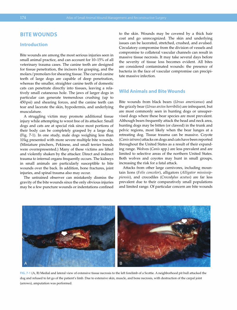
A struggling victim may promote additional tissue injury while attempting to wrest free of its attacker. Small dogs and cats are at special risk since most portions of their body can be completely grasped by a large dog (Fig. 7‐1). In one study, male dogs weighing less than 10 kg presented with more severe multiple bite wounds. (Miniature pinchers, Pekinese, and small terrier breeds were overrepresented.) Many of these victims are lifted and violently shaken by the attacker. Direct and indirect trauma to internal organs frequently occurs. The kidneys in small animals are particularly susceptible to bite wounds over the back. In addition, bone fractures, joint injuries, and spinal trauma also may occur.
The untrained observer can mistakenly dismiss the gravity of the bite wounds since the only obvious injuries may be a few puncture wounds or indentations confined
to the skin. Wounds may be covered by a thick hair coat and go unrecognized. The skin and underlying issues can be lacerated, stretched, crushed, and avulsed. Circulatory compromise from the division of vessels and compromise to collateral vascular channels can result in massive tissue necrosis. It may take several days before the severity of tissue loss becomes evident. All bites are considered contaminated wounds: the presence of bacteria in the face of vascular compromise can precipi-tate massive infection.
Wild Animals and Bite Wounds
Bite wounds from black bears (Ursus americanus) and the grizzly bear (Ursus arctos horribilis) are infrequent, but are most commonly seen in hunting dogs or unsuper-vised dogs where these bear species are most prevalent. Although bears frequently attack the head and neck area, hunting dogs may be bitten (or clawed) in the trunk and pelvic regions, most likely when the bear lunges at a retreating dog. Tissue trauma can be massive. Coyote (Canis iatrans) attacks on dogs and cats have been reported throughout the United States as a result of their expand-ing range. Wolves (Canis spp.) are less prevalent and are limited to selective areas of the northern United States. Both wolves and coyotes may hunt in small groups, increasing the risk for a fatal attack.
Attacks from other large carnivores, including moun-tain lions (Felis concolor), alligators (Alligator mississip-piensis), and crocodiles (Crocodylus acutus) are far less prevalent due to their comparatively small populations and limited range. Of particular concern are bite wounds
A B
FIG. 7-1 (A, B) Medial and lateral view of extensive tissue necrosis to the left forelimb of a Scottie. A neighborhood pit bull attacked the
dog and refused to let go of the patient’s limb. Due to extensive skin, muscle, and bone necrosis, with destruction of the carpal joint
(arrows), amputation was performed.

Management of Specific Wounds 175
from wild animals, including raccoons, skunks, and bats, in which rabies is an endemic problem (see Rabies and the Transmission of Infectious Diseases, later in this chapter).
Initial Patient Assessment
A complete physical exam is required. A history of the attack may help in locating the body region(s) bitten. Latex gloves must be worn during the examination and management of open wounds.
The patient’s head should be restrained or muzzled during examination to protect the practitioner from injury. The hair coat must be parted and skin areas visu-alized. Small spots of dried blood and matted hair fre-quently overlie puncture wounds. Careful palpation and observation may demonstrate muscle tears or hernias.
Care must be taken to minimize pain to the patient and avoid manipulating unstable fractures or spinal injuries. Analgesics or sedation may be used in an otherwise
stable patient. (A neurologic examination would be advisable prior to using medications that could obscure these injuries.)
Blood loss, shock, respiratory distress (including laryngeal wounds, tracheal injuries, pneumomediastinum, pneumothorax, hemothorax, flail chest) are emergency situations that frequently require the clinician’s immedi-ate attention before completing the examination (Figs 7‐2 and 7‐3). The prognosis of the patient, definitive course(s) of action required, and potential complications in man-aging the patient are essential to the owner’s decision regarding whether or not to proceed with treatment in the seriously injured pet. In the interim, basic life‐support procedural guidelines include the following areas of emergency resuscitation: (1) airway, (2) breath-ing, (3) cardiac function, and (4) hemorrhage control. Intravenous fluid support and whole blood would be indicated in the presence of extensive tissue trauma and blood loss. Details on emergency management can be obtained in standard textbooks. A complete blood count, serum chemistry profile, and urinalysis can serve as baseline data for the seriously injured patient.
A complete medical history should include the patient’s medical history and current rabies vaccination status.
Keep in mind that Elizabethan collars also can be useful in shield-ing veterinary personnel from being bitten, especially in the less aggressive canine and feline patients.
A B
FIG. 7-2 (A) Massive cervical trauma to the trachea and cervical muscles as a result of a dog fight. The patient was administered oxygen
prior to anesthesia. The cervical area was prepared for surgery. Anesthetic induction was immediately followed by a cervical incision, with
placement of a sterile endotracheal tube into the lacerated trachea. The gas anesthetic machine was connected to this tube. Once the patient
was stable, the technician passed a sterile endotracheal tube through the larynx. The surgeon then guided the tube past the tracheal tear.
(B) Surgical repair included tracheal anastomosis, thorough debridement of necrotic muscle, copious lavage, and delayed primary closure.
Small puncture wounds were uncapped and locally assessed using a pair of mosquito hemostats as tissue retractors. These wounds were
left open to heal by second intention. However, complete excision of small puncture wounds can permit primary closure.
Treatment of hemorrhage requires (1) recognition of the condi-tion, (2) control of further blood loss, and (3) intravenous fluid support to treat the patient’s hypovolemia.
In many cases, blood loss is not the result of an obvious spurt-ing artery. Internal hemorrhage can be difficult to quantitate. Individual bite wounds may result in little hemorrhage; collec-tively, multiple bite wounds can result in a sizeable loss of blood.

176 Atlas of Small Animal Wound Management and Reconstructive Surgery
Systemic Effects
Multiple and severe bite wounds can initiate a systemic inflammatory response syndrome (SIRS). What normally is a regional response to injury becomes an exaggerated systemic inflammatory response secondary to extensive tissue trauma. Resection of necrotic tissue and aggressive management of infection are critical to the prevention and management of this condition. Acute respiratory distress syndrome (ARDS) may be noted as a sequela to SIRS.
Bite Wound Management
All penetrating bite wounds are ideally explored under anesthesia. If the patient is in a critical condition, explora-tion, debridement, and definitive repair may be necessar-ily delayed until the patient can be stabilized. However, basic wound management can be instituted in the interim. The procedures for short‐term treatment before definitive exploration are described below.
Temporary Bite Wound Care in the Critical PatientThe following steps should be taken for the initial man-agement of bite wounds in the critical‐care patient in which general anesthesia cannot be administered.
1. Minimize further contamination of open wounds prior to preparing the wound for surgery. Cover the open wound with sterile gauze sponges; sterile water‐soluble gel or saline is applied to the gauze before application.
2. Liberally clip hair around each puncture wound.3. Gently cleanse the skin around each wound with
warm sterile saline and a surgical preparation solution.
4. Inject small amounts of lidocaine with a 25‐gauge hypodermic needle into and around the bite wound punctures. Trim off any tattered borders. Insert a pair of sterile hemostats and inspect the underlying tissues.
5. Perform liberal pressure lavage of the puncture site using an 18‐gauge needle and 35‐ml syringe with warm sterile saline. The wound should be opened sufficiently to permit fluid to flow out freely. A three‐way stopcock valve connected to sterile intravenous tubing can be used to facilitate refilling the syringe. A dilute povidone–iodine (1%: 1 part solution/9 parts sterile saline) or chlorhexidine (0.05%: 1 part solu-tion/40 parts sterile saline) solution can be made by adding these stock solutions to the lavage receptacle. The access site must be sufficiently large to allow the lavage solution to exit the area.
6. Apply a topical antimicrobial ointment and dressing over the open wound.
7. Systemic broad‐spectrum antibiotics may be adminis-tered if deemed necessary (see later in this chapter).
The primary goal of this short‐term therapy is to reduce the amount of contamination present and reduce the pos-sibility of further contamination from organisms in the hospital environment (nosocomial infection).
A B
FIG. 7-3 (A) Puncture to the cervical trachea as a result of a dog bite: intraoperative view. A canine tooth created the opening that enabled
air to pass into the cervical tissues, impairing the ability of the patient to breathe. Manual compression of the skin over the puncture site
halted the air excursion until anesthesia and intubation could be accomplished. (B) Closure was accomplished by conservative debride-
ment of the wound borders followed by placement of 3‐0 polydioxanone sutures.
Fully equipped emergency units with a dedicated staff address the critically injured patient. Critical care clinicians, surgeons, and the anesthesiology team working in concert will give the patient the best chance of surviving.
Management of Specific Wounds 177
Once stabilized, the patient can be anesthetized for wound exploration if necessary (see the next section.) On occasion, critically injured patients with serious underly-ing injuries cannot be stabilized to the desired degree. Under these circumstances surgical intervention would be necessary since these wounds are the primary cause of the patient’s deterioration (bowel rupture, sepsis, etc.). Such a serious endeavor requires the coordinated efforts of the emergency clinician, anesthesiologist/anesthetist, and the surgeon.
Definitive Management: Bite Wound ExplorationThe following steps summarize the approach for the definitive management of bite wounds.
1. General anesthesia.2. Open wounds should be temporarily covered or
packed with sterile gauze sponges moistened with saline; a water‐miscible lubricant can be applied to the sponges in place of saline in order to protect individual wounds from contamination associated with preparing the area for surgery.
3. The bite wound area should be clipped liberally. In particular, the thoracic and abdominal area should be completely clipped and prepared for surgery in the event the exploration requires conversion to a thoracotomy or exploratory laparotomy. The surgical area then is prepared and draped for aseptic surgery.
4. Puncture wounds are “uncapped” using a scalpel blade by excising the puncture‐wound borders, thereby creating a 1.0‐cm‐plus circular opening. Sterile mosquito hemostats are inserted into the opening and spread to expose the underlying hypodermis, fascial tissues, and muscle. Wounds with little or no underlying tissue damage may be left open to drain and heal by second intention or may be closed with one or two skin sutures after lavaging the wound.
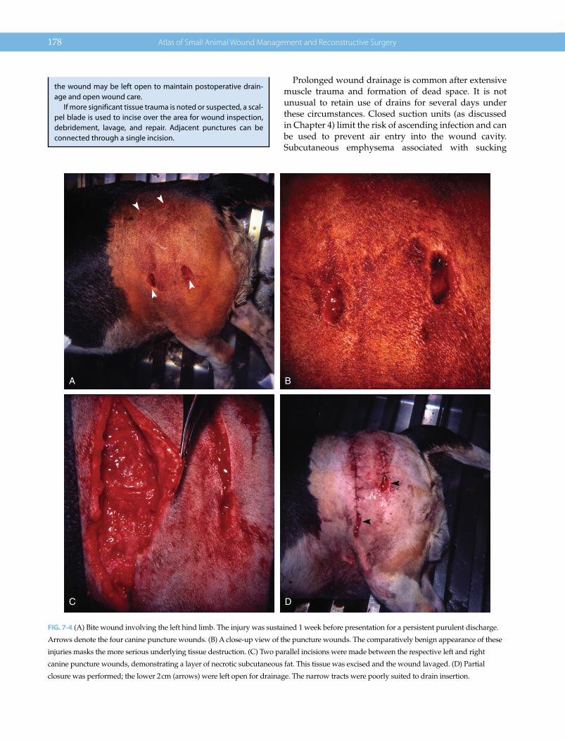
5. If significant tissue damage is suspected, a skin incision is made over the puncture site; tissue retractors are inserted to permit exploration and debridement of traumatized tissues. Adjacent puncture wounds may be connected with a single incision to facilitate exploration (Fig. 7‐4). Care must be taken not to inadvertently divide direct cutaneous arteries, especially those vessels supply-ing skin already compromised by bite wound trauma.
6. Any hair or foreign debris is removed. Shredded or necrotic muscle, fat, and fascia are excised. A more aggressive debridement can be performed for those tissues of questionable viability that are not essential
to normal function. As noted, additional care is warranted when exploring bite wounds over the thorax. Retraction of muscle and fascia may unseal penetrating thoracic wounds. Sucking sounds may emanate from the area as air enters the thoracic cavity, necessitating assisted ventilation, aspiration of the thorax, or temporary insertion of a thoracos-tomy tube.
7. Debridement ideally is accomplished in one stage, especially in critical areas (thoracic cavity and abdominal cavity involvement) where the presence of necrotic tissues can promote life‐threatening sepsis. In less critical areas, such as the extremities, a more conservative daily or staged debridement (open‐wound management followed by delayed primary closure, secondary closure, healing by second intention) is indicated for tissues of question-able viability, where important muscle groups and the limited availability of skin could compromise salvage of the limb.
8. Skin viability may be difficult to determine on initial presentation. Necrosis may not be evident for 5–7 days after injury. The loose skin available over the cervical area and trunk permits more aggressive debridement of skin of questionable viability. However, a more conservative “wait and reassess” approach is indicated for compromised skin of the lower extremities. With the limited amount of loose skin available for closure, unnecessarily wide debridement will increase the likelihood that reconstructive surgery will be required to close the resultant defect.
9. Wound drainage is necessary in areas where dead space is present, especially after wide debridement of contaminated bite wounds. Closed vacuum drains or Penrose drains may be considered (see Chapter 4).
10. Open wound management is advisable, if practical, in the presence of infection and tissues of question-able viability (see Chapter 3).
“Uncapping” the Puncture WoundA simple method of assessing a puncture wound is by lifting the puncture site with forceps and excising the area with a scalpel blade, thereby creating a 10–15‐mm opening to inspect the underlying tissues. A mosquito hemostat can be inserted and spread open, serving as a tissue retractor or speculum to exam-ine the subcutaneous tissues and underlying muscles. If the sub-cutaneous fat is undisturbed, the wound is considered minor. If the fat has been separated and fragmented, the underlying muscle is examined. Lavage and suctioning facilitate visualiza-tion of the tissues by removal of tissue fragments and debris.
If tissue damage is minor, the wound can be lavaged and apposed with one or two skin sutures; if there are any doubts,
178 Atlas of Small Animal Wound Management and Reconstructive Surgery
Prolonged wound drainage is common after extensive muscle trauma and formation of dead space. It is not unusual to retain use of drains for several days under these circumstances. Closed suction units (as discussed in Chapter 4) limit the risk of ascending infection and can be used to prevent air entry into the wound cavity. Subcutaneous emphysema associated with sucking
the wound may be left open to maintain postoperative drain-age and open wound care.
If more significant tissue trauma is noted or suspected, a scal-pel blade is used to incise over the area for wound inspection, debridement, lavage, and repair. Adjacent punctures can be connected through a single incision.
A B
C D
FIG. 7-4 (A) Bite wound involving the left hind limb. The injury was sustained 1 week before presentation for a persistent purulent discharge.
Arrows denote the four canine puncture wounds. (B) A close‐up view of the puncture wounds. The comparatively benign appearance of these
injuries masks the more serious underlying tissue destruction. (C) Two parallel incisions were made between the respective left and right
canine puncture wounds, demonstrating a layer of necrotic subcutaneous fat. This tissue was excised and the wound lavaged. (D) Partial
closure was performed; the lower 2 cm (arrows) were left open for drainage. The narrow tracts were poorly suited to drain insertion.
Management of Specific Wounds 179
wounds of the flank and axilla may be prevented with vacuum drains. Similarly, they can provide wound drain-age overlying the thorax or thoracic inlet where air entry (with the use of Penrose drains) may result in pneumo-thorax. Vacuum drains can be used effectively for the drainage of abdominal wall wounds and the abdominal cavity simultaneously (as needed).
Regional Considerations
Head and NeckThe head and neck areas are most commonly attacked by predators. Small dogs and cats can sustain skull fractures from larger predators (Fig. 7‐5). Injury to eyes
and ears are occasionally noted. Bite wounds involv-ing the pinna are common; occasionally, the ear canal will sustain punctures, lacerations, or avulsion injuries (at the junction of the vertical and horizontal ear canal, or canal avulsion at the external acoustic meatus). Cervical wounds must be closely assessed for injuries to the trachea, larynx, esophagus, pharynx, major ves-sels, and salivary glands, in particular. The spinal col-umn is most susceptible to trauma in small dogs and cats. Skin and muscle are most prone to injury. A tra-cheostomy may be necessary when upper respiratory distress is evident. Traumatic division of the trachea may require a prompt cervical incision upon anesthetic induction for proper intubation before tracheal repair can be instituted. Alternatively, an endotracheal tube can be temporarily inserted into the tracheal tear to stabilize the patient until proper placement can be instituted intraoperatively (Fig. 7‐2). Puncture or lac-eration of the rigid trachea also can result from pene-trating canine teeth without tearing of the overlying skin (Fig. 7‐3).
A B
C D
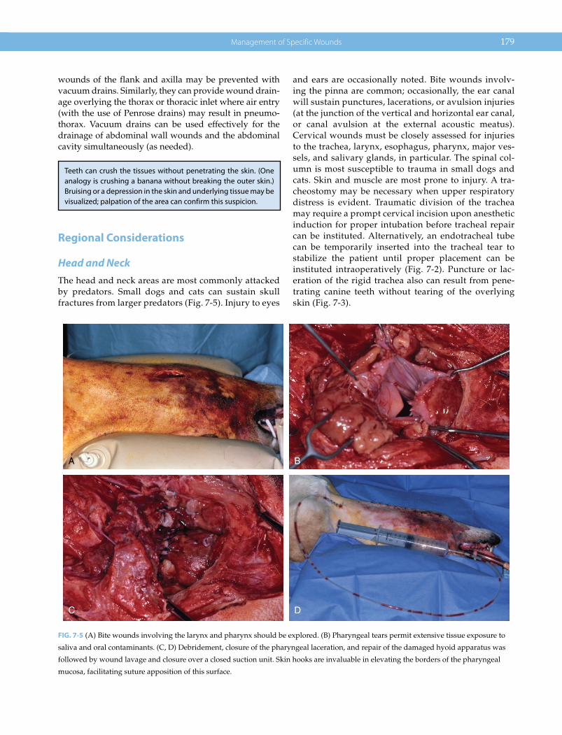
FIG. 7-5 (A) Bite wounds involving the larynx and pharynx should be explored. (B) Pharyngeal tears permit extensive tissue exposure to
saliva and oral contaminants. (C, D) Debridement, closure of the pharyngeal laceration, and repair of the damaged hyoid apparatus was
followed by wound lavage and closure over a closed suction unit. Skin hooks are invaluable in elevating the borders of the pharyngeal
mucosa, facilitating suture apposition of this surface.
Teeth can crush the tissues without penetrating the skin. (One analogy is crushing a banana without breaking the outer skin.) Bruising or a depression in the skin and underlying tissue may be visualized; palpation of the area can confirm this suspicion.

180 Atlas of Small Animal Wound Management and Reconstructive Surgery
ExtremitiesThe comparatively narrow diameter of the extremities make them particularly susceptible to extensive bite wound trauma (crushing, laceration), especially in the smaller patients. Circulatory compromise to the skin, muscle, and bone can result in massive necrosis, necessi-tating limb amputation in many cases. Overlooked punc-ture wounds involving the elbow, carpal, knee, and tarsal joints may result in problematic infections. A complete neurologic examination of the involved limb and close assessment of circulation is in order in the days following bite wound care. Fracture repair and stabilization nor-mally is performed at the time of bite wound exploration. However, in the face of extensive swelling and circulatory compromise, a delay in fracture repair may be advisable: surgical trauma could further compromise circulation to the lower extremity and precipitate loss of the limb. As discussed, when debriding extremity wounds, a more conservative approach is in order when tissue viability is equivocal (see Orthopedic Injuries and Spinal Trauma.)
Thoracic TraumaSmall patients are more susceptible to thoracic wall trauma, although the canine teeth in larger dogs are capable of penetrating the intercostal area of most dogs.
Open wounds at the thoracic inlet can result in pneumo-mediastinum; direct open wounds involving the thoracic wall can result in fatal pneumothorax. On occasion, flaps of tissue may overlap and seal the thoracic wall defect, limiting the severity of the pneumothorax until surgical repair can be performed.
Both pneumothorax and hemothorax should be imme-diately suspected in patients presenting with respiratory distress. Pyothorax may be noted in patients with older bite wounds (Fig. 7‐7). Hyperresonance would suggest pneumothorax, whereas dull areas of percussion would suggest fluid accumulation in the dependent areas of the
Retention of fractured teeth is rarely seen in small animals. The dense interosseous ligament between the radius and ulna is the most common region where the tip of a canine tooth can be bro-ken off. Its retention will result in a draining tract. Radiographs will easily identify the denser tooth fragment in contrast to the adjacent bone (Fig. 7‐6).
FIG. 7-6 Radiographs of the distal forelimb in a dog, demonstrating
retention of the tip of a canine tooth in the interosseous ligament
between the radius and ulna. The tooth fragment and associated
draining tract were removed. The greater density of the retained
canine helps to distinguish it from the adjacent bone. (Radiographs
courtesy of Dr. Paul Gambardella.)
A B
FIG. 7-7 (A) Bite wounds (arrows) overlying the sternum 4 days prior to presentation. The dog presented with respiratory distress as a
result of pyothorax. Necrotic sternal bone was excised followed by a thoracotomy. (B) A restrictive pleuritis (arrows) was noted as a result
of fibrin deposition and early fibrous connective tissue. Decortication of this layer with gauze pads was followed by closure after
placement of a thoracostomy tube for continuous closed suction postoperatively.

Management of Specific Wounds 181
thoracic cavity. In relatively calm and stable patients, a lateral thoracic radiograph may be obtained to confirm each condition. Thoracocentesis can be used to initially alleviate respiratory distress and confirm the presence of air or fluid. The diameter of a bronchus noted on radio-graphs can serve as a relative guideline for estimation of the size of the thoracostomy tube that may be required. Immediate placement of a thoracostomy tube may be preferable when tension pneumothorax or significant hemothorax is diagnosed at presentation. The tube can be connected to a continuous thoracic suction unit in more serious cases. Drainage may be continued for 48 hours after cessation of air leakage. Significant, unabated leaks beyond the initial 72‐hour period may require surgical (re)intervention. Similarly, brisk continuous hemorrhage following initial evacuation of blood in the thoracic cav-ity would justify prompt surgical intervention.
Open skin wounds can be temporarily stapled or sutured to limit air entry prior to surgery. Alternatively, a bandage containing a heavy layer of ointment can be cupped over the wound temporarily. Bite wounds entering the thoracic cavity are best explored, due to the significant risk of tissue necrosis and pyothorax present in these cases. Surgical staplers (TA, Covidien/USSC/Kendall) are useful in removing traumatized pul-monary tissue.
Fractured ribs with significant displacement can be repaired at the time of exploration. Sharp, pointed edges can be trimmed with rongeurs to reduce the risk of lung laceration. Cases of flail chest can be stabilized in a similar fashion with fine wire or nonabsorbable suture to realign the fracture segments. Holes can be drilled approximately 1 cm from the fracture ends. Stainless steel orthopedic wire (22‐gauge) is commonly used to realign the rib segments; overtightening should be avoided in this soft bone. In most cases of flail chest, preoperative external stabilization of the area is usually unnecessary.
Bite wounds involving the caudal thoracic and cranial abdominal areas are capable of tearing the underlying diaphragm: the pars costalis and pars sternalis are rela-tively superficial at the area of the xiphoid and caudal thoracic cage. Rents in the diaphragm may not be visible on initial thoracic radiographs; additional radiographs are indicated if deterioration of clinical signs warrants reevaluation of the patient. The diaphragm should always be inspected during exploratory laparotomy.
Abdominal TraumaThe larger canine teeth are capable of deep penetration through the soft and compressible abdominal wall into the peritoneal cavity, especially in smaller patients. It is important to note that the skin and abdominal wall may appear to be intact, although internal organs can be crushed. The kidneys, bowel, mesenteric vasculature, spleen, and liver can be traumatized by direct contact with the canine teeth or indirectly as a result of the stretching and tearing of tissues if the patient is shaken during the attack. Ultrasonography may be used to assess the integrity of the internal organs; abdominal radiographs can be used to assess the abdomen and determine the presence of free air and fluid. An intrave-nous pyelogram may be used to assess traumatized kidneys based on these findings.
A midline celiotomy is advisable when teeth have pen-etrated or crushed the abdominal cavity (as occasionally noted in small dogs and cats). Superficial bite wounds limited to the outer abdominal wall can be managed as discussed earlier. However, the abdomen should be pre-pared for possible exploratory surgery in the event that more significant trauma is noted during surgery.
Orthopedic Injuries and Spinal TraumaAs discussed, smaller dogs and cats are more susceptible to injury as a result of direct trauma (crushing, penetra-tion) or indirect trauma (violent shaking of the victim with
The hypodermic needle or, preferably, butterfly catheter, attached to a three‐way stopcock and 35–60‐ml syringe, can be used for thoracocentesis. The needle is angled slightly with the bevel toward the patient to reduce the risk of lung laceration. Plastic intravenous catheters also may be used to reduce the risk of lung laceration.
• If the patient is in lateral recumbency, air is drawn from the central third of the midthorax
• If the patient is standing, air is withdrawn from the upper third of the midthorax
• If the patient is standing or in lateral recumbency, fluid is re-moved from the lower third of the thorax, between the third and eighth ribs; care is required to prevent advancement of the needle into the pericardium
Internal hemorrhage can have an insidious onset and can be easily overlooked in the trauma patient. A fist‐sized hematoma may vary from 300–500 ml of blood. Retroperitoneal hemor-rhage may not be apparent on physical examination. For exam-ple, half the patient’s blood volume (40 ml/kg body weight) must be present to cause overt abdominal distention. If 20–25% of the blood volume is lost or sequestered over a 10‐minute period, profound hypovolemic shock will occur.
It is also worth noting that small volumes of blood loss from multiple small bite wounds and contusions cumulatively can result in a significant loss of red cells.
182 Atlas of Small Animal Wound Management and Reconstructive Surgery
wounds created distant to the point of direct contact). Most fractures can be stabilized at the time of bite wound management. External fixators may be particularly use-ful in fracture stabilization, thereby reducing the risk of internal fixation in proximity to a contaminated bite wound. As noted, surgery should be delayed in those cases where circulatory compromise to the lower extrem-ity is of concern. Resolution of tissue swelling normally follows improvement in circulation and lymphatic drain-age. A modified Robert Jones bandage or supportive splints may be used to stabilize the fracture while manag-ing the bite wounds.
A complete neurologic examination is essential to diag-nosing spinal trauma. Radiographs and supplemental diagnostic imaging are used to confirm vertebral frac-ture/luxations. Depending on the nature of the injury and the severity of clinical signs, external stabilization, internal stabilization, and/or decompression of the spi-nal cord may be necessary.
Infection and Bite Wounds
The polymicrobial flora of the oral cavity can inoculate wounds with aerobic and anaerobic bacteria. Bite wounds resulting in perforation of the gastrointestinal tract can result in the spillage of additional bacterial contaminants (Fig. 7‐8). In human studies, aerobic infections are more common than anaerobic infections alone. (Bite wounds have a 5–10% risk of infection in humans; by contrast, non-bite lacerations have an infection rate of 5%.) However, with the presence of anaerobic bacteria, the severity of the infection is often increased. Pasteurella spp. are Gram‐nega-tive nonmotile pleomorphic coccobacilli that are commonly
isolated from the oral cavity of dogs and cats. In one report, up to 50% of canine and 90% of feline bite wound infections in humans were the result of Pasteurella spp. Management of Pasteurella infections in humans can be problematic, especially in older, debilitated patients; individuals with orthopedic implants and internal prosthetic devices; and persons with a compromised immune system.
In the face of this acute tissue trauma, the selection and use of antibiotics remains somewhat controversial. Over the last decade, the prophylactic use of antibiotics has been demonstrated to be of limited clinical efficacy in preventing infection. Prophylactic use of antibiotics is of proven value only for carefully selected high‐risk proce-dures when properly administered before surgery. Antibiotics are normally administered after bite injuries, and several hours may pass before administration and effective blood levels are achieved. In general, 3 hours is considered the maximum acceptable delay in administra-tion of antibiotics in bite wound management. To avoid the several‐hour delay to achieve an adequate serum level associated with oral antibiotics, intravenous admin-istration is advisable for seriously injured patients and is 4–12 times faster than intramuscular administration for
A B
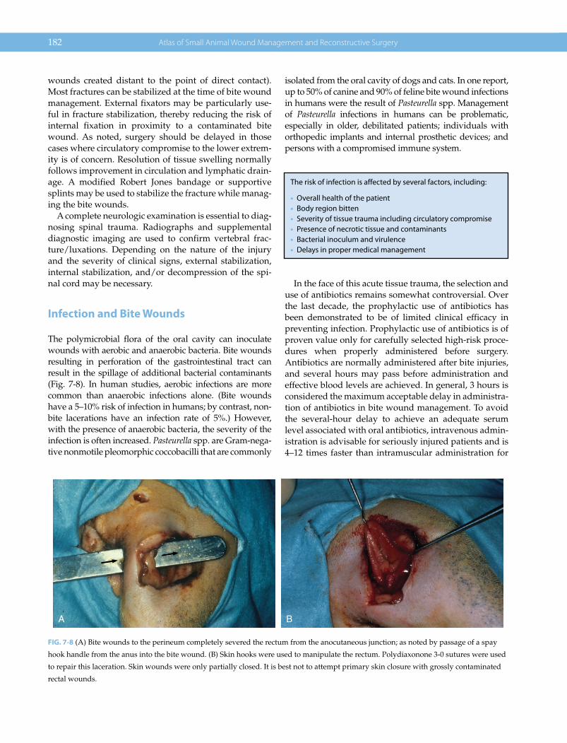
FIG. 7-8 (A) Bite wounds to the perineum completely severed the rectum from the anocutaneous junction; as noted by passage of a spay
hook handle from the anus into the bite wound. (B) Skin hooks were used to manipulate the rectum. Polydiaxonone 3‐0 sutures were used
to repair this laceration. Skin wounds were only partially closed. It is best not to attempt primary skin closure with grossly contaminated
rectal wounds.
The risk of infection is affected by several factors, including:
• Overall health of the patient• Body region bitten• Severity of tissue trauma including circulatory compromise• Presence of necrotic tissue and contaminants• Bacterial inoculum and virulence• Delays in proper medical management

Management of Specific Wounds 183
developing an effective tissue‐fluid concentration at the wound.
Bacteriocidal antibiotics are best employed in bite wounds. Cephalosporins, ampicillin, and penicillin rapidly enter the wound (within 1 hour). In contrast, erythromycin and gentamicin take 2–4 hours for wound concentrations to match serum concentrations, whereas tetracycline and clindamycin never reach wound levels equivalent to serum levels. Cephalosporins are gener-ally effective against Pasteurella spp. and a variety of other microorganisms. Amoxicillin or clavulanate potas-sium can be useful against Pasteurella multocida resistant to penicillins and β‐lactamase Staphylococcus spp. Fluoroquinolones are useful for resistant Gram‐positive and Gram‐negative infection. In the presence of Gram‐negative organisms, an aminoglycoside such as gen-tamicin should be considered. Antibiotics are not a substitute for the appropriate surgical management of bite wounds.
In the presence of infection, wound cultures (aerobic, anaerobic) are advisable in order to select the most appropriate antibiotic(s), especially in the septic patient. Culturing the acute uninfected bite wound is useless in determining the potential infection organisms. Culture samples should be submitted from deep within the wound by aspiration or incision, drainage, and explora-tion of the area. Aspiration of lymph nodes or areas of cellulitis also can be employed to obtain accurate culture samples. More superficial wound cultures are more likely to include contaminants that will lead to misleading results. The most accurate source for culturing infected wounds is from tissue samples from the abscess wall.
Although uncommonly performed in practice, Gram stains may be useful in determining the type of organ-isms present and the most appropriate initial antibiotic selection prior to obtaining definitive culture and sensi-tivity results.
Rabies and the Transmission of Infectious Diseases
Of the infectious diseases transmitted by bite wounds, rabies is the single greatest concern because of the human health implications of this viral disease.
It has been recommended that unvaccinated pets bit-ten by a wild mammal (unavailable for testing) should be euthanized. If the owner refuses, the pet must be isolated for 6 months and vaccinated 1 month before release.
Vaccinated pets should be revaccinated immediately, confined by the owner, and monitored carefully for 45 days. For specific details, the veterinarian should consult with local and state authorities to ensure that regulations are precisely followed.
Cats can transmit feline leukemia virus and feline immunodeficiency virus through bites. Assessment of transmission can be evaluated by serologic testing 6 months after the injury. These two diseases can result in nonhealing wounds.
BURNS
Veterinarians see few burns compared to human physi-cians. Most burns seen by the author are not life‐threaten-ing but are referred for definitive closure of the wound.
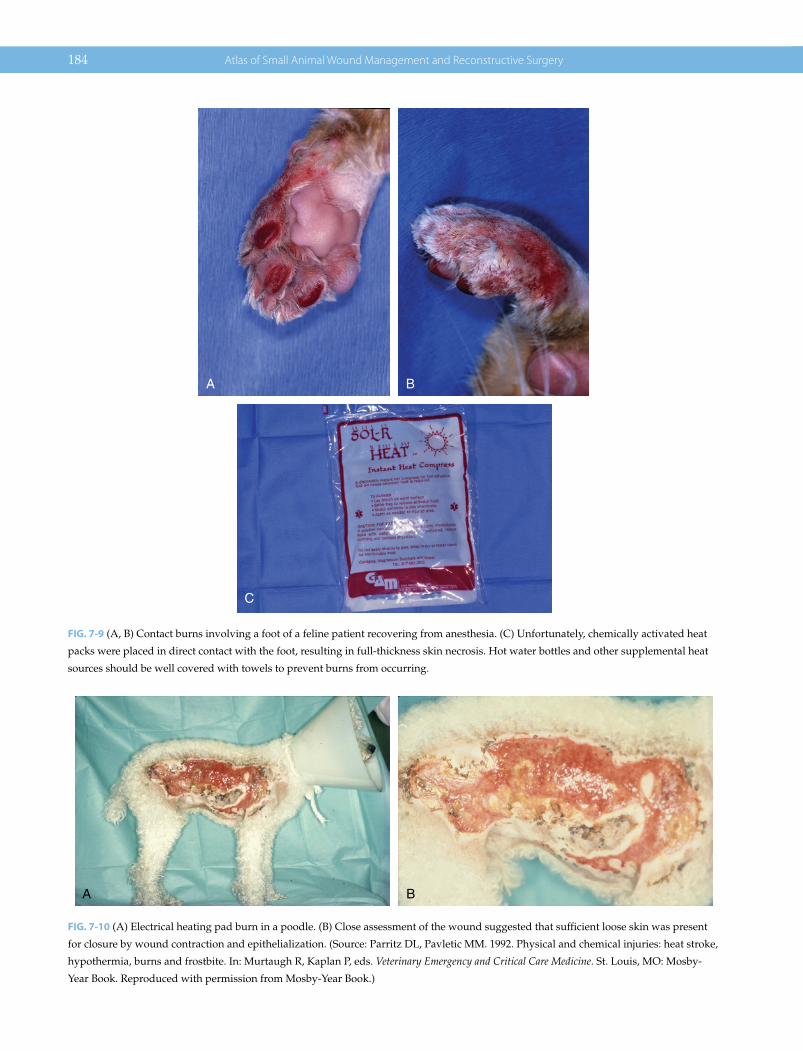
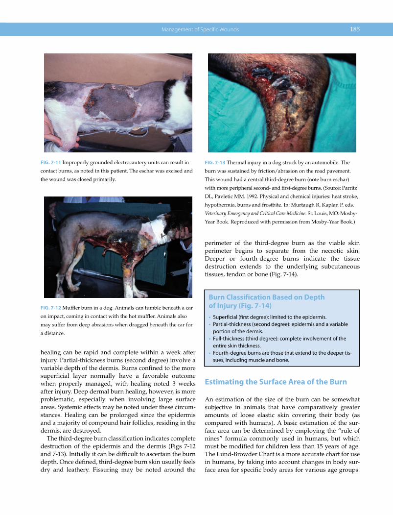
Ironically, the most common thermal injuries seen by the author are caused by veterinarians using various heat sources to warm the patient (Fig. 7‐9). Electrical heating pads in particular can generate considerable heat: pro-longed contact in combination with local tissue retention of heat can produce full‐thickness burns of considerable dimension (Fig. 7‐10). Less commonly seen are faulty ground plates used with electrocautery units, resulting in contact burns (Fig. 7‐11).
Burn Classification
The severity of the burn is evaluated by the degree or depth of the injury, as well as the percentage of the sur-face area involved. A full‐thickness burn site usually has three concentric zones of tissue injury: a central zone of coagulative necrosis, a middle zone of vascular stasis with compromised tissue perfusion, and an outer zone of hyperemia. Progressive circulatory compromise can result in necrosis extending into the middle zone, in an outward direction.
Superficial burns (first degree) are burns confined to the outermost epidermis. The skin can appear erythema-tous and is hyperesthetic to the touch. Properly managed,
Types of Burns Seen in Veterinary Practice1. Fire/flame burns2. Scalds3. Electrical heating pads4. Hot air dryers5. Heating lamps6. Exothermic (chemically activated) heat packs7. Electrical cords8. Faulty electrocautery units9. Wood‐burning stoves
10. Household radiators11. Stove tops12. Automobile mufflers13. Solar (actinic) radiation14. Chemical burns15. Radiation burns
184 Atlas of Small Animal Wound Management and Reconstructive Surgery
A B
C
FIG. 7-9 (A, B) Contact burns involving a foot of a feline patient recovering from anesthesia. (C) Unfortunately, chemically activated heat
packs were placed in direct contact with the foot, resulting in full‐thickness skin necrosis. Hot water bottles and other supplemental heat
sources should be well covered with towels to prevent burns from occurring.
A B
FIG. 7-10 (A) Electrical heating pad burn in a poodle. (B) Close assessment of the wound suggested that sufficient loose skin was present
for closure by wound contraction and epithelialization. (Source: Parritz DL, Pavletic MM. 1992. Physical and chemical injuries: heat stroke,
hypothermia, burns and frostbite. In: Murtaugh R, Kaplan P, eds. Veterinary Emergency and Critical Care Medicine. St. Louis, MO: Mosby‐
Year Book. Reproduced with permission from Mosby‐Year Book.)
Management of Specific Wounds 185
healing can be rapid and complete within a week after injury. Partial‐thickness burns (second degree) involve a variable depth of the dermis. Burns confined to the more superficial layer normally have a favorable outcome when properly managed, with healing noted 3 weeks after injury. Deep dermal burn healing, however, is more problematic, especially when involving large surface areas. Systemic effects may be noted under these circum-stances. Healing can be prolonged since the epidermis and a majority of compound hair follicles, residing in the dermis, are destroyed.
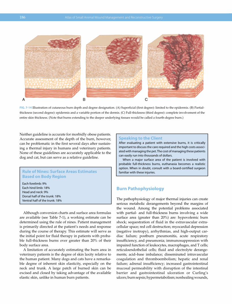
The third‐degree burn classification indicates complete destruction of the epidermis and the dermis (Figs 7‐12 and 7‐13). Initially it can be difficult to ascertain the burn depth. Once defined, third‐degree burn skin usually feels dry and leathery. Fissuring may be noted around the
perimeter of the third‐degree burn as the viable skin perimeter begins to separate from the necrotic skin. Deeper or fourth‐degree burns indicate the tissue destruction extends to the underlying subcutaneous tissues, tendon or bone (Fig. 7‐14).
Estimating the Surface Area of the Burn
An estimation of the size of the burn can be somewhat subjective in animals that have comparatively greater amounts of loose elastic skin covering their body (as compared with humans). A basic estimation of the sur-face area can be determined by employing the “rule of nines” formula commonly used in humans, but which must be modified for children less than 15 years of age. The Lund‐Browder Chart is a more accurate chart for use in humans, by taking into account changes in body sur-face area for specific body areas for various age groups.
FIG. 7-12 Muffler burn in a dog. Animals can tumble beneath a car
on impact, coming in contact with the hot muffler. Animals also
may suffer from deep abrasions when dragged beneath the car for
a distance.
FIG. 7-11 Improperly grounded electrocautery units can result in
contact burns, as noted in this patient. The eschar was excised and
the wound was closed primarily.
FIG. 7-13 Thermal injury in a dog struck by an automobile. The
burn was sustained by friction/abrasion on the road pavement.
This wound had a central third‐degree burn (note burn eschar)
with more peripheral second‐ and first‐degree burns. (Source: Parritz
DL, Pavletic MM. 1992. Physical and chemical injuries: heat stroke,
hypothermia, burns and frostbite. In: Murtaugh R, Kaplan P, eds.
Veterinary Emergency and Critical Care Medicine. St. Louis, MO: Mosby‐
Year Book. Reproduced with permission from Mosby‐Year Book.)
Burn Classification Based on Depth of Injury (Fig. 7‐14)• Superficial (first degree): limited to the epidermis.• Partial‐thickness (second degree): epidermis and a variable
portion of the dermis.• Full‐thickness (third degree): complete involvement of the
entire skin thickness.• Fourth‐degree burns are those that extend to the deeper tis-
sues, including muscle and bone.
186 Atlas of Small Animal Wound Management and Reconstructive Surgery
Neither guideline is accurate for morbidly obese patients. Accurate assessment of the depth of the burn, however, can be problematic in the first several days after sustain-ing a thermal injury in humans and veterinary patients. None of these guidelines are accurately applicable to the dog and cat, but can serve as a relative guideline.
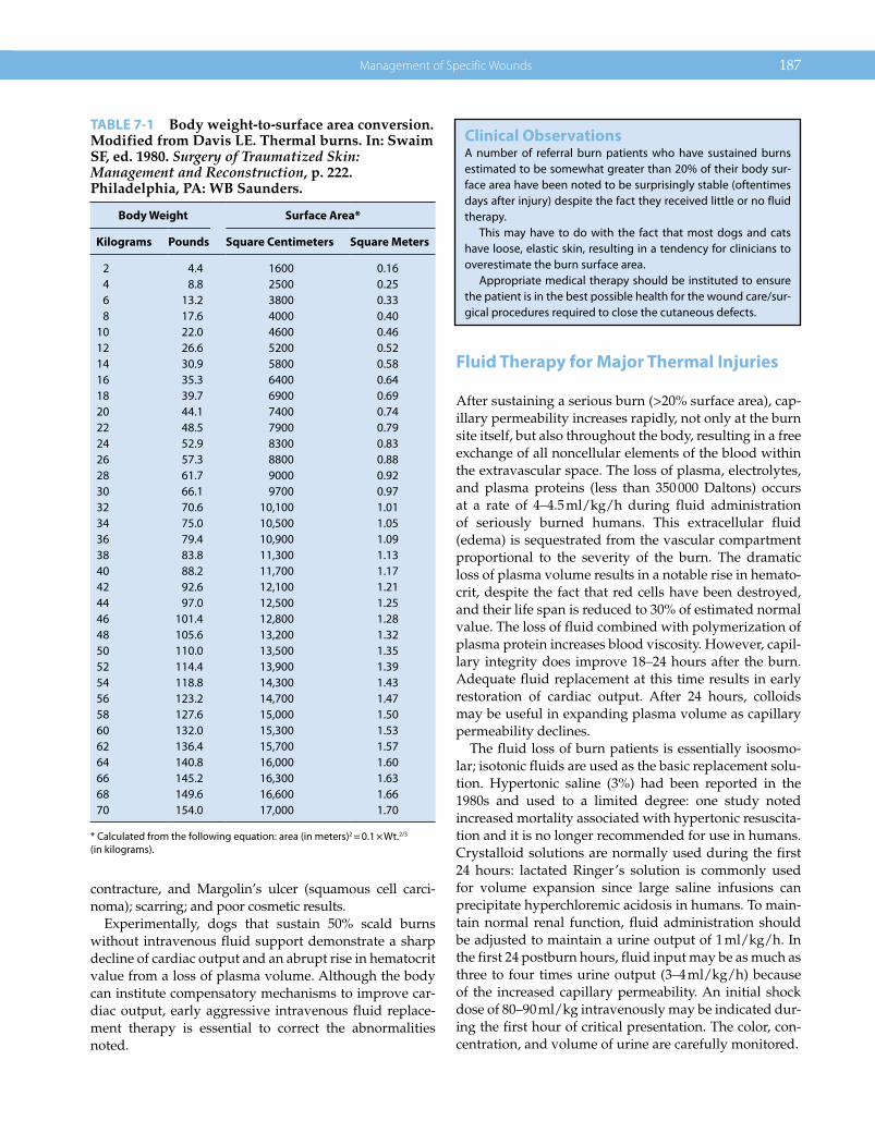
Although conversion charts and surface area formulas are available (see Table 7‐1), a working estimate can be determined using the rule of nines. Patient management is primarily directed at the patient’s needs and response during the course of therapy. This estimate will serve as the initial point for fluid therapy in patients with proba-ble full‐thickness burns over greater than 20% of their body surface area.
A limitation of accurately estimating the burn area in veterinary patients is the degree of skin laxity relative to the human patient. Many dogs and cats have a remarka-ble degree of inherent skin elasticity, especially on the neck and trunk. A large patch of burned skin can be excised and closed by taking advantage of the available elastic skin, unlike in human burn patients.
Burn Pathophysiology
The pathophysiology of major thermal injuries can create serious metabolic derangements beyond the margins of the wound. Among the potential problems associated with partial‐ and full‐thickness burns involving a wide surface area (greater than 20%) are: hypovolemic burn shock; sequestration of fluid in the extravascular extra-cellular space; red cell destruction; myocardial depression (negative inotropy), arrhythmias, and high‐output car-diac failure; postburn pneumonitis, acute respiratory insufficiency, and pneumonia; immunosuppression with impaired function of leukocytes, macrophages, and T cells; reticuloendothelial cells; fluid and electrolyte derange-ments; acid–base imbalance; disseminated intravascular coagulation and thromboembolism; hepatic and renal failure; adrenal insufficiency; increased gastrointestinal mucosal permeability with disruption of the intestinal barrier and gastrointestinal ulceration or Curling’s ulcers; burn sepsis; hypermetabolism; nonhealing wounds,
A B C
FIG. 7-14 Illustration of cutaneous burn depth and degree designation. (A) Superficial (first degree): limited to the epidermis. (B) Partial‐
thickness (second degree): epidermis and a variable portion of the dermis. (C) Full‐thickness (third degree): complete involvement of the
entire skin thickness. (Note that burns extending to the deeper underlying tissues would be called a fourth‐degree burn.)
Rule of Nines: Surface Areas Estimates Based on Body RegionEach forelimb: 9%Each hind limb: 18%Head and neck: 9%Dorsal half of the trunk: 18%Ventral half of the trunk: 18%
Speaking to the ClientAfter evaluating a patient with extensive burns, it is critically important to discuss the care required and the high costs associ-ated with managing the pet. The cost of managing these patients can easily run into thousands of dollars.
When a major surface area of the patient is involved with probable full‐thickness burns, euthanasia becomes a realistic option. When in doubt, consult with a board‐certified surgeon familiar with these injuries.
Management of Specific Wounds 187
contracture, and Margolin’s ulcer (squamous cell carci-noma); scarring; and poor cosmetic results.
Experimentally, dogs that sustain 50% scald burns without intravenous fluid support demonstrate a sharp decline of cardiac output and an abrupt rise in hematocrit value from a loss of plasma volume. Although the body can institute compensatory mechanisms to improve car-diac output, early aggressive intravenous fluid replace-ment therapy is essential to correct the abnormalities noted.
Fluid Therapy for Major Thermal Injuries
After sustaining a serious burn (>20% surface area), cap-illary permeability increases rapidly, not only at the burn site itself, but also throughout the body, resulting in a free exchange of all noncellular elements of the blood within the extravascular space. The loss of plasma, electrolytes, and plasma proteins (less than 350 000 Daltons) occurs at a rate of 4–4.5 ml/kg/h during fluid administration of seriously burned humans. This extracellular fluid (edema) is sequestrated from the vascular compartment proportional to the severity of the burn. The dramatic loss of plasma volume results in a notable rise in hemato-crit, despite the fact that red cells have been destroyed, and their life span is reduced to 30% of estimated normal value. The loss of fluid combined with polymerization of plasma protein increases blood viscosity. However, capil-lary integrity does improve 18–24 hours after the burn. Adequate fluid replacement at this time results in early restoration of cardiac output. After 24 hours, colloids may be useful in expanding plasma volume as capillary permeability declines.
The fluid loss of burn patients is essentially isoosmo-lar; isotonic fluids are used as the basic replacement solu-tion. Hypertonic saline (3%) had been reported in the 1980s and used to a limited degree: one study noted increased mortality associated with hypertonic resuscita-tion and it is no longer recommended for use in humans. Crystalloid solutions are normally used during the first 24 hours: lactated Ringer’s solution is commonly used for volume expansion since large saline infusions can precipitate hyperchloremic acidosis in humans. To main-tain normal renal function, fluid administration should be adjusted to maintain a urine output of 1 ml/kg/h. In the first 24 postburn hours, fluid input may be as much as three to four times urine output (3–4 ml/kg/h) because of the increased capillary permeability. An initial shock dose of 80–90 ml/kg intravenously may be indicated dur-ing the first hour of critical presentation. The color, con-centration, and volume of urine are carefully monitored.
TABLE 7-1 Body weight‐to‐surface area conversion. Modified from Davis LE. Thermal burns. In: Swaim SF, ed. 1980. Surgery of Traumatized Skin: Management and Reconstruction, p. 222. Philadelphia, PA: WB Saunders.
Body Weight Surface Area*
Kilograms Pounds Square Centimeters Square Meters
2 4.4 1600 0.164 8.8 2500 0.256 13.2 3800 0.338 17.6 4000 0.40
10 22.0 4600 0.4612 26.6 5200 0.5214 30.9 5800 0.5816 35.3 6400 0.6418 39.7 6900 0.6920 44.1 7400 0.7422 48.5 7900 0.7924 52.9 8300 0.8326 57.3 8800 0.8828 61.7 9000 0.9230 66.1 9700 0.9732 70.6 10,100 1.0134 75.0 10,500 1.0536 79.4 10,900 1.0938 83.8 11,300 1.1340 88.2 11,700 1.1742 92.6 12,100 1.2144 97.0 12,500 1.2546 101.4 12,800 1.2848 105.6 13,200 1.3250 110.0 13,500 1.3552 114.4 13,900 1.3954 118.8 14,300 1.4356 123.2 14,700 1.4758 127.6 15,000 1.5060 132.0 15,300 1.5362 136.4 15,700 1.5764 140.8 16,000 1.6066 145.2 16,300 1.6368 149.6 16,600 1.6670 154.0 17,000 1.70
* Calculated from the following equation: area (in meters)2 = 0.1 × Wt.2/3 (in kilograms).
Clinical ObservationsA number of referral burn patients who have sustained burns estimated to be somewhat greater than 20% of their body sur-face area have been noted to be surprisingly stable (oftentimes days after injury) despite the fact they received little or no fluid therapy.
This may have to do with the fact that most dogs and cats have loose, elastic skin, resulting in a tendency for clinicians to overestimate the burn surface area.
Appropriate medical therapy should be instituted to ensure the patient is in the best possible health for the wound care/sur-gical procedures required to close the cutaneous defects.

188 Atlas of Small Animal Wound Management and Reconstructive Surgery
Fluid Resuscitation FormulasBurn formulas (Parkland and modified Brooke formulas) have been developed as a general guideline for initial treatment of human patients sustaining major burns. These formulas combine both maintenance and replace-ment volumes based on the weight of the patient and surface area burned. The Parkland formula, commonly employed in humans, uses the percentage of surface area burned to estimate fluid needs: 4 ml/kg body weight × percentage of burn area. Half of this volume is given within the first 8 hours after injury. One quarter of this volume is given during the second 8‐hour period, followed by the remaining quarter in the last 8 hours. Delayed presentation of the patient would require a more aggressive approach. Colloid, plasma, or 5% albumin can be administered in this last 8‐hour period if urine output remains below normal value. Although this formula has been adapted to small animals, treatment must be tai-lored to the individual needs of the patient. Central venous pressure monitoring may be useful in assessing the rate of fluid administration.
When capillary permeability returns to near normal levels after 24 hours, less Ringer’s intravenous fluid support is required for intravascular volume expansion and urine output. With evaporative loss of water through a large burn surface area, 5% dextrose in water is addi-tionally used to offset this deficit in the second 24‐hour period: 1–2 ml/kg × percentage of burn per day. If needed, colloid or plasma at 0.3–0.5 ml/kg per percentage of burn or 0.5% albumin at 1 g/kg/d has been used in persons during this time period. They also have potential use in the veterinary patient. Body weight; urine output; and blood analysis of hematocrit, serum protein and albumin, electrolytes, blood urea nitrogen (BUN) and creatinine, blood glucose, blood gas determination; and serum/urine osmolality are closely monitored to determine the adequacy of fluid administration. BUN, hematocrit, total protein, and blood glucose samples can be repeated
throughout the day until chemistry profile results are obtained. Blood chemistry units that are common in many veterinary hospitals can be used for more frequent testing if necessary.
After 48 hours, mobilization of burn wound edema occurs and the body weight gradually returns to preburn levels over the following days. Less intravenous fluid support is required as the patient begins eating and drinking during this resorptive phase, although evapora-tive water loss must be assessed during this period. Continued analysis of blood and urine samples are nec-essary. Fluid therapy is judiciously tailored to maintain serum values in a low normal range as edema fluid is mobilized. Urine output is closely monitored. It is desir-able to maintain serum protein between 3.5 and 6.5 g/dl and a hematocrit above 25% to prevent hypoxia during the patient’s hypermetabolic state. Anemia may become apparent in this stage, necessitating packed red cells or whole blood transfusions.
Common Fluid and Electrolyte Problems
HypernatremiaHypernatremia is the most common electrolyte abnor-mality encountered in human burn patients, and it is a result of unreplaced evaporative water loss from the burn surface. It can be noted in dogs under similar circum-stances. The clinical response to this fluid deficit is loss of weight and blood volume. There is a rise in serum sodium and chloride above 145 mEq/l and 110 mEq/l, respec-tively. There is a concomitant rise in BUN as well as serum and urine osmolalities. Osmotic diuresis can cause similar signs in uncontrolled hyperalimentation regi-mens and sepsis. Serum sodium level rising to 170–180 mEq/l leads to delirium, convulsions, and death. Electrolyte‐free fluids (5% dextrose in water; D5W) or hypotonic solutions are used to reexpand the extracellu-lar fluid loss and correct this abnormality.
HyponatremiaHyponatremia, or water intoxication, is most prevalent in children who receive large volumes of electrolyte‐free (D5W) or hypotonic solution during resuscitation. Dogs allowed to consume large amounts of water before proper fluid resuscitation may also manifest this condition. Convulsions ensue when serum sodium and chloride levels plummet below 130 mEq/l and 80 mEq/l, respec-tively. Serum and urine osmolalities decrease and an increase in urine output with low specific gravity can be seen. This problem can be corrected by restriction of free water and sodium replacement (3–5% solution) adminis-tered slowly over 6–12 hours.
All formulas are potentially imprecise. Treatment is based on the patient’s response to monitoring by:
Vital signsMental statusSerial blood workBlood gas analysisCentral venous pressureUrine output and analysis
Adjustments in the rate of fluid administration are based on the serial measurements of these parameters.
Management of Specific Wounds 189
HyperkalemiaA modest degree of hyperkalemia is a common result of hemolysis and tissue necrosis in the first 48 hours in burned animals. Acidosis can enhance potassium elevation. Renal failure can cause a dramatic rise in serum potassium levels. Adrenocortical insufficiency in humans can promote hyperkalemia. Electrocardiograms can reflect the severity of hyperkalemia. Therapy for hyperkalemia is directed at protecting the heart from potassium’s depressive action on the conducting sys-tem and toward reducing those levels, while correcting the underlying cause(s). Serious cardiac conduction toxicity can be reversed with the judicious administra-tion of calcium. Serum potassium can be decreased by sodium bicarbonate or glucose and insulin to promote the shift of potassium from the extracellular space. Peritoneal dialysis, hemodialysis, and cation‐exchange resins have been used in severe cases of hyperkalemia in humans.
Potassium levels are ideally maintained at 3.5–4.5 mEq/l. Intravenous administration of fluids low in potassium, including 0.9% saline or lactated Ringer’s, should be used.
HypokalemiaAfter 48 hours, renal excretion of potassium is accelerated and may result in hypokalemia unless supplementation is instituted. Potassium (15–20 mEq/l) may be added to com-mercial replacement solutions (4–5 mEq/l) to maintain serum potassium in the normal range. Supplementation should be increased to 80 mEq/l if serum potassium falls below 2.5 mEq/l as long as renal function is maintained and an electrocardiogram is monitored for signs of potas-sium toxicity (mild hyperkalemia: peaked T waves, wid-ening QRS, decreased P wave; moderate hyperkalemia: amplitude prolonged P‐R; severe hyperkalemia: ventricu-lar fibrillation, asystole). A safe rate of potassium adminis-tration is 0.5 mEq/kg/h.
A second method to maintain adequate potassium levels is that of alternating commercial replacement flu-ids (containing 4–5 mEq/l potassium) with maintenance fluids (containing 13–35 mEq/l) on an equal basis.
AcidosisAcidosis is most commonly seen in the early postburn period. Metabolic acidosis is the result of poor tissue per-fusion but may be compounded by respiratory acidosis secondary to smoke inhalation or pulmonary disease. Patients attempt to compensate with an increased respir-atory rate to reduce pCO2. Blood and urine pH may drop below 7.35 and 5.5, respectively.
Treatment consists of improving tissue perfusion, maintaining adequate oxygen saturation, and the judi-cious use of sodium bicarbonate, if necessary, based on a blood gas analysis and base‐deficit calculations.
Overload SyndromeAdministration of crystalloids and colloids in excess of volume losses results in overexpansion of extracellular fluid spaces. One should be suspicious of fluid overload if urinary output approaches intravenous fluid admin-istration. In animals, rales on thoracic auscultation and the appearance of pulmonary edema on radiographs are most commonly noted. Peripheral edema unrelated to the burn injury, a decreased urine specific gravity, and an elevated serum sodium level should alert the clinician to this complication. Treatment consists of decreasing volume replacement, diuretics, and closely monitoring serum electrolytes.
OliguriaThe most frequent cause of oliguria is hypovolemia from inadequate fluid resuscitation in relation to fluid losses from evaporative water loss, diarrhea, hemorrhage, and increased capillary permeability. Acute tubular necrosis due to myoglobin casts in the kidney or administration of nephrotoxic drugs can result in complete renal failure.
Increased fluid volume replacement can reverse prere-nal oliguria by improving renal blood flow and restor-ing vascular volume. Additional medical management directed at improving renal perfusion may be necessary, including the judicious use of intravenous dopamine. Mannitol or furosemide can be instituted. Failure to respond suggests acute renal tubular necrosis. Acute renal shutdown necessitates intensive monitoring and dialysis to maintain the patient until renal function (if possible) can improve. Acute renal tubular necrosis in face of major thermal injuries is particularly foreboding in the veterinary patient.
AnemiaAssuming patients have a normal hematocrit in the preb-urn period, anemia in severely burned patients is the result of direct red cell destruction and a significant reduction of the life span of red cells. Following success-ful fluid resuscitation, the artificially high hematocrit noted in the early postburn period will decline after 48 hours. Hemolysis from sepsis, internal bleeding, and related complications can worsen the anemia. Whole blood or packed cell replacement should be instituted to maintain a hematocrit above 25%. Supplemental oxygen is advisable in seriously burned patients due to increased

190 Atlas of Small Animal Wound Management and Reconstructive Surgery
oxygen consumption in their hypermetabolic states. Conditions contributing to red cell destruction or a fall in production require correction.
Nutritional Support
In view of the hypermetabolic state of the severely burned patient and the expenditure of energy from evaporative heat loss through the burn wound, efforts must be initi-ated to keep the patient in a state of positive nitrogen balance. Nutritious high‐calorie, high‐protein diets can be introduced provided that renal and liver function are adequate. A balanced diet can be offered in increasing amounts after 48 hours. Vitamin supplements can be added to the nutritionally balanced diet. Keeping the room temperature at 31 °C can reduce the caloric expenditure of the patient. Patients who are able to dis-play a healthy appetite are introduced to greater amounts of food up to 2–2.5 times maintenance requirements, depending upon the severity of the burn. Weighing patients is useful to determine how they are responding, keeping in mind that resorption of extravascular extra-cellular fluid after 48 hours will account for initial weight reduction to a preburn weight.
Assessment of the Thermal Injury
Following initial patient stabilization, the primary goal in the management of all burn patients is early wound closure. Burn depth and regional involvement must be determined in order to develop a proper medical/surgical plan.
Early assessment of burn depth can be difficult. Pinching of the skin to assess burn depth by the fissure
created is inaccurate in the early hours after injury unless the wound is obviously deep. Advanced full‐thickness thermal necrosis is distinguishable as a thickened, leath-ery, brown‐black eschar (Fig. 7‐13). In some cases full‐thickness burns are a bloodless “pearl” white. A fissure or separation at the interface between viable and nonvi-able tissue commonly forms within 7–10 days after full‐thickness thermal injury. Superficial burns may appear as reddened, inflamed skin with a thin scab or crust formed on the surface. Partial‐thickness (second‐degree) burns, however, may be indistinguishable grossly from full‐thickness (third‐degree) burns in the initial days until separation of viable and nonviable tissue occurs. Unsinged hair may be plucked effortlessly from deep partial‐thickness and full‐thickness burns (Fig. 7‐15). Adding to the confusion is the fact that progressive circu-latory compromise from thrombosis may result in further tissue loss within the first 72 hours after injury. Depending on the heat source and its application, the veterinarian may see a halo effect of first‐ and second‐degree burns surrounding a third‐degree burn.
A B
FIG. 7-15 (A, B) Hair easily plucks out of the third‐degree scald injury on the shoulder of this cat.
Partial or Full Thickness?With the presence of a burn eschar, it is not possible to deter-mine immediately the amount of dermis damaged or destroyed. During conservative management, the necrotic surface may delaminate over several days, allowing assessment of the surviv-ing tissues.
At the time of surgical debridement, a scalpel blade can be used to partially incise through a portion of the eschar, giving a cross‐sectional view of the skin edge. In the case of partial‐thick-ness burns, viability can be observed in the lower dermal seg-ment. The depth of the burn is easier to assess in the thicker skin overlying the dorsum of the trunk and cervical areas using this technique.

Management of Specific Wounds 191
As noted in Chapter 1, the poorly developed capillary loops of the superficial plexus explain the general lack of blister formation with thermal injuries.
Extreme burns can result in destruction of tissues below the skin (fourth‐degree burns). A detailed physical examination is required to assess other regional struc-tures, including the eyes, ears, oral cavity, respiratory tract, urogenital tract, anus, and footpads. However, patients may be subject to multiple forms of trauma simultaneously: neurologic injuries, fractures, and other internal injuries should not be overlooked.
Burn Wound Infection
Sepsis presents the greatest threat to the seriously burned patient. Protection from sepsis includes containment and control of bacteria colonizing the burn wound, preven-tion of the accumulation of purulent discharge on the burn wound, prevention of secondary contamination, avoidance of additional tissue trauma, promotion of an environment conducive to healing, and removing all nonviable tissue as early as possible. In the case of large areas of necrotic skin, the author firmly believes in early surgical excision of the necrotic eschar. Early eschar removal does more to control infection and promote a viable vascular bed suitable for closure than any other treatment modality. This is reserved only for areas of dis-tinguishable full‐thickness skin necrosis. Superficial‐ and partial‐thickness burns are not routinely excised: topical management is instituted to control infection, promote separation of the nonviable surface, and promote epithe-lialization of the denuded areas.
Extensive burns generate extensive costs. Materials, supplies, medication, surgery, and hospitalization easily can result in bills up to several thousand dollars. Owners must be prepared for the financial and emotional commit-ment they will face in seriously burned animals. In some cases euthanasia is a humane and logical alternative.
Initial Wound Management
After excessive hair is carefully clipped from the burn area, the wound is examined to help determine its depth and extent. Because skin has low thermal conductance and releases retained heat slowly, thermal damage can continue after the initial injury. Application of chilled saline or water to the burn wound within 2 hours after injury can decrease the duration of thermal retention and reduce the depth of tissue injury. The optimal liquid tem-perature is 3–17 °C, and the burned area should be cooled by immersion or compresses for at least 30 minutes. Unfortunately, most cases are not presented promptly.
Avoid packing areas in ice and be aware that prolonged cold may further injure compromised tissues. Discretion must be employed when cold fluid is applied over a broad burn area in order to avoid hypothermia in a shocky patient. Topical cool water with a spray nozzle also is useful in removing caustic agents from the skin surface.
Analgesics are an integral part of burn wound man-agement. Sedatives or general anesthesia also may be required during procedures in which pain may be inflicted. Good systemic analgesics include the follow-ing: oxymorphone at 0.02–0.06 mg/kg IV every 4 hours (dogs); butorphanol 0.05–0.20 mg/kg IV every 6–12 hours (dogs, cats); and morphine sulfate 0.20–0.50 mg/kg SQ, IM every 4 hours (dogs). Consultation with an anesthe-siologist is advisable, not only to discuss optional anal-gesics and dosages, but also to select the most appropriate anesthetic protocols for surgery.
Topical Agents
Detergents, peroxides, and harsh antimicrobials should not be applied to burns. Additional tissue trauma and circulatory compromise can convert a partial‐thickness burn to a full‐thickness loss. Copious lavage with sterile isotonic solutions followed by a broad‐spectrum anti-microbial ointment is satisfactory for small superficial and partial‐thickness thermal injuries. Until the veteri-narian can clearly delineate between partial‐thickness and full‐thickness thermal injuries, this initial conserva-tive approach is appropriate.
Topical agents can be applied beneath a bulky, sterile, protective bandage. This is most effective when dealing with the lower extremities in order to protect the wound from contamination and external trauma. Extensive burns involving multiple regions are more difficult to cover. In many cases, topical agents may be applied more easily without a bandage. “Open therapy” by buttering burn ointment on the areas with a sterile glove or applicator can be performed two or three times daily. Areas can be gently dabbed or rinsed with warm isotonic solutions to remove debris prior to reapplication of the ointment. Serial debridement also may be performed as necrotic tissue separates.
A variety of nonirritating topical antimicrobial agents are available on the market. Selection is of minor impor-tance in small thermal injuries. In large thermal wounds, a broad‐spectrum ointment that is easily applied and rinsed is desirable. Furthermore, the ointment should be non-painful, nonirritating, minimally absorbed systemically, and nontoxic. Of the agents commonly available, includ-ing polymyxin–bacitracin, furacin, povidone–iodine, gen-tamicin, mafenide (Sulfamylon), and silver sulfadiazine
192 Atlas of Small Animal Wound Management and Reconstructive Surgery
(Silvadene); only silver sulfadiazine fulfills most of these criteria. This water‐miscible ointment is sold in jars or small tubes (see Chapter 4).
Escharotomy
A burn eschar that embraces the circumference of the trunk or extremity can form a biologic tourniquet that impairs blood and lymphatic flow. In the thoracic area, respiration can be impaired. Although a rare event in vet-erinary medicine, an escharotomy (eschar‐relaxing inci-sion) is indicated. Escharotomy incisions can be used to facilitate the penetration of enzymatic debriding agents (see Chapter 4).
Debridement
Debridement, or the removal of devitalized tissue, is a key component to management of deep partial‐thickness and full‐thickness burn areas. Conservative mechanical debridement (e.g., wet‐to‐dry dressings), aggressive sur-gical debridement, and enzymatic debridement are com-monly employed to remove necrotic tissue in humans. Removal of dead tissue is essential to the control of sepsis and the promotion of a viable vascular bed suitable for surgical closure.
Conservative DebridementConservative debridement in veterinary medicine includes the use of enzymatic debriding agents, the application of wetting agents to the injured area by immersion into water or isotonic solutions (hydrother-apy), and the application of wet dressings. All three methods can be used to facilitate softening and separa-tion of necrotic tissue from the surrounding and underly-ing viable tissues.
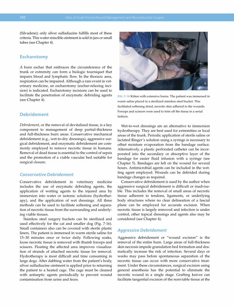
Stainless steel surgery buckets can be sterilized and used effectively for the cat and smaller dog (Fig. 7‐16). Small containers also can be covered with sterile plastic liners. The patient is immersed in warm sterile saline for 15–30 minutes once or twice daily. Following soaks, loose necrotic tissue is removed with thumb forceps and scissors. Floating the affected area improves visualiza-tion of strands of attached necrotic tissue for removal. Hydrotherapy is most difficult and time consuming in large dogs. After dabbing water from the patient’s body, silver sulfadiazine ointment is applied prior to returning the patient to a heated cage. The cage must be cleaned with antiseptic agents periodically to prevent wound contamination from urine and feces.
Wet‐to‐wet dressings are an alternative to immersion hydrotherapy. They are best used for extremities or local areas of the trunk. Periodic application of sterile saline or lactated Ringer’s solution using a syringe is necessary to offset moisture evaporation from the bandage surface. Alternatively, a plastic perforated catheter can be incor-porated into the secondary or absorptive layer of the bandage for easier fluid infusion with a syringe (see Chapter 5). Bandages are left on the wound for several hours. Antimicrobial agents can be included in the wet-ting agent employed. Wounds can be debrided during bandage changes as required.
Conservative debridement is used by the author when aggressive surgical debridement is difficult or inadvisa-ble. This includes the removal of small areas of necrotic tissue adherent to tendons, ligaments, or underlying body structures where no clear delineation of a fascial plane can be employed for accurate excision. When necrotic tissue is largely removed and infection is under control, other topical dressings and agents also may be considered (see Chapter 4).
Aggressive DebridementAggressive debridement or “wound excision” is the removal of the entire burn. Large areas of full‐thickness skin necrosis impede granulation bed formation and dra-matically increase the risk of infection. Several days or weeks may pass before spontaneous separation of the necrotic tissue can occur with more conservative treat-ment. Under these circumstances, surgical excision using general anesthesia has the potential to eliminate the necrotic wound in a single stage. Grafting knives can facilitate tangential excision of the nonviable tissue at the
FIG. 7-16 Kitten with extensive burns. The patient was immersed in
warm saline placed in a sterilized stainless steel bucket. This
facilitated softening dried, necrotic skin adhered to the wounds.
Forceps and scissors were used to trim off the tissue in a serial
fashion.
Management of Specific Wounds 193
level of the hypodermis (Fig. 7‐17). A healthy granulation bed, suitable for flap or graft closure, rapidly forms within 5–7 days. Electrocautery, vascular clips, and ligatures are essential for hemostasis.
Wound Closure Options
Once a viable vascular wound bed is free of necrotic tis-sue and infection, there are several options for closure. Superficial‐ and partial‐thickness burns frequently reepi-thelialize within 3 weeks with supportive care. Deep der-mal partial‐thickness burns take longer. Full‐thickness skin defects may heal by contraction and epithelializa-tion from the bordering skin. Extensive burns usually require closure with skin grafts or skin flaps, depending upon the extent and location of the wound(s). Skin stretchers can be used effectively for full‐thickness trunk burns (see Fig. 10‐8).
While allografts and xenografts have been used for temporary coverage of thermal injuries in humans,
autogenous free grafts are required for permanent cover-age of large full‐thickness thermal wounds. A partial‐thickness graft harvested with a dermatome and meshed with a 3:1 ratio is considered the best free‐grafting tech-nique for resurfacing large wounds. On occasion, axial pattern flaps can be used alone or in combination with free grafts and/or skin advancement techniques.
Excessive scarring and wound contracture are serious complications in the burned patient, often occurring when preventative medical/surgical intervention was not instituted for serious wounds. Areas subject to con-stant motion, such as flexion surfaces, are most suscepti-ble to wound contracture development if major cutaneous losses occur. Splints, braces, and other devices have been employed to minimize these complications in humans. Z‐plasty, pedicle grafts, and free grafts with scar division may be necessary to prevent and treat contractures. Prevention of contracture is obviously preferable to their treatment after formation.
Cosmetic results depend upon the extent of the burn. In contrast to the human, hair growth is an essential com-ponent to the cosmetic outcome of the veterinary patient. Superficial and minor partial‐thickness burns may heal and retain a majority of their hair follicles for regrowth of the hair coat. Deeper burns result in a greater loss of dermis and hair follicles. Complete surgical coverage with skin flaps and full‐thickness grafts can provide an adequate hair coat, whereas split‐thickness grafts provide fewer follicles and are less durable than full‐thickness skin coverage. On occasion, wound contraction,
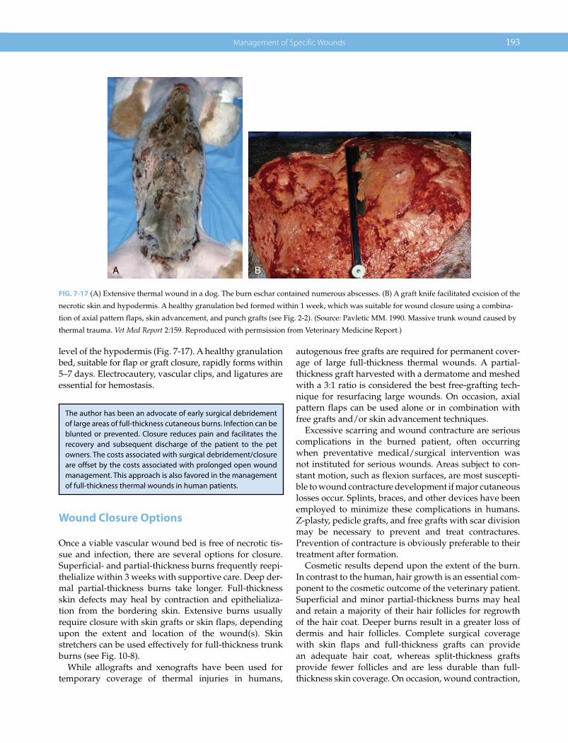
A B
FIG. 7-17 (A) Extensive thermal wound in a dog. The burn eschar contained numerous abscesses. (B) A graft knife facilitated excision of the
necrotic skin and hypodermis. A healthy granulation bed formed within 1 week, which was suitable for wound closure using a combina-
tion of axial pattern flaps, skin advancement, and punch grafts (see Fig. 2‐2). (Source: Pavletic MM. 1990. Massive trunk wound caused by
thermal trauma. Vet Med Report 2:159. Reproduced with permsission from Veterinary Medicine Report.)
The author has been an advocate of early surgical debridement of large areas of full‐thickness cutaneous burns. Infection can be blunted or prevented. Closure reduces pain and facilitates the recovery and subsequent discharge of the patient to the pet owners. The costs associated with surgical debridement/closure are offset by the costs associated with prolonged open wound management. This approach is also favored in the management of full‐thickness thermal wounds in human patients.
194 Atlas of Small Animal Wound Management and Reconstructive Surgery
combined with the overlapping of hair, can reduce the visibility of the burn scar with a satisfactory cosmetic outcome. It is important to discuss cosmetic results with owners who may have unrealistic expectations (Fig. 7‐18).
In summary, surgical management of burns is primar-ily limited to full‐thickness skin wounds, especially when the wound involves a significant area of the patient. Once defined, large burn eschars are best treated by surgical excision; a healthy granulation bed is expected within 5–7 days after careful and thorough debridement of the dead skin. Topical silver sulfadiazine, with nonadherent dressings, is commonly employed to protect the wound and reduce the bacterial population until split‐thickness mesh grafts, axial pattern flaps, or skin undermining and advancement can be employed for wound closure.
Skin Substitutes
There is no permanent, complete skin substitute at this time. Most burns are closed by second‐intention healing, skin advancement, skin flaps, and autogenous skin grafts. For extensive burns in humans, development of a per-manent replacement skin would be invaluable. Today, temporary skin substitutes or biologic dressings can provide early burn protection from bacteria, trauma, and a vapor barrier in preparation of the burn surface for closure. They also can reduce pain. Permanent skin substitutes would ideally replace the physiologic functions of the lost skin. While there is no permanent replacement for lost skin, there are partial substitutes. Some of these materials are designed as a scaffold which is incorporated into the
A
B
C
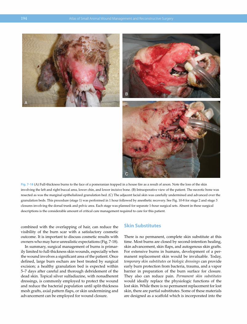
Fig. 7-18 (A) Full‐thickness burns to the face of a pomeranian trapped in a house fire as a result of arson. Note the loss of the skin
involving the left and right buccal area, lower chin, and lower incisive bone. (B) Intraoperative view of the patient. The necrotic bone was
resected as was the marginal epithelialized granulation bed. (C) The adjacent facial skin was carefully undermined and advanced over the
granulation beds. This procedure (stage 1) was performed in 1 hour followed by anesthetic recovery. See Fig. 10‐8 for stage 2 and stage 3
closures involving the dorsal trunk and pelvic area. Each stage was planned for separate 1‐hour surgical sets. Absent in these surgical
descriptions is the considerable amount of critical care management required to care for this patient.
Management of Specific Wounds 195
healing process. Applied to the vascular wound bed, they can serve as a template or scaffold to recreate a neodermis. Their woven dermal design provides a template for the ingrowth of capillaries, fibroblasts and other cellular components. Once vascularized, this artificial dermis can support the application of a free graft, ideally for better functional and cosmetic results (compared to grafting onto a granulation bed). Epicel (Genzyme) is an epithelial replacement which is designed to apply autologous small sheets of epithelial cells to large burn surfaces. Skin sub-stitutes are very expensive.
INHALATION INJURIES
Smoke contains several toxic gases including CO, SO4, NO2, aldehydes, H2SO4, HCl, benzene, halogens, and hydrogen cyanide. Many of these compounds combined with inhaled particulate matter (superheated soot) can reach the pulmonary tissue, causing serious damage. Bronchial spasm, laryngeal spasm, pulmonary edema, mucosal edema, loss of ciliary function, and compro-mised pulmonary surfactant will aggravate hypoxia associated with a smoke‐filled room. Hot, dry air has a low specific heat, and respiratory injuries are confined to the upper respiratory tract. Live steam, with a high specific heat, is capable of causing atelectasis, severe pulmonary edema, and alveolar damage.
Pulmonary parenchymal dysfunction from severe smoke inhalation is frequently fatal in humans. Hypoxemia refractory to oxygen therapy, with the presence of bronchospasm/pulmonary edema, requires aggressive treatment to save the patient. High mortality rates (60–70%) are associated with humans who develop pulmonary edema within 72 hours after injury. Upper airway edema is one of the immediate concerns with smoke inhalation injuries. Bronchospasm may be noted.
In humans, hoarseness and stridor may prompt endotra-cheal intubation and mechanical ventilation. Upper air-way edema normally resolves in 2–3 days. In 48–72 hours, humans may develop respiratory failure secondary to endobronchial sloughing, airway occlusion, and edema: infection can rapidly follow. If the human patient sur-vives, they may have normal long‐term clinical function impairment.
With major inhalation injuries, an adequate airway must be established. Intubation and ventilatory support with positive‐pressure ventilation are indicated in severe cases. Temporary tracheostomy should be considered for problematic patients; ventilatory support also can be used in conjunction with the tracheostomy tube. In mild cases of smoke inhalation and direct thermal injury, oxygen therapy alone may suffice. Nebulization of saline and coupage may facilitate clearance of respiratory secretions. In most cases, the tissue injury associated with mild direct thermal injury resolves within 5 days. Serial thoracic radiographs are useful in assessing changes in the respiratory tract. Bronchoscopy can be used to assess the severity of injury. It may be advisable to transfer the more serious patients to a veterinary emergency referral center that is better equipped to han-dle these challenging cases.
Carbon monoxide poisoning is suspected if the mucous membranes and blood are cherry red. Carbon monoxide blocks the oxygen‐carrying capacity of hemoglobin. Blood oxygen levels may be low despite normal levels of dis-solved oxygen in the bloodstream. Carbon monoxide poi-soning requires 100% oxygen therapy to rapidly reduce the level of carboxyhemoglobin. Standard care for humans exposed to carbon monoxide is 6 hours of normobaric oxygen. Mild cases of carbon monoxide poisoning in small animals may benefit from placement in an oxygen cage. In humans, the half‐life carboxyhemoglobin is 4 hours breathing room air, whereas this is dramatically
Examples of partial skin substitutes include:
• Alloderm (LifeCell, The Woodlands, TX):• Cell‐free dermal scaffold• Requires immediate application of a thin split‐thickness
autogenous skin graft• Epicel (Genzyme, Cambridge, MA):
• Cultured autologous epithelial sheets
Dogs and cats trapped in burning buildings often have a strong smoke odor. The hair coat may have soot particles. Pets close to flames may have singed hair. These clinical findings should alert the clinician to closely assess the patient for smoke‐ and heat‐related pulmonary injury.
Clinical SignsOvert clinical signs in dogs suffering from smoke inhalation include coughing, gagging, and respiratory distress. Occasionally, ataxia, foaming at the mouth, rubbing at the eyes, and nasal bleeding are noted.
Overt clinical signs in affected cats suffering from smoke inhalation include dyspnea, vocalization, open‐mouth breath-ing, lethargy, wheezing, gagging, nasal mucoid discharge, third eyelid protrusion, and foaming at the mouth.
Loss of consciousness may be indicative of hypoxia and pos-sible brain injury.
In two case reviews in the dog and cat (Drobaz), most patients with nonthreatening smoke inhalation clinically improved within 24 hours: severe cases worsened within this time frame.
196 Atlas of Small Animal Wound Management and Reconstructive Surgery
reduced to 45–60 minutes with oxygen administration by mask or nasal tube. If accessible, hyperbaric oxygen can reduce the half‐life of carboxyhemoglobin to 30 minutes or less. Carboxyhemoglobin can be measured to assess the severity of carbon monoxide poisoning and progress in treatment of the patient. Local human hospitals may be required for precise measurements in problematic patients. Hyperbaric oxygen therapy is normally not warranted in humans and is not without risk.
As previously stated, additional caution is necessary during the administration of intravenous fluids. Efficacy with short‐term (12–24 hours) use of corticosteroids is questionable: prolonged use can compound the immuno-suppression already seen with concomitant major thermal injuries. Diuretics (furosemide) may decrease intravascu-lar volume without significant benefits in the treatment of pulmonary edema. Bronchodilators (albuterol) and expec-torants may be beneficial. Prophylactic antibiotics are inef-fective and may select out resistant organisms. Their use should be based on airway culture. Many respiratory infections in animals are noted to be caused by Gram‐neg-ative bacteria. Because respiratory equipment is a poten-tial source of bacterial infection, care must be taken to ensure that sterile tubes and uncontaminated equipment are used.
Progressive respiratory failure is a common complica-tion in humans with serious inhalation injury. Fluid over-load, hypoproteinemia, airway occlusion, and pulmonary edema are contributing factors that may be complicated with the advent of pulmonary infection. Radiographic infiltrates or lobar consolidation suggest pneumonia. Prolonged respiratory support may be required under these circumstance.
CHEMICAL BURNS
Many caustic agents are available for home and indus-trial use. On occasion these chemicals cause “burns” to animals accidentally or from their malicious application to pets (Fig. 7‐19). The extent of the injury depends upon the chemical, its concentration, duration of contact and penetration, and its particular mechanism of action. Chemical agents damage the skin by oxidation or reduc-tion, dehydration, protein denaturation, corrosion, or vesication. Drain cleaners and oven cleaners may contain caustic soda, causing tissue corrosion. Phenolic disinfect-ants also may exert a corrosive effect on tissues. Sulfuric acid and hydrochloric acid may be found in cleaning compounds and exert a dehydrating effect on tissues. Hypochlorite, potassium permanganate, and chromic acid are oxidizing agents and cause protein coagulation. In contrast, picric acid, tannic acid, acetic acid, formic acid, and hydrofluoric acid cause protein denaturation.
Halogenated hydrocarbons, gasoline, ethylene oxide, and catharides are vesicants, liberating tissue amines (histamine and serotonin) and creating vesicles or blisters from tissue contact. Some solvents used for painting (tur-pentine, mineral spirits), furniture strippers, and concen-trated flea dip solutions are examples of household chemical agents also capable of causing superficial and partial‐thickness skin injuries with prolonged contact.
In general, caustic agents can be divided into strong acids and strong alkalis. Most of these agents destroy skin by coagulation necrosis. Vascular thrombosis may be noted with prolonged exposure to stronger agents. Strong alkalis in particular are capable of causing deep‐tissue destruction by colliquation, extraction of water, and precipitation of protein (Fig. 7‐20). An alkali–albumi-nate bond may form and redissolve in excess water. Alkalis form soluble soaps and protein complexes within the tissues, which facilitate the passage of hydroxyl ions into deeper layers. In contrast, hydrogen ions from acids are not complexed.
The medical history may reveal the nature of the injury, the chemical concentration, and duration of contact. However, most cases of caustic burns noted by the author are the result of malicious attacks in which the agent in question is unknown. Inhalation of vapors or consump-tion of the material should be determined by physical examination, thoracic radiographs, and oral/laryngeal/pharyngeal examination. Eyes should be examined very closely.
Management of Chemical Burns
From my experience, acid and alkali burns are often mali-cious in nature, using commonly accessible acid/alkali compounds used to open drains plugged with organic matter. Most chemical burns encountered should be lav-aged with large volumes of water. Eyes are best lavaged copiously with sterile saline when available. Veterinary staff should wear gloves and protective clothing when handling patients splashed with these caustic liquids. Under experimental conditions, the results of chemical burn treatment required returning skin pH to normal and varied between acid and alkali burns. While lavage of 30% acid burns for 2 hours returned the skin to normal pH, it took over 12 hours of lavage to reestablish normal pH following a burn with 50% sodium hydroxide. Deep chemical burns from alkali compounds, may require deep surgical excision if the contact has been prolonged. Alkali residue has a soapy feel and can be assessed by rubbing the gloved thumb and index fingers together.
Neutralizing agents are occasionally useful in limiting the extent of injury. However the primary concern is the exothermic reaction that may occur that could further
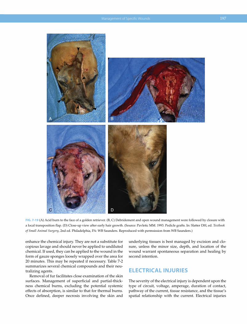
Management of Specific Wounds 197
enhance the chemical injury. They are not a substitute for copious lavage and should never be applied to undiluted chemical. If used, they can be applied to the wound in the form of gauze sponges loosely wrapped over the area for 20 minutes. This may be repeated if necessary. Table 7‐2 summarizes several chemical compounds and their neu-tralizing agents.
Removal of fur facilitates close examination of the skin surfaces. Management of superficial and partial‐thick-ness chemical burns, excluding the potential systemic effects of absorption, is similar to that for thermal burns. Once defined, deeper necrosis involving the skin and
underlying tissues is best managed by excision and clo-sure, unless the minor size, depth, and location of the wound warrant spontaneous separation and healing by second intention.
ELECTRICAL INJURIES
The severity of the electrical injury is dependent upon the type of circuit, voltage, amperage, duration of contact, pathway of the current, tissue resistance, and the tissue’s spatial relationship with the current. Electrical injuries
A B
C D
FIG. 7-19 (A) Acid burn to the face of a golden retriever. (B, C) Debridement and open wound management were followed by closure with
a local transposition flap. (D) Close‐up view after early hair growth. (Source: Pavletic MM. 1993. Pedicle grafts. In: Slatter DH, ed. Textbook
of Small Animal Surgery, 2nd ed. Philadelphia, PA: WB Saunders. Reproduced with permsission from WB Saunders.)

198 Atlas of Small Animal Wound Management and Reconstructive Surgery
are classified as low‐voltage (<120 V), intermediate‐volt-age (120–1000 V), or high‐voltage (>1000 V). Most dogs and cats present with low‐voltage (alternating household current in the United States) injuries. High‐voltage injuries are rarely seen but are often fatal: wild birds, cats, and animals capable of climbing poles occasionally contact high‐tension power lines, usually causing instant death. High‐voltage injuries are more common in humans as a result of industrial accidents or accidental contact.
Aqueous electrolyte solutions are relatively good elec-trical conductors with electrical current carried by mobile ions rather than free electrons. The frictional interaction of colliding molecules generates heat. An electrical current generates heat as it passes through body tissues. The rate of heating tissues depends upon the current intensity, the electrical and thermal properties of the tissues, and the spatial relationship of the tissue with the current. Tissues have different electrical conductivities and
dissipate heat at different rates. In tissues electrically parallel relative to the source of the current, heat gener-ation occurs at a rate directly proportional to tissue conductivity. In tissues that are electrically in series, heat generation is inversely proportional to tissue conductivity. In experimental models, muscle generates the highest temperatures; on cessation of the current, the muscle can heat other adjacent tissues, including bone. Convective transport of heat through blood is critical for rapid cooling. Tissue injury is severe where the current density is greatest—at the point of contact. Electrical contact with a small surface area results in a concentration of energy that causes coagulation necrosis. As a result, injuries to the tissue deep to the contact burn can be extensive. Electrical contact with a broad surface area of the body decreases the current density with dissipation of the energy. No burns may be noted in these cases, although fatal ventricular fibrillation may occur. More recent
A
B
C
FIG. 7-20 (A, B) Patient that fell into a caustic soda pit at a petrochemical plant. Extensive chemical burns were present. The patient was
bathed extensively with a water spray to remove any residual caustic soda. (C) Close‐up view of the patient demonstrating coagulation of
the dermis. The patient went into acute renal failure despite aggressive medical therapy and was euthanized.
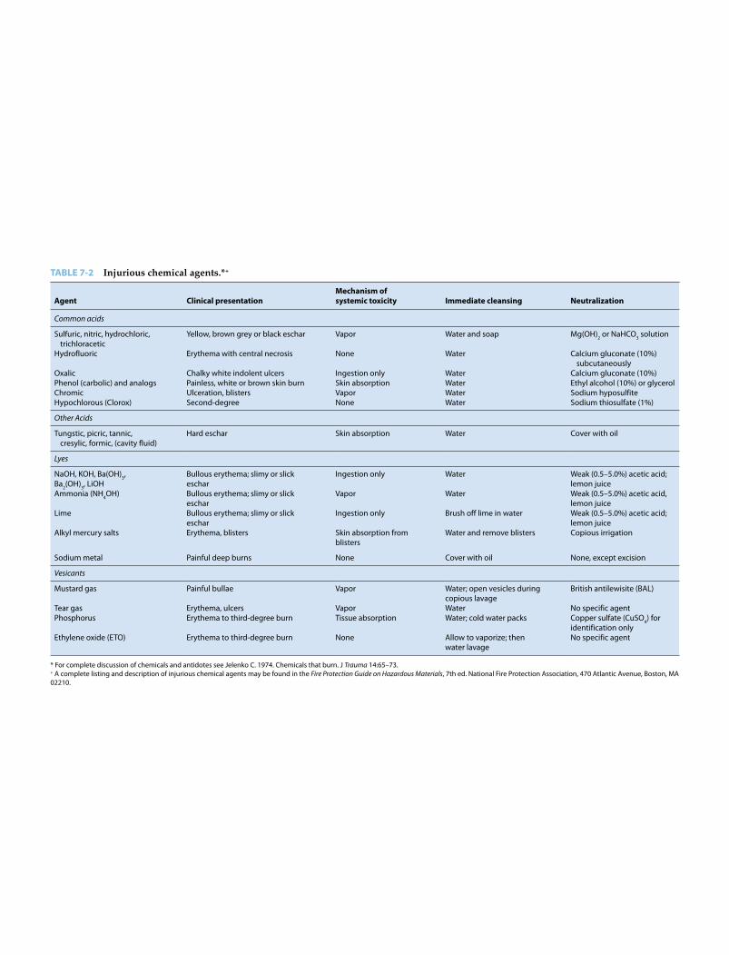
TABLE 7-2 Injurious chemical agents.*+
Agent Clinical presentationMechanism of systemic toxicity Immediate cleansing Neutralization
Common acids
Sulfuric, nitric, hydrochloric, trichloracetic
Yellow, brown grey or black eschar Vapor Water and soap Mg(OH)2 or NaHCO3 solution
Hydrofluoric Erythema with central necrosis None Water Calcium gluconate (10%) subcutaneously
Oxalic Chalky white indolent ulcers Ingestion only Water Calcium gluconate (10%)Phenol (carbolic) and analogs Painless, white or brown skin burn Skin absorption Water Ethyl alcohol (10%) or glycerolChromic Ulceration, blisters Vapor Water Sodium hyposulfiteHypochlorous (Clorox) Second‐degree None Water Sodium thiosulfate (1%)
Other Acids
Tungstic, picric, tannic, cresylic, formic, (cavity fluid)
Hard eschar Skin absorption Water Cover with oil
Lyes
NaOH, KOH, Ba(OH)3, Ba2(OH)3, LiOH
Bullous erythema; slimy or slick eschar
Ingestion only Water Weak (0.5–5.0%) acetic acid; lemon juice
Ammonia (NH4OH) Bullous erythema; slimy or slick eschar
Vapor Water Weak (0.5–5.0%) acetic acid, lemon juice
Lime Bullous erythema; slimy or slick eschar
Ingestion only Brush off lime in water Weak (0.5–5.0%) acetic acid; lemon juice
Alkyl mercury salts Erythema, blisters Skin absorption from blisters
Water and remove blisters Copious irrigation
Sodium metal Painful deep burns None Cover with oil None, except excision
Vesicants
Mustard gas Painful bullae Vapor Water; open vesicles during copious lavage
British antilewisite (BAL)
Tear gas Erythema, ulcers Vapor Water No specific agentPhosphorus Erythema to third‐degree burn Tissue absorption Water; cold water packs Copper sulfate (CuSO4) for
identification onlyEthylene oxide (ETO) Erythema to third‐degree burn None Allow to vaporize; then
water lavageNo specific agent
* For complete discussion of chemicals and antidotes see Jelenko C. 1974. Chemicals that burn. J Trauma 14:65–73.+ A complete listing and description of injurious chemical agents may be found in the Fire Protection Guide on Hazardous Materials, 7th ed. National Fire Protection Association, 470 Atlantic Avenue, Boston, MA 02210.
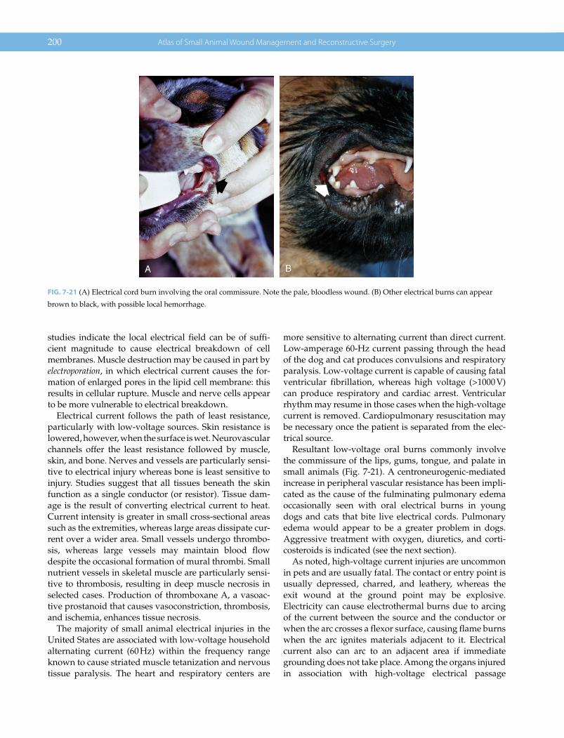
200 Atlas of Small Animal Wound Management and Reconstructive Surgery
studies indicate the local electrical field can be of suffi-cient magnitude to cause electrical breakdown of cell membranes. Muscle destruction may be caused in part by electroporation, in which electrical current causes the for-mation of enlarged pores in the lipid cell membrane: this results in cellular rupture. Muscle and nerve cells appear to be more vulnerable to electrical breakdown.
Electrical current follows the path of least resistance, particularly with low‐voltage sources. Skin resistance is lowered, however, when the surface is wet. Neurovascular channels offer the least resistance followed by muscle, skin, and bone. Nerves and vessels are particularly sensi-tive to electrical injury whereas bone is least sensitive to injury. Studies suggest that all tissues beneath the skin function as a single conductor (or resistor). Tissue dam-age is the result of converting electrical current to heat. Current intensity is greater in small cross‐sectional areas such as the extremities, whereas large areas dissipate cur-rent over a wider area. Small vessels undergo thrombo-sis, whereas large vessels may maintain blood flow despite the occasional formation of mural thrombi. Small nutrient vessels in skeletal muscle are particularly sensi-tive to thrombosis, resulting in deep muscle necrosis in selected cases. Production of thromboxane A, a vasoac-tive prostanoid that causes vasoconstriction, thrombosis, and ischemia, enhances tissue necrosis.
The majority of small animal electrical injuries in the United States are associated with low‐voltage household alternating current (60 Hz) within the frequency range known to cause striated muscle tetanization and nervous tissue paralysis. The heart and respiratory centers are
more sensitive to alternating current than direct current. Low‐amperage 60‐Hz current passing through the head of the dog and cat produces convulsions and respiratory paralysis. Low‐voltage current is capable of causing fatal ventricular fibrillation, whereas high voltage (>1000 V) can produce respiratory and cardiac arrest. Ventricular rhythm may resume in those cases when the high‐voltage current is removed. Cardiopulmonary resuscitation may be necessary once the patient is separated from the elec-trical source.
Resultant low‐voltage oral burns commonly involve the commissure of the lips, gums, tongue, and palate in small animals (Fig. 7‐21). A centroneurogenic‐mediated increase in peripheral vascular resistance has been impli-cated as the cause of the fulminating pulmonary edema occasionally seen with oral electrical burns in young dogs and cats that bite live electrical cords. Pulmonary edema would appear to be a greater problem in dogs. Aggressive treatment with oxygen, diuretics, and corti-costeroids is indicated (see the next section).
As noted, high‐voltage current injuries are uncommon in pets and are usually fatal. The contact or entry point is usually depressed, charred, and leathery, whereas the exit wound at the ground point may be explosive. Electricity can cause electrothermal burns due to arcing of the current between the source and the conductor or when the arc crosses a flexor surface, causing flame burns when the arc ignites materials adjacent to it. Electrical current also can arc to an adjacent area if immediate grounding does not take place. Among the organs injured in association with high‐voltage electrical passage
A B
FIG. 7-21 (A) Electrical cord burn involving the oral commissure. Note the pale, bloodless wound. (B) Other electrical burns can appear
brown to black, with possible local hemorrhage.
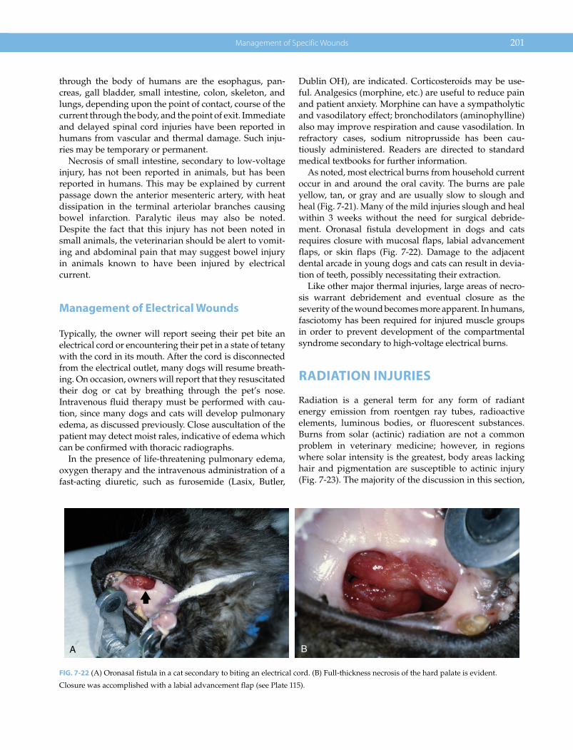
Management of Specific Wounds 201
through the body of humans are the esophagus, pan-creas, gall bladder, small intestine, colon, skeleton, and lungs, depending upon the point of contact, course of the current through the body, and the point of exit. Immediate and delayed spinal cord injuries have been reported in humans from vascular and thermal damage. Such inju-ries may be temporary or permanent.
Necrosis of small intestine, secondary to low‐voltage injury, has not been reported in animals, but has been reported in humans. This may be explained by current passage down the anterior mesenteric artery, with heat dissipation in the terminal arteriolar branches causing bowel infarction. Paralytic ileus may also be noted. Despite the fact that this injury has not been noted in small animals, the veterinarian should be alert to vomit-ing and abdominal pain that may suggest bowel injury in animals known to have been injured by electrical current.
Management of Electrical Wounds
Typically, the owner will report seeing their pet bite an electrical cord or encountering their pet in a state of tetany with the cord in its mouth. After the cord is disconnected from the electrical outlet, many dogs will resume breath-ing. On occasion, owners will report that they resuscitated their dog or cat by breathing through the pet’s nose. Intravenous fluid therapy must be performed with cau-tion, since many dogs and cats will develop pulmonary edema, as discussed previously. Close auscultation of the patient may detect moist rales, indicative of edema which can be confirmed with thoracic radiographs.
In the presence of life‐threatening pulmonary edema, oxygen therapy and the intravenous administration of a fast‐acting diuretic, such as furosemide (Lasix, Butler,
Dublin OH), are indicated. Corticosteroids may be use-ful. Analgesics (morphine, etc.) are useful to reduce pain and patient anxiety. Morphine can have a sympatholytic and vasodilatory effect; bronchodilators (aminophylline) also may improve respiration and cause vasodilation. In refractory cases, sodium nitroprusside has been cau-tiously administered. Readers are directed to standard medical textbooks for further information.
As noted, most electrical burns from household current occur in and around the oral cavity. The burns are pale yellow, tan, or gray and are usually slow to slough and heal (Fig. 7‐21). Many of the mild injuries slough and heal within 3 weeks without the need for surgical debride-ment. Oronasal fistula development in dogs and cats requires closure with mucosal flaps, labial advancement flaps, or skin flaps (Fig. 7‐22). Damage to the adjacent dental arcade in young dogs and cats can result in devia-tion of teeth, possibly necessitating their extraction.
Like other major thermal injuries, large areas of necro-sis warrant debridement and eventual closure as the severity of the wound becomes more apparent. In humans, fasciotomy has been required for injured muscle groups in order to prevent development of the compartmental syndrome secondary to high‐voltage electrical burns.
RADIATION INJURIES
Radiation is a general term for any form of radiant energy emission from roentgen ray tubes, radioactive elements, luminous bodies, or fluorescent substances. Burns from solar (actinic) radiation are not a common problem in veterinary medicine; however, in regions where solar intensity is the greatest, body areas lacking hair and pigmentation are susceptible to actinic injury (Fig. 7‐23). The majority of the discussion in this section,
A B
FIG. 7-22 (A) Oronasal fistula in a cat secondary to biting an electrical cord. (B) Full‐thickness necrosis of the hard palate is evident.
Closure was accomplished with a labial advancement flap (see Plate 115).
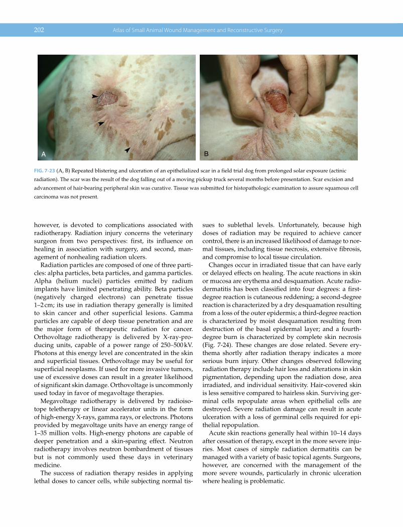
202 Atlas of Small Animal Wound Management and Reconstructive Surgery
however, is devoted to complications associated with radiotherapy. Radiation injury concerns the veterinary surgeon from two perspectives: first, its influence on healing in association with surgery, and second, man-agement of nonhealing radiation ulcers.
Radiation particles are composed of one of three parti-cles: alpha particles, beta particles, and gamma particles. Alpha (helium nuclei) particles emitted by radium implants have limited penetrating ability. Beta particles (negatively charged electrons) can penetrate tissue 1–2 cm; its use in radiation therapy generally is limited to skin cancer and other superficial lesions. Gamma particles are capable of deep tissue penetration and are the major form of therapeutic radiation for cancer. Orthovoltage radiotherapy is delivered by X‐ray‐pro-ducing units, capable of a power range of 250–500 kV. Photons at this energy level are concentrated in the skin and superficial tissues. Orthovoltage may be useful for superficial neoplasms. If used for more invasive tumors, use of excessive doses can result in a greater likelihood of significant skin damage. Orthovoltage is uncommonly used today in favor of megavoltage therapies.
Megavoltage radiotherapy is delivered by radioiso-tope teletherapy or linear accelerator units in the form of high‐energy X‐rays, gamma rays, or electrons. Photons provided by megavoltage units have an energy range of 1–35 million volts. High‐energy photons are capable of deeper penetration and a skin‐sparing effect. Neutron radiotherapy involves neutron bombardment of tissues but is not commonly used these days in veterinary medicine.
The success of radiation therapy resides in applying lethal doses to cancer cells, while subjecting normal tis-
sues to sublethal levels. Unfortunately, because high doses of radiation may be required to achieve cancer control, there is an increased likelihood of damage to nor-mal tissues, including tissue necrosis, extensive fibrosis, and compromise to local tissue circulation.
Changes occur in irradiated tissue that can have early or delayed effects on healing. The acute reactions in skin or mucosa are erythema and desquamation. Acute radio-dermatitis has been classified into four degrees: a first‐degree reaction is cutaneous reddening; a second‐degree reaction is characterized by a dry desquamation resulting from a loss of the outer epidermis; a third‐degree reaction is characterized by moist desquamation resulting from destruction of the basal epidermal layer; and a fourth‐degree burn is characterized by complete skin necrosis (Fig. 7‐24). These changes are dose related. Severe ery-thema shortly after radiation therapy indicates a more serious burn injury. Other changes observed following radiation therapy include hair loss and alterations in skin pigmentation, depending upon the radiation dose, area irradiated, and individual sensitivity. Hair‐covered skin is less sensitive compared to hairless skin. Surviving ger-minal cells repopulate areas when epithelial cells are destroyed. Severe radiation damage can result in acute ulceration with a loss of germinal cells required for epi-thelial repopulation.
Acute skin reactions generally heal within 10–14 days after cessation of therapy, except in the more severe inju-ries. Most cases of simple radiation dermatitis can be managed with a variety of basic topical agents. Surgeons, however, are concerned with the management of the more severe wounds, particularly in chronic ulceration where healing is problematic.
A B
FIG. 7-23 (A, B) Repeated blistering and ulceration of an epithelialized scar in a field trial dog from prolonged solar exposure (actinic
radiation). The scar was the result of the dog falling out of a moving pickup truck several months before presentation. Scar excision and
advancement of hair‐bearing peripheral skin was curative. Tissue was submitted for histopathologic examination to assure squamous cell
carcinoma was not present.

Management of Specific Wounds 203
Late or chronic changes noted in previously irradiated skin include thinning and flattening of the epidermis, a decline in adnexal structures, and obliteration of rete pegs. Grossly, the skin may demonstrate dryness, thin-ning, induration and loss of pliability from fibrosis, and increased pigmentation with a reduction of hair growth (Fig. 7‐25).
The most significant late change is a progressive decline in circulation over time. Chronic obliterative endarteritis is noted, characterized by endothelial proliferation, subinti-mal fibrosis, and gradual reduction in lumen diameter. Perivascular infiltrate also is present. The ischemia that ensues will impede healing to a variable degree. If incising into a previously irradiated field is unavoidable, the sur-geon should avoid or minimize surgical maneuvers that may further compromise tissue. Precautions are essential to prevent contamination during surgery, since ischemic tis-sues are more susceptible to infection. Open wounds have a particularly difficult time healing by second intention, since neovascularization, wound contraction (myofibroblastic activity), fibroplasia, and epithelialization are impaired. A loss of fibroblasts and their stem cells also impairs forma-tion of blood vessels. The low capillary content and colla-gen‐laden wound bed present form an unstable base for migrating epithelial cells. Any epithelialized surface formed is more susceptible to trauma. Split‐thickness grafts and direct distant flaps cannot be sustained on wound beds characterized by these chronic changes (Fig. 7‐26).
In general, an uncomplicated closed surgical wound can safely receive radiotherapy as early as 1 week post-
operatively. Similarly, elective surgery can be performed 4–8 weeks after radiation therapy is discontinued because inflammation secondary to irradiation is subsiding and circulation is still satisfactory. As time passes, however, tissue loses vascularity to a degree proportional to the
FIG. 7-25 Chronic ulcer several months after completion of
radiotherapy (arrow). Note the sparse hair growth, nonpigmented
hair at the periphery of the radiation field, and the dry, flaky
epithelial surface.
A B
FIG. 7-24 (A) Erythema and moist desquamation after completion of orthovoltage therapy for the treatment of a nasal carcinoma. (B) The
patient 10 days later. Moist crust patches have disappeared, the result of reepithelialization from germinal epithelial cells of the epidermis
and cutaneous adnexa.
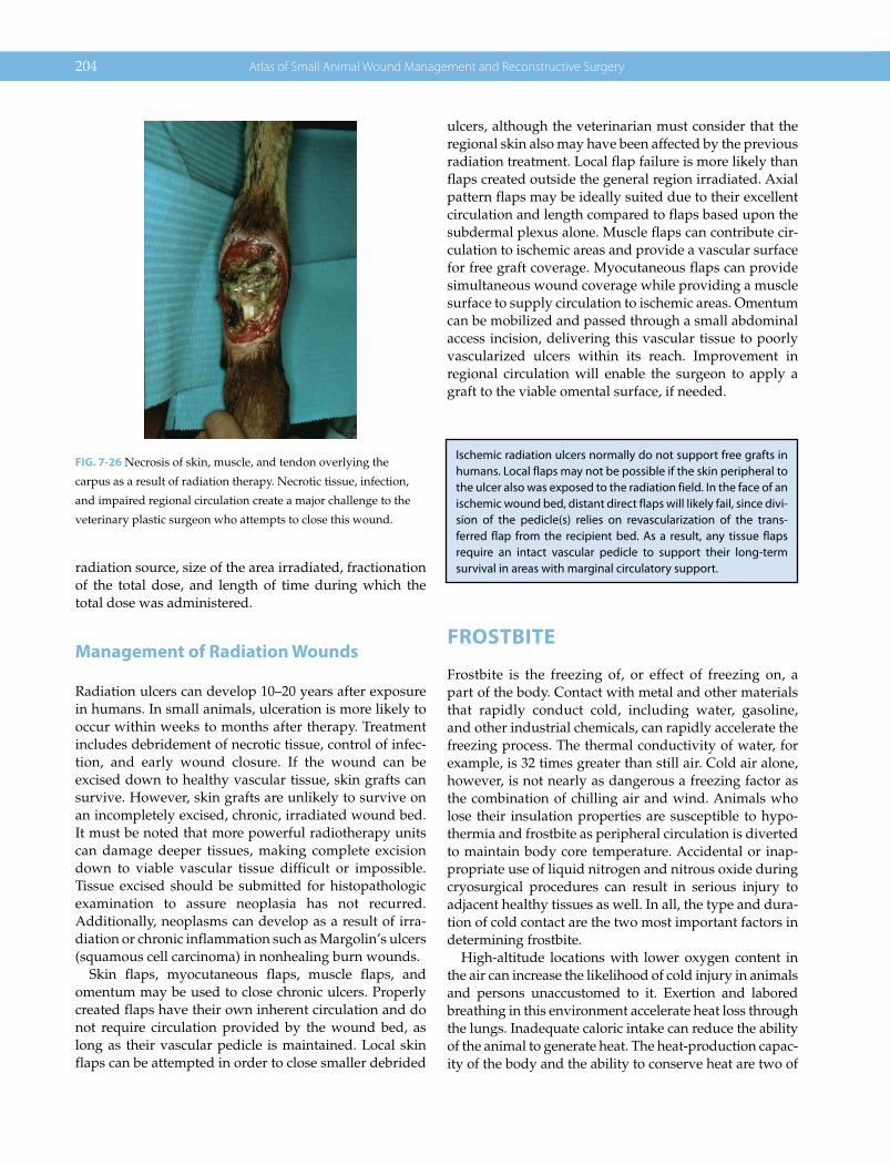
204 Atlas of Small Animal Wound Management and Reconstructive Surgery
radiation source, size of the area irradiated, fractionation of the total dose, and length of time during which the total dose was administered.
Management of Radiation Wounds
Radiation ulcers can develop 10–20 years after exposure in humans. In small animals, ulceration is more likely to occur within weeks to months after therapy. Treatment includes debridement of necrotic tissue, control of infec-tion, and early wound closure. If the wound can be excised down to healthy vascular tissue, skin grafts can survive. However, skin grafts are unlikely to survive on an incompletely excised, chronic, irradiated wound bed. It must be noted that more powerful radiotherapy units can damage deeper tissues, making complete excision down to viable vascular tissue difficult or impossible. Tissue excised should be submitted for histopathologic examination to assure neoplasia has not recurred. Additionally, neoplasms can develop as a result of irra-diation or chronic inflammation such as Margolin’s ulcers (squamous cell carcinoma) in nonhealing burn wounds.
Skin flaps, myocutaneous flaps, muscle flaps, and omentum may be used to close chronic ulcers. Properly created flaps have their own inherent circulation and do not require circulation provided by the wound bed, as long as their vascular pedicle is maintained. Local skin flaps can be attempted in order to close smaller debrided
ulcers, although the veterinarian must consider that the regional skin also may have been affected by the previous radiation treatment. Local flap failure is more likely than flaps created outside the general region irradiated. Axial pattern flaps may be ideally suited due to their excellent circulation and length compared to flaps based upon the subdermal plexus alone. Muscle flaps can contribute cir-culation to ischemic areas and provide a vascular surface for free graft coverage. Myocutaneous flaps can provide simultaneous wound coverage while providing a muscle surface to supply circulation to ischemic areas. Omentum can be mobilized and passed through a small abdominal access incision, delivering this vascular tissue to poorly vascularized ulcers within its reach. Improvement in regional circulation will enable the surgeon to apply a graft to the viable omental surface, if needed.
FROSTBITE
Frostbite is the freezing of, or effect of freezing on, a part of the body. Contact with metal and other materials that rapidly conduct cold, including water, gasoline, and other industrial chemicals, can rapidly accelerate the freezing process. The thermal conductivity of water, for example, is 32 times greater than still air. Cold air alone, however, is not nearly as dangerous a freezing factor as the combination of chilling air and wind. Animals who lose their insulation properties are susceptible to hypo-thermia and frostbite as peripheral circulation is diverted to maintain body core temperature. Accidental or inap-propriate use of liquid nitrogen and nitrous oxide during cryosurgical procedures can result in serious injury to adjacent healthy tissues as well. In all, the type and dura-tion of cold contact are the two most important factors in determining frostbite.
High‐altitude locations with lower oxygen content in the air can increase the likelihood of cold injury in animals and persons unaccustomed to it. Exertion and labored breathing in this environment accelerate heat loss through the lungs. Inadequate caloric intake can reduce the ability of the animal to generate heat. The heat‐production capac-ity of the body and the ability to conserve heat are two of
FIG. 7-26 Necrosis of skin, muscle, and tendon overlying the
carpus as a result of radiation therapy. Necrotic tissue, infection,
and impaired regional circulation create a major challenge to the
veterinary plastic surgeon who attempts to close this wound.
Ischemic radiation ulcers normally do not support free grafts in humans. Local flaps may not be possible if the skin peripheral to the ulcer also was exposed to the radiation field. In the face of an ischemic wound bed, distant direct flaps will likely fail, since divi-sion of the pedicle(s) relies on revascularization of the trans-ferred flap from the recipient bed. As a result, any tissue flaps require an intact vascular pedicle to support their long‐term survival in areas with marginal circulatory support.

Management of Specific Wounds 205
the most important factors in the prevention of cold injuries. Frostbite can occur in above‐freezing tempera-tures from prolonged immersion in animals who lack the insulation and protective physiologic mechanisms other species possess for adverse conditions.
Several mechanisms have been proposed to explain tis-sue injury and death after exposure to cold. Extracellular ice crystal formation may damage cells. Rapid freezing can result in fatal intracellular ice crystal formation. Endothelial cell injury may precipitate thrombosis. Arteriolar vasospasm combined with shunting of blood from arterioles to venules directs oxygen, nutrients, and warmth from the frozen tissue in an effort to preserve central core temperature. Ischemia‐reperfusion injury likely plays a significant role in the loss of tissue. As blood viscosity increases, sludging and thrombosis also may occur. Unless the source of cold is removed, tissues continue to freeze, and the depth of the damage increases with prolongation of contact.
Frostbite Assessment
Frostbite in humans generally affects the distal extremi-ties and extends proximally as exposure increases. Frostbite in small animals is not particularly common in the United States. In veterinary practice, it is usually diagnosed in retrospect, when tissue slough/loss is evi-dent. Animals are most susceptible to frostbite during bitterly cold days with high winds. Animals lacking shel-ter and protection from the elements combined with a prolonged exposure period may suffer frostbite to the toes, tips of the ears, scrotum, tip of the tail, preputial orifice, flank skin, or other cutaneous surfaces lacking an adequate protective hair coat. Unless examined by an astute owner or clinician immediately, the injury will remain undiagnosed and untreated until tissue separa-tion becomes evident days later.
Upon initial manual examination, superficial frost-bite is classically characterized by soft and resilient tissues underlying the outer layer of rigid tissue. Deep frostbite is characterized by firm or stiff tissue beneath the outer rigid tissue layer. Unless the injury is obvi-ously minor, the severity of the frostbite injury cannot be determined initially. Frostbite classification is made retrospectively: first‐degree results in cutaneous ery-thema after warming; second‐degree, in cutaneous blistering patchy skin; third‐degree, in skin necrosis; and fourth‐degree, in soft tissue loss or gangrene of the extremity. Mild and severe cases of frostbite may be indistinguishable for several days.
In humans, the Hennepin Score is used to quantify injury and tissue loss associated with frostbite. It enables the clinician to determine the efficacy of treatment
through the calculation of the salvage rate. As a result, more accurate assessment of treatment modalities can be determined.
Management of Frostbite
If a patient presents with early frostbite, the affected area(s) should be rewarmed (Fig. 7‐27). Immersion of the affected area in a receptacle of sufficient volume to main-tain a uniform temperature of 104–108 °F (40–42 °C) for approximately 20 minutes or until rewarming is judged complete is ideal. Tissues should never be rubbed or mas-saged. Soft, dry protective bandages are indicated in small animals. Restriction of activity is required when the feet are involved.
Rapid rewarming causes vasodilation, capillary extrava-sation, and vascular stasis. In more recent years, the use of thrombolytic drugs, including plasminogen activator, has shown considerable efficacy in reducing the need for amputation of extremities affected by severe frostbite.
Maintaining hydration and a balanced diet are essen-tial. Analgesics may be indicated. Topical antiseptics and judicious debridement of necrotic tissue or ruptured blisters is advisable as necrosis defines itself. Systemic antibiotics should be considered. As noted, because of the low incidence of frostbite in small animals and the initial lack of its recognition by most owners, the veteri-narian is usually contacted only after signs of necrosis become evident. Surgical debridement or amputation of the affected area may be necessary if natural separation has not occurred.
PROJECTILE INJURIES
Gunshot wounds to pets and wildlife are not uncommon in rural and inner city areas of the United States. Most veterinarians have limited knowledge of the subject of ballistics and the treatment of projectile injuries. This sec-tion reviews the basic concepts of this surprisingly com-plex subject, known as projectile injuries.
Ballistics is the science of motion of a projectile during its travel through the barrel of a firearm (interior ballis-tics), its subsequent path through the air (exterior ballis-tics), and its final path into the target (terminal ballistics). The seriousness of bullet wounds often is considered to be limited to those tissues in the direct path of the projec-tile. However, under some circumstances, the wounding potential of projectiles can occur beyond the pathway of a projectile.
Missiles are any projectiles, including bullets, pellets, fragments from grenades, and explosive shells. A car-tridge (round) is composed of four basic components:
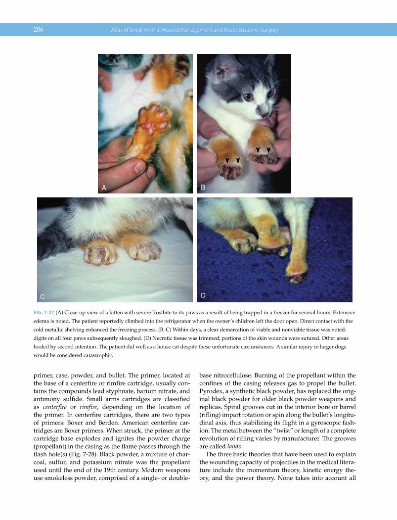
206 Atlas of Small Animal Wound Management and Reconstructive Surgery
primer, case, powder, and bullet. The primer, located at the base of a centerfire or rimfire cartridge, usually con-tains the compounds lead styphnate, barium nitrate, and antimony sulfide. Small arms cartridges are classified as centerfire or rimfire, depending on the location of the primer. In centerfire cartridges, there are two types of primers: Boxer and Berden. American centerfire car-tridges are Boxer primers. When struck, the primer at the cartridge base explodes and ignites the powder charge (propellant) in the casing as the flame passes through the flash hole(s) (Fig. 7‐28). Black powder, a mixture of char-coal, sulfur, and potassium nitrate was the propellant used until the end of the 19th century. Modern weapons use smokeless powder, comprised of a single‐ or double‐
base nitrocellulose. Burning of the propellant within the confines of the casing releases gas to propel the bullet. Pyrodex, a synthetic black powder, has replaced the orig-inal black powder for older black powder weapons and replicas. Spiral grooves cut in the interior bore or barrel (rifling) impart rotation or spin along the bullet’s longitu-dinal axis, thus stabilizing its flight in a gyroscopic fash-ion. The metal between the “twist” or length of a complete revolution of rifling varies by manufacturer. The grooves are called lands.
The three basic theories that have been used to explain the wounding capacity of projectiles in the medical litera-ture include the momentum theory, kinetic energy the-ory, and the power theory. None takes into account all
A
C
B
D
FIG. 7-27 (A) Close‐up view of a kitten with severe frostbite to its paws as a result of being trapped in a freezer for several hours. Extensive
edema is noted. The patient reportedly climbed into the refrigerator when the owner’s children left the door open. Direct contact with the
cold metallic shelving enhanced the freezing process. (B, C) Within days, a clear demarcation of viable and nonviable tissue was noted:
digits on all four paws subsequently sloughed. (D) Necrotic tissue was trimmed; portions of the skin wounds were sutured. Other areas
healed by second intention. The patient did well as a house cat despite these unfortunate circumstances. A similar injury in larger dogs
would be considered catastrophic.
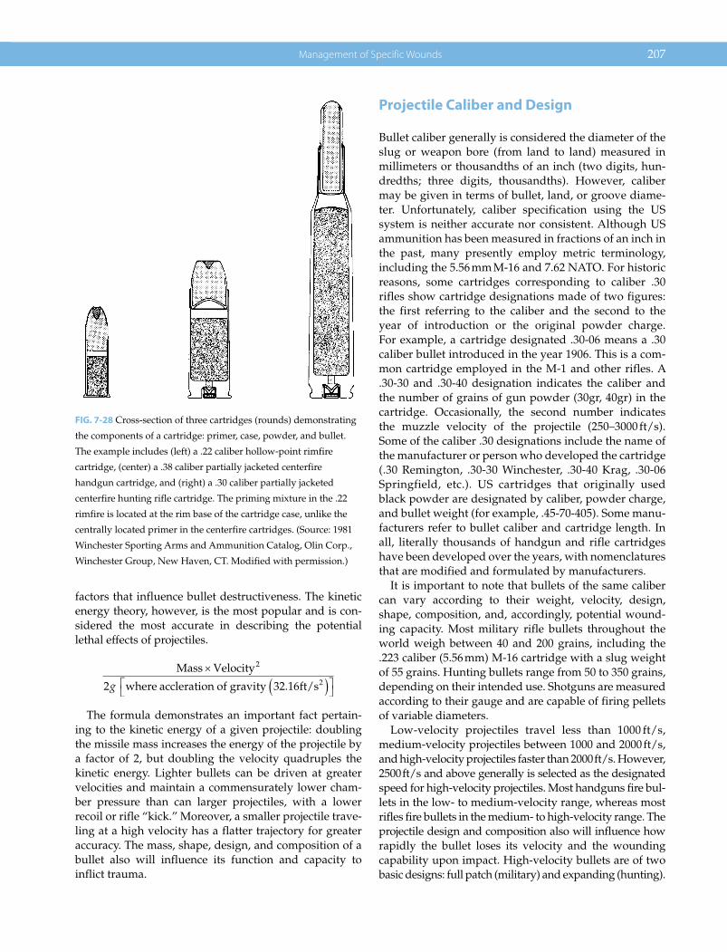
Management of Specific Wounds 207
factors that influence bullet destructiveness. The kinetic energy theory, however, is the most popular and is con-sidered the most accurate in describing the potential lethal effects of projectiles.
Mass Velocity
where accleration of gravity ft/s
2
22 32 16g .
The formula demonstrates an important fact pertain-ing to the kinetic energy of a given projectile: doubling the missile mass increases the energy of the projectile by a factor of 2, but doubling the velocity quadruples the kinetic energy. Lighter bullets can be driven at greater velocities and maintain a commensurately lower cham-ber pressure than can larger projectiles, with a lower recoil or rifle “kick.” Moreover, a smaller projectile trave-ling at a high velocity has a flatter trajectory for greater accuracy. The mass, shape, design, and composition of a bullet also will influence its function and capacity to inflict trauma.
Projectile Caliber and Design
Bullet caliber generally is considered the diameter of the slug or weapon bore (from land to land) measured in millimeters or thousandths of an inch (two digits, hun-dredths; three digits, thousandths). However, caliber may be given in terms of bullet, land, or groove diame-ter. Unfortunately, caliber specification using the US system is neither accurate nor consistent. Although US ammunition has been measured in fractions of an inch in the past, many presently employ metric terminology, including the 5.56 mm M‐16 and 7.62 NATO. For historic reasons, some cartridges corresponding to caliber .30 rifles show cartridge designations made of two figures: the first referring to the caliber and the second to the year of introduction or the original powder charge. For example, a cartridge designated .30‐06 means a .30 caliber bullet introduced in the year 1906. This is a com-mon cartridge employed in the M‐1 and other rifles. A .30‐30 and .30‐40 designation indicates the caliber and the number of grains of gun powder (30gr, 40gr) in the cartridge. Occasionally, the second number indicates the muzzle velocity of the projectile (250–3000 ft/s). Some of the caliber .30 designations include the name of the manufacturer or person who developed the cartridge (.30 Remington, .30‐30 Winchester, .30‐40 Krag, .30‐06 Springfield, etc.). US cartridges that originally used black powder are designated by caliber, powder charge, and bullet weight (for example, .45‐70‐405). Some manu-facturers refer to bullet caliber and cartridge length. In all, literally thousands of handgun and rifle cartridges have been developed over the years, with nomenclatures that are modified and formulated by manufacturers.
It is important to note that bullets of the same caliber can vary according to their weight, velocity, design, shape, composition, and, accordingly, potential wound-ing capacity. Most military rifle bullets throughout the world weigh between 40 and 200 grains, including the .223 caliber (5.56 mm) M‐16 cartridge with a slug weight of 55 grains. Hunting bullets range from 50 to 350 grains, depending on their intended use. Shotguns are measured according to their gauge and are capable of firing pellets of variable diameters.
Low‐velocity projectiles travel less than 1000 ft/s, medium‐velocity projectiles between 1000 and 2000 ft/s, and high‐velocity projectiles faster than 2000 ft/s. However, 2500 ft/s and above generally is selected as the designated speed for high‐velocity projectiles. Most handguns fire bul-lets in the low‐ to medium‐velocity range, whereas most rifles fire bullets in the medium‐ to high‐velocity range. The projectile design and composition also will influence how rapidly the bullet loses its velocity and the wounding capability upon impact. High‐velocity bullets are of two basic designs: full patch (military) and expanding (hunting).
FIG. 7-28 Cross‐section of three cartridges (rounds) demonstrating
the components of a cartridge: primer, case, powder, and bullet.
The example includes (left) a .22 caliber hollow‐point rimfire
cartridge, (center) a .38 caliber partially jacketed centerfire
handgun cartridge, and (right) a .30 caliber partially jacketed
centerfire hunting rifle cartridge. The priming mixture in the .22
rimfire is located at the rim base of the cartridge case, unlike the
centrally located primer in the centerfire cartridges. (Source: 1981
Winchester Sporting Arms and Ammunition Catalog, Olin Corp.,
Winchester Group, New Haven, CT. Modified with permission.)
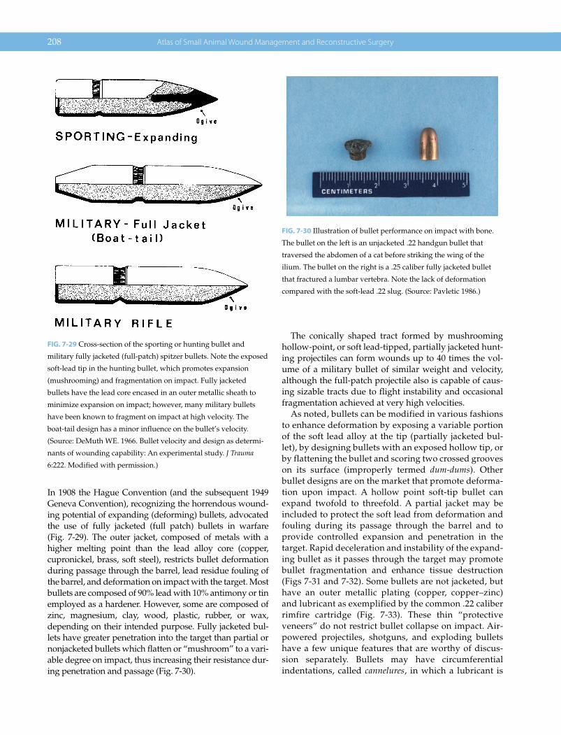
208 Atlas of Small Animal Wound Management and Reconstructive Surgery
In 1908 the Hague Convention (and the subsequent 1949 Geneva Convention), recognizing the horrendous wound-ing potential of expanding (deforming) bullets, advocated the use of fully jacketed (full patch) bullets in warfare (Fig. 7‐29). The outer jacket, composed of metals with a higher melting point than the lead alloy core (copper, cupronickel, brass, soft steel), restricts bullet deformation during passage through the barrel, lead residue fouling of the barrel, and deformation on impact with the target. Most bullets are composed of 90% lead with 10% antimony or tin employed as a hardener. However, some are composed of zinc, magnesium, clay, wood, plastic, rubber, or wax, depending on their intended purpose. Fully jacketed bul-lets have greater penetration into the target than partial or nonjacketed bullets which flatten or “mushroom” to a vari-able degree on impact, thus increasing their resistance dur-ing penetration and passage (Fig. 7‐30).
The conically shaped tract formed by mushrooming hollow‐point, or soft lead‐tipped, partially jacketed hunt-ing projectiles can form wounds up to 40 times the vol-ume of a military bullet of similar weight and velocity, although the full‐patch projectile also is capable of caus-ing sizable tracts due to flight instability and occasional fragmentation achieved at very high velocities.
As noted, bullets can be modified in various fashions to enhance deformation by exposing a variable portion of the soft lead alloy at the tip (partially jacketed bul-let), by designing bullets with an exposed hollow tip, or by flattening the bullet and scoring two crossed grooves on its surface (improperly termed dum‐dums). Other bullet designs are on the market that promote deforma-tion upon impact. A hollow point soft‐tip bullet can expand twofold to threefold. A partial jacket may be included to protect the soft lead from deformation and fouling during its passage through the barrel and to provide controlled expansion and penetration in the target. Rapid deceleration and instability of the expand-ing bullet as it passes through the target may promote bullet fragmentation and enhance tissue destruction (Figs 7‐31 and 7‐32). Some bullets are not jacketed, but have an outer metallic plating (copper, copper–zinc) and lubricant as exemplified by the common .22 caliber rimfire cartridge (Fig. 7‐33). These thin “protective veneers” do not restrict bullet collapse on impact. Air‐powered projectiles, shotguns, and exploding bullets have a few unique features that are worthy of discus-sion separately. Bullets may have circumferential indentations, called cannelures, in which a lubricant is
FIG. 7-29 Cross‐section of the sporting or hunting bullet and
military fully jacketed (full‐patch) spitzer bullets. Note the exposed
soft‐lead tip in the hunting bullet, which promotes expansion
(mushrooming) and fragmentation on impact. Fully jacketed
bullets have the lead core encased in an outer metallic sheath to
minimize expansion on impact; however, many military bullets
have been known to fragment on impact at high velocity. The
boat‐tail design has a minor influence on the bullet’s velocity.
(Source: DeMuth WE. 1966. Bullet velocity and design as determi-
nants of wounding capability: An experimental study. J Trauma
6:222. Modified with permission.)
FIG. 7-30 Illustration of bullet performance on impact with bone.
The bullet on the left is an unjacketed .22 handgun bullet that
traversed the abdomen of a cat before striking the wing of the
ilium. The bullet on the right is a .25 caliber fully jacketed bullet
that fractured a lumbar vertebra. Note the lack of deformation
compared with the soft‐lead .22 slug. (Source: Pavletic 1986.)

Management of Specific Wounds 209
inserted to improve passage through the barrel. A can-nelure may be present to allow the casing to crimp onto the bullet and seal the propellant.
Air‐Powered Projectiles
Air‐powered rifles, developed in 16th‐century Germany, use compressed air to drive the projectile in place of gases released by exploding powder. Today, air rifles, air guns, and air pistols are used for target shooting and
small‐game hunting. The air gun is distinguished from the air rifle in that the gun is a smooth‐bored barrel, whereas the rifle has a rifled barrel. Air pistols may have a smooth or rifled barrel. Standard calibers for air
FIG. 7-31 Various rifle rounds (from left to right): 5.56 mm full‐
patch M‐16; .243 Winchester hunting; 6.0 mm hunting; .30 military,
armor piercing; .30‐06 Springfield hunting; .444 Marlin hunting;
.45‐70 Government hunting. The partial copper jacket of the sport
or hunting bullet provides controlled expansion of the exposed
lead core on impact. (Source: Pavletic 1986.)
A B
FIG. 7-32 (A) Side and (B) top views of various handgun rounds (from left to right): .357 Magnum, partial jacket, hollow point;
.357 Magnum, partial jacket; .38 Special unjacketed; .38 Special partial jacket; .38 caliber wad cutter (flat face); .41 Magnum partial jacket;
.44 Remington Magnum; .45 caliber full‐patch military; .45 copper jacketed hollow point; .45 caliber unjacketed; .45 caliber unjacketed.
Each bullet varies in weight, velocity, and performance on impact. Bullets with an exposed lead core, especially with a hollow point,
would be expected to deform on impact compared with a full‐patch bullet. The full‐patch bullet would be expected to have greater
penetration, other factors being equal. (Source: Pavletic 1986.)
FIG. 7-33 These .22 cartridges demonstrate the various sizes and
designs of these rounds (left to right): 5.56 mm (.223), 55 grain,
full‐patch centerfire M‐16; .22 Magnum jacketed rimfire; .22
Winchester long rifle high power, 29 grain hollow point; .22
Remington long rifle, high velocity, 36 grain hollow point; .22 long
rifle with uncoated lead bullet. The .22 Winchester and .22
Remington have a metallic coating that has little influence on
minimizing bullet deformation. The decorative coating is reported
to shun dirt, grit, and lint. A waxy coating or priming on the
rimfire long rifle illustrated lubricates the bullet and minimizes
fouling of the gun barrel. Note that the .22 designation does not
indicate the projectile’s weight, composition, design, or velocity,
only its diameter. (Source: Pavletic 1986.)
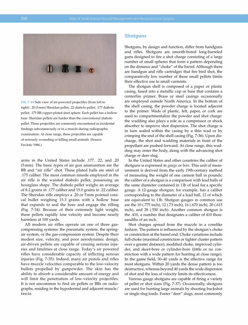
210 Atlas of Small Animal Wound Management and Reconstructive Surgery
arms in the United States include .177, .22, and .20 (5 mm). The basic types of air gun ammunition are the BB and “air rifle” shot. These plated balls are steel of .175 caliber. The most common missile employed in the air rifle is the waisted diabolo pellet, possessing an hourglass shape. The diabolo pellet weighs an average of 8.2 grains in .177 caliber and 15.0 grains in .22 caliber. The Sheridan rifle employs a .20 or 5 mm pointed coni-cal bullet weighing 15.3 grains with a hollow base that expands to seal the bore and engage the rifling (Fig. 7‐34). Because of their extremely light weight, these pellets rapidly lose velocity and become nearly harmless at 100 yards.
All modern air rifles operate on one of three gas‐compressing systems: the pneumatic system, the spring‐air system, or the gas‐compression system. Despite their modest size, velocity, and poor aerodynamic design, air‐driven pellets are capable of causing serious inju-ries and fatalities at close range. Today’s air powered rifles have considerable capacity of inflicting serious injuries (Fig. 7‐35). Indeed, many air pistols and rifles have muzzle velocities comparable to the low‐velocity bullets propelled by gunpowder. The skin has the ability to absorb a considerable amount of energy and will limit the penetration of low‐velocity projectiles. It is not uncommon to find air pellets or BBs on radio-graphs, residing in the hypodermal and adjacent muscle/fascia.
Shotguns
Shotguns, by design and function, differ from handguns and rifles. Shotguns are smooth‐bored long‐barreled guns designed to fire a shot charge consisting of a large number of small spheres that form a pattern depending on the distance and “choke” of the barrel. Although there are handgun and rifle cartridges that fire bird shot, the comparatively low number of these small pellets limits their effective use to small varmints.
The shotgun shell is composed of a paper or plastic casing, fused into a metallic cup or base that contains a centerfire primer. Brass or steel casings occasionally are employed outside North America. In the bottom of the shell casing, the powder charge is located adjacent to the primer. Wads of plastic, felt, paper, or cork are used to compartmentalize the powder and shot charge: the wadding also plays a role as a compressor or shock absorber to improve shot dispersion. The shot charge is in turn sealed within the casing by a thin wad or by crimping the end of the shell casing (Fig. 7‐36). Upon dis-charge, the shot and wadding materials in front of the propellant are pushed forward. At close range, this wad-ding may enter the body, along with the advancing shot charge or deer slug.
In the United States and other countries the caliber of shotguns is expressed in gauge or bore. This unit of meas-urement is derived from the early 19th‐century method of measuring the weight of one cannon ball in pounds; the caliber of a shotgun is a comparison with lead balls of the same diameter contained in 1 lb of lead for a specific gauge. A 12‐gauge shotgun, for example, has a caliber corresponding to the diameter of a lead ball, 12 of which are equivalent to 1 lb. Shotgun gauges in common use are the 10 (.775 inch), 12 (.73 inch), 16 (.670 inch), 20 (.615 inch), and 28 (.550 inch). Another common shotgun is the .410, a number that designates a caliber of 410 thou-sandths of an inch.
Shot charges spread from the muzzle in a conelike fashion. The pattern is influenced by the shotgun’s choke or constriction at the barrel end. Choke variations include full‐choke (maximal constriction or tighter cluster pattern over a greater distance), modified choke, improved cylin-der, and skeet‐bore or cylinder‐bore (little or no con-striction with a wide pattern for hunting at close range). In the game field, 30–40 yards is the effective range for most shotguns. Within 20 yards the dense pattern is too destructive, whereas beyond 40 yards the wide dispersion of shot and the loss of velocity limits its effectiveness.
Various gauge shotguns are capable of firing a variety of pellet or shot sizes (Fig. 7‐37). Occasionally, shotguns are used for hunting large animals by shooting buckshot or single slug loads. Foster “deer” slugs, most commonly
FIG. 7-34 Side view of air‐powered projectiles (from left to
right): .20 (5 mm) Sheridan pellet; .22 diabolo pellet; .177 diabolo
pellet; .175 BB copper‐plated steel sphere. Each pellet has a hollow
base. Sheridan pellets are harder than the conventional diabolo
pellet. These projectiles are commonly encountered as incidental
findings subcutaneously or in a muscle during radiographic
examination. At close range, these projectiles are capable
of seriously wounding or killing small animals. (Source:
Pavletic 1986.)

Management of Specific Wounds 211
used in North America, compare favorably with high‐powered rifles at ranges of 100 yards, but perform poorly past that range due to their mass and design, resulting in rapid dissipation of energy (Fig. 7‐38). Although uncom-monly used, shotgun shells designed to fire metallic arrowlike darts, called flechettes, have been employed primarily as a military weapon.
Charges fired from shotguns of all types have a muzzle velocity between 1100 and 1350 ft/s, although rifle slugs may reach a velocity of 1850 ft/s. Strikes to an animal in which the shot pattern is contained within a 12‐inch diameter will have a velocity of 1000–1350 ft/s (Fig. 7‐39).
Considering that a single pellet or shot imparts a striking force of 9.56 ft‐pounds at a muzzle velocity of 1295 ft/s and the combined force of 235 pellets at point blank range delivers 2247 ft‐pounds, there is a formidable comparison to the muzzle energy of the 1250 ft‐pounds of an M‐16 rifle. Buckshot and slugs, by virtue of their greater mass, maintain their velocity more effectively at longer ranges. Considering that large shot can have a weight compara-ble to a .22 bullet, with a velocity over 1000 ft/s, the com-bined wounding potential of the larger shot is evident.
The use of shotguns as weapons against dogs is not uncommon. The variety of loads, pellet sizes, and their
A B
C D
FIG. 7-35 (A) Fatal projectile wound from a .22 caliber diabolo pellet. Note the pellet passed through the intercostal muscle (arrow),
missing the adjacent ribs (left thorax). (B) Note the perforation of the left middle lung lobe. (C, D) Note the left entry wound (C) and right
exit wound (D) of the heart, causing a fatal cardiac tamponade. Today’s air rifles are capable of generating considerable velocity and
fatalities. Had this pellet struck a rib on entry, perforation of the lung and heart may not have occurred: the rib likely could have absorbed
the majority of the kinetic energy of this light‐weight projectile.
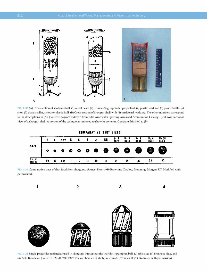
212 Atlas of Small Animal Wound Management and Reconstructive Surgery
CBA
FIG. 7-36 (A) Cross‐section of shotgun shell: (1) metal head, (2) primer, (3) gunpowder propellant, (4) plastic wad and (5) plastic baffle, (6)
shot, (7) plastic collar, (8) outer plastic hull. (B) Cross‐section of shotgun shell with (4) cardboard wadding. The other numbers correspond
to the descriptions in (A). (Source: Diagram redrawn from 1981 Winchester Sporting Arms and Ammunition Catalog). (C) Cross‐sectional
view of a shotgun shell. A portion of the casing was removed to show its contents. Compare this shell to (B).
FIG. 7-37 Comparative sizes of shot fired from shotguns. (Source: From 1980 Browning Catalog, Browning, Morgan, UT. Modified with
permission).
FIG. 7-38 Single projectiles (enlarged) used in shotguns throughout the world: (1) pumpkin ball, (2) rifle slug, (3) Brenneke slug, and
(4) Balle Blondeau. (Source: DeMuth WE. 1979. The mechanism of shotgun wounds. J Trauma 11:219. Redrawn with permission).

Management of Specific Wounds 213
wounding capability in relation to the distance from the muzzle adds a number of additional factors for the veterinarian to consider when confronted with such injuries.
Sherman and Parrish classified shotgun wounds into three basic types based on the pattern of distribution, depth of penetration, and range. Type I injuries, reflect-ing a relatively long distance between animal and shooter, are wounds in which the subcutaneous tissue and deep fascia are penetrated; type II injuries, produced at closer range, perforate structures beneath the deep fascia; and type III wounds, inflicted at point blank range (less than 3 yards), produce an extensive central zone of tissue destruction with a small peripheral halo of pellet holes. Extensive tissue destruction at close range is due to the high velocity and wider total surface area formed by the pellet group in flight as compared to a single projectile. Thus, at very close ranges, the shotgun is one of the most destructive weapons available (Figs 7‐40 and 7‐41).
Exploding Bullets
Although presently illegal to purchase and very difficult to obtain, exploding bullets were available in the 1980s for defense purposes to the public and law enforcement sectors. Exploding bullets initially were developed by the British in the early 1800s to penetrate barriers and ignite enemy powder caissons. They were subsequently employed in limited quantities during the American Civil War for antipersonnel use. There are several varia-tions of exploding bullets. Bullets have been designed to
split or fragment on impact. Alternatively, bullets con-taining an explosive charge were designed to explode on impact. These latter projectiles can be somewhat troublesome if they fail to detonate on impact with the patient: rough handling of the projectile during retrieval can result in their explosion. Undetonated bullets retained in the animal present a potential hazard to the patient and the surgeon attempting to remove them. Exploding bullets generally are semijacketed hollow point bullets
FIG. 7-39 Shot pattern at varying distances (Source: Adapted from Bell, MJ. 1981. The management of shotgun wounds. J Trauma 11:522.)
FIG. 7-40 Shotgun (type I) injury to the flanks caudal lumbar area
and hind limbs (No. 6 birdshot). The dog presented several weeks
after injury: considerable skin loss had occurred. Healing by
second intention was nearly complete. The shotgun blast occurred
at the level of the deep circumflex iliac artery and vein. Destruction
of these major direct cutaneous vessels and regional collateral
circulation was the major factor involved in the skin loss.

214 Atlas of Small Animal Wound Management and Reconstructive Surgery
filled with an explosive powder charge covered by a sin-gle lead shot and a percussion cap (or simply a primer anvil) covered by a wax coat. Deceleration of the bullet on impact ignites the percussion cap or primer, resulting in the explosion of the powder charge. From a historical perspective, the .22 bullet that struck President Reagan contained an aluminum canister in its nose filled with the explosive lead azide that is detonated by impact or high heat. Bullet deformation and the release of projec-tile fragments on impact reportedly increases the local tissue destruction compared to a regular bullet of similar size. However, this contention has been questioned by some authorities.
Interior and Exterior Ballistics
Modern bullets are fired from barrels that contain helical grooves (rifling) that serve to spin the projectile on its lon-gitudinal axis to stabilize its flight. Rifling improves the flight characteristics of a bullet in much the same way the helical fletching of an arrow serves to improve its flight. Despite rifling, projectiles traveling at a high velocity can become unstable in flight. A bullet can deviate from its longitudinal axis: it can yaw or tumble prior to or after impact with a body region, thereby increasing its profile in contact with tissues, resulting in greater tissue trauma. Flight instability enhances the likelihood of greater tissue destruction and promotes bullet fragmentation.
From an “offensive” standpoint, the ideal projectile has the following characteristics: (1) good ballistic shape (needlelike design); (2) high sectional density (ratio of projectile mass to area of presentation); (3) high velocity; and (4) the capability of deep penetration with a con-trolled expansion.
Terminal Ballistics
The seriousness of bullet wounds is often considered only in relation to those structures in the path of the bullet. Although this generally is true for low‐velocity projectiles and stab wounds, it does not account for the severity of injuries associated with some high‐velocity missile inju-ries. Bullets damage tissue in three ways: tissue laceration and crushing, shock waves, and cavitation. Both low‐ and high‐velocity projectiles create permanent tracts as they pass through tissues. Expanding bullets increase the tis-sue damage of a projectile by creating a larger wound cav-ity and enhancing energy absorption by the tissue. The tissue trauma from low‐velocity projectiles primarily is restricted to the crushing and laceration of tissues at the permanent tract. By comparison, high‐velocity projectiles can expend considerable energy to adjacent tissues in the form of shock waves and cavitation. Cavitation, a temporary cavity (5‐ to 30‐microsecond duration) up to 30 times the diameter of the projectile, is most evident as a projectile’s velocity markedly increases (Fig. 7‐42).
A B C
FIG. 7-41 (A) Massive soft tissue injury and a comminuted tibial fracture were the result of a (type III) shotgun injury. Some pellet entry
wounds are still evident. Maggots were present, but manually removed during the initial debridement procedure. (B) Serial debridement
was performed on a daily basis. (C) The wound healed by second intention. However, the external fixation was later replaced by internal
fixation with a bone plate and cancellous bone graft (successfully).

Management of Specific Wounds 215
Following the temporary lateral and cranial expansion of the surrounding tissues, a negative pressure forms with a sucking effect that has the potential of drawing debris and contaminants into the wound. The stretching and com-pression of tissues as a result of cavitation can enhance the magnitude of tissue destruction, especially if circulatory compromise occurs secondary to vascular disruption or
thrombosis: fractured bones, torn vessels, bowel ruptures, and contusions of the heart and lungs may occur without direct contact from high velocity projectiles.
A projectile may penetrate or perforate various tissues or structures, depending on its impact velocity, mass, design, and composition of the target struck. A pene-trating wound has an entry point with no exit, whereas a perforating wound has an entry and exit (through‐and‐through) without the projectile’s retention in a specified structure or region. To avoid confusion, it is best to restrict the terms penetrating and perforating to the anatomical structure in question. Projectiles that exit the body make it more difficult to determine the weapon involved. Projectiles retained in the patient without impacting bone are more likely to be lower‐velocity projectiles.
Besides velocity and mass, three factors are responsible for the greater destructiveness of projectiles, especially in higher‐velocity weapons: tumbling prior to impact, which permits a variety of positional contacts with the tissues; flight instability, apart from tumbling, thus add-ing an additional motion of the projectile; and secondary projectiles formed by missile fragmentation and bone shattering (Fig. 7‐43).
The entry wound usually is smaller than the exit wound because bullet tumbling, distortion, fragmenta-tion, and secondary projectile formation increase the bul-let’s destructiveness in a conelike fashion as it passes through the body. This general rule on wound size, how-ever, is misleading. If the gun muzzle is placed very close to the target when fired, expanding gases are released that accentuate the size of the entry wound, and powder burns also would be noted. The magnitude of the tempo-rary cavity is proportional to the energy imparted by the projectile during target penetration, with the missile’s energy decreasing exponentially with the distance pene-trated. Smaller projectiles may dissipate their energy more rapidly than larger projectiles with equal energy. A projectile that has expended a majority of its energy in
A
B
FIG. 7-42 (A) Note low velocity, no cavitation, and small entrance
and exit. (B) Greater cavitation is noted as velocity increases. Note
that the size of the exit wound is influenced by (1) target thickness
or depth, (2) tissue density, (3) projectile velocity and composition,
and (4) the amount of kinetic energy expended as the projectile(s)
exit the target area. (Source: Swan KG, Swan RC. 1980. Gunshot
Wounds. Littleton, MA: PSG Publishing. Modified with
permission.)
FIG. 7-43 Schematic diagram illustrating tumbling (forward rotation around the center of the mass). This unstable flight pattern increases
the projectile’s surface presentation to the target, enhancing tissue damage. (Source: Swan KG, Swan RC. 1980. Gunshot Wounds. Littleton,
MA, PSG Publishing. Modified with permission).

216 Atlas of Small Animal Wound Management and Reconstructive Surgery
the center of the target may exit without the gaping exit wound expected.
The specific gravity of the tissues struck also will influ-ence the wounds produced. Tissues with greater density, such as bone, are affected more adversely than are soft tissue structures. The retentive forces that combat the disruptive forces of the projectile also vary with the tis-sue struck. Skin and lung have elastic properties that better accommodate the energy insult which helps maintain the organ’s integrity. Liver and muscle have a similar specific gravity and absorb energy from a high‐velocity projectile in similar fashion. However, the tem-porary cavity and permanent tract formed in the liver are greater in magnitude compared to skeletal muscle because the liver parenchyma is less cohesive and resil-ient. Cavitation in pulmonary tissue is comparatively minor due to the elastic fibers within the spongy network of their parenchyma.
At high velocities, cavitation has also been recorded in bone. The explosive effect on the bone drives splinters of bone ahead of the projectile, increasing the magnitude of soft tissue damage and possibly the exit wound. Greater fragmentation will occur where cortical bone predomi-nates. Thus, long bones composed of large amounts of cortical bone have greater density as compared to flat bones, such as the ribs, with a greater proportion of can-cellous bone. High‐velocity rounds are more likely to cause massive tissue destruction when bone is struck. Paradoxically, any high‐velocity rounds can pass through soft tissue structures and exit the body without resulting in massive tissue necrosis. This can be explained primar-ily by the fact that a considerable proportion of the pro-jectile’s kinetic energy (mass, velocity) is not “deposited in” or “captured by” the body.
High‐velocity projectile injuries to the abdomen of anesthetized cats have demonstrated that the abdomen swells immediately and collapses, followed by a second expansion of less intensity, but with a prolonged dura-tion. Damage to the abdominal contents can occur with the first expansion, but the explosive injury noted devel-ops during the second expansion. Intestinal gas expands, resulting in multiple bowel perforations. The explosive shock wave displaces viscera, tears the mesentery, and can rupture blood vessels away from the permanent tract. The extent of soft tissue necrosis associated with high‐velocity projectiles is variable and can be difficult to assess. Muscle tissue is extremely resilient, and dam-age may occur only a few centimeters from the muscle tract. The tissue destruction occasionally noted hours to days later in some high‐velocity injuries, most likely is attributable to progressive vascular compromise to the tissues involved. Cavitation and the associated shock wave apparently result in blunt contusion of the associ-ated tissues. The volume of necrosis is influenced by
the severity of the vascular compromise. Vessels need not be struck directly by a high‐velocity projectile.
Stretching and tearing of the vessels can occur, result-ing in hemorrhage or thrombosis of the damaged vessel. The resulting damage has been likened to a crushing‐type injury. However, the large arteries, such as the canine femoral artery, are more resistant to indirect trauma. Circulatory insult and tissue necrosis increase the like-lihood of infection. Various studies using the thigh mus-cles of goats and dogs for high‐velocity projectile studies have demonstrated the surprising resilience of wound healing in the face of moderate trauma, as long as the circulation to the area remains predominantly intact. From clinical experience in humans, it is known that the extent of devitalization is affected by time.
Many researchers contend that bullet fragmentation better explains the size and severity of high‐velocity pro-jectile wounds. Bullet fragmentation and the dispersion of secondary projectiles causing muscle tearing enhances the absorption of the projectile’s energy by the target and is more likely responsible for the magnitude of the large conical‐shaped tracts formed by hunting bullets. In these cases, radiographs can help define the boundaries of the wounded tissue for debridement by the location of metal-lic fragments deposited in the area. Fragmentation of projectiles, in association with bone impact and fragmen-tation, best explains the massive trauma noted with high‐velocity rounds and the more powerful handguns.
The severity of tissue injury by a given projectile in large part is determined by the total kinetic energy absorbed by the patient. For example, a jacketed, high‐velocity bullet that perforates an upper extremity and exits intact without a significant loss in velocity would cause substantially less tissue injury compared to a bullet that deforms and rapidly decelerates in the same tissue. If this projectile is completely retained, the area has absorbed the damaging energy of the round. Thus, tissue trauma is better understood in terms of how rapidly and completely the energy is lost (absorbed by the body) and the tissues in which the energy is dissipated. As noted, certain body tissues that are elastic and maintain their architectural integrity better sustain projectile trauma.
Gunshot Wound Infection
All gunshot wounds are contaminated. Low‐ and high‐velocity projectiles are capable of driving or dragging topical debris (dirt, hair, etc.) into the permanent tract. Bacteria also are driven into the wound. As noted, the negative pressure formed during formation of the tempo-rary cavity associated with high‐velocity projectiles also is capable of “sucking” contaminants into the wound to a variable degree. The notion that projectiles, especially

Management of Specific Wounds 217
high‐velocity projectiles, are sterile due to heat generated during passage of the bullet through the barrel and air is a fallacy. The risk of infection is more likely in the case of high‐velocity projectile injuries where extensive tissue damage and circulatory embarrassment in the region are more likely. In general, the incidence of infection associ-ated with low‐velocity rounds limited to skin, muscle, and fascia is remarkably low.
Diagnosis and Management Considerations
In many cases, the owner is unaware of their pet being shot; clearly, pets that are allowed to roam unsupervised are at greatest risk of being shot. Without a history, skin
wounds may be mistaken for bite wounds or vehicular trauma. There are occasions in which the owner, a wit-ness, or police officer may provide specific information pertaining to the weapon. Knowledge of the weapon can help the veterinarian determine the potential tissue damage and appropriate surgical management of the injuries.
Most gunshot wounds to dogs and cats in a city are due to low‐velocity handguns (Figs 7‐44 and 7‐45). High‐velocity gunshot wounds from hunting rifles are seen more commonly in rural areas. Data from Angell Memorial Animal Hospital indicated that shotgun wounds occurred in urban, suburban, and rural areas. Radiographs of the patient are useful to determine the presence and location of retained bullets or fragments.
CA
B
FIG. 7-44 (A) German shepherd shot in the lower cervical area during a robbery attempt. (B, C) The .38 caliber round entered the thoracic inlet
and passed through the anterior mediastinum before coming to rest in the triceps musculature. Because the projectile round passed through
soft tissue only, without exiting the body, it was considered a low‐velocity round. Treatment consisted of only local wound preparation, lavage,
and application of a sterile dressing. The pneumomediastinum resolved without incident. No attempt was made to remove the projectile.
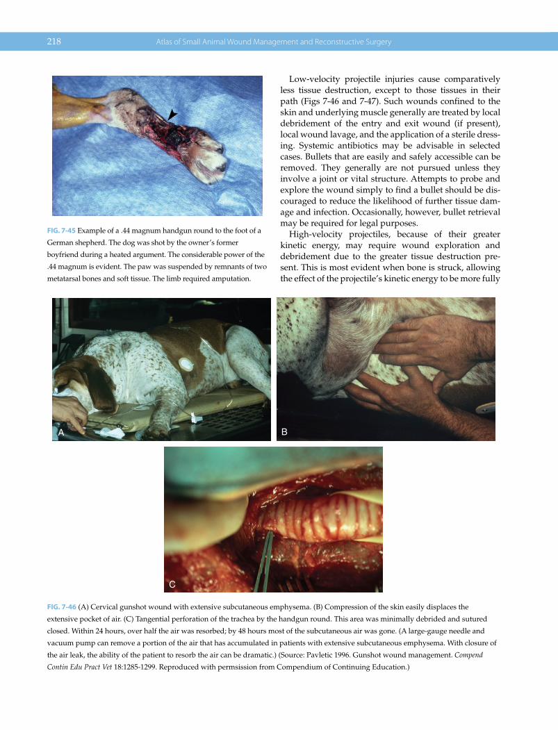
218 Atlas of Small Animal Wound Management and Reconstructive Surgery
Low‐velocity projectile injuries cause comparatively less tissue destruction, except to those tissues in their path (Figs 7‐46 and 7‐47). Such wounds confined to the skin and underlying muscle generally are treated by local debridement of the entry and exit wound (if present), local wound lavage, and the application of a sterile dress-ing. Systemic antibiotics may be advisable in selected cases. Bullets that are easily and safely accessible can be removed. They generally are not pursued unless they involve a joint or vital structure. Attempts to probe and explore the wound simply to find a bullet should be dis-couraged to reduce the likelihood of further tissue dam-age and infection. Occasionally, however, bullet retrieval may be required for legal purposes.
High‐velocity projectiles, because of their greater kinetic energy, may require wound exploration and debridement due to the greater tissue destruction pre-sent. This is most evident when bone is struck, allowing the effect of the projectile’s kinetic energy to be more fully
FIG. 7-45 Example of a .44 magnum handgun round to the foot of a
German shepherd. The dog was shot by the owner’s former
boyfriend during a heated argument. The considerable power of the
.44 magnum is evident. The paw was suspended by remnants of two
metatarsal bones and soft tissue. The limb required amputation.
C
BA
FIG. 7-46 (A) Cervical gunshot wound with extensive subcutaneous emphysema. (B) Compression of the skin easily displaces the
extensive pocket of air. (C) Tangential perforation of the trachea by the handgun round. This area was minimally debrided and sutured
closed. Within 24 hours, over half the air was resorbed; by 48 hours most of the subcutaneous air was gone. (A large‐gauge needle and
vacuum pump can remove a portion of the air that has accumulated in patients with extensive subcutaneous emphysema. With closure of
the air leak, the ability of the patient to resorb the air can be dramatic.) (Source: Pavletic 1996. Gunshot wound management. Compend
Contin Edu Pract Vet 18:1285‐1299. Reproduced with permsission from Compendium of Continuing Education.)

Management of Specific Wounds 219
expressed. Many of these cases may require considerable debridement, orthopedic repair, and a variable amount of open wound management.
The amount of kinetic energy lost by a bullet depends on four factors: (1) the kinetic energy possessed by the bullet on impact; (2) deviation of the longitudinal path of the bullet (yaw, tumble) enhancing projectile retardation; (3) loss of kinetic energy by loss of bullet mass and shape
during passage through tissues; (4) the tissues’ density, elasticity, and inherent integrity. High‐velocity hunting bullets, with an exposed soft lead tip or hollow point, can deform and fragment readily secondary to flight instabil-ity and tissue retardation, especially when bone is struck. A direct hit on dense cortical bone will cause a catastrophic deceleration of the bullet and its fragmentation. Bullet fragments and shards of bone are driven into the adjacent soft tissues (Figs 7‐48 and 7‐49). Disruption and purifica-tion of tissues combined with circulatory compromise results in massively destructive wounds. Skin, muscle, and fascia are sufficiently elastic to absorb a portion of this energy while maintaining their integrity, although cavita-tion is a major disruptive force during passage of a high‐velocity projectile through tissues. Passage of a high‐velocity round through the soft tissue of the upper thigh, for example, may not result in a wound that necessarily will require wide debridement: treatment may be more con-servative than that suggested by the literature.
With few exceptions, gunshot wounds to the brain and abdominal cavity require surgical exploration in the human. This approach can be justified in the small animal as well, although data are lacking regarding the successful management of gunshot wounds to the brain, primarily because of their grim outcome in most cases. Because abdominal gunshot wounds carry a high incidence of bowel injury and peritonitis, exploratory laparotomy is advisable (Figs 7‐50 and 7‐51). Thoracic gunshot wounds in the human are second in fatalities only to bullet wounds to the brain. Paradoxically, many of the cases in the human and the dog may be treated by conservative management. A chest tube may be required to treat a hemothorax and pneumothorax, but a thoracotomy is often unnecessary unless the esophagus has been penetrated or perforated,
FIG. 7-47 Great Pyrenees with a urethral tear dorsal to the scrotum.
A second hole was noted in the tail. What initially appeared to be
two gunshot wounds was actually one. The dog was shot from the
rear with his tail between his legs. The hole in the patient’s tail
lined up with the cutaneous hole above the scrotum.
BA
FIG. 7-48 (A) High‐velocity rifle round to the head of a German shepherd farm dog. Arrows denote the pathway of the projectile. Massive
bone and soft tissue destruction were evident. (B) Lateral view demonstrates the large exit wound through the bony orbit with destruction
of the right eye. A long incision was made connecting the entry/exit wounds, and bone fragments and necrotic soft tissue excised. The
cosmetic and functional results were comparable to a patient subjected to eye enucleation.

BA
FIG. 7-49 (A) Extensive trauma secondary to a high‐velocity rifle round. The entry wound was on the left side of the face of this Labrador.
Note the extensive trauma to the mandible. (B) The large irregular exit wound is noted on the right. There were fine fragments of the
projectile associated with the bilateral mandibular fractures. The majority of the high‐velocity round exited the patient, despite impact
with this dense bone.
BA
FIG. 7-50 (A) Air‐powered BB gun injury to a Siamese cat. Note the small, seemingly innocuous‐appearing entry wound. The patient
presented as depressed with abdominal pain. Abdominal radiographs demonstrated the retained projectile. (B) Laparotomy revealed the
bowel wall was perforated in two places (highlighted by probes) prompting intestinal resection and anastomosis. Prompt surgical
intervention prevented development of peritonitis. However, veterinarians should be aware that retained air‐powered projectiles are a
common incidental finding.
BA
FIG. 7-51 (A) A .22 Rimfire round to the lower abdomen of a cat. The bullet lodged against the wing of the ilium. The owner’s boyfriend
accidentally shot the cat with his handgun. (B) The owner elected to euthanize the cat. The abdomen was opened on postmortem revealing
multiple bowel perforations (asterisks) with the presence of tapeworm segments throughout the abdominal cavity (arrow). Exploration is
mandatory for projectile wounds to the abdomen.

Management of Specific Wounds 221
the heart was struck, a major tear in the tracheobronchial tree has occurred, or hemorrhage and air leakage into the thoracic cavity remains unchecked. Gunshot wounds to the neck in humans are explored routinely by many sur-geons due to the risk of esophageal injury and the pres-ence of vital vessels. However, it is controversial whether mandatory cervical exploration is justifiable in small ani-mals, depending on the severity of the wound and the neurological status of the animal.
If the bullet cannot be located, it has either exited the body or has passed into another area of the body. The body com-partment above and below the area shot should be radio-graphed in these situations. A bullet passing through the thorax into the abdomen will, at the very least, require an exploratory laparotomy, although a bullet passing in the opposite direction does not automatically mandate a thora-cotomy. Bullets have been known to migrate and embolize if they gain access to the circulatory system. Major vascular obstruction may lead to serious consequences. Similarly, bullets have been known to enter the tracheobronchial tree only to be coughed up and swallowed by humans (Fig. 7‐52).
Arrow Wounds
The bow and arrow has been an effective weapon for cen-turies. Despite its primitive origins, the bow and arrow is a highly lethal weapon in the hands of an experienced archer. Arrows lack the knockdown power and destruc-tive capacity of high‐velocity rifles. Rather, arrows rely on striking a vital area (lungs, heart, major vessels, liver, etc.), resulting in fatal hemorrhage. If an arrow weighing 450 grains has a velocity of 250 ft/s, it generates only 26 ft‐pounds of kinetic energy, considerably smaller than that generated by rifles.
Bows are measured by their draw weight, which is the number of pounds required to pull the drawstring to a full draw (full length of the arrow). In order to reduce the effort required to draw the bowstring of a conventional longbow or recurve bow, compound bows were devel-oped in 1967 that employ a series of pulleys, elliptical wheels, and cables. Improvements in the compound bow can reduce draw weights by 50–86%. Some states limit compound bows to those with a reduction of no greater than 65%.
A variation of the longbow is the crossbow. A weapon approximately 2000 years old, it gained popularity in the Middle Ages. The crossbow fires a bolt or quarrel approx-imately half the length (14–18 inches) of the average long-bow arrow. A crossbow can have a draw weight from 125–200 pounds. One crossbow has been developed with a draw weight of 300 pounds, whereas the upper draw weight of a longbow is around 70 pounds. A 125‐pound draw crossbow produces the same energy as a 60‐pound draw compound bow. A compound bow can fire a bolt with velocities of over 250 ft/s with an amazingly flat
BA
FIG. 7-52 (A) An unusual case of a migrating projectile. A Sheridan pellet was retrieved via urethrotomy at the level of the os penis. The
dog presented with an acute urinary tract obstruction during a walk with the observant owner. (B) Radiographs detected the pellet. Close
examination of the lateral abdominal wall revealed a healed entry scar. The dog had apparently been shot some weeks before, with the
pellet entering the urinary bladder. Its abrupt passage into the urethra caused the immediate urinary obstruction.
A Few Tips• If a projectile enters and exits a soft tissue area, it may
not be possible to determine low versus high velocity by examination of the entry and exit wounds
• If a projectile enters soft tissue and is retained in the tissues, it is most likely a low‐velocity projectile
• If a projectile impacts dense bony structures and exits the body, the projectile is likely a high‐velocity round
• Keep in mind that high‐velocity rounds decelerate over a dis-tance and can impact a target with a significantly lower velocity

222 Atlas of Small Animal Wound Management and Reconstructive Surgery
trajectory and short‐range accuracy. Crossbows have been criticized as silent, accurate, and highly lethal weap-ons that could be used to advantage by poachers. However, studies have indicated they are not silent and can be a difficult weapon to master. Furthermore, rifles, especially of a lower caliber with a sound suppressor, are a preferred weapon for illegal hunters, compared to a sin-gle‐shot crossbow. Crossbows are required by law to have a safety to prevent discharge of the bolt from jarring or dropping. Many states restrict the ownership and use of crossbows for hunting.
Arrows vary in length, weight, shaft composition, fletching, nock style, and arrow point (head). Arrow shafts are commonly composed of cedar, aluminum, or carbon fiber. Composite arrows of aluminum–carbon fiber or wood–fiberglass also have been introduced. Shaft length, width, and weight can be varied depending on the game hunted and bow employed (Fig. 7‐53).
Arrow fletching is the guidance system of the arrow, imparting twist or rotation to the shaft to stabilize its flight, similar to the rifling previously discussed. Turkey feathers once were the material of choice for fletching, but have been progressively replaced by plastic because of its durability and greater availability (Fig. 7‐54). The arrow nock adjacent to the fletching is the portion of the arrow that is notched to engage the “serving” area of the bow-string. The nock design varies according to the personal preference and requirements of the archer (Fig. 7‐55).
The head or point is the engaging end of the arrow and varies in shape, design, and weight. The arrowheads made by the early American Indians were chipped into points using flint, obsidian and other hard rock, antler,
and bone. Indians later created metal points from scraps of iron (Fig. 7‐56). Some primitive tribes use wooden tips formed by tapering the shaft that lacks fletching.
Modern arrowheads are exclusively metal, and are broadly classified as field points and hunting points (broadheads). Field points lack cutting blades, but have a pointed or dull tip for small game or target practice (Fig. 7‐57). In contrast, broadheads generally have two to four balanced cutting blades for larger game. There are a wide variety of broadhead designs. Many arrow shafts have threaded tips that allow the hunter to change hunt-ing heads. Some of the older arrows have a tapered shaft with the head applied with adhesives and crimping. Broadheads are usually razor sharp for maximum pene-tration and laceration. Broadheads are usually designed to slide past bone (ribs) to assure a clean strike to a vital spot. Broadheads are of three general types. Fixed‐blade broadheads are fixed to the shaft and sharpened manu-ally. Modular broadheads contain a center column or fer-rule holding three to four replaceable blades. Mechanical
FIG. 7-53 Arrows of various designs and materials. Note the short
(aluminum) quarrel of the crossbow (top) and 1800s North Dakota
Sioux Indian arrow, second from top (the feathered fletching has long
since deteriorated). The remaining arrows are composed of wood,
fiberglass, and aluminum. The bow‐fishing arrow was composed of
heavier fiberglass for greater penetration through water.
FIG. 7-54 Fletching imparts spin on the arrow to improve flight
stability. The left and middle arrows have plastic fletching; the
right arrow has turkey feather fletching.
FIG. 7-55 The arrow nock. Right is an example of the Sioux arrow.

Management of Specific Wounds 223
broadheads are designed to have recessed blades that fly open on impact: in a closed position, they have excellent ballistic properties. Barbed heads are illegal for hunting game (except for bow fishing) since the head will not work out of a wounded animal that receives a nonfatal shot (Fig. 7‐58).
Arrow wounds in humans and companion animals are uncommon in the United States compared to gunshot wounds. Few cases are reported in the human and veteri-nary medical literature. Many cases go unreported.
Management of Arrow WoundsMost arrows are not barbed and may be able to be removed without great difficulty. Target arrows have a smooth field point equal to the diameter of the shaft and should slide out unimpeded. Broadheads, with their rotating cutting edges, can “corkscrew” deep into the
A
B C
FIG. 7-56 (A) Excellent examples of Plains Indian arrows preserved from the 1800s. From the author’s collection. (B) Close‐up view of the
arrowheads: two flint arrows and a trader’s point metal arrowhead. American Indian arrow lengths and arrowhead designs (both stone
and metal) varied considerably. No two hand‐crafted arrows are alike. (C) Three (slightly offset) split turkey feathers were secured with
sinew spiraled around the shaft of the arrow. The feathered fletching imparts rotation to the long axis of the arrow, improving flight
stability as it travels to the target. Note the hand‐carved nock for seating against the bowstring.
FIG. 7-57 Field points and arrowhead for bow fishing. Field
points have a round silhouette that lacks cutting blades. Note
the bow‐fishing arrow has a metallic barb to capture the fish
upon penetration.
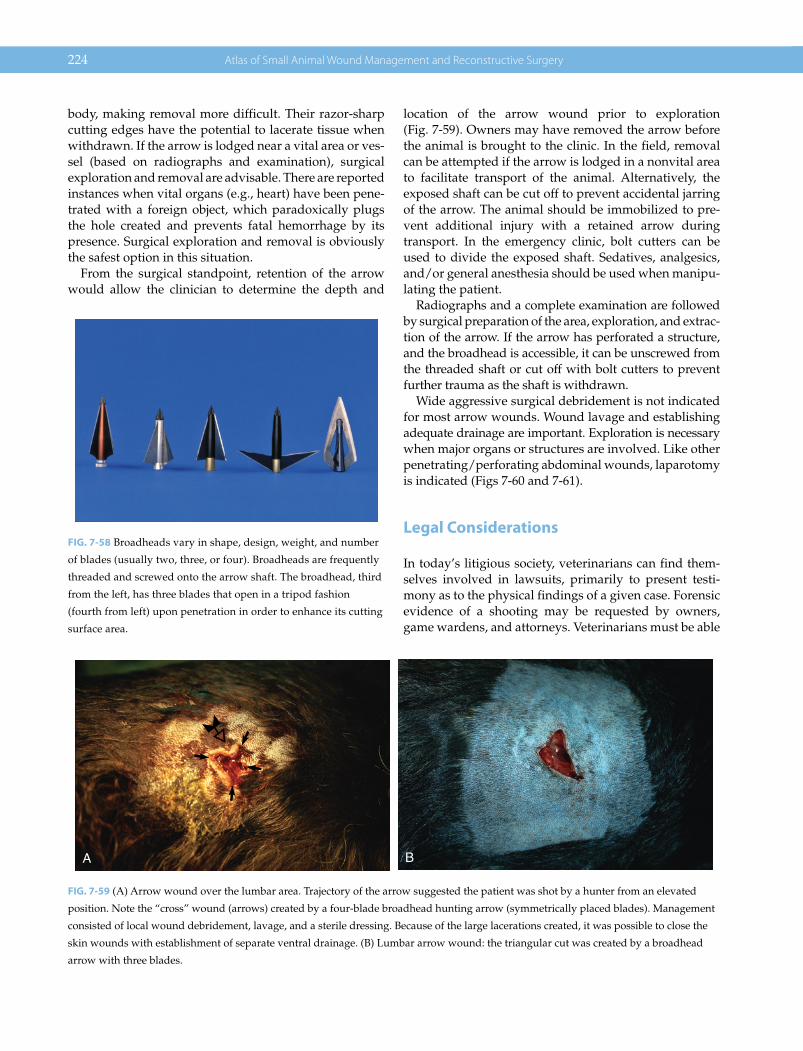
224 Atlas of Small Animal Wound Management and Reconstructive Surgery
body, making removal more difficult. Their razor‐sharp cutting edges have the potential to lacerate tissue when withdrawn. If the arrow is lodged near a vital area or ves-sel (based on radiographs and examination), surgical exploration and removal are advisable. There are reported instances when vital organs (e.g., heart) have been pene-trated with a foreign object, which paradoxically plugs the hole created and prevents fatal hemorrhage by its presence. Surgical exploration and removal is obviously the safest option in this situation.
From the surgical standpoint, retention of the arrow would allow the clinician to determine the depth and
location of the arrow wound prior to exploration (Fig. 7‐59). Owners may have removed the arrow before the animal is brought to the clinic. In the field, removal can be attempted if the arrow is lodged in a nonvital area to facilitate transport of the animal. Alternatively, the exposed shaft can be cut off to prevent accidental jarring of the arrow. The animal should be immobilized to pre-vent additional injury with a retained arrow during transport. In the emergency clinic, bolt cutters can be used to divide the exposed shaft. Sedatives, analgesics, and/or general anesthesia should be used when manipu-lating the patient.
Radiographs and a complete examination are followed by surgical preparation of the area, exploration, and extrac-tion of the arrow. If the arrow has perforated a structure, and the broadhead is accessible, it can be unscrewed from the threaded shaft or cut off with bolt cutters to prevent further trauma as the shaft is withdrawn.
Wide aggressive surgical debridement is not indicated for most arrow wounds. Wound lavage and establishing adequate drainage are important. Exploration is necessary when major organs or structures are involved. Like other penetrating/perforating abdominal wounds, laparotomy is indicated (Figs 7‐60 and 7‐61).
Legal Considerations
In today’s litigious society, veterinarians can find them-selves involved in lawsuits, primarily to present testi-mony as to the physical findings of a given case. Forensic evidence of a shooting may be requested by owners, game wardens, and attorneys. Veterinarians must be able
FIG. 7-58 Broadheads vary in shape, design, weight, and number
of blades (usually two, three, or four). Broadheads are frequently
threaded and screwed onto the arrow shaft. The broadhead, third
from the left, has three blades that open in a tripod fashion
(fourth from left) upon penetration in order to enhance its cutting
surface area.
BA
FIG. 7-59 (A) Arrow wound over the lumbar area. Trajectory of the arrow suggested the patient was shot by a hunter from an elevated
position. Note the “cross” wound (arrows) created by a four‐blade broadhead hunting arrow (symmetrically placed blades). Management
consisted of local wound debridement, lavage, and a sterile dressing. Because of the large lacerations created, it was possible to close the
skin wounds with establishment of separate ventral drainage. (B) Lumbar arrow wound: the triangular cut was created by a broadhead
arrow with three blades.
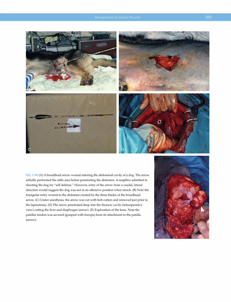
Management of Specific Wounds 225
BA
C D
E
FIG. 7-60 (A) A broadhead arrow wound entering the abdominal cavity of a dog. The arrow
initially perforated the stifle area before penetrating the abdomen. A neighbor admitted to
shooting the dog for “self defense.” However, entry of the arrow from a caudal, lateral
direction would suggest the dog was not in an offensive position when struck. (B) Note the
triangular entry wound to the abdomen created by the three blades of the broadhead
arrow. (C) Under anesthesia, the arrow was cut with bolt cutters and removed just prior to
the laparotomy. (D) The arrow penetrated deep into the thoracic cavity (intraoperative
view) cutting the liver and diaphragm (arrow). (E) Exploration of the knee. Note the
patellar tendon was severed (grasped with forceps) from its attachment to the patella
(arrow).
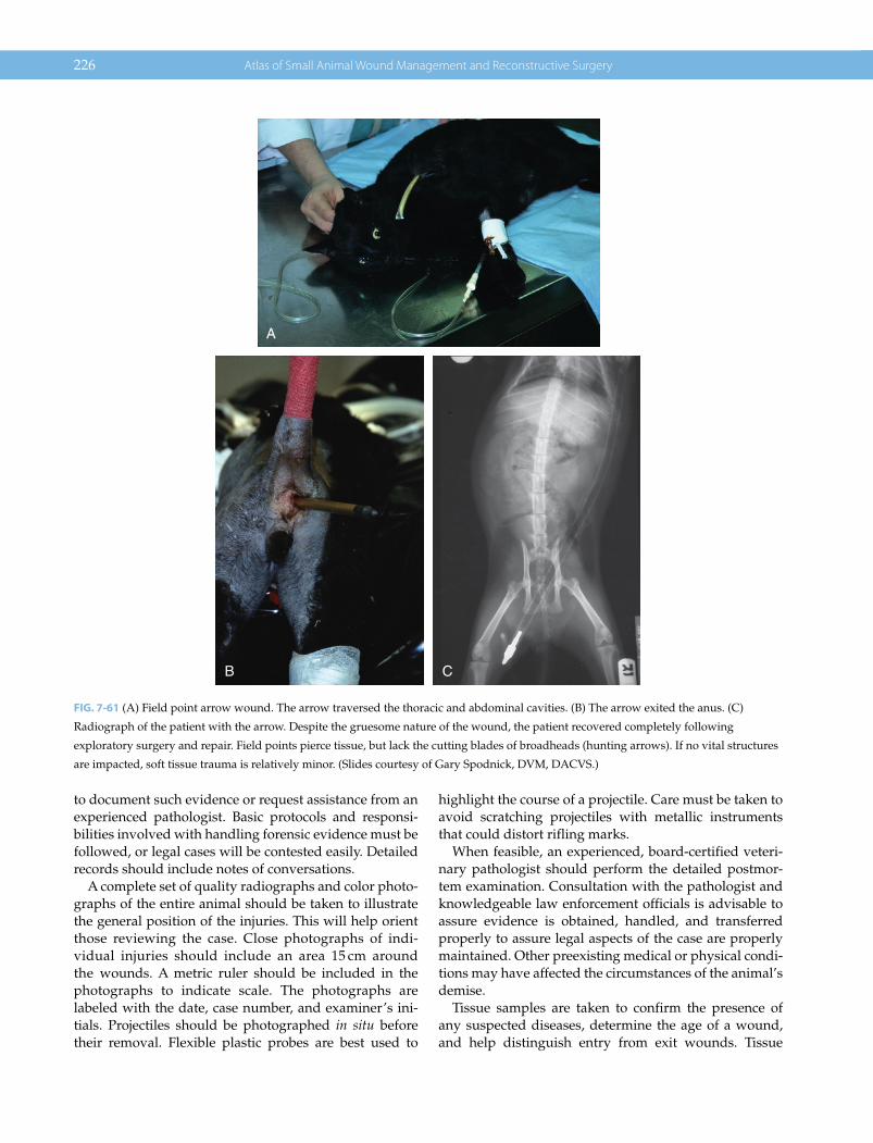
226 Atlas of Small Animal Wound Management and Reconstructive Surgery
to document such evidence or request assistance from an experienced pathologist. Basic protocols and responsi-bilities involved with handling forensic evidence must be followed, or legal cases will be contested easily. Detailed records should include notes of conversations.
A complete set of quality radiographs and color photo-graphs of the entire animal should be taken to illustrate the general position of the injuries. This will help orient those reviewing the case. Close photographs of indi-vidual injuries should include an area 15 cm around the wounds. A metric ruler should be included in the photographs to indicate scale. The photographs are labeled with the date, case number, and examiner’s ini-tials. Projectiles should be photographed in situ before their removal. Flexible plastic probes are best used to
highlight the course of a projectile. Care must be taken to avoid scratching projectiles with metallic instruments that could distort rifling marks.
When feasible, an experienced, board‐certified veteri-nary pathologist should perform the detailed postmor-tem examination. Consultation with the pathologist and knowledgeable law enforcement officials is advisable to assure evidence is obtained, handled, and transferred properly to assure legal aspects of the case are properly maintained. Other preexisting medical or physical condi-tions may have affected the circumstances of the animal’s demise.
Tissue samples are taken to confirm the presence of any suspected diseases, determine the age of a wound, and help distinguish entry from exit wounds. Tissue
B C
A
FIG. 7-61 (A) Field point arrow wound. The arrow traversed the thoracic and abdominal cavities. (B) The arrow exited the anus. (C)
Radiograph of the patient with the arrow. Despite the gruesome nature of the wound, the patient recovered completely following
exploratory surgery and repair. Field points pierce tissue, but lack the cutting blades of broadheads (hunting arrows). If no vital structures
are impacted, soft tissue trauma is relatively minor. (Slides courtesy of Gary Spodnick, DVM, DACVS.)
Management of Specific Wounds 227
samples occasionally are taken for more detailed testing and analysis by law‐enforcement officials.
Information commonly requested in court relates to the number and location of wounds, features of the wounds and related tissue areas, course of the projectiles, angle of fire, projectiles and foreign debris recovered, cause and time of death, and details pertaining to the handling and disposition of specimens collected. Entry wounds are closely inspected to determine the proximity of the weapon to the animal. A 15‐cm square around entrance wounds should be removed, pinned to a piece of rigid material, and then frozen for analysis to deter-mine the presence of propellant and projectile residue. This tissue specimen should not be washed or placed in formalin.
Careful examination and collection of tissues surround-ing the path of a projectile are useful in determining whether game has been illegally killed by a gun during restricted bow‐hunting seasons. Some hunters insert a broadhead arrow into a gunshot wound to mask the nature of the weapon. Flesh along the path of the bullet can be collected to recover particles of lead released by frangible projectiles. Lead residue can be identified by atomic absorption spectrophotometry in tissue surround-ing the tract. Tissue samples taken from a separate, unin-volved body region of the carcass serve as a control. Examination of gastric contents can also help the patholo-gist determine the circumstances of the incident.
The lands (elevated borders) and grooves of a rifled barrel embed marks on jacketed and nonjacketed bul-lets. Occasionally, the lead core separates from the outer jacket. Retrieval of the jacket is of great importance because the rifling marks are scored on its outer surface. The bullet should be washed with water and alcohol and then allowed to air dry. The washing removes blood and tissue fragments. The bullet can be marked on its base (bottom) to ensure that it can be identified later. Close‐up photographs of the bullet may help ensure its identification.
Projectiles can be wrapped in facial tissues and placed in vials or containers that can be sealed with tape. Some pathologists place projectiles in sealed envelopes. The body region from which each projectile was retrieved must be clearly identified and each projectile must be placed in a separate, marked container. The container is identified with the date, time, case number, and owner’s name, and it is initialed by those present at the necropsy. An indelible marker should be used on the container or on a nonremovable label. The projectiles should be secured from tampering or access by other individuals. Projectiles are turned over only to a qualified law‐enforcement officer. Any persons receiving the specimens must add the time and date of the transfer and their initials to the container.
Attention to the legal issues discussed in this section will help clinicians avoid the embarrassing errors and pitfalls associated with inappropriate case preparation in a court of law.
Arrows can have latent fingerprints on the shaft, unless blurred by passage through body tissues, contact with the entrance environment, or manipulation by individu-als. Depending upon the exact nature of the case, arrows can be removed without touching the shaft and placed in a secure area for presentation to law‐enforcement offi-cials. Under no circumstances should the projectile be transferred to the owner, thereby reducing the credibility of the evidence gathered.
EXPLOSIVE MUNITIONS: BALLISTIC, BLAST, AND THERMAL INJURIES
Military dogs, like soldiers in combat, are susceptible to explosives. Ballistic injuries are the result of flying shrap-nel, or fragments of metal, wood, glass, and concrete and other debris scattered by the explosion. Blast injuries are the result of the sonic shockwave from the explosion. Internal organs (and hollow organs in particular) are sus-ceptible to shockwave force: the dog also may be thrown violently against an object. Thermal injuries are a third form of trauma that may be associated with close proxim-ity of the blast. Incendiary explosives disperse hot chemi-cals that also cause thermal injuries. Dogs with multiple injuries require a team approach to stabilize the patient and prioritize the steps to manage the patient. A plan can be devised to approach wounds in a few “set surgeries” rather than undertaking multiple surgical procedures in a single prolonged anesthesia.
IMPALEMENT INJURIES
Impalement by definition is to pierce or transfix with a sharp object. In dogs and cats, this is most commonly the result of running or falling onto a sharp object. Dogs in particular are more susceptible to oropharyngeal and esophageal impalement wounds when chasing or carrying sticks. There are two common scenarios. (1) Dogs occasionally run after a stick or piece of wood tossed by the owner: as the object tumbles or momentarily imbeds into the ground the dog grabs the end in its mouth. The momentum of the dog drives the stick through their oropharynx, in many cases deep into the cervical tissues (Fig. 7‐62). (2) Dogs occasion-ally run with a stick or piece of wood in their mouth: if the end of the stick strikes the ground while the dog is running, the dog again impales their oropharynx.
Dogs occasionally run into a stationary pointed object, resulting in an impalement wound usually involving the

228 Atlas of Small Animal Wound Management and Reconstructive Surgery
head, neck, or anterior thorax (Fig. 7‐63). Dogs can impale their trunk by chasing and overrunning a tumbling stick tossed by the owner. One end of the stick contacts the ground while the vertical end of the stick impales the pet (Fig. 7‐64). Rarely, dogs and cats impale themselves by falling from an elevated position, striking fencing or other objects upon impact.
Impalement injuries, especially those involving sticks, should be explored to assure that no plant material (bark, wood splinters, leaf fragments, pine needles) is retained in the body. Such retained material normally results in abscess formation and draining tracts. Smooth‐surfaced objects (finished lumber, metal, plastic) are less problem-atic. Deeper cervical wounds should be explored through a ventral midline cervical approach. More superficial oropharyngeal wounds can be explored and repaired with an oral approach.
It is an advantage for the surgeon to have the impale-ment object left in place: exploration can be focused on the exact path and optimal approach to its removal. This also facilitates lavage, debridement, tissue repair, and postoperative drainage. If the object has been removed, examination of the object by the surgeon combined with the owner’s description of the wound can help determine the relative depth and direction of the impalement tract. Radiographs and ultrasonography of the involved region(s) can be useful in determining the best surgical approach to problematic impalement wounds. Endoscopic examination also may be useful in assessing the location of an esophageal tear. Magnetic resonance imaging has been suggested for better defining the location of prob-lematic esophageal injuries, although most tears should be visible by surgical exploration of the cervical area. Radiographic evidence of cervical emphysema is a
frequent finding with acute penetrating oropharyngeal and esophageal impalement. Repair of esophageal tears can be challenging, depending on the size and location of the tear. Most oral impalement wounds necessarily require debridement and closure of the mucosal wound to prevent oral contaminants from gaining access to the underlying tissues.
Overlooking retained sticks or delays in treatment of oropharyngeal/cervical esophageal tears can result in life‐threatening infection and extension into the anterior mediastinum. Prompt intervention can dramatically reduce this risk.
Cervical exploration includes careful inspection for retained plant material and debris. Following closure of the oropharyngeal/esophageal defect, copious lavage and suction of the area is indicated. A closed vacuum drain can be very effective in controlling dead space and assessing the fluid volume/composition retained in the reservoir after completion of surgery. Most dogs do not require a gastrostomy tube, depending on the assessment of the esophageal repair. Broad‐spectrum antibiotic administration is advisable.
PRESSURE ULCERS
Pressure ulcers are caused by prolonged compression of the skin over bony prominences, resulting in progres-sive ischemia. Shearing forces can contribute to their formation. They also can form from prolonged compres-sion from an external medical device, most commonly improperly fitting casts or splints. Animals incapable or unwilling to change body positions are prone to pressure ulcer formation, unless close attention is given to their
A B
FIG. 7-62 (A) This canine patient chased an arrow in flight. When the arrow struck the ground the dog grabbed the nock end of the arrow.
His body momentum drove the arrow through his oropharynx deep into the cervical tissues. (B) A ventral cervical incision was used to
remove the arrow and inspect the wound tract. The oropharyngeal tear was sutured. The cervical wound was lavaged and closed with a
vacuum drain. The patient made a complete recovery.
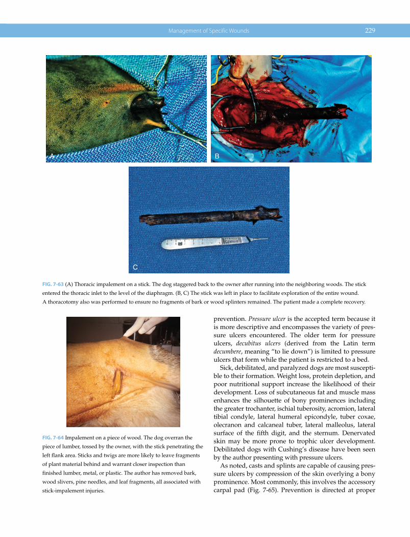
Management of Specific Wounds 229
prevention. Pressure ulcer is the accepted term because it is more descriptive and encompasses the variety of pres-sure ulcers encountered. The older term for pressure ulcers, decubitus ulcers (derived from the Latin term decumbere, meaning “to lie down”) is limited to pressure ulcers that form while the patient is restricted to a bed.
Sick, debilitated, and paralyzed dogs are most suscepti-ble to their formation. Weight loss, protein depletion, and poor nutritional support increase the likelihood of their development. Loss of subcutaneous fat and muscle mass enhances the silhouette of bony prominences including the greater trochanter, ischial tuberosity, acromion, lateral tibial condyle, lateral humeral epicondyle, tuber coxae, olecranon and calcaneal tuber, lateral malleolus, lateral surface of the fifth digit, and the sternum. Denervated skin may be more prone to trophic ulcer development. Debilitated dogs with Cushing’s disease have been seen by the author presenting with pressure ulcers.
As noted, casts and splints are capable of causing pres-sure ulcers by compression of the skin overlying a bony prominence. Most commonly, this involves the accessory carpal pad (Fig. 7‐65). Prevention is directed at proper
A B
C
FIG. 7-63 (A) Thoracic impalement on a stick. The dog staggered back to the owner after running into the neighboring woods. The stick
entered the thoracic inlet to the level of the diaphragm. (B, C) The stick was left in place to facilitate exploration of the entire wound.
A thoracotomy also was performed to ensure no fragments of bark or wood splinters remained. The patient made a complete recovery.
FIG. 7-64 Impalement on a piece of wood. The dog overran the
piece of lumber, tossed by the owner, with the stick penetrating the
left flank area. Sticks and twigs are more likely to leave fragments
of plant material behind and warrant closer inspection than
finished lumber, metal, or plastic. The author has removed bark,
wood slivers, pine needles, and leaf fragments, all associated with
stick‐impalement injuries.

230 Atlas of Small Animal Wound Management and Reconstructive Surgery
padding around the bony prominences to avoid a pres-sure cone effect to the area.
The intensity and duration of the pressure, along with the relative susceptibility of the tissues, influence pres-sure ulcer formation. Large dogs are more prone to pres-sure ulcer formation due to their greater weight (Fig. 7‐66). Debilitated greyhounds and other breeds with thin skin
and a sparse hair coat may be prone to pressure ulcer formation. The skin overlying the greater trochanter is the most common site for their development. Ischial tuberosity pressure ulcers are more common in smaller paraplegic dogs who have a tendency to sit up on their perineal region for prolonged periods of time on unpad-ded surfaces. In paraplegic dogs, the loss of sensation (denervation) increases the susceptibility of the tissues to pressure ulcer formation. Moreover, the patient is unaware of pain normally associated with prolonged compression of the skin overlying bony prominences and is incapable of adjusting their body position.
Cats rarely develop pressure ulcers due to their small size and less frequent spinal cord injuries. However, cats occasionally develop pressure ulcers over the calcaneal tuber. In cats that have a plantigrade stance, there is an increased the risk of developing ulceration in this area. Orthopedic and soft tissue trauma, weakening of the gas-trocnemius tendon, and an underlying neuropathy (dia-betes, etc.) may be contributing factors. The underlying cause must be addressed in addition to wound closure for long‐term resolution of this condition.
Nursing care is more difficult for larger dogs. Unfortunately, if they cannot be lifted from their cage, they may be inappropriately dragged out of the cage for treatments and cleaning. The fragile skin overlying the bony prominences then is stretched and abraded against the floor surface. Moisture accumulation on the skin from sweating, environmental moisture, urine, and feces can promote tissue maceration and infection. A dense hair coat affords some natural (padding) protection but may
A B
FIG. 7-65 Pressure ulcers associated with improper casting/padding. (A) Ulcer overlying the calcaneus. (B) Sloughing of the accessory
carpal pad.
FIG. 7-66 Pressure ulcer (grade II) over the elbow of a large
Labrador retriever. The dog developed the wound during its
hospitalization for a serious illness. Closure required the applica-
tion of a skin flap.

Management of Specific Wounds 231
promote moisture retention, mask early stages of pres-sure ulcer formation, and delay institution of additional preventative measures.
Prevention
Prevention of pressure ulcers requires an educated and devoted nursing staff, veterinarian, and owner. The fol-lowing management steps should be implemented.
1. Provide proper nutritional and fluid support2. Keep the patient’s skin clean and dry3. Change the patient’s body position every 2 hours (left
lateral, sternal, right lateral)4. Keep the patient on padded surfaces (artificial
washable fleece pads placed over easily cleaned support pads) placed on elevated grates or racks to separate the dog from urine and feces; water or air mattresses; disposable absorbent foam egg crate sheets (convoluted foam pads, Allegiance Health Care Corp., Wilsonville, OR); or coated or closed‐cell foam pads (Dubicrest Padding 4700, Alpha Protech, Tulsa, OK)
5. Assess skin overlying bony prominences daily for erythema, the first sign of pressure ulcer development
6. Avoid dragging over or dropping the patient on hard surfaces – if the patient cannot be lifted, slide the dog on a rug or fleece pad to prevent cutaneous abrasion
7. Place weak or paralyzed dogs in a sling support for 2–4 hours daily to reduce pressure on lateral bony prominences
8. Periodically, use hydrotherapy to keep the patient clean while improving muscle tone and circulation to the skin. The patient must be dried appropriately after each session
9. The appropriate use of applied padding to elevate the bony prominences, especially when patients are regaining the ability to ambulate (see Plates 10 and 11)
Properly maintained, a closed collection system can prevent urine scalding and skin maceration in patients incapable of urinating voluntarily. The perineal area can be clipped of hair to facilitate cleansing in the event of fecal incontinence. Water mattresses are useful as long as they support the weight of the dog. Covers should be applied over the mattresses to protect them from punc-tures and to facilitate cleaning. Unfortunately, many dogs will bite, chew, and scratch mattresses and foam pads. Because replacement can be costly, one option is to have the owner purchase pads, which they later can take home with them when the dog is discharged from the hospital. Many dogs will not cooperate with peri-odic attempts to change body position unless barriers (cardboard boxes, pads, etc.) are used to restrict their
movements. Providing large patients with appropriate nursing care is time consuming and expensive. However, the cost of pressure ulcer treatment far outweighs the cost of prevention.
Pressure Ulcer Classification
Pressure ulcer gradation is done according to the depth of injury to tissues overlying bony prominences (grades I to IV) (Fig. 7‐67). Table 7‐3 summarizes this classification.
Management of Pressure Ulcers
Permanently debilitated or paralyzed dogs prone to pres-sure ulcer formation also are prone to redevelopment of them after successful closure. Patients who regain ambu-lation have the greatest likelihood of maintaining perma-nent closure. Until patients reach this point, continuous preventative medicine is required to protect the area from reulceration. Several options are available for manage-ment of pressure ulcers; selection of the appropriate technique depends upon the severity of the lesion.
1. Open wound management—healing by second intention
2. Delayed primary closure3. Secondary closure4. Excision of the bony prominence in combination with
(2) or (3)5. Muscle flap coverage of the bony prominence in
combination with (2), (3), or (4)6. Skin flap coverage in combination with (2), (3), (4), or (5)
Grade I and grade II pressure ulcers may be amenable to second‐intention healing, unless they are persistent or recurrent ulcers. In many cases, grade II pressure ulcers are better closed by wound excision and primary closure, delayed primary closure, or secondary closure. Grade III, grade IV, or recurrent pressure ulcers may require open wound management to control infection followed by excision of a portion of the bony prominence. A local muscle flap may be used to add padding between the bone and skin. Muscle flaps can contribute circulation to diseased bone and help control chronic infection. The outer muscle surface, in turn, provides a stable vascular bed for skin flaps or cutaneous advancement.
In practice, the author will close most pressure ulcers by advancing healthy skin over the prepared ulcer, either by undermining and advancement of skin, or the use of a skin flap. If there is insufficient skin available for simple closure, skin flaps (including local flaps and axial pattern flaps) can provide a layer of subcutaneous tissue to cushion the skin over the bony prominence.

232 Atlas of Small Animal Wound Management and Reconstructive Surgery
Moreover, rotational flaps can provide full‐thickness skin coverage while placing the suture line peripheral to the bony prominence. On occasion the author will burr down the prominence with a Hall Air Drill to slightly flatten the most pointed surface beneath the skin prior to closure.
It must be noted that skin closure techniques that place the incision line directly over the bony promi-nence increase the likelihood of dehiscence: this is
not considered desirable for closure of pressure ulcers in humans that suffer from paralyzing injuries. Muscle flaps and skin flaps with resection of the bony promi-nence would be the most aggressive approach in these human patients. However. the author has used simple appositional techniques. Postoperatively, prolonged protection of the surgical closure area well into the maturation phase of wound healing is recommended (Fig. 7‐68). The key for long‐term success is getting the patient ambulatory, which includes the use of physical therapy.
Healing by second intention may not provide a dura-ble epithelial surface to prevent reinjury. The author nor-mally recommends closing pressure ulcers with healthy skin, unless you are dealing with an early pressure ulcer that can be managed conservatively. In dogs, closure of pressure ulcers does require prolonged wound protec-tion. Even then, postoperative protective measures must be instituted to avoid trauma and ischemia to the flap until the animal becomes ambulatory.
A
B
C
FIG. 7-67 (A) Debilitated geriatric patient with a pressure ulcer over the left acromion. Note the dog placed on a sheepskin. (B) A closer
view indicates a grade II pressure ulcer (arrow). (C) Two pressure ulcers also were noted over the wing of the ilium (grade II) and greater
trochanter (grade I progressing to a grade II) (arrows).
TABLE 7-3 Pressure Ulcer Classification.
Grade I Erythema; superficial to partial‐thickness skin lossGrade II Full‐thickness skin loss with variable involvement of
subcutisGrade III Ulceration extends to deep fascia overlying the bony
prominencesGrade IV Ulceration extends to bone. Osteomyelitis or joint
infection may be present

Management of Specific Wounds 233
A B
C D
EF
FIG. 7-68 (A) Chronic pressure ulcer over the greater trochanter. The dog developed the wound previously during hospitalization for the
repair of pelvic fractures. (B) Complete excision of the pressure ulcer was performed. Electrocautery and ligatures were required to control
the considerable hemorrhage that occurred during this procedure. (C) Skin borders were undermined, advanced, and apposed with a
subcuticular suture pattern, followed by skin sutures (vertical mattress and simple interrupted suture patterns). A Penrose drain was used
to control dead space. (D) A nonadherent dressing was applied over the incision. Two 6‐inch soft gauze rolls were placed in parallel
fashion cranial and caudal to the incision. Spray adhesive, used for the application of plastic surgical drapes, was used to stick the gauze
sponges into position. (E) Spray adhesive was used to provide additional adhesive power to the surgical tape placed over the area. (F) The
wound 14 days later. The ability of the patient to freely move and ambulate is critical to the treatment and prevention of recurrence of
pressure ulcers. (See Plate 11 for an alternative method to protect the greater trochanter.)

234 Atlas of Small Animal Wound Management and Reconstructive Surgery
Postoperative Care
Preventative measures, as described in the previous section, must be continued. In addition, bandages are advised in order to protect the healing wound from moisture, microorganisms, and compression and shear-ing forces that promote ischemic necrosis and dehis-cence. Soft, thick, “doughnut rings” fashioned from roll gauze have been used to protect the trochanteric area and lateral prominence of the limbs. The ring encircles the circumference of the bony prominence and protects it from contact with hard surfaces when the dog is placed in lateral recumbency. However, they can be dif-ficult to properly maintain. The use of doughnut rings is controversial since the doughnut ring can create a “halo” com-pression over the skin surrounding the bony prominence and compromise the cutaneous circulation required to promote healing to the injured area. Doughnut rings require close observation when used. Another option is the use of par-allel pads made from gauze rolls. These pads can pro-vide protection without the complete skin encirclement of doughnut rings. Spray adhesive used for plastic surgi-cal drapes can be applied around the neighboring skin followed by the application of surgical tape (Fig. 7‐68).
Soft foam pads, spicas, and tie‐over dressings may be used to protect bony prominences on the lateral surface of the trunk and limbs. Schroeder‐Thomas splints may be useful to manage pressure ulcers overlying the olec-ranon and calcaneal tuber after surgical closure. The author has had excellent results a simple foam padding system (PIPE) to protect the elbow region while allow-ing the dog to ambulate normally (Plate 10). Another technique used by the author, to protect the greater trochanter, is the pipe insulation loop technique (Fig. 7‐70) (see Plate 11).
Once healing is completed and sutures removed, splints and protective bandages may be removed on a trial basis, although proper nursing care is continued for as long as the dog is not ambulatory.
HYGROMA
An elbow hygroma is a serous subcutaneous fluid pocket overlying the olecranon in large‐ to giant‐breed dogs (great Danes, Irish wolfhounds, St. Bernards, and Newfoundlands), usually less than 2 years of age. Hygromas form as a result of repeated blunt trauma from the patient striking and compressing the skin against hard floor surfaces. The skin and subcutis in turn are compressed against this bony prominence. Although rare, hygromas have been reported overlying the carpus, nuchal crest, os calcis, and tuber ischium. Today, elbow hygromas are relatively uncommon, when compared to the population of cases seen by the author in the 1970s and 1980s. This may be due to changes in the dog popula-tion or the fact that most dogs live in the house with the availability of beds, couches, and rugs for their resting spots. Owners of the giant‐breed dogs also may be aware of hygromas and make an effort to provide their pet with soft bedding.
Most hygromas are painless fluctuations. Some hygro-mas remain relatively small and change little in size. Over time, hygromas can enlarge and ulcerate as the overlying skin thins and stretches over the fluid pocket. Ulcerated hygromas frequently become infected. Poor aseptic technique from attempted fluid aspiration can result in abscess formation; this problem may be com-pounded by futile attempts at prevention by injecting
In practice, the author has had the best results by debridement of the pressure ulcer, culturing the wound, and closing the wound with the inclusion of a vacuum drain system. While closure of pressure ulcers with the incision overlying a bony prominence is not ideal, it does work in most cases. Long‐term successful clo-sure includes: (1) prolonged protection of the surgical closure (6 weeks); (2) clean and dry soft bedding for the patient; and (3) rehabilitation of the patient to regain patient mobility (Fig. 7‐69).
Pressure ulcers over the elbow can be particularly challenging, especially in thin‐skinned breeds such as the greyhound.
In many cases, local flaps can be used to close the defect, the simplest being the use of bipedicle advancement flaps (release incisions). The thoracodorsal axial pattern flap is considered only for the most problematic skin defects overlying the elbow area. See Plate 40.
The most difficult pressure ulcers to manage in the dog involve the olecranon and greater trochanter. This is most commonly noted in large‐breed dogs.
Grade II–IV ulcers are best closed after the wound has been properly managed (debridement, control infection). Prolonged protection is critical in the postoperative period. Veterinarians commonly mistakenly think that healing is complete once the skin sutures are removed. Unfortunately, the surgical area may reulcerate days later.
As noted in Chapter 2, healing occurs for months. The key to improving durability at the surgical site is protecting the wound well into the maturation phase of healing, giving time for circula-tion and collagen deposition to improve and remodel the wound. Protecting the wounds for 4–6 weeks is a key to long‐term success in closure of pressure ulcers. Dogs that regain the ability to walk and stand on their own are less likely to have recurrence of pressure ulcers. Physical therapy can play an instru-mental role in this endeavor. Soft, clean, dry bedding is also important to long‐term protection of these susceptible areas.

Management of Specific Wounds 235
A
C D
B
FIG. 7-69 (A) Geriatric German shepherd with bilateral pressure ulcers overlying each acromion. The patient has a neuropathy: the owner
assists the dog in rising and walking with a body harness. (B) Close‐up view of the left pressure ulcer. Note skin and muscle necrosis
down to the acromion. (C) After debridement and lavage, a round Hemaduct drain was trimmed and inserted into the prepared wound
pocket. A Jackson‐Pratt reservoir was attached to the external drain. The local skin margins were mobilized and closed with intradermal
absorbable skin sutures, followed by the use of monofilament nylon interrupted skin sutures. (D) The Jackson‐Pratt reservoir was attached
to the adjacent harness. Note the pipe insulation attached to the harness, raising the shoulder profile from the bedding when the patient
reclines. Detailed instructions on drain care and soft bedding were discussed with the owners prior to discharging the patient.

236 Atlas of Small Animal Wound Management and Reconstructive Surgery
corticosteroids into the hygroma pocket. Both techniques are not advisable.
Conservative prevention and management are essen-tially the same: soft padded bedding (see bedding options for pressure ulcers in the preceeding section) and protec-tive elbow pads (protective dog leggings, Handicapped Pets.com; www.dogleggs.com). This would be the initial treatment of choice for small hygromas. Larger, persistent hygromas are likely surgical candidates. Large, ulcerated, and infected hygromas normally require more aggressive surgical intervention, including debridement and open wound management before attempting closure.
Surgical Options
Useful drainage options include the use of Penrose drains and the more recent introduction of the Jackson‐Pratt (JP) vacuum drain system. The Penrose drain technique is an effective method to manage hygromas. Of the two techniques, the JP drainage system does not require band-aging and is simple to use. The fluid accumulating in the JP reservoir can be measured quantitatively and serve as a general guideline when to remove the drain. Both tech-niques are described here. Although rarely employed today, surgical resection of hygromas also is discussed.
A
B C
FIG. 7-70 (A) Protection of a German shepherd after spinal surgery secondary to disc disease. A Grade I pressure ulcer was forming during
patient care and rehabilitation. Note the large pipe insulation loop (6 ft segment) is positioned round the hind limbs. The arrow denotes the
intersection of the pipe insulation: these junctions are secured with Elasticon or duct tape. Once secured, the dorsal pelvic portion of the
pipe insulation is secured to a loop of Elasticon tape that encircles the caudal abdominal area. (B) Elasticon secures the pipe insulation to
the Elasticon band encircling the caudal abdomen (arrow). (C) Right lateral view. An additional tape loop (arrow) is placed at the junction
of the pipe insulation loops to improve its positioning cranial and caudal to the greater trochanteric areas. This device was effective in
promoting healing of this early pressure ulcer overlying the left greater trochanter while protecting the right greater trochanter.
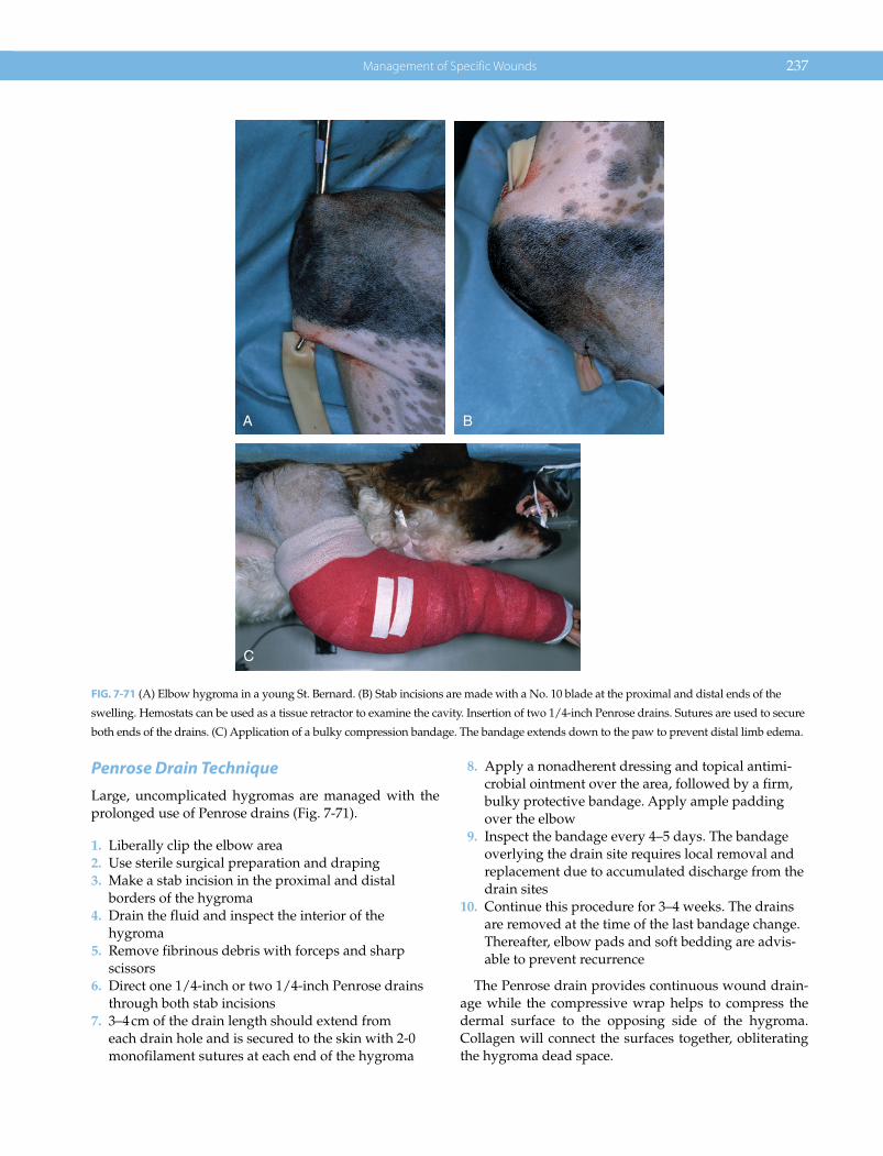
Management of Specific Wounds 237
Penrose Drain TechniqueLarge, uncomplicated hygromas are managed with the prolonged use of Penrose drains (Fig. 7‐71).
1. Liberally clip the elbow area2. Use sterile surgical preparation and draping3. Make a stab incision in the proximal and distal
borders of the hygroma4. Drain the fluid and inspect the interior of the
hygroma5. Remove fibrinous debris with forceps and sharp
scissors6. Direct one 1/4‐inch or two 1/4‐inch Penrose drains
through both stab incisions7. 3–4 cm of the drain length should extend from
each drain hole and is secured to the skin with 2‐0 monofilament sutures at each end of the hygroma
8. Apply a nonadherent dressing and topical antimi-crobial ointment over the area, followed by a firm, bulky protective bandage. Apply ample padding over the elbow
9. Inspect the bandage every 4–5 days. The bandage overlying the drain site requires local removal and replacement due to accumulated discharge from the drain sites
10. Continue this procedure for 3–4 weeks. The drains are removed at the time of the last bandage change. Thereafter, elbow pads and soft bedding are advis-able to prevent recurrence
The Penrose drain provides continuous wound drain-age while the compressive wrap helps to compress the dermal surface to the opposing side of the hygroma. Collagen will connect the surfaces together, obliterating the hygroma dead space.
A B
C
FIG. 7-71 (A) Elbow hygroma in a young St. Bernard. (B) Stab incisions are made with a No. 10 blade at the proximal and distal ends of the
swelling. Hemostats can be used as a tissue retractor to examine the cavity. Insertion of two 1/4‐inch Penrose drains. Sutures are used to secure
both ends of the drains. (C) Application of a bulky compression bandage. The bandage extends down to the paw to prevent distal limb edema.

238 Atlas of Small Animal Wound Management and Reconstructive Surgery
Vacuum Drain Management of Large HygromasJackson‐Pratt (JP) vacuum drains can be effectively used to manage elbow hygromas without the need for bandages and the expense associated with subsequent changes. The JP reservoir retains the fluid while keeping the hygroma collapsed. The volume of the fluid is recorded to quantitate the drainage (Fig. 7‐72). Plate 12 outlines the guidelines for this technique.
Surgical ResectionProblematic hygromas less commonly present with ulcerated skin, draining tracts, and an ongoing bacterial infection. Considerable scarring may be noted in the area. Under these circumstances, surgical resection of the ulcerated skin and underlying fibrotic scar should be considered. This surgical procedure runs the risk of wound dehiscence if skin closure is performed under tension. A Schroeder‐Thomas splint or rigid spica
A B
C D
FIG. 7-72 (A) Large elbow hygroma in a St. Bernard. (B, C) Placement of the 10‐mm flat drain. (D) Closure of the two access incisions.
Note the proximal finger trap knot and butterfly tape strip used to secure the external drain. The reservoir is attached to the patient’s collar.
(See Plate 12.)
Management of Specific Wounds 239
bandage may be needed for 2–3 weeks to prevent elbow flexion during the healing process. (See Plate 10 for addi-tional protection.) An incision lateral to the olecranon is best used to dissect the underlying diseased tissues from beneath the skin. (An incision directly over the olecranon has a higher probability of dehiscence.) Surgical efforts must be directed at preserving healthy elbow callus and as much skin coverage as possible. Failure to do so will likely result in wound dehiscence. Postoperatively, wound drainage (vacuum drains or Penrose drains) and protective bandaging are essential to control dead space and minimize motion until suture removal. Closure of smaller skin defects overlying the olecranon may be achieved with the assistance of lateral/medial release incisions. In extreme cases, wide skin resection will require reconstructive surgery, usually with the thoraco-dorsal axial pattern flap.
SNAKEBITE
Although relatively uncommon, snakebites are occasion-ally seen in the United States, primarily in those areas pop-ulated with poisonous snakes. An estimated 15 000 domestic animals are bitten by snakes annually in the United States. There are two main families of venomous snakes.
• Elapidae: elapids include coral snakes, cobras, mambas, kraits, and the tiger snake. Coral snakes can be found in the southeastern areas of the United States
• Vipers: rattlesnakes, copperheads, and cottonmouths (water moccasins) are found in various areas through-out the United States. Copperheads have a relatively low order of toxicity. Toxicity can vary between species. Rattlesnakes account for 80% of the poison-ous snakebite wounds in North America
In North America, 90% of snake bites occur between April and October. Toxicity is increased in young or very large snakes during the springtime.
Toxins in elapids are neurotoxic and hemolytic in nature; viper toxins cause local tissue damage (necro-genic) and may initiate systemic bleeding/coagulation (vasculotoxic). Diamondback rattlesnake venom may
contain a myocardial depressant factor that can result in cardiac dysrhythmias. Fang marks are a distinguishing feature of vipers; nonvenomous snake bites are charac-terized by a series of small punctures that reflect the cur-vature of the mouth. Almost one in four bites of vipers are “dry,” with little or no envenomation of the dog or cat. Coral snakes have short fangs and a small mouth, leaving a characteristic convex row of pinpoint puncture wounds.
Unless witnessed by the owner, snakebites can be dif-ficult to diagnose. Typically, viper bites cause local swell-ing, pain, erythema, petechiae, or ecchymoses, and later cyanosis with subsequent tissue necrosis. Close inspec-tion may reveal fang marks. Most dogs are bitten in the head and facial area and less commonly the paws. Facial swelling may be pronounced, and the patient’s airway is closely monitored in the event a tracheotomy is needed. In severe cases tissue around the fang marks may darken and ooze dark blood. Swelling may worsen over the first 24–48 hours, and the damaged tissues may form a hema-toma. Copperhead bites, from the author’s limited expe-rience, primarily result in regional tissue swelling, but lack the severity of local and systemic changes noted in the more toxic rattlesnake and water moccasin enveno-mation (Fig. 7‐73).
Elapid venom may have a delayed onset of 1–7.5 hours. Signs include salivation, vomiting, apprehensive behav-ior, followed by convulsions, quadriplegia, and respira-tory paralysis. Fortunately, documented coral snake envenomation is rare.
Baseline blood work (complete blood count, profile) and a urinalysis should be submitted on presentation. A coagulation profile is also advisable. Hemoconcentration,
FIG. 7-73 Louisiana dog that sustained a copperhead bite: note the
facial swelling. No tissue loss was noted: copperhead venom has a
relatively low order of toxicity compared to that of rattlesnakes.
Supportive fluid therapy resulted in complete recovery.
From my clinical experience, removal of hygromas is not neces-sary and rarely advisable over establishing drainage using a Jackson‐Pratt drainage system. It is worth remembering that wounds overlying the olecranon are among the most difficult defects to close successfully. Dehiscence secondary to hygroma resection is common and successful closure can be a frustrat-ingly expensive experience for the pet owner and veterinarian.
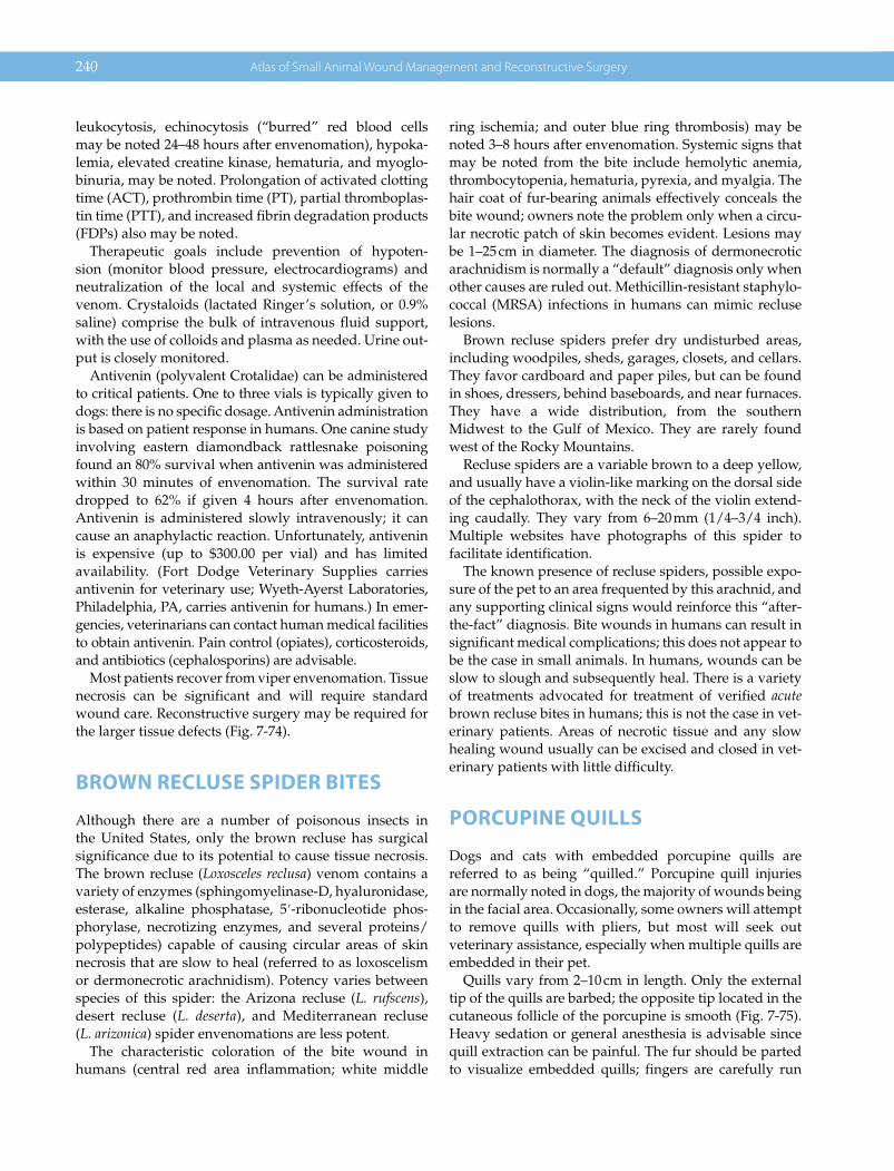
240 Atlas of Small Animal Wound Management and Reconstructive Surgery
leukocytosis, echinocytosis (“burred” red blood cells may be noted 24–48 hours after envenomation), hypoka-lemia, elevated creatine kinase, hematuria, and myoglo-binuria, may be noted. Prolongation of activated clotting time (ACT), prothrombin time (PT), partial thromboplas-tin time (PTT), and increased fibrin degradation products (FDPs) also may be noted.
Therapeutic goals include prevention of hypoten-sion (monitor blood pressure, electrocardiograms) and neutralization of the local and systemic effects of the venom. Crystaloids (lactated Ringer’s solution, or 0.9% saline) comprise the bulk of intravenous fluid support, with the use of colloids and plasma as needed. Urine out-put is closely monitored.
Antivenin (polyvalent Crotalidae) can be administered to critical patients. One to three vials is typically given to dogs: there is no specific dosage. Antivenin administration is based on patient response in humans. One canine study involving eastern diamondback rattlesnake poisoning found an 80% survival when antivenin was administered within 30 minutes of envenomation. The survival rate dropped to 62% if given 4 hours after envenomation. Antivenin is administered slowly intravenously; it can cause an anaphylactic reaction. Unfortunately, antivenin is expensive (up to $300.00 per vial) and has limited availability. (Fort Dodge Veterinary Supplies carries antivenin for veterinary use; Wyeth‐Ayerst Laboratories, Philadelphia, PA, carries antivenin for humans.) In emer-gencies, veterinarians can contact human medical facilities to obtain antivenin. Pain control (opiates), corticosteroids, and antibiotics (cephalosporins) are advisable.
Most patients recover from viper envenomation. Tissue necrosis can be significant and will require standard wound care. Reconstructive surgery may be required for the larger tissue defects (Fig. 7‐74).
BROWN RECLUSE SPIDER BITES
Although there are a number of poisonous insects in the United States, only the brown recluse has surgical significance due to its potential to cause tissue necrosis. The brown recluse (Loxosceles reclusa) venom contains a variety of enzymes (sphingomyelinase‐D, hyaluronidase, esterase, alkaline phosphatase, 5′‐ribonucleotide phos-phorylase, necrotizing enzymes, and several proteins/polypeptides) capable of causing circular areas of skin necrosis that are slow to heal (referred to as loxoscelism or dermonecrotic arachnidism). Potency varies between species of this spider: the Arizona recluse (L. rufscens), desert recluse (L. deserta), and Mediterranean recluse (L. arizonica) spider envenomations are less potent.
The characteristic coloration of the bite wound in humans (central red area inflammation; white middle
ring ischemia; and outer blue ring thrombosis) may be noted 3–8 hours after envenomation. Systemic signs that may be noted from the bite include hemolytic anemia, thrombocytopenia, hematuria, pyrexia, and myalgia. The hair coat of fur‐bearing animals effectively conceals the bite wound; owners note the problem only when a circu-lar necrotic patch of skin becomes evident. Lesions may be 1–25 cm in diameter. The diagnosis of dermonecrotic arachnidism is normally a “default” diagnosis only when other causes are ruled out. Methicillin‐resistant staphylo-coccal (MRSA) infections in humans can mimic recluse lesions.
Brown recluse spiders prefer dry undisturbed areas, including woodpiles, sheds, garages, closets, and cellars. They favor cardboard and paper piles, but can be found in shoes, dressers, behind baseboards, and near furnaces. They have a wide distribution, from the southern Midwest to the Gulf of Mexico. They are rarely found west of the Rocky Mountains.
Recluse spiders are a variable brown to a deep yellow, and usually have a violin‐like marking on the dorsal side of the cephalothorax, with the neck of the violin extend-ing caudally. They vary from 6–20 mm (1/4–3/4 inch). Multiple websites have photographs of this spider to facilitate identification.
The known presence of recluse spiders, possible expo-sure of the pet to an area frequented by this arachnid, and any supporting clinical signs would reinforce this “after‐the‐fact” diagnosis. Bite wounds in humans can result in significant medical complications; this does not appear to be the case in small animals. In humans, wounds can be slow to slough and subsequently heal. There is a variety of treatments advocated for treatment of verified acute brown recluse bites in humans; this is not the case in vet-erinary patients. Areas of necrotic tissue and any slow healing wound usually can be excised and closed in vet-erinary patients with little difficulty.
PORCUPINE QUILLS
Dogs and cats with embedded porcupine quills are referred to as being “quilled.” Porcupine quill injuries are normally noted in dogs, the majority of wounds being in the facial area. Occasionally, some owners will attempt to remove quills with pliers, but most will seek out veterinary assistance, especially when multiple quills are embedded in their pet.
Quills vary from 2–10 cm in length. Only the external tip of the quills are barbed; the opposite tip located in the cutaneous follicle of the porcupine is smooth (Fig. 7‐75). Heavy sedation or general anesthesia is advisable since quill extraction can be painful. The fur should be parted to visualize embedded quills; fingers are carefully run
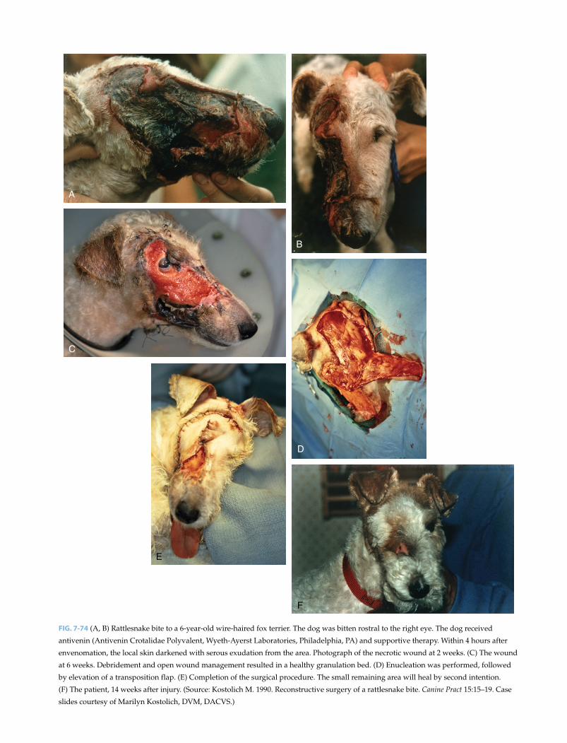
A
B
C
D
E
F
FIG. 7-74 (A, B) Rattlesnake bite to a 6‐year‐old wire‐haired fox terrier. The dog was bitten rostral to the right eye. The dog received
antivenin (Antivenin Crotalidae Polyvalent, Wyeth‐Ayerst Laboratories, Philadelphia, PA) and supportive therapy. Within 4 hours after
envenomation, the local skin darkened with serous exudation from the area. Photograph of the necrotic wound at 2 weeks. (C) The wound
at 6 weeks. Debridement and open wound management resulted in a healthy granulation bed. (D) Enucleation was performed, followed
by elevation of a transposition flap. (E) Completion of the surgical procedure. The small remaining area will heal by second intention.
(F) The patient, 14 weeks after injury. (Source: Kostolich M. 1990. Reconstructive surgery of a rattlesnake bite. Canine Pract 15:15–19. Case
slides courtesy of Marilyn Kostolich, DVM, DACVS.)

A
C D
E F
B
FIG. 7-75 (A) A “quilled” Retriever. (B) Note the multiple quills involving the right forelimb a paw. (Slides A and B courtesy of Dr. Patti
Ewing.) (C) Porcupine quills previously extracted from a dog. Note the finely tapered quill tip. (D) The external tip of the quill contains
multiple small barbs on the fine narrow tip. The opposite base/tip of the quill is smooth. (E) Close‐up view of the quill tip. Noted the
flattened V‐shaped barbs (resembling fish scales) along the circumference of the quill tip. The raised barbs are bent slightly backwards as a
result of their extraction from the skin. The angular barbs hook onto the dermal collagen fibers upon entry into the skin of the dog: this
impairs their extraction and explains why the barbed quills are capable of migrating into the body. (F) Recurrent facial abscess in a dog
secondary to a retained porcupine quill. Ultrasound identified the general location of the quill: this area was subsequently excised.

Management of Specific Wounds 243
through the fur to identify the exposed point of shorter or more deeply embedded quills. Needle holders can be used to grasp and remove each quill. Lightly embedded quills can be extracted with a firm tug, whereas deeply embedded quills can be extracted with quarter turns of the wrist with traction. A no. 11 scalpel blade can be aligned along the surface of problematic quills to create a small cutaneous stab incision to facilitate their removal. Few quill sites become infected after their removal; sys-temic antibiotics may be administered along with analge-sics when the patient is discharged from the hospital.
The frictional surface of quill tips can facilitate their migration deep into the body. Retained quills can result in recurrent abscess formation. Location of migrating quills can be problematic. Ultrasound can be useful in locating porcupine quills; CT may helpful in determining the general area of involvement.
Migrating quills can cause significant trauma to small animals. A porcupine quill was removed from a canine brain abscess on postmortem examination of a patient at Angell Memorial Animal Hospital: the quill had migrated through one of the foramina of the skull. The author has also removed quills from the lungs of dogs that have presented with spontaneous pneumothorax and lung abscessation (Fig. 7‐76). Although uncommon, cardiac “quilling” has been reported with associated pericarditis and endocarditis. Intestinal trauma also has been reported. A complete history from the owner is critically important to diagnosing internal injuries from porcupine
quill migration. Pets residing in an area with a porcupine population normally alerts the experienced veterinarian to include quill migration in the differential diagnosis.
LOWER EXTREMITY SHEARING WOUNDS
Shearing wounds are common in dogs and cats: they are invariably associated with vehicular trauma. The patient may be dragged on the pavement or the distal extremity is caught beneath the spinning wheel, result-ing in a variable degree of tissue avulsion with the paw trapped between the tire and road pavement (see Fig. 3‐13). Loss of skin, muscle, bone, and ligamentous support of the involved joint (carpal or tarsal) is com-monly noted. Rear extremity wounds are more common than forelimb shearing wounds. Despite the rather gruesome appearance of shearing wounds, most can be managed as an open wound with appropriate stabilization of the involved joint. The digits can be assessed for circulation and sensation to help deter-mine if salvage of the limb is feasible. There are a vari-ety of options to stabilize the unstable joint, including the use of screws, anchors, suture or wire, and external fixators. Arthrodesis may be necessary for severely unstable joints. Over time, granulation tissue forma-tion with subsequent collagen deposition may stabi-lize the slightly unstable joint with conventional bandage/splint application.
Skin loss is often exaggerated by the elastic retraction of the cutaneous margins aggravated by tissue edema. Conservative open wound management may be advisa-ble to allow time for resolution of tissue swelling over the first week to 10 days. This will allow time to more accu-rately assess the magnitude of the wound before consid-ering free grafts. If skin loss is less than 25–30% of the limb circumference, healing by second(ary) intention is likely to close the wound. Osteostixis (Chapter 3 and 6) may be used to facilitate granulation coverage of exposed bone. In this area, larger wounds may require a skin graft to close the cutaneous defect.
Suggested Reading
Bite WoundsAugust JR. 1988. Dog and cat bites. J Am Vet Med Assoc
193:1394–1398.Cabon Q, Deroy C, Ferrand FX, Pillard P, et al. 2015. Thoracic
bite trauma in dogs and cats: a retrospective study of 65 cases. Vet Comp Orthop Traumatol 28:448–454.
Chai O, Johnston DE, Shamir MH. 2008. Bite wounds involving the spine: characteristics, therapy and outcome in seven cases. Vet J 175:259–365.
Contrary to popular folklore, cutting a quill does not cause the quill to deflate, facilitating its removal.
FIG. 7-76 Porcupine quill in the left cranial lung lobe resulting in
pneumothorax. A detailed history revealed the dog encountered a
porcupine some months before presentation for pneumothorax.
244 Atlas of Small Animal Wound Management and Reconstructive Surgery
Cowell AK, Penwick RC. 1989. Dog bite wounds: a study of 93 cases. Compend Contin Educ Pract Vet 11:313–320
Davidson EB. 1998. Managing bite wounds in dogs and cats. Part II. Compend Contin Educ Pract Vet 20:974–991.
Jordan CJ, Halfacree ZJ, Tivers MS. 3013. Airway injury associ-ated with cervical bite wounds in dogs and cats: 56 cases. Vet Comp Orthop Traumatol 26:89–93.
Kolata RJ, Kraut NH, Hohnston DE. 1974. Patterns of trauma in urban dogs and cats: a study of 1000 cases. J Am Vet Med Assoc 164:499–502.
McKiernan BC, Adams WM, Huse DC. 1984. Thoracic bite wounds and associated internal injury in 11 dogs and 1 cat. J Am Vet Med Assoc 184:959–964.
Neal TM, Key JC. 1976. Principles of treatment of dog bite wounds. J Am Anim Hosp Assoc 12:657–660.
Pavletic MM. 1990. Bite Wounds. In: 2nd International Veterinary Emergency and Critical Care Symposium. Lakewood, CO: AAHA.
Pavletic MM. 1995. Bite Wound Management in Small Animals. AAHA Professional Library Series. Denver: American Animal Hospital Association.
Risselada M, deRooster H, Taeymans O, van Bree H. 2008. Penetrating injuries in dogs and cats. A study of 16 cases. Vet Comp Traumatol 21:434–439.
Shahar R, Shamir M, Johnston D. 1997. A technique for manage-ment of bite wounds of the thoracic wall in small dogs. Vet Surg 26:45–50.
Shamir MH, Leisner S, Klement E, et al. 2002. Dog bite wounds in dogs and cats: a retrospective study of 196 cases. J Vet Med 49:107–112.
Swaim SF. 1980. Surgery of Traumatized Skin: Management and Reconstruction in the Dog and Cat. Philadelphia, PA: WB Saunders.
Talan DA, Citron DM, Abrahamian FM, et al. 1999. Bacteriologic analysis of infected dog and cat bites N Engl J Med 340:85–92.
Thermal, Chemical, Radiation BurnsAggarwal SJ, Diller KR, Blake GK, Baxter CR. 1994. Burn‐
induced alterations in vasoactive function of the periph-eral cutaneous microcirculation. J Burn Care Rehabil 15:1–12.
Alvarado R, Chung KK, Cancio LC, Wolf SE. 2009. Burn resusci-tation. Burns 35:4–14.
Baxter CR. 1970. Present concepts in the management of major electrical injury. Surg Clin No Am 50:1401–1418.
Bedenice D. 2007. Smoke inhalation. In: Cote E, ed. Clinical Veterinary Advisor, 1011–1012. St. Louis, MO: Mosby.
Bostwick SA. 1987. Comprehensive rehabilitation after burn injury. Surg Clin N Am 67:159–166.
Bryant RA, Nix DP. 2016. Acute and Chronic Wounds: Current Management Concepts, 5th ed. St. Louis: Elsevier Inc.
Cabon Q, Deroy C, Ferrand F‐X, Pillard P, et al. 2015. Thoracic bite trauma in dogs and cats: a retrospective study of 65 cases. Vet Comp Orthop Traumatol 28:448–454.
Cowell K, Penwick C. 1989. Dog bite wounds: a study of 93 cases. Compend Contin Educ Vet 11:313–320.
Coyne BE, Bednarski RM, Bilbrey SA. 1993. Thermoelectric burns from improper grounding of electrocautery units: two case reports. J Am Anim Hosp Assoc 29:7–9.
Dahiya P. 2009. Burns as a model for SIRS. Front Biosci 14:4962–4967.
Davidson EB. 1998. Managing bite wounds in dogs and cats—Part I. Compend Contin Educ Vet 20:811–820.
Davidson EB. 1998. Managing bite wounds in dogs and cats—Part II. Compend Contin Educ Vet 20:974–990.
Demling RH. 1983. Improved survival after massive burns. J Trauma 23(3):179–184.
Demling RH. 1987. Fluid replacement in burn patients. Surg Clin N Am 67:15–30.
Demling RH, Buerstatte RPH, Perea A. 1980. Management of hot tar burns. J Trauma 20:242.
Dernell WS, Wheaton, LG. 1995. Surgical management of radia-tion injury—Part I. Compend Cont Educ Pract Vet 17:181–187.
Dernell WS, Wheaton, LG. 1995. Surgical management of radia-tion injury—Part II. Compend Cont Educ Pract Vet 17:499–510.
Dhupa N, Pavletic MM. 1997. Burns. In: Morgan R, ed. Handbook of Small Animal Practice, 3rd ed. Philadelphia, PA: WB Saunders.
Dorfelt R, Turkovic V, Teichmann S. 2014. Smoke inhalation in dogs and cats – a retrospective study over 5.5 years. Tierarztl Ausg K Kleintiere Heimtiere Prax 42:303–309.
Dow KD, Bucholtz JD, Iwamoto R, et al. 1997. Nursing Care in Radiation Oncology. Philadelphia, PA: WB Saunders.
Drobaz KJ, et al. 1999. Smoke exposure in dogs: 27 cases (1988–1997). J Am Vet Med Assoc 215:1306–1311.
Harris D, King GK, Bergman PJ. 1997. Radiation therapy toxici-ties. Vet Clin N Am 27:37–46.
HeimbachD, Engrav L, Grube B, Marvin J. 1992. Burn depth: a review. World J Surg 16:10–15.
Holt DE, Griffin GM. 2000. Bite wounds in dogs and cats. Vet Clin N Am Small Anim Pract 30:669–679.
Honari S. 2004. Topical therapies and antimicrobials in the management of burn wounds. Crit Care Nurs Clin North Am 16:1–11.
Hummel RP. 1982. Clinical Burn Therapy. Boston: John Wright PSG.
Johnston DE. 1985. Burns, electrical, chemical and cold injuries. In: Slatter DH, ed. Textbook of Small Animal Surgery. Philadelphia, PA: WB Saunders.
Jordan CJ, Halfacree ZJ, Tivers MS. 2013. Airway injury associ-ated with cervical bite wounds in dogs and cats: 56 cases. Vet Comp Orthop Traumatol 26:89–93.
Kolata RJ, Burrows CF. 1981. The clinical features of injury by chewing electrical cords in dogs and cats. J Am Anim Hosp Assoc 17:219–222.
Ladue TA. 2007. Radiation therapy: adverse reactions. In: Cote E, ed. Clinical Veterinary Advisor, 940–943. St. Louis, MO: Mosby.
Lee RC, Kolodney MS. 1987. Electrical injury mechanisms: dynamics of the thermal response. Plast Reconstr Surg 80:663–671.
Lee RC, Kolodney MS. 1987. Electrical injury mechanisms: elec-trical breakdown of cell membranes. Plast Reconstr Surg 80:672–679.
Luce EA. 1984. The irradiated wound. Surg Clin N Am 64:821–829.
Luce EA, Gottlieb SE. 1984. “True” high tension electrical inju-ries. Ann Plast Surg 12:321–326.
Monafo WW, Freedman B. 1987. Topical therapy in burns. Surg Clin N Am 67:133–145.
Meyers B, Schoeman JP, Goddard A, et al. 2008. The bacteriology and antimicrobial susceptibility of infected and non‐infected dog bite wounds. Vet Microbiol 127:360–368.
Monestry S, Hocksema H, Verbelen J, Pirayesh A, et al. 2008. Assessment of burn depth and burn wound healing. Burns 34:761–769.
Mozingo DW, Smith AA, McManus W, Pruitt BA, Mason AD. 1988. Chemical burns. J Trauma 28:642–647.
Mosier MJ, Gibran NS. 2009. Surgical excision of the burn wound Clin Plast Surg 36:617–635.

Management of Specific Wounds 245
Mouro S, Vilela CL, Niza MM. 2010. Clinical and Bacteriological assessment of dog‐to‐dog bite wounds.Vet Microbiol 144: 127–132.
Orgill DP, Piccolo N. 2009. Escharotomy and decompressive therapies in burns. J Burn Care Res 30:579–568.
O’Toole TE. 2007. Burns. In: Cote E., ed. Clinical Veterinary Advisor, 164–166. St Louis, MO: Mosby.
Pasulka PS, Wachtel TL. 1987. Nutritional considerations for the burned patient. Surg Clin No Am 67:109–131.
Pavletic MM. 1990. Management of thermal injuries. In: 2nd International Veterinary Emergency and Critical Care Symposium. San Antonio, TX.
Pope ER. 2003. Burns: thermal, electrical, chemical and cold injuries. In: Slatter DH, ed. Textbook of Small Animal Surgery, 3rd ed., 356–372. Philadelphia, PA: WB Saunders.
Reedy LM, Clubb FJ. 1991. Microwave burns in a toy poodle: a case report. J Am Anim Hosp Assoc 27:497–500.
Reinsch JF, Puckett CL. 1984. Management of radiation wounds. Surg Clin N Am 64:795–802.
Rosenkrantz K, Sheridan RL. 2002. Management of burned trauma patient: balancing conficting priorities. Burns 28:665–669.
Rudolph R, Noe JM. 1983. Chronic Problem Wounds. Boston: Little, Brown and Co.
Shamir MH, Leisner S, Klement E, et al. 2002. Dog bite wounds in dogs and cats: a retrospective study of 196 cases. J Vet Med A Physiol Pathol Clin Med 49:107–112.
Scheepens ET, Peeters ME, L’Eplattenier HF, Kirpensteijn J. 2006. Thoracic bite trauma in dogs: a comparison of clinical and radiological parameters with surgical results. J Sm Anim Pract 47:721–726.
Sheridan RL, Shank ES. 1999. Hyperbaric oxygen treatment: a brief overview of a controversial topic. J Trauma 47:426–435.
Sheridan RL. 2000. Evaluating and managing burn wounds. Dermatol Nurs 12:8–21.
Sheridan RL. 2001. Comprehensive treatment of burns. 2001. Curr Probl Surg 38:657–756.
Sheridan R. 2012. Burns: A Practical Approach to Immediate Treatment and Long‐Term Care. London: Manson Publishing Ltd.
Thrall DE. 1982. Radiation therapy in the dog: principles, indi-cations, and complications. Compend Cont Educ Pract Vet 4: 652–660.
White CE, Renz EM. 2008. Advances in surgical care: manage-ment of severe burn injury. Crit Care Med 36:S318–324.
FrostbiteBejerke HS, Tevar A. 2006. Frostbite. www.emedicine.comBritt LD, Dascombe WH, Rodriquez A. 1991. New horizons
in the management of hypothermia and Frostbite. Surg Clin N Am 71:345–370.
Bruen KJ, Ballaard JR, Morris SE, Cochran A, et al. 2007. Reduction of the incidence of amputation in frostbite injury with thrombolytic therapy. Arch Surg 142:546–551.
Carpenter HM, Hurley IA, Hardenbergh E, et al. 1971. Vascular injury due to cold: effects of rapid rewarming. Arch Pathol 92: 153–161.
Higdon B, Youngman L, Regehr M, Chiou A. 2015. Deep frostbite treated with hyperbaric oxygen and thrombolytic therapies. Wounds 27:215–223.
Hummel RP. 1982. Clinical Burn Therapy. Boston: John Wright PSG.Johnson AR, Jensen HL, Peltier G, DelaCruz E. 2011. Efficacy of
intravenous tissue plasminogen activator in frostbite patients
and presentation of a treatment protocol for frostbite patients. Foot Ankle Spec 4:344–348.
Johnston DE. 1985. Burns, electrical, chemical and cold injuries. In: Slatter DH, ed. Textbook of Small Animal Surgery, 516–533. Philadelphia, PA: WB Saunders.
Lapp NL, Juergens JL. 1965. Frostbite. Mayo Clin Proc 40:932.Lathrop T. 1975. Hypothermia: Killer of the Unprepared. Portland,
OR: The Mazamas.Martinez A, Golding MR, Sawyer PN, et al. 1966. The specific
arterial lesions in mild and severe frostbite: effect of sympa-thectomy. J Cardiovasc Surg 7:495–503.
McCauley RL, Hing DN, Robson MC, et al. 1983. Frostbite injuries. A rational approach based on the pathophysiology. J Trauma 23:143–147.
Nygaard RM, Whiteley AB, Fey RM, Wagner AL. 2016. The Hennepin Score: quantification of frostbite management efficacy. J of Burn Care Res 37:317–322.
Nygaard RM, Lacey AM, Lemere A, Dole M, et al. 2017. Time matters in severe frostbite: assessment of limb/digit salvage on the individual patient level. J Burn Care Res 38(1):53–59.
Pope ER. 2003. Burns: thermal, electrical, chemical, and cold injuries. In Slatter, DH, ed. Textbook of Small Animal Surgery, 3rd ed., 356–372. Philadelphia, PA: WB Saunders.
Twomey JA, Peltier GL, Zera RT. 2005. An open‐label study to evaluate the safety and efficacy of tissue plasmino-gen activator in treatment of severe frostbite. J Trauma 59:1350–1354.
Washburn B. 1975. Frostbite. Boston: Museum of Science.Wellehan JFX. 2003. Frostbite in birds: pathophysiology and
treatment. Compend Contin Ed Pract Vet 25:775–780.
Projectile InjuriesBarnes EC. 1997. Cartridges of the World, 8th ed. Northbrook, IL:
DBI Books.Bebchuk TN, Harari J. 1995. Gunshot injuries: pathophysiology
and treatments. Vet Clin North Am Sm Anim Pract 25:1111–1126.Bryant RA, Nix DP. 2016. Acute and Chronic Wounds: Current
Management Concepts, 5th ed. St. Louis: Elsevier Inc.Burnie AG, Kydd DM. 1985. Airgun injuries. Vet Rec
116:167–168.Combs R. 1987. Crossbows. Northbrook, IL: DBI Books.DiMaio VJM. 1993. Gunshot Wounds. Boca Raton, FL: CRC
Press.Dodd GD, Budzik RF. 1990. Identification of retained firearm
projectiles on plain radiographs. AJR 154:471–475.Fackler ML. 1996. Gunshot wound review. Ann Emerg Med
28:194–203.Ferguson T. 1991. Modern Law Enforcement Weapons and Tactics,
2nd ed. Northbrook, IL: DBI Books.Fullington RJ, Otto CM. 1997. Characteristics and management
of gunshot wounds in dogs and cats: 84 cases. J Am Vet Med Assoc 210:658–662.
Heard, BJ. 1997. Handbook of Firearms and Ballistics. New York: John Wiley and Sons.
James MR. 1992. The Bowhunter’s Handbook. Iowa, WI: DBI Books.
Keep JM. 1970. Gunshot injuries to urban dogs and cats. Aust Vet J 46:330–334.
Kettner F, Kirberger RM. 2006. Aortic foreign body (airgun pellet) embolism in a cat. J Sm Anim Pract 47:221–225.
Olsen LE, Streeter EM, DeCook RR. 2014. Review of gunshot injuries in caats and dogs and utility of a triage scoring system

246 Atlas of Small Animal Wound Management and Reconstructive Surgery
to predict short‐term outcome: 37 casees (2003–2008). J Am Vet Med Assoc 245:923–929.
Pavletic MM. 1985. A review of 121 gunshot wounds in the dog and cat. Vet Surg 14:61–62.
Pavletic MM. 1986. Gunshot wounds in veterinary medicine: projectile ballistics—Part I. Compend Contin Ed Pract Vet 8:47–60.
Pavletic MM. 1986. Gunshot wounds in veterinary medicine: projectile ballistics—Part II. Compen Contin Ed Pract Vet 8:125–134.
Pavletic MM. 1996. Gunshot wound management. Compend Cont Educ Pract Vet 18:1285–1299.
Pavletic MM, Trout N. 2006. Bullet, bite and burn wounds. Vet Clin N Am 36 (4):873–893.
Wallentine D. 1988. Making Indian Bows and Arrows… The Old Way. Liberty, UT: Eagle’s View Publishing Co.
Impalement InjuriesCollins MB, Hodgson DR, Hutchins DR, 1994. Pleural effusion
associated with acute and chronic pleuropneumonia and pleuritis secondary to thoracic wounds in horses: 42 cases (1982–1992). J Am Vet Med Assoc 205:1753–1758.
Doran IP, Wright CA, Moore AH. 2008. Acute oropharyngeal and esophageal stick injury in forty‐one dogs. Vet Surg 37:781–785.
Griffiths LG, et al. 2000. Oropharyngeal penetrating injuries in 50 dogs: a retrospective study. Vet Surg 29:383–388.
Laverty S, Lavoie JP, Pascoe JR, et al. 1996. Penetrating wounds of the thorax in 15 horses. Equine Vet 28:220–224.
Pratschke KM, Kirby BM. 2002. High rise syndrome with impalement in three cats. J Small Anim Pract 43:261–264.
Zitz J, Rozanski EA, Berg J, Penninck D. 2007. Managing dogs with thoracic impalement injuries: a review of nine cases. Vet Medicine 102:307–313.
Pressure UlcersBrunner LS, Suddarth DS. 1986. Manual of Nursing Practice,
4th ed. Philadelphia, PA: JB Lippincott.Bryant RA, Nix DP. 2016. Acute and Chronic Wounds: Current
Management Concepts, 5th ed. St. Louis: Elsevier Inc.Daniel RK, Hall EJ, MacLeod MK. 1979. Pressure sores—a
reappraisal. Ann Plast Surg 3:53–63.Griffith BH. 1979. Pressure sores. In: Grabb WC, Smith JW,
eds. Plastic Surgery. Boston: Little, Brown & Co.Kramek BA, Moise NS, Cooper B, Raffe MR. 1984. Neuropathy
associated with diabetes mellitus in the cat. J Am Vet Med Assoc 184:42–45.
Nwomeh B, Yager DR, Kelman CL. 1998. Physiology of the chronic wound. Clin Plast Surg 25(3):341–356.
Pavletic MM. 2011. Pipe insulation as a protective device for elbow wounds in dogs. 239:1225–1231.
Swaim SF, Hanson RR, Coates JR. 1996. Pressure wounds in ani-mals. Compend Cont Educ Pract Vet 18:203–218.
Swaim SF, Henderson RA. 1997. Small Animal Wound Management, 2nd ed. Baltimore: Williams and Wilkins.
Swaim SF, Lee AH, Henderson RA. 1989. Mobility vs immo-bility in the healing of open wounds. J Am Anim Hosp Asoc 25:91–96.
HygromaJohnston DE. 1975. Hygroma of the elbow in dogs. J Am Vet Med
Assoc 167:213–219.Pavletic MM, Brum DE. 2015. Successful closed suction drain
management of a canine elbow hygroma. J Sm Anim Pract 56:476–479.
White RAS. 2006. Management of specific skin wounds. Vet Clin No Am 36:905–907.
SnakebiteCarr A, Schultz J. 2015. Prospective evaluation of the incidence
of wound infection in rattlesnake envenomation in dogs. J Vet Emer 2008. Bg Crit Care (San Antonio) 25:546–551.
Gilliam LL, Brunker J. 2011. North American snake envenoma-tion in the dog and cat. Vet Clin North Am Small Animal Practice. 6:1239–1259.
Hackett TB, et al. 2002. Clinical findings associated with prairie rattlesnake bites in dogs: 100 cases (1989–1998). J Am Vet Med Assoc 220:1675–1680.
Hoose JA, Carr A. 2013. Retrospective analysis of clinical find-ings and outcome of cats with suspected rattlesnake enveno-mation in Southern California. J Vet Emerg Crit Care (San Antonio) 23:314–320.
Hudelson S, Hudelson P. 1995. Pathophysiology of snake enveno-mization and evaluation of treatments: Parts I, II, & III. Compend Contin Ed Pract Vet 17:889–897, 1035–1040, 1385–1396.
Julius TM, Kaelbie MK, Leech EB. 2012. Retrospective evaluation of neurotoxic rattlesnake envenomation in dogs and cats: 34 cases (2005–2010). J Vet Emerg Crit Care (San Antonio) 22:460–469.
Kostolich M. 1990. Reconstructive surgery of a snake bite wound. Canine Pract 15:15–19.
Lenchner I, Aroch I, Segev G, Kelmer E, et al. 2014. A retro-spective evaluation of Vipera Palaestinae envenomation in 18 cats (2006–2011). J Vet Emerg Crit Care (San Antonio). 24:425–433.
Paul A, Rozanski E. 2007. Snakebite. In: Cote, E, ed. Clinical Veterinary Advisor, 1012–1014. St. Louis, MO: Mosby.
Perez ML, Fox K, Schaer M. 2012. A retrospective evaluation of coral snake envenomation in dogs and cats: 20 cases. J Vet Emerg Crit Care (San Antonio) 22:682–689.
Peterson ME. 2001. Snake bite: pit vipers and coral snakes. In: Peterson ME, Talcott PA, eds. Small Animal Toxicology, 695–720. Philadelphia, PA: WB Saunders.
Peterson ME. Snake bite: coral wounds. 2006. Clin Tech Small Anim Pract 21:183–186.
Waldron DR, Zimmerman‐Pope N. 2002. Superficial skin wounds. In: Slatter D, ed. Textbook of Small Animal Surgery, 267–268. Philadelphia, PA: WB Saunders.
Willey JR, Schaer M. 2005. Eastern diamond rattlesnake (Crotalus adamanteus) envenomation of dogs: 31 cases (1982–2002). J Am Anim Hosp Assoc 41:22–33.
Spider Bite/Brown RecluseAmalsadvala T, Swaim SF. 2006. Management of hard‐to‐heal
wounds. Vet Clin N Am 36(4):704.Dunayer, EK. 2007. Spider envenomation. In: Cote E, ed. Clinical
Veterinary Advisor, 1020–1021. St. Louis, MO: Mosby.
Management of Specific Wounds 247
Roder JD. 2004. Spiders. In: Plumlee KH, ed. Clinical Veterinary Toxicology, 111–113. St. Louis, MO: Mosby.
Peterson ME. 2006. Brown spider envenomation. Clin Tech Small Anim Pract 21:191–193.
Porcupine QuillsCosta A, Lahmers S, Barry SL, Stanton J, Stern JA. 2014. Fungal
pericarditis and endocarditis secondary to porcupine quill migration in a dog. J Vet Cardiol 16:283–290.
Guevara JL, Holmes ES, Reetz J, Holt DE. 2015. Porcupine quill migration into the thoracic cavity of a German shorthaired pointer. J Am Anim Hosp Assoc 51:101–1106.
Marshak RR. 1952. Injury of gastrointestinal tract by porcupine quills. J Am Vet Med Assoc 121:477.
Nucci DJ, Liptak. 2016. The diagnosis and surgical management of intracardiac quill foreign body in a dog. J Am Anim Hosp Assoc 52:73–76.
Pavletic MM. 2002. Penetrating wounds. In: Wingfield WE, Raffe MR, eds. The Veterinary ICU Book, 966. Jackson Hole: Teton Newmedia.
Grahn BH, Szentimrey D, Pharr JW, et al. 1995. Ocular and orbital porcupine quills in 50 dogs: a retrospective study. Can Vet J 36:488–493.
Lower Extremity Shearing WoundsBeardsley SL, Schrader SC. 1995. Treatment of dogs with
wounds of the limbs caused by shearing forces: 98 cases (1975–1993). J Am Vet Med Assoc 207:1071–1075.
Corr S. Management of distal limb shearing injuries in cats. J Feline Med Surg. 11:747–757.
Diamond DW, Besso J, Boudrieau RJ. 1999. Evaluation of joint stabilization for treatment of shearing injuries of the tarsus in 20 dogs. J Am Anim Hosp Assoc 35:147–153.
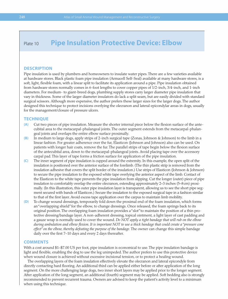
248 Atlas of Small Animal Wound Management and Reconstructive Surgery
Plate 10 Pipe Insulation Protective Device: Elbow
DESCRIPTIONPipe insulation is used by plumbers and homeowners to insulate water pipes. There are a few varieties available at hardware stores. Black plastic foam pipe insulation (Armacell Self‐Seal) available at many hardware stores, is a soft, light, flexible foam, with a linear split to facilitate its application around a pipe. Pipe insulation obtained from hardware stores normally comes in 6‐foot lengths to cover copper pipes of 1/2‐inch, 3/4‐inch, and 1‐inch diameters. For medium‐ to giant‐breed dogs, plumbing supply stores carry larger diameter pipe insulation that vary in thickness. Some of the larger diameter insulators do lack a split seam, but are easily divided with standard surgical scissors. Although more expensive, the author prefers these larger sizes for the larger dogs. The author designed this technique to protect incisions overlying the olecranon and lateral epicondylar areas in dogs, usually for the management/closure of pressure ulcers.
TECHNIQUE(A) Cut two pieces of pipe insulation. Measure the shorter internal piece below the flexion surface of the ante-
cubital area to the metacarpal‐phalangeal joints. The outer segment extends from the metacarpal‐phalan-geal joints and overlaps the entire elbow surface proximally.
(B) In medium to large dogs, apply strips of 2‐inch surgical tape (Zonas, Johnson & Johnson) to the limb in a linear fashion. For greater adherence over the fur, Elasticon (Johnson and Johnson) also can be used. On patients with longer hair coats, remove the fur. The parallel strips of tape begin below the flexion surface of the antecubital area, down to the metacarpal‐phalangeal joints. Avoid placing tape over the accessory carpal pad. This layer of tape forms a friction surface for application of the pipe insulation.
(C) The inner segment of pipe insulation is cupped around the extremity. In this example, the open split of the insulation is positioned over the anterior surface of the forelimb. (The thin plastic strip is removed from the insulation adhesive that covers the split border of the insulation.) Use strips of Elasticon (Johnson & Johnson) to secure the pipe insulation to the exposed white tape overlying the anterior aspect of the limb. Contact of the Elasticon to the white tape prevents the pipe insulation from slipping. Cut the longer (outer) piece of pipe insulation to comfortably overlap the entire olecranon, extending approximately 2–3 inches (5–8 cm) proxi-mally. (In this illustration, this outer pipe insulation layer is transparent, allowing us to see the short pipe seg-ment secured with bands of Elasticon.) Secure the insulation to the exposed surgical tape in a fashion similar to that of the first layer. Avoid excess tape application over the carpus to maintain limb mobility.
(D) To change wound dressings, temporarily fold down the proximal end of the foam insulation, which forms an “overlapping shield” for the elbow, to change dressings. Once released, the foam springs back to its original position. The overlapping foam insulation provides a “slot” to maintain the position of a thin pro-tective dressing/bandage layer. A non‐adherent dressing, topical ointment, a light layer of cast padding and a gauze wrap is normally used to cover the wound. Do NOT apply a tight bandage that will rub on the elbow during ambulation and elbow flexion. It is important NOT to use a thick bandage that could create a “pressure cone effect” on the elbow, thereby defeating the purpose of the bandage. The owner can change this simple bandage daily over the first 7–10 days and every 2 days thereafter.
COMMENTSWith a cost around $1–$7.00 US per foot, pipe insulation is economical to use. The pipe insulation bandage is light and flexible, enabling the dog to use the leg unimpeded. The author prefers to use this protective device when wound closure is achieved without excessive incisional tension, or to protect a healing wound.
The overlapping layers of the foam insulation effectively elevate the olecranon and lateral epicondyle from directly contacting hard flooring. An additional third can be applied either before or after application of the long segment. On the more challenging large dogs, two inner short layers may be applied prior to the longer segment. After application of the long segment, an additional (fourth) segment may be applied. Soft bedding also is strongly recommended to prevent recurrent trauma. Owners are advised to keep the patient’s activity level to a minimum when using this technique.
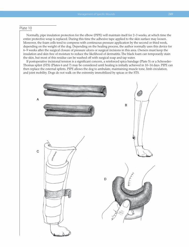
Management of Specific Wounds 249
Plate 10
Normally, pipe insulation protection for the elbow (PIPE) will maintain itself for 2–3 weeks, at which time the entire protective wrap is replaced. During this time the adhesive tape applied to the skin surface may loosen. Moreover, the foam cells tend to compress with continuous pressure application by the second or third week, depending on the weight of the dog. Depending on the healing process, the author normally uses this device for 6–9 weeks after the surgical closure of pressure ulcers or surgical incisions in this area. Owners must keep the insulation and skin free of moisture to reduce the likelihood of dermatitis. The black foam can temporarily stain the skin, but most of this residue can be washed off with surgical soap and tap water.
If postoperative incisional tension is a significant concern, a reinforced spica bandage (Plate 5) or a Schroeder‐Thomas splint (STS) (Plates 6 and 7) may be considered until healing is initially achieved in 10–14 days. PIPE can then replace the external splints. PIPE allows the dog to ambulate, maintaining muscle tone, limb circulation, and joint mobility. Dogs do not walk on the extremity immobilized by spicas or the STS.
A
C
D
B
250 Atlas of Small Animal Wound Management and Reconstructive Surgery
Plate 11 Pipe Insulation to Protect the Great Trochanter
DESCRIPTIONAs noted in Plate 10, pipe insulation is an effective method of protecting the olecranon area after wound closure. The author has adapted its use to help protect the greater trochanter after closure of pressure ulcers.
TECHNIQUE(A) A single 6 foot pipe insulation is centered over the dorsal pelvis. (The adherent borders are compressed to-
gether before application.) Each end is looped along the inner thighs and rotated back to the dorsal pelvis. Elasticon or duct tape are used to secure each limb loop to the pelvic pipe insulation segment. To prevent the pipe insulation from slipping caudally, a loop of Elasticon (Johnson and Johnson) is placed around the caudal abdomen: tape loops are used to secure the dorsal insulation segment to this Elasticon tape girdle.
(B) Lateral view of the trochanteric protector. Additional tape strips can be used to tighten the limb loop (asterisk) at the outer junction of the loop intersection with the dorsal pelvic segment. When the patient reclines, the segment cranial and caudal to the greater trochanter should take the brunt of the pressure when the patient reclines.
COMMENTSTo increase the thickness of the insulation, a smaller diameter pipe insulator can be inserted into a second, larger‐sized, insulation tube. The author normally uses the larger diameter (1 inch) Armcell pipe insulation with a ½‐ or ¾‐inch insulator as an insert. The device can be used for 6 weeks: the device may be changed/adjusted every 2–3 weeks depending on the weight of the dog.

Management of Specific Wounds 251
Plate 11
A
B
252 Atlas of Small Animal Wound Management and Reconstructive Surgery
Plate 12 Vacuum Drain Management of Elbow Hygromas
DESCRIPTIONVacuum drains can siphon fluid accumulating in hygromas continuously: the collapsed hygroma pocket can close after fibrous connective tissue connects the opposing walls. This system is simple to implement and postopera-tive care is minimal. No bandages and the multiple bandage changes associated with the use of Penrose drains can be avoided. The volume of exudate can be quantitated to help determine the best time for drain removal. Normally the drain can be removed in 3–4 weeks. This time frame is selected, as it corresponds to the maturation phase of healing (see Chapter 2).
TECHNIQUE(A) In this example, the patient will be placed in right lateral recumbency. The hair overlying the circumference
of the left elbow and lateral humeral regions is clipped, prepared aseptically for surgery, and draped.(B) A 2‐cm skin incision is created approximately 4 cm below the distal border of the hygroma. A long‐
handled curved forceps with a tapered tip is inserted into the lower wall of the hygroma; the forceps are opened and closed several times to widen the hole before exiting the dorsal wall in a similar fashion. Long, curved forceps are then directed subcutaneously approximately 10 cm dorsal and cranial to the hygroma. A small stab incision is created in the skin, over the tip of the forceps. The tip of the 10‐mm wide flat suction drain (Flat Drain, 10 mm, Tuzik, Boston, Ma, USA) is grasped and retracted to the level of the lower skin incision. (The white fenestrated portion of the drain is 20 cm long; the clear non‐fenestrated external drain component is 80 cm long.)
(C) A small subcutaneous pocket is created with Metzenbaum scissors below the hygroma to accommodate the terminal 6 cm of the fenestrated drain segment. The lower access incision is then sutured closed with 3‐0 monofilament nylon interrupted sutures. The central 8 cm of the fenestrated drain traverses the hygroma pocket, with the remaining 6 cm of the fenestrated drain segment residing in the subcutaneous pocket dorsal to the hygroma. The 4 cm of the nonfenestrated drain segment is inserted subcutaneously followed by application of a purse‐string suture using 2‐0 monofilament nylon. The two strands of this suture are tied around the external drain segment, using a “finger‐trap” suture pattern, to help prevent drain displacement.
(D) Two or three butterfly strips of 1‐inch surgical tape (Zonas Porous Tape, Johnson & Johnson,Skillman, NJ, USA) are applied to the external tubing dorsally, spaced along the length of the drain. 2‐0 monofilament nylon sutures are used to secure the applied tape tabs, on each side of the tubing, to the skin. The drain is attached to the 100 ml vacuum reservoir (Jackson Pratt, Cardinal Health, McGaw Park, Il,USA) and the activated reservoir is secured to the dog’s cervical collar. An Elizabethan collar is advisable to prevent the dog from chewing or displacing the vacuum drain system.
COMMENTSThis is a simple, minimally invasive technique that can be performed as a day case procedure. Patients are normally discharged on a broad‐spectrum antibiotic (Clavamox, Pfizer Animal Health, NY,NY, USA) and the analgesic Tramadol (Tramadol, Amneal, Hauppauge, NY, USA) orally every 12 hours for 7 days and 3 days respectively.
Owners are instructed on the use of the vacuum drain system (see Table 4‐1). Thick, soft bedding is procured for the dog to recline on at home: this is essential for long‐term prevention. On a daily basis, the owner records the volume of fluid as well as its color and clarity. Patient activity is minimized until drain removal.
The patient is examined weekly over the next 3–4 weeks. Skin sutures are removed at the time of the second visit. If drainage is uniformly low over the 3‐week period, the reservoir and drain tube are removed with instruc-tions to continue using soft bedding to protect the elbows from future trauma. If in doubt, retain the drain system for 1 additional week.
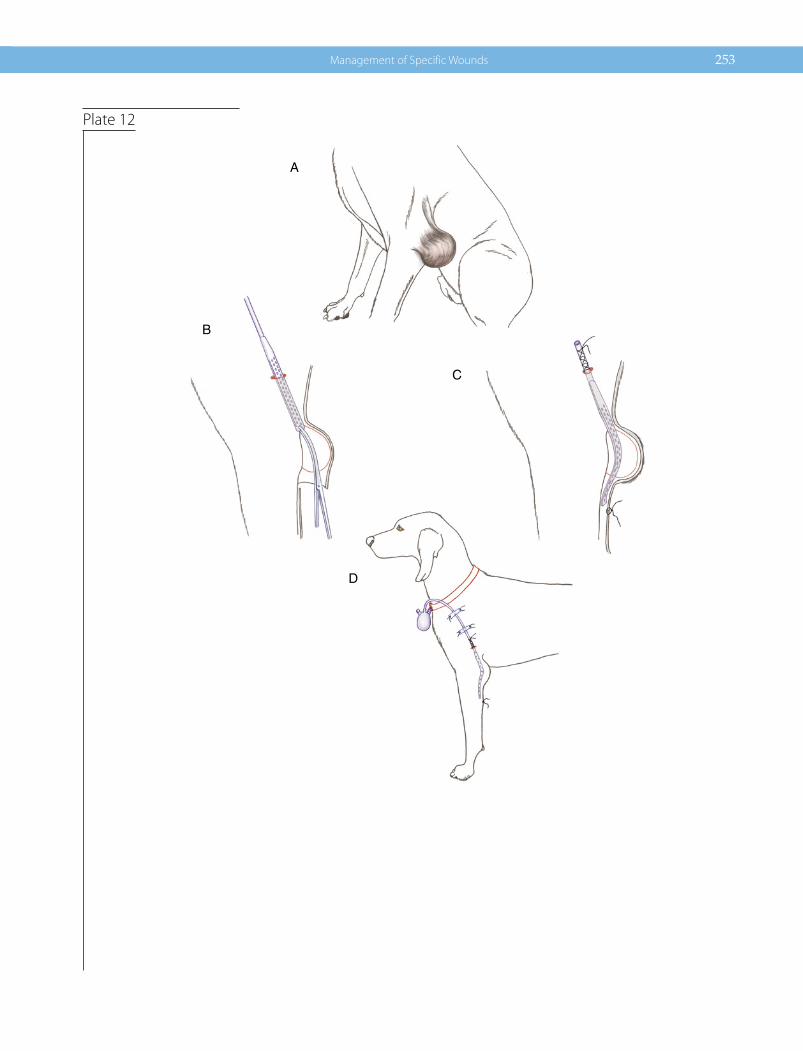
Management of Specific Wounds 253
Plate 12
A
B
C
D
Copyright © 2022 FDOKUMEN