The role of biofilm in wounds - CiteSeerX
341
The role of biofilm in wounds A thesis submitted to the University of Wales, Cardiff in candidature for the degree of DOCTOR OF PHILOSOPHY By Olusola Adeola Okhiria May 2010 The research was undertaken under the auspices of the University of Wales Institute, Cardiff (UWIC) at the Cardiff school of Health Sciences, Western Avenue, Llandaff, Cardiff.
-
Upload
khangminh22 -
Category
Documents
-
view
1 -
download
0
Transcript of The role of biofilm in wounds - CiteSeerX
The role of biofilm in wounds
A thesis submitted to the University of Wales, Cardiff in candidature for
the degree of
DOCTOR OF PHILOSOPHY
By
Olusola Adeola Okhiria
May 2010
The research was undertaken under the auspices of the University of Wales Institute,
Cardiff (UWIC) at the Cardiff school of Health Sciences,
Western Avenue, Llandaff, Cardiff.
ii
DECLARATION
This work has not been previously been accepted in substance for any degree and is not
being submitted in candidature for any degree.
Signed ………………………………………………………………….
(Candidate) Olusola A. Okhiria
Date ………………………………………………………….
STATEMENT 1
This Thesis is the result of my own investigations, except where otherwise stated in the
text. A bibliography is appended.
Signed ………………………………………………………………….
(Candidate) Olusola A. Okhiria
Date ………………………………………………………….
STATEMENT 2
I hereby give consent for my thesis if accepted, to be made available for photocopying
and for inter-library loan, and for the title and summary to be made available to outside
organisations.
Signed ………………………………………………………………….
(Candidate) Olusola A. Okhiria
Date ………………………………………………………….
iii
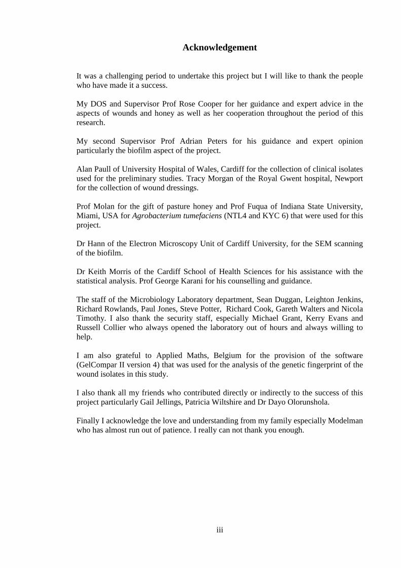
Acknowledgement
It was a challenging period to undertake this project but I will like to thank the people
who have made it a success.
My DOS and Supervisor Prof Rose Cooper for her guidance and expert advice in the
aspects of wounds and honey as well as her cooperation throughout the period of this
research.
My second Supervisor Prof Adrian Peters for his guidance and expert opinion
particularly the biofilm aspect of the project.
Alan Paull of University Hospital of Wales, Cardiff for the collection of clinical isolates
used for the preliminary studies. Tracy Morgan of the Royal Gwent hospital, Newport
for the collection of wound dressings.
Prof Molan for the gift of pasture honey and Prof Fuqua of Indiana State University,
Miami, USA for Agrobacterium tumefaciens (NTL4 and KYC 6) that were used for this
project.
Dr Hann of the Electron Microscopy Unit of Cardiff University, for the SEM scanning
of the biofilm.
Dr Keith Morris of the Cardiff School of Health Sciences for his assistance with the
statistical analysis. Prof George Karani for his counselling and guidance.
The staff of the Microbiology Laboratory department, Sean Duggan, Leighton Jenkins,
Richard Rowlands, Paul Jones, Steve Potter, Richard Cook, Gareth Walters and Nicola
Timothy. I also thank the security staff, especially Michael Grant, Kerry Evans and
Russell Collier who always opened the laboratory out of hours and always willing to
help.
I am also grateful to Applied Maths, Belgium for the provision of the software
(GelCompar II version 4) that was used for the analysis of the genetic fingerprint of the
wound isolates in this study.
I also thank all my friends who contributed directly or indirectly to the success of this
project particularly Gail Jellings, Patricia Wiltshire and Dr Dayo Olorunshola.
Finally I acknowledge the love and understanding from my family especially Modelman
who has almost run out of patience. I really can not thank you enough.

iv
List of publications and presentations
This work has been the subject of the following publications and presentations
Publications
Cooper RA and Okhiria O (2008). The issue of biofilms in wounds. Chapter 12; In
Advances in Wound Care. Ed. White R. Healthcare Comm. UK Limited: Aberdeen
pp189-204.
Rose Cooper and Olusola Okhiria (2006). Biofilms, wound infection and the issue of
control. Wounds UK; 2 (3) 48-57.
Posters and oral Presentations
Okhiria OA, Cooper R.A. and Peters A. (2009) Poster presentation of „‟Detection of
biofilm markers in wound dressings‟‟ at the Society for General Microbiology (SGM)
spring meeting, Harrogate, UK March 30 - April 2, 2009.
Okhiria OA (2009) Oral presentation of „‟ Prevalence of biofilm in wounds” at the
Research Seminar Series of Cardiff School of Health Sciences, UWIC on the 25th
of
February, 2009.
Okhiria O. A (2009) Poster presentation of „‟ A survey of used wound dressings for the
detection of biofilm markers” at the Annual Academic Associate Symposiums UWIC,
January 15, 2009.
Okhiria OA, Cooper RA, Peter A (2007) Poster presentation of „‟Developing an assay
method for screening honey for quorum sensing inhibition of biofilm organisms‟‟ at the
4th
American Society for Microbiology conference on Biofilms in Quebec, Canada
March 25-29, 2007.
Okhiria OA, Cooper RA, Peter A (2007) Poster presentation of „‟Biofilm formation by
Pseudomonas aeruginosa isolated from wounds‟‟ at the 4th
American Society for
Microbiology conference on Biofilms in Quebec, Canada March 25-29, 2007.
Okhiria OA (2006) Oral presentation of „‟ Developing an assay method for quorum
sensing inhibition by honey” at the Cardiff School of Health Sciences Research
Colloquiums, Gregynog, UK on the 16th
of May, 2006.

v
Contents
Contents
List of Tables
List of Figures
Abstract
Chapter 1
1.1
1.2
1.2.1
1.2.1.1
1.2.1.2
1.2.1.3
1.2.2
1.2.3
1.2.4
1.2.4.1
1.2.4.2
1.2.5
1.2.5.1
1.2.5.2
1.2.5.3
1.2.5.4
1.2.6
1.2.6.1
1.2.6.2
1.2.7
1.2.8
1.3
1.3.1
1.3.2
1.3.3
1.3.3.1
1.3.3.2
1.3.3.3
1.3.3.4
1.3.4
1.3.4.1
1.3.4.2
1.3.5
1.3.5.1
1.3.5.1
1.3.5.1
1.3.5.1
1.3.5.1
1.3.6
1.3.7
1.3.8
1.3.9
1.3.9.1
1.3.9.2
1.3.9.3
1.3.9.4
1.3.9.5
1.3.9.6
1.3.9.7
1.3.9.8
1.3.9.9
Page
v
x
xii
xvii
1
3
4
4
5
7
8
10
13
13
13
14
14
14
14
15
15
16
16
18
20
23
23
24
25
25
25
27
28
30
30
30
31
31
32
32
33
34
35
36
38
38
39
40
40
41
41
42
42
42
42
…………………………………………………………………………………………..
…………………………………………………………………………………………...
……………………………………………………………………………………………
……………………………………………………………………………………………
Introduction and Literature Review
Introduction………………………………………………………………………………
Biofilm……………………………………………………………………........................
Biofilm Formation………………………………………………………………………...
Attachment…………………………………………………………………………………………..
Formation of Microcolonies……………………………………………………………………..
Detachment and dispersal of biofilm organisms……………………………………………..
Biofilm physiology ………………………………………………………………………
Quorum sensing …………………………………………………………………………
Factors that influence Biofilm formation…………………………………... ……………
Adherence factors………………………………………………………………………………….
Nutritional Factors……………………………………………………………...........................
Effect of chemical and physical agents on biofilm………………………………………
Indole………………………………………………………………………………………………
Lactoferin………………………………………………………………………………………….
Electric current……………………………………………………………………………………
Water current (Turbulent or laminar flow)……………………………………………………
Resistance of biofilm to immune system and antimicrobial agents …………………….
Mechanism of biofilm resistance to body immune systems…………………………………
Mechanism of biofilm resistance to antimicrobials………………………………………….
Biofilm detection methods………………………………………………………………..
Medical importance of biofilm……………………………………………………………
Wound…………………………………………………………………………………….
Skin and wound formation………………………………………………………………...
Types of wounds…………………………………………………………………………..
Wound Healing Process…………………………………………………………………...
Haemostatic phase………………………………………………………………………………...
Inflammatory phase……………………………………………………………………………….
Proliferative phase…………………………………………………………………………………
Remodelling Phase…………………………………………………………………………………
Classification of Wounds………………………………………………………………….
Acute wounds……………………………………………………………………..........................
Chronic wounds…………………………………………………………………..........................
Microbial burden of Wound………………………………………………………………
Wound contamination……………………………………………………. ………………
Wound colonization……………………………………………………………………………….
Wound infection…………………………………………………………………………………..
Common pathogens in wounds………………………………………………………………….
Surgical site infection (SSI)………………………………………………………………
Diagnosis of wound infections……………………………………………………………
Complications and problems associated with chronic wounds…………………………..
Management and treatment of wounds……………………………………………………
Treatment of infected and chronic wounds ………………………………. ……………..
Wound cleansing and debridement……………………………………………………………
Autolytic debridement……………………………………………………………………………
Maggot (larval) Debridement…………………………………………………………………….
Enzymatic debridement……………………………………………………………………………
Surgical debridement……………………………………………………………………………..
Topical Oxygen Therapy………………………………………………………………………….
Hyperbaric oxygen therapy………………………………………………………………………
Negative pressure wound therapy (NPWT) via vacuum-assisted closure………………….
Antimicrobials……………………………………………………………………………………..

vi
1.3.9.9.1
1.3.9.10
1.4
1.5
1.5.1
1.5.2
1.5.3
1.5.4
1.5.5
1.5.6
1.6
1.7
Chapter 2
2.1
2.1.2
2.1.3
2.1.3.1
2.1.3.2
2.1.3.3
2.1.3.4
2.1.3.5
2.1.3.6
2.1.3.7
2.1.4
2.1.5
2.2.1
2.2.1.1
2.2.1.2
2.2.2
2.2.2.1
2.2.2.2
2.2.2.3
2.2.2.4
2.2.2.5
2.2.3
2.2.3.1
2.2.3.2
2.2.4
2.2.5
2.2.5.1
2.2.5.2
2.2.6
2.2.6.1
2.2.6.2
2.2.7
2.2.8
2.2.8.1
2.2.8.2
2,2,8,3
2.2.8.4
2.2.8.5
2.2.8.6
2.2.9
Page
43
45
45
49
49
49
50
51
51
52
52
53
55
57
58
58
59
60
62
63
65
67
69
70
70
70
70
72
72
73
73
74
74
75
75
75
76
77
78
78
78
78
79
80
80
81
81
81
82
82
83
83
Antiseptics/ topical applications…………………………………………………………………
Surgical interventions…………………………………………………………………………….
Effect of biofilm on wound healing………………………………………......................
Control and potential treatment of biofilm in wounds……………………………………
Interference with biofilm formation…………………………………………………………….
Attenuation of quorum sensing……………………………………………………………
Interference with iron metabolism in biofilm ……………………………………................
Enhancement of immune mechanism ……………………………………………………….
Application of infective agent………………………………………………………………
Other therapeutic agents used for biofilm treatment……………………………………….
Summary / Statement of intent …………………………………………………………….
Aims of the research ………………………………………………………………………..
Characterisation of Pseudomonas aeruginosa isolated from wounds
Introduction………………………………………………………………………………..
Choice of organism for this study…………………………………………………………..
Pseudomonas aeruginosa…………………………………………………………………..
Introduction and general characteristics of Pseudomonas aeruginosa................................
Pathogenicity and virulence factors of P. aeruginosa infections……………………………..
P. aeruginosa opportunistic and nosocomial infections………………………………………
Antibiotic resistance by Pseudomonas aeruginosa……………………………………………..
Mechanism of antimicrobial resistance by Pseudomonas aeruginosa……..........................
Biofilm formation and quorum sensing by P. aeruginosa ……………………………...........
P. aeruginosa in wound infections…………………………………………………………………
Aims ……………………………………………………………………………………….
Materials and Methods……………………………………………………….................
Organisms………………………………………………………………………………….
Sources of organisms………………………………………………………………………………
Maintenance of cultures……………………………………………………………………………
Media and reagent Preparation …………………………………………………………….
Preparation of solid media…………………………………………………………………………
Preparation of liquid media and buffers………………………………………………………….
Preparation of TBE buffer…………………………………………………………………………
Preparation of chelex solution……………………………………………………………………..
Preparation of stock antibiotic solutions…………………………………………………………
Growth Curve of Pseudomonas aeruginosa ………………………………………………..
Estimation of bacteria in culture (Total Viable count)………………………………………….
Turbidimetric estimation of bacteria in culture (optical density)…………………………….
Determination of antibiotic susceptibility of P. aeruginosa wound isolates……………….
Biofilm forming potential of Pseudomonas aeruginosa isolated from wounds……….......
Biofilm formation by P. aeruginosa wounds isolates in microtitre plate……………………
Microtitre plate biofilm reproducibility assay………………………………………………….
Examination of P. aeruginosa wound isolates for AHL production ………………………
AHL detection by cross feeding assay……………………………………………………………
Detection of AHL in P. aeruginosa wound isolates by indirect method (QSI assay)…. ….
Phenotypic characteristics of P. aeruginosa isolated from tap water ………………………
Polymerase chain reaction (PCR)…………………………………………………………
Preparation of DNA Solution and DNA Quantification………………………………………
PCR Master Mix solution………………………………………………………………………….
PCR amplification procedures and PCR optimisation reaction ……………………………..
Agarose gel bed preparation……………………………………………………………………..
Gel Electrophoresis and Staining………………………………………………………………….
PCR of P. aeruginosa wound isolates……………………………………………………………
Data analysis………………………………………………………………………………..

vii
2.2.9.1
2.2.9.2
2.3
2.3.1
2.3.2
2.3.3
2.3.3.1
2.3.3.2
2.3.3.3
2.3.4
2.3.4.1
2.3.4.2
2.3.5
2.3.6
2.3.7
2.3.8
2.4
2.5
Chapter 3
3.1
3.1.1
3.1.2
3.1.3
3.1.3.1
3.1.3.2
3.14
3.15
3.2
3.2.1
3.2.1.1
3.2.1.1.1
3.2.1.1.2
3.2.1.1.3
3.2.1.2
3.2.1.2.1
3.2.1.2.2
3.2.1.2.3
3.2.1.2.4
3.2.1.2.5
3.2.1.2.7
3.2.2
3.2.3
3.3
3.3.1
3.3.2
3.3.3
3.3.3.1
3.3.3.2
3.3.4
Page
83
83
84
84
86
86
86
87
88
89
89
90
92
92
93
97
99
110
111
113
114
114
114
116
118
118
120
120
120
120
121
122
122
122
123
124
125
125
126
126
126
128
128
130
132
133
135
139
Statistical analysis…………………………………………………………………………………
Gel Analysis ……………………………………………………………………………………….
Results and Interpretations……………………………………………………………..
Growth Curve for Pseudomonas aeruginosa NCIB 8626………………………...............
Antibiotic susceptibility assay of P. aeruginosa clinical isolates……………………….. Biofilm formation by P. aeruginosa wound isolates…………………………………….. Interpretations of results…………………………………………………………………………..
Microtitre plate biofilm formation reproducibility assays…………………………………….
Biofilm formation results for all P. aeruginosa wound isolates……………………………...
Production of AHL by P. aeruginosa wound isolates……………………………….........
AHL production by cross feeding assay…………………………………………………………
AHL detection by quorum sensing inhibition (QSI) assay……………………………………
Summary of biofilm markers …………………………………………………………….
Biofilm phenotypic expressions of P. aeruginosa isolated from tap water……………….
PCR characterisation of P. aeruginosa wound isolates…………………………………
Statistical Analysis ……………………………………………………………………….
Discussion ………………………………………………………………………………..
Conclusion………………………………………………………………………………..
The study of physiological and phenotypic characteristics of Pseudomonas
aeruginosa in a wound model
Introduction……………………………………………………………………………
Continuous culture………………………………………………………………………..
Phenotypic expressions of Pseudomonas aeruginosa in biofilm…………………………
Models /wound models for biofilm study…………………………………………………
In vitro models……………………………………………………………………………
In vivo wound models……………………………………………........................ ………
Study hypothesis …………………………………………………………………………
Aim of this chapter ………………………………………………………………………
Materials and Methods ………………………………………………………..............
Developing a wound model ………………………………………………………………
Batch Cultures…………………………………………………………………………....
Batch cultures in flasks for AHL detection at 1 cm distance……………………………..
Batch culture of P. aeruginosa for AHL detection at 1 mm distance……………………..
Determination of ideal distance for detection of AHL in gauze………………………….
Wound model (Continuous culture)………………………………………………………
Design of the wound model………………………………………………………………
Preliminary study of phenotypic diversity of P. aeruginosa strains in continuous culture
wound model with full strength Luria broth………………………………………………
Selection of media for wound model cultures (Effect of nutrient) ………………………
Examining the effect of flow rates of broth on the wound model culture………………...
Determination of reproducibility of assays for wound model culture ……………………
Examining the effect of serum on biofilm formation …………………………………….
Staining procedure for EPS detection and biofilm morphology …………………………
Scanning electron microscopic (SEM) examination of biofilm on the gauze from the
wound model culture……………………………………………………………………...
Results …………………………………………………………………………………...
Detection of AHL from gauze incubated in batch culture………………………………...
The Ideal distance for AHL detection in gauze…………………………………………..
Effect of nutrients on biofilm development in continuous culture ……………………….
Preliminary study of phenotypic diversity of P. aeruginosa strains in continuous culture
wound model …………………………………………………………………………….
Effect of nutrient concentration on biofilm……………………………………………….
Effect of broth flow rate…………………………………………………………………..

viii
3.3.5
3.3.6
3.3.7
3.3.7.1
3.3.7.2
3.4
3.5
Chapter 4
4.1
4.2
4.2.1
4.2.2
4.2.3
4.2.4
4.2.4.1
4.2.4.2
4.2.5
4.2.5.1
4.2.5.2
4.2.5.3
4.2.5.4
4.2.6
4.2.6.1
4.2.6.2
4.2.6.3
4.2.7
4.3
4.3.1
4.3.2
4.3.3
4.3.3.1
4.3.3.2
4.3.3.2.1
4.3.3.2.2
4.3.3.2.3
4.3.4
4.4
4.5
Chapter 5
5.1
5.1.1
5.1.2
5.1.2.1
5.1.2.2
5.1.2.3
Page
141
142
144
144
145
147
154
156
165
166
166
166
166
167
167
167
167
167
168
168
168
169
169
169
170
170
171
171
172
173
173
175
175
177
177
180
182
191
192
193
194
194
194
194
Reproducibility of assays …………………………………………………
Effect of serum on biofilm formation and AHL production ………………
Biofilm structure...........................................................................................
Light microscopic examination of biofilm culture from the wound model...
SEM examination of biofilm culture materials from the wound model……
Discussion………………………………………………………………….
Conclusion …………………………………………………………………
Detection of biofilm markers in chronic wound dressings
Introduction ……………………………………………………………….
Aim and objectives ………………………………………………………..
Materials and Methods ……………………………………….................
Research ethics approval………………………………………………….
Collection of chronic wound dressings …………………………………..
Taking samples from each chronic wound dressing ………………………
Investigations of chronic wound dressings for biofilm markers ………….
Detection of quorum sensing molecules (AHL) in wound dressings ……...
Detection of extrapolysaccharides (EPS) in wound dressings …………….
Isolation and identification of microorganisms in wound dressings……….
Culturing of wound dressings …………………………………………......
Preliminary identification of isolated organisms ………………………….
Maintenance of cultures (isolated organisms‟ stock cultures)…………….
Biochemical Tests………………………………………………………….
Examination of phenotypic characteristics of organisms isolated from
chronic wound dressings …………………………………………………...
Screening Gram negative wound dressing isolates for AHL production….
Screening wound dressings isolates for biofilm formation ……………….
Examination of phenotypic characteristics of Gram negative isolates from
chronic wound dressings in continuous culture wound model…………….
Data analysis ……………………………………………………………….
Results ……………………………………………………………………
Detection of AHL in wound dressings …………………………………..
Detection of extracellular polymeric substances (EPS) in wound dressings
Microorganisms isolated from chronic wound dressings ………………….
Species and frequency distribution of isolated organisms…………………
Biofilm phenotypic expressions of isolated organisms from chronic wound
dressings ……………………………………………………………………
AHL production by Gram negative bacteria isolated from the wound
dressings ……………………………………………………………………
Biofilm forming potential of organisms isolated from chronic wound
dressings …………………………………………………………………..
Biofilm phenotypic characteristics of Gram negative isolates from chronic
wound dressings in wound model continuous culture……………………..
Statistical analysis and correlation …………………………………………
Discussion …………………………………………………………………
Conclusion …………………………………………………………………
Antimicrobial effect of honey on biofilm and quorum sensing
Introduction……………………………………………………………….
Honey and medical history…………………………………………………
Physico-chemical properties of honey……………………………………..
pH of honey…………………………………………………………………
Osmolarity of honey ……………………………………………………….
Hydrogen peroxide activity of Honey ……………………………………..

ix
5.1.2.4
5.1.2.5
5.1.3
5.1.4
5.1.4.1
5.1.4.2
5.1.4.3
5.1.5
5.1.6
5.1.7
5.1.8
5. 2
5.2.1
5.2.1.1
5.2.1.2
5.2.1.3
5.2.2
5.2.3
5.2.3.1
5.2.3.2
5.2.4
5.2.4.1
5.2.4.2
5.2.4.3
5.2.5
5.2.6
5.3
5.3.1
5.3.2
5.3.3
5.3.3.1
5.3.4
5.3.5
4.3.6
5.4
5.5
5.6
Chapter 6
6.1
6.2
6.3
6.4
6.5
Appendices
References
Page
195
196
197 198
198
199
199
203
204
208
210
211
211
211
211
212
212
213
213
214
215
215
215
215
216
216
217
217
218
219
222
222
224
226
229
234
234
236
237
253
257
257
259
266
Phytochemical components of honey………………………………………
Antioxidants properties of honey…………………………………………..
Sterility of honey ………………………………………………………….
Medical importance of Honey ……………………………………………..
Antibacterial properties of honey…………………………………………...
Enhancement of immune function …………………………………………
Application of Honey in wound treatment …………………………………
Biofilm resistance and justification for alternative antimicrobials…………
Recent studies on the antibacterial activities of various honeys……………
Treatment of biofilm embedded in wounds with honey……………………
Aim of the chapter………………………………………………………….
Materials and Methods …………………………………………..............
Organisms and reagents ……………………………………………………
Preparation and maintenance of cultures…………………………………..
Preparation of reagents…………………………………………………….
Honeys used in this study…………………………………………………
Effect of manuka Honey on P. aeruginosa biofilm in a wound model
continuous culture………………………………………………………….
Effect of honey on growth and quorum sensing of C. violaceum…………
Determining growth and quorum sensing inhibitory properties of honey…
Interpretation of results of QSI culture…………………………………….
Developing a screening method for evaluating the growth and quorum
sensing inhibition potential of honeys (QSI assay………………………….
Determination of inoculum size and volume of honey for QSI assay…….
Effect of honey concentration on growth and QSI of C. violaceum ………
Reproducibility test for QSI assay …………………………………………
Evaluating the potency of various honeys with QS assay ………………….
Regression and statistical analysis………………………………………….
Results …………………………………………………………………….
Effect of manuka honey on P. aeruginosa biofilm in a continuous culture
wound model……………………………………………………………….
Growth and quorum sensing inhibitory properties of honey……………….
Effect of inoculum size on QSI assay ……………………………………...
The ideal inoculum size for QSI assay……………………………………..
Effect of honey concentration on QSI assay ……………………………….
Reproducibility of QSI assay……………………………………………….
Growth and quorum sensing inhibitory properties of 10 honeys. …………
Discussion………………………………………………………………….
Conclusion …………………………………………………………………
Recommendation for future studies ……………………………………….
Synopsis, conclusions and recommendations
Summary……………………………………………………………………
Synoptic discussion…………………………………………………………
Conclusions…………………………………………………………………
Limitations of study ……………………………………………………….
Recommendations …………………………………………………………
…………………………………………………………………………….
…………………………………………………………………………….

x
List of Tables
Table 1.1
Table 1.2
Table 2.1
Table 2.2
Table 2.3
Table 2.4
Table 2.5
Table 2.6
Table 2.7
Table 2.8
Table 2.9
Table 2.10
Table 2.11
Table 2.12
Table 3.1
Page
31
33
71
77
86
88
88
90
91
93
97
103
108
109
132
Some predisposing factors to chronic wounds..............................................
Potential wound pathogens…………………………………………………
Sources of Pseudomonas aeruginosa clinical isolates…………………….
Guidelines for interpretation of antibiotic susceptibility (BSAC, 2006)…..
Summary of the susceptibility results of the isolates to each antibiotic……
The summary of microtitre plate biofilm reproducibility assays………….
Data summary: Biofilm formation by P. aeruginosa wound isolates………
Data summary: AHL detection in P. aeruginosa wound isolates (cross-
feeding assay)………………………………………………………………
Data summary: AHL detection in P. aeruginosa wound isolates (QSI
assay)……………………………………………………………………….
Results of Biofilm, QS and QSI for P. aeruginosa from water pipes…….
Correlation between AHL production by isolates and antibiotic
susceptibility………………………………………………………………..
Relationship between biofilm formation and AHL production amongst
P. aeruginosa clinical isolates………………………………………………
Relationship between antibiotic susceptibility and genetic index of
isolates………………………………………………………………………
Biofilm characteristics and genetic similarity index of P. aeruginosa
isolated from wounds………………………………………………………
Colour intensity index of optimisation of AHL detection assay… ………...

xi
Table 3.2
Table 3.3
Table 3.4
Table 3.5
Table 4. 1
Table 4. 2
Table 5. 1
Page
134
138
141
143
171
176
212
Organisms released from gauze after 24 hour culture of each P.
aeruginosa strain in a continuous culture wound model using full strength
Luria broth at 37oC………………………………………………………
Detection of AHL during the development of Pseudomonas aeruginosa
biofilm in wound model continuous cultures……………………………….
Organism released from P. aeruginosa wound isolate biofilm culture at
different broth flow rates in a continuous culture wound model using 1/10
Luria broth………………………………………………………………….
Populations of bacteria released from gauze after each culture of P.98
biofilm using LB, or LB and FCS in wound model ………………………..
Summary of biofilm markers detected in 35 wounds dressings removed
from patients with chronic wounds ………………………………………...
Biofilm markers detected in chronic wound dressings……………………..
Sources and antibacterial activities of the honeys tested…………………...

xii
List of Figures
Figure 1.1
Figure 1.2
Figure 1.3
Figure 1.4
Figure 2.1
Figure 2.2a
Figure 2.2b
Figure 2.2c
Figure 2.3
Figure 2.4
Figure 2.5
Figure 2.6
Figure 2.7
Figure 2.8
Figure 2.9
Figure 2.10
Page
5
10
23
47
58
84
85
85
86
87
90
91
92
94
95
95
The 5 stages of biofilm formation………………………………………….
Networking in 2- component signal transduction…………………………
.
Diagrams of skin structure…………………………………………………
Development of a biofilm in a chronic infected wound: barrier to body
immune and antimicrobial strategies………………………………………
Pseudomonas aeruginosa pigmentation in cultures………………………...
The growth pattern of P. aeruginosa NCIB 8626 in a 24 hour Luria broth
culture………………………………………………………………………
The growth pattern of P. aeruginosa NCIB 8626 in a 24 hour broth culture
Association between optical density and growth of P. aeruginosa NCIB
8626 in 24 hour LB culture ……………………………………………….
Cultures of P. aeruginosa wound isolates on sensitivity agar plates with
antibiotic discs……………………………………………………………..
24 hour biofilm cultures of P. aeruginosa wound isolates in microtitre
plates……………………………………………………………………….
Detection of acylhomoserine lactone (AHL) by cross - feeding assay……..
AHL detection by quorum sensing inhibition assay……………………….
Histogram of biofilm markers of the P. aeruginosa clinical isolates………
Gel electrophoresis of PCR products of P. aeruginosa type culture and
wound isolates……………………………………………………………..
Analysis of bands of marker obtained from PCR at different times………
Fingerprint analysis of PCR products of P. aeruginosa wound isolates
using GelCompar II version 4………………………………………………

xiii
Figure 2.11
Figure 2.12
Figure 2.13
Figure 3.1
Figure 3.2
Figure 3.3
Figure 3.4
Figure 3.5
Figure 3.6
Figure 3.7
Figure 3.8
Figure 3.9
Figure 3.10
Page
96
98
98
121
123
128
130
131
134
135
136
137
137
Dendrogram of the complete linkage between the 97 P. aeruginosa wound
isolates ……………………………………………………………………..
Probability graph of similarity index of P. aeruginosa cohort isolated from
wounds ……………………………………………………………………..
A chart of the distribution pattern of optical densities of biofilms formed
by P. aeruginosa isolates from infected and chronic wounds ……………..
Batch culture wound model………………………………………………..
The Continuous Culture Wound Model……………………………………
Detection of AHL in gauze from 48 hours batch cultures………………….
Detection of AHL in gauze from batch culture placed 1mm from reporter
bacteria……………………………………………………………………..
AHL detection in gauze samples removed from P. aeruginosa 48 hour
batch cultures placed between 1 and 6 mm distances from the reporter
organism…………………………………………………………………….
Phenotypic characteristics of P. aeruginosa strains shown by growth curve
and AHL production in wound model continuous cultures….
AHL detection in culture effluent and sterilized gauze……………………
The 24 hour continuous cultures of a biofilm forming P. aeruginosa
wound isolate (P. 98) in the wound model with various Luria broth
concentrations
Growth curves of 48 hour continuous cultures of a biofilm forming P.
aeruginosa wound isolate (P. 98) in the wound model using various
strengths of Luria broth…………………………………………………….
The wound model continuous cultures of a non- biofilm forming P.
aeruginosa wound isolate (P.51) with various concentrations of Luria
broth

xiv
Figure 3.11
Figure 3.12
Figure 3.13
Figure 3.14
Figure 3.15
Figure 3.16
Figure 3.17
Figure 3.18
Figure 4.1
Figure 4.2
Figure 4.3
Figure 4.4
Figure 4.5
Figure 2.9
Figure 2.10
Page
138
140
140
142
143
144
145
146
172
173
173
174
174
The graphical representation of the continuous cultures of a non- biofilm
forming P. aeruginosa wound isolate (P. 51) in the wound model using
various strengths of Luria broth for a period of 48 hours………………….
Effect of broth flow rate on phenotypic expressions of P. aeruginosa
strains in continuous culture wound model…………………………………
Effect of flow rate on P. aeruginosa wound isolate biofilm formation in
continuous culture wound model…………………………………………...
Graph of reproducibility assay of a biofilm forming P. aeruginosa wound
isolate in wound model continuous cultures………………………………..
The effect of serum on growth of P. 98 and AHL production in the
continuous culture wound model. …………………………………………
Comparison of the growth curve and AHL production of a biofilm forming
P. aeruginosa wound isolate P. 98, in a wound model continuous culture
using LB, FCS and LB (1:1) and 1/5 faecal calf serum in
1/10 Luria broth….
Biofilm of P. aeruginosa wound isolate (P.98) in a wound model
continuous culture over a period of 48 hours………………………………
Scanning electron micrographs of gauze from 30 hour wound model
continuous cultures of P. aeruginosa biofilm and non – biofilm forming
wound isolates………………………………………………………………
Samples of chronic wound dressings tested for AHL………………………
EPS positive and EPS negative smears derived from chronic wound
dressings…………………………………………………………………….
Cultures of Staphylococcus aureus tested for methicillin susceptibility…
Frequency of isolated organisms from chronic wound dressings…………..
Distribution of organisms isolated from each chronic wound dressing…….
xv
Figure 4.6
Figure 4.7a
Figure 4.7b
Figure 4.7c
Figure 4.8a
Figure 4.8b
Figure 4.9
Figure 4.10
Figure 5.1
Figure 5.2
Figure 5.3
Figure 5.4a
Page
175
177
178
178
179
179
180
181
214
218
219
221
AHL positive and AHL negative Gram negative bacteria recovered from
chronic wound dressings……………………………………………………
Growth pattern of a Gram negative isolate P. aeruginosa M 16(5) from a
chronic wound dressing in continuous culture wound model………………
Biofilm developments of a wound dressing isolate P. aeruginosa M16 (5)
in wound model continuous culture (x10 objective)……………………
Biofilm development of a P. aeruginosa M16 (5), isolated from a wound
dressing in wound model continuous culture (x100 objective)…………….
Growth pattern of a Gram negative isolate, B. cepacia M 3(1), from a
chronic wound dressing in a wound model culture…………………………
Biofilm developments of a B. cepacia M3 (1) isolated from wound
dressing in wound model continuous culture (x100 0bjective)……………
Graphical representation of the relationship between AHL detection and
isolation of Pseudomonas aeruginosa from chronic wound dressings……..
Graphical representation of the relationship between detection of EPS in
chronic wound dressings and formation of biofilm by the isolated
organisms on the dressings………………………………………………….
Diagram of honey QSI assay………………………………………………..
A chart showing the effect of honey on biofilm in a wound model
continuous culture………………………………………………………….
Quorum sensing inhibition effect of honey on C. violaceum ATCC 31532
in LB agar…………………………………………………………………..
Effect of inoculum size of C. violaceum on honey inhibitory properties
using 50 μl of 50% (w/v) M109 honey solution in each well………………
xvi
Figure 5.4b
Figure 5.5
Figure 5.6a
Figure 5.6b
Figure 5.6c
Figure 5.6d
Figure 5.7a
Figure 5.7b
Figure 5.7c
Figure 5.7d
Page
222
223
224
225
225
226
227
227
228
228
Effect of inoculum size of C. violaceum on honey inhibitory properties
100 μl of 50% (w/v) M109 honey solution in each well……………………
Graphical representation of the effect of honey concentration on growth
and quorum sensing inhibitions of C. violaceum…………………………
Cultures showing the effect of honey concentrations on the growth and
quorum sensing of C. violaceum…………………………………………...
The effect of various concentrations of a medical grade honey (Activon® -
UMF 12) on the growth and quorum sensing of C. violaceum…………….
The effect of various concentrations of a manuka honey UMF 10+ on the
growth and quorum sensing of C. violaceum………………………………
The effect of various concentrations of a manuka honey UMF 5+ on the
growth and quorum sensing of C. violaceum…………………………………..
Growth and quorum sensing inhibition of C. violaceum by
chloramphenicol……………………………………………………………
Assessment of potency of honeys using growth inhibition of C. violaceum
Assessment of potency of honeys by QSI assay……………………………
Combined effect of each honey on growth and quorum sensing of C.
violaceum relative to concentrations……………………………………….

xvii
Abstract
Biofilms have long been implicated in persistent infections and have recently been
associated with chronic wounds. The role of bacteria in wounds is not yet fully
understood and their ability to form biofilm is yet to be fully elucidated. Biofilms are
associated with phenotypic features such as the signalling molecules for regulation of
activities within biofilm and secretion of extracellular polymeric substances (EPS). The
presence of biofilm may be confirmed by specialised microscopy techniques or by
detecting biofilm markers. Routine methods are not yet available for detecting biofilms
in wounds. The aims of this project were to investigate the role of biofilm in wounds, by
examining the ability of wound isolates to form biofilm and produce signalling
molecules and by developing a wound model; to relate laboratory findings with in vivo
activity by exploring the possibility of detecting biofilm markers in dressings removed
from chronic wounds.
Biofilm characteristics of 97 Pseudomonas aeruginosa strains isolated from wounds
were investigated. Their antibiotic susceptibility to commonly used antibiotics was
determined. The isolates were examined for ability to form biofilm and to produce acyl-
homoserine lactone (AHL) signalling molecules in batch culture and were characterised
using PCR. A wound model was developed for biofilm continuous culture using gauze
as the substratum for biofilm attachment and culture effluent was examined for AHL
production and detached fragments of biofilm. Gauze removed from the culture at 30
hours was examined with scanning electron microscopy (SEM). Thirty five dressings
removed from chronic wounds were investigated for the presence of biofilm markers
including AHL and EPS. Organisms from the wound dressings were isolated and
examined for biofilm formation, AHL and EPS. Thirty hour biofilm of P. aeruginosa
wound isolate was exposed to 40 % (w/v) honey in wound model continuous culture.
The potential of some honeys to inhibit the growth and quorum sensing (QS) of a
biofilm forming organism, Chromobacterium violaceum was investigated.
Over 90% of the 97 cultures of P. aeruginosa were resistant to 3 antibiotics while
resistance to 4 others ranged between 3 – 19%. Eighty eight of the isolates (90.72%)
formed biofilm while 78 (81.4%) produced AHL. PCR characterisation of the isolates
showed that 82 (84.53%) have 100% genetic similarity linkage to the cohort, 3 (3.09%)
have 75 - 99% while 12 (12.37%) are of 50 - 75% linkage. Examination of stained
culture effluent smear from wound model revealed biofilm embedded in EPS and AHL
was detected in sterilised culture effluent. SEM examination of gauze removed from
wound model after 30 hours culture confirmed biofilm structures. Eleven (31.4%)
wound dressings tested positive for AHL, 28 (80%) contained EPS (4 not tested for
EPS). Organisms that formed biofilm were isolated from 32 (91.4%) dressings while
Gram negative bacteria that produced AHL were isolated from 13 dressings. Two
selected Gram negative bacteria from wound dressing that were cultured in wound
model showed progressive biofilm formation with EPS and AHL production. The 30 hr
biofilm exposed to honey was dislodged within 6 hours and no viable organism was
recovered from culture. Honey inhibited the growth and QS of C. violaceum in a dose
dependent manner. Of the 10 honeys examined, 8 (80%) inhibited the growth and
quorum sensing of C. violaceum, 1 slightly inhibited quorum sensing while 1 showed no
inhibitory effect.
Indicators of biofilms detected in used wound dressings have the potential to be used in
the diagnosis of biofilms in chronic wounds. The antimicrobial effect of honey on
biofilm and quorum sensing as shown in this study suggests that application of honey in
wound management will provide effective treatment for wounds with biofilm.

Chapter 1
Introduction and Literature Review
Chapter 1 Introduction and literature review
1
1.1 Introduction
The ability of a microorganism to establish an infection depends on a number of factors
which include those of the host and the pathogen. The pathogenic factors are as
numerous as the host factors and diverse in their functions. The number of organisms
present at the site of infection could overwhelm the host defence system (Gardner et al,
2004; Sibbald et al, 2003) and to a large extent possession and expression of virulence
factors (Tang et al, 1996) by the organisms often determine the progression of microbial
colonisation to infection (Jensen et al, 2007; Williams et al, 2000) even in immune
competent individuals.
Infection occurs when colonisation of a host tissue by organisms results in invasion
from where the organisms are disseminated to the surrounding tissues causing diseases
locally or in remote parts of the body (Kadioglu et al, 2008). The attachment of
organisms to the cells or mucosal surfaces is mediated by adhesins which are usually
located at the tip of pili or fimbriae (Lasaro et al, 2009; Choy et al, 2007; Wu et al,
2007; Coutte et al, 2003; Johnson and Stell, 2000). The studies of host pathogen
relationship have shown that pathogenic organisms employ diverse strategies to
circumvent the innate defence mechanism of the host to establish infections (Cote et al,
2006; Leid et al, 2005). These strategies include the production of cytotoxic enzymes
and toxins to breakdown the host tissue for growth and other metabolic activities and
formation of protective shields against host defence and antimicrobial interventions.
Organisms like Pseudomonas aeruginosa produces enzymes like hyluronidase and
gelatinase (Naglik et al, 2003), and exotoxins (Johnson and Stell, 2000; Caldwell et al,
2009; Hamood et al, 1996) aggregation factors (clumping proteins) (Hall-Stoodley et al,
2004; Kayaoglu and Ørstavik, 2004) which have profound damaging effect on host
tissues sometimes leading to organ failure or death. Pathogenic effects on the host are
often times affected by the organism‟s ability to secrete protective shields such as
capsules (Cortés et al, 2002; Fritz et al, 2000), cysts (Fukasawa et al, 2006; Gebbers
Chapter 1 Introduction and literature review
2
and Marder, 2005), spore (Cote et al, 2006; Hoa et al, 2001), extracellular
polysaccharides (Feldmesser et al, 2001) and biofilm (Leid et al, 2005; Boyd A and
Chakrabarty, 1995). Studies have shown that protections offered by shields like biofilm
enhance the ability of pathogens to establish chronic infections (Brady et al, 2008;
Wolcott and Ehrlich, 2008; Donlan and Costerton, 2002). The roles of adhesins as an
important factor in chronic infections have been studied (Mahdavi et al, 2002) and
association with biofilm formation has been demonstrated (Peng et al, 2008; Bu et al,
2008; Froeliger and Fives-Taylor, 2001). The genetic units that are responsible for the
virulence capability of an organism are usually located within the genome at the
pathogenic islands (Schmidt H and Hensel, 2004; Hansen-Wester and Hensel, 2002).
Regulation of these wide varieties of virulence factors by several species of pathogenic
organisms has been associated with the production of diffusible chemical substances
known as quorum sensing molecules (QSMs) which are population density dependent
(Williams et al, 2007; Fuqua et al, 1996 and 1994). It has been shown that QSMs
known as autoinducer -1 are mainly utilised amongst the same species whereas
autoinducer - 2 bring about universal signal for interspecies communications amongst
several organisms (Atkinson and Williams, 2009; Williams, 2007; Sun et al, 2004).
The role of quorum sensing molecules in modulating virulence factors in pathogenic
organisms have been widely studied and reviewed; and include pigment production
(Wang et al, 2008; Fothergill et al, 2007), conjugation (Fuqua et al, 1994; Zhang et al,
1993), sporulation (Bassler and Losick, 2006; Camilli and Bassler, 2006; Enjalbert
and
Whiteway, 2005) production of enzymes (Zhu et al, 2002 b) and toxins (Ohtani et al,
2002; Jensen et al, 2007) and biofilm formation (Waters et al, 2008; Christensen et al,
2007).
Chapter 1 Introduction and literature review
3
1.2 Biofilm
Biofilm is a complex, heterogeneous and integrated community of surface attached
microorganisms of either single or multiple species that are encased within the
extracellular polymeric matrix produced by them (Chang et al, 2007; Sutherland,
2001; Costerton et al, 1999). They have been found attached to solid (abiotic) surfaces
including industrial water systems as well as medical environments and devices
(Jacobsen et al, 2008; Donlan and Costerton, 2002) and mucosal surfaces in humans
(biotic) (Hall-Stoodley et al, 2004; Costerton et al, 1987).
In the natural environment where it has been postulated that 99.9% of bacteria grow in
biofilms and in human infections biofilm communities are often characterized by
genetic diversity involving multiple microbial species (Donlan, 2002; Donlan and
Costerton, 2002). The intriguing fact about the population diversity of biofilm was said
to have been revealed when the dental plaque from the oral cavity of a volunteer was
found to contain up to 800 phylotypes (Palmer, 2009).
The major reasons for biofilm formation in the host include protection from the harmful
effects of the host defence system, effective colonisation and confiscation of a nutrient-
rich environment, utilization of cooperative benefits amongst the biofilm organisms and
innate ability of organisms to exist as biofilm (Jefferson, 2004). Protection of biofilm
against immune mechanisms has been shown (Cerca et al, 2006; Leid et al, 2005;
Davey and O‟Toole, 2000).
Although biofilms are beneficial to mankind in processes such as biodegradation (Mor
and Sivan, 2008; Zhang et al, 1995) and sewage treatment (Wu et al, 2006; Tsuno, et al,
2001) various problems have been associated with biofilms, including corrosion of
pipes (Teng et al, 2008; Keevil, 2004) and involvement with persistent infections in
animals and humans particularly in cystic fibrotic, surgical site, orthopaedic and wound
infections (Driffield et al, 2008; James et al, 2008; Gilbert et al, 1997; Anderson and
O'Toole, 2008; Costerton et al, 1987). The phenotypic and biochemical properties of

Chapter 1 Introduction and literature review
4
biofilm organisms are unique and distinct from their planktonic counterparts and enable
the organisms to greatly resist body immune system and antimicrobials hence biofilm is
associated with persistent infections (Costerton et al, 1999; Costerton, 2001).
1.2.1 Biofilm Formation
Most bacteria grow in a free-living planktonic state, but some are able to exhibit
different phenotypes which differ in physiological characteristics including structural
and metabolic changes (Sauer et al, 2002). Sauer and colleagues (2002) observed
P. aeruginosa converted from planktonic to sessile form and grew as biofilm, a complex
process that requires coordinated activities. The main stages of biofilm formation are
reversible attachment,
irreversible attachment, maturation-1, maturation-2,
and
dispersion (Figure 1. 1). These various stages of biofilm formation are initiated when
the planktonic organisms transform to the sessile form (O'Toole et al, 2000). Each of
the stages has distinguishing characteristic features (Sauer et al, 2002) and requires
regulation by quorum sensing molecules (Pearson et al, 1994).
1.2.1.1 Attachment
Motile (planktonic) bacteria transform to the sessile form prior to biofilm formation as
they adhere to a favourable surface; such as a medical device or the host tissue. In some
cases initial adhesion of biofilm forming microorganisms is achieved by means of
adhesins located on specialised organelles such as fimbriae (pili) (Lasaro et al, 2009;
Sauer et al, 2002). There are 2 stages of attachment; the reversible attachment occurs
when the organisms are able to revert back to the planktonic form and move away from
the surface of attachment but at the irreversible stage the organisms are attached and
biofilm formation is initiated.

Chapter 1 Introduction and literature review
5
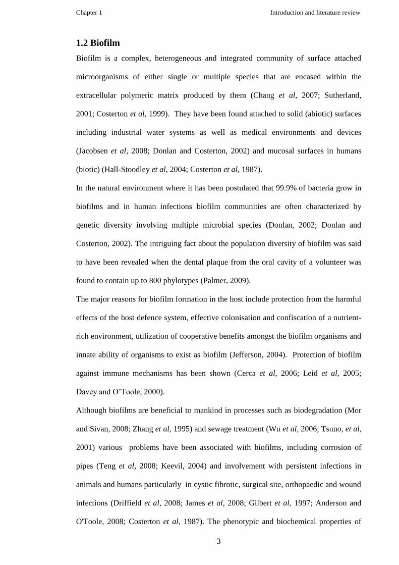
Figure 1.1 The 5 stages of biofilm formation (Monroe, 2007). The diagram and the
corresponding microscopic examination of biofilm growth showing (1) Initial
attachment; (2) irreversible attachment; (3) biofilm maturation I (4) biofilm maturation
II (5) dispersion. Each stage of development in the diagram is paired with a micrograph
of a developing P. aeruginosa biofilm.
1.2.1.2 Formation of Microcolonies
The cells aggregate as they divide on adhesion to a surface but the daughter cells
multiply outward and upward from the point of attachment to form cell clusters. The
dividing cells produce quorum sensing molecules and extracellular polymeric
substances (EPS), or polymer matrix. The matrix houses the aggregating cells in
microcolonies and also attaches the biofilm to the surface on which it is formed
(Watnick and Kolter, 1999; McKenney et al, 1998). Microcolonies become larger as
the number of organisms increase and the quantity of EPS produced also increases.
More signalling molecules and EPS are produced by the organisms within the
microcolonies at this stage. It has been found that microorganisms are usually not
distributed evenly throughout the biofilm but in aggregates of microcolonies although
Chapter 1 Introduction and literature review
6
flagellum - driven movement within biofilm have been observed amongst bacteria in
biofilm in vitro (Malic et al, 2009; Tolker - Neilsen et al, 2000). The fully mature
biofilm structure comprises of bacterial cells, the polymer matrix, and interstitial water
channels that facilitate the exchange of nutrients and wastes in and out of the biofilm
into the surrounding environment (Stoodley et al, 2002; Costerton et al, 1995; Sauer et
al, 2007). Expressions of some genes such as the one for alginate biosynthesis (algC) in
P. aeruginosa (Davies and Geesey, 1995; Davies et al, 1993) and wca locus in
Escherichia coli are usually upregulated during biofilm formation while certain genes
like those for the syntheses of flagellin (fliC) in E. coli are repressed (Prigent-Combaret
et al 1999) and expression of several genes have been observed during biofilm
formation (Goldsworthy 2008; Schembri et al, 2003; Sauer et al, 2002). It was found
that the average difference in detectable protein regulation between each of the stages of
development was 35% which were approximately 525 proteins and when planktonic
cells were compared with maturation-2 stage biofilm cells, more than
800 proteins were
upregulated at least six fold the level of planktonic cells, an expression level which was
over 50% of the proteome. P aeruginosa displayed multiple phenotypes during biofilm
development and biofilm cells at dispersion were found to be similar to the planktonic
cells in phenotypic expression. The results obtained for P. putida biofilm was higher
than those of P. aeruginosa showing the uniqueness of each organism (Sauer et al,
2002). In a similar study of the gene expression of E. coli biofilms, Schembri and his
colleagues (2003) observed 206 genes were upregulated and 27 down regulated in E.
coli biofilm than exponential phase while 389 were upregulated and 192 down regulated
in biofilm than during the stationary phase of E. coli culture. In summary, biofilm
formation is a multifactorial process which is a collective product of a variety of
interactions and adaptive responses of the organisms within the biofilms.

Chapter 1 Introduction and literature review
7
1.2.1.3 Detachment and dispersal of biofilm organisms
The biofilm environment is innately regulated and studies have shown that high
population density within a mature biofilm induced programmed detachment of bacteria
from biofilm through the secretion of chemical substances by the organisms (O'Toole et
al, 2000). Studies have shown that detachment occurs when the organisms respond to
chemical substances secreted by them such as signalling molecules (Stoodley et al,
2005), proteins and degradative enzymes (Barraud et al, 2006; Boyd and Chakrabarty
1995) and oxidative or nitrosative stress-inducing molecules such as nitric oxide (NO)
produced as a result of metabolic processes within a biofilm (Rice et al, 2005; Wood et
al, 2007; Schlag et al, 2007). It has also been shown that alginate lyase; a degradative
enzyme produced by biofilm organisms cleaves the polymer matrix into short
oligosaccharides. The cleavage antagonises the attachment characteristics of alginate
leading to increased detachment of biofilm organisms (Barraud et al, 2006; Boyd and
Chakrabarty, 1995)
. The presence of optimal amounts of nutrients has been observed as
an inducing factor for dispersal of biofilm organisms by increasing the growth of
organisms and production of autoinducers which usually aid the dispersal processes
within the biofilm (Rice et al, 2005). However other studies have shown detachment of
biofilm due to nutrient starvation (Hunt et al, 2004; Thormann et al, 2001; Delaquis et
al, 1989). Detachment processes enhance the sloughing of biofilm and switching of
sessile organisms within the biofilm to the planktonic, free-swimming phenotypes
which leave the biofilm. The detached organisms disperse to other locations to
recommence biofilm formation (Barraud et al, 2006); a process that aids the spread of
biofilm infections within a host and sometimes this might cause thromboembolism
which could lead to death (Wenzel, 2007). Intriguingly it has been observed that
biofilms with thick extrapolysaccharides have less chance of detachment which tends to
reduce the spread of infections (Xavier and Bassler, 2003).
Chapter 1 Introduction and literature review
8
1.2.2 Biofilm physiology
Biofilm has a complex physiology due to the collective effect of the activities of the
various organisms within the biofilm. The knowledge about biofilm physiology was
highly important in understanding the activities of biofilm organisms for effective
control and has become available through the use of various methods to examine the
ultra-structure including electron microscope (Davis et al, 2008; Stickler et al, 1998),
staining and examination with light microscope (Davis et al, 2008; Harrison – Balestra
et al, 2003; Serralta et al, 2001) epifluorescent microscope (Davis et al, 2008; Tolker -
Neilsen et al, 2000) confocal laser scanning microscope (CLSM) (Percival et al, 2008 a)
and detection of chemical products such as the quorum sensing molecules (Nakagami et
al, 2008; Singh et al, 2000; Stickler et al, 1998). The architecture is mainly influenced
by the extracellular polymeric substances (EPS) produced by the individual residents
and have been used in the study of biofilm. The EPS is often composed
of
polysaccharides (alginate and levan) (Laue et al, 2006), membrane vesicles (MVs)
(Schooling and Beveridge, 2006), proteins, lipids, enzymes and nucleic acids
(Steinberger and Holden, 2005). Although EPS aid the adhesion of biofilm to surfaces
and tissues but the main function is to serve as the niche within which biofilm
organisms inhabit. The EPS form the bulk of the biofilm architecture (Chang et al,
2007; Laue et al, 2006) forming 75 -90 % of biofilm while only 10 - 25% are cells.
Although alginates influence the architecture of biofilms by facilitating the non-specific
attachment of organisms to surfaces, holding together biofilm organisms during initial
development and reducing loss of water within the biofilm. However, it has been shown
that EPS such as alginate might not be necessary for biofilm formation (Stapper et al
2004; Wozniak et al, 2003). Biofilms of different species exhibit unique arrangements
of the cellular and extracellular structural components. Pseudomonas biofilms cells
were found to be most dense at the attachment surfaces and became increasingly diffuse

Chapter 1 Introduction and literature review
9
near the outer regions, whereas Vibrio biofilms exhibited the opposite trend (Lawrence
et al, 1991).
In general, biofilms are usually highly hydrated, with the open structures comprising of
up to 73 to 98% extracellular materials and space (channels) (Lawrence et al, 1991).
Chemical gradients (pH, redox potential, and ions) are known to occur usually within
the biofilm due to the various degrees of diffusion of nutrients, metabolic products, and
oxygen in all parts of the biofilm (Hunter and Beveridge, 2005). The rate of growth and
development of biofilm organisms are greatly influenced by chemical gradient, the
organisms at the biofilm-liquid interface having the fastest growth as against the much
slower rate of those in the interior of the microcolonies, possibly due to limited access
to nutrients (Møller et al, 1996). The cells within biofilm are able to maintain
intracellular pH homeostasis thus enhancing their physiological condition and acid
tolerance (McNeill and Hamilton, 2004). Transportation of nutrients within biofilm is
achieved through the water channels which allow intake of nutrients into the cells and
excretion of waste products into the surrounding environment (deBeer et al, 1994 and
1996; De Beer and Stoodley; 1995). Biofilms constituents are diverse as the microbes
within them especially the multispecies biofilms. Production of quorum sensing
molecules sometimes depends on the site of infection (Favre-Bonte et al, 2007) and
quorum sensing dependent phenotypes also vary according to the site where the
organisms are isolated. Various differences have been observed amongst the biofilms
from the wound, the dental plaque and those from the natural ecology like the rocks or
water especially in their microbial population (Reardon et al, 2004) as well as their
extracellular matrix (Sutherland, 2001; Branda et al, 2005) and production of virulence
factors (Favre-Bonte et al, 2007).

Chapter 1 Introduction and literature review
10
1.2.3 Quorum sensing
Microorganisms are capable of communicating with each other to coordinate complex
activities like multicellular organisms in addition to the basic role of nutrients
metabolism, growth and multiplication. Adaptation to an environment by
microorganisms involves response to a series of changes via signal transduction
systems, comprising of protein kinase that phosphorylates itself using adenosine
triphosphate and response regulator which accepts the phosphoryl groups (Stock et al,
1989; Barrett and Hoch, 1998) (Fig. 1.2).
Figure 1.2: Networking in 2- component signal transduction (Barrett and Hoch,
1998).
These coordinated activities are achieved through cell - cell communications signal
molecules (autoinducers) (Parsek and Greenberg, 2005; Williams et al, 2007), which are
produced by each of the organisms involved in biofilm formation (Yao et al, 2007;
Mcdougald et al, 2006; Rice et al, 2005).

Chapter 1 Introduction and literature review
11
Quorum sensing is a process by which organisms monitor and respond to the presence
of other organisms within the environment through the production of signalling
molecules. In biofilm, regulation of metabolic activities and population density is
controlled by a cell density-dependent gene expression as a result of accumulation of
signalling molecules in the medium. The autoinducer molecules produced by individual
microorganisms diffuse through the cell and accumulate in the medium and once a
threshold level is attained due to large numbers of organisms, the entire population of
microorganisms within the environment respond to the critical cell mass via
transcriptional regulation of various target genes (Sauer et al, 2002; de Kievit and
Iglewski 2000). Studies have shown that QS controls the processes of biofilm
formation in microorganisms and detection of other bacteria is through the secretion and
detection of autoinducer molecules that regulate mRNA production for specific genes in
response to the population density signal (Williams et al, 2007; Fuqua et al, 1996).
In Gram-negative bacteria such as P. aeruginosa, there are two types of autoinducers
(AIs); AI-1 and AI-2. AI-1 molecules are N-acyl-homoserine lactones (AHL) and AI-2,
a furanosyl borate diester. The AHL regulatory system consists of two structural genes;
the luxI that encodes the AI-1 synthase and luxR that encodes the AI-1 response
regulator. LuxI and LuxR homologues are present in a wide variety of Gram-negative
bacteria and control several genes processes including production of virulence factors
such as toxins, enzymes, biofilm formation and antibiotic resistance (Passador et al,
1993; Xu et al, 2000 & 2006; Patel, 2005; Sakuragi and Kolter, 2007; Kendall and
Sperandio, 2007). The gene responsible for AI-2 production (luxS) is highly conserved
across numerous species and it also has the ability to regulate gene expression in other
bacterial species (inter-species communication) and it has been correlated with
pathogenicity of several organisms whereas the AI-1 such as AHL is typical of intra-
species communication (de Kievit and Iglewski, 2000). In general quorum sensing plays

Chapter 1 Introduction and literature review
12
a key role in the regulation of biofilm architecture (Liu et al, 2007); dispersion of
organisms (Rice et al, 2005) and virulence factors (Schuster et al, 2003; Fuqua et al,
2001; Rumbaugh et al, 1999). The importance of QS in pathogenicity was
demonstrated in burn wounds of mice infected with P. aeruginosa (wild and mutant
strains). The colony forming units (CFU) of organisms recovered from the livers,
spleens and skins of mice with infected mutants were significantly lower than those of
the wild type (Rumbaugh et al, 1999). In another study, mice with implants were
infected with P. aeruginosa and on examination of the implants at days 4, 7, 14 and 21
post infection, the CFU of organisms on the implants were significantly lower in mutant
strains than the wild type suggesting that the clearance of organisms from the implants
by the immune system of the mice was QS dependent (Christensen et al, 2007).
Although quorum sensing molecules are required for biofilm formation, interestingly
some strains of pathogenic organisms such as P. aeruginosa have been found to
produce biofilm independent of quorum sensing (Favre – Bonte et al, 2007; Schaber et
al, 2004 and 2007) but QS is required for full biofilm formation (Favre – Bonte et al,
2003). It was also found that these QS deficient strains were able to cause infections
and are less susceptible to antimicrobials (Favre- Bonte et al, 2007 and 2003) indicating
some virulence factors do not depend on quorum sensing molecules (Schaber et al,
2007 b) and that QS dependent phenotypes vary according to the site of isolation of the
organisms (Favre-Bonte et al, 2007).
Because of the role of QS in the regulation of virulence factors including biofilm
formation, blockage of quorum sensing in pathogenic organisms has therefore been
suggested as a novel treatment strategy especially in the control of biofilm infections
(Bjarnsholt and Givskov, 2007). It has been shown that attenuation of P. aeruginosa QS
by garlic resulted in rapid clearing of P. aeruginosa from the lungs of mice models
(Bjarnsholt et al, 2005a) and susceptibility of P. aeruginosa biofilms to antimicrobials
Chapter 1 Introduction and literature review
13
(Bjarnsholt et al, 2005b; Rasmussen et al, 2005a) and reduced the killing of
Caenorhabditis elegans cells (Rasmussen et al, 2005a) and rapid clearance from the
lungs of infected mice (Rasmussen et al, 2005b).
1.2.4 Factors that influence Biofilm formation
Biofilm formation is influenced by various factors ranging from the microbial
physiology and phenotypic characteristics, environmental stress, nutritional
requirements to the presence of chemical agents.
1.2.4.1Adherence factors
The ability of the organisms to adhere to surfaces as well as the rate of adherence will
influence biofilm formation (Shanks et al, 2007; Lasaro et al, 2009). The twitching
motility of biofilm organisms which has been associated with type IV pili determines
the rate of adherence which consequently influences the formation of biofilm (O'Toole
and Kolter,1998). Flagella and pili therefore play significant roles at the initial stage of
biofilm formation since they are required for adherence (O'Toole and Kolter, 1998).
1.2.4.2 Nutritional Factors
Biofilm formation depends on availability of nutrients and also varies amongst species
(Mcdougald et al, 2006). It has been demonstrated that increased amount of nutrients
enhanced the production of quorum sensing molecules, enzymes and other essential
amino acids necessary for the formation and growth of biofilm (Rice et al, 2005;
Heydorn et al, 2002).

Chapter 1 Introduction and literature review
14
1.2.5 Effect of chemical and physical agents on biofilm
1.2.5.1 Indole
Indole, secreted by some gram negative organisms, is one of such substances which
have been shown to affect biofilm formation. It was observed that indole reduced
biofilm formation in organisms such as Escherichia coli that synthesize it whereas it
increased biofilm formation by some other organisms such as P. aeruginosa that do not
synthesize it (Lee et al, 2007).
1.2.5.2 Lactoferrin
The presence of certain substances in biofilm culture has a substantial effect on biofilm
formation and sustenance. Iron in low concentration favours the growth of biofilm but
reduces the rate of release of deoxyribonucleic acid (DNA) in cells at higher
concentrations thereby reducing the rate of biofilm formation (Yang et al, 2007; Banin,
et al, 2005; Banin et al, 2006; Dinty et al, 2005). Lactoferrin commonly found in
human secretions such as tears, airways secretions and milk also prevents biofilm
formation by chelating iron thereby initiating twitching motility of the organisms which
causes the organisms to move around and prevents them from attaching to surfaces to
form biofilm (Singh et al, 2002). However once the biofilm has formed biofilm
becomes resistant to lactoferrin (Singh et al, 2002).
1.2.5.3 Electric current
Application of electric currents to biofilms has shown reduction of biofilms with time
(van der Borden et al, 2004) hence its application in improving the efficacy of
antimicrobial killing on biofilm was explored (Caubet et al, 2004). Bioelectric
application in addition to exposure to antimicrobial provides synergistic killing effect on

Chapter 1 Introduction and literature review
15
biofilm cells especially the organisms that are susceptible to the antimicrobials in the
planktonic forms (Jass and Lappin-Scott, 1996).
1.2.5.4 Water current (Turbulent or laminar flow)
The flow of water or liquid either in the natural environment such as the ecosystem that
has high shear forces (turbulent) seems to enhance bacterial adhesion and biofilm
formation (Donlan and Costerton, 2002). In the laboratory the rate of flow has been
observed as a factor that affects biofilm formation, but the rate of flow can be
controlled, either a turbulent or the low flow rate (laminar) to suit the purpose of the
study. The effect of fluid flow has been shown to influence the tensile strength of
biofilms and the sloughing of organisms within biofilm, the tensile strength of biofilm is
higher with turbulent flow and the sloughing is reduced (Donlan and Costerton, 2002).
The impact of flow rate on QS was investigated by Kirisits and colleagues (2007) using
laminar flows (0.04 ml/min and 4.0 ml/min) and turbulent (380ml/min) and found that
biofilm growth was achieved in both laminar and turbulent flows. However it was
observed that the biomass required for full QS induction for the laminar flow rate was at
bacterial population of about 6x10⁷
cfu/ml whereas full QS induction of biofilm
population at the turbulent flow rate was not observed. The detection of QS is therefore
dependent on the rate of fluid flow and may not be fully operative at high flow rate.
1.2.6 Resistance of biofilm to immune system and antimicrobial agents
Biofilm organisms are known to cause persistent infections due to their inherent
resistance to body immune mechanism and antimicrobials (Leid et al, 2002 and 2005;
Webster et al, 2006; Chandra et al, 2001; Hentzer et al, 2001; Costerton et al, 1999;
Gilbert et al, 1997).

Chapter 1 Introduction and literature review
16
1.2.6.1 Mechanism of biofilm resistance to body immune systems
Antibiotic resistance in pathogenic organisms such as P. aeruginosa has been linked to
biofilm formation and phenotypic variation which arise at high frequency in cultures
(Drenkard and Ausubel, 2002). The entire biofilm architecture as well as the chemical
substances produced by the organisms within the biofilm aid resistance to body immune
mechanisms and antimicrobials. Biofilm organisms also grow at a slow rate (Adams and
McLean, 1999) which is a factor that enhances their ability to resist host immune
mechanisms and antimicrobial interventions The components of extracellular
polysaccharides (EPS) of biofilm usually prevent phagocytosis of biofilm organisms
(Leid et al, 2005; Jeff et al, 2005; Jesaitis et al, 2003). The older biofilm are more
resistant to phagocytic actions of polymorphonuclear neutrophils (PMNs) than the
younger biofilm (Günther et al, 2009). Some chemicals and enzymes produced by
Pseudomonas aeruginosa biofilm phenotypes such as proteases are some virulence
factors that damage host tissues and interfere with host antibacterial defence
mechanisms (Ołdak and Trafny, 2005) although human leucocyte was able to penetrate
and responded to the biofilm of Staphylococcus aureus (Leid et al, 2002) but generally
phagocytic and immune resistances of organisms are high in biofilm and have been
linked to the large molecules which pose problems to immune recognition and signal
molecules control (Jensen et al,, 2006; Bjarnsholt et al,, 2005) and protection by the
EPS (Leid et al, 2005).
1.2.6.2 Mechanism of biofilm resistance to antimicrobials
Organisms within the biofilm are able to resist antimicrobials through various
mechanisms due to the architecture and composition of biofilm. The mechanisms of
resistance are either innate due to the physiology and the architecture of biofilm and
may be due to genetic acquisition of resistant genes (Anderson and O'Toole, 2008; Fux

Chapter 1 Introduction and literature review
17
et al, 2005). The resistance of biofilm organisms to antimicrobial agents starts from the
attachment phase and increases with the development of the biofilm (Patel, 2005).
The mechanisms involved in biofilm resistance include the following
a) The extracellular matrices and their contents act as barrier which tends to physically
restrict the diffusion of antimicrobial agents into the biofilm niche thus reducing the
availability of antimicrobial inside the biofilm niche and protecting the organisms from
the effect of the antimicrobials.
(b) Nutrient and oxygen depletion within the biofilm environments with consequent
slow rate of growth of the organisms within the biofilm hence the organisms are less
susceptible to growth-dependent antimicrobial killing (Borriello et al, 2004).
(c) The heterogeneous environment within biofilm such as the pH, oxygen tension and
other chemical substances have been shown to reduce the activities of antimicrobials
hence the effect of antimicrobials will vary with the location within the biofilm
(Borriello et al, 2004)
(d) The development of biofilm/attachment specific phenotype which might be due to
the stress response by the organisms to the biofilm environment resulting in the
selection of subpopulations (Costerton et al, 1999)
(e) Transfer and acquisition of antimicrobial resistance genes amongst the organisms
within the biofilms (Gilbert et al, 1997). The ability of biofilm organisms to rapidly
mutate has been shown to influence the rate of antimicrobial resistance (Driffield et al,
2008).
(f) The regulation of genes by quorum sensing molecules including antimicrobial
resistance genes (Schertzer et al, 2009). Although QS deficient strains of P. aeruginosa
that caused infections have shown reduced susceptible to antimicrobials (Karatuna and
Yagci, 2010; Schaber et al, 2007).

Chapter 1 Introduction and literature review
18
(h) The age of biofilms tends to affect the resistance of the biofilm to antimicrobials. It
has been shown that older biofilms are more resistant to antibiotics (Donlan, 2001).
(i) Synergistic effects were shown to enhance biofilm biomass, resistance of the biofilm
organisms to antimicrobial agents and bacterial invasion in multispecies biofilms
(Burmolle et al, 2006; Mette et al, 2006).
1.2.7 Biofilm detection methods
Various methods have been used to determine the presence of biofilm in cultures in
vitro and tissues. These procedures have been used singly or in combinations to detect
biofilm in wounds and they include:
(1) Light microscopic examination of stained cultures and tissue samples: Light
microscopic examination of stained cultures will provide information about the intact
physiology of the biofilm. Stains such as Congo red in Tween 80 can be used to stain
biofilm EPS which often appear pinkish-orange while the organisms appear purplish-
red when stained with Ziehl carbol fuchsin. The stains reveal the structures of the
organisms within the biofilm matrix which usually provide information regarding the
arrangement of the organisms as well as the size of biofilm and quantity of EPS
(Harrison- Balestra et al, 2003). Basic information about the organisms such as shape
and size can be obtained and can be used for pure and mixed cultures. Examination of
tissue samples may show the location of the biofilm within the tissue and host
components such as the PMNs (Dongari-Bagtzoglou et al, 2009).
(2) Confocal laser scanning electron microscopic examination of biofilm: Confocal
laser scanning electron microscopic (CLSM) examination provides the opportunity for
live visualisation of biofilm to observe viable and non-viable organisms on continuous
basis. Activities such as multiplication of organisms, production of EPS or interaction
amongst the organisms within the biofilm can be observed. Organisms can also be

Chapter 1 Introduction and literature review
19
stained with dyes that will differentiate between dead or living organisms or reveal
movement of organisms from one location to another within the biofilm (Tolker-
Nielsen et al, 2000).
(3) Atomic force microscopy (AFM) of biofilm: Atomic force microscopy (AFM) can be
used to observe biofilm at high magnification and can provide measurement of each
organism within the biofilm (Surman et al, 1996).
(4) Scanning electron microscopic examination of tissue samples (SEM): Scanning
electron microscopic examination of tissue samples (SEM) reveals detailed structures of
the biofilm components especially the arrangement of the organisms within the biofilm
as well as the adherence of the organisms to the matrix (Davis et al, 2008).
(5) Epifluorescent microscopy: Fluorescent microscopy is employed when the
organisms are stained with fluorophores which absorb light emitted at specific
wavelengths to emit lights of longer wavelength (Davis et al, 2008) and the stained
preparations are examined with an epifluorescent microscope.
(6) Fluorescent in situ hybridization (FISH) or (Peptide nucleic acid-based
fluorescence): Fluorescent in situ hybridization (FISH) (Peptide nucleic acid-based
fluorescence) is the utilisation of fluorescence technique to stain specific components of
the cell such as the deoxyribonucleic acid (DNA). The fluorescent probes bind to the
specific component with genetic similarity for species identification of organisms
within a biofilm (Kirketerp-Moller et al, 2008; Lefmann et al, 2006).
(7) Sonication and/or vortex mixing and estimation of colony forming units (cfu): These
procedures are often used to estimate the extent of a biofilm. The numbers of organisms
provide indirect information about the size of biofilm relative to time or an indication of
the effect of substances such as antimicrobial on biofilm (Sun et al, 2008). The method
does not provide information about the morphology of the biofilm or the interaction
amongst the organisms within the biofilm.

Chapter 1 Introduction and literature review
20
(8) Detection of quorum sensing molecules: Biofilm markers including quorum sensing
molecules has been used for the detection of biofilm formation by a particular class of
organisms (like Gram negative organisms such as P. aeruginosa that produces AHL).
This was applied to catheters (Stickler et al, 1998); sputum (Singh et al, 2000) and
biopsy specimens from rats (Nakagami et al, 2008). In polymicrobial infections, other
tests are required to identify each of the organisms in such group.
(9) Detection of EPS components: EPS components such as alginate can be used to
determine the presence of biofilm in cultures, or materials, medical devices such as
catheters. The detection can be achieved by means of SEM examination (Stickler et al,
1998), staining and examination with light microscope (Harrison – Balestra et al, 2003)
or chemical methods (Kives et al, 2006). EPS can also be detected using antibody
(Roper et al, 2007) or by other chemical reactions.
(10) Biofilm adherence assay (in microtitre plate or on slides): This method is mostly
used for screening organisms for biofilm in vitro. The microtitre plate provides the
medium for biofilm forming organisms to adhere and form biofilm in a batch culture
(Christensen et al, 1985). Although the morphology of the biofilm can not be seen but
the ability of organisms to form biofilm can easily be determined in a simple and
reproducible analysis. Biofilm culture on slide can be achieved in a batch or continuous
culture and biofilm can be stained and examined with light microscope or by
determination of viable counts after releasing the organisms by sonication or vortex
(Sun et al, 2008).
1.2.7 Medical importance of biofilm
The physiological nature of biofilm particularly the protection of the organisms from
immune (Leid et al, 2002) and antimicrobial strategies by the EPS (Shih and Huang,
2002) and the slow growth rate of organisms arising from distribution gradient of air
and nutrients greatly enhanced the ability of biofilm organisms to further resist
Chapter 1 Introduction and literature review
21
antimicrobials thus resulting in chronic infections (Borriello et al, 2004). The effect of
quorum sensing molecules on the up- regulation of virulent factors (Goldsworthy 2008;
Schembri et al, 2003; Sauer et al, 2002) and the cumulative effects of the expression of
these virulence factors by the large numbers of organisms within biofilm could
aggravate pathogenic consequences (Asad and Opal, 2008; Dowd et al, 2008; Rhoads et
al, 2008). The proximity of the organisms within biofilm enhances the transfer and
acquisition of virulence genes such as antimicrobial resistance, toxin and enzyme
production amongst biofilms of mixed organisms (Burmølle et al, 2008; Weigel et al,
2006; Molin and Tolker-Nielsen, 2003; Donlan and Costerton 2002) which tends to
increase significantly the virulence of the organisms (Goldsworthy, 2008; Jacobsen,
2008). The cumulative factors of the resistant strategies employed by biofilm
organisms has resulted in huge resistance to antimicrobials which could range between
10 to 1000 times that of the planktonic counterparts (Lewis, 2001; Evans and Holmes,
1987; Nickel et al, 1985). The cells from biofilm detach to initiate infections at other
sites within a host including the bloodstream and the internal organs (Wood et al, 2007;
Rice et al, 2005). In certain circumstances the biofilm slough could be transported
through the veins and can lead to embolism which could be life threatening (Wenzel,
2007).
Biofilm organisms may colonise medical devices and equipment (Fux et al, 2006;
Donlan 2001), such as heart valves catheters, joint prostheses and the central venous
system devices (CNS) (Jacobsen et al, 2008; Donlan Costerton, 2002; Donlan, 2001)
where they serve as reservoirs for the organisms and sources of nosocomial infections
particularly in immunocompromised patients. Many species of bacteria implicated in
diseases form biofilms (Dowd et al, 2008; Donlan, 2001); including Streptococcus
species (Lembke et al, 2006), Staphylococcus species (O'Gara and Humphreys, 2001;
Donlan, 2001), Enterococcus species (Mohamed and Huang, 2007), Pseudomonas

Chapter 1 Introduction and literature review
22
aeruginosa (Hassett, 2009; Wagner and Iglewski, 2008), enteric bacteria (Gaetti-Jardim
et al, 2008) and Candida albicans (Douglas 2003; Chadra et al, 2001). Most biofilms
are usually polymicrobial and the synergistic effect of the virulence factors of the
organisms, consequently results in complications and chronicity of infections
(Bjarnsholt et al, 2008; Dowd et al, 2008).
In natural environments biofilm organisms can influence each other in a synergistic or
antagonistic manner depending on species-specific physical interactions between cells
or on extracellularly secreted factors which are less specific interactions. Studies have
shown that Candida species produce certain chemical substances which are inhibitory to
quorum sensing in P. aeruginosa (Rasmussen et al, 2005). In humans, it has been
shown that biofilms are responsible for over 60 - 80% of chronic infections (Costerton
et al, 1995; National Institutes of Health (USA), 1997) as well as 65% of nosocomial
infections (Mah and O'Toole, 2001; Potera, 1999) with associated high cost of
treatments (Wilson, 2001; Habash and Reid, 1999; Costerton et al, 1995). In the USA
over 14 million infections are caused by biofilm every year and over 350,000 cases
result in death (Wolcott et al, 2010). In the developed countries such as USA about 2%
of the population have from chronic wounds with the attendant complications (Gottrup
et al, 2004). Biofilms are implicated in a wide variety of microbial infections including
urinary tract (Jacobsen et al, 2008; Trautner and Darouiche 2004; Anderson et al, 2003)
and catheter infections (Fux et al, 2004; Stickler et al, 1998), otitis media (Hong et al,
2007) and other ear infections, dental infections (Thomas and Nakaishi, 2006; Robert
et al, 2003), endocarditis (Manetti et al, 2007; Nallapareddy et al, 2006), cystic fibrosis
(Hassett et al, 2009; Costerton et al, 1999) and human wounds (Malic et al, 2009;
Wolcott and Rhoads, 2008; James et al, 2008; Dowd et al, 2008; Ngo et al, 2007).

Chapter 1 Introduction and literature review
23
1.3 Wound
1.3.1 Skin and wound formation
The skin is a complex tissue that consists of various components that effectively protect
the body against the external environment due to its multi-layered and cornified
anatomical barrier and keeps other organs in place (Figure 1.3) (Thibodeau and Patton,
2007). Its role includes homeostatic preservation of body fluid (Thibodeau and Patton,
2007; Guyton and Hall, 2006), thermoregulation (Charkoudian, 2003) and body
protection against infection (Fuchs and Horsley, 2008; Fuchs, 2008). The acidity and
dryness of the skin, and the secretion of antibodies and antimicrobial inhibitory
substances as well as the microbial normal flora on the skin prevent infection (Drake et
al, 2008; Gariboldi et al, 2008).
Figure 1.3: Diagrams of skin structure. (A) Thick skin found on surfaces of the palms
and souls of the feet and (B) thin skin on most areas of the body. Raised epidermis at a
corner reveals the papillae of the dermis. (Thibodeau and Patton, 2007; with permission
- Appendix 1).
A
B

Chapter 1 Introduction and literature review
24
A wound is a tissue damage which could be caused by mechanical force or chemical
substances and it could involve more than one tissue or organ. The cause of injury could
be accidental, for example penetrating plants, animal bites (Bygott et al, 2008;
Goldstein et al, 1978) or deliberate cases such as surgical interventions and gun shots
(Fallouh, 2009; Ogston, 1898). The discontinuity of the skin at such sites allows
organisms and other foreign bodies to gain access into the tissues (Dombrádi et al,
2009; Davis et al, 2008; Nasser et al, 2003) or the internal organs. Proper management
of wounds is therefore vital for timely healing.
1.3.2 Types of wounds
(1) Abrasions: These are superficial injuries that damage the epidermis but do not
usually result in bleeding. Abrasions could occur on other organs of the body such as
the cornea due to the effect of surgical procedures (Batra and Bali, 1977).
(2) Bruises: These are blunt trauma or injuries to tissues, which often lead to blood
vessel damage resulting in blood leakage into surrounding tissues from damaged
capillaries, veins and arterioles. They may be superficial or deep seated within tissues or
organs. Bruises are more common with infants (Labbé and Caouette, 2001; Sugar et al,
1999) and the elderly (Schwendimann et al, 2006).
(3) Lacerations: They occur when the full depth of the skin is torn due to blunt trauma
which usually results in profuse bleeding (Milroy and Rutty, 1997; Kelly, 1911).
(4) Incised wounds: They are caused by sharp objects such as knives or broken glass
(Banasr et al, 2003; Milroy and Rutty, 1997; Kelly, 1911). The wound edges are
determined by the nature of the cutting edge of the object; razor will leave regular
margins, whereas a broken glass would create irregular margins. Incised wounds

Chapter 1 Introduction and literature review
25
include slash and stab wounds. Slash wounds have greater length than the depth and
can be life threatening when wound involves major blood vessels. Stab wounds are
those that the depth of injury is greater than the length due to deep penetration which
usually affect vital organs particularly in the chest and stomach (Banasr et al, 2003).
1.3.3 Wound Healing Process
This is a process by which the body repairs the damaged tissue which involves series of
stages including haemostatic, inflammatory, proliferative and remodelling phases but
these processes overlap in time.
1.3.3.1 Haemostatic phase
The injury to the skin or any tissue could cause damage to blood vessels as well as
other tissue components which would cause release of blood components. The
immediate response of the body to wound is homeostasis through blood clotting cascade
to limit blood loss as well as the restoration of the integrity of the damaged tissue
(Guyton and Hall, 2006). Homeostasis comprises three main stages: vasoconstriction,
platelet plug, and formation of a clot that seals the gap until the tissue is repaired thus
providing the temporary matrix for cellular migration (Grinnell et al, 1981). Some
scientists do not consider haemostasis as one of the healing phases but an immediate
response towards the restoration of homeostasis (Bentley, 2004; Singer and Clark,
1999). However, the involvement of fibrin clot in the migration of fibroblast has been
highlighted (Geer and Andreadis, 2003).
1.3.3.2 Inflammatory phase
Wounds are usually contaminated with microorganisms from various sources and

Chapter 1 Introduction and literature review
26
sometimes debris hence the role of polymorphonuclear neutrophils (PMNs) in their
removal is vital to wound healing (Park and Babul, 2004). At this phase, the white cells
remove the foreign bodies while fibroblast cells produce collagen and the exracellular
matrix. Activation of complement, platelets and initiation of molecular cascade of
processes usually result in infiltration of the wound with granulocytes or PMNs within
24 to 48 hours of injury to eliminate any microorganisms from the wound (Kim et al,
2008; Enoch and Price, 2004). The PMNs usually reach their maximal numbers in 24 -
48 hours and the number begins to reduce in the wound from 72 hours as the PMNs
clear the bacteria and cellular debris from wound within three to four days of wound
formation (Mercandetti and Cohen, 2008). Induction of proliferative signals on human
keratinocytes and fibroblasts occurs (Iyer et al, 1999) with the involvement of
neutrophils and macrophages (Sylvia, 2003). Other substances or proteins which
influence wound healing process such as transforming growth factors (TGFs), TGF-1,
cytokines and interleukin-1 (IL-1), interleukin-8 (IL-8), tumour necrosis factor (TNF),
are also secreted by macrophages at this time. Inflammatory substances such as p38
mitogen-activated kinase (MAPkinase) are released towards repairing the defect in
barrier within 6-8 hours of wounding (Kobayashi et al, 2003). Studies have shown that
wound contraction was significantly diminished by topical IL-8 (Blumenfeld et al,
2000; Rennekampff et al, 2000) indicating the function of endogenous IL-8 in all
phases of human wound healing. At the late inflammatory phase the monocytes change
to tissue macrophages and they further release more cytokines and growth factors into
the wound which recruit fibroblasts, keratinocytes and endothelial cells to repair of
damaged blood vessels. In normal healing process, a late inflammatory phase is
attained in three to four days after wounding, while the granulation tissue formation
follows four to five days later (Singer and Clark, 1999). Dead cells are cleared by
macrophages through phagocytosis or as sloughs of tissue layers (Schultz et al, 2005).

Chapter 1 Introduction and literature review
27
1.3.3.3 Proliferative phase
This phase is comprised of fibroplasia, matrix deposition, angiogenesis and re-
epithelialisation and may last up to 4 weeks in an uninfected wound (Mercandetti and
Cohen, 2008). Granulation also takes place when new blood vessels and collagen are
formed (Gabriel et al, 2009). In about five to seven days, fibroblasts migrate into the
wound with new collagen being put in place and the wound is covered with matrix and
fibronectin as they are being deposited (Schultz et al, 2005). Some authors believe that
the fibroblasts are the most significant cells in this phase and they attain the maximum
number about the seventh day after injury and are responsible for initiating
angiogenesis, epithelialisation, and collagen formation (Gabriel et al, 2009).
Angiogenesis is modulated by vascular endothelial growth factor while the formation of
new extracellular matrix as well as the migration, mitosis, and maturation of endothelial
cells are modulated by epidermal growth factors (EGF) and tissue growth factors (TGF)
(Gray et al, 1993). Re-epithelialisation of wounds has been shown to begin within
hours after injury on removal of clotted blood and damaged stroma from the wound
(Singer and Clark, 1999). Granulation tissue usually appears during this phase even as
early as the 4th
day after injury (Gabriel et al, 2009). A study in mice showed that the
reorganization of keratin filaments in wound edge corresponds with the onset of
epithelisation (Paladini et al, 1996) suggesting the role of keratinocyte migration in
wound healing. At this stage of epidermal cell migration the granular tissue starts to
contract for re-arrangement of cells into appropriate pattern within the tissue (Gabbiani
et al, 1978). Basement-membrane proteins reappear from the margin of the wound
inward and the stimulation of fibrinogen continues to promote the proliferation of
fibroblasts (Gray et al, 1993).

Chapter 1 Introduction and literature review
28
1.3.3.4 Remodelling
This is the final (maturation) stage of wound repair process and the peak is attained by
the third week after the wound is created but contraction of the wound is an ongoing
process involving the proliferation of myofibroblasts (Mercandetti and Cohen, 2008),
collagen and scar formation (Harding et al, 2002). Formation of granulation tissue
begins in the wound within four days after injury. Growth factors stimulate fibroblasts
within the tissue to proliferate while the fibroblasts synthesise, deposit, and remodel the
extracellular matrix. Collagen is degraded and deposited in the wound for contraction
of the wound and this can take years after the initial injury occurred. Contraction takes
place during the second week in primary healing when the newly formed matrix are
linked together and the surrounding tissue are drawn inwards towards achieving the
maximal tensile strength but it is usually around the 12th week during secondary
healing (Mercandetti and Cohen, 2008). The scar from a wound can only attain 80%
tensile strength of an uninjured tissue (Gabriel et al, 2009; Mercandetti and Cohen,
2008; Bentley, 2004). Chemokines are vital at all the stages of wound healing because
chemokines uniquely activate and selectively guide various leukocyte
subsets to specific
micro-anatomical sites of wounds (Gillitzer and Goebeler, 2001).
The temporal and spatial expression of macrophage chemokines at the surgical wound
site of a mice model wound was examined and there were differences in appearance and
level of expression over time suggesting distinctive functions for each chemokine and
their influences on macrophage recruitment during wound healing (Jackman et al,
2000). In a similar study in mice, the dynamics of inflammatory cytokines such as
interleukin-1α (IL-lα, interleukin-1β (IL-1β), interleukin-6 (IL-6) and tumour necrosis
factor-alpha (TNFα) in wound healing was examined by Kondo and Ohshima (1996).
The results indicate that TNFα and IL-1β play an important role at the commencement
of inflammation and at the proliferative stage. In humans Engelhardt and colleagues

Chapter 1 Introduction and literature review
29
(1998) studied wound healing with 14 healthy adult volunteers (eight women and six
men) on whom incisions five mm deep and five mm long were made on the ulnar
forearm confirmed the involvement of battery of inflammatory reactions when dynamic
set of chemokines were secreted with sequential participation and close regulation of
neutrophils, macrophages, and lymphocytes associated with skin wound healing.
During the study they also noticed that wound closure was completed after 2 and 4 days
indicating how rapid acute wounds can heal.
The clearance of organisms and cellular debris from cutaneous wounds was confirmed
in granulocyte macrophage colony-stimulating factor (GM-CSF) treated-mice or S.
aureus-infected mice and the clearance of organisms from the wound correlated with an
increase in the number of neutrophils infiltrating the wound unlike the results obtained
from saline injected mice ( Kim et al, 2008). The PMNs recognise and engulf
microorganisms in the wound during wound repair, although the process requires a lot
of signal transduction event but they are mainly confined to the site of infection which
limits pro-inflammatory and tissue-destructive processes (Brown, 1995).
The process of healing for all wounds are similar but the duration of each of the stages
differs from acute to chronic wounds (Bentley, 2004). A wound that heals by primary
intention has minimal tissue loss but when there is a moderate to large amount of tissue
loss, wounds heal by secondary intention with formation of granulation tissue at the
proliferation stage and development of obvious scar (Gabriel et al, 2009; Bentley,
2004).

Chapter 1 Introduction and literature review
30
1.3.4 Classification of Wounds
Wounds are classified into 2 main groups; acute and chronic.
1.3.4.1 Acute wounds
An acute wound is the one that shows obvious signs of healing without any
complication. In most individuals with competent immune systems, acute wounds heal
within a reasonable time frame, usually within a month (Enoch et al, 2006).
1.3.4.2 Chronic wounds
A chronic wound is a wound that does not heal in an orderly set of stages and/ or within
a predictable time frame as most wounds do (Enoch and Price, 2004; Lazarus et al,
1994). Such wound usually fails to heal despite all efforts to aid healing and becomes
persistent or reoccurs after a period of time when the wound has healed (Schultz et al,
2003; Gottrup, 2004). In general any wound that fails to heal within three months is
often considered a chronic wound.
Failure of keratinocyte migration during wound healing process has been established in
chronic wound (Usui et al, 2008; Martin 1997). Non-healing wounds are sometimes
associated with underlying conditions such as diabetes (Marston, 2006; Phillips et al,
1994), obesity (Wilson and Clark, 2003; Armstrong, 1998), age particularly from 60
years (Harding et al, 2002) and poor nutrition (Graue et al ,2008) (Table 1.1). Chronic
wounds include pressure ulcers, diabetic foot ulcers (Phillips et al, 1994) and venous leg
ulcers (Marston, 2006).

Chapter 1 Introduction and literature review
31
Table 1.1 Some predisposing factors to chronic wounds.
Local factors Systemic factors
Inadequate blood supply Advancing age
Increased skin tension Obesity
Poor surgical apposition Malnutrition
Wound dehiscence Systemic malignancy
Poor venous drainage Chemotherapy/radiotherapy
Presence of foreign body Neutrophil disorders
Presence of microorganisms Leucocyte adhesion deficiency
Excessive local mobility Immunosuppressant drugs
Impaired macrophage activity
Use of anticoagulants
1.3.5 Microbial burden of Wound
Wound formation exposes the subcutaneous tissue and microbial contamination
becomes inevitable (Dombrádi et al, 2009; Davis et al, 2008; Robson, 1997).
1.3.5.1 Wound contamination
The presence of microorganisms within the wound without any host reaction is referred
to as contamination (Ayton, 1985). The source of contamination may be endogenous or
exogenous. Endogenous sources include the surrounding tissue and the normal body
flora of the patient depending on wound location. The exogenous sources are diverse
and depend on the causes of the wounds and the environment; and may include sand,
bullets, water, medical devices, surgical instruments, hospital staff and airborne
contaminants (Gardner, 2008; Hollenbeak et al, 2006; Cooper et al, 2002; Bowler et al,
2001). In immunocompetent individuals the body immune system eliminates the

Chapter 1 Introduction and literature review
32
organisms. However contamination can lead to colonisation and sometimes wound
infection due to various factors.
1.3.5.2 Wound colonization
Colonisation is often referred to as the presence of microorganisms within the wound
that are either multiplying or associated with host reactions (Goyal et al, 2010;
Oluwatosin, 2005; Ayton, 1985). The wound environment provides favourable
conditions of warmth, moisture and nutrition for easy colonization of various species of
organisms hence colonisation of wounds are mostly polymicrobial (Bowler and Davies,
1999; Mousa, 1997). Protective colonization could occur in wounds whereby some
organisms within the wound could produce substances that will reduce or inhibit the
growth of others (Kingsley et al, 2001). Colonisation could become critical even
without any obvious host reaction and it may lead to infection particularly when
microbial proliferation rate is high (White and Cutting, 2006; Cutting and Harding,
1994).
1.3.5.3 Wound infection
Wound infection occurs when the microbial load and virulence factors overwhelm the
host‟s immune system (Landis, 2008; Cooper, 2005; Kingsley, 2001). In individuals with
compromised immunity, microbial growth may increase optimally within a short period
especially in devitalised tissues (Bowler et al, 2001). The microbial density (Gardner,
2008), types and virulence of microorganisms (Cooper et al, 2003; Bowler et al, 2001),
the host immune response (Kaya et al, 2007) and the condition of the tissue (Hollenbeak
et al, 2006) are the main factors that determine wound infection. A wound that has
microbial burden of more than 105 organisms per gram of tissue is considered to be
infected (Robson, 1997) but Levine and colleagues (1976) suggested a quantitative
swab culture method for viable bacteria count of 106 or more bacteria. Most often,

Chapter 1 Introduction and literature review
33
practitioners rely on clinical symptoms to confirm wound infection especially
increasing pain and wound breakdown (Gardener et al, 2001). The major signs of
infection are redness, swelling, heat and pain while others may include increased
exudates, odour, contact bleeding, delayed healing, and abnormal granulation of tissue
(Cutting and White, 2004). Wound infections are mostly polymicrobial (Landis 2008;
Dowd et al, 2008 a and b; Bowler et al, 2001) and usually associated with the
synergistic effect of the microbial virulence (Kingsley, 2001) and could lead to
septicaemia (Ceilley, 1977) and death (Everett et al, 1994). Proper management of
wound infections is therefore crucial to wound healing and general wellbeing of patient.
1.3.5.4 Common pathogens in wounds
Few of the pathogenic organisms commonly found in wounds are shown in Table 1.2
below.
Table 1.2: Potential wound pathogens
Group Organisms
Gram-positive cocci Streptococcus pyogenes
Enterococcus faecalis
Staphylococcus aureus
Gram-negative rods Pseudomonas aeruginosa
Escherichia coli
Klebsiella species
Proteus species
Gram-positive rods Bacteroides species
Clostridium species
Fungi Candida albicans / Candida species
other Yeasts

Chapter 1 Introduction and literature review
34
1.3.5.5 Surgical site infection (SSI)
SSIs could be a major problem (Chalidis et al, 2009; Benger et al, 1998) and the risk of
such infections is sometimes influenced by technical problems with the operation
(Singhal et al, 2009), medical equipment or hospital staff team (Kaminski et al, 2007).
Contamination from the surrounding tissues of the patient (Shukla et al, 2009;
Sanderson et al, 1979), underlying disease and the immune competence of the patient
also have impact on infection rate (Chalidis et al, 2009; McKinnon et al, 2001).
Technical problems such as bleeding, the size of devitalized tissue produced, and drains
within the wound with underlying disease such as obesity and diabetes could complicate
infection (Singhal et al, 2009; Rubin, 2006).
According to the guideline on surgical site infection: prevention and treatment of
surgical site infection by the national institute for clinical excellence (UK); the National
Prevalence Survey of Infections in Hospitals (1996) conducted in 157 hospitals in
England, Scotland, Wales and Ireland involving data from 37,111 patients reported an
overall prevalence of hospital acquired infection of 9.0% and prevalence of SSI was
10.7% amongst the identified groups of infections. In another survey, between 1997 and
2001 an incidence of SSIs of 4.2% was reported from 152 English hospitals. In 2006,
the prevalence survey suggested that approximately 8% of patients in hospital in the UK
have a health-care associated infection (HCAI) and SSIs accounted for 14% of these
infections and nearly 5% of patients who had undergone a surgical procedure were
found to have developed an SSI. Recently, a post-discharge surveillance to identify
colorectal surgical site infection rates and related costs; colorectal SSI, 29 (27%) out of
105 patients developed SSI with additional treatment costs of £10,523 per patient
(Tanner et al, 2009). In the USA, a similar study of the SSI in a 415-bed community
hospital in the 1990s, Kirkland and his group (1999) found that patients who developed
SSI had longer and costlier hospitalizations than patients who did not develop such

Chapter 1 Introduction and literature review
35
infections. They are twice as likely to die, 60% more likely to spend time in an
intensive care unit (ICU) and more than five times more likely to be readmitted to the
hospital. In another study Engemann and his group (2003) observed that methicillin
resistance is independently associated with increased mortality and hospital charges
among patients with S. aureus SSI. Another survey in the USA showed that
orthopaedic surgery patients with SSI required more rehospitalisations which obviously
increase overall cost of treatment and the quality of life was adversely affected for
patients with SSI (Whitehouse et al, 2002). In a study involving patients undergoing
abdominal surgeries in Nigeria 17.4% of the 144 patients studied developed surgical site
infections (Mofikoya et al, 2009).
A series of procedures are usually put in place to prevent SSIs (Tanner et al, 2008)
although they vary from place to place they aim towards minimising SSIs. The surgical
team tries to eliminate or reduce the risk of infections as much as possible (Budnar et al,
2009) through infection prevention control procedures. Preoperative antibiotic
prophylaxis is sometimes administered to further decrease the incidence of infections.
Although research shows poor compliance to guidelines (Parulekar et al, 2009;
Fennessy et al, 2006) but a recent review of the factors influencing antibiotic
prophylaxis administration for prevention of surgical site infection in general surgery
between 1996 and 2007 (Gagliardi et al, 2009) favours implementation of
multidisciplinary pathways, individualized performance data and written or
computerized order sets as quality-improvement strategies.
1.3.6 Diagnosis of wound infections
Although, diagnosis of wound infection mostly relies on clinical symptoms the
shortcoming of this approach has been observed. It was shown in a study that the
microbial load of 6 wound samples were higher than the predicable value of 1 x 10⁵
organisms /g of tissue yet no clinical signs of infection were perceived in the patients,

Chapter 1 Introduction and literature review
36
hence the need to reassess the guidelines of diagnosing wound infections (Cooper et al,
2008). Some wounds fail to show the clinical symptoms and it is still not uncommon to
see chronic wound infection progressing to septicaemia and death (Castillo et al, 1981).
Traditionally swab samples are obtained from wounds for aerobic and anaerobic
cultures to isolate pathogens and for selection of appropriate antibiotics for treatment of
infections. Recently, it is believed that available growth media for isolating and
identifying organisms in wounds would only isolate and identify about 5% of bacterial
species in wounds especially some fastidious organisms that are uncultivable (Amann et
al., 2001; Moter & Goebel, 2000) hence biopsy samples are being advocated as a
preferred option. The cultural methods can not detect biofilm hence biopsy samples
become inevitable when biofilm infections are suspected and the presence of biofilm in
human has been revealed (Malic et al, 2009; James et al, 2008; Ngo et al, 2007).
1.3.7 Complications and problems associated with chronic wounds
The complications of chronic wounds are numerous, some of which include formation
of sinus or fistula, unrecognised malignant tumour, Marjolin's ulcer, osteomyelitis,
contractures and deformity in surrounding joints, systemic amyloidosis, and heterotopic
calcification, colonisation by multi-drug resistant pathogens (Gardner and Frantz, 2008;
Paavola et al, 2000), anaemia and septicaemia and sometimes amputation (Armstrong et
al, 1997; Pecoraro et al, 1990). The effect of wound complications manifest mostly in
people with compromised immunity especially individuals with underlying health
conditions such as diabetes mellitus, the elderly, those that have prolonged hospital stay
or are bedridden. In diabetics the infections may progress rapidly from localized
infection to spreading infection and severe infection as often experienced in diabetes
foot ulcers (Edmonds, 2009; Pecoraro et al, 1990). It has been predicted that 15% of
diabetics will develop foot ulcers (Reiber et al, 2001). According to a study of the

Chapter 1 Introduction and literature review
37
causes of diabetic limb amputation (Pecoraro et al, 1990), 23 unique causal pathways to
diabetic limb amputation were identified of which minor trauma, cutaneous ulceration,
and wound-healing failure applied to 72% of the amputations due to association of
infection and gangrene. Cumulative proportion of incident showed that 46% of the
amputations were attributed to ischemia, 59% to infection, 61% to neuropathy, 81% to
faulty wound healing, 84% to ulceration, 55% to gangrene, and 81% to initial minor
trauma. This staggering revelation shows the importance of management of infections
in the diabetics‟ wounds. In the world it has been estimated that limb amputation occurs
every 30 seconds (Reiber, 2001). The complications associated with infections in
diabetic wounds have led to the guidelines on the treatment of diabetic wounds by
Infectious Diseases Society of America (IDSA) (Lipsky, 2004). In a longitudinal study
of 1666 persons with diabetes, an increase risk towards amputation of limbs was
observed (Lavery et al, 2007).
Human activities and the advancement in medical and health treatments to prolong lives
including surgical interventions often create wounds in some individuals with
compromised immune system (Chalidis et al, 2009). In such patients, complications
might occur with the healing process and wounds might not heal in a timely manner.
Bacterial infections of wounds usually complicate wound management which often
result in extra days of hospitalization and associated costs (Marston, 2006; Plowman,
2000). Surgical wounds commonly acquire hospital infections with high morbidity and
increased stay in the hospital due to delay in recovery consequently doubles hospital
costs incurred by such patients and occasionally results in death (Plowman, 2000). The
Nosocomial Infection National Surveillance Service (NINSS) survey report between
October 1997 and September 2001, showed that the incidence rate of hospital acquired
infection (HAI) related to surgical wounds was 10% with the attendant problem costing
the National Health Service (NHS) almost £1 billion pounds per annum (NINSS,2002).

Chapter 1 Introduction and literature review
38
The problems of delayed wound healing can be inconvenient ranging from pain
(Boutoille et al, 2008; Gardner et al, 2001) to partial or complete immobility (Boutoille
et al, 2008) infection and in extreme circumstances amputation especially in diabetic
foot ulcers (Boutoille et al, 2008; Harish et al, 2007; Wu and Armstrong, 2005) and
sometimes death (Hellinger et al, 2009; Horvath et al, 2007). Other related problems of
wound infections or chronicity include inability to work/ reduction in income,
absenteeism, reduced social interactions. Wound chronicity has also been associated
with other psychological problems (Cole-King and Harding, 2001).
1.3.8 Management and treatment of wounds
The process of wound healing involves the entire body‟s systems and the problems
associated with delayed healing can affect the quality of life enormously (Ruvo et al,
2005; Ragnarson and Apelqvist, 2000). The main purpose of treating wounds is
essentially to ensure that the wound heals within a reasonable time frame and the
management strategies depend on the condition of the wound as well as the patient‟s
circumstance. Wound healing process requires essential nutrients for the metabolic
activities of the body as well as the anabolic processes associated with wound healing.
The nutritional status of the patient plays a vital role in wound healing and poor and
imbalanced nutrition has been associated with delayed healing (Demling, 2009; Church
et al, 2006).
1.3.9 Treatment of infected and chronic wounds
Infected and chronic wounds often heal slowly or occasionally not healing at all due to
some predisposing factors such as underlying diseases (Prompers et al, 2008; Yasuhara
et al, 2008) and microbial infection especially with biofilm forming organisms (Dowd
et al, 2008 a and b; James at al, 2008; Ngo et al, 2007; Gjødsbøl et al, 2006). Various

Chapter 1 Introduction and literature review
39
strategies targeted towards enhancing the healing of such chronic wounds are usually
employed in their treatment depending on the factors responsible for the delayed
healing. Once clinical signs of infection are observed in wounds, efforts should be
made towards the diagnosis of infection by microbial identification and appropriate
antimicrobial treatments should be given as soon as possible to prevent complications
(Neil and Munro, 1997). In complicated and more difficult cases, other treatment
strategies are often explored including use of hyperbaric oxygen, negative pressure
therapy, electric stimulation and electromagnetic therapy, growth factors, ultrasound,
bone marrow derived cells, debridement and in severe conditions that are life
threatening surgical interventions for removal of tissues or amputation.
1.3.9.1 Wound cleansing and debridement
The presence of foreign bodies in a wound prevents healing (Grey et al, 2006; Granick
et al, 2006; Stadelmann et al, 1998) as the body tends to react to such objects thus
hampering healing processes (Ng et al, 1997). Similarly the presence of devitalised
tissue in wound hinders wound contraction and also provides an enabling environment
for microbial proliferation thereby encouraging infection (Leaper, 2002), which
consequently might delay or prevent healing. In order for wounds to heal foreign
bodies and devitalised tissues have to be removed from them. The process of removal of
microorganisms, foreign materials and slough from the wound is termed cleansing and
removal of devitalised tissue is debridement.
The first approach to wound management is usually cleansing to remove any
impediment to healing such as foreign substances, microbes and devitalised tissues.
There are various methods of cleansing the wound which may be mechanical, chemical
or biological. They include rinsing, irrigation and usage of agents that aid the removal
of debris such as aggregators and detergents (Anglen, 2001; Cooper et al, 2002) and
debridement (Steenvoorde and Oskam, 2006; Armstrong et al, 2005). Cleansing of

Chapter 1 Introduction and literature review
40
wounds can be done with water (Gannon 2007; Riyat and Quinton, 1997), saline
(Gannon 2007) by mechanical washing or irrigation (Luedtke-Hoffmann and Schafer,
2000). Antiseptic solutions are sometimes employed in the cleansing of acute wounds
to further reduce the bioburden and to minimise or prevent the proliferation of
microorganisms in the wounds (Ferguson et al, 2003; Anglen, 2001). Moore and
Cowman in their study of cleansing solutions in 2008 discovered that pressure ulcers
that were cleaned with saline spray containing aloe vera, silver chloride and decyl
glucoside had significant improvement in healing unlike those cleaned with saline
alone.
1.3.9.2 Autolytic debridement
Autolytic debridement occurs naturally in wounds, although when the wound is not
healing properly this process might be hampered. The process is influenced by the
moisture level of the wound hence autolytic debridement occurs better in moist wound
and it might be relatively impossible in a dry wound. It utilises the patients own
enzymes to debride the wound of eschar and slough under moist environment (Hofman,
2007; König et al, 2005).
1.3.9.3 Maggot (larval) Debridement
Maggots have been successfully employed in the debridement of wounds (Chan et al,
2007; Church and Courtenay, 2002). The larvae of the green bottle fly are mostly used
for wound debridement. Its effectiveness as wound debridement agent is linked to the
ability of the maggots to feed on the dead tissues and the larvae also secrete bactericidal
metabolic products that reduce the bioburden of the wounds (Steenvoorde and Oskam,
2006; Armstrong et al, 2005; Church and Courtenay, 2002). The side effect of maggot
debridement is the pain that is sometimes associated with the therapy. It has been

Chapter 1 Introduction and literature review
41
shown that P. aeruginosa QS molecules caused the death of debridement larva
(Andersen et al, 2010).
1.3.9.4 Enzymatic debridement
This is achieved with the use of enzymatic preparations from microorganisms such as
collagenase, fibrinolysin, streptokinase and those from plants, urea and papain or the
combination of urea and papain to digest the extracellular proteins of the slough or
eschar of a wound (Marazzi et al, 2006). Enzymatic debridement is more selective since
the enzyme will only digest the particular component of the tissue such as the digestion
of collagen from the slough of a wound with collagenase (Marazzi et al, 2006; Moore
and Jensen, 2004).
1.3.9.5 Surgical debridement
This is often used for removal of large amount of necrotic tissues and it is usually done
under anaesthesia in the operating theatres. It provides effective and fast form of
debridement for chronic wounds especially for patients with underlying conditions such
as diabetes (Werner et al, 2008; Wang et al, 2008; Armstrong et al, 2002). Armstrong
and his team (2002) described simple techniques by which surgical debridements of
diabetic neuropathic ulcers which otherwise could result in complications and probably
amputation can be achieved. Surgical debridement of chronic wounds in general often
reduces the chances of complications which might lead to amputations (Faglia et al,
2006).

Chapter 1 Introduction and literature review
42
1.3.9.6 Topical Oxygen Therapy
Oxygen therapy can be applied to a wound in the form of supersaturated oxygen which
will be delivered to a wound over a period of time (Davis et al, 2007; Gottrup, 2004).
This will increase the energy supply to the cells and improve protein production, cell
homeostasis and angiogenesis.
1.3.9.7 Hyperbaric oxygen therapy
Wound healing requires adequate tissue oxygen tension but this is sometimes lacking in
devitalized chronic wounds particularly in individuals with underlying conditions such
as diabetes (Kalani et al, 2002). Adequate oxygen tension can be achieved by
hyperbaric oxygenation (Flanagan et al, 2006; Roeckl-Wiedmann et al, 2005).
1.3.9.8 Negative pressure wound therapy (NPWT) via vacuum-assisted closure
Negative pressure therapy has been used in wound treatment to improve wound closure
and healing (Armstrong et al, 2007; Steenvoorde and Oskam, 2006) but there is still the
need for additional clinical evidence (Gregor et al, 2008).
1.3.9.9 Antimicrobials
Topical and systemic antibiotic therapies are employed for infected and chronic wounds
(Howell-Jones et al, 2005) but topical therapies are employed more often for peripheral
wounds because of the metabolism of the antibiotics and their reduced concentrations at
the arterioles and the skin. In order to prevent wound infection in immune deficient
individuals, prophylactic uses of antimicrobials for surgical wounds are more frequently
employed especially at sites such as the abdominal region with high microbial flora
(Bratzler et al, 2005; Classen et al, 1992) but such regimens provide beneficial results
when administered at the appropriate time (Silver et al, 1996). Prophylactic treatments

Chapter 1 Introduction and literature review
43
of minor injuries have been shown to yield positive results in terms of reduction of rates
of infection especially in children (Langford et al, 1997) and burn wounds (Church et
al, 2006).
Wound assessment to determine infection is important before the decision to use
antimicrobials (Wahie, and Lawrence, 2007; Grey et al, 2006; Bowler et al, 2001) and
to determine the route of antimicrobial administration (Nathwani, 2006). Broad-
spectrum antimicrobials can be used for prophylaxis but when infection has been
confirmed it is best practice to culture the wound sample to identify the organisms and
determine the appropriate antimicrobials before the treatment commences (Church et al,
2006). This will minimise the development of mutants and resistance strains of
microorganisms (Silver, 2003; Bridges et al, 1979; McHugh et al, 1975).
Antimicrobials could be applied as solutions for cleaning the wounds, or topically as
ointments or dressings, and they could be administered orally, injected subcutaneously
or intravenously when systemic application is required.
1.3.9.9.1 Antiseptics/ topical applications
Antiseptics and topical antibiotics are commonly used in the treatment of wounds to
reduce the microbial load of wounds and in some circumstances completely remove the
microorganisms. The antimicrobial could be used directly on the wound as a cleaning
agent (Moore and Cowman, 2008; Smith, 2005; Cooper and Lawrence, 1996) or as
dressing material (Ip et al, 2006; Church et al, 2006). Topic antiseptic applications can
be employed as a prophylaxis in preventing or reducing the risk of wound infections
(Heal et al, 2009; White et al, 2001) but development of resistant strains of
microorganisms to the prophylactic drug might occur (Cheng et al, 2008).

Chapter 1 Introduction and literature review
44
(a) Povidone iodine
It can be used alone or in combination with other antimicrobials (Ferguson et al, 2003;
Sherlock, 1984) for better antimicrobial activities. Although some research on the use of
povidone-iodine solution for treating wounds as an antiseptic suggested that it did not
reduce significantly the bioburden of wounds with time hence the limitation of its
efficacy; but iodine continues to be effective against microorganisms (Giacometti et al,
2002). In a recent review of the use of iodine as antimicrobial (Cooper, 2007)
appropriate use of iodine has been suggested as a potential source for wound
management being an effective, broad-spectrum antimicrobial agent without known
record of microbial resistance. Povidone iodine-containing formulations are less toxic
than most of the antimicrobials examined in a study done by Muller and Kramer (2008)
which makes iodine a better tolerated topical antimicrobial for wound management.
(b) Silver dressings
Treatment of wounds with topical antimicrobial dressings has proved successful. Such
antimicrobials include silver compounds (Parsons et al, 2005). There are various silver
impregnated or coated dressings available for the treatment of wounds and their
efficacies have been determined in vitro and in vivo (Ip et al, 2006; Church et al, 2006)
as silver sulphadiazine and silver-impregnated activated charcoal dressing. X-ray
microanalysis of silver treated bacteria cells revealed the presence of within the cells
which suggests that silver may cause loss of cellular ions that are vital for the
maintenance of cells (Hobot et al, 2008). Silver has lethal a effect on biofilm organisms
but usually at high concentrations which might be up to 10-100 times the concentration
required to kill the planktonic organisms (Knight et al, 2009; Bjarnsholt et al, 2007).
This has been demonstrated on single and mixed bacterial biofilms using silver
dressing which showed lethal effects on the biofilms from 3 hours of exposure and by

Chapter 1 Introduction and literature review
45
24 hours over 90% of organisms were killed (Percival et al, 2008) unlike the planktonic
organisms that were killed within 3 hours as observed with confocal laser scanning
microscope (Newman et al, 2006). Resistance of wound isolates to silver is still low
(Percival et al, 2008 b). The rate at which the silver is released from the dressings
determines the effectiveness of the antimicrobial activity of the silver dressing (Thomas
and McCubbin, 2003) and this factor should be considered when choosing the dressing.
Combinations of topical antimicrobials are often used for treating wounds because of
higher degree of efficacy which is achieved due to their synergistic effects. Such
preparations include 1% silver-zinc allantoinate cream (Margraf and Covey, 1977),
Povidone iodine and alcohol (Mertz et al, 1984), bacitracin-polymyxin ointment
(Palmieri and Greenhalgh, 2002). In some cases these preparations may not be efficient
probably due to heavy microbial burden (Mertz et al, 1984) or the presence of biofilm
(Percival and Bowler, 2004) particularly when the host competence is much lower than
the pathogenic effect of the microorganisms.
1.3.9.10 Surgical interventions
Non - healing wounds sometime need surgical interventions to forestall systemic
infections and complications that may lead to loss of vital organs and death. Necrotic
tissues usually reduce or prevent blood circulation to the affected site thus affecting
oxygen and nutritional supply to the tissues which consequently affect wound healing.
Surgical interventions include removal of dead tissues, skin replacement, skin flap
reconstruction and amputation of a toe, foot or the entire limb.
1.4 Effect of biofilm on wound healing
Pathogenicity of most organisms is mainly determined by the virulent capability of the
organisms within the wound such as production of tissue damaging toxins and enzymes
Chapter 1 Introduction and literature review
46
(Cooper et al, 2002) and biofilm formation and immunological status of the patient.
Biofilms have been associated with chronic infections in wounds (James et al, 2008;
Ngo et al, 2007), because these organisms often resist host mechanisms and
antimicrobial interventions (Figure 1.4) (Bjarnsholt, 2008; Patel, 2005; Anderson and
O'Toole, 2008; Fux et al, 2005; Leid et al, 2002).
Despite the efficiency of the body immune mechanism in eliminating microorganisms, a
single macrophage can clear up to 100 bacteria (Guyton and Hall, 2006) coupled with
the action of the neutrophils and the walling - off process that produces fibrinogen clot
to inhibit the spread of organisms, biofilms have been particularly recalcitrant. Biofilm
mostly exist as multispecies community of organisms so the microbial virulence
synergy often results in wound complications (Bjarmsholt et al, 2008; Rhoads et al,
2008). Colonisation of wound by virulent organisms such as S. aureus in biofilm has
been demonstrated in vivo in pigs and the biofilms were less susceptible to
antimicrobials than the planktonic counterparts (Davis et al, 2008).

Chapter 1 Introduction and literature review
47
Figure 1.4 Development of a biofilm in a chronic infected wound: barrier to body
immune and antimicrobial strategies (Bjarnsholt et al, 2008). Planktonic bacteria
develop over time into biofilms in the wound. The biofilm organisms are encased in
extracellular polymeric matrix and are able to impede the action of host immune system
and antimicrobials. The cell-to-cell signalling molecules produced by the organisms
during biofilm formation influence the production of virulence factors, which often
shield phagocytic activities by PMNs.
The ability of biofilm to resist phagocytic action (Günther, et al, 2009; Leid et al, 2005)
as well as prevention of opsonisation by macrophages especially due to protection by
extracellular polymeric substances (EPS) (Conway et al, 2004) and presence of high
level of antigens within a biofilm (Cerca et al, 2006) have grave consequences on
wound healing. Biofilms are often polymicrobial (Malic et al, 2009; Dowd et al, 2008 a
and b; Ngo et al, 2007) and the synergistic effect of the virulence factors produced by
individual organism would exert enormous effect within the wound space and in the
host at large (Bjarnsholt et al, 2008; Chen et al, 2009).

Chapter 1 Introduction and literature review
48
Quorum sensing molecules (QSMs) are required for biofilm formation and regulation of
virulent factors in pathogenic organisms (Parsek and Greenberg, 2005; Rice et al,
2005). Nakagami and team (2008) in an experimental study demonstrated the presence
of quorum sensing signals from P. aeruginosa infected ischaemic wound in rats.
Increased production of virulence factors such as cytotoxic enzymes would frequently
increase the breakdown of wounds. Higher production of toxins by the organisms
would further drain the immune mechanism of the patient and further reduce the healing
process. Loryman and Mansbridge (2007) observed the inhibition of keratinocyte
migration by lipopolysaccharide (LPS) derived from Gram negative bacteria such as P.
aeruginosa or E. coli due to the effect of QSMs. All these factors explain the inability
of wounds with biofilms from healing.
Biofilms innately resist immune system and when established in infections
antimicrobial interventions have limited effect on embedded organisms and eradication
of biofilm with conventional antimicrobials has been extremely difficult. It has even
been demonstrated that sub - lethal concentrations of imipenem enhanced biofilm
formation in P aeruginosa (Bagge et al, 2004) while sub-inhibitory concentrations of
aminoglycoside antibiotics induced biofilm formation in P. aeruginosa and E. coli
(Hoffman et al, 2005) the phenomenon that is likely to occur mostly within biofilm due
to slow penetration of antibiotics offered by biofilm EPS. Hoffman and colleagues
(2005) in their study found in P. aeruginosa, that a gene aminoglycoside response
regulator (arr) was essential for this induction and contributed to biofilm-specific
aminoglycoside resistance. Intriguingly, exposure of P. aeruginosa biofilms to high
levels of tobramycin antibiotic caused differential expression of 20 genes, the response
that was proposed as being critical for the development of biofilm resistance to the
antibiotic (Whitely et al, 2001).

Chapter 1 Introduction and literature review
49
A study found that resistant mutants readily developed against some antimicrobials such
as azithromycin that are bactericidal to P. aeruginosa biofilms, furthermore,
they
frequently show cross-resistance to other unrelated antipseudomonal agents such as
ciprofloxacin and hypersusceptibility to others such as imipenem or tobramycin (Mulet
et al, 2009). These resistance strategies by biofilms have contributed to chronic
infections and development of mutants (Hoffman et al, 2005) hence eradication of
biofilm is paramount in medical care and in particular wound management.
1.5 Control and potential treatment of biofilm in wounds
The best control for biofilm infections should be preventive; however the treatment
intervention is aimed at reducing the quantity of organisms and the virulent expressions
of the organisms for easy clearance by the immune mechanism.
1.5.1 Interference with biofilm formation
One of the commonly isolated pathogens from wound has been shown to form biofilm
in vitro within 10 hours of culture (Harrison – Balestra et al, 2003) and biofilm
formation has also been demonstrated in vivo in animals within 48-72 hours of bacterial
colonisation (Davis et al, 2008; Serralta et al, 2001). It is therefore relevant to prevent
biofilm formation soon after a wound is formed particularly in patients that are
predisposed to infection. Studies have shown that some agents prevent biofilm
formation; application of such agents in biofilm preventive control would alleviate the
agonizing effect of biofilm in wounds.
1.5.2 Attenuation of quorum sensing
Because of the role of QS in the regulation of virulence factors including biofilm
formation, blockage of quorum sensing in pathogenic organisms has therefore been

Chapter 1 Introduction and literature review
50
suggested as a novel treatment strategy especially in the control of biofilm infections
(Bjarnsholt and Givskov, 2007). Attenuation of P. aeruginosa QS by garlic has resulted
in rapid clearing of P. aeruginosa from the lungs of mice models (Bjarnsholt et al,
2005a) and susceptibility of P. aeruginosa biofilms to antimicrobials (Bjarnsholt et al,
2005b; Rasmussen et al, 2005a) and reduction of the killing of Caenorhabditis elegans
cells by the organisms was also observed (Rasmussen et al, 2005b). The synergistic
activity of tobramycin and bismuth against quorum sensing, virulence factors and
biofilms of P. aeruginosa was investigated (Alipour et al, 2010). The effectiveness of
both tobramycin and bismuth was superior when they were administered together as a
liposomal formulation. Honey has been found to reduce biofilm formed overtime in
vitro (Okhiria et al, 2009 and 2004; Merckoll et al, 2009) and the effect of quorum
sensing has also been demonstrated (Truchado et al, 2009; Okhiria et al, 2007). The in
vitro broad spectrum antimicrobial properties of honey, prophylactic use and topical
application in wound management have been extensively reviewed (Cooper et al, 2008,
Molan, 2006).
1.5.3 Interference with iron metabolism in biofilm
Lactoferrin, an iron-binding antimicrobial protein present in saliva and gingival
crevicular fluids, has been found to prevent biofilm formation in vitro (Wakabayashi et
al, 2009; Singh et al, 2002). Lactoferrin was used in combination with 4 antibiotics
against biofilms and was found that the amounts of a preformed biofilm reduced were
higher when compared with the level of reduction when the antibiotics were used alone
(Wakabayashi et al, 2009). Another agent that inhibits biofilm by chelating iron in the
medium is gallium and has been found to inhibit biofilm formation and kill biofilm
organisms in vitro (Kaneko et al, 2007).

Chapter 1 Introduction and literature review
51
Other synthetic iron chelators that have successfully inhibited biofilm include 2,2'-
dipyridyl (2DP),
diethylenetriaminepentacetic acid (DTPA), EDTA, deferoxamine
mesylate (DM) and ethylenediamine-N,N'-diacetic acid (EDDA) (O‟May et al, 2009).
1.5.4 Enhancement of immune mechanism
Enhancement of regenerative process of the skin has been achieved using naturally
occurring substances that are capable of stimulating cellular growth such as growth
factors (Brown et al, 1989). Treatment with bone marrow- derived cells (Badiavas and
Falanga, 2003), gene therapies (Asai et al, 2006) and stem cells (Branski et al, 2009)
have also been applied as topical treatment strategies. Garlic was found to enhance the
clearance of Pseudomonas in mice (Bjarnsholt et al, 2005). The quantity of garlic that
an adult will use to achieve this preventive measure is very high but research into the
extraction of the component involved in the inhibition will usher in the solution.
Shuford and colleagues (2005) also demonstrated the antibiofilm effect of fresh garlic
extract on biofilms of Candida species. Reduction of the killing of Caenorhabditis
elegans cells by the organisms was achieved on attenuation of QS (Rasmussen et al,
2005).
1.5.5 Application of infective agent
The use of infective agents such as phage to prevent biofilm formation on catheters has
been documented in vitro (Donlan, 2009; Curtin and Donlan, 2006) as well as
disrupting already formed biofilms (Sillankorva et al, 2008). Younger biofilms were
found to be more susceptible to the phage than the older biofilm (Sillankorva et al,
2008).

Chapter 1 Introduction and literature review
52
1.5.6 Other therapeutic agents used for biofilm treatment
Other treatment strategies include the use of ultrasound (Uhlemann et al, 2003),
electrical stimulation (Petrofsky et al, 2009; Liebano et al, 2008) and electromagnetic
therapy for soft tissue injuries and chronic wounds.
1.6 Summary / Statement of intent
Wound healing is a complex process by which the tissue restores its integrity after
injury at the earliest possible time. It is inevitable that wounds will be colonised with
microorganisms but the innate immune system of the body usually eliminate the
microbial load in the course of wound healing. However, when the immune system is
compromised it might be difficult to cope with the microbial burden and infection might
occur. Under certain circumstances even when the individual is immune competent, the
microbial load may be high or the virulence propensity of the colonising microbes
might overwhelm the immune system of the host. These problems often lead from
wound colonisation to infection and the healing processes will be adversely affected.
Adequate treatment strategies will resolve the problems associated with such
circumstances that may prolong or prevent healing. Biofilm formation by certain
pathogens such as P. aeruginosa can sometimes be rapid (Harrison Balestra et al, 2003)
and the presence of such organisms in wounds could lead to the development of
biofilms in such wounds even within a period of 24 to 48 hours after colonisation.
Biofilm formation is associated with quorum sensing molecules (Xu et al, 2006; Rice et
al, 2005; Davies et al, 1998) which also regulate other virulence factors (Sakuragi and
Kolter, 2007; Xu et al, 2000) and EPS formation (Hentzer et al, 2001; Costerton et al,
1987). Biofilm EPS and the large number of organisms within it make phagocytic
engulfment and opsonisation impossible (Günther et al, 2009; Leid et al, 2005). The
fast rate of genetic transfer within biofilm provides higher potential for biofilm

Chapter 1 Introduction and literature review
53
organisms to resist antimicrobials (Anderson and O‟Toole, 2008; Fux et al, 2005).
Quorum sensing molecules and biofilm formation are therefore a great burden in terms
of virulence expressions to the host environment. Biofilm infections in wounds have
resulted in chronicity and complications leading to amputations, high morbidity and
mortality. Treatment strategies are geared towards prevention or reduction of microbial
burden and control of any underlying conditions. Whatever the treatment intervention,
efforts should be focused on prevention of biofilm formation in wounds to enhance
wound healing and reduce the problem of wound chronicity and the attendant
complications.
1.7 Aims of the research
Presently, biopsy samples are used for diagnosis of biofilm in wounds; the procedures
for the collection of the samples are invasive and discomfort patients. Uneven
distribution of organisms within the wound space might prevent the full opportunity for
detection of biofilm in wounds. Furthermore the method of diagnosis is expensive and
not available for routine diagnosis. There is therefore the need for simple and less
expensive procedure for detecting the presence of biofilm in wounds. Also a less
cumbersome but convenient procedure for sampling might be necessary.
The problem of antibiotic resistance in biofilm has informed various suggestions such
as the use of compounds that target quorum sensing communication system in bacteria
to forestall biofilm formation (Bjarnsholt and Givskov, 2007; Hentzer and Givskov,
2003). This will further reduce the problems of resistance and selection of mutants
across various species because it is generic and will forestall virulence expressions.
This study aims to focus on the role of biofilm in wounds by investigating the potential
of organisms in wounds to form biofilm and development of non - invasive procedures
for investigating the presence of biofilm in chronic wounds. The antibacterial effect of

Chapter 1 Introduction and literature review
54
honeys on biofilm would also be explored and in particular the mode of action of honey
on biofilm organisms. The aims are
To investigate the characteristics of P. aeruginosa and other organisms isolated
from wounds in vitro.
To explore novel ways to detect the presence of biofilm in chronic wounds
To study the effect of honey on biofilm
Objectives
To determine the antibiotic sensitivity pattern of organisms isolated from
wounds.
To develop an assay method for screening wound isolates for biofilm
forming potential (biofilm index)
To screen organisms isolated from wounds for ability to form biofilm in
vitro
To investigate the relationship between quorum sensing molecules and
biofilm formation.
To develop a wound model for biofilms
To analyse phenotypic changes that occur during biofilm formation.
To develop assay methods for screening wounds for the presence of biofilm.
To examine the antimicrobial effect of honey on biofilm
To determine the effect of honey on quorum sensing
Chapter 2
Characterisation of Pseudomonas aeruginosa
isolated from wounds

Chapter 2 Characterisation of Pseudomonas aeruginosa cultures isolated from wounds
55
2.1 Introduction
Wound colonisation following contamination by microorganisms from the cause of an
injury, the patient‟s flora or the environment (Kühme et al, 2007; Lowbury et al, 1970)
is inevitable hence a wound is not usually sterile. During the process of wound healing,
particularly at the inflammatory stage, the body eliminates the colonising organisms
(Mercandetti and Cohen, 2008; Enoch and Price, 2004). However, few colonising
organisms may have the ability to overcome the immune system and establish infection
(Olson et al, 2002; Costerton et al, 2009). Biofilm formation is one of the evasive
strategies of pathogenic organisms (Donlan and Costerton, 2002). It has been
documented that microorganisms in biofilm flourish within the extra-polymeric
substances produced by them (Sutherland, 2001; Costerton et al, 1999). Series of
physiological and metabolic activities occur during biofilm formation including the
production of certain chemical substances known as quorum sensing signal molecules
utilised for the formation and maintenance of biofilm. Signal molecules can be species
specific (Fuqua et al, 1994) and are population dependent (Wilson, 2001). In Gram
negative bacteria such as P. aeruginosa, the most commonly utilised quorum sensing
signalling molecules during biofilm formation and maintenance of metabolic activities
within the biofilm are the acylhomoserine lactones (AHL) (Parsek and Greenberg,
2000). The association of signalling molecules with biofilm has been established
(Labbate et al, 2004; Fuqua et al, 2001; Davies et al, 1998) and this enhances
production of virulence factors (Camilli and Bassler, 2006; Zhu et al, 2002; Whitely et
al, 2001) which tends to potentiate the pathogenic effect of the organisms. The
influence of signalling molecules such as AHL on the inhibition of phagocytic activity
by biofilm has been demonstrated (Alhede et al, 2009; Bjarnsholt et al, 2005). These
signalling molecules are often used as biofilm markers to diagnose biofilm infections.
Stickler and colleagues (1998) established the presence of biofilm in urinary catheters

Chapter 2 Characterisation of Pseudomonas aeruginosa cultures isolated from wounds
56
by screening for Gram negative quorum sensing molecules (AHL). Singh and
colleagues (2000) confirmed the presence of biofilm in cystic fibrotic lung through
detection of signalling molecules in patient‟s sputum. Production of signalling
molecules by pathogenic bacteria can be site dependent (Soto et al, 2007; Choy et al,
2008; Hamood et al, 1996) as they play a vital role in the regulation of virulence factors
in such organisms (Choy et al, 2008; Soto et al, 2007; Favre-Bonté et al, 2007; Hamood
et al, 1996).
In an animal model study devised by Serralta and colleagues (2001), partial thickness
wounds made on pigs challenged with P. aeruginosa and covered with plastic
coverslips or polyurethane dressings produced biofilm within 72 hours which were
dislodged from the wounds by flushing with surfactant solution. The dislodged biomass
was cultured and results showed two distinct populations of bacteria in the wounds, the
planktonic and the biofilm phenotypes. Microscopic examination of the wound curetting
after staining revealed the presence of EPS matrix surrounding the bacteria which was
an indication of biofilm formation. In 2008, Davis and his colleagues also demonstrated
the evidence of biofilm associated wound colonisation in six pigs using light
microscopy (LM), epifluorescence microscopy (EpiM) and scanning electron
microscopy (SEM) in 2 sets of experiments. Partial thickness wounds were created on
the flanks and the back of each animal. Wounds were challenged with S. aureus and
covered with polyurethane wound dressing for 48 hours to allow colonisation. At 48
hours, a set of three wounds were cultured using flush and scrub technique for
establishment of biofilm baseline count. Biopsies were taken for LM, EpiM and SEM
examinations. Results of the LM examination of stained biopsy sample showed
aggregates of S. aureus surrounded by biofilm EPS matrix. SEM examination
demonstrated cocci embedded in amorphous matrix at 24 hours and after 48 hours the
aggregates were larger within the embedded matrix. The images of the growth of S.

Chapter 2 Characterisation of Pseudomonas aeruginosa cultures isolated from wounds
57
aurues within the wound showed evidence of biofilm matrix unlike the agar culture
which showed the organism in planktonic form devoid of EPS matrix.
The ability of bacteria such as P. aeruginosa isolated from wounds to produce biofilm
in vitro within 10 hours has been demonstrated (Harrisson – Balestra et al, 2003) which
suggests evidence that biofilm can form shortly after wound contamination especially in
patients with compromised immunity. Gristina and colleagues (1985) observed the
presence of biofilm in surgical debridement material from osteomyelitic bone through
scanning electron microscopy but it was over two decades later that their finding was
confirmed by various evidences through scanning of wound materials in humans.
Examinations of wound biopsies and debridement materials have provided evidence for
the presence of biofilm in human wounds (Malic et al, 2009; James et al, 2008, Davis et
al, 2008; Ngo et al, 2007).
2.1.2 Choice of organism for this study
P. aeruginosa is one of the opportunistic pathogens commonly found in wound
infections (Lyczak, 2000) and has been known to form biofilm easily and quickly
(Harrison- Balestra, 2003). It produces high levels of quorum sensing molecules
(Fuqua, 2006; Zhu et al, 2005) and alginate which potentiates its ability to evade
immune mechanisms (Leid et al, 2002 and 2005). The quorum sensing signalling
system of P. aeruginosa has been widely investigated and some detection methods are
simple, easy and less expensive. P. aeruginosa has been used extensively as a model
organism for studying physiological properties of Gram negative organisms due to its
ability to utilise various organic compounds as an energy source and requires minimal
nutrients for growth.

Chapter 2 Characterisation of Pseudomonas aeruginosa cultures isolated from wounds
58
2.1.3 Pseudomonas aeruginosa
2.1.3.1 Introduction and general characteristics of Pseudomonas aeruginosa
Pseudomonas aeruginosa is a Gram-negative, aerobic rod shaped bacterium that
belongs to the Pseudomonadaceae family. It measures about 0.5 to 0.8 µm by 1.5 to 3.0
µm and it is motile by means of a single polar flagellum. P. aeruginosa can thrive with
little oxygen especially in biofilm but can grow under anaerobic conditions (O'May,
2006) and can thrive within a wide range of temperature up to 42oC (Wolska, 2008)
with the optimum temperature of 37oC. It is a free-living organism, commonly found in
soil, vegetation and water, marshes, and coastal marine habitats (Van Delden and
Iglewski, 1998) and occasionally animals and human. About 4-12 % of the human
population harbour P. aeruginosa in the gastrointestinal tract (GIT) (Bodey et al 1983).
P. aeruginosa produces variety of pigments such as pyoverdin (also known as
fluorescein); a yellow-green fluorescent pigment is produced abundantly in media of
low-iron content, pyorubin (red-brown) and the blue/green pigment pyocyanin (Figure
2.1).
Figure 2.1: Pseudomonas aeruginosa pigmentation in cultures (A and B) Pseudomonas
selective medium with gentamicin supplement agar and (C) nutrient agar.
Pyocyanin is characteristic of suppurative infections caused by Pseudomonas
aeruginosa; “pyocyaneus” is known as the blue pus. The pigments particularly
pyocyanin plays a role in the pathogenicity of P. aeruginosa by inhibiting cellular
respiration thus accelerating neutrophil apoptosis (Allen et al, 2005; Usher et al, 2002)
A B C
Chapter 2 Characterisation of Pseudomonas aeruginosa cultures isolated from wounds
59
P. aeruginosa isolates from environment sources such as soil or water usually produce
small, rough colonies while those from clinical samples may appear as fried-egg
shaped, large, smooth with flat edges or mucoid in appearance due to production of
alginate, which plays a significant role in colonization and virulence (Pritt et al 2008; Li
et al, 2005; Mathee et al 1999).
2.1.3.2 Pathogenicity and virulence factors of P. aeruginosa infections
P. aeruginosa is a pathogen of plants, animals and humans with colonies as diverse as
the site of isolation and is able to cause devastating infections because of the strong
attachment potential with pili (Lyczak et al, 2000; Van Delden and Iglewski 1998) and
production of high level of virulence factors such as elastase (Blackwood et al, 1983),
phospholipase C (Lyczak et al, 2000), exotoxin A (Van Delden and Iglewski, 1998),
and exoenzyme S (Jia et al, 2006; Woods et al 1997; Hamood et al, 1996). Others
include proteases, lipopolysaccharide (Woods et al 1997) and alginate (Potvin et al,
2003). Some of the virulence factors act locally while some are remote in their activities
but the overall effect results in overcoming the host‟s immune system mainly due to
tissue damage, dissemination, systemic infection, multiple organ failure and
consequently death if infection is not checked (Tang et al, 1996).
Elastase produced by P. aeruginosa breaks down host elastin, and other connective
tissue proteins such as laminin and collagen types III and IV (Yanagihara et al, 2003;
Bejarano et al, 1989); it impairs the structure of the arteries causing haemorrhage
(Komori et al, 2001) and degradation of surfactant proteins A (SP-A) and D (SP-D)
(Mariencheck et al, 2003). These destructive events enhance the invasiveness of P
aeruginosa during infection. In the lungs P. aeruginosa protease IV contributes to lung
injury due to degradation of surfactant function (Malloy et al, 2005) and investigation
has shown the production of protease IV by P. aeruginosa isolates from the cystic
fibrotic (CF) lung as evidence of its important role in the pathogenesis of CF patients

Chapter 2 Characterisation of Pseudomonas aeruginosa cultures isolated from wounds
60
(Smith et al, 2006). P aeruginosa secretes exotoxin S to induce apoptosis pathways in
the host and inhibits the anti-apoptosis pathway, which can shut down host survival
signal pathways (Jia et al, 2006). The damaging effect of P. aeruginosa toxins on the
tissues and organs of the host coupled with initiation of host immune responses often
leads to septic shock and determines the mortality and morbidity of infections
associated with P aeruginosa (Ciornei et al, 2009; Vitkauskiene et al, 2005). In a
reported case of some patients in critical health conditions in a Brazilian hospital, P
aeruginosa infections resulted in the death of over 50% of the patients (Pinheiro et al,
2008). The smooth LPS O-antigen is known to initiate the host immune response
(Vitkauskiene et al, 2005) which results in high level of host‟s immune responses but
the rough strain LPS is easily destroyed by serum complement (Hancock et al, 1983).
Studies have shown that the level of virulence factors produced by P. aeruginosa was
site dependent; although elastase and phospholipase were produced at all infection sites
but toxin A and exoenzyme S production were found to be higher in wound infections
(Runbaugh et al 1999; Hamood et al, 1996) suggesting the importance of P. aeruginosa
virulence in wounds. The presence of exotoxins in wound has been found to prevent
healing (Engel and Balachandran, 2009).
2.1.3.3 P. aeruginosa opportunistic and nosocomial infections
The clinical relevance of P. aeruginosa in causing opportunistic infections in man and
nosocomial infections (Srinivasan et al, 2003; Moolenaar et al, 2000; Morrison and
Wenzel, 1984) has been on the increase in the last few decades, especially with the
advancement of medical interventions geared towards prolonging lives of people with
diseases. P. aeruginosa can survive on basic minimal nutritional resources particularly
on moist surfaces and has been found on medical devices, instruments and worktops in
the hospital environments, which sometimes serve as sources of infection (Kolar et al,

Chapter 2 Characterisation of Pseudomonas aeruginosa cultures isolated from wounds
61
2009; Srinivasan et al, 2003). Infections can also be acquired by patients through the
hospital staff (Moolenaar et al, 2000). In recent times P. aeruginosa infections in
healthy individuals have been reported (Hatchette et al, 2000). P. aeruginosa has been
implicated in various infections including the urinary tract (Marcus et al, 2008; Gupta et
al, 2005), respiratory system (Runbaugh et al, 1999), endocarditic (Bodey et al 1983),
wounds (Cooper et al, 2009; Fazli et al, 2009), severe burns (Estahbanati et al, 2002;
Lyczak et al, 2000) and surgical infections (Efem and Iwara 1992).
P. aeruginosa is frequently isolated as opportunistic pathogen in systemic infections,
particularly in patients with underlying health problems or compromised health
conditions such as cystic fibrosis (Santucci et al, 2003; Pier, 2000; Wine, 1999),
diabetes (Dowd et al, 2008), cancer (Kolar et al, 2009; Varaiya et al, 2008) and
autoimmune deficiency syndrome (AIDS) patients (Mendelson et al, 1994), as well as
patients with in situ medical devices (Hardalo & Edberg, 1997). According to a survey
carried out in a cardiovascular intensive unit in Ohio, USA, P aeruginosa was
responsible for 10% of the blood stream infections amongst 40,207 patients between
January 1986 and December 1997 (Gordon et al, 1998). Staphylococcus aureus seems
to be the most commonly isolated pathogen from wounds, however some studies have
found P. aeruginosa as the most prevalent of the isolated organisms (Agnihotri et al,
2004; Singh et al, 2003) while some studies found P. aeruginosa as one of the most
commonly isolated bacteria in wounds (Akinjogunla et al, 2009; Percival et al, 2004;
Pirnay et al, 2000).
P. aeruginosa is also prevalent in the wounds of patient with underlying health
problems especially in diabetic foot and leg ulcers (Ako-Nai et al, 2006), burns
(Bielecki et al, 2008; Estahbanati et al, 2002) and surgical wound infections (Hani et al,
2009; Masaadeh and Jaran, 2009). According to the 2006 England and Wales survey,
the number of reported cases of bacteraemia attributable to Pseudomonas species
Chapter 2 Characterisation of Pseudomonas aeruginosa cultures isolated from wounds
62
increased by 41% from 2605 in 2002 to 3663 in 2006; of the Pseudomonas species
isolated from bacteraemia 92% were P. aeruginosa. The increased rate for P.
aeruginosa bacteraemia alone rose from 2063 in 2002 to 2881 in 2006, an increase of
40%. In another survey, P. aeruginosa bacteraemia including susceptibility tests for at
least one antimicrobial agent has increased from 77% in 2002 to 90% in 2006 in the UK
(HPA, 2007). In the United State, according to the American Thoracic Association,
Infectious Diseases Society of America: Guidelines for the management of adults with
hospital-acquired, ventilator-associated, and healthcare-associated pneumonia (2005) P.
aeruginosa is second most common of the bacteria that cause nosocomial infections and
the major cause of death (Zavascki et al, 2006) especially due to multidrug resistance
(Obritsch et al, 2005). It is the second most prevalent organism in surgical site
infections (Richards et al, 1999). The prevalence and mortality rates have been on the
increase in the last few decades (Lautenbach et al, 2006) particularly in patients with
longer duration of hospital stay (Onguru et al, 2008) and associated high economic
impact especially antibiotic treatment (Inan et al, 2005).
2.1.3.4 Antibiotic resistance by Pseudomonas aeruginosa
P. aeruginosa can tolerate a wide variety of physiologic conditions such as high
concentrations of salts and dyes; and commonly used antimicrobials which have
influenced its pathogenicity in causing chronic infections. The microbial resistance of
P. aeruginosa has been on the increase (Kouda et al, 2009; Wang et al, 2005; Gür et al,
1995) and has been linked to exposure to antimicrobials (Onguru et al, 2008; Akinci et
al, 2005; Harris et al, 2002). In England and Wales, resistance of P. aeruginosa to some
antimicrobials increased from 8% to 11% for imipenem and from 6% to 10% for
meropenem from 2002 to 2006 (HPA, 2007). Increase in resistance to gentamicin was
observed between 2001 and 2002 from 5% to 8% while resistance to ciprofloxacin was

Chapter 2 Characterisation of Pseudomonas aeruginosa cultures isolated from wounds
63
from 10% to 12% during the same period according to the Department of Healthcare-
Associated Infection and Antimicrobial Surveillance of the Health Protection Agency
(HPA), UK report (2005). In 2008, 26% of the 230 P. aeruginosa isolates from diabetic
and cancer patients were resistant to carbapenem (Varaiya et al, 2008).
2.1.3.5 Mechanism of antimicrobial resistance by Pseudomonas aeruginosa
Pseudomonas aeruginosa is able to resist antimicrobials through various means, some
of which are intrinsic while others are acquired. The mechanisms of resistance include
the following:
(a) Reduced permeability of cell wall to antimicrobials: All strains of P. aeruginosa
intrinsically resist some antibiotics due to reduced permeability of antimicrobials
through the cell wall (Lambert, 2002; Brinkman et al, 2000). The antibiotics mainly
employed in the treatment of Pseudomonas infection have to cross the cell wall into the
cytoplasm to get to the targets. Reduced penetration does not allow adequate amount of
antibiotic to get through to the cytoplasm for effective interference with the mechanism
of protein synthesis of the organism. Antibiotics such as the aminoglycosides
(gentamicin or tobramycin) will not be able to effectively inhibit protein synthesis by
binding to the 30S subunit of the ribosome while the action of quinolones such as
ciprofloxacin binding to the A subunit of DNA gyrase will be reduced (Lambert, 2002).
The effect of permeability does not affect low molecule hydrophilic antibiotics like
-lactams (piperacillin) that inhibit the peptidoglycan-assembling transpeptidases
located on the outer face of the cytoplasmic membrane. Such antibiotics pass through
the aqueous porin proteins channels however loss of ability to produce a porin protein
(oprD) has been associated with resistance of P. aeruginosa to imipenem (Livermore,
2001). Antibiotics such as the aminoglycosides bind to the lipopolysaccharides (LPS)
on the outer surface of the cell wall and are transported actively into the bacteria where

Chapter 2 Characterisation of Pseudomonas aeruginosa cultures isolated from wounds
64
they interfere with ribosomal protein synthesis (Brodersen et al, 2000). Over -
expression of the outer membrane proteins prevents LPS from binding to such
antibiotics thus resisting the effect of the antibiotics.
(b) Efflux pump system: Another effective resistance mechanism is the efflux pump
system of P. aeruginosa (Laohavaleeson et al, 2008; Hocquet et al, 2003; Zhang et al,
2001; Nikaido, 1994) which eliminates microbial agents that penetrate through the cell
wall into the cytoplasm. P. aeruginosa has 4 types of efflux systems - mexAB-oprM,
mexXY-oprM, mexCD-oprJ and mexEF-oprN10 which are effective in the removal of
various groups of antibiotics (Lambert, 2002).
(c) β-lactamase production: All strains of P. aeruginosa possess the ampC gene for the
induction of β-lactamase production which destroys penicillins and cephalosporins
(Nicasio et al, 2008; Bradford, 2001).
(d) Production of extracellular substances: P. aeruginosa produces extracellular
substances such as mucus and capsule and alginate when in biofilm, which influence the
penetration of antimicrobials through the cell wall. Alginate in P. aeruginosa biofilm
prevents antimicrobial penetration (Hentzer et al, 2001). Some strains of P. aeruginosa
with alginate have been found to be sensitive to antimicrobials (Hoffmann et al, 2007)
due to the suppression of the virulence factors that are regulated via quorum sensing
(Skindersoe et al, 2008), which is indicative of the modulating effect of quorum sensing
signalling molecules on antibiotic resistance of bacteria.
(e) Mutational changes of target enzymes: Some antimicrobial agents target metabolic
enzymes and compete with substrates required for proteins and amino acids that are
required for metabolism of micro-organisms; but P. aeruginosa has the capability for
mutational changes of target enzymes and often render such antimicrobials ineffective
(Martinez and Baquero, 2000). Quinolones such as ciprofloxacin binds to the A subunit
of DNA gyrase. When the mutations in quinolone-resistance-determining regions

Chapter 2 Characterisation of Pseudomonas aeruginosa cultures isolated from wounds
65
(QRDR) of gyrA, gyrB, parC, and parE genes were examined in some clinical isolates,
mutations in parC or parE which lead to amino acid changes were found to confer
resistance to quinolones (Akasaka et al, 2001).
(f) Change of metabolic processes: Certain antimicrobials are targeted towards the
aerobic metabolic processes of bacteria but P. aeruginosa can grow anaerobically to
circumvent the activity of such antimicrobial. It has been demonstrated that P.
aeruginosa in the lungs of the cystic fibrosis patients has the ability to grow
anaerobically which might result in resistance or inefficacy of the antimicrobials that are
targeted towards the aerobic metabolic processes of the bacteria (Hassett et al, 2002).
(g) Transfer of genetic materials: P. aeruginosa is able to acquire antibiotic resistance
through genetic transfer (Enabulele et al, 2006; Bremner, 1979) and able to form
biofilm easily which has an implication on the antibiotic resistance potential. The high
rate of transfer of genetic materials within biofilm including antimicrobial resistance
gene (Ghigo, 2001) suggests that resistance to antimicrobials increase when in biofilm.
2.1.3.6 Biofilm formation and quorum sensing by P. aeruginosa
P. aeruginosa has the ability to grow in a wide range of conditions and forms biofilm
easily with high production of quorum sensing molecules (Kirisits and Parsek, 2006;
Stickler et al, 1998). Biofilm formation and high production of signalling molecules
have a profound influence on P. aeruginosa pathogenicity (Høiby et al, 2001;
Rumbaugh et al, 2000) especially in the lungs of cystic fibrotic patients (Hassett et al,
2009; Singh et al, 2000), and those with medical devices such as urinary catheters
(Stickler et al, 1998).
P. aeruginosa is a model species for studying group-related behaviour in bacteria
including biofilm formation. Production of biofilm by P. aeruginosa wound isolate has
been demonstrated within 10 hours of culture in vitro (Harrison-Balestra et al, 2003).

Chapter 2 Characterisation of Pseudomonas aeruginosa cultures isolated from wounds
66
Production of high levels of extracellular polymeric substances (EPS), which usually
circumvent antimicrobial action, has been associated with the bacteria (Prasad et al,
2009; Pritt et al, 2008; Li et al, 2005). Quorum sensing molecules are an essential
communication mechanism which biofilm forming organisms are known to produce and
utilise during biofilm formation and control of metabolic activities within the biofilm.
Acyl-homoserine lactones are mostly utilised by Gram negative organisms whereas
Gram positive organisms use peptides. P. aeruginosa as a Gram negative organism
utilises two quorum sensing systems: LasR-LasI and the RhlR-RhlI systems
(Schuster et al, 2003; Fuqua et al, 2001; Latifi et al, 1995; Gambello and Iglewski,
1991) for the production of AHL quorum sensing molecules. The LasR is also the
transcriptional regulator for elastase (Gambello and Iglewski, 1991). Schuster et al, in
2003 identified 315 quorum-induced and 38 quorum-repressed genes, which represent
almost 6% of the P. aeruginosa genome. AHL quorum sensing modulates the
expression of several genes which were induced at different stages of the culture.
According to Schuster and his team some genes were expressed early in growth, most
genes at the transition from the logarithmic phase to the stationary phase while some
were induced during the stationary phase. It was observed that genes were up-regulated
more than 5 times by AHL modulation and 85 genes which were repressed by furanone
were QS-controlled (Hentzer et al, 2003). P. aeruginosa major virulence factors such
as lasB gene encoding elastase, lasA encoding LasA protease, the rhlAB operon for
rhamnolipid production, the phzA-G operon encoding phenazine biosynthesis, the
hcnABC operon for hydrogen cyanide production and the chiC gene encoding chitinase
activity and 38 of such genes were repressed (Schuster et al, 2003). It was observed in
another study that 222 genes were repressed by AHL (Wagner et al, 2003). According
to the review summary by Smith and Iglewski (2003), 97 genes were common to the 3
studies that were done about the same period (Wagner et al, 2003; Schuster et al, 2003

Chapter 2 Characterisation of Pseudomonas aeruginosa cultures isolated from wounds
67
and Hentzer et al, 2003) although media composition and oxygen concentration had
significant effect on the expression (down or up-regulation) of the genes. AHL remains
an important factor in biofilm formation as well as virulence expression of P.
aeruginosa. There are three quorum-sensing systems, two of which are linked to R and
I genes as discussed previously but the third LuxR homologue which is the furanone
system is yet to be fully understood. The evidence of quorum sensing molecule
production in vivo by P. aeruginosa has been demonstrated in cystic fibrosis sputum
(Middleton et al, 2002; Singh et al, 2000), medical devices (catheter) in vivo and in
vitro (Stickler et al, 1998) and debridement samples from wounds (Rickard et al, 2010).
Dissemination of P. aeruginosa infections from biofilm to other locations is aided
through the production of alginate lysate, an enzyme that cleaves alginate in biofilm and
causes detachment of organisms within the biofilm. The process causes dispersion of
organisms from biofilm causing the spread of Pseudomonas infection (Boyd and
Chakrabarty, 1995) which further complicates a patient‟s condition. Although some
strains of P. aeruginosa are deficient in production of quorum sensing molecules such
strains are still able to cause infections and form biofilm (Favre-Bonte et al, 2007;
Schaber et al, 2007 and 2004).
2.1.3.7 P. aeruginosa in wound infections
P. aeruginosa has been found known to cause opportunistic and chronic infections in
wounds where it produces series of virulence factors such as enzymes and toxins which
breakdown the host‟s tissues (Yanagihara et al, 2003; Bejarano et al, 1989). Mucus and
extracellular polymeric substances including alginate are often produced by the
organism to prevent the action of phagocytes and other immune mechanisms (Leid et al,
2002 and 2005). The ability to multiply quickly and produce biofilm within a short
period (Harrison-Balestra et al, 2003) has enhanced its pathogenicity. P. aeruginosa is

Chapter 2 Characterisation of Pseudomonas aeruginosa cultures isolated from wounds
68
one of the organisms that are predominant in wound infections (Akinjogunla et al,
2009; Cooper et al, 2009; Fazli et al, 2009) and has been associated with large ulcers
and linked to biofilm formation in wounds (Malic et al, 2009; James et al, 2008; Ngo et
al, 2007).
The ability to form biofilm enhances the resistance to the body‟s immune system and
antimicrobials which further increases the virulence capability and this phenomenon
has been linked to the impediment of wound healing and cause of chronic wounds
(Bjarnsholt et al, 2008; Wolcott and Rhoads, 2008). According to the investigation
conducted by James and his colleagues (2008), in which wound debridement materials
and biopsies of chronic wounds in which biofilms were implicated, P. aeruginosa was
isolated from 35% of the 50 chronic wounds and 20% of the 16 acute wounds.
P. aeruginosa has been identified as the most predominant in multispecies biofilm of
chronic wounds (Malic et al, 2009) particularly embedded in the innermost niche of
wound biofilm (Kirketerp-Møller et al, 2008).
In this chapter, the ability of P. aeruginosa clinical isolates from infected and chronic
wounds to form biofilm and produce AHL was examined. Using polymerase chain
reaction (PCR) to screen the isolates, the possibility of the similarity in the genetic
diversity of the isolates to one another were explored and the results obtained analysed
in relation to biofilm forming potential and antibiotic susceptibility of the isolates. The
correlation between biofilm forming capability of the isolates and AHL production was
examined and compared with P. aeruginosa cultures from a non- clinical source (water
pipes) to assess virulence characteristics based on site of isolation.
The growth curves of a reference culture of P. aeruginosa NCIB 8626, a biofilm and
non-biofilm forming wound isolates were determined to serve as a guide for the growth
phases for subsequent assays. The cultures will also provide the opportunity to
Chapter 2 Characterisation of Pseudomonas aeruginosa cultures isolated from wounds
69
determine the exponential phase when the organism is most viable and metabolically
active as well as the period of stress (the stationary / decline phases) when the waste and
toxic products are high in the medium and the nutrients are depleted with the resultant
reduction in viability of the bacteria. The growth curves were also to serve as a guide
on the estimate of number of organisms in a culture at any given time during the
experiment. The growth rate was monitored by estimation of optical density and total
viable count of bacteria at pre-determined intervals during the period of culture.
2.1.4 Aims
To determine the biofilm phenotypic expressions of a cohort of P. aeruginosa
isolated from infected and chronic wounds in vitro.
To explore the link between biofilm phenotypic expressions, antimicrobial
susceptibility and similarities in genetic fingerprints of the isolates.
Objectives
To determine the sensitivity pattern of P. aeruginosa isolated from wounds to
some commonly used antibiotics.
To determine the biofilm forming capability of P. aeruginosa wound isolates.
To determine the ability of P. aeruginosa wound isolates to produce quorum
sensing molecules in cultures as a marker for biofilm formation.
To characterise P. aeruginosa isolates using RAPD - PCR to determine the
genotypic similarities
To investigate any associations between the above characteristics.
To compare phenotypic characteristics of P. aeruginosa strains from wounds
with the strains from water pipes as a study of relationship between site and
virulence expressions

Chapter 2 Characterisation of Pseudomonas aeruginosa cultures isolated from wounds
70
2.2 Materials and Methods
2.2.1 Organisms
2.2.1.1 Sources of organisms
Cultures of reference organisms: P. aeruginosa NCTC 27853, NCIB 8626 and PAO1;
Chromobacterium violaceum ATCC 12472 and 31532 were purchased from Health
Protection Agency (HPA), UK. Prof. Clay Fuqua of Indiana University kindly provided
the reporter bacteria A. tumefaciens NTL4 (pCF218) (pCF372) and A. tumefaciens
KYC6 (pCF218). Eighty P. aeruginosa wound isolates were obtained from the
Department of Medical Microbiology of the University Hospital of Wales from infected
wound swabs being routinely processed in the laboratory for diagnosis and seventeen
were provided from a collection made by Prof. Rose Cooper, who had isolated them
from chronic venous leg ulcers (cvlu) of out - patients attending the Wound Healing
Research Unit, Cardiff University (Table 2.1). Nine of biofilm forming P. aeruginosa
isolated from water pipes were selected from the stock culture collections of Dr Hugh
Griffiths of the Cardiff School of Health Sciences, University of Wales Institute Cardiff.
2.2.1.2 Maintenance of cultures
All cultures used for this study (reference organisms and clinical isolates) were
maintained at -80oC in broth with beads (PROTECT Bacterial Preserver System, UK).
A. tumefaciens NTL4 traR plasmid (218) codes for traR protein and (272) codes for traI-
lacZ for detection of acyl-homoserine lactones (AHLs) and KYC6 (pCF218) traR over-
expression plasmid (pCF218) producing high levels of autoinducers. To maintain A.
tumefaciens strains plasmids, the bacterium was cultured on LB with 4.5μg/ml
tetracycline and 100 μg/ml kanamycin antibiotics while NTL4 was maintained with
4.5μg/ml tetracycline and 50 μg/ml spectinomycin. Each organism except
A. tumefaciens was cultured on nutrient agar (Oxoid, Basingstoke, UK) and incubated at
37oC for 24 hours prior to each assay.

Chapter 2 Characterisation of Pseudomonas aeruginosa cultures isolated from wounds
71
Table 2.1: Sources of Pseudomonas aeruginosa clinical isolates
ID
No
Organism Wound
type
ID No Organism Wound
type
P.1 NCTC8626 Control P.56 53343 IW P.2 GW16 cvlu P.57 53646 IW P.4 MY20 cvlu P.58 53499 IW P.5 LEO8 cvlu P.59 50568 IW P.6 CR01 cvlu P.60 53453 IW P.7 62816 IW P.61 71569 IW P.8 74196 IW P.62 72401 IW P.9 64055 IW P.63 71331 IW P.10 62852 IW P.64 72081 IW P.11 74545 IW P.65 63875 IW P.12 62413 IW P.66 71523 IW P.13 79595 IW P.67 71083 IW P.14 62731 IW P.68 71668 IW P.15 62989 IW P.69 71327 IW P.16 64594 IW P.70 71324 IW P.17 62844 IW P.71 72793 IW P.18 74344 IW P.72 71127 IW P.19 62993 IW P.73 65942 IW P.20 64521 IW P.74 72057 IW P.21 62974 IW P.75 63551 IW P.22 74618 IW P.76 58261 IW P.23 74443 IW P.77 63256 IW P.24 62808 IW P.78 58267 IW P.25 WD3105 cvlu P.79 50993 IW P.28 BH2443 cvlu P.80 63080 IW P.29 JH1704 cvlu P.81 50996 IW P.30 PP15 cvlu P.82 71698 IW P.31 GW16 cvlu P.83 63380 IW P.33 OS3103 cvlu P.84 71575 IW P.34 PB18 cvlu P.85 71657 IW P.35 NJ19 cvlu P.86 71692 IW P.36 RC03 cvlu P.87 59008 IW P.37 AN3103 cvlu P.88 50785 IW P.39 JS13 cvlu P.89 71042 IW P.40 JT17 cvlu P.90 50822 IW P.41 53708 IW P.91 50870 IW P.42 53844 IW P.92 53940 IW P.44 50688 IW P.93 59074 IW P.45 50618 IW P.94 63642 IW P.46 50552 IW P.95 71037 IW P.47 50613 IW P.96 71196 IW P.48 53810 IW P.97 59371 IW P.49 50547 IW P.98 71330 IW P.50 50650 IW P.99 71323 IW P.51 53491 IW P.100 71190 IW P.52 50510 IW P.101 71219 IW P.53 53436 IW P.102 63979 IW P.54 53707 IW P.103 63998 IW P.55 50428 IW P.104 TE1261 cvlu
Key: cvlu = chronic venous leg ulcer; IW = Infected wound

Chapter 2 Characterisation of Pseudomonas aeruginosa cultures isolated from wounds
72
2.2.2 Media and reagent Preparation
2.2.2.1 Preparation of solid media
The range of solid media used in this study included nutrient agar (NA) for inoculating
organisms from -80oC, for propagation of organisms for total viable count and for
growing organisms for stock cultures. Luria broth (LB) agar for isolating the reporter
strains on addition of appropriate antibiotics and for AHL detection assays. Blood agar
(BA), chocolate agar, cysteine lactose electrolyte deficient (CLED) agar, fastidious
anaerobic agar, streptococcus and staphylococcus selective agar and Sabouraud agar
were utilised for the isolation of various organisms from the wound dressings (details
discussed in chapter 4). Appropriate quantity of each culture media was weighed into
the media preparator (AES Laboratoire, Combourg, France) bucket and the appropriate
volume of deionised water was added. The bucket was placed in the media preparator
and set at the required cycle programme for sterilisation of the media at 121oC for 15
minutes. On completion of the sterilising cycle, using the automatic media dispenser
(AES Laboratoire, Combourg, France), 25 mls of each media was dispensed into each
of the transparent 90 mm diameter triple vent Petri dishes (Fisher, UK). The media were
allowed to solidify and the plates were removed, labelled appropriately and kept in the
cold room at 4oC. The plates were dried in the oven (Gallenkamp, UK) at 45
oC for 20 -
30 minutes prior to use. Media with supplements or blood were prepared in 1 litre
reagent bottles and sterilised at 121oC for 15 minutes in the autoclave (LTE Scientific,
UK) and cooled to 50oC before the addition of supplements or blood (10% v/v).
Twenty ml of each media was dispensed aseptically into each Petri dish and Petri dishes
were covered while the media were allowed to set. The plates were packed and labelled
appropriately. All media in Petri dishes were kept in the cold room at 4oC prior to use.

Chapter 2 Characterisation of Pseudomonas aeruginosa cultures isolated from wounds
73
2.2.2.2 Preparation of liquid media and buffers
The liquid media include nutrient broth, Luria broth (LB), ½, ¼ and 1/10 strengths of LB
for continuous cultures of reference organisms and wound isolates. Tryptone soy broth
(TSB) for biofilm culture in microtitre plates and ¼ strength Ringer‟s solution for
dilution of cultures and recovery of organisms from wound dressings before plating on
to the isolation media. The phosphate buffered saline (PBS) was used for washing
biofilm cultures in microtitre plates to remove the planktonic organisms. Appropriate
quantities of each of the culture media (Nutrient or Luria broth (LB) (Oxoid) were
weighed into a litre conical flask and the appropriate amount of deionised water was
added. The broth was mixed until the solution became homogenous by placing
magnetic stir bars inside the flask containing the broth and placing the flask on the
magnetic stirrer (Stuart Scientific, UK). Ten millilitres of each broth was dispensed into
universal containers which were sterilised at 121oC for 15 minutes.
Phosphate buffered saline (PBS) (Oxoid, Cambridge, UK) and ¼ strength Ringers
solution (Oxoid, UK) were prepared in 1 litre reagent bottles according to
Manufacturer‟s instructions. The PBS was sterilised at 110oC for 10 minutes while the
¼ strength Ringers solution was sterilised at 121oC for 15 minutes. The broths, PBS and
the Ringers solution were appropriately labelled and stored at room temperature in the
laboratory prior to use.
2.2.2.3 Preparation of TBE buffer
Tris/ boric acid /EDTA (TBE) 90 mM buffer was prepared by weighing on a balance
(Fisherbrand) and dissolving 10.89 g of Tris acetate (BDH, UK) (90 mM), 5.56 g of
Boric acid (Sigma, UK) (90 mM) and 0.74 g of Ethylene-diamine-tetra-acetic acid
(EDTA) (Fisher Scientific, UK) (2 mM) in 1 litre of deionised water. The pH was
adjusted to pH 8.5 with hydrochloric acid (10 M) (BDH, UK).
Chapter 2 Characterisation of Pseudomonas aeruginosa cultures isolated from wounds
74
2.2.2.4 Preparation of Chelex Solution
One gram of iminodiacetid acid ionic resin (chelex) was weighed into a reagent bottle
and 10ml of sterile deionised water was added. The chelex was dissolved in the sterile
water while stirring at 60°C and the solution was allowed to cool.
2.2.2.5 Preparation of stock antibiotic solutions for maintenance of A. tumefaciens
Stock solutions of tetracycline (Sigma, UK) (0.045gm/10ml) and kanamycin (Sigma,
UK) (1gm/10ml) were prepared by weighing the appropriate quantity of each antibiotic
into a sterile universal bottle and ten ml of sterile de- ionised water was added to each
bottle according to the manufacturer‟s instructions. Stock solution of spectinomycin
(Sigma, UK) (0.5gm/5ml) was prepared accordingly and all the antibiotic solutions
were kept in the freezer at -20oC. Appropriate quantity of each antibiotic solution was
added into the media at 50oC during preparation to obtain the required final
concentration for the maintenance of the plasmids of the Agrobacterium tumefaciens
strains. A. tumefaciens NTL 4 was maintained with 4.5μg/ml tetracycline and 50μg/ml
spectinomycin while A. tumefaciens KYC6 was maintained with 4.5 μg/ml tetracycline
and 100μg/ml kanamycin.
2.2.2.6 Preparation of stock X-gal solution
X-gal solution 20 mg/ml (w/v) (Sigma-Aldrich, UK) was prepared in dimethyl-
formamide (BDH, UK) and sterilised with Millipore filters (Fisher Scientific, UK) in a
fume cupboard (AIRONE FC-640, carbon filter c-100). This solution must be prepared
prior to use to prevent deterioration.

Chapter 2 Characterisation of Pseudomonas aeruginosa cultures isolated from wounds
75
2.2.3 Growth Curve of Pseudomonas aeruginosa
A colony from the overnight NA plate culture of P. aeruginosa NCIB 8626, a biofilm
forming and non- biofilm forming wound isolate was each inoculated into 10 ml
LB(Oxoid, UK) in a universal container and incubated at 37oC for 18 hours in a shaking
waterbath (Grant OLS200, UK) at 100 cycles per minute. The culture was diluted 10-3
with Luria broth (LB) and 5 ml of the diluted culture was aseptically pipetted into 95 ml
LB in a conical flask. The flask was incubated at 37oC in a shaking waterbath for 24
hours at 100 cycles per minute. Samples of the culture were taken every hour up to 12
hours and at 24 hrs to determine the optical density at 550 nm with the
spectrophotometer (Cecil CE2010, UK) and total viable count (TVC) using spread plate
method.
2.2.3.1 Estimation of bacteria in culture (Total Viable count)
Ten fold serial dilutions were made from the culture samples with ¼ strength Ringers
solution (Oxoid, UK) and 20 µl of each diluted culture was dropped on to a dry nutrient
agar (NA) plate using sterile pipette tips. The culture was spread with the aid of a
plastic Lazy-L spreader (Fisher Scientific, UK) and plates were incubated at 37oC for 18
- 24 hours. The colonies were enumerated with a colony counter (Stuart, UK) and the
total viable count was calculated from the dilution factor and expressed as the colony
forming units per ml (cfu/ml) of culture. The TVC for each dilution was estimated in
duplicate to obtain a mean value.
2.2.3.2 Turbidimetric estimation of bacteria in culture (optical density)
0.5 ml of the culture was removed aseptically from the flask and poured into a
microcuvette (Fisher Scientific, UK) and the optical density was read at 550nm with a
spectrophotometer (Cecil CE2010, UK).

Chapter 2 Characterisation of Pseudomonas aeruginosa cultures isolated from wounds
76
2.2.4 Determination of antibiotic susceptibility of P. aeruginosa wound isolates
The test was performed according to the guideline of the British Society for
Antimicrobial Chemotherapy (BSAC) standardized disc diffusion susceptibility testing
method (version 5) of January, 2006. Two to three colonies of the reference
P. aeruginosa NCIB 8626 or each of the clinical isolates were touched with wire loop
and the organisms were emulsified into 2 ml of sterile distilled water in a test tube and
the suspension was matched with 0.5 McFarland standard to ensure it was the right
turbidity. The suspension was diluted to10-2
and it was streaked on the sensitivity agar
with the aid of a sterile cotton - wool swab, covering the entire surface of the plate. The
antibiotics discs (ciprofloxacin 1 µg, cefuroxime 30 µg, amoxicillin 10 µg, gentamicin
10 µg, imipenem 10 µg, trimethoprim 2.5 µg and piperacillin 75 µg) (all discs were
from Oxoid, UK) were placed on the plates on which the organism had been streaked.
The plates were incubated for 18 hours at 37oC and observed for zones of inhibition.
The diameters of the zones of inhibition were measured with callipers. The bacterial
growth inhibition zones due to effect of antibiotics (Fig. 2.3) were interpreted for
determination of susceptibility based on the guidelines provided by BSAC (version 5)
(2006) (Table 2.2). Pictures of some of the plates were taken with a Nikon digital
camera.

Chapter 2 Characterisation of Pseudomonas aeruginosa cultures isolated from wounds
77
Table 2.2: Guidelines for interpretation of antibiotic susceptibility (BSAC, 2006)
Antibiotics R ≤
(mm)
I
(mm)
S ≥
(mm)
Acceptable zone for
control organism
(mm)
Ciprofloxacin1 µg 12 13 - 22 23 24 - 30
Cefuroxime 30 µg 24 - 25 25 - 32
Amoxicillin 10 µg 11 12 - 14 15 24 - 30
Gentamicin 10 µg 17 - 18 22 - 28
Imipenem 10 µg 21 - 22 23 - 28
Trimethoprim 2.5 µg 14 15 - 19 20 30 – 37
Piperacillin 75 µg 23 - 24 27 - 34
Key: R = Resistant; I = Indeterminate and S = Susceptible
2.2.5 Biofilm forming potential of Pseudomonas aeruginosa isolated from wounds
2. 2.5.1 Biofilm formation by P. aeruginosa wounds isolates in microtitre plate
The protocol described by Christensen and colleagues in 1985 was employed in this
assay. A colony from each of the 24 hour cultures of P. aeruginosa NCIB 8626 and 97
P. aeruginosa clinical isolates from wounds was picked into Tryptone soy broth (TSB)
(Oxoid, UK) and incubated at 37oC for 18 hours in a shaking waterbath (Grant OLS200,
UK) at 100 cycles per minute (rpm). The overnight culture was diluted with TSB to 10-2
and 200 µl was pipetted into each microtitre plate well (8 replicates for each isolate).
The first column of wells in the microtitre plate was inoculated with TSB only as blank
control and the last with the reference organism P. aeruginosa NCIB 8626. The plates
were incubated in an incubator (Gallenkamp, UK) at 37oC for 24 hours. All the diluted
cultures were inoculated on nutrient agar (NA) (Oxoid, UK) and incubated at 37oC
overnight to check the purity of the cultures. The total viable count of the diluted
cultures of the reference organism and some selected clinical isolates were determined
as previously described in section 2.2.4 to confirm the inoculum size (≥108
cfu/ml). The

Chapter 2 Characterisation of Pseudomonas aeruginosa cultures isolated from wounds
78
microtitre plate cultures were removed from the incubator at 24 hours and the broth was
aseptically removed from each well with sterile tips (Sigma-Aldrich, UK) and
discarded. The wells were washed 4 times with sterile phosphate buffered saline (PBS)
(Oxoid, UK) using automatic plate washer (Mikura Ltd, UK) to release planktonic
bacteria from the culture. The biofilm was fixed with 200 µl of 2.5% (w/v)
glutaraldehyde for 5 minutes and the wells were washed 2 times with sterile PBS.
Biofilm in each well was stained with 200 µl of 0.25% (w/v) crystal violet stain for 5
minutes and the wells were washed 8 times with sterile PBS. The wells were drained
off by inverting the wells and wells were allowed to air dry. The crystal violet dye was
solubilised from the stained biofilm with 200 µl of acetone/ethanol (1:1) mixture. The
optical densities of the dye released from the biofilms were read using the Elisa plate
reader (Dynex technologies, UK; MRX revelation software) at 570 nm.
2.2.5.2 Microtitre plate biofilm reproducibility assay
The microtitre plate biofilm cultures of the reference organism P. aeruginosa NCIB
8626 and 21 selected clinical isolates were determined on 3 different occasions as
described above in section 2. 2.6.1 to determine the reproducibility of the method.
2.2.6 Examination of P. aeruginosa wound isolates for AHL production
2.2.6.1 AHL detection by cross feeding assay.
The protocol described by Stickler and his colleagues in 1998 was utilised for this assay
with modifications. Cultures of P. aeruginosa were inoculated on nutrient agar (Oxoid)
and incubated at 37oC; and those of A. tumefaciens on LB agar (Oxoid) supplemented
with spectinomycin and tetracycline (strain NTL4); tetracycline and kanamycin for
KYC6 and incubated at 30°C. Luria Bertani (LB) agar plates were dried at 45oC and
0.4µl of 20mg X-gal (w/v) (Sigma-Aldrich, UK) in dimethyl- formamide (BDH, UK)

Chapter 2 Characterisation of Pseudomonas aeruginosa cultures isolated from wounds
79
was dropped on each plate and spread over it with the aid of a sterile plastic Lazy-L
spreader (Fisher Scientific, UK). The plates were allowed to dry at room temperature
for about 1-2 hours. A colony from the 24 hour culture of the control organism P.
aeruginosa (NCIB 8626) or P. aeruginosa clinical isolate was streaked across each of
the appropriately labelled plate of X-gal covered LB agar. A. tumefaciens NTL4 was
streaked across the plate, parallel to the control organism or isolate, at 1cm distance.
The plates were incubated at 30oC for 48 hours. Cultures were observed at 24, 36 and
48 hours for evidence of quorum sensing molecule production by the test organism
which should result in change in the colour of A. tumefaciens NTL4 colonies from
cream to blue. Positive control (A. tumefaciens KYC6 and A. tumefaciens NTL4) and
negative control (A. tumefaciens KYC6 against itself) were included in the assay.
2.2.6.2 Detection of AHL in P. aeruginosa wound isolates by indirect method –
(Quorum sensing inhibition (QSI) assay)
A method used by McLean and colleagues (2004) was modified for this study. The
reference P. aeruginosa (NCIB 8626) and PAO1, and the clinical isolates were cultured
from -80oC on NA plates at 37
oC for 24 hours. A colony from each of the 24 hours
culture of the reference organisms (P. aeruginosa NCIB 8626 and PAO1), and each of
the clinical isolates was suspended into ¼ strength Ringers solution (Oxoid, UK) and
5µl of the suspension was dropped on to nutrient agar plate at the centre and incubated
at 37oC for 24 hours. Two colonies of C. violaceum ATCC 12472 were cultured in 10
ml of Luria broth at 30oC overnight in a waterbath. Ten ml of full strength LB with
0.6% agar (w/v) was melted and placed in a waterbath to cool to 45oC. The molten LB
agar was inoculated with 20µl of the overnight broth culture of C. violaceum and was
mixed gently for even distribution of the organisms within the molten agar. The
C. violaceum inoculated semi-solid LB agar was layered on top of the nutrient agar on
Chapter 2 Characterisation of Pseudomonas aeruginosa cultures isolated from wounds
80
which the colony of P. aeruginosa control organism or clinical isolate from wound was
grown overnight by pouring the semisolid LB agar gently on top of the LB agar. Each
plate was incubated at 30oC for 24 hours and examined for quorum sensing inhibition of
C. violaceum due to AHL production by P. aeruginosa control organism or the clinical
isolates. An AHL producer P. aeruginosa NCIB 8626 was used as positive control and
a non-AHL producer P. aeruginosa PAO1 as negative control bacteria. The inhibition
zones were measured and photographs were taken with Olympus plate reader (ProtoCol,
UK).
2.2.7 Phenotypic characteristics of P. aeruginosa isolated from tap water
The purpose of this assay was to compare the physiological and phenotypic
characteristics of the biofilm forming P. aeruginosa isolated from wounds with those
obtained from tap water. Nine P. aeruginosa biofilm forming cultures from water
pipes were cultured on NA plates for 24 hours at 37 o
C from stock culture for 24 hours.
Each of the cultures was tested for formation of biofilm in a microtitre plate, production
of AHL molecules and QSI as described in 2.2.5.1, 2.2.6.1 and 2.2.6.2, respectively.
2.2.8 Polymerase chain reaction (PCR)
The random amplified polymorphic deoxyribonucleic acid (RAPD) analysis of the
P. aeruginosa isolates from wounds was done using the protocol described by Babalola
and his colleagues (2002). RAPD analysis will provide the opportunity to examine the
genetic diversity of the isolates which will not be possible if specific genetic probes are
used for particular phenotypic characteristics.
Chapter 2 Characterisation of Pseudomonas aeruginosa cultures isolated from wounds
81
2.2.8.1 Preparation of DNA Solution and DNA Quantification
Eppendorf tubes (Sigma-Aldrich, UK) were labelled appropriately and 500µl solution of
10% chelex (w/v) solution was pipetted into each Eppendorf tube and 2 colonies of each
isolate were added to the chelex solution. The mixture was placed inside a water bath
(Grant, UK) at 95oC for 10 minutes to lyse the cell walls of the bacteria to release the
DNA. The solution was centrifuged (Amazon centrifuge, UK) at 13,000 g for 5 minutes.
The supernatant (DNA solution) was aseptically transferred into a sterile Eppendorf
tube. Into an Eppendorf tube 68 µl of DNA free sterile deionised water was pipetted
and 2 µl of the DNA solution was added and mixed. The DNA in the solution was
estimated spectrophotometrically with the DNA reader (Gene-quant probe, Amershan
Biosciences, UK) using DNA free water as blank.
2.2.8.2 PCR Master Mix solution
150 µl sterile deionised water, 21µl PCR buffer, 1.3µl Deoxinucleotide master mix
(DNTP), 1.0µl Taq polymerase enzyme and 1.3µl Primer (OPA-09:
5‟GGGTAACGCC3‟) (Sigma - Aldrich, UK) were pipetted into an Eppendorf tube and
mixed with the aid of vortex mixer (Stuart Scientific, UK).
2.2.8.3 PCR amplification procedures and PCR optimisation reaction
35µl of the PCR master mix solution was pipetted into each PCR tube (Fisher
Scientific, UK) and the appropriate Eppendorf tubes and various volumes (10, 7, 5, 3, 2
and 1 µl) of the DNA solution of the reference P. aeruginosa NCTC 27853 were
appropriately pipetted into the labelled PCR tubes containing the PCR solution. The
marker and the reagent blank were included in the assay. The tubes were placed in a
thermal cycler (Applied BioSystems, GeneAmp PCR system 9700, UK). The thermal
cycler was programmed for the amplification and annealing of the DNA (initial
denaturing time of 5 minutes at 94oC, denaturing cycles for 1minute at 94
oC, annealing

Chapter 2 Characterisation of Pseudomonas aeruginosa cultures isolated from wounds
82
for 1 minute at 40oC and at 2 minutes at 72
oC, final extension time for 10 minutes at
72oC in 35 cycles and final holding time at 4
oC till removal of PCR products). The
amplification products were analysed by gel electrophoresis on 1.5% agarose gels
stained with ethidium bromide and examined under ultra violet (UV) light with BioRad
UV Transilluminator 2000. Gel pictures (Fig. 2 .1A) were taken using Gel Doc™ 2000
and Quantity One™ 4.0.3 software (BioRad Laboratories, Hercules, CA).
2.2.8.4 Agarose gel bed preparation
Agarose powder (0.45g) (Sigma - Aldrich, UK) was weighed into a flask and 30 ml of
TRIS buffer (0.09M) were added to obtain 1.5% (w/v) agarose concentration. The
mixture was placed in the boiling water bath to dissolve the agarose. The molten
agarose solution was cooled to 55 - 60oC and was poured on to the bed receptacle inside
the electrophoresis tank and allowed to set.
2.2.8.5 Gel Electrophoresis and Staining
The electrophoresis tank (Life Technologies, USA; model 250) was filled with 150 mls
of the TRIS buffer. Drops of 8 µl of the loading buffer (0.25% Bromophenol blue) were
placed onto a sterile Petri dish, 7 µl of the PCR products of the reference organism
P. aeruginosa NCTC 27853 from each tube was added to each drop of loading buffer,
mixed together and the wells of the agarose gel were appropriately charged. The 1kb
marker (Sigma - Aldrich, UK) was loaded on the last lane (1.3 µl was mixed with 8µl of
the loading buffer) and the reagent blank (PCR reagents and loading buffer) was loaded
on the first lane wells respectively. The tank was closed, plugged into the socket and
the electricity (250 Amps and 1.5volts) was switched on to run for 90 minutes. The gel
was immersed in 100mls of ethidium bromide solution (0.02mg/100mls w/v) for 30
minutes to stain the DNA and the gel was washed under running tap water for 30 - 60

Chapter 2 Characterisation of Pseudomonas aeruginosa cultures isolated from wounds
83
minutes. The gel was later examined with the transluminator and photographs were
taken. The optimisation reaction showed prominent bands but the lines with 2 µl, 3µl
and 5 µl DNA solutions were better than the 1 µl, 7 µl and 10 µl volumes. The 5 µl
volume gave the most distinct bands and was chosen for subsequent PCR assays.
2.2.8.6 PCR of P. aeruginosa wound isolates
Based on the results of the optimization (section 2.2.8.3 – 2.2.8.5), 5 µl of the DNA
solution of each isolate was used for the PCR run as in 2.2.8.3 and 2.2.8.5 above.
2.2.9 Data analysis
2.2.9.1 Statistical analysis
The sets of data obtained from the growth curve and microtitre plate biofilm cultures
were analysed with Excel 2003 and mean values and SD were obtained as well as
calibration curves were represented in graphical forms. Tests of association between
biofilm formation and AHL production and antibiotic susceptibility were carried out
using Minitab (version 15) Pearson's chi-square tests. P values <0.05 were considered
significant and 95 % confidence intervals (CIs) were used to determine the strength of
the associations. The similarity linkage was analysed with Minitab using Jaccard
similarity coefficient and probability graphs were plotted.
2.2.9.2 Gel Analysis
The fingerprint data analysis of the bands was done with GelCompar II version 4
(Applied Maths, Belgium) (Fig. 2 .3 A and B) for the percentage similarities using
Jaccard similarity coefficient and the dendrogram (Fig. 2 .4).

Chapter 2 Characterisation of Pseudomonas aeruginosa cultures isolated from wounds
84
2.3 Results and Interpretations
2.3.1 Growth Curve for Pseudomonas aeruginosa NCIB 8626
Growth curves of the reference strain were obtained by plotting graphs of either the
relationship between the optical densities (OD) against time of growth (Fig. 2.2A) or the
number of viable organisms (TVC) against time of incubation (Fig. 2.2B). The
relationship between OD and TVC was also determined Fig. 2.2C. The OD was
readable when the number of bacteria in the culture increased to 107 organisms/ml. The
results gave the information about the growth phases of the species as a guide for the
expected number of bacteria in the culture at any particular time of an assay. This also
served as a guide in diluting cultures in order to obtain the inoculum size for an assay.
The growth patterns of the wound isolates (data not presented) were similar to that of
the reference organism
Figure 2.2A The growth pattern of P. aeruginosa NCIB 8626 in a 24 hour Luria
broth culture. The optical densities of the cultures were determined every hour to
monitor the growth pattern of the bacteria.
0
0.2
0.4
0.6
0.8
1
1.2
1.4
1.6
1.8
2
0 1 2 3 4 5 6 7 8 9 10 11 12 13 14 15 16 17 18 19 20 21 22 23 24
Time (Hours)
Op
tical
Den
sit
y (
OD
)

Chapter 2 Characterisation of Pseudomonas aeruginosa cultures isolated from wounds
85
Figure 2.2B: The growth pattern of P. aeruginosa NCIB 8626 in a 24 hour broth
culture. The growth was monitored by determining the total viable count.
Figure 2.2C: Relationship between optical density and growth of P. aeruginosa
NCIB 8626 in 24 hour LB culture.
0
0.2
0.4
0.6
0.8
1
1.2
1.4
1.6
1.8
2
1 2 3 4 5 6 7 8 9 10 11 12 13 14
Total viable count (log cfu/ml of culture)
Op
tical D
en
sit
y (
OD
)
4
5
6
7
8
9
10
11
0 1 2 3 4 5 6 7 8 9 10 11 12 13 14 15 16 17 18 19 20 21 22 23 24
Time (Hours)
To
tal
via
ble
co
un
t (L
og
cfu
/ml
of
cu
ltu
re)

Chapter 2 Characterisation of Pseudomonas aeruginosa cultures isolated from wounds
86
2.3.2 Antibiotic susceptibility assay of P. aeruginosa clinical isolates
The results of the antibiotic susceptibility for the 97 isolates (Fig. 2.3) as discussed in
section 2.2.4 above were summarised in Table 2.3.
Figure 2.3: Cultures of P. aeruginosa wound isolates on sensitivity agar plates with
antibiotic discs.
Table 2.3: Summary of the susceptibility results of the isolates to each antibiotic.
Antibiotics susceptible isolates Resistant isolates
Ciprofloxacin
(CIP)1µg 77 (78.38 %)
20 (20.62 %)
Cefuroxime (CXM)
30µg
1 (1.03 %)
96 (98.97 %)
Amoxicillin (AML)
10 µg
3 (3.1 %)
94 (96.9 %)
Gentamicin (CN)
10µg
79 (81.44 %)
18 (18.56%)
Imipenem (IPM)
10µg
89 (91.75 %)
8 (8.25 %)
Trimethoprim (W)
2.5 µg
1 (1.03 %)
96 (98.97 %)
Piperacillin (PRL)
75µg
92 (94.85 %)
5 (5.15 %)
2.3.3. Biofilm formation by P. aeruginosa wound isolates
2.3.3.1 Interpretations of results
The maximum optical density (OD) measurement that can be obtained from the Dynex
reader that was used for the measurement of the OD of the solubilised stained microtitre
plate biofilm cultures (Fig. 2.4 b) was 3.5. Some of the cultures had optical densities of
over 3.5 but for the purpose of the experiment any value above the maximum limit of
the calibration of the reader was recorded as OD of 3.5. The interpretation of results

Chapter 2 Characterisation of Pseudomonas aeruginosa cultures isolated from wounds
87
was done according to Christensen et al, 1985. The mean of all the blanks (global
mean) and the standard deviation (SD) were calculated. The SD was multiplied by 3 and
the result was added to the global mean of blank OD, any value below it was scored as
non adherent (Non-adherent value (NAV). The NAV was multiplied by 2; the value
between NAV and this value (2 NAV) was a weak biofilm formation value (WB). Any
value above the WB was scored as strong biofilm formation value (SB). The biofilm
index NAV was a negative result, WB was weak biofilm formation and SB was strong
biofilm formation.
Figure 2.4: 24 hour biofilm cultures of P. aeruginosa wound isolates in microtitre
plates. (a) Unstained and (b) Crystal violet stained biofilm cultures, in column 1 was
TSB as blank; columns 2 to 11 contained the clinical isolates and column 12 the
reference organism P. aeruginosa NCIB 8626 as a positive control.
2.3.3.2 Microtitre plate biofilm formation reproducibility assays
Biofilm assays of selected P. aeruginosa clinical isolates were performed at 3 different
times and each biofilm index score based on the optical density as explained in section
2.3.3.1 was determined. The isolates were classified into 3 categories as either strong,
weak or non-biofilm producing isolates according to the mean of the 3 assays. Reagent
blank and reference P. aeruginosa NCIB 8626 were included as controls. The
reproducibility assays for biofilm formation as detailed in Table 2.4 showed that the
majority of the assay results (90.86%) were consistent and the assay is reproducible (p <
0.05).
a b
a

Chapter 2 Characterisation of Pseudomonas aeruginosa cultures isolated from wounds
88
Table 2.4: The summary of microtitre plate biofilm reproducibility assays.
2.3.3.3 Biofilm formation results for all P. aeruginosa wound isolates
In total, 88 (90.7%) out of the 97 P. aeruginosa isolates that were tested formed biofilm
in microtitre plates; of which 68 (72.3 %) were strong biofilm formers and 20 (22.7 %)
formed biofilm weakly while 9 (8.3%) did not form biofilm (Table 2.5).
Table 2.5: Data summary: Biofilm formation by P. aeruginosa wound isolates
Category Number Percentage
Strong biofilm former 68 70.1%
Weak biofilm former 20 20.6%
All biofilm formers 88 90.7%
Non - biofilm former 9 8.3%
Wound
isolates/
controls
Assay 1 Assay 2 Assay 3 Mean Category Biofilm Index
Biofilm Index
Biofilm Index
Biofilm Index
Biofilm classification
Blank NAV NAV NAV NAV Blank control
1 SB SB SB SB Strong biofilm former 2 SB SB SB SB Strong biofilm former 4 NAV WB WB WB Weak biofilm former 5 SB SB SB SB Strong biofilm former 6 WB WB WB WB Weak biofilm former 7 SB SB SB SB Strong biofilm former 8 SB SB SB SB Strong biofilm former 9 WB NAV WB WB Weak biofilm former 10 SB SB SB SB Strong biofilm former 11 SB WB WB WB Weak biofilm former 12 SB SB WB SB Strong biofilm former 13 SB SB SB SB Strong biofilm former 14 NAV NAV NAV NAV Non - biofilm former 15 NAV NAV NAV NAV Non - biofilm former 16 NAV NAV NAV NAV Non - biofilm former 17 SB SB SB SB Strong biofilm former 18 NAV WB NAV NAV Non - biofilm former 19 SB SB WB SB Strong biofilm former 20 SB WB WB WB Weak biofilm former 21 WB WB WB WB Weak biofilm former 22 SB SB WB SB Strong biofilm former
NCIB8626 SB SB SB SB Strong biofilm former

Chapter 2 Characterisation of Pseudomonas aeruginosa cultures isolated from wounds
89
2.3.4 Production of AHL by P. aeruginosa wound isolates
2.3.4.1 AHL production by cross feeding assay
Some bacteria such as A. tumefaciens and Escherichia coli have a competent system of
metabolising lactose by conserving energy source and only switch on the gene for
production of lac operon protein when lactose is present in the medium. Three proteins
are involved in the metabolism of lactose and they are encoded in sequence on a DNA
called the lac operon. Lac operon, an inducible system is repressed in the absence of
lactose because it is bound by the repressor protein but when lactose is present in the
medium, the repressor protein unbinds from the Lac operator (Hensel and Xiao, 2009).
This process allows the RNA polymerase to transcribe the 3 genes of the lac operon; lac
Y, lac Z and lac A that work in cooperation (Oehler et al, 1990). This system was
observed to be expressed at higher levels in A. tumefaciens than E coli (Chen and
Winans, 1991).
The quorum sensing molecule, produced by A. tumefaciens KYC6 or P. aeruginosa
cultures diffused through the agar and resulted in the activation of the transcription of
traI-lacZ (lac operon) in A. tumefaciens (NTL4). The process leads to the production of
β-galactosidase enzyme which metabolises 5-bromo-4-chloro-3-indolyl-β-D-
galactopyranoside (X-gal) releasing galactose (colourless) and an insoluble blue
4-chloro-3-brom-indigo. The 4-chloro-3-brom-indigo forms precipitate that changes the
colonies of the reporter organism A. tumefaciens NTL4 from cream to blue (Figure 2.5a
and b). The negative control (NTL4 against itself) (Fig. 2.5 c) or lack of AHL
production by the non - AHL producing P. aeruginosa clinical isolates (Fig. 2.5 d) did
not result in any change of colour. Out of the 97 clinical isolates that were tested, 72
(74.2%) produced AHL while 25 (25.8%) gave negative results for AHL production in
this assay (Table 2.6).

Chapter 2 Characterisation of Pseudomonas aeruginosa cultures isolated from wounds
90
Figure 2.5 Detection of acylhomoserine lactone (AHL) by cross feeding assay. (a) AHL
producing organism Agrobacterium tumefaciens KYC6 and the reporter strain A.
tumefaciens NTL4 showing bluish colonies as positive control; (b) an AHL producing P.
aeruginosa isolate from wound; (c) A. tumefaciens KYC6 against itself as negative control
showing cream colonies and (d) a non- AHL producing P. aeruginosa isolate from wound.
Table 2.6: Data summary: AHL detection in P. aeruginosa wound isolates (cross-
feeding assay)
Category Number Percentage
AHL producers 72 74.2 %
Non-AHL producers 25 25.8 %
Total no of isolates 97 100 %
2.3.4.2 AHL detection by quorum sensing inhibition (QSI) assay
Production of violacein pigment by Chromobacterium violaceum is regulated by
N-hexanoyl-homoserine lactone (C6-HSL) quorum sensing molecule, which can be
inhibited by antagonists such as antibiotics, furanones or AHLs produced by other
bacteria. Gram negative quorum sensing molecules (AHL) produced by P. aeruginosa
interferes with the production of violacein by C. violaceum ATCC 12475 around the
vicinity of the AHL producer thereby resulting in whitish colonies of C. violaceum
ATCC 12475 instead of purple colonies; there was complete growth inhibition around
the colonies, an indication of complete inhibition of growth (Fig. 2.6 a and b). There
was no change in the colour of the reporter organism around the colonies of non
producer of AHL (Fig. 2.6 c and d). A total of 78 of the 97 (80.4%) P. aeruginosa
wound isolates tested positive while 19.6% showed negative results (Table 2. 7).
a b c d
NTL4 NTL4

Chapter 2 Characterisation of Pseudomonas aeruginosa cultures isolated from wounds
91
Figure 2.6: AHL detection by quorum sensing inhibition assay.
(a) AHL producing organism P. aeruginosa NCIB 8626 colony at the centre of the
plate showing clear zone of inhibition and whitish quorum sensing inhibition zone next
to the purple coloured reporter organism C. tumefaciens ATCC 12475 as positive
control; arrows indicating quorum sensing inhibition (QSI) zone, growth inhibition zone
and P. aeruginosa NCIB 8626 colony; (b) a Gram negative AHL producing P.
aeruginosa isolate from wound; (c) a non-AHL producing reference organism P.
aeruginosa PAO1 as negative control showing no inhibition zone and (d) a non - AHL
producing P. aeruginosa isolated from a wound. High level production of AHL also
resulted in complete inhibition of growth of the reporter organism at the zone closest to
the colonies of test organisms as shown above (Fig. 2.6 a and b).
Table 2.7 Data summary: AHL detection in P. aeruginosa wound isolates (QSI assay)
Category Number Percentage
QSI positive 78 80.4 %
QSI Negative 19 19.6 %
Total no of isolates 97 100 %
a b c d
Purple zone of C. violaceum
Quorum sensing inhibition (QSI) Zone
P. aeruginosa NCIB 8626 colony
Growth inhibition zone

Chapter 2 Characterisation of Pseudomonas aeruginosa cultures isolated from wounds
92
2.3.5 Summary of biofilm markers
In summary, of the 97 P. aeruginosa wound isolates 88 (90.7 %) formed biofilm in
vitro, 72 (74.2 %) tested positive for AHL production by cross feeding assay while 78
(80.4 %) were positive for AHL production by quorum sensing inhibition method as
shown in Figure 2.7.
Figure 2.7: Histogram of biofilm markers of the P. aeruginosa clinical isolates from
wounds. Columns A and B showing AHL detection in cultures by cross feeding assay
(QS) and by inhibition assay (QSI) respectively, and column C biofilm formation in
microtitre plate. The positive groups are shown as purple sections while the maroon
columns are the negatives.
2.3.6 Biofilm phenotypic expressions of P. aeruginosa isolated from tap water
Of the biofilm forming P. aeruginosa recovered from water 7 (77.8%) produced biofilm
strongly while 2 (22.2%) were weak biofilm formers. Five (55.6%) of the samples
produced quorum sensing molecules with either of both (cross feeding or inhibitory
method); 3 were positive with cross feeding assay and 2 for the inhibitory method but
none was positive for both assays together (Table 2.8).
0
10
20
30
40
50
60
70
80
90
100
Biofilm QS QSI
Biofilm Markers
Nu
mb
er
of
Iso
late
s Negative
Positive
B C A

Chapter 2 Characterisation of Pseudomonas aeruginosa cultures isolated from wounds
93
Table 2.8 Results of Biofilm, QS and QSI for P. aeruginosa from water pipes.
Organisms Biofilm
OD
Biofilm
Index
QS - AHL
(direct assay)
QSI - AHL
(indirect assay)
P. aeruginosa
NCIB 8626 2.65
2 Positive Positive
101 0.575 2 Negative Negative
105 0.84 2 Negative Negative
140 0.525 2 Positive Negative
149 0.685 2 Positive Negative
184 3.08 2 Negative Negative
190 0.51 2 Positive Negative
200 2.975 2 Negative Positive
212 0.55 2 Negative Negative
265 1.285 2 Negative Positive
2.3.7 PCR characterisation of P. aeruginosa wound isolates
The randomly amplified polymorphic DNA (RAPD)-PCR analysis characterisation
of the 97 isolates with OPA-09 (5‟GGGTAACGCC3‟) primer yielded numerous
products which were separated with gel electrophoresis. Each band pattern represents a
genetic fingerprint which can be employed in the characterisation of specific strains of
organisms (Fig. 2 .8). The bands for the isolates ranged mostly from 6 to 10 with
identical nucleic acid sequence of the same molecular weight having similar bands.

Chapter 2 Characterisation of Pseudomonas aeruginosa cultures isolated from wounds
94
Figure 2.8: Gel electrophoresis of PCR products of P. aeruginosa type culture and
wound isolates. (Gel A) PCR optimization of reference P. aeruginosa NCTC 27853:
lanes 1(blank), 2 -7(1μl, 2 μl, 3μl, 5 μl, 7 μl and 10 μl of P. aeruginosa NCTC 27853
DNA solution respectively and lane 8 (marker). Gels B, C and D are for some P.
aeruginosa wound isolates; lane 1 (blank); lanes 2 -7 wound isolates and lane 8
(marker).
Analysis of the bands of the marker for assays performed at different times shows the
consistency and reproducibility of the assay (Fig. 2.9). The fingerprint data analysis of
the bands for the isolates (Fig. 2.10) using similarity coefficient based on Jaccard;
linking all the isolates showed that 82 (84.53%) of all the isolates have 100% similarity
linkage to the cohort, 3 (3.09%) have 75 - 99% while 12 (12.37%) are of 50 - 75%
linkage (Fig. 2 .11). The similarity coefficient is based on the collective analysis of the
genetic diversity of the cohort and not on individual genetic or phenotypic
characteristic. The phylogenetic tree also indicated the subgroups within the cohort.
A B C D
1 2 3 4 5 6 7 8 1 2 3 4 5 6 7 8 1 2 3 4 5 6 7 8 1 2 3 4 5 6 7 8

Chapter 2 Characterisation of Pseudomonas aeruginosa cultures isolated from wounds
95
Figure 2.9: Analysis of bands of marker obtained from PCR at different times
GelCompar II (version 4) software was used for band analysis and results obtained from
the analysis confirmed the reproducibility of the assays.
Figure 2.10: Fingerprint analysis of PCR products of P. aeruginosa wound isolates
using GelCompar II version 4

Chapter 2 Characterisation of Pseudomonas aeruginosa cultures isolated from wounds
96
Figure 2.11: Dendrogram of the complete linkage between the 97 P. aeruginosa
wound isolates based on Jaccard similarity coefficient (Jaccard index).
All samples (97 entries)
Jaccard (Opt:10.00%) (Tol 10.0%-10.0%) (H>0.0% S>0.0%) [0.0%-100.0%]
1
10
0
98
96
94
92
90
88
86
84
82
80
78
76
74
72
70
68
66
64
62
60
58
56
54
52
50
48
46
44
42
40
38
36
34
32
30
28
26
24
22
20
18
100
0
0
61
71
100
100
0
0
0
41
57
100
62
100
100
0
0
56
100
0
100
0
0
0
0
40
66
64
100
65
66
100
50
100
91
61
71
100
100
100
100
0
45
57
100
62
100
58
100
50
52
100
0
100
100
100
0
53
100
67
56
49
67
100
100
50
54
50
100
100
93
64
64
67
100
100
93
100
0
0
0
55
100
0
45
63
77
68
100
69
100
72
81
100
83
Key
p2
p65
p89
p99
p23
p93
p18
p29
p104
p16
p6
p69
p87
p51
p92
p28
p7
p22
p82
p85
p91
p66
p97
p68
p15
p21
p67
p72
p77
p96
p58
p63
p75
p100
p94
p5
p103
p40
p98
p17
p39
p11
p13
p10
p24
p90
p34
p33
p8
p30
p31
p25
p73
p83
p35
p37
p71
p9
p88
p95
p12
p74
p78
p20
p36
p49
p81
p101
p62
p57
p70
p1
p4
p80
p59
p79
p54
p84
p55
p86
p42
p44
p46
p48
p52
p61
p14
p41
p76
p60
p53
p56
p64
p47
p50
p19
p45
QS
pos-3
neg
neg
pos-3
pos-2
pos-2
pos-3
pos-1
neg
pos-3
pos-3
pos-1
neg
neg
pos-2
pos-1
neg
pos-1
pos-1
pos-2
neg
pos-1
pos-3
pos-1
pos-3
pos-3
pos-1
pos-1
neg
neg
neg
pos-1
neg
pos-3
pos-2
pos-1
pos-1
neg
pos-3
pos-3
neg
pos-3
Pos-3
pos-3
pos-1
pos-2
pos-1
pos-1
pos-3
pos-1
pos-1
neg
pos-1
pos-1
pos-1
pos-1
pos-1
pos-3
pos-1
pos-3
pos-2
pos-1
pos-2
pos-3
pos-2
pos-2
pos-1
pos-3
pos-1
pos-1
pos-1
pos-1
pos-3
neg
pos-1
pos-2
pos-1
pos-1
pos-1
pos-1
pos-2
pos-1
neg
pos-1
neg
pos-1
pos-3
pos-1
neg
pos-1
pos-1
pos-1
neg
pos-1
pos-1
pos-3
pos-1
QSI
pos-2
pos-2
pos-3
pos-2
pos-2
pos-3
pos-3
pos-3
pos-2
pos-3
pos-1
pos-3
neg
neg
pos-2
pos-3
neg
pos-3
pos-3
pos-3
neg
pos-3
pos-2
pos-3
pos-3
pos-3
pos-3
pos-3
neg
pos-1
neg
pos-3
neg
pos-2
pos-2
pos-3
neg
neg
pos-3
pos-2
neg
pos-3
pos-2
pos-2
pos-3
pos-2
neg
neg
pos-3
pos-3
pos-3
pos-3
pos-3
pos-3
pos-3
neg
pos-3
pos-3
pos-1
pos-3
pos-2
neg
pos-2
pos-3
pos-3
pos-3
pos-3
pos-2
pos-3
pos-2
pos-2
pos-1
pos-3
neg
pos-3
pos-2
pos-3
pos-3
pos-3
pos-3
pos-3
pos-3
pos-3
pos-2
pos-2
pos-2
pos-3
neg
neg
pos-3
pos-3
pos-2
neg
pos-3
pos-3
pos-3
pos-3
GI
pos-3
neg
neg
neg
pos-3
pos-3
pos-2
neg
neg
pos-2
neg
pos-3
neg
neg
pos-2
pos-3
neg
pos-2
neg
pos-2
neg
pos-1
pos-3
pos-2
pos-3
pos-2
pos-2
pos-1
neg
neg
neg
pos-2
neg
pos-1
neg
neg
neg
neg
pos-3
pos-1
neg
pos-3
pos-1
pos-1
pos-3
pos-1
neg
neg
pos-1
pos-2
pos-1
pos-3
neg
pos-1
pos-3
neg
pos-3
pos-3
neg
pos-3
pos-1
neg
neg
pos-3
pos-3
pos-2
neg
pos-1
pos-3
neg
pos-2
neg
pos-2
neg
pos-2
pos-1
pos-3
pos-3
pos-3
pos-1
pos-3
pos-3
pos-3
pos-2
pos-1
neg
pos-2
neg
neg
pos-2
pos-3
neg
neg
pos-3
pos-2
pos-2
pos-2

Chapter 2 Characterisation of Pseudomonas aeruginosa cultures isolated from wounds
97
2.3.8 Statistical Analysis
The statistical analysis of data was performed with Minitab (version 15); Jaccard index
showed that the genetic diversity of the isolates was significantly similar (p < 0.005)
and the probability plot indicates that the majority have 100% similarity (Figure 2.12).
There was correlation between QS and QSI (AHL production assays), p <0.005. There
was no correlation between similarity index, biofilm formation and AHL production but
there was an association between the number of isolates that were positive for AHL
(QS, QSI) and biofilm formation using Pearson's chi-square test (p<0.006). The biofilm
forming potentials of the isolates were also significant (p<0.005), the distribution
pattern showing that the majority of the wounds‟ isolates formed biofilm (Figure 2.13).
Statistically there was no correlation between the biofilm forming potentials of the
isolates and the various antibiotics that were investigated in this study; however there
were significant correlations between AHL production and a few of the antibiotic
susceptibility patterns as shown in Table 2.9.
Table 2.9 Correlation between AHL production by isolates and antibiotic susceptibility.
Parameters for Pearson Correlation P values
Correlations: AHL production and amoxicillin P-Value = 0.0001
Correlations: AHL production and gentamicin P-Value = 0.002
Correlations: AHL production and piperacillin P-Value = 0.017
Correlations: AHL production and imipenem P-Value = 0.097

Chapter 2 Characterisation of Pseudomonas aeruginosa cultures isolated from wounds
98
Figure 2.12: Probability graph of similarity index of P. aeruginosa cohort isolated
from wounds. The majority of the wound isolates have 100% genetic similarity
linkage, p < 0.005.
Figure 2.13: A chart representation of the distribution pattern of optical densities
of biofilm formed by P. aeruginosa isolates from infected and chronic wounds.
The significance of the biofilm formation capability of the isolates is shown, p< 0.005.
140120100806040
99.9
99
95
90
80
7060504030
20
10
5
1
0.1
PCR% similarity
Pe
rce
nt
Mean 94.85
StDev 13.37
N 97
AD 27.149
P-Value <0.005
Probability Plot of PCR% similarityNormal
43210
99.99
99
95
80
50
20
5
1
Biofilm OD
Pe
rce
nt
Mean 0.8082
StDev 0.7587
N 97
AD 6.626
P-Value <0.005
Probability Plot of Biofilm ODNormal

Chapter 2 Characterisation of Pseudomonas aeruginosa cultures isolated from wounds
99
2.4 Discussion
The ability of microorganisms to cause infections is dependent on possession of virulent
factors. Pathogenic organisms overcome the host defence mechanisms by means of
virulence factors to initiate infection and for sustenance within the host‟s tissues. These
often result in extensive damages to the host and in some cases the infections may
become persistent particularly those involving biofilms (Soto et al, 2007; Choy et al,
2008). Signalling molecules are involved in the regulation of various physiological
processes of microorganisms (Schertzer et al, 2009; Williams et al, 2007; Parsek and
Greenberg, 2005) such as production of more or higher degrees of virulence factors
including biofilm formation (Choy et al, 2008; Soto et al, 2007) which consequently
increases the degree of pathogenicity (Bjarnsholt and Givskov, 2007). Gram negative
bacteria such as P. aeruginosa utilise acyl-homoserine lactones (AHL) for quorum
sensing (Kirisits et al, 2006; Favre-Bonté et al, 2003; Stickler et al,1998) which have
been found to influence the production of several virulence factors such as lipase (Sauer
et al, 2002; Pessi et al, 2001; Passador et al,1993;) exotoxin A, quinolones,
hyaluronidase, ß- lactamase and biofilm formation and interferes with the immune
system of the host such as reduction of cytokine and prevention of antibody production
(Telford et al, 1998). AHL deficient strains of P. aeruginosa were not able to cause
infection whereas the AHL producing strains caused severe keratitis of the cornea (Zhu
et al, 2005) and P. aeruginosa has been implicated in various biofilm related and
persistent infections (Costerton, 2001; Bowler et al, 1999). P. aeruginosa biofilm and
quorum sensing system have been widely studied and there are 3 quorum-sensing
systems, 2 of which are linked to R and I genes (Latifi et al, 1995; Fuqua et al, 2001)
and the third are the quinolones.

Chapter 2 Characterisation of Pseudomonas aeruginosa cultures isolated from wounds
100
The results of the antibiotic susceptibility assays showed large numbers of the isolates
were susceptible to ciprofloxacin (80.4%), gentamicin (92.78%), imipenem (91.75%)
and piperacillin (96.9%) (Fig. 2.3 and Table 2.3). This results were similar to the report
on the United States antimicrobial susceptibility surveillance of clinical isolates in 65
laboratories (1998 to 2001), over 90% of P. aeruginosa clinical isolates were
susceptible to piperacillin, 80 to 90% to imipenem while 70 to 80% of isolates were
susceptible to ciprofloxacin and gentamicin (Karlowsky et al, 2003) with the higher
susceptibility observed in gentamicin. In another survey of susceptibility rates
P. aeruginosa to some antibiotics were found to be susceptible to piperacillin-
tazobactam (86%), ceftazidime (80%), ciprofloxacin (68%), and levofloxacin (67%),
but gentamicin 72.3% and imipenem 78.8% between 2001 and 2003 (Karlowsky et al,
2005). In the United Kingdom, a survey of resistance of P. aeruginosa to six
commonly prescribed antimicrobial agents to patients with cystic fibrosis in 17 hospitals
reported a high incidence of resistance (Pitt et al, 2003). Amongst the 417 P.
aeruginosa isolates the resistant rates were gentamicin about 50%, ceftazidime (39%),
piperacillin (32%), ciprofloxacin (30%), tobramycin (10%), and colistin (3%) while the
multi resistant rate was almost 40%.
The results obtained from this study gave lower resistance rates for ciprofloxacin,
gentamicin, and piperacillin than those obtained from some studies (Karlowsky et al,
2005; Pitt et al, 2005). This could have been due to the site of isolation of the
organisms, since P. aeruginosa has been found to express virulence due to site of
isolation (Favre-Bonte et al, 2007; Hamood et al, 1996) and this may be linked to the
antibiotic sensitivity. This could also be due to prolonged exposure of the strain to
antibiotics especially those isolated from cystic fibrosis patients who often suffer
chronic infections. The isolates were resistant to amoxicillin (94.9%), cefuroxime
(99%), and trimethoprim (99 %). This was similar to the findings of Gad and colleagues

Chapter 2 Characterisation of Pseudomonas aeruginosa cultures isolated from wounds
101
(2008) on the study of the antimicrobial resistance of P. aeruginosa clinical and
environmental isolates in Egypt; and P. aeruginosa isolates from the skin infection
showed 100% resistant to ampicillin and 89% to cefuroxime amongst others.
According to past studies from different parts of the world, the resistance patterns of the
organisms vary from place to place which might probably be due to the exposure of the
strains to various antibiotics in use at such places (pattern of drug administration)
(Rogues et al, 2007) . Nonetheless, the increasing rate of antimicrobial resistance is a
common factor to all which might be the cumulative result of the effect of innate
antimicrobial resistant mechanisms, selection of mutants and acquisition of resistant
genes.
The results of the antimicrobial sensitivity pattern of the isolates as observed in this
study are similar to those obtained from other studies; ciprofloxacin (Karlowsky et al,
2003 and 2005); gentamicin (Karlowsky et al, 2003 and 2005; Dohar et al, 1995);
imipenem (Karlowsky et al, 2003); piperacillin (Karlowsky et al, 2003; Bouza et
al,1999). Although the resistance pattern was contrary to the ones obtained by some
studies particularly resistance to gentamicin (Gad et al, 2008; Strateva et al, 2007)
ciprofloxacin (Strateva et al, 2007; Pitt et al, 2005) and imipenem (Gad et al, 2008;
Strateva et al, 2007), this would be the cumulative result of the dose administration,
exposure of organism to the particular antibiotic, the sight of isolation as well as the
bacterial phenotype. The biofilm forming and AHL producing phenotypes would be
more resistant to antimicrobial interventions because of the role of biofilm such as
extracellular matrices barrier (Chandra et al, 2001; Costerton et al, 1999) and
acquisition of resistance gene (Anderson and O'Toole, 2008; Fux et al, 2005; Gilbert et
al, 1997) in antimicrobial resistance as well as that of AHL in the upregulation of genes
including antimicrobial resistance genes (Goldsworthy 2008; Sauer et al, 2002; de
Kievit and Iglewski 2000). However, some studies revealed that biofilm matrix

Chapter 2 Characterisation of Pseudomonas aeruginosa cultures isolated from wounds
102
polymers do not play any significant role in antibiotic resistance (Baillie and Douglas,
2000). All the isolates except one were resistant to cefuroxime which was a
confirmation of its inactivity against most Pseudomonas strains (Greenwood et al,
1976) although its efficacy is usually enhanced in combination with other agents (Zhang
and Li, 2001). While most of the isolates were susceptible to four of the antibiotics
tested, on the contrary almost all the isolates were resistant to the remaining three, the
P. aeruginosa wound isolates had similar antibiotic susceptibility pattern which was an
indication of close species identification. The antibiotic susceptibility patterns
confirmed that the isolates are P. aeruginosa and also suggested that the resistance to
some antibiotics such as ciprofloxacin, gentamicin and imipenem are on the increase.
Out of the 97 isolates, 88 (90.7 %) formed biofilm which supports other findings
(Tunney et al, 2007; Harrison-Balestra et al, 1998) while 9 (9.3 %) were not able to
form biofilm (Figure 2.4 and Table 2. 5). Out of the 88 isolates that tested positive for
biofilm; 68 (70.1%) formed strong biofilm and 20 (20.6 %) formed biofilm weakly
(Table 2. 5). It has been shown that P. aeruginosa formed biofilm in animal model
(Serralta et al, 2001); 35% of organisms isolated from chronic wound biofilms were
P. aeruginosa (James et al, 2008), and the implication is that the organisms might form
biofilm in wound.
The AHL production was determined by direct estimation of AHL through cross
feeding assay of A. tumefaciens NTL4 and indirectly by inhibition of quorum sensing
production by C. violaceum. A positive result for either of the 2 assays is indicative of
positive AHL production by the isolates, 72 (74.2 %) were positive by the direct assay
(Figure 2.5; Table 2. 6) while 78 (80.4 %) were positive for the inhibitory assay
(Figure 2.6; Tables 2. 7). It was observed from this study that all the isolates that tested
positive for AHL cross feeding assay also showed positive result for AHL by inhibition
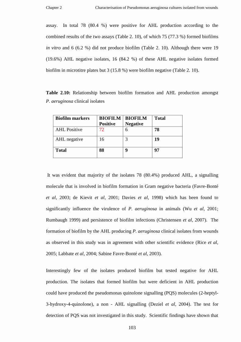
Chapter 2 Characterisation of Pseudomonas aeruginosa cultures isolated from wounds
103
assay. In total 78 (80.4 %) were positive for AHL production according to the
combined results of the two assays (Table 2. 10), of which 75 (77.3 %) formed biofilms
in vitro and 6 (6.2 %) did not produce biofilm (Table 2. 10). Although there were 19
(19.6%) AHL negative isolates, 16 (84.2 %) of these AHL negative isolates formed
biofilm in microtitre plates but 3 (15.8 %) were biofilm negative (Table 2. 10).
Table 2.10: Relationship between biofilm formation and AHL production amongst
P. aeruginosa clinical isolates
Biofilm markers BIOFILM
Positive
BIOFILM
Negative
Total
AHL Positive 72 6 78
AHL negative 16 3 19
Total 88 9 97
It was evident that majority of the isolates 78 (80.4%) produced AHL, a signalling
molecule that is involved in biofilm formation in Gram negative bacteria (Favre-Bonté
et al, 2003; de Kievit et al, 2001; Davies et al, 1998) which has been found to
significantly influence the virulence of P. aeruginosa in animals (Wu et al, 2001;
Rumbaugh 1999) and persistence of biofilm infections (Christensen et al, 2007). The
formation of biofilm by the AHL producing P. aeruginosa clinical isolates from wounds
as observed in this study was in agreement with other scientific evidence (Rice et al,
2005; Labbate et al, 2004; Sabine Favre-Bonté et al, 2003).
Interestingly few of the isolates produced biofilm but tested negative for AHL
production. The isolates that formed biofilm but were deficient in AHL production
could have produced the pseudomonas quinolone signalling (PQS) molecules (2-heptyl-
3-hydroxy-4-quinolone), a non - AHL signalling (Deziel et al, 2004). The test for
detection of PQS was not investigated in this study. Scientific findings have shown that

Chapter 2 Characterisation of Pseudomonas aeruginosa cultures isolated from wounds
104
AHL might not be necessary for biofilm formation (Schaber et al, 2004 & 2007). One
of the AHL deficient strains that were investigated by Schaber and his colleagues in
2004 produced biofilm of up to 82% that of PAO1 wild type control organism. AHL
plays vital role in the control of virulence factors and consequently the pathogenesis of
organisms; however studies have shown that AHL deficient P. aeruginosa are able to
cause infections especially where there are underlying health conditions (Schaber et al,
2007 and 2004; Boşgelmez-Tınaz et al, 2005). Boşgelmez-Tınaz and colleagues (2005)
isolated some AHL deficient P. aeruginosa from intensive care unit patients and the
ability of P. aeruginosa to cause acute infection in burn mice was demonstrated by
Schaber and his colleagues in 2007. In another study, Boşgelmez-Tınaz and Ulusoy
(2008) characterised 50 P. aeruginosa clinical isolates and found that 4 out of the
isolates were QS deficient which confirmed that QS deficient P. aeruginosa are capable
of causing infections.
Some of the wound isolates produced AHL but did not form biofilm in the laboratory.
However, studies have shown that other factors such as flagella and twitching motility
(O'Toole and Kolter, 1998), catabolite repression control (Crc) protein (O'Toole et al,
2000); GacA (Parkins et al, 2001) and pili (Klausen et al, 2003) amongst others are also
relevant for biofilm formation. Six of the isolates (6.18 %) produced AHL but failed to
form biofilm in vitro which might be due to defect in other genetic materials or
structures that aid biofilm formation. It is also possible that the organisms do not
produce biofilm since AHL modulates series of genetic activities.
In order to survive in a host an organism has to overcome environmental stresses
(Davey and O‟Toole, 2000) which are due to various factors such as opsonisation,
phagocytosis, antibody effect and other host defence system activities (Kumar et al,
2009; Medzhitov 2007; Bascones-Martínez et al, 2006). In natural environments the
degree of stress encountered by the organisms would be significantly less than in the

Chapter 2 Characterisation of Pseudomonas aeruginosa cultures isolated from wounds
105
human host environment. Therefore the likelihood for the organisms that infect human
or animal to develop some degree of resistance mechanism to the environmental stress
for sustenance might be higher. Pathogenicity, the ability of organisms to cause
infection is therefore dependent on the ability to overpower the host defence system
which is dependent on the expression of virulence of the organism. Virulence is
determined by the number of virulence factors possessed by an organism and the extent
of production of such virulence factors. Biofilm formation, one of the virulence factors
employed by pathogenic organisms in causing persistent infections is associated with
production of quorum sensing molecules (Waters et al, 2008; Parsek and Greenberg,
2005; De Kievit et al, 2001; Costerton et al, 1999). In Gram negative bacteria including
P. aeruginosa the most common signalling molecules are the AHLs (Fuqua, 2006;
Lequette et al, 2006; Fuqua et al, 1994). The role of quorum sensing including the
Gram negative quorum sensing signalling molecules (AHLs) in pathogenesis has been
extensively investigated and studies have shown that they play dual role in the
pathogenicity of the bacteria by modulating the production of virulence factors (Hooi et
al, 2004; Erickson et al, 2002; de Kievit et al, 2000) as well as inducing immune
responses in the host (Hooi et al, 2004; Smith et al, 2002 (a and b); Smith et al, 2001).
AHL involvement in the activation of regulatory systems for production of virulence
factors in P. aeruginosa has been observed previously (Hooi et al, 2004; Erickson et al,
2002; de Kievit et al, 2000; Rumbaugh et al, 2000; Whiteley et al, 1999). AHL
induction of inflammatory processes in the hosts (Smith et al, 2002 and 2001) has been
suggested as one of the major factors causing the damage in the lungs of the cystic
fibrosis patients.
Studies have also shown that production of quorum sensing molecules by biofilm
organisms may be site dependent (Favre-Bonté et al, 2007; Schuhmacher and Klose,
1999). In a recent study Prasad and his group (2009) investigated the ability of

Chapter 2 Characterisation of Pseudomonas aeruginosa cultures isolated from wounds
106
P. aeruginosa strains from different infection sites to produce virulence markers such as
slime and observed that the isolates from wounds produced higher quantities of slime
than the lung isolates suggesting that the wound isolates might be more virulent.
In order to confirm that P. aeruginosa strains exhibit virulence characteristics
depending on the site of colonisation, biofilm forming strains that were isolated from
water taps were investigated for biofilm characteristics in order to compare the
virulence potentials of both cohorts of P. aeruginosa strains. A great majority (90.7%)
of the 97 P. aeruginosa wound isolates that were tested formed biofilm in microtitre
plates. Out of these 88 biofilm positive isolates 78 of them produced AHL (88.6%).
However of 5 out of 9 (55.6 %) of the water isolated biofilm forming P. aeruginosa also
produced AHL in the laboratory. The greater percentage of the biofilm forming wound
isolates produced AHL unlike the biofilm forming tap water isolates according to the
results of this study which supports previous findings that virulence factors are site
dependent (Choy et al, 2008; Favre-Bonté et al, 2007; Schuhmacher and Klose, 1999).
Schuhmacher and Klose (1999) observed that bile activates the production of regulatory
protein ToxT, an activator of virulence factors in Vibrio cholera; which enhances the
pathogenicity of the bacteria in the gut. Favre-Bonté and his colleagues in 2007
observed that AHL production by P aeruginosa strains in the lungs were site dependent.
The results of their study showed that the type and degree of production of virulence
factors in the intubation devices differ from those obtained from the host‟s trachea
particularly the production of elastase, an enzyme that the bacteria utilise for proteolytic
damage of the host‟s tissue. Pseudomonas autoinducer has been suggested as causing
the production of a chemokine IL-8, which often result in the damage of the lungs of the
cystic fibrosis patients (Smith et al, 2001). P. aeruginosa isolated from wounds have
been found to produce higher degrees of virulence factors than those isolated from other
sites (Prasad et al, 2009). According to the study of the functional characterization of

Chapter 2 Characterisation of Pseudomonas aeruginosa cultures isolated from wounds
107
P. aeruginosa by Wiehlmann and his colleagues (2007), 15 proteins play highly diverse
roles in the cell and became habitat-specific virulence factors on exposure to specific
hosts and/or on exposure to specific stress conditions or host defence mechanisms. It
was expected that clinical isolates from wounds would express certain virulence factors
different from environmental strains and this has been demonstrated by this study.
The genetic similarity index showed that majority of all the isolates have 100%
similarity linkage (Figure 2.11) which may suggest that most of the organisms might
have some similar virulence characteristics. Fifteen (88.2%) of the isolates from
chronic venous leg ulcers (CVLU) have 100% similarity linkage to the member(s) of
the cohort while 83.8% of the strains from the infected wounds showed such similarity
in genetic diversity. The results showed that the greater percentages of the isolates
exhibited biofilm phenotypic characteristics including biofilm formation (Figures 2.4
and 2.13), AHL production (Figures 2.5 and 2.6), and antibiotic resistance (Figure 2.3);
the degree of which were similar to those of the genetic similarities results (Figures 2.11
and 2.12; Tables 2.11 and 2.12). Of the 17 chronic wound isolates, 16 (94.12 %)
formed biofilm in vitro (Table 2.10) while 72 (90%) out of 80 infected wound isolates
tested positive for biofilm (Table 2.11). Six of isolates from chronic wounds (35.3%)
did not produce AHL, five of which (83.3%) formed biofilm while 11 (84.62%) of the
13 AHL negative isolates from infected wounds formed biofilm.

Chapter 2 Characterisation of Pseudomonas aeruginosa cultures isolated from wounds
108
Table 2.11: Relationship between antibiotic susceptibility and genetic index of isolates
ID
No
C I
P
C X
M
A M
L
C N
I P
M
W
P R
L
Genetic
linkage to
member(s)
ID
No
C I
P
C X
M
A M
L
C N
I P
M
W
P R
L
Genetic
linkage to
member(s)
P2 S R R S S R S 100% P57 R R R R S R S 54%
P 4 R R R S S R S 100% P58 S R R S S R S 100%
P 5 S R R S S R S 60% P59 S R R S S R S 93%
P 6 S S R S S S S 100% P60 S R R S S R S 45%
P7 S R R S S R S 100% P61 S R R S S R S 55%
P8 R R R S S R R 100% P62 S R R S S R S 100%
P9 S R R S S R S 100% P63 S R R S S R S 100%
P10 S R R S S R S 100% P64 R R R R R R R 100%
P11 S R R S S R S 100% P65 S R R R S R S 100%
P12 S R R R S R S 100% P66 S R R S S R S 100%
P13 S R R S S R S 100% P67 S R R S S R S 100%
P14 S R R S S R R 100% P68 S R R S S R S 100%
P15 S R R S S R S 100% P69 R R R S S R S 100%
P16 S R R S S R S 100% P70 S R R S S R S 54%
P17 R R R R S R S 100% P71 S R R R S R S 100%
P18 S R R S S R S 100% P72 S R R S S R S 100%
P19 S R R R S R S 100% P73 S R R S S R S 100%
P20 S R R S S R S 100% P74 S R R S S R S 100%
P21 S R R S S R S 100% P75 S R S S S R S 66%
P22 S R R S S R S 100% P76 S R R S S R S 100%
P23 S R R S S R S 61% P77 S R R S S R S 100%
P24 S R R S S R S 100% P78 S R R S S R S 100%
P25 S R R S S R S 100% P79 S R R S S R S 67%
P28 S R R S S R S 100% P80 R R R R R R R 100%
P29 R R R S R R S 100% P81 S R R R S R S 100%
P30 S R R S S R S 100% P82 S R R S S R S 100%
P31 S R R S S R S 100% P83 S R R S S R S 50%
P33 R R R R S R S 100% P84 S R R S S R S 100%
P34 S R R S S R S 45% P85 S R R S S R S 100%
P35 S R R S S R S 100% P86 S R R S S R S 93%
P36 S R R S S R S 100% P87 S R S S R R S 100%
P37 R R R S S R S 100% P88 S R R S S R S 100%
P39 S R R S S R S 100% P89 R R S S S R S 100%
P40 S R R S S R S 100% P90 S R R S R R R 100%
P41 R R R R S R S 100% P91 R R R R R R S 100%
P42 S R R S S R S 100% P92 S R R S S R S 100%
P44 S R R S S R S 100% P93 S R R S S R S 71%
P45 S R R S S R S 100% P94 S R R S S R S 100%
P46 S R R S S R S 100% P95 S R R S R R S 100%
P47 R R R S S R S 100% P96 S R R S S R S 100%
P48 S R R S S R S 100% P97 S R R S S R S 100%
P49 S R R R S R S 100% P98 R R R S S R S 91%
P50 S R R S S R S 100% P99 S R R R S R S 100%
P51 S R R S S R S 100% P100 R R R R S R S 100%
P52 R R R R S R S 100% P101 S R R S S R S 100%
P53 S R R R S R S 63% P102 R R R S R R S 100%
P54 S R R S S R S 100% P103 R R R S S R S 100%
P55 S R R R S R S 100% P104 R R R S S R S 100%
P56 S R R S S R S 100%
Key: CIP = ciprofloxacin 1 µg; CXM = cefuroxime 30 µg; AML = amoxicillin10 µg;
CN = gentamicin10 µg; IPM = imipenem 10 µg; W = trimethoprim 2.5 µg;
PRL = piperacillin 75 µg; S = susceptible and R = resistant.

Chapter 2 Characterisation of Pseudomonas aeruginosa cultures isolated from wounds
109
Table 2.12: Biofilm characteristics and genetic similarity index of P. aeruginosa
isolated from wounds
ID
No
Biofilm QS QSI
Genetic
linkage to
cohort
ID
No
Biofilm QS QSI
Genetic
linkage to
cohort
P2 + + + 100% P57 + + + 54%
P 4 + + + 100% P58 - - - 100%
P 5 + + + 60% P59 + + + 93%
P 6 + + + 100% P60 + + + 45%
P7 + - - 100% P61 + + + 55%
P8 + + + 100% P62 + + + 100%
P9 + + + 100% P63 + + + 100%
P10 + + + 100% P64 + - - 100%
P11 + + + 100% P65 + - + 100%
P12 + + + 100% P66 + + + 100%
P13 + + + 100% P67 + + + 100%
P14 - + + 100% P68 + + + 100%
P15 - + + 100% P69 + + + 100%
P16 - + + 100% P70 + + + 54%
P17 + + + 100% P71 + + + 100%
P18 - + + 100% P72 + + + 100%
P19 + + + 100% P73 + + + 100%
P20 + + + 100% P74 + - - 100%
P21 + + + 100% P75 + - - 66%
P22 + + + 100% P76 + - - 100%
P23 + + + 61% P77 + - - 100%
P24 - + + 100% P78 + + + 100%
P 25 + - + 100% P79 + + + 67%
P28 + + + 100% P80 + - - 100%
P29 + + + 100% P81 + + + 100%
P30 + + + 100% P82 + + + 100%
P31 + + + 100% P83 + + + 50%
P33 + - - 100% P84 + + + 100%
P34 + - - 45% P85 + + + 100%
P35 + + + 100% P86 + + + 93%
P36 + + + 100% P87 + - - 100%
P37 - - - 100% P88 + + + 100%
P39 + - - 100% P89 + - + 100%
P40 + - - 100% P90 + + + 100%
P41 + - - 100% P91 + - - 100%
P42 + + + 100% P92 + + + 100%
P44 + + + 100% P93 + + + 71%
P45 + + + 100% P94 + + + 100%
P46 + - + 100% P95 + + + 100%
P47 + + + 100% P96 + - + 100%
P48 + + + 100% P97 - + + 100%
P49 + + + 100% P98 + + + 91%
P50 + + + 100% P99 + + + 100%
P51 - - - 100% P100 + + + 100%
P52 + - + 100% P101 + + + 100%
P53 + + + 63% P102 + - - 100%
P54 + + + 100% P103 + - - 100%
P55 + + + 100% P104 + + + 100%
P56 + + + 100%
Key: + = Positive result; - = Negative result

Chapter 2 Characterisation of Pseudomonas aeruginosa cultures isolated from wounds
110
Environmental factors often play a vital role in the virulence of an organism especially
in clinical infections (Prasad et al, 2009; Choy et al, 2008; Wu et al, 2003; Feltman et
al, 2001); the findings confirm that the wound isolates have high virulence potentials to
be able to cause infections in the host that the water isolates might not possess because
of less exposure to environmental stress. The percentage of the biofilm forming tap
water isolates that produced AHL was much lower than those of the wound isolates, an
indication that the wound isolates might be more virulent than the water tap isolates.
Although biofilm organisms usually show high resistance to antimicrobials (Hentzer et
al, 2001) the sensitivity pattern observed in these studies was not performed on the
biofilm but the planktonic cultures of the isolates. Bacterial resistance to antimicrobial
compounds sometimes depends on persister cells amongst the bacterial population
(Lewis, 2001; Spoering and Lewis, 2001) and not biofilm formation.
The P. aeruginosa wound isolates that were examined in this study have strong
potential for biofilm formation. Since wound colonisation is inevitable (Cooper et al,
2002; Bowler et al, 2001) the tendency for such bacteria to form biofilm in wounds
might be relatively high.
Conclusion
Most of the wound isolates formed biofilm and produced AHL, which are important to
pathogenicity. The findings suggest that the wound isolates have the potential to exhibit
virulence determinants that allow them to cause infections whereas water isolates did
not. There is the possibility for the organisms to form biofilm in wounds. In this study
an observation of a strong association between the genetic similarity index and biofilm
phenotypic characteristics of the P. aeruginosa wound isolates was found. The wound
isolates were largely sensitive to the most commonly used antimicrobials for
P. aeruginosa infections.

Chapter 3
The study of physiological and phenotypic
characteristics of Pseudomonas aeruginosa in a
wound model
Chapter 3 Biofilm phenotypic expressions of Pseudomonas aeruginosa wound isolates in wound model
111
3.1 Introduction
Generally, the growth of microorganisms depends on various factors such as nutrient
availability (Candy et al, 2009; Rochex and Lebeault, 2007) and temperature (Alves et
al, 2008). In a closed system or batch culture, depletion of essential growth
requirements as a result of an increase in bacterial population, reduces the growth rate
and the toxicity of metabolic wastes that are being released by the organisms further
contribute to the reduction of the bacterial population. In such cultures, apart from the
stress that these conditions pose to the organisms, culture periods are often limited since
they are dependent on the nutrients within the system at the onset of the culture. The
population size of organisms remains constant during the period of nutritional depletion
and accumulation of toxic waste (usually the end of exponential phase and the
beginning of the stationary phase) because the rate of multiplication has reduced and the
death of the organisms in the culture has commenced hence there is a state of
equilibrium as the new cells are replacing the dead ones (as demonstrated in section
2.2.3 of chapter 2). At this phase, phenotypic changes involving the expression of
various genes usually occur (Nakamura et al, 2003; Thompson et al, 2003) for the
survival of the organisms. Nakamura and colleagues studied 2 strains of Bordetella
pertussis and observed that during this transition from exponential phase to stationary
phase 144 array elements showed an increase in transcription, while 177 elements
showed a decrease in transcription rate. It was observed that there was a significant
switch in virulence gene expressions of Helicobacter pylori during the transition from
late exponential phase to stationary phase (Thompson et al, 2003). In the host
environment stress arising from the immune mechanism often induces the production of
virulence factors as a means of circumventing the circumstance. In an in vitro study the
acquisition and retention of the ability of Salmonella typhimurium to induce the death of
macrophages was observed only during the transition from late exponential and

Chapter 3 Biofilm phenotypic expressions of Pseudomonas aeruginosa wound isolates in wound model
112
stationary growth phases (Lundberg et al, 1999). Cowell‟s group (1999) also observed
in their study that Pseudomonas aeruginosa cells that were carbon limited showed a
greater increase in adhesion to artificial tear coated contact lenses; and that
lipopolysccharide may have been involved in the constitutive adhesion to the lenses that
occurred during carbon limitation which was an indication of biofilm formation. The
reduction in metabolic activities and switching on of virulence genes such as spore
formation, biofilm formation, production of cytotoxic enzymes and toxins have been
extensively reviewed (Siegele and Kolter, 1992; Brown et al, 1988). These phenomena
imply that pathogenic microorganisms are able to sense the conditions within the
environments including that of the host and adjust to such conditions through growth
control and virulent expressions. Studies have shown that nutrient depletion encourages
production of signalling molecules (Kay et al, 2006) and development of biofilm in
cultures of microorganisms in vitro (López et al, 2009; Haase et al, 2006). These
phenomena are often utilised by organisms within biofilms (Fux et al, 2005) to enhance
the ability to cause diverse infections and resist antimicrobials (Hill et al, 2005;
Borriello et al, 2004). Biofilm forming organisms such as Pseudomonas aeruginosa
therefore require minimal nutrients for growth and can thrive well in both aerobic and
anaerobic conditions (Waite and Curtis, 2009; O'May et al, 2006). An increase in
nutritional supply was observed to aid the dispersion of P. aeruginosa biofilm (Sauer et
al, 2004) as this tends to increase the expression of the flagella while reducing the
expression of pili thus promoting the detachment of the organisms from the biofilm.
Evidences of increased yield in biofilm culture in vitro with availability of nutrients
have been reported (Yang et al, 2008; Hawser and Douglas, 1994).
Once the nutrients become totally depleted, the organisms cease to multiply and
eventually die and this further limits the application of batch culture in studies that
require long period of microbial culture. In a host environment such as a wound

Chapter 3 Biofilm phenotypic expressions of Pseudomonas aeruginosa wound isolates in wound model
113
nutrients are usually available but the general conditions within the host due to host
defence against the organisms (Hu et al, 2009) and products released by colonising
organisms (Chen et al, 2009) would induce stress in the organisms. Zaborina and
colleagues (2007) in their study observed that P. aeruginosa can be alerted to the
presence of a physiological disturbance in its host by dynorphin released by humans
during severe stress to produce harmful toxins and to suppress the growth of probiotic
bacteria in the gut.
3.1.1 Continuous culture
Continuous culture is a means by which the culture of microorganisms is maintained
under constant conditions for a long period to enable for continuous growth. In this
culture procedure, the medium is constantly being replaced thus the metabolic products
are being eliminated while the nutrients are being maintained (Herbert et al, 1956).
Continuous cultures prevent cultural deterioration or mutant strain development
(Williams and. Wimpenny, 1978; Detrois, 1959) even for a long period. Continuous
culture methods have been recommended for the maintenance of phenotypic
characteristic of the organisms for a long period (Herbert et al, 1956) because of the
difficulty in cultivating organisms that will retain their phenotypic characteristics in a
static culture. Continuous cultures have been successfully utilised to determine
production of metabolic products (Guerra-Santos et al, 1984) and phenotypic
characteristic of biofilm organisms such as P. aeruginosa (Robinson et al, 2004;
Hentzer et al, 2001; Davies and Geesey, 1995; Davies et al, 1993).

Chapter 3 Biofilm phenotypic expressions of Pseudomonas aeruginosa wound isolates in wound model
114
3.1.2 Phenotypic expressions of Pseudomonas aeruginosa in biofilm
In the course of biofilm formation, quorum sensing signalling molecules are released for
the modulation of genetic expressions of biofilm forming organisms (Fuqua et al, 2001;
Whiteley et al, 1999) as well as other substances. The sensing and response to these
signal molecules or autoinducers transforms to coordinated group activities when the
quorum population density is attained (Bjarnsholt and Givskov, 2007; Parsek and
Greenberg, 2005; Waters and Bassler, 2005; Fuqua et al, 1994). In P. aeruginosa these
signalling molecules have been identified as mainly 2 systems of AHLs (Fuqua et al,
2001; Parsek Greenberg, 2000; Whiteley et al, 1999) and quinolones (Diggle et al,
2006; Pesci et al, 1999). Organisms within biofilm typically produce extrapolymeric
substances for encasement and for attachment (Watnick and Kolter, 1999; McKenny et
al, 1998). These phenotypic changes have been used as biomarkers in biofilm
infections (Rickard et al, 2010; Sing et al, 2000; Stickler et al, 1998).
3.1.3 Models /wound models for biofilm study
3.1.3.1 In vitro models
In vitro models are often employed for the study of agents such as antimicrobials and
healing enhancement agents on cells in the laboratory. The system is mostly cost
effective and the ability to test multiple agents and series of concentrations of agents at
the same time offers an advantage over the use of in vivo models. A variety of cell lines
and tissue cultures are usually employed as in-vitro wound models (Oberringer et al,
2007). The effect of cellular hypoxia on treatment in wound healing was demonstrated
in vitro using a cell line wound model (Oberringer et al, 2007). The proliferative and
cellular wound fill was shown to be more rapid with a particular cell (gingival
fibroblasts) than periodontal ligament cells (Oates et al, 2001). The study was able to
show an indication that cell specific attributes play a significant role in healing.

Chapter 3 Biofilm phenotypic expressions of Pseudomonas aeruginosa wound isolates in wound model
115
The main purpose of a study usually determines the type and the form of a model, as
well as the material used in the construction of the model. Various models have been
used in the study of biofilm cultures in the laboratory. P. aeruginosa biofilm
architecture and susceptibility of the biofilm to tobramycin antibiotics have been studied
in a continuous culture reactor system using polycarbonate chips as the stratum on
which the biofilm attached (Hentzer et al, 2001), whereas the effect of sucrose and
silver on biofilm of S. aureus was successfully studied using biofilm model grown on
microscope coverslips in rabbit plasma in tissue culture dishes for 24 hours (Akiyama et
al, 1998). Sriramulu and colleagues (2005) simulated the cystic fibrotic environment
as a biofilm model by growing P. aeruginosa in artificial sputum in a conical flask,
while Walker and Sedlacek (2007) developed a biofilm model with ceramic calcium
hydroxyapatite disks. Thorn and his colleagues (2009) studied the effects of iodine and
silver dressings on biofilms of S aureus and P. aeruginosa grown within cellulose
matrices using flat- bed perfusion biofilm model by inoculating the organisms and then
perfusing the culture for 24 hours. The 24 hour biofilms were removed and placed on
pre-incubated hydrogel polymer test-bed assay plates and dressings were cut (2.5 x 2.5
cm) and placed on top of the biofilms. Test samples were removed at intervals and
biofilm assessed by sonication and TVC determination. Both iodine and silver were
found to exert significant antimicrobial effects on the two biofilms, although iodine was
more potent than silver in biofilm control of the two organisms after 8 hours of
exposure. Evidence of biofilm growth was found under silver dressings after 24 hours.
The antimicrobial activity of silver on biofilms of P. aeruginosa, Candida albicans,
S. aureus and other organisms in a poloxamer gel was investigated (Percival et al,
2007). Of the two dressings tested, the silver-containing hydrofibre (SCH) dressings
showed higher antimicrobial activity against biofilms than the nanocrystalline silver
containing (NSC) dressings. Although the SCH dressings were supposed to contain 10

Chapter 3 Biofilm phenotypic expressions of Pseudomonas aeruginosa wound isolates in wound model
116
times the concentration of silver than the NSC it was found that the concentration did
not correlate to the level of activity.
Multispecies biofilms have been mostly recognised in wounds. An in vitro validation of
this polymicrobial biofilms was demonstrated in Lubbock chronic wound biofilm
(LCWB) model to study cooperation growth amongst the commonly isolated wound
pathogens (Sun et al, 2008). The 24 hour polymicrobial biofilm grown in the model
was tested against various concentrations of sodium hypochlorite (bleach) solution.
The (LCWB) model was also used to propagate the polymicrobial biofilm comprising
of aerobic and anaerobic bacteria commonly isolated from chronic wounds to simulate
the microbiota within the wound environment (Sun et al, 2009). The organisms
included 5 anaerobes and 3 aerobes and were cultured with Bolton broth, heparinised
plasma and laked blood media to represent the available nutritions in the wound
environment. Electron microscopic examination showed the association of the bacteria
in biofilm with the fibrous network of EPS. PCR analysis also confirmed the
identification of the various organisms within the biofilm and the results of the
population monitoring showed the dominance of P. aeruginosa at 24 and 48 hours over
the other bacteria.
Although the environment within the in vitro model can be controlled the inability to
simulate an environment that is akin to the natural one in the human system poses a
great limitation, hence the use of animal model to simulate a wound environment closer
to that of human.
3.1.3.2 In vivo wound models
These are used in the study of wound healing processes or the effect of agents on wound
healing in animals or humans. Many models have been used to study various

Chapter 3 Biofilm phenotypic expressions of Pseudomonas aeruginosa wound isolates in wound model
117
mechanisms and reactions as well as those involved in the pathophysiology of
cutaneous wounds. The most commonly used in wound studies are the animal models
because the opportunities to carry out controlled, clinical trials on human wounds are
limited (Sullivan et al, 2001). The animal models also reduce the variability often
encountered with human subjects. Although an animal model will not provide the full
concept due to the differences in the physiological and genetic make up, immunological
response and tissue architecture the information obtained usually provides relevant
information that can be useful in the treatment of human subjects.
Issues of costs, availability of a particular animal and maintenance of the animal during
the period of experiment also inform the decision on the type of model to use. The
wound inflicted depends on the experiment and could range from incisional, partial or
full thickness, contraction, blister and burn wounds of various degrees depending on the
study requirement. In some countries (in the European Union; EU regulation 76/768 of
Feb. 2003) the use of animal models is restricted for humane treatment of animals.
Wound models that are commonly used include porcine (pig) wound models (Davis et
al, 2008; Serralta et al, 2001) because the pig skin is physiologically close to the human
skin. Other animal models often used are rat models (Patil et al, 2009; Manjunatha et al,
2006) and mice (Manjunatha et al, 2006). A continuous biofilm model was designed
using murine models (Kadurugamuwa et al, 2003) to study formation of biofilm on
catheters. The study of biofilm formation and detection of AHL by clinical and
environmental isolates was studied in mice wound model (Kumar et al, 2009).
Although none of the in vitro and in vivo animal wound models can provide the ideal
situation in human wound they will provide necessary information and guidelines to
improve wound management.

Chapter 3 Biofilm phenotypic expressions of Pseudomonas aeruginosa wound isolates in wound model
118
3.1.4 Study hypothesis
The development of a model is dependent on various factors including the type of study,
sample size, the convenience / ease of maintenance, cost, size of model and availability
of material. The majority of the P. aeruginosa cultures from infected and chronic
wounds were able to produce biofilm and AHL in the batch cultures. Nutrient limitation
and accumulations of toxic wastes are stress factors that initiate phenotypic changes in
batch cultures (Nakagura et al, 2003; Thompson et al, 2003). Such stress factors are not
common in the wound continuum where nutritional factors are constantly available for
the colonising organism.
In this chapter, the phenotypic diversity of the P. aeruginosa wound isolates was
determined using continuous culture method in a wound model. The study of the
phenotypic expressions of the biofilm forming and the non- biofilm forming isolates
allowed more information about the diversity in the expression of the 2 phenotypes of
the bacteria which are often found in the patient during P. aeruginosa infections. The
results of the assay provided judgemental evidence regarding the potential of the
isolates to form biofilm in wound. Furthermore, detection of these markers in the
wound model would suggest that the markers can be detected in the wound by screening
the dressings that have been in contact with wounds for biofilm markers. The period for
the development of biofilm within the wound model and detection of biomarkers would
throw better light on the timescale for biofilm development in wound contaminated with
biofilm forming bacteria such as P. aeruginosa.
Aim of this chapter
To study the phenotypic characteristics of Pseudomonas aeruginosa wound
isolates in a continuous culture wound model.

Chapter 3 Biofilm phenotypic expressions of Pseudomonas aeruginosa wound isolates in wound model
119
Objectives
To monitor the growth of P. aeruginosa and AHL production on a wound
dressing in a batch culture
To develop a wound model for continuous biofilm culture
To study the growth pattern and AHL production of P. aeruginosa wound
isolates in a wound model.
To monitor the stages of biofilm formation of P. aeruginosa wound isolates in a
wound model.

Chapter 3 Biofilm phenotypic expressions of Pseudomonas aeruginosa wound isolates in wound model
120
3. 2 Materials and Methods
3.2.1 Developing a wound model
In this study, a wound model was devised for continuous cultures in order to study the
phenotypic characteristics of selected isolates without mutant strain development and to
prevent the accumulation of toxic waste which might affect the analysis. However, the
development of the wound model was done in two stages, first with a batch culture and
secondly with continuous culture system. Gauze, a conventional dressing material was
chosen as the support material for the wound model (the substratum on which
organisms were grown), since it is a traditional wound dressing. It was tested for its
ability to support the growth of the test organisms and to retain the Gram negative
quorum sensing molecules, such as the acyl- homoserine lactones (AHL).
3.2.1.1 Batch Cultures
Selected biofilm forming and non biofilm forming wound isolates and a reference
culture P. aeruginosa NCIB 8626 were investigated in batch cultures to determine the
phenotypic expression. AHL production was determined through the gauze that had
been incorporated in the medium to establish the phenotypic expressions of the selected
cultures.
3.2.1.1.1 Batch cultures in flasks for AHL detection at 1 cm distance.
Overnight culture of P. aeruginosa was diluted 10-3
with Luria broth (LB); 5 mls of the
diluted inoculum was pipetted into 95 ml of LB or LB and foetal calf serum (FCS) (1:1)
(Fisher – UK) in which some pieces of sterile gauze (8 layers of 2.5 x 2 cm per piece)
have been included (Fig. 3.1). The addition of FCS was to simulate wound environment
and to examine the effect of serum on biofilm formation. Blank controls flasks
containing LB with gauze or LB and FCS mixture with gauze pieces without test
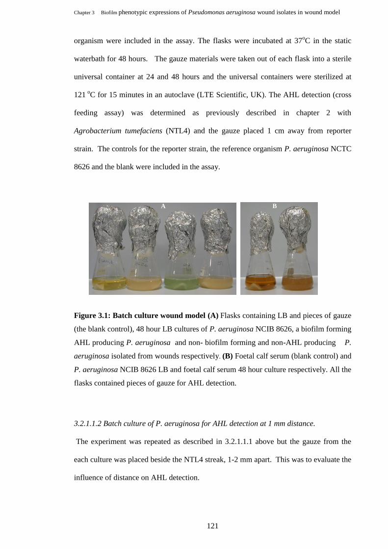
Chapter 3 Biofilm phenotypic expressions of Pseudomonas aeruginosa wound isolates in wound model
121
organism were included in the assay. The flasks were incubated at 37oC in the static
waterbath for 48 hours. The gauze materials were taken out of each flask into a sterile
universal container at 24 and 48 hours and the universal containers were sterilized at
121 o
C for 15 minutes in an autoclave (LTE Scientific, UK). The AHL detection (cross
feeding assay) was determined as previously described in chapter 2 with
Agrobacterium tumefaciens (NTL4) and the gauze placed 1 cm away from reporter
strain. The controls for the reporter strain, the reference organism P. aeruginosa NCTC
8626 and the blank were included in the assay.
Figure 3.1: Batch culture wound model (A) Flasks containing LB and pieces of gauze
(the blank control), 48 hour LB cultures of P. aeruginosa NCIB 8626, a biofilm forming
AHL producing P. aeruginosa and non- biofilm forming and non-AHL producing P.
aeruginosa isolated from wounds respectively. (B) Foetal calf serum (blank control) and
P. aeruginosa NCIB 8626 LB and foetal calf serum 48 hour culture respectively. All the
flasks contained pieces of gauze for AHL detection.
3.2.1.1.2 Batch culture of P. aeruginosa for AHL detection at 1 mm distance.
The experiment was repeated as described in 3.2.1.1.1 above but the gauze from the
each culture was placed beside the NTL4 streak, 1-2 mm apart. This was to evaluate the
influence of distance on AHL detection.
A B

Chapter 3 Biofilm phenotypic expressions of Pseudomonas aeruginosa wound isolates in wound model
122
3.2.1.1.3 Determination of ideal distance for detection of AHL in gauze.
The assay was to determine the farthest distance between the reporter strain and the
gauze at which the weak/ average AHL positive results could be detected. A reference
organism P. aeruginosa NCIB 8626 and a strongly positive AHL producing P.
aeruginosa clinical isolate (P.98) were investigated. The gauze materials from the
cultures were placed 1 - 6 mm away from the inoculation streak of the reporter
organism Agrobacterium tumefaciens NTL4; the cultures were incubated at 30oC and
the cultures were observed at 24, 36 and 48 hours and photographs were taken with
Nikon digital camera.
3.2.1.2 Wound model (Continuous culture)
3.2.1.2.1 Design of the wound model
The idea to design a wound model was to culture the isolates in a system that will
provide adequate nutrients at a steady state without flooding the culture and to allow the
removal of bacterial metabolic wastes. The wound model (Fig. 3.2) was prepared with 2
gauze rolls, (each with 8 layers of 2.5 by 2 cm gauze) inside a Leur lock glass syringe
(Fisher Scientific) (held in place with the aid of the stand and clamp) with tubing passed
from the media reservoir to a syringe (anti - grow back trap) from where the tubing was
connected to a Leur lock stainless steel needle (BD, USA) to continuously feed the
culture. The culture was fed with the drips of nutrients with the aid of a Watson
Marlow 505U peristaltic pump (Watson Marlow Ltd., England, UK), the effluent drips
from the syringe to the waste receptacle which collects the culture. The volume of the
broth contained in the gauze was determined by pumping the broth through the model
and counting the number of drops that completely soaked the piece of gauze and
multiplying by the volume of each drop which had been earlier determined. The 2 rolled
pieces of gauze used for each culture in the model, were found to retain between 1.41
ml and 1.54 ml of broth (an average of about 1.475 mls) was calculated. All

Chapter 3 Biofilm phenotypic expressions of Pseudomonas aeruginosa wound isolates in wound model
123
components parts excluding the pump were sterilised at 121oC for 15 minutes and dried
in the oven at 60oC prior to each culture.
Figure 3.2: The Continuous Culture Wound Model: A system designed and
assembled for the study of the growth pattern and phenotypic characteristics of biofilm
and non- biofilm forming P. aeruginosa in continuous culture. The system was sterile
and closed to the outside environment but equilibrium with
atmospheric pressure was
maintained by fitting to each bottle a Whatman® Hepa-vent air-permeable filter.
3.2.1.2.2 Preliminary study of phenotypic diversity of P. aeruginosa strains in
continuous culture wound model with full strength Luria broth.
The flow rate of the LB through the model was calculated from the amount of exudate
produced by wounds, especially external wounds or wounds associated with virulent
bacterial mechanisms that often cause vasodilation and extravasation (Wilson et al,
2002). High production of exudates had been estimated to be more than 10 ml every 24
hours (Mulder, 1994). This volume could not be achieved with the pump available for
this study and the minimum volume that could be delivered with the pump was 8.4 ml
per hour. This was achieved by using tubing with the lowest diameter (0.5mm) for the
pump and selecting the appropriate speed according to the calibration of the pump.
Anti-grow back trap
Clamp
Syringe with gauze
Media reservoir
Hepa-vent filter (air steriliser)
Flask for collecting culture
Peristaltic Pump
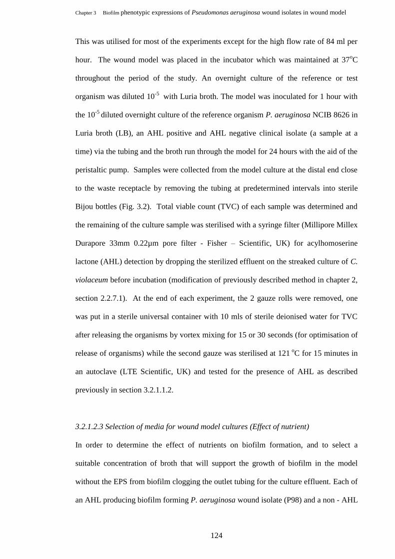
Chapter 3 Biofilm phenotypic expressions of Pseudomonas aeruginosa wound isolates in wound model
124
This was utilised for most of the experiments except for the high flow rate of 84 ml per
hour. The wound model was placed in the incubator which was maintained at 37oC
throughout the period of the study. An overnight culture of the reference or test
organism was diluted 10-5
with Luria broth. The model was inoculated for 1 hour with
the 10-5
diluted overnight culture of the reference organism P. aeruginosa NCIB 8626 in
Luria broth (LB), an AHL positive and AHL negative clinical isolate (a sample at a
time) via the tubing and the broth run through the model for 24 hours with the aid of the
peristaltic pump. Samples were collected from the model culture at the distal end close
to the waste receptacle by removing the tubing at predetermined intervals into sterile
Bijou bottles (Fig. 3.2). Total viable count (TVC) of each sample was determined and
the remaining of the culture sample was sterilised with a syringe filter (Millipore Millex
Durapore 33mm 0.22µm pore filter - Fisher – Scientific, UK) for acylhomoserine
lactone (AHL) detection by dropping the sterilized effluent on the streaked culture of C.
violaceum before incubation (modification of previously described method in chapter 2,
section 2.2.7.1). At the end of each experiment, the 2 gauze rolls were removed, one
was put in a sterile universal container with 10 mls of sterile deionised water for TVC
after releasing the organisms by vortex mixing for 15 or 30 seconds (for optimisation of
release of organisms) while the second gauze was sterilised at 121 o
C for 15 minutes in
an autoclave (LTE Scientific, UK) and tested for the presence of AHL as described
previously in section 3.2.1.1.2.
3.2.1.2.3 Selection of media for wound model cultures (Effect of nutrient)
In order to determine the effect of nutrients on biofilm formation, and to select a
suitable concentration of broth that will support the growth of biofilm in the model
without the EPS from biofilm clogging the outlet tubing for the culture effluent. Each of
an AHL producing biofilm forming P. aeruginosa wound isolate (P98) and a non - AHL

Chapter 3 Biofilm phenotypic expressions of Pseudomonas aeruginosa wound isolates in wound model
125
producing and non biofilm forming P. aeruginosa wound isolate (P51) was cultured in
the wound model for 48 hours with ½, ¼ and 1/10 strength LB. Samples were taken
from the model culture at intervals for total viable count (TVC) and acylhomoserine
lactone (AHL) detection using A. tumefaciens NTL 4 as the reporter organism.
3.2.1.2.4 Examining the effect of flow rates of broth on the wound model culture
A biofilm forming (P.98) and a non biofilm forming (P.51) wound isolates were each
cultured in the wound model using 1/10 LB at a high flow rate of 84 mls per hour. This
was to ensure that there was no trapping of the bacteria within the gauze and to observe
effect of the hydrodynamic flow on the culture in particular if the non biofilm former
will be washed out of the model. The model was inoculated for 1 hour at the flow rate
of 8.4 mls per hour and the rate of the media was thereafter increased to 84 mls per hour
for the 48 hour duration of the experiment. Samples of the effluent were collected at
intervals and the AHL was determined and smears were made on glass slides which
were stained were stained and examined for biofilm formation.
3.2.1.2.5 Determination of reproducibility of assays for wound model culture
A biofilm forming and AHL producing P. aeruginosa was cultured in the wound model
on 3 different occasions with 1/10 LB to determine the reproducibility of the model using
TVC and AHL assays. Viable counts of the culture samples were determined to
establish the growth pattern of the organism and the AHL assay to determine the period
of AHL production. Slide smears of the culture effluent samples were also made, fixed
and stained for EPS to study biofilm morphological structure. Slides were examined
with Olympus microscope with digital camera (Volocity software) for EPS and biofilm
morphology.

Chapter 3 Biofilm phenotypic expressions of Pseudomonas aeruginosa wound isolates in wound model
126
3.2.1.2.7 Examining the effect of serum on biofilm formation
A biofilm forming and AHL producing P. aeruginosa (P.98) was cultured in the wound
model using FCS and full strength LB (1:1) and FCS in 1/8 LB (1:4) (giving final
concentration of 1/5 FCS and
1/10 LB) to determine growth pattern and biofilm formation
as previously described section 3.2.1.2.2. Viable count of the culture samples and
production of quorum sensing molecules (AHL) were determined as above using culture
effluent samples.
3.2.2 Staining procedure for EPS detection and biofilm morphology
The slide smears were fixed with 10 mM cetylpyridium chloride (Sigma, UK) for 5
minutes and air dried for 30 minutes. Slides were fixed with gentle heat, cooled and
stained with 2:1 mixture (v/v) of saturated aqueous Congo red (BDH, UK)and 10%
Tween 80 (Sigma –Aldrich, UK) for 15 minutes. Slides were rinsed with water and
were counterstained with 10% Ziehl‟s Carbol Fuschin (BDH, UK) for 5 minutes. Slides
were rinsed with water and air dried (Harrison – Balestra et al, 2003).
3.2.3 Scanning electron microscopic (SEM) examination of biofilm on the gauze
from the wound model culture.
It was necessary to examine the gauze from the wound model to determine the presence
of biofilm. A biofilm forming P. aeruginosa isolate (P.98) and a non biofilm forming P.
aeruginosa isolate (P.51) were each cultured in the wound model for a period of 30
hours as described previously (section 3.2.1.2) using 1/10 strength Luria broth.
Samples of the culture effluent were collected at determined intervals for TVC and
AHL. Effluent slide smears were prepared, stained and examined to establish biofilm
formation. After terminating the culture at 30 hours, the gauze was fixed with 2.5%
(w/v) glutaraldehyde (Fluka, UK), dehydrated through ascending concentrations of

Chapter 3 Biofilm phenotypic expressions of Pseudomonas aeruginosa wound isolates in wound model
127
ethyl alcohol (30%, 50%, 70%, 80% and 90%) for 10 minutes each. The gauze was
finally dehydrated in three changes of 100% ethyl alcohol. It was dried in CPD030
(Bullzer‟s clinical drier) with carbon - dioxide to flush out the alcohol and saturate the
material completely at 10oC and 90 mmHg pressure for an hour. The pressure was
released and the temperature was increased to 40oC for 3 hours for critical point drying
(CPD). Sputter coating of the gauze was done by spraying the gauze with gold
(Emscope Gold Sputtering Coater AE 1231 (Emscope, Kent, UK) for 10 minutes and
was examined with a scanning electron microscope (Philips XL 20 SEM, Eindhoven,
Netherlands). This was done at the Electron Microscopy (EM) unit, Cardiff University.

Chapter 3 Biofilm phenotypic expressions of Pseudomonas aeruginosa wound isolates in wound model
128
3.3 Results
3.3.1 Detection of AHL from gauze incubated in batch culture
Gram negative quorum sensing (QS) molecules AHLs present in the gauze which was
produced by A. tumefaciens KYC6 (an AHL producer as positive control) and the P.
aeruginosa wound isolates (P.98 and P100), diffused through the medium changing
NTL4 colonies from cream to blue (Fig. 3.3A) as discussed in chapter 2 section 2.3.5.
Negative controls (Fig. 3.3B); the blank control (Fig. 3.3C) and the wound isolates
(P.51 and P.58) that did not produce QS molecules (Fig. 3.3E) remained unchanged
after 48 hours incubation at 30oC.
Figure 3.3: Detection of AHL in gauze from 48 hours batch cultures showing (A)
the reporter bacteria Agrobacterium tumefaciens NTL4 with A. tumefaciens KYC6 (an
AHL producing biofilm forming bacteria) as positive control; (B) KYC6 against itself
as negative control; (C) LB without inoculation of organism as blank control; (D) an
AHL producing biofilm forming P. aeruginosa isolate from wound; (E) a non-AHL
producing non- biofilm forming P. aeruginosa isolate from wound; (F) an AHL
producing biofilm forming P. aeruginosa NCIB8626 as positive culture control.
A B C
D E F

Chapter 3 Biofilm phenotypic expressions of Pseudomonas aeruginosa wound isolates in wound model
129
However the colour change from cream to blue that was observed between 36 - 48
hours for the strong AHL producing wound isolate (P.98) (Figure 3.3D) was faint and
not as obvious as had been observed with the solid agar culture in the cross feeding
assay as discussed in chapter 2, (section 2.3.5). AHL could not be detected in the gauze
from the reference organism NCIB 8626 as there was no visible change of colour on
plate (Fig. 3.3F) even though the organism had tested positive in the cross feeding
assay. In the cross feeding assay on solid agar plate, the AHLs being produced by the
bacteria diffused through the agar within the 1cm distance to initiate the effect that was
observed in the reporter bacteria. In the batch culture, the AHLs were being diluted by
the culture medium as were being produced and the amount in the gauze might not have
been high enough to initiate the expected reaction when it diffused over the distance of
1cm. Although when the level of AHLs produced by the bacteria is high due the
numbers of organisms or the ability of organisms to produce high level of AHL, the
detection might be possible at this distance but when the concentrations of the AHLs
produced in the culture medium are low, detection from such a distance might be
difficult. In order to confirm that the gauze sustained the growth of the organisms and
the AHLs produced by the organisms the gauze was placed one millimetre away from
the NTL4 in the plate. The results showed positive results with pronounced colour
intensity for the positive control (Fig. 3.4A), an AHL producing biofilm forming
P. aeruginosa isolate from wound (Fig. 3.4B) and an AHL producing biofilm forming
P. aeruginosa NCIB 8626 (Fig. 3.4C) as positive reference organism control. KYC6
against itself as negative control (Fig. 3.4D), LB without inoculation of organism as
blank control (Fig. 3.4E), and a non-AHL producing non- biofilm forming
P. aeruginosa isolate from wound (Fig. 3.4F) did not show any colour change.

Chapter 3 Biofilm phenotypic expressions of Pseudomonas aeruginosa wound isolates in wound model
130
Figure 3.4: Detection of AHL in gauze from batch culture placed 1mm from
reporter bacteria. (A) reporter bacteria A. tumefaciens NTL4 with A. tumefaciens
KYC6 (AHL producing biofilm forming bacteria) as positive control; (B) KYC6 against
itself as negative control; (C) LB without inoculation of organism as blank control;
(D) AHL producing biofilm forming P. aeruginosa isolate from wound (P.98); (E) a
non-AHL producing non-biofilm forming P. aeruginosa isolate from wound (P.51) and
(F) AHL producing biofilm forming P. aeruginosa NCIB 8626 as positive reference
culture.
3.3.2 The Ideal distance for AHL detection in gauze
In order to determine the ideal distance between the reporter strain and lowest AHL
concentrations in samples of gauze removed from wound model, various distances
between the reporter strain and the sterilised culture material (gauze) were investigated.
This was to serve as a guide for the greatest distance between the reporter strain and the
gauze at which the weak and average positive results can be determined. A type culture
P. aeruginosa NCIB 8626 and a strong AHL molecule producing wound isolate (P.98)
were investigated in an assay in which the sterilised gauze materials from the cultures
were placed between 1 and 6 mm (at 1mm interval) away from the NTL4 streak before
E F
NTL4
A B C
D
NTL4

Chapter 3 Biofilm phenotypic expressions of Pseudomonas aeruginosa wound isolates in wound model
131
incubation. Optimal colour development was observed at 36 hours which remained
stable at 48 hours. The strong QS molecule producer showed strong positive results for
the 1- 6 mm distance from NTL4 (Figure 3.5a ; Table3.1) while NCIB 8626 was
positive up to 3 mm with reduced colour intensity (Figure 3.5b ; Table3.1). These
results signified that the gauze should be placed as close as possible (about 1-2mm) to
the reporter strain to detect low AHL production.
Figure 3.5: AHL detection in gauze samples removed from P. aeruginosa 48 hour
batch cultures placed between 1 and 6 mm distance from the reporter organism.
(a) a strong biofilm and high AHL producing P. aeruginosa wound isolate and (b) a
reference P. aeruginosa NCIB 8626 a strong biofilm former and low AHL producer.
aA
b

Chapter 3 Biofilm phenotypic expressions of Pseudomonas aeruginosa wound isolates in wound model
132
Table 3.1: Colour intensity index of optimisation of AHL detection assay
Distance between gauze
and A. tumefaciens
Colour intensity index score
P. aeruginosa
NCIB 8626
P. aeruginosa
wound isolate (P.98)
1mm + +++
2mm + +++
3mm +/- +++
4mm - +++
5mm - +++
6mm - +++
Key: (±) slightly positive; (+) positive; (++) strong positive; (+++) very strong positive
The results of the batch cultures showed that gauze provided support for bacterial
growth and AHL detection. The AHL produced by biofilm forming organisms were
detectable from the gauze samples which were sterilized after removal from the culture.
The wound model batch cultures provided information to distinguish between biofilm
forming and the non- biofilm forming P. aeruginosa cultures isolated from wounds. The
ideal distance for detection of AHL was also established as 1 mm especially for
detection of low concentration of AHL.
3.3.3 Effect of nutrients on biofilm development in continuous culture
The effect of nutrients on bacterial growth and in particular on biofilm formation, was
determined using various concentrations of LB to culture each of the biofilm forming
and non- biofilm forming wound isolates in the wound model continuous culture.

Chapter 3 Biofilm phenotypic expressions of Pseudomonas aeruginosa wound isolates in wound model
133
3.3.3.1 Preliminary study of phenotypic diversity of P. aeruginosa strains in continuous
culture wound model
In this study, the growth pattern for the 3 organisms tested with full strength Luria broth
appeared similar, however AHL was first detected in isolate P.98 culture samples at 12
hours, when the number of organisms attained 9.6 x 10⁹ cfu/ml of culture (Fig. 3.6; 3.7
C and D) while the reference organism P. aeruginosa NCIB 8626 tested positive for
AHL only after 22 hours with the bacterial population of 6.46 x 10¹¹
cfu/ml of culture
whereas isolate P.51 did not produce AHL within the period studied (Figures 3.6 and
3.7 (E and F).
The results of the TVC for each effluent sample are presented in a graph for the LB
cultures of the P. aeruginosa wound isolates, a biofilm former (isolate P. 98), a non
biofilm former (isolate P.51) and a reference organism P. aeruginosa NCIB 8626 (Fig.
3.6). The organisms trapped within the gauze that were released at 15 seconds and 30
seconds showed higher numbers of organisms for the biofilm formers than the non
biofilm former. Larger numbers of P. aeruginosa wound isolate (P.98) were recovered
from the gauze than for the reference culture P. aeruginosa NCIB 8626 (Table 3.2)
while those for isolate P.51 were the lowest. AHL detection results for P.98 were more
pronounced than those for NCIB 8626 (Fig. 3. 4 D and F; Table 3.1). The stronger
biofilm former isolate P. 98 had more organisms trapped within the biofilm matrix
indicating that the larger the biofilm the greater the number of organisms within the
matrix. The 2 periods of sonication were used in the preliminary study for the purpose
of optimisation to determine the appropriate time for the release of organisms from the
gauze throughout the period of the study.

Chapter 3 Biofilm phenotypic expressions of Pseudomonas aeruginosa wound isolates in wound model
134
2
4
6
8
10
12
14
0 2 4 6 8 10 12 14 16 18 20 22 24 26
Time (Hours)
No
of
org
an
ism
s in
cu
ltu
re e
fflu
en
t
(lo
g c
fu/m
l)
Isolate 98 TVC:no AHL detectedin effluent
Isolate 51 TVC
NCIB 8626 TVC:no AHL detectedin effluent
Isolate 98 TVC:AHL detected ineffluent
NCIB 8626 TVC:AHL detected ineffluent
Figure 3.6: Phenotypic characteristics of Pseudomonas aeruginosa strains shown
by growth curve and AHL production in wound model continuous cultures. The
growth curves of P. aeruginosa wound isolates (biofilm forming P.98 and a non -
biofilm former P.51) and a reference organism P. aeruginosa NCIB 8626 in wound
model continuous culture using full strength LB at the flow rate of 8.4 ml/hr for a period
of 25 hours including one hour inoculation time are represented graphically.
Table 3.2 Organisms released from gauze after 24 hour culture of each P. aeruginosa
strain in a continuous culture wound model using full strength Luria broth at 37oC
Organisms 15 sec vortex
(Log cfu/ml)
30 sec vortex
(Log cfu/ml)
P. aeruginosa NCIB 8626 9.5 x 10¹⁰
3.28 x 10¹¹
Wound isolate (P.98) 2 x10⁹ 1.63 x 10
10
Wound isolate (P.51) 2.25 x 10⁹ 5.5 x 10
9

Chapter 3 Biofilm phenotypic expressions of Pseudomonas aeruginosa wound isolates in wound model
135
3.3.3.2 Effect of nutrient concentration on biofilm
The growth pattern and AHL production for the biofilm forming isolate P.98 for the
various concentrations of Luria broth (Full strength, 1/2, 1/4 and 1/10) appeared similar
over 24 and 48 hours (Fig. 3.8 and 3.9) (Table 3.3). The AHL was detectable at 12
hours for the full strength LB culture when the bacterial population was above log 9 per
ml and the 1/2, 1/4 and 1/10 strength LB cultures when the bacterial populations were
above log 7 per ml of culture. Although the non - biofilm forming isolate showed
similar growth pattern over 24 - hour and 48 - hour incubation periods (Fig. 3.10 and
3.11), AHL was not detected in any of the culture samples throughout the period of
experiment. It was apparent that the growth yield reduced with the decrease in
Figure 3.7: AHL detection in culture
effluent and sterilized gauze. (A) A.
tumefaciens KYC6 with A. tumefaciens
NTL 4 as positive control; (B) KYC 6
with KYC6 as negative AHL control;
(C) a drop of sterilised culture effluent
sample from a biofilm forming P.
aeruginosa wound isolate (P.98) taken
during the continuous culture in a wound
model (D) the gauze from the wound
model after the culture (P.98) with NTL4
tested positive for AHL; (E) a drop of the
sterilised culture effluent sample of a
non-biofilm forming P. aeruginosa
wound isolate (P.51) taken during the
continuous culture in a wound model and
(F) the gauze from the wound model at
the end of the culture (P.51) showing
negative result for AHL.
A
C
D
B
E
F

Chapter 3 Biofilm phenotypic expressions of Pseudomonas aeruginosa wound isolates in wound model
136
concentration of broth (Fig. 3.8, 3.9, 3.10 and 3.11). The continuous wound model
culture for the diluted LB (1/2, 1/4 and 1/10 strength LB) showed increase in TVC of
P.98 up till 36 hours whereas with full strength LB cultures numbers started declining
from 21 hours (Fig. 3.8, 3.9, 3.10 and 3.11). For the purpose of this study, it was
important to have a steady growth that would not be too rapid in order to study the
organism for a reasonable period of time. Excessive growth would cause blockage of
the tubing to the effluent collection flask with bacteria and EPS that are being produced
during biofilm formation which might cause the wound model to collapse. The broth
that sustained steady growth with the least growth rate, 1/10 Luria broth was chosen as
the ideal concentration of broth for the wound model cultures.
2
3
4
5
6
7
8
9
10
11
12
0 2 4 6 8 10 12 14 16 18 20 22 24
Time (Hours)
No
of
org
an
ism
s i
n c
ult
ure
eff
lue
nt
(lo
g c
fu/m
l)
Full strengthLB
1/2 strengthLB
1/4 strengthLB
1/10 strengthLB
Figure 3.8 The 24 hour continuous cultures of a biofilm forming P. aeruginosa
wound isolate (P. 98) in the wound model with various Luria broth concentrations.
The wound model continuous culture of the biofilm forming isolate (P. 98) was run at
8.4 mls per hour flow rate with each of the media for a period of 24 hours at different
times. TVC and AHL production were determined at intervals of 2 or 3 hours during the
period of culture.
AHL detection
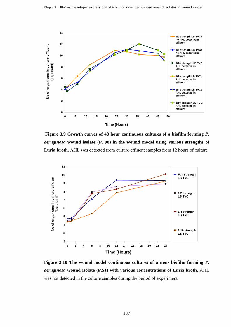
Chapter 3 Biofilm phenotypic expressions of Pseudomonas aeruginosa wound isolates in wound model
137
0
2
4
6
8
10
12
14
0 5 10 15 20 25 30 35 40 45 50
Time (Hours)
No
of
org
an
ism
s i
n c
ult
ure
eff
lue
nt
(lo
g c
fu/m
l)
1/2 strength LB TVC:no AHL detected ineffluent
1/4 strength LB TVC:no AHL detected ineffluent
1/10 strength LB TVC:AHL detected ineffluent
1/2 strength LB TVC:AHL detected ineffluent
1/4 strength LB TVC:AHL detected ineffluent
1/10 strength LB TVC:AHL detected ineffluent
Figure 3.9 Growth curves of 48 hour continuous cultures of a biofilm forming P.
aeruginosa wound isolate (P. 98) in the wound model using various strengths of
Luria broth. AHL was detected from culture effluent samples from 12 hours of culture
2
3
4
5
6
7
8
9
10
11
0 2 4 6 8 10 12 14 16 18 20 22 24
Time (Hours)
No
of
org
an
ism
s in
cu
ltu
re e
fflu
en
t
(lo
g c
fu/m
l)
Full strengthLB TVC
1/2 strengthLB TVC
1/4 strengthLB TVC
1/10 strengthLB TVC
Figure 3.10 The wound model continuous cultures of a non- biofilm forming P.
aeruginosa wound isolate (P.51) with various concentrations of Luria broth. AHL
was not detected in the culture samples during the period of experiment.

Chapter 3 Biofilm phenotypic expressions of Pseudomonas aeruginosa wound isolates in wound model
138
2
3
4
5
6
7
8
9
10
11
12
0 5 10 15 20 25 30 35 40 45 50
Time (Hours)
No
of
org
an
ism
s in
cu
ltu
re e
fflu
en
t
(lo
g c
fu/m
l)
1/2 strengthLB TVC
1/4 strengthLB TVC
1/10 strengthLB TVC
Figure 3.11 The graphical representation of the continuous cultures of a non-
biofilm forming P. aeruginosa wound isolate (P. 51) in the wound model using
various strengths of Luria broth for a period of 48 hours. AHL was not detected in
any of the culture samples throughout the experiment. Higher concentration of broth
produced higher numbers of organisms.
Table 3.3 Detection of AHL during the development of Pseudomonas aeruginosa
biofilm in wound model continuous cultures.
Variables (Media and Flow rate) Organism Time of AHL
detection
TVC
(log cfu/ml)
Full strength LB (isolate P. 98) 12 hours 9.6 x10⁹
½ strength LB (isolate P. 98) 12 hours 2.36x10⁸
¼ strength LB (isolate P. 98) 12 hours 1.78x10⁷
1/10 strength LB (isolate P. 98) 12 - 21 hours 1x10
9
Full strength LB + FCS (1:1) (isolate P. 98) 16 hours 3.3x10¹⁰
FCS +12.5%LB (20 % FCS in 1/10 LB) (isolate P. 98) 24 hours 1.99x10⁹
1/10 LB (High flow rate) (isolate P. 98) 30 hours 1.02x10¹⁰
Full strength LB (NCIB 8626) 22 hours 6.46x10¹¹

Chapter 3 Biofilm phenotypic expressions of Pseudomonas aeruginosa wound isolates in wound model
139
3.3.4 Effect of broth flow rate
Ten fold increase in the rate of broth flow to the culture provided increase availability of
nutrient for bacterial growth which increased the rate of discharge of culture effluent
from the wound model which had effect on the organisms‟ growth rate. Both the biofilm
forming and non-biofilm forming P. aeruginosa wound isolates for low and high flow
rates (Fig. 3.12) showed similar growth curves in the wound model. Whereas the
biofilm forming isolate (P. 98) grew at a slower rate during the lag phase of the culture
(prolonged lag phase), the non-biofilm forming isolate (P. 51) grew rapidly at the onset
of the culture. The growth of P.51 increased more rapidly with a longer log phase at the
higher 84 mls/hour flow rate than that of the 8.4 mls/hr flow rate. Whereas P. 98 had a
longer lag phase and decreased growth output during the log phase with the 84 mls/hour
flow rate although the growth curve for each isolate remained similar for the two flow
rates. The number of dislodged organisms from the gauze after culture was significantly
higher when 8.4 ml/hour flow rate was applied; log cfu/ml was 1.03 x 10¹⁰
whereas that
for the 84 mls/hr flow rate was 7.2 x 10⁸ (Table 3.4).
AHL detection with the 84 mls/hour flow rate was from 30 hours at the bacterial count
of 1.02x10¹⁰
cfu/ml of culture whereas AHL was detected at 21 hours when 8.4ml/hour
flow rate was applied with population of 1x10⁹
cfu/ml of culture (Table 3.3 and fig.
3.13). The non - biofilm former tested negative for AHL production throughout the
duration of the culture.

Chapter 3 Biofilm phenotypic expressions of Pseudomonas aeruginosa wound isolates in wound model
140
2
4
6
8
10
12
14
0 5 10 15 20 25 30 35 40 45 50
Time (Hours)
No
of
org
an
ism
s in
cu
ltu
re e
fflu
en
t
(lo
g c
fu/m
l)
Isolate 98(8.4ml/hr)
Isolate 51(8.4ml/hr)
Isolate 98(84ml/hr)
Isolate 51(84ml/hr)
Figure 3.12 Effect of broth flow rate on phenotypic expressions of P. aeruginosa
strains in continuous culture wound model. The growth curves of a biofilm forming
(P.98) and non-biofilm forming (P51) wound isolates using 2 different broth flow rates.
2
4
6
8
10
12
14
0 5 10 15 20 25 30 35 40 45 50
Time (Hours)
No
of
org
an
ism
s i
n c
ult
ure
eff
lue
nt
(lo
g c
fu/m
l)
8.4ml/hr flowrate TVC:no AHL detected ineffluent
84ml/hr flowrate TVC:no AHL detected ineffluent
8.4 ml/hr flowrate TVC:AHL detected in eflluent
84 mls/hr flowrate TVC:AHL detected in effluent
Figure 3.13 Effect of flow rate on P. aeruginosa wound isolate biofilm formation in
continuous culture wound model. The growth curves the isolate (P.98) using 2
different broth flow rates are shown. AHL production was detected at 21 hours at the
lower flow rate whereas at 30 hours at the higher flow rate.

Chapter 3 Biofilm phenotypic expressions of Pseudomonas aeruginosa wound isolates in wound model
141
Table 3.4 Organisms released from P. aeruginosa (P.98) wound isolate biofilm culture
at different broth flow rates in a continuous culture wound model using 1/10 LB.
Broth flow rate (mls/hr)
Using 1/10 LB
Organisms released from gauze after
48 hours culture at 37oC (Log cfu/ml)
8.4 1.03 x 10¹⁰
84 7.2 x 10⁸
3.3.5 Reproducibility of assays
The reproducibility of the analysis was examined by culturing a strong biofilm forming
P. aeruginosa wound isolate (P. 98) in the wound model using 1/10 strength Luria broth
on 3 different occasions. The TVCs were determined for each effluent culture sample;
the mean of the 3 assays including the standard deviations (SD) were calculated and
presented in Fig. 3.14. AHL was detectable in the wound model culture effluent
samples from 21 hours when the bacterial population was above 1 x 10 9
per ml of
culture effluent and in the gauze at the end of each culture (Figure 3.13 and Table3.2).
Statistical analysis of variance using Minitab (version 15) one-way ANOVA
(unstacked), p< 0.001 (95% CIs for mean based on pooled SD).

Chapter 3 Biofilm phenotypic expressions of Pseudomonas aeruginosa wound isolates in wound model
142
0
2
4
6
8
10
12
14
0 5 10 15 20 25 30 35 40 45 50
Time (Hours)
No
of
org
an
ism
s i
n c
ult
ure
eff
lue
nt
(lo
g c
fu/m
l)
Mean TVC: no AHLdetected ineffluent
Mean TVC: AHL detectedin effluent
Figure 3.14 Graph of reproducibility assay of a biofilm forming P. aeruginosa
wound isolate in wound model continuous cultures. The error bars indicate the
standard deviations between the sets of bacterial populations for the 3 separate
experiments. AHL was detected from 21 hours for the 3 cultures at TVC of over log10 9
cfu/ml of culture effluent.
3.3.6 Effect of serum on biofilm formation and AHL production
In this study it was intended to simulate the circumstances that exist in wound as closely
as possible. In the wound there is blood and the components of blood which are
contained in the serum particularly the immune components such as complement,
interferon or antibodies which might interfere with the metabolic activities of the
organisms. The presence of such agents might have an effect on the development of
biofilm structure and or production of quorum sensing molecules.
When P. 98 was cultured in the continuous wound model with full strength LB, AHL
was detected at 12 hours when TVC was 9.6 x10⁹
cfu/ml of culture sample but when
FCS was added to the LB AHL was detected at 16 hours with TVC of 3.3x10¹⁰
cfu/ ml
of culture sample (Fig. 3.15; Table 3.3). Similarly using 1/10 LB and P.98 AHL was
first detected between 12 and 21 hours with TVC of between 4.6 x10⁷ and 1x10
9 cfu/ml

Chapter 3 Biofilm phenotypic expressions of Pseudomonas aeruginosa wound isolates in wound model
143
of culture but when FCS (12.5% v/v) was added to LB at LB concentration of 1/10 AHL
was detected at 24 hours when the bacterial population was up to 1.99x10⁹ cfu/ml of
culure (Fig. 3.16; Table 3.3). Similarly the organisms released from gauze after each
culture showed higher number for LB (1.63 x 10¹⁰
) and 1/10 LB(1.03 x 10
10cfu/ml) than
FCS and LB (1:1) and FCS and 1/8 LB (1:4) which were (7.7 x10
9) and (2.45 x 10
9)
respectively (Table 3.5).
2
4
6
8
10
12
14
0 2 4 6 8 10 12 14 16 18 20 22 24 26
Time (Hours)
No
of
org
an
ism
s in
cu
ltu
re e
fflu
en
t
(lo
g c
fu/m
l)
98 (LB) TVC: noAHL detected ineffluent
98 (LB & FCS)TVC: no AHLdetected ineffluent
98 (LB) TVC:AHL detected ineffluent
98 (LB & FCS)TVC: AHLdetected ineffluent
Figure 3.15: The effect of serum on growth of P. 98 and AHL production in the
continuous culture wound model. AHL production by the isolate was detected at 12
hours in full strength LB, while AHL was detected 16 hours in LB/FCS (1:1).
Table 3.5 Populations of bacteria released from gauze after each culture of P.98 biofilm
using LB, or LB and FCS in wound model.
Culture media 30 sec vortex (Log cfu/ml)
LB 1.63 x 10¹⁰
1/10 LB 1.03 x 10
10
FCS and LB (1:1) 7.7 x 10 9
FCS and LB (1:4) 2.45 x 10⁹

Chapter 3 Biofilm phenotypic expressions of Pseudomonas aeruginosa wound isolates in wound model
144
It was found in this study that the addition of FCS to the culture medium did not prevent
growth and AHL production but the time of detection of AHL were affected by the
concentration of the FCS.
2
4
6
8
10
12
14
0 5 10 15 20 25 30 35 40 45 50
Time (Hours)
No
of
org
an
ism
s i
n c
ult
ure
eff
lue
nt
(L
og
cfu
/ml)
TVC (1/5 FCS in1/10 LB) : no AHLdetected ineffluent
TVC (1/5 FCS in1/10 LB) : AHLdetected ineffluent
TVC (1/10 LB) :no AHL detectedin effluent
TVC (1/10 LB) :AHL detected ineffluent
Figure 3.16 Comparison of the growth curve and AHL production of a biofilm
forming P aeruginosa wound isolate P. 98, in a wound model continuous culture
using LB, FCS and LB (1:1) and 1/5 faecal calf serum in
1/10 Luria broth.
3.3.7 Biofilm structure
3.3.7.1 Light microscopic examination of biofilm culture from the wound model
Stained slide smears of the culture effluent samples from the wound model showed the
progressive stages of biofilm formation, some of which are presented in fig. 3.17. There
is a relationship between AHL production and biofilm formation with time. The
detection of AHL in the culture effluents at 12 or 21 hours coincided with detection of
biofilm microcolonies and EPS in model effluent (Fig. 3.17) when 8.4 ml/hr flow rate
was used for the wound model continuous culture. AHL was detected at 30 hours in the
cultures with 84 ml/hr flow rate when biofilm was visible. The details of the time of
AHL detection in the corresponding cultures of the biofilm forming P. aeruginosa
isolate and reference P. aeruginosa NCIB 8626 are detailed in Table 3.3. Biofilm

Chapter 3 Biofilm phenotypic expressions of Pseudomonas aeruginosa wound isolates in wound model
145
microcolonies became larger and EPS formation increased with time in the culture
effluent of the biofilm forming isolate (P. 98). There was evidence of mature biofilm
from 36 hours and high rate of dispersion of organisms from the biofilm with the 8.4
ml/hr flow rate cultures. The culture usually entered the decline phase shortly at this
period (Fig. 3.8; 3.9; 3.12; 3.13; 3.14; 3.15 and 3.16). The non- biofilm forming isolate
(P. 51) did not produce AHL or biofilm throughout the period of experiment.
Figure 3.17: Biofilm of P. aeruginosa wound isolate (P.98) in a wound model
continuous culture over a period of 48 hours. Microscopic examination of stained
slide smears taken every 3 to 6 hours during culture showing progressive development
of biofilm micro-colonies and increase EPS production.
3.3.7.2 SEM examination of biofilm culture materials from the wound model
The scanning electron microscopy of the gauze removed from the wound model at 30
hour with biofilm forming P. aeruginosa wound isolate (P. 98) revealed the presence of
biofilm in the form of microcolonies that were attached to the gauze fibres, as well as
what appeared to be extracellular polymeric substances (EPS) (Figure 3. 18 A to C).
The gauze from the 30 hour culture of a non- biofilm forming P aeruginosa wound
isolate (P. 51) did not show any micro - colony or alginate (Figure 3.18 D to F).
6hrs
12hrs
24hrs
36hrs
45hrs
48hrs
7μm
7μm 7μm 7μm
7μm 7μm

Chapter 3 Biofilm phenotypic expressions of Pseudomonas aeruginosa wound isolates in wound model
146
Figure 3.18 Scanning electron micrographs of gauze from 30 hour wound model
continuous cultures of Pseudomonas aeruginosa biofilm and non – biofilm forming
wound isolates. Micrographs (A and B) showing biofilm microcolonies of the biofilm
forming isolate culture (P. 98) attached to the gauze fibres and (C) some bacteria that
were attached to EPS on gauze fibre. Micrographs (D to F) are showing the non –
biofilm forming wound isolate (P. 51) with few organisms attached to the strands of the
gauze, with no evidence of microcolony of extra-polymeric substances (EPS).
A
B
C
D
E
F

Chapter 3 Biofilm phenotypic expressions of Pseudomonas aeruginosa wound isolates in wound model
147
3.4 Discussion
Biofilms have been of particular interest in medicine because they have been implicated
in 65% of human infections (Potera, 1999) and about 80% of persistent infections
(Lewis, 2001). In fact the problems of failure to heal often encountered in chronic
wounds have been attributed to the presence of biofilms in such wounds (Bjarnsholt,
2008). Recently, the presence of biofilm in wounds was unequivocally established
(Malic et al, 2009; James et al, 2008; Ngo et al, 2007). Wound associated biofilms
have been found to be polymicrobial and the cumulative effect of the destructive
processes encountered from the release of enzymes and toxins by the organisms often
exert more devastating effect on the wound which further prevent healing. Although the
location of wounds (superficial or deep seated) and the viability of the tissues can
sometimes influence production of exudates the effect of bacterial virulence and
proteolytic activities mostly result in inflammation and production of higher exudates
(Wilson et al, 2002). Some organisms such as Pseudomonas aeruginosa that are
commonly isolated from wounds have been found to stimulate the release of heparin
binding protein (HBP) from neutrophils thereby aggravating chronic inflammation by
increasing endothelial hyper-permeability (Lundqvist et al, 2004). The amount of
exudates produced by any type of wound would therefore depend on various factors.
Quorum sensing is an important factor in the regulation of microbial phenotypic
expressions and has been found to play a significant role in the pathogenicity of
microorganisms of medical importance. The ability of most of these pathogenic
organisms to form biofilm has been associated with the production and response to
quorum sensing molecules (Parsek and Greenberg, 2005; De Kievit et al, 2001) and
detection of quorum sensing in biofilm has been established in vivo and in vitro (Singh
et al, 2000; Stickler et al, 1998).
The purpose for this chapter was to explore the possibility of growing isolated
P. aeruginosa from wounds in a continuous culture in which nutrients will be supplied

Chapter 3 Biofilm phenotypic expressions of Pseudomonas aeruginosa wound isolates in wound model
148
to simulate the condition in the wound. The wound model was to provide the
environment to study the ability of the isolates to form biofilm in vitro and produce
quorum sensing molecules. The deductions from the experiment would be a pointer to
the pathogenic role of the organisms within the wound environment particularly biofilm
formation and signalling molecules production.
The batch cultures of the biofilm forming P. aeruginosa NCIB 8626 and the wound
isolate (P. 98) in flasks containing pieces of gauze indicated that AHL could be detected
while the cultures of the non- biofilm forming P. aeruginosa wound isolate (P. 51)
tested negative for AHL. The detection of the AHL produced by P. 98 was weakly
detected when the gauze was placed 1 cm away from the reporter organism A.
tumefaciens ( Fig. 3.3D) and reference organism P. aeruginosa NCIB 8626 showed a
negative result (Fig. 3.3 F. When the gauze from the cultures were placed close to A.
tumefaciens (1-2 mm distance) both isolate P. 98 and P. aeruginosa NCIB 8626 tested
positive (Fig. 3.4 D and F) and isolate P. 98 showing stronger reaction than
P. aeruginosa NCIB 8626. In solid culture where the method is normally used the
population of organisms is high and the production of the AHL is easily detectable 1 cm
away from the reporter organism whereas in broth the AHL secreted by organisms
becomes diluted as it is being produced. It was imperative to modify the procedure to
suit the material being used in this study. The second factor that affected the result was
the concentration gradient as a substance diffused through a medium. AHL from the
gauze would have to diffuse through the agar to reach the reporter organism to initiate
the expression of lac operon gene in the reporter organism, a process that further diluted
the concentration of the AHL. The resultant effect is that the concentration of AHL
reduced as it diffused through the agar farther away from the gauze and detection

Chapter 3 Biofilm phenotypic expressions of Pseudomonas aeruginosa wound isolates in wound model
149
becomes weaker and more difficult. In order to detect AHL in the gauze it was vital to
place the gauze close to the reporter bacterium.
In order to have the adequate amount of AHL within the agar on diffusion to initiate a
gene switch in A. tumefaciens that will be visibly detectable it became necessary to
determine the ideal distance between the reporter organism and the gauze for detecting
AHL. The distance between the gauze and the reporter organism within which AHL
could be detected was determined for a strong AHL producing biofilm forming P.
aeruginosa (isolate P. 98) and a low AHL producing biofilm forming P. aeruginosa
NCIB 8626. The detection assays for AHL in gauze removed from the biofilm cultures
showed that it was ideal to place the gauze as close as possible to the reporter organism
A. tumefaciens, (Fig. 3. 4 D and F; Fig. 3. 5 A and B) otherwise low concentrations of
AHL might not be detectable (Fig. 3.3F and 3.5 B). It was apparent that the biofilm
forming isolate P. 98 and the reference organism P. aeruginosa NCIB 8626 produced
AHL in the batch culture and the AHL was detectable when the gauze was placed
beside the reporter organism A. tumefaciens. However when the gauze was placed
further away from reporter organism, the detection was weak or impossible particularly
when AHL concentrations are low. The gauze from the culture of non- biofilm forming
and non AHL producing P. aeruginosa wound isolate tested negative (Fig. 3.3E and 3.4
E) regardless of the distance.
Biofilm and non- biofilm P. aeruginosa wound isolates and the reference organism
were cultivated in the continuous culture wound model and the phenotypic diversity of
the strains were observed. Detection of AHL by the biofilm forming P. aeruginosa
wound isolate (P. 98) was dependent on media and flow rate of broth. AHL was
detected in wound model continuous cultures at the period when the populations of the
bacteria were higher than 1x10⁷ cfu/ml with broths of ¼ strength to full strength broth

Chapter 3 Biofilm phenotypic expressions of Pseudomonas aeruginosa wound isolates in wound model
150
and mostly around the late log phase and the beginning of stationary phase (Fig. 3.8;
3.9; 3.12 and 3.13, and Table 3.3). At those periods when AHL was detected,
microscopic examination of the stained culture effluent slide smears showed the
presence of biofilm structures (Fig. 3.17) and the scanning electron microscopic
examination of the culture material (gauze) confirmed its presence (Fig. 3.18). These
observations concur with other studies that production of virulence factors such as
AHL production, biofilm formation, production of cytotoxic enzymes and toxins by
pathogenic microorganisms occur during the late exponential and beginning of
stationary phases (Mari et al, 2006; Thompson et al, 2003; Lundberg et al, 1999). It has
also been observed that some bacteria usually develop resistance to antimicrobials
during this period (Siegele and Kolter, 1992).
Detection of AHL and biofilm formation became obvious when the nutrients in the
medium were becoming limited as the population of the bacteria within the wound
model increased as observed in this study. Evidence from studies has shown that the
higher the depletion of nutrient the greater the increase in adhesion of P. aeruginosa to
surfaces due to the production of EPS and biofilm formation (Soo et al, 2009; Ryu et al,
2004). EPS production was observed with the features of biofilm at population of
over1x107 cfu/ml of culture effluent when quorum sensing communication was detected
in the cultures. This coincidence between the detection of AHL in cultures and the
detection of biofilm features like EPS and microcolonies was in line with others that
indicated that AHL is responded to by biofilm forming organisms such as P. aeruginosa
to form biofilm (De Kievit et al, 2001; Davies et al, 1998) indicating the importance of
signalling molecules in the regulation of genes in biofilm organisms. Kiristis and
colleagues (2007) also observed full QS induction of biofilm cultures at bacterial
population of 6x10⁷ CFU/ml of cultures in laminar flow rates of 0.04ml/min and
0.4mlm/min biofilm cultures as observed in this study. At the stationary phase, though

Chapter 3 Biofilm phenotypic expressions of Pseudomonas aeruginosa wound isolates in wound model
151
the growth of the organisms was low and the population was reducing but biofilm
growth was increasing particularly the size of EPS (Fig. 3.17) because the nutrient was
diverted into EPS production which was in line with the study done by Tempest and
colleagues (1981).
The findings from this study suggests that AHL can be detected in gauze and if
dressings which are made of similar materials are screened for biofilm markers such as
AHL the presence of biofilm in such wounds could be determined. Other scientific
findings have utilised the presence of biofilm markers to confirm the presence of
biofilm in urine catheters (Stickler et al, 1998) and in cystic fibrosis patient‟s lungs by
examining the sputum (Singh et al, 2000).
It was evident that the continuous wound model cultures supported the growth of all the
bacteria studied and the results were consistent. The reliability of the model was
demonstrated through the phenotypic characteristics of the biofilm forming
P. aeruginosa under study which was shown by the reproducibility assay (Fig. 3.14).
The biofilm - forming phenotypes P. aeruginosa isolate P. 98 and the reference
organism P. aeruginosa NCIB 8626 produced AHL (Fig. 3.4; 3.5; 3.6; 3.7; 3.8; 3.9;
3.12 and 3.14) and formed biofilm (Fig. 3.17 and 3.18 A to C) while the non- biofilm
forming phenotype P. aeruginosa isolate P. 51 did not produce AHL (Fig. 3.4; 3.5; 3.10
and 3.11) or form biofilm (Fig. 3.18 D to F). The confirmation of biofilm structures
such as the biofilm microcolonies and extracellular polymeric substances (EPS) from
the stained slides (Fig. 3.17) and with electron microscopic scanning of the gauze from
wound model culture (Fig. 3.18) provided the evidence of this study. This finding has
been supported by other studies that demonstrated the presence of biofilm by staining
the EPS in biofilm matrix (Harrison - Balestra et al, 2003) and electron scanning
microscopy (Chandra et al, 2008; Stickler et al, 1998).

Chapter 3 Biofilm phenotypic expressions of Pseudomonas aeruginosa wound isolates in wound model
152
This continuous culture wound model can be used to culture biofilm forming bacteria in
order to study phenotypic characteristics of various strains.
P. aeruginosa biofilm requires minimal nutrients for growth (Waite and Curtis, 2009)
and the 1/10 LB provided the ideal medium for the continuous cultures of P. aeruginosa
in the wound model. The growth patterns of the biofilm forming isolate and the
reference organisms during the continuous biofilm cultures showed similar growth
patterns but the time of AHL detection and biofilm formation differed with the
nutritional supply (Fig.3.13). Biofilm formation was observed when the population of
the bacteria had increased to a point that the nutrient was becoming limited to all the
organisms usually from 1.78x10⁷ cfu/ml of culture effluent. It has been shown that
nutritional depletion enhances biofilm formation (Cowell et al, 1999) which was
observed in this study that biofilm structures was detected when the population was
high relative to the supply of nutrient (Fig. 3.6; 3.8; 3.9; 3.13 and 3.14). Cowell and
others observed that when the nutrient (nitrogen source) was reduced the attachment of
cells and biofilm formation became higher and lipopolysccharide increased the
attachment of carbon limited cells to surfaces confirming the role of EPS on biofilm
formation. In this study, EPS was observed at the stages when the population was
overwhelming for the nutrition and the biofilm grew bigger even at the stationary phase
due to more production of EPS.
At the mid / end of log phase when the bacterial population was high relative to nutrient
availability, AHL production was detected in the cultures and this coincided with the
appearance of biofilm structures in the culture effluent stained smears. When the
nutritional supply was increased by increasing the rate at which the broth was being
delivered to the culture, the period at which the biofilm structures appeared was longer
(from 12 hours to 21 hours) for biofilm structures and AHL detection to 18 - 30 hours

Chapter 3 Biofilm phenotypic expressions of Pseudomonas aeruginosa wound isolates in wound model
153
for biofilm structures and AHL detection. The results show that biofilm formation is
dependent on nutritional stress.
The variation of concentration of LB reduced the nutrients available to culture but did
not prevent biofilm formation. However, population of organisms in biofilm reduced
with concentration of LB. Various stages of biofilm formation were observed with
varying strengths of LB and AHL production was observed at 12 hours when full
strength LB, ½ strength and ¼ strength LB were supplied to the culture whereas AHL
production was observed at 21 hours with 1/10 LB. No samples were taken between 13
and 20 hours for the detection of AHL. In a similar study of biofilm formation in a
flow-through system, it was observed that difference in media composition did not
markedly affect biofilm formation (De Kievit et al, 2001).
The increase in nutritional supply due to increased broth flow rate caused reduction in
the growth of the biofilm relative to time when compared with the lower flow rate. The
nutrients available to the organisms at that period were in abundance which would
reduce nutritional stress and probably increased the expression of certain genes.
Increase in nutritional supply was observed to aid the dispersion of P. aeruginosa
biofilm (Sauer et al, 2004) as this tends to increase the expression of the flagella
activities while reducing the expression of pili activities. This was observed in this
study when the flow rate of the broth was increased ten fold thus increasing the supply
of nutrients to the organism.
The intensity of the AHL detection was reduced in the FCS and LB (1:1) culture with a
final LB concentration of ½ in the batch culture; this may not be due to dilution of the
nutrient since the intensity in the 1/10 LB had visible level of AHL in the continuous
culture but the effect of the serum on the organism‟s biofilm formation. The reduction
in the rate of biofilm development of the P. aeruginosa isolate P. 98 and longer time

Chapter 3 Biofilm phenotypic expressions of Pseudomonas aeruginosa wound isolates in wound model
154
before the AHL was detected in the wound model continuous culture was possibly due
to the inhibitory properties of serum constituents which might have delayed biofilm
formation by the bacteria. Although foetal calf serum that was used in this study
delayed biofilm formation it did not inhibit biofilm formation. This was contrary to
other findings that observed inhibition of biofim by human serum. Hammond and her
colleagues (2008) found that serum was inhibitory to P. aeruginosa biofilm formation
on plastics surfaces and intravenous catheters and bovine serum caused a significant
decrease in biofilm development and reduced an already-developed PAO1 biofilm. The
fact that clinical isolates are usually more virulent than environmental or wild type
strains might be the reason for the major factor why the biofilm was not inhibited by the
serum as observed in this study.
3.5 Conclusion
In this chapter it has been shown that gauze can be used as a substratum in a wound
model for the detection of biofilm markers such as AHL and EPS. It is therefore
possible that dressings obtained from wounds infected with biofilm might contain
biofilm markers which can be detected in the laboratory.
Biofilm structures were demonstrated in the culture effluents and on the gauze strands
microscopically. Wound dressings can also be examined for biofilm markers such as
AHL, microcolonies and EPS.
The wound model system was a simple and convenient method for studying biofilm and
could be used for testing efficacy of antimicrobial agents against microorganisms in
biofilm cultures.

Chapter 4
Detection of biofilm markers
in chronic wound dressings

Chapter 4 Detection of biofilm in chronic wounds through examination of wound dressings
155
4.1 Introduction
Bacteria exist in many different phenotypes, planktonic (free floating) and the sessile
forms but much attention has been given to the planktonic ones and the sessile forms
which are associated with biofilm formation are yet to be fully understood. Biofilms
have been mostly studied in human infections and colonisation of medical devices
including dental caries, endocarditis, urinary tract infections and chronic bronchitis
particularly in cystic fibrosis while recent studies are focusing on the elucidation of the
pathogenesis of biofilm in wounds. Application of biofilm science to the study
bacterial infections has been suggested as an important factor in the control of chronic
infections (Costerton et al, 2003). Characteristic features of biofilm such as
extracellular polymeric substances (EPS), signalling molecules and microcolonies have
been used to identify the presence of biofilm in vitro and in vivo.
The presence of bacterial biofilms in catheters that have been indwelling from three to
eighty-three days was established using electron scanning microscopy (Ganderton et al,
1992). There was evidence of biofilm microcolonies in 44 of the 50 catheters and
bacterial composition ranged from single to multispecies. There was no correlation
between biofilm formation and the number of days the catheter had been in situ
indicating that biofilm can be formed within a short period of time. Another study
examined the ability of Gram negative bacilli isolated from catheters to produce quorum
sensing molecules such as AHL. The isolated P. aeruginosa gave strong positive
reactions through cross feeing assay while other isolates showed weak or negative
results (Stickler et al, 1998). Cultures of P. aeruginosa isolated from patient catheter
were cultivated in artificial urine in the laboratory in which catheters were incorporated.
The sections of the catheters both cleaned and the sections not cleaned of biofilm
showed positive reactions to AHL detection assay. Production of AHL was determined
in nine catheters colonised by biofilm and freshly removed from patients, of which four

Chapter 4 Detection of biofilm in chronic wounds through examination of wound dressings
156
tested positive for AHL. The results of the catheters removed from patients then
confirmed that the AHLs that were detected were produced by the organisms in situ in
the bladder (Stickler et al, 1998). Singh and colleagues (2000) examined the sputum of
cystic fibrosis patients and detected QS molecules and P. aeruginosa. When the
isolates were later grown in the laboratory, they produced quorum-sensing signal to
indicate the presence of biofilm in the lungs of cystic fibrosis patients and that quorum-
sensing signal has been claimed to serve as a biofilm marker. Using samples of the lung
tissues P. aeruginosa infection was confirmed by AHL detection (Favre – Bonte et al,
2002). In that study, it was also found the level of AHL in vivo was considerably lower
than detected in vitro. In another study, Ramseier and colleagues (2009) examined 100
human subjects and used host and microbially derived biomarkers to identify
periodontal disease status from whole saliva and plaque biofilm. Salivary biomarker
data were correlated to comprehensive clinical, radiographic, and microbial plaque
biofilm levels.
Traditionally, diagnosis of wound infections has mostly relied on clinical symptoms and
recalcitrant infections have been investigated with swabs cultures. However, the use of
biopsies has been advocated because of the opinion that swab samples do not provide
opportunity to fully isolate the microorganisms usually associated with wound
infections. It has also been shown that some organisms are not cultivable in the
laboratory due to the fastidious nature of such organisms (Frankel et al, 2009).
Furthermore, it is known that currently available growth media for isolating and
identifying organisms in wounds would isolate and identify about 5% of bacterial
species in wounds (Amann et al, 2001; Moter and Goebel, 2000).
The distribution and ecology of bacteria in chronic wounds using fluorescent in situ
hybridization (FISH) was studied and no good correlation was found between bacteria
detected by culturing methods and FISH (Kirketerp – Moller et al, 2008). Patients for

Chapter 4 Detection of biofilm in chronic wounds through examination of wound dressings
157
the study were selected based on suspicion of P. aeruginosa infection due to colour,
odour and suspicion from an experience surgeon. Results showed that standard
culturing techniques showed 86% (19) of the 22 wounds were colonised with bacteria;
12 (60%) of which were colonised with S. aureus and less than 30% (5) were colonised
wit P. aeruginosa. Contrarily, FISH revealed 60% (13) of the wounds as being
colonised of which 15% (2) were S. aureus and 70% (9) were of P. aeruginosa.
Examination of the slides with PNA-FISH technique showed that S. aureus were
located on the surface while P. aeruginosa were situated inside the wound beds. The
importance of non - uniform distribution of microorganisms within the wounds was
highlighted. The spatial organization of Pseudomonas aeruginosa and S. aureus in
chronic wounds was investigated by obtaining wound biopsies and using peptide
nucleic acid-based fluorescence in situ hybridization (PNA-FISH) and
confocal laser
scanning microscopy (CLSM) to examine the sections (Fazli et al, 2009). The distance
of the bacterial aggregates to the wound surface were measured and the distance of
P. aeruginosa aggregates to the wound surface was found to be significantly higher than
that of S. aureus aggregates indicating that the distribution of organisms in chronic
wounds was non-random thus swab culturing techniques
may underestimate the
presence of organisms that are embedded in tissues. The study thus suggests the
inadequacy of swab samples in diagnosis of wound infections.
However, some practitioners are still of the opinion that cultural methods and use of
swab samples are still valuable in diagnosis of infection. Hence studies have been
performed to compare biopsy samples with swab samples in evaluating the microbial
burden of wounds. A comprehensive study of the microbiology of chronic venous leg
ulcers to evaluate the clinical predictive value of tissue biopsies and swabs was done
(Davies et al, 2007). The study compared the usefulness of surface swabs against tissue
biopsies. The microbiota of 70 chronic venous leg ulcers was quantified with swab

Chapter 4 Detection of biofilm in chronic wounds through examination of wound dressings
158
samples and biopsies. Regression analysis provided no additional prognostic
information when compared with analysis of surface microbiota since both sampling
methods gave almost the same microbial yield. Patients‟ response to colonisation was
considered more important than the number of bacteria within the wound. The
predominant organisms isolated were P. aeruginosa, (34.8% from swabs and 31.8%
from biopsy), S. aureus (71.2 % from both tissues and swab samples) and Candida
albicans (10.6%). Isolation of organisms suggested the role of the organisms in
impeding wound healing. It was concluded that the use of biopsies might not contribute
significantly to the patient management hence their use should be discouraged in
clinically uninfected wounds.
A pilot study in which the assessment of the microbiota of locally infected venous leg
ulcers were investigated using three sampling methods to compare the distribution of
microbial flora within the wounds (Cooper et al, 2009) showed a contrary view. The
swab gave the highest number of isolated organism followed by the polyvinyl acetate
(PVA) foam disc while the biopsy provided the lowest recovery of organisms. The
lowest agreement of colonisation and counts of microorganisms in the study was
particularly obvious between the distribution of anaerobes, corynebacteria,
P. aeruginosa and S. aureus. The study further increased the awareness that
microorganisms are not evenly distributed within the wound and the recovery of
organisms is dependent on the area of the wound sampled.
Despite these arguments about cultural methods, a vital issue in the diagnosis of wound
infection is the inability to cultivate biofilm in the laboratory using the conventional
cultural methods which has necessitated the use of other diagnostic methods which are
not simple and direct and they are often expensive to diagnose biofilm in the laboratory.
Examination of samples by microscopic examinations has been used mainly to confirm

Chapter 4 Detection of biofilm in chronic wounds through examination of wound dressings
159
the presence of biofilm in cultures, medical devices or implants such as catheters, tissue
or biopsy samples from wounds. Other methods in use recently include fluorescent in
situ hybridisation (FISH) and molecular methods such as bacterial tag encoded FLX
amplicon pyrosequencing (bTEFAP) analysis. As sophisticated as these methods are,
the common factor that affects the cultural methods still remains. The samples for these
methods are the tissue biopsies taken from specific locations in the wounds which are
still affected by the uneven distribution of organisms within the wound space, hence full
isolation of all the microbial pathogens in a wound at any given time might not be
possible. In this study therefore, wound dressings removed from chronic wounds were
investigated for biofilm markers to determine the presence of biofilms in the wounds.
Although, in vitro investigations provide the required information about the physiologic
and virulence expressions of pathogenic organisms, the environment and factors that
come to play in the host are usually different from those available in vitro. In order to
further reveal host - pathogen effect in biofilm study in wounds, animal models have
been used to shed more light into this relationship.
In rats, the presence of P. aeruginosa biofilm in vivo was confirmed by detection of
AHL in the infected rats (Nakagami et al, 2008). Pressure induced wounds were
created on 12 rats and then infected with P. aeruginosa. Quorum sensing signals were
then quantified for each rat sample on days three and seven. Microscopic examination
of tissue samples also revealed the presence of biofilm. The study suggested that QS
contributes to establishment of chronic wound infections and detection of auto-inducers
in chronic wounds could be a novel technique for diagnosing chronic wound infections.
In the study AHL was not detectable in rats with low bacterial count (<2 x 10⁶ CFU/g)
or high (> 1.1 x 10⁸) bacterial counts which could have affected the sensitivity of the
assay. To further expand the knowledge on biofilm in wounds, partial thickness
wounds were made on pigs with a dermatome and challenged with P. aeruginosa and

Chapter 4 Detection of biofilm in chronic wounds through examination of wound dressings
160
covered for 72 hours. Examination of Congo red stained curettage from the wounds
revealed the organisms surrounded by EPS (Serralta et al, 2001). The wounds were
flushed with sterile saline solution to dislodge the non-adherent bacteria and later
scrubbed with sterile spatula. The flushed and the scrubbed wound materials were
cultured and 2 phenotypes of bacterial populations were revealed, the biofilm phenotype
from the scrubbed material were less in number than the organisms not in biofilm in the
flushed samples which showed that two populations of organisms live in the wounds.
The study also demonstrated in vivo the presence of biofilm in wounds. In fact this
phenotypic variation in biofilm organisms has been linked to antibiotic resistance in
pathogenic biofilm forming organisms such as P. aeruginosa (Drenkard and Ausubel,
2002). In 2008, Davis and his colleagues created a biofilm associated wound using a
porcine model. S. aureus strain was inoculated into the partial thickness wounds.
Wound biopsies were taken immediately after inoculating the wounds with the
organism and 48 hours after inoculation. Biofilm-like structures in form of attached
microcolonies and bacterial colonies encased in extracellular matrix attached to wound
beds were observed after 48 hours on examination of the wounds biopsies with scanning
electron microscopy and epifluorescent microscopy. The structural and physiological
findings supported the hypothesis of the role of biofilm in wound colonisation and
infection (Davis et al, 2008). The stained microcolonies with calcoflour white revealed
bacterial colonies within the matrix when examined with light microscope. The study
also compared the colony of bacteria grown on agar plate which had no amorphous
matrix with the microcolony from the surface of the wound bed which were surrounded
with amorphous matrix. The study has also shown that biofilm can be established in
acute wounds soon after formation. In the same study the formed biofilm was found to
be more resistant to antimicrobials compared to the planktonic counterparts.

Chapter 4 Detection of biofilm in chronic wounds through examination of wound dressings
161
To establish the ability of clinical isolates to form biofilm in vitro, Harrison – Balestra
and her colleagues (2003) cultured Pseudomonas aeruginosa isolated from a human
wound in the laboratory and formed biofilm within 10 hours of culture. Biofilm features
were visualised by light microscopy after staining with saturated Congo red solution in
Tween 80 and Ziehl carbol fuschin. The rapidity of biofilm formation by the bacteria
suggested the ability of wound pathogens to form biofilm in wounds soon after
colonisation to shield immune mechanisms and subsequently antimicrobial intervention.
Laboratory studies have mostly utilised monospecies biofilm concept whereas in natural
environments and in the hosts mono-species biofilms are relatively rare but exist as a
range of species. This polymicrobial existence of biofilms composed of mixtures of
micro-organisms has been observed in wounds (Malic et al, 2009; James et al, 2008;
Dowd et al, 2008; Ngo et al, 2007). The communal existence amongst the
microorganisms aids the interspecies and intraspecies interactions and the molecular
complexity within biofilm.
In humans, the presence of biofilm in chronic wounds has been established on
microscopic examination of biopsy samples from chronic wounds which revealed the
colonial aggregation of microorganisms surrounded by EPS matrix (Malic et al, 2009;
James et al, 2008; Ngo et al, 2007). The investigators utilised multiple approach to the
investigations using light microscope, scanning electron microscope (SEM), confocal
laser microscope (CSLM) to visualise the microorganisms embedded within the biofilm
matrix. A revelation about the presence of biofilm in human chronic wounds was first
revealed when biopsy samples from 12 chronic wounds were observed with electron
microscope and 7 (58.3%) of the 12 wounds had biofilms (Ngo et al, 2007). The study
highlighted the inability of biopsy sampling to fully reveal the expected outcome of the
investigation because microorganisms are usually not distributed evenly within the

Chapter 4 Detection of biofilm in chronic wounds through examination of wound dressings
162
wounds hence some positive results could have been missed out. The invasiveness of
biopsy collection and discomfort to patient was discussed in the report of that study.
Fifty chronic wounds and 16 acute wounds biopsy specimens were examined using
scanning electron microscopic technique for the presence of biofilm (James et al, 2008).
Of the 50 chronic specimens, 30 (60%) revealed the presence of biofilms which
appeared as large aggregates whereas biofilm was observed in only 1 of the 16 (6.25%)
acute wound specimens. The samples were stained and examined with light
microscopes and the chronic wound samples showed large aggregates of organisms
while the acute wounds except 1 were mostly seen as individual cells. The biofilm
aggregates were seen comprising of mixed species of organisms corroborating the
polymicrobial nature of biofilm in wounds as well as the predominance of biofilm in
chronic wounds. Malic and his colleagues (2009) examined biopsies of tissues from
chronic wounds for identification of specific bacteria in the biofilm and also to
characterise the species distribution of in vitro biofilms to demonstrate the applicability
of the technique to tissues from chronic wounds. Characterization of bacterial
populations and communities within the wound biofilm was done using fluorescent in
situ hybridization (FISH) which allows individual bacteria in human disease states to be
visualized and identified in situ and slides were examined with confocal laser
microscopy (CSLM). The culture of multispecies biofilm in a biofilm model of constant
depth film fermentor and biopsies from patients with non-infected CVLU wounds were
examined. Examination of the multispecies biofilm revealed the predominance of
P. aeruginosa (49±15.75%) which was distributed throughout the biofilm followed by
S. aureus 5±7.86% that concentrated towards the surface of the biofilm and the cocci
were the least in numbers. The finding demonstrated the presence of biofilm in both the
in vitro cultures and the tissue biopsies. P. aeruginosa were seen in the deeper regions
of the tissues. It was also observed that once the integrity of epithelial barrier of the skin
Chapter 4 Detection of biofilm in chronic wounds through examination of wound dressings
163
has been disrupted, the bacteria can invade the tissue. Uneven distribution of wound
microbiota was also found in the study.
Efforts to utilise the various markers associated with biofilm to diagnose biofilm
infection has led to a recent study in which quorum sensing molecules was used in the
detection of biofilm in debridement materials from chronic wounds (Rickard et al,
2010). In the study, AHL and/or AI-2 signalling molecules were detected in 21 (70%)
of 30 debridement samples, although the microbial contents of the debridement samples
were not characterised. The ability of isolated organisms from other chronic wounds to
produce quorum sensing signals (AHL and autoinducer-2 (AI-2) cell–cell signalling
molecules were determined of which 69·6% were inferred to produce AI-2, while
19·6% were inferred to produce AHL molecules. It was suggested that the presence of
AHLs and AI-2 could be used as a predictor of wound severity.
The electron microscope, the most frequently utilised approach in diagnostic procedures
for biofilm detection is not available routinely and it is relatively expensive. Hence
there is need for a simple and inexpensive method of screening wounds for the presence
of biofilm. Collection of biopsy samples for microscopic examinations have been found
to be invasive and associated with inconveniences for the patients. The time spent on
collection of biopsies by medical and health practitioners is often longer unlike other
samples such as swabs. Uneven distribution of organisms within the wound space could
result in failure to identify biofilm.
During wound healing process exudates containing vital molecules and cells to support
the healing are normally produced in wounds (Baker and Leaper, 2000). However the
presence of virulent organisms within wound space especially in infected and chronic
wounds often increases the amount of exudates (Falanga, 2000), the composition of
which will include the organisms, degradative enzymes and toxins and other metabolic

Chapter 4 Detection of biofilm in chronic wounds through examination of wound dressings
164
products from the organisms as well as host‟s immune substances (White and Cutting,
2006).
It was therefore decided to investigate the presence of biofilm in wounds using an
approach that would address the disadvantages identified above by examining dressings
removed from chronic wounds for biofilm markers. By screening the organisms
isolated from the dressings for their ability to express biofilm phenotypic characteristics
and produce biofilm markers in vitro, it was hoped to provide evidence to confirm in
vivo observations.
Aim
To explore novel ways of detecting biofilms in wounds
Objectives
• To screen dressings collected from patients with chronic wounds for the
presence of biofilm markers.
• To isolate and identify organisms associated with dressings removed from
patients with chronic wounds.
• To investigate the potential of isolated organisms to exhibit biofilm markers in
vitro.

Chapter 4 Detection of biofilm in chronic wounds through examination of wound dressings
165
4.2 Materials and Methods
4.2.1 Research ethics approval
Approval for this project was obtained from the Research Committee of the University
of Wales Institute Cardiff (UWIC) and the research and development (Research Risk
Review Committee) of Gwent Healthcare NHS Trust, Newport, South Wales, United
Kingdom (Appendices 2 and 3). Patients‟ consent was not required because the used
wound dressings were normally discarded when removed from the wounds at dressing
changes and their collection for this study did not influence their treatment regimes.
4.2.2 Collection of chronic wound dressings
Thirty five wound dressings were kindly collected by a Tissue Viability Nurse (Tracy
Morgan) from either a chronic wound of patients attending an out-patient wound
healing clinic at Royal Gwent hospital or a patient receiving treatment at home. No
patient was used on more than one occasion. The dressing from each chronic wound
was removed during normal treatment and dressing change by the Nurse and was placed
immediately into a sterile stomacher bag (Fisher, UK) and labelled appropriately. The
stomacher bag was placed into another bag to prevent cross contamination. Each
dressing was transported immediately after collection to the laboratory in biohazard
transport box (Electrolux, UK) for processing in accordance with the British standard
operating procedures (BSOP 11) (Health Protection Agency (HPA), 2008).
4.2.3 Taking samples from each chronic wound dressing
Three samples were taken from each dressing by cutting 2 by 2.5 cm pieces from the
portion that had been in direct contact with the wound with sterile scissors inside the
safety cabinet (HERA safe, Thermo-Fisher Scientific, Basingstoke, UK). Each of the
samples was placed into an appropriately labelled sterile universal container. The

Chapter 4 Detection of biofilm in chronic wounds through examination of wound dressings
166
remaining part of each dressing was then discarded into the clinical waste bin in the
laboratory immediately.
4.2.4 Investigations of chronic wound dressings for biofilm markers
4.2.4.1 Detection of quorum sensing molecules (AHL) in wound dressings
One of the 2 x 2.5 mm pieces of each dressing contained in a universal container was
autoclaved at 121oC for 15 min. Then it was tested for the presence of AHL as
previously described in chapter 3, section 3.2.1.1.2(adapted from Stickler et al, 1998).
4.2.4.2 Detection of extrapolysaccharides (EPS) in wound dressings
The second sample from each dressing was smeared on to two microscope glass slides
and were fixed with 10 mM cetylpyridium chloride (CPC) and stained for EPS as
previously described in chapter 3, section 3.2.1.2.6 (Harrison - Balestra et al, 2003).
4.2.5 Isolation and identification of microorganisms in wound dressings
4.2.5.1Culturing of wound dressings
British standard operating procedure (BSOP) 11 from the Health Protection Agency
(HPA), UK which is usually used for processing wound swabs and soft tissues was used
as the basis for the wound dressings samples, because there was no SOP for wound
dressings. The third piece of each wound dressing sample was aseptically transferred
into a universal bottle containing 10 ml maximum recovery diluent (MRD) (Oxoid,
Cambridge, UK) and mixed using a vortex mixer (Stuart, UK) at 3000 g for 30 seconds
to release the organisms from the dressings into the diluent. 10 μl of the resulting
suspension was inoculated by streaking on to each of the isolation media: blood agar,
chocolate agar, staphylococcus and streptococcus selective media, fastidious anaerobic
Chapter 4 Detection of biofilm in chronic wounds through examination of wound dressings
167
agar supplemented with neomycin (Oxoid, UK), Sabouraud dextrose agar and cystine
lactose electrolyte deficient (CLED) (Oxoid, Basingstoke, UK). All plates were
incubated at 37oC for 24 - 48 hours; all media were incubated aerobically except
chocolate agar which was incubated in 5% carbon dioxide in candle jars while the
anaerobic agar was incubated anaerobically in an anaerobic cabinet (Don Whitey
Scientific Ltd, Yorkshire, UK). The blood agar served as an enriched medium for the
isolation of most organisms including fastidious organisms and differential medium for
detecting haemolysis. Staphylococcus and streptococcus selective served as the medium
for isolating staphylococci and streptococci. CLED agar was used as a selective
medium for the isolation of coliforms especially to prevent Proteus species from
swarming. It also provided means of preliminary identification between lactose
fermenters and non-fermenters. Chocolate agar was for isolating Haemophilus species
while the fastidious anaerobic agar with neomycin served as enriched and selective
medium for the isolation of anaerobes and Sabouraud agar to isolate fungi.
4.2.5.2 Preliminary identification of isolated organisms
The isolated organisms were identified by colonial appearance and Gram staining.
4.2.5.3 Maintenance of cultures (isolated organisms’ stock cultures)
Each of the different colonies recovered from dressings was cultured on nutrient agar
(Oxoid, Basingstoke, UK) to obtain pure colonies for further identification tests and
stock cultures. The stock cultures were prepared with the Protect bacterial preservation
system media and stored in the freezer at -80oC.
Chapter 4 Detection of biofilm in chronic wounds through examination of wound dressings
168
4.2.5.4 Biochemical Tests
A catalase test was performed on all the isolated Gram positive cocci using 3% v/v
hydrogen peroxide (Fisher Scientific, UK) while Staphylococcus aureus isolates were
confirmed with Staphylococcus agglutination test kit (Remel, USA). All S. aureus were
screened for methicillin susceptibility using the BSAC method. All the isolated Gram
positive bacteria except S. aureus were identified to species level with rapid diagnostic
kits (BBL Crystal; BD, USA) according to the manufacturers‟ instructions. All the
streptococcus species were further examined for Lancefield group using the
antigen/antibody agglutination rapid diagnostic kit (Remel, USA). All the isolated
Gram negative bacteria were tested for oxidase and indole (BBL Crystal; BD, USA) and
other biochemical tests such as substrate utilisation using rapid diagnostic kit (BBL
Crystal; BD, USA). All the anaerobic bacteria were tested for catalase and indole
production and further identification up to species level was done with BBL crystal
anaerobic rapid diagnostic kit in accordance with the manufacturer‟s guidelines. The
cultures of yeast (identified during Gram staining) were tested for germ tube production
and negative results were confirmed after 24 hours incubation. All germ tube negative
yeasts were later identified with rapid diagnostic kit (API 20 C AUX, bioMérieux,
France) according to the manufacturer‟s instructions.
4.2.6 Examination of phenotypic characteristics of organisms isolated from chronic
wound dressings
4.2.6.1 Screening Gram negative wound dressing isolates for AHL production
The isolated Gram negative bacteria were cultured at 37oC from the frozen stock culture
and tested for the production of AHL using cross feeding assay as previously described
in chapter 2, section 2.2.7.1 (Stickler et al, 1998).

Chapter 4 Detection of biofilm in chronic wounds through examination of wound dressings
169
4.2.6.2 Screening wound dressings isolates for biofilm formation
The potential of the isolated organisms to form biofilm at 37oC in microtitre plates was
investigated using the assay described previously in chapter 2, section 2.2.6.2
(Christensen et al, 1985).
4.2.6.3 Examination of phenotypic characteristics of Gram negative isolates from
chronic wound dressings in continuous culture wound model
Two of the chronic wound dressing isolates that formed biofilm in microtitre plate assay
(M3 (1) and M16 (5) were cultured in the continuous culture wound model to observe
their phenotypic characteristics. The culture samples were collected at intervals to
determine the growth pattern, AHL production and biofilm detection as described in
chapter 3, section 3.2.1.2.3.
4.2.7 Data analysis
Regression analysis of the data was done using Excel 2003 and graphs of the growth
curve of the organisms were plotted. Minitab version 15 was used to determine the
correlation between EPS detection in wound dressings and in vitro formation of biofilm
by the isolated organisms. AHL detection in dressings and isolation of Gram negative
bacteria was analysed. The association between the detection of AHL in dressings and
isolation of P. aeruginosa from the dressings was determined using Pearson correlation.
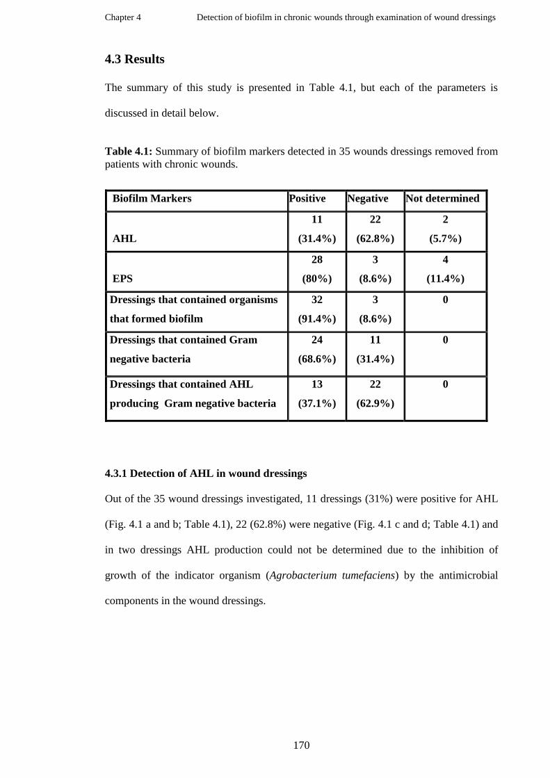
Chapter 4 Detection of biofilm in chronic wounds through examination of wound dressings
170
4.3 Results
The summary of this study is presented in Table 4.1, but each of the parameters is
discussed in detail below.
Table 4.1: Summary of biofilm markers detected in 35 wounds dressings removed from
patients with chronic wounds.
Biofilm Markers Positive Negative Not determined
AHL
11
(31.4%)
22
(62.8%)
2
(5.7%)
EPS
28
(80%)
3
(8.6%)
4
(11.4%)
Dressings that contained organisms
that formed biofilm
32
(91.4%)
3
(8.6%)
0
Dressings that contained Gram
negative bacteria
24
(68.6%)
11
(31.4%)
0
Dressings that contained AHL
producing Gram negative bacteria
13
(37.1%)
22
(62.9%)
0
4.3.1 Detection of AHL in wound dressings
Out of the 35 wound dressings investigated, 11 dressings (31%) were positive for AHL
(Fig. 4.1 a and b; Table 4.1), 22 (62.8%) were negative (Fig. 4.1 c and d; Table 4.1) and
in two dressings AHL production could not be determined due to the inhibition of
growth of the indicator organism (Agrobacterium tumefaciens) by the antimicrobial
components in the wound dressings.

Chapter 4 Detection of biofilm in chronic wounds through examination of wound dressings
171
Figure 4.1: Samples of chronic wound dressings tested for AHL. Examples of
sterilised wound dressings‟ samples (a and b) in which AHL was indicated by the
reporter strain A. tumefaciens NTL4, (c and d) wound dressings in which AHL was not
detected.
4.3.2 Detection of extracellular polymeric substances (EPS) in wound dressings
EPS were detected in 28 (80%) of the dressings (Table 4.1; Fig. 4.2a), 3 (8.6%)
dressings tested negative for EPS (Table 4.1, Fig 4.2b) and 4 dressings could not be
tested because the dressings were too dry to make suitable smears for EPS examination.
a b
c d

Chapter 4 Detection of biofilm in chronic wounds through examination of wound dressings
172
Figure 4.2 EPS positive and EPS negative smears derived from chronic wound
dressings. Stained slide smears of chronic wound exudates observed under light
microscope (x100 objective): (a) chronic wound dressing that tested positive for EPS
showing embedded organisms and (b) a wound dressing that did not contain EPS.
4.3.3 Microorganisms isolated from chronic wound dressings
4.3.3.1 Species and frequency distribution of isolated organisms
The organisms recovered from the dressings that were characterised included aerobic
and anaerobic bacteria and yeasts (Fig. 4.4). In total 108 isolates were recovered from
the dressings ranging from one to six organisms per dressing with a mean of three
isolates per dressing (Fig. 4.5). Twenty two Staphylococcus aureus were isolated, of
which eight were methicillin resistant S. aureus (MRSA) (Fig. 4.3). They were obtained
from 22.9 % of the dressings while P. aeruginosa was isolated from 14 dressings (40%)
and Corynebacterium striatum from 10 dressings (28.6%) (Fig. 4.4 and Appendices 4
and 5).
Figure 4.3: Cultures of Staphylococcus aureus tested for methicillin susceptibility.
(a) Reference S. aureus NCTC 6571, (b) a methicillin sensitive S. aureus (MSSA) and
(c) methicillin resistant S. aureus (MRSA) isolated from chronic wound dressings.
a c b
Figure 5.1:
a b
7μm 7μm

Chapter 4 Detection of biofilm in chronic wounds through examination of wound dressings
173
0
5
10
15
20
25
A. l
woffi
i
B. c
apill
osus
B. c
epac
ia
C. a
lbic
ans
C. f
amat
a
C. f
reundii
C. b
ovis
C.je
ikei
um
C. p
ropin
guum
C. p
seudodip
hther
iticu
m
C. r
enal
e grp
C. p
seudogen
italiu
m
C. s
tria
tum
C. u
lcer
ans
E. c
loac
ae
E. f
aeca
lis
E. c
oli
F. ovy
zihab
itans
G. h
aem
olysa
ns
K. o
xyto
ca
K.
pneum
oniae
L. aci
dophyllu
s
M. k
ristin
ae
P. t
etra
dius
P. l
oesch
eii
P. m
irab
ilis
P. v
ulgar
is
P.
aeru
ginosa
S.c
erev
isia
e
S. m
arce
scen
s
S. p
utref
acie
ns
S. a
ureus
S. a
uricu
laris
S. e
pider
mid
is
S. h
aem
olytic
us
S. s
acch
aroly
ticus
S. s
imulta
ns
S. a
galac
tiae
S. c
onstel
latu
s
Strep
toco
ccus
grp G
S. s
anguis
Organisms
Nu
mb
er
Figure 4.4: Frequency of isolated organisms from chronic wound dressings
0
1
2
3
4
5
6
7
8
9
10
1 2 3 4 5 6
Number of species per dressing
Nu
mb
er
of
dre
ssin
gs
Figure 4.5: Distribution of species of organisms isolated from each chronic wound
dressing. The number of species of organisms isolated from each dressing ranges from
1 to 6 with a mean of 3 species per dressing.

Chapter 4 Detection of biofilm in chronic wounds through examination of wound dressings
174
4.3.3.2 Biofilm phenotypic expressions of isolated organisms from chronic wound
dressings
4.3.3.2.1 AHL production by Gram negative bacteria isolated from the wound dressings
In total Gram negative bacteria were isolated from 23 of the 35 wound dressings but
Gram negative bacteria that produced AHL in vitro (Table 4.1; Fig. 4.6 a) were isolated
from 13 dressings (Tables 4.1 and 4.2) Gram negative bacteria from 10 dressings tested
negative for AHL production (Figure 4.6 b).
Figure 4.6 AHL positive and AHL negative Gram negative bacteria recovered
from chronic wound dressings. Pictures (a) A. tumefaciens NTL4 streaked beside an
AHL producing Gram negative bacteria isolated (b) a non- AHL producing Gram
negative isolate.
a b NTL4 NTL4
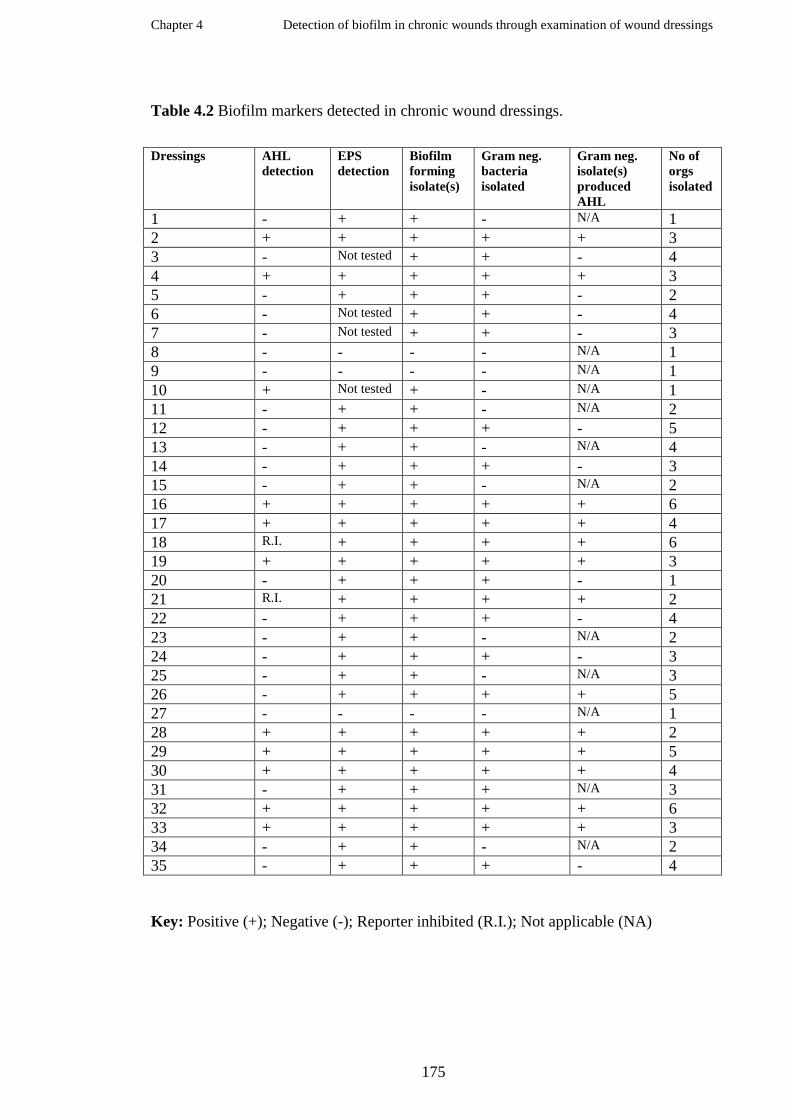
Chapter 4 Detection of biofilm in chronic wounds through examination of wound dressings
175
Table 4.2 Biofilm markers detected in chronic wound dressings.
Dressings AHL
detection
EPS
detection
Biofilm
forming
isolate(s)
Gram neg.
bacteria
isolated
Gram neg.
isolate(s)
produced
AHL
No of
orgs
isolated
1 - + + - N/A 1
2 + + + + + 3
3 - Not tested + + - 4
4 + + + + + 3
5 - + + + - 2
6 - Not tested + + - 4
7 - Not tested + + - 3
8 - - - - N/A 1
9 - - - - N/A 1
10 + Not tested + - N/A 1
11 - + + - N/A 2
12 - + + + - 5
13 - + + - N/A 4
14 - + + + - 3
15 - + + - N/A 2
16 + + + + + 6
17 + + + + + 4
18 R.I. + + + + 6
19 + + + + + 3
20 - + + + - 1
21 R.I. + + + + 2
22 - + + + - 4
23 - + + - N/A 2
24 - + + + - 3
25 - + + - N/A 3
26 - + + + + 5
27 - - - - N/A 1
28 + + + + + 2
29 + + + + + 5
30 + + + + + 4
31 - + + + N/A 3
32 + + + + + 6
33 + + + + + 3
34 - + + - N/A 2
35 - + + + - 4
Key: Positive (+); Negative (-); Reporter inhibited (R.I.); Not applicable (NA)
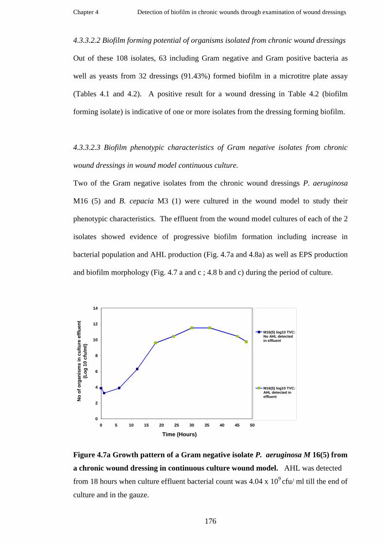
Chapter 4 Detection of biofilm in chronic wounds through examination of wound dressings
176
4.3.3.2.2 Biofilm forming potential of organisms isolated from chronic wound dressings
Out of these 108 isolates, 63 including Gram negative and Gram positive bacteria as
well as yeasts from 32 dressings (91.43%) formed biofilm in a microtitre plate assay
(Tables 4.1 and 4.2). A positive result for a wound dressing in Table 4.2 (biofilm
forming isolate) is indicative of one or more isolates from the dressing forming biofilm.
4.3.3.2.3 Biofilm phenotypic characteristics of Gram negative isolates from chronic
wound dressings in wound model continuous culture.
Two of the Gram negative isolates from the chronic wound dressings P. aeruginosa
M16 (5) and B. cepacia M3 (1) were cultured in the wound model to study their
phenotypic characteristics. The effluent from the wound model cultures of each of the 2
isolates showed evidence of progressive biofilm formation including increase in
bacterial population and AHL production (Fig. 4.7a and 4.8a) as well as EPS production
and biofilm morphology (Fig. 4.7 a and c ; 4.8 b and c) during the period of culture.
0
2
4
6
8
10
12
14
0 5 10 15 20 25 30 35 40 45 50
Time (Hours)
No
of
org
an
ism
s in
cu
ltu
re e
fflu
en
t
(Lo
g 1
0 c
fu/m
l)
M16(5) log10 TVC:No AHL detectedin effluent
M16(5) log10 TVC:AHL detected ineffluent
Figure 4.7a Growth pattern of a Gram negative isolate P. aeruginosa M 16(5) from
a chronic wound dressing in continuous culture wound model. AHL was detected
from 18 hours when culture effluent bacterial count was 4.04 x 10⁹ cfu/ ml till the end of
culture and in the gauze.

Chapter 4 Detection of biofilm in chronic wounds through examination of wound dressings
177
Figure 4.7b Biofilm developments of a wound dressing isolate P. aeruginosa
M16 (5) in wound model continuous culture. Microcolonies became bigger with
increase in number of organisms and EPS production relative to time as shown on
microscopic examination (x10 objectives).
Figure 4.7c Biofilm development of a P. aeruginosa M16 (5) isolated from a wound
dressing in wound model continuous culture. Microcolonies became bigger with
increase in bacterial population and EPS production with time as shown on microscopic
examination (x100 objectives).
24 hrs
70μm
45 hrs
6 hrs 12 hrs
36 hrs 30 hrs 48 hrs
18hrs 70μm 70μm
70μm 70μm 70μm 70μm
24 hrs 70μm 70μm
12Hrs 18Hrs 24Hrs 6 Hrs
30 Hrs 36 Hrs 45 Hrs 48 Hrs
7μm 7μm 7μm 7μm
7μm 7μm 7μm 7μm

Chapter 4 Detection of biofilm in chronic wounds through examination of wound dressings
178
0
2
4
6
8
10
12
0 5 10 15 20 25 30 35 40 45 50
Time (Hours)
No
of
org
an
ism
s in
cu
ltu
re e
fflu
en
t
(L
og
10 c
fu/m
l)
M3(1) log 10 TVC:No AHL detected ineffluent
M3(1) log10 TVC:AHL detected ineffluent
Figure 4.8a Growth pattern of a Gram negative isolate, B. cepacia M 3(1), from a
chronic wound dressing in a wound model culture. Biofilm formation was evident
from 12 hours and AHL was detected from 30 hours when the culture effluent bacterial
viable count was 1.63 x 10⁹ cfu/ ml up to 45 hours but no AHL was detected at 48 hours
and in the gauze.
Figure 4.8b Biofilm developments of a Burkholderia cepacia M3 (1) isolated from
wound dressing in wound model continuous culture. Microcolonies became bigger
with increase in number of organisms and EPS production with time as shown on
microscopic examination (x100 objectives).
12 Hrs 24 Hrs
36 Hrs 45 Hrs 48 Hrs
30 Hrs 7μm 7μm
7μm 7μm
7μm
7μm

Chapter 4 Detection of biofilm in chronic wounds through examination of wound dressings
179
4.3.4 Statistical analysis and correlation
The data were analysed with Minitab version 15. Pearson correlation shows a strong
association between the detection of AHL in dressings and isolation of P. aeruginosa
from the dressings (P< 0.005) (Fig. 4.9). There was also a correlation between EPS
detection in the dressings and in vitro formation of biofilm by the organisms isolated
from the dressings (P< 0.0005) (Fig. 4.10). AHL detection in dressings and isolation of
Gram negative bacteria was also significant, P< 0.034.
2.01.51.00.50.0
99
95
90
80
70
60
50
40
30
20
10
5
1
Dressing with P. aeruginosa
Pe
rce
nt
Mean 0.9333
StDev 0.2582
N 15
AD 5.203
P-Value <0.005
Normal - 95% CI
Probability Plot of Dressing with P. aeruginosa and AHL in dressing
Figure 4.9 Graphical representation of the relationship between AHL detection
and isolation of Pseudomonas aeruginosa from chronic wound dressings

Chapter 4 Detection of biofilm in chronic wounds through examination of wound dressings
180
2.01.51.00.50.0
99
95
90
80
70
60
50
40
30
20
10
5
1
Data
Pe
rce
nt
0.9033 0.3002 31 10.194 <0.005
0.9144 0.2837 35 11.781 <0.005
Mean StDev N AD P
EPS detection
Isolate(s) formed Biofilm
Variable
Probability Plot of EPS detection, Isolate(s) formed BiofilmNormal - 95% CI
Figure 4.10 Graphical representation of the relationship between detection of EPS
in chronic wound dressings and formation of biofilm by the isolated organisms
from the dressings.
Chapter 4 Detection of biofilm in chronic wounds through examination of wound dressings
181
4.4 Discussion
According to the US National Institute of Health about 80% of chronic infections are
due to biofilm (NIH, USA, 1997) which has been associated with the incessant
resistance to antimicrobial agents (Davies, 2003). It has been suggested that the
presence of biofilm in wounds impedes wound healing processes (Bjarnsholt, 2008). In
the past, various studies have been done to elucidate the growth and physiology of
biofilm as well as the resistance mechanisms to immune and antimicrobial interventions
in vitro and in vivo using animal models. To date investigations designed to determine
the presence of biofilm in wounds have relied on wound biopsies which might be
inconvenient to patients because the procedure is invasive, although recently wound
debridement samples have been used. It would be a novel idea to screen for biofilm in
wounds without any discomfort to the patient by examining used wound dressings.
It has also been observed that non- cultural methods are able to detect wound pathogens
more extensively than cultural methods, screening of wounds for biofilm using non-
cultural methods such as detection of biofilm markers in dressings removed from
chronic wounds might increase awareness in the diagnosis of biofilm in wounds and
contribute to wound management.
In infections involving biofilms, quorum sensing molecules are the main pivot of
biofilm formation (Huang et al, 2009; Costerton et al, 1999) and detection of these
molecules is therefore suggestive of biofilm colonisation of the wound.
In this study, there was evidence of biofilm markers including AHL and EPS in the
dressings removed from 35 chronic wounds, and the organisms isolated from the
dressings expressed biofilm phenotypic characteristics such as biofilm formation and
production of AHL and EPS in vitro.
Chapter 4 Detection of biofilm in chronic wounds through examination of wound dressings
182
AHL was detected in 11 dressings (Table 4. 1, Fig. 4.1 a and b) 10 (90.9%) of which
contained Gram negative bacteria that produced AHL in vitro (Table 4. 1, Fig. 4.6a) (p<
0.005). However no Gram negative bacteria were isolated from the remaining wound
and any organisms in this dressing might have been inhibited by a component present in
the wound dressing. The inability of the culture method to isolate the organism is one
of the factors that usually necessitate the use of alternative means of identification of
organisms such as PCR which relies on the bacterial DNA (Rayner et al, 1998). All of
the Gram negative bacteria that were isolated from the AHL positive dressings were
strong biofilm formers in the microtitre assay and this suggests the organisms have the
potential to form biofilm in a wound.
Detection of AHL in wound dressings could provide a useful means of diagnosis of
biofilm in wounds. In the past, the detection of AHL in urinary catheters (Stickler et al,
1998) and in the sputum of cystic fibrosis patients (Singh et al, 2000) has been used to
determine the presence of biofilm. Wu and colleagues (2000) confirmed the
pathogenicity of P. aeruginosa by detecting AHL in the lungs of P. aeruginosa
intratracheally infected mice. This finding corroborates previous studies of AHL
detection as biofilm marker and recently in chronic wound debridement samples
(Rickard et al, 2010).
In all, Gram negative bacteria were isolated from 23 dressings but Gram negative
bacteria that tested positive for AHL were isolated from only 13 dressings (56.5%). Out
of the 13 dressings, 10 dressings (43.5%) tested positive for AHL screening while 2
(8.7%) dressings results could not be determined due to inhibition of the growth of the
reporter strain. Gram negative bacteria that tested negative for AHL (Table 4.1; Fig. 4.6
b) were isolated from 10 dressings (43.5%) and the dressings with non-AHL producing
Gram negative bacteria tested negative for AHL. The detection of AHL in used wound

Chapter 4 Detection of biofilm in chronic wounds through examination of wound dressings
183
dressings seemed to be a sensitive assay and easily executed. AHL producing bacteria
were isolated from all the dressings in which AHL was detected during screening.
All of the dressings that tested positive for AHL were polymicrobial and in particular
contained biofilm forming P. aeruginosa. This confirms the importance of
P. aeruginosa biofilm formation in wounds and the involvement with multispecies
biofilms, which has been demonstrated by other studies (Malic et al, 2009). Malic and
colleagues in their study observed that P. aeruginosa was the most predominant
organism within multi-species biofilms. The detection of AHL in almost all of the
dressings in which P aeruginosa was isolated reveals the vital role of signalling
molecules in the formation of P. aeruginosa biofilm (Van Delden and Iglewski, 1998).
In particular the association of virulence expression by P. aeruginosa during infection
with increase in AHL production (Van Delden and Iglewski, 1998) has been shown and
usually coincides with pathogenicity of P. aeruginosa (Wu et al, 2000). Rickard and
colleagues (2010) found that although most of the resident organisms within the wound
produced AI-2 molecules but aggressive species such as P. aeruginosa produce AHLs
which was revealed in this study. A strong association between the presence of
P. aeruginosa and detection of AHL in chronic wounds has been observed in this
investigation.
In this study, one wound dressing tested positive for AHL but the culture of the dressing
did not yield the growth of any Gram negative organisms. It has been extensively
reviewed that the use of cultural methods often miss out some pathogens and lack of
identification of such organisms has a great impact on treatment strategies hence the
need to utilise non - cultural methods (Rogers et al, 2009). In a study of the 40 diabetic
foot ulcers by Dowd and his colleagues (2008), it was observed that traditional cultural
methods select for easily cultured organisms against the difficult to culture organisms.

Chapter 4 Detection of biofilm in chronic wounds through examination of wound dressings
184
In a similar study of the microbial microbiota of chronic wounds, Kirketerp-Moller and
his colleagues (2008) also support the development of new diagnostic strategies for
chronic wounds because they observed that more organisms were identified by non-
cultural methods than from the cultures. The detection of P. aeruginosa was higher in
non-cultural methods than the cultural method in the study. In another study the non-
cultural method revealed 4 times the number of organisms from the wounds than the
cultural methods (Price et al, 2009) and high prevalence of P. aeruginosa was observed
amongst patients treated with antibiotics. These might explain why a wound dressing
tested positive for AHL but no Gram negative organism was isolated from the dressing.
Although in this circumstance, the possibility of antibiotic resistance is suggested as the
main reason for inability to isolate the organism.
Investigation of the biofilm phenotypic characteristics of the selected Gram negative
wound dressing isolates showed that Burkholderia cepacia M3 (1) produced AHL in the
continuous culture investigation between 30 hours and 45 hours, after which the AHL
became undetectable (Figure 4. 8a), whereas the organism had tested negative for the
AHL in the cross feeding assay (1cm away from reporter strain). It has been observed
that some strains of biofilm forming B. cepacia show dynamic changes in cultures and
the production of AHL decreases rapidly during the stationary phase (Soo et al, 2009;
Chen et al, 2005) and similar response was observed in this study. The findings of the
analysis of QS and QSI in chapter 2 showed that the closer the reporter organism was to
the test organism, the higher the chance of detecting the AHL production. This low
production of AHL by some of the organisms could have resulted in false negatives
such as the case with M3 (1) that tested negative for cross- feeding assay but produced
AHL in wound model continuous culture from 30 hours of culture up till 45 hours. It
also tested negative for AHL at 48 hours (Figure 4. 8a).

Chapter 4 Detection of biofilm in chronic wounds through examination of wound dressings
185
Some studies have discovered that certain strains of bacteria such as P. aeruginosa form
biofilm independently of signalling molecules (Schaber et al, 2007; Favre - Bonte et al,
2007). It is possible to miss out the diagnosis of such biofilm organisms when
established in wounds by using signalling molecules as a lone means of diagnosis. This
study investigated AHL signalling molecule alone and not other signalling molecules.
Hence a limitation of this study is not detecting other signalling molecules, which
would have provided more information about the relevance of detecting signalling
molecules in wound dressings.
The results obtained from this study showed that most of the wound dressings
(28 (80%) of the 35 tested) contained EPS, three tested negative while four could not
be tested due to the nature of the dressings because they were dry. Organisms that
formed biofilm in a microtitre plate assay were isolated from 32 dressings inclusive of
all the 28 EPS positive dressings. There was a significant correlation between the
detection of EPS in dressings and biofilm formation of the organisms isolated from the
dressings; p< 0. 005. EPS serves as the housing for the biofilm community preventing
the organisms from the environmental stress such as body immune system and
antibiotics (Leid et al, 2005; Ryu and Beuchat, 2005) consequently resulting in the
persistence of infection therefore detection of EPS in the chronic wound dressings
suggests the presence of biofilm in such wounds. Many biofilm detection studies have
utilised methods that detect EPS (Harrison- Balestra et al, 2003), even for the
determination of biofilm age (Ahimou et al, 2007).
Of the wound dressing isolates tested here, two [M16 (5) and M3 (1)] demonstrated
progressive biofilm formation in continuous cultures with high production of EPS and
AHL. The presence of EPS in chronic wound dressings is another indication of the
presence of biofilms in the wounds from which the dressings were obtained, and the

Chapter 4 Detection of biofilm in chronic wounds through examination of wound dressings
186
organisms that were isolated from the dressings also produced biofilm in the laboratory
(Table 4.1). The three findings provide evidence of the presence of biofilm in the
chronic wounds where the dressings were removed.
A total of 108 organisms were isolated and characterised from the chronic wound
dressings, 63 (58.3%) which were isolated from 32 dressings (91.4%) formed biofilm in
vitro while 46 did not form biofilm suggesting the potential of the organisms to form
biofilm in wounds. Out of the 32 dressings from which biofilm forming organisms
were isolated, 28 (87.5%) tested positive for EPS but 4 could not be screened. There is a
strong correlation between EPS detection in the wound dressings and biofilm formation
by the isolated organisms (P< 0.005).
In a study the Pseudomonas aeruginosa strains biofilms were cultivated in a reactor to
compare the phenotypic expressions of type culture and clinical isolates. A type culture
PAO 1 and four clinical isolates, derived from CF patients were investigated for
phenotypic expressions of the organisms in the laboratory. The clinical variants
displayed many of the same characteristics as the laboratory variants, suggesting a link
between laboratory and cystic fibrosis biofilms (Kirisits et al, 2005). Applying the same
concept, detection of AHL in catheters removed from patients confirmed the presence
of biofilm in the bladder from which the catheters were obtained (Stickler et al, 1998).
The principle was also demonstrated by Malic and his colleagues (2009) using species
distribution of chronic wound biofilms bacteria in vitro to demonstrate biofilm organism
distribution in tissues from chronic wounds.
Most of the dressings were polymicrobial (82.9%) which was in agreement with other
studies (Dowd et al, 2008; Kennedy et al, 2009) especially the involvement of biofilms
in such wounds (Kennedy et al, 2009; Kathju et al, 2009; Dowd et al, 2008). The

Chapter 4 Detection of biofilm in chronic wounds through examination of wound dressings
187
abundance of P. aeruginosa in wounds as observed in this study has been confirmed by
previous studies (Price et al, 2009; Fazli et al, 2009; Oguntibeju and Nwobu, 2004).
In all 40 Gram negative bacteria were isolated from 23 dressings, 17 isolates (42.5%)
from 13 dressings produced AHL in vitro. Thirty four of the Gram negative bacteria
from 24 dressings formed biofilm in the laboratory. AHL detection can only be utilised
as a diagnostic tool for biofilms of AHL producing organisms hence the limitation of
this method alone in biofilm detection. AHL detection was highly sensitive and
detected most of the AHL producing bacteria, therefore it will provide a good diagnosis
for investigation of biofilm involving AHL producers in wounds.
Two of the isolates were cultured in a continuous culture system and evidence of AHL
(Fig. 4.7a and Fig. 4.8a) and EPS (Fig. 4.7b and c and Fig. 4.8b) production were
observed which confirmed that the AHL and EPS that were detected in the wound
dressings were produced by the biofilm forming organisms that were isolated from the
same dressings.
It was observed from this study that the most prevalent organisms isolated from the
dressings were Staphylococcus aureus (13%) or including MRSA (20.4%);
P. aeruginosa (13%), Corynebacterium striatum (9.3%) and MRSA (7.4%) (Fig. 4.4)
which was in agreement with other studies of wound flora (Malic et al, 2009; Kirketerp-
Møller et al, 2008).
The detection of biofilm markers such as AHL and EPS in dressings is a good indicator
of the presence of biofilm in wounds, which in turn is of clinical significance, in view of
the findings that the presence of biofilms in wounds is linked to failure to heal
(Bjarnsholt et al, 2008; Percival and Bowler, 2004). Detecting bacterial infections
through culture independent approaches has been suggested as the solution to inability
of cultural methods to isolate some pathogenic organisms (Rogers et al, 2009). In this

Chapter 4 Detection of biofilm in chronic wounds through examination of wound dressings
188
study, AHL was detected in particular in almost all of the dressings that biofilm forming
Ps. aeruginosa strains were isolated confirming the importance of quorum sensing in
the pathogenicity of Ps. aeruginosa infections. These findings also corroborate the role
of AHL in modulating the virulence of pathogenic organisms such as P. aeruginosa
(Jensen et al, 2006; Bjarnsholt et al, 2005; Sauer et al, 2002). The dressings from
which P. aeruginosa cultures were isolated were multi-microbial and most of the
organisms formed biofilm in vitro which also shows the vital role of quorum sensing to
biofilm community. Regrettably, other signalling molecules were not examined in this
study hence the precise number of dressings containing signalling molecules could not
be determined, to corroborate the biofilm forming potential of the isolated organisms in
vitro and the EPS detection in dressings.
EPS was detected in the majority of the dressings. EPS is known to protect biofilm
against body immune system and antibiotics (Leid et al, 2005; Ryu and Beuchat, 2005);
the implication is that the organisms will be able to resist treatment interventions and
persistently cause damage to the tissues and breakdown of wounds.
The presence of biofilms in wounds has been shown to interfere with the healing
process leading to chronicity of such wounds and recently the presence of biofilm in
wound have been reported (Malic et al, 2009; Dowd et al,, 2008; James et al, 2008;
Ngo et al, 2007). In previous studies that the presence of biofilm had been
investigated using tissue biopsy samples from wounds, out of 12 chronic wounds,
7 (58.3%) tested positive (Ngo et al, 2007), of 50 chronic wounds 30 (60%) tested
positive while 21 (70%) out of 30 tested positive when debridement materials were
used. From the results of the investigations, the fact that wound pathogens are not
evenly distributed within the wound plays a vital role in the ability to isolate or detect
the biofilm. This was discussed by one of the groups in their report as a factor that

Chapter 4 Detection of biofilm in chronic wounds through examination of wound dressings
189
probably affected the result because they were of the opinion that the positive results
ought to have been higher (Ngo et al, 2007). The trends of the result are increasing and
the debridement materials might likely have larger sampling area depending on whether
the whole wound surface was debrided and how deep since it has been found that some
organisms such as P. aeruginosa are found in the deeper regions of the wound.
Detection of AHL in 70% wound debridement samples has not only shed more light
into the diagnosis of biofilm in wounds through biofilm markers but also the high
prevalence of biofilm in wounds.
However, the results obtained in this study showed higher trend because wound
dressings cover a larger sampling areas than biopsies. Since the signalling molecules
would diffuse though the dressing while EPS from the organisms within the wound
surface would be picked as well as those of the organisms in the deeper region due to
the pressure of the dressing. In this study, of the 35 chronic wound dressings that were
investigated, only three were negative for all the biofilm markers. 32 of the dressings
contained organisms that formed biofilm in vitro, 28 of which contained EPS. Although
not all the dressings contained Gram negative organisms or AHL positive Gram
negative organisms but AHL positive bacteria were isolated from the dressings that
tested positive for AHL and the bacteria formed biofilm in microtitre assay, and
produced AHL in culture and EPS in continuous cultures to confirm biofilm phenotype.
Using the 2 parameters (EPS and biofilm formation) 80% of the chronic wounds
showed evidence of biofilm colonisation. But the 3 biomarkers: EPS, AHL and biofilm
formation by isolates, gave the result of 32 (91%) of the chronic wounds were inferred
to have been colonised by biofilm.

Chapter 4 Detection of biofilm in chronic wounds through examination of wound dressings
190
Conclusion
A high prevalence of biofilm markers in dressings removed from chronic wounds was
found in this study. The polymicrobial nature of wounds was also confirmed, especially
in wounds thought to be colonised with biofilm. S. aureus, P. aeruginosa and
Corynebacterium striatum were found to be the most frequently isolated organisms.
There was a strong association between the detection of AHL and the presence of
P. aeruginosa in wound. Examination of dressings removed from wounds for
microbially derived biomarkers may be used to detect biofilm in wounds. This study has
supported other studies that biofilm is present in chronic wounds.
Application of the techniques as used in this study as a diagnostic tool for detecting
biofilm could simplify the process of identification of infections associated with biofilm
in wounds. It could also be employed in assessing biofilm management strategies and
monitoring treatment s designed to eradicate biofilm from wounds.
AHL was the only signalling molecule tested here; therefore the ability to detect the
presence of other signalling molecules in wounds could not be fully accomplished.
There is a need to utilise other assay methods to detect all the various signalling
molecules of both Gram positive and Gram negative organisms, including the intra and
intercellular signalling molecules in future studies. A larger cohort might be needed to
determine whether detection of signalling molecules can be used as a lone indicator of
biofilm in wounds. In future scanning electron microscopic examination of the wound
dressings could be done to reveal biofilm architecture. Further studies are needed to link
the presence of biofilm in a wound in vivo to the presence of biofilm markers in
dressings.

Chapter 5
Antimicrobial effect of honey on biofilm
and quorum sensing

Chapter 5 Effect of honey on biofilm and quorum sensing
191
5.1 Introduction
Treatments of pathogenic infections with antimicrobials have evolved over the years
particularly from the discovery of penicillin, the first antibiotic derived from
Penicillium notatum fungi by Sir Alexander Fleming, Scottish biologist and
pharmacologist in 1929. Numerous synthetic antimicrobials have since been produced
for killing or rendering ineffective several microorganisms. The major effects of
antimicrobials on microorganisms are interference with nucleic acid, protein or cell wall
synthesis and inhibition of metabolic pathways (Tenover, 2006) or by increasing the
membrane permeability of the organisms (Mangoni et al, 2004).
The limitation of the magnificent achievements of antimicrobial therapy is the ability of
microorganisms to develop ways of circumventing the lethal effect of antimicrobials.
The resistance of pathogenic organisms to antimicrobial agents has been on the increase
due to various resistance mechanisms acquired by the pathogens as well as over- usage
of antimicrobials. The major factors are the innate ability of microorganisms to resist
specific antimicrobials and to acquire resistance genes from other organisms within the
environment by conjugation, transduction or transformation. Selection of mutants could
occur as a result of antibiotic treatment regimes which often kill susceptible bacteria,
allowing resistant strains to dominate (Tegnell et al, 2003). These genotypic changes
sometimes lead to the selection of mutants and consequent resistance to specific or
multiple antimicrobials which often result in treatment failures and other complications
that may be life threatening. Prolonged use of antibiotic could also lead to selection of
antibiotic resistant strains that may spread within the community or the hospital
(Pedersen et al, 1986). The economic implication of antibiotic resistance to healthcare is
enormous (Cosgrove, 2006). The available funds for developing new antimicrobials are
fast depleting and the lack of development of new antimicrobials is being envisaged as a
serious potential threat to public health (Norrby et al, 2005). The impact on treatment

Chapter 5 Effect of honey on biofilm and quorum sensing
192
of infections has therefore reawakened the awareness on the use of natural products
such as honey for treatment.
5.1.1 Honey and medical history
Honey is a nutritional product produced by bees from nectar which they collect from
plants as source of food. The composition of honey usually depends on the floral
sources particularly the phytochemical components of the honey (Molan, 2001). The
major components of honey are the sugars, fructose (about 38.5%) and glucose (about
31.0%), (White and Doner, 1980). Other components include the enzymes produced
by bees, sucrose between 1.0% and 12%, water 17.0% other sugars (such as maltose,
melezitose) 9.0%, ash 0.17% to 0.77 respectively according to the analysis of a typical
honeys (Kamal et al, 2002) and other components. The enzymes commonly found in
honeys are diastase (amylase), invertase (α-glucosidase), glucose oxidase, catalase and
acid which contribute to their overall activity. Honeys also contain phenols; the amount
of which influences the antimicrobial activity of the honey (Aljadi and Yusoff, 2003).
Other substances that have been identified in honeys are minerals such as K, Na, Ca,
Mg, Fe, Zn, Cu, Ni and Co, with K (Saif-ur-Rehman et al, 2008). The composition of
honey remains constant with storage at temperature of 20oC or below (Rybak-
Chmielewska and Szczesna, 1995) hence temperature is of high importance in the
storage of honey for the maintenance of potency when honey is to be used as a
medicament. Individual honeys vary from their components due to the type of bees that
produced them, the plants from where they are produced and the conditions of storage.
Higher storage temperatures tend to increase the hydroxymethylfurfuraldehyde (HMF)
of honey which could be up to 3 times within 12 months storage at high temperatures
(Rybak-Chmielewska and Szczesna, 1995) hence it is advisable to store honey below
4oC.

Chapter 5 Effect of honey on biofilm and quorum sensing
193
5.1.2 Physico-chemical properties of honey
The constituents of honey determine the various properties including the osmolarity, pH
and other chemical properties. The chemical properties are due mainly to the presence
of hydrogen peroxide, acids and enzymes such as catalase, glucose oxidase and sugars.
5.1.2.1 pH of honey
Although the pH of honey varies from honey to honey but generally they have an acidic
pH; ranging between 3.2 and 4.5 (White and Doner, 1980) and could be as low as 2. 19
(Ghazali, 2009) and this pH range is highly unfavourable for most microorganisms to
thrive. Other substances present in honey such as caffeic acid and ferulic acid have also
been found as contributory agents to the pH and antimicrobial effect of honey (Wahdan,
1998).
5.1.2.2 Osmolarity of honey
Honey has high osmolarity because it is mainly a highly saturated mixture of
monosaccharides with low water content, the molecules of which are mostly associated
with the sugars. The resultant osmotic effect prevents microbial growth but the
antimicrobial activity of honey is not exclusively due to the osmotic effect but other
substances (Molan, 2006; French et al, 2005). These were observed when diluted honey
inhibited microorganisms unlike concentrated sugar solution (French et al, 2005).
5.1.2.3Hydrogen peroxide activity of Honey
Hydrogen peroxide is produced in honey when glucose is broken down by glucose
oxidase to produce gluconic acid and hydrogen peroxide (White Jr. et al, 1963). The

Chapter 5 Effect of honey on biofilm and quorum sensing
194
hydrogen peroxide is broken down by the catalase enzyme which is also present in the
honey.
Glucose + H2O + O2 → gluconic acid + H2O2
In diluted honey, the effect of glucose oxidase enzyme lowers the pH which further
reduces the activity of the enzyme and consequently the production of peroxide (Molan,
1992). The slow release of the hydrogen peroxide prevents toxicity to the tissues but
acts as antiseptic without causing damage to the tissues when honey is applied to wound
(Molan, 2006). The oxygen free radicals released by the hydrogen is oxidised by the
iron present in honey which neutralises the effect on the tissue. The release of the
hydrogen peroxide has been particularly linked to the antimicrobial activity of some
honeys (Weston et al, 2000). Some honeys like manuka honey are non-peroxide honey
but the antimicrobial properties have been linked to other components especially the
phytochemical components (Atrott and Henle, 2009; Mavric et al, 2008; Adams et al,
2008).
5.1.2.4 Phytochemical components of honey
The phytochemical components have major effect on the antibacterial properties of
honeys which determines the potency of various honeys (Mboto et al, 2009; Molan,
2006) and they are dependent on the type of plants from which the bees derived the
nectar. In manuka honey, it has been shown that the antimicrobial properties are not
dependent on peroxide activity (Molan, 1992) and methylglyoxal (MGO) has been
identified as one of the major active antimicrobial components responsible for the
effectiveness against microorganisms (Atrott and Henle, 2009; Mavric et al, 2008;
Adams et al, 2008). In their studies Atrott and Henle (2009) analysed 61 manuka honey
samples and the results showed that the level of methylglyoxal in the honey samples
ranged from 189 to 835 mg/kg honey with corresponding antibacterial activities of 12.4

Chapter 5 Effect of honey on biofilm and quorum sensing
195
to 30.9 % equivalent phenol concentration. Mavric and colleagues (2008) evaluated the
MGO in six manuka honeys and found the concentrations of the MGO to be 1000 times
higher than the conventional honeys and that 15 -30% w/v manuka honey showed
antimicrobial effect whereas other honeys were only inhibitory to organisms at 80%
concentration. The study also linked the 1.1 -1.8 mM concentrations of MGO to 15 -
30% manuka honey concentration and to the unique manuka factor (UMF) values of
medical grade manuka honeys. Adams and colleagues (2008) characterized the
components of 49 manuka honeys alongside 39 other honeys from New Zealand; they
confirmed the non peroxide nature of manuka honey using an agar well diffusion assay
with S. aureus. Analysis with HPLC detection assay revealed the effective
antimicrobial component of manuka honey as methylglyoxal (Adams et al, 2008).
Adams and colleagues in 2009 explored the origin of methylglyoxal in manuka honey
by studying the chemical components of nectar from manuka shrubs (Leptospernum
scoparium) and other plants and linking the chemical composition to the methylglyoxal
in manuka honey. The activities of enzymes in honey are responsible for the conversion
of dihydroxyacetone to methylglyoxal (Adams et al, 2008). The concentration of
methylglyoxal in manuka honey increased with temperature according to the study
indicating the effect of temperature on the conversion of the chemical components of
honey (Adams et al, 2008). Another chemical component, defensin 1, has been
identified in honey recently (Kwakman et al, 2010).
5.1.2.5 Antioxidants properties of honey
Honey has antioxidant properties (Oddo et al, 2008; Henriques et al, 2006) which are
capable of neutralizing biologically destructive chemicals in the body. The antioxidant
contents of honey diminish with time particularly from six months of storage
notwithstanding the temperature and storage containers (Jiménez et al, 2006) hence

Chapter 5 Effect of honey on biofilm and quorum sensing
196
fresh honeys might be more beneficial than the older ones when used as antioxidant.
Total phenolic content have been shown to influence the antioxidant activity of honeys
although it is not solely responsible for it (Gheldof et al, 2002). Antioxidant activities
vary with the floral content and source of honey (Hegazi and El-Hady, 2007; Gheldof et
al, 2002 and 2003). The variations in the antioxidant property of honeys were
demonstrated by Henriques and colleagues (2006) with three types of honey. Manuka
honey (a medical grade honey) was found to have completed quenching of the
generated free radical by peroxide in 5 minutes whereas Pasture honey (another medical
grade honey) quenched the free radicals completely in 1 hour. The beneficial effect of
antioxidant property of honey can not be overemphasized in wound management
particularly in the removal of free radicals which are often produced by some
pathogenic organisms like Pseudomonas aeruginosa to invade tissues during infection.
5.1.3 Sterility of honey
Although there are numerous benefits of honey, honey sometimes contain
microorganisms (Cooper and Jenkins, 2009; Olaitan et al, 2007; Malika et al, 2005)
especially yeasts and spores of some organisms such as the Bacillus species (Cooper
and Jenkins, 2009) and Clostridium botulinum (Midura et al, 1979) which could be
from the bees, the environment or from the human handlers (Snowdon and Cliver,
1996). A similar range of microorganisms have been found in honeys obtained from
the same plant source suggesting that the source of the organisms might be from the
plants (Malika et al, 2005). Although due to the antimicrobial nature of honey, the
organisms might not thrive and be destroyed overtime but the ability of the spores of
some organisms to thrive in honey could jeopardize the beneficial effect especially in
children and the immuno-compromised. It is therefore pertinent to sterilise honeys that
are to be used for treatment of infections hence medically prepared honeys which are
Chapter 5 Effect of honey on biofilm and quorum sensing
197
usually sterilized to avoid the risk associated with the organisms should always be used
for treatment purposes. According to the study by Cooper and Jenkins (2009), while the
medical grade honey (MGH) examined was found to be sterile the non- MGHs (table
honeys) contained a range of microorganisms.
5.1.4 Medical importance of Honey
5.1.4.1 Antibacterial properties of honey
The antibacterial activities of honey have been extensively studied (Williams et al,
2009; George and Cutting, 2007; French et al, 2005; Cooper et al, 2002). Honey was
found to be many times more potent against coagulase-negative staphylococci than if
bacterial inhibition were due to their osmolarity alone when compared with artificial
honey or sugar solution (French et al, 2005; Efem and. Iwara, 1992) even when diluted
up to 7 to 15 fold honey still inhibited bacterial growth (Cooper et al, 2002).
Antimicrobial effects of honey have been demonstrated on common wound isolates
such as Pseudomonas (Cooper et al, 2002 b), Staphylococcus (French et al, 2005;
Cooper et al, 1999), fungi (Koc et al, 2009; Basson and Grobler, 2008), MRSA (Cooper
et al, 2002), VRE (Cooper et al, 2002a) Streptococcus species (Efem and Iwara,1992)
and recently on biofilms in vitro (Alandejani et al, 2009; Okhiria et al, 2009; Merckoll
et al, 2009). Although honey was antifungal Candida species were found to be more
resistant to honey than bacteria (Basson and Grobler, 2008). Koc and colleagues (2009)
tested the effect of various honeys on 40 yeast strains (Candida albicans, C. krusei,
C. glabrata and Trichosoporon species) and all the organisms were inhibited by the
honeys. According to a review of the laboratory and clinical evidence of antimicrobial
properties of honey by Cooper (2008) the evidence of broad antimicrobial effect of
honey against bacteria, fungi, viruses and protozoa has been recognised but much of the
evidence is anecdotal and further research is needed to appreciate the mechanism of

Chapter 5 Effect of honey on biofilm and quorum sensing
198
inhibition. Basson and Grobler‟s study also confirmed variation in honey‟s
antimicrobial effect as a result of the phytochemical properties which are dependent on
the plant from which the bees derive the nectar.
5.1.4.2 Enhancement of immune function
Scientific research with extracts from honey showed chemotactic activity for
neutrophils and antitumour characteristic in mice (Fukuda et al, 2009; Hamzaoglu et al,
2000). Various components in honey other than the sugars are responsible for the
potency of honey. Tonks and her group (2003) observed the stimulation of cytokines by
three honeys (jelly bush, manuka and pasture) and later (2007) discovered the 5.8-kDa
component of manuka honey stimulated cytokine production whereas the sugar solution
did not; and honey increased cellular viability which might explain the reason for the
effectiveness of the healing properties of honey particularly in wounds. When honey
extract were injected into mice, stimulation of the immune function and anti-tumour
activity were observed in the test mice unlike the control mice (Fukuda et al, 2009).
This enhancement of immune function has a great impact on the healing potential of
honey.
5.1.4.3 Application of Honey in wound treatment
The application of honey in the treatment of wounds dates back to the history of
mankind (Majno, 1975). There are unique properties of honey that enhance wound
healing potential which make honey an all encompassing remedy for wound treatment.
These factors include the antibacterial activity which prevents mal-odour and
inflammation, provides moist environment within the wound which aids debridement;
and stimulation of wound healing processes. The use of sugars in the honey by bacteria
instead of the amino acids in the dead tissues of the wounds further reduces the

Chapter 5 Effect of honey on biofilm and quorum sensing
199
unpleasant odour of the wounds (Molan, 2001). The uses of honey in treatment of
infected wounds and topical infections have been shown through several studies
(Robson et al 2009; Cooper and Molan, 1999; Subrahmanyam, 2005; Efem, 1988).
Honey has been used successfully in dressing infected wounds (Ingle et al, 2006;
Dunford et al, 2000).
It has been shown that honey keeps wound sterile even when such wounds have been
colonised and it debrides wounds easily (Efem, 1988). An acidic medium aids the
healing of wounds and the pH of honey reduces the pH and maintains lower pH when
applied to a wound. This reduction of wound pH on application of honey reduces
microbial load which aids wound healing (Molan, 2001). Gethin and her colleagues
(2008) observed that chronic wounds with pH of less than 7 which was achieved on the
application of honey healed faster than those at higher pH before honey was applied.
Honey is hygroscopic so when applied to wounds it keeps the wound moist and
prevents formation of scalds. The moisture also prevents the dressings from getting
stuck to the wound surface therefore reducing the discomfort to patients when dressings
are being removed. In a randomised control trial by Ingle and colleagues (2006) honey
treatment showed no significant advantage as effective healing agent over other agents
but treatment with honey offered other advantages of being cost-effective safe and
satisfying. Molan (2006) in a review article highlighted various positive evidence on the
application of honey in the treatment of wounds; including 17 randomized controlled
trials involving 1,965 participants, 5 clinical trials of other forms involving 97
participants and the effectiveness of honey in assisting wound healing demonstrated in
16 trials on 533 wounds. Lotfi (2008) discussed extensively the use of honey in animal
and human wounds. Simon and his colleagues (2009) exemplified the success rate of
wounds treated with honey in Germany especially the medical grade honey. Cooper
(2008) also reviewed the application of honey in the treatment of wounds and inhibition

Chapter 5 Effect of honey on biofilm and quorum sensing
200
of pathogens. The importance and benefits of honey as a modern medicine in the
treatment of wounds as well as the ways by which honey effectively heals wounds were
highlighted. Evidence of reduction of inflammation in wounds by honey has also been
reported (Dunford, 2000) so the effect of tissue damage and delayed healing through
excessive inflammation could be avoided which might be due to the antioxidant in
honey which removes the free radicals (Phillips et al, 2009; Henriques et al, 2006)
which often reduces inflammation (Molan, 2002).
Some researchers are of the opinion that honey has no better healing potential than other
topical agents that are being used as wound dressings because such dressings have
similar effect on wound healing as honey (Jull et al, 2008; Moore, 2008). The use of
honey in health care was recently reviewed by Bardy and others (2008) which include
43 studies in relation to burns, skin, cancer and other wounds. The results of the review
showed that many of the studies were not replicable because of poor description,
incomplete methodologies or inconsistencies between the groups studied while some
studies did not include standard deviations in their statistical data. Some studies did not
show the inclusion or exclusion criteria and sample size and lack of proper
documentation of informed consent in some studies particularly of note were those
involving minors. Meta-analysis of the systematic review was not possible due to the
heterogeneity amongst the studies (study designs, data presentations, variety of
conditions, different populations). Other factors that render the analysis inconclusive
include factors such as age, varying wound sizes and sites, presence of infection and
other conditions that may delay wound healing. They found that there is scope for usage
of honey for cancer patients. In conclusion, the analysis found honey to be particularly
effective when standard therapy failed and irrespective of the outcome of the review, it
was concluded that honey seems to provide an alternative treatment option according to
the overwhelming evidence. The application of honey in wounds particularly in

Chapter 5 Effect of honey on biofilm and quorum sensing
201
reduction of bioburden, minimising scarring and pain reduction was highlighted. The
use of honey in the management of other skin conditions such as dandruff, seborrheic
dermatitis, psoriasis and other fungal infections as well as chemotherapy or radiation
induced skin reactions was analysed. Evidence of reduction of oral mucosis in cancer
patients (Biswal et al, 2003) was shown to be comprehensive, well designed, controlled
and reliable. A major problem identified in the use of honey is the varying degrees of
potencies of various honeys which suggest that the antimicrobial effect of honeys might
vary significantly. The use of honey in oncology and medicine in general was diverse
and the significant role of honey in healthcare can not be overemphasized. Future
research was suggested to further unravel and elucidate the healing potentials of honey.
In another meta-analysis; the application of honey in wound treatment was reviewed
(Jull et al, 2008) which include acute and chronic wounds of all types. 19 trials were
included in the review including minor acute wounds, burns, acute and chronic wounds
such as pressure ulcers, diabetic ulcers, varicose ulcers and tropic ulcers. According to
the review, there does not seem to be enough evidence to support the use of honey in
minor uncomplicated wounds. In burn wounds treatment, honey was found to be more
effective than conventional dressings but there appears to be delayed healing in mixed
depth burns treated with honey. The reviewers are of the opinion that the rationale for
conducting the trials of the treatment of acute and chronic wounds except venous leg
ulcers is unclear and most of the trials are small therefore the findings might not
influence clinical practice. In summary, it was admitted that honey improves healing
over conventional dressing in mild to moderate superficial and partial thickness burns
but not chronic venous ulcer. Appropriate procedure for reporting clinical trials has
been recommended.
Simon and colleagues (2009) in a commentary of a brief review on honey titled
„‟Medical honey for wound care - still the latest resort?‟‟ opined that honey did not meet

Chapter 5 Effect of honey on biofilm and quorum sensing
202
the criteria for an ideal wound antiseptic because honey has no fast onset of activity.
Honey does not reduce bacteria and fungi by more than five log reductions in 1-10
minutes as an antiseptic. However unlike conventional antimicrobials such as iodine
and silver products that are involved with problems of metabolism honey is safe to use
even for paediatric patients. Raised liver enzymes has been reported in patients using
silver coated dressings (Trop et al, 2006) while the effect of iodine on thyroid functions
has been an issue to be considered when applying the product on infants. The uses of
honey as treatment interventions were highlighted in the review including recalcitrant
wounds (Simon et al, 2006;Blaster et al, 2007;Cooper et al, 2001), oral mucositis and
gingivalis in cancer patients (Biswal et al, 2003), wound care in neonates (Vardi et al,
1998), viral treatment (Al-Waili, 2004), ocular treatment (Albietz et al, 2006). Honey
treatment is less expensive than conventional antimicrobial agents (Ingle et al, 2006) an
additional benefit to encourage health professionals to use honey in wound care.
However certified honey with standardised antibacterial activity should be used as this
will eliminate the problem of the non- uniformity of efficacy of various honeys.
5.1.5 Biofilm resistance and justification for alternative antimicrobials
The problem of biofilm infections in particular resistance to body immune system and
commonly used antimicrobials have been highlighted and discussed extensively and
these phenomena often result in chronic infections. The formation of biofilm in wounds
by pathogenic organisms has been associated with impairment of healing processes due
to the combined effects of the virulence of the organisms within the biofilm (Bjarnsholt
et al, 2008). Biofilm formation involves a series of physiological and phenotypic
activities in which several genes are regulated in response to quorum sensing signalling
molecules produced by biofilm organisms (Sauer et al, 2002).

Chapter 5 Effect of honey on biofilm and quorum sensing
203
Inhibition of quorum sensing by some conventional antimicrobials such as azithromycin
(AZM), ceftazidime (CFT) and ciprofloxacin (CPR) were investigated by using DNA
micro array analysis and reverse transcription polymerase chain reaction (rt-PCR)
(Skindersoe et al, 2008). The results showed that the drugs decreased the expression of
a range of quorum sensing regulated virulence factors in P. aeruginosa. The effect of
attenuation of the organism through inhibition of quorum sensing signalling molecules
will allow the body defence system of the host (patient) to easily eliminate the organism
from the site of infection. A study in which P. aeruginosa quorum sensing was inhibited
resulted in the attenuation of the organism and the clearance from the mice lungs was
more effective (Hentzer et al, 2003).
Honey as an alternative antimicrobial has been explored in the treatment of infections
and as a result the antimicrobial effects of honey have been widely investigated on
various pathogenic organisms in the last two decades. Although many studies have
been done on the effect of honey on microorganisms (French et al, 2005; Cooper et al,
2002) including biofilm (Alandejani et al, 2009; Okhiria et al, 2009 and 2004; Merckoll
et al, 2009) the mode of action of honey on pathogenic organisms and biofilm in
particular is yet to be fully elucidated. More investigations will increase the
understanding of the mode of action of honey relevant to treatment of infections caused
by virulent pathogens in wounds and topical infections.
5.1.6 Recent studies on the antibacterial activities of various honeys
In a study done by Rasmussen and his colleagues (2005), various natural products (54)
were screened for ability to inhibit quorum sensing, of which 11 showed quorum
sensing inhibitory activity. The effect of garlic, one of the agents that inhibited QS, was
further tested on biofilm of PAO 1 and also in combination with tobramicin antibiotic.
It was observed that most of the bacteria in the biofilm treated with both garlic and

Chapter 5 Effect of honey on biofilm and quorum sensing
204
tobramicin died, while the cells at the top layer of the tobramycin treated biofilm died
but garlic extracts alone had no effect on biofilm. The control biofilm culture exhibited
the classical mushroom-like biofilm structure. The study showed evidence that natural
products alone or in combination with conventional antimicrobial might exert some
effect on biofilm by interfering with the quorum sensing systems.
The antimicrobial effects of honey on planktonic and biofilm embedded organisms in
chronic wounds were examined (Merckoll et al, 2009). In the study, bacteria were
grown as biofilm individually and as mixed cultures in broth and later exposed to
honey. The bacteria were also grown in various concentrations of irradiated and
commercially prepared honeys in broth with glucose. Difference in the susceptibility of
the individual organism was observed and the planktonic counterparts of the individual
organisms were susceptible to the honeys at low concentrations unlike the biofilms.
ESBL Klebsiella was least susceptible of the individual cultures as well as in the mixed
cultures. Medihoney® inhibited the growth of biofilm at 1.6% concentration but not at
0.8% and completely killed Pseudomonas at 6% (w/v) concentration whereas a
Norwegian honey was inhibitory to MRSA at 25% over the 20 hour period of
experiment. The study showed the variations in the resistance of various planktonic and
biofilm organisms to different honeys and that honey can support the growth of
microorganisms. Manuka honey was found to be of higher antimicrobial activity than
the Norwegian honey. The researchers highlighted the advantages of the multiple modes
of actions of honey on microorganisms. The interference of fructose content of honey
with fuctose, which is involved in binding of P. aeruginosa to surfaces, might further
prevent biofilm formation by P. aeruginosa (Lerrer et al, 2007).
In a study, 24 hour biofilms of 6 P. aeruginosa cultures (5 wound isolates and 1
reference culture) were exposed to 20% and 40% manuka honey (Okhiria et al, 2009).
Biofilms exposed to the 40% honey reduced in quantity overtime but increased growth

Chapter 5 Effect of honey on biofilm and quorum sensing
205
was observed after 48 hours whereas the 20% honey solution biofilm growth was
evident throughout the culture period for all the isolates and from 6 hours for the
reference organism. The study shows the lethal effect of manuka honey on biofilm at
40% concentration but the tendency of the sugars contained in honey to enhance growth
at sub-lethal concentration was revealed in the study.
Alandejani and his colleagues (2009) in a preliminary study examined the effects of
Sidr and manuka honeys on 11 cultures of each of methicillin susceptible S. aureus
(MSSA), methicillin resistant S. aureus (MRSA) and P. aeruginosa in both planktonic
and biofilm forms. The honeys were able to kill all the planktonic cultures but Sidr and
manuka honeys were effective against the biofilms of MSSA 63% -82%, MRSA 73% -
63% and P. aeruginosa 91% - 91% respectively at 1 in 2 dilutions of honeys showing
the increase resistance of biofilm than the planktonic counterparts. The strong
antimicrobial effects of the 2 honeys were demonstrated in their finding. Although the
initial study started with 4 honeys but 2 were dropped because of lack of cidal effect on
biofilm by the particular honeys, an indication that not all honeys should be used on
wounds that have biofilms since such honeys may enhance the growth of the biofilm
organisms (Okhiria et al, 2009).
In one of the recent studies, Ansari and Alexander (2009) tested the antimicrobial effect
of a honey produced by African sculata obtained from Guyana against Escherichia coli,
Pseudomonas species, S. aureus and Candida albicans using disc diffusion method. It
was observed that the honey was effective against all the bacteria but C. albicans was
not affected by the honey. The study showed that all honeys are not effective against all
pathogenic organisms that may be found in wound.
Another examination of the mode of inhibition of manuka honey on S. aureus NCTC
10017 (Henriques et al, 2010), scanning electron microscopy (SEM) and transmission
electron microscopy (TEM) of the organism before and after exposure to honey showed

Chapter 5 Effect of honey on biofilm and quorum sensing
206
that the inhibition of S. aurues 10017 by manuka honey was irreversible (MIC and
MBC values of 2.9 and 4.5% (w/v) respectively). The effect of 10% (w/v) manuka
honey on S. aureus revealed that manuka honey interfered with cell division of the
organism. There was an increase in the number of cells with crosswalls in the culture
treated with 10% (w/v) manuka honey unlike the cells in the control cultures indicating
that the cells could not divide.
Recently, the antimicrobial activity of 30 honeys on quorum sensing inhibition of
C. violaceum was investigated (Truchado et al, 2009). Inhibition of quorum sensing of
C violaceum by the honeys investigated was shown using agar diffusion method, and
estimation of violacein production by the reporter organism in broth culture. During the
investigation, the relationship between the peroxide activities and phenolic composition
of the various honeys were determined. The 15 unifloral honeys of different
geographical origins showed different QSI activities in dose dependent manner from
0.2g/ml. Reduced production of violacein pigment was found in broth cultures of
C. violaceum in the presence of honey samples and increase in honey concentration
resulted in significant increase in QSI activities of the honeys. The study also found
that QSI activity of individual honey is not solely dependent on the peroxide activity of
the honeys although there was reduction in QSI activities on addition of catalase to the
culture. There was no correlation between the phenolic content of the honeys and the
QSI activity of the honeys. The study demonstrated that QSI activity of honey which
was demonstrated by lack of violacein production by the reporter organism was not due
to inhibition of growth but inhibition of quorum sensing of the organism.
5.1.7 Treatment of biofilm embedded in wounds with honey
In the biofilm niche, signalling molecules mainly control the genetic and physiologic
activities which sequentially influence the pathogenicity of the organism (Zhu et al,
Chapter 5 Effect of honey on biofilm and quorum sensing
207
2002; de Kievit and Iglewski 2000). Biofilms have been found to be mostly
polymicrobial particularly in wounds (Malic et al, 2009; Dowd et al, 2008; James et al,
2008) and communication amongst organisms within biofilm could be via the intra or
interspecies QS molecules (Donlan and Costerton, 2002; Williams et al, 2007).
Although the Gram positive organisms utilise short chain peptides as QS, the Gram
negative organisms utilise the homoserine lactones such as AHL which are different but
interestingly the organisms are able to communicate with the global QS system across
the species and genera of microorganisms.
Organisms embedded in biofilms have been shown to be highly resistant to
microorganisms even up to 10 - 1000 times more resistant than their planktonic
counterparts (Costerton et al, 1995). This has been associated with the phenotypic
changes that occur when organisms are in biofilm state (Sauer et al, 2002) in response
to quorum sensing signalling molecules as well as the EPS secreted by the biofilm
organisms themselves (Hentzer et al, 2002). A study of the effect of silver sulfadiazine
(SSD) antimicrobial against biofilm in a continuous culture was published (Bjarnsholt et
al, 2007). Biofilms of P. aeruginosa were cultivated for 4 days in flow cells and
various concentrations silver sulfadiazine solution were tested against the cultures over
a period of 24 hours. Tobramycin antibiotic was tested in a similar manner. Results
showed that silver sulfadiazine concentrations that will kill biofilm were 10 – 100 times
higher than the lethal concentration for the planktonic counterpart, whereas tobramycin
had little or no effect on the mature biofilm even at concentration as high as 340 µg/ml.
In the same study the effect of QS on the sensitivity of SSD and tobramycin were tested.
The biofilms of P. aeruginosa were rendered sensitive to tobramycin when the QS was
inhibited whereas SSD‟s activity was not QS dependent having similar result as seen in
the wild type P. aeruginosa biofilm.

Chapter 5 Effect of honey on biofilm and quorum sensing
208
In biofilm infections various treatment interventions have been suggested and in
particular the disruption of the mode of interaction of biofilm organisms such as the
inhibition of quorum sensing molecules (Bjarnsholt and Givskov, 2007; Hentzer and
Givskov, 2003) and has been shown to reduce intercellular communication even across
bacterial species and biofilm formation (Bjarnsholt et al , 2005), and enhanced the
clearance of organisms by the host immune system (Wu et al, 2004; Balaban et al,
2003). Studies have confirmed that quorum-sensing inhibition of pathogens such as
P. aeruginosa by natural and synthetic compounds such as garlic and plants
consequently reduced the P. aeruginosa tolerance to tobramycin (Rasmussen et al,
2004). In similar study antibiotics like AZM, CFT, and CPR have been found to
decrease the expression of a range of QS-regulated virulence factors (Skindersoe et al,
2008) and it has been suggested that attenuation of virulent organisms such as
P. aeruginosa by using quorum-sensing inhibitors like salicylic acid, nifuroxazide, and
chlorzoxazone would provide antipathogenic effect (Yang and colleagues, 2009).
McLean and his colleagues (1997) devised a method to determine the inhibition of AHL
signalling molecules by synthetic analogues. In their study, a Gram negative bacterium
C. violaceum with characteristic of purple pigment (violacein) production was used as a
biosensor. C. violaceum produces purple pigmentation due to production of quorum
sensing molecules N-hexanoyl-L-homoserine lactones (HHL), inhibition of which
results in the lack of purple colouration but whitish colonies as shown in (MacLean et
al, 1997). This assay has been designed to suit a wide variety of assays involving AHL
and signalling molecules using C violaceum as a biosensor.
In this chapter the effect of honey as antimicrobial against biofilm in continuous culture
was determined. The mode of action of honey on quorum sensing of biofilm organism
was investigated using C. violaceum as an indicator organism.

Chapter 5 Effect of honey on biofilm and quorum sensing
209
5.1.8 Aim of the chapter
The aim of this study was to investigate the effect of honey on biofilm.
Objectives
To determine the effect of honey on P. aeruginosa biofilm in a continuous
culture wound model.
To study the effect of honey on quorum sensing in a biofilm forming organism.
To develop a screening method for evaluating the potency of honeys as
antimicrobial agent.
To determine the inhibitory effects of some honeys against biofilm forming
organism as a measure of potency.

Chapter 5 Effect of honey on biofilm and quorum sensing
210
5. 2 Materials and Methods
5.2.1 Organisms and reagents
5.2.1.1 Preparation and maintenance of cultures
Chromobacterium violaceum ATCC 31532 was obtained from the American type
culture collection, USA and cultured on Luria broth agar at 30oC for 48 hours. Colonies
were harvested into the bacterial preserve media with beads (PROTECT Bacterial
Preserver System, UK) and stored at -80oC. The bacteria were cultured on LB agar for
24 hours prior to use. Pseudomonas aeruginosa P.98 clinical isolate (source and
preservation have been discussed in chapter 2, section 2.1).
5.2.1.2 Preparation of reagents
Preparation of 0.3 % (w/v) Luria broth agar
Luria broth was prepared according to the manufacturer‟s specification and Technical
agar powder (Oxoid, UK) was added to obtain 0.3% (w/v) concentration. The contents
of the flask were boiled to dissolve the agar. Five ml aliquots of the media was
dispensed at 60 oC into each universal container and sterilised at 121
oC for 15 minutes
in an autoclave. The media was kept at room temperature prior to use.
Preparation of X-Gal solution
20 mg X-gal stock solution was prepared as described in chapter 2, section (2.2.2.6)
prior to use.
Preparation of Chloramphenicol antibiotic solution
Five ml of 50 µg/ml stock solution of Chloramphenicol antibiotic (Sigma, UK) was
prepared using sterile deionised water into a sterile Bijou bottle and the solution was
kept in a freezer at -20 oC prior to use.

Chapter 5 Effect of honey on biofilm and quorum sensing
211
5.2.1.3 Honeys used in this study
The details of the honeys used in this study are provided in Table 5.1 below.
Table 5.1: Sources and antibacterial activities of the honeys tested
Honey Grade Source
Total
activity*
Non-
peroxide
Activity**
Activon® (UMF 12) Medical grade New Zealand 18.9 18.9
Pasture honey Table honey New Zealand 14.8 0
M109 (UMF 18) Table honey New Zealand 18.3 18.3
Trinidad honey Table honey Trinidad 6.8 0
Kent honey Table honey United
Kingdom 0 0
Manuka Active UMF20+ Table honey New Zealand 14.6 15.1
Manuka Active UMF10+ Table honey New Zealand 13.0 13.2
Manuka Active UMF5+ Table honey New Zealand 6.4 7.1
Carmarthenshire honey Table honey United
Kingdom 12.3 0
Mel de Saule Table honey France Not tested Not tested
Comvita® (UMF18) Medical grade New Zealand 18.8 18.6
All honeys were kept in the refrigerator at 4 oC prior to use.
* Antimicrobial activity of honey in relation to phenol activity
** The antimicrobial activity of honey after the addition of catalase to remove hydrogen peroxide
5.2.2 Effect of manuka honey on P. aeruginosa biofilm in a wound model
continuous culture.
An overnight broth culture of the strong biofilm forming P. aeruginosa wound isolate
P.98 was diluted 10-5
with Luria broth to obtain an inoculum of approximately log 10 4
to 4.5 cfu/ml and TVC was determined. The wound model was set up as described in
chapter 3, section 3.2.1.2.2 and inoculated for 1 hour with the diluted culture. The
continuous wound model culture was supplied with 1/10 LB for 30 hours and was
replaced with 40% w/v honey (Comvita medical-woundcare, active UMF 18+) in LB
for 18 hours. Culture effluent samples were taken for TVC and AHL detection at known

Chapter 5 Effect of honey on biofilm and quorum sensing
212
time intervals throughout the period of culture. Slide smears were made from each
sample for biofilm detection as described previously in chapter 3 (section 3.2.1.2.2 and
3.2.2). At the end of the experiment the gauze from the wound model was removed
aseptically and sterilised at 121oC for 15 minutes in an autoclave (LTE Scientific, UK)
and tested for AHL. The minimum detection level of the organism was determined by
preparing serial dilution of the culture and determining the total viable count as
previously described in chapter 2, section 2.2.3.1.
5.2.3 Effect of honey on growth and quorum sensing of C. violaceum
5.2.3.1 Determining growth and quorum sensing inhibitory properties of honey
To determine the effect of honey on quorum sensing, a reporter organism, C. violaceum
was tested with various samples of honey in LB semi-solid agar at 30 oC for 24 hours
according to the method of McLean and colleagues (1997). Five millilitres of 0.3%
(w/v) sterile LB semi-solid agar was inoculated with 5 µl of the 24 hour C. violaceum
ATCC 31532 broth culture and poured onto a pre-warmed LB agar. The inoculated
agar was allowed to gel and form a thin layer before a well was bored into the centre of
the plate with a sterile 6 mm stainless steel cork borer. 50% (w/v) concentration of
M109 honey solution was prepared with sterile deionised water immediately before use;
either 50 µl or 100 µl of diluted honey sample was pipetted into the well (2 plates were
used for each volume of honey solution). The blank control included in the assay was
50 µl or 100 µl of sterile deionised water. The plates were incubated at 30 o
C for 24
hours and cultures were observed with plate reader (ProtoCol, UK) for growth and
quorum sensing inhibitions. Pictures of the plates were taken with the digital camera
which was incorporated into the plate reader (ProtoCol, UK).

Chapter 5 Effect of honey on biofilm and quorum sensing
213
5.2.3.2 Interpretation of results of QSI culture
The reporter bacteria C. violaceum 31532 regulates pigment production by N-hexanoyl-
HSL (C6-HSL) QS. Inhibition of QS molecules would be indicated by the lack of
pigment production by the reporter strain. Growth inhibition of the reporter organism
indicated an antibiotic effect of the honey sample (Fig.5.1). The diameter of the well
was subtracted from the diameter of growth inhibition (GI) zone to obtain the actual GI
value while the GI value was subtracted from the QSI zone to obtain the QSI value.
Figure 5.1 Diagram of honey QSI assay. C. violaceum ATCC 31532 culture showing
the well containing honey solution (golden yellow) at the centre of the plate, the white
zone representing growth inhibition and the grey zone showing the growth of C.
violaceum without colour production due to inhibition of quorum sensing of the
organism by honey while the growth of the organism with production of pigmentation
due to quorum sensing is shown in the purple area.
Growth inhibition zone
Well containing honey solution
Quorum sensing inhibition zone

Chapter 5 Effect of honey on biofilm and quorum sensing
214
5.2.4 Developing a screening method for evaluating the growth and quorum
sensing inhibition potential of honeys (QSI assay)
5.2.4.1 Determination of inoculum size and volume of honey for QSI assay
In order to determine the effect of inoculum size on the quorum sensing assay, honey
solutions were tested using various inocula of C. violaceum 31532 in the overlays. In
each well either 100 µl or 50 µl of 50% (w/v) M 109 honey solutions was used and each
set was tested in duplicate. The population size of C. violaceum in the overlay was
estimated by TVC of each inoculum.
5.2.4.2 Effect of honey concentration on growth and QSI of C. violaceum
Concentration is an important factor in the efficacy of antimicrobial agents. It was
therefore necessary to study the effect of honey concentration on the growth and
quorum sensing of the reporter organism C. violaceum ATCC 31532. Various (10% to
50% (w/v) concentrations of M109 honey solutions were prepared with sterile deionised
water immediately prior to use. Based on the result of the preliminary assays in section
(5.4.2.1), 50 µl of each diluted honey solutions was pipetted into each well (each test
was done in duplicate). The quorum sensing inhibition assay was performed by using
various concentrations of honey solutions against the reporter bacterium, C. violaceum
ATCC 31532. The cultures were examined for the inhibition of growth or quorum
sensing after 24 hours incubation at 30 oC.
5.2.4.3 Reproducibility test for QSI assay
The consistency and reliability of the assay method was ascertained by testing
(Activon®, manuka UMF 5+ and manuka UMF 10+) for ability to inhibit the growth
and quorum sensing of C. violaceum ATCC 31532. The tests were performed on three
different occasions using various concentrations (10%, 20%, 30%, 40% and 50%) of

Chapter 5 Effect of honey on biofilm and quorum sensing
215
each honey on each occasion and each test was performed in duplicate with 50 μl of
each honey solution in each well.
5.2.5 Evaluating the potency of various honeys with QS assay
The growth and quorum sensing inhibition potentials of 10 honeys were determined
once conditions for the QS assay were optimised. Various concentrations of each honey
were tested against C. violaceum ATCC 31532 (in duplicates) using 50 μl of each honey
solution per well. Chloramphenicol was used as a reference antibiotic at various
concentrations (50, 25, 12.5, 6.25 and 3.125 µg/ml.) and sterile deionised water as the
blank control.
5.2.6 Regression and statistical analysis
Regression analysis was done with Excel 2003 on each set of data obtained from the
estimation of the viable organisms to determine the calibration curves of the continuous
cultures, the histogram for the growth inhibition and the quorum sensing inhibition
zones to determine the effect of inocula on growth and quorum sensing inhibitory
properties of honey as well as the effect of honey concentration on the growth and
quorum inhibitory properties of honey. Standard deviations were included in the data
plot for the reproducibility assay data histogram plotted. Minitab (version15) was used
to determine the correlation (Pearson correlation) between honey concentration and
inhibitory effect as well as the UMF of honey and the inhibitory properties with 95%
confidence limit.
Chapter 5 Effect of honey on biofilm and quorum sensing
216
5.3 Results
5.3.1 Effect of manuka honey on P. aeruginosa biofilm in a continuous culture
wound model
The effect of 40% (w/v) comvita (medical grade – UMF 18+) honey on the 30 hour -
biofilm of a strong biofilm forming Pseudomonas aeruginosa isolated from wound
(isolate 98) in a continuous culture wound model showed evidence of an inhibitory
effect. The biofilm was cultured with 1/10 strength Luria broth for 30 hours so that once
the biofilm was established 40% (w/v) manuka honey (Comvita®) (UMF18) was added
for 18 hours. The growth curve was found to be similar to those observed for the isolate
previously (Fig. 5.2a) up to 30 hours and production of AHL by the bacteria was
detected from the effluent samples between 24 hours and 30 hours (Figure 5.2). The
bacterial growth was inhibited by the introduction of the honey to the model; from 36
hours (6 hours after the addition of honey) until the termination of the continuous
culture at 48 hours. During the 18 hour period of exposure of the biofilm culture to
40% (w/v) manuka honey solution, no viable organisms were recovered in the effluent
culture samples and AHL was not detected (Fig. 5.2). Minimum detection level for the
wound isolate P. aeruginosa P.98 used in this study was 2.4 x 101cfu/ml of culture.

Chapter 5 Effect of honey on biofilm and quorum sensing
217
0
2
4
6
8
10
12
0 5 10 15 20 25 30 35 40 45 50
Time (hours)
No
of
org
an
ism
s i
n c
ult
ure
eff
lue
nt
(Lo
g1
0 c
fu/m
l)
Test culture (P. 98) TVCwithout AHL detectedin effluent
Test culture (P.98) TVCwith AHL detected ineffluent
Control culture (P.98)TVC without AHLdetected in effluent
Control culture (P.98)TVC with AHL detectedin effluent
Figure 5.2: A chart showing effect of honey on biofilm in a wound model
continuous culture. The 48-hour growth curve of a strong biofilm forming P.
aeruginosa wound isolate (P.98) in a wound model continuous culture using 1/10 LB
for 48 hours is shown with the culture of the 30-hour biofilm that was exposed to 40%
(w/v) manuka honey in the wound model 18 hours. Biofilm growth increased and AHL
was detected in culture effluents between 24 and 30 hours prior to exposure to honey. A
sudden decline in growth was observed after the introduction of the honey sample; no
viable organism was recovered from the culture after 6 hours.
5.3.2 Growth and quorum sensing inhibitory properties of honey
A 50% (w/v) M109 honey solution inhibited the growth of the C. violaceum in the area
closest to the well containing the honey solution (clear zone). The honey also inhibited
quorum sensing in C violaceum which resulted in the reduction of the purplish
colouration of C. violaceum to greyish white due to honey solution interfering with
secretion of signalling molecule by C. violaceum. The colour of the C. violaceum was
restored from the zone where the effect of the honey was ineffective due to reduction in
concentration of honey (dilution effect) as it diffused through the agar (Fig. 5.3a) while
the blank control in which the well contained sterile deionised water showed

Chapter 5 Effect of honey on biofilm and quorum sensing
218
homogenous culture of C. violaceum without any growth or quorum sensing inhibition
(Fig. 5.3b).
Figure 5.3: Quorum sensing inhibition effect of honey on C. violaceum ATCC
31532 in LB agar. Pictures: (a) 50% M109 honey solution in a 6mm diameter well at
the centre showing culture of C. violaceum with growth and quorum sensing inhibition
(QSI) zones due to antimicrobial effect of honey (b) blank control well containing
sterile deionised water, C. violaceum culture showing no growth or QSI.
5.3.3 Effect of inoculum size on QSI assay
An inoculum below log107
cfu/ml of C. violaceum was found to give maximum GI
when 50 µl of 50% (w/v) honey solution was introduced to the well above which the GI
zones were reduced and remained almost constant (Fig. 5.4a). The QSI was higher
when inoculum was about log 107 cfu/ml (9.5x10
6 and 1.9x10
7) of culture but reduced
Quorum sensing inhibition zone
Well containing honey solution
Growth inhibition zone
Well containing sterile deionised
water (Negative control)
a
b

Chapter 5 Effect of honey on biofilm and quorum sensing
219
with higher inocula (4.75x10⁷
and 9.5x10⁷) cfu/ml (Fig. 5.4a). However when the 100
µl of 50% (w/v) honey solution was introduced, the GI zones were double those
observed with 50 µl volume while the QSI zones of C. violaceum were half of the
values observed with the 50 µl volume (Figure 5.4b). The lower inocula showed
bigger zones of QSI than the higher inocula when the 100 µl honey sample was
introduced which was the reverse of the effect of the 50 µl volumes. Interestingly the
log 4.75x10⁷
cfu/ml inoculum has lower QSI values for both the 50 µl and 100 µl of
honey than 9.5x10⁷cfu/ml inoculum (Fig. 5.4a and 5.4b).
The cumulative effect of the 50% (w/v) honey solution on the inoculum of C. violaceum
as shown in table 5.2 indicates that the combined effect of the growth and quorum
sensing inhibition activities of honey decreases with higher inoculum size (Figure 5.4a;
Figure 5.4b) in relation to the volume of honey applied, p<0.0005. The purpose of this
study was to examine the effect of honey on quorum sensing hence it was vital to
prevent complete inhibition of growth of the organism therefore the 50μl volume of
honey samples were found to be more useful than the 100μl volume.

Chapter 5 Effect of honey on biofilm and quorum sensing
220
0
2
4
6
8
10
12
6.98 7.28 7.68 7.98
Inoculum (log10 cfu/ml)
Dia
me
ter
of
Inh
ibit
ion
zo
ne
s (
mm
)Quorumsensinginhibition
Growthinhibition
Figure 5.4a: Effect of inoculum size of C. violaceum on honey inhibitory properties
using 50 μl of 50% (w/v) M109 honey solution in each well. The effect of honey
against the growth and quorum sensing of reporter organism C. violaceum using various
inocula against 50 μl of 50% (w/v) honey solution in each well revealed varying degrees
of growth and quorum sensing inhibition zones. The effect on the growth of the reporter
organism was higher when the inoculum was less than 107 organisms per ml of culture
but lower when the inoculum was greater than 107 organisms per ml of culture.
Key: log 6.98 = 9.5 x 10⁶; log 7.28 = 1.9 x 10
7; log7.68 = 4.75 x 10
7 and log 7.98 = 9.5 x 10
7.
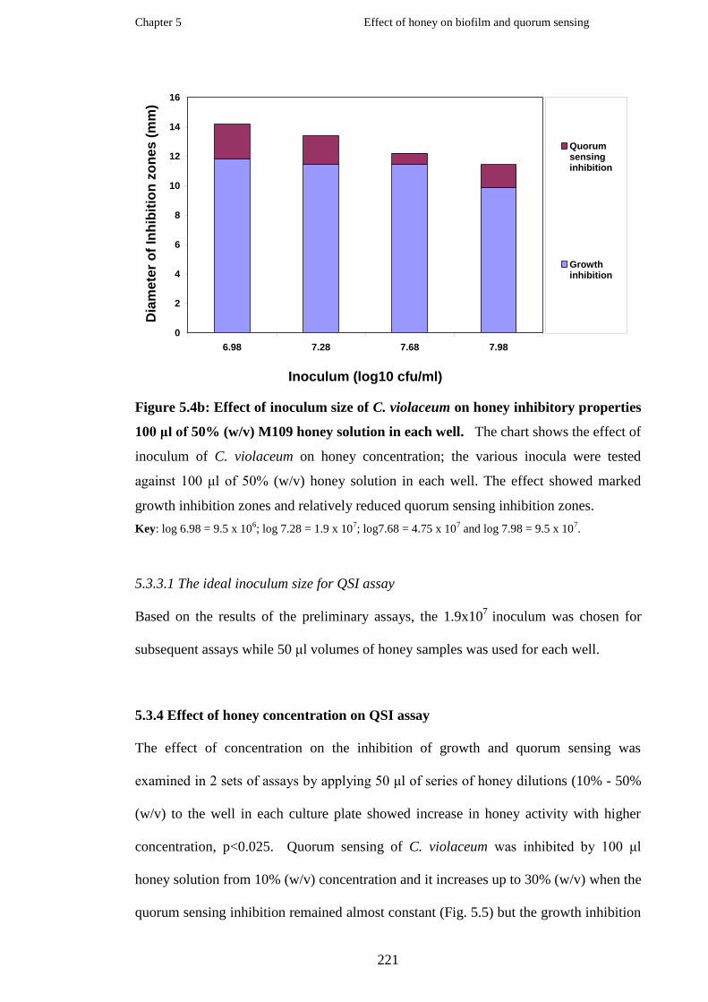
Chapter 5 Effect of honey on biofilm and quorum sensing
221
0
2
4
6
8
10
12
14
16
6.98 7.28 7.68 7.98
Inoculum (log10 cfu/ml)
Dia
mete
r o
f In
hib
itio
n z
on
es (
mm
)Quorumsensinginhibition
Growthinhibition
Figure 5.4b: Effect of inoculum size of C. violaceum on honey inhibitory properties
100 μl of 50% (w/v) M109 honey solution in each well. The chart shows the effect of
inoculum of C. violaceum on honey concentration; the various inocula were tested
against 100 μl of 50% (w/v) honey solution in each well. The effect showed marked
growth inhibition zones and relatively reduced quorum sensing inhibition zones.
Key: log 6.98 = 9.5 x 10⁶; log 7.28 = 1.9 x 10
7; log7.68 = 4.75 x 10
7 and log 7.98 = 9.5 x 10
7.
5.3.3.1 The ideal inoculum size for QSI assay
Based on the results of the preliminary assays, the 1.9x10⁷
inoculum was chosen for
subsequent assays while 50 μl volumes of honey samples was used for each well.
5.3.4 Effect of honey concentration on QSI assay
The effect of concentration on the inhibition of growth and quorum sensing was
examined in 2 sets of assays by applying 50 μl of series of honey dilutions (10% - 50%
(w/v) to the well in each culture plate showed increase in honey activity with higher
concentration, p<0.025. Quorum sensing of C. violaceum was inhibited by 100 μl
honey solution from 10% (w/v) concentration and it increases up to 30% (w/v) when the
quorum sensing inhibition remained almost constant (Fig. 5.5) but the growth inhibition

Chapter 5 Effect of honey on biofilm and quorum sensing
222
of C. violaceum culture increases with increase in honey concentration (Fig. 5.5). The
total effect of antimicrobial activities of honey was higher when 100 µl honey solution
was applied to the culture than the 50 µl of honey solution.
0
2
4
6
8
10
12
14
16
18
20
10 20 30 40 50
Honey concentration (w/v%)
Dia
mete
r o
f In
hib
itio
n z
on
es (
mm
)
Quorumsensinginhibition
Growthinhibition
Figure 5.5: Graphical representation of the effect of honey concentration on
growth and quorum sensing inhibitions of C. violaceum. Antibacterial activities of
various concentrations of M 109 manuka honey were tested against C. violaceum in QSI
assay. The growth and quorum sensing inhibitions of C. violaceum using 50 μl of honey
solution in 6mm diameter well in each culture showed increase in honey activities with
higher concentrations of honey solution.
The inhibition of C. violaceum was dependent on the concentration of honey. The
cumulative (total) effect of the antimicrobial activities of honey on C. violaceum
(combined growth and quorum sensing inhibition zones) also increased with
concentration (Fig. 5.5) indicating the significance of honey concentration on the
activity of honey, p< 0.025 using one – way ANOVA; Minitab (version 15) software.

Chapter 5 Effect of honey on biofilm and quorum sensing
223
5.3.5 Reproducibility of QSI assay
Reproducibility of QSI assay was determined by testing three honeys on three separate
occasions in duplicate. Serial dilutions of each honey showed that each of the honeys
tested inhibited the growth and quorum sensing of the reporter bacterium in a dose
dependent manner thus demonstrating the effect of concentration on the activity of the
honey samples (Fig. 5.6a, 5.6b, 5.6c and 5.6d).
Figure 5.6a: Cultures showing the effect of honey concentrations on the growth
and quorum sensing of C. violaceum.
The results were analysed with Excel statistical data analysis software and the charts of
the means and standard deviations of the analysis were plotted (Fig. 5.6b, 5.6c and
5.6d). The results obtained from the 3 honeys were reproducible and there was
significant correlation between honey concentrations and inhibition of growth and
quorum sensing of C. violaceum (p< 0.025), using one-way ANOVA, Minitab (version
15). The honey with higher UMF factor 10 + (Fig. 5.6d) had greater activity than the
honey with UMF factor 5+ (Fig. 5.6c) which is also indicative of the reliability of the
assay.
40% Honey
30% Honey
50% Honey
10% Honey 20% Honey Sterile D/ water

Chapter 5 Effect of honey on biofilm and quorum sensing
224
0
2
4
6
8
10
12
14
10 20 30 40 50
Honey Concentrations (% w/v)
Inh
ibit
ion
s z
on
es
( m
m ) Mean
quorumsensinginhibition
Mean growthinhibition
Figure 5.6b: The effect of various concentrations of a medical grade honey
(Activon® - UMF 12) on the growth and quorum sensing of C. violaceum.
0
1
2
3
4
5
6
7
10 20 30 40 50
Honey Concentrations (%w/v)
Dia
me
ter
of
Inh
ibit
ion
Zo
ne
s (
mm
)
Meanquorumsensinginhibition
Meangrowthinhibition
Figure 5.6c: The effect of various concentrations of a manuka honey UMF 10+ on
the growth and quorum sensing of C. violaceum.

Chapter 5 Effect of honey on biofilm and quorum sensing
225
0
1
2
3
4
5
6
7
10 20 30 40 50
Honey Concentrations (%w/v)
Dia
me
ter
of
Inh
ibit
ion
Zo
ne
s (
mm
)
Meanquorumsensinginhibition
Mean growthinhibition
Figure 5.6d: The effect of various concentrations of a manuka honey UMF 5+ on
the growth and quorum sensing of C. violaceum.
4.3.6 Growth and quorum sensing inhibitory properties of 10 honeys.
Out of the 10 honeys that were screened, 8 inhibited growth and quorum sensing of C.
violaceum. A table honey (Kent honey) exhibited no growth or quorum sensing
inhibitory effect on C. violaceum while another table honey (Miel de Saule) had no
growth inhibitory effect but exhibited quorum sensing inhibitory effect on C. violaceum.
A reference antibiotic (chloramphenicol) only inhibited the growth of the reporter
bacterium at concentrations of 50 µg/ml and quorum sensing inhibition at 25 and 50
µg/ml (Fig. 5.7a). The different honeys revealed varying degrees of inhibitory activities
against the reporter bacterium (Fig. 5.7b and 5.7c). Increase in both growth and quorum
sensing inhibitory activities of honeys were directly related to increase in honey
concentrations (Fig. 5.7b and 5.7c) p< 0.025. The activities of the manuka honeys with
known unique manuka factors (UMF) showed activities relative to the UMF factors
(Fig. 5.7d). It was found that the higher the UMF the stronger the inhibitory effect
p<0.005, (Pearson correlation- Minitab version 15).

Chapter 5 Effect of honey on biofilm and quorum sensing
226
3.125
6.25
12.5
25
50
GI QSI
0
1
2
3
4
5
6
7
8
9
Dia
me
ter
of
zo
ne
of
inh
ibit
ion
(mm
)
Conc.(µg/m
l)
Figure 5.7a: Growth and quorum sensing inhibition of C violaceum by
chloramphenicol.
1020304050
Acti
vo
n®
Pastu
re
M1
09
Tri
nid
ad
Ken
t
Man
uka U
MF
20+
Man
uka U
MF
10+
Man
uka U
MF
5+
Carm
ath
en
sh
ire
Mie
l d
e S
au
le
0
1
2
3
4
5
6
7
8
Dia
me
ter
of
inh
ibit
ion
zo
ne
s
(mm
)
Conc.
(w/v%)
Figure 5.7b: Assessment of potency of honeys using growth inhibition of C.
violaceum. The chart shows the growth inhibitory activities of 10 honeys at various
concentrations against C. violaceum culture in LB semi solid agar using 50 µl of each
honey solution in each well.

Chapter 5 Effect of honey on biofilm and quorum sensing
227
1020304050
Acti
vo
n®
Pastu
re
M109
Tri
nid
ad
Ken
t
Man
uka U
MF
20+
Man
uka U
MF
10+
Man
uka U
MF
5+
Carm
ath
en
sh
ire
Mie
l d
e S
au
le
0
0.5
1
1.5
2
2.5
3
3.5
4
Dia
me
ter
of
inh
ibit
ion
zo
ne
s
(mm
)
Conc.
(w/v%)
Figure 5.7c: Assessment of potency of honeys by QSI assay. The chart representation
of quorum sensing inhibitory properties of various concentrations of 10 honeys against
C violaceum in LB semisolid agar culture using 50 µl of each honey solution in 6mm
diameter well.
Figure 5.7d: Combined effect of each honey on growth and quorum sensing of C.
violaceum relative to concentrations. The honeys showed various degrees of growth
or QS inhibition of the reporter organism.
0
1
2
3
4
5
6
7
8
9
10
10
20
30
40
50
10
20
30
40
50
10
20
30
40
50
10
20
30
40
50
10
20
30
40
50
10
20
30
40
50
10
20
30
40
50
10
20
30
40
50
10
20
30
40
50
10
20
30
40
50
Honey Concentrations (w/v%)
Dia
me
ter
of
Inh
ibit
ion
Zo
ne
s (
mm
)
QuorumSensingInhibitionzones
GrowthInhibitionzones
Activon®
Pasture
M109
Trinidad
Kent
Manuka
UMF20+ Manuka
UMF10
+ Manuka
UMF 5+
Carmarthenshire
Miel
de
Saule

Chapter 5 Effect of honey on biofilm and quorum sensing
228
5.4 Discussion
The effect of honey on growth and quorum sensing inhibition of a medical grade honey
(Comvita®) was examined in a continuous culture wound model. The exposure of 30
hour biofilm of P. aeruginosa wound isolate (P.98) to 40% (w/v) solution of a medical
grade honey (Comvita medical-woundcare, active UMF 18+) in a wound model culture
resulted in the complete inhibition of the P. aeruginosa within 6 hours. The AHL
signalling molecule which was earlier detected in the culture samples between 24 and
30 hours prior to exposure of the culture to honey was not detected after exposing the
culture to honey until the termination of the culture (Fig. 5.2b). This is an indication that
the 40% (w/v) honey solution had penetrated the biofilm, and inhibited the organisms
embedded within the biofilm. The consequence of the lethal effect was the sudden drop
in the viable count of the culture from 3.5x10⁹ cfu/ml bacteria of culture
to undetectable
level of bacteria. The ability of the continuous culture wound model to grow biofilm
had been previously determined in chapter 3, section 3.2.1.2, the reproducibility assay
was found to be sensitive, and according statistical analysis using one-way ANOVA
(unstacked), p< 0.001 (95% confidence limits for mean based on pooled SD). This
finding was in line with the previous findings of inhibition of P. aeruginosa biofilm
cultures in microtitre plates (Okhiria et al, 2004 and 2009) and the susceptibility of 91%
of 11 P. aeruginosa biofilms to honeys (Alandejani et al, 2009). In the microtitre plate
assay, the dissolved biofilm components as a result of the action of honey remained in
the culture because they were not washed out. Biofilm EPS components were quantified
by the measurement of absorbed dye in microtitre plate culture. Adherent biofilm
biomass retained in the microtitre plate could still contribute to the overall value of
biofilm estimation whether it was viable or not, whereas dissolved biofilm components
were eliminated in the continuous culture as the medium was being replaced at a
constant rate. This suggests that the effect of honey on biofilm would be better
Chapter 5 Effect of honey on biofilm and quorum sensing
229
appreciated in the wound model which is a continuous culture in comparison with the
microtitre plate biofilm culture assay. Biofilm viability would be better assessed by
using fluorescent dyes such as LIVE/DEAD BacLight bacterial viability kit that can
stain the nucleic acids of both living and dead bacteria and the second dye that will not
be able to penetrate living bacteria with intact cells except the dead cells. There are
some of such dyes that are not toxic to the organisms and the cultures can still be used
for further assay when there is need to keep the culture for longer period during the
experiment. This study therefore reveals the full potential of the action of honey on
biofilm suggesting the in vivo effectiveness of honey in wound management. Manuka
honey has been shown to exert a bactericidal effect on planktonic P. aeruginosa cultures
(Cooper et al, 2002) even though biofilm can resist antimicrobial up to 500 to 1000
times that of the planktonic counterpart (Amorena et al, 1999) the antibacterial effect of
honey on biofilm at 40% (w/v) concentration shows that honey can inhibit biofilm and
confirms previous findings (Okhiria et al, 2004 and 2009). Despite an observation that
nutritional starvation and high cell density enhance the resistance of biofilm organisms
to antimicrobial tolerance (Fux et al, 2005) P. aeruginosa biofilm was susceptible to the
40% (w/v) manuka honey. This study has confirmed the high antimicrobial effect of
manuka honey on biofilm.
In this study, inhibition of growth and quorum sensing of C. violaceum by various
concentrations (10 - 50%) (w/v) of M109 (manuka honey) was observed (Fig. 5.3 and
5.6a). Inhibition of quorum sensing of biofilm organisms by honey will prevent biofilm
formation and expression of certain genes that are vital to the virulence of the
organisms. This suggests that honey can be used alone or in combination with other
antimicrobials to effectively treat biofilm infections.

Chapter 5 Effect of honey on biofilm and quorum sensing
230
After establishing the mode of action of honey on biofilm, an assay method was
modified for assessing the growth and quorum sensing inhibitory properties of honey.
The method was optimised by assessing the effect of inoculum and honey
concentrations on the activity of honey and the reproducibility of the assay. The effect
of various inocula on the activity of M109 honey was examined and 50% (w/v)
concentration found to inhibit the growth and quorum sensing of C. violaceum at the
various inocula (Fig. 5. 4 a and b).
Inhibition of growth and quorum sensing by honey was dependent on concentration.
Inhibition of quorum sensing was observed on honey concentrations as low as 10%
(w/v) honey was bactericidal to planktonic cultures of P. aeruginosa (Cooper et al,
2002) at 10% concentration (v/v) but at this concentration M109 honey showed some
antimicrobial properties against P. aeruginosa biofilm by preventing quorum sensing.
The larger proportion of biofilm constituents are made up of water, Zhang and his
colleagues in their study discovered that 97% of biofilm is water while another study
estimated the water contents of biofilm to be 70% to 80% water (Sandt et al, 2008).
The high osmolarity of honey would have a dehydrating effect on the biofilm because
of the high water content of biofilm while the acids in the honey would reduce the pH of
the biofilm culture environment which would have further increased the lethal effect of
honey on the organisms within the biofilm. The antimicrobial properties of honey are
multifactorial with all the factors working synergistically to produce a strong lethal
effect on biofilm microorganisms; the chances of biofilm organisms developing
resistance to honey might be remote.
Three samples of manuka honey (Activon®, manuka honey UMF 10+ and manuka
honey UMF 5+) were tested to ascertain the reproducibility of the QSI assay. The
assays showed evidence of reproducibility, with low standard deviations. A higher

Chapter 5 Effect of honey on biofilm and quorum sensing
231
activity for the manuka UMF 10+ honey over that of manuka honey UMF 5+ was
observed in the analysis which confirmed the effect of concentration on the potency of
the honeys. This also shows the importance of grading in honey and the relationship of
grading to the effectiveness or potency of the honeys.
Ten honeys were screened for their potential to inhibit the growth and quorum sensing
of C. violaceum as an assessment of their potency. Eight honeys showed varying
degrees of growth and quorum sensing inhibitions to C. violaceum whereas two of the
table honeys had little or no effect on the reporter organism. One of the honeys only
inhibited the quorum sensing of C. violaceum at 50% (w/v) concentration while the
second honey had no inhibitory effect on the growth and quorum sensing of the
organism. The effect of the investigated honeys on QS has confirmed the recent
findings by Truchado and her colleagues (2009).
The activities of the honeys relative to phenolic concentrations were also found to be
correlated with the quorum sensing inhibition of C. violaceum as well as the growth
inhibition. Using Minitab version 15, one way Anova unstacked, there were correlations
between the total activity of the honeys and quorum sensing inhibition p ≤ 0.0001, as
well as total activity of the honeys and growth inhibition, p ≤ 0.002. There was
significant correlation between QSI and UMF factor of the manuka honeys, p ≤ 0.001
while UMF and QSI showed significant correlation, p ≤ 0.0001. The varying potencies
of different honeys as indicated by the UMF values means that care should be taken in
selecting honey for topical and wound treatment. The higher potency of medical grade
honeys over the potency of some table honeys as observed in this study was in
agreement with the study done by Cooper and Jenkins (2009).
Five manuka honeys were included in this study and all of the honeys showed effective
growth and quorum sensing inhibitory effects relative to concentrations and UMF
Chapter 5 Effect of honey on biofilm and quorum sensing
232
factors confirming the high potency of medical grade honeys and manuka honey in
particular. Although manuka honey is a non-peroxide honey the phytochemical content
which has been identified as methylglyoxal (Adams et al, 2008; Mavric et al, 2008) has
given manuka the antimicrobial effect. The potencies of these honeys as observed in this
study was similar to the bactericidal activities of New Zealand manuka honey and
Yemen honey on biofilm demonstrated by Alandejani‟s group (2009).
The use of honey in modern medicine is gaining increased awareness, especially in
wound treatment (Cooper, 2008; Molan, 2006; Cooper & Molan, 1999). Recently,
biofilms have been implicated in chronic wounds (James et al, 2008; Ngo et al, 2007)
but any honey to be used on biofilm infected wounds ought to be able to effect
antimicrobial action on the biofilm. Exposure of microorganisms to sub-lethal
concentrations could lead to development of resistance or reduced susceptibility (Moore
et al, 2008) which could occur if organisms are constantly exposed to honeys that are
ineffective. However, Blair and her colleagues (2009) found that S. aureus and
P. aeruginosa developed resistance to conventional antibiotics such as tetracycline,
oxacillin or ciprofloxacin but not against Leptospermum (manuka) honey and peroxide
producing honeys even when each of the known components were eliminated during the
assay. It was also found that there was no cross resistance to honey as observed in the
organisms against other antimicrobials via decreased cell wall permeability or drug
accumulation or increased efflux of drugs.
It has been reported that quorum sensing molecules regulate genes involved in relieving
oxidative stress in biofilm organisms, a process that increases the tolerance of the
organisms to hydrogen peroxide (Bjarnsholt et al, 2005; Hassett et al, 1999). Inhibition
of quorum sensing by honey will therefore interfere with the tolerance of biofilm
organisms and render them susceptible to hydrogen peroxide released from honey via

Chapter 5 Effect of honey on biofilm and quorum sensing
233
glucose oxidase activity on glucose. Honey would therefore provide a unique
multifaceted treatment strategy for biofilm in wounds.
It is important to investigate and identify possible active components of other honeys
that are used for wound treatment. It has been shown that honey can enhance the
growth of biofilm (Okhiria et al, 2009) especially at low concentrations and at high
concentration the dilution effect and metabolic activities of the microorganisms could
reduce the effect of honey to sub-inhibitory level. However, in order to standardize the
use of honey as an antimicrobial agent and prevent development of mutants and
resistant strains to honey, it is pertinent that honeys being used for treatment are potent
enough to kill pathogenic organisms. Therefore determining the potency of honey
before being used as topical agent on infected wounds might be necessary.
5.5 Conclusion
In this study, the ability of manuka honey to disrupt biofilm and inhibit organisms
embedded in biofilm within 6 hours in a continuous wound model has been confirmed.
Inhibition of quorum sensing of biofilm organism by manuka and other honeys has also
been demonstrated. Varying antibacterial activities of the honeys were also observed
although a table honey did not show any effect on the reporter bacterium. Further
studies are necessary to reconcile the in vitro effect of honey on biofilm to the in vivo
activity of honey in wounds with biofilm.
5.6 Recommendation for future studies
There is need to link the quorum sensing inhibitory effect of honey in vitro to the effect
of honey in wound. Molecular techniques could be used to investigate the effect of
honey on biofilm. Determination of minimum inhibitory concentration (MIC) and

Chapter 5 Effect of honey on biofilm and quorum sensing
234
minimum bactericidal concentration (MBC) of honeys on P. aeruginosa cultures in the
microtitre and the continuous culture assays relative to time will provide more
information about the effect of honey on biofilm.

Chapter 6
Synopsis and conclusions

Chapter 6 Synopsis, conclusion and recommendations
235
6.1 Summary
In this thesis the role of biofilms in wounds has been investigated by studying the
antibiotic susceptibility pattern and the ability of Pseudomonas aeruginosa isolated
from infected and chronic wounds to form biofilm and produce quorum sensing
signalling molecules in vitro in batch and continuous cultures. The genetic fingerprint
patterns of the isolates were determined by PCR and gel electrophoresis and the
similarity between each isolate‟s fingerprints to the cohort was studied in relation to
antibiotic susceptibility, biofilm formation and acyl-homoserine lactones (AHL)
production. The ability of gauze to support the growth of biofilm in batch cultures and
detection of AHL in the gauze after sterilization was established. A wound model was
then developed for biofilm continuous culture using gauze as the substratum and
phenotypic expressions of the isolates were studied including biofilm development and
structure, AHL and extracellular polymeric substances (EPS) production. The presence
of biofilm in chronic wounds was investigated using dressings that were removed from
chronic wounds for the presence of biofilm markers including quorum sensing
molecules and EPS. Furthermore organisms from the wound dressings were isolated
and examined for biofilm phenotypic expressions such as biofilm formation in batch
culture and continuous cultures. All isolated Gram negative bacteria were examined for
production of quorum sensing signalling molecules. The results of biofilm markers
obtained from the wound dressings were linked with those of the isolated organisms
from each of the dressings to draw conclusions for the presence of biofilm in chronic
wounds. The effect of honey on biofilm was investigated in continuous culture wound
model; biofilm was disrupted within six hours of exposure to 50% (w/v) honey. Ten
honeys were investigated for quorum sensing inhibitory activity and all but one
exhibited quorum sensing inhibitory activity against biofilm forming organism,
C. violaceum in a dose dependent manner.

Chapter 6 Synopsis, conclusion and recommendations
236
6. 2 Synoptic discussion
In this study the susceptibility of the isolates to the commonly employed antibiotics was
determined using the British Society for Antimicrobial Chemotherapy (BSAC)
guidelines (version 4) (Andrews, 2005). Interestingly, almost all the organisms were
resistant to cefuroxime, amoxicillin and trimethoprim while about one fifth of the cohort
of organisms showed resistance to ciprofloxacin. On the other hand less than 10% of
the entire isolates were resistant to imipenem, gentamicin and piperacillin. This finding
is in support of other studies regarding the resistance of biofilm organisms to antibiotics
(Anderson and O'Toole, 2008; Fux et al, 2005; Donlan, 2000). This result reveals more
information about the role of biofilm forming organisms with respect to antimicrobial
resistance. The resistance of biofilm organisms has been shown to be due to various
factors including the protection by the extracellular polymeric substances (EPS) in
which they are encased acting as barrier against the penetration of chemical substances
such as antibiotics and the acquisition of resistant genes within biofilm. Some strains of
P. aeruginosa, particularly the biofilm forming phenotypes are known to produce high
level of alginate which usually enhances the resistant potential of the organism (Hentzer
et al, 2001). In their study, Hentzer and colleagues found that the non-mucoid biofilm
was almost entirely susceptible to tobramycin, while
the mucoid biofilm strains
remained viable. The antibiotic assay was done on solid agar and the organisms were
therefore in a quasi sessile form, which may stimulate the production of alginate.
Biofilm formation by P. aeruginosa isolated from infected and chronic wounds was
determined in microtitre plate (Christensen et al, 1985) and 88 (90.7%) of the 97
isolates were able to produce biofilm in cultures within 24 hours. The majority of the
isolates produced biofilm strongly; an indication of high probability to produce biofilm
in the wound by the organisms and this can happen as soon as the wound is

Chapter 6 Synopsis, conclusion and recommendations
237
contaminated by such organisms. It has been shown that a wound isolated P. aeruginosa
produced biofilm within 10 hours in vitro (Harrison – Balestra et al, 2003). In animal
models, biofilms were formed in wounds within 48 – 72 hours after challenging the
wounds with S. aureus or P. aeruginosa (Davis et al, 2008; Serralta et al, 2001).
Production of AHL was examined using cross feeding (Stickler et al, 1998) and quorum
sensing inhibition (QSI) (MacLean et al, 2004) assays. The results showed 78 (80.4%)
of the isolates produced AHL in the laboratory. There was a significant correlation
between the number of isolates that formed biofilm and produced AHL p< 0.005.
It was found that the QSI assay was more sensitive than the cross-feeding assay in
detecting AHL produced by the isolates tested here.
Several authors have linked signalling molecules such as AHL to biofilm formation
(Williams et al, 2007; Singh et al 2000; Rumbaugh et al, 2000; Chandra et al, 2001;
Costerton et al, 1999). Recently, AHL has been detected in chronic wound isolates and
debridement samples (Rickard et al, 2010). An inference is that the marked production
of virulence factors (such as cytotoxic enzymes, toxins and biofilm formation), will
further promote wound breakdown and impairment of healing processes. Pseudomonas
aeruginosa has been associated with delayed wound healing (Dowd et al, 2008;
Howell- Jones et al, 2005; Bowler et al, 2001) and particularly with large ulcers
(Halbert et al, 1992).
Interestingly, it was found that 16 (84.2%) of the 19 AHL negative isolates formed
biofilm in vitro. Studies have shown that some pathogenic P. aeruginosa formed
biofilm independent of AHL signalling molecules (Schaber et al, 2007 and 2004; Favre
– Bonte, 2007). P. aeruginosa have signalling molecules other than AHL such as the
quinolones (Deziel, 2004) which could have been used by the AHL negative but biofilm
positive isolates. Other signalling molecules were not investigated in this study; there is

Chapter 6 Synopsis, conclusion and recommendations
238
a need for further study to determine other signalling molecules, such as quinolones or
autoinducer-2 which are used by P. aeruginosa for regulation of virulence factors
including biofilm formation in the AHL negative strains that produced biofilm.
Although the majority of the P. aeruginosa wound isolates that formed biofilm
produced AHL, six (7.7%) of the 78 AHL positive wound isolates did not form biofilm
in vitro. Signalling molecules such as AHL are involved in the regulation of virulence
by pathogenic organisms (Bassler and Losick, 2006; de Kievit and Iglewski, 2000) even
across species (Atkinson and Williams, 2009; Williams et al, 2007; Williams, 2007).
The production of AHL by the biofilm negative isolates could be for the regulation of
virulence factors other than biofilm formation and more investigations need to be done
to unravel this finding. It has been observed that virulence expression could be site
dependent (Favre Bonte, 2007; Schuhmacher and Klose, 1999); it is possible for the
isolates to form biofilm in wounds even though the laboratory assay showed a negative
result. The regulatory protein ToxT directly activates the transcription of virulence
factors in Vibrio cholerae, including cholera toxin (CT) is modulated by environmental
signals with little or no expression outside the host (Schuhmacher and Klose, 1999).
The implication for the wound is that a biofilm forming organism that contaminates a
wound could form biofilm within 24 hours, making it especially difficult for the body‟s
immune system to eliminate the organisms consequently delaying wound healing.
The relationship between virulence and site of isolation of organism was studied by
using biofilm forming P. aeruginosa isolated from water pipes. Biofilm formation
(Christensen et al, 1985) and AHL production by cross-feeding (Stickler et al, 1998)
and QSI (McLean et al, 1997) of the water isolates were determined and the results
were compared with those of the wound isolates. Seventy two (74.2%) of the 97 wound
isolates tested positive by cross -feeding assay and 78 were positive by QSI assay,

Chapter 6 Synopsis, conclusion and recommendations
239
whereas of the 9 biofilm forming P. aeruginosa water isolates 5 (55.6%) were positive
for AHL production by either of the 2 assays (3 by cross-feeding and 2 by QSI assay)
but none showed positive for both methods. This shows that the strains from wound
expressed higher virulence propensity than the water isolates. Studies have shown that
virulence expressions are site dependent, particularly outside the host environment
(Schuhmacher and Klose, 1999). Series of phenotypic changes which characterise
biofilm formation (Sauer et al, 2002) are modulated by quorum sensing signalling
molecules (Parsek, et al, 2005; Williams et al, 2007; Parsek and Greenberg, 2000); the
presence of which has been demonstrated in vivo in animal (rat) wounds (Nakagami et
al, 2008).
The genetic relationships of the wound isolates were then determined by polymerase
chain reaction (PCR) and analysis of the complete linkage of the amino acid bands
(GelCompar II version 4) with Jacquard similarity index revealed that the majority of
the isolates (84.5%) had 100% genetic similarity, 3% were 75 - 99% linked while
(12.4%) were of about 50 - 75% similarity. The high genetic similarity therefore might
be linked to the biofilm phenotypic expressions such as biofilm formation, antibiotic
susceptibility and AHL production observed in this study. Further study is needed to
link the specific gene that codes for each factor with the expression of the factors in the
wound isolates. The results obtained from the biofilm phenotypic characterisation of
the isolates (antibiotic sensitivity, biofilm formation, AHL production and PCR genetic
fingerprint) was intriguing and particularly the similarity of the trends which strongly
suggest that the isolates might have genetic similarities.
Gauze has been in use as a primary or secondary wound dressing for centuries because
it is absorptive and allows moisture to escape. Gauze dressings have been utilised in
diverse ways as dry or moist; alone (Ubbink et al, 2008; Motta et al, 2004) or

Chapter 6 Synopsis, conclusion and recommendations
240
impregnated with agents such as petrolatum, antiseptics (Motta et al, 2004; Cazzaniga
et al, 2002) or other healing agents. The ability of gauze to support the growth of
biofilm and retain AHL secreted by biofilm organisms was determined in batch
cultures. The results of the cultures showed that gauze satisfactorily supported the
growth of biofilm and was able to retain the AHL which was detected after sterilising
the gauze at 121oC for 15 minutes with reporter organism C. violaceum.
Growing microorganisms in batch cultures normally provides abundant nutrients at the
onset of the culture but nutrients reduce with time and microbial wastes accumulate in
the culture medium. Such cultures would not be appropriate for studies that require long
period of culture of organisms since the accumulation of wastes and nutrient depletion
usually leads to the death of the organisms and affects the phenotypic expression of the
organisms. The continuous system offers a long period of study for the organisms
without change in phenotypic expressions because there is constant replenishment of the
media as well as the continuous elimination of the metabolic wastes from the culture.
In order to elucidate the phenotypic characteristics in biofilm, a wound model was
developed for biofilm in continuous culture using gauze as the substratum for biofilm.
In this study the phenotypic expressions of selected biofilm and non- biofilm forming
wound isolates were investigated in a wound model to simulate what happens in vivo.
The isolates were examined for biofilm formation and AHL production over a period of
48 hours. The microscopic examination of the cultures of biofilm forming isolates
revealed progressive formation of biofilm microcolonies and increase in EPS
throughout the duration of the experiment as well as production of AHL which was
detectable when the bacterial population attained 107 cfu/ml of culture and also
coincided with the appearance of biofilm structures while a non-biofilm forming
isolates did not produce biofilm or AHL. The detection of AHL at the bacterial

Chapter 6 Synopsis, conclusion and recommendations
241
population of 107 cfu/ml of culture using the low flow rate was similar to the finding of
Kirisits and colleagues (2007). In their study QS induction for the laminar flow rate
(2.4 and 24 ml per minute) was at bacterial population of about 6x10⁷ cfu/ml. The
growth patterns of a biofilm and a non - biofilm forming P. aeruginosa wound isolates
were similar but the biofilm forming isolate formed biofilm and produced AHL which
was detected in culture effluent samples (Fig. 3.6, 3.7 B and C, 3.8, 3.9) and the
microscopic examination of stained slide smears of culture effluents (Fig. 3.17) and
electron microscopic scanning of gauze after 30 hours of culture (Fig.3.18) whereas the
non- biofilm forming isolate was not able to produce biofilm (Chapter 3; Fig. 3.19) or
AHL (Fig. 3.6, 3.7 E and F,3.10 and 3.11) in wound model cultures. In the wound
model, the development of biofilm microcolonies were evident and increase in EPS
formation and biofilm features at all stages up to maturation were observed at
determined intervals during the period of culture. The demonstration of biofilm features
was in agreement with other studies that biofilms have been cultured and demonstrated
in vitro (Harrison – Balestra et al, 2003).
The findings from these investigations of the P. aeruginosa isolates from wounds have
revealed the biofilm forming potential of most of these isolates in the laboratory which
suggested an ability to form biofilm in vivo. The link between the ability of the
organisms to form biofilm in the laboratory with formation of biofilm in the wound was
therefore explored. This hypothesis was investigated in wounds by examining wound
dressings for the presence of biofilm although previous studies had used biopsies and
debridement samples.
Clinical assessment of wounds for infection is not straightforward since it involves the
consideration of various parameters such as pain, bleeding, production of purulent
exudates or discharge (Grey et al, 2006; Petherick et al, 2006) as well as localised

Chapter 6 Synopsis, conclusion and recommendations
242
swelling, redness or heat (Petherick et al, 2006). Once infection is established, it is best
practice for wound infection to be confirmed by isolation of organisms from wound
swab or wound biopsy (Petherick et al, 2006) for appropriate antibiotic treatment
because of the problem of resistance (Ciofu et al, 2009; Neu, 1999). In practice infected
wounds are treated on the basis of clinical signs and symptoms before identifying
causative agents and this might encourage antimicrobial resistance and development of
mutants. It has been shown that the use of antimicrobials induces the alteration of
community structure by reducing some bacteria while selecting for others (Price et al,
2009).
Isolation of organisms by cultural methods provides opportunity to determine the
susceptibility of the organisms to antimicrobials for the selection of the most
appropriate antimicrobial for treatment. But the inadequacy of cultural methods have
been revealed recently, particularly the inability to isolate some fastidious organisms
(Price et al, 2009; Singh et al, 2009; James et al, 2008; Dowd et al, 2008 a and b;
Kirketerp-Møller et al, 2008). The studies have compared cultural and non- cultural
methods and confirmed that more organisms were identified when non - cultural
methods were used (Price et al, 2009; Sing et al, 2009; Dowd et al, 2008 a and b). Price
and colleagues observed in their study that microbial burden of the investigated wounds
were 4 times more than estimated culture-based method.
According to the study by James and colleagues (2008) using 77 chronic wounds and 16
acute wounds specimens, 50 of the chronic wounds were analysed with light and
scanning electron microscopy techniques and 60 % had biofilm whereas just 1 (6%) of
the 16 acute wounds was positive for biofilm while molecular analyses of the remaining
27 chronic wounds specimens showed diverse polymicrobial communities which was
not observed by cultures. Kirketerp-Møller and colleagues confirmed that the organisms

Chapter 6 Synopsis, conclusion and recommendations
243
within wounds are aggregated in microcolonies. The presence of biofilm has therefore
been confirmed in human wounds (Price et al, 2009; Singh et al, 2009; James et al,
2008; Dowd et al, 2008 a and b; Kirketerp-Møller et al,2008; Ngo et al, 2007) but
cultural methods would not usually reveal the presence of biofilm. The inadequacies of
cultural methods have led to the use of culture independent approach to studying
microorganisms and have been suggested (Rogers et al, 2009; Davies et al, 2001).
The present procedure for the assessment of wounds for biofilm involve collection of
wound biopsies (which is an invasive procedure) and the available procedures for
processing the samples such as scanning electron microscopy (SEM), confocal laser
scanning microscopy (CLSM) or fluorescent in –situ hybridisation (FISH) are quite
expensive and usually not available for routine use. Wounds normally produce
exudates which would increase when the wounds are infected because the
microorganisms metabolic activities and toxins continually breakdown the tissues
within the wound space (Kingsley, 2001). The processes stimulate the body immune
system to increase the production of inflammatory cytokines and proteases such as
TNF-alpha, the interleukins and heparin-binding protein (HBP) (Lundqvist et al, 2004)
and infiltration of neutrophils (Kim et al, 2008) to eliminate the organisms from the
wound. All these activities increase the blood flow to the wound area consequently
increasing the amount of exudates (Ovington, 2003; Kingsley, 2001).
Since biofilms are often polymicrobial (Malic et al, 2009; Dowd et al, 2008 a and b;
James et al, 2008), the presence of biofilm in wound would result in the combined and
synergistic effect of all the organisms‟ virulent expressions, such as the production of
proteolytic enzymes and toxins within the biofilm which further aggravates the
breakdown of tissues, the consequence of which usually contribute to the inability of
such wounds to heal (Bjarnsholt et al, 2008). Problems associated with biofilm such as
Chapter 6 Synopsis, conclusion and recommendations
244
the prevention of phagocytic activities by PMNs, the effects of signalling molecules on
the expression of virulence factors and inhibition of antimicrobials by biofilm
organisms give rise to overwhelming numbers of organisms; the phenomena that further
complicate the healing process of wounds with biofilm (Bjarnsholt et al, 2008). Wound
exudates will ooze out into the dressings and usually contain the products released due
to proteolytic activities of the organisms in addition to the body fluid.
In this study wound dressings were obtained to provide some insight into the presence
of biofilm in wounds by examining the exudates for biofilm markers such as EPS and
AHL and isolating organisms from the dressings. The isolated organisms were then
examined for biofilm phenotypic expressions in vitro.
In wounds, exudate production varies according to the state of the wound (White and
Cutting, 2006), the acute wounds exudates are usually low but higher in infected
wounds due to microbial virulence expressions (Wilson et al, 2002) and internal
wounds. The diverse range of exudate production in wounds was taken into
consideration in the construction of the wound model for continuous cultures regarding
the quantities of media that were delivered into the culture during the period of each
culture.
The presence of biofilm in human wounds has been established with the examination of
wound biopsies using histological methods, fluorescence in situ hybridization (FISH),
confocal laser scanning microscopy (CSLM) and scanning electron microscopy (SEM)
(Malic et al, 2009; Kirketerp-Møller et al, 2008; James et al, 2008; Ngo et al, 2007).
In the past detection of biofilm markers such as AHL signalling molecules have been
utilised to confirm the presence of biofilm in urinary catheters (Stickler et al, 1998) and
cystic fibrosis lung by examining the sputum (Singh et al, 2000). Recently, debridement

Chapter 6 Synopsis, conclusion and recommendations
245
materials have been examined for biofilm markers such as signalling molecules (AHL
and autoinducer-2) (Rickard et al, 2010).
Here the examination of wound dressings that were removed from patients with chronic
wounds was a novel idea in the determination of the presence of biofilm in wounds. The
method was expected to provide a quick and less expensive screening for the detection
of biofilm in wounds.
The dressings from 35 patients with chronic wounds were screened for biofilm markers
such as AHL signalling molecule, EPS and the ability of the isolated organisms from
the dressings to express biofilm characteristics. Since microorganisms within the
wound space would form part of the constituents of wound exudates (White and
Cutting, 2006) and the organisms to a large extent would present the microbial flora of
such wound, detection of biofilm markers would give an indication of the possible
presence of biofilm in the wounds.
The investigation of chronic wound dressings revealed that the majority (80%) of the
35 dressings contained EPS although 11.4% could not be tested. Interestingly,
organisms that produced biofilm in vitro were isolated from 32 out of the 35 chronic
wound dressings (91.4%) inclusive of the 80% in which EPS was detected. Since EPS
produced by biofilm organisms during biofilm formation serves as the means by which
biofilm adheres to abiotic and biotic surfaces (Flemming et al, 2007; Costerton et al,
1995) the detection of EPS in the wounds dressings suggests that it was produced by
organisms present in the wounds. Some constituents of certain wound dressings include
alginate but to prevent false positive results, examination of the stained smear of the
exudates were carefully monitored and only evidence of microcolonies in which
organisms were embedded were taken as a positive result (Fig. 4.2a).

Chapter 6 Synopsis, conclusion and recommendations
246
AHL was detected in 11 dressings, 10 (77%) of which had AHL producing bacteria (P.
aeruginosa); although in total AHL producing bacteria (Chapter 4; Fig. 4.1 a) were
isolated from 13 dressings but the rate of detection of AHL in the dressings was high
since 10 were detected. Statistical analysis using one way ANOVA (Minitab version
15), the relationship between AHL detection in the wound dressings and production of
AHL by the isolates in vitro was significant p< 0. 005. All of the isolates that produced
AHL also formed biofilm in the microtitre plate assay. These results suggest that the
AHLs that were detected in the dressings were produced by the isolated organisms, as
these organisms were able to produce the AHLs in vitro. One dressing tested positive
for AHL but no Gram negative organism was isolated from it, which might be due to
the inhibitory effect of the antimicrobial agent in the dressing. This situation supports
the fact that cultural methods often fail to isolate some organisms in wounds which non
- cultural methods are able to detect (Price et al, 2009; Sing et al, 2009; James et al,
2008; Dowd et al, 2008; Kirketerp-Møller et al,2008 ).
It was observed in this study that the detection of AHL in wound dressings was
associated with the presence of P. aeruginosa, p < 0.005. A similar study in which
AHL was detected in wound debridement materials found an association between AHL
detection and the presence of certain Gram negative bacteria including P. aeruginosa
(Rickard et al, 2010).
Gram negative bacteria were isolated from 23 dressings out of which 13 produced
AHL, therefore AHL detection method is only useful for AHL producing biofilm
organisms and may not be applicable for general biofilm screening involving Gram
positive and non-AHL producing Gram negative organisms. This study has supported
previous findings that P. aeruginosa usually produces high level of AHL during biofilm
formation (Christensen et al, 2007; Kirisits and Parsek, 2006).

Chapter 6 Synopsis, conclusion and recommendations
247
P. aeruginosa were isolated from 14 dressings but 1 did not produce biofilm in the
laboratory and AHL was not detected in the dressing from which the organism was
isolated, even though it has been shown that some bacteria such as P. aeruginosa can
cause infection and form biofilm without producing AHL (Favre-Bonte et al, 2007;
Schaber et al, 2007 and 2004).
Most of the chronic wound dressings that were investigated were polymicrobial with a
mean of 3 organisms per dressing, a situation that has been found in wound infections
and in particular chronic wounds (Price et al, 2009; Dowd et al, 2008; Dowd et al,
2008). The most prevalent organisms were Staphylococcus aureus (20.4%) (MSSA:
13%) and MRSA: 7.41%), P. aeruginosa (13%) and Corynebacterium striatum (9.3%).
Some studies have shown that S. aureus and P. aeruginosa were prevalent in chronic
wounds (Price et al, 2009; Dowd et al, 2008) especially antibiotic resistant strains
(Lipsky, 2008) whereas Corynebacterium striatum is mainly prevalent in chronic
diabetic ulcers (Dowd et al, 2008; Citron et al, 2007). The results obtained from this
chronic leg ulcers study seem to follow a similar pattern to previous studies.
The wounds in which P. aeruginosa were isolated were also polymicrobial which
confirms the association of P. aeruginosa with polymicrobial wound infections (Price et
al, 2009; Sing et al, 2009; James et al, 2008; Dowd et al, 2008 a and b; Kirketerp-Møller
et al, 2008).
Infected wounds often lead to complications and delay in healing but treatments of
wounds have evolved over a long period of time especially the management of chronic
wounds (Forrest, 1982; Ayton, 1985).

Chapter 6 Synopsis, conclusion and recommendations
248
A series of problems are associated with wounds including pain, malodour and mobility
constraints to patients (White and Cutting, 2006; Percival and Bowler, 2004) with the
attendant socio-economic problems (Church et al, 2006; Wysocki, 2002; Philips et al,
1994). The development of resistance strains of pathogenic organisms to conventional
antimicrobials has aggravated the problems associated with chronic wounds particularly
health related and life threatening complications such as septicaemia, damage to organs
(Church et al, 2006), amputations (Ramsey et al, 1999) and in extreme circumstances
death (Ramsey et al, 1999).
Various studies have shown that resistance to antimicrobials by microorganisms is
higher in biofilm organisms than the planktonic counterparts (Lewis, 2001; Nickel et al,
1985) and biofilm organisms have been associated with chronic wounds (Bjarnsholt et
al, 2008; Costerton et al, 2003 and 1999). Biofilm infected wounds are often
polymicrobial and the damaging impact on the wounds is much higher due to the
combined pathogenic effect of the organisms within the biofilm (Dowd et al, 2008;
Rhoads et al, 2008; Bjarnsholt et al, 2008). In recent times, because of the problem of
antimicrobial resistance (Khorasani et al, 2008; Zavascki et al, 2006; McDonald et al,
2004), the use of honey in the treatment of wounds has been revisited (Cooper et al,
2008; Molan, 2006, Cooper et al 2001; Cooper and Molan, 1999).
The effectiveness of honey against virulent organisms such as S. aureus and
opportunistic pathogen like P. aeruginosa has been linked to the composition and the
physio-chemical properties of honey. These properties include the acidic pH, high
osmolarity due to sugar content, release of hydrogen peroxide which increases with the
dilution of honey and the phyto-chemical properties. The phyto-chemical property of
honey is determined by the plants from which the honey is formed and it influences the
potency of honey (Mboto et al, 2009; Molan, 2006) hence the variation in the

Chapter 6 Synopsis, conclusion and recommendations
249
antimicrobial properties of different honeys (Cooper and Jenkins, 2009). Several
studies have demonstrated the antimicrobial effect of honey against many
microorganisms (Williams et al, 2009; Amghalia et al, 2009; George et al, 2007;
French et al, 2005; Cooper et al, 2002). The application of honey in wound treatment
has been extensively evaluated (Majtán 2009; Cooper, 2008; Molan, 2006; Cooper &
Molan, 1999); in particular the effect of honey on microorganisms with respect to the
low pH (Simon et al, 2009; Lotfi, 2008), acid content (Wahdan, 1998), osmotic effect
(Cooper et al, 2002; Molan, 2006), peroxidase activity (Molan, 2006 and 1992; White
Jr. et al, 1963) antioxidant effect on free radical (Henriques et al, 2006; Frankel, 1998)
and phytochemical effect (Mboto et al, 2009; Molan, 2006). The antimicrobial effects
of honey have been demonstrated in the past on planktonic microorganisms (French et
al, 2005; Mulu, 2004; Cooper et al, 2002; Efem and Iwara, 1992) and biofilm
organisms (Alandejani et al, 2009; Okhiria et al, 2009; Merckoll et al, 2009). However,
there is need for more information about the antimicrobial effect of honey on
pathogenic organisms.
In this study the effect of honey on 30 hours biofilm of a strong biofilm forming P.
aeruginosa wound isolate in a continuous wound model culture was examined. The
biofilm was penetrated and the EPS were dissolved (dispersed) within 6 hours of
exposure of the biofilm to the 40% (w/v) manuka honey solution. The organisms could
not be recovered from wound model effluent when cultured on nutrient agar after
exposure to honey. This result indicates that 40% (w/v) manuka honey had a
bactericidal effect on P. aeruginosa biofilm. This finding has supported previous
findings on the effect of honey on biofilm (Alandejani et al, 2009; Okhiria et al, 2009;
Merckoll et al, 2009).

Chapter 6 Synopsis, conclusion and recommendations
250
Signalling molecules are vital to the formation of biofilm and virulence of pathogenic
organisms particularly regulation of various genetic activities involved with biofilm
formation as well as the population density of biofilm community. The need to
understand the mode of action of honey on biofilm phenotypic expression is important
to the application of honey in wound treatment especially the effect of honey on
signalling molecules which are the global gene regulator of biofilm organisms.
In this study, the effect of honey on the action of Gram negative signalling molecules,
acyl- homoserine lactones (AHL) was investigated. The study revealed the inhibitory
effect of honey against quorum sensing of a biofilm organism, Chromobacterium
violaceum, in a dose dependent manner (Fig. 5.5; and 5.6 a – d).
A screening method was developed for the evaluation of the potency of honey as a
topical and wound dressing antimicrobial (MacLean et al, 1997) and 10 honeys were
examined. The growth and quorum sensing inhibitory effects of the various
concentrations of each honey against C. violaceum were determined and the potencies
of various honeys were then evaluated based on the obtained results. The investigation
revealed the various antimicrobial effects of the different honeys on biofilm and the
effects were dose dependent. The majority (8) of the 10 honeys examined in this study
showed both growth and quorum sensing inhibitory effects on biofilm forming
organisms whereas one honey did not exhibit such properties (Fig. 5.7 b - d). The
inhibition of quorum sensing signalling of the organisms within biofilm would probably
have prevented the formation and secretion of more EPS and penetration of honey
through the EPS would have led to dissolution of the biofilm which consequently gave
room for the honey to act directly on individual cell. The cumulative effect has resulted
in the death of the organisms as evident from the result of this study. The potency of
manuka honey in eradicating pathogenic organisms within biofilm is further restated.

Chapter 6 Synopsis, conclusion and recommendations
251
Previous studies have shown the reliability of the potency of medical grade honey on
pathogenic organisms (Cooper and Jenkins, 2009) and P. aeruginosa biofilm in batch
culture (Okhiria et al, 2009). Inhibition of quorum sensing of C. violaceum by some
honeys has recently been demonstrated (Truchado et al, 2009).
This study has shown that one of the antibacterial effects of honey on biofilm- forming
organisms is inhibition of quorum sensing. Quorum sensing inhibition will stall most of
the genetic expressions that are vital to proteolytic activities of the organisms, which
has been suggested as treatment strategy for pathogenic infections. The effect of honey
on the regulator of metabolic activities that are vital to the existence and sustenance of
biofilm consequently lead to the sudden death of the organisms. The signalling
molecule being the modulator of the metabolic and other relevant genetic activities
within the biofilm (Passador et al, 1993; Xu et al, 2000 and 2006; Patel, 2005)
including virulence factors (Xu et al, 2000 and 2006; Patel, 2005; de Kievit and
Iglewski 2000) and production of EPS matrix that houses and protect the biofilm
organisms (Flemming et al, 2007; Costerton et al, 1995) from environmental stress.
Quorum sensing inhibition of pathogens such as P. aeruginosa has been shown to
reduce the tolerance to antimicrobials (Hoffmann et al, 2007) which would further
increase the chances of the elimination of such organisms from the wound. It has also
been shown that inhibition of P. aeruginosa signalling molecules reduced or completely
suppressed the production of some virulence factors (Hentzer et al, 2002).
Communication amongst bacterial species with AHL signalling molecules have been
demonstrated (Rasmussen et al, 2000; Riedel et al, 2001) indicating that a particular
species of bacteria can utilise a signalling molecule from another species for modulation
of virulence factors. Inhibition of quorum sensing signalling molecules might be the
solution to the problem of biofilm infections and in particular development of mutants

Chapter 6 Synopsis, conclusion and recommendations
252
by biofilm organism. Application of honey as an inhibitor of quorum sensing is a
potential benefit that needs to be utilised.
The importance of inhibition of quorum sensing by honey can not be overemphasized as
one of the key factors that determine the effectiveness of honey as a treatment
intervention in the management of wounds with biofilm. The ability of honey to
dissolve the EPS of biofilm rapidly shows the organisms within the biofilm would be
exposed to other antibacterial effects of honey thereby allowing the killing of the
organisms and enhancing wound healing. The inhibitory effect of honey on quorum
sensing signalling molecules would further reduce the organisms‟ virulence expressions
or totally render the organisms ineffective thus providing an enabling environment for
wound healing.
6.3 Conclusions
In this thesis, the ability of wound isolates to form biofilm and produce signalling
molecules such as AHL in vitro both in batch and continuous cultures has been
demonstrated. Production of EPS concordant with biofilm development was observed in
cultures during these investigations. Examination of wound dressings revealed evidence
of biofilm markers such as AHL and EPS an evidence of biofilm formation in vivo and
the organisms isolated from the dressings demonstrated biofilm phenotypic
characteristics in vitro.
This thesis has presented evidence of the potential of P. aeruginosa isolated
from wounds to form biofilm in the laboratory. The majority of the isolates
formed strong biofilm within the 24 - hour period of investigation.

Chapter 6 Synopsis, conclusion and recommendations
253
This study showed evidence of AHL production by P. aeruginosa wound
isolates and most of the AHL positive isolates also produced biofilm.
The evidence of antibiotic resistance by P. aeruginosa wound isolate was
demonstrated in this study.
The genetic diversity of P. aeruginosa wound isolates was investigated and
showed that most of the isolates have 100% genetic similarity linkage during the
fingerprint analysis using GelCompar II software.
In this study a wound model was designed for in vitro study of biofilm in
continuous cultures. Biofilm phenotypic expression of P. aeruginosa wound
isolates including biofilm formation; AHL and EPS production were observed in
continuous cultures in the wound model even within 12 hours of culture. This
assay could be used to predict biofilm formation capability in contaminated
wound.
This study established the use of wound dressing to determine the presence of
biofilm in wounds by screening the dressings for biofilm markers such as
signalling molecules and extracellular polymeric substances (EPS).
The ability of the various species of organisms that had been isolated from
wound dressings removed from patients with chronic wounds to form biofilm in
the laboratory was demonstrated. Isolated Gram negative bacteria produced
AHL while selected Gram negative isolates (Pseudomonas aeruginosa and

Chapter 6 Synopsis, conclusion and recommendations
254
Burkholderia cepacia) displayed biofilm phenotypic expressions in continuous
cultures in the wound model.
It was revealed that the presence of P. aeruginosa was associated with
polymicrobial wounds.
A screening method for honey was developed in this work as a tool for
establishing the potency of honey as a topical antimicrobial agent and in
particular for biofilm eradication in wounds.
In this study some honeys were screened for growth and quorum sensing
inhibitory effect on biofilm forming organism, Chromobacterium violaceum
This work has established the inhibitory effect of manuka and other honeys on
the quorum sensing of a biofilm forming organism. The phenomenon that might
prevent the formation of biofilm in a wound colonised by pathogenic biofilm
forming organisms.
The effect of manuka honey in disrupting biofilm in continuous culture in a
wound model within 6 hours of exposure is also presented in this study. This
demonstrates that manuka honey is highly effective against biofilm in vitro.
In conclusion, this study has added to the knowledge of evidence of biofilm in chronic
wounds and provided another way to determine the presence of biofilm in wounds and
the microbial population of the wound through examination of wound dressings. The
formation of biofilm was evident within 12 hours of culture in the continuous cultures

Chapter 6 Synopsis, conclusion and recommendations
255
and the implication is that biofilm can be formed in a wound within 24 hours of
colonisation by pathogenic organisms. The presence of biofilm in wounds may delay
healing; prevention of biofilm formation in wound at the earliest possible time will play
a vital role in wound management and enhancement of wound healing.
The resistance of planktonic forms of P. aeruginosa biofilm forming isolates to
conventional antimicrobials was apparent but curiously the biofilm counterpart of the
organism was susceptible to 40% (w/v) manuka honey. The inhibitory effect of honey
on quorum sensing of biofilm organism might be the key factor in the ability of honey
to kill biofilm organisms. The ability of honey to prevent quorum sensing and
consequently biofilm formation or preventing the maintenance of biofilm activities by
quorum sensing molecules when biofilm already exists would play a major role in the
management of infected wounds. Application of honey as topical antimicrobial agent in
wound treatment might resolve the problem of resistance and prevent the development
of mutants in wound infections.
6.4 Limitations of study
The constraints in terms of equipment have not allowed the wound model
biofilm culture to be examined under confocal scanning laser microscope.
The screening of the wound isolate with biofilm forming gene array to
determine the ability of the isolates to form biofilm via gene analysis.
The use of fluorescence in situ hybridization (FISH) to examine the biofilm in
wound model cultures and wound dressings would have made the organisms

Chapter 6 Synopsis, conclusion and recommendations
256
within the biofilms more visible and distinctively reveal the organisms from the
matrix.
6.5 Recommendations
Further studies are necessary to connect the presence of biofilm markers in
wound dressings to the presence of biofilm in wounds; it will be necessary to
examine wound biopsies alongside wound dressings in a single study.
This study investigated Gram negative quorum sensing molecules (acyl-
homoserine lactones), future work should investigate other signalling molecules
employed by Gram negative and Gram positive organisms. This will give more
chances for detection of signalling molecules in the wound dressings and higher
correlation between signalling molecules and biofilm for more accurate
diagnosis.
The possibility of detecting virulence factors in biofilm forming organisms such
as P. aeruginosa should be explored and the link between these virulence factors
and biofilm formation could be investigated.
It is also pertinent to study the bactericidal activity of honey against the biofilms
of commonly isolated wound pathogens in the continuous culture in particular
the determination of MIC and MBC. The wound model used in this study will
provide a useful tool for such study.
Since wounds are mostly polymicrobial, the possibility of the effect of honey on
polymicrobial biofilm culture should also be explored in order to simulate the
Chapter 6 Synopsis, conclusion and recommendations
257
condition that exists in most wounds. The wound model can easily be adapted to
such analysis.
There is need to connect the effect of honey in disrupting formed biofilm in vitro
with in vivo activity in the wound.
The effect of honey on quorum sensing of biofilm organisms in vitro should be
linked with in vivo effect of honey in wounds. This will further boost the
credence of the acclaimed role of honey in wound management.
Appendices

Apppendices
258
(Appendix 1)
Dear Olusola Okhiria,
We hereby grant you permission to reprint the material detailed below at no charge in
your thesis subject to the following conditions: 1. If any part of the material to be used (for example, figures) has appeared in our
publication with credit or acknowledgement to another source, permission must
also be sought from that source. If such permission is not obtained then that
material may not be included in your publication/copies.
2. Suitable acknowledgment to the source must be made, either as a footnote or in
a reference list at the end of your publication, as follows:
“This article was published in Publication title, Vol number, Author(s), Title of
article, Page Nos, Copyright Elsevier (or appropriate Society name) (Year).”
3. Your thesis may be submitted to your institution in either print or electronic
form.
4. Reproduction of this material is confined to the purpose for which permission is
hereby given.
5. This permission is granted for non-exclusive world English rights only. For
other languages please reapply separately for each one required. Permission
excludes use in an electronic form other than submission. Should you have a
specific electronic project in mind please reapply for permission. 6. Should your thesis be published commercially, please reapply for permission.
Kind Regards, Marek Gorczyca Rights Assistant Elsevier LTD Phone Number +441865843841 Fax Number +441865853333 [email protected]

Apppendices
259
(Appendix 2)
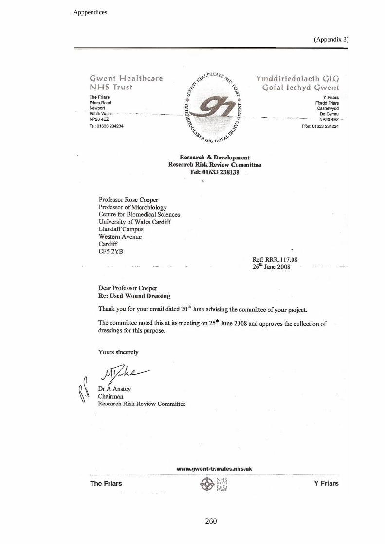
Apppendices
260
(Appendix 3)

Apppendices
261
(Appendix 4)
Frequency of isolates recovered from chronic wound dressings
Species
No
Species
No
Acinetobacter lwoffii 1
Lactobacillus acidophyllus 1
Bacteroides capillosus 1 Micrococcus kristinae 1
Burkholderia cepacia 3 Peptostreptococcus tetradius
1
Candida albicans 2 Prevotella loescheii 1
Candida famata 1 Proteus mirabilis 6
Citrobacter freundii 3 Proteus vulgaris 2
Corynebacterium bovis 1 Pseudomonas aeruginosa 14
Corynebacterium jeikeium 1 Saccharomyces cerevisiae 1
Corynebacterium propinguum 1 Serratia marcescens 2
Corynebacterium pseudodiphtheriticum 1 Shewanella putrefaciens 1
Corynebacterium renale grp 1 Staphylococcus aureus (8 MRSA) 22
Corynebacterium pseudogenitalium 1 Staphylococcus auricularis 1
Corynebacterium striatum 9 Staphylococcus epidermidis 4
Corynebacterium ulcerans 1 Staphylococcus haemolyticus 1
Enterobacter cloacae 1 Staphylococcus saccharolyticus 1
Enterococcus faecalis 4 Staphylococcus simultans 2
Escherichia coli 3 Streptococcus agalactiae 3
Flavimonas ovyzihabitans 1 Streptococcus constellatus 2
Gemella haemolysans 1 Streptococcus grp G 1
Klebsiella oxytoca 1 Streptococcus sanguis 1
Klebsiella pneumoniae 2

Apppendices
262
(Appendix 5)
Organisms isolated from dressings removed from chronic wounds
S/No Dressing ID Organisms
1 1 M1(1) Staphylococcus aureus (MRSA)
2 2 M2(2) Pseudomonas aeruginosa
3 M2(3) Staphylococcus aureus (MRSA)
4 M2(4) Enterococcus faecalis
5 3 M3(1) Burkholderia cepacia
6 M3(2) Corynebacterium striatum
7 M3(3) Staphylococcus aureus
8 M3(9) Escherichia coli
9 4 M4(1) Pseudomonas aeruginosa
10 M4(2) Staphylococcus aureus
11 M4(4) Candida albicans
12 5 M5(1) Pseudomonas aeruginosa
13 M5(2) Corynebacterium striatum
14 6 M6(1) Proteus mirabilis
15 M6(4) Corynebacterium striatum
16 M6(5) Gemella haemolysans
17 M6(9) Candida albicans
18 7 M7(1) Klebsiella pneumoniae
19 M7(2) Corynebacterium bovis
20 M7(8) Staphylococcus epidermidis
21 8 M8(1) Corynebacterium renale grp
22 9 M9(1) Corynebacterium propinguum
23 10 M10(1) Micrococcus kristinae
24 11 M11(1) Staphylococcus aureus (MRSA)
25 M11(3) Corynebacterium striatum
26 12 M12(2) Staphylococcus aureus (MRSA)
27 M12(3) Streptococcus agalactiae
28 M12(4) Enterobacter cloacae
29 M12(7) Bacteroides capillosus
30 M12(8) Staphylococcus saccharolyticus
31 13 M13(1) Staphylococcus aureus (MRSA)
32 M13(2) Streptococcus agalactiae
33 M13(4) Staphylococcus haemolyticus
34 M13(5) Prevotella loescheii
35 14 M14(1) Klebsiella pneumoniae
36 M14(2) Staphylococcus aureus
37 M14(3) Staphylococcus epidermidis
38 15 M15(1) Staphylococcus aureus
39 M15(2) Streptococcus grp G
40 16 M16(1) Klebsiella oxytoca
41 M16(2) Citrobacter freundii
42 M16(3) Corynebacterium striatum
43 M16(4) Proteus mirabilis
44 M16(5) Pseudomonas aeruginosa
45 M16(6) Staphylococcus aureus (MRSA)
46 17 M17(1) Flavimonas oryzihabitans
47 M17(2) Pseudomonas aeruginosa

Apppendices
263
S/No Dressing ID Organisms
48 M17(3) Corynebacterium ulcerans
49 M17(4) Citrobacter freundii
50 18 M18(1) Pseudomonas aeruginosa
51 M18(3) Enterococcus faecalis
52 M18(4) Staphylococcus simulans
53 M18(8) Lactobacillus acidophyllus
54 M18(9) Peptostreptococcus tetradius
55 M18(10) Staphlococcus aureus
56 19 M19(1) Pseudomonas aeruginosa
57 M19(2) Staphylococcus aureus (MRSA)
58 M19(3) Corynebacterium striatum
59 20 M20(1) Escherichia coli
60 21 M21(1) Saccharomyces cerevisiae
61 M21(2) Proteus mirabilis
62 22 M22(2) Pseudomonas aeruginosa
63 M22(3) Streptococcus agalactiae
64 M22(4) Staphylococcus aureus
65 M22(6) Proteus mirabilis
66 23 M23(1) Staphylococcus aureus
67 M23(4) Staphylococcus simultans
68 24 M24(1) Staphylococcus aureus
69 M24(2) Staphylococcus epidermidis
70 M24(3) Corynebacterium jeikeium
71 25 M25(1) Staphylococcus aureus
72 M25(2) Streptococcus saiguis
73 M25(3) Corynebacterium striatum
74 26 M26(1) Serratia marcescens
75 M26(2) Streptococcus constellatus
76 M26(3) Burkholderia cepacia
77 M26(4) Pseudomonas aeruginosa
78 M26(5) Proteus vulgaris
79 27 M27(1) Staphylococcus aureus
80 28 M28(1) Burkholderia cepacia
81 M28(2) Pseudomonas aeruginosa
82 29 M29(1) Streptococcus constellatus
83 M29(2) Staphylococcus aureus
84 M29(3) Proteus mirabilis
85 M29(4) Pseudomonas aeruginosa
86 M29(5) Corynebacterium pseudodiphtheriticum
87 30 M30(1) Citrobacter freundii
88 M30(2) Pseudomonas aeruginosa
89 M30(4) Staphylococcus aureus (MRSA)
90 M30(5) Proteus mirabilis
91 31 M31(1) Escherichia coli
92 M31(2) Corynebacterium striatum
93 M31(3) Enterococcus faecalis
94 32 M32(1) Pseudomonas aeruginosa
95 M32(2) Shewanella putrefaciens
96 M32(3) Corynebacterium pseudogenitalium
97 M32(5) Serratia marcescens
98 M32(6) Staphylococcus aureus

Apppendices
264
S/No Dressing ID Organisms
99 M32(7) Proteus vulgaris
100 33 M33(1) Pseudomonas aeruginosa
101 M33(2) Staphylococcus epidermidis
102 M33(3) Staphylococcus auricularis
103 34 M34(1) Staphylococcus aureus
104 M34(3) Enterococcus faecalis
105 35 M35(1) Staphylococcus aureus
106 M35(2) Corynebacterium striatum
107 M35(4) Acinetobacter lwoffii
108 M35(5) Candida famata

References

References
265
Adams CJ, Boult CH, Deadman BJ, Farr JM, et al (2008) Isolation by HPLC and
characterisation of the bioactive fraction of New Zealand manuka (Leptospermum
scoparium) honey .Carbohydrate Research; 343 (4): 651-659.
Adams JL and Mclean RJC (1999) Impact of rpoS Deletion on Escherichia coli
Biofilms. Applied and Environmental Microbiology; 65(9): 4285 - 4287.
Agnihotri N, Gupta V and Joshi RM (2004) Aerobic bacterial isolates from burn wound
infections and their antibiograms - a five-year study. Burns 30(3): 241-243.
Ahimou F, Semmens MJ, Haugstad G and Novak PJ (2007) Effect of protein,
polysaccharide, and oxygen concentration profiles on biofilm cohesiveness. Applied
and Environmental Microbiology; 73(9): 2905 - 2910.
Akinci E, Colpan A, Bodur H, et al (2005) Risk factors for ICU-acquired imipenem-
resistant Gram-negative bacterial infections. J Hosp Infect; 59(4):317-23.
Akinjogunla OJ, Adegoke AA, Mboto CI, et al (2009) Bacteriology of automobile
accident wounds infection. International Journal of Medicine and Medical Sciences;
1(2): 023-027.
Akiyama H, Yamasaki O, Kanzaki H, Tada J and Arata J (1998) Effects of sucrose and
silver on Staphylococcus aureus biofilms. Journal of Antimicrobial Chemotherapy; 42:
629-634.
Ako-Nai AK, Ikem IC, Akinloye OO, et al (2006) Characterization of bacterial isolates
from diabetic foot infections in Ile-Ife, south western Nigeria. The Foot; 16(3): 158-164.
Alhede M, Bjarnsholt T, Jensen PØ, et al (2009) Pseudomonas aeruginosa recognizes
and responds aggressively to the presence of polymorphonuclear leukocytes.
Microbiology; 155: 3500-3508.
Alipour M, Suntres ZE, Lafrenie RM and Omri A (2010) Attenuation of Pseudomonas
aeruginosa virulence factors and biofilms by co-encapsulation of bismuth–ethanedithiol
with tobramycin in liposomes. Journal of Antimicrobial Chemotherapy; 0: dkq036v1-
dkq036.
Aljadi AM and Yusoff KM (2003) Isolation and identification of phenolic acids in
Malaysian honey with antibacterial properties. Turkish Journal of Medical Sciences; 33:
229-236.
Alandejani T, Marsan J, Ferris W, Robert Slinger and Frank Chan (2009) Effectiveness
of honey on Staphylococcus aureus and Pseudomonas aeruginosa biofilms.
Otolaryngology - Head and Neck Surgery; 141(1): 114-118.
Albietz JM and Lenton LM (2006) Effect of antibacterial honey on the ocular flora in
tear deficiency and meibomian gland disease. Cornea; 25(9):1012-1019.
Alves MJ, Martins LO and SÁ-Correia I (2008) Temperature profiles and alginate
synthesis in mucoid and non-mucoid variants of Pseudomonas aeruginosa. Letters in
Applied Microbiology; 12(6): 244 - 248.

References
266
Al-Waili NS (2004) Topical honey application vs. acyclovir for the treatment of
recurrent herpes simplex lesions. Medical Science Monitor; 10(8):MT94-8.
Allen L, Dockrell DH, Pattery T, et al (2005) Pyocyanin production by Pseudomonas
aeruginosa induces neutrophil apoptosis and impairs neutrophil-mediated host defences
in vivo. The Journal of Immunology; 174: 3643-3649.
Amghalia E, Al- Haj NA, Shamsudin MN, et al (2009) Natural product activity against
methicillin- resistant Staphylococcus aureus genes. Research Journal of Biological
Sciences; 4(4):449 – 452.
Amorena B, Gracia E, Monzón M, et al (1999) Antibiotic susceptibility assay for
Staphylococcus aureus in biofilms developed in vitro. Journal of Antimicrobial
Chemotherapy; 44: 43-55.
Andersen AS, Joergensen B, Bjarnsholt T, et al (2010) Quorum- sensing - regulated
virulence factors in Pseudomonas aeruginosa are toxic to Lucilia sericata maggots.
Microbiology; 156:400 – 407.
Anderson GG and O'Toole GA (2008) Innate and induced resistance mechanisms of
bacterial biofilms. Current Topics in Microbiology and Immunology; 322:85-105.
Anderson GG, Palermo JJ, Schilling JD et al. (2003) Intracellular bacterial biofilm-like
pods in urinary tract infections. Science 301 (5629):105 - 107.
Andrews JM (2005) BSAC standardised disc susceptibility testing method (version 4).
Journal of antimicrobial Chemotherapy; 3-48
Anglen JO (2001) Wound irrigation in musculoskeletal injury. Journal of the American
Academy of Orthopaedic Surgeons; 9(4):219-26.
Ansari AA and Alexander C (2009) Effect of natural honey produced by African sculata
in Guyana against bacteria (Pseudomonas aeruginosa, Escherichia coli and
Staphylococcus aureus) and fungus (Candida albicans). World Journal of Dairy &
Food Sciences; 4 (1): 73-77.
Armstrong M (1998) Obesity as an intrinsic factor affecting wound healing. Journal of
Wound Care; 7(5):220- 221.
Armstrong DG, Lavery LA and Boulton AJ (2007) Negative pressure wound therapy
via vacuum-assisted closure following partial foot amputation: what is the role of
wound chronicity? International Wound Journal; 4(1):79-86.
Armstrong DG, Lavery LA, Vazquez JR, Nixon BP and Boulton AJM (2002) How and
why to surgically debride neuropathic diabetic foot wounds. Journal of the American
Podiatric Medical Association; 92 (7): 402 - 404.
Armstrong DG, Salas P, Short B, et al (2005) Maggot therapy in "lower-extremity
hospice" wound care; fewer amputations and more antibiotic-free days. Journal of the
American Podiatric Medical Association; 95 (3): 254-257.
References
267
Asad S and Opal SM (2008) Bench-to-bedside review: Quorum sensing and the role of
cell to-cell communication during invasive bacterial infection. Critical Care; 12:236.
Asai J, Takenaka H, Kusano KF, et al (2006) Topical sonic hedgehog gene therapy
accelerates wound healing in diabetes by enhancing endothelial progenitor cell–
mediated microvascular remodelling. Circulation; 113:2413-2424.
Atkinson S and Williams P (2009) Quorum sensing and social networking in the
microbial world. Journal of the Royal Society Interface; 6(40):959-978.
Atrott J and Henle T (2009) Methylglyoxal in manuka honey - correlation with
antibacterial properties. Czech Journal of Food Sciences; 27: S163-S165.
Ayton M (1985) Wound care: wounds that won't heal. Nursing Times; 81(46): 16-19.
Babalola OO, Osir
EO and Sanni
AI (2002) Characterization of potential ethylene-
producing rhizosphere bacteria of Striga-infested maize and sorghum. African Journal
of Biotechnology; 1 (2): 67 - 69.
Badiavas EV and Falanga V (2003) Treatment of chronic wounds with bone marrow -
derived cells. Archives of Dermatology; 139: 510 - 516.
Bagge N, Hentzer M, Andersen, JB, Ciofu O, Givskov M and Hoiby N (2004a)
Dynamics and spatial distribution of beta-lactamase expression in Pseudomonas
aeruginosa biofilms. Antimicrobial Agents and Chemotherapy; 48(4):1168 - 1174.
Bagge N, Schuster M, Hentzer M, et al (2004) Pseudomonas aeruginosa biofilms
exposed to imipenem exhibit changes in global gene expression and β-Lactamase and
alginate production. Antimicrobial Agents and Chemotherapy; 48(4):1175 -1187.
Baker EA and Leaper DJ (2000) Proteinases, their inhibitors, and cytokine profiles in
acute wound fluid. Wound Repair and Regeneration; 8(5):392 - 398.
Balaban N, Giacometti A, Cirioni O, Gov Y, et al (2003) Use of the quorum-sensing
inhibitor RNAIII-inhibiting peptide to prevent biofilm formation in vivo by drug-
resistant Staphylococcus epidermidis. The Journal of Infectious Diseases; 187:625 -630.
Banin E, Brady KM and Greenberg EP (2006) Chelator - induced dispersal and killing
of Pseudomonas aeruginosa cells in a biofilm. Applied and Environmental
Microbiology; 72 (3):2064-9.
Banin E, Vasil ML and Greenberg EP (2005) Iron and Pseudomonas aeruginosa
biofilm formation. Proceedings of the National Academy of Sciences; 102(31):11076 -
11081.
Bardy J, Slevin NJ, Mais KL and Molassiotis A (2008) A systematic review of honey
uses and its potential value within oncology care. Journal of Clinical Nursing;
17(19):2604 – 2623.
Barraud N, Hassett DJ, Hwang S, Rice RA, et al (2006) Involvement of nitric oxide in
biofilm dispersal of Pseudomonas aeruginosa. Journal of Bacteriology; 188(21): 7344 -
7353.

References
268
Barrett JF and Hoch JA (1998) Two - component signal transduction as a target for
microbial anti-infective therapy. Antimicrobial Agents and Chemotherapy; 42:1529–36.
Bascones-Martínez A, Muñoz-Corcuera M, Noronha S, et al (2009) Host defence
mechanisms against bacterial aggression in periodontal disease: Basic mechanisms.
Medicina Oral Patologia Oral Cirugía Bucal; 14 (12):e680-5.
Baker EA and Leaper DJ (2008) Proteinases, their inhibitors, and cytokine profiles in
acute wound fluid. Wound Repair and Regeneration; 8(5): 392 - 398.
Bassler BL and Losick R (2006) Bacterially Speaking. Cell; 125: 237 -246.
Basson NJ and Grobler SR (2008) Antimicrobial activity of two South African honeys
produced from indigenous Leucospermum cordifolium and Erica species on selected
micro-organisms. BMC Complementary and Alternative Medicine; 8:41.
Batra YK and Bali IM (1977) Corneal abrasions during general anesthesia. Anesthesia
Analgesia; 56:363-365.
Bejarano PA, Langeveld JP, Hudson BG and Noelken ME (1989) Degradation of
basement membranes by Pseudomonas aeruginosa elastase. Infection and Immunology;
57(12): 3783-3787.
Benger JR, Kelly AJ and Winson IG (1998) Does early wound infection after elective
orthopaedic surgery lead on to chronic sepsis? Journal of the Royal College of Surgeons
of Edinburgh; 43(1): 43-44.
Bentley J (2004) Understanding the wound healing process. Practice Nursing 15(4):
181 -188.
Bielecki P, Glik J, Kawecki M and dos Santos VAPM (2008) Towards understanding
Pseudomonas aeruginosa burn wound infections by profiling gene expression.
Biotechnology Letters; 30(5): 777-790.
Biswal BM, Zakaria A and Ahmad NM (2003) Topical application of honey in the
management of radiation mucositis. A Preliminary study. Supportive Care in Cancer;
11(4): 242-248.
Bjarnsholt T, Jensen PO, Burmolle M, Hentzer M, et al (2005a) Pseudomonas
aeruginosa tolerance to tobramycin, hydrogen peroxide and polymorphonuclear
leukocytes is quorum-sensing dependent. Microbiology; 151: 373 - 383.
Bjarnsholt T, Jensen
PØ, Rasmussen
TB, Christophersen L, Calum H
et al (2005b)
Garlic blocks quorum sensing and promotes rapid clearing of pulmonary Pseudomonas
aeruginosa infections. Microbiology; 151:3873-3880.
Bjarnsholt T, Kirketerp-Møller K, Kristiansen S, Phipps RK, et al (2007) Silver against
Pseudomonas aeruginosa biofilms. Acta Pathologica, Microbiologica et Immunologica
Scandinavica; 115 (8): 921-8.

References
269
Bjarnsholt T and Givskov M (2007) Quorum-sensing blockade as a strategy for enhancing host defences
against bacterial pathogens. The Philosophical Transactions of the Royal Society;
362(1483):1213-1222.
Bjarnsholt T, Kirketerp-Møller K, Jensen PØ, Madsen KG, et al (2008) Why chronic
wounds will not heal: a novel hypothesis. Wound Repair and Regeneration; 16(1): 2 -
10.
Blackwood LL, Stone RM, Iglewski BH, and Pennington JE (1983) Evaluation of
Pseudomonas aeruginosa exotoxin A and elastase as virulence factors in acute lung
infection. Infection and Immunity; 39(1): 198 - 201.
Blair SE, Cokcetin NN, Harry EJ, Carter DA, et al (2009) The unusual antibacterial
activity of medical-grade Leptospermum honey: antibacterial spectrum, resistance, and
transcriptome analysis. European Journal of Clinical Microbiology and Infectious
Diseases; 28(10): 1199 - 1208.
Blumenfeld I, Ullmann Y, Laufer D, Livne E. Enhancement of burn healing by growth
factors and IL-8(2000) Ann Burns Fire Disasters; 13(4):220 - 230.
Bodey GP, Bolivar R, Fainstein V, Jadeja L, et al (1983) Infections caused by
Pseudomonas aeruginosa. Review of infectious Diseases; 5(2): 279- 313.
Borriello G, Werner E, Roe F, Kim AM, et al (2004) Oxygen limitation contributes to
antibiotic tolerance of Pseudomonas aeruginosa in biofilms. Antimicrobial Agents and
Chemotherapy; 48(4): 2659 - 2664.
Boşgelmez-Tınaz G, Ulusoy S, Arıdoğan B, et al (2005) N-butanoyl-l-homoserine
lactone (BHL) deficient Pseudomonas aeruginosa isolates from an intensive care unit.
Microbiological Research; 160(4): 399 - 403.
Boşgelmez-Tinaz G and Ulusoy S (2008) Characterization of N-butanoyl-L-homoserine
lactone (C4-HSL) deficient clinical isolates of Pseudomonas aeruginosa. Microbial
Pathogenesis; 44(1):13-9.
Bouza E, Garcia-Garrote F, Cercenado E, Marin M, and Diaz MS for The Spanish
Pseudomonas aeruginosa study group (1999) Pseudomonas aeruginosa: a survey of
resistance in 136 hospitals in Spain. Antimicrobial Agents and Chemotherapy; 43(4):
981-982.
Bowler PG and Davies BJ (1999) The microbiology of acute and chronic wounds.
Wounds; 11:72 - 79.
Bowler P, Duerden B and Armstrong D (2001) Wound microbiology and associated
approaches to wound management. Clinical Microbiology Reviews; 14(2):244-69.
Boyd A and Chakrabarty AM (1995) Pseudomonas aeruginosa biofilms: role of the
alginate exopolysaccharide. Journal of Industrial Microbiology and Biotechnology;
15(3): 162-168.

References
270
Bradford PA (2001) Extended-spectrum beta-lactamases in the 21st century:
characterization, epidemiology, and detection of this important resistance threat.
Clinical Microbiology Reviews; 14(4):933-51.
Brady RA, Leid JG, Calhoun JH, Costerton JW and Shirtliff ME (2007) Osteomyelitis
and the role of biofilms in chronic infection. FEMS immunology and medical
microbiology; 52(1):13-22.
Branda SS, Vik S, and Kolter R (2005) Biofilms: the matrix revisited. Trends in
Microbiology; 13:20 - 26.
Branski LK; Gauglitz GG; Herndon DN; Jeschke MG (2009) A review of gene and
stem cell therapy in cutaneous wound healing. Burns; 35(2):171-80.
Bratzler DW, Houck PM, Richards C, et al (2005) Use of antimicrobial prophylaxis for
major surgery: baseline results from the national surgical infection prevention project. Archives of Surgery; 140(2):174 - 82.
Bremner DA (1979) Transfer of plasmid-mediated antibiotic resistance in strains of
pseudomonas aeruginosa isolated in Auckland. Journal of Medical Microbiology; 12:
303-310.
Bridges K, Kidson A, Lowbury EJL and Wilkins MD (1979) Gentamicin - and silver-
resistant Pseudomonas in a burns unit. British Medical Journal; 1:446 - 449.
Brinkman FS, Bains M and Hancock RE (2000) The amino terminus of Pseudomonas
aeruginosa outer membrane protein OprF forms channels in lipid bilayer membranes:
correlation with a three-dimensional model. Journal of Bacteriology; 182(18):5251 -
5255.
Brodersen DE, William M. Clemons WM, Carter AP, et al (2000) The structural basis
for the action of the antibiotics tetracycline, pactamycin, and hygromycin B on the 30S
ribosomal subunit. Cell; 103(7): 1143-1154.
Brown EJ (1995) Phagocytosis. Bioessays; 17(2):109-17.
Brown MRW, Allison DG and Gilbert P (1988) Resistance of bacterial biofilms to
antibiotics: a growth-rate related effect? Journal of Antimicrobial Chemotherapy; 22:
777-783.
Brown GL, Nanney LB, Griffen J, et al (1989) Enhancement of wound healing by
topical treatment with epidermal growth factor. New England Journal of Medicine;
321(2):76-79.
Brown LL, Shelton HT, Bornside GH and Cohn Jr I (1978) Evaluation of wound
irrigation by pulsatile jet and conventional methods. Annals of Surgery; 187(2): 170 -
173.
Bu S, Li Y, Zhou M, et al (2008) Interaction between two putative glycosyltransferases
is required for glycosylation of a serine-rich streptococcal adhesin. Journal of
Bacteriology; 190(4): 1256 -1266.

References
271
Budnar VM, Amirfeyz R, Ng M, Bannister GC and Blom AW (2009) Primary total
knee replacement: is suction a portal of infection? Annals of the Royal College of
Surgeons of England; 91(3):224-5.
Burmølle M, Webb JS, Rao D, et al (2006) Enhanced biofilm formation and increased
resistance to antimicrobial agents and bacterial invasion are caused by synergistic
interactions in multispecies biofilms. Applied and Environmental Microbiology; 72(6):
3916–3923.
Caldwell CC, ChenY, Goetzmann HS, et al (2009) Pseudomonas aeruginosa exotoxin
pyocyanin causes cystic fibrosis airway Pathogenesis. American Journal of Pathology;
175: 2473 - 2488.
Camilli A and Bassler BL (2006) Bacterial small-molecule signalling pathways.
Science; 311(5764): 1113 - 1116.
Candy RM, Blight KR and Ralph DE (2009) Specific iron oxidation and cell growth
rates of bacteria in batch culture. Hydrometallurgy; 98 (1-2):148-155.
Caubet R, Pedarros-Caubet F, Chu M, et al (2004) A radio frequency electric current
enhances antibiotic efficacy against bacterial biofilms. Antimicrobial Agents and
Chemotherapy; 48(12): 4662 - 4664.
Chávez de Paz LE, Hamilton IR and Svensäter G (2008) Oral bacteria in biofilms
exhibit slow reactivation from nutrient deprivation. Microbiology; 154 (7): 1927-1938.
Cazzaniga A, Serralta V, Davis S, et al (2002) The effect of an antimicrobial gauze
dressing impregnated with 2 - percent polyhexanethylene biguanide as a barrier to
prevent Pseudomonas aeruginosa wound invasion. Wounds; 14(5): 169 - 176.
Ceilley RI (1977) Foot ulceration and vertebral osteomyelitis with Corynebacterium
haemolyticum. Archives of Dermatology; 113: 646 - 647.
Cerca N, Jefferson KK, Oliveira R, Pier GB and Azeredo J
(2006) Comparative
antibody-mediated phagocytosis of Staphylococcus epidermidis cells grown in a biofilm
or in the planktonic state. Infection and immunity; 74 (8): 4849 – 4855.
Chan DCW, Fong DHF, Leung JYY, Patil NG, Leung GKK (2007) Maggot
debridement therapy in chronic wound care. Hong Kong Medical Journal; 13:382-6.
Chandra J, Kuhn DM, Mukherjee PK, Hoyer LL, et al (2001) Biofilm formation by the
fungal pathogen Candida albicans: development, architecture and drug resistance.
Journal of bacteriology; 183 (18):5385-5394.
Chandra J, Mukherjee PK and Ghannoum MA (2008) In vitro growth and analysis of
Candida biofilms. Nature Protocols; 3:1909 - 1924.
Chang W, van de Mortel M, Nielsen L, de Guzman GN, et al (2007) Alginate
production by Pseudomonas putida creates a hydrated microenvironment and
contributes to biofilm architecture and stress tolerance under water-limiting conditions.
Journal of Bacteriology; 189(22): 8290 - 8299.

References
272
Charkoudian N (2003) Skin blood flow in adult human thermoregulation: How it works,
when it does not, and why. Mayo Clinic Proceedings; 78(5): 603-612.
Chen LY, Pan WW, Chen M, Li JD, Liu W, et al (2009) Synergistic induction of
inflammation by bacterial products lipopolysaccharide and fMLP: an important
microbial pathogenic mechanism. Journal of Immunology; 182(4):2518-24
Chen C, Riadi L, Suh S, Ohman DE and Ju L (2005) Degradation and synthesis kinetics
of quorum-sensing autoinducer in Pseudomonas aeruginosa cultivation. Journal of
Biotechnology; 117(1):1-10.
Choy HA, Kelley MM, Chen TL, et al (2007) Physiological osmotic induction of
Leptospira interrogans adhesion: LigA and LigB bind extracellular matrix proteins and
fibrinogen. Infection and Immunity, 75(5): 2441-2450.
Choy MH, F Stapleton, MDP Willcox and H Zhu (2008) Comparison of virulence
factors in Pseudomonas aeruginosa strains isolated from contact lens- and non-contact
lens-related keratitis. Journal of Medical Microbiology; 57: 1539-1546.
Ciofu O, Giwercman B, Høiby N and Pedersen SS (2009) Development of antibiotic
resistance in Pseudomonas aeruginosa during two decades of antipseudomonal
treatment at the Danish CF centre. Acta Pathologica, Microbiologica et Immunologica
Scandinavica; 102(7-12): 674-680.
Christensen LD, Moser C, Jensen PØ, Rasmussen TB, et al (2007) Impact of
Pseudomonas aeruginosa quorum sensing on biofilm persistence in an in vivo
intraperitoneal foreign-body infection model. Microbiology; 153: 2312-2320.
Christensen GD, Simpson WA, Younger JJ, et al (1985) Adherence of coagulase -
negative Staphylococci to plastic tissue culture plates: a quantitative model for the
adherence of Staphylococci to medical devices. Journal of Clinical Microbiology;
22:996-1006.
Church JCT and Courtenay M (2002) Maggot debridement therapy for chronic wounds.
International Journal of Lower Extremity Wounds; 1(2): 129 - 134.
Church D, Elsayed S, Reid O, Winston B, and Lindsay R (2006) Burn wound
infections. Clinical Microbiology Reviews; 419 (2): 403-43.
Ciornei CD, Novikov A, Beloin C, et al (2009) Biofilm-forming Pseudomonas
aeruginosa bacteria undergo lipopolysaccharide structural modifications and induce
enhanced inflammatory cytokine response in human monocytes. Innate Immunity; 0:
1753425909341807v1.
Classen DC, Evans RS, Pestotnik SL, et al (1992) The timing of prophylactic
administration of antibiotics and the risk of surgical-wound infection. New England
Journal of Medicine; 326:281-286.
Cooper RA (2007) Iodine revisited. International Wound Journal; 4: 124 - 137.
Cooper R (2008) Using honey to inhibit wound pathogens. Nursing Times; 104 (3): 46 -
49.

References
273
Cooper RA, Ameen H, Price P, McCulloch DA and Harding KG (2009) A clinical
investigation into the microbiological status of 'locally infected' leg ulcers. International
Wound Journal; 6(6): 453 - 462.
Cooper RA, Halas E, Molan PC (2002b) The Efficacy of honey in inhibiting strains of
Pseudomonas aeruginosa from infected burns. Journal of Burn Care and
Rehabilitation; 23(6): 366-370.
Cooper RA and Jenkins L (2009) A comparison between medical grade honey and table
honeys in relation to antimicrobial efficacy. Wounds; 21(2):29 - 36.
Cooper R, Kingsley A and White R (2002 c) Wound infection and microbiology.
Medical Communications UK Ltd, Holsworthy, United Kingdom.
Cooper RA and Molan PC (1999) The use of honey as an antiseptic in managing
Pseudomonas infection. Journal of Wound Care; 8(4):161-164.
Cooper RA, Molan PC and Harding KG (1999) Antibacterial activity of honey against
strains of Staphylococcus aureus from infected wounds. Journal of the Royal Society of
Medicine; 92 (6):283-5.
Cooper RA, Molan PC and Harding KG (2002a) The sensitivity to honey of Gram-
positive cocci of clinical significance isolated from wounds. Journal of Applied
Microbiology; 93(5): 857 - 863.
Cooper R, Molan P, Krishnamoorthy L and Harding K (2001) Manuka honey used
to heal a recalcitrant surgical wound. European Journal of Clinical Microbiology &
Infectious Diseases; 20(10): 758-759.
Cooper RA, Molan PC and White R (2008) Honey in modern wound management.
Wounds UK Ltd, Upperkirkgate, Aberdeen. United Kingdom.
Cortés G, Álvarez D, Saus C and Albertí S (2002) Role of lung epithelial cells in
defence against Klebsiella pneumoniae pneumonia. Infection and Immunity; 70(3):
1075 - 1080.
Cosgrove SE (2006) The relationship between antimicrobial resistance and patient
outcomes: Mortality, length of hospital stay, and health care costs. Clinical Infectious
Diseases; 42(2):S82- S89.
Costerton JW (2001) Cystic fibrosis pathogenesis and the role of biofilms in persistent
infection. Trends in Microbiology; 9(2):50 - 52.
Costerton JW, Cheng KJ, Geesey GG, Ladd TI et al (1987) Bacterial biofilms in nature
and disease. Annual Review of Microbiology; 41: 435 - 464.
Costerton JW, Lewandowski Z, Caldwell DE, Korber DR and Lappin-Scott HM (1995)
Microbial biofilms. Annual Review of Microbiology; 49:711 - 45.
Costerton JW, Stewart PS, and Greenberg EP (1999) Bacterial biofilms: a common
cause of persistent infections. Science; 284:1318-1322.

References
274
Costerton W, Veeh R, Shirtliff M, Pasmore M, Post C and Ehrlich G (2003) The
application of biofilm science to the study and control of chronic bacterial infections.
Journal of Clinical Investigation; 112:1466-1477.
Cote CK, Nico Rooijen V and Welkos SL (2006) Roles of macrophages and neutrophils
in the early host response to Bacillus anthracis spores in a mouse model of infection.
Infection and Immunity; 74(1) 469-480.
Coutte L, Alonso S, Reveneau N et al (2003) Role of adhesin release for mucosal
colonization by a bacterial pathogen. The Journal of Experimental Medicine; 197(6):
735-742.
Cutler RR (1983). Gentamicin resistance in Staphylococcus aureus - a new mechanism?
Journal of Antimicrobial Chemotherapy; 11: 263-269.
Cowell BA, Willcox P, Herbert B and Schneider RP (1999) Effect of nutrient limitation
on adhesion characteristics of Pseudomonas aeruginosa. Journal of Applied
Microbiology; 86: 944-54.
Cutting K F and Harding K (1994) Criteria for identifying wound infections. Journal of
Wound Care; 3(4):198-201.
Cutting KF and White R (2004) Defined and redefined criteria for identifying wound
infection revisited. British Journal of Community Nursing; 9(3): S6-S15.
Davies D (2003) Understanding biofilm resistance to antibacterial agents. Nature
Reviews Drug Discovery; 2: 114 - 22.
Davies DG, Chakrabarty AM and Geesey GG (1993) Exopolysaccharide production in
biofilms: substratum activation of alginate gene expression by Pseudomonas
aeruginosa. Applied Environmental Microbiology; 59(4): 1181-1186.
Davies DG and Geesey GG (1995) Regulation of the alginate biosynthesis gene algC in
Pseudomonas aeruginosa during biofilm development in continuous culture. Applied
Environmental Microbiology; 61:860 - 867.
Davies DG, Parsek MR, Pearson JP, et al (1998) The involvement of cell-to-cell signals
in the development of a bacterial biofilm. Science; 280:295 - 298.
Davies CE, Hill KE, Newcombe RG, et al (2007) A prospective study of the
microbiology of chronic venous ulcers to reevaluate the clinical predictive value of
tissue biopsies and swabs. Wound Repair and Regeneration; 15: 17 – 22.
Davies CE, Wilson MJ, Hill KE, Stephens P, Hill MC, et al (2001) Use of molecular
techniques to study microbial diversity in the skin: Chronic wounds reevaluated.
Wound Repair and Regeneration; 9:332 - 40.
Davis SC, Cazzaniga AL, Ricotti C, Zalesky (2007). Topical oxygen emulsion, novel
wound therapy. Archives of Dermatology; 143(10):1252-1256.

References
275
Davis SC, Ricotti C, Cazzaniga A, et al (2008) Microscopic and physiologic evidence
for biofilm-associated wound colonization in vivo. Wound Repair and Regeneration;
16(1):23 - 9.
Davey ME and O'Toole GA (2000) Microbial biofilms: from ecology to molecular
genetics, Microbiology and Molecular Biology Reviews; 64:847 - 867.
De Beer D and Stoodley P (1995) Relation between the structure of an aerobic biofilm
and transport phenomena. Water Science and Technology; 32(8): 11-18(8).
De Beer D, Stoodley P and Lewandowski Z (1996) Liquid flow and mass transport in
heterogeneous biofilms. Water Research; 30 (11): 2761 - 2765.
de Kievit TR, Gillis R, Marx S, Brown C and Iglewski BH (2001) Quorum-sensing
genes in Pseudomonas aeruginosa biofilms: their role and expression patterns. Applied
Environmental Microbiology; 67: 1865 - 1873.
de Kievit TR and Iglewski BH (2000) Bacterial quorum sensing in pathogenic
relationships. Infection and Immunity; 68: 4839 - 4849.
Delaquis PJ, Caldwell DE, Lawrence JR and McCurdy AR (1989) Detachment of
Pseudomonas fluorescens from biofilms on glass surfaces in response to nutrient stress.
Microbial Ecology; 18(3): 199-210.
Demling RH (2009) Nutrition, anabolism, and the wound healing process: An overview.
ePlasty; 9:65-94.
Deziel E, Lepine F, Milot S, et al (2004) Analysis of Pseudomonas aeruginosa
4-hydroxy-2-alkylquinolines (HAQs) reveals a role for 4-hydroxy-2-heptylquinoline in
cell-to-cell communication. Proceedings of the National Academy of Sciences; 101:
1339 - 1344.
Diggle SP, Cornelis P, Williams P and Cámara M (2006) 4-Quinolone signalling in
Pseudomonas aeruginosa: Old molecules, new perspectives. International Journal of
Medical Microbiology; 296 (2-3): 83-91.
Dombrádi Z, Bodnár F, Orosi P, Dombrádi V and Szabó J (2009) A case report of
vancomycin-resistant Enterococcus faecalis colonization of a femoral wound in central
Europe. Central European Journal of Medicine; 4(2): 259 - 261.
Dongari-Bagtzoglou A, Kashleva H, Dwivedi P, Diaz P, Vasilakos J (2009)
Characterization of mucosal Candida albicans biofilms. 4(11): e7967.
Donlan RM (2001) Biofilm formation: a clinically relevant microbiological process.
Clinical Infectious Diseases; 33(8):1387-92.
Donlan RM (2000) Role of biofilms in antimicrobial resistance. American Society of
Artificial Internal Organs Journal; 46 (6): S47-S52.
Donlan RM (2002) Biofilms: microbial life on surfaces. Emerging Infectious Diseases;
8(9): 881-890.

References
276
Donlan RM (2009) Preventing biofilms of clinically relevant organisms using
bacteriophage. Trends in Microbiology; 17(2): 66-72.
Donlan RM and Costerton JW (2002) Biofilms: Survival mechanisms of clinically
relevant microorganisms. Clinical Microbiology Reviews; 15(2): 167 - 193.
Douglas LJ (2003) Candida biofilms and their role in infection. Trends in Microbiology;
11(1):30-6.
Dowd SE, Sun Y, Secor PR, et al (2008) Survey of bacterial diversity in chronic
wounds using pyrosequencing, DGGE, and full ribosome shotgun sequencing. Survey
of bacterial diversity in chronic wounds using pyrosequencing, DGGE, and full
ribosome shotgun sequencing. BMC Microbiology; 8:43
Dowd SE, Wolcott RD, Sun Y, McKeehan T, et al (2008) Polymicrobial nature of
chronic diabetic foot ulcer biofilm infections determined using bacterial tag encoded
FLX amplicon pyrosequencing (bTEFAP). PLoS ONE; 3(10): e3326.
Drake DR, Brogden KA, Dawson DV and Wertz PW (2008) Thematic review series:
Skin lipids. Antimicrobial lipids at the skin surface. Journal of Lipid Research; 49: 4-
11.
Drenkard E and Ausubel FM (2002) Pseudomonas biofilm formation and antibiotic
resistance are linked to phenotypic variation. Nature; 416:740-743.
Driffield K, Miller K, Bostock JM, O'Neill AJ and Chopra I (2008) Increased mutability
of Pseudomonas aeruginosa in biofilms. Journal of Antimicrobial Chemotherapy;
61(5):1053 -1056.
Dunford CE, Cooper RA, Molan P and White R (2000) The use of honey in wound
management. Nursing Standard; 15(1):63-8.
Efem SEE (1988) Clinical observations on the wound healing properties of honey.
British Journal of Surgery; 75:679-81.
Efem SEE and Iwara CI (1992) The antimicrobial spectrum of honey and its clinical
significance. Infection; 20(4): 227-229.
Enabulele IO and Yah SC, Yusuf EO and Eghafona NO (2006) Emerging quinolones
resistant transfer genes among gram-negative bacteria, isolated from faeces of
HIV/AIDS patients attending some clinics and hospitals in the city of Benin, Edo State,
Nigeria. Online Journal of Health and Allied Sciences; 5(3):1-9.
Engel J and Balachandran P (2009) Role of Pseudomonas aeruginosa type III effectors
in disease. Current Opinion in Microbiology; 12(1): 61-66.
Engelhardt E, Toksoy A, Goebeler M, Debus S, Bröcker EB, Gillitzer R (1998)
Chemokines IL-8, Gro , MCP-1, IP-10, and Mig are sequentially and differentially
expressed during phase-specific infiltration of leukocyte subsets in human wound
healing. American Journal of Pathology; 153(6): 1849-1860.

References
277
Engemann JJ, Carmeli Y, Cosgrove SE, et al (2003) Adverse clinical and economic
outcomes attributable to methicillin resistance among patients with Staphylococcus
aureus surgical site infection. Clinical Infectious Diseases; 36(5):592 - 598.
Enjalbert B and Whiteway M (2005) Release from quorum-sensing molecules triggers
hyphal formation during Candida albicans resumption of growth. Eukaryotic Cell; 4(7):
1203 - 1210.
Enoch S, Grey JE and Harding KG (2006) ABC of wound healing: Recent advances and
emerging treatments. British Medical Journal; 332(7547):962 - 965.
Enoch S and Price P (2004) Cellular, molecular and biochemical differences in the
pathophysiology of healing between acute wounds, chronic wounds and wounds in the
elderly. @ Worldwidewounds.com.
Erickson DL, Endersby R, Kirkham A, Stuber K, Vollman DD, et al (2002)
Pseudomonas aeruginosa quorum sensing systems may control virulence expression in
the lungs of patients with cystic fibrosis. Infection and Immunology; 70(4): 1783-1970.
Estahbanati HK, Kashani PP and Ghanaatpisheh F (2002) Frequency of Pseudomonas
aeruginosa serotypes in burn wound infections and their resistance to antibiotics. Burns;
28 (4): 340-348.
Evans RC and Holmes CJ (1987) Effect of vancomycin hydrochloride on
Staphylococcus epidermidis biofilm associated with silicone elastomer. Antimicrobial
Agents and Chemotherapy; 31(6):889-94.
Everett JE, Wahoff DC, Statz C, et al (1994) Characterization and impact of wound
infection after pancreas transplantation. Archives of Surgery; 129(12): 1310 - 1317.
Faglia E, Clerici G, Caminiti M, et al (2006) The role of early surgical debridement and
revascularization in patients with diabetes and deep foot space abscess: Retrospective
review of 106 patients with diabetes. The journal of Foot and Ankle Surgery; 45(4): 220
- 226.
Falanga V (2000) Classification for wound bed preparation and stimulation of chronic
wounds. Wound Repair and Regeneration; 8 (5): 347 - 352.
Falanga V, Grinnell F, Gilchrest B, Maddox YT, Moshell A (1994) Workshop on the
pathogenesis of chronic wounds. Journal of Investigative Dermatology; 102(1):125 -
127.
Fallouh HB, Venugopal PS and Newton A (2009) Occult gunshot wounds in the
emergency department. The Lancet; 373(9664): 631 - 632.
Ghazali FC (2009) Morphological characterization study of Malaysian honey
-A VPSEM, EDX: Ramdomised attempt. Annals of Microscopy; 9:93 - 102.
Graue N, Körber A, Cesko E, Piel S, Jansen T, Dissemond J (2008) Malnutrition in
patients with leg ulcers. Results of a clinical trial. Der Hautarzt; Zeitschrift für
Dermatologie, Venerologie, und verwandte Gebiete; 59(3):212-9.

References
278
Favre-Bonté S, Chamot E, Köhler T, Romand JA and van Delden C
(2007) Autoinducer
production and quorum-sensing dependent phenotypes of Pseudomonas aeruginosa
vary according to isolation site during colonization of intubated patients. BMC
Microbiology; 7:33.
Favre-Bonté S, T Köhler and Van Delden C (2003) Biofilm formation by Pseudomonas
aeruginosa: role of the C4-HSL cell-to-cell signal and inhibition by azithromycin.
Journal of Antimicrobial Chemotherapy; 52: 598 - 604.
Favre-Bonté S, Pache JC, Robert J, Blanc D, Pechère JC and van Delden C (2002)
Detection of Pseudomonas aeruginosa cell-to-cell signals in lung tissue of cystic
fibrosis patients. Microbial Pathogenesis; 32(3):143-7.
Fazli M, Bjarnsholt T, Kirketerp-Møller K, Jørgensen B, Andersen AS, et al (2009) Non-
random distribution of Pseudomonas aeruginosa and Staphylococcus aureus in chronic
wounds. Journal of Clinical Microbiology; 47(12): 4084 - 4089.
Feltman H, Schulert G, Khan S, Jain M, et al (2001) Prevalence of type III secretion
genes in clinical and environmental isolates of Pseudomonas aeruginosa. Microbiology;
147: 2659-2669.
Fennessy BG, O‟sullivan MJ, Fulton GJ, Kirwan WO, Redmond HP (2006) Prospective
study of use of perioperative antimicrobial therapy in general surgery. Surgical
Infections; 7(4): 355-360.
Ferguson MW and O'Kane S (2004) Scar-free healing: from embryonic mechanisms to
adult therapeutic intervention. Philosophical Transactions of the Royal Society B:
Biological Sciences; 359(1445):839-50.
Flanagan CE, Daramola OO, Maisel RH, Adkinson C, Odland RM (2006) To review
our management of cervical necrotizing fasciitis (CNF) with the use of adjunctive
hyperbaric oxygen therapy (HBO Evaluation of ten patients with CNF between 2001 to
2006. Head and Neck Surgery; 140(5): 730 -734.
Flemming H, Neu TR and Wozniak DJ (2007) The EPS Matrix: The “house of biofilm
cells” Journal of Bacteriology; 189(22): 7945 – 7947.
Fothergill JL, Panagea S, Hart CA, et al (2007) Widespread pyocyanin over-production
among isolates of a cystic fibrosis epidemic strain. BMC Microbiology; 7(45):
Frankel YM, Melendez JH, Wang N, et al (2009) Defining wound microbial flora:
Molecular microbiology opening new horizons. Archives of Dermatology; 145(10):
1193 - 1195.
French VM, Cooper RA and Molan PC (2005) The antibacterial activity of honey
against coagulase-negative staphylococci. Journal of Antimicrobial Chemotherapy;
56(1):228-231.
Fritz DL, Vogel P, Brown DR, Deshazer D and Waag DM (2000) Mouse model of
sublethal and lethal intraperitoneal glanders (Burkholderia mallei). Veterinary
Pathology; 37:626-636.

References
279
Froeliger EH and Fives-Taylor P (2001) Streptococcus parasanguis fimbria-associated
adhesin Fap1 is required for biofilm formation. Infection and Immunology; 69: 2512 -
2519.
Fuchs E (2008) Skin stem cells: Rising to the surface. The Journal of Cell Biology;
180(2): 273 - 284.
Fuchs E and Valerie Horsley (2008) More than one way to skin . . . Genes &
Development; 22: 976 - 985.
Fukasawa C, Ohkusu K, Sanayama Y, et al (2006) A mixed bacterial infection of a
bronchogenic lung cyst diagnosed by PCR. Journal of Medical Microbiology; 55: 791 –
794.
Fukuda M, Kobayashi K, Hirono Y, Miyagawa M, Ishida
T, et al (2009) Jungle honey
enhances immune function and anti-tumour activity. Evidence based Complimentary
and Alternative Medicine; 1093/ecam/nen086: 1 - 8.
Fuqua C (2006) The QscR quorum-sensing regulon of Pseudomonas aeruginosa: an
orphan claims its identity. Journal of Bacteriology; 188(9): 3169 – 3171.
Fuqua C, Parsek MR, Greenberg EP (2001) Regulation of gene expression by cell-to-
cell communication: acyl-homoserine lactone quorum sensing. Annual Review of
Genetics; 35:439-68.
Fuqua WC, Winans SC and Greenberg EP (1994) Quorum sensing in bacteria: the
LuxR-LuxI family of cell density-responsive transcriptional regulators. Journal of
Bacteriology; 176:269 - 275.
Fuqua C, Winans SC and Greenberg EP (1996) Census and consensus in bacterial
ecosystems: the LuxR-LuxI family of quorum-sensing transcriptional regulators. Annual
Review of Microbiology; 50: 727–751.
Fux CA, Costerton JW, Stewart PS, Stoodley P (2005) Survival strategies of infectious
biofilms. Trends in Microbiology; 13(1): 34-40.
Fux CA, Quigley M, Worel AM, Post C, et al (2006) Biofilm-related infections of
cerebrospinal fluid shunts. Clinical Microbiology Infections; 12(4):331-7.
Fux CA, Wilson S and Stoodley P (2004) Detachment characteristics and oxacillin
resistance of Staphyloccocus aureus biofilm emboli in an in vitro catheter infection
model. Journal of Bacteriology; 186: 4486 - 4491.
Gabbiani G, Chaponnier C, Huttner I (1978) Cytoplasmic filaments and gap junctions in
epithelial cells and myofibroblasts during wound healing. Journal of Cell Biology;
76:561 - 568.
Gabriel A, Mussman J, Rosenberg LZ and de la Torre JI (2009) Wound healing, Growth
factor. @ emedicine.medscape.com.
Gad GF, El-Domany RA and Ashour HM (2008). Antimicrobial susceptibility profile of
Pseudomonas aeruginosa isolates in Egypt. The Journal of Urology; 180(1): 176-181.

References
280
Gad GF, El-Domany
RA, Zaki S and Ashour
HM (2007) Characterization of
Pseudomonas aeruginosa isolated from clinical and environmental in Minia, Egypt.
Journal of Antimicrobial Chemotherapy; 60: 1010-1017.
Gaetti-Jardim Jr E, Nakano V, Wahasugui TC, et al (2008) Occurrence of yeasts,
enterococci and other enteric bacteria in subgingival biofilm of HIV-positive patients
with chronic gingivitis and necrotizing periodontitis. Brazilian Journal of
Microbiology; 39 (2): 257-261.
Gagliardi AR, Fenech D, Eskicioglu C, Nathens AB, McLeod R (2009) Factors
influencing antibiotic prophylaxis for surgical site infection prevention in general
surgery: a review of the literature. Canadian Journal of Surgery; 52(6):481- 489.
Gambello MJ, Iglewski BH (1991) Cloning and characterization of the Pseudomonas
aeruginosa lasR gene, a transcriptional activator of elastase expression. Journal of
Bacteriology; 173:3000 - 3009.
Ganderton L, Chawla J, Winters C, Wimpenny J, Stickler D (1992) Scanning electron
microscopy of bacterial biofilms on indwelling bladder catheters. European Journal of
Clinical Microbiology and Infectious Diseases; 11(9):789-96.
Gardner SE and Frantz RA (2008) Wound bioburden and infection-related
complications in diabetic foot ulcers. Biological Research for Nursing; 10(1): 44-53.
Gardner SE, Frantz RA, Saltzman CL, Dodgson KJ (2004) Staphylococcus aureus is
associated with high microbial load in chronic wounds. Wounds; 16(8): 251 - 257.
Gardner SE, Frantz RA, and Doebbeling BN (2001) The validity of the clinical signs
and symptoms used to identify localized chronic wound infection. Wound Repair and
Regeneration; 9(3):178 - 186.
Gariboldi S, Palazzo M, Zanobbio L, et al (2008) Low molecular weight hyaluronic
acid increases the self-defence of skin epithelium by induction of β-Defensin 2 via
TLR2 and TLR4. Journal of Immunology; 181(3): 2103 - 2110.
Gebbers JO and Marder HP (2005) Unusual in vitro formation of cyst-like structures
associated with human intestinal spirochaetosis. European Journal of Clinical
Microbiology & Infectious Diseases; 8 (4): 302-306.
Geer DJ and Andreadis ST (2003) A novel role of fibrin in epidermal healing:
plasminogen-mediated migration and selective detachment of differentiated
keratinocytes. Journal of Investigative Dermatology; 121(5): 1210 - 6.
George NM and Cutting KF (2007) Antibacterial honey (Medihoney ™): in-vitro
activity against clinical isolates of MRSA, VRE, and other multiresistant Gram-negative
organisms including Pseudomonas aeruginosa. Wounds; 19(9): 231-236.
Gheldof N, Wang XH and Engeseth NJ (2002) Identification and quantification of
antioxidant components of honeys from various floral sources. Journal of Agriculture
and Food Chemistry; 50(21): 5870 - 5877.

References
281
Gheldof N, Wang XH and Engeseth NJ (2003) Buckwheat honey increases serum
antioxidant capacity in humans. Journal Agriculture and Food Chemistry; 51:1500 -
1505.
Gethin G and Cowman S (2008) Manuka honey vs. hydrogel - a prospective, open label,
multicentre, randomised controlled trial to compare desloughing efficacy and healing
outcomes in venous ulcers. Journal of Clinical Nursing; 18(3):466 - 474.
Ghigo JM (2001) Natural conjugative plasmids induce bacterial biofilm development.
Nature; 412 :( 6842):442 - 445.
Gilbert P, Das J and Foley I (1997) Biofilm susceptibility to antimicrobials. Advances in
Dental Research; 11:160-167.
Gillitzer R and Goebeler M (2001) Chemokines in cutaneous wound healing. Journal of
Leukocyte Biology; 69:513-521.
Gjødsbøl K, Christensen JJ, Karlsmark T, Jørgensen B, Klein BM, Krogfelt KA (2006)
Multiple bacterial species reside in chronic wounds: a longitudinal study. International
Wound Journal; 3 (3):225-31.
Goldstein EJ, Citron DM, Wield B, et al (1978) Bacteriology of human and animal bite
wounds. Journal of Clinical Microbiology; 8(6): 667-672.
Goldsworthy MJH (2008) Gene expression of Pseudomonas aeruginosa and MRSA
within a catheter-associated urinary tract infection biofilm model. Bioscience Horizons;
1: 28 - 37.
Gordon SM, Serkey JM, Keys TF, Ryan T, Fatica CA et al (1998) Secular trends in
nosocomial bloodstream infections in a 55-bed cardiothoracic intensive care unit.
Annals of Thoracic Surgery; 65:95-100.
Gottrup F (2004) Oxygen in wound healing and infection. World Journal of Surgery;
28(3) 312-315.
Granick M, Boykin J, Gamelli R, Schultz G and Tenenhaus M (2006) Toward a
common language: surgical wound bed preparation and debridement. Wound Repair
and Regeneration; 14: S1- S10.
Gray AJ, Bishop JE, Reeves JT and Laurent GJ (1993) A alpha and B beta chains of
fibrinogen stimulate proliferation of human fibroblasts. Journal of Cell Science; 104
(2): 409-413.
Greenwood D, Pearson NJ and O'Grady F (1976) Cefuroxime: a new cephalosporin
antibiotic with enhanced stability to enterobacterial ß-lactamases. Journal of
Antimicrobial Chemotherapy; 2: 337-343.
Gregor S, Maegele M, Sauerland S et al (2008) Negative pressure wound therapy: a
vacuum of evidence. Archives of Surgery; 143(2):189-96.
Grey JE and Enoch S, Harding KG (2006) ABC of wound healing. Wound assessment.
British Medical Journal; 332 (7536): 285 - 288.

References
282
Grinnell F, Billingham RE, Burgess L (1981) Distribution of fibronectin during wound
healing in vivo. Journal of Investigative Dermatology; 76(3): 181- 89.
Gristina AG (1985) Adherent bacterial colonisation in the pathogenesis of
osteomyelitis. Science; 228: 990 – 993.
Guerra-Santos L, Käppeli O and Fiechter A (1984) Pseudomonas aeruginosa
biosurfactant production in continuous culture with glucose as carbon source. Applied
Environmental Microbiology; 48(2): 301- 305.
Günther F, Wabnitz GH, Stroh P et al (2009) Host defence against Staphylococcus
aureus biofilms infection: Phagocytosis of biofilms by polymorphonuclear neutrophils
(PMN). Molecular Immunology; 46(8-9): 1805-1813.
Gupta A, Dwivedi M, Gowda GAN, et al (2005)
H NMR spectroscopy in the diagnosis
of Pseudomonas aeruginosa-induced urinary tract infection; 18 (5): 293 - 299.
Gür D, Ünal S and Akalin HE (1995) Resistance patterns in Turkey. International
Journal of Antimicrobial Agents; 6(1): 23-26.
Guyton AC and Hall JE (2006) Textbook of Medical Physiology, 11th
edition; (Chapter
33: Resistance of the body to infection: I. Leukocytes, Granulocytes, the Monocytes-
Macrophage system and inflammation); pages 429 - 438. Elsevier Saunders;
Philadelphia, Pennyslvania, USA.
Haba E, Vidal-Mas J, Bassas M, et al (2007) Poly 3-(hydroxyalkanoates) produced
from oily substrates by Pseudomonas aeruginosa 47T2 (NCBIM 40044): Effect of
nutrients and incubation temperature on polymer composition. Biochemical
Engineering Journal; 35 (2): 99-106.
Habash M and Reid G (1999) Microbial biofilms: Their development and significance
for medical device-related infections. Journal of Clinical Pharmacology; 39:887-98.
Halbert AR Stacey MC, Rohr JB and Jopp-McKay A (1992) The effect of bacterial
colonization on venous ulcer healing. Australasian Journal of Dermatology; 33:75-80.
Hall-Stoodley L, Costerton JW and Stoodley P (2004) Bacterial biofilms: From the
natural environment to infectious diseases. Nature Reviews Microbiology; 2: 95-108.
Hammond A, Dertien J, Colmer-Hamood JA, Griswold JA and Hamood AN (2008)
Serum inhibits P. aeruginosa biofilm formation on plastic surfaces and intravenous
catheters. Journal of Surgical Research; 159(2): 735-746.
Hamood AN, Griswold JA and Duham CM (1996) Production of extracellular virulence
factors by Pseudomonas aeruginosa isolates obtained from tracheal, urinary tract, and
wound infections. Journal of Surgical Research; 61(2):425-32.
Hamzaoglu I, Saribeyoglu K, Durak H, Karahasanoglu T (2000) Protective covering of
surgical wounds with honey impedes tumour implantation. Archive of Surgery;
135:1414-1417.

References
283
Hancock REW, Mutharia LM, Chan L, et al (1983) Pseudomonas aeruginosa isolates
from patients with cystic fibrosis: A class of serum-sensitive, non-typable strains
deficient in lipopolysaccharide O side chains. Infection and Immunity; 42:170 – 177.
Hansen-Wester I and Hensel M (2002) Genome-Based Identification of Chromosomal
Regions Specific for Salmonella spp. Infection and Immunity; 70(5): 2351-2360.
Hardalo C and Edberg SC (1997) Pseudomonas aeruginosa: assessment of risk from
drinking water. Critical Reviews in Microbiology; 23(1):47-75.
Harding KG, Morris HL and Patel GK (2002) Science, medicine and the future: Healing
chronic wounds. British Medical Journal; 324: 160-163.
Harris AD, Smith D, Johnson JA, Bradham DD, Roghmann MC (2002) Risk factors for
imipenem-resistant Pseudomonas aeruginosa among hospitalized patients. Clinical
Infectious Diseases; 34:340 - 345.
Harrison-Balestra C, Cazzaniga AL, Davis SC and Mertz PM (2003) A wound-isolated
Pseudomonas aeruginosa grows a biofilm in vitro within 10 hours and is visualized by
light microscopy. Dermatologic Surgery; 29(6): 631 - 635.
Hassett DJ, Ma JF, Elkins JG, McDermott TR, Ochsner UA et al (1999) Quorum
sensing in Pseudomonas aeruginosa controls expression of catalase and superoxide
dismutase genes and mediates biofilm susceptibility to hydrogen peroxide. Molecular
Microbiology; 34 (5): 1082-1093.
Hassett DJ, Cuppoletti J, Trapnell B, et al (2002) Anaerobic metabolism and quorum
sensing by Pseudomonas aeruginosa biofilms in chronically infected cystic fibrosis
airways: rethinking antibiotic treatment strategies and drug targets. Advanced Drug
Delivery Reviews; 54(11): 1425-1443.
Hassett DJ, Sutton MD, Schurr MJ, et al (2009) Pseudomonas aeruginosa hypoxic or
anaerobic biofilm infections within cystic fibrosis airways. Trends in Microbiology;
17(3): 130-138.
Hatchette T, Gupta R, Marrie TJ (2000) Pseudomonas aeruginosa community acquired
pneumonia in previously healthy adult: case report and review of literature. Clinical
Infectious Diseases; 31 (6):1349 - 1356.
Hawser SP and Douglas LJ (1994) Biofilm formation by Candida species on the surface
of catheter materials in vitro. Infection and Immunology; 62:915 - 921.
Heal CF, Buettner PG, Cruickshank R, Graham D, Browning S, et al (2009) Does single
application of topical chloramphenicol to high risk sutured wounds reduce incidence of
wound infection after minor surgery? Prospective randomised placebo controlled double
blind trial. British Medical Journal; 338:a2812.
Health Protection Agency (2008) Antimicrobial resistance and prescribing in England,
Wales and Northern Ireland, 2008. Accessed online @
http://www.hpa.org.uk/web/HPAwebFile/HPAweb_C/1216798080469.

References
284
Health Protection Agency (2008) Inoculation of culture media. National Standard
Method QSOP52 Issue 1. @ http://www.hpa-standardmethods.org.uk/pdf
bacteriology.asap.
Health Protection Agency (2006) Investigation of skin, superficial and non- surgical
wound swabs. National standard Method BSOP 11 issue 4 @ http://www.hpa-
standardmethods.org.uk/pdf _sops.asp
Health Protection Agency (HPA) (2007) Pseudomonas spp and Stenotrophomonas
maltophilia bacteraemia in England, Wales and Northern Ireland: 2002 to 2006
species-specific data. Health protection report wkly [serial online] 2007; 01 (29): @
http://www.hpa.org.uk/web/HPAweb&HPAwebStandard/HPAweb_C/1200471675429.
Hill D, Rose B, Pajkos A, Robinson M, et al (2005) Antibiotic susceptibilities of
Pseudomonas aeruginosa isolates derived from patients with cystic fibrosis under
aerobic, anaerobic, and biofilm conditions. Journal of Clinical Microbiology;
43(10):5085-5090.
Hegazi AG and Abd El-Hady FK (2007) Influence of honey on the suppression of
human low density lipoprotein (LDL) peroxidation (In vitro). Evidence-based
Complimentary and Alternative Medicine; 1 – 9.
Hellinger WC, Crook JE, Heckman MG, et al (2009) Surgical site infection after liver
transplantation: Risk factors and association with graft loss or death. Transplantation;
87 (9): 1387-1393.
Henriques A, Jackson S, Cooper R and Burton N (2006) Free radical production and
quenching in honeys with wound healing potential. Journal of Antimicrobial
Chemotherapy; 58(4): 773-777.
Henriques AF, Jenkins RE, Burton NF and Cooper RA (2010) The intracellular effects
of manuka honey on Staphylococcus aureus. European Journal of Clinical
Microbiology & Infectious Diseases; 29(1): 45-50.
Hentzer M and Givskov M (2003) Pharmacological inhibition of quorum sensing for the
treatment of chronic bacterial infections. The Journal of Clinical Investigation;
112(9):1300 - 1307.
Hentzer M, Teitzel GM, Balzer GJ, Heydorn A, Molin S, et al (2001) Alginate
overproduction affects Pseudomonas aeruginosa biofilm structure and function. Journal
of Bacteriology; 183(18): 5395-5401.
Hentzer M, Riedel K, Rasmussen TB
et al (2002) Inhibition of quorum sensing in
Pseudomonas aeruginosa biofilm bacteria by a halogenated furanone compound.
Microbiology; 148: 87-102.
Hentzer M, Wu H, Andersen JB, Riedel K, et al (2003) Attenuation of Pseudomonas
aeruginosa virulence by quorum sensing inhibitors. The EMBO Journal; 22, 3803–
3815.
References
285
Herbert D, Elsworth R and Telling RC (1956) The continuous culture of bacteria; a
theoretical and experimental study. Journal of General Microbiology; 14: 601-622.
Heydorn A, Ersbøll B, Kato J et al (2002) Statistical Analysis of Pseudomonas
aeruginosa biofilm development: Impact of mutations in genes involved in twitching
motility, cell-to-cell signalling, and stationary-phase sigma factor expression. Applied
and Environmental Microbiology; 68(4): 2008 - 2017.
Hoa TT, Duc LH, Isticato R, et al (2001) Fate and dissemination of Bacillus subtilis
spores in a murine model. Applied and Environmental Microbiology; 67(9): 3819-3823.
Hobot J, Walker M, Newman G and Bowler P (2008) Effect of Hydrofiber® wound
dressings on bacterial ultrastructure. Journal of Electron Microscopy; 57(2):67-75.
Hocquet D, Vogne C, El Garch F, et al (2003). MexXY-OprM efflux pump is necessary
for adaptive resistance of Pseudomonas aeruginosa to aminoglycosides. Antimicrobial
Agents and Chemotherapy; 47(4):1371 - 1375.
Hoffmann N, Lee B, Hentzer M et al (2007) Azithromycin blocks quorum sensing and
alginate polymer formation and increases the sensitivity to serum and stationary-
growth-phase killing of Pseudomonas aeruginosa and attenuates chronic P. aeruginosa
lung infection in Cftr Mice. Antimicrobial Agents and Chemotherapy, 51(10):3677-
3687.
Hofman D (2007) The autolytic debridement of venous leg ulcers. Wound essentials; 2:
68-73.
Høiby N, Johansen HK, Moser C, Song Z, et al (2001) Pseudomonas aeruginosa and
the in vitro and in vivo biofilm mode of growth. Microbes and Infection; 3 (1): 23-35.
Hooi DSW, Bycroft BW, Chhabra SR, et al (2004) Differential immune modulatory
activity of Pseudomonas aeruginosa quorum-sensing signal molecules. Infection and
Immunity; 72(11): 6463 - 6470.
Hollenbeak CS, Lave JR, Zeddies T, et al (2006) Factors associated with risk of surgical
wound infections. American Journal of Medical Quality; 21(6):29S-34S.
Horan TC, Gaynes RP, Martone WJ, Jarvis WR and Emori TG (1992) CDC definitions
of nosocomial surgical site infections, 1992: a modification of CDC definitions of
surgical wound infections. Infection Control and Hospital Epidemiology; 13(10):606-
608.
Horvath EE, Murray CK, Vaughan GM, et al (2007) Fungal wound infection (not
colonisation) is independently associated with mortality in burn patients. Annals of
Surgery; 245(6): 978 - 985.
Howell-Jones RS, Wilson MJ, Hill KE, et al (2005) A review of the microbiology,
antibiotic usage and resistance in chronic skin wounds. Journal of Antimicrobial
Chemotherapy; 55: 143 - 149.
Hu J, Wang Y, Xie L (2009) Potential role of macrophages in experimental
keratomycosis. Investigative Ophthalmology and Visual Science; 50(5):2087-94.

References
286
Huang Z, Meric G, Liu Z, Ma R, Tang Z, Lejeune P (2009) LuxS-based quorum-
sensing signalling affects biofilm formation in Streptococcus mutans. Journal of
Molecular Microbiology and Biotechnology; 17:12-19.
Hunt SM, Werner EM, Huang B, Hamilton MA and Stewart PS (2004) Hypothesis for
the role of nutrient starvation in biofilm detachment. Applied and Environmental
Microbiology; 70(12): 7418-7425.
Hunter RC and Beveridge TC (2005) Application of a pH-sensitive fluoroprobe (C-
SNARF-4) for pH microenvironment analysis in Pseudomonas aeruginosa biofilms.
Applied and Environmental Microbiology; 71(5): 2501-2510.
Inan D, Saba R, Gunseren F, et al (2005) Daily antibiotic cost of nosocomial infections
in a Turkish university hospital. BMC Infectious Diseases; 5:5
Ingle R, Levin J and Polinder K (2006) Wound healing with honey – a randomised
controlled trial. South African Medical Journal; 96(9):831-835.
Ip M, Lui SL, Poon VKM, Lung I and Burd A (2006) Antimicrobial activities of silver
dressings: an in vitro comparison. Journal of Medical Microbiology; 55: 59 - 63.
Iyer VR, Eisen MB, Ross DT, Schuler G, et al (1999) The transcriptional program in
the response of human fibroblasts to serum. Science; 283: 83-87.
Jackman SH, Yoak MB, Keerthy S and Beaver BL (2000) Differential expression of
chemokines in a mouse model of wound healing. Annals of Clinical and Laboratory
Science; 30:201-207.
Jacobsen SM, Stickler DJ, Mobley HLT, and Shirtliff ME (2008) Complicated catheter-
associated urinary tract infections due to Escherichia coli and Proteus mirabilis.
Clinical Microbiology Reviews; 21(1): 26-59.
James GA, Swogger E, Wolcott R, Pulcini E, Secor P et al (2008) Biofilms in chronic
wounds. Wound Repair and Regeneration; 16(1): 37 - 44.
Jass J and Lappin-Scott HM (1996) The efficacy of antibiotics enhanced by electrical
currents against Pseudomonas aeruginosa biofilms. Journal of Antimicrobial
Chemotherapy; 38 (6): 987-1000.
Jefferson KK (2004) What drives bacteria to produce a biofilm? FEMS microbiology
letters; 236(2):163-73.
Jensen PØ, Bjarnsholt T, Phipps R,
et al (2007) Rapid necrotic killing of
polymorphonuclear leukocytes is caused by quorum-sensing-controlled production of
rhamnolipid by Pseudomonas aeruginosa. Microbiology; 153: 1329 - 1338.
Jensen PO, Bjarnsholt T, Rasmussen TB, Calum H, et al (2006) Killing of PMNs is
caused by quorum sensing controlled production of rhamnolipid by Pseudomonas
aeruginosa. Microbiology; 3: 225 - 31.

References
287
Jesaitis AJ, Franklin MJ, Berglund D, et al (2003) Compromised host defence on
Pseudomonas aeruginosa biofilms: Characterization of neutrophil and biofilm
interactions. The Journal of Immunology; 171: 4329- 4339.
Jia J, Wang Y, Zhou L and Jin S (2006) Expression of Pseudomonas aeruginosa toxin
exoS effectively induces apoptosis in host cells. Infection and Immunity; 74(12): 6557-
6570.
Jiménez M, Mateo JJ, Huerta T, Mateo R (2006) Influence the storage conditions on
some physicochemical and mycological parameters of honey. Journal of the Science of
Food and Agriculture; 64(1): 67-74.
Johnson JR, Stell AL (2000) Extended virulence genotypes of Escherichia coli strains
from patients with urosepsis in relation to phylogeny and host compromise. Journal of
Infectious Diseases; 181:261–72.
Jull A, Walker N, Parag V, Molan, P and Rodgers A (2008) Randomized clinical trial of
honey-impregnated dressings for venous leg ulcers. British Journal of Surgery; 95(2):
175-182.
Kadioglu A, Weiser JN, Paton JC, Andrew PW (2008) The role of Streptococcus
pneumoniae virulence factors in host respiratory colonization and disease. Nature
reviews. Microbiology; 6(4):288-301.
Kadurugamuwa JL, Sin L, Albert E, Yu J, et al (2003) Direct continuous method for
monitoring biofilm infection in a mouse model. Infection and Immunity; 71(2): 882-
890.
Kalani M, Jörneskog G, Naderi N, Lind F and Brismar K (2002) Hyperbaric oxygen
(HBO) therapy in treatment of diabetic foot ulcers: Long-term follow-up. Journal of
Diabetes and its Complications; 16 (2) 153-158.
Kamal A, Raza S, Rashid N, Hameed T, et al (2002) Comparative study of honey
collected from different flora of Pakistan. OnLine Journal of Biological Sciences;
2 (9): 626-627.
Kaminski A, Kammler J, Wick M, et al (2007) Transmission of methicillin-resistant
Staphylococcus aureus among hospital staff in a German trauma centre; a problem
without a current solution? Journal of Bone and Joint Surgery [Br]; 89-B: 642-5.
Kaneko Y, Thoendel M, Olakanmi O, Britigan BE and Singh PK (2007) The transition
metal gallium disrupts Pseudomonas aeruginosa iron metabolism and has antimicrobial
and antibiofilm activity. The Journal of Clinical Investigation; 117(4): 877 - 888.
Karatuna O and Yagci A (2010) Analysis of the quorum sensing-dependent virulence
factor production and its relationship with antimicrobial susceptibility in Pseudomonas
aeruginosa respiratory isolates. Clinical Microbiology and Infection; [Epub ahead of
print].

References
288
Karlowsky JA, Draghi DC, Jones ME, et al (2003) Surveillance for antimicrobial
susceptibility among clinical isolates of Pseudomonas aeruginosa and Acinetobacter
baumannii from hospitalized patients in the United States, 1998 to 2001. Antimicrobial
Agents and Chemotherapy; 47(5):1681-1688.
Karlowsky JA, Jones ME, Thornsberry C, et al (2005) Stable Antimicrobial
susceptibility rates for clinical isolates of Pseudomonas aeruginosa from the 2001–
2003: Tracking resistance in the United States today surveillance studies. Clinical
Infectious Diseases; 40:S89–S98.
Kaya D, Agartan CA and Yucel M (2007) Agents as a cause of surgical wound
infections: An overview of host factors. Wounds; 19(8):218 - 222.
Kayaoglu G and Ørstavik D (2004) Virulence factors of enterococcus faecalis:
relationship to endodontic disease. Critical Reviews in Oral Biology & Medicine; 15(5):
308 - 320.
Kendall MM and Sperandio V (2007) Quorum sensing by enteric pathogens. Current
Opinion in Gastroenterology; 23(1):10-15.
Kennedy P, Brammah S and Wills E (2009) Burns, biofilm and a new appraisal of burn
wound sepsis. Burns; 36(1):49 - 56.
Keevil CW (2004) The physico-chemistry of biofilm-mediated pitting corrosion of
copper pipe supplying potable water. Water Science & Technology; 49 (2): 91 - 98.
Kelly JA (1911) The treatment of lacerated and incised wounds of the extremities, with
a report of five typical cases. Annals of Surgery; 54(5): 696 - 705.
Kives J, Orgaz B and SanJosé C (2006) Polysaccharide differences between planktonic
and biofilm-associated EPS from Pseudomonas fluorescens B52. Colloids and Surfaces
B: Biointerfaces; 52(2): 123 - 127.
Khorasani G, Salehifar E and Eslami G (2008) Profile of microorganisms and
antimicrobial resistance at a tertiary care referral burn centre in Iran: Emergence of
Citrobacter freundii as a common microorganism. Burns; 34 (7): 947 - 952.
Kim MH, Liu W, Borjesson DL, Curry FR, Miller LS et al (2008) Dynamics of
neutrophil infiltration during cutaneous wound healing and infection using fluorescence
imaging. Journal of Investigative Dermatology; 128: 1812-1820.
Kingsley A (2001) A proactive approach to wound infection. Nursing Standard;
15(30):50-4, 56, 58.
Kirisits MJ, Margolis JJ, Purevdorj-Gage BL et al (2007). Influence of the
hydrodynamic environment on quorum sensing in Pseudomonas aeruginosa biofilms.
Journal of Bacteriology; 189 (22): 8357-8360.
Kirisits MJ and Parsek MR (2006) Does Pseudomonas aeruginosa use intercellular
signalling to build biofilm communities? Cell Microbiology; 8(12):1841-9.

References
289
Kirisits MJ, Prost L, Starkey M, and Parsek MR (2005) Characterization of colony
morphology variants isolated from Pseudomonas aeruginosa biofilms. Applied and
Environmental Microbiology; 71(871):4809 - 4821.
Kirketerp-Møller K, Jensen PØ, Fazli M; Madsen KG, ; Pedersen J et al (2008)
Distribution, organization and ecology of bacteria in chronic wounds. Journal of
Clinical Microbiology; 46(8): 2717-2722.
Kirkland KB, Briggs JP, Trivette SL, Wilkinson WE and Sexton DJ (1999) The impact
of surgical-site infections in the 1990s: Attributable mortality, excess length of
hospitalization, and extra costs. Infection Control and Hospital Epidemiology; 20:725–
730.
Klausen M, Heydorn A, Ragas P, Lambertsen L, et al (2003) Biofilm formation by
Pseudomonas aeruginosa wild type, flagella and type IV pili mutants. Molecular
Microbiology; 48:1511-1524.
Knight GM, McIntyre JM, Craig GG, et al (2009) Inability to form a biofilm of
Streptococcus mutans on silver fluoride- and potassium iodide-treated demineralised
dentin. Quintessence International; 40(2):155-61.
Kobayashi H, Aiba S, Yoshino Y and Tagami H (2003) Acute cutaneous barrier
disruption activates epidermal p44/42 and p38 mitogen-activated protein kinases in
human and hairless guinea pig skin. Experimental Dermatology; 12: 734-746.
Koc AN, Silici S, Ercal BD, Kasap F, Hörmet-Oz HT and Mavus-Buldu H (2009)
Antifungal activity of Turkish honey against Candida spp. and Trichosporon spp: an in
vitro evaluation. Medical Mycology; 47(7):707-12.
Kolar M, Sauer P, Faber E, Kohoutova J, et al (2009) Prevalence and spread of
Pseudomonas aeruginosa and Klebsiella pneumoniae strains in patients with
haematological malignancies. New Microbiology; 32(1):67-76.
Komori Y, Nonogaki T and Nikai T (2001) Haemorrhagic activity and muscle
damaging effect of Pseudomonas aeruginosa metalloproteinase (elastase). Toxicon
39(9):1327-1332.
Kondo T and Ohshima T (1996) The dynamics of inflammatory cytokines in the healing
process of mouse skin wound: A preliminary study for possible wound age
determination. International Journal of Legal Medicine; 108(5):231 -236.
König M, Vanscheidt W, Augustin M, Kapp H (2005) Enzymatic versus autolytic
debridement of chronic leg ulcers: a prospective randomised trial. Journal of Wound
Care; 14 (7): 320 - 323.
Kouda S, Ohara M, Onodera M, et al (2009) Increased prevalence and clonal
dissemination of multidrug-resistant Pseudomonas aeruginosa with the blaIMP-1 gene
cassette in Hiroshima. Journal of Antimicrobial Chemotherapy; 64(1):46-51.
Küçük M, Kolaylı S, Karaoğlu S, Ulusoy E, et al (2007) Biological activities and
chemical composition of three honeys of different types from Anatolia. Food
Chemistry; 100 (2): 526-534.

References
290
Kühme T, Isaksson B and Dahlin L (2007) Wound contamination in cardiac surgery:
A systematic quantitative and qualitative study of bacterial growth in sternal wounds in
cardiac surgery patients. Acta Pathologica, Microbiologica et Immunologica
Scandinavica; 115(9):1001 -1007.
Kumar R, Chhibber S, Harjai K (2009) A comparative study of clinical and
environmental isolates of Pseudomonas aeruginosa in terms of quorum sensing, outer
membrane proteins and their ability to cause urinary tract infection. Journal of
Chemical and Pharmaceutical Research; 1(1): 304 - 310.
Kumar R, Chhibber S and Harjai K (2009) Quorum sensing is necessary for the
virulence of Pseudomonas aeruginosa during urinary tract infection. Kidney
International; 76: 286 - 292.
Kwakman PHS, te Velde AA, de Boer L, Speijer D, Christina MJE et al (2010) How
honey kills bacteria. The FASEB Journal; 0fj09-150789v1.
Labbate M, Queck SY, Koh KS, et al (2004) Quorum Sensing-Controlled Biofilm
Development in Serratia liquefaciens MG1. Journal of Bacteriology; 186 (3): 692-698.
Labbé J and Caouette G (2001) Recent skin injuries in normal children. Paediatrics; 108
(2): 271-276.
Lambert PA (2002) Bacterial resistance to antibiotics: Modified target sites. Advanced
Drug Delivery Reviews; 57(10): 29 1471-1485.
Lambert PA (2002) Mechanisms of antibiotic resistance in Pseudomonas aeruginosa.
Journal of the Royal Society of Medicine; 95(41):22 - 26.
Landis SJ (2008) Chronic wound infection and antimicrobial use. Advances in Skin &
Wound Care: The Journal for Prevention and Healing; 21(11): 531 - 540.
Langford JH, Artemi P and Benrimoj SI (1997) Topical antimicrobial prophylaxis in
minor wounds. Annals of Pharmacotherapy; 31: 559 - 563.
Laohavaleeson S, Lolans K, Quinn JP, Kuti JL and Nicolau DP (2008) Expression of
the MexXY-OprM efflux system in Pseudomonas aeruginosa with discordant
cefepime/ceftazidime susceptibility profiles. Infection and Drug Resistance; 1:51-55.
Latifi A, Winson MK, Foglino M, Bycroft BW, et al (1995) Multiple homologues of
LuxR and LuxI control expression of virulence determinants and secondary metabolites
through quorum sensing in Pseudomonas aeruginosa PAO1. Molecular Microbiology;
17(2):333-43.
Lasaro MA, Salinger N, Zhang J et al (2009) F1C fimbriae play an important role in
biofilm formation and intestinal colonization by the Escherichia coli commensal strain
Nissle 1917. Applied and Environmental Microbiology; 75(1): 246-251.
Laue H, Schenk A, Li H et al (2006) Contribution of alginate and levan production to
biofilm formation by Pseudomonas syringae. Microbiology; 152: 2909-2918.

References
291
Lautenbach E, Weiner MG, Nachamkin I, Bilker WB et al (2006) Imipenem resistance
among Pseudomonas aeruginosa isolates: Risk factors for infection and impact of
resistance on clinical and economic outcomes. Infection Control and Hospital
Epidemiology; 27:893 - 900.
Lawrence JR, Korber DR, Hoyle BD, Costerton JW and Caldwell DE (1991) Optical
sectioning of microbial biofilms. Journal of Bacteriology; 173(20): 6558 - 6567.
Lazarus GS, Cooper DM, Knighton DR, et al (1994) Definitions and guidelines for
assessment of wounds and evaluation of healing. Archives of Dermatology; 130(4): 489-
93.
Leaper D (2002) Sharp technique for wound debridement. @ Worldwide Wounds.
http://www.worldwidewounds.com/2002/december/Leaper/SharpDebridement.html.
Lee J, Jayaraman A, Wood TK (2007) Indole is an inter-species biofilm signal mediated
by SdiA. BMC Microbiology; 18 (7):42.
Lefmann M, Schweickert B, Buchholz P, Göbel UB, Ulrichs T, Seiler P, Theegarten D,
and Moter A (2006) Evaluation of peptide nucleic acid-fluorescence in situ
hybridization for identification of clinically relevant mycobacteria in clinical specimens
and tissue sections. Journal of Clinical Microbiology; 44(10): 3760 – 3767.
Leid JG, Shirtliff ME, Costerton JW, Stoodley AP (2002) Human leukocytes adhere to,
penetrate, and respond to Staphylococcus aureus biofilms. Infection and Immunity;
70(11):6339-45.
Leid JG, Willson CJ, Shirtliff ME, Hassett DJ, et al (2005) The exopolysaccharide
alginate protects Pseudomonas aeruginosa biofilm bacteria from IFN- γ- mediated
macrophage killing. The Journal of Immunology; 175: 7512-7518.
Lembke C, Podbielski A, Hidalgo-Grass C, et al (2006) Characterization of biofilm
formation by clinically relevant serotypes of Group A Streptococci. Applied and
Environmental Microbiology; 72(4): 2864 - 2875.
Lerrer B, Zinger-Yosovich KD, Avrahami B and Gilboa-Garber N (2007) Honey and
royal jelly, like human milk, abrogate lectin-dependent infection-preceding
Pseudomonas aeruginosa adhesion. The ISME Journal; 1: 149 - 155.
Levine NS, Lindberg RB, Mason AD and Pruitt BA (1976) The quantitative swab
culture and smear: A quick, simple method for determining the number of viable
aerobic bacteria on open wounds. Journal of Trauma; 16(2):89 - 94.
Lewis K, (2001) Riddle of biofilm resistance. Antimicrobial Agents and Chemotherapy;
45 (2001): 999 - 1007.
Lequette Y, Lee JH, Ledgham F, Lazdunski A, Greenberg EP (2006) A distinct QscR
regulon in the Pseudomonas aeruginosa quorum-sensing circuit. Journal of
Bacteriology; 188(9): 3365-70.

References
292
Liebano RE, Abla LEF and Ferreira LM (2008) Effect of low-frequency transcutaneous
electrical nerve stimulation (TENS) on the viability of ischemic skin flaps in the rat: An
amplitude study. Wound Repair and Regeneration; 16(1):65 - 69.
Li Z, Kosorok MR, Farrell PM, et al (2005) Longitudinal development of mucoid
Pseudomonas aeruginosa infection and lung disease progression in children with cystic
fibrosis. Journal of the American Medical Association; 293:581-588.
Lipsky BA (2008) New developments in diagnosing and treating diabetic foot
infections. Diabetes /Metabolism Research and Reviews; 24 (Suppl 1):S66-71.
Liu GY and Nizet V (2009) Colour me bad: microbial pigments as virulence factors.
Trends in Microbiology; 17(9): 406-413.
Liu Z, Stirling FR and Zhu J (2007) Temporal quorum-sensing induction regulates
Vibrio cholerae biofilm architecture. Infection and Immunity; 75(1): 122 - 126.
López D, Vlamakis H, Losick R and Kolter R (2009) Cannibalism enhances biofilm
development in Bacillus subtilis. Molecular Microbiology; 74(3): 609 - 618.
Loryman C and Mansbridge J (2008) Inhibition of keratinocyte migration by
lipopolysaccharide. Wound Repair Regeneration; 16(1): 45-51.
Lotfi (2008) Use of honey as a medicinal product in wound dressing (Human and
animal studies): A review. Research Journal of Biological Sciences; 3 (1): 136-140.
Lowbury EJL, Thom BT, Lilly HA, Babb JR and Whittall K (1970) Sources of infection
with Pseudomonas aeruginosa in patients with tracheostomy. Journal of Medical
Microbiology; 3: 39-56.
Luedtke-Hoffmann KA and Schafer DS (2000) Pulsed lavage in wound cleansing.
Physical Therapy; 80(3): 292-300.
Lundberg U, Vinatzer U, Berdnik D, et al (1999) Growth phase-regulated induction of
Salmonella-induced macrophage apoptosis correlates with transient expression of SPI-1
Genes. Journal of Bacteriology; 181 (11): 3433-3437.
Lundqvist K, Herwald H, Sonesson A, Schmidtchen A (2004) Heparin binding protein
is increased in chronic leg ulcer fluid and released from granulocytes by secreted
products of Pseudomonas aeruginosa. Thrombosis and Haemostasis; 92: 281 – 7.
Lyczak JB., Carolyn L. Cannon and Gerald B. Pier (2000) Establishment of
Pseudomonas aeruginosa infection: lessons from a versatile opportunist. Microbes and
Infection; 2(9):1051-1060.
Mahdavi J, Sondén B, Hurtig M et al (2002) Helicobacter pylori SabA Adhesin in
Persistent Infection and Chronic Inflammation. Science; 297 (5581): 573 - 578.
Mah TF and O'Toole GA (2001) Mechanisms of biofilm resistance to antimicrobial
agents. Trends in Microbiology; 9:34-8.

References
293
Majno G (1975) The healing hand. Man and wound in the ancient world. Harvard
University Press Cambridge, Massachusetts.
Malic S, Hill KE, Hayes A, Percival SL, Thomas DW and Williams DW (2009)
Detection and identification of specific bacteria in wound biofilms using peptide nucleic
acid fluorescent in situ hybridization (PNA FISH). Microbiology; 155: 2603 - 2611.
Malika N, Mohamed F and Chakib E (2005) Microbiological and physico-chemical
Properties of Moroccan honey. International Journal of Agriculture and Biology; 7
(5):73 - 776.
Malloy JL, Veldhuizen RA, Thibodeaux BA, et al (2005) Pseudomonas aeruginosa
protease IV degrades surfactant proteins and inhibits surfactant host defence and
biophysical functions. American Journal of Physiology Lung Cellular and Molecular
Physiology; 288(2):L409-18.
Manetti AGO, Zingaretti C, Falugi F, Capo S, et al (2007) Streptococcus pyogenes pili
promote pharyngeal cell adhesion and biofilm formation. Molecular Microbiology; 64
(4): 968 – 983.
Mangoni ML, Papo N, Barra D, et al (2004) Effects of the antimicrobial peptide
temporin L on cell morphology, membrane permeability and viability of Escherichia
coli. Biochemical Journal; 380(Pt 3): 859 - 865.
Manjunatha BK, Vidya SM, Krishna V, Mankani KL (2006) Wound healing activity of
Leucas hirta. Indian Journal of Pharmaceutical Sciences; 68:380 - 4.
Masaadeh HA and Jaran AS (2009) Incident of Pseudomonas aeruginosa in post-
operative wound infection. American Journal of Infectious Diseases; 5 (1): 1-6.
Marazzi M, Stefani A, Chiaratti A, et al (2006) Effect of enzymatic debridement with
collagenase on acute and chronic hard-to-heal wounds. Journal of wound care; 15 (5):
222 - 227.
Marcus N, Ashkenazi S, Samra Z, Cohen A and Livni G (2008) Community-acquired
Pseudomonas aeruginosa urinary tract infections in children hospitalized in a tertiary
centre: Relative frequency, risk factors, antimicrobial resistance and treatment.
Infection; 36 (5): 421-426.
Margraf HW and Covey TH Jr (1977) A trial of silver-zinc-allantoinate in the treatment
of leg ulcers. Archives of Surgery; 112 (6):699-704.
Marston WA (2006) Risk factors associated with healing chronic diabetic foot ulcers:
the importance of hyperglycaemia. Ostomy Wound Management; 52(3):26-8, 30, 32.
Martin P (1997) Wound healing-Aiming for perfect skin regeneration. Science;
276(5309): 75 – 81.
Marta Feldmesser, Yvonne Kress and Arturo Casadevall (2001) Dynamic changes in
the morphology of Cryptococcus neoformans during murine pulmonary infection.
Microbiology 147 (2001), 2355-2365.

References
294
Martinez JL and Baquero F (2000) Mutation frequencies and antibiotic resistance.
Antimicrobial Agents and Chemotherapy; 44(7): 1771 - 1777.
Mathee K, Ciofu O, Sternberg C et al (1999) Mucoid conversion of Pseudomonas
aeruginosa by hydrogen peroxide: a mechanism for virulence activation in the cystic
fibrosis lung. Microbiology; 145: 1349-1357.
Mavric E, Wittmann
S, Barth
G, Henle T (2008) Identification and quantification of
methylglyoxal as the dominant antibacterial constituent of Manuka (Leptospermum
scoparium) honeys from New Zealand. Molecular Nutrition & Food Research;
52(4):483 - 489.
Mboto CI, Eja ME, Adegoke AA, Iwatt GD, et al (2009) Phytochemical properties and
antimicrobial activities of combined effect of extracts of the leaves of Garcinia kola,
Vernonia amygdalina and honey on some medically important microorganisms. African
Journal of Microbiology Research; 3(9): 557-559.
Mcdougald D, Lin WH, Rice SA, and Kjelleberg S (2006) The role of quorum sensing
and the effect of environmental conditions on biofilm formation by strains of Vibrio
vulnificus. Biofouling; 22 (3-4): 133-144.
McDonald M, Grabsch E, Marshall C, Forbes A (1998) Single-versus multiple-dose
antimicrobial prophylaxis for major surgery: a systematic review. The Australian and
New Zealand journal of surgery; 68: 388-396.
McGowan JE Jr. (1983) Antimicrobial resistance in hospital organisms and its relation
to antibiotic use. Review of Infectious Diseases; 5(6):1033 -1048.
McHugh SL, Moellering RC, Hopkins CC, Swartz MN (1975) Salmonella typhimurium
resistant to silver nitrate, chloramphenicol, and ampicillin. Lancet; 1: 235 - 240.
McKenney D, Hubner J, Muller E, Wang Y, Goldmann DA and Pier GB (1998) The ica
locus of Staphylococcus epidermidis encodes production of the capsular
polysaccharide/adhesin. Infection and Immunity; 66:4711 - 4720.
McLean RJC, Pierson III LS, and Fuqua C (2004) A simple screening protocol for the
identification of quorum signal antagonists. Journal of Microbiological Methods;
58:351-360.
McLean RJC, Winston MK, Fish L, Tailor A, Chhabara SR et al (1997) Quorum
sensing and Chromobacterium violaceum production and inhibition for the detection of
N- acylhomoserine lactones. Microbiology; 143:3703 – 3711.
McNeill K and Hamilton IR (2004) Effect of acid stress on the physiology of biofilm
cells of Streptococcus mutans. Microbiology; 150: 735-742.
Medzhitov R (2007) Review Article Recognition of microorganisms and activation of
the immune response. Nature; 449: 819-826.
Mercandetti M and Cohen AJ (2008) Wound Healing, Healing and Repair. Accessed
online @ http://emedicine.medscape.com/article/1298129-overview; on 26/10/2009.

References
295
Merckoll P , Jonassen TØ, Vad ME, Jeansson SL and KK Melby (2009) Bacteria,
biofilm and honey: A study of the effects of honey on „planktonic‟ and biofilm-
embedded chronic wound bacteria. Scandinavian Journal of Infectious Diseases; 41(5):
341-347.
Middleton B, Rodgers HC, Cámara M, et al (2002) Direct detection of N-
acylhomoserine lactones in cystic fibrosis sputum. FEMS Microbiology Letters;
207(1):1-7.
Midura TF, Snowden S, Wood RM and Arnon SS (1979) Isolation of Clostridium
botulinum from honey. Journal of Clinical Microbiology; 9(2): 282-283.
Mofikoya BO, Neimogha MI, Ogunsola FT and Atoyebi OA (2009) Bacterial agents of
abdominal surgical site infections in Lagos Nigeria. European Journal of Scientific
Research; 38 (3): 509-513.
Mohamed JA and Huang DB (2007) Biofilm formation by enterococci. Journal of
Medical Microbiology; 56: 1581-1588.
Molan PC (2006) The evidence supporting the use of honey as a wound dressing.
International Journal of Lower Extremity Wounds; 5(1):40-54.
Molan PC (2002) Re-introducing honey in the management of wounds and ulcers
theory and practice. Ostomy Wound Management; 48(11): 28 - 40.
Molan PC (2001) Potential of honey in the treatment of wounds and burns. American
Journal of Clinical Dermatology; 2(1):13-19.
Molan P (1992) The antibacterial activity of honey: 2. Variation in the potency of the
antibacterial activity. Bee World, 73(2), 59-76.
Molin S and Tolker-Nielsen T (2003) Gene transfer occurs with enhanced efficiency in
biofilms and induces enhanced stabilisation of the biofilm structure. Current Opinion in
Biotechnology; 14 (3): 255-261.
Møller S, Pedersen AR, Poulsen LK, Arvin E, Molin S (1996) Activity and three-
dimensional distribution of toluene-degrading Pseudomonas putida in a multispecies
biofilm assessed by quantitative in situ hybridization and scanning confocal laser
microscopy. Applied Environmental Microbiology; 62:4632 - 4640.
Monroe D (2007) Looking for Chinks in the Armor of Bacterial Biofilms. Public
Library of Science Biology; 5(11): e307.
Moolenaar RL, Crutcher JM, San Joaquin VH, et al (2000) A prolonged outbreak of
Pseudomonas aeruginosa in a neonatal intensive care unit: did staff fingernails play a
role in disease transmission? Infection Control and Hospital Epidemiology; 21:80 - 85.
Moore LE, Ledder RG, Gilbert P, and McBain AJ (2008) An in-vitro study of the effect
of cationic biocides on bacterial population dynamics and susceptibility. Applied and
Environmental Microbiology; 74(15):4825 - 4834.

References
296
Moore J and Jensen P (2004) Assessing the role and impact of enzymatic debridement.
Podiatry Today; 17(7): 54-61.
Moore Z and Cowman S (2008) A systematic review of wound cleansing for pressure
ulcers. Journal of Clinical Nursing; 17(15):1963-72.
Moore J and Jensen P (2004) Assessing the role and impact of enzymatic debridement.
Podiatry Today; 17(7) 54-61.
Morrison AJ Jr and Wenzel RP (1984) Epidemiology of infections due to Pseudomonas
aeruginosa. Reviews of Infectious Diseases; 6(Suppl 3):S627- 42.
Motta GJ, Milne CT and Corbett LQ (2004) Impact of antimicrobial gauze on bacterial
colonies in wounds that require packing. Ostomy Wound Management; 50(8): 48 - 62.
Mor R and Sivan A (2008) Biofilm formation and partial biodegradation of polystyrene
by the actinomycete Rhodococcus rubber. Biodegradation; 19(6): 851-858.
Muller G and Kramer A (2008) Biocompatibility of index of antiseptic agents by
parallel assessment of antimicrobial activity and cellular cytotoxicity. Journal of
Antimicrobial Chemotherapy; 61(6):1281- 1287.
Musk DJ, Banko DA and Hergenrother PJ (2005) Iron salts perturb biofilm formation
and disrupt existing biofilms of Pseudomonas aeruginosa. Chemistry & Biology; 12 (7):
789-796.
Naglik JR, Challacombe SJ and Hube B (2003) Candida albicans secreted aspartyl
proteinases in virulence and pathogenesis. Microbiology and Molecular Biology
Reviews; 67(3): 400 - 428.
Nakagami G, Sanada H, Sugama J, Morohoshi T, Ikeda T, Ohta Y (2008) Detection of
Pseudomonas aeruginosa quorum sensing signals in an infected ischaemic wound: An
experimental study in rats. Wound Repair and Regeneration; 16 (1): 30 - 36.
Nakamura MM, Liew S, Cummings CA, et al (2003) Growth phase- and nutrient
limitation-associated transcript abundance regulation in Bordetella pertussis. Infection
and Immunity; 71(5):2643 - 2655.
Nallapareddy SR, Singh KV, Sillanpää J, et al (2006) Endocarditis and biofilm-
associated pili of Enterococcus faecalis. Journal of Clinical Investigation; 116(10):
2799-2807.
Nasser S, Mabrouk A and Maher A (2003) Colonization of burn wounds in Ain Shams
University Burn Unit. Journal of the International Society for Burn Injuries; 29(3): 229-
233.
National Institute for Clinical Excellence (UK) for surgical site infection: prevention
and treatment of surgical site infection. The national prevalence survey of infections in
Hospitals (1996) @ http://www.nice.org.uk/nicemedia/pdf/SSI_Final_Scope0810.pdf.
References
297
National Nosocomial Infections Surveillance (NNIS) System Report, data summary
from January 1992 through June 2003, issued August 2003. American Journal of
Infection Control; 31(8):481-98.
National Nosocomial Infections Surveillance (NNIS) System Report, data summary
from January 1992 through June 2004, issued October 2004. A report from the NNIS
System. American Journal of Infection Control; 32 (8): 470-485.
National Nosocomial Infections Surveillance (NNIS) System Report: data summary
from October 1986 to April 1998, issued June 1998. American Journal of Infection
Control; 26: 522–533.
Neil JA and Munro CL (1997) A comparison of two culturing methods for chronic
wounds. Ostomy Wound Management; 43:20-30.
Neu HC (1992) The crisis in antibiotic resistance. Science; 257 (5073):1064 - 1073.
Newman GR, Walker M, Hobot JA, Bowler PG (2006) Visualisation of bacterial
sequestration and bactericidal activity within hydrating Hydrofibre wound dressings.
Biomaterials; 27(7):1129-39.
Ng KH, Siar CH, Ganesapillai T (1997) Sarcoid-like foreign body reaction in body
piercing: A report of two cases. Oral Surgery, Oral Medicine, Oral Pathology, Oral
Radiology, and Endodontology; 84(1): 28 - 31.
Nickel JC, Wright JB, Ruseska, I et al (1985) Antibiotic resistance of Pseudomonas
aeruginosa colonizing a urinary catheter. European Journal of Clinical Microbiology;
4:213-218.
Ngo Q, Vickery K and Deva AK (2007) Role of bacterial biofilms in chronic wounds.
ANZ Journal of Surgery; 77(1):A66-A66.
Nicasio AM, Kuti JL and Nicolau DP (2008) The current state of multidrug-resistant
Gram-negative bacilli in North America. Pharmacotherapy; 28(2):235-249.
Nikaido H (1994) Prevention of drug access to bacterial targets: permeability barriers
and active efflux. Science; 264(5157):382-8.
Nisbet M, Briggs S, Ellis-Pegler R, et al (2007) Propionibacterium acnes: an under-
appreciated cause of post-neurosurgical infection. Journal of Antimicrobial
Chemotherapy; 60: 1097 - 1103.
Norrby SR, Nord CE, Finch R and for the European Society of Clinical Microbiology
and Infectious Diseases (ESCMID) (2005) Lack of development of new antimicrobial
drugs: a potential serious threat to public health. The Lancet Infectious Diseases; 5 (2):
115-119.
Oberringer M, Meins C, Bubel M and Pohlemann T (2007) A new in vitro wound
model based on the co-culture of human dermal microvascular endothelial cells and
human dermal fibroblasts. Biology of the Cell; 99: 197 - 207.

References
298
Obritsch MD, Fish DN, MacLaren R and Jung R (2005) Nosocomial Infections Due to
Multidrug-Resistant Pseudomonas aeruginosa: Epidemiology and Treatment Options.
Pharmacotherapy; 25(10):1353-1364.
Oddo LP, Heard TA, Rodríguez-Malaver A, Pérez RA, et al (2008) Composition and
antioxidant activity of Trigona carbonaria honey from Australia. Journal of Medicinal
Food; 11(4): 789-794.
O'Gara JP and Humphreys H (2001) Staphylococcus epidermidis biofilms: importance
and implications. Journal of Medical Microbiology; 50: 582-587.
Ogston A (1898) The wounds produced by modern small-bore bullets: The Dum-Dum
bullet and the soft-nosed mauser. British Medical Journal; 2:813-815.
Oguntibeju OO, Nwobu RAU (2004) Occurrence of Pseudomonas aeruginosa in post-
operative wound infection. Pakistan Journal of Medical Sciences; 20(3): 187-191.
Ohtani K, Hayashi H and Shimizu T (2002) The luxS gene is involved in cell-cell
signalling for toxin production in Clostridium perfringens. Molecular Biology; 44
(1): 171-179.
Okhiria OA, Cooper RA and Peters A (2007) Poster presentation of „‟Developing an
assay method for screening honey for quorum sensing inhibition of biofilm organisms‟‟
at the 4th
American Society for Microbiology conference on Biofilms in Quebec,
Canada March 25-29, 2007.
Okhiria OA, Henriques AFM, Burton NF, Peters A and Cooper RA (2009) Honey
modulates biofilms of Pseudomonas aeruginosa in a time and dose dependent manner.
Journal of ApiProduct & ApiMedical Science; 1 (1): 6 -10.
Okhiria O, Henriques A, Burton N, Peters A, Cooper RA (2004) Poster presentation of
“The potential of manuka honey for the disruption of biofilms produced by strains of
Pseudomonas aeruginosa isolated from wounds” at 155th meeting of Society for
General Microbiology meeting in Dublin 6-9th September 2004.
Olaitan PB, Adeleke OE, and Ola IO (2007) Honey: a reservoir for microorganisms and
an inhibitory agent for microbes. African Health Sciences; 7(3): 159 - 165.
Ołdak E and Trafny EA (2005) Secretion of proteases by Pseudomonas aeruginosa
biofilms exposed to ciprofloxacin. Antimicrobial Agents and Chemotherapy; 49(8):
3281-3288.
Olson ME, Ceri H, Morck DW, et al (2002) Biofilm bacteria: formation and
comparative susceptibility to antibiotics. Canadian Journal of Veterinary Research;
66:86-92.
Oluwatosin OO (2005) Surgical wound infection: A general overview. Annals of Ibadan
Postgraduate Medicine; 3 (2):26 – 31.

References
299
O‟May CY, Reid DW and Kirov SM (2006) Anaerobic culture conditions favour
biofilm-like phenotypes in Pseudomonas aeruginosa isolates from patients with cystic
fibrosis. FEMS Immunology and Medical Microbiology; 48: 373 - 380.
O'May CY, Sanderson K, Roddam LF (2009) Iron-binding compounds impair
Pseudomonas aeruginosa biofilm formation, especially under anaerobic conditions.
Journal of Medical Microbiology; 58: 765-773.
Onguru P, Erbay A, Bodur H, et al (2008) Imipenem-resistant Pseudomonas
aeruginosa: Risk factors for nosocomial infections. Journal of Korean Medical Science;
23(6): 982–987.
O'Toole G, Kaplan HB and Kolter R (2000) Biofilm formation as microbial
development. Annual Review of Microbiology; 54: 49-79.
O'Toole GA and Kolter R (1998) Flagellar and twitching motility are necessary for
Pseudomonas aeruginosa biofilm development, Molecular Microbiology; 30:295 - 304.
Ovington L (2003) Bacterial toxins and wound healing. Ostomy Wound Management;
49(Suppl. 7A):8-12.
Paavola M, Orava S, Leppilahti J, et al (2000) Chronic achilles tendon overuse injury:
Complications after surgical treatment: An analysis of 432 consecutive patients.
American Journal of Sports Medicine; 28(1): 77- 82.
Paladini RD, Takahashi K, Bravo NS and Coulombe PA (1996) Onset of re-
epithelialization after skin injury correlates with a reorganization of keratin filaments in
wound edge keratinocytes: defining a potential role for keratin 16. The Journal of Cell
Biology; 132(3): 381-397.
Palmer RJ (2009) Oral bacterial biofilms – history in progress. Microbiology; 155:
2113-2114.
Park JE and Barbul A (2004) Understanding the role of immune regulation in wound
healing. American Journal of Surgery; 187 (5): S11-S16.
Parulekar L, Soman R, Singhal T, et al (2009) How good is compliance with surgical
antibiotic prophylaxis guidelines in a tertiary care private hospital in India? A
prospective study. Indian Journal of Surgery; 71(1) 15 -1 8.
Passador L, Cook JM, Gambello MJ, Rust L, and Iglewski BH (1993) Expression of
Pseudomonas aeruginosa virulence genes requires cell-to-cell communication. Science
260: 1127 - 1130.
Patel, R (2005) Biofilms and antimicrobial resistance. Clinical Orthopaedics and
Related Research; 437: 41- 47.
Park JE and Barbul A (2004) Understanding the role of immune regulation in wound
healing. American Journal of Surgery; 187(5A): 11S - 16S.

References
300
Parkins MD, Ceri H and Storey DG (2001) Pseudomonas aeruginosa GacA, a factor of
multi-host virulence is also essential for biofilm formation. Molecular Microbiology;
40(5): 1215-1226.
Parsek MR and Greenberg EP (2000) Acyl-homoserine lactone quorum sensing in
Gram-negative bacteria: A signalling mechanism involved in associations with higher
organisms. Proceedings of the National Academy of Sciences, USA; 97(16): 8789 -
8793.
Parsek MR and Greenberg EP (2005) Sociomicrobiology: the connections between
quorum sensing and biofilms. Trends in Microbiology; 13(1): 27 – 33.
Parsons D, Bowler PG, Myles V and Jones S (2005) Silver antimicrobial dressings in
wound management: A comparison of antibacterial, physical and chemical
characteristics. Wounds; 17(8):222 – 232.
Paulitsch AH, Willinger B, Zsalatz B, Stabentheiner E, et al (2009) In-vivo Candida
biofilms in scanning electron microscopy. Medical Mycology; 47(7): 690 - 696.
Pearson JP, Gray KM, Passador L, Tucker KD, et al (1994) Structure of the autoinducer
required for expression of Pseudomonas aeruginosa virulence genes. Proceedings of the
National Academy of Sciences, U S A; 91(1):197-201.
Pecoraro RE, Reiber GE and Burgess EM (1990) Pathways to diabetic limb amputation.
Basis for prevention. Diabetes Care; 13 (5): 513-521.
Pedersen SS, Koch C, Heiby N and Rosendal K (1986) An epidemic spread of
multiresistant Pseudomonas aeruginosa in a cystic fibrosis centre. Journal of
Antimicrobial Chemotherapy; 17: 505-516.
Peng HL, Novick RP, Kreiswirth B, Kornblum J and Schlievert P (1988) Cloning,
characterization, and sequencing of an accessory gene regulator (agr) in Staphylococcus
aureus. Journal of Bacteriology; 170: 4365 - 4372.
Peng Z, Wu H, Ruiz T, Chen Q, Zhou M, Sun B, Fives-Taylor P (2008) Role of gap3 in
Fap1 glycosylation, stability, in vitro adhesion, and fimbrial and biofilm formation of
Streptococcus parasanguinis. Oral Microbiology and Immunology; 23(1):70-8.
Percival SL (2008) Prevalence of silver resistance in bacteria isolated from diabetic foot
ulcers and efficacy of silver-containing wound dressings; Ostomy Wound Management;
54(3):30 - 40.
Percival SL and Bowler PG (2004) Biofilms and their potential role in wound healing,
Wounds; 16(7):234 - 40.
Percival S and Bowler P (2004) Understanding the effects of bacterial communities and
biofilms on wound healing. @ worldwidewounds.com .
Percival SL, Bowler PG, Dolman J (2007) Antimicrobial activity of silver-containing
dressings on wound microorganisms using an in vitro biofilm model. International
Wound Journal; 4(2):186-91.

References
301
Percival SL, Bowler P, Woods EJ (2008) Assessing the effect of an antimicrobial
wound dressing on biofilms. Wound Repair Regeneration; 16(1):52 - 7.
Percival SL, Woods E, Nutekpor M, et al (2008) Prevalence of silver resistance in
bacteria isolated from diabetic foot ulcers and efficacy of silver-containing wound
dressings. Ostomy Wound Management; 54(3):30 - 40.
Pessi G, Williams F, Hindle Z, et al (2001) The global posttranscriptional regulator
RsmA modulates production of virulence determinants and N-acylhomoserine lactones
in Pseudomonas aeruginosa. Journal of Bacteriology; 183 (22): 6676 - 6683.
Petherick ES, Dalton JE, More PJ and Cullum N (2006) Methods for identifying
surgical wound infection after discharge from the hospital: A systematic review. BMC
Infectious Diseases; 6:170.
Phillips T, Stanton B, Provan A and Lew R (1994) A study of the impact of leg ulcers
on quality of life: Financial, social, and psychologic implications. Journal of the
American Academy of Dermatology; 31 (1):49 - 53.
Pier GB and Ames P (1984) Mediation of the killing of rough, mucoid isolates of
Pseudomonas aeruginosa from patients with cystic fibrosis by the alternative pathway
of complement. Journal of Infectious Diseases; 150:223 - 228.
Pinheiro MRS, Lacerda HR, Melo RGL and Maciel MA (2008) Pseudomonas
aeruginosa infections: factors relating to mortality with emphasis on resistance pattern
and antimicrobial treatment. Brazilian Journal of Infectious Diseases; 12 (6): 509 - 515.
Pirnay JP, De Vos D, Duislaeger L, et al (2000) Quantitation of Pseudomonas
aeruginosa in wound biopsy samples; from bacterial culture to rapid “real time”
polymerase chain reaction. Critical Care; 4: 255-262.
Pitt TL, Sparrow M, Warner M, Stefanidou M (2003) Survey of resistance of
Pseudomonas aeruginosa from UK patients with cystic fibrosis to six commonly
prescribed antimicrobial agents. Thorax; 58(9):794-6.
Plowman R (2000) The socioeconomic burden of hospital acquired infection. Euro
Surveillance; 5(4): 49-50.
Poole K, Krebes K, McNally C and Neshat S (1993) Multiple antibiotic resistance in
Pseudomonas aeruginosa: evidence for involvement of an efflux operon. Journal of
Bacteriology; 175(22): 7363 - 7372.
Potera C (199) Forging a link between biofilms and disease. Microbiology; 283:1837 -
1839.
Prasad SV, Ballal M, Shivananda PG (2009) Action of heavy metals on Pseudomonas
aeruginosa strains isolated from infected wounds. Journal of Chinese Clinical
Medicine; 4(3) 132-135.

References
302
Price LB, Liu CM, Melendez JH, Frankel YM, Engelthaler D, et al (2009) Community
analysis of chronic wound bacteria using 16S rRNA gene-based pyrosequencing:
Impact of diabetes and antibiotics on chronic wound microbiota. PLoS ONE; 4(7):
e6462.
Prigent-Combaret C, Vidal O, Dorel C and Lejeune P (1999) Abiotic surface sensing
and biofilm-dependent regulation of gene expression in Escherichia coli. Journal of
Bacteriology; 181(19): 5993 - 6002.
Pritt B, O'Brien L and Winn W (2007) Mucoid Pseudomonas in cystic fibrosis.
American Journal of Clinical Pathology; 128(1):32-34.
Prompers L, Schaper N, Apelqvist J, et al (2008) Prediction of outcome in individuals
with diabetic foot ulcers: focus on the differences between individuals with and without
peripheral arterial disease. The Eurodiale study. Diabetologia; 51(5): 747 – 755.
Pruitt BA Jr, McManus AT, Kim SH and Goodwin CW (1998) Burn wound infections:
Current status. World Journal of Surgery; 22(12): 135-45.
Ramage G, Saville SP, Wickes BL, and López-Ribot JL (2002)
Inhibition of Candida
albicans biofilm formation by farnesol, a quorum-sensing molecule. Applied
Environmental Microbiology; 68(11): 5459–5463.
Ramseier CA, Kinney JS, Herr AE, Braun T, et al (2009) Identification of pathogen and
host-response markers correlated with periodontal disease. Journal of Periodontology;
80(3): 436-46.
Ramsey SD, Newton K, Blough D, et al (1999) Incidence, outcomes, and cost of foot
ulcers in patients with diabetes. Diabetes Care; 22(3):382- 387.
Rasmussen TB, Bjarnsholt T, Skindersoe ME, Hentzer M, et al (2005) Screening for
quorum-sensing inhibitors (QSI) by use of a novel genetic system, the QSI selector.
Journal of Bacteriology; 187: 1799 -1814.
Rasmussen TB, Manefield M, Andersen JB, et al (2000) How Delisea pulchra
furanones affect quorum sensing and swarming motility in Serratia liquefaciens MG1.
Microbiology; 146: 3237 - 3244.
Rasmussen TB, Skindersoe ME, Bjarnsholt T, Phipps RK, Christensen KB (2005)
Identity and effects of quorum-sensing inhibitors produced by Penicillium species.
Microbiology; 151: 1325 - 1340.
Rayner MG, Zhang Y, Gorry MC, Chen Y et al (1998) Evidence of bacterial metabolic
activity in culture-negative otitis media with effusion. Journal of the American Medical
Association; 279: 296 - 299.
Reardon CL, Cummings DE, Petzke LM, et al (2004) Composition and diversity of
microbial communities recovered from surrogate minerals incubated in an acidic
uranium-contaminated aquifer. Applied Environmental Microbiology; 70(10): 6037-
6046.

References
303
Rennekampff HO, Hansbrough JF, Kiessig V, Doré C, et al (2000) Bioactive
interleukin-8 is expressed in wounds and enhances wound healing. Journal of Surgical
Research; 93(1):41-54.
Riedel K, Hentzer M, Geisenberger O, et al (2001) N-Acylhomoserine-lactone-
mediated communication between Pseudomonas aeruginosa and Burkholderia cepacia
in mixed biofilm. Microbiology; 147:3249 - 3262.
Rhoads DD, Wolcott RD and Percival (2008) Biofilms in wounds: management
strategies. Journal of Wound Care; 17(11).502 - 508.
Rice SA, Koh KS, Queck SY, Labbate M, Lam KW and Kjelleberg S (2005) Biofilm
formation and sloughing in Serratia marcescens are controlled by quorum sensing and
nutrient cues. Journal of Bacteriology; 187(10): 3477 - 3485.
Richards MJ, Edwards JR, Culver DH, Gaynes RP (1999). Nosocomial infections in
paediatric intensive care units in the United States. National Nosocomial Infections
Surveillance System. Paediatrics; 103(4):e39.
Rickard AH, Colacino KR, Manton KM, Morton RI, Pulcini E et al (2010) Production
of cell–cell signalling molecules by bacteria isolated from human chronic wounds.
Journal of Applied Microbiology; 108 (5): 1509 - 1522.
Riyat MS and Quinton DN (1997) Tap water as a wound cleansing agent in accident
and emergency. Emergency Medical Journal; 14: 165 - 166.
Robson MC (1997) Wound infection. A failure of wound healing caused by an
imbalance of bacteria. Surgical Clinics of North America; 77(3):637 - 650.
Robson V, Dodd S and Thomas S (2009) Standardized antibacterial honey
(Medihoney™) with standard therapy in wound care: randomized clinical trial. Journal
of Advanced Nursing; 65(3): 565 - 575.
Robinson JA, Trulear MG, Characklis WG (1984) Cellular reproduction and
extracellular polymer formation by Pseudomonas aeruginosa in continuous culture.
Biotechnology and Bioengineering; 26(12):1409 -1417.
Rochex A and Lebeault JM (2007) Effects of nutrients on biofilm formation and
detachment of a Pseudomonas putida strain isolated from a paper machine. Water
Research; 41(13): 2885-2892.
Rogers GB, Carroll MP and Bruce KD (2009) Studying bacterial infections through
culture-independent approaches. Medical Microbiology; 58: 1401-1418.
Rogues AM, Dumartin C, Amadéo B, et al (2007) Relationship between rates of
antimicrobial consumption and the incidence of antimicrobial resistance in
Staphylococcus aureus and Pseudomonas aeruginosa isolates from 47 French hospitals.
Infection Control and Hospital Epidemiology; 28:1389 - 1395.
Rolle U (2000) Haemophilus influenzae cellulitis after bite injuries in children. Journal
of Paediatric Surgery; 35(9):1408-9.

References
304
Romeo T (2006) When the party is over: a signal for dispersal of Pseudomonas
aeruginosa biofilms. Journal of Bacteriology; 188(21): 7325 - 7327.
Roper MC, Greve LC, Labavitch JM, and Kirkpatrick BC (2007) Detection and
visualization of an exopolysaccharide produced by Xylella fastidiosa in vitro and in
planta. Applied and Environmental Microbiology; 73(22): 7252 - 7258.
Rubin RH (2006) Surgical wound infection: epidemiology, pathogenesis, diagnosis and
management. BMC Infectious Diseases; 6:171
Rumbaugh KP, Griswold JA and Hamood AN (2000) The role of quorum sensing in the
in vivo virulence of Pseudomonas aeruginosa. Microbes and Infection; 2: 1721 -1731.
Rumbaugh KP, Griswold JA, Iglewski BH and Hamood AN (1999) Contribution of
quorum sensing to the virulence of Pseudomonas aeruginosa in burn wound infections.
Infection and Immunology; 67: 5854 - 5862.
Runbaugh KP, Griswold JA, Hamood AN (1999) Pseudomonas aeruginosa strains
obtained from patients with tracheal, urinary tract and wound infection: variations in
virulence factors and virulence genes. Journal of Hospital Infection; 43(3):211- 218.
Rumbaugh KP, Griswold JA, Hamood AN (2000) The role of quorum sensing in the in
vivo virulence of Pseudomonas aeruginosa. Microbes and Infection; 2(14):1721-31.
Rybak-Chmielewska H and Szczesna T (1995) The hydromethylfufural (HMF) content
of buckwheat honey. @ http://lnmcp.mf.unilj.si/Fago/SYMPO/1998SympoEach/1998s-
3-85.pdf.
Ryu JH, Kim H, Beuchat LR (2004) Attachment and biofilm formation by Escherichia
coli O157:H7 on stainless steel as influenced by exopolysaccharide production, nutrient
availability, and temperature. Journal of Food Protection; 67(10):2123 - 31.
Ryu J and Beuchat LR (2005) Biofilm formation by Escherichia coli O157:H7 on
stainless steel: Effect of exopolysaccharide and curli production on its resistance to
chlorine. Applied and Environmental Microbiology; 71 (1): 247-254.
Saf-ur-Rehman, Khan ZF and Magbool T (2008) Physical and spectronic
characterization of Pakistani honey. Ciencia e Investigacion Agraria; 35(2): 199 – 204.
Sakuragi Y and Kolter R (2007) Quorum-sensing regulation of the biofilm matrix genes
(pel) of Pseudomonas aeruginosa. Journal of Bacteriology; 189(14): 5383 - 5386.
Sandt C, Palmer TS, Pink J and Pink D (2008) Quantification of local water and
biomass in wild type PA01 biofilms by confocal Raman microspectroscopy. Journal of
Microbiological Methods; 75(1):148-152.
Santucci SG, Gobara S, Santos CR, Fontana C, Levin AS (2003) Infections in a burn
intensive care unit: experience of 7 years. Journal of Hospital Infection; 53(1):6 - 13.
Sauer K, Rickard AH and Davies DG (2007) Biofilms and biocomplexity. Microbe;
2(7): 347-353.
References
305
Sauer K, Camper AK, Ehrlich GD, Costerton JW and Davies DG (2002) Pseudomonas
aeruginosa displays multiple phenotypes during development as a biofilm. Journal of
Bacteriology; 184(4): 1140 - 1154.
Sauer K, Cullen MC, Rickard AH (2004) Characterization of nutrient-induced
dispersion in Pseudomonas aeruginosa PAO1 biofilm. Journal of Bacteriology; 186
(21): 7312-7326.
Schaber JA, Carty NL, McDonald NA, Graham ED, Cheluvappa R, Griswold JA and
Hamood AN (2004) Analysis of quorum sensing-deficient clinical isolates of
Pseudomonas aeruginosa. Journal of Medical Microbiology. 53(9):841-853.
Schaber JA, Hammond A, Carty NL, Williams SC, Colmer-Hamood, et al (2007a)
Diversity of biofilms produced by quorum-sensing- deficient clinical isolates of
Pseudomonas aeruginosa. Journal of Medical Microbiology; 56: 738-748.
Schaber JA, Triffo JW, Suh SJ, Oliver JW, Hastert MC et al (2007b) Pseudomonas
aeruginosa forms biofilm in acute infection independent of cell-cell signalling. Infection
and Immunity; 75(8):3715-21.
Schembri MA, Kjaergaard K, Klemm P (2003) Global gene expression in Escherichia
coli biofilms. Molecular Microbiology; 48(1):253-67.
Schertzer JW, Boulette ML and Whiteley M (2009) More than a signal: non-signalling
properties of quorum sensing molecules. Trends in Microbiology; 17(5): 189-195.
Schlag S, Christiane Nerz C, Birkenstock TA et al (2007) Inhibition of Staphylococcal
biofilm formation by nitrite. Journal of Bacteriology; 189(21): 7911-7919.
Schmidt H and Hensel M (2004) Pathogenicity islands in bacterial pathogenesis
.Clinical Microbiology Reviews; 17 (1):14-56.
Schuhmacher DA and Klose KE (1999) Environmental signals modulate ToxT-
dependent virulence factor expression in Vibrio cholerae. Journal of Bacteriology;
181(5): 1508-1514.
Schuster M, Lostroh CP, Ogi T, Greenberg EP (2003) Identification, timing, and signal
specificity of Pseudomonas aeruginosa quorum-controlled genes: a transcriptome
analysis. Journal of Bacteriology; 185(7):2066-79.
Schooling SR and Beveridge TJ (2006) Membrane vesicles: an overlooked component
of the matrices of biofilms. Journal of Bacteriology; 188:5945-5947.
Schooling SR, Charaf UK, Allison DG and Gilbert P (2004) A role for rhamnolipid in
biofilm dispersion. Biofilms; 1(2):91-99.
Schultz GS, Sibbald RG, Falanga V, et al (2003) Wound bed preparation: a systematic
approach to wound management. Wound Repair and Regeneration; 11(Suppl 1):S1-
S28.
Schultz GS, Ladwig G, Wysocki A (2005) Extracellular matrix: review of its roles in
acute and chronic wounds. @ worldwidewounds.com

References
306
Schwendimann R, Buhler H, De Geest S, and Milisen K (2006) Falls and consequent
injuries in hospitalised patients: effects of an interdisciplinary falls prevention program.
BMC Health Services Research; 6:99.
Serralta VW, Harrison-Balestra C, Cazzaniga AL, Davis SC, Mertz M (2001) Lifestyles
of bacteria in wounds: Presence of biofilms? Wounds; 13:29 - 34.
Shanks RMQ, Stella NA, Kalivoda EJ et al (2007) A Serratia marcescens OxyR
homolog mediates surface attachment and biofilm formation. Journal of Bacteriology,
189 (20): 7262 - 7272.
Sherlock DJ, Ward A and Holl-Allen RT (1984) Combined preoperative antibiotic
therapy and intraoperative topical povidone-iodine. Reduction of wound sepsis
following emergency appendectomy. Archives of Surgery; 119(8):909-911.
Shih P and Huang C (2002) Effects of quorum-sensing deficiency on Pseudomonas
aeruginosa biofilm formation and antibiotic resistance. Journal Antimicrobial
Chemotherapy; 49: 309 - 314.
Shuford JA Steckelberg JM and Patel R (2005) Effects of fresh garlic extract on
Candida albicans biofilms. Antimicrobial Agents and Chemotherapy; 49(1):473.
Shukla S, Nixon M, Acharya M, Korim MT and Pandey R (2009) Incidence of MRSA
surgical-site infection in MRSA carriers in an orthopaedic trauma unit. Journal of Bone
and Joint Surgery - British Volume; 91-B (2): 225 - 228.
Sibbald RG, Orsted H, Schultz GS, Coutts P, and Keast D (2003) Preparing the wound
bed. Ostomy Wound Management; 49(11):24 – 51.
Siegele DA and Kolter R (1992) Life after log. Journal of Bacteriology; 174(2):345 -
348.
Sillankorva S, Neubauer P and Azeredo J (2008) Pseudomonas fluorescens biofilms
subjected to phage phiIBB-PF7A. BMC Biotechnology; 8:79.
Silver A, Eichorn A, Kral J et al (1996) Timeliness and use of antibiotic prophylaxis in
selected inpatient surgical procedures. The antibiotic prophylaxis study group.
American Journal of Surgery; 171(6):548-52.
Silver S (2003) Bacterial silver resistance: molecular biology and uses and misuses of
silver compounds. FEMS Microbiology Reviews; 27(2-3):341 - 53.
Simon A, Sofka K, Wiszniewsky G, et al (2006) Wound care with antibacterial honey
(Medihoney) in paediatric haematology–oncology. Supportive Care in Cancer;
14(1):91-97.
Simon A, Traynor K, Santos K, Blaser G, Bode U and Molan P (2009) Medical honey
for wound care - Still the „latest resort‟? Evidence-based Complimentary and
Alternative Medicine; 6(2):165-173.

References
307
Singer AJ and Clark RA (1999) Cutaneous wound healing. New England Journal of
Medicine; 341(10):738-46.
Singh NP, Goyal R, Manchanda V, Das S, Kaur I and Talwar V (2003) Changing trends
in bacteriology of burns in the burns unit, Delhi, India. Journal of the International
Society for burns; 29(2): 129-132.
Singh PK, Parsek MR, Greenberg EP and Welsh MJ (2002) A component of innate
immunity prevents bacterial biofilm development. Nature; 417: 552 - 555.
Singh PK, Schaefer AL, Parsek MR, et al (2000) Quorum-sensing signals indicate that
cystic fibrosis lungs are infected with bacterial biofilms. Nature; 407: 762-764.
Skindersø ME, Alhede M, Phipps RK, et al (2008) Effects of antibiotics on quorum
sensing in pseudomonas aeruginosa. Antimicrobial Agents and Chemotherapy; 52 (10):
3648 - 3663.
Smith RG (2005) A critical discussion of the use of antiseptics in acute traumatic
wounds. Journal of the American Podiatric Medical Association; 95 (2): 148-153.
Smith RS and Iglewski BH (2003) Pseudomonas aeruginosa quorum - sensing systems
and virulence. Current Opinion in Microbiology; 6(1):56 - 60.
Smith RS, Fedyk ER, Springer TA, Mukaida N, Iglewski BH and Phipps RP (2001) IL-
8 production in human lung fibroblasts and epithelial cells activated by the
Pseudomonas autoinducer N-3-oxododecanoyl homoserine lactone is transcriptionally
regulated by NF- B and activator protein-2. Journal of Immunology; 167: 366-374.
Smith RS, Harris SG, Phipps R and Iglewski B (2002) The Pseudomonas aeruginosa
quorum-sensing molecule N-(3-Oxododecanoyl) homoserine lactone contributes to
virulence and induces inflammation in vivo. Journal of Bacteriology; 184(4): 1132 -
1139.
Smith RS, Kelly R, Iglewski BH and Phipps RP (2002) The Pseudomonas autoinducer
N-(3-oxododecanoyl) homoserine lactone induces cyclooxygenase-2 and prostaglandin
E2 production in human lung fibroblasts: implications for inflammation. Journal of
Immunology; 169: 2636 - 2642.
Snowdon JA and Cliver DO (1996) Microorganisms in honey. International Journal of
Food Microbiology; 31(1-3): 1- 26.
Soo YS, Lee YJ, Lee YH and Kim JH (2009) Effects of nutrients on quorum signals and
secondary metabolite productions of Burkholderia sp 033. Journal of Microbiology and
Biotechnology; 19 (10): 1142 - 1149.
Soong G, Parker D, Magargee M, and Prince AS (2008) The Type III Toxins of
Pseudomonas aeruginosa Disrupt Epithelial Barrier Function. Journal of Bacteriology;
190(8): 2814 - 2821.
Soto S, Smithson A, Martinez J, Horcajada J, Mensa J, Vila J (2007) Biofilm formation
in uropathogenic Escherichia coli strains: Relationship with prostatitis, urovirulence
factors and antimicrobial resistance. The Journal of Urology; 177 (1):365 - 368

References
308
Spoering AL, Lewis K (2001) Biofilms and planktonic cells of Pseudomonas
aeruginosa have similar resistance to killing by antimicrobials. Journal of Bacteriology;
183 (23): 6746-51.
Srinivasan A, Wolfenden LL, Song X, et al (2003) An outbreak of Pseudomonas
aeruginosa infections associated with flexible bronchoscopes. New England Journal of
Medicine; 348: 221 - 227.
Stadelmann WK, Digenis AG and Tobin GR (1998) Impediments to wound healing.
The American Journal of Surgery; 176(2A):39S - 47S.
Stapper AP, Narasimhan G, Ohman DE, Barakat J, Hentzer M, et al (2004) Alginate
production affects Pseudomonas aeruginosa biofilm development and architecture, but
is not essential for biofilm formation. Journal of Medical Microbiology; 53(7):679 - 90.
Stickler DJ, Morris NS, McLean RJC and Fuqua C (1998) Biofilms on indwelling
urethral catheters produce quorum-sensing signal molecules in situ and in vitro. Applied
and Environmental Microbiology; 64(9): 3486 - 3490.
Stock JB, Ninfa AJ, and Stock AM (1989) Protein phosphorylation and regulation of
adaptive responses in bacteria. Microbiology and Molecular Biology Reviews; 53(4):
450 - 490.
Stoodley P, Purevdorj-Gage B and Costerton JW (2005) Clinical significance of seeding
dispersal in biofilms: a response. Microbiology; 151(11): 3453.
Sperandio V, Torres AG, Giron JA, et al (2001) Quorum sensing is a global regulatory
mechanism in enterohemorrhagic Escherichia coli O157:H7. Journal of Bacteriology;
183: 5187 - 5197.
Sriramulu DD, Lünsdorf H, Lam JS and Römling
U (2005) Microcolony formation: a
novel biofilm model of Pseudomonas aeruginosa for the cystic fibrosis lung. Journal of
Medical Microbiology; 54: 667- 676.
Steenvoorde P and Oskam J (2006) Modern wound treatment of infected transtibial
amputation: Case reports. Journal of Prosthetics and Orthotics; 18 (1): 17- 20.
Steinberger RE and Holden PA (2005) Extracellular DNA in single and multi-species
unsaturated biofilms. Applied and Environmental Microbiology; 71: 5404 - 5410.
Stoodley P, Sauer K, Davies DG, Costerton JW (2002) Biofilm as complex
differentiated communities. Annual Review of Microbiology; 56:187-209.
Strateva T, Ouzounova-Raykova V, Markova B, et al (2007) Problematic clinical
isolates of Pseudomonas aeruginosa from the university hospitals in Sofia, Bulgaria:
current status of antimicrobial resistance and prevailing resistance mechanisms. Journal
of Medical Microbiology; 56(7):956 - 963.
Subrahmanyam M (1991) Topical application of honey in treatment of burns. British
Journal of Surgery; 78: 497- 498.

References
309
Sugar NF, James A. Taylor, Kenneth W. Feldman, and the Puget Sound Paediatric
Research Network (1999) Bruises in infants and toddlers: Those who don't cruise rarely
bruise. .Archives of Paediatrics & Adolescent Medicine; 153 (4): 399- 403.
Sun, Daniel R, wagner-Dobler I and Zeng A (2004) Is autoinducer - 2 a universal signal
for interspecies communication: a comparative genomic and phylogenetic analysis of
the synthesis and signal transduction pathways. BMC Evolutionary Biology; 4:36.
Sun Y, Dowd SE, Smith E, Rhoads DD, Wolcott RD (2008) In vitro multispecies
Lubbock chronic wound biofilm model. Wound Repair and Regeneration; 16(6): 805 -
813.
Sun Y, Smith E, Wolcott R, Dowd SE (2009) Propagation of anaerobic bacteria within
an aerobic multi-species chronic wound biofilm model. Journal of Wound Care;
18(10): 426 - 31.
Surman SB, Walker JT, Goddard DT, et al (1996) Comparison of microscope
techniques for the examination of biofilms. Journal of Microbiological Methods; 25(1):
57 - 70.
Sutherland IW (2001) The biofilm matrix - an immobilized but dynamic microbial
environment. Trends in Microbiology; 9(5): 222 - 227.
Sylvia CJ (2003) The role of neutrophil apoptosis in influencing tissue repair. Journal
of Wound Care; 12(1): 13 - 6.
Tang HB, DiMango E, Bryan R, Gambello M, et al (1996) Contribution of specific
Pseudomonas aeruginosa virulence factors to pathogenesis of pneumonia in a neonatal
mouse model of infection. Infection and Immunology; 64(1): 37 - 43.
Tanner J, Khan D, Aplin C, Ball J, Thomas M, Bankart J (2009) Post-discharge
surveillance to identify colorectal surgical site infection rates and related costs. Journal
of Hospital Infection; 72(3): 243-250.
Tanner J, Swarbrook S, Stuart J (2008) Surgical hand antisepsis to reduce surgical site
infection. Cochrane Database of Systematic Reviews; 23(1): CD004288.
Tegnell A, Grabowska K, Jacobsson A, Andersson K, et al (2003) Study of developed
resistance due to antibiotic treatment of coagulase-negative Staphylococci. Microbial
Drug Resistance; 9(1): 1-6.
Telford G, Wheeler D, Williams P, Tomkins PT, Appleby P, et al (1998) The
Pseudomonas aeruginosa quorum-sensing signal molecule N-(3- oxododecanoyl)-L-
homoserine lactone has immunomodulatory activity. Infection and Immunity; 66(1):
36 - 42.
Teng F, Guan YT and Zhu WP (2008) Effect of biofilm on cast iron pipe. Corrosion
Science; 50(10): 2816 - 2823.
Tenover FC (2006) Mechanisms of antimicrobial resistance in bacteria. American
Journal of Infection Control; 34(5 Suppl 1): S3 - 10.

References
310
Thiobodeau GA and Patton KT (2007) Anatomy and physiology 6th
ed. (Chapter 6; Skin
and its appendages, pages195-228). Mosby Elsevier. St Louis, Missouri, USA.
Thomas S and McCubbin P (2003) A comparison of the antimicrobial effects of four
silver-containing dressings on three organisms. Journal of Wound Care; 12(3):101-7.
Thomas JG and Nakaishi LA (2006) Managing the complexity of a dynamic biofilm.
Journal of American Dental Association; 137(3): 10S-15S.
Thomas TA, Taylor SM, Crane MM, et al (1999) An analysis of limb-threatening lower
extremity wound complications after 1090 consecutive coronary artery bypass
procedures. Vascular Medicine; 4: 83 - 88.
Thompson LJ, Merrell DS, Neilan BA, et al (2003) Gene expression profiling of
Helicobacter pylori reveals a growth-phase-dependent switch in virulence gene
expression. Infection and Immunity; 71(5): 2643 - 55.
Thorn RM, Austin AJ, Greenman J, Wilkins JP, Davis PJ (2009) In vitro comparison of
antimicrobial activity of iodine and silver dressings against biofilms. Journal of Wound
Care; 18(8): 343-6.
Thormann KM, Saville RM, Shukla S, and Spormann AM (2005) Induction of rapid
detachment in Shewanella oneidensis MR-1 Biofilms. Journal of Bacteriology; 187 (3):
1014-1021.
Tolker-Nielsen T, Brinch UC, Ragas PC, Andersen JB, Jacobsen CS and Molin S
(2000) Development and Dynamics of Pseudomonas sp. Biofilms. Journal of
Bacteriology; 182(22): 6482 - 6489.
Tonks AJ, Cooper RA, Jones KP, et al (2003) Honey stimulates inflammatory cytokine
production from monocytes. Cytokine; 21(5): 242-7.
Tonks AJ, Dudley E, Porter NG, Parton J, et al (2007) A 5.8-kDa component of manuka
honey stimulates immune cells via TLR4. Journal of Leukocyte Biology; 82:1147-1155.
Trautner BW, Darouiche RO (2004) Role of biofilm in catheter-associated urinary tract
infection. American Journal of Infection Control; 32(3):177 - 83.
Trengove N J, Stacey M C, McGechie DF, Mata S (1996) Qualitative bacteriology and
leg ulcer healing. Journal of Wound Care; 5:277 - 280.
Trop M, Novak M, Rodl S, Hellbom B, Kroell W and Goessler W (2006) Silver-coated
dressing acticoat caused raised liver enzymes and argyria-like symptoms in burn patient.
Journal of Trauma; 60(3): 648 - 52.
Truchado P, Gil-Izquierdo A, Tom s-Barber n F and Allende A (2009) Inhibition by
chestnut honey of N-acyl-L-homoserine lactones and biofilm formation in Erwinia
carotovora, Yersinia enterocolitica, and Aeromonas hydrophila. Journal of Agricultural
and Food Chemistry; 57 (23): 11186 - 11193.
Tsuno H, Somiya I, Kanjo Y, Hidaka T (2001) Advanced treatment of sewage by pre-
coagulation and biofilm process. Water Science and Technology; 43(1): 327 - 34.

References
311
Ubbink DT, Vermeulen H, Goossens A, et al (2008) Occlusive vs. gauze dressings for
local wound care in surgical patients. Archives of Surgery; 143(10): 950 - 955.
Uhlemann C, Heinig B and Wollina U (2003) Therapeutic ultrasound in lower extremity
wound management. International Journal of Lower Extremity Wounds; 2 (3): 152 -
157.
Usui ML, Mansbridge JN, Carter WG, Fujita M and Olerud JE (2008) Keratinocyte
migration, proliferation, and differentiation in chronic ulcers from patients with diabetes
and normal wounds. Journal of Histochemistry and Cytochemistry; 56 (7): 687 - 696.
Van Delden C and Iglewski BH (1998) Cell-to-cell signalling and Pseudomonas
aeruginosa infections. Emerging Infectious Diseases; 4(4): 551-560.
Vandeputte J and Waeyenberge V (2003) Clinical evaluation of L- Mesitran: a honey-
based wound ointment. Professional Nursing Today; 11(2): 26 - 31.
van der Borden AJ, van der Werfel H, van der Mei HC and Busscher HJ (2004) Electric
current-induced detachment of Staphylococcus epidermidis biofilms from surgical
stainless steel. Applied and Environmental Microbiology; 70(11): 6871 - 6874.
Vardi A, Barzilay Z, Linder N, Cohen HA, et al (1998) Local application of honey for
treatment of neonatal postoperative wound infection. Acta Paediatrica; 87 (4): 429 -
432.
Vitkauskiene A, Scheuß S, Sakalauskas R, et al (2005) Pseudomonas aeruginosa strains
from nosocomial pneumonia are more serum resistant than P. aeruginosa strains from
non-infectious respiratory colonization processes. Infection; 33(5-6) 356-361.
Wagner VE, Bushnell D, Passador L, Brooks AI, Iglewski BH (2003) Microarray
analysis of Pseudomonas aeruginosa quorum-sensing regulons: effects of growth phase
and environment. Journal of Bacteriology; 185(7): 2080 - 2095.
Wagner VE, Iglewski BH (2008) P. aeruginosa biofilms in CF infection. Clinical
Reviews in Allergy & Immunology.; 35(3): 124 - 134.
Wahdan HAL (1998) Causes of the antimicrobial activity of honey. Infection; 28(1): 26
- 31.
Wahie S and Lawrence CM (2007) Wound complications following diagnostic skin
biopsies in dermatology inpatients. Archives of Dermatology; 143(10): 1267 - 1271.
Waite RD and Curtis MA (2009) Pseudomonas aeruginosa PAO1 pyocin production
affects population dynamics within mixed-culture biofilms. Journal of Bacteriology;
191(4): 1349 - 1354.
Wakabayashi H, Yamauchi K, Kobayashi T, et al (2009) Inhibitory effects of lactoferrin
on growth and biofilm formation of Porphyromonas gingivalis and Prevotella
intermedia. Antimicrobial Agents and Chemotherapy; 53(8): 3308 - 3316.
Walker C and Sedlacek MJ (2007) An in vitro biofilm model of subgingival plaque.
Oral Microbiology and Immunology; 22(3): 152 - 161.

References
312
Wang H, Chen M and China Nosocomial Pathogens Resistance Surveillance Study
Group (2005). Surveillance for antimicrobial resistance among clinical isolates of
Gram-negative bacteria from intensive care unit patients in China, 1996 to 2002.
Diagnostic Microbiology and Infectious Disease; 51(3): 201-208.
Wang Y, Ikawa A, Okaue S, et al (2008) Quorum sensing signalling molecules involved
in the production of violacein by Pseudoalteromonas. Bioscience, Biotechnology, and
Biochemistry; 72 (7):1958 - 1961.
Waters CM, Lu W, Rabinowitz JD and Bassler BL (2008) Quorum sensing controls
biofilm formation in Vibrio cholerae through modulation of cyclic Di-GMP levels and
repression of vpsT. Journal of Bacteriology; 190(7): 2527 - 2536.
Watnick PI and Kolter R (1999) Steps in the development of a Vibrio cholerae El Tor
biofilm. Molecular Microbiology; 34(3): 586 - 595.
Webster P, Wu S, Gomez G, Apicella M, et al (2006) Distribution of bacterial proteins
in biofilms formed by non-typeable Haemophilus influenzae. Journal of Histochemistry
and Cytochemistry; 54 (7): 829-842, 2006.
Weigel LM, Donlan RM, Shin DH, et al (2007) High-level vancomycin-resistant
Staphylococcus aureus isolates associated with a polymicrobial biofilm. Antimicrobial
Agents and Chemotherapy; 51(1): 231 - 238.
Wenzel RP (2007) Health care-associated infections: major issues in the early years of
the 21st century. Clinical Infectious Diseases; 15(45 Suppl 1):S85-8.
Wenzler S, Schmidt-Eisenlohr E and Daschner F (2004) Comparative in vitro activities
of three new quinolones and azithromycin against aerobic pathogens causing respiratory
tract and abdominal wound infections. Chemotherapy; 50(1): 40 - 42.
Werner CML, Pierpont Y and Pollak AN (2008) The urgency of surgical débridement
in the management of open fractures. Journal of the American Academy of Orthopaedic
Surgeons; 16(7): 369-375.
White RJ, Cooper R, Kingsley A (2001) Wound colonization and infection: the role of
topical antimicrobials. British Journal of Nursing; 10(9): 563 - 578.
White RJ and Cutting KF (2006) Critical Colonization - The concept under scrutiny.
Ostomy Wound Management; 52(11): 50-56.
White R and Cutting K F (2006) Modern exudate management: a review of wound
treatments. @worldwidewounds.com.
White RJ, Cutting KF and Kingsley AR (2006) The use of topical antimicrobials to
control wound bioburden. Ostomy Wound Management; 52(8): 26 - 58.
Whiteley M, Lee KM and Greenberg EP (1999) Identification of genes controlled by
quorum sensing in Pseudomonas aeruginosa. Proceedings of the National Academy of
Sciences, U S A; 96(24): 13904 - 13909.

References
313
White JW Jr, Subers MH and Schepartz AI (1963) The identification of inhibine, the
antibacterial factor in honey, as hydrogen peroxide and its origin in a honey glucose-
oxidase system. Biochimica et Biophysica Acta; 73: 57 - 70.
White JW Jr and Doner LW (1980) Honey composition and properties. Beekeeping in
the United States. Agricultural Handbook; 335: 82 - 91.
Whiteley M, Bangera MG, Bumgarner RE, Parsek MR, et al (2001) Gene expression in
Pseudomonas aeruginosa biofilms. Nature; 413(6858): 860 - 864.
Wiehlmann L, Munder A, Adams T, Juhas M, et al (2007) Functional genomics of
Pseudomonas aeruginosa to identify habitat-specific determinants of pathogenicity.
International Journal of Medical Microbiology; 297 (7-8): 615 - 623.
Williams Paul (2007) Quorum sensing, communication and cross-kingdom signalling in
the bacterial world. Microbiology; 153: 3923 - 3938.
Williams P, Camara M, Hardman A et al (2000) Quorum sensing and the population-
dependent control of virulence. Philosophical Transactions of the Royal Society B:
Biological Sciences; 355(1397): 667 - 680.
Williams ET, Jeffrey J, Barminas JT and Toma I (2009) Studies on the effects of the
honey of two floral types (Ziziphus spp. and Acelia spp.) on organism associated with
burn wound infections. African Journal of Pure and Applied Chemistry; 3 (5): 98-101.
Williams P, Winzer K, Chan WC and Cámara M (2007) Look who‟s talking:
communication and quorum sensing in the bacterial world. Transactions of the Royal
Society B: Biological Sciences; 362(1483): 1119 - 1134.
Williams P, Miguel Cámara (2009) Quorum sensing and environmental adaptation in
Pseudomonas aeruginosa: a tale of regulatory networks and multifunctional signal
molecules. Current Opinion in Microbiology; 12(2): 182 - 191.
Wilson M (2001) Bacterial biofilms and human disease. Science Progress; 84:235-54.
Wilson JA and Clark JJ (2003) Obesity: impediment to wound healing. Critical care
nursing quarterly; 26(2):119-32.
Wilson JW, Schurr MJ, LeBlanc CL, Ramamurthy P, Buchanan KL, Nickerson CA
(2002). Mechanisms of bacterial pathogenicity. Postgraduate Medical Journal;
78(918): 216 - 224.
Wine JJ (1999) The genesis of cystic fibrosis lung disease. Journal of Clinical
Investigation; 103(3):309 - 312.
Wolcott RD and Rhoads DD (2008) A study of biofilm-based wound management in
subjects with critical limb ischaemia. Journal of Wound Care; 17(4): 145 – 155.
Wolcott RD and Ehrlich GD (2008) Biofilms and chronic infections. Journal of the
American Medical Association; 299(22): 2682 - 2684.

References
314
Wolcott RD, Rhoads DD, Bennett ME, Wolcott BM, et al (2010) Chronic wounds and
the medical biofilm paradigm. Journal of Wound Care; 19(2):45-6, 48-50, 52-3.
Wolska K (2008) Identification of AmpC β−lactamases in clinical Pseudomonas
aeruginosa strains. Advances in Clinical and Experimental Medicine; 17(5): 519 - 523.
Woods DE, Lam JS, Paranchych W, Speert DP, Campbell M and Godfrey AJ (1997)
Correlation of Pseudomonas aeruginosa virulence factors from clinical and
environmental isolates with pathogenicity in the neutropaenic mouse. Canadian Journal
of Microbiology; 43(6): 541 - 51.
Wood SR, Firoved AM, Ornatowski W, Mai T, Deretic V and Timmins GS (2007)
Nitrosative stress inhibits production of the virulence factor alginate in mucoid
Pseudomonas aeruginosa. Free Radical Research; 41(2):208-15.
Wozniak DJ, Wyckoff TJO, Starkey M, et al (2003) Alginate is not a significant
component of the extracellular polysaccharide matrix of PA14 and PAO1 Pseudomonas
aeruginosa biofilms. Proceedings of the National Academy of Sciences, U S A; 100(13):
7907 - 7912.
Wright GD (2005) Bacterial resistance to antibiotics: Enzymatic degradation and
modification. Advanced Drug Delivery Reviews; 57(10):1451-1470.
Wu S, Armstrong DG (2005) Risk assessment of the diabetic foot and wound.
International Wound Journal; 2(1):17 -24.
Wu Q, Gao T, Zeng S and Chua H (2006) Plant-biofilm oxidation ditch for in situ
treatment of polluted waters. Ecological Engineering; 28 (2): 124 - 130.
Wu H, Bu S, Newell P, Chen Q and Fives-Taylor P (2007) Two gene determinants are
differentially involved in the biogenesis of Fap1 precursors in Streptococcus
parasanguis. Journal of Bacteriology; 189(4): 1390 -1398.
Wu H, Song Z, Hentzer M et al (2004) Synthetic furanones inhibit quorum-sensing and
enhance bacterial clearance in Pseudomonas aeruginosa lung infection in mice. Journal
of Antimicrobial Chemotherapy; 53: 1054 - 1061.
Wu H, Song Z, Hentzer M, Andersen JB, Heydorn A, et al (2000) Detection of N-
acylhomoserine lactones in lung tissues of mice infected with Pseudomonas aeruginosa.
Microbiology; 146: 2481-2493.
Wu H., Song Z, Givskov M, Doring G, et al (2001) Pseudomonas aeruginosa mutations
in lasI and rhlI quorum sensing systems result in milder chronic lung infection.
Microbiology; 147: 1105 - 1113.
Wu L, Holbrook C, Zaborina O, Ploplys E, Rocha F, et al (2003) Pseudomonas
aeruginosa expresses a lethal virulence determinant, the PA-I lectin/adhesin, in the
intestinal tract of a stressed host: The role of epithelia cell contact and molecules of the
quorum sensing signalling system. Annals of Surgery; 238 (5): 754 - 764.
Wysocki AB (2002) Evaluating and managing open skin wounds: colonization versus
infection. AACN Clinical Issues; 13 (3):382-97.

References
315
Xavier KB and Bassler BL (2003) LuxS quorum sensing: more than just a numbers
game. Current Opinion in Microbiology; 6(2): 191-197.
Xu L, Li H, Vuong C, Vadyvaloo V, Wang J, et al (2006) Role of the luxS quorum-
sensing system in biofilm formation and virulence of Staphylococcus epidermidis.
Infection and Immunity; 74(1): 488 - 496.
Xu KD, McFeters GA, Stewart PS (2000) Biofilm resistance to antimicrobial agents.
Microbiology; 146:547 - 549.
Yanagihara K, Tomono K, Kaneko Y et al (2003) Role of elastase in a mouse model of
chronic respiratory Pseudomonas aeruginosa infection that mimics diffuse
panbronchiolitis. Journal of Medical Microbiology; 52: 531-535.
Yang L, Haagensen JAJ, Jelsbak L, Johansen HK, et al (2008) In Situ Growth Rates
and Biofilm Development of Pseudomonas aeruginosa Populations in Chronic Lung
Infections. Journal of Bacteriology; 190 (8): 2767 - 2776.
Yang L, Barken KB, Skindersoe ME, et al (2007) Effects of iron on DNA release and
biofilm development by Pseudomonas aeruginosa. Microbiology; 153: 1318 - 1328.
Yang L, Rybtke MT, Tim Holm Jakobsen TH, et al (2009) Computer-aided
identification of recognized drugs as Pseudomonas aeruginosa quorum-sensing
inhibitors. Antimicrobial Agents and Chemotherapy; 53(6):2432-2443.
Yao W, Yue D, Yong Z, YangBo H, BaoYu Y and ShiYun C (2007) Effects of quorum
sensing autoinducer degradation gene on virulence and biofilm formation of
Pseudomonas aeruginosa. Science in China Series C: Life Sciences; 50 (3): 385-391.
Yasuhara H, Hattori T and Shigeta O (2008) Significance of phlebosclerosis in non-
healing ischaemic foot ulcers of end-stage renal disease. European Journal of Vascular
and Endovascular Surgery; 36(3): 346 - 52.
Zavascki AP, Barth AL, Fernandes JF, et al (2006) Reappraisal of Pseudomonas
aeruginosa hospital-acquired pneumonia mortality in the era of metallo-β-lactamase-
mediated multidrug resistance: a prospective observational study. Critical Care;
10:R114.
Zhang Y and Li J (2001) The in vitro activity of sulbactam combined with third
generation cephalosporins against third generation cephalosporin-resistant bacteria.
International Journal of Antimicrobial Agents; 17(2): 143 - 146.
Zhang L, Murphy PJ, Kerr A, Tate ME (1993) Agrobacterium conjugation and gene
regulation by N-acyl-L-homoserine lactones. Nature; 362(6419): 446 - 8.
Zhang L, Xian-Zhi Li and Keith Poole (2001) Fluoroquinolone susceptibilities of
efflux-mediated multidrug-resistant Pseudomonas aeruginosa, Stenotrophomonas
maltophilia and Burkholderia cepacia. Journal of Antimicrobial Chemotherapy; 48:
549-552.

References
316
Zhang TC, Fu YC, Bishop PL, Kupferle M, FitzGerald S, Jiang HH and Harmer C
(1995) Transport and biodegradation of toxic organics in biofilms. Journal of
Hazardous Materials; 41(2-3): 267 - 285.
Zhu J, Miller MB, Vance RE et al (2002) Quorum-sensing regulators control virulence
gene expression in Vibrio cholerae. Proceedings of the National Academy of Sciences,
U S A; 99 (5): 3129 - 3134.
Zhu H, Thuruthyil SJ, Willcox MDP (2002) Determination of quorum-sensing signal
molecules and virulence factors of Pseudomonas aeruginosa isolates from contact lens-
induced microbial keratitis. Journal of Medical Microbiology; 51: 1063 - 1070.