Investigations of Rhizobium biofilm formation: Rhizobia form biofilms
12
Investigations of Rhizobium bio¢lm formation Nancy A. Fujishige 1 , Neel N. Kapadia 1 , Peter L. De Hoff 2 & Ann M. Hirsch 1,2 1 Department of Molecular, Cell and Developmental Biology and 2 Molecular Biology Institute, University of California-Los Angeles, Los Angeles, USA Correspondence: Ann M. Hirsch, Department of Molecular, Cell and Developmental Biology and University of California-Los Angeles, 405 Hilgard Avenue, Los Angeles, California, 90095-1606 USA. Tel.: 1310 206-8673; fax 1310 206-5413; e-mail [email protected] Received 10 December 2004; revised 12 September 2005; accepted 13 September 2005. First published online 5 January 2006. doi:10.1111/j.1574-6941.2005.00044.x Editor: Angela Sessitsch Keywords biofilm; attachment; rhizobia; exopolysaccharide (EPSI); flagella. Abstract The development of nitrogen-fixing nodules of the Rhizobium-legume symbiosis, especially the early stages of root hair deformation and curling, infection thread formation, and nodule initiation, has been well studied from a genetic standpoint. In contrast, the factors important for the colonization of surfaces by rhizobia, including roots–an important prerequisite for nodule formation–have not been as thoroughly investigated. We developed conditions for analyzing the ability of two fast-growing rhizobia, Sinorhizobium meliloti and Rhizobium leguminosarum bv. viciae, to produce biofilms on abiotic surfaces such as glass, plastic microtiter plates, sand and soil as a prelude to characterizing the genes important for aggregation and attachment. Factors involved in adherence to abiotic surfaces are likely to be used in rhizobial attachment to legume root cells. In this report, we show that S. meliloti exopolysaccharide-deficient mutants as well as exopolysac- charide overproducers exhibit reduced biofilm phenotypes that show parallels with their nodulation abilities. We also investigated two flagella-less S. meliloti mutants and found them to have reduced biofilming capabilities. To investigate whether there was a symbiotic phenotype, we tested one of the Fla mutants on two different S. meliloti hosts, alfalfa and white sweetclover, and found that nodule formation was significantly delayed on the latter. Introduction Rhizobia are best known for their ability to induce nodules on legume roots, which results in nitrogen self-sufficiency for the host plant. Thus, the vast majority of studies on the legume–rhizobia symbiosis have focused on the develop- ment of the nodule and the initiation of nitrogen fixation by bacteroids, the differentiated rhizobial cells that fix atmo- spheric nitrogen (N 2 ) into ammonia, within the cells of the root nodule. Legumes develop two types of nodule, deter- minate and indeterminate. The development of determinate nodules, which are spherical in shape because meristematic activity ceases early, is usually initiated by the so-called ‘slow growing’ rhizobia, e.g. species of Bradyrhizobium. By con- trast, indeterminate nodule development is correlated with the presence of fast-growing rhizobia such as Rhizobium leguminosarum bv. viciae (Rlv) and Sinorhizobium meliloti (Sm). Indeterminate nodules are cylindrical due to the presence of a persistent meristem at their distal ends (Hirsch, 1992). The specificity of the interaction between Rlv and its hosts (species of Pisum, Vicia and Lens) and Sm and its hosts (species of Medicago, Melilotus and Trigonella) is mediated in part by Nod factor, a substituted lipochitooligosaccharide molecule of three to five b 1,4-linked GlcNAc residues, synthesized following the activation of rhizobial nodulation (nod) genes by host-produced flavonoids (Long, 1996). How- ever, other rhizobial-produced factors are also critical for nodulation, including protein and polysaccharide components on the bacterial cell surface. For example, S. meliloti with mutations in genes important for the synthesis of the exopo- lysaccharide known as succinoglycan (EPSI) cannot fully invade the root to establish infection threads, and the nodules remain uninfected (Finan et al., 1985; Leigh et al., 1985). These nodules also fail to establish persistent nodule meristems and, hence, remain small and spherical (Yang et al., 1992). Although cell surface components are involved in the early stages of nodulation elicited by rhizobia, in many bacterial species, surface molecules, especially exopolysac- charide, flagella and lipopolysaccharide, are critical for the formation of a biofilm–a community of bacterial cells, either a single species or a consortia composed of numerous species, adherent to a surface and to each other, and enclosed in a self-produced polymeric matrix (Costerton et al., 1995). Once attached to a surface, bacterial micro- colonies are established, and later, three-dimensional FEMS Microbiol Ecol 56 (2006) 195–206 c 2005 Federation of European Microbiological Societies Published by Blackwell Publishing Ltd. All rights reserved
-
Upload
independent -
Category
Documents
-
view
1 -
download
0
Transcript of Investigations of Rhizobium biofilm formation: Rhizobia form biofilms
InvestigationsofRhizobiumbio¢lmformationNancy A. Fujishige1, Neel N. Kapadia1, Peter L. De Hoff2 & Ann M. Hirsch1,2
1Department of Molecular, Cell and Developmental Biology and 2Molecular Biology Institute, University of California-Los Angeles, Los Angeles, USA
Correspondence: Ann M. Hirsch,
Department of Molecular, Cell and
Developmental Biology and University of
California-Los Angeles, 405 Hilgard Avenue,
Los Angeles, California, 90095-1606 USA.
Tel.: 1310 206-8673; fax 1310 206-5413;
e-mail [email protected]
Received 10 December 2004; revised 12
September 2005; accepted 13 September 2005.
First published online 5 January 2006.
doi:10.1111/j.1574-6941.2005.00044.x
Editor: Angela Sessitsch
Keywords
biofilm; attachment; rhizobia;
exopolysaccharide (EPSI); flagella.
Abstract
The development of nitrogen-fixing nodules of the Rhizobium-legume symbiosis,
especially the early stages of root hair deformation and curling, infection thread
formation, and nodule initiation, has been well studied from a genetic standpoint.
In contrast, the factors important for the colonization of surfaces by rhizobia,
including roots–an important prerequisite for nodule formation–have not been as
thoroughly investigated. We developed conditions for analyzing the ability of two
fast-growing rhizobia, Sinorhizobium meliloti and Rhizobium leguminosarum bv.
viciae, to produce biofilms on abiotic surfaces such as glass, plastic microtiter
plates, sand and soil as a prelude to characterizing the genes important for
aggregation and attachment. Factors involved in adherence to abiotic surfaces are
likely to be used in rhizobial attachment to legume root cells. In this report, we
show that S. meliloti exopolysaccharide-deficient mutants as well as exopolysac-
charide overproducers exhibit reduced biofilm phenotypes that show parallels with
their nodulation abilities. We also investigated two flagella-less S. meliloti mutants
and found them to have reduced biofilming capabilities. To investigate whether
there was a symbiotic phenotype, we tested one of the Fla� mutants on two
different S. meliloti hosts, alfalfa and white sweetclover, and found that nodule
formation was significantly delayed on the latter.
Introduction
Rhizobia are best known for their ability to induce nodules
on legume roots, which results in nitrogen self-sufficiency
for the host plant. Thus, the vast majority of studies on the
legume–rhizobia symbiosis have focused on the develop-
ment of the nodule and the initiation of nitrogen fixation by
bacteroids, the differentiated rhizobial cells that fix atmo-
spheric nitrogen (N2) into ammonia, within the cells of the
root nodule. Legumes develop two types of nodule, deter-
minate and indeterminate. The development of determinate
nodules, which are spherical in shape because meristematic
activity ceases early, is usually initiated by the so-called ‘slow
growing’ rhizobia, e.g. species of Bradyrhizobium. By con-
trast, indeterminate nodule development is correlated with
the presence of fast-growing rhizobia such as Rhizobium
leguminosarum bv. viciae (Rlv) and Sinorhizobium meliloti
(Sm). Indeterminate nodules are cylindrical due to the
presence of a persistent meristem at their distal ends
(Hirsch, 1992).
The specificity of the interaction between Rlv and its hosts
(species of Pisum, Vicia and Lens) and Sm and its hosts
(species of Medicago, Melilotus and Trigonella) is mediated in
part by Nod factor, a substituted lipochitooligosaccharide
molecule of three to five b 1,4-linked GlcNAc residues,
synthesized following the activation of rhizobial nodulation
(nod) genes by host-produced flavonoids (Long, 1996). How-
ever, other rhizobial-produced factors are also critical for
nodulation, including protein and polysaccharide components
on the bacterial cell surface. For example, S. meliloti with
mutations in genes important for the synthesis of the exopo-
lysaccharide known as succinoglycan (EPSI) cannot fully
invade the root to establish infection threads, and the nodules
remain uninfected (Finan et al., 1985; Leigh et al., 1985). These
nodules also fail to establish persistent nodule meristems and,
hence, remain small and spherical (Yang et al., 1992).
Although cell surface components are involved in the
early stages of nodulation elicited by rhizobia, in many
bacterial species, surface molecules, especially exopolysac-
charide, flagella and lipopolysaccharide, are critical for the
formation of a biofilm–a community of bacterial cells, either
a single species or a consortia composed of numerous
species, adherent to a surface and to each other, and
enclosed in a self-produced polymeric matrix (Costerton
et al., 1995). Once attached to a surface, bacterial micro-
colonies are established, and later, three-dimensional
FEMS Microbiol Ecol 56 (2006) 195–206 c� 2005 Federation of European Microbiological SocietiesPublished by Blackwell Publishing Ltd. All rights reserved
microbial communities of variable depth and architecture,
which are frequently permeated by channels through which
nutrients and water flow, are elaborated (Stanley & Lazaz-
zera, 2004). The change from planktonic lifestyle to biofilm
is mediated by numerous environmental signals, including
nutrient availability, osmotic potential, and quorum sensing
(Stanley & Lazazzera, 2004). Biofilms are the most common
life strategy for bacteria in natural environments, including
the rhizosphere.
In this report, we describe the conditions for establishing
single-species biofilms of Rlv and two Sm strains–RCR2011
and its derivative, Rm1021, which is the sequenced strain
(Galibert et al., 2001) – on plastic, glass, soil and sand. As a
proof of concept that the microtiter plate assay is an
excellent method for screening for mutants affected in
attachment, we also describe the biofilming ability of four
Sm mutants with cell surface alterations, two exopolysac-
charide (exoY and exoS96) mutants and the flagella-less
(Fla� ) mutants, G910 and G911. We also describe the
phenotype of the Fla� Sm mutants on two legume hosts,
alfalfa and white sweetclover. Previous investigations re-
ported that Fla� Sm mutants elicit nitrogen-fixing nodules
on alfalfa (Ames & Bergman, 1981; Finan et al., 1995). We
confirm this finding, but also show that nodule develop-
ment is delayed, especially on white sweetclover.
Materials andmethods
Strains
The laboratory strains, Rlv 128C153 and two wild-type Sm
strains (Rm1021 and RCR2011), were utilized for this study,
as were mutant strains derived from Rm1021 (Table 1). We
investigated both Sm wild-type strains because each is
widely used by the Sinorhizobium meliloti community and
because a large number of mutants are available in both
genetic backgrounds for further study. Although RCR2011 is
the parent of Rm1021, the two Sm strains show differences
in their degree of gumminess–Rm1021 is a dry variant of
RCR2011 str3 (H. Meade, pers. comm.) – and also in other
traits (Krol & Becker, 2004). For example, recent evidence
shows that Rm1021 carries a frame shift mutation in the
pstC gene, which results in reduced phosphate uptake via the
PstSCAB transport system (T. Finan, pers. comm.).
Genes encoding green fluorescence protein (GFP) on the
plasmid pHC60 were introduced into Rlv and the assorted
Sm strains via a triparental mating using pRK2013 as a
helper plasmid (Figurski & Helinski, 1979). pHC60 is a
stable IncP plasmid that constitutively expresses gfp (Cheng
& Walker, 1998). Dr H.-P. Cheng generously provided
pHC60 and the GFP-labeled exoS96<Tn5 strain. The Sm
Fla� mutants (G910, G911; Table 1) were a gift from Dr T.
Finan. The O Km insertion in RmG910 disrupts the fliP gene,
whereas the O Km insertion in RmG911, which is in the
upstream EcoRI site, falls in the flgH gene (T. Finan, pers.
comm.). This insertion is polar and disrupts the downstream
fliL and fliP genes.
Rm1021, Rm7210 (exoY<Tn5) and Rm7096 (exoS96<
Tn5) (Table 1) with a constitutively expressed gusA gene
were also constructed using the plasmid pFAJ31.2 (Van de
Broek et al., 1993) via a triparental mating, and used to
inoculate Medicago sativa var. Iroquois roots.
Media,plant inoculationandgrowth conditions
Tryptone-yeast extract (TY) medium (Beringer, 1974), Rhi-
zobium defined medium (RDM) (Vincent, 1970), and one-
quarter strength Hoagland’s medium (Machlis & Torrey,
1956) were used for bacterial growth. For the GFP and
b-glucoronidase (GUS) plasmids, the medium was supple-
mented with tetracycline at 10 mg mL� 1. The bacterial
cultures were grown at 28 1C.
For nodulation studies, alfalfa or white sweetclover (Me-
lilotus alba Desr.) seeds were scarified for 30 s and surface-
sterilized for 60 min in full-strength commercial bleach after
a 5-min pretreatment in 95% ethanol. After copious rinsing
with sterile distilled water, the seeds were placed on water-
agar (1%) to germinate in the dark. After 72 h, the seedlings
from the water-agar plates were transferred to solidified
one-quarter strength Hoagland’s medium minus nitrogen
(Machlis & Torrey, 1956) in square plastic dishes. The plants
were flood-inoculated with either wild-type Rm1021 or
with the mutant strains. In some experiments, the strains
carried a constitutively expressed GUS reporter gene. The
bottom half of a stack of plates was covered with aluminum
foil, and the plants were incubated in an upright position in
a Percival growth cabinet with a 16-h light/8-h dark photo-
period and a 23 1C day/20 1C night temperature. Progress in
nodule development was tracked daily, starting 3 days post-
inoculation (dpi). The nodules were counted and harvested
3 weeks after inoculation. In some experiments, the nodules
were stained for GUS activity (Jefferson et al., 1987).
Table 1. Rhizobial strains used in this study
Strain Relevant characteristic(s) Source or reference
Rlv128C53 Wild-type Rhizobium
leguminosarum bv. viciae
Laboratory strain
Rm1021 Wild-type Sinorhizobium
meliloti
Laboratory strain
RCR2011 Wild-type Sinorhizobium
meliloti
J. Denarie
Rm7210 exoY210<Tn5 Leigh et al. (1985)
Rm7095 exoS95<Tn5 Cheng & Walker
(1998)
RmG910 1021<fliPO kan Finan et al. (1995)
RmG911 1021<flgH � kanEco T. Finan (unpublished)
FEMS Microbiol Ecol 56 (2006) 195–206c� 2005 Federation of European Microbiological SocietiesPublished by Blackwell Publishing Ltd. All rights reserved
196 N. A. Fujishige et al.
Buried slideassays
Sterile 50-mL conical centrifuge tubes were filled with
moistened soil mix, which had been autoclaved for 40 min
at 121 1C, after a sterilized glass slide or sheet of polyvinyl
chloride (PVC) had been placed inside the tube. To facilitate
drainage, a hole was punched in the bottom of the tube with
a hot needle. Two different soil mixes were used. The
composted soil is a blend of humus, composted rice hulls
and other plant material, and is available as ‘Amend’ from
Kellogg Garden Products (Carson, CA). The greenhouse soil
mix (B.D. White Top Soil Co., Torrance, CA), used for
routine planting in the UCLA Plant Growth Center, is
composed of 1.5 parts Redondo Beach loam, 1.5 parts
washed plaster sand, two parts screened peat moss, one part
vermiculite, and one part perlite. GFP-labeled Rm1021 or
RCR2011 cells were grown to OD600 = 0.2 (ca.
1� 107 cells mL� 1) and added to the sterilized soil after the
bacteria had been rinsed several times in sterile distilled
water to remove the culture medium. The conical tubes were
placed into a 28 1C incubator. The slides were periodically
removed from the centrifuge tubes, rinsed five times with
sterile water to remove any large adhering particles, and
examined using phase and fluorescence microscopy. If the
slides were in the soil for more than 7 days, 5 mL of sterile
water were added to the centrifuge tube to replenish the soil
moisture.
For determination of CFUs, the glass slides were removed
from the soil, and rinsed with 1� phosphate-buffered
saline (PBS) (Sambrook et al., 1989) until the majority of
external debris was removed. The slides were then trans-
ferred to 50-mL sterile centrifuge tubes filled with 25 mL of
PBS, and the tubes were vortexed for 1 min. An additional
25 mL of PBS were added, and the tubes were re-vortexed.
Serial dilutions were plated on RDM with the appropriate
antibiotics. The slides were examined under the microscope
to verify that all the bacteria had been removed.
Sandassays
We modified a sand attachment assay developed by Hinsa
et al. (Hinsa et al., 2003). Rhizobial cells were grown to
OD600 = 2.0 (ca. 1� 108 cells mL� 1) in RDM containing
2% sucrose. They were then washed and resuspended in the
same medium to OD600 = 0.2 (ca. 1� 107 cells mL� 1). Five
hundred mL of sterile sand were placed in the wells of a 24-
well Costar 3526 polystyrene (PS) dish (Corning Incorpo-
rated, Corning, NY), and the sand was inoculated with 500
mL of cells or with the same volume of culture medium only.
The dish was then incubated at 28 1C, and the cells were
harvested at different time points. Using a P200 pipette
fitted with a wide-bore tip, 100 mL of sand and liquid were
removed and transferred to a pre-weighed microcentrifuge
tube. All traces of liquid culture were removed by pipeting,
and only the sand and tube were weighed. One-half of the
sand samples were washed once with 10 mM MgSO4. To
each sample, washed or otherwise, 150 mL of fresh 10 mM
MgSO4 were added, and the tubes were vortexed extensively
to dissociate cells from the sand particles. The liquid
containing the dislodged cells was serially diluted and plated
on selective medium for obtaining CFUs. The CFUs were
normalized to the amount of sand weighed.
Microtiter plateassays
We followed the procedure for the microtiter plate assay
used by O’Toole et al. (1999) with some modifications. The
rhizobial cells were grown to OD600 = 2.0 in RDM contain-
ing 2% sucrose. They were then washed and resuspended in
the same medium to OD600 = 0.2 (ca. 1� 107 cells mL� 1).
One hundred microliters of cells or RDM alone were added
to individual wells of a 96-well PVC plate (Falcon 3911,
Becton Dickinson, Franklin Lakes, NY). The plates were
sealed with sterile rayon adhesive film (AeraSeal, Excel
Scientific, Wrightwood, CA) and incubated at 28 1C. At
defined times, the OD595 was read in a BioRad (Richmond,
CA) Microtiter Plate reader (Model No. 680) to verify that
there were no differences in growth rate among the wells. At
the final time point, the medium was removed, and the
biofilms were stained with 0.01% crystal violet for 20 min.
Excess dye was washed away with three changes of sterile
water. The dye that stained the biofilm was then solubilized
with 95% ethanol, and the amount of dye was quantified by
measuring the absorbance at 570 nm.
Biofilms were also grown in Costar PS plates. Five
hundred mL of Sm cells, which had been grown in RDM
with 2% sucrose and diluted to an OD600 of 0.2 with the
same medium, were added to each well of a Costar plate.
Fresh RDM with 2% sucrose was added to the PS wells in the
morning and in the evening after the spent medium had
been removed. The Costar plates were incubated on a
gyrorotary shaker model G2 (New Brunswick Scientific
Co., Edison, NJ) at 50 rpm.
Biofilmson plastic tabs
GFP-labeled bacteria were grown as described above and
transferred to individual wells of a 24-well Costar PS plate
containing a tab of PVC. The plate was placed on top of a
gyrorotary shaker oscillating at 50 rpm. The tabs were
aseptically removed from the wells, washed once and placed
in a depression slide, which was covered with a glass cover-
slip. The slides were examined under a fluorescence micro-
scope. The Rlv strain was also stained with 0.02% Calcofluor
White in HEPES buffer for 1 h and examined under
epifluorescence.
FEMS Microbiol Ecol 56 (2006) 195–206 c� 2005 Federation of European Microbiological SocietiesPublished by Blackwell Publishing Ltd. All rights reserved
197Rhizobia form biofilms
Microscopy
A Zeiss Axiovert 200 (Zeiss, Thornwood, NY) motorized,
inverted fluorescent microscope and a Zeiss Axiophot light
microscope with phase and fluorescent capabilities were
used for observing the attachment of the GFP-labeled
rhizobia to the glass and PVC substrates in the buried slide
experiments. Phase and fluorescence microscopy was also
used for examination of the biofilms on plastic tabs. The
biofilms established on the Costar PS plates were examined
using an Olympus CK40 (Olympus, Melville, NY) inverted
microscope at 40� under phase optics, and the nodulated
roots were examined with an Olympus SZX12 Stereomicro-
scope. Images were taken digitally or with Kodak Ekta-
chrome Tungsten 160 slide film (Kodak, Rochester, NY) and
prepared with Adobe Photoshop (Adobe, San Jose, CA).
Proteingels
Rm1021 and the Fla� mutant G911 were grown in 100 mL
of filtered RDM containing 10 mM luteolin for 48 h. The
OD600 of each culture was determined and the bacteria were
removed from the medium by centrifugation at 11 300 g for
30 min at 4 1C. The supernatants were stored at 4 1C
for 24–48 h.
The sample volumes were brought to 100 mL in a solu-
tion containing 10 mM Na2O5S2. Total of 65 g of (NH4)2SO4
was added to the mixture at 4 1C and stirred for 12–24 h.
The samples were then centrifuged at 20 000 g for 30 min at
4 1C. After decanting the supernatant, the pellet was rinsed
from the walls of the tube with 1 mL of protein resuspension
buffer (50 mM Tris pH 8.0, 300 mM NaCl, 10% glycerol)
and dialyzed overnight against 50 mM Tris pH 8.0, 300 mM
NaCl, 1% PEG-8000. The material was subjected to a second
round of dialysis against 50 mM Tris pH 8.0, 300 mM NaCl,
5% PEG-8000 for ca. 6 h at 4 1C to concentrate the protein.
The samples were then quantified using the Bradford assay.
After precipitation in trichloroacetic acid (TCA), the
pellet was resuspended in 50 mL of a 1� SDS loading
solution (62.5 mM Tris pH 6.8, 10% glycerol, 2% b-mer-
captoethanol, 2% SDS, 1 mg/ml bromophenol blue) and
boiled for ca. 10 min. Equal amounts of protein were loaded
onto two individual SDS gels. Five microliters of BioRad
Precision Plus dual color protein standards were also loaded
to permit in-gel and on-blot visualization of the protein
sizes. One of the gels was stained with Coomassie Brilliant
Blue and the other was transferred to a PVDF membrane
(Millipore Immobilon, Billerica, MA) at 100 V for 3 h in
Western Blot Transfer Buffer (25 mM Tris, 19.2 mM glycine,
10% MeOH, 0.02% SDS) in a BioRad Mini Protein Western
Apparatus.
After the transfer, the blot was blocked with 20% bovine
calf serum, 4% dried milk, and PBS, 0.1% Tween-20 (PBST)
overnight at 4 1C. The blocking solution was removed, and
then a 1 : 10 000 dilution of primary rabbit anti-S. meliloti
flagellin polyclonal antibody (a gift from Dr B. Scharf) was
added for ca. 1 h with shaking at room temperature. The
blot was extensively washed (5� 5 min PBST rinses) and
subsequently, a 1 : 15 000 anti-goat anti-rabbit HRP-conju-
gated secondary antibody (Calbiochem, La Jolla, CA) was
added in blocking solution, and the blot was then incubated
on a shaker for 1 h, followed by 5� 5 min PBST rinses. The
antibody-antigen complex was detected by chemilumines-
cence.
Results
We developed conditions for analyzing biofilms of wild-type
Rlv and Sm on abiotic surfaces, using a number of assays,
including the buried slide technique (Parkinson et al., 1971),
a sand attachment assay (Hinsa et al., 2003), and a micro-
titer plate assay (O’Toole et al., 1999). We also examined the
phenotype of wild-type Rlv and Sm biofilms on plastic tabs
and on the bottom of microtiter plate wells. The buried slide
and sand attachment assays represent conditions that ap-
proximate the rhizosphere, thereby providing potential
methods for analyzing the environmental factors that influ-
ence biofilm formation. The plate assay, which involves the
analysis of attachment to artificial surfaces such as PVC, PS
and glass, facilitates the screening of mutants defective in
attachment as well as in biofilm formation.
Buried slideassays
Biofilms were established by both wild-type Sm strains
Rm1021 and RCR2011 carrying GFP, but overall the latter
formed better biofilms. Although biofilms formed equally
well on either the glass slides or PVC inserts, we found it
more convenient to monitor the glass slides on the micro-
scope because of their rigidity and lack of autofluorescence
at the monitored wavelengths.
Figure 1(a) illustrates a fluorescent and a merged, fluor-
escent-phase photograph of a Rm1021-GFP labeled biofilm
established on a sheet of PVC. Most of the bacteria are
associated with the dark particles (arrow), presumably soil
or organic matter, found in the compost. Bare spots on the
PVC or glass slide may reflect the fact that there were either
fewer bacteria attached or that fewer soil particles were
present. In Fig. 1(b), the fluorescent image, and in Fig. 1(c),
the same fluorescent image merged with the phase photo-
graph made of a GFP-labeled Rm1021 biofilm formed on a
glass slide that was buried in compost soil is shown. Figure
1(d) was made of a GFP-labeled Rm1021 biofilm formed on
a glass slide, which was examined under epifluorescence at a
higher magnification. Discrete rhizobial clumps are ob-
served on the glass slide; the arrows point to autofluorescent
FEMS Microbiol Ecol 56 (2006) 195–206c� 2005 Federation of European Microbiological SocietiesPublished by Blackwell Publishing Ltd. All rights reserved
198 N. A. Fujishige et al.

material. The biofilms illustrated in Figs 1(a–d) had been in
contact with the buried slide for 16 days.
When we examined the components to which the rhizo-
bia were attaching after 48 h more closely, we found that
there was little adherence of the rhizobia to the inorganic
components of the greenhouse soil other than to sand and
vermiculite particles (Figs 1e and f). There was also minimal
adherence of the rhizobial cells to peat remnants (data not
shown). After 2 weeks, however, very few green-fluorescing
cells were detected in the greenhouse soil (data not shown)
in contrast to the compost soil (Figs 1a–d). Compost soil
contains significantly more dead plant material, including
rice hulls and root fragments, as well as their associated
mycorrhizal fungi. GFP-labeled Sm cells were found at-
tached to plant fragments, particularly dead roots (data not
shown).
The degree of biofilm formation as measured by the area
covered or the height of the aggregated bacteria was difficult
to quantify in these experiments because of the irregularity of
the soil material, so we measured CFUs 1, 2 and 3 weeks after
the start of the experiment. We found that the compost soil
supported more CFUs than did than the greenhouse soil mix
after weeks 1 and 2 (Fig. 2). After the third week, however,
both soil types exhibited a significant decrease in CFUs.
Sandassays
We tested the biofilming ability of the Sm wild-type strains
on quartz sand because this is a natural environment for
Fig. 1. Sm biofilms formed on buried slides. (a) Rm1021 biofilm after 16 days in composted soil on a sheet of PVC. Merged fluorescent and phase
image. The arrow points to soil particles. Bar, 40 mm. (b) Rm1021 biofilm after 16 days on a glass slide buried in compost soil. Fluorescent image. The
arrow points to the fluorescent cells in the biofilm. Bar, 40mm. (c) Merged phase and fluorescent image of (b). The arrow points to soil particles attached
to the slide. (d) Higher magnification of Rm1021 biofilm on a glass slide buried in compost soil. Fluorescent image. The arrows point to autofluorescent
material. Bar, 10mm. (e) RCR2011 biofilm on a quartz sand particle from a glass slide buried in greenhouse soil and examined after 48 h. Bar, 40 mm. (f)
RCR2011 biofilm on a particle of vermiculite from a glass slide buried in greenhouse soil and examined after 48 h. Bar, 40 mm.
Fig. 2. Quantitative assessment of biofilming activity of RCR2011 based
on CFUs from compost vs. greenhouse soil in a buried slide experiment
after 1, 2, and 3 weeks after inoculation. The bars indicate the standard
deviation from the mean.
FEMS Microbiol Ecol 56 (2006) 195–206 c� 2005 Federation of European Microbiological SocietiesPublished by Blackwell Publishing Ltd. All rights reserved
199Rhizobia form biofilms
many bacteria and also because it is less complex than soil,
thereby allowing us to test potential biofilm mutants
acquired in a plate assay in a defined, but still natural
substrate. RCR2011 exhibited significantly more attachment
to sand particles than Rm1021 24 h-post-inoculation (hpi);
6.5� 108� 1.1� 107 cells g� 1 of sand were recovered com-
pared to 1.8� 108� 6.6� 107 Rm1021 cells g� 1 of sand.
Although the level of attachment based on CFUs was not
as high if the sand had been washed, RCR2011 still
adhered significantly more than Rm1021 (1.6� 108�1.5� 107 cells g� 1 of sand vs. 4.4� 107� 4.6� 106 cells g� 1
of sand).
Plateassays
We analyzed the growth conditions for biofilm formation in
a 96-well microtiter plate assay system for both Rlv and the
two Sm wild-type laboratory strains, Rm1021 and RCR2011.
In preliminary experiments, Rlv exhibited significantly more
biofilm formation in the microtiter plate assay than Rm1021
(Fig. 3a). This was confirmed in experiments where the
amount of crystal violet staining was measured (cf. Figs 4a
and b).
Rlv 128C53 and the Sm strains grow in a number
of culture media ranging from tryptone-yeast extract
(TY) medium, a relatively nutrient-rich medium, to su-
crose-supplemented one quarter-strength Hoagland’s
medium, which in addition to a carbon source contains
only those mineral nutrients essential for plant growth.
For testing which nutrients were important for biofilm
formation, Rlv was grown in TY, RDM, or one-quarter
strength Hoagland’s medium supplemented with 1%
sucrose for 24 h. The best biofilm formation was observed
in Hoagland’s medium and the worst in TY (Fig. 3b).
Like Rlv, Sm biofilms were more robust when the cells
were grown in RDM as compared to TY. However,
in sucrose-supplemented Hoagland’s medium, Sm
did not grow well or make significant biofilms (data not
shown). RDM was chosen for all subsequent experiments
because it is the standard medium for many rhizobial
strains (Vincent, 1970), and yielded consistent biofilm
formation.
We tested whether nitrogen was important for biofilm
formation in RDM containing 1% sucrose. Figure 3(c)
shows that without N there was neither bacterial growth
nor biofilm formation. Similarly, we found that without
sucrose in the medium, neither growth nor biofilm forma-
tion occurred (data not shown). As the sucrose concentra-
tion was increased to 4%, better biofilms were established,
but there was also a greater possibility of fungal contamina-
tion. Thus, for most experiments, we utilized 1.5% or 2%
sucrose as the final concentration in RDM to establish
rhizobial biofilms.
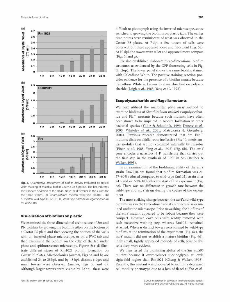
Figures 4(a–c) illustrate the time courses over a
4–28 h period of biofilm development for Rm1021,
RCR2011 and Rlv, respectively, in microtiter plate wells
containing RDM with 2% sucrose. After an initial lag
period of ca. 12 h, the levels of attachment increased
significantly for all strains. The intensity of crystal violet
staining at 28 h in the PVC plates, as indicated by
the absorbance units on the Y-axis, was such that
Rm1021oRCR2011oRlv. RCR2011 accumulated more
than twice the biofilm mass, evaluated by crystal violet
intensity, than Rm1021. Because Rlv exhibited a different
growth rate than the Sm strains, the two species cannot be
directly compared. However, there was no significant differ-
ence in growth rate between Rm1021 and RCR2011 in the
microtiter plate wells (data not shown), and thus the
difference in biofilming capabilities must be attributed to
other features.
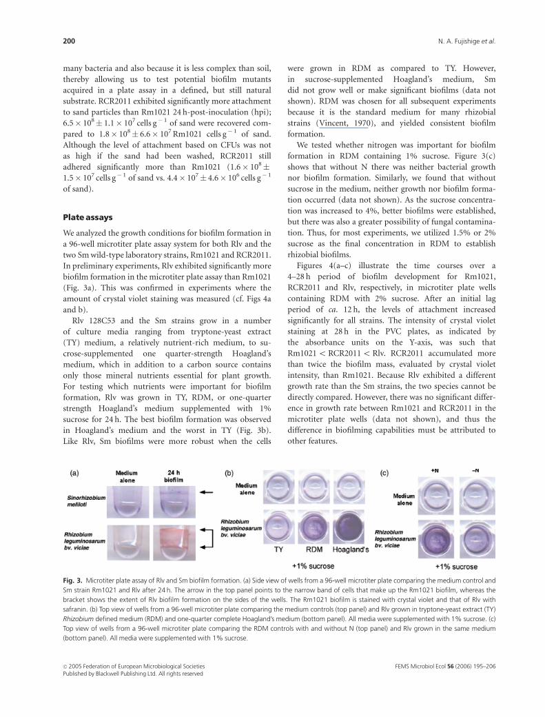
Fig. 3. Microtiter plate assay of Rlv and Sm biofilm formation. (a) Side view of wells from a 96-well microtiter plate comparing the medium control and
Sm strain Rm1021 and Rlv after 24 h. The arrow in the top panel points to the narrow band of cells that make up the Rm1021 biofilm, whereas the
bracket shows the extent of Rlv biofilm formation on the sides of the wells. The Rm1021 biofilm is stained with crystal violet and that of Rlv with
safranin. (b) Top view of wells from a 96-well microtiter plate comparing the medium controls (top panel) and Rlv grown in tryptone-yeast extract (TY)
Rhizobium defined medium (RDM) and one-quarter complete Hoagland’s medium (bottom panel). All media were supplemented with 1% sucrose. (c)
Top view of wells from a 96-well microtiter plate comparing the RDM controls with and without N (top panel) and Rlv grown in the same medium
(bottom panel). All media were supplemented with 1% sucrose.
FEMS Microbiol Ecol 56 (2006) 195–206c� 2005 Federation of European Microbiological SocietiesPublished by Blackwell Publishing Ltd. All rights reserved
200 N. A. Fujishige et al.
Visualizationof biofilmson plastic
We examined the three-dimensional architecture of Sm and
Rlv biofilms by growing the biofilms either on the bottom of
a Costar PS plate and then viewing the bottom of the wells
with an inverted phase microscope, or on a PVC tab and
then examining the biofilm on the edge of the tab under
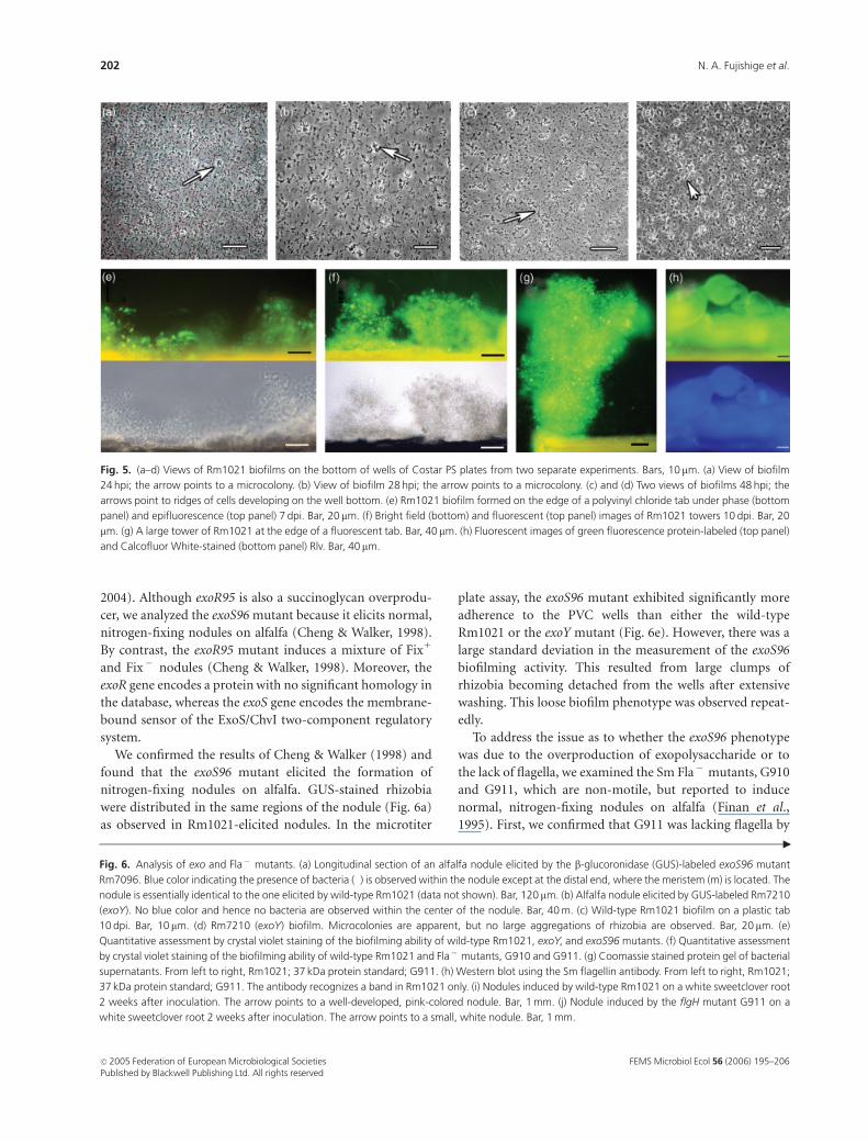
phase and epifluorescence microscopy. Figures 5(a–d) illus-
trate different stages of Rm1021 biofilm formation on
Costar PS plates. Microcolonies (arrows, Figs 5a and b) are
established 24 to 28 hpi, and by 48 hpi, distinct ridges and
small towers were observed (arrows, Figs 5c and d).
Although larger towers were visible by 72 hpi, these were
difficult to photograph using the inverted microscope, so we
switched to growing the biofilms on plastic tabs. The earlier
time points were reminiscent of what was observed in the
Costar PS plates. At 7 dpi, a few towers of cells were
observed, but these appeared loose and flocculent (Fig. 5e).
At 10 dpi, the towers were taller and appeared more compact
(Figs 5f and g).
Rlv also established elaborate three-dimensional biofilm
structures as evidenced by the GFP-fluorescing cells in Fig.
5h (top). The lower panel shows the same biofilm stained
with Calcofluor White. The positive staining reaction pro-
vides evidence for the presence of a biofilm matrix because
Calcofluor White is known to stain rhizobial exopolysac-
charide (Leigh et al., 1985; Yang et al., 1992).
Exopolysaccharideand flagellamutants
We next utilized the microtiter plate assay method to
examine biofilms of Sinorhizobium meliloti exopolysacchar-
ide and Fla� mutants because such mutants have often
been shown to be impaired in biofilm formation in other
bacterial species (Yildiz & Schoolnik, 1999; Danese et al.,
2000; Whiteley et al., 2001; Matsukawa & Greenberg,
2004). Previous research demonstrated that Sm Exo�
mutants elicit on alfalfa roots ineffective (Fix� ), meristem-
less nodules that are not colonized internally by rhizobia
(Finan et al., 1985; Yang et al., 1992) (Fig. 6b). The exoY
gene encodes a galactosyl-1-P transferase that carries out
the first step in the synthesis of EPSI in Sm (Reuber &
Walker, 1993).
In an examination of the biofilming ability of the exoY
strain Rm7210, we found that biofilm formation was ca.
57–60% reduced compared to wild-type Rm1021 strain after
24 h and ca. 50% 40 h after the start of the experiment (Fig.
6e). There was no difference in growth rate between the
wild-type and exoY strain during the course of the experi-
ment.
The most striking change between the exoY and wild-type
biofilms was in the three-dimensional architecture as exam-
ined under the microscope. Prior to washing, the biofilms of
the exoY mutant appeared to be robust because they were
compact. However, exoY cells were readily removed with
each successive washing step, whereas Rm1021 remained
attached. Whereas distinct towers were formed by wild-type
biofilms at the termination of the experiment (Fig. 6c), the
exoY mutant did not establish a mature biofilm (Fig. 6d).
Only small, tightly appressed mounds of cells, four or five
cells deep, were evident.
We then tested the biofilming ability of the Sm exoS96
mutant because it overproduces succinoglycan at levels
eight-fold higher than Rm1021 (Cheng & Walker, 1998).
Recently, this mutant was discovered to exhibit a decreased
cell motility phenotype due to a loss of flagella (Yao et al.,
Fig. 4. Quantitative assessment of biofilm activity evaluated by crystal
violet staining of rhizobial biofilms over a 28-h period. The bar indicates
the standard deviation of the mean. Note the difference in the Y-axes for
the three strains. (a) Sinorhizobium meliloti wild-type Rm1021. (b)
S. meliloti wild-type RCR2011. (F) Wild-type Rhizobium leguminosarum
bv. viciae, Rlv.
FEMS Microbiol Ecol 56 (2006) 195–206 c� 2005 Federation of European Microbiological SocietiesPublished by Blackwell Publishing Ltd. All rights reserved
201Rhizobia form biofilms
2004). Although exoR95 is also a succinoglycan overprodu-
cer, we analyzed the exoS96 mutant because it elicits normal,
nitrogen-fixing nodules on alfalfa (Cheng & Walker, 1998).
By contrast, the exoR95 mutant induces a mixture of Fix1
and Fix� nodules (Cheng & Walker, 1998). Moreover, the
exoR gene encodes a protein with no significant homology in
the database, whereas the exoS gene encodes the membrane-
bound sensor of the ExoS/ChvI two-component regulatory
system.
We confirmed the results of Cheng & Walker (1998) and
found that the exoS96 mutant elicited the formation of
nitrogen-fixing nodules on alfalfa. GUS-stained rhizobia
were distributed in the same regions of the nodule (Fig. 6a)
as observed in Rm1021-elicited nodules. In the microtiter
plate assay, the exoS96 mutant exhibited significantly more
adherence to the PVC wells than either the wild-type
Rm1021 or the exoY mutant (Fig. 6e). However, there was a
large standard deviation in the measurement of the exoS96
biofilming activity. This resulted from large clumps of
rhizobia becoming detached from the wells after extensive
washing. This loose biofilm phenotype was observed repeat-
edly.
To address the issue as to whether the exoS96 phenotype
was due to the overproduction of exopolysaccharide or to
the lack of flagella, we examined the Sm Fla� mutants, G910
and G911, which are non-motile, but reported to induce
normal, nitrogen-fixing nodules on alfalfa (Finan et al.,
1995). First, we confirmed that G911 was lacking flagella by
Fig. 5. (a–d) Views of Rm1021 biofilms on the bottom of wells of Costar PS plates from two separate experiments. Bars, 10 mm. (a) View of biofilm
24 hpi; the arrow points to a microcolony. (b) View of biofilm 28 hpi; the arrow points to a microcolony. (c) and (d) Two views of biofilms 48 hpi; the
arrows point to ridges of cells developing on the well bottom. (e) Rm1021 biofilm formed on the edge of a polyvinyl chloride tab under phase (bottom
panel) and epifluorescence (top panel) 7 dpi. Bar, 20 mm. (f) Bright field (bottom) and fluorescent (top panel) images of Rm1021 towers 10 dpi. Bar, 20
mm. (g) A large tower of Rm1021 at the edge of a fluorescent tab. Bar, 40 mm. (h) Fluorescent images of green fluorescence protein-labeled (top panel)
and Calcofluor White-stained (bottom panel) Rlv. Bar, 40 mm.
Fig. 6. Analysis of exo and Fla� mutants. (a) Longitudinal section of an alfalfa nodule elicited by the b-glucoronidase (GUS)-labeled exoS96 mutant
Rm7096. Blue color indicating the presence of bacteria (�) is observed within the nodule except at the distal end, where the meristem (m) is located. The
nodule is essentially identical to the one elicited by wild-type Rm1021 (data not shown). Bar, 120 mm. (b) Alfalfa nodule elicited by GUS-labeled Rm7210
(exoY). No blue color and hence no bacteria are observed within the center of the nodule. Bar, 40 m. (c) Wild-type Rm1021 biofilm on a plastic tab
10 dpi. Bar, 10 mm. (d) Rm7210 (exoY) biofilm. Microcolonies are apparent, but no large aggregations of rhizobia are observed. Bar, 20 mm. (e)
Quantitative assessment by crystal violet staining of the biofilming ability of wild-type Rm1021, exoY, and exoS96 mutants. (f) Quantitative assessment
by crystal violet staining of the biofilming ability of wild-type Rm1021 and Fla� mutants, G910 and G911. (g) Coomassie stained protein gel of bacterial
supernatants. From left to right, Rm1021; 37 kDa protein standard; G911. (h) Western blot using the Sm flagellin antibody. From left to right, Rm1021;
37 kDa protein standard; G911. The antibody recognizes a band in Rm1021 only. (i) Nodules induced by wild-type Rm1021 on a white sweetclover root
2 weeks after inoculation. The arrow points to a well-developed, pink-colored nodule. Bar, 1 mm. (j) Nodule induced by the flgH mutant G911 on a
white sweetclover root 2 weeks after inoculation. The arrow points to a small, white nodule. Bar, 1 mm.
"
FEMS Microbiol Ecol 56 (2006) 195–206c� 2005 Federation of European Microbiological SocietiesPublished by Blackwell Publishing Ltd. All rights reserved
202 N. A. Fujishige et al.

performing a western blot with an antibody prepared
against S. meliloti flagella. In contrast to Rm1021, no protein
cross-reacting to the antibody was observed (Figs 6g and h).
Then we inoculated both alfalfa and white sweetclover roots
with the Fla� mutant, G911. Although nodules developed
on the roots of the two legume hosts, they were slower to
develop than on the roots inoculated with Rm1021. For
sweetclover, the first nodule appeared 48 h after the first one
developed on the Rm1021-inoculated roots. For alfalfa,
nodulation was delayed ca. 24 h. When examined on the
FEMS Microbiol Ecol 56 (2006) 195–206 c� 2005 Federation of European Microbiological SocietiesPublished by Blackwell Publishing Ltd. All rights reserved
203Rhizobia form biofilms
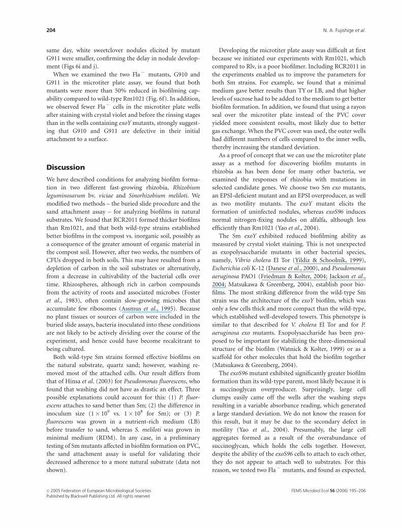
same day, white sweetclover nodules elicited by mutant
G911 were smaller, confirming the delay in nodule develop-
ment (Figs 6i and j).
When we examined the two Fla� mutants, G910 and
G911 in the microtiter plate assay, we found that both
mutants were more than 50% reduced in biofilming cap-
ability compared to wild-type Rm1021 (Fig. 6f). In addition,
we observed fewer Fla� cells in the microtiter plate wells
after staining with crystal violet and before the rinsing stages
than in the wells containing exoY mutants, strongly suggest-
ing that G910 and G911 are defective in their initial
attachment to a surface.
Discussion
We have described conditions for analyzing biofilm forma-
tion in two different fast-growing rhizobia, Rhizobium
leguminosarum bv. viciae and Sinorhizobium meliloti. We
modified two methods – the buried slide procedure and the
sand attachment assay – for analyzing biofilms in natural
substrates. We found that RCR2011 formed thicker biofilms
than Rm1021, and that both wild-type strains established
better biofilms in the compost vs. inorganic soil, possibly as
a consequence of the greater amount of organic material in
the compost soil. However, after two weeks, the numbers of
CFUs dropped in both soils. This may have resulted from a
depletion of carbon in the soil substrates or alternatively,
from a decrease in cultivability of the bacterial cells over
time. Rhizospheres, although rich in carbon compounds
from the activity of roots and associated microbes (Foster
et al., 1983), often contain slow-growing microbes that
accumulate few ribosomes (Assmus et al., 1995). Because
no plant tissues or sources of carbon were included in the
buried slide assays, bacteria inoculated into these conditions
are not likely to be actively dividing over the course of the
experiment, and hence could have become recalcitrant to
being cultured.
Both wild-type Sm strains formed effective biofilms on
the natural substrate, quartz sand; however, washing re-
moved most of the attached cells. Our result differs from
that of Hinsa et al. (2003) for Pseudomonas fluorescens, who
found that washing did not have as drastic an effect. Three
possible explanations could account for this: (1) P. fluor-
escens attaches to sand better than Sm; (2) the difference in
inoculum size (1� 109 vs. 1� 108 for Sm); or (3) P.
fluorescens was grown in a nutrient-rich medium (LB)
before transfer to sand, whereas S. meliloti was grown in
minimal medium (RDM). In any case, in a preliminary
testing of Sm mutants affected in biofilm formation on PVC,
the sand attachment assay is useful for validating their
decreased adherence to a more natural substrate (data not
shown).
Developing the microtiter plate assay was difficult at first
because we initiated our experiments with Rm1021, which
compared to Rlv, is a poor biofilmer. Including RCR2011 in
the experiments enabled us to improve the parameters for
both Sm strains. For example, we found that a minimal
medium gave better results than TY or LB, and that higher
levels of sucrose had to be added to the medium to get better
biofilm formation. In addition, we found that using a rayon
seal over the microtiter plate instead of the PVC cover
yielded more consistent results, most likely due to better
gas exchange. When the PVC cover was used, the outer wells
had different numbers of cells compared to the inner wells,
thereby increasing the standard deviation.
As a proof of concept that we can use the microtiter plate
assay as a method for discovering biofilm mutants in
rhizobia as has been done for many other bacteria, we
examined the responses of rhizobia with mutations in
selected candidate genes. We choose two Sm exo mutants,
an EPSI-deficient mutant and an EPSI overproducer, as well
as two motility mutants. The exoY mutant elicits the
formation of uninfected nodules, whereas exoS96 induces
normal nitrogen-fixing nodules on alfalfa, although less
efficiently than Rm1021 (Yao et al., 2004).
The Sm exoY exhibited reduced biofilming ability as
measured by crystal violet staining. This is not unexpected
as exopolysaccharide mutants in other bacterial species,
namely, Vibrio cholera El Tor (Yildiz & Schoolnik, 1999),
Escherichia coli K-12 (Danese et al., 2000), and Pseudomonas
aeruginosa PAO1 (Friedman & Kolter, 2004; Jackson et al.,
2004; Matsukawa & Greenberg, 2004), establish poor bio-
films. The most striking difference from the wild-type Sm
strain was the architecture of the exoY biofilm, which was
only a few cells thick and more compact than the wild-type,
which established well-developed towers. This phenotype is
similar to that described for V. cholera El Tor and for P.
aeruginosa exo mutants. Exopolysaccharide has been pro-
posed to be important for stabilizing the three-dimensional
structure of the biofilm (Watnick & Kolter, 1999) or as a
scaffold for other molecules that hold the biofilm together
(Matsukawa & Greenberg, 2004).
The exoS96 mutant exhibited significantly greater biofilm
formation than its wild-type parent, most likely because it is
a succinoglycan overproducer. Surprisingly, large cell
clumps easily came off the wells after the washing steps
resulting in a variable absorbance reading, which generated
a large standard deviation. We do not know the reason for
this result, but it may be due to the secondary defect in
motility (Yao et al., 2004). Presumably, the large cell
aggregates formed as a result of the overabundance of
succinoglycan, which holds the cells together. However,
despite the ability of the exoS96 cells to attach to each other,
they do not appear to attach well to substrates. For this
reason, we tested two Fla� mutants, and found as expected,
FEMS Microbiol Ecol 56 (2006) 195–206c� 2005 Federation of European Microbiological SocietiesPublished by Blackwell Publishing Ltd. All rights reserved
204 N. A. Fujishige et al.
based on results from other bacteria, that they are reduced in
their ability to make a biofilm due to poor adherence. This
would suggest that the increase in biofilm formation by
exoS96 is due to the presence of excess EPSI. We are
currently screening for Tn5 mutants that are superior
biofilmers, but that are Fla1, to see if these are EPSI
overproducers.
In P. aeruginosa and many other bacteria, flagella are
necessary for the initial attachment to the surface for biofilm
establishment (O’Toole & Kolter, 1998). We have confirmed
that the motility mutants, G910 (fliP) and G911 (flgH), are
impaired in biofilm formation. Furthermore, we have vali-
dated the finding that G911 lacks flagella, based on the lack
of reaction to an antibody to flagellin (Figs 6g and h). Both
fliP and flgH are important for Sm motility. FliP is part of
the flagellar export apparatus in E. coli and Salmonella
typhimurium (Ohnishi et al., 1997), and is absolutely
essential for bacterial motility in these species. It is not
known for certain whether FliP is part of the flagellar basal
body in Sm. However, the Sm FliP and a homologous gene
in Agrobacterium tumefaciens belong to the same super-
family as the E. coli and Salmonella proteins (Deakin et al.,
1997), strongly suggesting that the FliP proteins in these
plant-associated bacteria have the same function. In many
bacteria, flgH genes are important for flagella synthesis; they
encode the L-ring protein of the basal body.
Nevertheless, neither FlgH nor FliP appears to be abso-
lutely essential for nodulation because both alfalfa and white
sweetclover establish nodules, although they develop later,
the earliest 24 and 48 h later, respectively, than those elicited
by the wild-type Rm1021. The developmental time lag may
explain why previous studies have found Sm Fla� mutants
to have a competitive disadvantage compared to the motile,
wild-type strains (Ames & Bergman, 1981).
With these methods, we are able to examine the effects of
other symbiotic mutations, such as nodulation mutants, as
well as to screen mutant libraries for as-yet uncharacterized
genes that affect the establishment of rhizobial biofilms, and
ultimately, rhizobial attachment to plant root cells.
Acknowledgements
This paper was written in partial fulfillment of the PhD
thesis of N.A.F. to the Department of Molecular, Cell and
Developmental Biology (UCLA). We are grateful to Drs K.
Hill, P. Johnson, B. Lazazzera and N. Stanley of the UCLA
Department of Microbiology, Immunology and Molecular
Genetics for the use of microscopes and for guidance with
biofilm experiments. We thank Dr G.A. O’Toole (Dart-
mouth) for the sand procedure, and Drs. H.-P. Cheng
(Lehman College, CUNY), T. Finan (Hamilton, Ontario,
Canada) and J. Denarie (Toulouse, France) for strains. T.
Finan is also thanked for communicating results before
publication. We are grateful to Dr B. Scharf (Regensburg,
Germany) for the gift of flagellin antibody and to Dr H.
Meade (GTC Biotherapeutics) for reviewing with us the
history of Rm1021. Lastly, we acknowledge the help of
UCLA undergraduates, K. Jankaew, C. Butcher and L. Rios,
who provided invaluable assistance, and we also thank
Dr M. R. Lum, who helped with the microscopy. We
dedicate this paper to the memory of Georges Truchet, a
pioneer in the study of legume-rhizobia interactions. This
research was supported in part by a Shanbrom Family
Foundation grant to A.M.H. and by a UCLA Doctoral
Dissertation grant to N.A.F.
References
Ames P & Bergman K (1981) Competitive advantage provided by
bacterial motility in the formation of nodules by Rhizobium
meliloti. J Bacteriol 148: 728–729.
Assmus B, Hutzler P, Kirchhof G, Amann R, Lawrence JR &
Hartmann A (1995) In situ localization of Azospirillum
brasilense in the rhizosphere of wheat with fluorescently
labeled, rRNA-targeted oligonucleotide probes and scanning
confocal laser microscopy. Appl Environ Microbiol 61:
1013–1019.
Beringer JE (1974) R-factor transfer in Rhizobium
leguminosarum. J Gen Microbiol 120: 421–429.
Van de Broek A, Michiels M, Van Gool A &Vanderleyden J (1993)
Spatial-temporal colonization patterns of Azospirillum
brasilense on the wheat root surface and expression of the
bacterial nifH gene during association. Mol Plant-Microbe
Interact 6: 592–600.
Cheng H-P & Walker GC (1998) Succinoglycan is required for
initiation and elongation of infection threads during
nodulation of alfalfa by Rhizobium meliloti. J Bacteriol 180:
5183–5191.
Costerton JW, Lewandowski Z, Caldwell DE, Korber DR &
Lappin-Scott HM (1995) Microbial biofilms. Annu Rev
Microbiol 49: 711–745.
Danese PN, Pratt LA & Kolter R (2000) Exopolysaccharide
production is required for development of Escherichia coli
K-12 biofilm architecture. J Bacteriol 182: 3593–3596.
Deakin WJ, Furniss CSM, Parker VE & Shaw CH (1997) Isolation
and characterization of a linked cluster of genes from
Agrobacterium tumefaciens encoding proteins involved in
flagellar basal-body structure. Gene 180: 135–137.
Figurski DH & Helinski DR (1979) Replication of an origin-
containing derivative of plasmid RK2 dependent on a plasmid
function provided in trans. Proc Natl Acad Sci USA 73:
1648–1652.
Finan TM, Hirsch AM, Leigh JA, Johansen E, Kuldau GA, Deegan
S, Walker GC & Signer ER (1985) Symbiotic mutants of
Rhizobium meliloti that uncouple plant from bacterial
differentiation. Cell 40: 869–877.
FEMS Microbiol Ecol 56 (2006) 195–206 c� 2005 Federation of European Microbiological SocietiesPublished by Blackwell Publishing Ltd. All rights reserved
205Rhizobia form biofilms
Finan TM, Gough C & Truchet G (1995) Similarity between the
Rhizobium meliloti fliP gene and pathogenicity-associated
genes from animal and plant pathogens. Gene 152: 65–77.
Foster RC, Roira AD & Cock TW (1983) Ultrastructure of the
Root-Soil Interface, pp. 1–8. The American Phytopathological
Society, St. Paul, MN.
Friedman L & Kolter R (2004) Two genetic loci produce distinct
carbohydrate-rich structural components of the Pseudomonas
aeruginosa biofilm matrix. J Bacteriol 186: 4457–4465.
Galibert F, Finan TM, Long SR & 53 additional authors (2001)
The composite genome of the legume symbiont Sinorhizobium
meliloti. Science 293: 668–672.
Hinsa SM, Espinosa-Urgel M, Ramos JL & O’Toole GA (2003)
Transition from reversible to irreversible attachment during
biofilm formation by Pseudomonas fluorescens WCS365
requires an ABC transporter and a large secreted protein. Mol
Microbiol 49: 905–918.
Hirsch AM (1992) Developmental biology of legume nodulation.
New Phytol 112: 211–237.
Jackson KD, Starkey M, Kremer S, Parsek MR & Wozniak DJ
(2004) Identification of psl, a locus encoding a potential
exopolysaccharide that is essential for Pseudomonas aeruginosa
PAO1 biofilm formation. J Bacteriol 186: 4466–4475.
Jefferson RA, Kavanagh TA & Bevan MW (1987) GUS fusions:
b-glucuronidase as a sensitive and versatile gene fusion marker
in higher plants. EMBO J 6: 3901–3907.
Krol E & Becker A (2004) Global transcriptional analysis of the
phosphate starvation response in Sinorhizobium meliloti
strains 1021 and 2011. Mol Gen Genomics 272: 1–17.
Leigh JA, Signer ER & Walker GC (1985) Exopolysaccharide-
deficient mutants of Rhizobium meliloti that form ineffective
nodules. Proc Natl Acad Sci USA 82: 6231–6235.
Long SR (1996) Rhizobium symbiosis: Nod factors in perspective.
Plant Cell 8: 1885–1898.
Machlis L & Torrey JG (1956) Plants in Action. A Laboratory
Manual of Plant Physiology. W.H. Freeman & Co., New York.
Matsukawa M & Greenberg EP (2004) Putative
exopolysaccharide genes influence Pseudomonas aeruginosa
biofilm development. J Bacteriol 186: 4449–4456.
Ohnishi K, Fan F, Schoenhals GJ, Kihara M & Macnab RM (1997)
The FliO, FliP, FliQ, and FliR proteins of Salmonella
typhimurium: putative components for flagellar assembly.
J Bacteriol 179: 6092–6099.
O’Toole GA & Kolter R (1998) Flagellar and twitching motility
are necessary for Pseudomonas aeruginosa biofilm
development. Mol Microbiol 30: 295–304.
O’Toole GA, Pratt LA, Watnick PI, Newman DK, Weaver VB &
Kolter R (1999) Genetic approaches to study of biofilms.
Methods Enzymol 310: 91–109.
Parkinson D, Gray TRG & Williams ST (1971) Methods for
Studying the Ecology of Soil Micro-Organisms. IBP Handbook
No. 19. Blackwell Scientific Publications, Edinburgh.
Reuber TL & Walker GC (1993) Biosynthesis of succinoglycan, a
symbiotically important exopolysaccharide of Rhizobium
meliloti. Cell 74: 269–280.
Sambrook J, Fritsch EF & Maniatis T (1989) Molecular Cloning, a
Laboratory Manual. 2nd edn. Cold Spring Harbor Laboratory
Press, New York.
Stanley NR & Lazazzera BA (2004) Environmental signals and
regulatory pathways that influence biofilm formation. Mol
Microbiol 52: 917–924.
Vincent JM (1970) A Manual for the Practical Study of Root-
Nodule Bacteria. Blackwell Scientific Publications, London.
Watnick PI & Kolter R (1999) Steps in the development of a
Vibrio cholerae El Tor biofilm. Mol Microbiol 34: 586–595.
Whiteley M, Bangera MG, Bumgarner RE, Parsek MR, Teitzel
GM, Lory S & Greenberg EP (2001) Gene expression in
Pseudomonas aeruginosa biofilms. Nature 413: 860–864.
Yang C, Signer ER & Hirsch AM (1992) Nodules initiated by
Rhizobium meliloti exopolysaccharide mutants lack a discrete,
persistent nodule meristem. Plant Physiol 98: 143–151.
Yao S-Y, Luo L, Har KJ, Becker A, Ruberg S, Yu G-Z, Zhu J-B &
Cheng HP (2004) Sinorhizobium meliloti ExoR and ExoS
proteins regulate both succinoglycan and flagellum
production. J Bacteriol 186: 6042–6049.
Yildiz FH & Schoolnik GK (1999) Vibrio cholerae O1 El Tor:
identification of a gene cluster required for the rugose colony
type, exopolysaccharide production, chlorine resistance, and
biofilm formation. Proc Natl Acad Sci USA 96: 4028–4033.
FEMS Microbiol Ecol 56 (2006) 195–206c� 2005 Federation of European Microbiological SocietiesPublished by Blackwell Publishing Ltd. All rights reserved
206 N. A. Fujishige et al.


![Comparison between the reference [i]Rhizobium - Archive ...](https://static.fdokumen.com/doc/165x107/63134e0faca2b42b580d2177/comparison-between-the-reference-irhizobium-archive-.jpg)