Microbial Biofilms in Healthcare - MDPI
168
Microbial Biofilms in Healthcare Formation, Prevention and Treatment Printed Edition of the Special Issue Published in Materials www.mdpi.com/journal/materials Karen Vickery Edited by
-
Upload
khangminh22 -
Category
Documents
-
view
4 -
download
0
Transcript of Microbial Biofilms in Healthcare - MDPI

Microbial Biofilms in HealthcareFormation, Prevention and Treatment
Printed Edition of the Special Issue Published in Materials
www.mdpi.com/journal/materials
Karen VickeryEdited by
Microbial Biofilm
s in Healthcare • Karen Vickery

Microbial Biofilms in Healthcare


Microbial Biofilms in Healthcare
Formation, Prevention and Treatment
Special Issue Editor
Karen Vickery
MDPI • Basel • Beijing • Wuhan • Barcelona • Belgrade • Manchester • Tokyo • Cluj • Tianjin

Special Issue Editor
Karen Vickery
Macquarie University
Australia
Editorial Office
MDPI
St. Alban-Anlage 66
4052 Basel, Switzerland
This is a reprint of articles from the Special Issue published online in the open access journal
Materials (ISSN 1996-1944) (available at: https://www.mdpi.com/journal/materials/special issues/
microbial biofilm healthcare).
For citation purposes, cite each article independently as indicated on the article page online and as
indicated below:
LastName, A.A.; LastName, B.B.; LastName, C.C. Article Title. Journal Name Year, Article Number,
Page Range.
ISBN 978-3-03928-410-8 (Pbk)
ISBN 978-3-03928-411-5 (PDF)
c© 2020 by the authors. Articles in this book are Open Access and distributed under the Creative
Commons Attribution (CC BY) license, which allows users to download, copy and build upon
published articles, as long as the author and publisher are properly credited, which ensures maximum
dissemination and a wider impact of our publications.
The book as a whole is distributed by MDPI under the terms and conditions of the Creative Commons
license CC BY-NC-ND.
Contents
About the Special Issue Editor . . . . . . . . . . . . . . . . . . . . . . . . . . . . . . . . . . . . . . vii
Preface to ”Microbial Biofilms in Healthcare: Formation, Prevention and Treatment” . . . . . ix
Karen Vickery
Special Issue: Microbial Biofilms in Healthcare: Formation, Prevention and TreatmentReprinted from: Materials 2019, 12, 2001, doi:10.3390/ma12122001 . . . . . . . . . . . . . . . . . . 1
Shamaila Tahir, Matthew Malone, Honghua Hu, Anand Deva and Karen Vickery
The Effect of Negative Pressure Wound Therapy with and without Instillation on MatureBiofilms In VitroReprinted from: Materials 2018, 11, 811, doi:10.3390/ma11050811 . . . . . . . . . . . . . . . . . . . 5
Lasserre Jerome Frederic, Brecx Michel and Toma Selena
Oral Microbes, Biofilms and Their Role in Periodontal and Peri-Implant DiseasesReprinted from: Materials 2018, 11, 1802, doi:10.3390/ma11101802 . . . . . . . . . . . . . . . . . . 17
Maria Mempin, Honghua Hu, Durdana Chowdhury, Anand Deva and Karen Vickery
The A, B and C’s of Silicone Breast Implants: Anaplastic Large Cell Lymphoma, Biofilm andCapsular ContractureReprinted from: Materials 2018, 11, 2393, doi:10.3390/ma11122393 . . . . . . . . . . . . . . . . . . 35
Marie Beitelshees, Andrew Hill, Charles H. Jones and Blaine A. PfeiferImplications for Anti-BiofilmPhenotypic Variation during Biofilm Formation:
Therapeutic DesignReprinted from: Materials 2018, 11, 1086, doi:10.3390/ma11071086 . . . . . . . . . . . . . . . . . . 47
Katarzyna Ledwoch and Jean-Yves Maillard
Candida auris Dry Surface Biofilm (DSB) for Disinfectant Efficacy TestingReprinted from: Materials 2019, 12, 18, doi:10.3390/ma12010018 . . . . . . . . . . . . . . . . . . . 65
Nor Fadhilah Kamaruzzaman, Tan Li Peng, Khairun Anisa Mat Yazid, Shamsaldeen Ibrahim
Saeed, Ruhil Hayati Hamdan, Choong Siew Shean, Wong Weng Kin, Alexandru Chivu and
Amanda Jane Gibson
Targeting the Bacterial Protective Armour; Challenges and Novel Strategies in the Treatment ofMicrobial BiofilmReprinted from: Materials 2018, 11, 1705, doi:10.3390/ma11091705 . . . . . . . . . . . . . . . . . . 75
Phillip A. Laycock, John J. Cooper, Robert P. Howlin, Craig Delury, Sean Aiken and Paul Stoodley
In Vitro Efficacy of Antibiotics Released from Calcium Sulfate Bone Void Filler BeadsReprinted from: Materials 2018, 11, 2265, doi:10.3390/ma11112265 . . . . . . . . . . . . . . . . . . 103
Muhammad Yasir, Mark Duncan Perry Willcox and Debarun Dutta
Action of Antimicrobial Peptides against Bacterial BiofilmsReprinted from: Materials 2018, 11, 2468, doi:10.3390/ma11122468 . . . . . . . . . . . . . . . . . . 119
Bindu Subhadra, Dong Ho Kim, Kyungho Woo, Surya Surendran and Chul Hee Choi
Control of Biofilm Formation in Healthcare: Recent Advances Exploiting Quorum-SensingInterference Strategies and Multidrug Efflux Pump InhibitorsReprinted from: Materials 2018, 11, 1676, doi:10.3390/ma11091676 . . . . . . . . . . . . . . . . . . 135
v


About the Special Issue Editor
Karen Vickery graduated from Veterinary Science with honors in 1979 and worked in general
veterinary practice in Australia and the United Kingdom. She obtained her Ph.D. in 1996 investigating
hepatitis B virus (HBV) and developed the method for biocide efficacy testing against HBV for
disinfectant registration referenced by the Australian Therapeutic Goods Administration. She is
the Scientific Director of the Surgical Infection Research Group at Macquarie University, Australia
and is primarily responsible for investigating medically important biofilms. Her research aims to
prevent healthcare associated infections by focusing on both surgical strategies for preventing biofilm
infection of medical implants, treating biofilm infections of chronic wounds, and strategies that
improve instrument and environmental decontamination.
vii


Preface to ”Microbial Biofilms in Healthcare:
Formation, Prevention and Treatment”
An estimated 99% of the world’s bacteria live in close proximity with other bacteria in a biofilm. In a biofilm, the bacteria are enclosed in exopolymeric substances (EPS) and are generally attached to a surface. The biofilm phenotype and the surrounding EPS increase the tolerance of bacteria to desiccation and biocide action, resulting in bacterial persistence on surfaces long after free-swimming or planktonic bacteria are killed. In this book, we investigate the role of biofilms in breast and dental implant disease and cancer. We include in vitro models for investigating treatment of chronic wounds and disinfectant action against Candida sp. Also included are papers on the most recent strategies for treating biofilm infection ranging from antibiotics incorporated into bone void fillers to antimicrobial peptides and quorum sensing.
Karen Vickery
Special Issue Editor
ix


materials
Editorial
Special Issue: Microbial Biofilms in Healthcare:Formation, Prevention and Treatment
Karen Vickery
Surgical Infection Research Group, Faculty of Medicine and Health Sciences, Macquarie University,Sydney 2109, Australia; [email protected]
Received: 19 June 2019; Accepted: 21 June 2019; Published: 22 June 2019
Abstract: Biofilms are a structured community of microorganisms that are attached to a surface.Individual bacteria are embedded in a bacterial-secreted matrix. Biofilms have significantly increasedtolerance to removal by cleaning agents and killing by disinfectants and antibiotics. This specialissue is devoted to diagnosis and treatment of biofilm-related diseases in man. It highlights thedifferences between the biofilm and planktonic (single cell) lifestyles and the diseases biofilms causefrom periodontitis to breast implant capsular contracture. Biofilm-specific treatment options aredetailed in experimental and review manuscripts.
Keywords: biofilms; dry surface biofilms; periodontitis; breast implants; Candida auris; calciumsulphate; antibiotic; topical negative pressure wound therapy; antimicrobial peptides
Introduction
Biofilms are ubiquitous with an estimated 99% of the world’s bacteria living enclosed in a biofilm.The problems that biofilm cause in industry have been well documented and methods to reduce theirimpact have been explored since before the middle of the last century. However, the extent to whichbiofilms play a significant detrimental role in chronic disease and implantable medical device failurehas only been acknowledged over the past few decades whilst the role they play in surface and surgicalinstrument decontamination failure has only recently been highlighted.
Biofilms are a structured community of microorganisms that are attached to a surface. In healthcare,environmental biofilms take three forms: traditional hydrated biofilms which form in wet areas suchas showers, water pipes and sinks; biofilms that form on dry surfaces such as benchtops and curtains,called dry surface biofilms (DSB); and build-up biofilms (BUB) that form on surgical instrumentssubjected to cycles of use, decontamination (cleaning and disinfection) and drying during storage.In addition, biofilm forms in human tissue such as the lung of cystic fibrosis sufferers and in chronicwounds, and biofilms on implantable medical devices lead to their failure. The importance of biofilms inhealthcare arises due to biofilms’ increased tolerance to biocides and increased tolerance to desiccationwhen compared with planktonic organisms of the same species.
Biofilms’ increased tolerance to desiccation means that they can survive dry conditions whichreadily kills planktonic bacteria. DSB have been shown to survive over 12 months in a sterile container,on a bench without any nutrition, and they are particularly tolerant to disinfectants [1,2]. DSB havebeen detected on over 90% of dry hospital surfaces in four countries (Australia, Brazil, Saudi Arabiaand the United Kingdom) [1,3–5]. In this special issue, Ledwoch and Maillard investigated the efficacyof 12 commercial disinfectants and 1000 ppm sodium hypochlorite (recommended as the disinfectant ofchoice by Public Health England) against DSB composed of Candida auris [6]. They initially developeda DSB model of this emerging pathogen and then used this model DSB in a modification of theASTM2967-15 Wiperator test to measure decrease in C. auris viability, transfer of C. auris and biofilmre-growth following treatment. Similar to bacterial DSB, C. auris DSB showed increased tolerance tocommon disinfectant agents.
Materials 2019, 12, 2001; doi:10.3390/ma12122001 www.mdpi.com/journal/materials1

Materials 2019, 12, 2001
Bacteria can attach to host tissue and any implantable medical device. In this issue, Kamaruzzamanet al. review the bacterial species that are principally isolated from healthcare associated infection, thebody sites where biofilms cause disease, diagnosis and treatment options [7]. They go on to describe themechanisms of antimicrobial tolerance and evasion of host immune response which biofilm exhibits.Mempin et al. review the surface characteristics of different types of breast implants and how thisaffects bacterial attachment [8]. They also describe how biofilm formation on breast implants leads tocapsular contracture and its possible role in the potentiation of breast implant associated AnaplasticLarge Cell Lymphoma (ALCL). Frédéric et al. review the role that oral biofilm plays in periodontitisand peri-implantitis and the limitations of treatment options [9]. The poor response of chronic woundsto treatment promoted Tahir et al. to investigate whether physically altering biofilms’ architectureincreased its sensitivity to biocides [10]. They did this by utilizing their topical negative pressurewound therapy model. In this special issue, other treatment options that were experimentally exploredincluded Laycock et al.’s work on the efficacy of antibiotic release from calcium sulphate bone void fillerbeads [11]. As calcium sulphate is completely biocompatible and absorbed by the body, combining itwith antibiotics and using this combination locally would serve to increase antibiotic release at fracturesites and reduce the need for high dose systemic use of antibiotics.
In this special issue, three reviews address various antibiofilm treatment strategies. Biofilmformation and maturation can be stopped by preventing bacterial attachment or by interfering withbacterial quorum sensing. Once formed, biofilm removal can be induced by use of chemical andquorum sensing dispersal agents. Beitelshees et al. reviewed biofilm formation and how bacterialphenotype changes during biofilm development [12]. They relate bacterial phenotype to the anti-biofilmstrategy. Yasir et al. reviewed the major antibiofilm mechanisms of the action of antimicrobial peptidesand how these prevent biofilm formation and disrupt mature biofilms [13]. Subhadra et al. reviewedthe recent advances in preventing biofilm formation and inducing its dispersal by interfering withquorum sensing [14].
Conflicts of Interest: The author declares no conflict of interest.
References
1. Hu, H.; Johani, K.; Gosbell, I.B.; Jacombs, A.; Almatroudi, A.; Whiteley, G.S.; Deva, A.K.; Jensen, S.; Vickery, K.Intensive care unit environmental surfaces are contaminated by multiresistant bacteria in biofilms: Combinedresults of conventional culture, pyrosequencing, scanning electron microscopy and confocal laser microscopy.J. Hosp. Infect. 2015, 91, 35–44. [CrossRef] [PubMed]
2. Almatroudi, A.; Gosbell, I.; Hu, H.; Jensen, S.; Espedido, B.; Tahir, S.; Glasbey, T.; Legge, P.; Whiteley, G.;Deva, A.; et al. Staphylococcus aureus dry-surface biofilms are not killed by sodium hypochlorite:Implications for infection control. J. Hosp. Infect. 2016, 93, 263–270. [CrossRef] [PubMed]
3. Costa, D.; Johani, K.; Melo, D.S.; Lopes, L.; Lima, L.L.; Tipple, A.; Hu, H.; Vickery, K.; Costa, D.D.M.;Lopes, L.K.D.O.; et al. Biofilm contamination of high-touched surfaces in intensive care units: Epidemiologyand potential impacts. Lett. Appl. Microbiol. 2019, 68, 269–276. [CrossRef] [PubMed]
4. Johani, K.; Abualsaud, D.; Costa, D.M.; Hu, H.; Whiteley, G.; Deva, A.; Vickery, K. Characterization ofmicrobial community composition, antimicrobial resistance and biofilm on intensive care surfaces. J. Infect.Public Health 2018, 11, 418–424. [CrossRef] [PubMed]
5. Ledwoch, K.; Dancer, S.; Otter, J.; Kerr, K.; Roposte, D.; Rushton, L.; Weiser, R.; Mahenthiralingam, E.;Muir, D.; Maillard, J.-Y. Beware biofilm! Dry biofilms containing bacterial pathogens on multiple healthcaresurfaces; a multi-centre study. J. Hosp. Infect. 2018, 100, e47–e56. [CrossRef] [PubMed]
6. Ledwoch, K.; Maillard, J.-Y. Candida auris Dry Surface Biofilm (DSB) for Disinfectant Efficacy Testing.Materials 2018, 12, 18. [CrossRef] [PubMed]
7. Kamaruzzaman, N.F.; Tan, L.P.; Yazid, K.A.M.; Saeed, S.I.; Hamdan, R.H.; Choong, S.S.; Wong, W.K.;Chivu, A.; Gibson, A.J.; Yazid, K.M. Targeting the Bacterial Protective Armour; Challenges and NovelStrategies in the Treatment of Microbial Biofilm. Materials 2018, 11, 1705. [CrossRef] [PubMed]
2
Materials 2019, 12, 2001
8. Mempin, M.; Hu, H.; Chowdhury, D.; Deva, A.; Vickery, K. The A, B and C’s of Silicone Breast Implants:Anaplastic Large Cell Lymphoma, Biofilm and Capsular Contracture. Materials 2018, 11, 2393. [CrossRef][PubMed]
9. Lasserre, J.F.; Brecx, M.C.; Toma, S.; Frédéric, L.J.; Michel, B.; Selena, T. Oral Microbes, Biofilms and TheirRole in Periodontal and Peri-Implant Diseases. Materials 2018, 11, 1802.
10. Tahir, S.; Malone, M.; Hu, H.; Deva, A.; Vickery, K. The Effect of Negative Pressure Wound Therapy with andwithout Instillation on Mature Biofilms In Vitro. Materials 2018, 11, 811. [CrossRef] [PubMed]
11. Laycock, P.A.; Cooper, J.J.; Howlin, R.P.; Delury, C.; Aiken, S.; Stoodley, P. In Vitro Efficacy of AntibioticsReleased from Calcium Sulfate Bone Void Filler Beads. Materials 2018, 11, 2265. [CrossRef] [PubMed]
12. Beitelshees, M.; Hill, A.; Jones, C.H.; Pfeifer, B.A. Phenotypic Variation during Biofilm Formation: Implicationsfor Anti-Biofilm Therapeutic Design. Materials 2018, 11, 1086. [CrossRef] [PubMed]
13. Yasir, M.; Willcox, M.D.P.; Dutta, D. Action of Antimicrobial Peptides against Bacterial Biofilms. Materials2018, 11, 2468. [CrossRef]
14. Subhadra, B.; Kim, D.H.; Woo, K.; Surendran, S.; Choi, C.H. Control of Biofilm Formation in Healthcare:Recent Advances Exploiting Quorum-Sensing Interference Strategies and Multidrug Efflux Pump Inhibitors.Materials 2018, 11, 1676. [CrossRef]
© 2019 by the author. Licensee MDPI, Basel, Switzerland. This article is an open accessarticle distributed under the terms and conditions of the Creative Commons Attribution(CC BY) license (http://creativecommons.org/licenses/by/4.0/).
3


materials
Article
The Effect of Negative Pressure Wound Therapy withand without Instillation on Mature Biofilms In Vitro
Shamaila Tahir 1,*, Matthew Malone 2,3,4, Honghua Hu 1, Anand Deva 1 and Karen Vickery 1
1 Surgical Infection Research Group, Faculty of Medicine and Health Sciences, Macquarie University,Sydney 2109, Australia; [email protected] (H.H.); [email protected] (A.D.);[email protected] (K.V.)
2 Infectious Diseases and Microbiology, School of Medicine, Western Sydney University, Sydney 2751,Australia; [email protected]
3 Liverpool Diabetes Collaborative Research Unit, Ingham Institute of Applied Medical Research,Sydney 2170, Australia
4 High Risk Foot Service, Liverpool Hospital, South West Sydney LHD, Sydney 2170, Australia* Correspondence: [email protected]
Received: 15 March 2018; Accepted: 14 May 2018; Published: 16 May 2018
Abstract: Background: To investigate the effect of negative pressure wound therapy (NPWT)with and without instillation (NPWTi) on in vitro mature biofilm. Methods: Mature biofilms ofPseudomonas aeruginosa and Staphylococcus aureus were grown under shear (130 rpm) on polycarbonatecoupons in a CDC biofilm reactor for 3 days. Coupons containing biofilms were placed in a sterilepetri dish and sealed using NPWT or NPWTi. Coupons were exposed to treatment for 24 h withNPWT alone or with instillation of: Povidone iodine solution (PVP-I) (10% w/v equivalent to 1% w/vavailable iodine, BETADINE®, Mundipharma, Singapore), surfactant based antimicrobial solutionwith polyhexamethylene biguanide (SBPHMB) (Prontosan®, B Braun Medical, Melsungen, Germany),Gentamicin 1 μg/mL (GM) (G1264 Sigma-Aldrich Pty Ltd., Castle Hill, Australia) Rifampicin24 μg/mL (RF) (R3501 Sigma-Aldrich Pty Ltd., Castle Hill, Australia) and NaCl 0.9% (Baxter,Deerfield, IL, USA). Bacterial cell viability and biofilm architecture pre-and post-treatment wereassessed using colony forming units (cfu), Live/Dead viability staining, confocal laser scanningmicroscopy (CLSM) and scanning electron microscopy (SEM). Results: Significant reductions wereobtained in S. aureus biofilm thickness (65%) and mass (47%) when treated with NPWTi as comparedto NPWT only. NPWTi with instillation of SBPHMB, PVP-I and RF achieved between 2 and 8 log10
reductions against S. aureus biofilm (p < 0.05–0.001). Conversely, PVP-I and SBMO achieved a 3.5 log10
reduction against P. aeruginosa (p < 0.05). Conclusions: NPWT alters biofilm architecture by reducingbiofilm thickness and mass, but this does not affect bacterial cell viability. NPWT with instillation ofcertain antimicrobials solutions may provide a further synergistic effect in reducing the number ofviable biofilm microorganisms. Our in vitro model may be used for screening the effectiveness ofantimicrobials used under instillation prior to animal or human studies.
Keywords: biofilm; chronic wounds; instillation therapy; in vitro
1. Introduction
The causality of a wound that experiences a delay in healing can be multifactorial and attributedto factors such as local tissue hypoxia/poor perfusion, repetitive ischemia-reperfusion injury [1],microbial infection [2], inadequate offloading or compression therapy [3]. Perhaps the mostsignificant of these factors are the cases where chronic wounds become complicated by pathogenicmicroorganisms. These may exist as planktonic rapidly dividing cells that invade host tissues andinduce an acute infection [4]. Conversely, some microorganisms that complicate chronic wounds may
Materials 2018, 11, 811; doi:10.3390/ma11050811 www.mdpi.com/journal/materials5

Materials 2018, 11, 811
alter their phenotype, differing markedly in both their physiology and activity. These microorganismsare sessile, attach to surfaces or other microorganisms, form aggregates, and regulate the productionof an extracellular polymeric substance (biofilm) [5]. The hallmark features of these microorganismsare their tolerance to antimicrobials, the host immune responses and environmental stresses.
These wounds are a challenge for any clinician and ensuring their resolution can often involvea complex array of pathways that may involve surgical or sharp conservative debridement of anyinfected non-viable tissue. Even in this scenario the ability for a surgeon/clinician to remove allnon-viable tissue and any microorganisms not visible to the naked eye is likely not possible [6].Post-surgical debridement wound care is therefore a critical step to ensure a newly ‘acute’ woundcontinues through the orderly continuum of repair. To augment this process negative pressure woundtherapy with instillation (NPWTi) and dwell time is an adjunctive treatment modality for selectedcomplex wounds complicated by invasive infection or extensive biofilm [7,8].
Evidence for NPWT with or without instillation/dwell time on the microbial load of woundsis limited [9–11] with little data available for its action/s against microbial biofilms [12]. Previouslyour group demonstrated that NPWT resulted in a physical disruption to biofilm architecture [13].This change resulted in a synergism between NPWT and a solid dressing (silver impregnatedfoam) eradicating an in vitro biofilm [14]. In this study we aim to test the effectiveness ofNPWT with instillation and dwell time of topical antimicrobial solutions, against 3-day matureS. aureus and P. aeruginosa biofilms. We hypothesize that NPWT alters biofilm architecture and thusimproves penetration of antimicrobials through the extracellular polymeric substance of biofilmforming microorganisms.
2. Materials and Methods
2.1. Bacterial Test Strains
Biofilm forming reference strains utilized in vitro were S. aureus (ATCC® 25923™),(methicillin-sensitive S. aureus (MSSA) and P. aeruginosa (ATCC® 25619™).
2.2. Solutions Used for Instillation Therapy
Details regarding the solutions used, any incorporated antimicrobials and tested concentrationlevels, and their respective manufacturers are noted in the Supplementary Materials (Table S1). Briefly,surfactant based antimicrobial solution with polyhexamethylene biguanide (SBPHMB; Prontosan®,B Braun Medical, Melsungen, Germany), povidone iodine (PVP-I) antimicrobial solution 10% w/vequivalent to 1% w/v available iodine (BETADINE®, Mundipharma, Singapore), Saline (NaCl) 0.9%(Baxter, Deerfield, IL, USA). The systemic antimicrobials tested were, gentamicin (GM) 1 μg/mL andRifampicin (RF) 24 μg/mL, both diluted in NaCl 0.9% (Baxter International, Deerfield, IL, USA).
2.3. In Vitro CDC Biofilm Reactor
P. aeruginosa and S. aureus were grown separately under shear (130 rpm) at 35 ◦C on 24 removablepolycarbonate coupons in a CDC biofilm reactor (BioSurface Technologies Corp., Bozeman, MT, USA).S. aureus biofilm was grown in 15 g/L (50%) tryptone soya broth (TSB) (Sigma Aldrich, St. Louis, MO,USA) in batch phase for 24 h and then replaced with fresh media 6 g/L (20% TSB) flowing through thechamber at 80 mL/h for a further 48 h. P. aeruginosa was grown in 600 mg/L (2%) TSB in batch phasefor 24 h and then with fresh media (TSB 2%) flowing through the chamber at 80 mL/h for a further48 h. Coupons were harvested by washing gently, three times, in phosphate buffered saline (PBS) toremove loosely attached and planktonic bacteria. The number of bacteria per coupon was 3.52 × 10⁷
and 2.3 × 10⁷ for S. aureus and P. aeruginosa, respectively. Bacterial biofilm was gently scraped off fromthe outer side of each coupon using a 12.5% sodium hypochlorite-soaked paper towel, and then againwashed three times in TSB to remove residual chlorine.
6

Materials 2018, 11, 811
2.4. In Vitro NPWTi Model
The NPWTi utilized in this study was the V.A.C. Ulta negative pressure wound therapy system(Acelity, San Antonio, TX, USA) incorporating the V.A.C. Veraflo therapy that allows the controlledinstillation of topical solutions. Modifications to the system were necessary due to the tubular shape ofthe V.A.C. Veraflo dressing system, in keeping with previously published reports [13].
Five biofilm containing coupons were placed on top of 3% bacteriological agar (Thermo Scientific,Basingstoke, UK) in a sterile petri dish. The sterile NPWT dressing (V.A.C.® GRANUFOAM™)were added on top of the coupons until the petri dish was completely full, thus ensuring equalpressure application to all five biofilm covered coupons. An airtight seal was produced using a sterilesemi-impervious dressing (V.A.C.® dressing system). In order to emulate wound exudate, couponswere bathed with TSB (30 g/L) at a flow rate of 40 mL/h via an inflow channel [15,16]. Excess fluidwas drained via a gravity drainage tube, situated on the opposite side from the nutrition in-flow, forchambers not subjected to NPWT and via a centrally placed V.A.C.® Veralink Cassette for chamberssubjected to NPWT (Figure 1).
(a)
(b)
(c)
Figure 1. (a) Schematic presentation of modified wound model with polycarbonate coupons (green).(b) New wound model with Veraflo dressing system. (c) Experimental setup of instillation + V.A.C.therapy. Green circles represent biofilm coated polycarbonate coupons.
7

Materials 2018, 11, 811
Five coupons for each test antimicrobial solution were exposed to the following treatmentvariables: (i) control with no treatment; (ii) NPWT alone with no instillation; (iii) instillation ofantimicrobial plus continuous NPWT at 125 mmHg (except during the 20 min instillation treatmentperiods); and (iv) instillation of antimicrobial solution with no NPWT. The instillation cycles were asfollows: instillation every 6 h with 35 mL of saline or test antimicrobial with a 20 min dwell time in a24 h time period. Instillation with TSB was then continued for another 6 h before harvesting.
2.5. Bacterial Viability cfu/log10
At the end of each treatment period, the numbers of residual bacterial colony forming units (cfu)per coupon were tested in triplicate by sonication in an ultrasonic bath (Soniclean, Stepney, Australia)for 10 min with a sweeping frequency of 42–47 kH at 20 ◦C.
Coupons were then vortexed for two min in 2 mL of PBS followed by sequential 10-fold dilutionand plate count. Pre- and post-exposure average cfu/coupon was expressed as log10.
2.6. Confocal Laser Scanning Microscopy
Bacterial cell viability pre- and post-exposure was also assessed using BacLight™ (Live/DeadBacterial Viability Kit, 7012, Molecular Probes, Invitrogen, Carlsbad, CA, USA) in conjunction withconfocal laser scanning microscopy (CLSM) (Olympus FluoView™ FV1000, Tokyo, Japan). Followingstaining, coupons were fixed with 4% paraformaldehyde for 1 h and washed thrice with PBS for10 min. 2D images were obtained within 24–48 h of staining. 3D images were obtained from threeseparate areas per coupon. Images were built with 0.2 μm optical sections and analyzed for averagethickness, biofilm mass and percentage of viable cells, using the IMARIS 7.7.2 software (Bitplane,Zurich, Switzerland) and ImageJ program (scriptable Java application for scientific image processing).A 63× water immersion objective lens was used to capture images with reduced background noise at10×, 20× and 40× magnifications. To minimize image artefacts these dual labelled (Syto-9 andpropidium iodide) samples were sequentially scanned at 488 nm fluorescence excitation (greenemission) and then at 543 nm (red emission) collected in the green and red regions, respectively.Line averaging (×2) was used to capture images with reduced noise.
Biofilm architecture was analyzed using IMARIS (Bitplane AG, Zurich, Switzerland) softwareto quantify 3-D CLSM images by: (1) Average thickness is the distance (μm) between the top of abiofilm and the substratum on which the biofilm resides. It provides a measure of the spatial size ofbiofilm; (2) Average biofilm biomass (μm3), is defined as the volume of bacterial cells below μm2 area.The value excludes the non-cellular components (e.g., EPS and water channels) of biofilm volume.
2.7. Scanning Electron Microscopy
For SEM, one coupon each from selected antimicrobial treatment were fixed in 3% glutaraldehyde,dehydrated through serial dilutions of ethanol and then immersed in hexamethyldisilazane(Polysciences Inc., Warrington, FL, USA) for 10 min before being aspirated dry and air dried forat least 48 h. Coupons were then mounted on specimen stubs, gold coated and examined at low andhigh magnifications (JOEL 6480LA SEM, Tokyo, Japan).
Statistical Analysis
Statistical analysis on cfu data was performed using the Sigma Plot 11 statistical program(Scientific Graphing Software: SigmaPlot® Version 11 by Systat Software, Inc., San Jose, CA, USA).Pre and post bacterial viability between treatment groups were analyzed by performing one-wayanalysis of variance (ANOVA). For non-normally distributed data a Kruskal-Wallis one-way analysisof variance on ranks was performed, and if significant, the Tukey test for all pair wise multiplecomparisons were conducted to determine which treatment groups were significantly different fromeach other.
8
Materials 2018, 11, 811
3. Results
All experiments were conducted over a 24 h test period. Control coupons of S. aureus andP. aeruginosa receiving no treatment increased in the number of biofilm bacteria from a starting cfu of7.4 log10 cfu/coupon to 8.3 log10 cfu/coupon (0.9 log10 cfu/coupon increase, p = 1.0). The effects ofNPWT, NPWTi and instillation alone on bacterial viability after 24 h are reported.
3.1. NPWT on Bacterial Viability cfu/log10
NPWT had little effect on S. aureus biofilms demonstrating a 1.2 log10 cfu/coupon reduction(control no treatment = 7.4 log10 cfu/coupon vs. NPWT = 6.2 log10 cfu/coupon p > 1.0) whilenumbers increased in the case of P. aeruginosa by 0.7 log10 cfu/coupon (control no treatment = 7.4 log10
cfu/coupon vs. NPWT = 8.1 log10 cfu/coupon p > 1.0).
3.2. Instillation Alone vs. NPWTi on Bacterial Viability cfu/log10
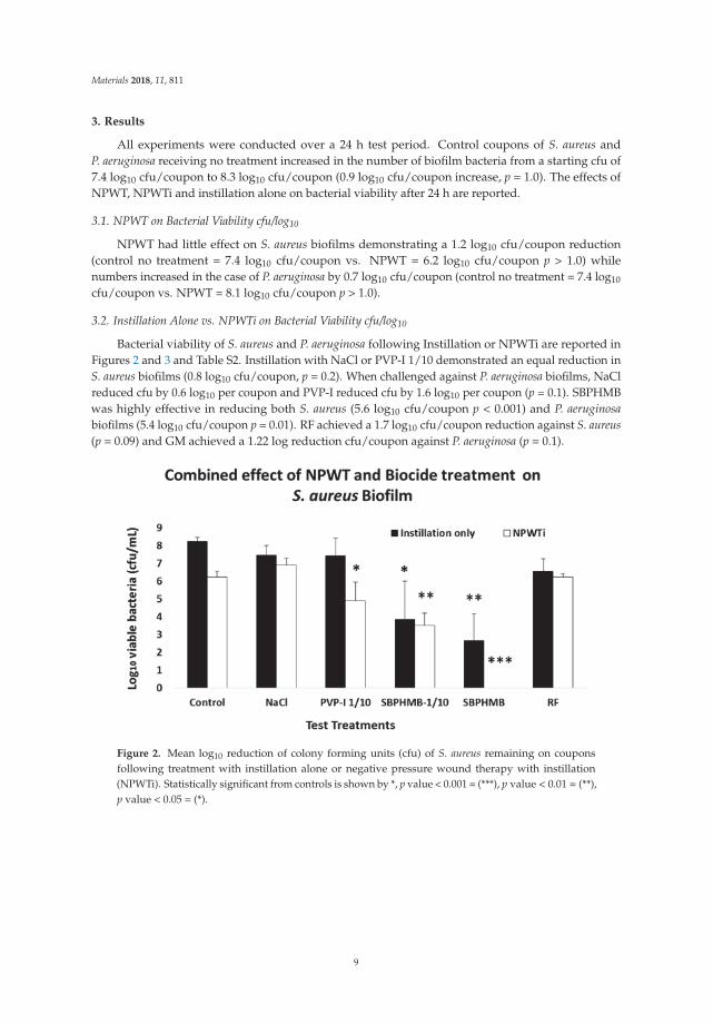
Bacterial viability of S. aureus and P. aeruginosa following Instillation or NPWTi are reported inFigures 2 and 3 and Table S2. Instillation with NaCl or PVP-I 1/10 demonstrated an equal reduction inS. aureus biofilms (0.8 log10 cfu/coupon, p = 0.2). When challenged against P. aeruginosa biofilms, NaClreduced cfu by 0.6 log10 per coupon and PVP-I reduced cfu by 1.6 log10 per coupon (p = 0.1). SBPHMBwas highly effective in reducing both S. aureus (5.6 log10 cfu/coupon p < 0.001) and P. aeruginosabiofilms (5.4 log10 cfu/coupon p = 0.01). RF achieved a 1.7 log10 cfu/coupon reduction against S. aureus(p = 0.09) and GM achieved a 1.22 log reduction cfu/coupon against P. aeruginosa (p = 0.1).
Figure 2. Mean log10 reduction of colony forming units (cfu) of S. aureus remaining on couponsfollowing treatment with instillation alone or negative pressure wound therapy with instillation(NPWTi). Statistically significant from controls is shown by *, p value < 0.001 = (***), p value < 0.01 = (**),p value < 0.05 = (*).
9

Materials 2018, 11, 811
Figure 3. Mean log10 reduction of cfu of P. aeruginosa remaining on coupons following treatment with(n = 5) and without (n = 5) application of topical negative pressure. Statistically significant from controlsis shown by p value < 0.01 = (**).
NPWTi demonstrated significant increases in effectiveness against both S. aureus and P. aeruginosawhen compared to instillation alone. When challenged against S. aureus, NPWTi using NaCldemonstrated a 1.4 log10 cfu/coupon reduction, (p = 0.1), PVP-I a 3.4 log10 cfu/coupon reduction(p = 0.05), SBPHMB an 8.3 log10 cfu/coupon reduction (p = 0.001) and RF a 2.1 log10 cfu/couponreduction (p = 0.05). For P. aeruginosa, NPWTi using NaCl demonstrated a 0.4 log10 cfu/coupon(p = 0.1), PVP-I a 1.3 log10 cfu/coupon reduction (p = 0.2), SBPHMB a 7.3 log10 cfu/coupon reduction(p < 0.0001) and GM a 4.1 log10 cfu/coupon reduction (p = 0.01).
3.3. Microscopy
Given that SBPHMB demonstrated a greater efficacy than all other antimicrobials used in ourbiofilm models, we explored this agent in greater detail using both SEM and confocal microscopywith LIVE/DEAD stain. SEM images of S. aureus and P. aeruginosa biofilms from coupons undergoinginstillation only with SBPHMB or NPWTi using SBPHMB are depicted in Figure 4. Minimal changesin biofilm structure of S. aureus and P. aeruginosa are noted in the SEM images (Figure 4a,c) followinginstillation of SBPHMB for 24 h. SEM identified dense coccoid and rod shaped microbial aggregates,respectively, embedded in a thick continuous EPS. Therefore, post-treatment with instillation only wasineffective. In contrast, SEM images of NPWTi using SBPHMB treated coupons showed significantreductions in biofilm EPS and in the number of microbial aggregates (Figure 4b,d). This was particularlyevident for S. aureus biofilms, which demonstrated complete eradication of cocci aggregates andextracellular polymeric substance (EPS) (Figure 4b).
10

Materials 2018, 11, 811
(a) (b)
(c) (d)
Figure 4. (a) SEM images of S. aureus biofilm coupon using SBPHMB/Instillation only for 24-h identifiesdense coccoid bacteria embedded in EPS after treatment; (b) demonstrates an overall reduction inS. aureus biofilm after 24-h treatment with NPWTi using SBPHMB; (c) illustrates P. aeruginosa biofilmcoupon using SPHMB/Instillation only for 24-h; (d) after 24-h treatment with NPWTi using SBPHMB.Arrows indicate biofilm aggregates.
Bacterial cell viability pre- and post-exposure of NPWTi with SBPHMB were analyzed usingLIVE/DEAD stain with CLSM. This identified up to 97% reduction in live cells while live cells reducedby 3% in NPWT only treated S. aureus biofilm (Figure 5). The effects of NPWT with or withoutinstillation on biofilm thickness and biomass are noted in Figures 6 and 7. Control coupons receivingno treatment had an average biofilm thickness of 57 μm and an average biomass of 1,264,111 μm3.Coupons with S. aureus biofilms treated with NPWT alone had average thickness of 41 μm and biomassof 1,081,458 μm3. In comparison, coupons with S. aureus biofilms treated with NPWTi SBPHMBexperienced a 65% reduction in biofilm thickness (pre-treatment biofilm thickness = 57 μm versuspost-treatment biofilm thickness = 19.8 μm, p < 0.0003), and a 48% reduction in total biofilm biomass(pre-treatment biofilm biomass = 1,264,111 μm3 versus post-treatment biofilm biomass = 770,968 μm3,p = 0.05).
11

Materials 2018, 11, 811
(a)
(b)
Figure 5. CLSM (30 μm) images of S. aureus biofilm with LIVE/DEAD® BacLight™ Bacterial ViabilityKit, (a) pre-treatment with NPWTi using SBPHMB and (b) post-treatment with NPWTi using SBPHMB.Live bacteria are stained green and dead bacteria are stained red.
(a) (b)
Figure 6. Demonstrates >60% reduction in S. aureus biofilm thickness (a) and >45% reduction inS. aureus biofilm biomass (b). S. aureus biofilm biomass pre-treatment NPWTi using SBPHMB.
12

Materials 2018, 11, 811
Figure 7. Demonstrates a 97% reduction in S. aureus biomass of live/dead cells after NPWTiusing SBPHMB.
4. Discussion
NPWTi is reported to improve wound healing over NPWT alone, by enhancing autolytic andmechanical debridement and reducing the microbial load [17]. However there has been limitedinformation detailing the increased efficacy of NPWTi over standard negative pressure with respect tomicrobial biofilms. The outcomes of this study confirm our previous work [13] and clearly demonstratethat NPWT alters in vitro biofilm architecture, reduces biofilm thickness and biomass, and decreasesthe diffusion distances for the penetration of antimicrobials through biofilms. We suggest that thisaction likely creates a synergy with antimicrobial solutions used under NPWTi (some antimicrobialshave higher efficacy than others).
Given there has been a tremendous surge in research exploring anti-biofilm strategies for usein healthcare associated chronic infections, various agents have been explored that have includedpeptides, antiseptics, oral and topical antimicrobials. The methods of delivering these treatments havealso varied and have included coatings, drug eluting, wound gels, nanoparticles, irrigations, andsolutions. To complicate the picture, various methodologies to quantify outcomes measures have beenused both in vitro and in animal models, and this lack of standardization makes comparing the resultsof different studies difficult [18]. The in vitro model utilized in this study is standardized, reproducibleand easy-to-use. Furthermore this in vitro model offers a useful screening tool to identify potentialantimicrobial solutions with greater efficacy against microbial biofilms when used under NPWTi, priorto undertaking animal or human studies.
These studies are often more complex requiring both a skilled laboratory/research group inaddition to being costly. This has likely contributed to the limited studies to date (either in vitro oranimal models), which have explored NPWTi using saline solutions or antimicrobial solutions againstmature biofilms. Singh et al. [19] used an in vivo animal model to demonstrate the role of NPWTwith antimicrobial instillation (Prontosan®, B Braun Medical, Melsungen, Germany) against clearanceof infection and biofilm formation of infected spinal implants compared to traditional treatmentmodalities. A biofilm-forming methicillin-resistant S. aureus strain was grown for seven days in vivo,on implanted titanium rods that were then subjected to either wet to dry dressings (control arm) orNPWTi for a further seven days. The mean bacterial loads and presence of biofilm were lower inpigs receiving NPWTi (the experimental group was 6647 cfus/mL and 13,303 cfus/mL in the controlgroup and SEM revealed the presence of uniform biofilm formation across the surface of control group
13
Materials 2018, 11, 811
instrumentation, the experimental group was positive for biofilm formation but with many skippedareas with no biofilm).
In another porcine skin explant model using NPWTi of various antimicrobial solutions,Phillips et al. [12] identified that SBPHMB and PVP-I reduced 3-day mature biofilms of P. aeruginosaby 4 log10 and 5 log10 respectively. In concluding, the authors hypothesize the potential synergyexperienced was due to macrodistortion/microdistortion forces produced by negative pressure therapy,altering the biofilm EPS matrix structure sufficiently to enhance the penetration of the antimicrobialagents into the biofilm.
This can be clearly demonstrated by our confocal microscopy and in previous work by ourgroup [13], which illustrates changes to biofilm architecture under negative pressure. We illustratethat NPWT significantly reduced biofilm thickness but has no effect on biomass which is concordancewith viability results showing no reduction in bacterial numbers. In other words, the NPWT physicallydisrupts biofilm architecture by compressing it but does not kill the bacteria. In comparison, NPWTisignificantly reduced both S. aureus biofilm thickness (reduced by 65%) and total biofilm mass (reducedby 48%), which was mirrored by a significant reduction in cell viability. This suggests a potentialsynergism between NPWT and the antimicrobial solution. The study results reflect that the synergismbetween antimicrobials and NPWT seems dependent upon bacterial species in addition to the type ofantimicrobial used. For example, at the concentrations used for Rifampicin during instillation only,there was little to no effect on S. aureus biofilms even when RF was used under NPWTi.
5. Conclusions
With regards to wound care products in general, the majority of data on anti-biofilm strategieshave been undertaken in vitro. This represents a challenge for clinicians where in vitro data may bebased on laboratory methods that lack both standardized approaches and clinical relevance. Theresults obtained from in vitro testing which identify an effective wound care product, may thereforenot translate into the same efficacy or outcomes when used clinically in vivo. Our in vitro modelallows the simple and effective screening of antimicrobial solutions that may be used by clinicians aspart of NPWTi therapy. However, the in vitro data generated from this model are a necessary precursorto further testing in more clinically relevant scenarios (that are often significantly more expensive)such as animal models or human studies, where results can be correlated.
Supplementary Materials: The following are available online at http://www.mdpi.com/1996-1944/11/5/811/s1,Table S1: Biocide clinical concentration and concentration used for in vitro wound model efficacy testing. Betadineand Prontosan concentration expressed as dilution of commercially available product. Table S2: Mean number ofS. aureus and P. aeruginosa remaining on no treatment coupons and treatment coupons following four instillationsof saline or biocide in 24 h with and without application of NPWT. Controls for S. aureus is 4.23 × 10⁷ andfor P. aeruginosa is 2.3 × 10⁷. Number of samples, n = 5. Statistically significant from controls is shown by *,p value < 0.001 = (***), p value < 0.01 = (**), p value < 0.05 = (*).
Author Contributions: “S.T., K.V. and A.D. conceived and designed the experiments; S.T. and H.H. performedthe experiments; S.T. and K.V. analyzed the data; M.M. wrote the paper.” Authorship must be limited to thosewho have contributed substantially to the work reported.
Acknowledgments: We would like to thank KCI Medical Pty. Ltd. for providing the V.A.C.® machines andconsumables. Shamaila Tahir was in receipt of Australian postgraduate award (APA). A/Professor Vickery was inreceipt of a Macquarie University Vice Chancellor Innovation Fellowship.
Conflicts of Interest: None of the authors here have any conflict of interest or financial interest or otherwise inany component of the experimental work presented here.
References
1. Sen, C.K. Wound healing essentials: Let there be oxygen. Wound Repair Regen. 2009, 17, 1–18. [CrossRef][PubMed]
2. Malone, M.; Johani, K.; Jensen, S.O.; Gosbell, I.B.; Dickson, H.G.; Hu, H.; Vickery, K. Next generation DNAsequencing of tissues from infected diabetic foot ulcers. EBioMedicine 2017, 21, 142–149. [CrossRef] [PubMed]
14

Materials 2018, 11, 811
3. Armstrong, D.G.; Boulton, A.J.M.; Bus, S.A. Diabetic foot ulcers and their recurrence. N. Engl. J. Med. 2017,376, 2367–2375. [CrossRef] [PubMed]
4. Lipsky, B.A.; Aragón-Sánchez, J.; Diggle, M.; Embil, J.; Kono, S.; Lavery, L.; Senneville, É.; Urbancic-Rovan, V.;Van Asten, S.; Peters, E.J.G.; et al. IWGDF guidance on the diagnosis and management of foot infections inpersons with diabetes. Diabetes Metab. Res. Rev. 2016, 32, 45–74. [CrossRef] [PubMed]
5. Burmølle, M.; Thomsen, T.R.; Fazli, M.; Dige, I.; Christensen, L.; Homøe, P.; Tvede, M.; Nyvad, B.;Tolker-Nielsen, T.; Givskov, M.; et al. Biofilms in chronic infections—A matter of opportunity—Monospeciesbiofilms in multispecies infections. FEMS Immunol. Med. Microbiol. 2010, 59, 324–336. [CrossRef] [PubMed]
6. Schwartz, J.A.; Goss, S.G.; Facchin, F.; Avdagic, E.; Lantis, J.C. Surgical debridement alone does not adequatelyreduce planktonic bioburden in chronic lower extremity wounds. J. Wound Care 2014, 23, S4–S13. [CrossRef][PubMed]
7. Gupta, S.; Gabriel, A.; Lantis, J.; Téot, L. Clinical recommendations and practical guide for negative pressurewound therapy with instillation. Int. Wound J. 2016, 13, 159–174. [CrossRef] [PubMed]
8. Andros, G.; Armstrong, D.G.; Attinger, C.E.; Boulton, A.J.; Frykberg, R.G.; Joseph, W.S.; Lavery, L.A.;Morbach, S.; Niezgoda, J.A.; Toursarkissian, B. Tuscon expert consensus conference. Consensusstatement on negative pressure wound therapy (V.A.C therapy) for management of diabetic foot wounds.Ostomy Wound Manag. 2006, 18, 1–32.
9. Mouës, C.M.; Vos, M.C.; Van Den Bemd, G.-J.C.M.; Stijnen, T.; Hovius, S.E.R. Bacterial load in relation tovacuum-assisted closure wound therapy: A prospective randomized trial. Wound Repair Regen. 2004, 12,11–17. [CrossRef] [PubMed]
10. Weed, T.; Ratliff, C.; Drake, D.B. Quantifying bacterial bioburden during negative pressure wound therapy:Does the wound VAC enhance bacterial clearance? Ann. Plast. Surg. 2004, 52, 276–279. [CrossRef] [PubMed]
11. Yusuf, E.; Jordan, X.; Clauss, M.; Borens, O.; Mäder, M.; Trampuz, A. High bacterial load in negative pressurewound therapy (NPWT) foams used in the treatment of chronic wounds. Wound Repair Regen. 2013, 21,677–681. [CrossRef] [PubMed]
12. Phillips, P.L.; Yang, Q.; Schultz, G.S. The effect of negative pressure wound therapy with periodic instillationusing antimicrobial solutions on pseudomonas aeruginosa biofilm on porcine skin explants. Int. Wound J.2013, 10, 48–55. [CrossRef] [PubMed]
13. Ngo, Q.D.; Vickery, K.; Deva, A.K. The effect of topical negative pressure on wound biofilms using an in vitrowound model. Wound Repair Regen. 2012, 20, 83–90. [CrossRef] [PubMed]
14. Valente, P.M.D.S.; Deva, A.; Ngo, Q.; Vickery, K. The increased killing of biofilms in vitro by combiningtopical silver dressings with topical negative pressure in chronic wounds. Int. Wound J. 2016, 13, 130–136.[CrossRef] [PubMed]
15. Goeres, D.M.; Loetterle, L.R.; Hamilton, M.A.; Murga, R.; Kirby, D.W.; Donlan, R.M. Statistical assessment ofa laboratory method for growing biofilms. Microbiology 2005, 151, 757–762. [CrossRef] [PubMed]
16. Hadi, R.; Vickery, K.; Deva, A.; Charlton, T. Biofilm removal by medical device cleaners: Comparison of twobioreactor detection assays. J. Hosp. Infect. 2010, 74, 160–167. [CrossRef] [PubMed]
17. Goss, S.G.; Schwartz, J.A.; Facchin, F.; Avdagic, E.; Gendics, C.; Lantis, J.C. Negative pressure wound therapywith instillation (NPWTI) better reduces post-debridement bioburden in chronically infected lower extremitywounds than NPWT alone. J. Am. Coll. Clin. Wound Spec. 2012, 4, 74–80. [CrossRef] [PubMed]
18. Malone, M.; Goeres, D.M.; Gosbell, I.; Vickery, K.; Jensen, S.; Stoodley, P. Approaches to biofilm-associatedinfections: The need for standardized and relevant biofilm methods for clinical applications. Expert Rev.Anti-Infect. Ther. 2017, 15, 147–156. [CrossRef] [PubMed]
19. Singh, D.P.; Gowda, A.U.; Chopra, K.; Tholen, M.; Chang, S.; Mavrophilipos, V.; Semsarzadeh, N.; Rasko, Y.;Holton, L., III. The Effect of Negative Pressure Wound Therapy with Antiseptic Instillation on BiofilmFormation in a Porcine Model of Infected Spinal Instrumentation. Wounds 2017, 28, 175–180. [PubMed]
© 2018 by the authors. Licensee MDPI, Basel, Switzerland. This article is an open accessarticle distributed under the terms and conditions of the Creative Commons Attribution(CC BY) license (http://creativecommons.org/licenses/by/4.0/).
15

materials
Review
Oral Microbes, Biofilms and Their Role inPeriodontal and Peri-Implant Diseases
Jérôme Frédéric Lasserre *, Michel Christian Brecx and Selena Toma
Department of Periodontology, Université catholique de Louvain, 1348 Louvain-la-Neuve, Belgium;[email protected] (M.C.B.); [email protected] (S.T.)* Correspondence: [email protected]
Received: 20 August 2018; Accepted: 20 September 2018; Published: 22 September 2018
Abstract: Despite many discoveries over the past 20 years regarding the etio-pathogenesis ofperiodontal and peri-implant diseases, as well as significant advances in our understanding ofmicrobial biofilms, the incidence of these pathologies still continues to rise. This review presentsa general overview of the main protagonists and phenomena involved in oral health and disease.A special emphasis on the role of certain keystone pathogens in periodontitis and peri-implantitisis underlined. Their capacity to bring a dysregulation of the homeostasis with their host and themicrobial biofilm lifestyle are also discussed. Finally, the current treatment principles of periodontitisand peri-implantitis are presented and their limits exposed. This leads to realize that new strategiesmust be developed and studied to overcome the shortcomings of existing approaches.
Keywords: periodontitis; peri-implantitis; biofilms; oral bacteria
1. Introduction
1.1. Microbes and Their Human Hosts
Humans are usually colonized from birth by many microbes that usually live in harmony withtheir host as commensal or symbiotic communities [1]. Among these, bacteria live in or on the humanbody, on mucosal surfaces or on the skin, and contribute in many ways to the host’s life [2]. Indeed,in the healthy state, the commensal microbiota plays a protective role, like an invisible shield, againstexogenous pathogens. Microbes also participate in food digestion, contribute to the synthesis ofcertain vitamins, and can educate our immune system [2,3]. It is estimated that the number of bacteriacovering the human body is ten times greater than that of the eukaryotic cells of which we arecomposed [2,4]. Humans have, most likely, co-evolved with these microbes that have provided us withgenetic and metabolic attributes [5]. Consequently, they are defined as “metaorganisms” [3]. Most of themicroorganisms in humans are located in the gastrointestinal tract, where their concentration reachesits highest level in the colon with approximately 1011–1012 cells/mL [6]. In fact, these indigenousmicrobes are usually essential in maintaining a healthy state, contrary to what was previously believed.
1.2. The Oral Microbiome
At the entrance of the upper digestive tract is the oral cavity (mouth), which is a very complexecosystem that can harbor more than 150 different species of bacteria in one individual [7], as well asother types of microbes including archaea, fungi, protozoa and viruses [8]. More than 700 bacterialspecies have been isolated and identified from oral samples. They normally act as symbioticcommunities with the host [9] but are also able to initiate a number of diseases in certain situations.The difference between a commensal and a symbiotic microbiota is subtle but important to clarify.The term commensal refers to partners that can live together but have no obvious mutual benefits [1].A symbiosis is more than that; it is a relationship where both individuals (host and microbes) live in
Materials 2018, 11, 1802; doi:10.3390/ma11101802 www.mdpi.com/journal/materials17
Materials 2018, 11, 1802
harmony and co-dependently. It is a real cooperation, a host–microbe mutualism [5]. For instance,periodontal pockets provide an ideal habitat for anaerobic proteolytic bacteria to grow, with anaerobicconditions and nutrients like peptides secreted in the gingival crevicular fluid [10]. Conversely, humanscan also take advantage of the presence of oral microbes in various ways. First, commensals act asa natural barrier against exogenous or opportunistic pathogens. This barrier of resistance towardscolonization is well illustrated when oral candidiasis develops after an antibiotic regimen [11]. Anotherexample is the capability of the oral microbiota to metabolize inorganic nitrate from green vegetables,which is beneficial to the human body. Oral bacteria reduce inorganic nitrate into nitrite, which isthen absorbed in the stomach before entering the blood stream [12]. There, it is transformed intonitric oxide, which is antihypertensive and vasoprotective [13]. Bacterial nitrite production by oralnitrate-reducing bacteria has also been shown to have antimicrobial effects against acidogenic bacteriasuch as Streptococcus mutans and to consequently reduce bacterial acid production and contribute tocaries prevention [14]. These examples illustrate the mutual benefits between oral microbes and theirhuman host.
Approximately 60% of the bacterial species that inhabit the oral cavity are not cultivable [15].Culture-independent methods developed in the last two decades, such as checkerboard DNA–DNAhybridization or 16S rRNA gene sequencing, have provided considerable additional knowledge on thenature of the microbiotas associated with oral health and disease [16,17]. In the mouth, various types oftissues, growth conditions and nutrients are encountered in the development of different communities.Indeed, some bacteria are much more prevalent in some environments of the oral cavity than in othersbecause they find ideal conditions to survive. For instance, the microbiota of the saliva resembles thatof the tongue and differs significantly from that present on teeth and root surfaces [18,19]. Microbialcommunity differences also occur between different people, even in health [15].
At present, the oral microbiota is one of the best-characterized microbiotas in humans becausesaliva and biofilms are easily harvested from oral surfaces. Its analysis is important for ourunderstanding of its role in the development and pathogenesis of infectious oral diseases. It hasbeen studied in various sites and conditions, such as around teeth or oral implants, and significantdifferences in its constitution have been demonstrated between health and disease states [15,17,20–24].The principal findings of these studies showed that archaea, a group of single-celled microorganisms,were restricted to a small number of methanogen species, whereas more than 700 oral bacterial speciesbelonging to different phyla (Actinobacteria, Bacteroidetes, Firmicutes, Proteobacteria, Spirochaetes,Synergistetes and Tenericutes and the uncultured divisions GN02, SR1 and TM7) were observed [8].
1.3. Oral Microbial-Shift Diseases
Oral health is linked to the equilibrium between the host and its commensal microbiota.Qualitative and/or quantitative shifts of the oral microbiome can lead to dysbiosis, an imbalancethat is responsible for the development of microbe-related pathologies [25]. For instance, variousoral diseases like periodontitis and peri-implantitis are strongly associated with dysbiotic microbialcommunities [26]. Some studies also reported a more relative but interesting association betweensome oral bacteria and a priori non-infectious disease like oral cancer. Hence, Porphyromonas gingivalisand Fusobacterium nucleatum have potential antigens like FimA and FadA adhesins that could lead tothe development and progression of carcinomas (epithelial cell cancers) [27]. Additionally, a clinicalstudy revealed an association between inadequate dental hygiene and an increased risk of oral cancer,especially in heavy alcohol consumers [28]. The authors of that report proposed that the risk could berelated to the production of acetaldehyde; indeed, oral bacteria in saliva can metabolize ethanol intoacetaldehyde, a known carcinogen. Furthermore, non-oral infections such as endocarditis, brain orlung abscesses, hip arthroplasty infections, and septicemias have also been correlated to oral bacteriathat can access the blood stream through untreated caries lesions or via the periodontal/peri-implantpockets [29]. Finally, several systemic conditions like diabetes, preterm birth and cardiovasculardiseases have been associated with periodontal disease and their microbiota in epidemiological
18
Materials 2018, 11, 1802
studies [30–32]. Many human diseases are thus caused or influenced, directly or indirectly, by the oralmicrobiome. The present article will focus on two important infectious oral diseases: periodontal andperi-implant diseases.
2. Periodontal and Peri-Implant Diseases
2.1. Definition
Periodontitis and peri-implantitis are two major oral diseases that we have to deal with inperiodontal practice. They are polymicrobial inflammatory diseases that lead to the destruction of thetissue supporting the tooth/implant. Without treatment, they result in tooth/implant loss.
2.2. Epidemiology
Periodontitis is one of the most frequent infections in humans and is often recognized as theleading cause of tooth loss in adults [25]. It can lead to oral and potentially systemic disabilities.Recent epidemiological data from the Global Burden of Disease (GBD) 2010 study suggest thatperiodontitis is the sixth-most prevalent condition in the world [33]. Its frequency has increasedslightly since 1990 and ranges between 10.5% and 12% of the population, depending on the region [34].Additional information from the National Health and Nutrition Survey (NHANES) 2009–2010 presentsthe periodontal health status of adults in the U.S.; nearly 4000 patients (aged >30 years) were examined,and periodontitis was observed in more than 47% of the sample [35]. More precisely, 8.7%, 30.0%and 8.5% had mild, moderate and severe periodontitis, respectively. The prevalence was significantlyhigher in older participants (periodontitis was present in around 25% of young adults versus 70% ofpatients older than 65 years).
The epidemiology of peri-implantitis, a biological complication of oral implants, is less wellstudied compared to that of periodontal diseases because of the relatively recent development ofthis disease. Two important Swedish cross-sectional studies of 662 and 216 subjects evaluated theprevalence of peri-implantitis on Brånemark System® implants with a documented function time ofat least 5 years. The recorded values of peri-implantitis were 28% and 16% for the studied patients,and 12% and 7% at the implant level, respectively [36,37]. However, it was stated at the sixth EuropeanWorkshop on Periodontology, organized by the European Federation of Periodontology in 2008, thatvery few epidemiological data of peri-implant diseases were available and that research should beconducted in a way that establishes accurate estimations of the disease and associated risk factors [38].Since then, several studies have evaluated the prevalence and incidence of peri-implantitis in variouspopulations, implant systems and clinical situations [39–45]. The values varied widely between thestudies, from 9% to 47% at the patient level. These differences were partly due to the definition ofperi-implantitis, which differed between the studies, and to the mean function time of the implants.The incidence of peri-implantitis tended to increase in patients without supportive therapy [46] andwith time of function [42]. A recent systematic review, which evaluated the current epidemiologyof peri-implant diseases, retained only 15 articles reporting on the topic and meeting the inclusioncriteria [47]. In that review, no limits on function time were applied but at least 100 patients had tobe included and subject-level data had to be reported for the study to be eligible. Weighted meanprevalence of mucositis and peri-implantitis at the patient level was 43% and 22%, respectively.Although the first consequence of these biological complications is implant loss, the systemic effects ofsuch infections are still unknown.
2.3. Etiology
An emerging concept is the strong association between oral dysbiosis and oral disease [48]. In thehealthy mouth, teeth are surrounded by the periodontium. This entity represents the tooth-supportingtissues and is composed of five elements: the gingiva, the alveolar mucosa, the alveolar bone,the periodontal ligament and the cementum (Figure 1). Each of these components is essential for
19

Materials 2018, 11, 1802
maintaining the proper attachment and function of the teeth. All the structures found in the mouth(including teeth and implants) are permanently soaked in saliva containing billions of microorganisms(bacteria, viruses, archaea, protozoa, fungi; 108 cells/mL) [49]. In the healthy mouth, conditionsare appropriate for these microbes, which live in harmony with the host and participate in manyphysiological reactions. Using various saliva proteins, they can adhere to biotic and abiotic surfaces,and form oral biofilms. On mucosal surfaces, the shedding mechanism occurring during oral epithelialturnover is a natural effective means of reducing microbial adhesion. But this protective phenomenondoes not occur on tooth or implant surfaces, where the dental biofilm can accumulate in theperiodontal/peri-implant crevice and stay in contact with the gingival epithelium (Figure 1).
Figure 1. Periodontal/peri-implant tissues in health and disease. In the diseased state, the dysbiotic oralbiofilm (yellow) that accumulates on the tooth/implant surface is responsible for the destructionof the supporting tissues through unresolved inflammation. This leads to the formation ofperiodontal/peri-implant pockets.
In susceptible patients, if dysbiotic, these sticky microbial communities elicit an inflammatoryhost response that can damage the surrounding tissues including the alveolar bone. The precisepathogenic pathways that lead to tissue destruction are still poorly understood but research conductedduring the past decade has provided significant insight into this old enigma [25,50]. For example,the dysbiotic oral microbiota involved in these pathologies can induce direct tissue destruction throughproteolytic enzymes. Additionally, the periodontal/peri-implant tissues will be damaged because ofa non-resolving innate and acquired immunity response [25].
The microbial etiology of periodontal disease was first proposed in the late 1800s, when thegerm theory changed the world’s understanding of disease. However, specific pathogens remainedelusive at this time, which led in the mid-1920s–1930s to the suggestion of other causes like trauma ordisuse atrophy. Then, in the late 1950s, when it was observed that gingival inflammation resolved afterroutine cleaning and dental plaque removal, the belief returned that microbes were non-specificallyinvolved in the etiology of periodontal disease: the so-called “non-specific plaque hypothesis” [51].This theory placed importance on the entire community as a causative entity, rather than potentialspecific periodontal pathogens. Later, in the 1970s and 1980s, detailed cultural studies characterizeddental plaque bacterial composition and revealed significant differences between healthy mouthsand those with periodontitis [52]. This led to the “specific plaque hypothesis”, in which the diseasewas strongly believed to be associated with the presence of certain pathogenic microorganisms [53]because these species were not (or were hardly) detectable by culture in healthy subjects. Considerableresearch efforts were then engaged to identify pathogens responsible for periodontal disease,and molecular technologies allowed the manufacturing of DNA probes. In 1998, a landmark studywas performed in 185 volunteers (25 healthy; 160 with periodontitis) using whole genomic probes
20

Materials 2018, 11, 1802
and the DNA–DNA checkerboard hybridization technique [16]. The researchers collected 13,261subgingival plaque samples and identified three major pathogenic bacteria that were very oftenencountered together and strongly associated with severe periodontitis. These bacteria, namelyPorphyromonas gingivalis, Tannerella forsythia and Treponema denticola, were called the “red complex”bacteria and accepted as strong etiological agents of periodontal disease. However, although theseperiopathogens were identified as potential causative agents of periodontitis at this time usingmicroarray techniques, data collected since the early 2000s, during a period that saw enormousadvances in microbiome characterization—first with Sanger sequencing and then with next generationsequencing—demonstrated that the situation is much more complex than that. Indeed, many worksduring the past 15 years have focused on the precise characterization of microbial profiles associatedwith oral health, and periodontal and peri-implant diseases using these novel technologies thatsequence the bacterial 16s rRNA gene for microbial identification [15,17,21,23,24,54]. New informationcame out of these studies: first, the well-known microorganisms of the red complex could be foundin sites and subjects in the absence of disease; second, new potential periopathogens emerged, someof which were not necessarily Gram-negative (Filifactor alocis, Peptostreptococcus spp.). These newcandidates also outnumber the classical red complex species in the diseased sites but their pathogenicproperties remain to be discovered [55].
The current model of periodontal/peri-implant disease, the “polymicrobial synergy and dysbiosis”model, tries to integrate the numerous theories from the past. It is based on the hypothesis that disease isprovoked by a dysbiotic community shaped progressively by the introduction (even at low abundance)of keystone pathogens like Porphyromonas gingivalis [56]. In some clinical situations (physical disruptionof the epithelium, antibiotic regimen, pathogen infection, host genetic defects, bacterial genemodification, tobacco smoking), these kinds of pathogens could colonize and develop into thecommensal community by immune subversion, and then influence the whole symbiotic microbiotato become more pathogenic and initiate disease [20,48]. The microbiota is then progressively shapedby environmental changes into a more inflammophilic community composed of large proportions ofpathobionts capable of maintaining dysbiosis and subsequent disease [57–59]. This model combines theprevious “polymicrobial disruption of homeostasis” [25] and the “keystone pathogen hypothesis” [60].According to this model, the key pathogens do not directly cause disease (as specific pathogens),but manipulate, through bacterial communication, the commensal microbiota that globally changes itsmetabolic activities to increase its pathogenicity.
2.4. Microbial Ecology of Dental Plaque
Saliva contains thousands of free-floating bacteria per milliliter that progressively deposit andadhere to dental/implant surfaces, first by non-specific physicochemical means and then by specificinteractions with surface-adsorbed saliva proteins. The initial colonizers of early dental plaque inthe first few days are essentially composed of Gram-positive bacteria, mostly cocci. The populationthen becomes increasingly complex, shifting progressively to a largely Gram-negative communitywith the appearance of rods, filamentous organisms, vibrios and spirochetes [61]. This maturation ofundisturbed dental plaque is very important because it is associated with the clinical developmentof gingival and peri-implant mucosal inflammation [62,63]. This microbial succession is mediated bycoaggregation between different bacterial species that corresponds to intergeneric specific cell-to-cellrecognition via surface adhesins and receptors (Figure 2) [64].
21

Materials 2018, 11, 1802
Figure 2. Intergeneric coaggregation among oral bacteria [64].
Later, in more advanced disease states such as periodontitis, the diversity of the periodontalmicrobiota increases further. It is composed supragingivally of a dense filament-containing plaque andsubgingivally of flagellated bacteria, spirochetes and small Gram-negative bacteria [65]. More recentstudies analyzing the initial composition of early dental plaque have confirmed, with moleculartechniques, that most of the early colonizers were Gram-positive and belonged to the generaStreptococcus spp. and Actinomyces. Some Gram-negative genera, like Neisseria (aerobes) or Veillonella(anaerobes), were also observed [66,67]. Nevertheless, these culture-independent methods havedemonstrated that even in healthy situations and in early dental plaque, some periopathogens likethose of the red complex or Aggregatibacter actinomycetemcomitans could be found. This was also thecase in the pockets of newly abutment-connected dental implants [68]. Surprisingly, these studieshighlighted real differences in the microbial profiles of the participants, demonstrating a significantsubject-specificity of the initial dental plaque biofilm.
After a few days, the accumulation of dental plaque biofilms in the periodontal or peri-implantsulcus induces clinical signs of inflammation, including increases in probing pocket depths, gingivalindex and gingival crevicular fluid (GCF) flow [62,63]. Thus, the early colonizers, mainly Gram-positiveaerobes composed of Streptococcus spp. and Actinomyces spp., influence the local environment, which,in turn, becomes suitable for secondary colonizers such as Fusobacterium nucleatum. This bacteriumacts as a “bridging species”. Indeed, through coaggregation, it allows the adhesion of late colonizersand periopathogens like Porphyromonas gingivalis [10,64]. This succession during the colonizationof the periodontal/peri-implant crevice shows how the accumulation of commensal bacteria caninduce (if important and undisturbed) a change in the local habitat (↑pH, ↑GCF, ↓Eh (Redox potential),↓O2) that allows periopathogens to colonize the periodontal/peri-implant crevice. This shift froma symbiotic microbial community to a more complex and aggressive microbiota is a risk predisposingthe site to disease. This sequence is in accordance with what has been called the “ecological plaquehypothesis” of periodontal disease (Figure 3) [69].
22

Materials 2018, 11, 1802
Figure 3. The ecological plaque hypothesis (Adapted from [69]).
It is now well accepted that dental biofilms play a key role in the initiation and progression ofperiodontal and peri-implant diseases. However, the precise mechanisms leading to homeostasisdisruption and the detailed pathways of pathogenesis are still unclear.
2.5. Pathogenesis of Periodontal and Peri-Implant Diseases
Although the etiology of periodontal and peri-implant diseases is bacterial, and somewell-characterized pathogens display destructive virulence factors, the pathogenesis of periodontitisand peri-implantitis is essentially mediated by the host response [70]. Certain advances in the past15 years instilled a new appreciation of pathogenesis. Indeed, it has been demonstrated that even inhealth, the periodontal or peri-implant tissues that are in close contact with the dental biofilm show anactive immune response, which is physiological. This low-grade inflammation is complex and involvesboth innate and acquired immunity as well as the complement system, the major link between thetwo arms of the immune system [71,72]. Dysregulation in the production of inflammatory mediatorsin response to the dysbiotic microbial challenge leads to the production of toxic products by the hostcells. When produced in excess, these toxic products are responsible for tissue destruction aroundteeth and oral implants [70]. Additionally, the identification of Toll-like receptors (TLRs) highlightedhow both commensal and pathogenic bacteria can initiate innate immune responses [73,74]. Finally,the discovery that most bacteria live in biofilms as tenacious multicellular communities has beenimportant for our understanding of how microorganisms could resist the host immune responseand even some conventional anti-infective approaches [25]. Figure 4 illustrates the pathogenesis ofperiodontal and peri-implant diseases.
23

Materials 2018, 11, 1802
Figure 4. Schematic of the pathogenesis of periodontal and peri-implant diseases (PMNs:polymorphonuclear neutrophils; MMPs: matrix metalloproteinases; LPS: lipopolysaccharides).(Adapted from [75]).
To cope with constant contact with microorganisms and their virulence factors in the periodontaland peri-implant pockets, the host orchestrates the expression of defense mediators. First, directrecognition of bacteria (or virulence factors) by resident cells occurs through interaction between theTLR and bacteria, and this mediates the production of chemokines. Thereafter, intercellular adhesionmolecules and E-selectin are produced at the surface of local endothelial cells. These moleculesinitiate the transit of polymorphonuclear neutrophils (PMNs) from the gingival vessels to thejunctional epithelium, where they act as the first line of defense in the periodontium and peri-implantmucosae. This migration is guided by a gradient of interleukin (IL)-8, a cytokine produced inabundance by gingival epithelial cells [76]. PMNs are essential in maintaining periodontal health,as individuals with congenital diseases characterized by a deficiency of PMNs, such as leukocyteadhesion deficiencies or neutropenia, systematically develop periodontal diseases [77]. To eliminateaggressive pathobionts, PMNs employ various antimicrobial strategies, including phagocytosis,reactive oxygen species production and intra- or extracellular degranulation of specific enzymes [78].In addition to IL-8, the host also expresses other mediators that contribute to innate immunity andthat are encountered in the gingival or junctional epithelia. Of these innate molecules, β-defensins,CD14 and lipopolysaccharide-binding protein play a role in the neutralization of oral pathogens aswell as TLRs [74] and neutrophil extracellular traps (NETs) [79]. TLRs are host-cell receptors thatrecognize commensal and pathogenic microbes and launch immune reaction pathways to defend thehost against microbial invasion. NETs form a web-like structure of decondensed nuclear chromatinor mitochondrial DNA that is released in the extracellular spaces by PMNs and is associated with anarray of antimicrobial molecules, including peptides. Their aim is to eliminate invading periodontalor peri-implant pathogens. In parallel to this innate immunity, periodontal and peri-implant tissuesproduce numerous cytokine and chemokine molecules that, in a refined equilibrium, help maintaina healthy situation. However, some of them—like IL-1β, tumor necrosis factor (TNF)-α, IL-6 andIL-17—are known as strong pro-inflammatory molecules that, without appropriate control, can leadto tissue destruction. Indeed, these signaling molecules stimulate the activation of enzymes andtranscription factors that in turn recruit more immune cells and degrade the surrounding tissues bymaintaining a continual loop of local inflammation [71,75]. Three protein pathways—nuclear factorkappa B (NF-κB), cyclo-oxygenase (COX) and lipo-oxygenase (LOX)—are activated in periodontal andperi-implant diseases and play key roles in maintaining inflammation and bone resorption. COX andLOX produce lipid mediators such as prostaglandins and leukotrienes (eicosanoids) by the oxidationof arachidonic acid. These lipid signaling molecules are pro-inflammatory, and the consequence ofthe prolonged elevation of their concentration is alveolar bone resorption [70]. The NF-κB pathway is
24
Materials 2018, 11, 1802
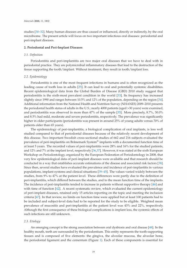
another system probably related to bone resorption in periodontitis and peri-implantitis. Normally,the mechanisms that regulate bone deposition and resorption during remodeling are mediatedthrough the refined equilibrium between the expression of two molecules: receptor activator ofNF-κB ligand (RANKL) and osteoprotegerin (OPG) [25]. Indeed, RANKL, produced by severalcell types, interacts with its receptor, RANK, located on the membrane of osteoclast precursors.This interaction allows them to finish their differentiation into active osteoclasts that will resorbthe alveolar bone. OPG is a soluble RANKL receptor that is secreted by osteoblasts and, at highconcentrations, prevents RANK–RANKL interaction and limits bone resorption. OPG formation isregulated by transforming growth factor β, and RANKL is induced by pro-inflammatory cytokineslike IL-1β and TNF-α. Together, this demonstrates how the production of pro-inflammatory mediatorscan influence the RANKL/OPG ratio and contribute to periodontitis and peri-implantitis (Figure 5).
Figure 5. The NF-κB model of bone resorption during periodontal and peri-implant diseases [25].
Periodontitis and peri-implantitis also involve the destruction of the connective tissues includingcollagens, proteoglycans and other components of the extracellular matrix. The degradation ofthis extracellular matrix is performed by matrix metalloproteinases, a group of enzymes includingcollagenases. They are released locally by immune cells (macrophages or PMNs) or resident tissue cells(mostly gingival fibroblasts, because of their high number) [75]. All these immune and inflammatoryreactions that lead to periodontal and peri-implant diseases are induced by the microorganisms thatdevelop on enamel and titanium surfaces as microbial biofilms in close contact with the junctional andsulcular epithelia of their host. The biofilm mode of growth of dental plaque is likely to influence thepathogenicity of oral microbes.
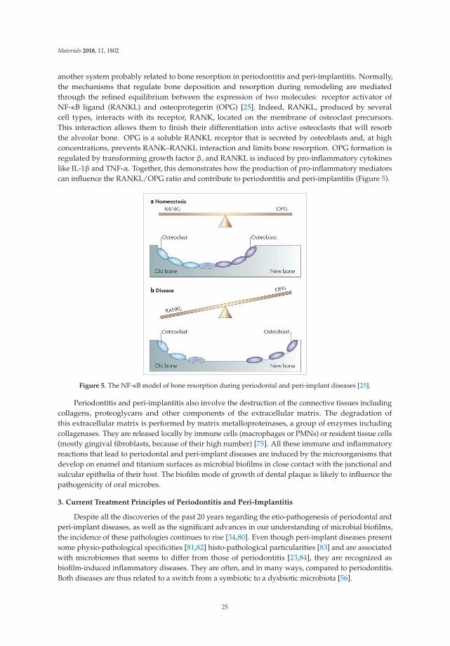
3. Current Treatment Principles of Periodontitis and Peri-Implantitis
Despite all the discoveries of the past 20 years regarding the etio-pathogenesis of periodontal andperi-implant diseases, as well as the significant advances in our understanding of microbial biofilms,the incidence of these pathologies continues to rise [34,80]. Even though peri-implant diseases presentsome physio-pathological specificities [81,82] histo-pathological particularities [83] and are associatedwith microbiomes that seems to differ from those of periodontitis [23,84], they are recognized asbiofilm-induced inflammatory diseases. They are often, and in many ways, compared to periodontitis.Both diseases are thus related to a switch from a symbiotic to a dysbiotic microbiota [56].
25
Materials 2018, 11, 1802
The main objective of peri-implantitis treatment is hence anti-infective as it is for periodontitis [85].Control of the subgingival dysbiotic dental biofilm to restore homeostasis between the microbialcommunity and its host remains the main purpose of currently available clinical treatments for thesepathologies. This primarily involves giving instructions for proper oral hygiene, as well as nonsurgicalmechanical debridement of the periodontal and peri-implant pockets. If performed carefully, thesenoninvasive mechanical therapeutic approaches most often allow the control of inflammation anddisease in periodontitis [86]. Unfortunately, for advanced lesions with probing pocket depths of≥7 mm, these treatments are less efficient, with about 15% showing no improvement [87]. Results ofsuch treatment on furcation-involved teeth are also less beneficial, requiring more aggressive (surgicalflaps) or alternative anti-infective approaches [88].
Few studies have investigated the efficacy of treatments of peri-implantitis lesions, but recentclinical trials and systematic reviews have shown that a nonsurgical mechanical approach is notsufficient for controlling the disease [89–93]. The relative failure of mechanical treatments in theseclinical situations can be related to the fact that the disinfection is insufficient, leading to recolonizationof the affected pocket and continued disease progression. Some local factors, such as a deep pocket,unfavorable root anatomy, or a rough surface threads, can explain the difficulty of achieving completeand efficient mechanical debridement. The recolonization of the periodontal or peri-implant pocketby a dysbiotic community can also come from pathogens that had previously infiltrated the dentintubuli [94] or the periodontal/peri-implant tissues [95]. To improve clinical results, some authorsproposed the use of conventional antibiotics or local antiseptics as adjunctive methods to themechanical debridement of diseased pockets. Indeed, the clinical benefits of antibiotics used in thisway have been demonstrated, and their use is now recommended for the treatment of aggressiveperiodontitis [96]. The use of antibiotics has also been suggested for severe periodontitis, though onlywhen necessary, as the body of evidence for this is weaker [97]. However, considering the increase inincidence of microbial resistance (and the mild risk of the patient developing an allergy), the use ofantibiotics should be kept to a minimum.
Subgingival irrigation as a nonsurgical treatment of periodontal and peri-implant diseasesremains controversial [98], with a lack of randomized controlled clinical trials, and data that donot allow the discrimination between the relative efficacy of various available methods [99]. Therefore,the development of new strategies to better treat severe periodontitis and peri-implantitis is stillneeded. To this end, two distinct approaches can be considered. The first would try to improve thehost’s immune/inflammatory response to the microbial challenge. The second would investigate newpre-clinical and clinical strategies to control more efficiently the periodontal and peri-implant biofilmsand pathogens associated with periodontitis and peri-implantitis.
4. New or Recent Antibiofilm Strategies
It is estimated that 99% of bacteria on earth live in biofilm aggregates [100] and most infectiousdiseases (65%) are related to the development of such sessile communities [101]. To counteract thenatural resistance/tolerance of microbial biofilms against antimicrobial agents and to mitigate theirpathologic consequences, new strategies are being considered and studied. They can be classified intofour main categories according to Bjarnsholt et al. [102]: (a) prevention, (b) weakening, (c) disruption,and (d) killing (Figure 6).
The first approach, (a), aims to prevent biofilm formation through antibiotic prophylaxis orby modifying surface characteristics using antimicrobial or anti-adhesive coatings. For instance,silver nanoparticles on titanium surfaces are currently being evaluated for use in dentistry [103].
Weakening, (b), refers to interfering with signaling molecules, virulence factors and/orbiofilm-forming properties to make the biofilm more susceptible to conventional antimicrobial agentsand to the natural host defense system. To achieve this, quorum sensing inhibitors, inhibitors of smallRNAs (messengers involved in biofilm formation), specific antibodies against virulence factors, ormetabolically inactive metal ions that can interfere with iron metabolism are being tested in vitro [102].
26

Materials 2018, 11, 1802
Figure 6. The four anti-biofilm strategies. (Adapted from [102]).
The third antibiofilm approach, (c), aims to disorganize the biofilm structure in order to disruptthe communication network between its cells and to make them more susceptible to antimicrobials.For example, Alhede et al. [104] showed that vortexing Pseudomonas aeruginosa biofilms was a valuablein vitro method of increasing their sensitivity towards tobramycin. Another method for the samepurpose is to use biological molecules such as enzymes (DNAses or dispersin B) that targetthe extracellular matrix components of the biofilm that are responsible, in part, for its cohesiveproperty [105,106]. Nevertheless, the most efficient method for treating a biofilm infection is bymechanically or surgically removing the biofilm. Two basic examples of biofilm removal are toothbrushing and scaling, and root planning. These mechanical methods are the first choice when thecontaminated surface can be accessed. Unfortunately, this is not always possible and innovativeantibiofilm approaches need to be developed.
The fourth strategy for controlling microbial aggregates, (d), is by killing biofilm cells byspecific and/or nonspecific anti-infective means. New synergistic approaches involving smallmolecules (2-aminoimidazoles) capable of dispersing biofilm bacteria, and conventional antibiotics,have been proposed to better combat biofilm aggregates [107]. Furthermore, the use of bacteriophages(viruses that contaminate bacterial cells) [108], antimicrobial peptides [109] or photodynamictherapy [110] have also been investigated in the past decade as innovative antibiofilm strategies.Finally, among the numerous antibiofilm approaches that have been suggested, an interesting electricalenhancement of the effects of several antibiotics and industrial biocides have been described againstdifferent types of bacterial biofilms [111–113]. Electric currents have also been reported to havea bactericidal effect [114] in addition to detaching adherent bacteria [115] and preventing theiradhesion [116]. The mechanisms by which these phenomena occur have not been elucidated in detail,but may be related to a better diffusion of antimicrobials through the biofilm [117], the electrolyticgeneration of oxygen [118] or the electrochemical formation of antimicrobial oxidants [119]. Electriccurrents might also lead to the degradation of the bacterial membrane, resulting in the leakageof the cytoplasmic constituents [114]. They could potentially have another physical effect on thecharged extracellular matrix, which would induce a weakening of its structure. These antibiofilm
27

Materials 2018, 11, 1802
phenomena, which have been described as bioelectric and electricidal effects, could be of great interestto the field of oral care, particularly in the treatment of advanced or refractory periodontal andperi-implant infections.
5. Concluding Remarks
Periodontitis and peri-implantitis are two major biofilm-induced inflammatory diseases that leadto the loss of teeth and oral implants if left untreated. These diseases are also associated with systemicconditions such as diabetes or cardiovascular diseases. Unfortunately, currently available treatmentsare not always successful, so innovative antibiofilm approaches need to be developed. Many strategieshave been developed in that way and tested in vitro, as outlined above, but their clinical applicabilityis sometimes difficult. Considerable research efforts are needed, and additional anti-inflammatoryapproaches should be investigated to improve treatment efficacy.
Funding: This research received no external funding.
Acknowledgments: The authors are very grateful to Professor Gaëtane Leloup (Université catholique de Louvain,Belgium), to Professor Anton Sculean (Bern Universität, Switzerland) and to Professor Henri Tenenbaum(Université de Strasbourg, France) for their precious advice and discussions.
Conflicts of Interest: The authors declare no conflicts of interest.
References
1. Hooper, L.V.; Gordon, J.I. Commensal host-bacterial relationships in the gut. Science 2001, 292, 1115–1118.[CrossRef] [PubMed]
2. Maynard, C.L.; Elson, C.O.; Hatton, R.D.; Weaver, C.T. Reciprocal interactions of the intestinal microbiotaand immune system. Nature 2012, 489, 231–241. [CrossRef] [PubMed]
3. Candela, M.; Maccaferri, S.; Turroni, S.; Carnevali, P.; Brigidi, P. Functional intestinal microbiome,new frontiers in prebiotic design. Int. J. Food Microbiol. 2010, 140, 93–101. [CrossRef] [PubMed]
4. Ley, R.E.; Peterson, D.A.; Gordon, J.I. Ecological and evolutionary forces shaping microbial diversity in thehuman intestine. Cell 2006, 124, 837–848. [CrossRef] [PubMed]
5. Backhed, F.; Ley, R.E.; Sonnenburg, J.L.; Peterson, D.A.; Gordon, J.I. Host-bacterial mutualism in the humanintestine. Science 2005, 307, 1915–1920. [CrossRef] [PubMed]
6. Whitman, W.B.; Coleman, D.C.; Wiebe, W.J. Prokaryotes: The unseen majority. Proc. Natl. Acad. Sci. USA1998, 95, 6578–6583. [CrossRef] [PubMed]
7. Moore, W.E.; Moore, L.V. The bacteria of periodontal diseases. Periodontology 2000 1994, 5, 66–77. [CrossRef][PubMed]
8. Wade, W.G. The oral microbiome in health and disease. Pharmacol. Res. 2013, 69, 137–143. [CrossRef][PubMed]
9. Dewhirst, F.E.; Chen, T.; Izard, J.; Paster, B.J.; Tanner, A.C.; Yu, W.H.; Lakshmanan, A.; Wade, W.G. The humanoral microbiome. J. Bacteriol. 2010, 192, 5002–5017. [CrossRef] [PubMed]
10. Socransky, S.S.; Haffajee, A.D. Periodontal microbial ecology. Periodontology 2000 2005, 38, 135–187.[CrossRef] [PubMed]
11. Sullivan, Å.; Edlund, C.; Nord, C.E. Effect of antimicrobial agents on the ecological balance of humanmicroflora. Lancet Infect. Dis. 2001, 1, 101–114. [CrossRef]
12. Lundberg, J.M. An audience with... Jan M. Lundberg. Nat. Rev. Drug Discov. 2005, 4, 452. [CrossRef][PubMed]
13. Kapil, V.; Milsom, A.B.; Okorie, M.; Maleki-Toyserkani, S.; Akram, F.; Rehman, F.; Arghandawi, S.; Pearl, V.;Benjamin, N.; Loukogeorgakis, S.; et al. Inorganic nitrate supplementation lowers blood pressure in humans:Role for nitrite-derived NO. Hypertension 2010, 56, 274–281. [CrossRef] [PubMed]
14. Doel, J.J.; Hector, M.P.; Amirtham, C.V.; Al-Anzan, L.A.; Benjamin, N.; Allaker, R.P. Protective effect ofsalivary nitrate and microbial nitrate reductase activity against caries. Eur. J. Oral Sci. 2004, 112, 424–428.[CrossRef] [PubMed]
15. Aas, J.A.; Paster, B.J.; Stokes, L.N.; Olsen, I.; Dewhirst, F.E. Defining the normal bacterial flora of theoral cavity. J. Clin. Microbiol. 2005, 43, 5721–5732. [CrossRef] [PubMed]
28

Materials 2018, 11, 1802
16. Socransky, S.S.; Haffajee, A.D.; Cugini, M.A.; Smith, C.; Kent, R.L., Jr. Microbial complexes in subgingivalplaque. J. Clin. Periodontol. 1998, 25, 134–144. [CrossRef] [PubMed]
17. Abusleme, L.; Dupuy, A.K.; Dutzan, N.; Silva, N.; Burleson, J.A.; Strausbaugh, L.D.; Gamonal, J.; Diaz, P.I.The subgingival microbiome in health and periodontitis and its relationship with community biomass andinflammation. ISME J. 2013, 7, 1016–1025. [CrossRef] [PubMed]
18. Mager, D.L.; Ximenez-Fyvie, L.A.; Haffajee, A.D.; Socransky, S.S. Distribution of selected bacterial species onintraoral surfaces. J. Clin. Periodontol. 2003, 30, 644–654. [CrossRef] [PubMed]
19. Paster, B.J.; Olsen, I.; Aas, J.A.; Dewhirst, F.E. The breadth of bacterial diversity in the human periodontalpocket and other oral sites. Periodontology 2000 2006, 42, 80–87. [CrossRef] [PubMed]
20. Mason, M.R.; Preshaw, P.M.; Nagaraja, H.N.; Dabdoub, S.M.; Rahman, A.; Kumar, P.S. The subgingivalmicrobiome of clinically healthy current and never smokers. ISME J 2015, 9, 268–272. [CrossRef] [PubMed]
21. Griffen, A.L.; Beall, C.J.; Campbell, J.H.; Firestone, N.D.; Kumar, P.S.; Yang, Z.K.; Podar, M.; Leys, E.J. Distinctand complex bacterial profiles in human periodontitis and health revealed by 16S pyrosequencing. ISME J.2012, 6, 1176–1185. [CrossRef] [PubMed]
22. Zaura, E.; Mira, A. Editorial: The oral microbiome in an ecological perspective. Front. Cell. Infect. Microbiol.2015, 5, 39. [CrossRef] [PubMed]
23. Kumar, P.S.; Mason, M.R.; Brooker, M.R.; O’Brien, K. Pyrosequencing reveals unique microbial signaturesassociated with healthy and failing dental implants. J. Clin. Periodontol. 2012, 39, 425–433. [CrossRef][PubMed]
24. Paster, B.J.; Boches, S.K.; Galvin, J.L.; Ericson, R.E.; Lau, C.N.; Levanos, V.A.; Sahasrabudhe, A.; Dewhirst, F.E.Bacterial diversity in human subgingival plaque. J. Bacteriol. 2001, 183, 3770–3783. [CrossRef] [PubMed]
25. Darveau, R.P. Periodontitis: A polymicrobial disruption of host homeostasis. Nat. Rev. Microbiol. 2010, 8,481–490. [CrossRef] [PubMed]
26. Duran-Pinedo, A.E.; Frias-Lopez, J. Beyond microbial community composition: Functional activities of theoral microbiome in health and disease. Microbes Infect. 2015, 17, 505–516. [CrossRef] [PubMed]
27. Whitmore, S.E.; Lamont, R.J. Oral bacteria and cancer. PLoS Pathog. 2014, 10, e1003933. [CrossRef] [PubMed]28. Homann, N.; Tillonen, J.; Rintamaki, H.; Salaspuro, M.; Lindqvist, C.; Meurman, J.H. Poor dental status
increases acetaldehyde production from ethanol in saliva: A possible link to increased oral cancer risk amongheavy drinkers. Oral Oncol. 2001, 37, 153–158. [CrossRef]
29. Parahitiyawa, N.B.; Jin, L.J.; Leung, W.K.; Yam, W.C.; Samaranayake, L.P. Microbiology of odontogenicbacteremia: Beyond endocarditis. Clin. Microbiol. Rev. 2009, 22, 46–64. [CrossRef] [PubMed]
30. Seymour, G.J.; Ford, P.J.; Cullinan, M.P.; Leishman, S.; Yamazaki, K. Relationship between periodontalinfections and systemic disease. Clin. Microbiol. Infect. 2007, 13, 3–10. [CrossRef] [PubMed]
31. Rautemaa, R.; Lauhio, A.; Cullinan, M.P.; Seymour, G.J. Oral infections and systemic disease—An emergingproblem in medicine. Clin. Microbiol. Infect. 2007, 13, 1041–1047. [CrossRef] [PubMed]
32. Rakoto-Alson, S.; Tenenbaum, H.; Davideau, J.L. Periodontal diseases, preterm births, and low birth weight:Findings from a homogeneous cohort of women in Madagascar. J. Periodontol. 2010, 81, 205–213. [CrossRef][PubMed]
33. Marcenes, W.; Kassebaum, N.J.; Bernabe, E.; Flaxman, A.; Naghavi, M.; Lopez, A.; Murray, C.J. Global burdenof oral conditions in 1990-2010: A systematic analysis. J. Dent. Res. 2013, 92, 592–597. [CrossRef] [PubMed]
34. Kassebaum, N.J.; Bernabe, E.; Dahiya, M.; Bhandari, B.; Murray, C.J.; Marcenes, W. Global burden of severeperiodontitis in 1990–2010: A systematic review and meta-regression. J. Dent. Res. 2014, 93, 1045–1053.[CrossRef] [PubMed]
35. Eke, P.I.; Dye, B.A.; Wei, L.; Thornton-Evans, G.O.; Genco, R.J.; Cdc Periodontal Disease SurveillanceWorkgroup. Prevalence of periodontitis in adults in the United States: 2009 and 2010. J. Dent. Res. 2012, 91,914–920. [CrossRef] [PubMed]
36. Fransson, C.; Lekholm, U.; Jemt, T.; Berglundh, T. Prevalence of subjects with progressive bone loss atimplants. Clin. Oral Implants Res. 2005, 16, 440–446. [CrossRef] [PubMed]
37. Roos-Jansaker, A.M.; Lindahl, C.; Renvert, H.; Renvert, S. Nine- to fourteen-year follow-up of implanttreatment. Part II: Presence of peri-implant lesions. J. Clin. Periodontol. 2006, 33, 290–295. [CrossRef] [PubMed]
38. Lindhe, J.; Meyle, J.; Group D of European Workshop on Periodontology. Peri-implant diseases: ConsensusReport of the Sixth European Workshop on Periodontology. J. Clin. Periodontol. 2008, 35, 282–285. [CrossRef][PubMed]
29
Materials 2018, 11, 1802
39. Koldsland, O.C.; Scheie, A.A.; Aass, A.M. Prevalence of peri-implantitis related to severity of the diseasewith different degrees of bone loss. J. Periodontol. 2010, 81, 231–238. [CrossRef] [PubMed]
40. Marrone, A.; Lasserre, J.; Bercy, P.; Brecx, M.C. Prevalence and risk factors for peri-implant disease in Belgianadults. Clin. Oral Implants Res. 2013, 24, 934–940. [CrossRef] [PubMed]
41. Mir-Mari, J.; Mir-Orfila, P.; Figueiredo, R.; Valmaseda-Castellon, E.; Gay-Escoda, C. Prevalence ofperi-implant diseases. A cross-sectional study based on a private practice environment. J. Clin. Periodontol.2012, 39, 490–494. [CrossRef] [PubMed]
42. Meijer, H.J.; Raghoebar, G.M.; de Waal, Y.C.; Vissink, A. Incidence of peri-implant mucositis andperi-implantitis in edentulous patients with an implant-retained mandibular overdenture during a 10-yearfollow-up period. J. Clin. Periodontol. 2014, 41, 1178–1183. [CrossRef] [PubMed]
43. Schwarz, F.; Becker, K.; Sahm, N.; Horstkemper, T.; Rousi, K.; Becker, J. The prevalence of peri-implantdiseases for two-piece implants with an internal tube-in-tube connection: A cross-sectional analysis of 512implants. Clin. Oral Implants Res. 2015. [CrossRef] [PubMed]
44. Derks, J.; Schaller, D.; Hakansson, J.; Wennstrom, J.L.; Tomasi, C.; Berglundh, T. Effectiveness of ImplantTherapy Analyzed in a Swedish Population: Prevalence of Peri-implantitis. J. Dent. Res. 2016, 95, 43–49.[CrossRef] [PubMed]
45. Tenenbaum, H.; Bogen, O.; Severac, F.; Elkaim, R.; Davideau, J.L.; Huck, O. Long-term prospective cohortstudy on dental implants: Clinical and microbiological parameters. Clin. Oral Implants Res. 2016, 28, 86–94.[CrossRef] [PubMed]
46. Costa, F.O.; Takenaka-Martinez, S.; Cota, L.O.; Ferreira, S.D.; Silva, G.L.; Costa, J.E. Peri-implant disease insubjects with and without preventive maintenance: A 5-year follow-up. J. Clin. Periodontol. 2012, 39, 173–181.[CrossRef] [PubMed]
47. Derks, J.; Tomasi, C. Peri-implant health and disease. A systematic review of current epidemiology. J. Clin.Periodontol. 2015, 42, S158–S171. [CrossRef] [PubMed]
48. Jiao, Y.; Hasegawa, M.; Inohara, N. The Role of Oral Pathobionts in Dysbiosis during PeriodontitisDevelopment. J. Dent. Res. 2014, 93, 539–546. [CrossRef] [PubMed]
49. Marsh, P.D.; Do, T.; Beighton, D.; Devine, D.A. Influence of saliva on the oral microbiota. Periodontology 20002016, 70, 80–92. [CrossRef] [PubMed]
50. Curtis, M. The oral commensal microbiota bites back through Nod1. Cell Host Microbe 2013, 13, 503–505.[CrossRef] [PubMed]
51. Schultz-Haudt, S.; Bruce, M.A.; Bibby, B.G. Bacterial factors in nonspecific gingivitis. J. Dent. Res. 1954, 33,454–458. [CrossRef] [PubMed]
52. Socransky, S.S. Microbiology of periodontal disease—Present status and future considerations. J. Periodontol.1977, 48, 497–504. [CrossRef] [PubMed]
53. Loesche, W.J. Clinical and microbiological aspects of chemotherapeutic agents used according to the specificplaque hypothesis. J. Dent. Res. 1979, 58, 2404–2412. [CrossRef] [PubMed]
54. Faveri, M.; Figueiredo, L.C.; Shibli, J.A.; Perez-Chaparro, P.J.; Feres, M. Microbiological diversity ofperi-implantitis biofilms. Adv. Exp. Med. Biol. 2015, 830, 85–96. [CrossRef] [PubMed]
55. Kumar, P.S.; Griffen, A.L.; Moeschberger, M.L.; Leys, E.J. Identification of candidate periodontal pathogensand beneficial species by quantitative 16S clonal analysis. J. Clin. Microbiol. 2005, 43, 3944–3955. [CrossRef][PubMed]
56. Hajishengallis, G.; Lamont, R.J. Beyond the red complex and into more complexity: The polymicrobialsynergy and dysbiosis (PSD) model of periodontal disease etiology. Mol. Oral Microbiol. 2012, 27, 409–419.[CrossRef] [PubMed]
57. Hajishengallis, G. The inflammophilic character of the periodontitis-associated microbiota. Mol. OralMicrobiol. 2014, 29, 248–257. [CrossRef] [PubMed]
58. Maekawa, T.; Krauss, J.L.; Abe, T.; Jotwani, R.; Triantafilou, M.; Triantafilou, K.; Hashim, A.; Hoch, S.;Curtis, M.A.; Nussbaum, G.; et al. Porphyromonas gingivalis manipulates complement and TLR signaling touncouple bacterial clearance from inflammation and promote dysbiosis. Cell Host Microbe 2014, 15, 768–778.[CrossRef] [PubMed]
59. Hajishengallis, G.; Lamont, R.J. Breaking bad: Manipulation of the host response by Porphyromonasgingivalis. Eur. J. Immunol. 2014, 44, 328–338. [CrossRef] [PubMed]
30

Materials 2018, 11, 1802
60. Hajishengallis, G.; Darveau, R.P.; Curtis, M.A. The keystone-pathogen hypothesis. Nat. Rev. Microbiol. 2012,10, 717–725. [CrossRef] [PubMed]
61. Theilade, E.; Wright, W.H.; Jensen, S.B.; Loe, H. Experimental gingivitis in man. II. A longitudinal clinicaland bacteriological investigation. J. Periodontal. Res. 1966, 1, 1–13. [CrossRef] [PubMed]
62. Loe, H.; Theilade, E.; Jensen, S.B. Experimental Gingivitis in Man. J. Periodontol. 1965, 36, 177–187. [CrossRef][PubMed]
63. Zitzmann, N.U.; Berglundh, T.; Marinello, C.P.; Lindhe, J. Experimental peri-implant mucositis in man.J. Clin. Periodontol. 2001, 28, 517–523. [CrossRef] [PubMed]
64. Kolenbrander, P.E.; Palmer, R.J., Jr.; Rickard, A.H.; Jakubovics, N.S.; Chalmers, N.I.; Diaz, P.I. Bacterialinteractions and successions during plaque development. Periodontology 2000 2006, 42, 47–79. [CrossRef][PubMed]
65. Listgarten, M.A.; Hellden, L. Relative distribution of bacteria at clinically healthy and periodontally diseasedsites in humans. J. Clin. Periodontol. 1978, 5, 115–132. [CrossRef] [PubMed]
66. Diaz, P.I.; Chalmers, N.I.; Rickard, A.H.; Kong, C.; Milburn, C.L.; Palmer, R.J., Jr.; Kolenbrander, P.E. Molecularcharacterization of subject-specific oral microflora during initial colonization of enamel. Appl. Environ.Microbiol. 2006, 72, 2837–2848. [CrossRef] [PubMed]
67. Li, J.; Helmerhorst, E.J.; Leone, C.W.; Troxler, R.F.; Yaskell, T.; Haffajee, A.D.; Socransky, S.S.; Oppenheim, F.G.Identification of early microbial colonizers in human dental biofilm. J. Appl. Microbiol. 2004, 97, 1311–1318.[CrossRef] [PubMed]
68. Quirynen, M.; Vogels, R.; Pauwels, M.; Haffajee, A.D.; Socransky, S.S.; Uzel, N.G.; van Steenberghe, D. InitialSubgingival Colonization of ‘Pristine’ Pockets. J. Dent. Res. 2005, 84, 340–344. [CrossRef] [PubMed]
69. Marsh, P.D. Microbial ecology of dental plaque and its significance in health and disease. Adv. Dent. Res.1994, 8, 263–271. [CrossRef] [PubMed]
70. Van Dyke, T.E.; Serhan, C.N. Resolution of inflammation: A new paradigm for the pathogenesis of periodontaldiseases. J. Dent. Res. 2003, 82, 82–90. [CrossRef] [PubMed]
71. Cekici, A.; Kantarci, A.; Hasturk, H.; Van Dyke, T.E. Inflammatory and immune pathways in the pathogenesisof periodontal disease. Periodontology 2000 2014, 64, 57–80. [CrossRef] [PubMed]
72. Ricklin, D.; Hajishengallis, G.; Yang, K.; Lambris, J.D. Complement: A key system for immune surveillanceand homeostasis. Nat. Immunol. 2010, 11, 785–797. [CrossRef] [PubMed]
73. Benakanakere, M.; Abdolhosseini, M.; Hosur, K.; Finoti, L.S.; Kinane, D.F. TLR2 promoter hypermethylationcreates innate immune dysbiosis. J. Dent. Res. 2015, 94, 183–191. [CrossRef] [PubMed]
74. Gay, N.J.; Symmons, M.F.; Gangloff, M.; Bryant, C.E. Assembly and localization of Toll-like receptor signallingcomplexes. Nat. Rev. Immunol. 2014, 14, 546–558. [CrossRef] [PubMed]
75. Page, R.C.; Offenbacher, S.; Schroeder, H.E.; Seymour, G.J.; Kornman, K.S. Advances in the pathogenesis ofperiodontitis: Summary of developments, clinical implications and future directions. Periodontology 20001997, 14, 216–248. [CrossRef] [PubMed]
76. Tonetti, M.S.; Imboden, M.A.; Lang, N.P. Neutrophil migration into the gingival sulcus is associated withtransepithelial gradients of interleukin-8 and ICAM-1. J. Periodontol. 1998, 69, 1139–1147. [CrossRef][PubMed]
77. Hajishengallis, G.; Moutsopoulos, N.M. Etiology of leukocyte adhesion deficiency-associated periodontitisrevisited: Not a raging infection but a raging inflammatory response. Expert Rev. Clin. Immunol. 2014, 10,973–975. [CrossRef] [PubMed]
78. Jaillon, S.; Galdiero, M.R.; Del Prete, D.; Cassatella, M.A.; Garlanda, C.; Mantovani, A. Neutrophils in innateand adaptive immunity. Semin. Immunopathol. 2013, 35, 377–394. [CrossRef] [PubMed]
79. White, P.C.; Chicca, I.J.; Cooper, P.R.; Milward, M.R.; Chapple, I.L. Neutrophil Extracellular Traps inPeriodontitis: A Web of Intrigue. J. Dent. Res. 2016, 95, 26–34. [CrossRef] [PubMed]
80. Tarnow, D.P. Increasing Prevalence of Peri-implantitis: How Will We Manage? J. Dent. Res. 2016, 95, 7–8.[CrossRef] [PubMed]
81. Lang, N.P.B.T. Working Group 4 of Seventh European Workshop on P. 2011. Periimplant diseases: Where arewe now?—Consensus of the Seventh European Workshop on Periodontology. J. Clin. Periodontol. 2011, 38,178–181. [CrossRef] [PubMed]
31

Materials 2018, 11, 1802
82. Becker, S.T.; Beck-Broichsitter, B.E.; Graetz, C.; Dorfer, C.E.; Wiltfang, J.; Hasler, R. Peri-implantitis versusperiodontitis: Functional differences indicated by transcriptome profiling. Clin. Implant Dent. Relat. Res.2014, 16, 401–411. [CrossRef] [PubMed]
83. Carcuac, O.; Berglundh, T. Composition of human peri-implantitis and periodontitis lesions. J. Dent. Res.2014, 93, 1083–1088. [CrossRef] [PubMed]
84. Lafaurie, G.I.; Sabogal, M.A.; Castillo, D.M.; Rincón, M.V.; Gómez, L.A.; Lesmes, Y.A.; Chambrone, L.Microbiome and microbial biofilm profiles of peri-implantitis: A systematic review. J. Periodontol. 2017, 88,1066–1089. [CrossRef] [PubMed]
85. Lasserre, J.F.; Toma, S.; Bourgeois, T.; El Khatmaoui, H.; Marichal, E.; Brecx, M.C. Influence of low directelectric currents and chlorhexidine upon human dental biofilms. Clin. Exp. Dent. Res. 2016, 2, 146–154.[CrossRef] [PubMed]
86. Badersten, A.; Nilveus, R.; Egelberg, J. Effect of nonsurgical periodontal therapy. I. Moderately advancedperiodontitis. J. Clin. Periodontol. 1981, 8, 57–72. [CrossRef] [PubMed]
87. Badersten, A.; Nilveus, R.; Egelberg, J. Effect of nonsurgical periodontal therapy. II. Severely advancedperiodontitis. J. Clin. Periodontol. 1984, 11, 63–76. [CrossRef] [PubMed]
88. Adriaens, P.A.; Adriaens, L.M. Effects of nonsurgical periodontal therapy on hard and soft tissues.Periodontology 2000 2004, 36, 121–145. [CrossRef] [PubMed]
89. Karring, E.S.; Stavropoulos, A.; Ellegaard, B.; Karring, T. Treatment of peri-implantitis by the Vector system.Clin. Oral Implants Res. 2005, 16, 288–293. [CrossRef] [PubMed]
90. Renvert, S.; Roos-Jansaker, A.M.; Claffey, N. Non-surgical treatment of peri-implant mucositis andperi-implantitis: A literature review. J. Clin. Periodontol. 2008, 35, 305–315. [CrossRef] [PubMed]
91. Renvert, S.; Samuelsson, E.; Lindahl, C.; Persson, G.R. Mechanical non-surgical treatment of peri-implantitis:A double-blind randomized longitudinal clinical study. I: Clinical results. J. Clin. Periodontol. 2009, 36,604–609. [CrossRef] [PubMed]
92. Renvert, S.; Lindahl, C.; Roos Jansaker, A.M.; Persson, G.R. Treatment of peri-implantitis using an Er:YAGlaser or an air-abrasive device: A randomized clinical trial. J. Clin. Periodontol. 2011, 38, 65–73. [CrossRef][PubMed]
93. Schwarz, F.; Becker, K.; Renvert, S. Efficacy of air polishing for the non-surgical treatment of peri-implantdiseases: A systematic review. J. Clin. Periodontol. 2015. [CrossRef] [PubMed]
94. Adriaens, P.A.; Edwards, C.A.; De Boever, J.A.; Loesche, W.J. Ultrastructural observations on bacterialinvasion in cementum and radicular dentin of periodontally diseased human teeth. J. Periodontol. 1988, 59,493–503. [CrossRef] [PubMed]
95. Lamont, R.J.; Chan, A.; Belton, C.M.; Izutsu, K.T.; Vasel, D.; Weinberg, A. Porphyromonas gingivalis invasionof gingival epithelial cells. Infect. Immun. 1995, 63, 3878–3885. [PubMed]
96. Keestra, J.A.; Grosjean, I.; Coucke, W.; Quirynen, M.; Teughels, W. Non-surgical periodontal therapywith systemic antibiotics in patients with untreated aggressive periodontitis: A systematic review andmeta-analysis. J. Periodontal Res. 2015, 50, 689–706. [CrossRef] [PubMed]
97. Keestra, J.A.; Grosjean, I.; Coucke, W.; Quirynen, M.; Teughels, W. Non-surgical periodontal therapy withsystemic antibiotics in patients with untreated chronic periodontitis: A systematic review and meta-analysis.J. Periodontal Res. 2015, 50, 294–314. [CrossRef] [PubMed]
98. Greenstein, G.; Research, S.; Therapy Committee of the American Academy of, P. Position paper: The role ofsupra- and subgingival irrigation in the treatment of periodontal diseases. J. Periodontol. 2005, 76, 2015–2027.[CrossRef] [PubMed]
99. Esposito, M.; Grusovin, M.G.; Worthington, H.V. Interventions for replacing missing teeth: Treatment ofperi-implantitis. Cochrane Database Syst. Rev. 2012, 1, Cd004970. [CrossRef] [PubMed]
100. Potera, C. Studying slime. Environ. Health Perspect. 1998, 106, A604–A606. [CrossRef] [PubMed]101. Potera, C. Forging a link between biofilms and disease. Science 1999, 283, 1837–1839. [CrossRef] [PubMed]102. Bjarnsholt, T.; Ciofu, O.; Molin, S.; Givskov, M.; Hoiby, N. Applying insights from biofilm biology to drug
development—Can a new approach be developed? Nat. Rev. Drug Discov. 2013, 12, 791–808. [CrossRef][PubMed]
32

Materials 2018, 11, 1802
103. Zhong, X.; Song, Y.; Yang, P.; Wang, Y.; Jiang, S.; Zhang, X.; Li, C. Titanium Surface Primingwith Phase-Transited Lysozyme to Establish a Silver Nanoparticle-Loaded Chitosan/Hyaluronic AcidAntibacterial Multilayer via Layer-by-Layer Self-Assembly. PLoS ONE 2016, 11, e0146957. [CrossRef][PubMed]
104. Alhede, M.; Kragh, K.N.; Qvortrup, K.; Allesen-Holm, M.; van Gennip, M.; Christensen, L.D.; Jensen, P.O.;Nielsen, A.K.; Parsek, M.; Wozniak, D.; et al. Phenotypes of non-attached Pseudomonas aeruginosaaggregates resemble surface attached biofilm. PLoS ONE 2011, 6, e27943. [CrossRef] [PubMed]
105. Shields, R.C.; Mokhtar, N.; Ford, M.; Hall, M.J.; Burgess, J.G.; ElBadawey, M.R.; Jakubovics, N.S. Efficacy ofa marine bacterial nuclease against biofilm forming microorganisms isolated from chronic rhinosinusitis.PLoS ONE 2013, 8, e55339. [CrossRef] [PubMed]
106. Izano, E.A.; Wang, H.; Ragunath, C.; Ramasubbu, N.; Kaplan, J.B. Detachment and killing of Aggregatibacteractinomycetemcomitans biofilms by dispersin B and SDS. J. Dent. Res. 2007, 86, 618–622. [CrossRef] [PubMed]
107. Rogers, S.A.; Huigens, R.W., 3rd; Cavanagh, J.; Melander, C. Synergistic effects between conventionalantibiotics and 2-aminoimidazole-derived antibiofilm agents. Antimicrob. Agents Chemother. 2010, 54,2112–2118. [CrossRef] [PubMed]
108. Pires, D.P.; Oliveira, H.; Melo, L.D.; Sillankorva, S.; Azeredo, J. Bacteriophage-encoded depolymerases: Theirdiversity and biotechnological applications. Appl. Microbiol. Biotechnol. 2016, 100, 2141–2151. [CrossRef][PubMed]
109. Gorr, S.U.; Abdolhosseini, M. Antimicrobial peptides and periodontal disease. J. Clin. Periodontol. 2011, 38,126–141. [CrossRef] [PubMed]
110. Bassetti, M.; Schar, D.; Wicki, B.; Eick, S.; Ramseier, C.A.; Arweiler, N.B.; Sculean, A.; Salvi, G.E. Anti-infectivetherapy of peri-implantitis with adjunctive local drug delivery or photodynamic therapy: 12-month outcomesof a randomized controlled clinical trial. Clin. Oral Implants Res. 2014, 25, 279–287. [CrossRef] [PubMed]
111. Blenkinsopp, S.A.; Khoury, A.E.; Costerton, J.W. Electrical enhancement of biocide efficacy againstPseudomonas aeruginosa biofilms. Appl. Environ. Microbiol. 1992, 58, 3770–3773. [PubMed]
112. Jass, J.; Lappin-Scott, H.M. The efficacy of antibiotics enhanced by electrical currents against Pseudomonasaeruginosa biofilms. J. Antimicrob. Chemother. 1996, 38, 987–1000. [CrossRef] [PubMed]
113. Haddad, P.A.; Mah, T.F.; Mussivand, T. In Vitro Assessment of Electric Currents Increasing the Effectivenessof Vancomycin Against Staphylococcus epidermidis Biofilms. Artif. Organs 2015. [CrossRef]
114. del Pozo, J.L.; Rouse, M.S.; Mandrekar, J.N.; Steckelberg, J.M.; Patel, R. The electricidal effect: Reductionof Staphylococcus and pseudomonas biofilms by prolonged exposure to low-intensity electrical current.Antimicrob. Agents Chemother. 2009, 53, 41–45. [CrossRef] [PubMed]
115. Poortinga, A.T.; Smit, J.; van der Mei, H.C.; Busscher, H.J. Electric field induced desorption of bacteria froma conditioning film covered substratum. Biotechnol. Bioeng. 2001, 76, 395–399. [CrossRef] [PubMed]
116. Hong, S.H.; Jeong, J.; Shim, S.; Kang, H.; Kwon, S.; Ahn, K.H.; Yoon, J. Effect of electric currents on bacterialdetachment and inactivation. Biotechnol. Bioeng. 2008, 100, 379–386. [CrossRef] [PubMed]
117. Costerton, J.W.; Ellis, B.; Lam, K.; Johnson, F.; Khoury, A.E. Mechanism of electrical enhancement of efficacyof antibiotics in killing biofilm bacteria. Antimicrob. Agents Chemother. 1994, 38, 2803–2809. [CrossRef][PubMed]
118. Stewart, P.S.; Wattanakaroon, W.; Goodrum, L.; Fortun, S.M.; McLeod, B.R. Electrolytic generation of oxygenpartially explains electrical enhancement of tobramycin efficacy against Pseudomonas aeruginosa biofilm.Antimicrob. Agents Chemother. 1999, 43, 292–296. [PubMed]
119. Mohn, D.; Zehnder, M.; Stark, W.J.; Imfeld, T. Electrochemical disinfection of dental implants—A proof ofconcept. PLoS ONE 2011, 6, e16157. [CrossRef] [PubMed]
© 2018 by the authors. Licensee MDPI, Basel, Switzerland. This article is an open accessarticle distributed under the terms and conditions of the Creative Commons Attribution(CC BY) license (http://creativecommons.org/licenses/by/4.0/).
33

materials
Review
The A, B and C’s of Silicone Breast Implants:Anaplastic Large Cell Lymphoma, Biofilm andCapsular Contracture
Maria Mempin, Honghua Hu, Durdana Chowdhury, Anand Deva and Karen Vickery *
Faculty of Medicine and Health Sciences, Macquarie University, Macquarie Park, NSW, 2109, Australia;[email protected] (M.M.); [email protected] (H.H.); [email protected] (D.C.);[email protected] (A.D.)* Correspondence: [email protected]; Tel.: +61-298-502-773
Received: 5 November 2018; Accepted: 26 November 2018; Published: 28 November 2018
Abstract: Breast implantation either for cosmetic or reconstructive e purposes is one of the mostcommon procedures performed in plastic surgery. Biofilm infection is hypothesised to be involvedin the development of both capsular contracture and anaplastic large cell lymphoma (ALCL).Capsular contracture is one of the principal reasons for breast revision surgery and is characterisedby the tightening and hardening of the capsule surrounding the implant, and ALCL is an indolentlymphoma found only in women with textured implants. We describe the types of breast implantsavailable with regard to their surface characteristics of surface area and roughness and how this mightcontribute to capsular contracture and/or biofilm formation. The pathogenesis of capsular contractureis thought to be due to biofilm formation on the implant, which results in on-going inflammation.We describe the current research into breast implant associated ALCL and how implant propertiesmay affect its pathogenesis, with ALCL only occurring in women with textured implants.
Keywords: biofilm; breast implant; textured: capsular contracture; anaplastic large cell lymphoma;BIA-ALCL
1. Introduction
Breast implantation, either for cosmetic or reconstructive purposes, is one of the most commonprocedures performed in plastic surgery. In 2015, in the United States of America alone, more than280,000 women had breast enlargement surgery and an estimated 106,000 breast cancer patientsunderwent post-mastectomy breast reconstruction, which often involved insertion of implantablemedical devices [1].
2. Breast Implants
Basic designed silicone breast implants were first introduced in the early 1960s [2]. As eachnew “generation” of implant has been introduced, their design has undergone major improvements.Modern breast implants can be divided into categories based on implant filling (silicone or saline),surface texture (textured or smooth), and shape (round or anatomic), each of which have slightlydifferent properties [3,4].
Silicone or saline implant filling:Saline implants are sold as empty silicone elastomer shells and are filled to the appropriate volume
with sterile saline in the operating room. The silicone filling comes as either a “fluid form” that isnot cohesive enough to maintain an anatomic shape or a “form-stable” more viscous and greatercross-linked silicone gel that has cohesive properties [5]. The cohesive gel increases form stability
Materials 2018, 11, 2393; doi:10.3390/ma11122393 www.mdpi.com/journal/materials35
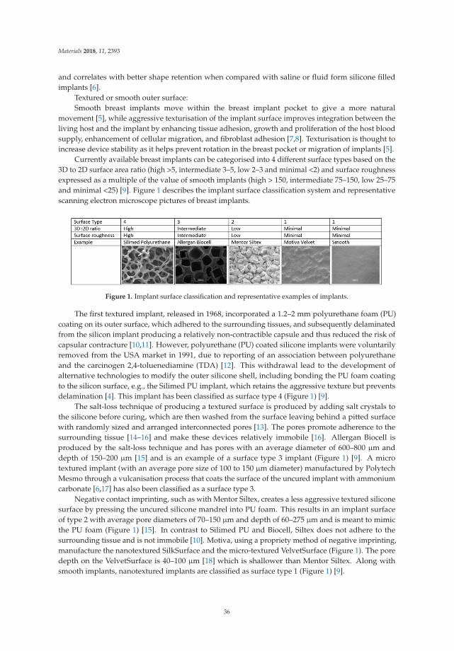
Materials 2018, 11, 2393
and correlates with better shape retention when compared with saline or fluid form silicone filledimplants [6].
Textured or smooth outer surface:Smooth breast implants move within the breast implant pocket to give a more natural
movement [5], while aggressive texturisation of the implant surface improves integration between theliving host and the implant by enhancing tissue adhesion, growth and proliferation of the host bloodsupply, enhancement of cellular migration, and fibroblast adhesion [7,8]. Texturisation is thought toincrease device stability as it helps prevent rotation in the breast pocket or migration of implants [5].
Currently available breast implants can be categorised into 4 different surface types based on the3D to 2D surface area ratio (high >5, intermediate 3–5, low 2–3 and minimal <2) and surface roughnessexpressed as a multiple of the value of smooth implants (high > 150, intermediate 75–150, low 25–75and minimal <25) [9]. Figure 1 describes the implant surface classification system and representativescanning electron microscope pictures of breast implants.
Figure 1. Implant surface classification and representative examples of implants.
The first textured implant, released in 1968, incorporated a 1.2–2 mm polyurethane foam (PU)coating on its outer surface, which adhered to the surrounding tissues, and subsequently delaminatedfrom the silicon implant producing a relatively non-contractible capsule and thus reduced the risk ofcapsular contracture [10,11]. However, polyurethane (PU) coated silicone implants were voluntarilyremoved from the USA market in 1991, due to reporting of an association between polyurethaneand the carcinogen 2,4-toluenediamine (TDA) [12]. This withdrawal lead to the development ofalternative technologies to modify the outer silicone shell, including bonding the PU foam coatingto the silicon surface, e.g., the Silimed PU implant, which retains the aggressive texture but preventsdelamination [4]. This implant has been classified as surface type 4 (Figure 1) [9].
The salt-loss technique of producing a textured surface is produced by adding salt crystals tothe silicone before curing, which are then washed from the surface leaving behind a pitted surfacewith randomly sized and arranged interconnected pores [13]. The pores promote adherence to thesurrounding tissue [14–16] and make these devices relatively immobile [16]. Allergan Biocell isproduced by the salt-loss technique and has pores with an average diameter of 600–800 μm anddepth of 150–200 μm [15] and is an example of a surface type 3 implant (Figure 1) [9]. A microtextured implant (with an average pore size of 100 to 150 μm diameter) manufactured by PolytechMesmo through a vulcanisation process that coats the surface of the uncured implant with ammoniumcarbonate [6,17] has also been classified as a surface type 3.
Negative contact imprinting, such as with Mentor Siltex, creates a less aggressive textured siliconesurface by pressing the uncured silicone mandrel into PU foam. This results in an implant surfaceof type 2 with average pore diameters of 70–150 μm and depth of 60–275 μm and is meant to mimicthe PU foam (Figure 1) [15]. In contrast to Silimed PU and Biocell, Siltex does not adhere to thesurrounding tissue and is not immobile [10]. Motiva, using a propriety method of negative imprinting,manufacture the nanotextured SilkSurface and the micro-textured VelvetSurface (Figure 1). The poredepth on the VelvetSurface is 40–100 μm [18] which is shallower than Mentor Siltex. Along withsmooth implants, nanotextured implants are classified as surface type 1 (Figure 1) [9].
36

Materials 2018, 11, 2393
3. Capsular Contracture
Complications of breast augmentation include hematoma, seroma, infection, altered nipplesensation, asymmetry, scarring, swelling, rupture, leakage and capsular contracture (CC) but CC isthought to be the most common complication and frequently requires surgical revision [19]. In 2015,43,000 implant removal procedures were reported in the United States of America [1], and the Foodand Drug Administration (FDA) reports that between 20 to 40% of augmentation patients and 40 to70% of reconstruction patients had reoperations during the first eight to ten years after receiving theirbreast implants [20]. CC is a common reason for reoperation in Australia, being responsible for 38.9%of the 5290 breast implant revisions occurring between 2012 and 2016 [21]. Surgical revisions followingCC result in poorer aesthetic outcome and a high rate of recurrence of CC [22,23].
Upon insertion of a breast implant, a foreign body reaction is induced, which is essentially anexcessive fibrotic response that encloses the implant. CC is contracture of the peri-prosthetic capsule,which is characterised by the tightening and hardening of the tissue capsule around the breast implant.CC eventually leads to distortion of the implant [24,25]. Individual studies have reported incidencerates of CC ranging from 1.3 to 45% [23,26–30]. The wide range of CC rates is attributed to differencesin follow-up times, as CC rate increases with time following implantation, as well as different type ofimplants and differing surgical techniques being used throughout the various studies [4].
The degree of CC is classified using the Baker clinical grading system which divides CC into fourgrades [31]. A grade I breast looks and feels natural, while grade II breasts have minimal contracturewhere the surgeon can tell surgery has been performed but there are no clinical symptoms. Grade IIIand IV are clinically significant and symptomatic, where grade III describes moderate contracturewith some firmness felt by the patient, and grade IV describes severe contracture that is obvious fromobservation and symptomatic in the patient [31].
With each new implant generation, the incidence of CC has decreased, although whether this isdue to implant design or better surgical technique, or a combination of both, is unclear. Historically, thetype of fill was thought to influence the development of CC. Older generation silicone gel devices werecharacterised by higher gel bleeds and rupture rates compared to current generation implants [5,32,33].The rates of CC were six-fold higher with these older devices than with devices containing low-bleedsilicone gel fillings [34] or cohesive silicone gel fill implants [22,35–39].
The benefits of textured implants in reducing CC remains controversial. Systematic reviewsof comparative clinical studies concluded texturisation may reduce the incidence of early capsularcontracture if the implant was placed under the breast glandular tissue, but had no significant effectif placed under the pectoral muscle [29,40]. Smaller comparative or split breast studies, insertingone smooth and one textured implant in the same patient, are evenly divided as to the benefit oftexturisation [41–48]. Many of these published reports lack adequate description of implant type,surgical technique, outcome assessment, and have short follow-up or the time period of follow-up isnot stated. Several early randomised controlled trials reported textured implants had lower rates ofclinically significant CC compared to smooth surface implants [42,45]. Similarly, some later prospectivetrials and metanalysis of 16 randomised controlled trials combined with two case-control studies,involving 4412 patients, have shown that smooth implants are more likely to develop CC [40,48,49].However, the follow-up of most of these studies has been less than five years. When 715 of thesepatients were followed for 10 years there was no difference in the rate of CC [23]. It is likely thatthe effect of surface technology is of some benefit but is one of many factors that impact on clinicaloutcome, and the aetiopathogenesis of CC is likely to be multifactorial.
4. Aetiopathogenesis of Capsular Contracture (CC)
In 1981, Burkhardt and co-workers [50] were the first to propose that subclinical infection led toCC. However, the lack of culture positivity in many clinical studies of CC delayed the acceptance ofthis hypothesis. The detection of a Staphylococcus epidermidis biofilm in a patient with recurrent CCled to the hypothesis that the proposed subclinical infection is due to biofilm formation on the breast
37
Materials 2018, 11, 2393
implant [51]. The presence of biofilm on implants obtained from CC patients was confirmed usingscanning electron microscopy [25]. The likelihood of bacterial isolation was increased by mincing,sonication, and broth culture of a piece of implant or tissue, rather than using a swab to collect samples.Using this improved method of culture, the authors found a significant relationship between culturepositivity (p < 0.0006) and the presence of S. epidermidis (p < 0.01) with CC. Subsequently, the degree ofBaker grade CC has been shown to directly correlate with the number of bacteria in humans [52] andin the porcine model [53]. The biofilm hypothesis helps explain the lack of culture positivity in olderstudies where sonication was not employed, as biofilm bacteria are notoriously difficult to culture [54].
An alternative, to the biofilm hypothesis is that CC is purely an immunological response (reviewedin Headon [4]). The principal cell type within the capsule include activated macrophages, lymphocytes,and fibrocytes, and the number of lymphocytes and fibrocytes correlate with Baker grade [4]. However,the trigger for activating these cells is unknown. The presence of silicon particles has been postulatedas a trigger. The amount of silicon in capsular macrophages is greater in higher grade CC and isassociated with increased inflammation [55]. In contrast, the biofilm hypothesis proposes that theimmunological response is activated by biofilm infection. The patient’s endogenous flora or bacteriapresent at the time of surgery gain access to the breast implant during or following placement. Once incontact with the implant, they attach to the prosthetic surface and form a biofilm. If implants arecontaminated with only low numbers of bacteria, the host can contain the biofilm to a level thatproduces minimal inflammation [53]. However, once bacterial numbers reach a critical point, the hostresponse is overwhelmed, and the bacteria continue to proliferate and trigger a chronic inflammatoryresponse, leading to subsequent fibrosis and accelerated CC [53].
Frequently, organisms that are part of the microflora of the skin or the breast, suchas Cutibacterium acnes (formally Proprionibacterium acnes) and coagulase-negative staphylococci,particularly S. epidermidis, are commonly isolated from CC samples [25,50,52,56–59]; however, anybacterial species can be involved and multiple species can be grown from one breast [25,59].
Further evidence for bacterial involvement in CC aetiopathogenesis is provided by artificialinoculation of implants, resulting in increased CC development in animal models [56,60,61].In the porcine model, breast pocket inoculation of S. epidermidis led to biofilm development, andbiofilm formation was associated with a four-fold increased risk of developing contracture (oddsratio = 4.1667) [61].
Additional evidence to support the subclinical biofilm hypothesis is that strategies to preventbreast implant infection appear to be effective. Animal studies have shown that antimicrobial coatedimplants can significantly reduce the genesis of biofilm and subsequent CC [62,63], whilst clinicalstudies utilising antibiotic or antiseptic breast implant pocket irrigation at time of surgery have showna significant reduction in CC [24,64]. The reduction in CC following biocide irrigation has beenconfirmed in two comparative clinical trials that showed a 10-fold reduction in CC utilising eitherbetadine and/or topical antibiotics in pocket irrigation [65,66].
Other strategies to prevent bacterial contamination of the implant by modifying surgical techniquehave resulted in decreased CC rates (reviewed by Deva et al. [19]). These include modification ofimplantation site (subpectoral position reduces access of breast flora to the implant through the naturalmusculofascial barrier); avoiding periareolar and transaxillary incisions, which have higher rates ofCC compared to submammary incisions; use of a nipple shield; and use of an introductory shield toprevent the implant touching the skin surface [19].
The occurrence of unilateral contracture following bilateral insertion of identical breast implantsmeans that systemic or implant material-related causes are also less likely [52]. Thus, althoughcontracture remains poorly understood, it is likely to be multifactorial in origin, and of all the theorieson the potential aetiology of CC, the subclinical infection hypothesis remains the leading theory.
38

Materials 2018, 11, 2393
5. Breast Implant Associated Anaplastic Large Cell Lymphoma
In 2011, the FDA identified a possible association between textured breast implants and anaplasticlarge cell lymphoma (BIA-ALCL) [67], a rare T- or null-cell non-Hodgkin lymphoma first described byStein and co-workers [68]. It was recognised as a distinct cancer by the World Health Organisation in2016 [20]. As of 2017, over 500 cases were reported worldwide, and recent epidemiological studiessuggest that the number will continue to rise [69–71]. Australia has a high incidence rate with70 confirmed cases of BIA-ALCL, including four deaths by August 2016 [70,72]. The AustralianTherapeutic Goods Administration estimates the risk of developing BIA-ALCL to be between 1:1000and 1:10,000 for women with breast implants [70,72]. However, the true incidence of BIA-ALCL islikely to be higher due to under reporting and the lack of accurate breast implant sales figures.
BIA-ALCL generally presents as a localised late peri-implant seroma containing malignant cellsin one breast and less commonly as a tumour mass attached to the capsule, and regional lymph nodeinvolvement is seen in around 5–10% of patients. In the Australian cohort, all patients were exposed totextured implants with 85% of cases associated with implants with a high surface area (surface type 3or 4, Figure 1) [70]. BIA-ALCL occurs an average of seven to ten years after implant placement butcan range from 0.4 to 20 years [70,73–75]. Treatment for the majority of patients consists of completesurgical excision of diseased tissue, implants, and the surrounding fibrosis capsule, while adjuvantchemotherapy is only recommended for patients with advanced disease (reviewed by Clemens andco-workers [76]).
BIA-ALCL seroma fluid is composed of large, pleomorphic cells with horseshoe-shaped nuclei andare anaplastic lymphoma kinase (ALK) negative. Immunophenotypically they are diffusely positivefor CD30 and T-cell markers such as CD3, CD4 [76–80]. Additionally, in cell lines developed fromclinical cases of BIA-ALCL antigen presentation markers (HLA-DR, CD80, CD86), IL-2 receptors (CD25,CD122) and IL-6 receptors are present [80–82]. BIA-ALCL cells show clonal TCR gene arrangementand/or the demonstration of phenotypic aberrancy, including CD4 and CD8 co-expression [76,79,80].
The aetiopathogenesis of BIA-ALCL is unknown, but it is thought that chronic inflammatorystimulus leads to T-cell dysplasia in patients that are genetically susceptible. It is postulated that amilieu rich in immune stimulatory cytokines, which promotes rapid division of host lymphocytes, maycause the initial tumorigenic changes that lead to BIA-ALCL in some patients. Autocrine productionof IL-6 has been identified as a driver of tumorigenesis in some diffuse large B-cell lymphomas, aswell as solid tumours, including breast, lung, and ovarian carcinomas [83–85]. The cytokine profileof BIA-ALCL cell lines, specifically IL-6, TGF-β and IL-10, has also been shown to induce immunesuppressor cell populations (Tregs and myeloid-derived suppressor cells), which may inhibit hostanti-tumour immunity and facilitate cancer development [86,87].
One theory is that biofilm infection, combined with host factors such as the patient’s geneticbackground and their immune response, activate T-lymphocytes and trigger polyclonal proliferationand, with time, in some cases monoclonal proliferation and the eventual development of ALCL [88]Figure 2.
In support of the biofilm infection theory, chronic biofilm infection with Helicobacter pylori, andhence ongoing inflammation, is recognized as being the causal agent in the development of gastriclymphoma [89], and antibiotic treatment alone in patients with low grade malignancy results inremission in 80% [90]. Similarly, a phase II clinical trial showed regression of adnexal marginal zonelymphoma in 65% of patients given doxycycline monotherapy for the treatment of Chlamydophila psittaci(n = 34) [91]. Therefore, it is plausible that chronically infected breast implants may mediate similarinflammatory and neoplastic processes resulting in the development of a T cell lymphoma. In supportof biofilm being the chronic inflammatory stimulus, significantly more bacteria attach to texturedimplants compared to smooth implants [9]. In the porcine model this correlated with a 63-fold increasein the number of lymphocytes attached to textured implants compared to smooth implants, whilstin clinical samples of CC the number of lymphocytes surrounding breast implants is positively andsignificantly correlated (r = 0.83) with the number of bacteria [53].
39

Materials 2018, 11, 2393
Figure 2. Suggested biofilm aetiopathogenesis of breast implant associated-anaplastic large celllymphoma (BIA-ALCL).
The strongest support for the role of bacterial biofilm in the aetiopathogenesis of BIA-ALCLwas the detection of biofilm in clinical samples using qPCR, with visual confirmation of biofilmpresence using fluorescent in situ hybridisation and scanning electron microscopy [88]. Analysis ofthe microbiome (bacterial community genetic profile), using next generation sequencing, showed asignificantly greater proportion of Gram-negative bacteria in BIA-ALCL specimens compared withnon-tumour CC specimens (Figure 3), suggesting that different bacterial species may preferentiallytrigger lymphocyte activation [88].
Figure 3. Percentage of Gram-positive (coloured blue) and Gram-negative (coloured red) organisms incapsules obtained from contracted breasts and in BIA-ALCL samples [88].
The development of BIA-ALCL is likely to be a complex process resulting from an interplay ofhost, implant and microbial factors, including the patient’s genetic background, immune response,the textured implant surface, and bacterial phenotype. However, the rarity of BIA-ALCL presentsa challenge for conducting meaningful epidemiologic studies, and although the pathogenesis ofBIA-ALCL is undergoing active research, the drivers of this malignancy remains poorly understood.
Author Contributions: Writing—Original Draft Preparation, M.M. and K.V.; Writing—Review and Editing, K.V.,H.H., and A.D.; Scanning Electron Microscopy, D.C.
Funding: This research received no external funding.
Conflicts of Interest: A.D. is a consultant, research coordinator, and educator to Allergan, Mentor(Johnson & Johnson), Sientra, Motiva and Acelity (KCI.) He has previously coordinated industry-sponsoredresearch for these companies relating to both biofilms and breast prostheses. MM, DC, HH, and KV have noconflicts of interest.
40

Materials 2018, 11, 2393
References
1. ASPS. ASPS Plastic Surgery Statistics Report. Available online: www.plasticsurgery.org (accessed on30 June 2018).
2. Cronin, T.D.; Gerow, F.J. Augmentation mammaplasty: A new “natural feel” prosthesis. In Proceedings ofthe Transactions of the Third International Congress of Plastic and Reconstructive Surgery, Washington, DC,USA, 13–18 October 1963; Excerpta Medica: Amsterdam, The Netherlands, 1964; pp. 41–49.
3. Namnoum, J.D.; Largent, J.; Kaplan, H.M.; Oefelein, M.G.; Brown, M.H. Primary breast augmentation clinicaltrial outcomes stratified by surgical incision, anatomical placement and implant device type. J. Plast. Reconstr.Aesthet. Surg. 2013, 66, 1165–1172. [CrossRef] [PubMed]
4. Headon, H.; Kasem, A.; Mokbel, K. Capsular contracture after breast augmentation: An update for clinicalpractice. Archiv. Plast. Surg. 2015, 42, 532–543. [CrossRef] [PubMed]
5. O’Shaughnessy, K. Evolution and update on current devices for prosthetic breast reconstruction. Gland Surg.2015, 4, 97–110. [PubMed]
6. Henderson, P.W.; Nash, D.; Laskowski, M.; Grant, R.T. Objective Comparison of Commercially AvailableBreast Implant Devices. Aesthet. Plast. Surg. 2015, 39, 724–732. [CrossRef] [PubMed]
7. Clubb, F.J.; Ciapper, D.L.; Deferrari, D.A.; Hu, S.; Stare, W.J., Jr.; Capik, P.P.; Armstrong, J.; McGEE, M.G.;Bilings, L.A.; Fuqua, J.M. Surface texturing and coating of biomaterial implants: Effects on tissue integrationand fibrosis. ASAIO J. 1999, 45, 281–287. [CrossRef] [PubMed]
8. Dalby, M.J.; Yarwood, S.J.; Riehle, M.O.; Johnstone, H.J.; Affrossman, S.; Curtis, A.S. Increasing fibroblastresponse to materials using nanotopography: Morphological and genetic measurements of cell response to13-nm-high polymer demixed islands. Exp. Cell Res. 2002, 276, 1–9. [CrossRef] [PubMed]
9. Jones, P.; Mempin, M.; Hu, H.; Chowdhury, D.; Foley, M.; Cooter, R.; Adams, W.P., Jr.; Vickery, K.; Deva, A.K.The Functional Influence of Breast Implant Outer Shell Morphology on Bacterial Attachment and Growth.Plast. Reconstr. Surg. 2018, 142, 837–849. [CrossRef] [PubMed]
10. Ashley, F.L. Further studies on the natural-y breast prosthesis. Plast. Reconstr. Surg. 1972, 49, 414–419.[CrossRef] [PubMed]
11. Sinclair, T.M.; Kerrigan, C.L.; Buntic, R. Biodegradation of the polyurethane foam covering of breast implants.Plast. Reconstr. Surg 1993, 92, 1003–1013; discussion 1014. [CrossRef] [PubMed]
12. Chan, S.C.; Birdsell, D.C.; Gradeen, C.Y. Detection of toluenediamines in the urine of a patient withpolyurethane-covered breast implants. Clin. Chem. 1991, 37, 756–758. [PubMed]
13. Barr, S.; Bayat, A. Breast surgery review article: Breast implant surface development: Perspectives ondevelopment and manufacture. Aesthet. Surg. J. 2011, 31, 56–67. [CrossRef] [PubMed]
14. Barone, F.E.; Perry, L.; Keller, T.; Maxwell, G.P. The biomechanical and histopathologic effects of surfacetexturing with silicone and polyurethane in tissue implantation and expansion. Plast. Reconstr. Surg. 1992,90, 77–86. [CrossRef] [PubMed]
15. Danino, A.M.; Basmacioglu, P.; Saito, S.; Rocher, F.; Blanchet-Bardon, C.; Revol, M.; Servant, J.-M. Comparisonof the capsular response to the Biocell RTV and Mentor 1600 Siltex breast implant surface texturing: Ascanning electron microscopic study. Plast. Reconstr. Surg. 2001, 108, 2047–2052. [CrossRef] [PubMed]
16. Maxwell, G.P.; Hammond, D.C. Breast implants: Smooth versus textured. Adv. Plast. Reconstr. Surg. 1993,9, 209–220.
17. Barr, S.; Hill, E.W.; Bayat, A. Functional biocompatibility testing of silicone breast implants and a novelclassification system based on surface roughness. J. Mech. Behav. Biomed. Mater. 2017, 75, 75–81. [CrossRef][PubMed]
18. Sforza, M.; Zaccheddu, R.; Alleruzzo, A.; Seno, A.; Mileto, D.; Paganelli, A.; Sulaiman, H.; Payne, M.;Maurovich-Horvat, L. Preliminary 3-Year Evaluation of Experience with SilkSurface and VelvetSurfaceMotiva Silicone Breast Implants: A Single-Center Experience with 5813 Consecutive Breast AugmentationCases. Aesthet. Surg. J. 2018, 38, S62–S73. [CrossRef] [PubMed]
19. Deva, A.K.; Adams, W.P.; Vickery, K. The role of bacterial biofilms in device-associated infection.Plast. Reconstr. Surg. 2013, 132, 1319–1328. [CrossRef] [PubMed]
20. FDA. Breast Implant- Associated Anaplastic Large Cell Lymphoma (BIA-ALCL); FDA: Silver Spring, MD,USA, 2018.
41

Materials 2018, 11, 2393
21. Hopper, I.; Parker, E.; Pelligrini, B.; Mulvany, C.; Pase, M.; Ahem, S.; Earnest, A.; McNeil, J. The AustralianBreast Device Registry 2016 Report; Monash University: Melbourne, Australia, 2018.
22. Bengtson, B.P.; Van Natta, B.W.; Murphy, D.K.; Slicton, A.; Maxwell, G.P.; Style 410 U.S. Core Clinical StudyGroup. Style 410 highly cohesive silicone breast implant core study results at 3 years. Plast. Reconstr. Surg.2007, 120, 40S–48S. [CrossRef] [PubMed]
23. Spear, S.L.; Murphy, D.K. Natrelle round silicone breast implants: Core study results at 10 years.Plast. Reconstr. Surg. 2014, 133, 1354–1361. [CrossRef] [PubMed]
24. Adams, W.P., Jr.; Rios, J.L.; Smith, S.J. Enhancing patient outcomes in aesthetic and reconstructive breastsurgery using triple antibiotic breast irrigation: Six-year prospective clinical study. Plast. Reconstr. Surg.2006, 117, 30–36. [CrossRef] [PubMed]
25. Pajkos, A.; Deva, A.K.; Vickery, K.; Cope, C.; Chang, L.; Cossart, Y.E. Detection of subclinical infection insignificant breast implant capsules. Plast. Reconstr. Surg. 2003, 111, 1605–1611. [CrossRef] [PubMed]
26. Ersek, R.A. Rate and incidence of capsular contracture: A comparison of smooth and textured siliconedouble-lumen breast prostheses. Plast. Reconstr. Surg. 1991, 87, 879–884. [CrossRef] [PubMed]
27. Ersek, R.A.; Salisbury, A.V. Textured surface, nonsili- cone gel breast implants: Four years’ clinical outcome.Plast. Reconstr. Surg. 1997, 100, 1729–1739. [CrossRef] [PubMed]
28. Handel, N.; Jensen, J.A.; Black, Q.; Waisman, J.R.; Silverstein, M.J. The fate of breast implants: A criticalanalysis of complications and outcomes. Plast. Reconstr. Surg. 1995, 96, 1521–1533. [CrossRef] [PubMed]
29. Barnsley, G.P.; Sigurdson, L.J.; Barnsley, S.E. Textured surface breast implants in the prevention ofcapsular contracture among breast augmentation patients: A meta-analysis of randomized controlledtrials. Plast. Reconstr. Surg. 2006, 117, 2182–2190. [CrossRef] [PubMed]
30. Araco, A.; Caruso, R.; Araco, F.; Overton, J.; Gravante, G. Capsular contractures: A systematic review.Plast. Reconstr. Surg. 2009, 124, 1808–1819. [CrossRef] [PubMed]
31. Spear, S.L.; Baker, J.L., Jr. Classification of capsular contracture after prosthetic breast reconstruction.Plast. Reconstr. Surg. 1995, 96, 1119–1123; discussion 1124. [CrossRef] [PubMed]
32. Malata, C.M.; Varma, S.; Scott, M.; Liston, J.C.; Sharpe, D.T. Silicone breast implant rupture: Common/seriouscomplication? Med. Prog. Technol. 1994, 20, 251–260. [PubMed]
33. Maxwell, G.P.; Gabriel, A. The evolution of breast implants. Clin. Plast. Surg. 2009, 36, 1–13. [CrossRef][PubMed]
34. Chang, L.; Caldwell, E.; Reading, G.; Wray, R.C., Jr. A comparison of conventional and low-bleed implantsin augmentation mammaplasty. Plast. Reconstr. Surg. 1992, 89, 79–82. [CrossRef] [PubMed]
35. Bogetti, P.; Boltri, M.; Balocco, P.; Spagnoli, G. Augmentation mammaplasty with a new cohesive gelprosthesis. Aesthet. Plast. Surg. 2000, 24, 440–444. [CrossRef] [PubMed]
36. Heden, P.; Jernbeck, J.; Hober, M. Breast augmentation with anatomical cohesive gel implants: The world’slargest current experience. Clin. Plast. Surg. 2001, 28, 531–552. [PubMed]
37. Drever, J. Cohesive gel implants for breast augmentation. Aesthet. Surg. J. 2003, 23, 405–409. [CrossRef]38. Brown, M.H.; Shenker, R.; Silver, S.A. Cohesive silicone gel breast implants in aesthetic and reconstructive
breast surgery. Plast. Reconstr. Surg. 2005, 116, 768–779. [CrossRef] [PubMed]39. Henriksen, T.F.; Fryzek, J.P.; Holmich, L.R.; McLaughlin, J.K.; Krag, C.; Karlsen, R.; Kjøller, K.; Olsen, J.H.;
Friis, S. Reconstructive breast implantation after mastectomy for breast cancer: Clinical outcomes in anationwide prospective cohort study. Arch. Surg. 2005, 140, 1152–1159. [CrossRef] [PubMed]
40. Wong, C.-H.; Samuel, M.; Tan, B.-K.; Song, C. Capsular contracture in subglandular breast augmentationwith textured versus smooth breast implants: A systematic review. Plast. Reconstr. Surg. 2006, 118, 1224–1236.[CrossRef] [PubMed]
41. Coleman, D.J.; Foo, I.T.; Sharpe, D.T. Textured or smooth implants for breast augmentation? A prospectivecontrolled trial. Br. J. Plast. Surg. 1991, 44, 444–448. [CrossRef]
42. Hakelius, L.; Ohlsen, L. Tendency to capsular contracture around smooth and textured gel-filled siliconemammary implants: A five-year follow-up. Plast. Reconstr. Surg. 1997, 100, 1566–1569. [CrossRef] [PubMed]
43. Burkhardt, B.R.; Demas, C.P. The effect of Siltex texturing and povidone-iodine irrigation on capsularcontracture around saline inflatable breast implants. Plast. Reconstr. Surg. 1994, 93, 123–128; discussion 129–130.[CrossRef] [PubMed]
42
Materials 2018, 11, 2393
44. Burkhardt, B.R.; Eades, E. The effect of Biocell texturing and povidone-iodine irrigation on capsularcontracture around saline-inflatable breast implants. Plast. Reconstr. Surg. 1995, 96, 1317–1325. [CrossRef][PubMed]
45. Asplund, O.; Gylbert, L.; Jurell, G.; Ward, C. Textured or smooth implants for submuscular breastaugmentation: A controlled study. Plast. Reconstr. Surg. 1996, 97, 1200–1206. [CrossRef] [PubMed]
46. Malata, C.M.; Feldberg, L.; Coleman, D.J.; Foo, I.T.; Sharpe, D.T. Textured or smooth implants for breastaugmentation? Three year follow-up of a prospective randomised controlled trial. Br. J. Plast. Surg. 1997,50, 99–105. [CrossRef]
47. Poeppl, N.; Schreml, S.; Lichtenegger, F.; Lenich, A.; Eisenmann-Klein, M.; Prantl, L. Does the surfacestructure of implants have an impact on the formation of a capsular contracture? Aesthet. Plast. Surg. 2007,31, 133–139. [CrossRef] [PubMed]
48. Stevens, W.G.; Nahabedian, M.Y.; Calobrace, M.B.; Harrington, J.L.; Capizzi, P.J.; Cohen, R.; d’Incelli, R.C.;Beckstrand, M. Risk factor analysis for capsular contracture: A 5-year Sientra study analysis using round,smooth, and textured implants for breast augmentation. Plast. Reconstr. Surg. 2013, 132, 1115–1123.[CrossRef] [PubMed]
49. Liu, X.; Zhou, L.; Pan, F.; Gao, Y.; Yuan, X.; Fan, D. Comparison of the postoperative incidence rate of capsularcontracture among different breast implants: A cumulative meta-analysis. PLoS ONE 2015, 10, e0116071.[CrossRef] [PubMed]
50. Burkhardt, B.R.; Fried, M.; Schnur, P.L.; Tofield, J.J. Capsules, infection, and intraluminal antibiotics.Plast. Reconstr. Surg. 1981, 68, 43–47. [CrossRef] [PubMed]
51. Deva, A.K.; Chang, L.C. Bacterial biofilms: A cause for accelerated capsular contracture? Aesthet. Surg. J.1999, 19, 130–133. [CrossRef]
52. Rieger, U.; Mesina, J.; Kalbermatten, D.; Haug, M.; Frey, H.; Pico, R.; Frei, R.; Pierer, G.; Lüscher, N.;Trampuz, A. Bacterial biofilms and capsular contracture in patients with breast implants. Br. J. Surg. 2013,100, 768–774. [CrossRef] [PubMed]
53. Hu, H.; Jacombs, A.; Vickery, K.; Merten, S.L.; Pennington, D.G.; Deva, A.K. Chronic biofilm infectionin breast implants is associated with an increased T-cell lymphocytic infiltrate: Implications for breastimplant-associated lymphoma. Plast. Reconstr. Surg. 2015, 135, 319–329. [CrossRef] [PubMed]
54. Fux, C.A.; Stoodley, P.; Hall-Stoodley, L.; Costerton, J.W. Bacterial biofilms: A diagnostic and therapeuticchallenge. Expert Rev. Antiinfect. Ther. 2003, 1, 667–683. [CrossRef]
55. Prantl, L.; Pöppl, N.; Horvat, N.; Heine, N.; Eisenmann-Klein, M. Serologic and histologic findings in patientswith capsular contracture after breast augmentation with smooth silicone gel implants: Is serum hyaluronana potential predictor? Aesthet. Plast. Surg. 2005, 29, 510–518. [CrossRef] [PubMed]
56. Shah, Z.; Lehman, J.A., Jr.; Tan, J. Does Infection Play a Role in Breast Capsular Contracture?Plast. Reconstr. Surg. 1981, 68, 34–38. [CrossRef] [PubMed]
57. Virden, C.P.; Dobke, M.K.; Stein, P.; Parsons, C.L.; Frank, D.H. Subclinical infection of the silicone breastimplant surface as a possible cause of capsular contracture. Aesthet. Plast. Surg. 1992, 16, 173–179. [CrossRef]
58. Netscher, D.T. Subclinical infection in breast capsules. Plast. Reconstr. Surg. 2004, 114, 818–820. [CrossRef][PubMed]
59. Del Pozo, J.L.; Tran, N.V.; Petty, P.M.; Johnson, C.H.; Walsh, M.F.; Bite, U.; Clay, R.P.; Mandrekar, J.N.;Piper, K.E.; Steckelberg, J.M. Pilot study of association of bacteria on breast implants with capsularcontracture. J. Clin. Microbiol. 2009, 47, 1333–1337. [CrossRef] [PubMed]
60. Marques, M.; Brown, S.A.; Cordeiro, N.D.; Rodrigues-Pereira, P.; Cobrado, M.L.; Morales-Helguera, A.;Queirós, L.; Luís, A.; Freitas, R.; Gonçalves-Rodrigues, A. Effects of coagulase-negative staphylococci andfibrin on breast capsule formation in a rabbit model. Aesthet. Surg. J. 2011, 31, 420–428. [CrossRef] [PubMed]
61. Tamboto, H.; Vickery, K.; Deva, A.K. Subclinical (biofilm) infection causes capsular contracture in a porcinemodel following augmentation mammaplasty. Plast. Reconstr. Surg. 2010, 126, 835–842. [CrossRef] [PubMed]
62. Darouiche, R.O.; Meade, R.; Mansouri, M.D.; Netscher, D.T. In vivo efficacy of antimicrobe-impregnatedsaline-filled silicone implants. Plast. Reconstr. Surg. 2002, 109, 1352–1357. [CrossRef] [PubMed]
63. Jacombs, A.; Allan, J.; Hu, H.; Valente, P.M.; Wessels, W.L.F.; Deva, A.K.; Vickery, K. Prevention ofBiofilm-Induced Capsular Contracture with Antibiotic-Impregnated Mesh in a Porcine Model. Aesthet. Surg. J.2012, 32, 886–891. [CrossRef] [PubMed]
43
Materials 2018, 11, 2393
64. Adams, W.P., Jr.; Conner, W.C.H.; Barton, F.E., Jr.; Rohrich, R.J. Optimizing breast-pocket irrigation: Thepost-Betadine era. Plast. Reconstr. Surg. 2001, 107, 1596–1601. [CrossRef] [PubMed]
65. Blount, A.L.; Martin, M.D.; Lineberry, K.D.; Kettaneh, N.; Alfonso, D.R. Capsular contracture rate in alow-risk population after primary augmentation mammaplasty. Aesthet. Surg. J. 2013, 33, 516–521. [CrossRef][PubMed]
66. Giordano, S.; Peltoniemi, H.; Lilius, P.; Salmi, A. Povidone-iodine combined with antibiotic topical irrigationto reduce capsular contracture in cosmetic breast augmentation: A comparative study. Aesthet. Surg. J. 2013,33, 675–680. [CrossRef] [PubMed]
67. FDA. Anaplastic Large Cell Lymphoma (ALCL) in Women with Breast Implants: Preliminary FDA Findings andAnalyses; Center for Devices and Radiological Health: Silver Spring, MD, USA, 2011.
68. Stein, H.; Foss, H.-D.; Dürkop, H.; Marafioti, T.; Delsol, G.; Pulford, K.; Pileri, S.; Falini, B. CD30+ anaplasticlarge cell lymphoma: A review of its histopathologic, genetic, and clinical features. Blood 2000, 96, 3681–3695.[PubMed]
69. Knight, R.; Loch-Wilkinson, A.-M.; Wessels, W.; Papadopoulos, T.; Magnusson, M.; Lofts, J.; Connell, T.;Hopper, I.; Beath, K.; Lade, S. Epidemiology and risk factors for Breast implant-associated anaplastic largecell lymphoma (BIA-ALCL) in Australia & New Zealand. Plast. Reconstr. Surg. 2016, 4, 94–95.
70. Loch-Wilkinson, A.; Beath, K.J.; Knight, R.J.W.; Wessels, W.L.F.; Magnusson, M.; Papadopoulos, T.; Connell, T.;Lofts, J.; Locke, M.; Hopper, I.; et al. Breast Implant-Associated Anaplastic Large Cell Lymphoma inAustralia and New Zealand: High-Surface-Area Textured Implants Are Associated with Increased Risk.Plast. Reconstr. Surg. 2017, 140, 645–654. [CrossRef] [PubMed]
71. Doren, E.L.; Miranda, R.N.; Selber, J.C.; Garvey, P.B.; Liu, J.; Medeiros, L.J.; Butler, C.E.; Clemens, M.W. USEpidemiology of Breast Implant–Associated Anaplastic Large Cell Lymphoma. Plast. Reconstr. Surg. 2017,139, 1042–1050. [CrossRef] [PubMed]
72. TGA. Breast Implants and Anaplastic Large Cell Lymphoma. Update—Additional Confirmed Cases of AnaplasticLarge Cell Lymphoma; TGA: Symonston, Australia, 2018.
73. Bishara, M.R.; Ross, C.; Sur, M. Primary anaplastic large cell lymphoma of the breast arising in reconstructionmammoplasty capsule of saline filled breast implant after radical mastectomy for breast cancer: An unusualcase presentation. Diagn. Pathol. 2009, 4, 11. [CrossRef] [PubMed]
74. Thompson, P.A.; Lade, S.; Webster, H.; Ryan, G.; Prince, H.M. Effusion-associated anaplastic large celllymphoma of the breast: Time for it to be defined as a distinct clinico-pathological entity. Haematologica 2010,95, 1977–1979. [CrossRef] [PubMed]
75. Rupani, A.; Frame, J.D.; Kamel, D. Lymphomas Associated with Breast Implants: A Review of the Literature.Aesthet. Surg. J. 2015, 35, 533–544. [CrossRef] [PubMed]
76. Clemens, M.W.; Brody, G.S.; Mahabir, R.C.; Miranda, R.N. How to Diagnose and Treat BreastImplant-Associated Anaplastic Large Cell Lymphoma. Plast. Reconstr. Surg. 2018, 141, 586e–599e. [CrossRef][PubMed]
77. Roden, A.C.; Macon, W.R.; Keeney, G.L.; Myers, J.L.; Feldman, A.L.; Dogan, A. Seroma-associated primaryanaplastic large-cell lymphoma adjacent to breast implants: An indolent T-cell lymphoproliferative disorder.Mod. Pathol. 2008, 21, 455–463. [CrossRef] [PubMed]
78. Swerdlow, S.H. WHO Classification of Tumours of Haematopoietic and Lymphoid Tissues; WHO Classification ofTumours; WHO: Geneva, Switzerland, 2008; Volume 22008, p. 439.
79. Jewell, M.; Spear, S.L.; Largent, J.; Oefelein, M.G.; Adams, W.P., Jr. Anaplastic large T-cell lymphoma andbreast implants: A review of the literature. Plast. Reconstr. Surg. 2011, 128, 651–661. [CrossRef] [PubMed]
80. Lechner, M.G.; Lade, S.; Liebertz, D.J.; Prince, H.M.; Brody, G.S.; Webster, H.R.; Epstein, A.L.Breast implant-associated, ALK-negative, T-cell, anaplastic, large-cell lymphoma: Establishment andcharacterization of a model cell line (TLBR-1) for this newly emerging clinical entity. Cancer 2011,117, 1478–1489. [CrossRef] [PubMed]
81. Kadin, M.E.; Deva, A.; Xu, H.; Morgan, J.; Khare, P.; MacLeod, R.A.; Van Natta, B.W.; Adams, W.P., Jr.;Brody, G.S.; Epstein, A.L. Biomarkers Provide Clues to Early Events in the Pathogenesis of BreastImplant-Associated Anaplastic Large Cell Lymphoma. Aesthet. Surg. J. 2016, 36, 773–781. [CrossRef][PubMed]
44
Materials 2018, 11, 2393
82. Lechner, M.G.; Megiel, C.; Church, C.H.; Angell, T.E.; Russell, S.M.; Sevell, R.B.; Jang, J.K.; Brody, G.S.;Epstein, A.L. Survival signals and targets for therapy in breast implant-associated ALK-anaplastic large celllymphoma. Clin. Cancer Res. 2012, 18, 4549–4559. [CrossRef] [PubMed]
83. Grivennikov, S.; Karin, M. Autocrine IL-6 signaling: A key event in tumorigenesis? Cancer Cell 2008, 13, 7–9.[CrossRef] [PubMed]
84. Lam, L.T.; Wright, G.; Davis, R.E.; Lenz, G.; Farinha, P.; Dang, L.; Chan, J.W.; Rosenwald, A.; Gascoyne, R.D.;Staudt, L.M. Cooperative signaling through the signal transducer and activator of transcription 3 and nuclearfactor-κB pathways in subtypes of diffuse large B-cell lymphoma. Blood 2008, 111, 3701–3713. [CrossRef][PubMed]
85. Scuto, A.; Kujawski, M.; Kowolik, C.; Krymskaya, L.; Wang, L.; Weiss, L.M.; DiGiusto, D.; Yu, H.; Forman, S.;Jove, R. STAT3 inhibition is a therapeutic strategy for ABC-like diffuse large B-cell lymphoma. Cancer Res.2011, 71, 3182–3188. [CrossRef] [PubMed]
86. Lechner, M.G.; Liebertz, D.J.; Epstein, A.L. Characterization of cytokine-induced myeloid-derived suppressorcells from normal human peripheral blood mononuclear cells. J. Immunol. 2010, 185, 2273–2284. [CrossRef][PubMed]
87. Stewart, T.J.; Smyth, M.J. Improving cancer immunotherapy by targeting tumor-induced immunesuppression. Cancer Metastasis Rev. 2011, 30, 125–140. [CrossRef] [PubMed]
88. Hu, H.; Johani, K.; Almatroudi, A.; Vickery, K.; Van Natta, B.; Kadin, M.E.; Brody, G.; Clemens, M.;Cheah, C.Y.; Lade, S.; et al. Bacterial Biofilm Infection Detected in Breast Implant-Associated AnaplasticLarge-Cell Lymphoma. Plast. Reconstr. Surg. 2016, 137, 1659–1669. [CrossRef] [PubMed]
89. Wang, M.-Y.; Chen, C.; Gao, X.-Z.; Li, J.; Yue, J.; Ling, F.; Wang, X.-C.; Shao, S.-H. Distribution of Helicobacterpylori virulence markers in patients with gastroduodenal diseases in a region at high risk of gastric cancer.Microb. Pathog. 2013, 59–60, 13–18. [CrossRef] [PubMed]
90. Matysiak-Budnik, T.; Fabiani, B.; Hennequin, C.; Thieblemont, C.; Malamut, G.; Cadiot, G.; Bouché, O.;Ruskoné-Fourmestraux, A. Gastrointestinal lymphomas: French Intergroup clinical practice recommendationsfor diagnosis, treatment and follow-up (SNFGE, FFCD, GERCOR, UNICANCER, SFCD, SFED, SFRO, SFH).Dig. Liver Dis. 2018, 50, 124–131. [CrossRef] [PubMed]
91. Ferreri, A.J.M.; Govi, S.; Pasini, E.; Mappa, S.; Bertoni, F.; Zaja, F.; Montalbán, C.; Stelitano, C.; Cabrera, M.E.;Resti, A.G.; et al. Chlamydophila Psittaci eradication with doxycycline as first-line targeted therapy forocular adnexae lymphoma: Final results of an international phase II trial. J. Clin. Oncol. 2012, 30, 2988–2994.[CrossRef] [PubMed]
© 2018 by the authors. Licensee MDPI, Basel, Switzerland. This article is an open accessarticle distributed under the terms and conditions of the Creative Commons Attribution(CC BY) license (http://creativecommons.org/licenses/by/4.0/).
45


materials
Review
Phenotypic Variation during Biofilm Formation:Implications for Anti-Biofilm Therapeutic Design
Marie Beitelshees 1, Andrew Hill 1,2, Charles H. Jones 2,* and Blaine A. Pfeifer 1,3,*
1 Department of Chemical and Biological Engineering, University at Buffalo, The State Universityof New York, Buffalo, NY 14260, USA; [email protected] (M.B.);[email protected] (A.H.)
2 Abcombi Biosciences Inc., 1576 Sweet Home Road, Amherst, NY 14228, USA3 Department of Biomedical Engineering, University at Buffalo, The State University of New York,
Buffalo, New York, NY 14260, USA* Correspondence: [email protected] (C.H.J.); [email protected] (B.A.P.);
Tel: +1-(716)-213-8414 (C.H.J.); +1-(716)-645-1198 (B.A.P.)
Received: 20 April 2018; Accepted: 22 June 2018; Published: 26 June 2018
Abstract: Various bacterial species cycle between growth phases and biofilm formation, of which thelatter facilitates persistence in inhospitable environments. These phases can be generally characterizedby one or more cellular phenotype(s), each with distinct virulence factor functionality. In addition,a variety of phenotypes can often be observed within the phases themselves, which can be dependenton host conditions or the presence of nutrient and oxygen gradients within the biofilm itself(i.e., microenvironments). Currently, most anti-biofilm strategies have targeted a single phenotype;this approach has driven effective, yet incomplete, protection due to the lack of consideration ofgene expression dynamics throughout the bacteria’s pathogenesis. As such, this article provides anoverview of the distinct phenotypes found within each biofilm development phase and demonstratesthe unique anti-biofilm solutions each phase offers. However, we conclude that a combinatorialapproach must be taken to provide complete protection against biofilm forming bacterial and theirresulting diseases.
Keywords: bacterial biofilms; commensal bacteria; bacterial phenotypes; anti-biofilm strategies;anti-adhesion; dispersion
1. Introduction
Until recently, there was little appreciation for the relationship between a bacterial phenotypeand the organism’s pathogenesis. However, recent work has provided evidence that biofilms act as aprimary stage of pathogenesis for up to 80% of bacterial diseases [1]. A list of common opportunisticpathogens can be found in Table 1. Interestingly, bacterial communities themselves are oftenasymptomatic and potentially beneficial (i.e., the microbiome). They can form on respiratory, digestive,skin, and urogenital epithelial cells, altogether colonizing a combined surface area of 300–400 m2 oftissue in humans [2]. While these colonies do not normally cause disease directly, disturbances inthe local environment, such as viral infections or mechanical disruption, can trigger a phenotypicshift, which causes the dispersion of virulent bacteria from the biofilm. This phenotypic shift hasbeen associated with the upregulation of virulence factors that enable the bacteria to disseminateinto normally sterile regions such as the middle ear, lungs, brain, and blood, thus causing clinicalconditions including otitis media, pneumonia, bacterial meningitis, and bacteremia, respectively [3,4].
Diseases caused by these dispersed bacteria are currently regulated through the use of antibiotics.However, the increasing prevalence of antibiotic resistance highlights that these measures may beshort lived. Furthermore, most antibiotics are unsuccessful at clearing recalcitrant bacterial biofilms,
Materials 2018, 11, 1086; doi:10.3390/ma11071086 www.mdpi.com/journal/materials47

Materials 2018, 11, 1086
which have the ability to partially protect normally susceptible bacteria even from high levels ofantibiotics [5]. Incomplete clearance can also leave behind metabolically dormant bacteria, such aspersister cells, a distinct cell type which is unaffected by antibiotics [6]. This is of particular concernas biofilms have been implicated in chronic infections, inflammation, and various genetic conditions,thus further driving the need for alternative approaches [7]. For example, Pseudomonas aeruginosacolonies exacerbate complications such as chronic inflammation in patients suffering from cysticfibrosis, a genetic condition of the lungs [8,9].
Table 1. List of colonizing bacterial pathogens.
Pathogen Disease Colonization Site Incidence Rate A Fatality Rate A
Streptococcus pneumoniae Pneumonia Nasopharynx 9.5 A 1.14 A
Staphylococcus aureus (MRSA) Skin infection Nasopharynx, Skin 22.72 A 2.88 A
Group A Streptococcus Strep throat Pharynx 5.8 A 0.58 A
Haemophilus influenzae Bacteremia Nasopharynx 1.99 A 0.29 A
Neisseria meningitidis Meningitis Nasopharynx 0.12 A 0.01 A
Legionellosis Atypical pneumonia Lungs 1.42 A 0.1 A
Moraxella catarrhalis Otitis media Nasopharynx N/A 0 A
Group B Streptococcus Septicemia Gastrointestinal tract 9.6 A 0.53 A
Porphyromonas gingivalis Periodontal disease Oral Cavity 9.24 B -
Escherichia coliPseudomonas aeruginosa
Klebsiella pneumoniae
Catheter- Associated UrinaryTract Infection (CAUTI) Bladder Catheter 3.3 C 17.3 C
Ventilator-AssociatedPneumonia (VAP) Ventilator 3.3 C 15.2 C
Escherichia coliStaphylococcus aureus
Pseudomonas aeruginosa
Prosthetic JointInfections (PJI) Prosthetic Joints (e.g., hip, knee) 1.5–2.5 D 2.5 D
A Incidence or Fatality rate of disease per 100,000 obtained from CDC’s Active Bacterial Core Surveillance program.B [10]. C Incidence rate per 1000 Catheter/Ventilator-days; Fatality rate per 100 CATUI/VAP cases [11]. D Incidencerate per 100 arthroplasties; Fatality rate per 100 PJI cases [12].
One shortcoming of current antimicrobial strategies is the inability to compensate for thetranscriptional and phenotypic differences present in unique phases of bacterial pathogenesis.For example, commercially available pneumococcal conjugate vaccines (PCVs) target capsularpolysaccharides (CPs) that are expressed during the colonizing phase of Streptococcus pneumoniaeinfection. However, PCVs protect against only 13 of the >95 serotypes (serotypes correspond to differentversions of CPs) of S. pneumoniae that cause disease in humans. Therefore, these vaccines are notcapable of preventing colonization of non-vaccine type (NVT) S. pneumoniae, which has led to a markedglobal increase in infectious pneumococcal disease (IPD) caused by NVT serotypes [13]. Furthermore,these vaccines are ineffective at providing protection against virulent bacteria released from thebiofilm which have shed their CPs [14]. Therefore, using PCVs as the example, further protectionmay be offered by taking into account characteristics of other phenotypes (e.g., biofilm-detached)observed during S. pneumoniae pathogenesis. However, the development of such therapeutics is furthercomplicated by phenotype variation that results from the presence of microenvironments [15,16].It should also be noted that the presence of multiple species can affect bacterial phenotype, which hasbeen previously covered in detail [17] and will not be discussed in this review. Finally, even if a strategysucceeds in dispersing existing biofilms, the method of dispersion may result in virulent bacteriathat are phenotypically distinct from both their planktonic and biofilm counterparts [18,19]. As thesebacteria could result in the subsequent biofilm formation or infectious disease, this long neglectedphenotype should be accounted for in anti-biofilm strategies.
In this review, we provide an overview of the various phenotypes that exist throughout thepathogenesis of single species bacterial biofilms and highlight those studies that have made use ofthis knowledge to develop specific antimicrobial therapies (Table 2). However, no solution presentedbelow is likely to become a comprehensive anti-microbial. Instead, we contend that, by understanding
48

Materials 2018, 11, 1086
the phenotypes observed in each phase of biofilm development, a comprehensive picture of a targetpathogen can be leveraged to inform the development of next-generation therapeutics and vaccines.
Table 2. Summary of anti-biofilm strategies by biofilm phase.
Target Bacteria Anti-Microbial Strategy Reference
Anti-Adhesion Phenotype Strategies
Type I Pili Escherichia coliPilicide ec240 [20]
SAMan [21]
P-fimbrate Escherichia coli Synthetic galabinose [22]
Spy0128 and Spy0130 Group A Streptococcus Vaccination [23]
StrA Streptococcus mutans Morin [24]
StrA Staphylococcus aureus pyrazolethione and pyridazinone [25]
Anti-Biofilm Phenotype Strategies
AHL Molecules Pseudomonas aeruginosa SsoPox-W263I [26]
c-di-GMP Stenotrophomonas maltophilia BsmR [27]
LuxS Streptococcus pneumoniae CRISPR [28,29]
PIA Staphylococcus dispersin B [30]
eDNA Pseudomonas aeruginosa DNAse I (Pulmozyme®) [31]
PNAG S. aureus Monoclonal Antibody [32]
Persister Cells Escherichia coli Mitomycin C [33]
Persister Cells Pseudomonas aeruginosa Cisplatin [34]
Persister Cells Pseudomonas aeruginosaEscherichia coli cis-2-Decenoic Acid [35]
Anti-Dispersed Bacteria Phenotype Strategies
GlpO Streptococcus pneumoniae Vaccine with GlpO Antigen [36,37]
PncO Streptococcus pneumoniae Vaccine with PncO Antigen [36,37]
2. Biofilm Development Overview
Upon entering a host, bacteria are confronted with several environmental challenges such asshear forces generated by bodily fluids, host immune responses, and shifts in nutrient availability.To survive, bacteria adapt by regulating gene transcription to exhibit more favorable phenotypes for thehost environment, which often culminates in biofilm development [15,38–40]. However, throughoutthis process, a diversity of factors result in many phenotypes that differ between biofilm phasesand within the biofilm itself [16]. As this results in inconsistently expressed therapeutic targets andchanges in metabolic state, the heterogeneity of phenotypes present a distinct challenge for developingantimicrobial treatments.
The first step in biofilm formation involves the adherence of planktonic bacteria to anatomicalsurfaces, such as host epithelial cells, followed by their propagation into complex cellular communities.This process can be generalized into four stages: (1) reversible bacterial adhesion; (2) semi-irreversibleattachment; (3) biofilm maturation; and (4) induced bacterial dispersion, all of which are representedby unique phenotypes [41]. Each phase offers many targets for anti-biofilm strategies (Figure 1).The first stage, initial reversible adhesion, is driven by locomotive appendages (i.e., flagella) andinitiated as a response to environmental factors such as interactions with host immune cells, van derWaals and electrostatic interactions between bacterial and host surfaces, and shear forces withinthe body [42–44]. The second stage, semi-irreversible attachment, is driven by a variety of complexmechanisms that involve bacterial surface anchor proteins and macromolecule assemblies such as
49

Materials 2018, 11, 1086
pili [45–47]. After a semi-irreversible attachment has been achieved, biofilm maturation is initiatedwith the production of an external matrix composed of extracellular polymeric substances (EPS) suchas polysaccharides, extracellular DNA (eDNA), lipids, and proteins [48]. During or after biofilmmaturation, environmental stimuli (i.e., changes in microenvironment, temperature, pH, nutrientconcentration, microbial variability, and cell density) can induce the release of bacteria from the biofilmmatrix (the last stage), which can then disseminate to new anatomical locations and cause disease [49].
Figure 1. Biofilm formation and therapeutic targets. Schematic drawing of three generalized phasesof biofilm formation: bacterial adhesion, biofilm maturation, and dispersion. Characteristics foreach phase that represent therapeutic targets or provide opportunities for anti-biofilm strategiesare highlighted.
Quorum Sensing During Biofilm Formation
To understand the variety of bacterial phenotypes observed during biofilm formation, it isessential to have an understanding of quorum sensing (QS). In general, this process makes use of atwo-component signaling transduction system (TCSTS), which consists of an intercellular responseregulator, a membrane-bound histidine kinase sensor, and a signal peptide (i.e., autoinducer (AI)).When AIs accumulate to a threshold concentration, the signaling system will directly or indirectlyregulate the transcription of important genes [50,51]. Both Gram-positive and Gram-negativebacteria are known to make use of QS; however, Gram-negative bacteria use luminescence (Lux)I/LuxR-type quorum sensing, which utilizes the signaling molecule acyl-hormoserine lactone (AHL),while Gram-positive pathogens encode for an oligopeptide-two-component-type quorum sensingsystem. A third QS pathway, distinguished by a luxS-encoded autoinducer 2 (AI-2), has also beenidentified in both Gram-negative and -positive bacteria [52] and has recently been described as themost widespread QS system identified to date [53,54].
The QS pathways have been shown to promote biofilm growth and dispersion throughthe regulation of essential virulence factors. For example, the expression of luxS in immaturepneumococcal biofilms has been shown to upregulate the virulence factor ply and lytA genes [55].Interestingly however, the upregulation of this pathway has exhibited an inhibitory effect onStaphylococcus epidermidis, thus demonstrating the complexity of QS in bacterial biofilms. Other factors
50

Materials 2018, 11, 1086
for early biofilm development, such as the release of eDNA, have been linked to the expression of thecyclic-peptide-dependent accessory gene regulator (agr). Under certain conditions, this QS systemregulates the production of autolysin E (AtlE), an enzyme that instigates the release of eDNA andfacilitates surface attachment [56].
Another ubiquitous bacterial signaling system utilizing the second messenger signal,cyclic di-GMP (c-di-GMP), has been shown to control the transition from planktonic to biofilm bacteriaand vice versa in multiple bacterial species [57], including P. aeruginosa [58,59] and Vibrio cholerae [60].Unlike QS, which relies on a small number of signaling cascades to regulate transcription, c-di-GMPsignaling requires multiple pathways dependent on the c-di-GMP levels to control a vast number ofcellular functions [61]. This variation in molecule concentration is achieved through the use of twoclasses of enzymes, diguanylate cyclase (DGC) and phosphodiesterase (PDE), capable of producingor degrading c-di-GMP molecules, respectively. The resulting increase or decrease in c-di-GMPconcentration is sensed by either riboswitch RNAs or c-di-GMP receptor proteins [62]. An increasein c-di-GMP levels has been linked with biofilm formation, while a decrease in concentration hasbeen shown to result in biofilm dispersion [63]. This trend has been well defined in P. aeruginosa,which possesses genes encoding for five DGCs (WspR, SadC, RoeA, SiaD, and YfiN/TpbB) that helpcontrol c-di-GMP levels and regulate the transcription of genes for the transition from planktonicto biofilm bacteria and at least three PDEs (DipA (Pch), RbdA, and NbdA) that have been linkedto biofilm dispersal [63]. The DGC WspR, for example, regulates the EPS production necessary forbiofilm formation [64], while the PDE known as NbdA initiates biofilm dispersion upon exposure tonitric oxide [65].
3. Bacterial Adhesion
As mentioned above, the first critical step in biofilm formation is reversible bacterial adhesionto a surface within an anatomical location (e.g., the nasopharynx), which occurs in response toenvironmental stimuli, such as changes in nutrient availability and adhesion surface characteristics(i.e., surface roughness and charge) [42,66]. Other factors, such as the deposition of material bynon-adhering, or detached, bacteria and the presence of naturally occurring eDNA have also beenshown to increase the rate of bacterial adhesion [67,68]. During this process, planktonic bacteria aresequestered to cellular surfaces through the physical forces in the surrounding fluid or through theuse of locomotive appendages. These appendages (e.g., flagella), as well as other adhesion structures(pili and curli), define the phenotypes observed during this phase, and adhesion often does not occurwithout them. For example, one study demonstrated that Streptococcus pyogenes cells lacking functionalpili were unable to bind to tonsil epithelium or human keratinocytes [45].
Perhaps due to their importance in bacterial survival, both Gram-negative and -positive bacteriaexpress a variety of pili that facilitate adhesion to host cells. The most characterized cell-surfaceadhesion molecule in Gram-negative bacteria is the type 1 fimbrin D-mannose specific adhesin (FimH),which facilitates bacterial binding to host glycoproteins through the use of surface-exposed terminalmannose residues [69]. Other structures of note include the P-pili, which use the PapG adhesin to bindto host oligosaccharides, and the thin amyloid fibers known as curli. The latter adhesion appendage isfound in a fraction of biofilm-forming bacteria, such as clinical isolates of Escherichia coli, and lacksspecific receptor-ligand affinity [70]. In contrast to Gram-negative bacteria, whose pili are embeddedwithin the outer membrane, Gram-positive bacteria adhesion structures are embedded within their cellwall. Although only two types of Gram-positive pili have been identified to date (sortase assembledpili and type IV pili), these structures have demonstrated mechanisms of adhesion similar to analogousstructures in Gram-negative bacteria [71].
Not long after bacteria have accumulated at cellular surfaces, cells begin to form irreversibleattachments leading to the initiation of biofilm formation [41,43]. During this critical step,various bacterial genes encoding diverse and vital adhesion surface structures are upregulated,such as those responsible for the expression of surface-anchored proteins that promote adhesion
51

Materials 2018, 11, 1086
to host receptors [45–47]. The most well studied group of surface proteins, primarily observed inGram-positive bacteria, are microbial surface components recognizing adhesive matrix molecules(MSCRAMMs) [72]. Examples of these molecules include clumping factor B (ClfB) of Staphylococcusaureus [73], pneumococcal adherence and virulence factor B (PavB) of S. pneumoniae [74], and theM protein of S. pyogenes [75]. Interestingly, there is a large diversity of adhesion proteins which mayhave arisen as an evolutionary mechanism for evading host immune responses by interfering with thecomplement system and promoting inflammation. S. aureus, for example, expresses 24 different surfaceadhesion proteins that are implicated in immune evasion [76]. One of these proteins, clumping factorA (ClfA), promotes the destruction of complement factor C3b, which is recognized by receptorson host phagocytes [77–79]. Furthermore, expression of these proteins is dependent on locationwithin the host, thus suggesting that host-pathogen relationships may have driven the evolution ofMSCRAMMs [80,81].
Anti-Adhesion Therapies
A better understanding of adhesion phenotypes led to antimicrobial strategies that target bacteriain the early phases of biofilm development. This is most evident in the number of strategies targetingadhesion structures, which often include competitively inhibiting bacterial adhesins and/or hostreceptors with the use of receptor-like molecules [82,83]. This approach has many advantagesas carbohydrate-based inhibitors closely mimic host molecules and therefore are unlikely to betoxic or immunogenic [84]. For example, one study found that synthetic galabinose compoundsoutcompete the binding of P-fimbrated E. coli to galabinose-containing structures expressed onhost cell surfaces [22]. In a similar fashion, several mannosides and mannose conjugates havebeen examined for their ability to inhibit type 1 pili-mediated adhesion, which has led to theidentification of a potential therapeutic derived from the mannosidic squaric acid derivative SAMan(p-[N-(4-ethylamino-2,3-dioxocyclobut-1-enyl)amino]phenyl a-D-mannoside) [21]. This compoundexhibited a 90% inhibition of E. coli attachment to human epithelial cells, making it a strong potentialcandidate for anti-adhesion therapy. A second popular anti-adhesion strategy is to inhibit the assemblyof bacterial pili [85–87]. These molecules, often called pilicides, are small molecule inhibitors designedto dysregulate these adhesion appendages and prevent colonization. For example, Greene et al.engineered a molecule called pilicide ec240 which targets type 1 piliation of uropathogenic E. coli(UPEC) by downregulating genes in the fim operon, the same operon that encodes for FimH, a fimbrialadhesin associated with surface adhesion [20].
Many studies have also demonstrated the protective capabilities of vaccines composed of pilicomponents [88–91]. Encouraging results have been observed when using pilus component proteinsSpy0128 and Spy0130 from Group A Streptococci (GAS), which were able to confer >70% protectionin murine models [23]. However, there is evidence that the interaction between pathogen adhesionstructures and host immune response could improve bacterial adhesion. It has been found that,when recognized by the host immune system, the FimH adhesion properties are significantly enhancedby the resulting antibodies [92], suggesting that this protein may be ineffective as an antigen target inanti-bacterial vaccines.
The inhibition of MSCRAMMs has also demonstrated potential as a method to prevent bacterialadhesion. To prevent S. aureus and other Gram-positive infections, some studies have targeted sortaseA (StrA), which enables bacterial adhesion to host cell membranes. Since this protein is not essential forbacterial growth, using StrA inhibitors creates minimal selective pressure that would lead bacteria todevelop drug resistance, giving it a strong advantage over some current strategies (i.e., antibiotics) [93].Interestingly, some promising StrA inhibitors are currently derived from biological sources such asplants and marine invertebrates [94]. For example, a compound found in many Chinese medicinalherbs, known as morin, has shown remarkable capabilities to reduce Streptococcus mutans biofilmformation through the inhibition of StrA [24]. Not surprisingly, there has also been interest indeveloping synthetic small molecule MSCRAMM inhibitors. These include pyrazolethione and
52
Materials 2018, 11, 1086
pyridazinone compounds, both of which were found to have a significant negative effect on pathogendocking [25].
One cited advantage of anti-adhesion therapy is the belief that bacteria are less likely to identify anevolutionary escape mechanism, as doing so would adversely affect the pathogen’s ability to colonizethe host [95]. However, many pathogens encode for more than one mechanism of adhesion whichallows for host localization even when one mechanism has been blocked [95,96]. Therefore, long-termand effective anti-adhesion strategies must compensate for these diverse biological strategies.
4. Biofilm Maturation
As a biofilm matures, bacteria begin to shift away from adhesion phenotypes by downregulatinggenes controlling the expression of ahesions and pili and upregulating factors essential to survivalin a bacterial community. In fact, as much as 50% of bacterial proteomes within a biofilm can bedifferentially expressed when compared to planktonic bacteria of the same species [97]. Multiplephenotypes arise within the biofilm during this process due to the presence of microenvironments(i.e., gradients of signaling compounds, nutrients, chemicals, oxygen, and bacterial waste) which canthen govern the bacterial function and metabolic state. Therefore, cells in a mature biofilm are notonly phenotypically distinct from planktonic and adhering bacteria, they also form phenotypicallydistinct regions within an individual biofilm [16,98]. This heterogeneity has also been shown to leadto a division of labor in which cells perform specialized tasks to benefit the cellular community [99].Common examples found within mature biofilms include biofilm matrix producers and persister cells,both of which provide unique challenges as well as promising targets for anti-biofilm strategies.
4.1. Extracellular Matrix Producers
The matrix producing cells within bacterial biofilms are responsible for the production of EPSs(i.e., polysaccharides, nucleic acids, lipids, and proteins) [100]. In some bacterial biofilms, such asBacillus subtilis, this cell type is located primarily in the core of the biofilm in order to maintain itsstructure and rigidity [98]. Beyond providing structure, the EPS produced enhances biofilm formationby facilitating cell-cell communication and acting as a shield against numerous environmental hazards(i.e., antibiotics). They also serve as an external digestion system by breaking down lysed bacterialcells into nutrients that can be recycled to cells within the biofilm [48]. The most common componentsof the biofilm matrix are exopolysaccharides, eDNA, and proteins; however, the composition of EPSsis highly dependent on bacterial species and host conditions.
Matrix producing bacteria have been shown to excrete exopolysaccharides, the type of whichcan also impact bacterial phenotype. For example, during early biofilm formation, P. aeruginosaexpresses a non-mucoid phenotype. These bacteria primarily produce Pel and Psl as structuralexopolysaccharides, which have been found to play roles in increasing biofilm cell density andinitial cell attachment, respectively [101–103]. However, over time, this bacterium can switch to themucoid phenotype, which poses particular problems for cystic fibrosis patients. This switch, due togenetic mutations of the anti-sigma factor MucA, has been attributed to the overexpression of theexopolysaccharide alginate which enhances resistance to antibiotics and host immune cells as wellas provides matrix structure [104,105]. As with all EPSs, the type of exopolysaccharide producedvaries between bacterial species. However, there is one that is conserved throughout many microbialspecies, encompassing both Gram-positive and –negative bacteria: poly-β(1-6)-N-acetylglucosamine(PNAG) [102,106]. In bacteria such as S. aureus, this polysaccharide provides the main functionalcomponent of intracellular adhesion [107].
A second category of EPS, eDNA, which can facilitate adhesion during the early phase ofbiofilm development, has recently been shown to provide structural support within the biofilmmatrix. However, the structural contribution of eDNA varies between bacterial species. For example,while eDNA is only a minor component in S. epidermidis biofilms, it is a major structural componentin P. aeruginosa biofilms. To demonstrate this, an absence of eDNA in P. aeruginosa biofilms has been
53

Materials 2018, 11, 1086
shown to negatively impacts 3-dimensional (3D) biofilm development without impairing individualcell growth, further establishing a role for eDNA in biofilm structure [108]. To build upon this theory,studies have hypothesized that eDNA may be used as scaffolding for the initial 3D structure as theintegrity of mature biofilms is only minimally impacted by DNase, an enzyme that can completelydissolve biofilms in their early phases [108]. eDNA is also capable of interacting with other EPSs,such as Psl and Pel, which results in strong biofilm “skeletons” capable of reducing the effectivenessof DNase [109]. The presence of eDNA within the matrix could be the result of passive releasefrom dead cells, active release from physiologically active cells, or bacteriophage infection spurringrelease [110]. For example, in some cases, eDNA appears to originate from a small subpopulation ofautolytic cells, the activation of which are controlled by the cidA gene in S. aureus [111]. This gene,in turn, is regulated by the LytSR two-component regulatory system [112]. In contrast, nontypeableHaemophilus influenzae (NTHI) has recently been shown to secrete chromosomal DNA during biofilmmaturation via a two-pore system. It has been shown that this bacteria uses an inner-membrane pore(TraCG) to transport eDNA to the periplasm and ComE to secrete the EPS to the biofilm matrix [113].
Matrix-producing bacteria also excrete matrix proteins. While EPS and eDNA have understoodroles in biofilm structure, little is known about the roles proteins play in the biofilm matrix andinformation regarding the identities of these proteins is scarce. A recent proteomic study soughtto characterize the proteins found in the P. aeruginosa matrix and found that they were composedlargely of outer membrane proteins, secreted proteins, and the contents of lysed cells, includingmany well-characterized virulence factors [114]. One such protein, cyclic diguanylate-regulated TPSpartner A (CdrA), was shown to link cells to the Psl exopolysaccharide, thus reinforcing the biofilmmatrix [115]. In addition, DNA binding proteins, like DNA-binding protein HU (PA1804), are alsocommon contributors to EPSs and are thought to alter gene transcription. However, despite their DNAbiding properties, it is not yet known if they interact with eDNA [116].
4.2. Persister Cells
During biofilm maturation, microenvironments form that have distinct impacts on bacterialphenotype. These regions can be divided into three generalized categories: (1) an oxygen and substraterich zone on or near the surface of the biofilm; (2) an intermediate substrate rich and oxygen depletedzone in which cells depend heavily on fermentation; (3) and a substrate and oxygen depleted zoneconsisting of metabolically dormant near the adhesion surface [16].
Most matrix-producing cells can be found within the first two zones. However, within substrateand oxygen depleted zones of a biofilm a divergent subpopulation of persister cells can be found,which has been a hindrance to the development of effective antimicrobial strategies. These cells exist as asmall portion of biofilms that are tolerant to antibiotics while remaining protected from the host immunesystem. As such, the susceptible cells are killed during an antibiotic regimen, leaving the persistercells to repopulate the biofilms. This not only renders the antimicrobial strategy ineffectual, but canalso lead to chronic infections [6]. Like antibiotic resistant bacteria, which obtain resistance throughgenetic mutations, persister cells are notoriously difficult to treat. However, in contrast to antibioticresistant cells, persister cells obtain tolerance via transition to a metabolically dormant state that nolonger expresses most antimicrobial targets, without undergoing any genetic modifications [117,118].
4.3. Anti-Biofilm Strategies
As bacterial sensing (i.e., QS and c-di-GMP signaling) drives phenotypic variation during biofilmmaturation, it is unsurprising that inhibition of these processes can provide effective antimicrobialstrategies. A number of methods have been developed that fall under two categories: QS inhibitersand quorum quenchers (QQs) (Figure 2). The first method, QS inhibition, aims to block QS byintroducing small-molecule analogs that outcompete signal molecules [119]. Using this strategy,it is possible to alter cellular phenotypes expressed within mature biofilms. For example, the agrsignaling pathway, despite being implicated in initial cell adhesion under certain conditions, is also
54

Materials 2018, 11, 1086
capable of inhibiting important biofilm matrix proteins (fibronectin-binding proteins (FnBPs) andProtein A) produced by S. aureus [56,120]. To exploit this natural system as an anti-biofilm strategy,it is possible to activate the arg signaling system through the addition of autoinducing peptides(AIPs). When combined with serine proteases, AIPs were highly effective at dispersing established,mature biofilms; however, this strategy would be ineffective at targeting agr deficient strains [121].Conversely, quorum quenchers shut down QS via enzyme inhibitors which can be classified as:(1) lactonases; (2) acylases; and (3) oxidoreductases [122]. Most QQ enzymes identified to date fallunder the category of lactonases, due to their ability to degrade AHL molecules [123–125]. One suchenzyme, SsoPox-W263I, was capable of decreasing the virulence of clinical isolates of P. aeruginosa fromdiabetic foot ulcers by disrupting QS and reducing biofilm formation [26]. Finally, many strategies haveattempted to disrupt c-di-GMP signaling due to its large role in regulating the bacterial phenotypesthat produce exopolysaccharides and matrix proteins. For example, high levels of c-di-GMP havebeen linked to increased production of the polysaccharides Pel and Psl and the protein CdrA inP. aeruginosa [115,126]. Therefore, degradation of this compound presents an interesting anti-biofilmsolution. One PDE of interest, the regulatory enzyme BsmR not only degrades c-di-GMP, but alsoupregulates genes associated with biofilm dispersal, making it a strong antimicrobial candidate [27].
Figure 2. Targeting quorum sensing. Schematic of QS in bacteria as well as methods to block thissignaling mechanism. AHL dependent QS within biofilms (left) can be blocked using competitiveQS inhibition that outcompete AHL for AHL receptors (middle) or quorum quenching enzymes thatinactivate AHL signals (right).
An additional strategy to modify phenotypes within mature biofilms is the transcriptionalalteration of essential enzymes. This is possible due to technologies such as CRISPR (clustered regularlyinterspaced short palindromic repeats), which allows for the alteration of any target gene [127].One target of particular interest is LuxS, the enzyme that synthesizes AI-2, as it has been shown toinfluence matrix producing phenotypes. For example, it regulates the production of eDNA throughthe activation of LytA-dependent autolysis activity in S. pneumoniae [128]. As eDNA provides structureto biofilms, inhibition of LuxS could prevent biofilm formation or weaken existing communities.Interestingly, recent studies demonstrated that CRISPR inhibition (i.e., CRISPRi) could knockdownluxS which, in turn, prevented metabolically active E. coli from developing biofilms, likely due to theprevention of EPS production through AI-2 inhibition [28,29]. These studies appear to be the first
55

Materials 2018, 11, 1086
attempts to utilize CRISPR in an effort to prevent or eliminate biofilms; however, it may be possible toknockout other genes essential to biofilm maturation.
Instead of targeting various phenotypes, it is also possible to target cellular products in orderto weaken or disperse mature biofilms [129,130]. The introduction of dispersion proteins, such asdispersin B of Aggregatibacter actinomycetemcomitans, has the ability to inhibit initial biofilm formation,detach existing colonies, and compromise the physical integrity of the matrix in staphylococcal biofilmsby attaching to the exopolysaccharide PIA [30,48,49,131,132]. The enzyme DNase may also be effectiveat dispersing early-staged biofilms and increasing their susceptibility to antibiotics by targetingeDNA within the matrix, thus leading many researchers to analyze its potential as an anti-biofilmtherapy [30,133,134]. This strategy has culminated in Genentech’s Pulmozyme®, a recombinant humanDNase I that targets P. aeruginosa infections of cystic fibrosis patients. Antibody-based therapies(i.e., monoclonal antibodies) may also be able to eliminate mature biofilms [135–137]. One antigen ofinterest is PNAG, an exopolysaccharide that is conserved in many bacterial species, and antibodiestargeting this antigen have been shown to prevent and eliminate bacterial biofilms [138]. This successhas led to the development of an anti-PNAG monoclonal antibody which has completed a Phase Iclinical trial [32,139].
While the strategies detailed above may be effective at targeting the matrix producing phenotypes,resulting dispersion may leave persister cells behind which can repopulate the biofilm. As thesecells are metabolically dormant, anti-microbial compounds targeting persister cells must be capableof entering cells without the need for active transport. In addition, their mechanism of action mustrequire no innate cellular machinery [140]. Such compounds exist and include the chemotherapeuticagents mitomycin C and cisplatin [33,141]. Additionally, studies have shown that persister cells can bereawakened, after which they can be targeted with traditional antibiotics. This has been done withcis-2-decenoic acid, which revitalized protein synthesis within the previously dormant cells [35].
5. Dispersion
During or after biofilm maturation, a subpopulation of bacteria can disperse from the biofilmmatrix to colonize other regions of the host. This population represents a phenotype distinct fromboth planktonic and biofilm bacteria [141,142]. During this phase, genes for motility and virulenceare upregulated in response to environmental cues (i.e., cell density, febrile conditions, bacteriophageinfection, changes in nutrient availability) [4,143]. Dispersion can occur as either single motile cells oras multicellular aggregates.
Interestingly, the dispersed single motile cell phenotype has a greater capacity to develop biofilmswhen compared with planktonic bacteria [19]. However, high density aggregates, which retain similarphenotypes to biofilm bacteria, surpass both planktonic and single dispersed cells in the abilityto form biofilms, making them important to disease pathogenesis [144]. As further evidence thatbiofilm detachment plays a large role in bacterial diseases, there is also increasing evidence thatcertain methods of dispersal can result in the release bacteria more virulent than their planktoniccounterparts [4]. For example, fever-induced biofilm release of pneumococci has been shown to resultin bacterial dissemination to the lungs and blood to a greater degree than observed for planktonicbacteria in murine models [36], and it is now apparent that the biofilm colonization phase is essentialfor disease progression. As the end goal of most anti-biofilm strategies is biofilm dissemination, there isa possibility of inadvertently activating this phenotype; therefore, secondary measures (e.g., antibiotics,vaccines) should be taken to prevent further spread of disease.
In addition, many biofilms are asymptomatic and potentially beneficial to the host(i.e., microbiota) [145]. Therefore, a better understanding of the biofilm-released phase offers thepotential for innovative strategies targeting opportunistic pathogens while leaving potentially beneficialbacteria in place. This idea was put into practice during the recent transcriptome analysis ofplanktonic, biofilm-forming, and biofilm-released cells which identified protein antigens specific tothe biofilm-release phase [4]. Two of these proteins, alpha-glycerophosphate oxidase (GlpO) and
56

Materials 2018, 11, 1086
the bacterocin PncO, were found to be homologous throughout S. pneumoniae strains and offeredcomplete protection against this virulent phenotype in murine models when combined into a singlevaccine [36,37]. To our knowledge, this is the first strategy to specifically target biofilm-detachedbacteria, while leaving the biofilm intact.
6. Conclusions
To develop effective therapeutic strategies against biofilm-forming bacteria, it is essential tounderstand the phenotypic diversity that is observed within these biofilms. These differences pose manychallenges to researchers targeting these colonizing bacteria. However, with a better understandingcomes the potential for effective treatment and vaccination strategies such as those mentioned above.While each strategy mentioned in this review has its strengths, few have ever demonstrated thecoverage needed to provide full protection against their target pathogen. This is due to the factthat many strategies focus only on one aspect of bacterial diversity. Therefore, combining strategiesmay provide universal protection against diseases caused by biofilm forming bacteria. For example,as biofilm dispersion therapies may result in virulent biofilm-released bacteria, secondary treatmentsmay be required to prevent the further spread of disease. In summary, this review presents evidencethat taking phenotypic differences into account will enable the development of widely effectiveanti-infective solutions.
Author Contributions: M.B. was responsible for organizing and writing the manuscript, as well as creatingthe figures. A.H. constructed the tables. A.H., B.A.P. and C.H.J. were responsible for reviewing and editingthe manuscript.
Funding: The authors recognize the support from National Institutes of Health (NIH) awards AI088485 andAI117309 (to B.A.P.) and the National Institute of Allergy and Infectious Diseases (NIAID) award R41AI124851(to C.H.J.).
Conflicts of Interest: C.H.J., A.H. and B.A.P. are co-founders of Abcombi Biosciences Inc., a company focused onvaccine design. M.B. declares that she has no competing interests.
References
1. National Institutes of Health. Available online: https://grants.nih.gov/grants/guide/pa-files/PA-07-288.html (accessed on 3 October 2017).
2. Ribet, D.; Cossart, P. How bacterial pathogens colonize their hosts and invade deeper tissues. Microbes Infect.2015, 17, 173–183. [CrossRef] [PubMed]
3. Chao, Y.; Marks, L.R.; Pettigrew, M.M.; Hakansson, A.P. Streptococcus pneumoniae biofilm formation anddispersion during colonization and disease. Front. Cell. Infect. Microbiol. 2014, 4, 194. [CrossRef] [PubMed]
4. Pettigrew, M.M.; Marks, L.R.; Kong, Y.; Gent, J.F.; Roche-Hakansson, H.; Hakansson, A.P. Dynamic changesin the Streptococcus pneumoniae transcriptome during transition from biofilm formation to invasive diseaseupon influenza a virus infection. Infect. Immun. 2014, 82, 4607–4619. [CrossRef] [PubMed]
5. Lebeaux, D.; Ghigo, J.-M.; Beloin, C. Biofilm-related infections: Bridging the gap between clinicalmanagement and fundamental aspects of recalcitrance toward antibiotics. Microbiol. Mol. Biol. Rev.2014, 78, 510–543. [CrossRef] [PubMed]
6. Lewis, K. Persister cells, dormancy and infectious disease. Nat. Rev. Microbiol. 2007, 5, 48–56. [CrossRef][PubMed]
7. Stewart, P.S.; Costerton, J.W. Antibiotic resistance of bacteria in biofilms. Lancet 2001, 358, 135–138. [CrossRef]8. Costerton, J.W.; Stewart, P.S.; Greenberg, E.P. Bacterial biofilms: A common cause of persistent infections.
Science 1999, 284, 1318–1322. [CrossRef] [PubMed]9. Hoiby, N.; Ciofu, O.; Bjarnsholt, T. Pseudomonas aeruginosa biofilms in cystic fibrosis. Future Microbiol. 2010, 5,
1663–1674. [CrossRef] [PubMed]10. Rafiei, M.; Kiani, F.; Sayehmiri, F.; Sayehmiri, K.; Sheikhi, A.; Zamanian Azodi, M. Study of Porphyromonas
gingivalis in periodontal diseases: A systematic review and meta-analysis. Med. J. Islam. Repub. Iran. 2017, 31,62. [CrossRef] [PubMed]
57

Materials 2018, 11, 1086
11. Rosenthal, V.D.; Bijie, H.; Maki, D.G.; Mehta, Y.; Apisarnthanarak, A.; Medeiros, E.A.; Leblebicioglu, H.;Fisher, D.; Álvarez-Moreno, C.; Khader, I.A. International nosocomial infection control consortium (INICC)report, data summary of 36 countries, for 2004–2009. Am. J. Infect. Control 2012, 40, 396–407. [CrossRef][PubMed]
12. Lentino, J.R. Prosthetic joint infections: Bane of orthopedists, challenge for infectious disease specialists.Clin. Infect. Dis. 2003, 36, 1157–1161. [CrossRef] [PubMed]
13. Balsells, E.; Guillot, L.; Nair, H.; Kyaw, M.H. Serotype distribution of Streptococcus pneumoniae causinginvasive disease in children in the post-PCV era: A systematic review and meta-analysis. PLoS ONE 2017, 12,e0177113. [CrossRef] [PubMed]
14. Hanage, W.P. Serotype replacement in invasive pneumococcal disease: Where do we go from here?J. Infect. Dis. 2007, 196, 1282–1284. [CrossRef] [PubMed]
15. Boles, B.R.; Thoendel, M.; Singh, P.K. Self-generated diversity produces “insurance effects” in biofilmcommunities. Proc. Natl. Acad. Sci. USA 2004, 101, 16630–16635. [CrossRef] [PubMed]
16. Stewart, P.S.; Franklin, M.J. Physiological heterogeneity in biofilms. Nat. Rev. Microbiol. 2008, 6, 199–210.[CrossRef] [PubMed]
17. Elias, S.; Banin, E. Multi-species biofilms: Living with friendly neighbors. FEMS Microbiol. Rev. 2012, 36,990–1004. [CrossRef] [PubMed]
18. Marks, L.R.; Davidson, B.A.; Knight, P.R.; Hakansson, A.P. Interkingdom signaling induces Streptococcuspneumoniae biofilm dispersion and transition from asymptomatic colonization to disease. mBio 2013, 4,e00438-13. [CrossRef] [PubMed]
19. Cécile, R.; Laurent, G.; Jean, G. Biofilm-detached cells, a transition from a sessile to a planktonicphenotype: A comparative study of adhesion and physiological characteristics in Pseudomonas aeruginosa.FEMS Microbiol. Lett. 2008, 290, 135–142.
20. Greene, S.E.; Pinkner, J.S.; Chorell, E.; Dodson, K.W.; Shaffer, C.L.; Conover, M.S.; Livny, J.;Hadjifrangiskou, M.; Almqvist, F.; Hultgren, S.J. Pilicide ec240 disrupts virulence circuits in uropathogenicEscherichia coli. mBio 2014, 5, e02038-14. [CrossRef] [PubMed]
21. Hartmann, M.; Papavlassopoulos, H.; Chandrasekaran, V.; Grabosch, C.; Beiroth, F.; Lindhorst, T.K.; Röhl, C.Inhibition of bacterial adhesion to live human cells: Activity and cytotoxicity of synthetic mannosides.FEBS Lett. 2012, 586, 1459–1465. [CrossRef] [PubMed]
22. Salminen, A.; Loimaranta, V.; Joosten, J.A.; Khan, A.S.; Hacker, J.; Pieters, R.J.; Finne, J. Inhibition ofp-fimbriated Escherichia coli adhesion by multivalent galabiose derivatives studied by a live-bacteriaapplication of surface plasmon resonance. J. Antimicrob. Chemother. 2007, 60, 495–501. [CrossRef] [PubMed]
23. Mora, M.; Bensi, G.; Capo, S.; Falugi, F.; Zingaretti, C.; Manetti, A.G.O.; Maggi, T.; Taddei, A.R.; Grandi, G.;Telford, J.L. Group A Streptococcus produce pilus-like structures containing protective antigens andLancefield T antigens. Proc. Natl. Acad. Sci. USA 2005, 102, 15641–15646. [CrossRef] [PubMed]
24. Huang, P.; Hu, P.; Zhou, S.Y.; Li, Q.; Chen, W.M. Morin inhibits sortase a and subsequent biofilm formationin Streptococcus mutans. Curr. Microbiol. 2014, 68, 47–52. [CrossRef] [PubMed]
25. Suree, N.; Yi, S.W.; Thieu, W.; Marohn, M.; Damoiseaux, R.; Chan, A.; Jung, M.E.; Clubb, R.T. Discovery andstructure–activity relationship analysis of Staphylococcus aureus sortase a inhibitors. Bioorg. Med. Chem. 2009,17, 7174–7185. [CrossRef] [PubMed]
26. Guendouze, A.; Plener, L.; Bzdrenga, J.; Jacquet, P.; Rémy, B.; Elias, M.; Lavigne, J.-P.; Daudé, D.; Chabrière, E.Effect of quorum quenching lactonase in clinical isolates of Pseudomonas aeruginosa and comparison withquorum sensing inhibitors. Front. Microbiol. 2017, 8, 227. [CrossRef] [PubMed]
27. Liu, W.; Tian, X.-Q.; Wei, J.-W.; Ding, L.-L.; Qian, W.; Liu, Z.; Wang, F.-F. BsmR degrades c-di-GMP tomodulate biofilm formation of nosocomial pathogen Stenotrophomonas maltophilia. Sci. Rep. 2017, 7, 4665.[CrossRef] [PubMed]
28. Zuberi, A.; Misba, L.; Khan, A.U. CRISPR interference (crispri) inhibition of luxs gene expression in E. coli:An approach to inhibit biofilm. Front. Cell. Infect. Microbiol. 2017, 7, 214. [CrossRef] [PubMed]
29. Kang, S.; Kim, J.; Hur, J.K.; Lee, S.-S. CRISPR-based genome editing of clinically important Escherichia coliSE15 isolated from indwelling urinary catheters of patients. J. Med. Microbiol. 2017, 66, 18–25. [CrossRef][PubMed]
30. Kaplan, J.B. Therapeutic potential of biofilm-dispersing enzymes. IJAO 2009, 32, 545–554. [CrossRef]
58

Materials 2018, 11, 1086
31. Shak, S.; Capon, D.J.; Hellmiss, R.; Marsters, S.A.; Baker, C.L. Recombinant human DNAse I reduces theviscosity of cystic fibrosis sputum. Proc. Natl. Acad. Sci. USA 1990, 87, 9188–9192. [CrossRef] [PubMed]
32. Kelly-Quintos, C.; Cavacini, L.A.; Posner, M.R.; Goldmann, D.; Pier, G.B. Characterization of the opsonicand protective activity against Staphylococcus aureus of fully human monoclonal antibodies specific for thebacterial surface polysaccharide poly-n-acetylglucosamine. Infect. Immun. 2006, 74, 2742–2750. [CrossRef][PubMed]
33. Kwan, B.W.; Chowdhury, N.; Wood, T.K. Combatting bacterial infections by killing persister cells withmitomycin c. Environ. Microbiol. 2015, 17, 4406–4414. [CrossRef] [PubMed]
34. Chowdhury, N.; Wood, T.L.; Martinez-Vazquez, M.; Garcia-Contreras, R.; Wood, T.K. DNA-crosslinkercisplatin eradicates bacterial persister cells. Biotechnol. Bioeng. 2016, 113, 1984–1992. [CrossRef] [PubMed]
35. Marques, C.N.H.; Morozov, A.; Planzos, P.; Zelaya, H.M. The fatty acid signaling molecule cis-2-decenoicacid increases metabolic activity and reverts persister cells to an antimicrobial-susceptible state.Appl. Environ. Microbiol. 2014, 80, 6976–6991. [CrossRef] [PubMed]
36. Li, Y.; Hill, A.; Beitelshees, M.; Sha, S.; Lovell, J.F.; Davidson, B.A.; Knight, P.R.; Hakansson, A.P.; Pfeifer, B.A.;Jones, C.H. Directed vaccination against pneumococcal disease. Proc. Natl. Acad. Sci. USA 2016, 113,6898–6903. [CrossRef] [PubMed]
37. Jones, C.H.; Zhang, G.; Nayerhoda, R.; Beitelshees, M.; Hill, A.; Rostami, P.; Li, Y.; Davidson, B.A.; Knight, P.;Pfeifer, B.A. Comprehensive vaccine design for commensal disease progression. Sci. Adv. 2017, 3, e1701797.[CrossRef] [PubMed]
38. Roilides, E.; Simitsopoulou, M.; Katragkou, A.; Walsh, T.J. How biofilms evade host defenses. Microbiol. Spectr.2015, 3, 3.
39. Thurlow, L.R.; Hanke, M.L.; Fritz, T.; Angle, A.; Aldrich, A.; Williams, S.H.; Engebretsen, I.L.; Bayles, K.W.;Horswill, A.R.; Kielian, T. Staphylococcus aureus biofilms prevent macrophage phagocytosis and attenuateinflammation in vivo. J. Immunol. 2011, 186, 6585–6596. [CrossRef] [PubMed]
40. Medini, D.; Serruto, D.; Parkhill, J.; Relman, D.A.; Donati, C.; Moxon, R.; Falkow, S.; Rappuoli, R.Microbiology in the post-genomic era. Nat. Rev. Microbiol. 2008, 6, 419–430. [CrossRef] [PubMed]
41. Landini, P.; Antoniani, D.; Burgess, J.G.; Nijland, R. Molecular mechanisms of compounds affecting bacterialbiofilm formation and dispersal. Appl. Microbiol. Biotechnol. 2010, 86, 813–823. [CrossRef] [PubMed]
42. Garrett, T.R.; Bhakoo, M.; Zhang, Z. Bacterial adhesion and biofilms on surfaces. Prog. Nat. Sci. Mater. 2008,18, 1049–1056. [CrossRef]
43. O’Toole, G.; Kaplan, H.B.; Kolter, R. Biofilm formation as microbial development. Annu. Rev. Microbiol. 2000,54, 49. [CrossRef] [PubMed]
44. Hermansson, M. The DLVO theory in microbial adhesion. Colloids Surf. B Biointerfaces 1999, 14, 105–119.[CrossRef]
45. Abbot, E.L.; Smith, W.D.; Siou, G.P.; Chiriboga, C.; Smith, R.J.; Wilson, J.A.; Hirst, B.H.; Kehoe, M.A.Pili mediate specific adhesion of Streptococcus pyogenes to human tonsil and skin. Cell. Microbiol. 2007, 9,1822–1833. [CrossRef] [PubMed]
46. Maisey, H.C.; Hensler, M.; Nizet, V.; Doran, K.S. Group B Streptococcal pilus proteins contribute to adherenceto and invasion of brain microvascular endothelial cells. J. Bacteriol. 2007, 189, 1464–1467. [CrossRef][PubMed]
47. Manetti, A.G.; Zingaretti, C.; Falugi, F.; Capo, S.; Bombaci, M.; Bagnoli, F.; Gambellini, G.; Bensi, G.; Mora, M.;Edwards, A.M.; et al. Streptococcus pyogenes pili promote pharyngeal cell adhesion and biofilm formation.Mol. Microbiol. 2007, 64, 968–983. [CrossRef] [PubMed]
48. Flemming, H.C.; Wingender, J. The biofilm matrix. Nat. Rev. Microbiol. 2010, 8, 623–633. [CrossRef] [PubMed]49. Kaplan, J.B. Biofilm dispersal: Mechanisms, clinical implications, and potential therapeutic uses. J. Dent. Res.
2010, 89, 205–218. [CrossRef] [PubMed]50. Cvitkovitch, D.G.; Li, Y.-H.; Ellen, R.P. Quorum sensing and biofilm formation in streptococcal infections.
J. Clin. Investig. 2003, 112, 1626–1632. [CrossRef] [PubMed]51. Jayaraman, A.; Wood, T.K. Bacterial quorum sensing: Signals, circuits, and implications for biofilms and
disease. Annu. Rev. Biomed. Eng. 2008, 10, 145–167. [CrossRef] [PubMed]52. Li, Y.H.; Tian, X. Quorum sensing and bacterial social interactions in biofilms. Sensors (Basel) 2012, 12,
2519–2538. [CrossRef] [PubMed]
59
Materials 2018, 11, 1086
53. Chen, X.; Schauder, S.; Potier, N.; Van Dorsselaer, A.; Pelczer, I.; Bassler, B.L.; Hughson, F.M. Structuralidentification of a bacterial quorum-sensing signal containing boron. Nature 2002, 415, 545–549. [CrossRef][PubMed]
54. Xu, L.; Li, H.; Vuong, C.; Vadyvaloo, V.; Wang, J.; Yao, Y.; Otto, M.; Gao, Q. Role of the luxS quorum-sensingsystem in biofilm formation and virulence of Staphylococcus epidermidis. Infect. Immun. 2006, 74, 488–496.[CrossRef] [PubMed]
55. Vidal, J.E.; Ludewick, H.P.; Kunkel, R.M.; Zahner, D.; Klugman, K.P. The luxS-dependent quorum-sensingsystem regulates early biofilm formation by Streptococcus pneumoniae strain d39. Infect. Immun. 2011, 79,4050–4060. [CrossRef] [PubMed]
56. Qin, Z.; Ou, Y.; Yang, L.; Zhu, Y.; Tolker-Nielsen, T.; Molin, S.; Qu, D. Role of autolysin-mediated DNA releasein biofilm formation of Staphylococcus epidermidis. Microbiology 2007, 153, 2083–2092. [CrossRef] [PubMed]
57. Cotter, P.A.; Stibitz, S. C-di-GMP-mediated regulation of virulence and biofilm formation. Curr. Opin. Microbiol.2007, 10, 17–23. [CrossRef] [PubMed]
58. Simm, R.; Morr, M.; Kader, A.; Nimtz, M.; Romling, U. GGDEF and EAL domains inversely regulate cyclicdi-gmp levels and transition from sessility to motility. Mol. Microbiol. 2004, 53, 1123–1134. [CrossRef][PubMed]
59. Ha, D.G.; O’Toole, G.A. C-di-GMP and its effects on biofilm formation and dispersion: A Pseudomonasaeruginosa review. Microbiol. Spectr. 2015, 3, 2. [CrossRef] [PubMed]
60. Tischler, A.D.; Camilli, A. Cyclic diguanylate (c-di-GMP) regulates Vibrio cholerae biofilm formation.Mol. Microbiol. 2004, 53, 857–869. [CrossRef] [PubMed]
61. Srivastava, D.; Waters, C.M. A tangled web: Regulatory connections between quorum sensing and cyclicdi-GMP. J. Bacteriol. 2012, 194, 4485–4493. [CrossRef] [PubMed]
62. Hengge, R. Principles of c-di-GMP signalling in bacteria. Nat. Rev. Microbiol. 2009, 7, 263. [CrossRef][PubMed]
63. Valentini, M.; Filloux, A. Biofilms and cyclic di-GMP (c-di-GMP) signaling: Lessons from Pseudomonasaeruginosa and other bacteria. J. Biol. Chem. 2016, 291, 12547–12555. [CrossRef] [PubMed]
64. Hickman, J.W.; Tifrea, D.F.; Harwood, C.S. A chemosensory system that regulates biofilm formation throughmodulation of cyclic diguanylate levels. Proc. Natl. Acad. Sci. USA 2005, 102, 14422–14427. [CrossRef][PubMed]
65. Li, Y.; Heine, S.; Entian, M.; Sauer, K.; Frankenberg-Dinkel, N. No-induced biofilm dispersion in Pseudomonasaeruginosa is mediated by an MHYT domain-coupled phosphodiesterase. J. Bacteriol. 2013, 195, 3531–3542.[CrossRef] [PubMed]
66. Yoda, I.; Koseki, H.; Tomita, M.; Shida, T.; Horiuchi, H.; Sakoda, H.; Osaki, M. Effect of surface roughness ofbiomaterials on Staphylococcus epidermidis adhesion. BMC Microbiol. 2014, 14, 234. [CrossRef] [PubMed]
67. Das, T.; Sharma, P.K.; Busscher, H.J.; van der Mei, H.C.; Krom, B.P. Role of extracellular DNA in initialbacterial adhesion and surface aggregation. Appl. Environ. Microbiol. 2010, 76, 3405–3408. [CrossRef][PubMed]
68. Sjollema, J.; van der Mei, H.C.; Hall, C.L.; Peterson, B.W.; de Vries, J.; Song, L.; Jong, E.D.d.; Busscher, H.J.;Swartjes, J.J.T.M. Detachment and successive re-attachment of multiple, reversibly-binding tethers result inirreversible bacterial adhesion to surfaces. Sci. Rep. 2017, 7, 4369. [CrossRef] [PubMed]
69. Sauer, M.M.; Jakob, R.P.; Eras, J.; Baday, S.; Eris, D.; Navarra, G.; Bernèche, S.; Ernst, B.; Maier, T.;Glockshuber, R. Catch-bond mechanism of the bacterial adhesin FimH. Nat. Commun. 2016, 7, 10738.[CrossRef] [PubMed]
70. Kline, K.A.; Fälker, S.; Dahlberg, S.; Normark, S.; Henriques-Normark, B. Bacterial adhesins in host-microbeinteractions. Cell. Host Microbe 2009, 5, 580–592. [CrossRef] [PubMed]
71. Proft, T.; Baker, E.N. Pili in Gram-negative and Gram-positive bacteria—Structure, assembly and their rolein disease. Cell. Mol. Life Sci. 2009, 66, 613–635. [CrossRef] [PubMed]
72. Patti, J.M.; Allen, B.L.; McGavin, M.J.; Hook, M. MSCRAMM-mediated adherence of microorganisms to hosttissues. Annu. Rev. Microbiol. 1994, 48, 585–617. [CrossRef] [PubMed]
73. Walsh, E.J.; Miajlovic, H.; Gorkun, O.V.; Foster, T.J. Identification of the Staphylococcus aureus MSCRAMMclumping factor b (ClfB) binding site in the alphac-domain of human fibrinogen. Microbiology 2008, 154,550–558. [CrossRef] [PubMed]
60

Materials 2018, 11, 1086
74. Jensch, I.; Gamez, G.; Rothe, M.; Ebert, S.; Fulde, M.; Somplatzki, D.; Bergmann, S.; Petruschka, L.; Rohde, M.;Nau, R.; et al. PavB is a surface-exposed adhesin of Streptococcus pneumoniae contributing to nasopharyngealcolonization and airways infections. Mol. Microbiol. 2010, 77, 22–43. [CrossRef] [PubMed]
75. Nobbs, A.H.; Lamont, R.J.; Jenkinson, H.F. Streptococcus adherence and colonization. Microbiol. Mol. Biol. Rev.2009, 73, 407–450. [CrossRef] [PubMed]
76. Foster, T.J.; Geoghegan, J.A.; Ganesh, V.K.; Höök, M. Adhesion, invasion and evasion: The many functions ofthe surface proteins of Staphylococcus aureus. Nat. Rev. Microbiol. 2014, 12, 49–62. [CrossRef] [PubMed]
77. Hair, P.S.; Ward, M.D.; Semmes, O.J.; Foster, T.J.; Cunnion, K.M. Staphylococcus aureus clumping factor Abinds to complement regulator factor I and increases factor I cleavage of C3b. J. Infect. Dis. 2008, 198, 125–133.[CrossRef] [PubMed]
78. Hair, P.S.; Echague, C.G.; Sholl, A.M.; Watkins, J.A.; Geoghegan, J.A.; Foster, T.J.; Cunnion, K.M. Clumpingfactor A interaction with complement factor I increases C3b cleavage on the bacterial surface of Staphylococcusaureus and decreases complement-mediated phagocytosis. Infect. Immun. 2010, 78, 1717–1727. [CrossRef][PubMed]
79. Sharp, J.A.; Echague, C.G.; Hair, P.S.; Ward, M.D.; Nyalwidhe, J.O.; Geoghegan, J.A.; Foster, T.J.;Cunnion, K.M. Staphylococcus aureus surface protein SdrE binds complement regulator factor H as animmune evasion tactic. PLoS ONE 2012, 7, e38407. [CrossRef] [PubMed]
80. Ghasemian, A.; Najar Peerayeh, S.; Bakhshi, B.; Mirzaee, M. The microbial surface components recognizingadhesive matrix molecules (MSCRAMMs) genes among clinical isolates of Staphylococcus aureus fromhospitalized children. Iran. J. Pathol. 2015, 10, 258–264. [PubMed]
81. McCarthy, A.J.; Lindsay, J.A. Genetic variation in Staphylococcus aureus surface and immune evasion genes islineage associated: Implications for vaccine design and host-pathogen interactions. BMC Microbiol. 2010, 10,173. [CrossRef] [PubMed]
82. Kato, K.; Ishiwa, A. The role of carbohydrates in infection strategies of enteric pathogens. Trop. Med. Health2015, 43, 41–52. [CrossRef] [PubMed]
83. Pieters, R.J. Intervention with bacterial adhesion by multivalent carbohydrates. Med. Res. Rev. 2007, 27,796–816. [CrossRef] [PubMed]
84. Cozens, D.; Read, R.C. Anti-adhesion methods as novel therapeutics for bacterial infections. Expert Rev.Anti-Infect. Ther. 2012, 10, 1457–1468. [CrossRef] [PubMed]
85. Svensson, A.; Larsson, A.; Emtenäs, H.; Hedenström, M.; Fex, T.; Hultgren, S.J.; Pinkner, J.S.; Almqvist, F.;Kihlberg, J. Design and evaluation of pilicides: Potential novel antibacterial agents directed againsturopathogenic Escherichia coli. ChemBioChem 2001, 2, 915–918. [CrossRef]
86. Pinkner, J.S.; Remaut, H.; Buelens, F.; Miller, E.; Aberg, V.; Pemberton, N.; Hedenstrom, M.; Larsson, A.;Seed, P.; Waksman, G.; et al. Rationally designed small compounds inhibit pilus biogenesis in uropathogenicbacteria. Proc. Natl. Acad. Sci. USA 2006, 103, 17897–17902. [CrossRef] [PubMed]
87. Chorell, E.; Pinkner, J.S.; Phan, G.; Edvinsson, S.; Buelens, F.; Remaut, H.; Waksman, G.; Hultgren, S.J.;Almqvist, F. Design and synthesis of C-2 substituted thiazolo and dihydrothiazolo ring-fused 2-pyridones:Pilicides with increased antivirulence activity. J. Med. Chem. 2010, 53, 5690–5695. [CrossRef] [PubMed]
88. Margarit, I.; Rinaudo, C.D.; Galeotti, C.L.; Maione, D.; Ghezzo, C.; Buttazzoni, E.; Rosini, R.; Runci, Y.;Mora, M.; Buccato, S.; et al. Preventing bacterial infections with pilus-based vaccines: The Group BStreptococcus paradigm. J. Infect. Dis. 2009, 199, 108–115. [CrossRef] [PubMed]
89. Li, B.; Jiang, L.; Song, Q.; Yang, J.; Chen, Z.; Guo, Z.; Zhou, D.; Du, Z.; Song, Y.; Wang, J.; et al.Protein microarray for profiling antibody responses to Yersinia pestis live vaccine. Infect. Immun. 2005, 73,3734–3739. [CrossRef] [PubMed]
90. Williamson, E.D.; Flick-Smith, H.C.; LeButt, C.; Rowland, C.A.; Jones, S.M.; Waters, E.L.; Gwyther, R.J.;Miller, J.; Packer, P.J.; Irving, M. Human immune response to a plague vaccine comprising recombinant F1and V antigens. Infect. Immun. 2005, 73, 3598–3608. [CrossRef] [PubMed]
91. Strindelius, L.; Filler, M.; Sjoholm, I. Mucosal immunization with purified flagellin from salmonella inducessystemic and mucosal immune responses in C3H/HeJ mice. Vaccine 2004, 22, 3797–3808. [CrossRef][PubMed]
92. Tchesnokova, V.; Aprikian, P.; Kisiela, D.; Gowey, S.; Korotkova, N.; Thomas, W.; Sokurenko, E.Type 1 fimbrial adhesin FimH elicits an immune response that enhances cell adhesion of Escherichia coli.Infect. Immun. 2011, 79, 3895–3904. [CrossRef] [PubMed]
61

Materials 2018, 11, 1086
93. Guo, Y.; Cai, S.; Gu, G.; Guo, Z.; Long, Z. Recent progress in the development of sortase A inhibitors as novelanti-bacterial virulence agents. RSC Adv. 2015, 5, 49880–49889. [CrossRef]
94. Cascioferro, S.; Cusimano, M.G.; Schillaci, D. Antiadhesion agents against gram-positive pathogens.Future Microbiol. 2014, 9, 1209–1220. [CrossRef] [PubMed]
95. Ofek, I.; Hasty, D.L.; Sharon, N. Anti-adhesion therapy of bacterial diseases: Prospects and problems.FEMS Immunol. Med. Microbiol. 2003, 38, 181–191. [CrossRef]
96. Krachler, A.M.; Orth, K. Targeting the bacteria–host interface: Strategies in anti-adhesion therapy. Virulence2013, 4, 284–294. [CrossRef] [PubMed]
97. Sauer, K.; Camper, A.K.; Ehrlich, G.D.; Costerton, J.W.; Davies, D.G. Pseudomonas aeruginosa displays multiplephenotypes during development as a biofilm. J. Bacteriol. 2002, 184, 1140–1154. [CrossRef] [PubMed]
98. Vlamakis, H.; Aguilar, C.; Losick, R.; Kolter, R. Control of cell fate by the formation of an architecturallycomplex bacterial community. Genes Dev. 2008, 22, 945–953. [CrossRef] [PubMed]
99. Van Gestel, J.; Vlamakis, H.; Kolter, R. Division of labor in biofilms: The ecology of cell differentiation.Microbiol. Spectr. 2015, 3, MB-0002. [CrossRef] [PubMed]
100. Branda, S.S.; Vik, A.; Friedman, L.; Kolter, R. Biofilms: The matrix revisited. Trends Microbiol. 2005, 13, 20–26.[CrossRef] [PubMed]
101. Wozniak, D.J.; Wyckoff, T.J.; Starkey, M.; Keyser, R.; Azadi, P.; O’Toole, G.A.; Parsek, M.R. Alginate is not asignificant component of the extracellular polysaccharide matrix of PA14 and PAO1 Pseudomonas aeruginosabiofilms. Proc. Natl. Acad. Sci. USA 2003, 100, 7907–7912. [CrossRef] [PubMed]
102. Ghafoor, A.; Hay, I.D.; Rehm, B.H. Role of exopolysaccharides in Pseudomonas aeruginosa biofilm formationand architecture. Appl. Environ. Microbiol. 2011, 77, 5238–5246. [CrossRef] [PubMed]
103. Jackson, K.D.; Starkey, M.; Kremer, S.; Parsek, M.R.; Wozniak, D.J. Identification of Psl, a locus encoding apotential exopolysaccharide that is essential for pseudomonas aeruginosa PAO1 biofilm formation. J. Bacteriol.2004, 186, 4466–4475. [CrossRef] [PubMed]
104. Owlia, P.; Nosrati, R.; Alaghehbandan, R.; Lari, A.R. Antimicrobial susceptibility differences among mucoidand non-mucoid Pseudomonas aeruginosa isolates. GMS Hyg. Infect. Control 2014, 9, 2.
105. Cabral, D.A.; Loh, B.A.; Speert, D.P. Mucoid Pseudomonas aeruginosa resists nonopsonic phagocytosis byhuman neutrophils and macrophages. Pediatr. Res. 1987, 22, 429. [CrossRef] [PubMed]
106. Cywes-Bentley, C.; Skurnik, D.; Zaidi, T.; Roux, D.; DeOliveira, R.B.; Garrett, W.S.; Lu, X.; O’Malley, J.;Kinzel, K.; Zaidi, T.; et al. Antibody to a conserved antigenic target is protective against diverse prokaryoticand eukaryotic pathogens. Proc. Natl. Acad. Sci. USA 2013, 110, E2209–E2218. [CrossRef] [PubMed]
107. Arciola, C.R.; Campoccia, D.; Ravaioli, S.; Montanaro, L. Polysaccharide intercellular adhesin in biofilm:Structural and regulatory aspects. Front. Cell. Infect. Microbiol. 2015, 5, 7. [CrossRef] [PubMed]
108. Whitchurch, C.B.; Tolker-Nielsen, T.; Ragas, P.C.; Mattick, J.S. Extracellular DNA required for bacterialbiofilm formation. Science 2002, 295, 1487. [CrossRef] [PubMed]
109. Wang, S.; Liu, X.; Liu, H.; Zhang, L.; Guo, Y.; Yu, S.; Wozniak, D.J.; Ma, L.Z. The exopolysaccharide Psl-eDNAinteraction enables the formation of a biofilm skeleton in Pseudomonas aeruginosa. Environ. Microbiol. Rep.2015, 7, 330–340. [CrossRef] [PubMed]
110. De Aldecoa, A.L.I.; Zafra, O.; González-Pastor, J.E. Mechanisms and regulation of extracellular DNA releaseand its biological roles in microbial communities. Front. Microbiol. 2017, 8, 1390. [CrossRef] [PubMed]
111. Rice, K.C.; Mann, E.E.; Endres, J.L.; Weiss, E.C.; Cassat, J.E.; Smeltzer, M.S.; Bayles, K.W. The cidA mureinhydrolase regulator contributes to DNA release and biofilm development in Staphylococcus aureus. Proc. Natl.Acad. Sci. USA 2007, 104, 8113–8118. [CrossRef] [PubMed]
112. Sharma-Kuinkel, B.K.; Mann, E.E.; Ahn, J.-S.; Kuechenmeister, L.J.; Dunman, P.M.; Bayles, K.W.The Staphylococcus aureus LytSR two-component regulatory system affects biofilm formation. J. Bacteriol.2009, 191, 4767–4775. [CrossRef] [PubMed]
113. Jurcisek, J.A.; Brockman, K.L.; Novotny, L.A.; Goodman, S.D.; Bakaletz, L.O. Nontypeable Haemophilusinfluenzae; releases DNA and DNABII proteins via a T4SS-like complex and come of the type IV pilusmachinery. Proc. Natl. Acad. Sci. USA 2017, 114, E6632–E6641. [CrossRef] [PubMed]
114. Toyofuku, M.; Roschitzki, B.; Riedel, K.; Eberl, L. Identification of proteins associated with the Pseudomonasaeruginosa biofilm extracellular matrix. J. Proteome Res. 2012, 11, 4906–4915. [CrossRef] [PubMed]
62

Materials 2018, 11, 1086
115. Borlee, B.R.; Goldman, A.D.; Murakami, K.; Samudrala, R.; Wozniak, D.J.; Parsek, M.R. Pseudomonasaeruginosa uses a cyclic-di-GMP-regulated adhesin to reinforce the biofilm extracellular matrix. Mol. Microbiol.2010, 75, 827–842. [CrossRef] [PubMed]
116. Dillon, S.C.; Dorman, C.J. Bacterial nucleoid-associated proteins, nucleoid structure and gene expression.Nat. Rev. Microbiol. 2010, 8, 185. [CrossRef] [PubMed]
117. Archer, N.K.; Mazaitis, M.J.; Costerton, J.W.; Leid, J.G.; Powers, M.E.; Shirtliff, M.E. Staphylococcus aureusbiofilms: Properties, regulation, and roles in human disease. Virulence 2011, 2, 445–459. [CrossRef] [PubMed]
118. Wood, T.K.; Knabel, S.J.; Kwan, B.W. Bacterial persister cell formation and dormancy. Appl. Environ. Microbiol.2013, 79, 7116–7121. [CrossRef] [PubMed]
119. Brackman, G.; Coenye, T. Quorum sensing inhibitors as anti-biofilm agents. Curr. Pharm. Des. 2015, 21, 5–11.[CrossRef] [PubMed]
120. Solano, C.; Echeverz, M.; Lasa, I. Biofilm dispersion and quorum sensing. Curr. Opin. Microbiol. 2014, 18,96–104. [CrossRef] [PubMed]
121. Boles, B.R.; Horswill, A.R. Agr-mediated dispersal of Staphylococcus aureus biofilms. PLoS Pathog. 2008, 4,e1000052. [CrossRef] [PubMed]
122. LaSarre, B.; Federle, M.J. Exploiting quorum sensing to confuse bacterial pathogens. Microbiol. Mol. Biol. Rev.2013, 77, 73–111. [CrossRef] [PubMed]
123. Dong, Y.-H.; Gusti, A.R.; Zhang, Q.; Xu, J.-L.; Zhang, L.-H. Identification of quorum-quenching N-acylhomoserine lactonases from Bacillus species. Appl. Environ. Microbiol. 2002, 68, 1754–1759. [CrossRef][PubMed]
124. Lin, Y.H.; Xu, J.L.; Hu, J.; Wang, L.H.; Ong, S.L.; Leadbetter, J.R.; Zhang, L.H. Acyl-homoserine lactoneacylase from Ralstonia strain XJ12B represents a novel and potent class of quorum-quenching enzymes.Mol. Microbiol. 2003, 47, 849–860. [CrossRef] [PubMed]
125. Dong, Y.-H.; Wang, L.-H.; Xu, J.-L.; Zhang, H.-B.; Zhang, X.-F.; Zhang, L.-H. Quenchingquorum-sensing-dependent bacterial infection by an n-acyl homoserine lactonase. Nature 2001, 411, 813–817.[CrossRef] [PubMed]
126. Parsek, M.R. Controlling the connections of cells to the biofilm matrix. J. Bacteriol. 2016, 198, 12–14. [CrossRef][PubMed]
127. Doudna, J.A.; Charpentier, E. The new frontier of genome engineering with CRISPR-cas9. Science 2014, 346,1258096. [CrossRef] [PubMed]
128. Romao, S.; Memmi, G.; Oggioni, M.R.; Trombe, M.-C. LuxS impacts on LytA-dependent autolysis and oncompetence in Streptococcus pneumoniae. Microbiology 2006, 152, 333–341. [CrossRef] [PubMed]
129. Verez-Bencomo, V.; Fernández-Santana, V.; Hardy, E.; Toledo, M.E.; Rodríguez, M.C.; Heynngnezz, L.;Rodriguez, A.; Baly, A.; Herrera, L.; Izquierdo, M.; et al. A synthetic conjugate polysaccharide vaccineagainst Haemophilus influenzae type b. Science 2004, 305, 522–525. [CrossRef] [PubMed]
130. Ramsay, M.E.; Andrews, N.; Kaczmarski, E.B.; Miller, E. Efficacy of meningococcal serogroup C conjugatevaccine in teenagers and toddlers in England. Lancet 2001, 357, 195. [CrossRef]
131. Jefferson, K. What drives bacteria to produce a biofilm? FEMS Microbiol. Lett. 2004, 236, 163–173. [CrossRef][PubMed]
132. Morgan, R.; Kohn, S.; Hwang, S.H.; Hassett, D.J.; Sauer, K. Bdla, a chemotaxis regulator essential for biofilmdispersion in Pseudomonas aeruginosa. J. Bacteriol. 2006, 188, 7335–7343. [CrossRef] [PubMed]
133. Hall-Stoodley, L.; Nistico, L.; Sambanthamoorthy, K.; Dice, B.; Nguyen, D.; Mershon, W.J.; Johnson, C.;Hu, F.Z.; Stoodley, P.; Ehrlich, G.D.; et al. Characterization of biofilm matrix, degradation by DNAse treatmentand evidence of capsule downregulation in Streptococcus pneumoniae clinical isolates. BMC Microbiol. 2008, 8,173. [CrossRef] [PubMed]
134. Tetz, G.V.; Artemenko, N.K.; Tetz, V.V. Effect of DNAse and antibiotics on biofilm characteristics.Antimicrob. Agents Chemother. 2009, 53, 1204–1209. [CrossRef] [PubMed]
135. Sun, D.; Accavitti, M.A.; Bryers, J.D. Inhibition of biofilm formation by monoclonal antibodies againstStaphylococcus epidermidis RP62A accumulation-associated protein. Clin. Diagn. Lab. Immunol. 2005, 12,93–100. [CrossRef] [PubMed]
136. Novotny, L.A.; Jurcisek, J.A.; Goodman, S.D.; Bakaletz, L.O. Monoclonal antibodies against DNA-bindingtips of DNABII proteins disrupt biofilms in vitro and induce bacterial clearance in vivo. EBioMedicine 2016,10, 33–44. [CrossRef] [PubMed]
63

Materials 2018, 11, 1086
137. Ray, V.A.; Hill, P.J.; Stover, K.C.; Roy, S.; Sen, C.K.; Yu, L.; Wozniak, D.J.; DiGiandomenico, A. Anti-Psltargeting of Pseudomonas aeruginosa biofilms for neutrophil-mediated disruption. Sci. Rep. 2017, 7, 16065.[CrossRef] [PubMed]
138. Skurnik, D.; Davis, J.M.R.; Benedetti, D.; Moravec, K.L.; Cywes-Bentley, C.; Roux, D.; Traficante, D.C.;Walsh, R.L.; Maira-Litràn, T.; Cassidy, S.K.; et al. Targeting pan-resistant bacteria with antibodies to a broadlyconserved surface polysaccharide expressed during infection. J. Infect. Dis. 2012, 205, 1709–1718. [CrossRef][PubMed]
139. Skurnik, D.; Cywes-Bentley, C.; Pier, G.B. The exceptionally broad-based potential of active and passivevaccination targeting the conserved microbial surface polysaccharide PNAG. Expert Rev. Vaccines 2016, 15,1041–1053. [CrossRef] [PubMed]
140. Wood Thomas, K. Strategies for combating persister cell and biofilm infections. Microb. Biotechnol. 2017, 10,1054–1056. [CrossRef] [PubMed]
141. Chua, S.L.; Liu, Y.; Yam, J.K.H.; Chen, Y.; Vejborg, R.M.; Tan, B.G.C.; Kjelleberg, S.; Tolker-Nielsen, T.;Givskov, M.; Yang, L. Dispersed cells represent a distinct stage in the transition from bacterial biofilm toplanktonic lifestyles. Nat. Commun. 2014, 5, 4462. [CrossRef] [PubMed]
142. Uppuluri, P.; Lopez-Ribot, J.L. Go forth and colonize: Dispersal from clinically important microbial biofilms.PLoS Pathog. 2016, 12, e1005397. [CrossRef] [PubMed]
143. Sauer, K.; Cullen, M.C.; Rickard, A.H.; Zeef, L.A.H.; Davies, D.G.; Gilbert, P. Characterization ofnutrient-induced dispersion in Pseudomonas aeruginosa PAO1 biofilm. J. Bacteriol. 2004, 186, 7312–7326.[CrossRef] [PubMed]
144. Kragh, K.N.; Hutchison, J.B.; Melaugh, G.; Rodesney, C.; Roberts, A.E.L.; Irie, Y.; Jensen, P.Ø.; Diggle, S.P.;Allen, R.J.; Gordon, V.; et al. Role of multicellular aggregates in biofilm formation. mBio 2016, 7, e00237-16.[CrossRef] [PubMed]
145. Jandhyala, S.M.; Talukdar, R.; Subramanyam, C.; Vuyyuru, H.; Sasikala, M.; Reddy, D.N. Role of the normalgut microbiota. World J. Gastroenterol. 2015, 21, 8787–8803. [CrossRef] [PubMed]
© 2018 by the authors. Licensee MDPI, Basel, Switzerland. This article is an open accessarticle distributed under the terms and conditions of the Creative Commons Attribution(CC BY) license (http://creativecommons.org/licenses/by/4.0/).
64

materials
Article
Candida auris Dry Surface Biofilm (DSB) forDisinfectant Efficacy Testing
Katarzyna Ledwoch and Jean-Yves Maillard *
School of Pharmacy and Pharmaceutical Sciences, Cardiff University, Cardiff CF10 3NB, UK;[email protected]* Correspondence: [email protected]; Tel.: +44(0)-292-0879-088
Received: 20 November 2018; Accepted: 14 December 2018; Published: 21 December 2018
Abstract: Candida auris is an emerging pathogen that needs to be controlled effectively due to itsassociation with a high mortality rate. The presence of biofilms on dry surfaces has been shown tobe widespread in healthcare settings. We produced a C. auris dry surface biofilm (DSB) on stainlesssteel surfaces following sequential hydration and desiccation cycles for 12 days. The ASTM2967-15was used to measure the reduction in viability of 12 commercially wipe-based disinfectants andsodium hypochlorite (1000 ppm) against C. auris DSB. We also evaluated C. auris transferabilityand biofilm regrowth post-treatment. A peracetic acid (3500 ppm) product and two chlorine-basedproducts (1000 ppm available chlorine) were successful in reducing C. auris viability and delayingDSB regrowth. However, 50% of the products tested failed to decrease C. auris viability, 58% failed toprevent its transferability, and 75% did not delay biofilm regrowth. Using three different parametersto measure product efficacy provided a practical evaluation of product effectiveness against C. aurisDSB. Although log10 reduction in viability is traditionally measured, transferability is an importantfactor to consider from an infection control and prevention point of view as it allows for determinationof whether the surface is safe to touch by patients and hospital staff post-treatment.
Keywords: Candida auris; dry-biofilm; disinfection; peracetic acid; sodium hypochlorite; chlorinedioxide; sodium dichloroisocyanurate; transferability; regrowth
1. Introduction
Candida auris was first isolated and identified in Japan in 2009 [1]. C. auris is an emerging pathogenresponsible for many life-threating infections and it can be associated with high mortality rates [2].C. auris infections are difficult to treat mostly due to the unpredictable resistance profile of the yeastto anti-fungal agents, frequent misidentification, non-aggregative phenotype, and its ability to formbiofilm [3]. Higher risk of candidemia occurs in immunocompromised patients, patients that haveundergone antibiotic or anti-fungal therapy, patients after surgeries, and patients with central venouscatheters [4].
Contaminated surfaces in healthcare settings contribute to the transmission of infectiousdiseases [5–7]. Vickery and colleagues [8] showed that pathogens embedded in dry surface biofilms(DSB) remain on hospital surfaces despite rigorous surface decontamination. The widespreadpresence of dry surface biofilms on healthcare surfaces has now been established [9,10]. C. auriscan persist on surfaces for weeks [11,12] and transmission of C. auris in healthcare settings havebeen reported [2,4,13,14]. The elimination of C. auris from surfaces is therefore important to consider.However, not many studies have investigated the effectiveness of disinfectants against C. auris [15].Public Health England guidance [16] for the management and infection prevention and control ofC. auris recommends using hypochlorite at 1000 ppm available chlorine to terminally clean room orbed space after the discharge of a C. auris infected or colonized patient. Not surprisingly, the majority
Materials 2019, 12, 18; doi:10.3390/ma12010018 www.mdpi.com/journal/materials65

Materials 2019, 12, 18
of efficacy studies against C. auris relates to chlorine-releasing agents [14,17–19], although otherbiocides have been considered, such as quaternary ammonium compounds [14,18], acetic acid [18],peracetic acid [19], and hydrogen peroxide [17]. All these studies but one studied planktonic(suspension of) C. auris [20]. None of these studies investigated the transferability of C. auris toother surfaces post-treatment. Here, we investigated the efficacy of 12 commercially available productsand sodium hypochlorite 1000 ppm against C. auris DSB using a modified product efficacy testprotocol ASTM2967-15 [21] to measure decreases in viability, transferability, and biofilm regrowthpost-treatment. The evaluation of three different parameters provides a better and more practicalunderstanding of product efficacy against this pathogen.
2. Materials and Methods
C. auris Growth and Maintenance
C. auris (DSM 21092) was propagated overnight in malt extract broth (MEB, Oxoid, ThermoScientific™, Loughborough, UK) at 25 ◦C and the pellet was re-suspended in MEB followingcentrifugation at 1200× g. The yeast suspension was adjusted to 1 × 10⁶ CFU/mL.
C. auris Organic Load (OL) Dry-Biofilm Model
The C. auris DSB model is based on a recently developed Staphylococcus aureus DSB protocol [22].Briefly, dry-biofilm formation consists of alternating hydration and desiccation phases in the presenceof an organic load (OL). Stainless steel AISI 430 discs (0.7 ± 0.07 mm thickness; 10 ± 0.5 mm diameter,Goodfellow Cambridge Limited, Huntington, UK) were used as support. Sterile discs were placedin wells of a Corning™ Costar™ flat-bottom cell culture plates (Fisher Scientific™, Loughborough,UK), containing 1 mL of MEB with 5% anhydrous D-glucose (Fisher Scientific, Loughborough, UK),3 g/L bovine serum albumin (BSA; Sigma® Life Science, Dorset, UK), and 106 CFU/mL washedC. auris suspension. Yeasts were first allowed to attach and form a biofilm on the disc surface for2 days at 25 ◦C under gentle agitation using an Orbit P4 plate rocker (Labnet International, Edison, NJ,USA). The suspension was then drained from the wells, and plates were incubated at 25 ◦C for 48 h.Following this dry phase, 1 mL of MEB with 3 g/L BSA was added to each well, and a new hydratedphase began for 48 h. Hydrated and dry phases alternated every 48 h for a period of 12 days, endingwith biofilm in a dry phase.
Scanning Electron Microscopy (SEM) Imaging
C. auris DSB samples were prepared by overnight incubation of discs in a 2.5% glutaraldehydesolution (ACROS Organics™, Fisher Scientific, Loughborough, UK) followed by immersion insuccessive concentrations of 10%, 25%, 50%, 70%, 90%, and 100% ethanol (Honeywell, Fisher ScientificLtd., Loughborough, UK) for 10 min each. Prior to scanning electron microscopy (SEM, Carl Zeiss Ltd.,Cambridge, UK) scanning, samples were coated with 20 nm of AuPd coating with a sputter coater(SC500, Biorad, UK). Secondary electron images were acquired with a beam energy of 5 kV using anin-lens detector on a Sigma HD Field Emission Gun Scanning Electron Microscope (Carl Zeiss Ltd.,Cambridge, UK) at ×2000 and ×10,000 magnification and a 5 mm working distance. SEM images werefalse-colored to help visualization and contrast using GNU Image manipulation program (GIMP 2.8)software. Images were not otherwise altered.
Product Tested
The effectiveness of four commercially available wipes and eight commercially available liquiddisinfectants was tested against C. auris OL dry-biofilm (Table 1). Disinfectants were preparedaccording to manufacturers’ instructions and combined with Rubbermaid® HYGEN™ disposablemicrofiber cloth (Rubbermaid Products, Surrey, UK), allowing 2.5 mL of disinfectant per 1 g of wipe.Wipes were cut into 3 × 3 cm2 squares prior to testing.
66

Materials 2019, 12, 18
Ta
ble
1.
Dis
infe
ctan
tste
sted
.
Ab
bre
via
tio
n Ma
inA
ctiv
eIn
gre
die
nt
1E
xci
pie
nts
(fro
mM
SD
S)
1C
on
cen
tra
tio
no
fth
eM
ain
Act
ive
Ing
red
ien
t4
pH
5M
ech
an
ism
of
Dis
infe
cta
nt
Act
ion
6W
ipe
Ma
teri
al
BZK
Benz
alko
nium
chlo
ride
,po
lyhe
xam
ethy
lene
bigu
anid
e(P
HM
B)D
idec
yldi
met
hyla
mm
oniu
mch
lori
de<
0.5%
(<50
00pp
m)
5.41
Mem
bran
eac
tive
agen
ts;d
amag
ecy
topl
asm
icm
embr
ane
and
incr
ease
perm
eabi
lity
[23]
Non
-Wov
enW
ipe
7
ClO
2-1
Chl
orin
edi
oxid
eSo
dium
chlo
rite
,sod
ium
dode
cyl
sulp
hate
,sod
ium
carb
onat
e,ci
tric
acid
,so
dium
dich
loro
isoc
yanu
rate
300
ppm
5.05
Aff
ectm
embr
ane
perm
eabi
lity
ofth
em
embr
ane
and
inhi
bits
cellu
lar
resp
irat
ion
[23]
Mic
rofib
ercl
oth
8
ClO
2-2
Chl
orin
edi
oxid
eN
otm
enti
oned
1000
ppm
4.31
Mic
rofib
ercl
oth
8
NaD
CC
-1So
dium
dich
loro
isoc
yanu
rate
Adi
pic
acid
,ary
lsul
fona
tes,
sodi
umfa
tty
acid
sarc
osid
es10
00pp
m6.
31
Perm
eabi
lizat
ion
ofth
ecy
topl
asm
icm
embr
ane
[24]
,pro
gres
sive
oxid
atio
nof
thio
lgr
oups
todi
sulp
hide
s[2
5]an
dde
lete
riou
sef
fect
son
DN
Asy
nthe
sis
[26]
Mic
rofib
ercl
oth
8
NaD
CC
-2So
dium
dich
loro
isoc
yanu
rate
Adi
pic
acid
,sod
ium
tolu
ene
sulp
hona
te,
sodi
umn-
laur
oyls
arco
sina
te10
00pp
m5.
93M
icro
fiber
clot
h8
NaD
CC
-3So
dium
dich
loro
isoc
yanu
rate
Sulf
onic
acid
10,0
00pp
m5.
77N
on-w
oven
wip
e9
NaD
CC
-4So
dium
dich
loro
isoc
yanu
rate
Adi
pic
acid
,sod
ium
carb
onat
e10
00pp
m5.
86M
icro
fiber
clot
h8
NaD
CC
-5So
dium
dich
loro
isoc
yanu
rate
Adi
pic
acid
,sod
ium
tolu
enes
ulph
onat
e,so
dium
N-l
auro
ylsa
rcos
inat
e10
00pp
m5.
64M
icro
fiber
clot
h8
NaO
Cl-R
ef2
Sodi
umhy
poch
lori
teN
/A10
00pp
m11
.31
Bios
ynth
etic
alte
rati
ons
ince
llula
rm
etab
olis
m[2
7],p
hosp
holip
idde
grad
atio
n,ir
reve
rsib
leen
zym
atic
inac
tiva
tion
inba
cter
ia,
lipid
and
fatt
yac
idde
grad
atio
n[2
8]
Mic
rofib
ercl
oth
8
NaO
Cl-
2So
dium
hypo
chlo
rite
Sodi
umhy
drox
ide,
sodi
umch
lori
de50
0pp
m8.
68N
on-w
oven
wip
e7
NaO
Cl-
3So
dium
hypo
chlo
rite
phos
phor
icac
id(t
riso
dium
salt
,do
deca
hydr
ate)
,sod
ium
hydr
oxid
e,ph
osph
oric
acid
1000
ppm
13.1
3N
on-w
oven
wip
e7
PAA
-1Pe
race
tic
acid
sodi
umpe
rcar
bona
te,c
itri
cac
id35
00pp
m8.
82R
uptu
reor
disl
ocat
ion
ofce
llw
all,
disr
upti
onof
bioc
hem
ical
proc
esse
sin
terc
ellu
larl
y[2
9]an
dim
pair
men
tofD
NA
repl
icat
ion
[30]
Non
-wov
enw
ipe
9
PAA
-2Pe
race
tic
acid
Not
men
tion
ed25
0pp
m7.
74M
icro
fibre
clot
h8
Wat
er3
N/A
N/A
N/A
6.99
N/A
Mic
rofib
recl
oth
8
1 :Mai
nac
tive
ingr
edie
ntan
dex
cipi
ents
men
tione
din
the
MSD
Sin
form
atio
nof
the
com
mer
cial
prod
ucts
used
inth
isst
udy.
2 :Unf
orm
ulat
edso
dium
hypo
chlo
rite
(100
0pp
m),
used
asre
fere
nce.
3 :Ste
rile
deio
nize
dw
ater
.4 :Con
cent
ratio
nof
avai
labl
ech
lori
ne/p
erac
etic
acid
conc
entr
atio
nw
asm
easu
red
with
Pock
etC
olor
imet
er™
(HA
CH
®,M
anch
este
r,U
K)(
rega
rdle
ssof
the
pro
du
ctcl
aim
onla
bel)
via
the
N,N
-die
thyl
-p-p
heny
lene
dia
min
e(D
PD
)m
etho
d.
5 :p
Hw
asm
easu
red
bybe
nch
top
pH
met
er(H
AN
NA
®In
stru
men
ts,L
eigh
ton
Bu
zzar
d,
UK
).6 :R
epor
ted
mec
hani
sms
ofac
tion
,mai
nly
from
stud
ies
ofba
cter
ia.7 :
Wip
eor
igin
ally
moi
stur
ized
wit
hd
isin
fect
antb
yth
em
anuf
actu
rer.
8 :D
isin
fect
antp
repa
red
acco
rdin
gto
man
ufac
ture
r’s
inst
ruct
ion
and
then
plac
edon
Rub
berm
aid®
HY
GEN
™di
spos
able
mic
rofib
ercl
oth
(2.5
mL
ofliq
uid
per
1g
ofcl
oth)
.9 :Dry
non-
wov
enw
ipe
impr
egna
ted
with
pow
der
part
icle
s—ne
eds
tobe
wet
ted
acco
rdin
gto
man
ufac
ture
rin
stru
ctio
nspr
ior
tous
e.
67
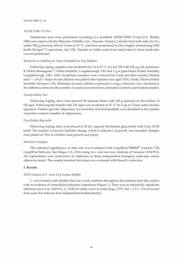
Materials 2019, 12, 18
ASTM E2967-15 Test
Disinfection tests were performed according to a modified ASTM E2967-15 test [21]. Briefly,DSB were wiped with the Wiperator (Filtaflex Ltd., Almonte, Ontario, Canada) from both sides for 10 sunder 500 g pressure, left for 2 min at 25 ◦C, and then neutralized in Dey-Engley neutralizing (DE)broth (Neogen® Corporation, Ayr, UK). Transfer of viable yeasts from used wipes to clean sterile discwas not performed.
Reduction in Viability for Yeasts Embedded in Dry Biofilms
Following wiping, samples were incubated for 1 h at 25 ◦C in 2 mL DE with 100 μg/mL proteinaseK (Fisher Bioreagents™, Fisher Scientific, Loughborough, UK) and 1 g of glass beads (Fisher Scientific,Loughborough, UK). After incubation, samples were vortexed for 2 min and then serially diluted,and 3 × 10 μL2 drops of each dilution was plated onto tryptone soya agar (TSA; Oxoid, Thermo FisherScientific, Newport, UK). Reduction in yeast viability, expressed as a log10 reduction, was calculated asthe difference between the number of yeasts recovered from untreated (control) and treated samples.
Transferability Test
Following wiping, discs were pressed 36 separate times with 100 g pressure on the surface ofDE agar. Following the transfer test, DE agar was incubated at 25 ◦C for a up to 5 days until coloniesappeared. Positive growth/adpression was recorded, and transferability was calculated as the numberof positive contact/number of adpressions.
Dry-Biofilm Regrowth
Following wiping, discs were placed in 30 mL capacity flat bottom glass bottle with 2 mL of DEbroth. The number of days for turbidity change, which is indicative of growth, was recorded. Sampleswere plated on TSA to confirm yeast growth and purity.
Statistical Analysis
The statistical significance of data sets was evaluated with GraphPad PRISM® (version 7.04,GraphPad Software, San Diego, CA, USA) using two- and one-way Analysis of Variance (ANOVA).All experiments were performed in triplicates in three independent biological replicates unlessotherwise stated. The sample standard deviation was evaluated with Bassel’s correction.
3. Results
SEM Analysis of C. auris Dry Surface Biofilm
C. auris formed a thin biofilm that was evenly scattered throughout the stainless-steel disc surfacewith no evidence of extracellular polymeric substances (Figure 1). There was no statistically significantdifference (two-way ANOVA, p = 0.06) in viable count of yeasts (log10 CFU/mL = 7.8 ± 0.3) recoveredfrom each disc between four independent biofilm batches.
68
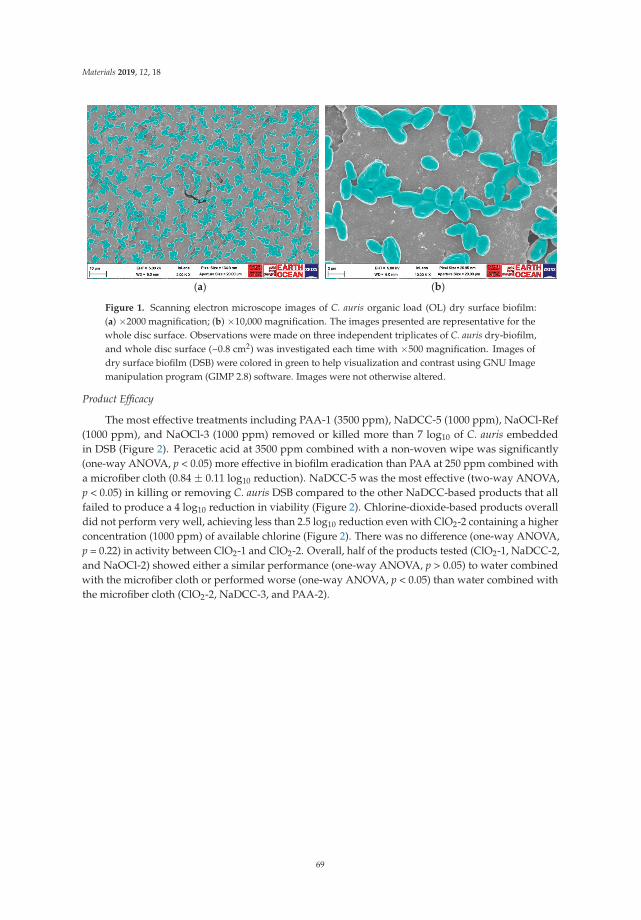
Materials 2019, 12, 18
(a) (b)
Figure 1. Scanning electron microscope images of C. auris organic load (OL) dry surface biofilm:(a) ×2000 magnification; (b) ×10,000 magnification. The images presented are representative for thewhole disc surface. Observations were made on three independent triplicates of C. auris dry-biofilm,and whole disc surface (~0.8 cm2) was investigated each time with ×500 magnification. Images ofdry surface biofilm (DSB) were colored in green to help visualization and contrast using GNU Imagemanipulation program (GIMP 2.8) software. Images were not otherwise altered.
Product Efficacy
The most effective treatments including PAA-1 (3500 ppm), NaDCC-5 (1000 ppm), NaOCl-Ref(1000 ppm), and NaOCl-3 (1000 ppm) removed or killed more than 7 log10 of C. auris embeddedin DSB (Figure 2). Peracetic acid at 3500 ppm combined with a non-woven wipe was significantly(one-way ANOVA, p < 0.05) more effective in biofilm eradication than PAA at 250 ppm combined witha microfiber cloth (0.84 ± 0.11 log10 reduction). NaDCC-5 was the most effective (two-way ANOVA,p < 0.05) in killing or removing C. auris DSB compared to the other NaDCC-based products that allfailed to produce a 4 log10 reduction in viability (Figure 2). Chlorine-dioxide-based products overalldid not perform very well, achieving less than 2.5 log10 reduction even with ClO2-2 containing a higherconcentration (1000 ppm) of available chlorine (Figure 2). There was no difference (one-way ANOVA,p = 0.22) in activity between ClO2-1 and ClO2-2. Overall, half of the products tested (ClO2-1, NaDCC-2,and NaOCl-2) showed either a similar performance (one-way ANOVA, p > 0.05) to water combinedwith the microfiber cloth or performed worse (one-way ANOVA, p < 0.05) than water combined withthe microfiber cloth (ClO2-2, NaDCC-3, and PAA-2).
69

Materials 2019, 12, 18
1, 3 2 1, 3 2, 3 1
3
12
Figure 2. Product efficacy in killing/removing C. auris embedded in a DSB. 1: indicates nostatistical difference (one-way ANOVA, p > 0.05) in log10 reduction/removal from surfaces; 2: log10
reduction/removal lower than wiping with water (one-way ANOVA, p < 0.05); and 3: indicates thatonly two biological replicates were performed.
Only two products, PAA-1 and NaOCl-3, prevented C. auris transfer after treatment (Figure 3).Seven out of 12 commercially available disinfectants were not effective in lowering the transferabilityof C. auris from DSB post-wiping. For four product/materials combinations (ClO2-2, NaDCC-2,NaDCC-3, and NaOCl-1), there was no statistically significant difference (one-way ANOVA, for eachpair p > 0.05) between their performance and that of water (Figure 3). The remaining three treatments(NaDCC-4, NaOCl-2 and PAA-2) were even less effective than wiping with water (one-way ANOVA,for each pair p < 0.05).
3
1
3
1, 3
1
2, 3 1, 3 2
3
12
Figure 3. Product efficacy in preventing C. auris transferability post-wiping. 1: indicates no statisticaldifference (one-way ANOVA, for each pair p > 0.05) in transferability; 2: higher transferability (one-wayANOVA, for each pair p < 0.05) than with water control; and 3: indicates that only two biologicalreplicates were performed.
70

Materials 2019, 12, 18
The best commercial products, NaDCC-3, NaDCC-5, and PAA-1, delayed the recovery of biofilmpost-treatment for more than 4 days (5.0 ± 0.0, 4.7 ± 1.2 and 6.5 ± 2.1 days, respectively; Figure 4).Such activity was similar (two-way ANOVA, p = 0.53) to NaOCl-Ref which delayed regrowth by4.5 ± 0.7 days. Nine out of 12 commercial treatments failed to prevent the regrowth of C. auris DSB formore than 2 days (Figure 4).
1
1
Figure 4. Efficacy of products in preventing regrowth post-wiping. 1: indicates that only two biologicalreplicates were performed.
The less-reactive chemistry (as compared to the oxidizing chemistries) based on a quaternaryammonium compound produced a 4 log10 reduction in C. auris on surfaces and reduced transferabilitypost-wiping to 20%.
4. Discussion
Environmental surfaces play an important role in the transmission of infection [5,6]. Infectioncontrol regimens include the combination of cleaning and disinfection processes [16] and are oftenbased on the use of a disinfectant or cleaner combined with diverse materials [31]. Performance ofbiocidal products still relies, however, on testing the efficacy of the formulation and not the combinationof formulation and material [22]. A number of US-based protocols to test wipe activity have beendescribed, but all had severe limitations in their setting or performance [31]. Recently, the ASTM2967-15and the EN16615-15 protocols [32] have been recommended for evaluating the efficacy of antimicrobialwipes, although EN16615-15 has recently been shown to lack stringency [33]. Both tests evaluatethe reduction in microbial number from the test surface and the transfer of micro-organisms duringwiping. Attaining a > 5log10 reduction in microbial inoculum following treatment has been, until now,deemed to provide enough assurance that all micro-organisms would be killed on surfaces in practice.Although this might be the case where surfaces are contaminated with a low number of microorganisms,this might not be correct with surfaces contaminated with a high number of microorganisms or whendry surface biofilms are present. Hence, the evaluation of microbial transferability post-treatmentis important to take into consideration to provide reassurance that a surface would be safe to touch.The presence of DSB on healthcare surfaces has been established [8–10], and the resilience of DSBto disinfection has been described [8]. The presence of C. auris DSB has not been yet established inhealthcare settings, although its persistence in healthcare settings has been described [11,12].
Here, we have successfully produced reproducible dry surface biofilms of C. auris on stainless steelsurfaces, for which appearance and characteristics are not dissimilar to artificial DSB of Staphylococcus
71
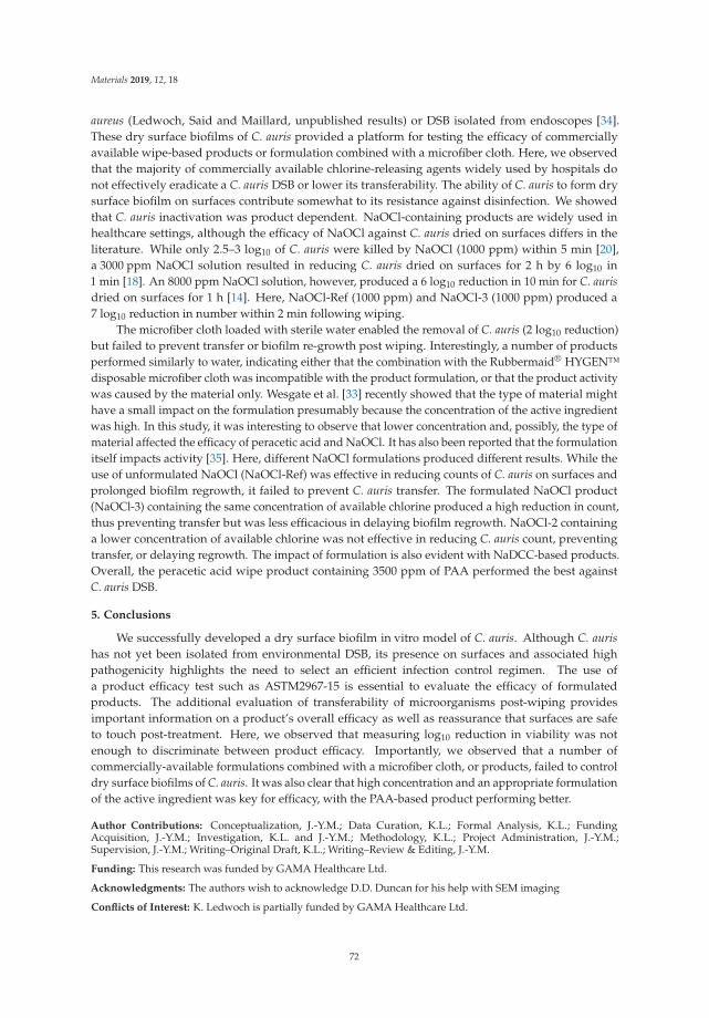
Materials 2019, 12, 18
aureus (Ledwoch, Said and Maillard, unpublished results) or DSB isolated from endoscopes [34].These dry surface biofilms of C. auris provided a platform for testing the efficacy of commerciallyavailable wipe-based products or formulation combined with a microfiber cloth. Here, we observedthat the majority of commercially available chlorine-releasing agents widely used by hospitals donot effectively eradicate a C. auris DSB or lower its transferability. The ability of C. auris to form drysurface biofilm on surfaces contribute somewhat to its resistance against disinfection. We showedthat C. auris inactivation was product dependent. NaOCl-containing products are widely used inhealthcare settings, although the efficacy of NaOCl against C. auris dried on surfaces differs in theliterature. While only 2.5–3 log10 of C. auris were killed by NaOCl (1000 ppm) within 5 min [20],a 3000 ppm NaOCl solution resulted in reducing C. auris dried on surfaces for 2 h by 6 log10 in1 min [18]. An 8000 ppm NaOCl solution, however, produced a 6 log10 reduction in 10 min for C. aurisdried on surfaces for 1 h [14]. Here, NaOCl-Ref (1000 ppm) and NaOCl-3 (1000 ppm) produced a7 log10 reduction in number within 2 min following wiping.
The microfiber cloth loaded with sterile water enabled the removal of C. auris (2 log10 reduction)but failed to prevent transfer or biofilm re-growth post wiping. Interestingly, a number of productsperformed similarly to water, indicating either that the combination with the Rubbermaid® HYGEN™disposable microfiber cloth was incompatible with the product formulation, or that the product activitywas caused by the material only. Wesgate et al. [33] recently showed that the type of material mighthave a small impact on the formulation presumably because the concentration of the active ingredientwas high. In this study, it was interesting to observe that lower concentration and, possibly, the type ofmaterial affected the efficacy of peracetic acid and NaOCl. It has also been reported that the formulationitself impacts activity [35]. Here, different NaOCl formulations produced different results. While theuse of unformulated NaOCl (NaOCl-Ref) was effective in reducing counts of C. auris on surfaces andprolonged biofilm regrowth, it failed to prevent C. auris transfer. The formulated NaOCl product(NaOCl-3) containing the same concentration of available chlorine produced a high reduction in count,thus preventing transfer but was less efficacious in delaying biofilm regrowth. NaOCl-2 containinga lower concentration of available chlorine was not effective in reducing C. auris count, preventingtransfer, or delaying regrowth. The impact of formulation is also evident with NaDCC-based products.Overall, the peracetic acid wipe product containing 3500 ppm of PAA performed the best againstC. auris DSB.
5. Conclusions
We successfully developed a dry surface biofilm in vitro model of C. auris. Although C. aurishas not yet been isolated from environmental DSB, its presence on surfaces and associated highpathogenicity highlights the need to select an efficient infection control regimen. The use ofa product efficacy test such as ASTM2967-15 is essential to evaluate the efficacy of formulatedproducts. The additional evaluation of transferability of microorganisms post-wiping providesimportant information on a product’s overall efficacy as well as reassurance that surfaces are safeto touch post-treatment. Here, we observed that measuring log10 reduction in viability was notenough to discriminate between product efficacy. Importantly, we observed that a number ofcommercially-available formulations combined with a microfiber cloth, or products, failed to controldry surface biofilms of C. auris. It was also clear that high concentration and an appropriate formulationof the active ingredient was key for efficacy, with the PAA-based product performing better.
Author Contributions: Conceptualization, J.-Y.M.; Data Curation, K.L.; Formal Analysis, K.L.; FundingAcquisition, J.-Y.M.; Investigation, K.L. and J.-Y.M.; Methodology, K.L.; Project Administration, J.-Y.M.;Supervision, J.-Y.M.; Writing–Original Draft, K.L.; Writing–Review & Editing, J.-Y.M.
Funding: This research was funded by GAMA Healthcare Ltd.
Acknowledgments: The authors wish to acknowledge D.D. Duncan for his help with SEM imaging
Conflicts of Interest: K. Ledwoch is partially funded by GAMA Healthcare Ltd.
72

Materials 2019, 12, 18
References
1. Satoh, K.; Makimura, K.; Hasumi, Y.; Nishiyama, Y.; Uchida, K.; Yamaguchi, H. Candida auris sp. nov.,a novel ascomycetous yeast isolated from the external ear canal of an inpatient in a Japanese hospital.Microbiol. Immunol. 2009, 53, 41–44. [CrossRef]
2. Araúz, A.B.; Caceres, D.H.; Santiago, E.; Armstrong, P.; Arosemena, S.; Ramos, C.; Espinosa-Bode, A.;Borace, J.; Hayer, L.; Cedeño, I.; et al. Isolation of Candida auris from 9 patients in Central America:Importance of accurate diagnosis and susceptibility testing. Mycoses 2018, 61, 44–47. [CrossRef]
3. Morales-López, S.E.; Parra-Giraldo, C.M.; Ceballos-Garzón, A.; Martínez, H.P.; Rodríguez, G.J.;Álvarez-Moreno, C.A.; Rodríguez, J.Y. Invasive Infections with Multidrug-Resistant Yeast Candida auris,Colombia. Emerg. Infect. Dis. 2017, 23, 162. [CrossRef]
4. Calvo, B.; Melo, A.S.A.; Perozo-Mena, A.; Hernandez, M.; Francisco, E.C.; Hagen, F.; Meis, J.F.; Colombo, A.L.First report of Candida auris in America: Clinical and microbiological aspects of 18 episodes of candidemia.J. Infect. 2016, 73, 369–374. [CrossRef]
5. Weber, D.J.; Rutala, W.A.; Miller, M.B.; Huslage, K.; Sickbert-Bennett, E. Role of hospital surfaces in thetransmission of emerging healthcare associated pathogens: Norovirus, Clostridium difficile, and Acinetobacterspecies. Am. J. Infect. Control 2010, 38, S25–S33. [CrossRef] [PubMed]
6. Weber, D.J.; Anderson, D.; Rutala, W.A. The role of the surface environment in healthcare-associatedinfections. Curr. Opin. Infect. Dis. 2013, 26, 338–344. [CrossRef]
7. Otter, J.A.; Yezli, S.; Salkeld, J.A.G.; French, G.L. Evidence that contaminated surfaces contribute to thetransmission of hospital pathogens and an overview of strategies to address contaminated surfaces inhospital settings. Am. J. Infect. Control 2013, 41, S6–S11. [CrossRef] [PubMed]
8. Vickery, K.; Deva, A.; Jacombs, A.; Allan, J.; Valente, P.; Gosbell, I.B. Presence of biofilm containing viablemultiresistant organisms despite terminal cleaning on clinical surfaces in an intensive care unit. J. Hosp. Infect.2012, 80, 52–55. [CrossRef] [PubMed]
9. Hu, H.; Johani, K.; Gosbell, I.B.; Jacombs, A.S.W.; Almatroudi, A.; Whiteley, G.S.; Deva, A.K.; Jensen, S.;Vickery, K. Intensive care unit environmental surfaces are contaminated by multidrug-resistant bacteriain biofilms: Combined results of conventional culture, pyrosequencing, scanning electron microscopy,and confocal laser microscopy. J. Hosp. Infect. 2015, 91, 35–44. [CrossRef]
10. Ledwoch, K.; Dancer, S.J.; Otter, J.A.; Kerr, K.; Roposte, D.; Rushton, L.; Weiser, R.; Mahenthiralingam, E.;Muir, D.D.; Maillard, J.-Y. Beware biofilm! Dry biofilms containing bacterial pathogens on multiple healthcaresurfaces; a multi-centre study. J. Hosp. Infect. 2018, 100, 47–56. [CrossRef]
11. Piedrahita, C.T.; Cadnum, J.L.; Jencson, A.L.; Shaikh, A.A.; Ghannoum, M.A.; Donskey, C.J. Environmentalsurfaces in healthcare facilities are a potential source for transmission of Candida auris and other Candidaspecies. Infect. Control Hosp. Epidemiol. 2017, 38, 1107–1109. [CrossRef] [PubMed]
12. Welsh, R.M.; Bentz, M.L.; Shams, A.; Houston, H.; Lyons, A.; Rose, L.; Litvintseva, A.P. Survival, persistence,and isolation of the emerging multidrug-resistant pathogenic yeast Candida auris on a plastic health caresurface. J. Clin. Microbiol. 2017, 55, 2996–3005. [CrossRef] [PubMed]
13. Vallabhaneni, S.; Kallen, A.; Tsay, S.; Chow, N.; Welsh, R.; Kerins, J.; Kemble, S.K.; Pacilli, M.; Black, S.R.;Landon, E.; et al. Investigation of the first seven reported cases of Candida auris, a globally emerging invasive,multidrug-resistant fungus-United States, May 2013–August 2016. Am. J. Transplant. 2017, 17, 296–299.[CrossRef] [PubMed]
14. Biswal, M.; Rudramurthy, S.M.; Jain, N.; Shamanth, A.S.; Sharma, D.; Jain, K.; Yaddanapudi, L.N.;Chakrabarti, A. Controlling a possible outbreak of Candida auris infection: Lessons learnt from multipleinterventions. J. Hosp. Infect. 2017, 97, 363–370. [CrossRef] [PubMed]
15. Ku, T.S.N.; Walraven, C.J.; Lee, S.A. Candida auris: Disinfectants and implications for infection control.Front. Microbiol. 2018, 9, 1–12. [CrossRef] [PubMed]
16. Guidance for the Laboratory Investigation, Management and Infection Prevention and Control for Cases of Candidaauris (August 2017 v2.0); Public Health England: London, UK, 2016; PHE publications gateway number:2016122.
17. Abdolrasouli, A.; Armstrong-James, D.; Ryan, L.; Schelenz, S. In vitro efficacy of disinfectants utilised forskin decolonization and environmental decontamination during a hospital outbreak with Candida auris.Mycoses 2017, 60, 758–763. [CrossRef] [PubMed]
73

Materials 2019, 12, 18
18. Cadnum, J.L.; Shaikh, A.A.; Piedrahita, C.T.; Sankar, T.; Jencson, A.L.; Larkin, E.L.; Ghannoum, M.A.;Donskey, C.J. Effectiveness of disinfectants against Candida auris and other Candida species. Infect. ControlHosp. Epidemiol. 2017, 38, 1240–1243. [CrossRef] [PubMed]
19. Moore, G.; Schelenz, S.; Borman, A.M.; Johnson, E.M.; Brown, C.S. Yeasticidal activity of chemicaldisinfectants and antiseptics against Candida auris. J. Hosp. Infect. 2017, 97, 371–375. [CrossRef]
20. Kean, R.; Sherry, L.; Townsend, E.; McKloud, E.; Short, B.; Akinbobola, A.; Mackay, W.G.; Williams, C.;Jones, B.L.; Ramage, G. Surface disinfection challenges for Candida auris: An in vitro study. J. Hosp. Infect.2018, 98, 433–436. [CrossRef]
21. ASTM E2967-15 Standard Test Method for Assessing the Ability of Pre-wetted Towelettes to Remove andTransfer Bacterial Contamination on Hard, Non-Porous Environmental Surfaces Using the Wiperator; ASTM:West Conshohocken, PA, USA, 2015.
22. Ledwoch, K.; Said, J.; Maillard, J.-Y. Artificial dry-biofilm models for disinfectant efficacy testing. Lett. Appl.Microbiol. 2018, under review.
23. Maris, P. Modes of action of disinfectants. Rev. Sci. Tech. 1995, 14, 47–55. [CrossRef]24. Virto, R.; Mañas, P.; Álvarez, I.; Condon, S.; Raso, J. Membrane damage and microbial inactivation by
chlorine in the absence and presence of a chlorine-demanding substrate. Appl. Environ. Microbiol. 2005, 71,5022–5028. [CrossRef] [PubMed]
25. Russell, A.D. Microbial sensitivity and resistance to chemical and physical agents. In Topley and Wilson’sMicrobiology and Microbial Infections, 9th ed.; Edward Arnold: London, UK, 1998; pp. 149–184.
26. McDonnell, G.; Russell, A.D. Antiseptics and disinfectants: Activity, action and resistance. Clin. Microbiol.Rev. 1999, 12, 147–179. [CrossRef] [PubMed]
27. Estrela, C.; Estrela, C.R.A.; Barbin, E.L.; Spanó, J.C.E.; Marchesan, M.A.; Pécora, J.D. Mechanism of action ofsodium hypochlorite. Braz. Dent. J. 2002, 13, 113–117. [CrossRef] [PubMed]
28. Block, S.S. Disinfection, Sterilization and Preservation, 4th ed.; Lea & Febiger Press: Philadelphia, PA, USA, 1991.29. Kitis, M. Disinfection of wastewater with peracetic acid: A review. Environ. Int. 2004, 30, 47–54. [CrossRef]30. Tutumi, M.; Imamura, K.; Hatano, S.; Watanabe, T. Antimicrobial action of peracetic acid. J. Food Hyg. Soc.
Jpn. 1973, 14, 443–447. [CrossRef]31. Sattar, S.A.; Maillard, J.Y. The crucial role of wiping in decontamination of high-touch environmental surfaces:
Review of current status and directions for the future. Am. J. Infect. Control 2013, 41, S97–S104. [CrossRef]32. Chemical Disinfectants and Antiseptics-Quantitative Test Method for The Evaluation of Bactericidal and Yeasticidal
Activity on Non-Porous Surfaces with Mechanical Action Employing Wipes in the Medical Area (4-Field Test);British Standards Institute: London, UK, 2015.
33. Wesgate, R.; Robertson, A.; Barrell, M.; Tesca, P.; Maillard, J.-Y. Impact of test protocols and material bindingon antimicrobial wipes efficacy. J. Hosp. Infect. 2018, in press. [CrossRef]
34. Pajkos, A.; Vickery, K.; Cossart, Y. Is biofilm accumulation on endoscope tubing a contributor to the failureof cleaning and decontamination? J. Hosp. Infect. 2004, 58, 224–229. [CrossRef]
35. Forbes, S.; Cowley, N.; Humphreys, G.; Mistry, H.; Amézquita, A.; McBain, A.J. Formulation of biocidesincreases antimicrobial potency and mitigates the enrichment of nonsusceptible bacteria in multispeciesbiofilms. Appl. Environ. Microbiol. 2017, 83, AEM.03054-16. [CrossRef]
© 2018 by the authors. Licensee MDPI, Basel, Switzerland. This article is an open accessarticle distributed under the terms and conditions of the Creative Commons Attribution(CC BY) license (http://creativecommons.org/licenses/by/4.0/).
74
materials
Review
Targeting the Bacterial Protective Armour; Challengesand Novel Strategies in the Treatment ofMicrobial Biofilm
Nor Fadhilah Kamaruzzaman 1,*, Li Peng Tan 1, Khairun Anisa Mat Yazid 1,
Shamsaldeen Ibrahim Saeed 1, Ruhil Hayati Hamdan 1, Siew Shean Choong 1, Weng Kin Wong 2,
Alexandru Chivu 3 and Amanda Jane Gibson 4
1 Faculty of Veterinary Medicine, Universiti Malaysia Kelantan, Pengkalan Chepa 16100, Kelantan, Malaysia;[email protected] (L.P.T.); [email protected] (K.A.M.Y.); [email protected] (S.I.S.);[email protected] (R.H.H.); [email protected] (S.S.C.)
2 School of Health Sciences, Universiti Sains Malaysia, Kubang Kerian 16150, Kelantan, Malaysia;[email protected]
3 UCL Centre for Nanotechnology and Regenerative Medicine, Division of Surgery & Interventional Science,University College London, London NW3 2PF, UK; [email protected]
4 Royal Veterinary College, Pathobiology and Population Sciences, Hawkshead Lane, North Mymms,Hatfield AL9 7TA, UK; [email protected]
* Correspondence: [email protected]
Received: 1 August 2018; Accepted: 9 September 2018; Published: 13 September 2018
Abstract: Infectious disease caused by pathogenic bacteria continues to be the primary challengeto humanity. Antimicrobial resistance and microbial biofilm formation in part, lead to treatmentfailures. The formation of biofilms by nosocomial pathogens such as Staphylococcus aureus (S. aureus),Pseudomonas aeruginosa (P. aeruginosa), and Klebsiella pneumoniae (K. pneumoniae) on medical devicesand on the surfaces of infected sites bring additional hurdles to existing therapies. In this review, wediscuss the challenges encountered by conventional treatment strategies in the clinic. We also provideupdates on current on-going research related to the development of novel anti-biofilm technologies.We intend for this review to provide understanding to readers on the current problem in health-caresettings and propose new ideas for new intervention strategies to reduce the burden related tomicrobial infections.
Keywords: biofilms; anti-biofilms; nosocomial pathogens; Staphylococcus aureus; Pseudomonasaeruginosa; Klebsiella pneumoniae
1. Microbial Biofilms and the Challenges in Infectious Disease
Bacterial infections remain a threat to human health despite the progress made in improvingthe quality of health care, and continuous development of antibiotics and vaccines to control disease.Bacterial infections can occur at any stage in human life and can often be controlled by a healthyimmune system of the host. During hospitalization, patients are exposed to pathogen sources withinthe environment including medical equipment, other infected patients, and healthcare staff and arethus susceptible to hospital-acquired infections (HAI) [1]. Immunocompromised patients, the elderlyor patients with existing chronic disease such as diabetes, cancer, cardiovascular diseases, or breakageof skin barrier such occurs with wounds, are reported to face a higher risk of nosocomial infection [2].The typical medical device associated with HAI are related to central line bloodstream infections,ventilator pneumonia, and catheter urinary infections [1].
The increasing trend of nosocomial bacterial infections in immunocompromised hospitalizedpatients is worrying as HAI are the primary cause of morbidity in the health-care setting [3,4]. In the
Materials 2018, 11, 1705; doi:10.3390/ma11091705 www.mdpi.com/journal/materials75

Materials 2018, 11, 1705
United States alone, it was estimated that a total of 1.7 million HAI occurred in the year 2002 alone,with 4.5 cases occurring for every 100 admissions, resulting in almost 99,000 deaths [5]. At the Europeanlevel, more than 2 million patients contracted HAI, with 175,000 deaths per year [6]. The incidenceof HAI are substantially lower at 7.1% and 4.5% in Europe and USA, respectively, compared to thelow and middle-income countries (LMICs), where the average prevalence of HAI is 15.5% [7]. Amongthe leading bacteria that cause the HAI is Staphylococcus aureus (S. aureus), Pseudomonas aeruginosa(P. aeruginosa), Escherichia coli (E. coli), Klebsiella pneumoniae (K. pneumoniae), Acinetobacter baumanii,Clostridium difficile, and Enterococci [8,9]. The ability of these bacteria to form biofilms at the infectedsite or on medical devices has been increasingly recognized as one of the factors causing failure in thetreatment of HAI, with biofilms estimated to contribute to approximately half of HAI [8,10]. Figure 1shows the common site of infections related to the formation of bacterial biofilms.
1.1. Methicillin-Resistant Staphylococcus aureus (MRSA)
Staphylococcus aureus is a Gram-positive cocci-shaped bacterium that forms part of the normal floraon the body and is frequently isolated from the skin, respiratory tract, and female lower reproductivetract [11,12]. Infections by S. aureus were once treatable by penicillin. However, increasing resistancetowards penicillin led to the introduction of methicillin for the treatment in 1960 [13]. However, soonafter that, S. aureus acquired resistance towards methicillin, giving rise to methicillin-resistant S. aureus(MRSA) clones [12,13]. Currently, the Center for Disease Control (CDC) reported that infections byMRSA are the second most common cause of HAI in the USA [14]. The most common route of MRSAtransmission is through direct contact. The ability of the organism to form biofilms on tissues suchas the skin and inert indwelling device surfaces such as intravenous catheters and surgical implantsfurther expose susceptible individuals [15]. Development of worldwide antibiotic resistance towardsfirst-line therapies such as vancomycin and teicoplanin continues to hinder and restrict the successfultreatment of MRSA infection [15]. Additionally, S. aureus also can invade host cells and evade theantimicrobial effects of administered therapies [16]. Together, these characteristics allow this organismto remain an important pathogen.
1.2. Pseudomonas aeruginosa
P. aeruginosa is a Gram-negative, rod-shaped, facultative anaerobe ubiquitous in the environmentand forms part of the normal gut flora. Increasing resistance towards the multiple antibiotics, e.g.,cephalosporins and carbapenem further compounds the problem due to the emergence of extremelydrug-resistant (XDR) P. aeruginosa infections [17,18]. The last available treatment resort is colistin,a polymyxin antibiotic which was avoided for the past three decades as it may cause both neuro-and nephrotoxicity [19–21]. Patients dependent on breathing machines or fitted with an invasivedevice such as a catheter, are at risk of severe and life-threatening illness from P. aeruginosa capableof forming biofilms on medical device surfaces [22–25]. P. aeruginosa biofilms were reported to causeendocardial valve infection through endocardial tubes, ventilator-associated pneumonia (VAP), andcatheter-associated urinary tract infections (CAUTI). Additionally, P. aeruginosa has also been reportedto be able to grow in intravenous fluid and could enter the bloodstream and cause sepsis [26–28].
1.3. Klebsiella pneumoniae
Klebsiella pneumoniae (K. pneumoniae) is a non-fastidious, Gram-negative bacillus, and is usuallyencapsulated. The bacterium is one of the normal flora present in the mouth, skin, and intestine, yetit has been reported to cause pneumonia, urinary tract infections, and bacteremia in patients fromhospitals, nursing homes, and other healthcare facilities [18]. K. pneumoniae is known as a remarkablyresilient pathogen as it can evade and survive rather than actively suppress many components of theimmune system of the infected host [29]. The bacteria have developed resistance towards almost allavailable antibiotics; fluoroquinolones, third-generation cephalosporins and aminoglycosides [30].Recently, the emergence of the carbapenem-resistant K. pnemoniae strains which currently circulating
76

Materials 2018, 11, 1705
across the globe has forced the administration of colistin, an old and considered the last availableantibiotic [31]. Additionally, resistance towards colistin was recently reported, showed that the bacteriaare capable of evading all types of available antibiotics, leaving no drugs left for the treatment [32].Compounding the problem, this organism can survive and grow within the intravenous fluid andform biofilm on medical devices such as the urinary catheter, leading to detrimental septicemia inpatients [26,33–35].
2. The Physiology of Biofilms
2.1. Definition and the Structure of the Biofilm
Biofilms have been described in environmental and technical microbiology for more than 90 years.However, the importance of microbial biofilms in medicine has only been recognized since the early1980s [36]. A microbial biofilm is defined as a structured consortium of microbial cells surrounded bythe self-produced matrix [37]. The rough structure can be described as polymicrobial aggregates thatresemble mats, flocs, or sludge that accumulate at interfaces. Because of the soft and fragile structure,it is difficult to physically characterize the structure from the infected or adhered surfaces in-vivo. Thus,characterizations of biofilm were mostly performed using in-vitro cultured biofilm cells. The thicknessof biofilms can vary depending on the species of the bacteria, the duration and the method usedto grow the biofilm. In-vitro, the biofilms of P. aeruginosa can reach 209 μm; S. aureus, 8.0 μm; andK. pneumoniae, 231 μm [33,38,39]. Figure 2 shows the three-dimensional structure of S. aureus biofilmsvisualized with a confocal microscope with the thickness of approximately 8.0 μm.
Biofilms are built of planktonic (individual) bacterial cells ‘glued’ together with self-releasedextra-polymeric substances (EPS) which consists of lipopolysaccharides, proteins, lipids, glycolipids,and nucleic acids [40]. The type of polysaccharides found within the biofilm depends on the bacteriaspecies, S. aureus and S. epidermidis produce poly-ß(1,6)-N-acetyl-D-glucosamine (PNAG) [41], whileP. aeruginosa produces Pel (a cationic exopolysaccharide composed of 1–4 linked galactosamineand glucosamine sugars) and Psl (a penta-saccharide composed of D-glucose, D-mannose andL-rhamnose) [42,43]. The nucleic acid found in EPS is known as extracellular DNA (eDNA), generatedby lysis of a subpopulation of the same bacteria under the control of quorum sensing, a mode ofcommunication between cells. The role of EPS is to promote adhesion and aggregation of bacteriato the surfaces and provides stability to the biofilm structure. Direct interaction of eDNA withextracellular calcium (Ca2+) within the biofilm induces bacterial aggregation via cationic bridging.The positive charge of the extracellular Ca2+ modifies the bacterial cell surface charge by neutralizingthe electrostatic repulsion between negatively charged biofilm components, thus assisting cellularaggregation and adherence of bacteria to material and tissue surface [44,45]. Once the biofilmis established, negatively charged eDNA acts as a biofilm defence mechanism by chelating thecationic antimicrobial peptides from the host immune system. eDNA also chelates divalent cationstriggering the bacterial transcription of genes responsible for increasing pathogenicity and resistanceto antimicrobials [46].
77

Materials 2018, 11, 1705
Figure 1. Common site of infections related to the formation of bacterial biofilms.
78
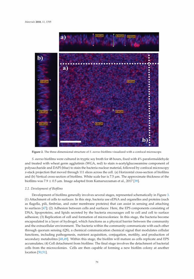
Materials 2018, 11, 1705
Figure 2. The three-dimensional structure of S. aureus biofilms visualized with a confocal microscope.
S. aureus biofilms were cultured in tryptic soy broth for 48 hours, fixed with 4% paraformaldehydeand treated with wheat germ agglutinin (WGA, red) to stain n-acetylglucoseamine component ofpolysaccharide and DAPI (blue) to stain the bacteria nuclear material, followed by confocal microscopyz-stack projection that moved through 111 slices across the cell. (a) Horizontal cross-section of biofilmsand (b) Vertical cross-section of biofilms. White scale bar is 7.5 μm. The approximate thickness of thebiofilms was 7.9 ± 0.5 μm. Image adapted from Kamaruzzaman et al., 2017 [39].
2.2. Development of Biofilms
Development of biofilms generally involves several stages, represented schematically in Figure 3.(1) Attachment of cells to surfaces: In this step, bacteria use eDNA and organelles and proteins (suchas flagella, pili, fimbriae, and outer membrane proteins) that can assist in sensing and attachingto surfaces [47]; (2) Adhesion between cells and surfaces: Here, the EPS components consisting ofDNA, lipoproteins, and lipids secreted by the bacteria encourages cell to cell and cell to surfaceadhesion; (3) Replication of cell and formation of microcolonies: In this stage, the bacteria becomeencapsulated in a layer of hydrogel, which functions as a physical barrier between the communityand the extracellular environment. The bacteria within the community communicate with each otherthrough quorum sensing (QS), a chemical communication chemical signal that modulates cellularfunctions, including pathogenesis, nutrient acquisition, conjugation, motility, and production ofsecondary metabolites [48,49]. Within this stage, the biofilm will mature as cells replicate and EPSaccumulates; (4) Cell detachment from biofilms: The final stage involves the detachment of bacterialcells from the microcolonies. Cells are then capable of forming a new biofilm colony at anotherlocation [50,51].
79

Materials 2018, 11, 1705
Fig
ure
3.
Stag
esin
biofi
lmin
form
atio
nTh
eim
age
isad
apte
dfr
om[5
0].
80

Materials 2018, 11, 1705
2.3. Mechanism of Antibiotic Resistance in Microbial Biofilms
Bacterial cells existing as biofilms can be 10–1000 times more resistant to antibiotics [52–54].This condition is attributed by several factors and briefly summarised in Table 1. An excellent review ondetailed mechanism of biofilm mediated antimicrobial resistance and tolerance was recently publishedby Hall & Mah (2017), thus readers are invited to refer to this article for detailed information [55].
Table 1. Mechanism of biofilm-mediated antimicrobial resistance.
Physiology of Biofilm Mechanism of Antimicrobial Resistance References
Component of the biofilm matrix
• Thick biofilm matrix • Reduced permeation of antimicrobials acrossbiofilm matrix
[52,54,56,57]
• Expression of polysaccharidesubunits in biofilm matrix
• Expression of PsI polysaccharides in P. aeruginosasequesters antimicrobials in the biofilm matrix viaelectrostatic interaction
[42]
• Expression of antimicrobialmodifying enzymes in thebiofilm matrix
• Beta lactamases produced by Klebsiella pneumoniaebiofilm degrade beta lactam antibiotics [58]
• Expression of eDNA• eDNA interacts with antimicrobials and prevent further
penetration of the agent across the biofilm matrix toreach cellular targets
[59]
Nutritional factors
• Oxygen deprived environment
• Deeper layer of the biofilm is oxygen deprived (hypoxic).Hypoxic conditions reduce outer membrane potential ofbacteria, diminishing intracellular transport of antibioticsinto the bacterial cells
• Hypoxia induces increased (or is new expression?)expression of bacterial membrane efflux pumps,potentially reducing antimicrobial accumulation inthe cell
[60–62]
• Amino acid deprived environment • Amino acid starvation activates a stringent response inbacteria within biofilm increasing antimicrobial tolerance
[57]
Physiology of bacteria
• Small colony variants andpersister cells
• Surviving bacteria within the biofilm may change intosmall colony variants and persister cells. These variantspresent different phenotypes compared to the wild typeplanktonic cells including greater tolerancetowards antimicrobials
[63]
Table adapted from Hall & Mah (2017) [55].
2.4. Immune Evasion of Biofilms
Bacteria within the biofilm can evade recognition by the innate immune system and thus avoidbeing eliminated or controlled by the immune system. While the purpose of this article is not toprovide a comprehensive review of immune evasion of biofilms, there are aspects worth comment inthe context of novel therapies and control strategies. We direct the reader to reviews by Roilides et al.,Le et al., and Gunn et al. for more extensive information [64–66]. Despite a clear shortage of studiesconcerning biofilm recognition by the immune system compared to planktonic cells, several commonevasion mechanisms have been described. Biofilms evade the natural antimicrobial properties of innateimmune cells by thwarting pathogen recognition, phagocytosis, and cellular activation [67]. In somecases, the cells of the innate immune system assist the process of biofilm growth and maturation [68].
Biofilms modulate leukocyte function by reducing phagocytosis, despite active migration of cellstowards the biofilm. Phagocytosis rates of biofilm material are thought to be diminished through alack of microbial recognition by pattern recognition receptors due to concealed or obscured bacterialligands such as lipoproteins, lipopolysaccharides, and nucleic acids (i.e., though EPS protection) [69].
81

Materials 2018, 11, 1705
EPS protection also retains bacterial content within the biofilm, resulting in large complexes whichleukocytes are incapable of engulfing. Detached or homogenised (via sonication) bacteria frombiofilms are readily phagocytosed suggesting that leukocyte modulation is temporary [67]. Leukocyteantimicrobial function and killing via oxidative burst (production of reactive oxygen species and nitricoxide) is generally considered to be greatly reduced when considering responses against biofilms [70].Oxidative burst is readily linked to reduced receptor mediated recognition, further highlighting therole of the protective function of the biofilm matrix in immune evasion.
Experimental investigations with relevant HAI associated biofilms such as those of S. aureus andP. aeruginosa continue to enhance the knowledge surrounding immune recognition in the context ofbiofilms. Neutrophils are attracted to and migrate towards both S. aureus and P. aeruginosa biofilmsupon which a reduced phagocytic behaviour is observed along with decreased microbial killing [67].Lack of S. aureus recognition by pattern recognition receptors known as Toll-like Receptors (TLR) islikely due to ligand inaccessibility within the biofilm [69]. TLR binding and subsequent activationpotentiates phagocytosis and antimicrobial activity through oxidative burst mechanisms, thus thearrangement of biofilms within a protective matrix serves to conceal internal components. Disruptionof P. aeruginosa biofilms to homogenised individual cells exhibited an increase in phagocytosisdemonstrating the importance of the protective nature of the biofilm matrix [67].
Understanding fundamental interactions between biofilms and the immune system presentsopportunities for novel control strategies. Glycolipids involved in quorum sensing produced byP. aeruginosa, rhamnolipids, protects against neutrophil activity by inducing lytic necrosis [71].Disruption of this protection via mutation or quorum inhibitors increases phagocytosis rates, thustargeting active evasion mechanisms offers a route for innovative biofilm treatment strategies.
3. Guideline for Management of Biofilm Associated Infection
Tackling the central issue of HAI biofilms requires that diagnosis and treatment be collectivelyexamined (in a concerted manner) and selected to avoid senseless efforts and increase chances foreffective therapy. Several guidelines and recommendations are available for diagnosis and treatmentof biofilm infection [72,73] and are summarised in Table 2.
Table 2. Summary of guidelines for diagnosis and treatment of biofilm infections.
Steps Detail Action
Biofilm diagnosis1. Sampling on tissue biopsy or device/prosthesis (foreign bodies).2. Microbial cultivation and identification.3. Antibiotic susceptibilities test.
Reporting of biofilm associated infections
Biofilm associated infections may be reported using descriptive terms.
1. Detected
e.g., ‘Microscopy shows Gram-negative rods in biofilm-like structures’
2. Suspected
e.g., ‘Growth of/PCR-detected microorganisms possibly from a biofilm infection’
Treatment of biofilm related infections
1. Removal sources of infections1. Debridement of tissues2. Removal of foreign bodies
2. Administration of topical antiseptics1. Empiric therapy to prevent biofilm formation after debridement of tissues2. Pre-emptive therapy to prevent possible biofilm reformation when specific
bacteria is detected from the debride tissue
3. Administration of antibiotics1. Selection of antibiotics suitable for biofilm associated infections2. Administration of antibiotics3. Optimization of dosage
Monitoring1. Assessment of wounds2. Appropriate/maintenance debridement3. Re-evaluation of antiseptics/antibiotics efficacy
82
Materials 2018, 11, 1705
Table 2. Cont.
Steps Detail Action
Standard care 1. Set-up the advance therapies2. Incorporation of novel techniques
Diagnosis and treatment of biofilm can be achieved by various methods, approaches in diagnosing and treatingbiofilm infections will be further discussed in the following sections.
4. Diagnosis of Biofilm Mediated Infections
The diagnosis of biofilm-related infections is complex and should combine different approachesand a multidisciplinary perspective from clinical and laboratory diagnosis [74]. The European Societyfor Clinical Microbiology and Infectious Disease (ESCMID) has provided a detailed guideline toaid clinicians in the diagnosis of biofilm related infections. Briefly the guideline emphasizes thecombination of clinical and laboratory diagnosis to facilitate clinicians in providing an effectivetherapy to patients [73]. Generally, the clinical diagnosis may include clinical signs, case history,failure of antibiotic therapy and persistent infection. For laboratory diagnosis, the followingtechniques have demonstrated high efficacy, successfully discriminating between planktonic andbiofilm mediated infections.
4.1. Sonication
Sonication is a process that uses acoustic energy to agitate particles in a solution. This method isuseful and recommended to be applied to remove microbial biofilms from the biomaterial surface [75].The suspected source of infection (e.g., implant) is removed from the patient and subjected forsonication. The process disrupts strongly adhered biomass and releases the biofilm into the solutionallowing further analysis to further identify the microbial species [76]. Sonication has been shown tobe useful in removing biofilms from urinary catheters, cardiac implantable electronic devices, andprosthetic implants [75–77].
4.2. Polymerase Chain Reaction (PCR)
PCR is one of the common methods used for detection of pathogens directly from clinicalspecimens. The method is based on the amplification of specific conserved regions within the targetedorganism, providing high specificity and sensitivity in discriminating species of bacteria. Due tothe robustness of the method, PCR-based diagnostic tests have been used for detecting a range ofpathogens including bacteria, viruses, parasites, and fungi [78–81]. In biofilm diagnosis, PCR has beenapplied to detect the microbial species that forms biofilms on tissues or biomaterials following thesonication process. For example, PCR was used to detect coagulase negative staphylococci, S. aureus,Cutibacterium species, Enterococci, and Candida spp. following sonication of implants in the case oforthopedic hardware associated infections [77].
4.3. Matrix-Assisted Laser Desorption Ionization Time-of-Flight Mass Spectrometry (MALDI-TOF MS)
In recent years, matrix-assisted laser desorption ionization time-of-flight mass spectrometry(MALDI-TOF MS) devices have become widely available, changing laboratory workflows foridentification of pathogens in most clinical microbiology laboratories [82]. The bacterial proteinprofiles obtained from intact or cell extracts can be compared to a database of bacterial reference massspectra for rapid identification at the genus, species, and subspecies level [83]. Also, the method allowsfor differentiation between planktonic and biofilm cells, as these two populations display differentspectra. The method has been tested in different species of Gram-positive and negative bacteria [84].
4.4. Fluorescence In Situ Hybridization (FISH)
FISH is a method that involves binding of short (between 18–25 base pairs), fluorescentlylabeled oligonucleotides that can bind to the specific ribosomal RNA of the target organism (bacteria,
83

Materials 2018, 11, 1705
protozoa, and yeast). Analysis involves visualization of targeted ribosomal RNA by fluorescencemicroscopy [85]. This method can be applied directly on intact specimen fragments, without the needfor time consuming sub-culture. Identification of biofilms using FISH is further enhanced by the abilityto easily detect bacterial aggregation. The method has been successfully applied to detect biofilm of thefollowing bacteria: Streptoccous spp.; S. aureus; Gardnerella vaginalis; and Atopobium vaginae in vaginalbiofilms [85–88].
4.5. Microscopy
Microscopy is commonly used following the sonication or FISH to visualize the bacterialaggregates in the specimen. Bacterial growth within biofilms are commonly found as aggregatescompared to individual dispersed cells observed during the growth of planktonic cells [85]. The mostcommon microscopy technique used to visualize microbial biofilms are scanning electron microscopy(SEM), transmission electron microscopy (TEM), and scanning confocal laser microscopy (SCLM).The high resolution provided by these techniques allows for depth analysis within the biofilm structureand thus assists the diagnosis of infection mediated by microbial biofilm [89–91]. Table 3 summarizesthe common laboratory techniques employed to assist the diagnosis of biofilm mediated infections.
Table 3. The common laboratory techniques applied for laboratory diagnosis of biofilm mediated infections.
Biofilm Mediated Infections Bacterial Species Techniques References
Chronic otitis media S. pneumoniae, M. catarrhalis, P. aeruginosa, S.aureus, and K. pneumoniae PCR and SEM [92]
Periprosthetic Infection Coagulase-negative Staphylococci, Propionibacteriumspp., Streptococci and Enterococci Sonication & PCR [93]
Chronic wound P. aeruginosa and S. aureus TEM & FISH [93]
Catheter-associated infection E. coli Sonication & SEM [76]
Chronic rhinosinusitis S. aureus and P. acnes FISH [94]
Cystic fibrosis P. aeruginosa FISH and lightmicroscope [93]
5. The Potency of Existing Therapies against Microbial Biofilm
Existing antibiotics are useful resources for the treatment of infections with potential formationof biofilm and are summarized in Table 4. Application of suitable antibiotics that are effectiveagainst biofilm can improve the outcome, reduce relapse, and improve recovery of the patients.Effective eradication of biofilms could be achieved with high doses of antibiotics, combined withdrugs that weaken the biofilm. Efficient treatment of biofilm infections demands a well-establishedmultidisciplinary collaboration which includes: removal of the infected foreign bodies, selectionof biofilm-active and -penetrating antibiotics, systemic or topical antibiotic administration in highdosage combinations, and administration of anti-quorum sensing or biofilm dispersal agents [72].Specifically, treatment of biofilms requires effective and well-penetrating antibiotics to ensure asufficient concentration of effective antibiotic at the site of biofilm infection. In general, macrolides,lincosamides, tetracyclines, rifamycins, quinolones, fusidic acid, nitroimidazole, sulfonamides,and oxazolidinones penetrate better in tissues and cells than beta-lactam (including penicillins,cephalosporins, and carbapenems), aminoglycosides, glycopeptide, and polymyxin [72]. Antimicrobialagents such as meropenem, colistin, and azithromycin are good candidates for combination strategieswhich maintain their activity under conditions of reduced oxygen tension and low metabolic activityas found in deeper layers of biofilms. Some of the widely used antibiotics such as azithromycin,ceftazidime, and ciprofloxacin have Quorum Sensing Inhibitor (QSI) activity in addition to theirconventional antibiotic activity. An example of a drug that can be included in combination approachesfor P. aeruginosa biofilm infection is azithromycin. The drug has been shown to inhibit or reduce theproduction of several of the virulence factors (elastase and rhamnolipids) of P. aeruginosa, as well asthe matrix component alginate as well as inhibiting quorum sensing. More drugs like azithromycinare required, ideally with higher potency [95].
84

Materials 2018, 11, 1705
Ta
ble
4.
Sum
mar
yof
exis
ting
anti
biot
icre
gim
ens
acco
rdin
gto
the
spec
ific
mic
robi
alsp
ecie
s.
Ba
cte
ria
Bio
film
Sit
eo
fIn
fect
ion
An
tib
ioti
cR
eg
ime
nD
ura
tio
nR
ou
teo
fA
dm
inis
tra
tio
nR
efe
ren
ces
P.ae
rugi
nosa
Lung
infe
ctio
nin
cyst
icfib
rosi
s(C
F)
0.5–
2M
Uco
listi
n,tw
ice
daily
Con
tinu
ous
Inha
lati
on[9
6]30
0m
gto
bram
ycin
,tw
ice
daily
28da
yson
/off
cycl
es
75m
gaz
treo
nam
,thr
eeti
mes
daily
28da
yson
/off
cycl
es
32.5
mg
or65
mg
cipr
oflox
acin
,onc
eda
ily28
days
Lung
infe
ctio
nin
non-
CF
bron
chie
stas
is
1M
Uco
listi
n,tw
ice
daily
Con
tinu
ous
Inha
lati
on[9
7]
32.5
mg
cipr
oflox
acin
,tw
ice
daily
28da
ysIn
hala
tion
[98]
P.ae
rugi
nosa
and/
orS.
aure
usR
hino
sinu
siti
s3
drop
sofl
oxac
in0.
3%,t
hree
tim
esda
ily28
days
Nas
aldr
ops
[99]
S.au
reus
Wou
nds
Mup
iroc
in2%
oint
men
t-
Cut
aneo
us[1
00]
S.au
reus
Cat
hete
rs
50m
g/m
Lda
ptom
ycin
24h
Cat
hete
rlu
men
[101
]10
mg/
mL
tige
cycl
ine
10m
g/m
Lri
fam
pici
n
10m
g/m
Lco
trim
oxaz
ole
+25
00U
/mL
hepa
rin
12–2
4h
Cat
hete
rlu
men
[102
]
Min
ocyc
line-
rifa
mpi
n-
Coa
ting
[28]
K.p
neum
onia
eC
athe
ters
dori
pene
man
dto
bram
ycin
-C
athe
ter
lum
en[1
03]
P.ae
rugi
nosa
Ort
hope
dic
proc
edur
es1
gto
bram
ycin
+12
or24
MU
colis
tin
+40
gpo
lym
ethy
lmet
hacr
ylat
e-
Intr
aope
rati
ve(P
MM
Abe
ads)
[104
]
S.au
reus
Ort
hope
dic
proc
edur
es
40m
g/m
Lto
bram
ycin
+1
gva
ncom
ycin
+10
mL
pack
etof
calc
ium
sulf
ate
-In
trao
pera
tive
(cal
cium
sulf
ate
bead
s)[1
04]
2m
g/m
Lge
ntam
icin
aque
ous
solu
tion
-In
trao
pera
tive
(inj
ecti
on)
[105
]
85

Materials 2018, 11, 1705
The current combination regimen recommended for the treatment of carbapenem-resistantK. pneumoniae (CR-KP) includes high-dose carbapenem therapy (first-line antibiotic), which is combinedwith colistin and or tigecycline, gentamicin, or fosfomycin (second-line antibiotics). Synergisticinteractions between the first-line and second-line antibiotics minimize the use of extremely highdoses and the emergence of resistance, as well as potentiate the effectiveness of individual agents.Rifampicin is also occasionally considered for inclusion in combination regimens because of its abilityto penetrate intracellular sites and biofilms, which could be important in the treatment of CR-KPinfections involving prosthetic material. A new antimicrobial with activity against CR-KP, ceftolozone,is a potent new cephalosporin that is not degraded by current AmpC cephalosporinases or affected byknown porin mutations and efflux pumps circulating in CR-KP strains. When tested in combinationwith tazobactam, the drug has demonstrated promising activity in-vitro against multi-drug resistantGram-negative isolates, including CR-KP [72].
6. Promising Novel Therapies for Prevention and Treatment of Biofilm Associated Infections
6.1. Nanoparticles
Nanoparticles are particles that have an internal structural measurement or external dimensionswithin the nanometers size and can be acquired from metallic, metal oxide, semiconductor, polymer, orcarbon-based materials [16,106]. There are two major groups of nanoparticles, organic (e.g., micelles,dendrimers, liposomes, hybrid, and compact polymer) and inorganic nanoparticles (e.g., fullerenes,quantum dots, silica, gold, and graphene) [107]. Nanoparticle-mediated antibacterial activitiesdepend on the composition, surface modification, intrinsic properties, and the bacterial species [108].The reported mechanisms of antibacterial activities include disruption of the bacterial membrane,condensation of the bacterial genome and induction of reactive oxygen species that can be harmfulto the physiology of the bacteria [109–111]. Graphene, one of the recently discovered nanoparticles,exhibits antibacterial activity through direct interaction of the compound with the bacterial membrane,causing stress to the membrane, releasing the intracellular contents and bacterial cell death [107].
Additionally, nanoparticles are reported to have anti-biofilm activities by effectively removingand preventing formation of biofilm on surfaces. Table 5 summaries nanoparticles with anti-biofilmactivities. The mechanisms of nanoparticle anti-biofilm activity have only been partially understood.For example, silver nanoparticles mediate anti-biofilm activity by suppressing the polysaccharideintercellular adhesion (PIA) synthesis, thus preventing bacterial adhesion to surfaces [112]. On theother hand, zinc oxide (ZnO) prevents biofilm formation by inhibiting microbial growth and preventsbiofilm to reach a steady state [113]. Additionally, Yadav et al. demonstrated that graphene oxidecoated on surfaces reduced attachment of bacteria, thus preventing biofilm formation [107].
Application of nanoparticles as an antibacterial provides several advantages compared toconventional antibiotics. Nanoparticles have multiple modes of action and range of bacterial targetswhich would require several bacterial mutations to resist antimicrobial activity. As such nanoparticlespresent a considerable advantage over conventional antibiotics as they offer an option where thereis a reduced likelihood to develop resistance based on intrinsic properties [114]. This is especiallyimportant when there is an increase of bacteria resistance of new strains against most potent antibiotics.
86
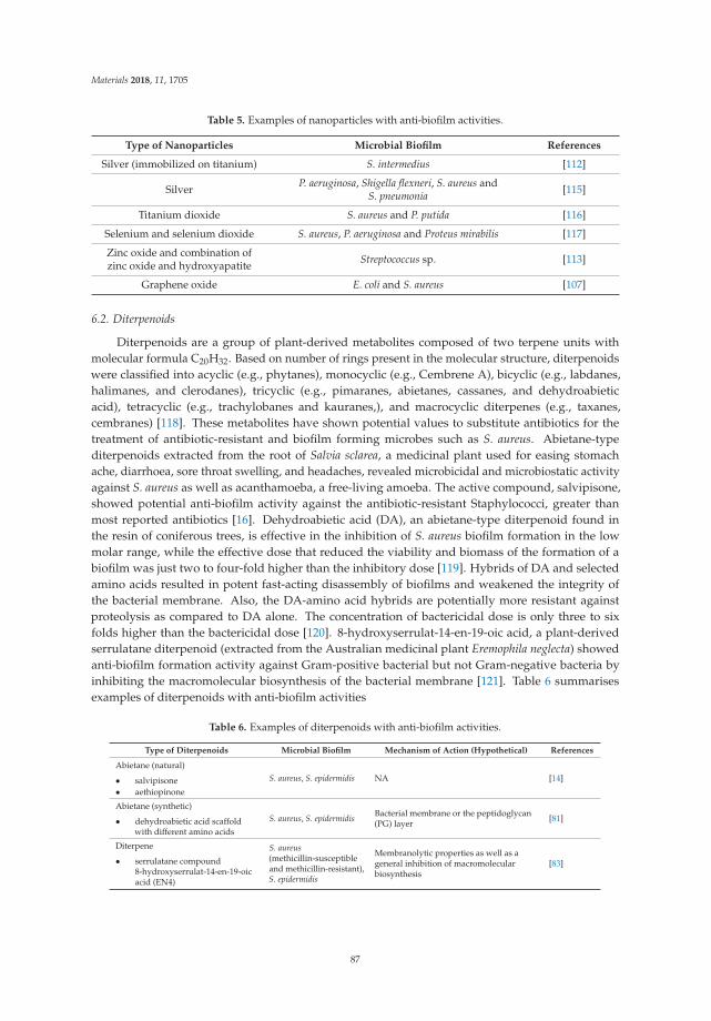
Materials 2018, 11, 1705
Table 5. Examples of nanoparticles with anti-biofilm activities.
Type of Nanoparticles Microbial Biofilm References
Silver (immobilized on titanium) S. intermedius [112]
Silver P. aeruginosa, Shigella flexneri, S. aureus andS. pneumonia [115]
Titanium dioxide S. aureus and P. putida [116]
Selenium and selenium dioxide S. aureus, P. aeruginosa and Proteus mirabilis [117]
Zinc oxide and combination ofzinc oxide and hydroxyapatite Streptococcus sp. [113]
Graphene oxide E. coli and S. aureus [107]
6.2. Diterpenoids
Diterpenoids are a group of plant-derived metabolites composed of two terpene units withmolecular formula C20H32. Based on number of rings present in the molecular structure, diterpenoidswere classified into acyclic (e.g., phytanes), monocyclic (e.g., Cembrene A), bicyclic (e.g., labdanes,halimanes, and clerodanes), tricyclic (e.g., pimaranes, abietanes, cassanes, and dehydroabieticacid), tetracyclic (e.g., trachylobanes and kauranes,), and macrocyclic diterpenes (e.g., taxanes,cembranes) [118]. These metabolites have shown potential values to substitute antibiotics for thetreatment of antibiotic-resistant and biofilm forming microbes such as S. aureus. Abietane-typediterpenoids extracted from the root of Salvia sclarea, a medicinal plant used for easing stomachache, diarrhoea, sore throat swelling, and headaches, revealed microbicidal and microbiostatic activityagainst S. aureus as well as acanthamoeba, a free-living amoeba. The active compound, salvipisone,showed potential anti-biofilm activity against the antibiotic-resistant Staphylococci, greater thanmost reported antibiotics [16]. Dehydroabietic acid (DA), an abietane-type diterpenoid found inthe resin of coniferous trees, is effective in the inhibition of S. aureus biofilm formation in the lowmolar range, while the effective dose that reduced the viability and biomass of the formation of abiofilm was just two to four-fold higher than the inhibitory dose [119]. Hybrids of DA and selectedamino acids resulted in potent fast-acting disassembly of biofilms and weakened the integrity ofthe bacterial membrane. Also, the DA-amino acid hybrids are potentially more resistant againstproteolysis as compared to DA alone. The concentration of bactericidal dose is only three to sixfolds higher than the bactericidal dose [120]. 8-hydroxyserrulat-14-en-19-oic acid, a plant-derivedserrulatane diterpenoid (extracted from the Australian medicinal plant Eremophila neglecta) showedanti-biofilm formation activity against Gram-positive bacterial but not Gram-negative bacteria byinhibiting the macromolecular biosynthesis of the bacterial membrane [121]. Table 6 summarisesexamples of diterpenoids with anti-biofilm activities
Table 6. Examples of diterpenoids with anti-biofilm activities.
Type of Diterpenoids Microbial Biofilm Mechanism of Action (Hypothetical) References
Abietane (natural)
• salvipisone• aethiopinone
S. aureus, S. epidermidis NA [14]
Abietane (synthetic)
• dehydroabietic acid scaffoldwith different amino acids
S. aureus, S. epidermidis Bacterial membrane or the peptidoglycan(PG) layer [81]
Diterpene
• serrulatane compound8-hydroxyserrulat-14-en-19-oicacid (EN4)
S. aureus(methicillin-susceptibleand methicillin-resistant),S. epidermidis
Membranolytic properties as well as ageneral inhibition of macromolecularbiosynthesis
[83]
87
Materials 2018, 11, 1705
6.3. Biomacromolecules
Biomacromolecules such as polysaccharides, naturally secreted polymers, aliphatic, cyclic, andaromatic organic acids were studied for their potential in preventing and constraining biofilm formationor resolving the formed biofilm. Distinct from antibiotics, these macromolecules do not target specificintracellular molecules within the microbes. Instead, with their amphiphilic characteristics, the cationicgroup of the molecules facilitates microbial targeting and water solubility; while the hydrophobicgroup induces membrane lysis; being attracted to the microbial membrane via interaction betweentheir cationic groups and the anionic membrane’s surface. Upon physical interaction, the hydrophobicgroup penetrates the microbial membrane leading to membrane destabilization and cytoplasmiccontent leakage. Antimicrobial agents with physical membrane disruption mechanisms are less likelyto be targeted by antimicrobial resistance [122]. Polysaccharides produced by a bacterium may affectbiofilm formation of other species through competition and cooperation phenomena [123]. For instance,K. pneumoniae capsular polysaccharide was suggested to restrict biofilm formation in several clinicallyimportant Gram-positive and Gram-negative bacterial species such as S. epidermidis, S. aureus, E. coli,and Enterobacter aerogenes [124,125]. Extracellular polymeric substances such as natural high molecularweight polymers secreted by bacteria have also been suggested to be anti-biofilm candidates [126].Interruption of bacterial signaling systems such as quorum sensing affects bacterial biofilm formation.In case of a mixed culture, acyl-homoserine lactonase produced by Bacillus cereus showed inhibitionand settlement of Vibrio cholera biofilm [127]. On the other hand, commercial organic acid productsusually used in food industry showed the ability to reduce Salmonella enterica viable count and biofilmpost-treatment, but not total elimination of the bacterium [128].
Incorporating macromolecules with anti-biofilm properties onto the surface of biomaterials hasgained increasing interest, especially in implant-associated medical devices such as intravascularcatheters, urinary catheters, and orthopedic implants [129]. Coating of biomaterial surface withnatural or modified polysaccharide polymers such as hyaluronic acid, heparin, and chitosan revealedpromising findings in battling implant-associated biofilm infections [130]. Table 7 summarisesexamples of examples of biomacromolecules with anti-biofilm activities.
Table 7. Examples of biomacromolecules with anti-biofilm activities.
Type of Macromolecule Microbial Biofilm Mechanism of Action (Hypothetical) References
Polyether etherketone–octafluoropentyl methacrylate
surface- Reduced protein adsorption [91]
AHL lactonase (AiiA), ametallo-beta-lactamase produced by
Bacillus spp.Vibrio cholerae
blocks quorum sensing inGram-negative bacteria by hydrolyzing
N-acyl-homoserine lactones (AHLs)[89]
Chitosan-based surface coating S. aureus, S. epidermidis anti-adhesive and bactericidal viacontact membrane disruption [92]
K. pneumoniae capsular polysaccharide S. aureus, S. epidermidis, E. coli NA [87]
Commercially available organic acidwater additives Salmonella Typhimurium biofilms
Interference to intracellular pHhomeostasis, membrane structure,
osmolality and macromoleculesynthesis
[90]
Synthetic PDMEA MeI polymers S. epidermidis, S. aureus, E. coli andP. aeruginosa, Candida albicans Membrane disruption [84]
6.4. Honey
Honey, an ancient wound remedy has gathered renewed interest in its clinical potential forinhibiting a wide range of infectious agents and promoting rapid wound healing [131]. The availabilityof medical grade honey with laboratory proven effects at the cellular and molecular level againstcertain microorganisms is not uncommon [131–134]. Mechanisms of bacterial inhibition within biofilmformation attributed to honey is a particular focus due to the increasing multidrug resistance ofbiofilm-associated organisms. Fortunately, there is accumulating evidence to show honey displays
88

Materials 2018, 11, 1705
activity in both preventing the formation of a biofilm either through interfering with adherence to hostcells or interfering with quorum sensing and disrupting an established biofilm. Several types of honeyhave been shown to exhibit anti-biofilm activity in-vitro (Table 8).
Besides being used alone, honey also exhibits synergistic activity in combination therapy withcertain antibiotics for planktonic cells [135–137]. Although promising, the application of honey asan antibacterial, anti-biofilm, and wound healing promoting alternative is still primarily confinedto in-vitro testing. This is mainly due to the varying reports describing different sources of honey,the strain of bacteria, biofilm stage, optimum dosage used, and wound/biotic conditions that mightinfluence the effectiveness of honey when being applied. A few in-vivo model using merino sheepand albino mice demonstrated that anti-biofilm activity was found in honey used, but advised thatoptimal clinical application should be titrated carefully as tissue toxicity and rejection of the necrosedarea from the epidermis was also found with increasing concentration [138,139]. However, care alsoshould be taken on the biofilm-enhancing action of low doses (<MIC) of honey that could be due toa stress response, which has been observed when bacteria in biofilms are exposed to sub-inhibitoryconcentrations of antibiotics [140].
Table 8. Examples of honey with anti-biofilm activity.
Type of Honey Microbial Biofilm Source
Manuka • P. aeruginosa, Streptococcus pyogenes, S. aureus, Klebsiella spp., Proteusmirabilis *, E. coli, Acinetobacter baumannii, Clostridium difficile
[141–148]
Clover • P. aeruginosa, S. aureus, Klebsiella spp., Proteus mirabilis [141,149]
Pumpkin • Bacillus subtilis (B. subtilis) [150]
Chestnut and thyme • B. subtilis, S. aureus [150]
Euphorbia • B. subtilis, S. aureus, P. aeruginosa, E. coli [150,151]
Chaste • B. subtilis, S. aureus, S. epidermis [150]
Multifloral• B. subtilis, S. aureus, S. epidermis• Staphylococcus mutans• Listeria monocytogenes
[150]
Eucalyptus • B. subtilis, S. aureus [150]
Honeydew • B. subtilis, S. aureus, S. epidermis, S. aureus, S. agalactiae, P. aeruginosa,E. faecalis *
[145,150]
Lavender, strawberryand citrus • P. aeruginosa, S. aureus & MRSA [152]
Sidr • P. aeruginosa, S. aureus, E. coli [151]
* With a certain degree of resistance.
6.5. Antimicrobial Peptides
Antimicrobial peptides (AMPs) are peptides molecules which are produced by many tissues andcell types in a variety of invertebrate, plant, and animal species. The natural peptides are generallymade up of 10–50 amino acid residues, positively charged (+2 to +9), with around 50% of hydrophobicproperties and diverse sequences and structures [153]. The antimicrobial activities of these peptidesare attributed by the amino acid composition, ampipathicy, cationic charge, and size that allow themto attach and insert themselves into the bacterial membrane bilayers to form pores and thus, kill thebacteria [154]. Other than working effectively against planktonic bacterial cells, AMP has also beenshown to be effective against biofilms. Mataraci, 2012 evaluated the anti-biofilm activities of twoAMPs, indolicin and CAMA: cecropin (1–7)-melittin A (2–9) amide which was found to inhibit MRSAbiofilms formation [9]. Additionally other AMPs are currently highlighted as a promising approach toprevent biofilm formation or to treat established biofilms, for instance, LL-37, HBD3, hep-20, IDR-1018are able to inhibit several species of biofilm formation by either down-regulating the genes essentialfor biofilm development or up-regulate the expression of genes resulting in a marked attenuation of
89

Materials 2018, 11, 1705
biofilm production and even by altering the architecture and reducing the amount of extracellularmatrix [155–157]. AMPs also are known as host defense peptides are essential components of innateimmunity in higher organisms, contributing to the first line of defense against infections [158]. Whileinvestigating possible anti-biofilm peptides from natural resources, synthetic peptides produced eitherby de novo synthesis or by modification gained increased interest based on their improved biologicalfunctions and reduced size, which in turn reduces production costs [159,160]. A curated list of usefulAMP along with their antimicrobial properties has been documented by de Luca and held within adatabase called BaAMP accessed via www.baamps.it [161].
Several comprehensive works reviewing various aspects of AMPs have been performed [155–157].Success of AMPs in both antimicrobial and anti-biofilm activities are collectively due to severalcharacteristics namely: (i) rapid bactericidal effects; (ii) high plasticity in different microenvironments;(iii) good penetration into the matrix of extracellular polymeric substances (EPS); (iv) anti-quorumsensing; (v) host response modulator; and (vi) synergistic effects with other conventional andunconventional antimicrobial compounds [155]. The mechanisms exhibited by AMPs are illustrated ina different state in Figure 4.
6.6. Antimicrobial Polymer
Antimicrobial polymers are synthetic polymers covalently linked with functional groups withhigh antimicrobial activity such as amino, hydroxyl, and carboxyl groups [162]. Antimicrobialpolymers are effective against a range of bacteria including the bacteria commonly associated withHAIs [163]. Due to the long and repeating chain of active and charged functional groups, the commonmechanism of antimicrobial activity is through disruption of the cell wall or cytoplasmic membranes.Takahashi et al. demonstrated that cationic homopolymer PE0 and copolymer PE31 containing 31% ofethyl methacrylate was effective in removing biofilm of Staphylococcus mutans (S. mutans) compared tochlorohexidine and the cationic surfactant that was tested at the same concentration [162]. Anotherstudy by Li et al. showed that cationic monomer, methacryloxylethylcetyl dimethyl ammoniumchloride (DMAE-CB) was also effective in the removal of a biofilm of S. mutans, the common bacteriaassociated dental problems [164]. Peng et al. modified the polyurethane compound, the maincompound for catheter by copolymerization of an amine functionalized N-substituted diol to givea cationic polyurethane (Tecoflex-NH3), and showed that the cationic polyurethane shown contactkilling of E. coli and prevent build-up of biofilm on the surfaces, thus, reducing the chances of CAUTI,one of the main causes of the nosocomial infections [165]. Another example of an antimicrobialpolymer is Polyhexamethlene biguanide (PHMB), a cationic polymer that has been used in the clinicfor over than 40 years with no sign of bacterial resistance [166]. PHMB mediates antibacterial activitiesthrough disruption of the cell wall and condensation of the bacterial chromosome [111], and recentdiscovery demonstrate PHMB efficacy in killing intracellular bacteria [16,167]. PHMB is also effectiveagainst biofilms from a range of bacterial species and thus effective when applied for the treatment ofwound infections [39,168,169]. Though there are increasing findings for polymer mediated anti-biofilmactivities, the complete mechanism of the activities is not fully understood. The anti-biofilm activitiesposed by the polymers could possibly be due to the interaction of the charged group on the polymerstructure with the eDNA or Ca2+, thus disrupting the biofilm structure and cause destabilization.
90

Mat
eria
ls2
01
8,1
1,17
05
Fig
ure
4.
The
thre
em
ain
step
sof
the
biofi
lmlif
e-cy
cle
(att
achm
entt
oa
surf
ace,
mat
urat
ion,
and
disp
ersa
l)an
dth
em
echa
nism
sex
hibi
ted
byA
ntim
icro
bial
pept
ides
(AM
Ps)i
nev
ery
step
.
91

Materials 2018, 11, 1705
7. Summary and Outlook
Biofilms, a form of protective armour for the bacteria, are generally more resistant to the treatment.Biofilms will remain a challenge for the prevention and control of infection; thus it is critical thatwe continue to explore and understand the physiology and structure of biofilms to develop animproved, innovative, and novel targeted therapies. Based on promising in-vitro studies and reportsinvestigating the use of nanoparticles, AMPs, diterpenoids, and biomacromolecules; we propose thatthese compounds should be the focus of future novel biofilm control strategies. Further studies focusingon the efficacy and tolerability in-vivo are required to ascertain the level of translation of in-vitro resultsto clinical resolution of infections caused by biofilms. Together with the increased understanding of thefundamental biology of biofilms, the application of novel or repurposed compounds will undoubtedlyimprove the prospect of treating and resolving biofilm infection within clinical settings.
Funding: This research received no external funding
Acknowledgments: Khairun Anisa Mat Yazid is funded by UMK Zamalah Scheme, Shamsaldeen Ibrahim Saeedis funded by Universiti of Nyala, Sudan, Alexandru Chivu is funded by UCL LiDO scheme, and Amanda JaneGibson is funded by BBSRC.
Conflicts of Interest: The authors declare no conflicts of interest
References
1. Khan, H.A.; Baig, F.K.; Mehboob, R. Nosocomial infections: Epidemiology, prevention, control andsurveillance. Asian Pac. J. Trop. Biomed. 2017, 7, 478–482. [CrossRef]
2. Bjarnsholt, T. The role of bacterial biofilms in chronic infections. Apmis 2013, 121, 1–58. [CrossRef] [PubMed]3. Jiang, W.; Hu, X.; Hu, Z.; Tang, Z.; Wu, H.; Chen, L. Morbidity and mortality of nosocomial infection after
cardiovascular surgery: A report of 1606 cases. Curr. Med. Sci. 2018, 38, 329–335. [CrossRef] [PubMed]4. Barrasa-Villar, J.I.; Aibar-Remón, C.; Prieto-Andrés, P.; Mareca-Doñate, R.; Moliner-Lahoz, J. Impact on
morbidity, mortality and length of stay of hospital acquired infections by resistant microorganisms. Clin.Infect. Dis. 2017, 65, 644–652. [CrossRef] [PubMed]
5. Klevens, R.M.; Edwards, J.R.; Richards, C.; Horan, T.C.; Gaynes, R.P.; Pollock, D.A.; Cardo, D.M. EstimatingHealth Care-Associated Infections and Deaths in U.S. Hospitals, 2002. Public Health Rep. 2007, 122, 160–166.[CrossRef] [PubMed]
6. Chopra, I.; Schofield, C.; Everett, M.; O’Neill, A.; Miller, K.; Wilcox, M.; Frere, J.M.; Dawson, M.;Czaplewski, L.; Urleb, U.; et al. Treatment of health-care-associated infections caused by Gram-negativebacteria: A consensus statement. Lancet Infect. Dis. 2008, 8, 133–139. [CrossRef]
7. Allegranzi, B.; Nejad, S.B.; Combescure, C.; Graafmans, W.; Attar, H.; Donaldson, L.; Pittet, D. Burden ofendemic health-care-associated infection in developing countries: Systematic review and meta-analysis.Lancet 2011, 377, 228–241. [CrossRef]
8. Herman-Bausier, P.; Dufrene, Y.F. Force matters in hospital-acquired infections. Science 2018, 359, 1464–1465.[CrossRef] [PubMed]
9. Mataraci, E.; Dosler, S. In vitro activities of antibiotics and antimicrobial cationic peptides alone and incombination against methicillin-resistant Staphylococcus aureus biofilms. Antimicrob. Agents Chemother. 2012,56, 6366–6371. [CrossRef] [PubMed]
10. Percival, S.L.; Suleman, L.; Donelli, G. Healthcare-Associated infections, medical devices and biofilms: Risk,tolerance and control. J. Med. Microbiol. 2015, 64, 323–334. [CrossRef] [PubMed]
11. Al-Talib, H.; Yean, C.; Al-Jashamy, K.; Hasan, H. Methicillin-resistant Staphylococcus aureus nosocomialinfection trends in Hospital Universiti Sains Malaysia during 2002–2007. Ann. Saudi Med. 2010, 30, 358–363.[CrossRef] [PubMed]
12. Macmorran, E.; Harch, S.; Athan, E.; Lane, S.; Tong, S.; Crawford, L.; Krishnaswamy, S.; Hewagama, S. Therise of methicillin resistant Staphylococcus aureus: Now the dominant cause of skin and soft tissue infection inCentral Australia. Epidemiol. Infect. 2017, 145, 2817–2826. [CrossRef] [PubMed]
92
Materials 2018, 11, 1705
13. Enright, M.C.; Robinson, D.A.; Randle, G.; Feil, E.J.; Grundmann, H.; Spratt, B.G. The evolutionary history ofmethicillin-resistant Staphylococcus aureus (MRSA). Proc. Natl. Acad. Sci. USA 2002, 99, 7687–7692. [CrossRef][PubMed]
14. Malani, P.N. National burden of invasive methicillin-resistant Staphylococcus aureus infection. JAMA 2014,311, 1438–1439. [CrossRef] [PubMed]
15. Sisirak, M.; Zvizdic, A.; Hukic, M. Methicillin-resistant Staphylococcus aureus (MRSA) as a cause of nosocomialwound infections. Bosn. J. Basic Med. Sci. 2010, 10, 32–37. [CrossRef] [PubMed]
16. Kamaruzzaman, N.F.; Firdessa, R.; Good, L. Bactericidal effects of polyhexamethylene biguanide againstintracellualar Staphylococcus aureus EMRSA-15 and USA 300. J. Antimicrob. Chemother. 2016, 71, 1252–1259.[CrossRef] [PubMed]
17. Zhou, Y.; Li, G. Diagnosis and management of complicated intra-abdominal infection in adults and children:Guidelines by the surgical infection society and the infectious diseases society of America. Chin. J. Infect.Chemother. 2010, 10, 241–247. [CrossRef]
18. Codjoe, F.; Donkor, E. Carbapenem Resistance: A Review. Med. Sci. 2017, 6, 1. [CrossRef] [PubMed]19. Johansen, H.K.; Moskowitz, S.M.; Ciofu, O.; Pressler, T.; Høiby, N. Spread of colistin resistant non-mucoid
Pseudomonas aeruginosa among chronically infected Danish cystic fibrosis patients. J. Cyst. Fibros. 2008, 7,391–397. [CrossRef] [PubMed]
20. Criss, A.K.; Katz, B.Z.; Seifert, H.S. Resistance of Neisseria gonorrhoeae to non-oxidative killing by adherenthuman polymorphonuclear leucocytes. Cell. Microbiol. 2009, 11, 1074–1087. [CrossRef] [PubMed]
21. Fiaccadori, E.; Antonucci, E.; Morabito, S.; d’Avolio, A.; Maggiore, U.; Regolisti, G. Colistin Use in PatientsWith Reduced Kidney Function. Am. J. Kidney Dis. 2016, 68, 296–306. [CrossRef] [PubMed]
22. Chatterjee, M.; Anju, C.P.; Biswas, L.; Anil Kumar, V.; Gopi Mohan, C.; Biswas, R. Antibiotic resistance inPseudomonas aeruginosa and alternative therapeutic options. Int. J. Med. Microbiol. 2016, 306, 48–58. [CrossRef][PubMed]
23. Cornejo-Juárez, P.; Vilar-Compte, D.; Pérez-Jiménez, C.; Ñamendys-Silva, S.A.; Sandoval-Hernández, S.;Volkow-Fernández, P. The impact of hospital-acquired infections with multidrug-resistant bacteria in anoncology intensive care unit. Int. J. Infect. Dis. 2015, 31, 31–34. [CrossRef] [PubMed]
24. Golan, Y. Empiric therapy for hospital-acquired, Gram-negative complicated intra-abdominal infection andcomplicated urinary tract infections: A systematic literature review of current and emerging treatmentoptions. BMC Infect. Dis. 2015, 15, 1–7. [CrossRef] [PubMed]
25. Yayan, J.; Ghebremedhin, B.; Rasche, K. Antibiotic resistance of Pseudomonas aeruginosa in pneumonia at asingle university hospital center in Germany over a 10-Year Period. PLoS ONE 2015, 10, 1–20. [CrossRef][PubMed]
26. Jamal, M.; Ahmad, W.; Andleeb, S.; Jalil, F.; Imran, M.; Nawaz, M.A.; Hussain, T.; Ali, M.; Rafiq, M.;Kamil, M.A. Bacterial biofilm and associated infections. J. Chin. Med. Assoc. 2018, 81, 7–11. [CrossRef][PubMed]
27. Mcguffie, B.A.; Vallet-gely, I.; Dove, S.L. σ factor and anti-σ factor that control swarming motility and biofilmformation in Pseudomonas aeruginosa. J. Bacteriol. 2016, 198, 755–765. [CrossRef] [PubMed]
28. Chaftari, A.M.; Zakhem, A.E.; Jamal, M.A.; Jiang, Y.; Hachem, R.; Raad, I. The use of minocycline-rifampincoated central venous catheters for exchange of catheters in the setting of Staphylococcus aureus central lineassociated bloodstream infections. BMC Infect. Dis. 2014, 14, 1–5. [CrossRef] [PubMed]
29. Paczosa, M.K.; Mecsas, J. Klebsiella pneumoniae: Going on the offense with a strong defense. Microbiol. Mol.Biol. Rev. 2016, 80, 629–661. [CrossRef] [PubMed]
30. Surveillance of Antimicrobial Resistance in Europe 2016; Annual report of the European Antimicrobial REsistanceSurveillance Network (EARS-Net); European Centre for Disease Prevention and Control: Solna, Sweden,2017; ISBN 9789294980991.
31. Livermore, D.M.; Maya, J.J.; Nordmann, P.; Wang, H.; Woodford, N.; Quinn, J.P. Clinical epidemiology of theglobal expansion of Klebsiella pneumoniae carbapenemases. Lancet Infect. Dis. 2013, 13, 785–796. [CrossRef]
32. Otter, J.A.; Doumith, M.; Davies, F.; Mookerjee, S.; Dyakova, E.; Gilchrist, M.; Brannigan, E.T.; Bamford, K.;Galletly, T.; Donaldson, H.; et al. Emergence and clonal spread of colistin resistance due to multiplemutational mechanisms in carbapenemase-producing Klebsiella pneumoniae in London. Sci. Rep. 2017, 7, 1–8.[CrossRef] [PubMed]
93
Materials 2018, 11, 1705
33. Singla, S.; Harjai, K.; Chhibber, S. Artificial Klebsiella pneumoniae biofilm model mimicking in vivo system:Altered morphological characteristics and antibiotic resistance. J. Antibiot. 2014, 67, 305–309. [CrossRef][PubMed]
34. Chung, P.Y. The emerging problems of Klebsiella pneumoniae infections: Carbapenem resistance and biofilmformation. FEMS Microbiol. Lett. 2016, 363, 1–6. [CrossRef] [PubMed]
35. Anderl, J.N.; Franklin, M.J.; Stewart, P.S. Role of antibiotic penetration limitation in Klebsiella pneumoniaebiofilm resistance to ampicillin and ciprofloxacin. Antimicrob. Agents Chemother. 2000, 44, 1818–1824.[CrossRef] [PubMed]
36. Jendresen, M.D.; Glantz, P.O. Clinical adhesiveness of selected dental materials: An in-vivo study. ActaOdontol. Scand. 1981, 39, 39–45. [CrossRef] [PubMed]
37. Høiby, N. A personal history of research on microbial biofilms and biofilm infections. Pathog. Dis. 2014, 70,205–211. [CrossRef] [PubMed]
38. Werner, E.; Roe, F.; Bugnicourt, A.; Michael, J.; Heydorn, A.; Molin, S.; Pitts, B.; Stewart, P.S.; Franklin, M.J.Stratified growth in Pseudomonas aeruginosa biofilms stratified growth in Pseudomonas aeruginosa biofilms.Appl. Environ. Microbiol. 2004, 70, 6188–6196. [CrossRef] [PubMed]
39. Kamaruzzaman, N.F.; Chong, S.Q.Y.; Edmondson-Brown, K.M.; Ntow-Boahene, W.; Bardiau, M.; Good, L.Bactericidal and anti-biofilm effects of polyhexamethylene Biguanide in models of intracellular and biofilmof Staphylococcus aureus isolated from bovine mastitis. Front. Microbiol. 2017, 8, 1–10. [CrossRef] [PubMed]
40. Czaczyk, K.; Myszka, K. Biosynthesis of extracellular polymeric substances (EPS) and its role in microbialbiofilm formation. Pol. J. Environ. Stud. 2007, 16, 799–806.
41. Izano, E.A.; Amarante, M.A.; Kher, W.B.; Kaplan, J.B. Differential roles of poly-N-acetylglucosamine surfacepolysaccharide and extracellular DNA in Staphylococcus aureus and Staphylococcus epidermidis biofilms. Appl.Environ. Microbiol. 2008, 74, 470–476. [CrossRef] [PubMed]
42. Billings, N.; Ramirez Millan, M.; Caldara, M.; Rusconi, R.; Tarasova, Y.; Stocker, R.; Ribbeck, K.The extracellular matrix component Psl provides fast-acting antibiotic defense in Pseudomonas aeruginosabiofilms. PLoS Pathog. 2013, 9, e1003526. [CrossRef] [PubMed]
43. Jennings, L.K.; Storek, K.M.; Ledvina, H.E.; Coulon, C.; Marmont, L.S.; Sadovskaya, I.; Secor, P.R.; Tseng, B.S.;Scian, M.; Filloux, A.; et al. Pel is a cationic exopolysaccharide that cross-links extracellular DNA in thePseudomonas aeruginosa biofilm matrix. Proc. Natl. Acad. Sci. USA 2015, 112, 11353–11358. [CrossRef][PubMed]
44. Das, T.; Sehar, S.; Koop, L.; Wong, Y.K.; Ahmed, S.; Siddiqui, K.S.; Manefield, M. Influence of calcium inextracellular DNA mediated bacterial aggregation and biofilm formation. PLoS ONE 2014, 9, 1–11. [CrossRef][PubMed]
45. Bos, R.; Van Der Mei, H.C.; Busscher, H.J. Physico-chemistry of initial microbial adhesive interactions-itsmechanisms and methods for study. FEMS Microbiol. Rev. 1999, 23, 179–229. [CrossRef] [PubMed]
46. Okshevsky, M.; Meyer, R.L. The role of extracellular DNA in the establishment, maintenance andperpetuation of bacterial biofilms. Crit. Rev. Microbiol. 2013, 41, 1–11. [CrossRef] [PubMed]
47. Montanaro, L.; Poggi, A.; Visai, L.; Ravaioli, S.; Campoccia, D.; Speziale, P.; Arciola, C.R. Extracellular DNAin biofilms. Int. J. Artif. Organs 2011, 34, 824–831. [CrossRef] [PubMed]
48. Davies, D.G.; Parsek, M.R.; Pearson, J.P.; Iglewski, B.H.; Costerton, J.W.; Greenberg, E.P. The involvement ofcell-to-cell signals in the development of a bacterial biofilm. Science 1998, 280, 295–298. [CrossRef] [PubMed]
49. Braga, R.M.; Dourado, M.N.; Araújo, W.L. Microbial interactions: Ecology in a molecular perspective. Braz. J.Microbiol. 2016, 47, 86–98. [CrossRef] [PubMed]
50. Costerton, A.J.W.; Stewart, P.S.; Greenberg, E.P. Bacterial Biofilms: A Common Cause of Persistent Infections.Science 2011, 284, 1318–1322. [CrossRef]
51. Stewart, P.S.; Franklin, M.J. Physiological heterogeneity in biofilms. Nat. Rev. Microbiol. 2008, 6, 199–210.[CrossRef] [PubMed]
52. Mah, T.C.; Toole, G.A.O. Mechanisms of biofilm resistance to antimicrobial agents. Trends Microbiol. 2001, 9,34–39. [CrossRef]
53. La, B.; Prosser, T.; Taylor, D.; Dix, B.A.; Cleeland, R.O.Y. Method of evaluating effects of antibiotics onbacterial biofilm. Antimicrob. Agents Chemother. 1987, 31, 1502–1506.
94
Materials 2018, 11, 1705
54. Nickel, J.C.; Ruseska, I.; Wright, J.B.; Costerton, J.W. Tobramycin resistance of cells of Pseudomonas aeruginosagrowing as biofilm on urinary catheter material. Antimicrob. Agents Chemother. 1985, 27, 619–624. [CrossRef][PubMed]
55. Hall, C.W.; Mah, T.F. Molecular mechanisms of biofilm-based antibiotic resistance and tolerance in pathogenicbacteria. FEMS Microbiol. Rev. 2017, 41, 276–301. [CrossRef] [PubMed]
56. Singh, R.; Sahore, S.; Kaur, P.; Rani, A.; Ray, P. Penetration barrier contributes to bacterial biofilm-associatedresistance against only select antibiotics, and exhibits genus-, strain- and antibiotic-specific differences.Pathog. Dis. 2016, 74, 1–6. [CrossRef] [PubMed]
57. Nguyen, D.; Joshi-Datar, A.; Lepine, F.; Bauerle, E.; Olakanmi, O.; Beer, K.; McKay, G.; Siehnel, R.;Schafhauser, J.; Wang, Y.; et al. Active starvation responses mediate antibiotic tolerance in biofilms andnutrient-limited bacteria. Science 2011, 334, 982–986. [CrossRef] [PubMed]
58. Bowler, L.L.; Zhanel, G.G.; Ball, T.B.; Saward, L.L. Mature Pseudomonas aeruginosa biofilms prevail comparedto young biofilms in the presence of ceftazidime. Antimicrob. Agents Chemother. 2012, 56, 4976–4979.[CrossRef] [PubMed]
59. Johnson, L.; Horsman, S.R.; Charron-Mazenod, L.; Turnbull, A.L.; Mulcahy, H.; Surette, M.G.; Lewenza, S.Extracellular DNA-induced antimicrobial peptide resistance in Salmonella enterica serovar Typhimurium.BMC Microbiol. 2013, 13, 115. [CrossRef] [PubMed]
60. Walters, M.C.; Roe, F.; Bugnicourt, A.; Franklin, M.J.; Stewart, P.S. Contributions of Antibiotic Penetration,Oxygen Limitation. Antimicrob. Agents Chemother. 2003, 47, 317–323. [CrossRef] [PubMed]
61. Borriello, G.; Werner, E.; Roe, F.; Kim, A.M.; Ehrlich, G.D.; Stewart, P.S. Oxygen limitation contributesto antibiotic tolerance of Pseudomonas aeruginosa in biofilms oxygen limitation contributes to antibiotictolerance of Pseudomonas aeruginosa in biofilms. Antimicrob. Agents Chemother. 2004, 48, 2659–2664. [CrossRef][PubMed]
62. Stewart, P.S.; Franklin, M.J.; Williamson, K.S.; Folsom, J.P.; Boegli, L.; James, G.A. Contribution of stressresponses to antibiotic tolerance in Pseudomonas aeruginosa biofilms. Antimicrob. Agents Chemother. 2015, 59,3838–3847. [CrossRef] [PubMed]
63. Waters, E.M.; Rowe, S.E.; O’Gara, J.P.; Conlon, B.P. Convergence of Staphylococcus aureus persister and biofilmresearch: Can biofilms be defined as communities of adherent persister cells? PLoS Pathog. 2016, 12, 2–6.[CrossRef] [PubMed]
64. Le, K.Y.; Park, M.D.; Otto, M. Immune evasion mechanisms of Staphylococcus epidermidis biofilm infection.Front. Microbiol. 2018, 9, 1–8. [CrossRef] [PubMed]
65. Gunn, J.S.; Bakaletz, L.O.; Wozniak, D.J. What’s on the outside matters: The role of the extracellular polymericsubstance of gram-negative biofilms in evading host immunity and as a target for therapeutic intervention.J. Biol. Chem. 2016, 291, 12538–12546. [CrossRef] [PubMed]
66. Roilides, E.; Simitsopoulou, M.; Katragkou, A.; Walsh, T.J. How Biofilms Evade Host Defenses. In MicrobialBiofilms, 2nd ed.; American Society for Microbiology: Washington, DC, USA, 2015; pp. 287–300.ISBN1 978-1-55581-745-9. ISBN2 978-1-55581-746-6.
67. Leid, J.G.; Shirtliff, M.E.; Costerton, J.W.; Stoodley, P. Human leukocytes adhere to, penetrate, and respond toStaphylococcus aureus biofilms. Infect. Immun. 2002, 70, 6339–6345. [CrossRef] [PubMed]
68. Walker, T.S.; Tomlin, K.L.; Worthen, G.S.; Poch, R.; Lieber, J.G.; Saavedra, M.T.; Michael, B.; Malcolm, K.C.;Vasil, M.L.; Jerry, A.N.; et al. Enhanced Pseudomonas aeruginosa biofilm development mediated by humanneutrophils enhanced Pseudomonas aeruginosa biofilm development mediated by human neutrophils. Infect.Immun. 2005, 73, 3693–3701. [CrossRef] [PubMed]
69. Thurlow, L.R.; Hanke, M.L.; Fritz, T.; Angle, A.; Aldrich, A.; Williams, S.H.; Engebretsen, I.L.; Bayles, K.W.;Horswill, A.R.; Kielian, T. Staphylococcus aureus biofilms prevent macrophage phagocytosis and attenuateinflammation in vivo. J. Immunol. 2011, 186, 6585–6596. [CrossRef] [PubMed]
70. Jesaitis, A.J.; Franklin, M.J.; Berglund, D.; Sasaki, M.; Lord, C.I.; Bleazard, J.B.; Duffy, J.E.; Beyenal, H.;Lewandowski, Z. Compromised host defense on Pseudomonas aeruginosa biofilms: Characterization ofneutrophil and biofilm interactions. J. Immunol. 2003, 171, 4329–4339. [CrossRef] [PubMed]
71. Jensen, E.T.; Kharazmi, A.; Lam, K.; Costerton, J.W.; Hoiby, N. Human polymorphonuclear leukocyteresponse to Pseudomonas aeruginosa grown in biofilms. Infect. Immun. 1990, 58, 2383–2385. [PubMed]
72. Wu, H.; Moser, C.; Wang, H.Z.; Høiby, N.; Song, Z.J. Strategies for combating bacterial biofilm infections. Int.J. Oral. Sci. 2015, 7, 1–7. [CrossRef] [PubMed]
95

Materials 2018, 11, 1705
73. Høiby, N.; Bjarnsholt, T.; Moser, C.; Bassi, G.L.; Coenye, T.; Donelli, G.; Hall-Stoodley, L.; Holá, V.; Imbert, C.;Kirketerp-Møller, K.; et al. ESCMID* guideline for the diagnosis and treatment of biofilm infections 2014.Clin. Microbiol. Infect. 2015, 21, S1–S25. [CrossRef] [PubMed]
74. Macià, M.D.; del Pozo, J.L.; Díez-Aguilar, M.; Guinea, J. Microbiological diagnosis of biofilm-relatedinfections. Enferm. Infecc. Microbiol. Clin. 2018, 36, 375–381. [CrossRef] [PubMed]
75. Almani, S.A.; Naseer, A.; Maheshwari, S.K.; Maroof, P. Detection of biofilm-associated implant pathogens incardiac device infections: High sensitivity of sonication fluid culture even in the presence of antimicrobials.J. Glob. Infect. Dis. 2017, 9, 135–138. [CrossRef] [PubMed]
76. Mandakhalikar, K.D.; Rahmat, J.N.; Chiong, E.; Neoh, K.G.; Shen, L.; Tambyah, P.A. Extraction andquantification of biofilm bacteria: Method optimized for urinary catheters. Sci. Rep. 2018, 8, 1–9. [CrossRef][PubMed]
77. Renz, N.; Cabric, S.; Morgenstern, C.; Schuetz, M.A.; Trampuz, A. Value of PCR in sonication fluid for thediagnosis of orthopedic hardware-associated infections: Has the molecular era arrived? Injury 2018, 49,806–811. [CrossRef] [PubMed]
78. Schuurs, T.A.; Koelewijn, R.; Brienen, E.A.T.; Kortbeek, T.; Mank, T.G.; Mulder, B.; Stelma, F.F.;Van Lieshout, L.; Van Hellemond, J.J. Harmonization of PCR-based detection of intestinal pathogens:Experiences from the Dutch external quality assessment scheme on molecular diagnosis of protozoa in stoolsamples. Clin. Chem. Lab. Med. 2018, 1–6. [CrossRef] [PubMed]
79. Stavnsbjerg, C.; Frimodt-Møller, N.; Moser, C.; Bjarnsholt, T. Comparison of two commercial broad-rangePCR and sequencing assays for identification of bacteria in culture-negative clinical samples. BMC Infect.Dis. 2017, 17, 1–7. [CrossRef] [PubMed]
80. Morio, F.; Dannaoui, E.; Chouaki, T.; Cateau, E.; Malard, O.; Bonfils, P.; Page, C.; Dufour, X.; Cottrel, C.;Erwan, T.; et al. PCR-based detection of Aspergillus fumigatus and absence of azole resistance due toTR34/L98H in a french multicenter cohort of 137 patients with fungal rhinosinusitis. Mycoses 2018, 61, 30–34.[CrossRef] [PubMed]
81. Davis, L.E.; Tyler, K.L. Molecular Diagnosis of CNS Viral Infections; Elsevier Inc.: New York, NY, USA, 2005;Voluem 76, ISBN 9780128138069.
82. Bizzini, A.; Greub, G.; Greub, G. Matrix-assisted laser desorption ionization time-of-flight mass spectrometry,a revolution in clinical microbial identification. Clin. Microbiol. Infect. 2010, 16, 1614–1619. [CrossRef][PubMed]
83. Kliem, M.; Sauer, S. The essence on mass spectrometry based microbial diagnostics. Curr. Opin. Microbiol.2012, 15, 397–402. [CrossRef] [PubMed]
84. Gaudreau, A.M.; Labrie, J.; Goetz, C.; Dufour, S.; Jacques, M. Evaluation of MALDI-TOF mass spectrometryfor the identification of bacteria growing as biofilms. J. Microbiol. Methods 2018, 145, 79–81. [CrossRef][PubMed]
85. Frickmann, H.; Zautner, A.E.; Moter, A.; Kikhney, J.; Hagen, R.M.; Stender, H.; Poppert, S. Fluorescence insitu hybridization (FISH) in the microbiological diagnostic routine laboratory: A review. Crit. Rev. Microbiol.2017, 43, 263–293. [CrossRef] [PubMed]
86. Schrøder, S.A.; Eickhardt, S.; Bjarnsholt, T.; Nørgaard, T.; Homøe, P. Morphological evidence of biofilm inchronic obstructive sialadenitis. J. Laryngol. Otol. 2018, 132, 1–4. [CrossRef] [PubMed]
87. Al-Ahmad, A.; Wunder, A.; Auschill, T.M.; Follo, M.; Braun, G.; Hellwig, E.; Arweiler, N.B. The in vivodynamics of Streptococcus spp., Actinomyces naeslundii, Fusobacterium nucleatum and Veillonella spp. in dentalplaque biofilm as analysed by five-colour multiplex fluorescence in situ hybridization. J. Med. Microbiol.2007, 56, 681–687. [CrossRef] [PubMed]
88. Hardy, L.; Jespers, V.; Dahchour, N.; Mwambarangwe, L.; Musengamana, V.; Vaneechoutte, M.; Crucitti, T.Unravelling the bacterial vaginosis-associated biofilm: A multiplex Gardnerella vaginalis and Atopobiumvaginae fluorescence in situ hybridization assay using peptide nucleic acid probes. PLoS ONE 2015, 10, 1–16.[CrossRef] [PubMed]
89. Hu, H.; Johani, K.; Gosbell, I.B.; Jacombs, A.S.W.; Almatroudi, A.; Whiteley, G.S.; Deva, A.K.; Jensen, S.;Vickery, K. Intensive care unit environmental surfaces are contaminated by multidrug-resistant bacteria inbiofilms: Combined results of conventional culture, pyrosequencing, scanning electron microscopy, andconfocal laser microscopy. J. Hosp. Infect. 2015, 91, 35–44. [CrossRef] [PubMed]
96

Materials 2018, 11, 1705
90. Mohmmed, S.A.; Vianna, M.E.; Penny, M.R.; Hilton, S.T.; Mordan, N.; Knowles, J.C. Confocal laser scanning,scanning electron, and transmission electron microscopy investigation of Enterococcus faecalis biofilmdegradation using passive and active sodium hypochlorite irrigation within a simulated root canal model.Microbiologyopen 2017, 6, 1–9. [CrossRef] [PubMed]
91. Tani, S.; Lepetsos, P.; Stylianakis, A.; Vlamis, J.; Birbas, K.; Kaklamanos, I. Superiority of the sonicationmethod against conventional periprosthetic tissue cultures for diagnosis of prosthetic joint infections. Eur. J.Orthop. Surg. Traumatol. 2018, 28, 51–57. [CrossRef] [PubMed]
92. Hayes, J.; Forbes, M.; Greenberg, D.P.; Dice, B.; Burrows, A.; Wackym, P.A.; Kerschner, J.E. Direct Detectionof Bacterial Biofilms on the Middle-Ear Mucosa of Children. JAMA 2006, 296, 202–211.
93. Renz, N.; Feihl, S.; Cabric, S.; Trampuz, A. Performance of automated multiplex PCR using sonication fluidfor diagnosis of periprosthetic joint infection: A prospective cohort. Infection 2017, 45, 877–884. [CrossRef][PubMed]
94. Boase, S.; Foreman, A.; Cleland, E.; Tan, L.; Melton-Kreft, R.; Pant, H.; Hu, F.Z.; Ehrlich, G.D.; Wormald, P.J.The microbiome of chronic rhinosinusitis: Culture, molecular diagnostics and biofilm detection. BMC Infect.Dis. 2013, 13, 1–9. [CrossRef] [PubMed]
95. Bjarnsholt, T.; Ciofu, O.; Molin, S.; Givskov, M.; Høiby, N. Applying insights from biofilm biology to drugdevelopment-can a new approach be developed? Nat. Rev. Drug Discov. 2013, 12, 791–808. [CrossRef][PubMed]
96. Cantón, R.; Máiz, L.; Escribano, A.; Olveira, C.; Oliver, A.; Asensio, O.; Gartner, S.; Roma, E.;Quintana-Gallego, E.; Salcedo, A.; et al. Spanish Consensus on the Prevention and Treatment of Pseudomonasaeruginosa Bronchial Infections in Cystic Fibrosis Patients. Arch. Bronconeumol. 2015, 51, 140–150. [CrossRef][PubMed]
97. Haworth, C.S.; Foweraker, J.E.; Wilkinson, P.; Kenyon, R.F.; Bilton, D. Inhaled colistin in patients withbronchiectasis and chronic Pseudomonas aeruginosa infection. Am. J. Respir. Crit. Care Med. 2014, 189, 975–982.[CrossRef] [PubMed]
98. Wilson, R.; Welte, T.; Polverino, E.; De Soyza, A.; Greville, H.; O’Donnell, A.; Alder, J.; Reimnitz, P.; Hampel, B.Ciprofloxacin dry powder for inhalation in non-cystic fibrosis bronchiectasis: A phase II randomised study.Eur. Respir. J. 2013, 41, 1107–1115. [CrossRef] [PubMed]
99. Ezzat, W.F.; Fawaz, S.A.; Rabie, H.; Hamdy, T.A.; Shokry, Y.A. Effect of topical ofloxacin on bacterialbiofilms in refractory post-sinus surgery rhino-sinusitis. Eur. Arch. Oto-Rhino-Laryngol. 2015, 272, 2355–2361.[CrossRef] [PubMed]
100. Kaiser, M.; Gil, J.; Treu, R.; Valdes, J.; Davis, S. An in vitro analysis of the effects of various topicalantimicrobial agents on methicillin-resistant and methicillin-sensitive strains of Staphylococcus aureus. OstomyWound Manag. 2014, 60, 18–28.
101. Hogan, S.; Zapotoczna, M.; Stevens, N.T.; Humphreys, H.; O’Gara, J.P.; O’Neill, E. In vitro approach foridentification of the most effective agents for antimicrobial lock therapy in the treatment of intravascularcatheter-related infections caused by Staphylococcus aureus. Antimicrob. Agents Chemother. 2016, 60, 2923–2931.[CrossRef] [PubMed]
102. Moghaddas, A.; Abbasi, M.R.; Gharekhani, A.; Dashti-Khavidaki, S.; Razeghi, E.; Jafari, A.; Khalili, H.Prevention of hemodialysis catheter-related blood stream infections using a cotrimoxazole-lock technique.Future Microbiol. 2015, 10, 169–178. [CrossRef] [PubMed]
103. Mataraci Kara, E.; Ozbek Celik, B. Investigation of the effects of various antibiotics against Klebsiellapneumoniae biofilms on in vitro catheter model. J. Chemother. 2018, 30, 82–88. [CrossRef] [PubMed]
104. Krajewski, J.; Bode-Boger, S.M.; Tröger, U.; Martens-Lobenhoffer, J.; Mulrooney, T.; Mittelstädt, H.;Russlies, M.; Kirchner, R.; Knobloch, J.K.M. Successful treatment of extensively drug-resistantPseudomonas aeruginosa osteomyelitis using a colistin- and tobramycin-impregnated PMMA spacer. Int. J.Antimicrob. Agents 2014, 44, 363–366. [CrossRef] [PubMed]
105. Cancienne, J.M.; Tyrrell Burrus, M.; Weiss, D.B.; Yarboro, S.R. Applications of Local Antibiotics in OrthopedicTrauma. Orthop. Clin. N. Am. 2015, 46, 495–510. [CrossRef] [PubMed]
106. Subbenaik, S.C. Plant Nanotechnology: Principles and Practices; Khodakovskaya, C.K.S.K.V., Ed.; SpringerInternational Publishing: Basel, Switzerland, 2016; ISBN 9783319421544.
97

Materials 2018, 11, 1705
107. Yadav, N.; Dubey, A.; Shukla, S.; Saini, C.P.; Gupta, G.; Priyadarshini, R.; Lochab, B. Graphene oxide-coatedsurface: Inhibition of bacterial biofilm formation due to specific surface-interface interactions. ACS Omega2017, 2, 3070–3082. [CrossRef] [PubMed]
108. Hajipour, M.J.; Fromm, K.M.; Akbar Ashkarran, A.; Jimenez de Aberasturi, D.; De Larramendi, I.R.; Rojo, T.;Serpooshan, V.; Parak, W.J.; Mahmoudi, M. Antibacterial properties of nanoparticles. Trends Biotechnol. 2012,30, 499–511. [CrossRef] [PubMed]
109. Aditya, A.; Chattopadhyay, S.; Jha, D.; Gautam, H.K.; Maiti, S.; Ganguli, M. Zinc Oxide nanoparticlesdispersed in ionic liquids show high antimicrobial efficacy to skin-specific bacteria. ACS Appl. Mater.Interfaces 2018, 10, 15401–15411. [CrossRef] [PubMed]
110. Van Acker, H.; Coenye, T. The Role of Reactive Oxygen species in antibiotic-mediated killing of bacteria.Trends Microbiol. 2017, 25, 456–466. [CrossRef] [PubMed]
111. Chindera, K.; Mahato, M.; Kumar Sharma, A.; Horsley, H.; Kloc-Muniak, K.; Kamaruzzaman, N.F.; Kumar, S.;McFarlane, A.; Stach, J.; Bentin, T.; et al. The antimicrobial polymer PHMB enters cells and selectivelycondenses bacterial chromosomes. Sci. Rep. 2016, 6, 23121. [CrossRef] [PubMed]
112. Qin, H.; Cao, H.; Zhao, Y.; Zhu, C.; Cheng, T.; Wang, Q.; Peng, X.; Cheng, M.; Wang, J.; Jin, G.; et al.In vitro and in vivo anti-biofilm effects of silver nanoparticles immobilized on titanium. Biomaterials 2014,35, 9114–9125. [CrossRef] [PubMed]
113. Abdulkareem, E.H.; Memarzadeh, K.; Allaker, R.P.; Huang, J.; Pratten, J.; Spratt, D. Anti-biofilm activity ofzinc oxide and hydroxyapatite nanoparticles as dental implant coating materials. J. Dent. 2015, 43, 1462–1469.[CrossRef] [PubMed]
114. Allaker, R.P. Critical review in oral biology & medicine: The use of nanoparticles to control oral biofilmformation. J. Dent. Res. 2010, 89, 1175–1186. [CrossRef] [PubMed]
115. Gurunathan, S.; Han, J.W.; Kwon, D.N.; Kim, J.H. Enhanced antibacterial and anti-biofilm activities ofsilver nanoparticles against Gram-negative and Gram-positive bacteria. Nanoscale Res. Lett. 2014, 9, 1–17.[CrossRef] [PubMed]
116. Jalvo, B.; Faraldos, M.; Bahamonde, A.; Rosal, R. Antimicrobial and antibiofilm efficacy of self-cleaningsurfaces functionalized by TiO2 photocatalytic nanoparticles against Staphylococcus aureus and Pseudomonasputida. J. Hazard. Mater. 2017, 340, 160–170. [CrossRef] [PubMed]
117. Shakibaie, M.; Forootanfar, H.; Golkari, Y.; Mohammadi-Khorsand, T.; Shakibaie, M.R. Anti-biofilm activityof biogenic selenium nanoparticles and selenium dioxide against clinical isolates of Staphylococcus aureus,Pseudomonas aeruginosa, and Proteus mirabilis. J. Trace Elem. Med. Biol. 2015, 29, 235–241. [CrossRef][PubMed]
118. Tchimene, M.K.; Okunji, C.O.; Iwu, M.M.; Kuete, V. Monoterpenes and Related Compounds from theMedicinal Plants of Africa. In Medicinal Plant Research in Africa; Elsevier: New York, NY, USA, 2013;pp. 261–300. ISBN 9780124059276.
119. Manner, S.; Vahermo, M.; Skogman, M.E.; Krogerus, S.; Vuorela, P.M.; Yli-Kauhaluoma, J.; Fallarero, A.;Moreira, V.M. New derivatives of dehydroabietic acid target planktonic and biofilm bacteria in Staphylococcusaureus and effectively disrupt bacterial membrane integrity. Eur. J. Med. Chem. 2015, 102, 68–79. [CrossRef][PubMed]
120. Kuzma, Ł.; Rózalski, M.; Walencka, E.; Rózalska, B.; Wysokinska, H. Antimicrobial activity of diterpenoidsfrom hairy roots of Salvia sclarea L.: Salvipisone as a potential anti-biofilm agent active against antibioticresistant Staphylococci. Phytomedicine 2007, 14, 31–35. [CrossRef] [PubMed]
121. Nowakowska, J.; Griesser, H.J.; Textor, M.; Landmann, R.; Khanna, N. Antimicrobial properties of8-hydroxyserrulat-14-en-19-oic acid for treatment of implant-associated infections. Antimicrob. AgentsChemother. 2013, 57, 333–342. [CrossRef] [PubMed]
122. Tan, J.P.K.; Coady, D.J.; Sardon, H.; Yuen, A.; Gao, S.; Lim, S.W.; Liang, Z.C.; Tan, E.W.; Venkataraman, S.;Engler, A.C.; et al. Broad Spectrum Macromolecular Antimicrobials with Biofilm Disruption Capability andIn Vivo Efficacy. Adv. Healthc. Mater. 2017, 6, 1–9. [CrossRef]
123. Nadell, C.D.; Drescher, K.; Foster, K.R. Spatial structure, cooperation and competition in biofilms. Nat. Rev.Microbiol. 2016, 14, 589–600. [CrossRef] [PubMed]
124. Donlan, R.M. Biofilms and device-associated infections. Emerg. Infect. Dis. 2001, 7, 277–281. [CrossRef][PubMed]
98
Materials 2018, 11, 1705
125. Dos Santos Goncalves, M.; Delattre, C.; Balestrino, D.; Charbonnel, N.; Elboutachfaiti, R.; Wadouachi, A.;Badel, S.; Bernardi, T.; Michaud, P.; Forestier, C. Anti-biofilm activity: A function of Klebsiella pneumoniaecapsular polysaccharide. PLoS ONE 2014, 9, e99995. [CrossRef]
126. Brian-Jaisson, F.; Molmeret, M.; Fahs, A.; Guentas-Dombrowsky, L.; Culioli, G.; Blache, Y.; Cérantola, S.;Ortalo-Magné, A. Characterization and anti-biofilm activity of extracellular polymeric substances producedby the marine biofilm-forming bacterium pseudoalteromonas ulvae strain TC14. Biofouling 2016, 32, 547–560.[CrossRef] [PubMed]
127. Augustine, N.; Kumar, P.; Thomas, S. Inhibition of Vibrio cholerae biofilm by AiiA enzyme produced fromBacillus spp. Arch. Microbiol. 2010, 192, 1019–1022. [CrossRef] [PubMed]
128. Pande, V.; McWhorter, A.R.; Chousalkar, K.K. Anti-bacterial and anti-biofilm activity of commercial organicacid products against Salmonella enterica isolates recovered from an egg farm environment. Avian Pathol.2018, 47, 189–196. [CrossRef] [PubMed]
129. Francolini, I.; Donelli, G. Prevention and control of biofilm-based medical-device-related infections. FEMSImmunol. Med. Microbiol. 2010, 59, 227–238. [CrossRef] [PubMed]
130. Junter, G.A.; Thébault, P.; Lebrun, L. Polysaccharide-based antibiofilm surfaces. Acta Biomater. 2016, 30,13–25. [CrossRef] [PubMed]
131. Cooper, R. Honey as an effective antimicrobial treatment for chronic wounds: Is there a place for it in modernmedicine? Chronic Wound Care Manag. Res. 2014, 1, 15–22. [CrossRef]
132. Simon, A.; Traynor, K.; Santos, K.; Blaser, G.; Bode, U.; Molan, P. Medical honey for wound carestill the latestresort. Evid.-Based Complement. Altern. Med. 2009, 6, 165–173. [CrossRef] [PubMed]
133. Biglari, B.; Swing, T.; Büchler, A.; Ferbert, T.; Simon, A.; Schmidmaier, G.; Moghaddam, A. Medical honey inprofessional wound care. Expert Rev. Dermatol. 2013, 8, 51–56. [CrossRef]
134. Watts, R.; Frehner, E. Evidence Summary: Wound management: Medical-grade honey. Wound Pract. Res.2017, 25, 117–120. [CrossRef]
135. Müller, P.; Alber, D.G.; Turnbull, L.; Schlothauer, R.C.; Carter, D.A.; Whitchurch, C.B.; Harry, E.J. Synergismbetween Medihoney and Rifampicin against Methicillin-Resistant Staphylococcus aureus (MRSA). PLoS ONE2013, 8, 1–9. [CrossRef] [PubMed]
136. Liu, M.Y.; Cokcetin, N.N.; Lu, J.; Turnbull, L.; Carter, D.A.; Whitchurch, C.B.; Harry, E.J. Rifampicin-manukahoney combinations are superior to other antibiotic-manuka honey combinations in eradicatingStaphylococcus aureus biofilms. Front. Microbiol. 2018, 8, 1–12. [CrossRef] [PubMed]
137. Liu, M.; Lu, J.; Müller, P.; Turnbull, L.; Burke, C.M.; Schlothauer, R.C.; Carter, D.A.; Whitchurch, C.B.;Harry, E.J. Antibiotic-specific differences in the response of Staphylococcus aureus to treatment withantimicrobials combined with manuka honey. Front. Microbiol. 2014, 5, 1–9. [CrossRef] [PubMed]
138. Paramasivan, S.; Drilling, A.J.; Jardeleza, C.; Jervis-Bardy, J.; Vreugde, S.; Wormald, P.J.Methylglyoxal-augmented manuka honey as a topical anti-Staphylococcus aureus biofilm agent: Safetyand efficacy in an in vivo model. Int. Forum Allergy Rhinol. 2014, 4, 187–195. [CrossRef] [PubMed]
139. El-Kased, R.F.; Amer, R.I.; Attia, D.; Elmazar, M.M. Honey-based hydrogel: In vitro and comparative in vivoevaluation for burn wound healing. Sci. Rep. 2017, 7, 1–11. [CrossRef]
140. Lu, J.; Carter, D.A.; Turnbull, L.; Rosendale, D.; Hedderley, D.; Stephens, J.; Gannabathula, S.; Steinhorn, G.;Schlothauer, R.C.; Whitchurch, C.B.; et al. The Effect of New Zealand Kanuka, Manuka and Clover The effectof New Zealand kanuka, manuka and clover honeys on bacterial growth dynamics and cellular morphologyvaries according to the species. PLoS ONE 2013, 8, e55898. [CrossRef] [PubMed]
141. Abbas, H.A. Comparative Antibacterial and Antibiofilm Activities of Manuka Honey and Egyptian CloverHoney. Asian J. Appl. Sci. 2014, 2, 110–115.
142. Majtan, J.; Bohova, J.; Horniackova, M.; Klaudiny, J.; Majtan, V. Anti-biofilm effects of honey against woundpathogens proteus mirabilis and enterobacter cloacae. Phyther. Res. 2014, 28, 69–75. [CrossRef] [PubMed]
143. Campeau, M.E.M.; Patel, R.; Campeau, M.E.M.; Patel, R. Antibiofilm Activity of Manuka Honey inCombination with Antibiotics. Int. J. Bacteriol. 2014, 2014, 1–7. [CrossRef] [PubMed]
144. Wang, Y.-L.; Yu, Q.-H.; Chen, S.-K.; Wang, Y.-H. In-vitro Activity of Honey and Topical Silver in Wound CareManagement. Drug Res. 2015, 65, 592–596. [CrossRef] [PubMed]
145. Sojka, M.; Valachova, I.; Bucekova, M.; Majtan, J. Antibiofilm efficacy of honey and bee-derived defensin-1on multispecies wound biofilm. J. Med. Microbiol. 2016, 65, 337–344. [CrossRef] [PubMed]
99

Materials 2018, 11, 1705
146. Halstead, F.D.; Webber, M.A.; Oppenheim, B.A. Use of an engineered honey to eradicate preformed biofilmsof important wound pathogens: An in vitro study. J. Wound Care 2017, 26, 442–450. [CrossRef] [PubMed]
147. Piotrowski, M.; Karpinski, P.; Pituch, H.; van Belkum, A.; Obuch-Woszczatynski, P. Antimicrobial effects ofManuka honey on in vitro biofilm formation by Clostridium difficile. Eur. J. Clin. Microbiol. Infect. Dis. 2017,36, 1661–1664. [CrossRef] [PubMed]
148. Emineke, S.; Cooper, A.J.; Fouch, S.; Birch, B.R.; Lwaleed, B.A. Diluted honey inhibits biofilm formation:Potential application in urinary catheter management? J. Clin. Pathol. 2017, 70, 140–144. [CrossRef] [PubMed]
149. Lu, J.; Turnbull, L.; Burke, C.M.; Liu, M.; Carter, D.A.; Schlothauer, R.C.; Whitchurch, C.B.; Harry, E.J.Manuka-type honeys can eradicate biofilms produced by Staphylococcus aureus strains with differentbiofilm-forming abilities. PeerJ 2014, 2, e326. [CrossRef] [PubMed]
150. Ceylan, O.; Ugur, A.; Isiloglu, M.; Ozcan, F. Evaluatıon of the Antıbacterıal, Antıbıofılm, Antıoxıdant, andCytotoxıc Effects of Some Turkısh Honeys. In Proceedings of the XXXXIII International Apicultural Congres,Kyiv, Ukraine, 29 September–4 October 2013.
151. Aissat, S.; Ahmed, M.; Djebli, N. Propolis-Sahara honeys preparation exhibits antibacterial and anti-biofilmactivity against bacterial biofilms formed on urinary catheters. Asian Pac. J. Trop. Dis. 2016, 6, 873–877.[CrossRef]
152. da Silva, C.I.; Aazza, S.; Faleiro, M.L.; Miguel, M.D.G.; Neto, L. Propiedades antibacterianas, anti-biofilm,anti-inflamatorias y de inhibición de la virulencia de mieles portuguesas. J. Apic. Res. 2016, 55, 292–304.[CrossRef]
153. Hancock, R.E.W.; Sahl, H.G. Antimicrobial and host-defense peptides as new anti-infective therapeuticstrategies. Nat. Biotechnol. 2006, 24, 1551–1557. [CrossRef] [PubMed]
154. Ganz, T.; Lehrer, R.I. Antibiotic peptides from higher eukaryotes: Biology and applications. Mol. Med. Today1999, 5, 292–297. [CrossRef]
155. Batoni, G.; Maisetta, G.; Esin, S. Antimicrobial peptides and their interaction with biofilms of medicallyrelevant bacteria. Biochim. Biophys. Acta -Biomembr. 2016, 1858, 1044–1060. [CrossRef] [PubMed]
156. Pletzer, D.; Hancock, R.E.W.W. Anti-biofilm peptides: Potential as broad-spectrum agents. J. Bacteriol. 2016,198, 2572–2578. [CrossRef] [PubMed]
157. Chung, P.Y.; Khanum, R. Antimicrobial peptides as potential anti-biofilm agents against multidrug-resistantbacteria. J. Microbiol. Immunol. Infect. 2017, 50, 405–410. [CrossRef] [PubMed]
158. Zasloff, M. Antimicrobial peptides of multicellular organisms. Nature 2002, 415, 389–395. [CrossRef][PubMed]
159. Wang, J.; Chou, S.; Xu, L.; Zhu, X.; Dong, N.; Shan, A.; Chen, Z. High specific selectivity andMembrane-Active Mechanism of the synthetic centrosymmetric α-helical peptides with Gly-Gly pairs.Sci. Rep. 2015, 5, 1–19. [CrossRef] [PubMed]
160. De La Fuente-Núñez, C.; Cardoso, M.H.; De Souza Cândido, E.; Franco, O.L.; Hancock, R.E.W. Syntheticantibiofilm peptides. Biochim. Biophys. Acta -Biomembr. 2016, 1858, 1061–1069. [CrossRef] [PubMed]
161. Di Luca, M.; Maccari, G.; Maisetta, G.; Batoni, G. BaAMPs: The database of biofilm-active antimicrobialpeptides. Biofouling 2015, 31, 193–199. [CrossRef] [PubMed]
162. Takahashi, H.; Nadres, E.T.; Kuroda, K. Cationic amphiphilic polymers with antimicrobial activity fororal care applications: Eradication of S. mutans biofilm. Biomacromolecules 2017, 181, 257–265. [CrossRef][PubMed]
163. Muñoz-Bonilla, A.; Fernández-García, M. Polymeric materials with antimicrobial activity. Prog. Polym. Sci.2012, 37, 281–339. [CrossRef]
164. Li, F.; Chai, Z.G.; Sun, M.N.; Wang, F.; Ma, S.; Zhang, L.; Fang, M.; Chen, J.H. Anti-biofilm effect of dentaladhesive with cationic monomer. J. Dent. Res. 2009, 88, 372–376. [CrossRef] [PubMed]
165. Peng, C.; Vishwakarma, A.; Li, Z.; Miyoshi, T.; Barton, H.; Joy, A. Modification of a conventional polyurethanecomposition provides significant anti-biofilm activity against Escherichia coli. Polym. Chem. 2018, 776.[CrossRef]
166. Kaehn, K. Polihexanide: A safe and highly effective biocide. Skin Pharmacol. Physiol. 2010, 23, 7–16.[CrossRef] [PubMed]
167. Kamaruzzaman, N.F.; Pina, M.D.F.; Chivu, A.; Good, L. Polyhexamethylene biguanide andnadifloxacin self-assembled nanoparticles: Antimicrobial effects against intracellular methicillin-resistantStaphylococcus aureus. Polymers (Basel) 2018, 10, 521. [CrossRef]
100

Materials 2018, 11, 1705
168. Lefebvre, E.; Lembre, P.; Picard, J.; El-Guermah, L.; Seyer, D.; Larreta Garde, V. Ephemeral biogels to controlanti-biofilm agent delivery: From conception to the construction of an active dressing. Mater. Sci. Eng. C2018, 82, 210–216. [CrossRef] [PubMed]
169. Cutting, K.F. Addressing the challenge of wound cleansing in the modern era. Br. J. Nurs. 2010, 19, S24–S29.[CrossRef] [PubMed]
© 2018 by the authors. Licensee MDPI, Basel, Switzerland. This article is an open accessarticle distributed under the terms and conditions of the Creative Commons Attribution(CC BY) license (http://creativecommons.org/licenses/by/4.0/).
101

materials
Article
In Vitro Efficacy of Antibiotics Released fromCalcium Sulfate Bone Void Filler Beads
Phillip A. Laycock 1, John J. Cooper 1, Robert P. Howlin 2, Craig Delury 1, Sean Aiken 1
and Paul Stoodley 3,4,*
1 Biocomposites Ltd., Keele Science Park, Keele, Staffordshire ST5 5NL, UK; [email protected] (P.A.L.);[email protected] (J.J.C.); [email protected] (C.D.); [email protected] (S.A.)
2 National Institute for Health Research Southampton Respiratory Biomedical Research Unit, SouthamptonCentre for Biomedical Research, University of Southampton NHS Foundation Trust,Southampton SO17 1BJ, UK; [email protected]
3 National Centre for Advanced Tribology at Southampton (nCATS), Dept, Mechanical Engineering,University of Southampton, Southampton SO17 IBJ, UK
4 Department of Microbial Infection and Immunity and Orthopedics, The Ohio State University,Columbus, OH 43210, USA
* Correspondence: [email protected]; Tel.: +1-614-292-7826
Received: 15 October 2018; Accepted: 10 November 2018; Published: 13 November 2018
Abstract: 15 different antibiotics were individually mixed with commercially available calciumsulfate bone void filler beads. The antibiotics were: amikacin, ceftriaxone, cefuroxime, ciprofloxacin,clindamycin, colistamethate sodium, daptomycin, gentamicin, imipenem/cilastatin, meropenem,nafcillin, rifampicin, teicoplanin, tobramycin and vancomycin. The efficacy of specific releasedantibiotics was validated by zone of inhibition (ZOI) testing using a modified Kirby–Bauerdisk diffusion method against common periprosthetic joint infection pathogens. With a subsetof experiments (daptomycin, rifampin, vancomycin alone and rifampin and vancomycin incombination), we investigated how release varied over 15 days using a repeated ZOI assay. We alsotested the ability of these beads to kill biofilms formed by Staphylococcus epidermidis 35984, a prolificbiofilm former. The results suggested that certain antibiotics could be combined and releasedfrom calcium sulfate with retained antibacterial efficacy. The daptomycin and rifampin plusvancomycin beads showed antimicrobial efficacy for the full 15 days of testing and vancomycinin combination with rifampin prevented resistant mutants. In the biofilm killing assay, all of theantibiotic combinations showed a significant reduction in biofilm bacteria after 24 h. The exposuretime was an important factor in the amount of killing, and varied among the antibiotics.
Keywords: calcium sulfate; antibiotics; release; zone of inhibition; biofilm
1. Introduction
Musculoskeletal infection represents a serious problem for patients, operating surgeons and theeconomic wellbeing of the healthcare system. Infection is a serious and debilitating complicationof many clinical conditions and surgical procedures. Increasing resistance to current antibiotics is,according to the World Health Organization (WHO), “a problem so serious that it threatens theachievements of modern medicine” [1]. Reported in the Review of Antimicrobial Resistance, by 2050,“today’s already large 700,000 deaths every year would become an extremely disturbing 10 millionevery year, more people than currently die from cancer” [2]. The levels of antibiotics required tosuccessfully manage infection are rising as bacterial resistance increases, to such a point wheresystemic levels required to be effective against the infection are increasingly toxic to the patient.An area gaining increasing interest is the combination and local release of antibiotics from suitable
Materials 2018, 11, 2265; doi:10.3390/ma11112265 www.mdpi.com/journal/materials103

Materials 2018, 11, 2265
implantable materials. The benefits of local release at the site of infection are significant and include theability to provide very high local levels of antibiotic, many times the minimal inhibitory concentration(MIC), with serum levels and associated toxicity remaining low. Reduced systemic administration maythen be possible as, in addition to toxicity, exposure to antibiotics can lead to disruption of the normalhuman colonic flora and increased susceptibility to colonization and toxin production by Clostridiumdifficile (C. difficile). C. difficile causes an inflammation of the colon and deadly diarrhea, and is one ofthe most common microbial causes of healthcare associated infections in USA hospitals [3]. One of themost adopted materials used in combination with antibiotics is poly-methyl methacrylate bone cement(PMMA) [3]. A number of commercially available antibiotic-loaded PMMA bone cements are shownin Table 1, where gentamicin is the most common combined antibiotic.
Table 1. Antibiotic-loaded PMMA cements.
Name Antibiotic Manufacturer CE Mark FDA 510 (k)
CEMEX® Genta CEMEX® Genta CEMEX® Genta Yes K043403
Copal® G + CGentamicin,Clindamycin
Heraeus GmbH,Hanau, Germany Yes No
Copal® G + VGentamicin,Vancomycin
Heraeus GmbH,Hanau, Germany Yes No
Palacos® R + G Gentamicin Heraeus GmbH,Hanau, Germany Yes K031673
Palamed® G Gentamicin Heraeus GmbH,Hanau, Germany Yes K050855
Smartset GHV GentamicinDepuy
Orthopaedics,Rosemont, IL, USA
Yes K033563
Simplex® P TobramycinStryker,
Kalamazoo, MI,USA
Yes K014199
VancogeneX® Vancomycin,Gentamicin
Tecres S.P.A,Verona, Italy Yes No
There is valid evidence to support the use of antibiotic-loaded PMMA in the form of spacers,bone cement for anchoring a prosthesis or as beads on a wire, however, there are still a number ofsignificant disadvantages. PMMA is not absorbed in the body and needs a further operation forremoval. In addition, the dead space following removal of the PMMA beads must be managed.Due to the way the antibiotic is contained within the material, following an initial burst release, it cancontinue to release sub-inhibitory levels over an extended time period increasing the risk of bacterialresistance [4–6]. PMMA is not suitable for thermosensitive antibiotics because of the high temperaturesgenerated during curing. The combination of antibiotics with alternative biomaterials is now the subjectof extensive investigation. A number of antibiotic-loaded biomaterials have received the European CEmark approval (Table 2) but have yet to be approved by the USA FDA (Food and Drug Administration).All these materials have advantages including good biocompatibility and drug/material compatibilitybut can have inherent disadvantages such as too rapid or inconsistent elution or, in the cases of calciumphosphates and composites, slow to incomplete absorption, which may present a potential nidus forinfection. There is also a risk of damage to articulating surfaces from hard, non-absorbable biomaterialssuch as Hydroxyapatite (HA) with the migration of particles into the joint space, producing third-bodywear and subsequent osteolysis [7].
104
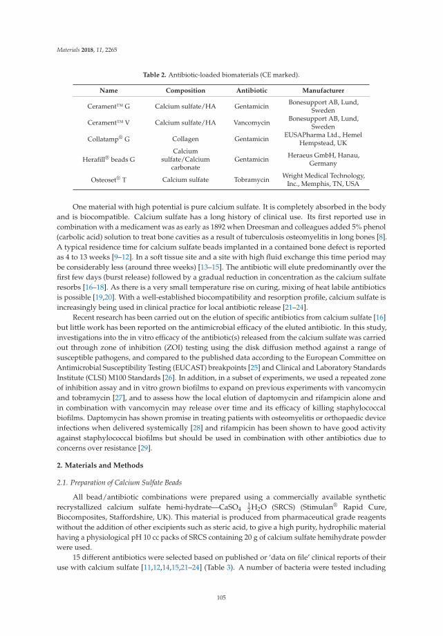
Materials 2018, 11, 2265
Table 2. Antibiotic-loaded biomaterials (CE marked).
Name Composition Antibiotic Manufacturer
Cerament™ G Calcium sulfate/HA Gentamicin Bonesupport AB, Lund,Sweden
Cerament™ V Calcium sulfate/HA Vancomycin Bonesupport AB, Lund,Sweden
Collatamp® G Collagen Gentamicin EUSAPharma Ltd., HemelHempstead, UK
Herafill® beads GCalcium
sulfate/Calciumcarbonate
Gentamicin Heraeus GmbH, Hanau,Germany
Osteoset® T Calcium sulfate Tobramycin Wright Medical Technology,Inc., Memphis, TN, USA
One material with high potential is pure calcium sulfate. It is completely absorbed in the bodyand is biocompatible. Calcium sulfate has a long history of clinical use. Its first reported use incombination with a medicament was as early as 1892 when Dreesman and colleagues added 5% phenol(carbolic acid) solution to treat bone cavities as a result of tuberculosis osteomyelitis in long bones [8].A typical residence time for calcium sulfate beads implanted in a contained bone defect is reportedas 4 to 13 weeks [9–12]. In a soft tissue site and a site with high fluid exchange this time period maybe considerably less (around three weeks) [13–15]. The antibiotic will elute predominantly over thefirst few days (burst release) followed by a gradual reduction in concentration as the calcium sulfateresorbs [16–18]. As there is a very small temperature rise on curing, mixing of heat labile antibioticsis possible [19,20]. With a well-established biocompatibility and resorption profile, calcium sulfate isincreasingly being used in clinical practice for local antibiotic release [21–24].
Recent research has been carried out on the elution of specific antibiotics from calcium sulfate [16]but little work has been reported on the antimicrobial efficacy of the eluted antibiotic. In this study,investigations into the in vitro efficacy of the antibiotic(s) released from the calcium sulfate was carriedout through zone of inhibition (ZOI) testing using the disk diffusion method against a range ofsusceptible pathogens, and compared to the published data according to the European Committee onAntimicrobial Susceptibility Testing (EUCAST) breakpoints [25] and Clinical and Laboratory StandardsInstitute (CLSI) M100 Standards [26]. In addition, in a subset of experiments, we used a repeated zoneof inhibition assay and in vitro grown biofilms to expand on previous experiments with vancomycinand tobramycin [27], and to assess how the local elution of daptomycin and rifampicin alone andin combination with vancomycin may release over time and its efficacy of killing staphylococcalbiofilms. Daptomycin has shown promise in treating patients with osteomyelitis or orthopaedic deviceinfections when delivered systemically [28] and rifampicin has been shown to have good activityagainst staphylococcal biofilms but should be used in combination with other antibiotics due toconcerns over resistance [29].
2. Materials and Methods
2.1. Preparation of Calcium Sulfate Beads
All bead/antibiotic combinations were prepared using a commercially available syntheticrecrystallized calcium sulfate hemi-hydrate—CaSO4
12 H2O (SRCS) (Stimulan® Rapid Cure,
Biocomposites, Staffordshire, UK). This material is produced from pharmaceutical grade reagentswithout the addition of other excipients such as steric acid, to give a high purity, hydrophilic materialhaving a physiological pH 10 cc packs of SRCS containing 20 g of calcium sulfate hemihydrate powderwere used.
15 different antibiotics were selected based on published or ‘data on file’ clinical reports of theiruse with calcium sulfate [11,12,14,15,21–24] (Table 3). A number of bacteria were tested including
105

Materials 2018, 11, 2265
S. epidermidis ATCC12228, P. aeruginosa NCTC 13437, S. aureus ATCC 6538, Acinetobacter Baumanii NCTC134242, S. aureus 12493 MRSA, E. faecalis NCTC 12202, P. acnes NCTC 737 and E. faecalis NCTC 12201.For daptomycin, the maximum dose was referenced from the USP (United States Pharmacopeia) [30].Sterile saline was used in the mixing of amikacin and daptomycin, replacing some or all of the mixingsolution provided in the SRCS pack. The use of a 0.9% sodium chloride solution was found to speed upthe setting reaction for these retarding antibiotics. The resultant paste, together with added antibioticwas pressed into hemispherical cavities, 6 mm diameter, in a flexible rubber mold where it was allowedto hydrate and set accordingly.
Table 3. Antibiotics tested in this study.
Antibiotic Manufacturer
Amikacin Sulfate Hospira Ltd., Maidenhead, UKCeftriaxone Sodium Apotex Corporation, Toronto, ON, Canada
Cefuroxime Stravencon, London, UKCiprofloxacin Hydrochloride Medisca Inc., Las Vegas, NV, USA
Colistamethane Sodium Sigma-Aldrich, Dorset, UKClindamycin (Dalacin C® Phosphate) Pfizer, Tadworth, UK
Daptomycin (Cubicin®) Novartis, Basel, SwitzerlandGentamicin Sulfate Hospira Ltd., Maidenhead, UK
Imipenem/Cilastatin (Zienam®) Merck & Co., Inc, Kenilworth, NJ, USAMeropenem Trihydrate Fresenius Kabi Ltd., Cestrian, UK
Nafcillin Sodium Sandoz, Princeton, NJ, USARifampicin Sigma-Aldrich, Dorset, UK
Teicoplanin (Targocid®) Sanofi-Aventis, Guildford, UKTobramycin Sulfate Hospira Ltd., Maidenhead, UK
Vancomycin Hydrochloride Hospira Ltd., Maidenhead, UK
2.2. Zone of Inhibition Testing
Tryptone soya agar (TSA) plates were seeded with a 0.2 mL suspension of the relevant organismcontaining approximately 108 CFU (Colony Forming Units)/mL. The plates were transferred to anincubator operating at 33 ± 2 ◦C for 30 min. The plates were then removed from the incubator anda single 6 mm antibiotic-loaded bead was placed on the surface of the agar. The plates were thenincubated at 33 ± 2 ◦C for 24 h, after which time they were removed from the incubator and examinedfor any clear zones around the test sample. Zones were measured to the nearest mm where no obviousgrowth could be detected by the unaided eye. Samples were tested in triplicate and an averagediameter was recorded. Zone diameters were compared to published data according to the EuropeanCommittee on Antimicrobial Susceptibility Testing (EUCAST) breakpoints and Clinical and LaboratoryStandards Institute (CLSI) M100 Standards [26] where applicable. It is important to note that thisassay was not designed to determine breakpoint zones of the challenge strains against the variousantibiotics. Breakpoint testing is performed using well-defined amounts of antibiotics loaded ontofilter papers, which have well-characterized and reproducible release kinetics. Rather this assay wasused to determine that the antibiotic was (1) released from the bead and (2) had retained antibioticpotency. Presence of a zone of inhibition was demonstrative that both these conditions were true.The known breakpoints from the standard method guidelines were provided as a reference guide.
2.3. Repeat Zone of Inhibition Testing
To assess how long the beads may release antibiotic, we used a modified Kirby–Bauer diskdiffusion assay as previously reported [27]. Staphylococcus aureus NCTC 13143 EMRSA-16 (an MRSAstrain) and S. epidermidis ATCC 35984, a prolific biofilm former was used as the challenge organisms.First, a lawn of bacteria was spread onto TSA plates using 50 μL of an overnight culture grown for15 h at 37 ◦C. A single bead containing (in mg/5 cc pack SRCS) either (a) daptomycin (500 mg),(b) vancomycin (500 mg), (c) rifampicin (300 mg) or (d) a combination of vancomycin and rifampicin
106
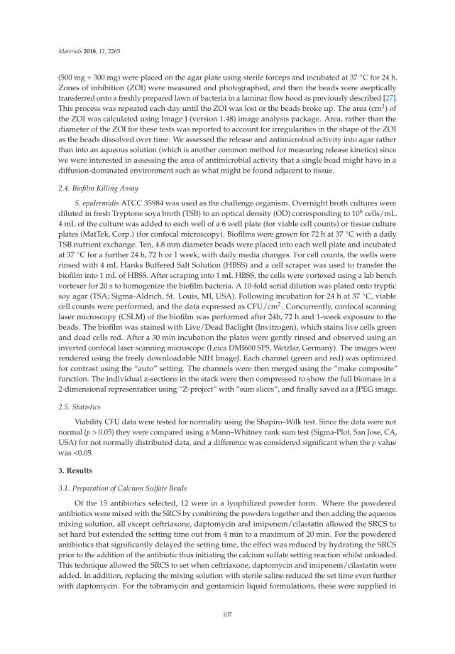
Materials 2018, 11, 2265
(500 mg + 300 mg) were placed on the agar plate using sterile forceps and incubated at 37 ◦C for 24 h.Zones of inhibition (ZOI) were measured and photographed, and then the beads were asepticallytransferred onto a freshly prepared lawn of bacteria in a laminar flow hood as previously described [27].This process was repeated each day until the ZOI was lost or the beads broke up. The area (cm2) ofthe ZOI was calculated using Image J (version 1.48) image analysis package. Area, rather than thediameter of the ZOI for these tests was reported to account for irregularities in the shape of the ZOIas the beads dissolved over time. We assessed the release and antimicrobial activity into agar ratherthan into an aqueous solution (which is another common method for measuring release kinetics) sincewe were interested in assessing the area of antimicrobial activity that a single bead might have in adiffusion-dominated environment such as what might be found adjacent to tissue.
2.4. Biofilm Killing Assay
S. epidermidis ATCC 35984 was used as the challenge organism. Overnight broth cultures werediluted in fresh Tryptone soya broth (TSB) to an optical density (OD) corresponding to 106 cells/mL.4 mL of the culture was added to each well of a 6 well plate (for viable cell counts) or tissue cultureplates (MatTek, Corp.) (for confocal microscopy). Biofilms were grown for 72 h at 37 ◦C with a dailyTSB nutrient exchange. Ten, 4.8 mm diameter beads were placed into each well plate and incubatedat 37 ◦C for a further 24 h, 72 h or 1 week, with daily media changes. For cell counts, the wells wererinsed with 4 mL Hanks Buffered Salt Solution (HBSS) and a cell scraper was used to transfer thebiofilm into 1 mL of HBSS. After scraping into 1 mL HBSS, the cells were vortexed using a lab benchvortexer for 20 s to homogenize the biofilm bacteria. A 10-fold serial dilution was plated onto trypticsoy agar (TSA; Sigma-Aldrich, St. Louis, MI, USA). Following incubation for 24 h at 37 ◦C, viablecell counts were performed, and the data expressed as CFU/cm2. Concurrently, confocal scanninglaser microscopy (CSLM) of the biofilm was performed after 24h, 72 h and 1-week exposure to thebeads. The biofilm was stained with Live/Dead Baclight (Invitrogen), which stains live cells greenand dead cells red. After a 30 min incubation the plates were gently rinsed and observed using aninverted confocal laser scanning microscope (Leica DMI600 SP5, Wetzlar, Germany). The images wererendered using the freely downloadable NIH ImageJ. Each channel (green and red) was optimizedfor contrast using the “auto” setting. The channels were then merged using the “make composite”function. The individual z-sections in the stack were then compressed to show the full biomass in a2-dimensional representation using “Z-project” with “sum slices”, and finally saved as a JPEG image.
2.5. Statistics
Viability CFU data were tested for normality using the Shapiro–Wilk test. Since the data were notnormal (p > 0.05) they were compared using a Mann–Whitney rank sum test (Sigma-Plot, San Jose, CA,USA) for not normally distributed data, and a difference was considered significant when the p valuewas <0.05.
3. Results
3.1. Preparation of Calcium Sulfate Beads
Of the 15 antibiotics selected, 12 were in a lyophilized powder form. Where the powderedantibiotics were mixed with the SRCS by combining the powders together and then adding the aqueousmixing solution, all except ceftriaxone, daptomycin and imipenem/cilastatin allowed the SRCS toset hard but extended the setting time out from 4 min to a maximum of 20 min. For the powderedantibiotics that significantly delayed the setting time, the effect was reduced by hydrating the SRCSprior to the addition of the antibiotic thus initiating the calcium sulfate setting reaction whilst unloaded.This technique allowed the SRCS to set when ceftriaxone, daptomycin and imipenem/cilastatin wereadded. In addition, replacing the mixing solution with sterile saline reduced the set time even furtherwith daptomycin. For the tobramycin and gentamicin liquid formulations, these were supplied in
107

Materials 2018, 11, 2265
2 mL vials, each containing 80 mg of antibiotic. For both these antibiotics, 3 vials were used to providethe required 6 mL for hydration of the SRCS, giving a dose of 240 mg. This was a limiting factor inthe maximum dose, which may be combined. Two of the liquid antibiotic formulations; amikacinand clindamycin, were commercially provided in volumes of 2 mL and 4 mL. Additional fluid wasrequired to make the volume up to 6 mL, required to fully hydrate the SRCS. For these antibiotics,a quantity of the aqueous mixing solution was accurately added via a syringe to make the volumeup to the required 6 mL. Neither of these antibiotic combinations would set hard when the aqueousmixing solution was used, therefore, sterile saline was investigated as an alternative. This methodallowed for the production of fully hardened amikacin-loaded beads. The clindamycin, which is in theform of a phosphate, may chemically react with the calcium sulfate, precipitating out as an insolublecalcium phosphate. In addition, this formulation of clindamycin contained benzyl alcohol and EDTA(ethylenediaminetetraacetic acid) as excipients. The ability of SRCS to set hard with Clindamycinin the form tested here was not achieved, therefore was unable to be combined in this way and nomicrobiology data was obtained.
3.2. Zone of Inhibition Testing
Zone diameters for each antibiotic/SRCS bead combination are shown in Table 4. Where EUCASTclinical breakpoint data [31] or CLSI M100 data [26] were available, they were included for reference.SRCS beads mixed with rifampicin achieved zones which fell below the CLSI M100 breakpoint datafor both S. epidermidis and a methicillin-resistant strain of S. aureus. Interestingly, SRCS beads mixedwith ciprofloxacin had varying success, depending on the species of bacteria under investigation.Ciprofloxacin-loaded SRCS beads were tested against a range of Gram-negative species includingP. aeruginosa and Acinetobacter Baumanii. The zones of inhibition recorded for these species, fell belowthe EUCAST and CLSI breakpoints for the respective bacteria (12 and 15 mm respectively) (Table 4).Conversely, when SRCS were mixed with ciprofloxacin and tested against a range of Gram-positivespecies, the SRCS-loaded beads were able to generate zones measuring 36 and 25 mm (for S. epidermidisand S. aureus respectively). The observed zones were greater than the EUCAST and CLSI breakpointfor these bacterial species tested against this antibiotic. All other antibiotic combinations produceda clear zone against susceptible species and where breakpoint data was available, exceeded thebreakpoint diameter, demonstrating an above MIC elution concentration and maintained efficacy(Table 4). For the vancomycin SRCS beads with Enterococcus faecalis, two vancomycin resistant controlstrains were used. NCTC 12201 (VanA-type glycopeptide resistance, Erythromycin resistant) andNCTC 12202 (VanA-type glycopeptide resistance). The results demonstrated that the high burst releaseconcentration could exceed the vancomycin MIC of these two strains which are reported to exceed256 μg/mL [32]. EUCAST and CLSI breakpoints have not been published for these strains but thezones observed were larger than the published breakpoints for susceptible strains of E. faecalis (Table 4).
Table 4. Zones of Inhibition (ZOI) data with EUCAST clinical breakpoint tables v 6.0 and CLSIM100 breakpoints.
Antibiotic Conc.per 10cc Pack SRCS
SpeciesZone
Diameter(mm)
EUCAST/CLSI M100
Zone DiameterBreakpoint (mm)
Note
Susceptible≥ Resistant ≤
Amikacin500 mg/2 mL
S. epidermidis(ATCC 12228) 30
EUCAST 22 a 19 aa Coagulase negative
staphylococciCLSI - - n/a
P. aeruginosa(NCTC 13437) 20
EUCAST 18 c 15 c c Pseudomonas spp.CLSI 15 12 -
Ceftriaxone1 g
S. aureus(ATCC 6538) 47
EUCAST - - n/aCLSI - - n/a
S. epidermidis(ATCC 12228) 31
EUCAST - - n/aCLSI - - n/a
108
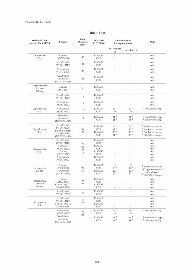
Materials 2018, 11, 2265
Table 4. Cont.
Antibiotic Conc.per 10cc Pack SRCS
SpeciesZone
Diameter(mm)
EUCAST/CLSI M100
Zone DiameterBreakpoint (mm)
Note
Susceptible≥ Resistant ≤
Cefuroxime1.5 g
S. aureus(ATCC 6538) 47
EUCAST - - n/aCLSI - - n/a
S. epidermidis(ATCC 12228) 22 EUCAST
CLSI--
--
n/an/a
P. aeruginosa(NCTC 13437) 28
EUCAST - - n/aCLSI - - n/a
AcinetobacterBaumannii
(NCTC 134242)22 EUCAST
CLSI--
--
n/an/a
ColistamethaneSodium400 mg
S. aureus(ATCC 6538) 9 EUCAST
CLSI--
--
n/an/a
S. epidermidis(ATCC 12228) 11 EUCAST
CLSI--
--
n/an/a
P. aeruginosa(NCTC 13437) 13 EUCAST
CLSI--
--
n/an/a
Ciprofloxacin1 g
P. aeruginosa(NCTC 13437) 17 EUCAST
CLSI25 c
2122 c
15
c Pseudomonas spp.-
AcinetobacterBaumannii
(NCTC 134242)15 EUCAST
CLSI21 d
21 d21 d
15 d
d Acinetobacter spp.d Acinetobacter spp.
Ciprofloxacin1 g
S. epidermidis(ATCC 12228)
S.aureus (NCTC12493) MRSA
(NCTC 134242)
3625
EUCASTCLSI
EUCASTCLSI
20 b
21 b
20 b
21 b
20 b
15 b
20 b
15 b
b Staphylococcus spp.b Staphylococcus spp.b Staphylococcus spp.b Staphylococcus spp.
Daptomycin1 g
S. epidermidis(ATCC 12228)
E. faecalis *(NCTC 12202)
2513.53111
EUCASTCLSI
EUCASTCLSI
----
----
n/an/an/an/a
P. acnes(NCTC 737)P. aeruginosa
(NCTC 13437)
EUCASTCLSI
EUCASTCLSI
----
----
n/an/an/an/a
Gentamicin240 mg
S. aureus(ATCC 6538)S. epidermidis(ATCC 12228)
2030
EUCASTCLSI
EUCASTCLSI
1815 b
22 a
15 b
1812 b
22 a
12 b
-b Staphylococcus spp.
a Coagulase negativestaphylococci
b Staphylococcus spp.
Imipenem &Cilastatin
500 mg
S. aureus(ATCC 6538) 58
49
EUCASTCLSI
--
--
n/an/a
S. aureus (NCTC12493) MRSA
EUCASTCLSI
--
--
n/an/a
S. epidermidis(ATCC 12228) 60 EUCAST
CLSI--
--
n/an/a
Meropenem1 g
S. epidermidis(ATCC 12228) 56
37
EUCASTCLSI
--
--
n/an/a
S. aureus (NCTC12493) MRSA
EUCASTCLSI
--
--
n/an/a
P. aeruginosa(NCTC 13437) 28
22
EUCASTCLSI
24 c
1918 c
15
c Pseudomonas spp.-
AcinetobacterBaumannii
(NCTC 134242)
EUCASTCLSI
21 d
18 d15 d
14 d
d Acinetobacter spp.d Acinetobacter spp.
109
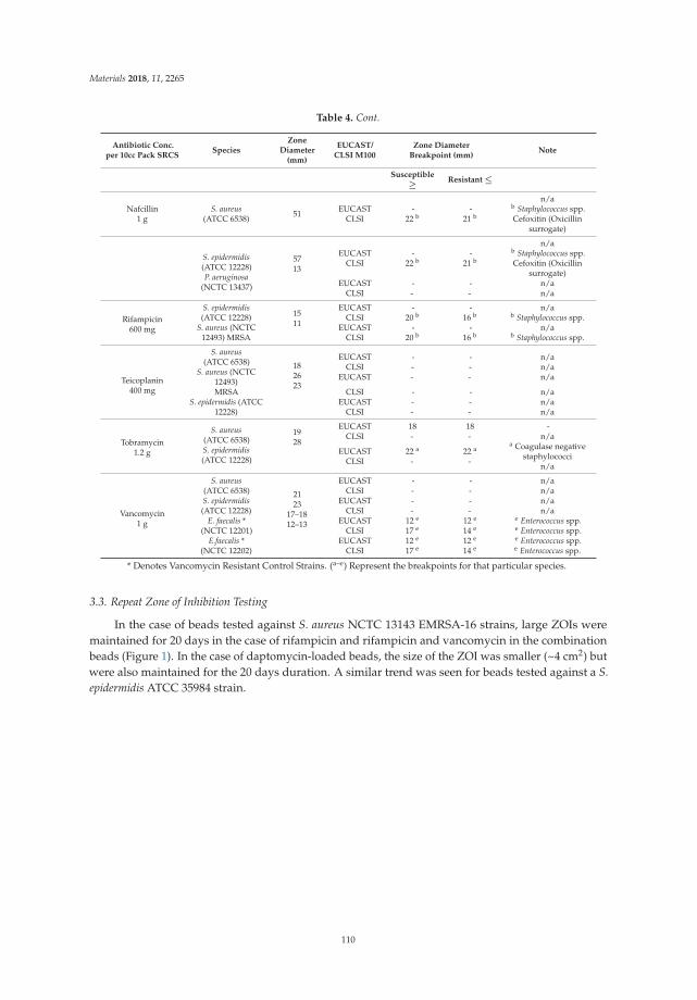
Materials 2018, 11, 2265
Table 4. Cont.
Antibiotic Conc.per 10cc Pack SRCS
SpeciesZone
Diameter(mm)
EUCAST/CLSI M100
Zone DiameterBreakpoint (mm)
Note
Susceptible≥ Resistant ≤
Nafcillin1 g
S. aureus(ATCC 6538) 51 EUCAST
CLSI-
22 b-
21 b
n/ab Staphylococcus spp.Cefoxitin (Oxicillin
surrogate)
S. epidermidis(ATCC 12228)P. aeruginosa
(NCTC 13437)
5713
EUCASTCLSI
-22 b
-21 b
n/ab Staphylococcus spp.Cefoxitin (Oxicillin
surrogate)EUCAST
CLSI--
--
n/an/a
Rifampicin600 mg
S. epidermidis(ATCC 12228) 15
11
EUCASTCLSI
-20 b
-16 b
n/ab Staphylococcus spp.
S. aureus (NCTC12493) MRSA
EUCASTCLSI
-20 b
-16 b
n/ab Staphylococcus spp.
Teicoplanin400 mg
S. aureus(ATCC 6538)
S. aureus (NCTC12493)
182623
EUCASTCLSI
EUCAST
---
---
n/an/an/a
MRSAS. epidermidis (ATCC
12228)
CLSIEUCAST
CLSI
---
---
n/an/an/a
Tobramycin1.2 g
S. aureus(ATCC 6538)S. epidermidis(ATCC 12228)
1928
EUCASTCLSI
18-
18-
-n/a
EUCASTCLSI
22 a
-22 a
-
a Coagulase negativestaphylococci
n/a
Vancomycin1 g
S. aureus(ATCC 6538)S. epidermidis(ATCC 12228)
2123
17–1812–13
EUCASTCLSI
EUCASTCLSI
----
----
n/an/an/an/a
E. faecalis *(NCTC 12201)
E.faecalis *(NCTC 12202)
EUCASTCLSI
EUCASTCLSI
12 e
17 e
12 e
17 e
12 e
14 e
12 e
14 e
e Enterococcus spp.e Enterococcus spp.e Enterococcus spp.e Enterococcus spp.
* Denotes Vancomycin Resistant Control Strains. (a–e) Represent the breakpoints for that particular species.
3.3. Repeat Zone of Inhibition Testing
In the case of beads tested against S. aureus NCTC 13143 EMRSA-16 strains, large ZOIs weremaintained for 20 days in the case of rifampicin and rifampicin and vancomycin in the combinationbeads (Figure 1). In the case of daptomycin-loaded beads, the size of the ZOI was smaller (~4 cm2) butwere also maintained for the 20 days duration. A similar trend was seen for beads tested against a S.epidermidis ATCC 35984 strain.
110
Materials 2018, 11, 2265
Figure 1. Repeated zone of inhibition (ZOI) of S. aureus NCTC 13143 EMRSA-16 and S. epidermidisATCC 35984 Stimulan® beads loaded with rifampicin, rifampicin and vancomycin or daptomycin.Assays were performed in triplicate and data expressed as the mean and 1SD.
At the end of the 20 day assay, suspected resistance was noted developing in the rifampicin-onlyloaded beads, as shown by an internal ring of viable bacteria within the ZOI (Figure 2A,C). This wasnot observed on plates that had been exposed to beads containing rifampicin and vancomycin incombination (Figure 2B,D).
Figure 2. Representative image of the Zones of Inhibition (ZOI) observed with (A,B) S. epidermidisATCC 35984 and(C,D) S. aureus NCTC 13143 EMRSA-16 at day 20 of rifampicin and vancomycin incombination, showing no evidence of resistant colonies (B,D) and rifampicin alone (A,C) showingpotential resistant mutant colonies growing within the ZOI (black arrows).
3.4. Biofilm Killing Assay
Beads loaded with rifampicin, and vancomycin and rifampicin in combination were able toachieve a 2-log reduction in CFU/cm2 at 24 h (Figure 3) with CSLM imaging showing a concurrentmarked reduction in biofilm mass (Figure 4). After 72 h contact time, rifampicin alone achieved a
111

Materials 2018, 11, 2265
5-log reduction in CFU/cm2 (Figure 3) with the combination of vancomycin and rifampicin showingno growth on the plate. CSLM imaging showed almost complete removal of the biofilm with only afew single cells remaining (Figure 4). However, at one week, regrowth was noted in rifampicin andrifampicin and vancomycin treatment groups and reformation of microcolonies noted with CSLMimaging (Figure 4). Additionally, the combination of rifampicin and vancomycin demonstrated a3-log regrowth relative to CFU/cm2 data at 72 h suggesting that the regrowth may have been due tosome cells that were rendered viable but nonculturable at 72 h. Daptomycin-loaded beads resulted inan approximate 7-log reduction in CFU/cm2 relative to unloaded beads after 24 h contact time withmature S. epidermidis biofilms (Figure 3). There was little further change over the next seven days.CSLM imaging corroborated the CFU data showing a significant reduction in biofilm and total surfacecoverage observed at day 7 relative to unloaded beads (Figure 4). Interestingly, in the biofilm exposedto the rifampicin only beads, after 7 days there was the appearance of larger aggregates, which hadboth live (green) and dead (red) cells suggesting the proliferation of resistant mutants. In the controlmost of the cells were still live (green) however, there were patches of red (dead or compromised)cells suggesting that the cells may have been undergoing starvation due to nutrient limitation. In thebiofilms exposed to the combination of vancomycin and rifampicin there was a sparse covering ofcells, which were mainly live, while in the daptomycin-exposed biofilm there were very few cells andthese were only very faintly stained green (Figure 4).
Figure 3. Effect of unloaded beads as well as vancomycin (Vanco), rifampicin (Rif), rifampicin andvancomycin in combination (Vanco + Rif) and daptomycin (Dapto) loaded beads on established S.epidermidis ATCC 35984 biofilms at contact times at days (D) 1,3 and 7. Dashed line is the detection limit.No beads were added as a positive control for biofilm growth. Mean and 95% CI (n = 3), *indicatesstatistically significant differences from the unloaded beads (p < 0.05).
112

Materials 2018, 11, 2265
Figure 4. Representative CSLM images showing S. epidermidis ATCC 35984 biofilm (live cells stainedgreen and dead and membrane compromised cells stained red or yellow) following treatment for 24h and 1 week with unloaded beads (negative control) and beads loaded with rifampicin (Rifampin),rifampicin and vancomycin, and daptomycin. Scale bars: 25 μm.
4. Discussion
With the exception of clindamycin, the calcium sulfate in combination with the antibiotics tested,set hard, maintaining its function as a void filler. All antibiotics used in this study were IV formulations.Antibiotic capsules and tablets contain different excipients to IV formulations and these may createadditional risks if administered locally. Ceftriaxone, although included here, is contraindicated whenmixing with a calcium-containing product such as Ringer’s solution, Hartmann’s solution or parenteralnutrition containing calcium. The reason for this contraindication is the risk of crystalline precipitatesforming in the lungs and kidneys. Although this risk is for IV administration, the risks shouldbe considered if used in combination with calcium sulfate in this application and warrants furtherinvestigation. The ability of SRCS or any biomaterial to mix and set with antibiotics, and demonstraterelease and potency alone or in combination in in vitro laboratory studies, does not imply safety orefficacy in clinical use, and in vitro results may not be indicative of clinical performance.
A number of independent studies have shown that calcium sulfate beads may be used in revisionarthroplasty for PJI (Periprosthetic Joint Infection), with favorable outcomes [21,33,34]. However,their use is not without risk. One potential risk is transitory hypercalcemia in vivo and this has beenreported in literature, [15,21] although only when the material has been implanted in soft tissue sitesand in larger volumes. More recent studies however, have shown that there is no significant differencein incidences of hypercalcemia when compared with other antibiotic-loaded materials [35].
This study demonstrates that a wide range of antibiotics can be incorporated into the SRCS andretain potency against a wide range of common bacterial pathogens associated with PJI using an in vitrozone of inhibition assay. Where EUCAST or CLSI breakpoints were available for specific antibioticsagainst susceptible pathogens, the same antibiotics when eluted from the SRCS all maintained theirantimicrobial activity with the exception of ciprofloxacin and rifampicin. This assay is a relativelysimple test which can be used to assess whether antibiotics as monotherapy or in combination, which (a)are currently used to treat orthopedic infections systemically or (b) show promise in in vitro studiesalso have potential to be eluted locally by SRCS made up as beads or in other forms. Although thestandardized Kirby–Bauer disc diffusion method is used as a quick and convenient method of assessingthe susceptibility of a given organism to a given antibiotic [36], modifications of the assay allow it to be
113

Materials 2018, 11, 2265
used to assess the release and maintenance of potency from materials other than filter paper, such ashow we have done with materials such as SRCS, and PMMA as detailed in previous studies [27].The results for ciprofloxacin against Pseudomonas aerugonisa (NCTC 13437) and Acinetobacter baumannii(NCTC 134242) showed a smaller zone than the breakpoint data for Pseudomonas spp. (Table 4).This may be due to the susceptibility of the specific species or other factors such as compatibility withthe SRCS or reduced elution.
The quantity of each antibiotic held in the disks used in the EUCAST or CLSI M100 may differ tothe quantity of antibiotic in the SRCS beads, therefore, correlation to the clinical breakpoints should notbe inferred and these values are included for reference and demonstration of susceptibility only. Futurework should focus on testing the release of antibiotics from SRCS as per the guidance of EUCAST orCLSI in order to make a more direct comparison of the efficacy of released antibiotic from SRCS beads.
We further modified the method to assess the longevity of release in vitro by placing SRCSon new spread plates daily. The ZOI for daptomycin started at approximately 4 cm2 and slowlyreduced to 2 cm2 after 20 days of the experiment for both the EMRSA and S. epidermidis challengestrains. This behavior was very similar to that seen with vancomycin and tobramycin assessedpreviously [27]. Interestingly the SRCS beads containing vancomycin and rifampicin or rifampicinalone had much greater ZOIs of between 14 cm2 and 10 cm2 against EMRSA and S. epidermidis,respectively. These remained relatively constant over the full 20 days of testing.
Rifampicin has been noted to have “excellent activity on adherent staphylococci” [37] andbiofilms [29]. However, since it is known that there is rapid emergence of resistance when usedin monotherapy [38] it is used in combination with other antibiotics. Indeed, in our assay we notedgrowth within the ZOI of the beads loaded with rifampicin alone (Figure 2). Since the plates werespread freshly every day it was possible that the growth came from bacteria carried over with thebead that had been exposed to rifampicin for an extended period. Interestingly, the beads with bothvancomycin and rifampicin showed no evidence of the emergence of resistance. However, to more fullyassess the potential for the development of resistance with vancomycin and rifampicin, more stringentstudies are required. The S. epidermidis biofilm killing assay showed different patterns of killingwith the various antibiotics. Vancomycin-alone only caused an approximate 1-log reduction after1 and 3 days exposure but by 7 days there was a 6-log reduction illustrating the importance ofprolonged exposure to high concentrations of antibiotics when treating biofilms. Rifampicin alone wasmore effective, showing an approximate 2-log reduction after 1 day and 5-log reduction after 3 days,however, there was significant regrowth at day 7. The confocal image showed large cell clusters hadformed suggesting the emergence of resistant mutants within this time period (Figure 4). Rifampicinand vancomycin in combination reduced the biofilm to below detectable levels after 3 days (>7-logreduction) but there was a “bounce” back at day 7 to an approximate 5-log reduction compared tothe control unloaded beads. Although there was no evidence of new colony formation from theconfocal data, more work needs to be done to assess the potential for the emergence of resistancewith this combination. However, a combination of vancomycin and rifampicin has been shown tobe more effective than vancomycin alone in treating PJI in a mouse model [39], and the local releaseof rifampicin from SRCS in combination with other antibiotics to treat orthopedic biofilm infectionsshows promise. Although daptomycin resulted in the lowest ZOI in the diffusion assay, it caused thegreatest reduction in biofilm (approximately 6 logs) after only 1-day exposure, however, little furtherreduction was seen after the longer exposure periods.
5. Conclusions
The use of locally released antibiotics from synthetic recrystallized calcium sulfate may offersignificant benefits in the management of surgical site infections. The ability to combine differentantibiotics could enable a therapy tailored to the offending pathogens. The exposure time, type ofantibiotic(s) released from SRCS and the challenge bacterial strain can all influence the kinetics ofbiofilm killing and thus, in vitro results with one set of conditions should not be over generalized.
114

Materials 2018, 11, 2265
Author Contributions: Conceptualization, P.A.L., J.J.C., S.A., and P.S.; methodology, P.A.L., J.J.C. and R.P.H.;formal analysis, P.A.L., R.H., P.S.; resources, S.A. and P.S.; writing—original draft preparation, P.A.L., P.S. and CD.;writing—review and editing, P.A.L., C.D., S.A. and P.S.; visualization, P.A.L., R.H., C.D., and P.S.; supervision,P.A.L. and P.S.; project administration, S.A. and P.S.; funding acquisition, S.A. and P.S.
Funding: This research was funded by Biocomposites Ltd.
Acknowledgments: In this section you can acknowledge any support given which is not covered by the authorcontribution or funding sections. This may include administrative and technical support, or donations in kind(e.g., materials used for experiments).
Conflicts of Interest: P.A.L., J.J.C., C.D. and S.A. are employees of Biocomposites Ltd. R.H. and P.S. consult forBiocomposites Ltd. The funders contributed to the design, collection, analyses or interpretation of data and inthe writing of the manuscript for the zone of inhibition study. The funders had no role in the original design,execution, data analysis or writing up of the repeated zone of inhibition and biofilm killing parts of the study.The funders did prepare the beads used in these parts of the study and reviewed the experiments but offeredno amendments.
References
1. Antimicrobial Resistance: Global Report on Surveillance; World Health Organization: Geneva, Switzerland, 2014.2. Tackling Drug-Resistant Infections Globally: Final Report and Recommendations; Review on Antimicrobial
Resistance: London, UK, 2016.3. Saleh, K.E.O.; El Othmani, M.M.; Tzeng, T.H.; Mihalko, W.M.; Chmabers, M.C.; Grupp, T.M. Acrylic bone
cement in total joint arthroplasty: A review. J. Orthop. Res. 2016, 34, 737–744. [CrossRef] [PubMed]4. Ma, D.; Shanks, R.M.Q.; Davis, C.M.; Craft, D.W.; Wood, T.K.; Hamlin, B.R.; Urish, K.L. Viable bacteria
persist on antibiotic spacers following two-stage revision for periprosthetic joint infection. J. Orthop. Res.2017. [CrossRef] [PubMed]
5. Van de Belt, H.; Neut, D.; Schenk, W.; van Horn, J.R.; van der Mei, H.C.; Busscher, H.J. Gentamicin releasefrom polymethylmethacrylate bone cements and Staphylococcus aureus biofilm formation. Acta Orthop.Scand. 2000, 71, 625–629. [CrossRef] [PubMed]
6. Van de Belt, H.; Neut, D.; Schenk, W.; van Horn, J.R.; van der Mei, H.C.; Busscher, H.J. Staphylococcusaureus biofilm formation on different gentamicin-loaded polymethlmethacrylate bone cements. Biomaterials2001, 22, 1607–1611. [CrossRef]
7. Morscher, E.W.; Hefti, A.; Aebi, U. Severe osteolysis after third-body wear due to hydroxyapatite particlesfrom acetabular cup coating. J. Bone Jt. Surg. 1998, 80, 267–272. [CrossRef]
8. Dreesman, H. Über Knochenplombierung. Beitr Klin Chir. 1892, 9, 804–810.9. McKee, M.D.; Wild, L.M.; Schemitsch, E.H.; Waddell, J.P. The use of an antibiotic-impregnated,
osteoconductive, bioabsorbable bone substitute in the treatment of infected long bone defects: Early resultsof a prospective trial. J. Orthop. Trauma 2002, 16, 622–627. [CrossRef] [PubMed]
10. Beuerlein, M.J.; McKee, M.D. Calcium sulfates: What is the evidence? J. Orthop. Trauma 2010, 24 (Suppl. 1),S46–S51. [CrossRef] [PubMed]
11. Ferguson, J.Y.; Dudareva, M.; Riley, N.D.; Stubbs, D.; Atkins, B.L.; McNally, M.A. The use of a biodegradableantibiotic-loaded calcium sulphate carrier containing tobramycin for the treatment of chronic osteomyelitis:A series of 195 cases. Bone Jt. J. 2014, 96, 829–836. [CrossRef] [PubMed]
12. Cierny, G., III; DiPasquale, D. Comparing OsteoSet and Stimulan as Antibiotic-loaded, Calcium SulfateBeads in the Management of Musculoskeletal Infection. In Proceedings of the 19th Annual Open ScientificMeeting of the Musculoskeletal Infection Society, San Diego, CA, USA, 7–8 August 2009.
13. Oliver, R.A.; Lovric, V.; Yu, Y.; Christou, C.; Aiken, S.S.; Cooper, J.J.; Walsh, W.R. Development of a NovelModel for the Assessment of Dead-Space Management in Soft Tissue. PLoS ONE 2015, 10, e0136514.[CrossRef] [PubMed]
14. Maale, G.E.; Casa-Ganem, J.E. The Use of Antibiotic Loaded Synthesized Calcium Sulfate Pellets in theOne Stage Treatment for Osteomyelitis. In Proceedings of the 19th Annual Open Scientific Meeting of theMusculoskeletal Infection Society, San Diego, CA, USA, 7–8 August 2009.
15. Kallala, R.; Nizam, I.; Haddad, F. Outcomes Following Use of Antibiotic-Eluting, Absorbable, CalciumSulphate Beads in Revision Hip and Knee Surgery for Periprosthetic Infection. Bone Jt. J. Orthop. Proc. Suppl.2013, 95, 364.
115

Materials 2018, 11, 2265
16. Aiken, S.S.; Cooper, J.J.; Florance, H.; Robinson, M.T.; Michell, S. Local release of antibiotics for surgical siteinfection management using high-purity calcium sulfate: An in vitro elution study. Surg. Infect. 2015, 16,54–61. [CrossRef] [PubMed]
17. McConoughey, S.J.; Howlin, R.P.; Wiseman, J.; Stoodley, P.; Calhoun, J.H. Comparing PMMA and calciumsulfate as carriers for the local delivery of antibiotics to infected surgical sites. J. Biomed. Mater. Res. Part BAppl. Biomater. 2015, 103, 870–877. [CrossRef] [PubMed]
18. Roberts, R.; McConoughey, S.J.; Calhoun, J.H. Size and composition of synthetic calcium sulfate beadsinfluence dissolution and elution rates in vitro. J. Biomed. Mater. Res. Part B Appl. Biomater. 2014, 102, 667–673.[CrossRef] [PubMed]
19. Traub, W.H.; Leonhard, B. Heat stability of the antimicrobial activity of sixty-two antibacterial agents.J. Antimicrob. Chemother. 1995, 35, 149–154. [CrossRef] [PubMed]
20. Albright, S.B.; Xue, A.S.; McKnight, A.; Wolfswinkel, E.M.; Hollier, L.H.J.; Brown, R.H.; Bullocks, J.M.;Izaddoost, S.A. Pilot Study: One-Step Salvage of Infected Prosthetic Breast Reconstructions UsingAntibiotic-Impregnated Polymethylmethacrylate Plates and Concurrent Tissue Expander Exchange.Ann. Plast. Surg. 2016, 77, 280–285. [CrossRef] [PubMed]
21. McPherson, E.J.; Dipane, M.V.; Sherif, S.M. Dissolvable Antibiotic Beads in Treatment of Periprosthetic JointInfection and Revision Arthroplasty. The Use of Synthetic Pure Calcium Sulfate (Stimulan®) Impregnatedwith Vancomycin & Tobramycin. Reconstr. Rev. 2013, 3, 32–43.
22. Gauland, C. Managing lower-extremity osteomyelitis locally with surgical debridement and syntheticcalcium sulfate antibiotic tablets. Adv. Skin Wound Care 2011, 24, 515–523. [CrossRef] [PubMed]
23. Agarwal, S.; Healey, B. The use of antibiotic impregnated absorbable calcium sulphate beads in managementof infected joint replacement prostheses. J. Arthrosc. Jt. Surg. 2014, 1, 72–75. [CrossRef]
24. Jogia, R.M.; Modha, D.E.; Nisal, K.; Berrington, R.; Kong, M.-F. Use of Highly Purified Synthetic CalciumSulfate Impregnated with Antibiotics for the Management of Diabetic Foot Ulcers Complicated byOsteomyelitis. Diabetes Care 2015, 38, e79–e80. [CrossRef] [PubMed]
25. The European Committee on Antimicrobial Susceptibility Testing. Available online: http://www.eucast.org(accessed on 20 October 2017).
26. Weinstein, M.P. M100 Performance Standards for Antimicrobial Susceptibility Testing, 28th ed.; Clinical andLaboratory Standards Institute: Wayne, PA, USA, 2018.
27. Howlin, R.P.; Brayford, M.J.; Webb, J.S.; Cooper, J.J.; Aiken, S.S.; Stoodley, P. Antibiotic-loaded syntheticcalcium sulfate beads for the prevention of bacterial colonisation and biofilm formation in periprostheticinfections. Antimicrob. Agents Chemother. 2015, 59, 111–120. [CrossRef] [PubMed]
28. Malizos, K.; Sarma, J.; Seaton, R.A.; Militz, M.; Menichetti, F.; Riccio, G.; Gaudias, J.; Trostmann, U.; Pathan, R.;Hamed, K. Daptomycin for the treatment of osteomyelitis and orthopaedic device infections: Real-worldclinical experience from a European registry. Eur. J. Clin. Microbiol. Infect. Dis. 2016, 35, 111–118. [CrossRef][PubMed]
29. Zimmerli, W. Clinical presentation and treatment of orthopaedic impant-associated infection. J. Intern. Med.2014, 276, 111–119. [CrossRef] [PubMed]
30. MERCK. Prescribing Information. CUBICIN (Daptmycin for Infection). Available online: https://www.cubicin.com/prescribing-information (accessed on 6 July 2016).
31. Breakpoint Tables for Interpretation of MICs and Zone Diameters. Available online: http://www.eucast.org(accessed on 16 May 2018).
32. Raja, N.S.; Karunakaran, R.; Ngeow, Y.F.; Awang, R. Community-acquired vancomycin-resistantEnterococcus faecium: A case report from Malaysia. J. Med. Microbiol. 2005, 54, 901–903. [CrossRef][PubMed]
33. McPherson, E.J. Dissolvable antibiotic beads in treatment of periprosthetic joint infection—The use ofcommercially pure calcium sulfate (Stimulan™) impregnated with vancomycin & tobramycin. Reconstr. Rev.2012, 2, 55–56.
34. Stancil, R.D.; Summers, N.W.; Fernando, N.D.; Chansky, H.A.; Sassoon, A. Prophylactic Use of AntibioticImpregnated Calcium Sulfate Beads in Revision Hip and Knee Arthroplasty Procedures at High Risk forProsthetic Joint Infection. In Discoveries; Department of Orthopaedics and Sports Medicine, University ofWashington: Seattle, WA, USA, 2016; pp. 56–58.
116
Materials 2018, 11, 2265
35. Sandiford, N.A.; Veerapen Pierce, R.N.; Fahmy, M.; Dabis, J.; Trompeter, A.; Hutt, J.; Mitchell, P.A.Hypercalcaemia in the management of bone and joint infection—A comparison of 2 calcium sulphateantibiotic delivery systems. In Proceedings of the 7th National Orthopaedic Infection Forum, London, UK,27 June 2017.
36. Bauer, A.W.; Kirby, W.M.; Sherris, J.C.; Turck, M. Antibiotic susceptibility testing by a standardized singledisk method. Am. J. Clin. Pathol. 1966, 45, 493–496. [CrossRef] [PubMed]
37. Trampuz, A.; Zimmerli, W. Antimicrobial agents in orthopaedic surgery: Prophylaxis and treatment. Drugs2006, 66, 1089–1105. [CrossRef] [PubMed]
38. Forrest, G.N.; Tamura, K. Rifampin combination therapy for nonmycobacterial infections. Clin. Microbiol.Rev. 2010, 23, 14–34. [CrossRef] [PubMed]
39. Niska, J.A.; Shahbazian, J.H.; Ramos, R.I.; Francis, K.P.; Bernthal, N.M.; Miller, L.S. Vancomycin plusRifampicin Combination Therapy has Enhanced Efficacy Against an Experimental Staphylococcus aureusProsthetic Joint Infection. Antimicrob. Agents Chemother. 2013, 57, 5080–5086. [CrossRef] [PubMed]
© 2018 by the authors. Licensee MDPI, Basel, Switzerland. This article is an open accessarticle distributed under the terms and conditions of the Creative Commons Attribution(CC BY) license (http://creativecommons.org/licenses/by/4.0/).
117


materials
Review
Action of Antimicrobial Peptides againstBacterial Biofilms
Muhammad Yasir, Mark Duncan Perry Willcox * and Debarun Dutta
School of Optometry and Vision Science, University of New South Wales, Sydney, NSW 2052, Australia;[email protected] (M.Y.); [email protected] (D.D.)* Correspondence: [email protected] or [email protected]; Tel.: +61-2-93854164
Received: 3 November 2018; Accepted: 1 December 2018; Published: 5 December 2018
Abstract: Microbes are known to colonize surfaces and form biofilms. These biofilms are communitiesof microbes encased in a self-produced matrix that often contains polysaccharides, DNA and proteins.Antimicrobial peptides (AMPs) have been used to control the formation and to eradicate maturebiofilms. Naturally occurring or synthetic antimicrobial peptides have been shown to preventmicrobial colonization of surfaces, to kill bacteria in biofilms and to disrupt the biofilm structure.This review systemically analyzed published data since 1970 to summarize the possible anti-biofilmmechanisms of AMPs. One hundred and sixty-two published reports were initially selected for thisreview following searches using the criteria ‘antimicrobial peptide’ OR ‘peptide’ AND ‘mechanism ofaction’ AND ‘biofilm’ OR ‘antibiofilm’ in the databases PubMed; Scopus; Web of Science; MEDLINE;and Cochrane Library. Studies that investigated anti-biofilm activities without describing the possiblemechanisms were removed from the analysis. A total of 17 original reports were included which havearticulated the mechanism of antimicrobial action of AMPs against biofilms. The major anti-biofilmmechanisms of antimicrobial peptides are: (1) disruption or degradation of the membrane potentialof biofilm embedded cells; (2) interruption of bacterial cell signaling systems; (3) degradation ofthe polysaccharide and biofilm matrix; (4) inhibition of the alarmone system to avoid the bacterialstringent response; (5) downregulation of genes responsible for biofilm formation and transportationof binding proteins.
Keywords: biofilm; antimicrobial peptides; mechanism of action; medical devices; biomaterials
1. Biofilms
A biofilm is a group of organisms such as fungi, bacteria, and viruses, existing in a sessile form andsurrounded by a self-produced extracellular matrix. Costerton et al. [1] proposed a basic definition ofbiofilm as “a structured community of bacterial cells enclosed in a self-produced polymeric matrix andadherent to an inert or living surface” and Hall-Stoodley et al. [2] defined biofilms as “surface-associatedmicrobial communities, surrounded by an extracellular polymeric substance (EPS) matrix”. A biofilmcan also be called “an aggregate of microbial cells adherent to a living or nonliving surface, embeddedwithin a matrix of EPS of microbial origin” [3]. Recently, biofilms have been described as complexsessile communities of microbes found either attached to a surface or buried firmly in an extracellularmatrix as aggregates [4]. The matrix can be composed of exopolysaccharides, proteins, nucleic acids,and other cellular debris collectively called extra polymeric substances (EPS) [5–7].
1.1. Biofilm Formation
The process of biofilm formation on biomaterials begins by the initial adhesion of planktonicbacteria to surfaces and then aggregation into smaller groups of bacteria known as microcolonies.Following attachment, EPS such as proteins, glycoproteins, glycolipids, and extracellular DNA are
Materials 2018, 11, 2468; doi:10.3390/ma11122468 www.mdpi.com/journal/materials119

Materials 2018, 11, 2468
synthesized [8]. Glycopeptides, glycolipids and lipopolysaccharides help to keep the biofilms intact [9].In mature biofilms, the microcolonies differentiate into distinct phenotypes which are significantlydifferent in gene expression than their planktonic counterparts [10]. The differentiation can be triggeredby the accumulation of quorum sensing molecules such as N-acyl homoserine lactones that facilitatecell to cell communication [1].
Starvation conditions are known to promote the formation of biofilms, and bacteria grown orliving under starvation are known to have higher antibiotic tolerance. Biofilm formation can be anadaptation of microorganisms to hostile environments [11,12]. Under hostile conditions bacteria canactivate the stringent response (which can be characterized by the production of “alarmones”) bysynthesizing the signaling nucleoside guanosine pentaphosphate or tetraphosphate ((p)ppGpp) whichcan cause the inhibition of RNA synthesis when amino acids are in low concentrations [13]. RelAand SpoT are homologous proteins that are responsible for modulating intracellular concentrations of(p)ppGpp, often conserved among Gram-negative and Gram-positive bacteria, with a few exceptionssuch as S. aureus [14]. This stringent response plays an important role in the development of biofilmsas mutants lacking RelA and SpoT produce comparatively fragile and antibiotic sensitive biofilms [15].The exact role of (p)ppGpp in biofilm formation is not known, but it is likely that hostile conditionstrigger transcription of hundreds of genes responsible for altered intracellular metabolism and energyconservation through suspension of cell division [15].
Biofilm formation can occur on a variety of surfaces, including living tissues, medical devices,industrial or potable water system piping, or on surfaces in the natural aquatic environment [16].Approximately 99% of the microbial world exists as biofilms [17,18] and these biofilms are diversecontaining a wide range of microbes [19]. For example, more than 500 types of bacteria are present inbiofilms in the oral cavity [20].
1.2. Characteristics of Biofilms
Biofilm embedded cells are not as sensitive to antimicrobials compared with their planktoniccounterparts. [21] They are highly resistant to conventional antibiotics, up to 1000 times more thanplanktonic bacteria. This is related in part to the slow growth rate and low metabolic activity of cellsin biofilms [15,22,23]. In addition, the EPS matrix surrounding biofilms, which can make up to 50%to 90% of the total biomass of biofilms, resists the penetration of antimicrobials [16,24–30]. Moreover,microbes in biofilms can have a high rate of mutation and exchange of resistance genes on mobilegenetic elements [31,32] which can also lead to increase in the overall resistance of cells in biofilms.
1.3. Biofilm-Associated Infections
Biofilms pose a serious threat to public health because of their potential to cause biomaterial-associated infections due in-part to the high resistance of biofilms to antimicrobials agents [33].About 80% of bacterial infections in humans are caused by biofilms [1,12,23]. Biofilm mediated infectioncan be divided into two categories, non-device and device associated infections [34,35].
The first category involves biofilm formation on host tissues such as epithelial, mucosal surfaces,and teeth. These can cause infections associated with cystic fibrosis (CF) patients, foot ulcers in diabeticpatients, chronic otitis media or rhinosinusitis, chronic prostatitis, recurrent urinary tract infections,and dental caries and periodontitis [36,37].
The second category of infections arises due to the microbial colonization of abiotic objects,for example indwelling medical devices such as central venous or urinary catheters, joint or dentalprostheses, heart valves, endotracheal tubes, intrauterine devices, and dental implants [34,35,38].Microbes can detach from these biofilms and disseminate to the surrounding tissues or to thebloodstream, further exacerbating the infection [39]. Worldwide production of biomedical devicesand tissue engineering-related objects is approximately $180 billion per annum [37]. According tocurrent estimates, over 5 million medical devices or implants are used annually in the U.S.A. alone [37].About 60–70% of nosocomial infections are associated with biomaterials or implants [37]. Regardless
120

Materials 2018, 11, 2468
of the sophistication of the biomedical implant and tissue engineering constructs, all medical devicesare susceptible to microbial colonization and can cause infections [40–42]. Biofilm growth on medicaldevices can be extremely difficult to eradicate, with only a few treatment options such as removal ofthe infected device or use of large doses of antibiotics [43]. However, this increases treatment costsand may increase the potential for the development of antibiotic resistance and cytotoxicity [44].Moreover, removal may not be an easy option for patients with medical devices for critical care suchas pacemakers. The clinical significance of biofilm-associated infections and their inherent resistanceto antimicrobials urgently demand development of novel anti-biofilm compounds.
2. Antimicrobial Peptides
Antimicrobial peptides (AMPs) have a varying number (from five to over a hundred) of aminoacids, most commonly L forms, with molecular weights between 1–5 KDa. AMPs have a broadspectrum of activity ranging from viruses to parasites [45]. AMPs are generally cationic in nature,and often referred as cationic host defense peptides because of their role in the immune response [46].They are also known as cationic amphipathic peptides [47], cationic AMPs [48], and α-helical AMPs [49].Recently, a few anionic antimicrobial peptides have been reported which have a net charge rangingfrom −1 to −7, and a length from 5 to circa 7 amino acid residues [50]. AMPs have been recognized aspromising alternatives to conventional antibiotics due to their multiple target sites and non-specificmechanism of action which reduces the chances of resistance development. AMPs exhibit stronganti-biofilm activity against multidrug resistant as well as clinically isolated bacterial biofilms [51].AMPs can interfere in the early stages of biofilm formation to prevent the initial adhesion of bacteria tosurfaces [51]. They can destroy mature biofilms by encouraging microbial detachment or killing [52].Here we focus on the anti-biofilm action of AMPs against different Gram-positive and Gram-negativebacteria, with emphasis on their mechanism of action.
Based on their secondary structure, AMPs are generally categorized into four groups (1) α-helicalAMPs; (2) β-sheet AMPs; (3) extended AMPs; and (4) cationic loop AMPs [53]. Alpha-helical peptidesare the largest group of AMPs representing 30–50% of all AMPs of known secondary structure [54–56].These peptides commonly consist of 12–40 amino acids and contain an abundance of helix stabilizingamino acids such as alanine, leucine, and lysine [56]. Beta-sheet AMPs usually consist of two to tencysteine residues that from one to five inter-chain disulfide bonds that help the peptides to formthe beta-sheet [57]. Beta-sheet antimicrobial peptides include the defensin family of peptides [58,59].Defensins consist of two to three antiparallel beta-sheets however, in some cases alpha-helical orunstructured segments can be found at their N- or C-termini [60]. Compared with α-helical antimicrobialpeptides, the defensins adopt a globular structure in aqueous solutions [60,61]. Despite extensivevariations in length, amino acid composition and net positive charge, β-strands are observed in all α-and β-defensins [62,63]. Extended AMPs are not folded into α-helix or β-sheet structures. These AMPsoften contain high numbers of arginine, tryptophan, proline or cystine residues [64]. Some of these AMPscan fold into defined amphipathic molecules in bacterial membranes, but often these are not membraneactive [65]. The proline-rich insect-derived pyrrhocoricin, drosocin and apidaecin peptides penetratemembranes and exert their antimicrobial activities by interacting with intracellular proteins such asthe heat-shock protein DnaK and GroEL to inhibit the DnaK ATPase and chaperone-assisted proteinfolding related activities, respectively [66,67]. Cationic loop AMPs are proline-arginine rich peptides,and because of their high numbers of proline residues, they rarely form amphipathic characteristics andtend to adopt polyproline helical type-II structures [68].
2.1. Mechanism of Action of AMPs against Planktonic Bacteria
The mechanism of action of AMPs usually starts by interacting with negatively charged moietiessuch as lipopolysaccharides (LPS) in the outer membranes of Gram-negative bacteria and lipoteichoicacid (LTA) in the cell wall of Gram-positive bacteria [69–71]. Once AMPs cross or produce pores in theouter membrane or the cell wall of bacteria, disruption of cytoplasmic membranes occurs followed by
121

Materials 2018, 11, 2468
cell lysis [72]. The mechanisms of action of AMPs have been divided into pore-forming and non-poremodels [73]. Pore-forming models include the barrel stave and the toroidal pore models. Non-poremodels include the carpet model. AMPs can also inhibit the synthesis of cell walls, DNA, RNA andprotein, and activate enzymes such as autolysins that induce autolytic death [66,74,75].
2.2. Mechanism of Action against Biofilms
In this review, we systemically analyzed all published data since 1970 to summarize all thepossible anti-biofilm mechanisms of antimicrobial peptides. A total of 162 published reports wereinitially selected for this review following search criteria using ‘antimicrobial peptide’ OR ‘peptide’AND ‘mechanism of action’ AND ‘biofilm’ OR ‘antibiofilm’ in the databases PubMed, Scopus, Web ofScience, MEDLINE, and Cochrane Library. The studies investigated the antimicrobial activity of AMPsagainst a variety of microorganisms. A total of 17 original reports qualified for our review which havearticulated the mechanism of anti-biofilm action of AMPs. These reports are included in this review.
Several overlapping anti-biofilm mechanisms of AMPs are reported in the literature. Followingcareful consideration, we found five major anti-biofilm mechanisms: (1) disruption or degradation ofthe membrane potential of biofilm embedded cells; (2) interruption of bacterial cell signaling systems;(3) degradation of the polysaccharide and biofilm matrix; (4) inhibition of the alarmone system toavoid the bacterial stringent response; (5) downregulation of genes responsible for biofilm formationand transportation of binding proteins.
Certain synthetic AMPs can rapidly degrade pre-established biofilms of P. aeruginosa [52].Although the mechanism of biofilm degradation is poorly understood, the rapid destruction of biofilmembedded cells [52] may indicate that they act by disrupting the membranes of the bacteria. Table 1and Figure 1 summarize the mechanisms of biofilm inhibition and degradation of various AMPs.Mechanistic studies have tended to focus on the membrane-disrupting properties of AMPs [76,77].
(i) disruption or degradation of the membrane potential of biofilm embedded cells
Three bacteriocins (nisin A, lacticin Q, and nukacin ISK-1) can destroy the membrane potentialof biofilm embedded cells of S. aureus (an MRSA strain) and can cause the release of ATP from thecells [78]. An engineered peptide RN3(5-17P22-36) [79] derived from the cationic proteins of eosinophilgranules [80,81] can kill bacteria via membrane disruption. However, this membrane depolarization ofcells in biofilms was 2–3-fold less compared with planktonic bacteria at the same concentration [79].A frog skin-derived AMP esculentin (Esc (1-21) can permeabilize the cytoplasmic membrane ofP. aeruginosa PAO1 in biofilms and cause release of β-galactosidase [82]. However, this effect wasslower and did not result in comparable β-galactosidase release compared to its action on planktoniccells [82]. The AMP (CSA)-13 can quickly penetrate into biofilms and permeabilize the cell membranesof biofilm cells of P. aeruginosa [83].
(ii) interruption of the bacterial cell signaling system
Human cathelicidic LL-37 and indolicidin can prevent biofilm formation of P. aeruginosa possiblyby down-regulating the transcription of two major quorum-sensing systems, Las and Rhl [84].Another mechanism by which AMPs have been shown to inhibit the formation of biofilms is byincreasing twitching motility in P. aeruginosa by stimulating the expression of genes needed for type IVpilli biosynthesis and function [84,85]. The main function of type IV pilli is to increase the movementof bacteria on surfaces, which may facilitate removal of cells [86].
(iii) degradation of the polysaccharide and biofilm matrix
AMPs can also act on the extracellular polymeric matrix of bacterial biofilms. For example,peptide PI can degrade the EPS produced by Streptococcus mutans leading to reductions in biofilmsformed on polystyrene or and saliva-coated hydroxyapatite [87]. An anti-biofilm peptide derived frommaggots of the blowfly Calliphora vicina can degrade the biofilm matrix produced by drug resistant
122

Materials 2018, 11, 2468
Escherichia coli, Staphylococcus aureus and Acinetobacter baumannii but the mechanism of degradationwas not investigated [88]. Human liver-derived antimicrobial peptide hepcidin 20 can reduce the massof extracellular matrix and alter the architecture of biofilms of S. epidermidis by targeting polysaccharideintercellular adhesin (PIA) [89]. Another peptide S4(1–16) M4Ka, a derivative of S4, has been shownto act against immature P. aeruginosa biofilms by disintegration and release of membrane lipids,detachment of bacteria and inhibition of biofilm formation [90]. The fish derived AMP piscidin-3 hasnucleosidase activity and can destroy extracellular DNA of P. aeruginosa by coordinating with Cu2+
through its N-terminus [91].
(iv) inhibition of the alarmone system to avoid the bacterial stringent response
Anti-biofilm peptides may act by targeting an almost universal stringent stress response in bothGram-positive and Gram-negative bacteria [92]. Many bacteria produce the signaling nucleotidesguanosine 5′-diphosphate 3′-diphosphate (ppGpp) and (p)ppGpp, that can regulate the expressionof a plethora of genes [93,94] and are important in biofilm formation [95]. The AMPs 1018, DJK-5,and DJK-6 can block the synthesis and trigger degradation of (p)ppGpp in both Gram-positive andGram-negative bacteria, and this can lead to reduction in biofilm formation which in turn increasessusceptibility to AMPs [15]. Some other AMPs such as DJK-5 and 1018 can act on the stringent responsein P. aeruginosa by suppressing spoT promoter activity [96]. DJK-5 and DJK-6 can degrade (p)ppGppon P. aeruginosa biofilms to higher extent than 1018 [14].
(v) downregulation of genes responsible for biofilm formation and transportation of binding proteins
Biofilm formation by staphylococci is an accumulative process which crucially depends uponthe synthesis of polysaccharide intercellular adhesin molecule PIA encoded by icaADBC locus instaphylococci [97]. Human β-defensin 3 (hBD-3) can reduce the expression of icaA, icaD and icaR genesof Staphylococus epidermidis ATCC 35,984 thereby reducing biofilm formation [98]. AMPs can inhibitgenes controlling the mobility of extrachromosomal elements and transport and binding proteins [99].A peptide Nal-P-113, can inhibit Porphyromonas gingivalis biofilm formation by down-regulatinggenes such as PG0282 and PG1663 which encode ABC transporter and ATP-binding protein [99].ABC transporters have been involved in cell-to-surface and cell-to-cell interactions in biofilmsformation [100,101]. Figure 2 summarizes the targets sites of representative anti-biofilm AMPs.
Figure 1. Anti-biofilm activity and mechanism of action of antimicrobial peptides (AMPs). AMPs effectmainly involve prevention of bacterial attachment and inhibition of biofilm formation or disruption of
pre-formed biofilms. activation inhibition.
123

Materials 2018, 11, 2468
Ta
ble
1.
Rep
rese
ntat
ive
AM
Psan
dth
eir
anti
-bio
film
mec
hani
smof
acti
on
AM
Ps
So
urc
es
Am
ino
Aci
ds
Seq
uen
ceM
icro
org
an
ism
sP
rop
ose
dM
ech
an
ism
of
Act
ion
Ref
LL-3
7H
uman
LLG
DFF
RK
SKEK
IGK
EFK
RIV
QR
IKD
FLR
NLV
PRTE
SPs
eudo
mon
asae
rugi
nosa
Red
uces
swim
min
gan
dsw
arm
ing
mot
iliti
es,
prom
otes
twit
chin
gm
otili
ty,d
ownr
egul
ates
the
gene
sre
quir
edfo
rbi
ofilm
form
atio
nan
din
fluen
ces
QS
syst
em
[84,
85]
1037
Den
ovo
KR
FRIR
VRV
Pseu
dom
onas
aeru
gino
sa
1018
Den
ovo
VR
LIVA
VR
IWR
RPs
eudo
mon
asae
rugi
nosa
Dec
reas
ein
trac
ellu
lar
(p)P
pGpp
[14]
Escu
lent
in-1
a(1
–21)
Den
ovo
GIF
SKLA
GK
KIK
NLL
ISG
LKG
Pseu
dom
onas
aeru
gino
saD
isru
pts
cell
mem
bran
e[8
2]
Nis
inA
Den
ovo
MST
KD
FNLD
LVSV
SKK
DSG
ASP
RSt
aphy
loco
ccus
aure
usD
epol
ariz
esce
llm
embr
ane
[78]
lact
icin
QD
enov
oM
AG
FLK
VV
QLL
AK
YG
SKA
VQ
MA
WA
NK
GK
ILD
WLN
AG
QA
IDK
VV
SKIK
QIL
GIK
Stap
hylo
cocc
usau
reus
Dep
olar
izes
cell
mem
bran
e[7
8]
Nuk
acin
ISK
-1D
enov
oK
K-K
SGV
IPTV
SHG
CH
MN
SFQ
FVFT
CC
Stap
hylo
cocc
usau
reus
Dep
olar
izes
cell
mem
bran
e[7
8]
RN
3(5-
17P2
2-36
)D
enov
oR
PFT
RA
QW
FAIQ
HIS
PRTI
AM
RA
INN
YR
WR
Pseu
dom
onas
aeru
gino
saD
epol
ariz
esan
dpe
rmea
biliz
ece
llm
embr
ane
[79]
S4(1
–16)
Den
ovo
ALW
KTL
LKK
VLK
AA
AK
Pseu
dom
onas
aeru
gino
saD
isin
tegr
ates
and
rele
ase
mem
bran
elip
ids
[90]
P1C
allip
hora
vici
naFV
DR
NR
IPR
SNN
GPK
IPII
SNP
Esch
eric
hia
coli,
Stap
hylo
cocc
usau
reus
,A
cine
toba
cter
baum
anni
iD
egra
des
biofi
lmm
atri
x[8
8]
L-K
6L9
Den
ovo
LKLL
KK
LLK
KLL
KLL
Pseu
dom
onas
aeru
gino
saD
egra
des
biofi
lms
mat
rix
[52]
Pisc
idin
-3Fi
shFI
HH
IFR
GIV
HA
GR
SIG
RFL
TGPs
eudo
mon
asae
rugi
nosa
Deg
rade
seD
NA
[91]
PITi
ckPA
RK
AR
AA
TAA
TAA
TAA
TAA
TSt
rept
ococ
cus
mut
ans
Inte
rfer
esan
dde
grad
eEP
Sm
atri
x[8
7]
Hep
cidi
n20
Hum
anIC
IFC
CG
CC
HR
SHC
GM
CC
KT
Stap
hylo
cocc
usep
ider
mid
isA
cts
onpo
lysa
ccha
ride
inte
rcel
lula
rad
hesi
n(P
IA)
[88]
Nal
-P-1
13D
enov
oA
KR
-Nal
-Nal
-GY
KR
KF-
Nal
-Po
rphy
rom
onas
ging
ival
isD
own
regu
late
sge
nes
rela
ted
totr
ansp
orta
ndbi
ndin
gpr
otei
ns[9
9]
Hum
anβ
-de
fens
in3
(hBD
-3)
Hum
anG
IIN
TLQ
KY
YC
RVR
GG
RC
AV
LSC
LPK
EEQ
IGK
CST
RG
RK
CC
RR
KK
Stah
yloc
cocu
sep
ider
mid
isTa
rget
sic
aA,i
caD
and
icaR
gene
s[9
8]
DJK
-5D
enov
oV
QW
RA
IRV
RVIR
Pseu
dom
onas
aeru
gino
saSu
ppre
sssp
oTpr
omot
erac
tivi
ty[9
6]
124

Materials 2018, 11, 2468
Figure 2. Representation of the different targets of anti-biofilm AMPs. × signs indicate inhibitionand/or action on targets sites.
3. Biofilm Resistance to AMPs
3.1. Interaction with EPS
It is thought that biofilm mediated resistance to AMPs is mainly due to their interaction withEPS, however the exact mechanism of interaction remained unknown in large number of cases [102].Although most of the substances in EPS are negatively charged, the positively charged exopolymerPIA (which is composed of poly-N-acetyl glucosamine) can cause electrostatic repulsion of the cationicAMPs [103]. PIA protects S. epidermidis and S. aureus from the bactericidal actions of cationic AMPssuch as LL-37 and human β-defensin [103]. PIA can also protect bacteria in biofilm from anionic AMPsuch as dermcidin (a human epithelial secreted) [102]. So, the role of PIA in protection of bacterialbiofilms may be due to sequestration of AMPs along with electrostatic repulsion [102].
Gram negative bacteria such as P. aeruginosa secrete an anionic extracellular polysaccharide knownas alginate which is made up of the uronic acid D-mannuronate and C-5 epimer-L guluronate [104,105].Alginate can interact with positively charge AMPs and protect P. aeruginosa biofilm embedded cellsfrom attack of AMPs [106]. Wild-type strains such as PAO1, PA14 (a mucoid cystic fibrosis strain),and FRD1 (a mutant which lacks alginate producing ability) can be easily killed by human leukocytesand their peptides within 4 h of exposure, [107] but became resistant in the presence of alginate [107].Alginate can bind and induce an α-helical conformation for AMPs such as magainin II and cecropin P1which is similar to their interaction with cytoplasmic membranes, suggesting that alginate can mediatehydrophobic interactions with AMPs despite its hydrophilic nature [106]. Alginate can trap AMPs inhydrophobic microdomains which consist of pyranosyl C–H groups that are inducible upon formationof AMPs-alginate complexes due to charge neutralization between the two species [108]. However,with the exception of cystic fibrosis, mucoid strains of P. aeruginosa account for only 1% of isolates frominfections [109] so the role of mucoid strains in medical device related infections is limited. In contrastto mucoid strains, non-mucoid strains contain low levels of alginate [110] but can use either Pel or Psl(a structural cationic exopolysaccharide) to develop biofilms [111].
3.2. Adaptive Resistance Mechanism
Staphylococci have a peptide sensing system known as aps, which was first recognized in S.epidermidis [112]. The aps consist of two-component system that has a sensor histidine kinase (ApsS)
125

Materials 2018, 11, 2468
and a DNA-binding response regulator (ApsR). A third component (ApsX) is also found only in somestaphylococci species [112]. This aps system can protect Gram positive bacteria including methicillinresistant S. aureus (MRSA) strains from action of AMPs [113]. The aps system upregulates D-alanylationof teichoic acid and increases the expression of putative AMP efflux pumps [114]. A D-alanine deficientmutant of E. faecalis produced less biofilm but was more resistant to AMPs than the wild type [115].The PhoP/PhoQ genetic system found in P. aeruginosa and Salmonella enterica [116] is used to senseAMPs [117]. This system tends to change the structure of LPS by addition of aminoarabinose to lipidA, which has the effect of decreasing the net negative charge of lipopolysaccharides [118]. Therefore,this system may also confer resistance of biofilm bacteria to AMPs. A two-component regulatorysystem pmrA-pmrB identified in P. aeruginosa that regulates resistance to polymyxin B, polymyxins E,cattle indolicidin and LL-37 [119] modifies lipopolysaccharides in the outer membrane of bacteria andthis reduces the AMPs interaction with the outer membrane [120,121] this confering resistance.
3.3. Heterogeneity
Biofilms consist of structurally and functionally diverse bacterial populations and maintain amicro-environment which controls microbial activity, intracellular signaling and metabolic and geneticmaterial exchange [122]. These properties can establish cellular and communal behaviors which resultin tolerance and persistence of cells in the presence of antimicrobials [122]. For example, colistin cankill low metabolically active P. aeruginosa in biofilms but cannot destroy metabolically active cells [123].This resistance to colistin in biofilms may be due to physiological tolerance [124]. E. coli possessingIncF plasmids can differentiate into structured and unstructured biofilms and can produce geneticallyregulated tolerant subpopulations [124]. Colistin can kill a small number of genetically tolerant bacteriain structured biofilms but can kill a high number of bacteria in unstructured biofilms. [124].
3.4. Synergy of Anti-Biofilm AMPs with Antibiotics
The anti-biofilm activity of AMPs can be enhanced against biofilms by combining themwith antibiotics [125–128]. Combination strategies are useful since they can target a variety ofmicrobial communities present with different metabolisms cells in low pH, hypoxic or low nutritiousenvironments [129]. AMP-1018 can prevent initial bacterial attachment to surfaces by inhibiting thesynthesis of (p)ppGpp [23]. When 1018 was used in combination with ceftazidime, ciprofloxacin,imipenem, or tobramycin, at sub-MIC this combination could inhibit 50% biofilms produced byP. aeruginosa, E. coli, A. baumannii, K. pneumoniae, S. enterica, and methicillin-resistant S. aureus(MRSA) [23]. Similarly, colistin in combination with temporin A (TEMP-A), citropin 1.1 (CIT-1.1) andtachyplesin I (TP-I-L) can eradiate mature biofilms of drug resistant P. aeruginosa and S. aureus [130].AMPs can act synergistically with antibiotics against biofilm following two types of mechanism. Firstly,AMPs-antibiotic combinations can degrade biofilms matrix then AMPs act alone and disperse biofilmsembedded cells [131]. AMP-antibiotic combinations can also be used against fungal biofilms [132].An antifungal plant defensin derived peptide HsLin06_18 acts synergistically with caspofungin againstCandida glabrata and Candida albicans. HsLin06_18 was shown to act by permeabilization cell membranewhich facilitated caspofungin penetration into the fungal cells, inducing death at a sub-inhibitoryconcentration [132].
4. Future Considerations
Treating bacterial infections caused by biofilm-producing microorganisms is a troublesome taskand a major challenge for health care systems. Antibiotic therapy or antibiotic releasing products are notadequate to control biofilm related infections, particularly due to the emergence of antibiotic resistantinfections. Currently, there is no clear answer for the management and prevention of these infections.Use of very high concentrations of antibiotics in attempts to disrupt or prevent biofilm formationcan be associated with cytotoxicity and poor prognosis. Hence, finding an alternative class of drugsto address biofilm-related infections represents a promising strategy. AMPs have broad-spectrum
126
Materials 2018, 11, 2468
antimicrobial activity and are generally immune to development of bacterial resistance [45,133] andcan work synergistically with first line antibiotics. AMPs have several promising characteristics thatcan be used to inhibit biofilms. However, there is limited information on the interaction of AMPs withbiofilm components. More research is needed to understand their precise mechanisms of action suchas inhibiting QS signals that restrict biofilm formation and interfere with signaling pathways involvedin the synthesis of EPS. Molecular modelling approaches may provide insights on action of AMPs onbiofilms. AMP-AMP and AMP-drug combinations that can induce biofilm matrix degradation couldbe the potential areas of future anti-biofilm research.
In conclusion, this review found that AMPs have a variety of active anti-biofilm mechanismsthat could be exploited for clinical applications to eradicate biofilms. It is clear that AMPs havehigh potential for further development as an active anti-biofilm agent, particularly in the high-riskenvironments such as hospital settings. AMPs could be used as a stand-alone therapy or in combinationwith other antimicrobials to eradicate biofilms. Further in vivo investigations are warranted to betterunderstand the complex host environment that may affect their efficacy by reducing their activity andstability. Moreover, the role of immunomodulatory activities must be evaluated in complex biofilmenvironment in vivo.
Funding: This research was funded by Australian Research Council (ARC) discovery project funding scheme(project number DP160101664).
Acknowledgments: The first author received PhD scholarship from Higher Education Commission (HEC) ofPakistan and the University of New South Wales, Australia.
Conflicts of Interest: The authors declare no conflict of interest.
References
1. Costerton, J.W.; Stewart, P.S.; Greenberg, E.P. Bacterial biofilms: A common cause of persistent infections.Science 1999, 284, 1318–1322. [CrossRef] [PubMed]
2. Hall-Stoodley, L.; Stoodley, P. Evolving concepts in biofilm infections. Cell. Microbiol. 2009, 11, 1034–1043.[CrossRef] [PubMed]
3. Hall-Stoodley, L.; Stoodley, P.; Kathju, S.; Høiby, N.; Moser, C.; William Costerton, J.; Moter, A.; Bjarnsholt, T.Towards diagnostic guidelines for biofilm-associated infections. FEMS Immun. Med. Microbiol. 2012, 65,127–145. [CrossRef] [PubMed]
4. Roy, R.; Tiwari, M.; Donelli, G.; Tiwari, V. Strategies for combating bacterial biofilms: A focus on anti-biofilmagents and their mechanisms of action. Virulence 2018, 9, 522–554. [CrossRef] [PubMed]
5. Costerton, J.W.; Cheng, K.J.; Geesey, G.G.; Ladd, T.I.; Nickel, J.C.; Dasgupta, M.; Marrie, T.J. Bacterial biofilmsin nature and disease. Annu. Rev. Microbiol. 1987, 41, 435–464. [CrossRef] [PubMed]
6. Anwar, H.; Dasgupta, M.K.; Costerton, J.W. Testing the susceptibility of bacteria in biofilms to antibacterialagents. Antimicrob. Agents Chemother. 1990, 34, 2043–2046. [CrossRef] [PubMed]
7. Matz, C.; Bergfeld, T.; Rice, S.A.; Kjelleberg, S. Microcolonies, quorum sensing and cytotoxicity determinethe survival of Pseudomonas aeruginosa biofilms exposed to protozoan grazing. Environ. Microbiol. 2004, 6,218–226. [CrossRef] [PubMed]
8. Shirtliff, M.E.; Peters, B.M.; Jabra-Rizk, M.A. Cross-kingdom interactions: Candida albicans and bacteria.FEMS Microbiol. Lett 2009, 299, 1–8. [CrossRef] [PubMed]
9. Flemming, H.C.; Wingender, J. The biofilm matrix. Nat. Rev. Microbiol. 2010, 8, 623–633. [CrossRef] [PubMed]10. Stoodley, P.; Sauer, K.; Davies, D.G.; Costerton, J.W. Biofilms as complex differentiated communities. Ann.
Rev. Microbiol. 2002, 56, 187–209. [CrossRef]11. De la Fuente-Núñez, C.; Reffuveille, F.; Fernández, L.; Hancock, R.E. Bacterial biofilm development as a
multicellular adaptation: Antibiotic resistance and new therapeutic strategies. Curr. Opin. Microbiol. 2013,16, 580–589. [CrossRef] [PubMed]
12. Hall-Stoodley, L.; Costerton, J.W.; Stoodley, P. Bacterial biofilms: From the natural environment to infectiousdiseases. Nat. Rev. Microbiol. 2004, 2, 95. [CrossRef] [PubMed]
127

Materials 2018, 11, 2468
13. Porat, Y.; Marynka, K.; Tam, A.; Steinberg, D.; Mor, A. Acyl-substituted dermaseptin S4 derivatives withimproved bactericidal properties, including on oral microflora. Antimicrob. Agents Chemother. 2006, 50,4153–4160. [CrossRef] [PubMed]
14. De la Fuente-Nunez, C.; Reffuveille, F.; Mansour, S.C.; Reckseidler-Zenteno, S.L.; Hernandez, D.;Brackman, G.; Coenye, T.; Hancock, R.E. D-enantiomeric peptides that eradicate wild-type and multidrug-resistant biofilms and protect against lethal Pseudomonas aeruginosa infections. Chem. Biol. 2015, 22, 196–205.[CrossRef] [PubMed]
15. De la Fuente-Núñez, C.; Reffuveille, F.; Haney, E.F.; Straus, S.K.; Hancock, R.E. Broad-spectrum anti-biofilmpeptide that targets a cellular stress response. PloS. Pathog. 2014, 10, e1004152. [CrossRef] [PubMed]
16. Donlan, R.M. Biofilms: Microbial Life on Surfaces. Emerg. Inf. Dis. 2002, 8, 881–890. [CrossRef] [PubMed]17. Stoica, P.; Chifiriuc, M.C.; Rapa, M.; Lazăr, V. Overview of biofilm-related problems in medical devices.
In Biofilms and Implantable Medical Devices; Deng, Y., Lv, W., Eds.; Woodhead Publishing: Cambridge, UK,2017; pp. 3–23.
18. Dalton, H.M.; March, P.E. Molecular genetics of bacterial attachment and biofouling. Curr. Opin. Biotechnol.1998, 9, 252–255. [CrossRef]
19. Garrett, T.R.; Bhakoo, M.; Zhang, Z. Bacterial adhesion and biofilms on surfaces. Prog. Nat. Sci. 2008, 18,1049–1056. [CrossRef]
20. Whittaker, C.J.; Klier, C.M.; Kolenbrander, P.E. Mechanisms of adhesion by oral bacteria. Ann. Rev. Microbiol.1996, 50, 513–552. [CrossRef] [PubMed]
21. Costerton, J.W. Introduction to biofilm. Int. J. Antimicrob. Agents. 1999, 11, 217–221. [CrossRef]22. de la Fuente-Núñez, C.; Cardoso, M.H.; de Souza Cândido, E.; Franco, O.L.; Hancock, R.E. Synthetic
antibiofilm peptides. Biochimic. Biophys. Acta. Biomem. 2016, 1858, 1061–1069. [CrossRef] [PubMed]23. Reffuveille, F.; de la Fuente-Núñez, C.; Mansour, S.; Hancock, R.E.W. A Broad-Spectrum Antibiofilm Peptide
Enhances Antibiotic Action against Bacterial Biofilms. Antmicrob. Agents Chemother. 2014, 58, 5363–5371.[CrossRef] [PubMed]
24. Stewart, P.S. Theoretical aspects of antibiotic diffusion into microbial biofilms. Antmicrob. Agents Chemother.1996, 40, 2517–2522. [CrossRef]
25. Mah, T.-F.C.; O’toole, G.A. Mechanisms of biofilm resistance to antimicrobial agents. Trends. Microbiol. 2001,9, 34–39. [CrossRef]
26. Arciola, C.R.; Campoccia, D.; Speziale, P.; Montanaro, L.; Costerton, J.W. Biofilm formation in Staphylococcusimplant infections. A review of molecular mechanisms and implications for biofilm-resistant materials.Biomaterials 2012, 33, 5967–5982. [CrossRef] [PubMed]
27. Lewis, K. Riddle of biofilm resistance. Antmicrob. Agents Chemother. 2001, 45, 999–1007. [CrossRef] [PubMed]28. Hoiby, N.; Ciofu, O.; Johansen, H.K.; Song, Z.J.; Moser, C.; Jensen, P.O.; Molin, S.; Givskov, M.;
Tolker-Nielsen, T.; Bjarnsholt, T. The clinical impact of bacterial biofilms. Int. J. Oral Sci. 2011, 3, 55–65.[CrossRef] [PubMed]
29. Stewart, P.S.; Roe, F.; Rayner, J.; Elkins, J.G.; Lewandowski, Z.; Ochsner, U.A.; Hassett, D.J. Effect of catalaseon hydrogen peroxide penetration into Pseudomonas aeruginosa biofilms. Appl. Environ. Microbiol. 2000, 66,836–838. [CrossRef] [PubMed]
30. Fux, C.; Costerton, J.W.; Stewart, P.S.; Stoodley, P. Survival strategies of infectious biofilms. Trends Microbiol.2005, 13, 34–40. [CrossRef] [PubMed]
31. Hoiby, N.; Bjarnsholt, T.; Givskov, M.; Molin, S.; Ciofu, O. Antibiotic resistance of bacterial biofilms. Int. J.Antimicrob. Agents 2010, 35, 322–332. [CrossRef]
32. Mah, T.F. Biofilm-specific antibiotic resistance. Future Microbiol. 2012, 7, 1061–1072. [CrossRef] [PubMed]33. Donlan, R.M. Biofilm formation: A clinically relevant microbiological process. Clin. Infec. Dis. 2001, 33,
1387–1392. [CrossRef] [PubMed]34. Høiby, N.; Bjarnsholt, T.; Moser, C.; Bassi, G.; Coenye, T.; Donelli, G.; Hall-Stoodley, L.; Hola, V.;
Imbert, C.; Kirketerp-Møller, K. ESCMID∗ guideline for the diagnosis and treatment of biofilm infections.Clin. Microbiol. Infect. 2015, 21, S1–S25. [CrossRef] [PubMed]
35. Romling, U.; Kjelleberg, S.; Normark, S.; Nyman, L.; Uhlin, B.E.; Akerlund, B. Microbial biofilm formation:A need to act. J. Intern. Med. 2014, 276, 98–110. [CrossRef] [PubMed]
36. Romling, U.; Balsalobre, C. Biofilm infections, their resilience to therapy and innovative treatment strategies.J. Intern. Med. 2012, 272, 541–561. [CrossRef] [PubMed]
128

Materials 2018, 11, 2468
37. Bryers, J.D. Medical Biofilms. Biotechnol. Bioeng. 2008, 100, 1–18. [CrossRef] [PubMed]38. Costerton, J.W.; Montanaro, L.; Arciola, C.R. Biofilm in implant infections: Its production and regulation.
Int. J. Art. Organs 2005, 28, 1062–1068. [CrossRef]39. Costerton, W.; Veeh, R.; Shirtliff, M.; Pasmore, M.; Post, C.; Ehrlich, G. The application of biofilm science
to the study and control of chronic bacterial infections. J. Clin. Investig. 2003, 112, 1466–1477. [CrossRef][PubMed]
40. Veerachamy, S.; Yarlagadda, T.; Manivasagam, G.; Yarlagadda, P.K. Bacterial adherence and biofilm formationon medical implants: A review. Proc. Inst. Mech. Eng. Part. H J. Eng. Med. 2014, 228, 1083–1099. [CrossRef][PubMed]
41. Bryers, J.D.; Ratner, B.D. Bioinspired implant materials befuddle bacteria. ASM News-Am. Soc. Microbiol.2004, 70, 232.
42. Castelli, P.; Caronno, R.; Ferrarese, S.; Mantovani, V.; Piffaretti, G.; Tozzi, M.; Lomazzi, C.; Rivolta, N.; Sala, A.New trends in prosthesis infection in cardiovascular surgery. Surg. Infect. 2006, 7 (Suppl. 2), S45–S47.[CrossRef]
43. Carmen, J.C.; Roeder, B.L.; Nelson, J.L.; Ogilvie, R.L.R.; Robison, R.A.; Schaalje, G.B.; Pitt, W.G. Treatment ofbiofilm infections on implants with low-frequency ultrasound and antibiotics. Am. J. Infect. Cont. 2005, 33,78–82. [CrossRef] [PubMed]
44. Paterson, I.K.; Hoyle, A.; Ochoa, G.; Baker-Austin, C.; Taylor, N.G. Optimising antibiotic usage to treatbacterial infections. Sci Rep 2016, 6, 37853. [CrossRef] [PubMed]
45. Bahar, A.A.; Ren, D. Antimicrobial peptides. Pharmaceuticals 2013, 6, 1543–1575. [CrossRef] [PubMed]46. Brown, K.L.; Hancock, R.E. Cationic host defense (antimicrobial) peptides. Curr. Opin. Immunol. 2006, 18,
24–30. [CrossRef] [PubMed]47. Groenink, J.; Walgreen-Weterings, E.; van‘t Hof, W.; Veerman, E.I.; Nieuw Amerongen, A.V. Cationic
amphipathic peptides, derived from bovine and human lactoferrins, with antimicrobial activity against oralpathogens. FEMS Microbiol. Lett. 1999, 179, 217–222. [CrossRef] [PubMed]
48. Bradshaw, J.P. Cationic antimicrobial peptides. BioDrugs 2003, 17, 233–240. [CrossRef] [PubMed]49. Huang, Y.; Huang, J.; Chen, Y. Alpha-helical cationic antimicrobial peptides: Relationships of structure and
function. Protein Cell 2010, 1, 143–152. [CrossRef]50. Harris, F.; Dennison, S.R.; Phoenix, D.A. Anionic antimicrobial peptides from eukaryotic organisms.
Curr. Prot. Pep. Sci. 2009, 10, 585–606. [CrossRef]51. Batoni, G.; Maisetta, G.; Esin, S. Antimicrobial peptides and their interaction with biofilms of medically
relevant bacteria. Biochim. Biophys. Acta 2016, 1858, 1044–1060. [CrossRef]52. Segev-Zarko, L.; Saar-Dover, R.; Brumfeld, V.; Mangoni, M.L.; Shai, Y. Mechanisms of biofilm inhibition and
degradation by antimicrobial peptides. Biochem. J. 2015, 468, 259–270. [CrossRef] [PubMed]53. Bowdish, D.M.; Davidson, D.J.; Hancock, R.E. A re-evaluation of the role of host defence peptides in
mammalian immunity. Curr. Protein Pept. Sci. 2005, 6, 35–51. [CrossRef] [PubMed]54. Boman, H.G. Peptide antibiotics and their role in innate immunity. Ann. Rev. Immunol. 1995, 13, 61–92.
[CrossRef] [PubMed]55. Tossi, A.; Sandri, L.; Giangaspero, A. Amphipathic, alpha-helical antimicrobial peptides. Biopolymers 2000,
55, 4–30. [CrossRef]56. Ebenhan, T.; Gheysens, O.; Kruger, H.G.; Zeevaart, J.R.; Sathekge, M.M. Antimicrobial peptides: Their role as
infection-selective tracers for molecular imaging. BioMed. Res. Int. 2014, 2014, 867381. [CrossRef] [PubMed]57. Tossi, A.; Sandri, L. Molecular diversity in gene-encoded, cationic antimicrobial polypeptides. Curr. Pharm. Des.
2002, 8, 743–761. [CrossRef] [PubMed]58. Ganz, T. Defensins: Antimicrobial peptides of innate immunity. Nat. Rev. Immunol. 2003, 3, 710–720. [CrossRef]59. Lehrer, R.I. Primate defensins. Nat. Rev. Microbiol. 2004, 2, 727–738. [CrossRef]60. Takahashi, D.; Shukla, S.K.; Prakash, O.; Zhang, G. Structural determinants of host defense peptides for
antimicrobial activity and target cell selectivity. Biochimie 2010, 92, 1236–1241. [CrossRef]61. Zasloff, M. Antimicrobial peptides of multicellular organisms. Nature 2002, 415, 389. [CrossRef]62. Selsted, M.E.; Harwig, S.S. Determination of the disulfide array in the human defensin HNP-2. A covalently
cyclized peptide. J. Biol. Chem. 1989, 264, 4003–4007. [PubMed]63. Tang, Y.Q.; Selsted, M.E. Characterization of the disulfide motif in BNBD-12, an antimicrobial beta-defensin
peptide from bovine neutrophils. J. Biol. Chem. 1993, 268, 6649–6653. [PubMed]
129
Materials 2018, 11, 2468
64. Cruz, J.; Ortiz, C.; Guzman, F.; Fernandez-Lafuente, R.; Torres, R. Antimicrobial peptides: Promisingcompounds against pathogenic microorganisms. Curr. Med. Chem. 2014, 21, 2299–2321. [CrossRef] [PubMed]
65. Su, L.Y.; Willner, D.L.; Segall, A.M. An antimicrobial peptide that targets DNA repair intermediates in vitroinhibits Salmonella growth within murine macrophages. Antimicrob. Agents Chemother. 2010, 54, 1888–1899.[CrossRef] [PubMed]
66. Brogden, K.A. Antimicrobial peptides: Pore formers or metabolic inhibitors in bacteria? Nat. Rev. Microbiol.2005, 3, 238–250. [CrossRef] [PubMed]
67. Kragol, G.; Lovas, S.; Varadi, G.; Condie, B.A.; Hoffmann, R.; Otvos, L. The antibacterial peptide pyrrhocoricininhibits the ATPase actions of DnaK and prevents chaperone-assisted protein folding. Biochemistry 2001, 40,3016–3026. [CrossRef] [PubMed]
68. Conti, S.; Radicioni, G.; Ciociola, T.; Longhi, R.; Polonelli, L.; Gatti, R.; Cabras, T.; Messana, I.; Castagnola, M.;Vitali, A. Structural and functional studies on a proline-rich peptide isolated from swine saliva endowedwith antifungal activity towards Cryptococcus neoformans. Biochim. Biophys. Acta Biomem. 2013, 1828,1066–1074. [CrossRef]
69. Shai, Y. Mechanism of the binding, insertion and destabilization of phospholipid bilayer membranes byalpha-helical antimicrobial and cell non-selective membrane-lytic peptides. Biochimic. Biophys. Acta 1999,1462, 55–70. [CrossRef]
70. Peschel, A.; Sahl, H.-G. The co-evolution of host cationic antimicrobial peptides and microbial resistance.Nat. Rev. Microbiol. 2006, 4, 529. [CrossRef]
71. Schuller, F.; Benz, R.; Sahl, H.G. The peptide antibiotic subtilin acts by formation of voltage-dependentmulti-state pores in bacterial and artificial membranes. Eur. J. Biochem. 1989, 182, 181–186. [CrossRef]
72. Tennessen, J.A. Molecular evolution of animal antimicrobial peptides: Widespread moderate positiveselection. J. Evol. Biol. 2005, 18, 1387–1394. [CrossRef] [PubMed]
73. Wimley, W.C.; Hristova, K. Antimicrobial Peptides: Successes, challenges and unanswered questions.J. Memb. Biol. 2011, 239, 27–34. [CrossRef] [PubMed]
74. Straus, S.K.; Hancock, R.E. Mode of action of the new antibiotic for Gram-positive pathogens daptomycin:Comparison with cationic antimicrobial peptides and lipopeptides. Biochim. Biophys. Acta 2006, 1758,1215–1223. [CrossRef]
75. Sang, Y.; Blecha, F. Antimicrobial peptides and bacteriocins: Alternatives to traditional antibiotics.Anim. Health Res. Rev. 2008, 9, 227–235. [CrossRef] [PubMed]
76. Haney, E.F.; Mansour, S.C.; Hancock, R.E. Antimicrobial Peptides: An Introduction. Meth. Mol. Biol. 2017,1548, 3–22. [CrossRef]
77. Sun, E.; Belanger, C.R.; Haney, E.F.; Hancock, R.E. Host defense (antimicrobial) peptides. In PeptideApplications in Biomedicine, Biotechnology and Bioengineering; Elsevier: Amsterdam, the Netherlands, 2018;pp. 253–285.
78. Okuda, K.; Zendo, T.; Sugimoto, S.; Iwase, T.; Tajima, A.; Yamada, S.; Sonomoto, K.; Mizunoe, Y. Effects ofbacteriocins on methicillin-resistant Staphylococcus aureus biofilm. Antimicrob. Agents Chemother. 2013, 57,5572–5579. [CrossRef]
79. Pulido, D.; Prats-Ejarque, G.; Villalba, C.; Albacar, M.; González-López, J.J.; Torrent, M.; Moussaoui, M.;Boix, E. A novel RNase 3/ECP peptide for Pseudomonas aeruginosa biofilm eradication. Combiningantimicrobial, lipopolysaccharide binding and cell agglutinating activities. Antimicrob. Agents Chemother.2016, 60, 6313–6325. [CrossRef]
80. Venge, P. Eosinophil cationic protein (ECP): Molecular and biological properties and the use of ECP as amarker of eosinophil activation in disease. Clin. Exp. Allergy 1999, 29, 1172–1186. [CrossRef]
81. Acharya, K.R.; Ackerman, S.J. Eosinophil granule proteins: Form and function. J. Biol. Chem. 2014. [CrossRef]82. Luca, V.; Stringaro, A.; Colone, M.; Pini, A.; Mangoni, M.L. Esculentin(1-21), an amphibian skin
membrane-active peptide with potent activity on both planktonic and biofilm cells of the bacterial pathogenPseudomonas aeruginosa. Cell. Mol. Life. Sci. 2013, 70, 2773–2786. [CrossRef]
83. Nagant, C.; Pitts, B.; Stewart, P.S.; Feng, Y.; Savage, P.B.; Dehaye, J.P. Study of the effect of antimicrobialpeptide mimic, CSA-13, on an established biofilm formed by P seudomonas aeruginosa. Microbiologyopen2013, 2, 318–325. [CrossRef] [PubMed]
84. Overhage, J.; Campisano, A.; Bains, M.; Torfs, E.C.; Rehm, B.H.; Hancock, R.E. Human host defense peptideLL-37 prevents bacterial biofilm formation. Infect. Immun. 2008, 76, 4176–4182. [CrossRef] [PubMed]
130

Materials 2018, 11, 2468
85. de la Fuente-Núñez, C.; Korolik, V.; Bains, M.; Nguyen, U.; Breidenstein, E.B.M.; Horsman, S.; Lewenza, S.;Burrows, L.; Hancock, R.E.W. Inhibition of Bacterial Biofilm Formation and Swarming Motility by a SmallSynthetic Cationic Peptide. Antimicrob. Agents Chemother. 2012, 56, 2696–2704. [CrossRef] [PubMed]
86. Jorge, P.; Lourenco, A.; Pereira, M.O. New trends in peptide-based anti-biofilm strategies: A review of recentachievements and bioinformatic approaches. Biofouling 2012, 28, 1033–1061. [CrossRef] [PubMed]
87. Ansari, J.M.; Abraham, N.M.; Massaro, J.; Murphy, K.; Smith-Carpenter, J.; Fikrig, E. Anti-biofilm activity ofa self-aggregating peptide against Streptococcus mutans. Front. Microbiol. 2017, 8, 488. [CrossRef] [PubMed]
88. Gordya, N.; Yakovlev, A.; Kruglikova, A.; Tulin, D.; Potolitsina, E.; Suborova, T.; Bordo, D.; Rosano, C.;Chernysh, S. Natural antimicrobial peptide complexes in the fighting of antibiotic resistant biofilms:Calliphora vicina medicinal maggots. PLoS ONE 2017, 12, e0173559. [CrossRef] [PubMed]
89. Brancatisano, F.L.; Maisetta, G.; Di Luca, M.; Esin, S.; Bottai, D.; Bizzarri, R.; Campa, M.; Batoni, G. Inhibitoryeffect of the human liver-derived antimicrobial peptide hepcidin 20 on biofilms of polysaccharide intercellularadhesin (PIA)-positive and PIA-negative strains of Staphylococcus epidermidis. Biofouling 2014, 30, 435–446.[CrossRef]
90. Quiles, F.; Saadi, S.; Francius, G.; Bacharouche, J.; Humbert, F. In situ and real time investigation ofthe evolution of a Pseudomonas fluorescens nascent biofilm in the presence of an antimicrobial peptide.Biochimic. Biophys. Acta 2016, 1858, 75–84. [CrossRef]
91. Libardo, M.D.J.; Bahar, A.A.; Ma, B.; Fu, R.; McCormick, L.E.; Zhao, J.; McCallum, S.A.; Nussinov, R.; Ren, D.;Angeles-Boza, A.M.; et al. Nuclease activity gives an edge to host-defense peptide piscidin 3 over piscidin 1,rendering it more effective against persisters and biofilms. FEBS J. 2017, 284, 3662–3683. [CrossRef]
92. Pletzer, D.; Coleman, S.R.; Hancock, R.E. Anti-biofilm peptides as a new weapon in antimicrobial warfare.Curr. Opin. Microbiol. 2016, 33, 35–40. [CrossRef]
93. Potrykus, K.; Cashel, M. (p)ppGpp: Still magical? Ann. Rev. Microbiol. 2008, 62, 35–51. [CrossRef] [PubMed]94. Braeken, K.; Moris, M.; Daniels, R.; Vanderleyden, J.; Michiels, J. New horizons for (p) ppGpp in bacterial
and plant physiology. Trends Microbiol. 2006, 14, 45–54. [CrossRef] [PubMed]95. Åberg, A.; Shingler, V.; Balsalobre, C. (p) ppGpp regulates type 1 fimbriation of Escherichia coli by modulating
the expression of the site-specific recombinase FimB. Mol. Microbiol. 2006, 60, 1520–1533. [CrossRef][PubMed]
96. Pletzer, D.; Wolfmeier, H.; Bains, M.; Hancock, R.E.W. Synthetic Peptides to Target StringentResponse-Controlled Virulence in a Pseudomonas aeruginosa Murine Cutaneous Infection Model.Front. Microbiol. 2017, 8. [CrossRef] [PubMed]
97. Rohde, H.; Frankenberger, S.; Zähringer, U.; Mack, D. Structure, function and contribution of polysaccharideintercellular adhesin (PIA) to Staphylococcus epidermidis biofilm formation and pathogenesis ofbiomaterial-associated infections. Eur. J. Cell. Biol. 2010, 89, 103–111. [CrossRef] [PubMed]
98. Zhu, C.; Tan, H.; Cheng, T.; Shen, H.; Shao, J.; Guo, Y.; Shi, S.; Zhang, X. Human beta-defensin 3 inhibitsantibiotic-resistant Staphylococcus biofilm formation. J. Surg. Res. 2013, 183, 204–213. [CrossRef]
99. Wang, H.-Y.; Lin, L.; Tan, L.-S.; Yu, H.-Y.; Cheng, J.-W.; Pan, Y.-P. Molecular pathways underlying inhibitoryeffect of antimicrobial peptide Nal-P-113 on bacteria biofilms formation of Porphyromonas gingivalis W83by DNA microarray. BMC Microbiol. 2017, 17, 37. [CrossRef]
100. Hinsa, S.M.; Espinosa-Urgel, M.; Ramos, J.L.; O’Toole, G.A. Transition from reversible to irreversibleattachment during biofilm formation by Pseudomonas fluorescens WCS365 requires an ABC transporterand a large secreted protein. Mol. Microbiol. 2003, 49, 905–918. [CrossRef]
101. Andersen, R.N.; Ganeshkumar, N.; Kolenbrander, P.E. Cloning of the Streptococcus gordonii PK488 gene,encoding an adhesin which mediates coaggregation with Actinomyces naeslundii PK606. Infect. Immun.1993, 61, 981–987.
102. Otto, M. Bacterial evasion of antimicrobial peptides by biofilm formation. Curr. Top. Microbiol. Immunol.2006, 306, 251–258.
103. Vuong, C.; Voyich, J.M.; Fischer, E.R.; Braughton, K.R.; Whitney, A.R.; DeLeo, F.R.; Otto, M. Polysaccharideintercellular adhesin (PIA) protects Staphylococcus epidermidis against major components of the humaninnate immune system. Cell. Microbiol. 2004, 6, 269–275. [CrossRef] [PubMed]
104. Gacesa, P. Bacterial alginate biosynthesis-recent progress and future prospects. Microbiology 1998, 144,1133–1143. [CrossRef] [PubMed]
131

Materials 2018, 11, 2468
105. Evans, L.R.; Linker, A. Production and characterization of the slime polysaccharide of Pseudomonasaeruginosa. J. Bacteriol. 1973, 116, 915–924. [PubMed]
106. Chan, C.; Burrows, L.L.; Deber, C.M. Helix induction in antimicrobial peptides by alginate in biofilms.J. Biol. Chem. 2004, 279, 38749–38754. [CrossRef] [PubMed]
107. Leid, J.G.; Willson, C.J.; Shirtliff, M.E.; Hassett, D.J.; Parsek, M.R.; Jeffers, A.K. The exopolysaccharidealginate protects Pseudomonas aeruginosa biofilm bacteria from IFN-gamma-mediated macrophage killing.J. Immunol. 2005, 175, 7512–7518. [CrossRef] [PubMed]
108. Kuo, H.H.; Chan, C.; Burrows, L.L.; Deber, C.M. Hydrophobic interactions in complexes of antimicrobialpeptides with bacterial polysaccharides. Chem. Biol. Drug. Des. 2007, 69, 405–412. [CrossRef]
109. Doggett, R.G.; Harrison, G.M.; Stillwell, R.N.; Wallis, E.S. An atypical Pseudomonas aeruginosa associatedwith cystic fibrosis of the pancreas. J. Ped. 1966, 68, 215–221. [CrossRef]
110. Stapper, A.P.; Narasimhan, G.; Ohman, D.E.; Barakat, J.; Hentzer, M.; Molin, S.; Kharazmi, A.; Hoiby, N.;Mathee, K. Alginate production affects Pseudomonas aeruginosa biofilm development and architecture, butis not essential for biofilm formation. J. Med. Microbiol. 2004, 53, 679–690. [CrossRef] [PubMed]
111. Colvin, K.M.; Irie, Y.; Tart, C.S.; Urbano, R.; Whitney, J.C.; Ryder, C.; Howell, P.L.; Wozniak, D.J.; Parsek, M.R.The Pel and Psl polysaccharides provide Pseudomonas aeruginosa structural redundancy within the biofilmmatrix. Environ. Microbiol. 2012, 14, 1913–1928. [CrossRef] [PubMed]
112. Li, M.; Lai, Y.; Villaruz, A.E.; Cha, D.J.; Sturdevant, D.E.; Otto, M. Gram-positive three-componentantimicrobial peptide-sensing system. Proc. Natl. Acad. Sci. USA 2007, 104, 9469–9474. [CrossRef] [PubMed]
113. Yang, S.-J.; Bayer, A.S.; Mishra, N.N.; Meehl, M.; Ledala, N.; Yeaman, M.R.; Xiong, Y.Q.; Cheung, A.L.The Staphylococcus aureus two-component regulatory system, GraRS, senses and confers resistance toselected cationic antimicrobial peptides. Infect. Immun. 2012, 80, 74–81. [CrossRef] [PubMed]
114. Otto, M. Bacterial sensing of antimicrobial peptides. Cont. Microbiol. 2009, 16, 136–149. [CrossRef]115. Fabretti, F.; Theilacker, C.; Baldassarri, L.; Kaczynski, Z.; Kropec, A.; Holst, O.; Huebner, J. Alanine esters
of enterococcal lipoteichoic acid play a role in biofilm formation and resistance to antimicrobial peptides.Infect. Immun. 2006, 74, 4164–4171. [CrossRef] [PubMed]
116. Skiada, A.; Markogiannakis, A.; Plachouras, D.; Daikos, G.L. Adaptive resistance to cationic compounds inPseudomonas aeruginosa. Int. J. Antimicrob. Aents 2011, 37, 187–193. [CrossRef] [PubMed]
117. Bader, M.W.; Sanowar, S.; Daley, M.E.; Schneider, A.R.; Cho, U.; Xu, W.; Klevit, R.E.; Le Moual, H.; Miller, S.I.Recognition of antimicrobial peptides by a bacterial sensor kinase. Cell 2005, 122, 461–472. [CrossRef][PubMed]
118. Ramsey, M.M.; Whiteley, M. Pseudomonas aeruginosa attachment and biofilm development in dynamicenvironments. Mol. Microbiol. 2004, 53, 1075–1087. [CrossRef]
119. McPhee, J.B.; Lewenza, S.; Hancock, R.E. Cationic antimicrobial peptides activate a two-componentregulatory system, PmrA-PmrB, that regulates resistance to polymyxin B and cationic antimicrobial peptidesin Pseudomonas aeruginosa. Mol. Microbiol. 2003, 50, 205–217. [CrossRef] [PubMed]
120. Gunn, J.S.; Lim, K.B.; Krueger, J.; Kim, K.; Guo, L.; Hackett, M.; Miller, S.I. PmrA-PmrB-regulated genesnecessary for 4-aminoarabinose lipid A modification and polymyxin resistance. Mol. Microbiol. 1998, 27,1171–1182. [CrossRef] [PubMed]
121. Gunn, J.S.; Miller, S.I. PhoP-PhoQ activates transcription of pmrAB, encoding a two-component regulatorysystem involved in Salmonella typhimurium antimicrobial peptide resistance. J. Bacteriol. 1996, 178,6857–6864. [CrossRef]
122. Koo, H.; Allan, R.N.; Howlin, R.P.; Hall-Stoodley, L.; Stoodley, P. Targeting microbial biofilms: Current andprospective therapeutic strategies. Nat. Rev. Microbiol. 2017, 15, 740–755. [CrossRef]
123. Pamp, S.J.; Gjermansen, M.; Johansen, H.K.; Tolker-Nielsen, T. Tolerance to the antimicrobial peptide colistinin Pseudomonas aeruginosa biofilms is linked to metabolically active cells, and depends on the pmr andmexAB-oprM genes. Mol. Microbiol. 2008, 68, 223–240. [CrossRef] [PubMed]
124. Folkesson, A.; Haagensen, J.A.; Zampaloni, C.; Sternberg, C.; Molin, S. Biofilm induced tolerance towardsantimicrobial peptides. PLoS ONE 2008, 3, e1891. [CrossRef] [PubMed]
125. Mishra, N.M.; Briers, Y.; Lamberigts, C.; Steenackers, H.; Robijns, S.; Landuyt, B.; Vanderleyden, J.;Schoofs, L.; Lavigne, R.; Luyten, W. Evaluation of the antibacterial and antibiofilm activities of novelCRAMP–vancomycin conjugates with diverse linkers. Org. Biomol. Chem. 2015, 13, 7477–7486. [CrossRef][PubMed]
132
Materials 2018, 11, 2468
126. Rudilla, H.; Fusté, E.; Cajal, Y.; Rabanal, F.; Vinuesa, T.; Viñas, M. Synergistic antipseudomonal effects ofsynthetic peptide AMP38 and carbapenems. Molecules 2016, 21, 1223. [CrossRef] [PubMed]
127. Ribeiro, S.M.; de la Fuente-Núñez, C.; Baquir, B.; Faria-Junior, C.; Franco, O.L.; Hancock, R.E. Antibiofilmpeptides increase the susceptibility of carbapenemase-producing Klebsiella pneumoniae clinical isolates toβ-lactam antibiotics. Antimicrob. Agents Chemother. 2015, 59, 3906–3912. [CrossRef]
128. Gopal, R.; Kim, Y.G.; Lee, J.H.; Lee, S.K.; Chae, J.D.; Son, B.K.; Seo, C.H.; Park, Y. Synergistic effects andantibiofilm properties of chimeric peptides against multidrug-resistant Acinetobacter baumannii strains.Antimicrob. Agents Chemother. 2014, 58, 1622–1629. [CrossRef] [PubMed]
129. Grassi, L.; Maisetta, G.; Esin, S.; Batoni, G. Combination Strategies to Enhance the Efficacy of AntimicrobialPeptides against Bacterial Biofilms. Front. Microbiol. 2017, 8, 2409. [CrossRef] [PubMed]
130. Jorge, P.; Grzywacz, D.; Kamysz, W.; Lourenço, A.; Pereira, M.O. Searching for new strategies againstbiofilm infections: Colistin-AMP combinations against Pseudomonas aeruginosa and Staphylococcus aureussingle-and double-species biofilms. PLoS ONE 2017, 12, e0174654. [CrossRef] [PubMed]
131. Chung, P.Y.; Khanum, R. Antimicrobial peptides as potential anti-biofilm agents against multidrug-resistantbacteria. J. Microbiol. Immunol. Infect. 2017, 50, 405–410. [CrossRef] [PubMed]
132. Cools, T.L.; Struyfs, C.; Drijfhout, J.W.; Kucharikova, S.; Lobo Romero, C.; Van Dijck, P.; Ramada, M.H.S.;Bloch, C., Jr.; Cammue, B.P.A.; Thevissen, K. A Linear 19-Mer Plant Defensin-Derived Peptide ActsSynergistically with Caspofungin against Candida albicans Biofilms. Front. Microbiol. 2017, 8, 2051. [CrossRef][PubMed]
133. Mahlapuu, M.; Håkansson, J.; Ringstad, L.; Björn, C. Antimicrobial Peptides: An Emerging Category ofTherapeutic Agents. Front. Cell. Infect. Microbiol. 2016, 6, 194. [CrossRef] [PubMed]
© 2018 by the authors. Licensee MDPI, Basel, Switzerland. This article is an open accessarticle distributed under the terms and conditions of the Creative Commons Attribution(CC BY) license (http://creativecommons.org/licenses/by/4.0/).
133


materials
Review
Control of Biofilm Formation in Healthcare:Recent Advances Exploiting Quorum-SensingInterference Strategies and Multidrug EffluxPump Inhibitors
Bindu Subhadra, Dong Ho Kim, Kyungho Woo, Surya Surendran and Chul Hee Choi *
Department of Microbiology and Medical Science, Chungnam National University School of Medicine,Daejeon 35015, Korea; [email protected] (B.S.); [email protected] (D.H.K.);[email protected] (K.W.); [email protected] (S.S.)* Correspondence: [email protected]; Tel.: +82-42-580-8246
Received: 5 June 2018; Accepted: 7 September 2018; Published: 10 September 2018
Abstract: Biofilm formation in healthcare is an issue of considerable concern, as it results inincreased morbidity and mortality, imposing a significant financial burden on the healthcare system.Biofilms are highly resistant to conventional antimicrobial therapies and lead to persistent infections.Hence, there is a high demand for novel strategies other than conventional antibiotic therapiesto control biofilm-based infections. There are two approaches which have been employed so farto control biofilm formation in healthcare settings: one is the development of biofilm inhibitorsbased on the understanding of the molecular mechanism of biofilm formation, and the other is tomodify the biomaterials which are used in medical devices to prevent biofilm formation. This reviewwill focus on the recent advances in anti-biofilm approaches by interrupting the quorum-sensingcellular communication system and the multidrug efflux pumps which play an important role inbiofilm formation. Research efforts directed towards these promising strategies could eventually leadto the development of better anti-biofilm therapies than the conventional treatments.
Keywords: biofilm formation; healthcare; biofilm inhibition; quorum sensing; multidrug efflux pumps
1. Introduction
Biofilms are surface-attached groups of microbial cells that are embedded in a self-producedextracellular matrix and are highly resistant to antimicrobial agents [1–3]. Biofilms can attach to allkinds of surfaces, including metals, plastics, plant and body tissue, medical devices and implantmaterials [4]. Biofilm formation on indwelling medical devices and implants such as heart valves,pacemakers, vascular grafts, catheters, prosthetic joints, intrauterine devices, sutures and contactlenses poses a critical problem of infection [5]. The use of intravascular catheters for patient care cangive rise to central line-associated blood stream infection (CLABSI), and approximately 250,000 casesof primary blood stream infections are reported each year in the USA [6]. Thus, CLABSI results insignificant morbidity and mortality and huge increases in healthcare costs. The bacteria most frequentlyassociated with healthcare-associated infections include Staphylococcus aureus, Staphylococcus epidermidis,Enterococcus faecalis, Escherichia coli, Klebsiella pneumoniae, Proteus mirabilis, Pseudomonas aeruginosaand Acinetobacter spp. [5,7]. Among the biofilm-forming bacteria, S. aureus and S. epidermidis arepredominantly isolated from cardiovascular devices [8,9]. It has been estimated that S. aureusand S. epidermidis contribute to 40–50% of prosthetic heart valve infections and 50–70% of thecatheter biofilm infections [10]. In recent years, Acinetobacter spp. have emerged as the mostimportant nosocomial pathogens involved in a variety of nosocomial infections, including bacteremia,urinary tract infection, soft-tissue infections and secondary meningitis [11–14]. The Acinetobacter spp.
Materials 2018, 11, 1676; doi:10.3390/ma11091676 www.mdpi.com/journal/materials135

Materials 2018, 11, 1676
have the ability to colonize and form biofilms on medical devices such as implants, cardiac valves,artificial joints, catheters, etc. [11,15].
Biofilm formation is initiated when the cells attach and adhere to surfaces. The attachment ofmicrobial cells to biomaterials can be facilitated by factors such as bacterial motility, increased shearforces, and hydrodynamic and electrostatic interactions between the microorganism and surface [16].The adherence of bacteria to biomaterials through cell-surface and biomaterial-surface interactions ismediated by multiple factors, which include cell surface proteins, capsular polysaccharide/adhesin,protein autolysin, etc. [17,18]. For example, Staphylococcal species display cell-surface proteins, namelystaphylococcal surface protein-1 and -2 (SSP-1 and SSP-2) [17], which are essential for adhesion ofS. epidermidis to polystyrene [19]. In addition, host factors can also mediate the adherence of bacterialcells to implants, as the implant surfaces are usually covered by host plasma and other extracellularcomponents [20]. Once attached to the surfaces, the bacterial cells will proliferate, aggregate anddifferentiate into biofilm structures [21]. Bacterial cells can detach from mature biofilms and spread toother organ systems, thereby contributing to persistent chronic infections [21,22].
Biofilms are complex structures with customized living environments with differing pH, nutrientavailability and oxygen [23]. A worrying feature of biofilm-based infections is the increased toleranceof biofilm cells to biocides compared to planktonic bacteria [24]. The increased drug resistance couldbe attributed to plasmids containing genes for multidrug resistance, as biofilms form an ideal niche forplasmid exchange [25]. The mechanisms by which biofilms represent increased drug resistance alsoinclude slow or incomplete penetration of antimicrobial agents through the extracellular polymericmatrix, the formation of persister or dormant cells in a spore-like non-dividing state, slow growth rateof cells in the biofilm, thereby reducing the number of targets for antimicrobial molecules, etc. [26–28].In addition to the difficulty in treating biofilm with conventional antimicrobial therapy, the treatment isfurther hindered by increased antibiotic resistance, as bacterial cells acquire resistance under antibioticselective pressure [29]. For example, it has been reported that more than 70% of hospital isolates ofS. epidermidis are methicillin resistant [30]. Thus, there is a high demand for alternative strategies tocontrol biofilm-based infections other than antibiotic therapy. Considering the number of patientssuffering from biofilm-based device-related infections, several strategies have been developed in thepast few decades. This review will discuss the most successful antibiofilm approaches so far, as well assome of the more promising prospects for the control of these biofilm-based infections.
2. Strategies for the Control of Biofilms
There have been three major strategies considered so far to control biofilm formation or to targetdifferent stages of biofilm development. The first approach is inhibiting the initial attachment ofbacteria to biofilm-forming surfaces, thereby reducing the chances of biofilm development. The secondapproach targets the disruption of biofilm during the maturation process [31]. The third strategy is thesignal interference approach, in which the bacterial communication system or the quorum sensing (QS)system is interfered with as QS coordinates biofilm formation/maturation in pathogenic bacteria [32].The different antibiofilm strategies and agents discussed in this review are summarized in Table 1.
Table 1. Various strategies for the control of biofilms.
Strategy Methods/Agents Examples References
Inhibition ofinitial biofilm
attachment
(i) Altering chemicalproperties of biomaterials (i) Antibiotics, biocides, iron coatings (i) [33–43]
(ii) Changing physicalproperties of biomaterials
(ii) Use of hydrophilic polymers, superhydrophobiccoatings, hydrogel coatings, heparin coatings (ii) [44–49]
136
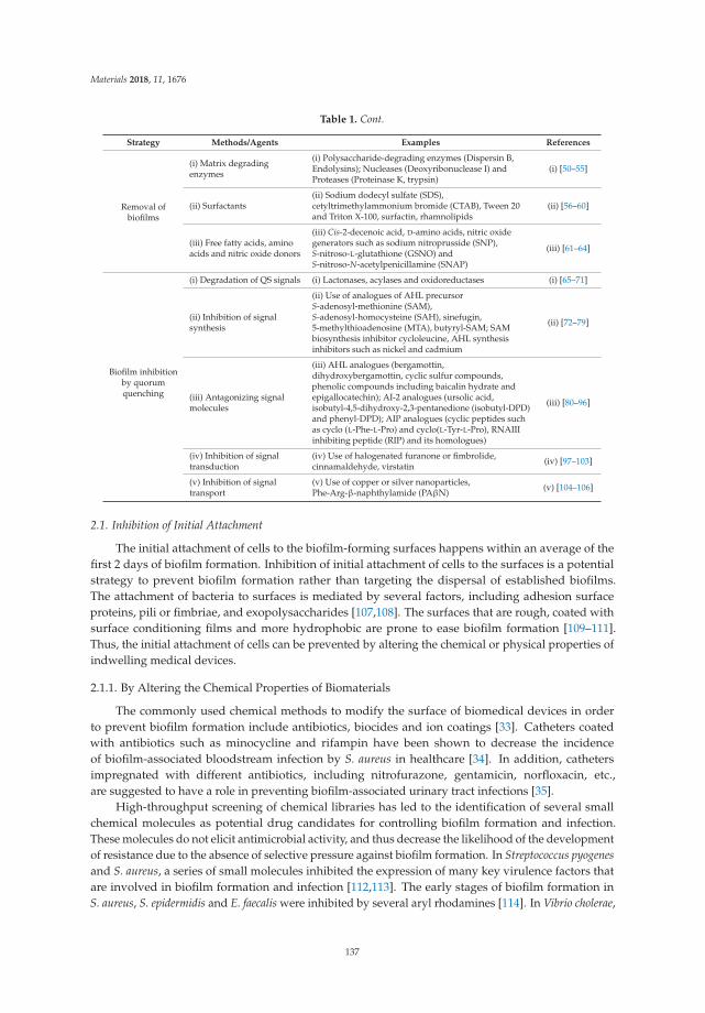
Materials 2018, 11, 1676
Table 1. Cont.
Strategy Methods/Agents Examples References
Removal ofbiofilms
(i) Matrix degradingenzymes
(i) Polysaccharide-degrading enzymes (Dispersin B,Endolysins); Nucleases (Deoxyribonuclease I) andProteases (Proteinase K, trypsin)
(i) [50–55]
(ii) Surfactants(ii) Sodium dodecyl sulfate (SDS),cetyltrimethylammonium bromide (CTAB), Tween 20and Triton X-100, surfactin, rhamnolipids
(ii) [56–60]
(iii) Free fatty acids, aminoacids and nitric oxide donors
(iii) Cis-2-decenoic acid, D-amino acids, nitric oxidegenerators such as sodium nitroprusside (SNP),S-nitroso-L-glutathione (GSNO) andS-nitroso-N-acetylpenicillamine (SNAP)
(iii) [61–64]
Biofilm inhibitionby quorumquenching
(i) Degradation of QS signals (i) Lactonases, acylases and oxidoreductases (i) [65–71]
(ii) Inhibition of signalsynthesis
(ii) Use of analogues of AHL precursorS-adenosyl-methionine (SAM),S-adenosyl-homocysteine (SAH), sinefugin,5-methylthioadenosine (MTA), butyryl-SAM; SAMbiosynthesis inhibitor cycloleucine, AHL synthesisinhibitors such as nickel and cadmium
(ii) [72–79]
(iii) Antagonizing signalmolecules
(iii) AHL analogues (bergamottin,dihydroxybergamottin, cyclic sulfur compounds,phenolic compounds including baicalin hydrate andepigallocatechin); AI-2 analogues (ursolic acid,isobutyl-4,5-dihydroxy-2,3-pentanedione (isobutyl-DPD)and phenyl-DPD); AIP analogues (cyclic peptides suchas cyclo (L-Phe-L-Pro) and cyclo(L-Tyr-L-Pro), RNAIIIinhibiting peptide (RIP) and its homologues)
(iii) [80–96]
(iv) Inhibition of signaltransduction
(iv) Use of halogenated furanone or fimbrolide,cinnamaldehyde, virstatin (iv) [97–103]
(v) Inhibition of signaltransport
(v) Use of copper or silver nanoparticles,Phe-Arg-β-naphthylamide (PAβN) (v) [104–106]
2.1. Inhibition of Initial Attachment
The initial attachment of cells to the biofilm-forming surfaces happens within an average of thefirst 2 days of biofilm formation. Inhibition of initial attachment of cells to the surfaces is a potentialstrategy to prevent biofilm formation rather than targeting the dispersal of established biofilms.The attachment of bacteria to surfaces is mediated by several factors, including adhesion surfaceproteins, pili or fimbriae, and exopolysaccharides [107,108]. The surfaces that are rough, coated withsurface conditioning films and more hydrophobic are prone to ease biofilm formation [109–111].Thus, the initial attachment of cells can be prevented by altering the chemical or physical properties ofindwelling medical devices.
2.1.1. By Altering the Chemical Properties of Biomaterials
The commonly used chemical methods to modify the surface of biomedical devices in orderto prevent biofilm formation include antibiotics, biocides and ion coatings [33]. Catheters coatedwith antibiotics such as minocycline and rifampin have been shown to decrease the incidenceof biofilm-associated bloodstream infection by S. aureus in healthcare [34]. In addition, cathetersimpregnated with different antibiotics, including nitrofurazone, gentamicin, norfloxacin, etc.,are suggested to have a role in preventing biofilm-associated urinary tract infections [35].
High-throughput screening of chemical libraries has led to the identification of several smallchemical molecules as potential drug candidates for controlling biofilm formation and infection.These molecules do not elicit antimicrobial activity, and thus decrease the likelihood of the developmentof resistance due to the absence of selective pressure against biofilm formation. In Streptococcus pyogenesand S. aureus, a series of small molecules inhibited the expression of many key virulence factors thatare involved in biofilm formation and infection [112,113]. The early stages of biofilm formation inS. aureus, S. epidermidis and E. faecalis were inhibited by several aryl rhodamines [114]. In Vibrio cholerae,
137

Materials 2018, 11, 1676
small molecules inhibited the induction of cyclic di-GMP, which is a second messenger controllingthe switch between planktonic and sessile lifestyle of bacteria [115,116]. In addition, N-acetylcysteine,a mucolytic agent, was reported to inhibit the production of exopolysaccharides in biofilms inS. epidermidis [36].
Several antimicrobial peptides are also known to interfere with biofilm formation in differentbacterial pathogens. For example, peptide 1018 is considered to be a biofilm inhibitor in P. aeruginosa,E. coli, A. baumannii, K. pneumoniae, S. aureus, Salmonella typhimurium, Burkholderia cenocepacia [37].In addition, lantibiotics (nisin, subtilin, epidermin and gallidermin), a class of peptide antibiotics,are reported to inhibit biofilm formation in S. aureus, Lactococcus lactis and S. epidermidis [38,39].
Chelators that interfere with the function of metal ions in biofilm formation are also considered tobe biofilm inhibitors [117]. Metallic silver, silver salts, and silver nanoparticles have been widely usedas antimicrobial agents in medical implants against bacteria such as E. coli, S. aureus, Klebsiella species,P. aeruginosa, S. typhimurium, and Candida albicans [118,119]. The silver treatment inhibits the replicationof DNA, expression of ribosomal and cellular proteins, and respiration process, leading to celldeath [40–42]. It has been reported that silver ion-coated implants inhibited S. aureus biofilm formationwithout causing silver accumulation in host tissues [120]. In addition, in the presence of nanoparticles,antibiotics such as penicillin G, amoxicillin, erythromycin, clindamycin, and vancomycin displayedincreased antibacterial activity against S. aureus [121].
The antibacterial agent coatings on medical devices are typically effective for a short timeperiod due to the leaching of the agent over the course of time [33]. Thus, the immobilization ofantimicrobial agents on device surfaces using long, flexible polymeric chains has been an effectivecontribution in controlling biofilm formation in the long run. For example, the attachment ofN-alkylpyridinium bromide, an antibacterial agent, to a polymer, poly(4-vinyl-N-hexylpyridine)was capable of inactivating 99% of S. epidermidis, E. coli, and P. aeruginosa on medical devices [43].
2.1.2. By Changing the Physical Properties of Biomaterials
Biofilm formation begins with a weak reversible adhesion of bacterial cells to the surface ofmedical devices. If bacteria are not immediately detached from the surface of devices, they anchorpermanently, using cell adhesion structures such as pili, and form biofilms [44]. Hydrophobicityand surface charge of implant materials play an important role in determining the ability ofbacteria to anchor to surfaces [43]. Thus, modification of the surface charge and hydrophobicityof polymeric materials using several backbone compounds and antimicrobial agents has provento be effective for biofilm prevention [43]. Hydrophilic polymers such as hyaluronic acid [45] andpoly N-vinylpyrrolidone [46] on polyurethane catheters and silicone shuts, respectively, have beenknown to reduce the adhesion of S. epidermidis. In addition, various hydrogel coatings whichreduce bacterial adhesion due to their hydrophilic properties have also been developed especiallyfor ureteral stents [47]. Superhydrophobic surfaces are reported to reduce bacterial adhesionand biofilm formation due to their extremely low wettability [49,122,123]. Tang et al. observedreduced adherence of S. aureus on superhydrophobic titanium surfaces [124]. Also, the adhesionof S. aureus and P. aeruginosa was significantly reduced on superhydrophobic fluorinated silicacoating [125]. Crick et al.demonstrated reduced adhesion of S. aureus and E. coli on AACVD (aerosolassisted chemical vapor deposition)-coated superhydrophobic surfaces compared to uncoated plainglass [123]. It has been reported that heparin interferes with bacterial adhesion and colonization [48].The heparin coating makes the vascular catheter negatively charged, thereby preventing thrombosisand microbial colonization, eventually contributing to reduction of catheter-related infections [126,127].Surface roughness can also influence biofilm formation, as rough, high-energy surfaces are moreconducive to biofilm formation compared to smooth surfaces [128]. It is noted that the surfaceroughness can alter the hydrophobicity, thus in turn affecting bacterial adherence [128].
138

Materials 2018, 11, 1676
2.2. Biofilm Removal
Mature biofilms are highly tolerant to antimicrobials due to the altered growth rate of cellsin the biofilm and the emergence of resistant subpopulations [129,130]. Also, biofilms favor thehorizontal transfer of antibiotic resistance genes among cells [131]. Thus, it is of utmost importanceto understand the antibiotic resistance properties of strains in biofilms when designing new drugtreatments. Though conventional antibiotics have been proven to be critical in eliminating bacterialpathogens, they extensively damage the host microbiota, making the environment favorable foropportunistic pathogens. Hence, the agents that interfere with the initial biofilm development orbiofilm structure have great potential in controlling biofilm-related infections.
2.2.1. Matrix-Degrading Enzymes
The biofilm matrix is usually composed of exopolysaccharides (EPS), extracellular DNAs (eDNAs),and proteins [132–134]. The EPS and eDNAs contribute to antibiotic resistance by preventingthe diffusion of antimicrobials or by inducing antibiotic resistance [135,136]. Dissociation of thebiofilm matrix is an effective antibiofilm approach, as the matrix accounts for more than 90% ofdry mass, and dissociation of the same will expose the sessile cells to antibiotics and host immunedefence [137]. Biofilm matrix-degrading enzymes fall into three categories: polysaccharide-degradingenzymes, nucleases and proteases [50]. Dispersin B is a bacterial glycoside hydrolase produced byActinobacillus actinomycetemcomitans which hydrolyzes poly-N-acetylglucosamine (PNAG), a majormatrix exopolysaccharide of Staphylococcus spp. and E. coli [138]. In addition, the applicationof Dispersin B in combination with triclosan effectively reduced biofilm formation in S. aureus,S. epidermidis, and E. coli [51]. Endolysins, a class of peptidoglycan hydolases produced bybacteriophages are reported to digest the cell wall of bacteria thereby disrupting biofilms [52].Deoxyribonuclease I which is capable of digesting eDNA is known to disperse biofilms inseveral bacteria including Staphylococcus strains, A. baumannii, E. coli, Haemophilus influenzae,Klebsiella pneumoniae, Psuedomonas aeruginosa, etc. [53,54]. The matrix proteins can be effectively cleavedby Proteinase K contributing to biofilm prevention and biofilm dispersal [55]. It was demonstratedthat the treatment with dispersin B followed by Proteinase K or trypsin successfully eradicatedStaphylococcus biofilms [55]. The in vivo application of matrix-degrading enzymes is limited, as thetreatment can elicit inflammatory and allergic reactions in the host against these enzymes [139].
2.2.2. Surfactants
Surfactants are reported to have antimicrobial and antibiofilm activities [140]. The surfactantssodium dodecyl sulfate (SDS), cetyltrimethylammonium bromide (CTAB), Tween 20 and Triton X-100are known to promote either biofilm dispersal or detachment [56–58]. A biosurfactant, surfactin,which is a cyclic lipopeptide produced by B. subtilis, is reported to inhibit biofilm formation and inducebiofilm dispersal in S. typhimurium, E. coli and P. mirabilis [59]. Rhamnolipids are principal glycolipidsproduced by many bacteria, including P. aeruginosa, and cause biofilm dispersal in a number bacterialstrains [57,60].
2.2.3. Free Fatty Acids, Amino Acids and Nitric Oxide Donors
Free fatty acids are shown to have antibiofilm activity against several pathogenic bacteria. It wasreported that P. aeruginosa produces an organic compound cis-2-decenoic acid which is capableof dispersing the already established biofilms by E. coli, K. pneumoniae, P. mirabilis, S. pyogenes,B. subtilis, S. aureus, and C. albicans [61]. The diffusible signal factor, cis-11-methyl-2-decanoicacid produced by Xanthomonas campestris induces biofilm dispersal by controlling the productionof exopolysaccharide-degrading enzyme [141]. However, it has also been reported that fatty acids playan important role in the initial stages of biofilm formation in B. subtilis, as the lipids form structuralcomponent of extracellular matrix of biofilms [142]. In S. aureus, B. subtilis and P. aeruginosa, a mixture
139
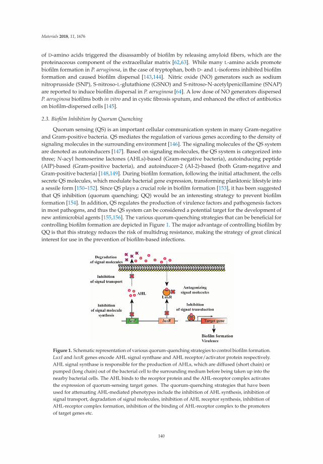
Materials 2018, 11, 1676
of D-amino acids triggered the disassambly of biofilm by releasing amyloid fibers, which are theproteinaceous component of the extracellular matrix [62,63]. While many L-amino acids promotebiofilm formation in P. aeruginosa, in the case of tryptophan, both D- and L-isoforms inhibited biofilmformation and caused biofilm dispersal [143,144]. Nitric oxide (NO) generators such as sodiumnitroprusside (SNP), S-nitroso-L-glutathione (GSNO) and S-nitroso-N-acetylpenicillamine (SNAP)are reported to induce biofilm dispersal in P. aeruginosa [64]. A low dose of NO generators dispersedP. aeruginosa biofilms both in vitro and in cystic fibrosis sputum, and enhanced the effect of antibioticson biofilm-dispersed cells [145].
2.3. Biofilm Inhibition by Quorum Quenching
Quorum sensing (QS) is an important cellular communication system in many Gram-negativeand Gram-positive bacteria. QS mediates the regulation of various genes according to the density ofsignaling molecules in the surrounding environment [146]. The signaling molecules of the QS systemare denoted as autoinducers [147]. Based on signaling molecules, the QS system is categorized intothree; N-acyl homoserine lactones (AHLs)-based (Gram-negative bacteria), autoinducing peptide(AIP)-based (Gram-positive bacteria), and autoinducer-2 (AI-2)-based (both Gram-negative andGram-positive bacteria) [148,149]. During biofilm formation, following the initial attachment, the cellssecrete QS molecules, which modulate bacterial gene expression, transforming planktonic lifestyle intoa sessile form [150–152]. Since QS plays a crucial role in biofilm formation [153], it has been suggestedthat QS inhibition (quorum quenching; QQ) would be an interesting strategy to prevent biofilmformation [154]. In addition, QS regulates the production of virulence factors and pathogenesis factorsin most pathogens, and thus the QS system can be considered a potential target for the development ofnew antimicrobial agents [155,156]. The various quorum-quenching strategies that can be beneficial forcontrolling biofilm formation are depicted in Figure 1. The major advantage of controlling biofilm byQQ is that this strategy reduces the risk of multidrug resistance, making the strategy of great clinicalinterest for use in the prevention of biofilm-based infections.
Figure 1. Schematic representation of various quorum-quenching strategies to control biofilm formation.LuxI and luxR genes encode AHL signal synthase and AHL receptor/activator protein respectively.AHL signal synthase is responsible for the production of AHLs, which are diffused (short chain) orpumped (long chain) out of the bacterial cell to the surrounding medium before being taken up into thenearby bacterial cells. The AHL binds to the receptor protein and the AHL-receptor complex activatesthe expression of quorum-sensing target genes. The quorum-quenching strategies that have beenused for attenuating AHL-mediated phenotypes include the inhibition of AHL synthesis, inhibition ofsignal transport, degradation of signal molecules, inhibition of AHL receptor synthesis, inhibition ofAHL-receptor complex formation, inhibition of the binding of AHL-receptor complex to the promotersof target genes etc.
140

Materials 2018, 11, 1676
2.3.1. Degradation of QS Signals
AHLs can be degraded by specific enzymes such as lactonases that hydrolyze the lactone ring inthe homoserine moiety and acylases that cleave off the acyl side chain, and the activity can be alteredby reductases and oxidases [157]. Most of the AHL-degrading enzymes were discovered in bacterialspecies [158], though some are found in eukaryotes [159,160]. It has been reported that the applicationof QQ enzymes inhibits biofilm formation in several bacterial strains [65–70]. Quorum-quenchingenzymes disrupt the biofilm architecture, which increases the antibiotic susceptibility of the cells [71].Significant reduction of biofilm formation and increased sensitivity to antibiotics was noticed inP. aeruginosa after treatment with lactonase [71]. The oxidoreductases reduced the signaling moleculesAHL and AI-2 to QS-inactive hydroxy-derivatives in K. oxytoca and K. pneumoniae [70].
2.3.2. Inhibition of Signal Synthesis
Several reports have shown that mutations affecting AHL synthesis have an adverse effect on biofilmformation. For example, P. aeruginosa strain that lacked the production of 3-oxo-C12-HSL resulted inimpaired biofilm formation [161]. The mutation in the gene encoding for AHL synthesis enzyme inB. cenocepacia K56-2, B. cenocepacia J2315, Aeromonas hydrophila and Serratia liquefaciens led to defectivebiofilm formation [162–165]. In addition, the mutants of several Vibrio spp., Streptococcus spp. andStaphylococcus spp. that are deficient in AI-2 synthesis were not able to produce biofilms properly.Thus, blocking signal production has been considered as a promising strategy to control biofilm formation.Analogues of AHL precursor molecule, S-adenosyl-methionine (SAM), such as S-adenosyl-homocysteine(SAH), sinefugin, 5-methylthioadenosine (MTA), and butyryl-SAM, are known to inhibit biofilm formationin P. aeruginosa [72]. Also, the SAM biosynthesis inhibitor cycloleucine is reported to inhibit AHLproduction [73]. The antibiotic azithromycin interferes with signal synthesis in P. aeruginosa, and thussignificantly clears biofilm in mouse model of cystic fibrosis [74,75]. In addition, several inhibitorsfor the key enzymes (5′-methylthioadenosine/S-adenosylhomo-cysteine nucleosidase (MTAN) andS-ribosylhomocysteinase (LuxS) involved in AI-2 synthesis are shown to reduce biofilm formation [76,77].In B. multivorans, nickel (Ni2+) and cadmium (Cd2+) inhibited the expression of genes responsible for AHLproduction thereby inhibiting cell-cell signaling and subsequently biofilm formation [79]. The inhibitoryeffect of Cd2+ in quorum sensing was also reported in Chromobacterium violaceum [78].
2.3.3. Antagonizing the Signal Molecules
Researchers have screened for many signal analogues that antagonize QS signaling, therebypreventing biofilm formation [166–168]. AHL analogues in which the lactone ring was replaced bya cyclopentyl or a cyclohexanone ring adversely affected biofilm formation in Serratia marcescensand P. aeruginosa [169,170]. Many natural compounds are also reported to antagonize AHL-based QSsignaling, and those include bergamottin and dihydroxybergamottin from grapefruit juice, cyclic sulfurcompounds from garlic, patulin, and penicillic acid from a variety of fungi, etc. [80–82]. Treatment withpatulin, ajoene and garlic extracts resulted in increased antibiotic susceptibility of P. aeruginosa biofilmsand increased clearance of P. aeruginosa in in vivo pulmonary infection model [83–85]. In addition,some phenolic compounds including baicalin hydrate and epigallocatechin blocked AHL QS andaffected biofilm formation of B. cenocepacia, B. multivorans and P. aeruginosa [86–88]. It was noted that theantibiotic susceptibility of B. cenocepacia and P. aeruginosa increased after treatment with baicalin hydratein different in vitro biofilm models [86–88]. Thus, the concept of combining QS inhibitor (QSI) andantibiotics would be a better strategy to control biofilm formation by pathogenic bacteria. In addition,it has been noticed that biofilm formation can be effectively controlled by combining QSIs andQQ enzymes. Recently, Fong et al. reported the synergistic effect of a QS inhibitor, G1, which competeswith AHL to bind to the response regulator and QQ enzyme, AHL lactonase, to effectively controlbiofilm formation and virulence by P. aeruginosa [171].
141
Materials 2018, 11, 1676
Several compounds that antagonize AI-2 signaling have also been reported to exhibitantibiofilm activity. The AI-2 analogues ursolic acid, isobutyl-4,5-dihydroxy-2,3-pentanedione(isobutyl-DPD) and phenyl-DPD inhibited biofilm formation and removed preformed biofilms inE. coli and P. aeruginosa [89,90]. Although other compounds, including pyrogallol and its derivatives,some nucleoside analogues, boronic acids, and sulfones have been identified to antagonize AI-2signaling, only a few have been investigated for their antibiofilm activity [172,173].
Several AIP analogues, such as truncated forms of AIP, and probiotic bacteria-producing naturalcyclic dipeptides, such as cyclo(l-Phe-l-Pro) and cyclo(l-Tyr-l-Pro), have been developed to antagonizeQS signaling in Gram-positive bacteria. However, the experimental evidence on the anti-biofilmactivities of these compounds is highly limited [91–93]. The most investigated QS inhibiting peptideis the RNAIII inhibiting peptide (RIP), which is produced by coagulase-negative Staphylococci.RIP interferes with the QS response by inhibiting the production of RNAIII, a key component ofQS response in S. aureus [94]. RIP and several RIP homologues have been reported to have anti-QSand anti-biofilm activity against Staphylococcus spp. A RIP analogue, FS3, prevented S. aureus biofilmformation in a rat vascular graft model [95]. In addition, a non-peptide RIP analogue, hamamelitannin,blocked QS in Staphylococcus spp., and potentially inhibited biofilm formation in in vitro and in vivo ratmodel of graft infection [96]. Several natural compounds, including phytol, anthocyanidins, extractsfrom Ricinus communis, freshwater bryozoan Hyalinella punctata and selected sponges, and ricininederivatives, are also known to exhibit anti-biofilm or anti-microbial and anti-quorum sensing activitiesin P. aeruginosa [174–178]. However, the exact mechanisms by which these compounds displayanti-quorum sensing activities are not known.
2.3.4. Inhibition of Signal Transduction by Interfering with Response Regulator Activity
The QS system can also be hindered at the level of signal transduction cascade. The naturalcompounds, halogenated furanone or fimbrolide and cinnamaldehyde which are isolated from redalgae Delisea pulchra and cinnamon bark, respectively, interfere with signal transduction and affectbiofilm formation, thereby increasing antibiotic susceptibility in several pathogenic bacteria [97–100].Both compounds block AI-2 and AHL-type QS systems, and thereby affect biofilm formation inV. harveyi [101,102]. The halogenated furanone and cinnamaldehyde inhibits AI-2 QS and AHL QSby decreasing the DNA-binding ability of the response regulator LuxR, which is important for thesignal transduction cascade, or by displacing AHL from its receptor, respectively [101,102,179,180].In addition, the natural furanone inactivates LuxS and accelerates LuxR turnover, thereby blockingAI-2 and AHL QS signaling system, respectively [181,182]. Cinnamaldehyde is widely used asa flavoring agent in food and beverages, while the application of furanones is limited because of theirtoxicity [100,183]. We reported previously that virstatin, a small organic molecule, prevents biofilmformation by interfering with the QS system in A. nosocomialis [103]. It was noticed that virstatininhibits the expression of the response regulator, AnoR, which is a positive regulator of the AHLsynthase gene, anoI in A. nosocomialis [103]. The repression of AnoR leads to decreased synthesisof AHL (Figure 2), adversely affecting the signal transduction cascade. Virstatin or its derivativescan be considered potential agents to inhibit the QS system and to control biofilm-based infections,and further studies in this direction could lead to the development of better antibacterial therapeutics.
142

Materials 2018, 11, 1676
Figure 2. Effect of virstatin on the production of AHL. Bioassay was carried out to check the effect ofvirstatin on the production of AHLs in A. nosocomialis. For this, the strain was cultivated overnight inLuria Bertani (LB) medium at 30 ◦C, and the cells were washed with LB and diluted to an OD600 of 1.The cells were treated with different concentrations of virstatin (20, 50, 100 mM), which was dissolvedin dimethyl sulfoxide (DMSO), and 5 μL of the samples were spotted onto chromoplate overlaid withA. tumefaciens NT1 (pDCI41E33) [184,185]. Synthetic N-(3-hydroxy-dodecanoyl)-L-homoserine lactone(OH-dDHL) was spotted as a positive control. The plates were incubated at 30 ◦C for 22 h, followedby the detection of the color zone surrounding the bacteria. A representative chromoplate imagewith 100 mM virstatin and images of color zones from different concentrations of virstatin are shown.WT, A. nosocomialis wild type; +ve C, OH-dDHL; vir20, vir50 and vir100; wild-type cells treated with20, 50 and 100 mM virstatin respectively.
2.3.5. Inhibition of Signal Transport
The signaling molecules need to be exported and released into the extracellular space to besensed by other bacteria for effective cell-to-cell communication. The role of multidrug-resistant(MDR) efflux pumps in signal traffic was first reported in P. aeruginosa, in which AHLs with long sidechains are actively transported across the cell membrane through the MexAB-OprM efflux pump [186].In P. aeruginosa, the expression of the autoinducer-producing gene and the genes encoding the virulencefactors is limited by the intracellular concentration of the autoinducer [187]. The involvement of theMDR efflux pump in the QS system has also been reported in E. coli, in which the overexpression of theQS regulator SdiA led to the increased expression of the AcrAB efflux pump [188]. In Bacteroides fragilis,an opportunistic pathogen of the gastrointestinal tract, the BmeB efflux pump controls the intracellularAHL concentration by effluxing AHL outside of cells [189]. In addition, the expression of the MDRefflux pump, BpeAB-OprB, was reported to be essential for the export of six AHL inducers to theextracellular environment in B. pseudomallei [190,191]. Thus, the inhibition of the efflux pump would bea promising strategy to alter QS signaling cascade, thereby preventing biofilm formation and virulence.
Several studies have provided evidence to show the link between the physiological function ofefflux pump and biofilm formation. In E. coli and Klebsiella strains, the inhibition of the efflux pumpactivity using efflux pump inhibitors (EPIs) reduced biofilm formation [192]. The genetic inactivationor the chemical inhibition of efflux pump activity resulted in impaired biofilm formation in S. entericaserovar typhimurium [193]. The effect of efflux pump inhibitors to prevent biofilm formation was also
143

Materials 2018, 11, 1676
demonstrated in P. aeruginosa and S. aureus [105], in which copper nanoparticles work well as EPIand anti-biofilm agents [104]. In addition, in P. aeruginosa, the MDR efflux pump, MexAM-OPrM wasdisrupted by silver nanoparticles [194]. Recently, it was observed that the well-characterized EPI,Phe-Arg-β-naphthylamide (PAβN) alter the expression of QS molecules and QS-dependent virulencephenotypes in P. aeruginosa PAO1, as well as in clinical isolates [106]. The application of EPIs not onlyhelps to reduce the biofilm-forming capacity of bacteria, but also to revive the bactericidal effect ofconventional antibiotics [195].
It has been reported previously that AHLs with long side chains are exported out through theMexAB-OprM pump in P. aeruginosa [186], and the expression of the pump is modulated by theintracellular concentration of autoinducer molecules [196]. In addition, we have identified previouslythat A. nosocomialis produces AHL with long side chain, N-(3-hydroxy-dodecanoyl)-L-homoserinelactone (3OH-C12-AHL) as signaling molecules [103], and these AHLs might be actively transportedthrough the efflux pumps. Thus, it can be postulated that the QS system controls the activity ofthe efflux system, contributing to the effective transport of AHLs across the cell membrane, in turncontributing to virulence and biofilm formation. Further studies in this direction would unravel indepth the role of the QS system in controlling the activity of these efflux pumps. The MDR effluxpumps and the regulators modulating them would be potential targets for the development of bettertherapeutics for biofilm-based infections.
3. Conclusions
In this review, we discuss the current strategies and future perspectives for developing improvedtherapeutics for controlling biofilm-based infections. The various approaches for modulating biofilmformation on medical devices are addressed in detail, with special emphasis on quorum-quenchingstrategies. Significant advances have been made in understanding the role of quorum sensing in biofilmformation in the past few years. In addition, several studies have shown that multidrug efflux pumpsplay a potential role in controlling biofilm formation. However, the fundamental mechanisms by whichthe QS systems exert the regulatory functions on biofilm formation are poorly understood. In thisreview, we postulate that QS systems regulate the activity of multidrug efflux pumps in transportingQS molecules across the cell membrane, thereby affecting biofilm formation. We propose that thetranscriptional factors modulating the QS system and/or efflux pumps would be potential targets fordeveloping QSIs. It is of utmost importance to improve our understanding of the molecular mechanismby which QS systems regulate biofilm formation and multidrug efflux pumps, as it will eventually beof help in developing better therapeutics for the treatment of problematic biofilm-related infections.Furthermore, detailed research is needed to understand the effect of these QSIs on different stagesof biofilm formation and to validate their applicability on humans. Since QSIs do not induce anyantibiotic resistance, they can be of great potential in the future for the treatment of biofilm-basedinfections in healthcare settings.
Funding: This research was funded by the research fund of Chungnam National University (2017).Also, this research was supported by the Basic Science Research Program through the National ResearchFoundation of Korea (NRF) funded by the Ministry of Education (NRF-2014R1A61029617).
Conflicts of Interest: The authors declare no conflict of interest.
References
1. Costerton, J.W.; Irvin, R.T.; Cheng, K.J. The bacterial glycocalyx in nature and disease. Annu. Rev. Microbiol.1981, 35, 299–324. [CrossRef] [PubMed]
2. Kunin, C.M.; Steele, C. Culture of the surfaces of urinary catheters to sample urethral flora and study theeffect of antimicrobial therapy. J. Clin. Microbiol. 1985, 21, 902–908. [PubMed]
144

Materials 2018, 11, 1676
3. Nickel, J.C.; Ruseska, I.; Wright, J.B.; Costerton, J.W. Tobramycin resistance of Pseudomonas aeruginosacells growing as a biofilm on urinary catheter material. Antimicrob. Agents Chemother. 1985, 27, 619–624.[CrossRef] [PubMed]
4. Donlan, R.M. Biofilms: Microbial life on surfaces. Emerg. Infect. Dis. 2002, 8, 881–890. [CrossRef] [PubMed]5. Donlan, R.M. Biofilms and device-associated infections. Emerg. Infect. Dis. 2001, 7, 277–281. [CrossRef] [PubMed]6. Haddadin, Y.; Regunath, H. Central Line Associated Blood Stream Infections (CLABSI); StatPearls Publishing:
Treasure Island, FL, USA, 2017.7. Tien, H.C.; Battad, A.; Bryce, E.A.; Fuller, J.; Mulvey, M.; Bernard, K.; Brisebois, R.; Doucet, J.J.; Rizoli, S.B.;
Fowler, R.; et al. Multi-drug resistant Acinetobacter infections in critically injured Canadian forces soldiers.BMC Infect. Dis. 2007, 7, 95. [CrossRef] [PubMed]
8. Otto, M. Staphylococcal biofilms. In Bacterial Biofilms; Romeo, T., Ed.; Current Topics in Microbiology andImmunology book series; Springer: Berlin, Germany, 2008; Volume 322, pp. 207–228.
9. Otto, M. Staphylococcus epidermidis—The ‘accidental’ pathogen. Nat. Rev. Microbiol. 2009, 7, 555–567. [CrossRef][PubMed]
10. Agarwal, A.; Singh, K.P.; Jain, A. Medical significance and management of staphylococcal biofilm.FEMS Immunol. Med. Microbiol. 2010, 58, 147–160. [CrossRef] [PubMed]
11. Pour, N.K.; Dusane, D.H.; Dhakephalkar, P.K.; Zamin, F.R.; Zinjarde, S.S.; Chopade, B.A. Biofilm formationby Acinetobacter baumannii strains isolated from urinary tract infection and urinary catheters. FEMS Immunol.Med. Microbiol. 2011, 62, 328–338. [CrossRef] [PubMed]
12. Dijkshoorn, L.; Nemec, A.; Seifert, H. An increasing threat in hospitals: Multidrug-resistant Acinetobacterbaumannii. Nat. Rev. Microbiol. 2007, 5, 939–951. [CrossRef] [PubMed]
13. Patwardhan, R.B.; Dhakephalkar, P.K.; Niphadkar, K.B.; Chopade, B.A. A study on nosocomial pathogens inICU with special reference to multiresistant Acinetobacter baumannii harbouring multiple plasmids. Indian J.Med. Res. 2008, 128, 178–187. [PubMed]
14. Bergogne-Bérézin, E.; Towner, É.K.J. Acinetobacter spp. as nosocomial pathogens: Microbiological, clinicaland epidemiological features. Clin. Microiol. Rev. 1996, 9, 148–165.
15. Litzler, P.Y.; Benard, L.; Barbier-Frebourg, N.; Vilain, S.; Jouenne, T.; Beucher, E.; Bunel, C.; Lemeland, J.F.;Bessou, J.P. Biofilm formation on pyrolytic carbon heart valves: Influence of surface free energy, roughness,and bacterial species. J. Thorac. Cardiovasc. Surg. 2007, 134, 1025–1032. [CrossRef] [PubMed]
16. Percival, S.L.; Suleman, L.; Vuotto, C.; Donelli, G. Healthcare-associated infections, medical devices andbiofilms: Risk, tolerance and control. J. Med. Microbiol. 2015, 64, 323–334. [CrossRef] [PubMed]
17. Von Eiff, C.; Heilmann, C.; Peters, G. New aspects in the molecular basis of polymer-associated infectionsdue to Staphylococci. Eur. J. Clin. Microbiol. Infect. Dis. 1999, 18, 843–846. [CrossRef] [PubMed]
18. Muller, E.; Hübner, J.; Gutierrez, N.; Takeda, S.; Goldmann, D.A.; Pier, G.B. Isolation and characterization oftransposon mutants of Staphylococcus epidermidis deficient in capsular polysaccharide/adhesin and slime.Infect. Immun. 1993, 61, 551–558. [PubMed]
19. Veenstra, G.J.; Cremers, F.F.; van Dijk, H.; Fleer, A. Ultrastructural organization and regulation ofa biomaterial adhesin of Staphylococcus epidermidis. J. Bacteriol. 1996, 178, 537–541. [CrossRef] [PubMed]
20. Götz, F. Staphylococcus and biofilms. Mol. Microbiol. 2002, 43, 1367–1378. [CrossRef] [PubMed]21. Stoodley, P.; Sauer, K.; Davies, D.G.; Costerton, J.W. Biofilms as complex differentiated communities.
Annu. Rev. Microbiol. 2002, 56, 187–209. [CrossRef] [PubMed]22. Fey, P.D.; Olson, M.E. Current concepts in biofilm formation of Staphylococcus epidermidis. Future Microbiol.
2010, 5, 917–933. [CrossRef] [PubMed]23. Lawrence, J.R.; Korber, D.R.; Wolfaardt, G.M. Heterogeneity of natural biofilm communities. Cells Mater.
1996, 6, 175–191.24. Aslam, S. Effect of antibacterials on biofilms. Am. J. Infect. Control 2008, 36, S175.e9–S175.e11. [CrossRef]
[PubMed]25. Francolini, I.; Donelli, G. Prevention and control of biofilm-based medical-device-related infections.
FEMS Immunol. Med. Microbiol. 2010, 59, 227–238. [CrossRef] [PubMed]26. Stewart, P.S.; Davison, W.M.; Steenbergen, J.N. Daptomycin rapidly penetrates a Staphylococcus epidermidis
biofilm. Antimicrob. Agents Chemother. 2009, 53, 3505–3507. [CrossRef] [PubMed]
145

Materials 2018, 11, 1676
27. Borriello, G.; Werner, E.; Roe, F.; Kim, A.M.; Ehrlich, G.D.; Stewart, P.S. Oxygen limitation contributes toantibiotic tolerance of Pseudomonas aeruginosa in biofilms. Antimicrob. Agents Chemother. 2004, 48, 2659–2664.[CrossRef] [PubMed]
28. Lewis, K. Persister cells, dormancy and infectious disease. Nat. Rev. Microbiol. 2007, 5, 48–56. [CrossRef][PubMed]
29. Martínez, J.L.; Baquero, F. Interactions among strategies associated with bacterial infection: Pathogenicity,epidemicity, and antibiotic resistance. Clin. Microbiol. Rev. 2002, 15, 647–679. [CrossRef] [PubMed]
30. Diekema, D.J.; Pfaller, M.A.; Schmitz, F.J.; Smayevsky, J.; Bell, J.; Jones, R.N.; Beach, M.; Group, S.P. Surveyof infections due to Staphylococcus species: Frequency of occurrence and antimicrobial susceptibility ofisolates collected in the United States, Canada, Latin America, Europe, and the Western Pacific region for theSENTRY antimicrobial surveillance program, 1997–1999. Clin. Infect. Dis. 2001, 32, S114–S132. [CrossRef][PubMed]
31. Kalia, V.C.; Purohit, H.J. Quenching the quorum sensing system: Potential antibacterial drug targets.Crit. Rev. Microbiol. 2011, 37, 121–140. [CrossRef] [PubMed]
32. Wright, J.S., III; Lyon, G.J.; George, E.A.; Muir, T.W.; Novick, R.P. Hydrophobic interactions driveligand-receptor recognition for activation and inhibition of staphylococcal quorum sensing. Proc. Natl. Acad.Sci. USA 2004, 101, 16168–16173. [CrossRef] [PubMed]
33. Dror, N.; Mandel, M.; Hazan, Z.; Lavie, G. Advances in microbial biofilm prevention on indwelling medicaldevices with emphasis on usage of acoustic energy. Sensors 2009, 9, 2538–2554. [CrossRef] [PubMed]
34. Ramos, E.R.; Reitzel, R.; Jiang, Y.; Hachem, R.Y.; Chaftari, A.M.; Chemaly, R.F.; Hackett, B.; Pravinkumar, S.E.;Nates, J.; Tarrand, J.J.; et al. Clinical effectiveness and risk of emerging resistance associated with prolongeduse of antibiotic-impregnated catheters: More than 0.5 million catheter days and 7 years of clinical experience.Crit. Care Med. 2011, 39, 245–251. [CrossRef] [PubMed]
35. Schumm, K.; Lam, T.B. Types of urethral catheters for management of short-term voiding problems inhospitalised adults. Cochrane Database Syst. Rev. 2008, CD004013. [CrossRef]
36. Pérez-Giraldo, C.; Rodríguez-Benito, A.; Morán, F.J.; Hurtado, C.; Blanco, M.T.; Gómez-García, A.C. Influence ofN-acetylcysteine on the formation of biofilm by Staphylococcus epidermidis. J. Antimicrob. Chemother. 1997, 39, 643–646.[CrossRef] [PubMed]
37. De la Fuente-Núñez, C.; Reffuveille, F.; Haney, E.F.; Straus, S.K.; Hancock, R.E. Broad-spectrum anti-biofilmpeptide that targets a cellular stress response. PLoS Pathog. 2014, 10, e1004152. [CrossRef] [PubMed]
38. Parisot, J.; Carey, S.; Breukink, E.; Chan, W.C.; Narbad, A.; Bonev, B. Molecular mechanism of target recognition bysubtilin, a class I lanthionine antibiotic. Antimicrob. Agents Chemother. 2008, 52, 612–618. [CrossRef] [PubMed]
39. Saising, J.; Dube, L.; Ziebandt, A.K.; Voravuthikunchai, S.P.; Nega, M.; Götz, F. Activity of gallidermin onStaphylococcus aureus and Staphylococcus epidermidis biofilms. Antimicrob. Agents Chemother. 2012, 56, 5804–5810.[CrossRef] [PubMed]
40. Feng, Q.L.; Wu, J.; Chen, G.Q.; Cui, F.Z.; Kim, T.N.; Kim, J.O. A mechanistic study of the antibacterial effectof silver ions on Escherichia coli and Staphylococcus aureus. J. Biomed. Mater. Res. 2000, 52, 662–668. [CrossRef]
41. Yamanaka, M.; Hara, K.; Kudo, J. Bactericidal actions of a silver ion solution on Escherichia coli, studied byenergy-filtering transmission electron microscopy and proteomic analysis. Appl. Environ. Microbiol. 2005, 71, 7589–7593.[CrossRef] [PubMed]
42. Klasen, H.J. A historical review of the use of silver in the treatment of burns. II. Renewed interest for silver.Burns 2000, 26, 131–138. [CrossRef]
43. Jansen, B.; Kohnen, W. Prevention of biofilm formation by polymer modification. J. Ind. Microbiol. 1995, 15, 391–396.[CrossRef] [PubMed]
44. Marlow, V.L.; Porter, M.; Hobley, L.; Kiley, T.B.; Swedlow, J.R.; Davidson, F.A.; Stanley-Wall, N.R.Phosphorylated DegU manipulates cell fate differentiation in the Bacillus subtilis biofilm. J. Bacteriol. 2014,196, 16–27. [CrossRef] [PubMed]
45. Cassinelli, C.; Morra, M.; Pavesio, A.; Renier, D. Evaluation of interfacial properties of hyaluronan coatedpoly(methylmethacrylate) intraocular lenses. J. Biomater. Sci. Polym. Ed. 2000, 11, 961–977. [CrossRef] [PubMed]
46. Boelens, J.J.; Tan, W.F.; Dankert, J.; Zaat, S.A. Antibacterial activity of antibiotic-soaked polyvinylpyrrolidone-graftedsilicon elastomer hydrocephalus shunts. J. Antimicrob. Chemother. 2000, 45, 221–224. [CrossRef] [PubMed]
47. John, T.; Rajpurkar, A.; Smith, G.; Fairfax, M.; Triest, J. Antibiotic pretreatment of hydrogel ureteral stent. J. Endourol.2007, 21, 1211–1216. [CrossRef] [PubMed]
146

Materials 2018, 11, 1676
48. Appelgren, P.; Ransjö, U.; Bindslev, L.; Espersen, F.; Larm, O. Surface heparinization of central venouscatheters reduces microbial colonization in vitro and in vivo: Results from a prospective, randomized trial.Crit. Care Med. 1996, 24, 1482–1489. [CrossRef] [PubMed]
49. Falde, E.J.; Yohe, S.T.; Colson, Y.L.; Grinstaff, M.W. Superhydrophobic materials for biomedical applications.Biomaterials 2016, 104, 87–103. [CrossRef] [PubMed]
50. Li, X.H.; Lee, J.H. Antibiofilm agents: A new perspective for antimicrobial strategy. J. Microbiol. 2017, 55, 753–766.[CrossRef] [PubMed]
51. Darouiche, R.O.; Mansouri, M.D.; Gawande, P.V.; Madhyastha, S. Antimicrobial and antibiofilm efficacy oftriclosan and dispersinb combination. J. Antimicrob. Chemother. 2009, 64, 88–93. [CrossRef] [PubMed]
52. Shen, Y.; Köller, T.; Kreikemeyer, B.; Nelson, D.C. Rapid degradation of Streptococcus pyogenes biofilmsby PlyC, a bacteriophage-encoded endolysin. J. Antimicrob. Chemother. 2013, 68, 1818–1824. [CrossRef][PubMed]
53. Kaplan, J.B.; LoVetri, K.; Cardona, S.T.; Madhyastha, S.; Sadovskaya, I.; Jabbouri, S.; Izano, E.A. Recombinant humanDNase I decreases biofilm and increases antimicrobial susceptibility in Staphylococci. J. Antibiot. 2012, 65, 73–77.[CrossRef] [PubMed]
54. Tetz, G.V.; Artemenko, N.K.; Tetz, V.V. Effect of DNase and antibiotics on biofilm characteristics.Antimicrob. Agents Chemother. 2009, 53, 1204–1209. [CrossRef] [PubMed]
55. Chaignon, P.; Sadovskaya, I.; Ragunah, C.; Ramasubbu, N.; Kaplan, J.B.; Jabbouri, S. Susceptibility ofstaphylococcal biofilms to enzymatic treatments depends on their chemical composition. Appl. Microbiol. Biotechnol.2007, 75, 125–132. [CrossRef] [PubMed]
56. Simões, M.; Pereira, M.O.; Vieira, M.J. Action of a cationic surfactant on the activity and removal of bacterialbiofilms formed under different flow regimes. Water Res. 2005, 39, 478–486. [CrossRef] [PubMed]
57. Boles, B.R.; Thoendel, M.; Singh, P.K. Rhamnolipids mediate detachment of Pseudomonas aeruginosa frombiofilms. Mol. Microbiol. 2005, 57, 1210–1223. [CrossRef] [PubMed]
58. Chen, X.; Stewart, P.S. Biofilm removal caused by chemical treatments. Water Res. 2000, 34, 4229–4233. [CrossRef]59. Mireles, J.R., 2nd; Toguchi, A.; Harshey, R.M. Salmonella enterica serovar typhimurium swarming mutants
with altered biofilm-forming abilities: Surfactin inhibits biofilm formation. J. Bacteriol. 2001, 183, 5848–5854.[CrossRef] [PubMed]
60. Abdel-Mawgoud, A.M.; Lépine, F.; Déziel, E. Rhamnolipids: Diversity of structures, microbial origins androles. Appl. Microbiol. Biotechnol. 2010, 86, 1323–1336. [CrossRef] [PubMed]
61. Davies, D.G.; Marques, C.N. A fatty acid messenger is responsible for inducing dispersion in microbialbiofilms. J. Bacteriol. 2009, 191, 1393–1403. [CrossRef] [PubMed]
62. Kolodkin-Gal, I.; Romero, D.; Cao, S.; Clardy, J.; Kolter, R.; Losick, R. D-amino acids trigger biofilmdisassembly. Science 2010, 328, 627–629. [CrossRef] [PubMed]
63. Jermy, A. Biofilms: Disassembly instructions included. Nat. Rev. Microbiol. 2012, 10, 376. [CrossRef]64. Barraud, N.; Schleheck, D.; Klebensberger, J.; Webb, J.S.; Hassett, D.J.; Rice, S.A.; Kjelleberg, S. Nitric oxide
signaling in Pseudomonas aeruginosa biofilms mediates phosphodiesterase activity, decreased cyclic di-GMPlevels, and enhanced dispersal. J. Bacteriol. 2009, 191, 7333–7342. [CrossRef] [PubMed]
65. dos Reis Ponce, A.; Martins, M.L.; de Araujo, E.F.; Mantovani, H.C.; Vanetti, M.C. Aiia quorum-sensing quenchingcontrols proteolytic activity and biofilm formation by Enterobacter cloacae. Curr. Microbiol. 2012, 65, 758–763.[CrossRef] [PubMed]
66. Ivanova, K.; Fernandes, M.M.; Francesko, A.; Mendoza, E.; Guezguez, J.; Burnet, M.; Tzanov, T. Quorum-quenchingand matrix-degrading enzymes in multilayer coatings synergistically prevent bacterial biofilm formation onurinary catheters. ACS Appl. Mater. Interfaces 2015, 7, 27066–27077. [CrossRef] [PubMed]
67. Jo, S.J.; Kwon, H.; Jeong, S.Y.; Lee, S.H.; Oh, H.S.; Yi, T.; Lee, C.H.; Kim, T.G. Effects of quorum quenching onthe microbial community of biofilm in an anoxic/oxic mbr for wastewater treatment. J. Microbiol. Biotechnol.2016, 26, 1593–1604. [CrossRef] [PubMed]
68. Lee, J.; Lee, I.; Nam, J.; Hwang, D.S.; Yeon, K.M.; Kim, J. Immobilization and stabilization of acylase oncarboxylated polyaniline nanofibers for highly effective antifouling application via quorum quenching. ACS Appl.Mater. Interfaces 2017, 9, 15424–15432. [CrossRef] [PubMed]
69. Vinoj, G.; Vaseeharan, B.; Thomas, S.; Spiers, A.J.; Shanthi, S. Quorum-quenching activity of theAHL-lactonase from Bacillus licheniformis DAHB1 inhibits vibrio biofilm formation in vitro and reducesshrimp intestinal colonisation and mortality. Mar. Biotechnol. 2014, 16, 707–715. [CrossRef] [PubMed]
147

Materials 2018, 11, 1676
70. Weiland-Bräuer, N.; Kisch, M.J.; Pinnow, N.; Liese, A.; Schmitz, R.A. Highly effective inhibition of biofilm formationby the first metagenome-derived AI-2 quenching enzyme. Front. Microbiol. 2016, 7, 1098. [CrossRef] [PubMed]
71. Kiran, S.; Sharma, P.; Harjai, K.; Capalash, N. Enzymatic quorum quenching increases antibiotic susceptibilityof multidrug resistant Pseudomonas aeruginosa. Iran. J. Microbiol. 2011, 3, 1–12. [PubMed]
72. Parsek, M.R.; Val, D.L.; Hanzelka, B.L.; Cronan, J.E., Jr.; Greenberg, E.P. Acyl homoserine-lactonequorum-sensing signal generation. Proc. Natl. Acad. Sci. USA 1999, 96, 4360–4365. [CrossRef] [PubMed]
73. Hanzelka, B.L.; Greenberg, E.P. Quorum sensing in Vibrio fischeri: Evidence that S-adenosylmethionine is theamino acid substrate for autoinducer synthesis. J. Bacteriol. 1996, 178, 5291–5294. [CrossRef] [PubMed]
74. Favre-Bonté, S.; Köhler, T.; Van Delden, C. Biofilm formation by Pseudomonas aeruginosa: Role of the C4-HSLcell-to-cell signal and inhibition by azithromycin. J. Antimicrob. Chemother. 2003, 52, 598–604. [CrossRef][PubMed]
75. Hoffmann, N.; Lee, B.; Hentzer, M.; Rasmussen, T.B.; Song, Z.; Johansen, H.K.; Givskov, M.; Høiby, N.Azithromycin blocks quorum sensing and alginate polymer formation and increases the sensitivity to serumand stationary-growth-phase killing of Pseudomonas aeruginosa and attenuates chronic P. aeruginosa lunginfection in Cftr−/− mice. Antimicrob. Agents Chemother. 2007, 51, 3677–3687. [CrossRef] [PubMed]
76. Shen, G.; Rajan, R.; Zhu, J.; Bell, C.E.; Pei, D. Design and synthesis of substrate and intermediate analogueinhibitors of S-ribosylhomocysteinase. J. Med. Chem. 2006, 49, 3003–3011. [CrossRef] [PubMed]
77. Gutierrez, J.A.; Crowder, T.; Rinaldo-Matthis, A.; Ho, M.C.; Almo, S.C.; Schramm, V.L. Transition stateanalogs of 5′-methylthioadenosine nucleosidase disrupt quorum sensing. Nat. Chem. Biol. 2009, 5, 251–257.[CrossRef] [PubMed]
78. Thornhill, S.G.; Kumar, M.; Vega, L.M.; McLean, R.J.C. Cadmium ion inhibition of quorum signalling inChromobacterium violaceum. Microbiology 2017, 163, 1429–1435. [CrossRef] [PubMed]
79. Vega, L.M.; Mathieu, J.; Yang, Y.; Pyle, B.H.; McLean, R.J.C.; Alvarez, P.J.J. Nickel and cadmium ions inhibit quorumsensing and biofilm formation without affecting viability in Burkholderia multivorans. Int. Biodeterior. Biodegrad. 2014,91, 82–87. [CrossRef]
80. Kim, C.; Kim, J.; Park, H.Y.; Park, H.J.; Lee, J.H.; Kim, C.K.; Yoon, J. Furanone derivatives as quorum-sensingantagonists of Pseudomonas aeruginosa. Appl. Microbiol. Biotechnol. 2008, 80, 37–47. [CrossRef] [PubMed]
81. Rasmussen, T.B.; Skindersoe, M.E.; Bjarnsholt, T.; Phipps, R.K.; Christensen, K.B.; Jensen, P.O.; Andersen, J.B.;Koch, B.; Larsen, T.O.; Hentzer, M.; et al. Identity and effects of quorum-sensing inhibitors produced byPenicillium species. Microbiology 2005, 151, 1325–1340. [CrossRef] [PubMed]
82. Galloway, W.R.; Hodgkinson, J.T.; Bowden, S.D.; Welch, M.; Spring, D.R. Quorum sensing in Gram-negativebacteria: Small-molecule modulation of AHL and AI-2 quorum sensing pathways. Chem. Rev. 2011, 111, 28–67.[CrossRef] [PubMed]
83. Bjarnsholt, T.; Jensen, P.Ø.; Rasmussen, T.B.; Christophersen, L.; Calum, H.; Hentzer, M.; Hougen, H.P.;Rygaard, J.; Moser, C.; Eberl, L.; et al. Garlic blocks quorum sensing and promotes rapid clearing ofpulmonary Pseudomonas aeruginosa infections. Microbiology 2005, 151, 3873–3880. [CrossRef] [PubMed]
84. Rasmussen, T.B.; Bjarnsholt, T.; Skindersoe, M.E.; Hentzer, M.; Kristoffersen, P.; Köte, M.; Nielsen, J.; Eberl, L.;Givskov, M. Screening for quorum-sensing inhibitors (QSI) by use of a novel genetic system, the QSI selector.J. Bacteriol. 2005, 187, 1799–1814. [CrossRef] [PubMed]
85. Jakobsen, T.H.; van Gennip, M.; Phipps, R.K.; Shanmugham, M.S.; Christensen, L.D.; Alhede, M.; Skindersoe, M.E.;Rasmussen, T.B.; Friedrich, K.; Uthe, F.; et al. Ajoene, a sulfur rich molecule from garlic, inhibits genes controlledby quorum sensing. Antimicrob. Agents Chemother. 2012, 56, 2314–2325. [CrossRef] [PubMed]
86. Brackman, G.; Cos, P.; Maes, L.; Nelis, H.J.; Coenye, T. Quorum sensing inhibitors increase the susceptibilityof bacterial biofilms to antibiotics in vitro and in vivo. Antimicrob. Agents Chemother. 2011, 55, 2655–2661.[CrossRef] [PubMed]
87. Brackman, G.; Hillaert, U.; Van Calenbergh, S.; Nelis, H.J.; Coenye, T. Use of quorum sensing inhibitors tointerfere with biofilm formation and development in Burkholderia multivorans and Burkholderia cenocepacia.Res. Microbiol. 2009, 160, 144–151. [CrossRef] [PubMed]
88. Huber, B.; Eberl, L.; Feucht, W.; Polster, J. Influence of polyphenols on bacterial biofilm formation andquorum-sensing. Z. Naturforsch. C 2003, 58, 879–884. [CrossRef] [PubMed]
89. Ren, D.; Zuo, R.; González Barrios, A.F.; Bedzyk, L.A.; Eldridge, G.R.; Pasmore, M.E.; Wood, T.K. Differential geneexpression for investigation of Escherichia coli biofilm inhibition by plant extract ursolic acid. Appl. Environ. Microbiol.2005, 71, 4022–4034. [CrossRef] [PubMed]
148

Materials 2018, 11, 1676
90. Roy, V.; Meyer, M.T.; Smith, J.A.; Gamby, S.; Sintim, H.O.; Ghodssi, R.; Bentley, W.E. AI-2 analogs and antibiotics:A synergistic approach to reduce bacterial biofilms. Appl. Microbiol. Biotechnol. 2013, 97, 2627–2638. [CrossRef][PubMed]
91. George, E.A.; Novick, R.P.; Muir, T.W. Cyclic peptide inhibitors of staphylococcal virulence prepared byFmoc-based thiolactone peptide synthesis. J. Am. Chem. Soc. 2008, 130, 4914–4924. [CrossRef] [PubMed]
92. Li, J.; Wang, W.; Xu, S.X.; Magarvey, N.A.; McCormick, J.K. Lactobacillus reuteri-produced cyclic dipeptidesquench agr-mediated expression of toxic shock syndrome toxin-1 in Staphylococci. Proc. Natl. Acad. Sci. USA2011, 108, 3360–3365. [CrossRef] [PubMed]
93. Scott, R.J.; Lian, L.Y.; Muharram, S.H.; Cockayne, A.; Wood, S.J.; Bycroft, B.W.; Williams, P.; Chan, W.C.Side-chain-to-tail thiolactone peptide inhibitors of the staphylococcal quorum-sensing system. Bioorg. Med.Chem. Lett. 2003, 13, 2449–2453. [CrossRef]
94. Gov, Y.; Bitler, A.; Dell’Acqua, G.; Torres, J.V.; Balaban, N. RNAIII inhibiting peptide (RIP), a global inhibitor ofStaphylococcus aureus pathogenesis: Structure and function analysis. Peptides 2001, 22, 1609–1620. [CrossRef]
95. Cirioni, O.; Mocchegiani, F.; Cacciatore, I.; Vecchiet, J.; Silvestri, C.; Baldassarre, L.; Ucciferri, C.; Orsetti, E.;Castelli, P.; Provinciali, M.; et al. Quorum sensing inhibitor FS3-coated vascular graft enhances daptomycinefficacy in a rat model of staphylococcal infection. Peptides 2013, 40, 77–81. [CrossRef] [PubMed]
96. Kiran, M.D.; Adikesavan, N.V.; Cirioni, O.; Giacometti, A.; Silvestri, C.; Scalise, G.; Ghiselli, R.; Saba, V.;Orlando, F.; Shoham, M.; et al. Discovery of a quorum-sensing inhibitor of drug-resistant staphylococcalinfections by structure-based virtual screening. Mol. Pharmacol. 2008, 73, 1578–1586. [CrossRef] [PubMed]
97. Ren, D.; Sims, J.J.; Wood, T.K. Inhibition of biofilm formation and swarming of Bacillus subtilis by(5Z)-4-bromo-5-(bromomethylene)-3-butyl-2(5H)-furanone. Lett. Appl. Microbiol. 2002, 34, 293–299. [CrossRef][PubMed]
98. Hume, E.B.H.; Baveja, J.; Muir, B.W.; Schubert, T.L.; Kumar, N.; Kjelleberg, S.; Griesser, H.J.; Thissen, H.;Read, R.; Poole-Warren, L.A.; et al. The control of Staphylococcus epidermidis biofilm formation and in vivoinfection rates by covalently bound furanones. Biomaterials 2004, 25, 5023–5030. [CrossRef] [PubMed]
99. He, Z.Y.; Wang, Q.; Hu, Y.J.; Liang, J.P.; Jiang, Y.T.; Ma, R.; Tang, Z.S.; Huang, Z.W. Use of the quorum sensinginhibitor furanone C-30 to interfere with biofilm formation by Streptococcus mutans and its luxS mutant strain.Int. J. Antimicrob. Agents 2012, 40, 30–35. [CrossRef] [PubMed]
100. Janssens, J.C.; Steenackers, H.; Robijns, S.; Gellens, E.; Levin, J.; Zhao, H.; Hermans, K.; De Coster, D.;Verhoeven, T.L.; Marchal, K.; et al. Brominated furanones inhibit biofilm formation by Salmonella entericaserovar typhimurium. Appl. Environ. Microbiol. 2008, 74, 6639–6648. [CrossRef] [PubMed]
101. Niu, C.; Afre, S.; Gilbert, E.S. Subinhibitory concentrations of cinnamaldehyde interfere with quorum sensing.Lett. Appl. Microbiol. 2006, 43, 489–494. [CrossRef] [PubMed]
102. Defoirdt, T.; Miyamoto, C.M.; Wood, T.K.; Meighen, E.A.; Sorgeloos, P.; Verstraete, W.; Bossier, P. The naturalfuranone (5Z)-4-bromo-5-(bromomethylene)-3-butyl-2(5H)-furanone disrupts quorum sensing-regulatedgene expression in Vibrio harveyi by decreasing the DNA-binding activity of the transcriptional regulatorprotein luxR. Environ. Microbiol. 2007, 9, 2486–2495. [CrossRef] [PubMed]
103. Oh, M.H.; Choi, C.H. Role of LuxIR homologue AnoIR in Acinetobacter nosocomialis and the effect of virstatinon the expression of anoR gene. J. Microbiol. Biotechnol. 2015, 25, 1390–1400. [CrossRef] [PubMed]
104. Christena, L.R.; Mangalagowri, V.; Pradheeba, P.; Ahmed, K.B.; Shalini, B.I.; Vidyalakshmi, M.; Anbazhagan, V.;Sai subramanian, N. Copper nanoparticles as an efflux pump inhibitor to tackle drug resistant bacteria. RSC Adv.2015, 5, 12899–12909. [CrossRef]
105. Baugh, S.; Phillips, C.R.; Ekanayaka, A.S.; Piddock, L.J.; Webber, M.A. Inhibition of multidrug efflux asa strategy to prevent biofilm formation. J. Antimicrob. Chemother. 2014, 69, 673–681. [CrossRef] [PubMed]
106. Rampioni, G.; Pillai, C.R.; Longo, F.; Bondì, R.; Baldelli, V.; Messina, M.; Imperi, F.; Visca, P.; Leoni, L. Effect ofefflux pump inhibition on Pseudomonas aeruginosa transcriptome and virulence. Sci. Rep. 2017, 7, 11392.[CrossRef] [PubMed]
107. Conrady, D.G.; Brescia, C.C.; Horii, K.; Weiss, A.A.; Hassett, D.J.; Herr, A.B. A zinc-dependent adhesionmodule is responsible for intercellular adhesion in staphylococcal biofilms. Proc. Natl. Acad. Sci. USA 2008,105, 19456–19461. [CrossRef] [PubMed]
108. Maira-Litrán, T.; Kropec, A.; Abeygunawardana, C.; Joyce, J.; Mark, G., 3rd; Goldmann, D.A.; Pier, G.B.Immunochemical properties of the staphylococcal poly-N-acetylglucosamine surface polysaccharide. Infect. Immun.2002, 70, 4433–4440. [CrossRef] [PubMed]
149

Materials 2018, 11, 1676
109. Mittelman, M.W. Adhesion to biomaterials. In Bacterial Adhesion: Molecular and Ecological Diversity;Fletcher, M., Ed.; Wiley-Liss, Inc.: New York, NY, USA, 1996.
110. Characklis, W.G.; McFeters, G.A.; Marshall, K.C. Physiological ecology in biofilm systems. In Biofilms;Characklis, W.G., Marshall, K.C., Eds.; John Wiley & Sons: New York, NY, USA, 1990.
111. Fletcher, M.; Loeb, G.I. Influence of substratum characteristics on the attachment of a marine pseudomonadto solid surfaces. Appl. Environ. Microbiol. 1979, 37, 67–72. [PubMed]
112. Sun, H.; Xu, Y.; Sitkiewicz, I.; Ma, Y.; Wang, X.; Yestrepsky, B.D.; Huang, Y.; Lapadatescu, M.C.; Larsen, M.J.;Larsen, S.D.; et al. Inhibitor of streptokinase gene expression improves survival after group A Streptococcusinfection in mice. Proc. Natl. Acad. Sci. USA 2012, 109, 3469–3474. [CrossRef] [PubMed]
113. Ma, Y.; Xu, Y.; Yestrepsky, B.D.; Sorenson, R.J.; Chen, M.; Larsen, S.D.; Sun, H. Novel inhibitors ofStaphylococcus aureus virulence gene expression and biofilm formation. PLoS ONE 2012, 7, e47255. [CrossRef][PubMed]
114. Opperman, T.J.; Kwasny, S.M.; Williams, J.D.; Khan, A.R.; Peet, N.P.; Moir, D.T.; Bowlin, T.L. Aryl rhodaninesspecifically inhibit staphylococcal and enterococcal biofilm formation. Antimicrob. Agents Chemother. 2009, 53,4357–4367. [CrossRef] [PubMed]
115. Jenal, U.; Dorman, C.J. Small molecule signaling. Curr. Opin. Microbiol. 2009, 12, 125–128. [CrossRef] [PubMed]116. Römling, U.; Balsalobre, C. Biofilm infections, their resilience to therapy and innovative treatment strategies.
J. Intern. Med. 2012, 272, 541–561. [CrossRef] [PubMed]117. Abraham, N.M.; Lamlertthon, S.; Fowler, V.G.; Jefferson, K.K. Chelating agents exert distinct effects on biofilm
formation in Staphylococcus aureus depending on strain background: Role for clumping factor B. J. Med. Microbiol.2012, 61, 1062–1070. [CrossRef] [PubMed]
118. Chernousova, S.; Epple, M. Silver as antibacterial agent: Ion, nanoparticle, and metal. Angew. Chem. Int. Ed.2013, 52, 1636–1653. [CrossRef] [PubMed]
119. Besinis, A.; Hadi, S.D.; Le, H.R.; Tredwin, C.; Handy, R.D. Antibacterial activity and biofilm inhibition bysurface modified titanium alloy medical implants following application of silver, titanium dioxide andhydroxyapatite nanocoatings. Nanotoxicology 2017, 11, 327–338. [CrossRef] [PubMed]
120. Secinti, K.D.; Özalp, H.; Attar, A.; Sargon, M.F. Nanoparticle silver ion coatings inhibit biofilm formation ontitanium implants. J. Clin. Neurosci. 2011, 18, 391–395. [CrossRef] [PubMed]
121. Shahverdi, A.R.; Fakhimi, A.; Shahverdi, H.R.; Minaian, S. Synthesis and effect of silver nanoparticles on theantibacterial activity of different antibiotics against Staphylococcus aureus and Escherichia coli. Nanomedicine2007, 3, 168–171. [CrossRef] [PubMed]
122. Liu, T.; Yin, B.; He, T.; Guo, N.; Dong, L.; Yin, Y. Complementary effects of nanosilver and superhydrophobiccoatings on the prevention of marine bacterial adhesion. ACS Appl. Mater. Interfaces 2012, 4, 4683–4690.[CrossRef] [PubMed]
123. Crick, C.R.; Ismail, S.; Pratten, J.; Parkin, I.P. An investigation into bacterial attachment to an elastomericsuperhydrophobic surface prepared via aerosol assisted deposition. Thin Solid Films 2011, 519, 3722–3727.[CrossRef]
124. Tang, P.; Zhang, W.; Wang, Y.; Zhang, B.; Wang, H.; Lin, C.; Zhang, L. Effect of superhydrophobic surface oftitanium on Staphylococcus aureus adhesion. J. Nanomater. 2011, 2011, 2. [CrossRef]
125. Privett, B.J.; Youn, J.; Hong, S.A.; Lee, J.; Han, J.; Shin, J.H.; Schoenfisch, M.H. Antibacterial fluorinated silicacolloid superhydrophobic surfaces. Langmuir 2011, 27, 9597–9601. [CrossRef] [PubMed]
126. Russell, P.B.; Kline, J.; Yoder, M.C.; Polin, R.A. Staphylococcal adherence to polyvinyl chloride andheparin-bonded polyurethane catheters is species dependent and enhanced by fibronectin. J. Clin. Microbiol.1987, 25, 1083–1087. [PubMed]
127. Abdelkefi, A.; Achour, W.; Ben Othman, T.; Ladeb, S.; Torjman, L.; Lakhal, A.; Ben Hassen, A.; Hsairi, M.;Ben Abdeladhim, A. Use of heparin-coated central venous lines to prevent catheter-related bloodstreaminfection. J. Support. Oncol. 2007, 5, 273–278. [PubMed]
128. Meiron, T.S.; Saguy, I.S. Adhesion modeling on rough low linear density polyethylene. J. Food Sci. 2007, 72, E485–E491.[CrossRef] [PubMed]
129. Donlan, R.M.; Costerton, J.W. Biofilms: Survival mechanisms of clinically relevant microorganisms. Clin. Microbiol. Rev.2002, 15, 167–193. [CrossRef] [PubMed]
130. Ito, A.; Taniuchi, A.; May, T.; Kawata, K.; Okabe, S. Increased antibiotic resistance of Escherichia coli in maturebiofilms. Appl. Environ. Microbiol. 2009, 75, 4093–4100. [CrossRef] [PubMed]
150

Materials 2018, 11, 1676
131. Savage, V.J.; Chopra, I.; O’Neill, A.J. Staphylococcus aureus biofilms promote horizontal transfer of antibioticresistance. Antimicrob. Agents Chemother. 2013, 57, 1968–1970. [CrossRef] [PubMed]
132. Flemming, H.C.; Wingender, J. The biofilm matrix. Nat. Rev. Microbiol. 2010, 8, 623–633. [CrossRef] [PubMed]133. Mann, E.E.; Wozniak, D.J. Pseudomonas biofilm matrix composition and niche biology. FEMS Microbiol. Rev.
2012, 36, 893–916. [CrossRef] [PubMed]134. Whitchurch, C.B.; Tolker-Nielsen, T.; Ragas, P.C.; Mattick, J.S. Extracellular DNA required for bacterial
biofilm formation. Science 2002, 295, 1487. [CrossRef] [PubMed]135. Mulcahy, H.; Charron-Mazenod, L.; Lewenza, S. Extracellular DNA chelates cations and induces antibiotic
resistance in Pseudomonas aeruginosa biofilms. PLoS Pathog. 2008, 4, e1000213. [CrossRef] [PubMed]136. Mah, T.F.C.; O’Toole, G.A. Mechanisms of biofilm resistance to antimicrobial agents. Trends Microbiol. 2001, 9, 34–39.
[CrossRef]137. Kaplan, J.B. Therapeutic potential of biofilm-dispersing enzymes. Int. J. Artif. Organs 2009, 32, 545–554. [CrossRef]
[PubMed]138. Ramasubbu, N.; Thomas, L.M.; Ragunath, C.; Kaplan, J.B. Structural analysis of dispersin B, a biofilm-releasing
glycoside hydrolase from the periodontopathogen Actinobacillus actinomycetemcomitans. J. Mol. Biol. 2005, 349, 475–486.[CrossRef] [PubMed]
139. Chen, M.; Yu, Q.; Sun, H. Novel strategies for the prevention and treatment of biofilm related infections.Int. J. Mol. Sci. 2013, 14, 18488–18501. [CrossRef] [PubMed]
140. Van Hamme, J.D.; Singh, A.; Ward, O.P. Physiological aspects. Part 1 in a series of papers devoted tosurfactants in microbiology and biotechnology. Biotechnol. Adv. 2006, 24, 604–620. [CrossRef] [PubMed]
141. Deng, Y.; Lim, A.; Lee, J.; Chen, S.; An, S.; Dong, Y.H.; Zhang, L.H. Diffusible signal factor (DSF) quorumsensing signal and structurally related molecules enhance the antimicrobial efficacy of antibiotics againstsome bacterial pathogens. BMC Microbiol. 2014, 14, 51. [CrossRef] [PubMed]
142. Pedrido, M.E.; de Oña, P.; Ramirez, W.; Leñini, C.; Goñi, A.; Grau, R. Spo0a links de novo fatty acid synthesisto sporulation and biofilm development in Bacillus subtilis. Mol. Microbiol. 2013, 87, 348–367. [CrossRef][PubMed]
143. Bernier, S.P.; Ha, D.G.; Khan, W.; Merritt, J.H.; O’Toole, G.A. Modulation of Pseudomonas aeruginosasurface-associated group behaviors by individual amino acids through c-di-GMP signaling. Res. Microbiol.2011, 162, 680–688. [CrossRef] [PubMed]
144. Brandenburg, K.S.; Rodriguez, K.J.; McAnulty, J.F.; Murphy, C.J.; Abbott, N.L.; Schurr, M.J.; Czuprynski, C.J.Tryptophan inhibits biofilm formation by Pseudomonas aeruginosa. Antimicrob. Agents Chemother. 2013, 57, 1921–1925.[CrossRef] [PubMed]
145. Howlin, R.P.; Cathie, K.; Hall-Stoodley, L.; Cornelius, V.; Duignan, C.; Allan, R.N.; Fernandez, B.O.;Barraud, N.; Bruce, K.D.; Jefferies, J.; et al. Low-dose nitric oxide as targeted anti-biofilm adjunctivetherapy to treat chronic Pseudomonas aeruginosa infection in cystic fibrosis. Mol. Ther. 2017, 25, 2104–2116.[CrossRef] [PubMed]
146. Socransky, S.S.; Haffajee, A.D. Dental biofilms: Difficult therapeutic targets. Periodontol 2000, 28, 12–55.[CrossRef]
147. Frias, J.; Olle, E.; Alsina, M. Periodontal pathogens produce quorum sensing signal molecules. Infect. Immun.2001, 69, 3431–3434. [CrossRef] [PubMed]
148. Eberhard, A.; Burlingame, A.L.; Eberhard, C.; Kenyon, G.L.; Nealson, K.H.; Oppenheimer, N.J. Structuralidentification of autoinducer of Photobacterium fischeri luciferase. Biochemistry 1981, 20, 2444–2449. [CrossRef][PubMed]
149. Waters, C.M.; Bassler, B.L. Quorum sensing: Cell-to-cell communication in bacteria. Annu. Rev. Cell Dev. Biol.2005, 21, 319–346. [CrossRef] [PubMed]
150. Brözel, V.S.; Strydom, G.M.; Cloete, T.E. A method for the study of de novo protein synthesis in Pseudomonasaeruginosa after attachment. Biofouling 1995, 8, 195–210. [CrossRef]
151. Davies, D.G.; Chakrabarty, A.M.; Geesey, G.G. Exopolysaccharide production in biofilms: Substratum activationof alginate gene expression by Pseudomonas aeruginosa. Appl. Environ. Microbiol. 1993, 59, 1181–1186. [PubMed]
152. Sauer, K.; Camper, A.K. Characterization of phenotypic changes in Pseudomonas putida in response tosurface-associated growth. J. Bacteriol. 2001, 183, 6579–6589. [CrossRef] [PubMed]
153. Parsek, M.R.; Greenberg, E.P. Sociomicrobiology: The connections between quorum sensing and biofilms.Trends Microbiol. 2005, 13, 27–33. [CrossRef] [PubMed]
151
Materials 2018, 11, 1676
154. Brackman, G.; Coenye, T. Quorum sensing inhibitors as anti-biofilm agents. Curr. Pharm. Des. 2015, 21, 5–11.[CrossRef] [PubMed]
155. Hentzer, M.; Riedel, K.; Rasmussen, T.B.; Heydorn, A.; Andersen, J.B.; Parsek, M.R.; Rice, S.A.; Eberl, L.;Molin, S.; Høiby, N.; et al. Inhibition of quorum sensing in Pseudomonas aeruginosa biofilm bacteria bya halogenated furanone compound. Microbiology 2002, 148, 87–102. [CrossRef] [PubMed]
156. Hentzer, M.; Wu, H.; Andersen, J.B.; Riedel, K.; Rasmussen, T.B.; Bagge, N.; Kumar, N.; Schembri, M.A.;Song, Z.; Kristoffersen, P.; et al. Attenuation of Pseudomonas aeruginosa virulence by quorum sensinginhibitors. EMBO J. 2003, 22, 3803–3815. [CrossRef] [PubMed]
157. Uroz, S.; Chhabra, S.R.; Cámara, M.; Williams, P.; Oger, P.; Dessaux, Y. N-acylhomoserine lactone quorum-sensingmolecules are modified and degraded by Rhodococcus erythropolis W2 by both amidolytic and novel oxidoreductaseactivities. Microbiology 2005, 151, 3313–3322. [CrossRef] [PubMed]
158. Tang, K.; Zhang, X.H. Quorum quenching agents: Resources for antivirulence therapy. Mar. Drugs 2014, 12, 3245–3282.[CrossRef] [PubMed]
159. Yang, F.; Wang, L.H.; Wang, J.; Dong, Y.H.; Hu, J.Y.; Zhang, L.H. Quorum quenching enzyme activity iswidely conserved in the sera of mammalian species. FEBS Lett. 2005, 579, 3713–3717. [CrossRef] [PubMed]
160. Uroz, S.; Heinonsalo, J. Degradation of N-acyl homoserine lactone quorum sensing signal molecules byforest root-associated fungi. FEMS Microbiol. Ecol. 2008, 65, 271–278. [CrossRef] [PubMed]
161. Bjarnsholt, T.; Tolker-Nielsen, T.; Høiby, N.; Givskov, M. Interference of Pseudomonas aeruginosa signallingand biofilm formation for infection control. Expert Rev. Mol. Med. 2010, 12, e11. [CrossRef] [PubMed]
162. Tomlin, K.L.; Malott, R.J.; Ramage, G.; Storey, D.G.; Sokol, P.A.; Ceri, H. Quorum-sensing mutations affectattachment and stability of Burkholderia cenocepacia biofilms. Appl. Environ. Microbiol. 2005, 71, 5208–5218.[CrossRef] [PubMed]
163. Huber, B.; Riedel, K.; Hentzer, M.; Heydorn, A.; Gotschlich, A.; Givskov, M.; Molin, S.; Eberl, L. The cepquorum-sensing system of Burkholderia cepacia H111 controls biofilm formation and swarming motility.Microbiology 2001, 147, 2517–2528. [CrossRef] [PubMed]
164. Lynch, M.J.; Swift, S.; Kirke, D.F.; Keevil, C.W.; Dodd, C.E.; Williams, P. The regulation of biofilm developmentby quorum sensing in Aeromonas hydrophila. Environ. Microbiol. 2002, 4, 18–28. [CrossRef] [PubMed]
165. Labbate, M.; Queck, S.Y.; Koh, K.S.; Rice, S.A.; Givskov, M.; Kjelleberg, S. Quorum sensing-controlled biofilmdevelopment in Serratia liquefaciens MG1. J. Bacteriol. 2004, 186, 692–698. [CrossRef] [PubMed]
166. Smith, K.M.; Bu, Y.; Suga, H. Induction and inhibition of Pseudomonas aeruginosa quorum sensing by syntheticautoinducer analogs. Chem. Biol. 2003, 10, 81–89. [CrossRef]
167. Smith, K.M.; Bu, Y.; Suga, H. Library screening for synthetic agonists and antagonists of a Pseudomonasaeruginosa autoinducer. Chem. Biol. 2003, 10, 563–571. [CrossRef]
168. Kim, C.; Kim, J.; Park, H.Y.; Park, H.J.; Kim, C.K.; Yoon, J.; Lee, J.H. Development of inhibitors againstTraR quorum-sensing system in Agrobacterium tumefaciens by molecular modeling of the ligand-receptorinteraction. Mol. Cells 2009, 28, 447–453. [CrossRef] [PubMed]
169. Ishida, T.; Ikeda, T.; Takiguchi, N.; Kuroda, A.; Ohtake, H.; Kato, J. Inhibition of quorum sensing in Pseudomonasaeruginosa by N-acyl cyclopentylamides. Appl. Environ. Microbiol. 2007, 73, 3183–3188. [CrossRef] [PubMed]
170. Morohoshi, T.; Shiono, T.; Takidouchi, K.; Kato, M.; Kato, N.; Kato, J.; Ikeda, T. Inhibition of quorum sensingin Serratia marcescens AS-1 by synthetic analogs of N-acylhomoserine lactone. Appl. Environ. Microbiol. 2007,73, 6339–6344. [CrossRef] [PubMed]
171. Fong, J.; Zhang, C.; Yang, R.; Boo, Z.Z.; Tan, S.K.; Nielsen, T.E.; Givskov, M.; Wu, B.; Su, H.; Yang, L.Synergy of quorum quenching enzyme and quorum sensing inhibitor in inhibiting P. aeruginosa quorumsensing. bioRxiv 2017, 182543. [CrossRef]
172. Ni, N.T.; Choudhary, G.; Li, M.Y.; Wang, B.H. Pyrogallol and its analogs can antagonize bacterial quorum sensing inVibrio harveyi. Bioorg. Med. Chem. Lett. 2008, 18, 1567–1572. [CrossRef] [PubMed]
173. Brackman, G.; Celen, S.; Baruah, K.; Bossier, P.; Van Calenbergh, S.; Nelis, H.J.; Coenye, T. AI-2quorum-sensing inhibitors affect the starvation response and reduce virulence in several Vibrio species, mostlikely by interfering with LuxPQ. Microbiology 2009, 155, 4114–4122. [CrossRef] [PubMed]
174. El-Naggar, M.H.; Elgaml, A.; Abdel Bar, F.M.; Badria, F.A. Antimicrobial and antiquorum-sensing activity ofRicinus communis extracts and ricinine derivatives. Nat. Prod. Res. 2018, 32, 1–7. [CrossRef] [PubMed]
152
Materials 2018, 11, 1676
175. Pejin, B.; Ciric, A.; Dimitric Markovic, J.; Glamoclija, J.; Nikolic, M.; Sokovic, M. An insight into anti-biofilmand anti-quorum sensing activities of the selected anthocyanidins: The case study of Pseudomonas aeruginosaPAO1. Nat. Prod. Res. 2017, 31, 1177–1180. [CrossRef] [PubMed]
176. Pejin, B.; Ciric, A.; Glamoclija, J.; Nikolic, M.; Sokovic, M. In vitro anti-quorum sensing activity of phytol.Nat. Prod. Res. 2015, 29, 374–377. [CrossRef] [PubMed]
177. Pejin, B.; Talevska, A.; Ciric, A.; Glamoclija, J.; Nikolic, M.; Talevski, T.; Sokovic, M. Anti-quorum sensing activityof selected sponge extracts: A case study of Pseudomonas aeruginosa. Nat. Prod. Res. 2014, 28, 2330–2333. [CrossRef][PubMed]
178. Pejin, B.; Ciric, A.; Karaman, I.; Horvatovic, M.; Glamoclija, J.; Nikolic, M.; Sokovic, M. In vitro antibiofilm activityof the freshwater bryozoan Hyalinella punctata: A case study of Pseudomonas aeruginosa PAO1. Nat. Prod. Res. 2016,30, 1847–1850. [CrossRef] [PubMed]
179. Brackman, G.; Defoirdt, T.; Miyamoto, C.; Bossier, P.; Van Calenbergh, S.; Nelis, H.; Coenye, T. Cinnamaldehyde andcinnamaldehyde derivatives reduce virulence in Vibrio spp. by decreasing the DNA-binding activity of the quorumsensing response regulator LuxR. BMC Microbiol. 2008, 8, 149. [CrossRef] [PubMed]
180. Manefield, M.; de Nys, R.; Kumar, N.; Read, R.; Givskov, M.; Steinberg, P.; Kjelleberg, S. Evidence thathalogenated furanones from Delisea pulchra inhibit acylated homoserine lactone (AHL)-mediated geneexpression by displacing the AHL signal from its receptor protein. Microbiology 1999, 145, 283–291. [CrossRef][PubMed]
181. Kuehl, R.; Al-Bataineh, S.; Gordon, O.; Luginbuehl, R.; Otto, M.; Textor, M.; Landmann, R. Furanone at subinhibitoryconcentrations enhances staphylococcal biofilm formation by luxS repression. Antimicrob. Agents Chemother. 2009,53, 4159–4166. [CrossRef] [PubMed]
182. Manefield, M.; Rasmussen, T.B.; Henzter, M.; Andersen, J.B.; Steinberg, P.; Kjelleberg, S.; Givskov, M. Halogenatedfuranones inhibit quorum sensing through accelerated LuxR turnover. Microbiology 2002, 148, 1119–1127.[CrossRef] [PubMed]
183. Han, Y.; Hou, S.; Simon, K.A.; Ren, D.; Luk, Y.Y. Identifying the important structural elements of brominatedfuranones for inhibiting biofilm formation by Escherichia coli. Bioorg. Med. Chem. Lett. 2008, 18, 1006–1010.[CrossRef] [PubMed]
184. Zhang, L.; Murphy, P.J.; Kerr, A.; Tate, M.E. Agrobacterium conjugation and gene regulation byN-acyl-L-homoserine lactones. Nature 1993, 362, 446–448. [CrossRef] [PubMed]
185. Park, S.Y.; Lee, S.J.; Oh, T.K.; Oh, J.W.; Koo, B.T.; Yum, D.Y.; Lee, J.K. AhlD, an N-acylhomoserine lactonase inArthrobacter sp., and predicted homologues in other bacteria. Microbiology 2003, 149, 1541–1550. [CrossRef][PubMed]
186. Pearson, J.P.; Van Delden, C.; Iglewski, B.H. Active efflux and diffusion are involved in transport ofPseudomonas aeruginosa cell-to-cell signals. J. Bacteriol. 1999, 181, 1203–1210. [PubMed]
187. Evans, K.; Passador, L.; Srikumar, R.; Tsang, E.; Nezezon, J.; Poole, K. Influence of the MexAB-OprMmultidrug efflux system on quorum sensing in Pseudomonas aeruginosa. J. Bacteriol. 1998, 180, 5443–5447.[PubMed]
188. Rahmati, S.; Yang, S.; Davidson, A.L.; Zechiedrich, E.L. Control of the AcrAB multidrug efflux pump byquorum-sensing regulator SdiA. Mol. Microbiol. 2002, 43, 677–685. [CrossRef] [PubMed]
189. Pumbwe, L.; Skilbeck, C.A.; Wexler, H.M. Presence of quorum-sensing systems associated with multidrugresistance and biofilm formation in Bacteroides fragilis. Microb. Ecol. 2008, 56, 412–419. [CrossRef] [PubMed]
190. Chan, Y.Y.; Chua, K.L. The Burkholderia pseudomallei BpeAB-OprB efflux pump: Expression and impact onquorum sensing and virulence. J. Bacteriol. 2005, 187, 4707–4719. [CrossRef] [PubMed]
191. Chan, Y.Y.; Bian, H.S.; Tan, T.M.; Mattmann, M.E.; Geske, G.D.; Igarashi, J.; Hatano, T.; Suga, H.; Blackwell, H.E.;Chua, K.L. Control of quorum sensing by a Burkholderia pseudomallei multidrug efflux pump. J. Bacteriol. 2007,189, 4320–4324. [CrossRef] [PubMed]
192. Kvist, M.; Hancock, V.; Klemm, P. Inactivation of efflux pumps abolishes bacterial biofilm formation.Appl. Environ. Microbiol. 2008, 74, 7376–7382. [CrossRef] [PubMed]
193. Baugh, S.; Ekanayaka, A.S.; Piddock, L.J.; Webber, M.A. Loss of or inhibition of all multidrug resistanceefflux pumps of Salmonella enterica serovar typhimurium results in impaired ability to form a biofilm.J. Antimicrob. Chemother. 2012, 67, 2409–2417. [CrossRef] [PubMed]
153

Materials 2018, 11, 1676
194. Nallathamby, P.D.; Lee, K.J.; Desai, T.; Xu, X.H. Study of the multidrug membrane transporter of single livingPseudomonas aeruginosa cells using size-dependent plasmonic nanoparticle optical probes. Biochemistry 2010,49, 5942–5953. [CrossRef] [PubMed]
195. Gupta, D.; Singh, A.; Khan, A.U. Nanoparticles as efflux pump and biofilm inhibitor to rejuvenate bactericidaleffect of conventional antibiotics. Nanoscale Res. Lett. 2017, 12, 454. [CrossRef] [PubMed]
196. Maseda, H.; Sawada, I.; Saito, K.; Uchiyama, H.; Nakae, T.; Nomura, N. Enhancement of the mexAB-oprM effluxpump expression by a quorum-sensing autoinducer and its cancellation by a regulator, MexT, of the mexEF-oprNefflux pump operon in Pseudomonas aeruginosa. Antimicrob. Agents Chemother. 2004, 48, 1320–1328. [CrossRef][PubMed]
© 2018 by the authors. Licensee MDPI, Basel, Switzerland. This article is an open accessarticle distributed under the terms and conditions of the Creative Commons Attribution(CC BY) license (http://creativecommons.org/licenses/by/4.0/).
154
MDPISt. Alban-Anlage 66
4052 BaselSwitzerland
Tel. +41 61 683 77 34Fax +41 61 302 89 18
www.mdpi.com
Materials Editorial OfficeE-mail: [email protected]
www.mdpi.com/journal/materials


MDPI St. Alban-Anlage 66 4052 Basel Switzerland
Tel: +41 61 683 77 34 Fax: +41 61 302 89 18
www.mdpi.com ISBN 978-3-03928-411-5