The Stem Cell Marker CD133 (Prominin-1) Is Expressed in Various Human Glandular Epithelia
17
Volume 56(11): 977–993, 2008 Journal of Histochemistry & Cytochemistry http://www.jhc.org ARTICLE The Stem Cell Marker CD133 (Prominin-1) Is Expressed in Various Human Glandular Epithelia Jana Karbanová, 1 Ewa Missol-Kolka, 1 Ana-Violeta Fonseca, Christoph Lorra, Peggy Janich, Hana Hollerová, József Jászai, Jiří Ehrmann, Zdeněk Kolář, Cornelia Liebers, Stefanie Arl, Danuše ˇ Subrtová, Daniel Freund, Jaroslav Mokrý, Wieland B. Huttner, and Denis Corbeil Department of Histology and Embryology, Faculty of Medicine in Hradec Králové, Charles University in Prague, Hradec Králové, Czech Republic (JK,HH,DŠ,JM); Department of Pathology and Laboratory of Molecular Pathology, Faculty of Medicine and Dentistry, Palacký University, Olomouc, Czech Republic (JE,ZK); Department of Molecular Biology, Maria Sklodowska-Curie Memorial Cancer Center and Institute of Oncology, Gliwice, Poland (EM-K); Max-Planck-Institute of Molecular Cell Biology and Genetics, Dresden, Germany (EM-K,CLorra,CLiebers,SA,WBH); and Tissue Engineering Laboratories, BIOTEC and DFG Research Center and Cluster of Excellence for Regenerative Therapies Dresden, Technische Universität Dresden, Dresden, Germany (JK,EM-K,A-VF,PJ,JJ,DF,DC) SUMMARY Human prominin-1 (CD133) is expressed by various stem and progenitor cells originating from diverse sources. In addition to stem cells, its mouse ortholog is expressed in a broad range of adult epithelial cells, where it is selectively concentrated in their apical domain. The lack of detection of prominin-1 in adult human epithelia might be explained, at least in part, by the specificity of the widely used AC133 antibody, which recognizes an epitope that seems dependent on glycosylation. Here we decided to re-examine its expres- sion in adult human tissues, particularly in glandular epithelia, using a novel monoclonal antibody (80B258) generated against the human prominin-1 polypeptide. In examined tis- sues, we observed 80B258 immunoreactivity at the apical or apicolateral membranes of po- larized cells. For instance, we found expression in secretory serous and mucous cells as well as intercalated ducts of the large salivary and lacrimal glands. In sweat glands including the gland of Moll, 80B258 immunoreactivity was found in the secretory (eccrine and apocrine glands) and duct (eccrine glands) portion. In the liver, 80B258 immunoreactivity was identi- fied in the canals of Hering, bile ductules, and small interlobular bile ducts. In the uterus, we detected 80B258 immunoreactivity in endometrial and cervical glands. Together these data show that the overall expression of human prominin-1 is beyond the rare primitive cells, and it seems to be a general marker of apical or apicolateral membrane of glandular epithelia. This manuscript contains online supplemental material at http://www.jhc.org. Please visit this article online to view these materials. (J Histochem Cytochem 56:977–993, 2008) KEY WORDS prominin-1 glandular epithelia salivary gland sweat gland lacrimal gland uterus liver SINCE ITS IDENTIFICATION 10 years ago, the mammalian cholesterol-binding, pentaspan membrane glycoprotein prominin-1 (CD133) has received considerable inter- est because of its expression by various somatic stem cells originating from distinct organ systems (for re- views, see Fargeas et al. 2006; Bauer et al. 2008). Mouse prominin-1 was described as a novel marker of neuro- epithelial progenitor cells (Weigmann et al. 1997), whereas its human ortholog is a novel antigen—defined by the widely used MAb AC133—with its expression limited to hematopoietic stem and progenitor cells (Yin et al. 1997). In murine embryos, prominin-1 is observed in devel- oping epithelia of the three germinal layers (Weigmann et al. 1997; Ito et al. 2007), and its expression persists in various adult epithelia including the ependymal cells (Weigmann et al. 1997; Coskun et al. 2008). For instance, it is detected in kidney proximal tubules (Weigmann et al. 1997), ducts of salivary and lacrimal glands (Jászai et al. 2007), epididymis, and developing Correspondence to: Denis Corbeil, Tissue Engineering Laboratories, BIOTEC, Tatzberg 47-49, 01307 Dresden, Germany. E-mail: corbeil@ biotec.tu-dresden.de. Co-corresponding author: Jana Karbanová. E-mail: [email protected] 1 These authors contributed equally to this work. Received for publication May 17, 2008; accepted July 9, 2008 [DOI: 10.1369/jhc.2008.951897]. C The Histochemical Society, Inc. 0022-1554/08/$3.30 977 The Journal of Histochemistry & Cytochemistry
-
Upload
independent -
Category
Documents
-
view
1 -
download
0
Transcript of The Stem Cell Marker CD133 (Prominin-1) Is Expressed in Various Human Glandular Epithelia

Volume 56(11): 977–993, 2008Journal of Histochemistry & Cytochemistry
http://www.jhc.org
ARTICLE
The Stem Cell Marker CD133 (Prominin-1) Is Expressed in VariousHuman Glandular Epithelia
Jana Karbanová,1 Ewa Missol-Kolka,1 Ana-Violeta Fonseca, Christoph Lorra, Peggy Janich,Hana Hollerová, József Jászai, Jiří Ehrmann, Zdeněk Kolář, Cornelia Liebers, Stefanie Arl,Danuše Subrtová, Daniel Freund, Jaroslav Mokrý, Wieland B. Huttner, and Denis Corbeil
Department of Histology and Embryology, Faculty of Medicine in Hradec Králové, Charles University in Prague, HradecKrálové, Czech Republic (JK,HH,DŠ,JM); Department of Pathology and Laboratory of Molecular Pathology, Faculty ofMedicine and Dentistry, Palacký University, Olomouc, Czech Republic (JE,ZK); Department of Molecular Biology, MariaSklodowska-Curie Memorial Cancer Center and Institute of Oncology, Gliwice, Poland (EM-K); Max-Planck-Institute ofMolecular Cell Biology and Genetics, Dresden, Germany (EM-K,CLorra,CLiebers,SA,WBH); and Tissue EngineeringLaboratories, BIOTEC and DFG Research Center and Cluster of Excellence for Regenerative Therapies Dresden, TechnischeUniversität Dresden, Dresden, Germany (JK,EM-K,A-VF,PJ,JJ,DF,DC)
SUMMARY Human prominin-1 (CD133) is expressed by various stem and progenitor cellsoriginating from diverse sources. In addition to stem cells, its mouse ortholog is expressed ina broad range of adult epithelial cells, where it is selectively concentrated in their apicaldomain. The lack of detection of prominin-1 in adult human epithelia might be explained,at least in part, by the specificity of the widely used AC133 antibody, which recognizes anepitope that seems dependent on glycosylation. Here we decided to re-examine its expres-sion in adult human tissues, particularly in glandular epithelia, using a novel monoclonalantibody (80B258) generated against the human prominin-1 polypeptide. In examined tis-sues, we observed 80B258 immunoreactivity at the apical or apicolateral membranes of po-larized cells. For instance, we found expression in secretory serous and mucous cells as wellas intercalated ducts of the large salivary and lacrimal glands. In sweat glands including thegland of Moll, 80B258 immunoreactivity was found in the secretory (eccrine and apocrineglands) and duct (eccrine glands) portion. In the liver, 80B258 immunoreactivity was identi-fied in the canals of Hering, bile ductules, and small interlobular bile ducts. In the uterus, wedetected 80B258 immunoreactivity in endometrial and cervical glands. Together these datashow that the overall expression of human prominin-1 is beyond the rare primitive cells, andit seems to be a general marker of apical or apicolateral membrane of glandular epithelia.This manuscript contains online supplemental material at http://www.jhc.org. Please visitthis article online to view these materials. (J Histochem Cytochem 56:977–993, 2008)
KEY WORDS
prominin-1
glandular epithelia
salivary gland
sweat gland
lacrimal gland
uterus
liver
SINCE ITS IDENTIFICATION 10 years ago, the mammaliancholesterol-binding, pentaspan membrane glycoproteinprominin-1 (CD133) has received considerable inter-est because of its expression by various somatic stemcells originating from distinct organ systems (for re-views, see Fargeas et al. 2006; Bauer et al. 2008). Mouse
prominin-1 was described as a novel marker of neuro-epithelial progenitor cells (Weigmann et al. 1997),whereas its human ortholog is a novel antigen—definedby the widely used MAb AC133—with its expressionlimited to hematopoietic stem and progenitor cells (Yinet al. 1997).
In murine embryos, prominin-1 is observed in devel-oping epithelia of the three germinal layers (Weigmannet al. 1997; Ito et al. 2007), and its expression persistsin various adult epithelia including the ependymalcells (Weigmann et al. 1997; Coskun et al. 2008). Forinstance, it is detected in kidney proximal tubules(Weigmann et al. 1997), ducts of salivary and lacrimalglands (Jászai et al. 2007), epididymis, and developing
Correspondence to:Denis Corbeil, Tissue Engineering Laboratories,BIOTEC, Tatzberg 47-49, 01307 Dresden, Germany. E-mail: [email protected]. Co-corresponding author: Jana Karbanová.E-mail: [email protected]
1These authors contributed equally to this work.Received for publication May 17, 2008; accepted July 9, 2008
[DOI: 10.1369/jhc.2008.951897].
C The Histochemical Society, Inc. 0022-1554/08/$3.30 977
TheJourna
lof
Histoch
emistry&
Cytoc
hemistry

spermatozoa found in seminiferous tubules (Fargeaset al. 2004). Certain non-epithelial cells, e.g., photo-receptor cells and glial cells, express it as well (Mawet al. 2000; unpublished data). In all epithelial cells,prominin-1 is restricted to the apical plasma membranedomain, where it is selectively concentrated in plasmamembrane protrusions such as microvilli (Weigmannet al. 1997) and primary cilium (Dubreuil et al. 2007).Thus, although generally considered as a stem cellmarker, prominin-1 is widely expressed in differentiatedmurine tissues.
In the human embryo, prominin-1 (i.e., AC133epitope) is expressed in various developing epithelia(Corbeil et al. 2000), like in the mouse, but althoughhigh levels of prominin-1 mRNA are detected in nu-merous adult human tissues (Miraglia et al. 1997;Corbeil et al. 2000; Fargeas et al. 2003b; Florek et al.2005), AC133 expression seems to be downregulatedin most adult epithelia (Miraglia et al. 1997), with somenotable exceptions such as pancreatic ducts (Lardonet al. 2008). In other organ systems, prominin-1 remainsdetectable only in very rare cells with stem cell prop-erties (Alessandri et al. 2004; Belicchi et al. 2004;Richardson et al. 2004; Bussolati et al. 2005). The ex-pression by these primitive cells solely could not accountfor the abundance of prominin-1 mRNA detected in sev-eral adult human tissues. This discrepancy might be ex-plained, at least in part, by the fact that the prominin-1AC133 epitope seems to be dependent on the glyco-sylated structure (Miraglia et al. 1997), and its glyco-sylation profile might be distinct in differentiated cells,leading to the lack of its detection (Corbeil et al. 2000;Florek et al. 2005; Hemmoranta et al. 2007). Alterna-tively, the AC133 epitope might be masked under certaincircumstances, particularly when prominin-1 is embed-ded in a specific membrane microdomain (lipid raft),where protein–lipid and protein–protein interactionsare implicated (Röper et al. 2000; Janich and Corbeil2007). Thus, several parameters might interfere withthe IHC detection of the prominin-1 AC133 epitope(Florek et al. 2005; Lardon et al. 2008).
In this study, we reinvestigated the expression pro-file of prominin-1 in healthy adult human tissues usinga novel MAb 80B258 generated against the humanprominin-1 polypeptide. Our analysis showed a widetissue distribution of human prominin-1 as previouslyreported for its murine counterpart.
Materials and Methods
Monoclonal Antibody 80B258 Against HumanProminin-1 (CD133) Polypeptide
The glutathione S-transferase (GST)-hE2 fusion pro-tein containing residues Gly240–Ser388 of the secondextracellular domain of human prominin-1 (Miraglia
et al. 1997) was produced and purified as describedpreviously (Florek et al. 2005) and used to immunizeBalb/c mice. After initial immunization, animals re-ceived two additional injections at 3- to 4-week inter-vals. The first immunization was performed withFreund’s complete adjuvant, whereas booster immu-nizations were with Freund’s incomplete adjuvant.Four days before fusion, mice received a final injectionwith the same material in PBS. Mouse spleen cells werefused with P3X63Ag8.653 mouse myeloma cells ac-cording to standard polyethylene glycol fusion protocol(Harlow and Lane 1988). The cell culture medium waschanged 24 hr after fusion from DMEM medium con-taining 20% FCS (PAA Laboratories; Pasching, Austria)to hypoxanthine, aminopterin, and thymidine selection(HAT) medium (all cell culture reagents were obtainedfrom Gibco Invitrogen; Paisley, UK). After 1 week ofHAT selection, medium was changed to hypoxanthineand thymidine (HT) medium. Hybridoma supernatantswere initially screened by electrochemiluminescenceassay using Multi-Array 96 plates (Meso Scale Discov-ery; Gaithersburg, MD) coated with GST-hE2 fusionprotein (100 ng/well) and then by immunoblotting usingan extract of human Caco-2 cells as an antigen source(Corbeil et al. 2000). Positive hybridoma clones weresubsequently subcloned by limited dilution. The newlyidentified MAb 80B258 (isotype: IgG1, k) was purifiedby protein G chromatography. The MAb 80B258 rec-ognizes its antigen in humans but not in mice.
Cell Culture and Transfection
Chinese hamster ovary (CHO) cells and human colon–derived epithelial Caco-2 cells were cultured as de-scribed (Corbeil et al. 2000) and transiently transfectedwith the eukaryotic expression pCR3.1-Uni plasmidcontaining the human prominin-1 cDNA under thecontrol of the cytomegalovirus promoter (Miragliaet al. 1997; Corbeil et al. 2000) using the nucleofectionmethod with solution T and program U-23 (Amaxa;Cologne, Germany). [Under these conditions, a goodexpression of the transgene was observed without thepreincubation of cells with sodium butyrate (Corbeilet al. 2000).] After 48 hr, cells were processed for indi-rect immunofluorescence (see below), or a detergent cellextract was prepared as described previously (Corbeilet al. 1999).
Primary hematopoietic (HSCs) and mesenchymal(MSCs) stem cells were collected from healthy donorsafter informed consent and approval of the local ethicscommittee (Medical Clinic and Polyclinic I, UniversityHospital, Dresden, Germany). Isolation of MSCs wasperformed as described (Freund et al. 2006a). Mobi-lized peripheral blood was obtained by subcutaneousinjection of 7.5 mg/kg granulocyte colony-stimulatingfactor per day (Granocyte; Chugai Pharma, Frankfurt,
978 Karbanová, Missol-Kolka, Fonseca, Lorra, Janich, Hollerová, Jászai, Ehrmann, Kolář, Liebers, Arl,Subrtová, Freund, Mokrý, Huttner, Corbeil
TheJourna
lof
Histoch
emistry&
Cytoc
hemistry

Germany) for 5 days. CD341 MACS-immunoisolatedHSCs from leukapheresis products (for technical de-tails, see Freund et al. 2006b) were cultured on MSCsgrowing on fibronectin-coated petri 100-mm dishes(12 3 104 HSCs in 12 ml medium) in serum-free me-dium (Freund et al. 2006a) supplemented with early-acting cytokines (50 ng/ml stem cell factor, 50 ng/mlfetal liver tyrosine kinase-3 ligand, 15 ng/ml interleukin-3). The coculture HSCs/MSCs were incubated at 37Cin a humidified 5% CO2 atmosphere for 7 days be-fore experimental use. The non-adherent HSCs werecollected, and detergent cell extracts (correspondingto one quarter of a dish per loading well) were usedfor immunoblotting.
Endoglycosidase Digestion and Immunoblotting
CHO detergent extracts corresponding to one tenth ofa confluent 85-mm dish were incubated overnight at37C in the absence or presence of 1 unit peptide-N-glycosidase F (PNGase F) according to the manufactur-er’s instructions (Roche Molecular Biochemicals; Man-nheim, Germany). Proteins from either CHO or HSCdetergent extracts were analyzed by SDS-PAGE(7.5%) and transferred to polyvinylidene difluoride(PVDF) membranes (pore size, 0.45 mm; MilliporeCorp., Bedford, MA) using a semi-dry transfer cell sys-tem (Cti; Idstein, Germany) as described (Corbeil et al.2001a). After transfer, membranes were incubatedovernight at 4C in blocking buffer (PBS containing5% low-fat milk powder and 0.3% Tween-20). Hu-man prominin-1 was detected using either mouse MAb80B258 (1 mg/ml) or mouse MAb AC133 (0.25 mg/ml,CD133/1; Milteny Biotec, Bergisch Gladbach, Ger-many) or rabbit ahE2 antiserum (1:2500; Florek et al.2005). All primary antibodies were diluted in blockingbuffer, and incubation was performed for 1 hr at roomtemperature. Antigen–antibody complexes were shownusing horseradish peroxidase–conjugated secondaryantibodies (Dianova; Hamburg, Germany) followed byenhanced chemiluminescence (ECL System; Amersham,Piscataway, NJ).
Indirect Immunofluorescence andConfocal Microscopy
Indirect immunofluorescence microscopy analysis ofparaformaldehyde (PFA)-fixed transfected CHO cellsgrown on glass coverslips was performed as describedpreviously (Florek et al. 2005). Briefly, PFA-fixed cellswere incubated in SDS buffer (0.005% SDS and 0.2%gelatin in PBS) for 30 min at room temperature andwashed with PBS containing 0.2% gelatin for 10 minto remove the residual SDS. Cells were double-labeledfor 30 min at room temperature with ahE2 antiserum(1:500; Florek et al. 2005) and mouse MAb 80B258
(10 mg/ml), followed by Cy2-conjugated goat anti-rabbit IgG (H 1 L) and Cy3-conjugated goat anti-mouse IgG (H 1 L; Jackson ImmunoResearch Labs,West Grove, PA), all diluted in PBS containing 0.2%gelatin. Coverslips were rinsed and mounted in Moviol4.88 (Calbiochem; Darmstadt, Germany). Cells wereobserved with a Zeiss 510 Meta confocal laser-scanningmicroscope (Jena, Germany). The confocal microscopesettings were such that the photomultipliers werewithin their linear range. The images shown were pre-pared from confocal data files using LSM 5 LIVE(Göttingen, Germany) and Adobe Photoshop and Illus-trator software (San Jose, CA).
Flow Cytometry
Three-day-old postconfluent Caco-2 cells were har-vested by trypsin treatment for 10 min at 37C. Afterthe inactivation of the trypsin by adding the completemedium, cells were centrifuged for 5 min at 400 3 g.Cells were washed with PBS and either directly labeledwith antibodies or fixed with 2% PFA for 15 min fol-lowed by incubation in SDS buffer (0.005% SDS and0.2% gelatin in PBS) for 30 min at room temperature.Fixed cells were washed with PBS containing 0.2% gel-atin for 10 min to remove the residual SDS. Cell sus-pensions (living or fixed cells) were adjusted to 1 3106 cells/ml, and 100-ml aliquots were incubated witheither the AC133 or 80B258 MAb for 30 min at 4C. Forthe negative control, the primary antibody was omitted.After washing with PBS, a goat anti-mouse IgG-AlexaFluor 488 secondary antibody was applied for 30 minat 4C. Unbound antibodies were removed by washingthe labeled cells with PBS, and 10,000 events were ac-quired on a LSRII flow cytometer (BD Biosciences;Franklin Lakes, NJ). Data were analyzed using FlowJosoftware (TreeStar; Ashland, OR).
Tissue Samples
Specimens of adult human tissues were obtained fromarchives of Department of Pathology, University Hos-pital in Hradec Králové, and Department of Pathology,University Hospital in Olomouc. For this study, wechose the samples derived either from biopsy or nec-ropsy taken from normal male or female tissues (agerange, 36–71 years) that did not show signs of any dis-ease. Uterine samples originated from biopsy of normalendometrium in the proliferative phase (31-, 42-, and50-year-old women). All samples were derived and pro-cessed under general ethical criteria accepted at theUniversity Hospitals in Hradec Králové and Olomouc,and the study was performed in accordance with theHelsinki Declaration of 1975.
Tissues were fixed with 10% formalin (preparedfrom 40% formaldehyde stock solution; Penta, Prague,
Expression of CD133 in Glandular Epithelia 979
TheJourna
lof
Histoch
emistry&
Cytoc
hemistry

Czech Republic) for at least 1 week at room tempera-ture. Tissue processing was performed in an automatedtissue processor (TP1020; Leica, Wetzlar, Germany).Therein, tissues were dehydrated with increasing con-centrations of ethanol (70%, 80%, 90%, 2 3 96%,90 min each), twice in acetone (Penta) for 20 min each,and finally three times with xylene (Penta) for 90 mineach, all at room temperature. The dehydrated sampleswere incubated in paraffin for 3 hr at 56C and for anadditional 6 hr with fresh paraffin. At the end, tissueswere embedded in tissue blocks and kept at roomtemperature. Serial 5- to 6-mm-thick sections were cut,mounted on glass slides pretreated with chrome alum-gelatin, and dried overnight at room temperature.
IHC
Paraffin-embedded sections were deparaffinized by xy-lene treatment overnight, hydrated with decreasingconcentrations of ethanol (96%, 80%, and 70%) for10 min each at room temperature, and rinsed twicewith distilled water for 10 min each. Two independenttechniques were used for the antigen retrieval. Sectionswere either exposed to microwaves (700 W) in sodiumcitrate solution (pH 5 6.0) for 2 3 5 min or treatedwith 0.005% SDS in PBS for 30 min. After thoroughwashing with PBS, samples were incubated with 5%H2O2 (3 3 10 min) to quench the endogenous per-oxidase activity. Sections were incubated in blockingbuffer containing either 5% normal goat serum (Sigma;Darmstadt, Germany) or 5% normal donkey serum(Jackson ImmunoResearch Labs) in PBS for 20 min atroom temperature and exposed to either mouse MAb80B258 (10 mg/ml) or mouse MAb AC133 (2 mg/ml;CD133/1; Miltenyi Biotec) diluted in antibody diluentwith background reducing components (DAKO Cyto-mation; Glostrup, Denmark) overnight at 4C. Afterwashing with PBS, sections were incubated with Biotin-SP–conjugated AffiniPure donkey anti-mouse antibodyIgG (H 1 L, 1:500; Jackson ImmunoResearch Labs)for 45 min at room temperature, rinsed with PBS,and followed by horseradish peroxidase–conjugatedstreptavidin (1:300; DAKO Cytomation) for 45 minat room temperature. Alternatively, an anti-mouseEnVision peroxidase kit (DAKO Cytomation) wasused according to the manufacturer’s protocol. Colorreactions were performed with the chromogen DAB(2 mg/ml; Fluka, Darmstadt, Germany) according tothe manufacturer’s instructions. After washing withdistilled water, DAB precipitate was intensified with3% CuSO4 solution for 5 min. Sections were counter-stained with Meyer’s hematoxylin-eosin or light green(BDH Stains; Poole, UK), dehydrated, and mounted inDPX mounting medium (Fluka). As negative controls,adjacent or similar sections were processed in paralleleither without the primary antibody or with a mouse
isotype control (IgG1, k; MOPC-21, 10 mg/ml; Sigma).Slides were examined with an Olympus BX61 micro-scope (Hamburg, Germany) and Leica DM5000 B,and the images were prepared using Adobe Photoshopand Illustrator software.
Results
A Novel Monoclonal Antibody Directed AgainstHuman Prominin-1 Polypeptide
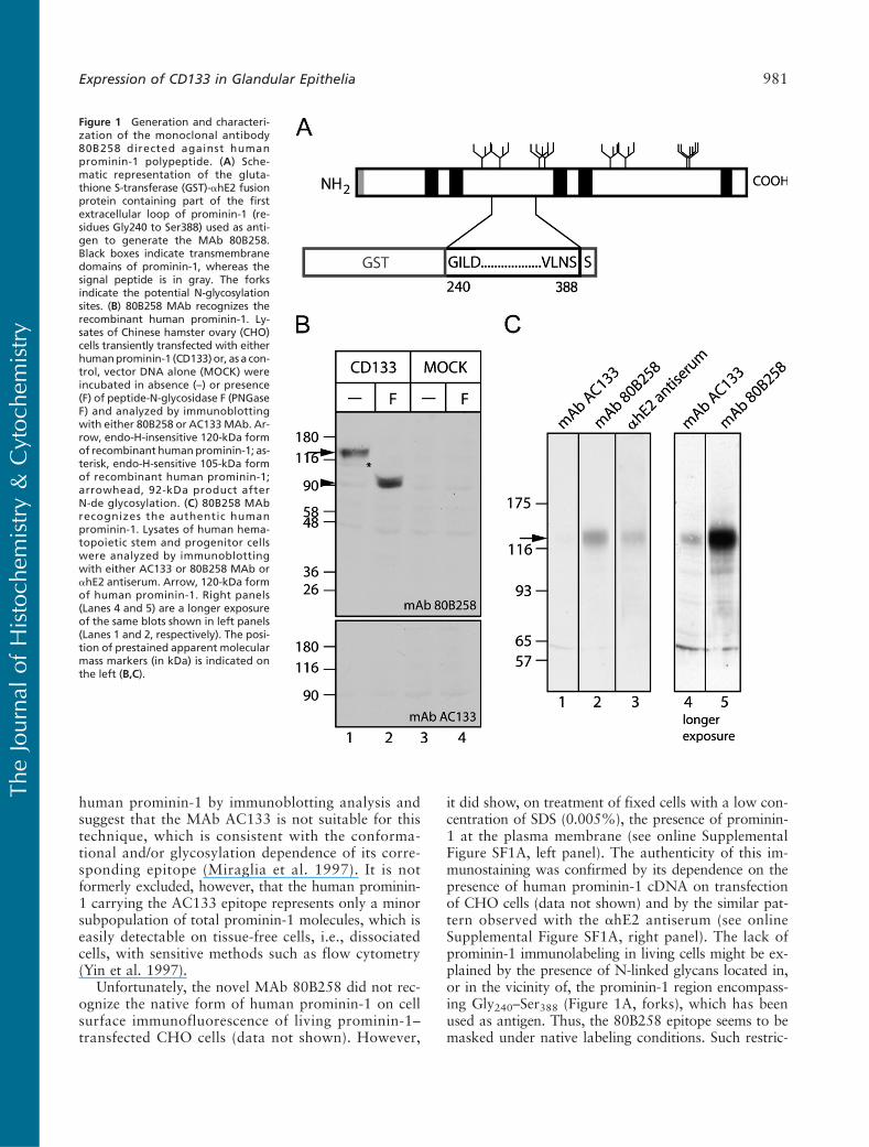
To be able to reinvestigate the expression of theprominin-1 protein in adult human tissues with toolsother than the MAb AC133 (Miltenyi Biotec), whichrecognizes an epitope thought to be dependent, at leastin part, on glycosylation (Miraglia et al. 1997), we gen-erated a novel mouse MAb, designated 80B258, againsta human prominin-1 fragment corresponding to aminoacid residues Gly240–Ser388 (Figure 1A). In CHO cellstransiently transfected with human prominin-1 cDNA,immunoblotting with the MAb 80B258 showed a ma-jor band with apparent molecular mass of ?120 kDa(Figure 1B, top panel, Lane 1, arrow) and a very mi-nor ?105-kDa band (Figure 1B, top panel, Lane 1, as-terisk), which reflects its overexpression. Indeed, the105-kDa form, but not 120-kDa form, was sensitiveto digestion with endo-b-N-acetyl glucosaminidase H(endo H) (data not shown) and therefore representedthe high-mannose form localized in endoplasmic retic-ulum and/or early Golgi compartment. PNGase F treat-ment converted both forms of human prominin-1 intoa product of ?92 kDa (Figure 1B, top panel, Lane 2,arrowhead), indicating that the MAb 80B258 recog-nizes both the N-glycosylated and deglycosylated formsof the recombinant human prominin-1. The same immu-noreactive bands were obtained using the rabbit ahE2antiserum (data not shown), which was raised againstthe same human prominin-1 fragment, as previously re-ported (Florek et al. 2005). No immunoreactivity wasdetected when CHO cells were transfected with the ex-pression vector alone (Figure 1B, Lanes 3 and 4). Underthe same conditions, the MAb AC133 did not show anyimmunoreactivity (Figure 1B, bottom panel).
Does the MAb 80B258 recognize prominin-1 underphysiological conditions, i.e., without a heterologousexpression? To answer this question, we blotted ex-tracts of the human HSCs, which express endogenouslyprominin-1 (Yin et al. 1997). The 120-kDa immuno-reactive band corresponding to prominin-1 was detectedwith both the MAb 80B258 and the ahE2 antiserum(Figure 1C, Lanes 2 and 3, respectively, arrow), whereasthe AC133 immunoreactivity was barely detectable(Figure 1C, Lane 1), which was nevertheless observableon a longer exposure of the blot (Figure 1C, Lane 4).
Taken together, these data show that the MAb80B258 recognizes the recombinant and the authentic
980 Karbanová, Missol-Kolka, Fonseca, Lorra, Janich, Hollerová, Jászai, Ehrmann, Kolář, Liebers, Arl,Subrtová, Freund, Mokrý, Huttner, Corbeil
TheJourna
lof
Histoch
emistry&
Cytoc
hemistry
human prominin-1 by immunoblotting analysis andsuggest that the MAb AC133 is not suitable for thistechnique, which is consistent with the conforma-tional and/or glycosylation dependence of its corre-sponding epitope (Miraglia et al. 1997). It is notformerly excluded, however, that the human prominin-1 carrying the AC133 epitope represents only a minorsubpopulation of total prominin-1 molecules, which iseasily detectable on tissue-free cells, i.e., dissociatedcells, with sensitive methods such as flow cytometry(Yin et al. 1997).
Unfortunately, the novel MAb 80B258 did not rec-ognize the native form of human prominin-1 on cellsurface immunofluorescence of living prominin-1–transfected CHO cells (data not shown). However,
it did show, on treatment of fixed cells with a low con-centration of SDS (0.005%), the presence of prominin-1 at the plasma membrane (see online SupplementalFigure SF1A, left panel). The authenticity of this im-munostaining was confirmed by its dependence on thepresence of human prominin-1 cDNA on transfectionof CHO cells (data not shown) and by the similar pat-tern observed with the ahE2 antiserum (see onlineSupplemental Figure SF1A, right panel). The lack ofprominin-1 immunolabeling in living cells might be ex-plained by the presence of N-linked glycans located in,or in the vicinity of, the prominin-1 region encompass-ing Gly240–Ser388 (Figure 1A, forks), which has beenused as antigen. Thus, the 80B258 epitope seems to bemasked under native labeling conditions. Such restric-
Figure 1 Generation and characteri-zation of the monoclonal antibody80B258 directed against humanprominin-1 polypeptide. (A) Sche-matic representation of the gluta-thione S-transferase (GST)-ahE2 fusionprotein containing part of the firstextracellular loop of prominin-1 (re-sidues Gly240 to Ser388) used as anti-gen to generate the MAb 80B258.Black boxes indicate transmembranedomains of prominin-1, whereas thesignal peptide is in gray. The forksindicate the potential N-glycosylationsites. (B) 80B258 MAb recognizes therecombinant human prominin-1. Ly-sates of Chinese hamster ovary (CHO)cells transiently transfected with eitherhumanprominin-1 (CD133)or, as a con-trol, vector DNA alone (MOCK) wereincubated in absence (–) or presence(F) of peptide-N-glycosidase F (PNGaseF) and analyzed by immunoblottingwith either 80B258 or AC133MAb. Ar-row, endo-H-insensitive 120-kDa formof recombinant humanprominin-1; as-terisk, endo-H-sensitive 105-kDa formof recombinant human prominin-1;arrowhead, 92-kDa product afterN-de glycosylation. (C) 80B258 MAbrecognizes the authentic humanprominin-1. Lysates of human hema-topoietic stem and progenitor cellswere analyzed by immunoblottingwith either AC133 or 80B258 MAb orahE2 antiserum. Arrow, 120-kDa formof human prominin-1. Right panels(Lanes 4 and 5) are a longer exposureof the same blots shown in left panels(Lanes 1 and 2, respectively). The posi-tion of prestained apparent molecularmass markers (in kDa) is indicated onthe left (B,C).
Expression of CD133 in Glandular Epithelia 981
TheJourna
lof
Histoch
emistry&
Cytoc
hemistry
tion might preclude its use for fluorescence-activated cellsorting using living cells. We studied this issue using thehuman colon–derived epithelial cell line Caco-2, whichexpresses endogenously prominin-1 (Corbeil et al. 2000;Florek et al. 2005). As suspected, the MAb 80B258, incontrast to MAb AC133 (see online Supplemental Fig-ure SF1B, blue area), did not recognize its correspondingepitope under native conditions by flow cytometry anal-ysis (see online Supplemental Figure SF1B, red area).However, it did using treatment of fixed cells with alow concentration of SDS (see online SupplementalFigure SF1B, green area). Thus, the novel anti-humanprominin-1 antibody could not be used for cell isolation.
IHC of Human Tissues
Throughout this study, we used two independent tech-niques to unmask the 80B258 epitope. As pretreat-ment, sections were either heated in the microwaveor incubated with a solution containing a low concen-tration of SDS. Although the microwave pretreatmentis more favorable for antigen retrieval, the tissue archi-tecture is better preserved with SDS preincubation,which under certain circumstances, e.g., examinationof delicate plasma membrane structures, is beneficial.For the sake of comprehension, we have outlined inthe following sections all relevant cellular structureswith either dotted or full lines.
Expression of Prominin-1 in the Major Salivary Glands
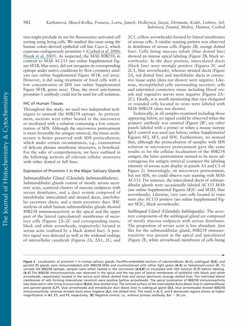
Submandibular Gland (Glandula Submandibularis).Submandibular glands consist of mostly serous secre-tory acini, scattered clusters of mucous endpieces withserous demilunes, and a duct system composed ofintralobular intercalated and striated ducts, interlobu-lar excretory ducts, and a main excretory duct. IHCanalysis of adult human submandibular glands showed80B258 immunoreactivity at the apical and the upperpart of the lateral (apicolateral) membranes of secre-tory cells (Figures 2A–2C and corresponding insets,black and white arrowheads, respectively) located inserous acini (outlined by a black dotted line). A posi-tive signal was detected as well at the widened endingsof intercellular canaliculi (Figures 2A, 2A1, 2C, and
2C1, yellow arrowheads) formed by lateral membranesof serous cells. A similar staining pattern was observedin demilunes of serous cells (Figure 2B, orange dottedline). Cells lining mucous tubuli (blue dotted line)showed an intense apical labeling (Figure 2B, black ar-rowheads). In the duct portion, intercalated ducts(black line) were strongly positive (Figures 2C and2C1, blue arrowheads), whereas striated ducts (Figure2A, red dotted line) and interlobular ducts in connec-tive tissue septa (data not shown) were negative. Like-wise, myoepithelial cells surrounding secretory cellsand interstitial connective tissue including blood ves-sels and vegetative nerves were negative (Figures 2A–2C). Finally, it is worth mentioning that rare elongatedor rounded cells located in veins were labeled withMAb 80B258 (data not shown).
Technically, in all samples examined including thoseappearing below, no signal could be observed when theprimary antibody was omitted (Figure 2B′; see otherpanels labeled with a prime) or when a mouse isotypeIgG1 control was used (see below; online SupplementalFigures SF2, SF3, and SF4). Furthermore, we noticedthat, although the preincubation of samples with SDSsolution or microwave pretreatment gave the sameresults as for the cellular localization of the 80B258antigen, the latter pretreatment seemed to be more ad-vantageous for antigen retrieval (compare the labelingintensity of serous acini display in panels A1 and C1 ofFigure 2). Interestingly, on microwave pretreatment,but not SDS, we could observe rare staining with MAbAC133. For instance, the intercalated ducts of subman-dibular glands were occasionally labeled AC133 MAb(see online Supplemental Figures SF2C and SF2D, bluearrowheads). Likewise, very rare cells located in veinswere also AC133 positive (see online Supplemental Fig-ure SF2C, black arrowheads).
Sublingual Gland (Glandula Sublingualis). The secre-tory components of the sublingual gland are composedof mostly mucous endpieces with serous demilunes.The proportion of serous acini is less abundant. Justlike for the submandibular gland, 80B258 immuno-reactivity was present at the apical and apicolateral(Figure 2E, white arrowhead) membrane of cells lining
'
Figure 2 Localization of prominin-1 in human salivary glands. Paraffin-embedded sections of submandibular (A–C), sublingual (D,E), andparotid (F) glands were immunolabeled with 80B258 MAb and counterstained with either light green (A–E) or hematoxylin-eosin (F). Tounmask the 80B258 epitope, samples were either heated in the microwave (A,B,B′) or incubated with SDS solution (C–F) before labeling.(A–F) The 80B258 immunoreactivity was observed in the apical and the top part of lateral membranes of epithelial cells (black and whitearrowheads, respectively) located in the serous acini (black dotted line) and serous demilunes (orange dotted line). The restricted lateralmembranes of cells forming intercellular canaliculi were positive (yellow arrowheads). The apical localization of 80B258 immunoreactivitywas observed in cells lining mucous tubuli (B,D,E, blue dotted line). The luminal surface of the intercalated ducts (black line) in submandibularand parotid glands (C,F1, blue arrowheads) and intralobular duct (black line) in sublingual gland (D,E, blue arrowheads) showed 80B258immunoreactivity, whereas striated ducts were negative (A,F, red dotted line). The insets in A, C, and F demarcate regions shown at highermagnification in A1, C1, and F1, respectively. (B′) Negative control, i.e., without primary antibody. Bar 5 50 mm.
982 Karbanová, Missol-Kolka, Fonseca, Lorra, Janich, Hollerová, Jászai, Ehrmann, Kolář, Liebers, Arl,Subrtová, Freund, Mokrý, Huttner, Corbeil
TheJourna
lof
Histoch
emistry&
Cytoc
hemistry

Expression of CD133 in Glandular Epithelia 983
TheJourna
lof
Histoch
emistry&
Cytoc
hemistry

serous acini and apical membrane of mucous tubuli(Figures 2D and 2E, black arrowheads), and thosefound either in intralobular ducts (Figures 2D and2E, blue arrowheads) or small interlobular ducts (datanot shown).
Parotid Gland (Glandula Parotis). The parotid glandis the largest human salivary gland. It contains onlyserous exocrine acini—the major secretory elements—that are connected to a well-developed duct systemcomposed of intercalated, striated, and interlobular ex-cretory ducts. 80B258 immunoreactivity was detectedat the apical and the top part of lateral membranes(Figure 2F, see inset F1, white arrowhead) of secretorycells of serous acini (black dotted line) and the apicalmembrane of intercalated ducts (Figure 2F1, blue ar-rowhead). As in submandibular glands, a strong stain-ing was detected in cells located at the widened endingsof intercellular canaliculi (Figure 2F1, yellow arrow-head). No signal was detected in striated ducts (Fig-ure 2F, red dotted line).
Expression of Prominin-1 in the Sweat Glandsof Axilla
Eccrine Sweat Glands. The secretory portion of eccrinesweat glands is formed by tubules lined with a pseudo-stratified epithelium consisting of secretory small clearand large dark cells (Saga 2001). Small intercellularcanaliculi are developed between them (Saga 2001).The MAb 80B258 labeled the apical surface of tubules(Figures 3A, 3C, and 3E, black arrowheads) and thelateral membrane of cells forming intercellular canalic-uli (Figures 3A and 3C–3E, yellow arrowheads). In theduct portion, 80B258 immunoreactivity was found atthe apical membrane of epithelial cells (Figures 3A–3C,blue arrowheads) located in the superficial layer ofducts (red dotted line). Secretion found in their lumenwas positive (Figures 3A–3C, white asterisks). Occa-sionally, we could observe ducts that were negative(data not shown). Myoepithelial cells surrounding se-cretory cells were negative (data not shown).
Apocrine and Apoeccrine-like Sweat Glands. Apocrinesweat glands exhibited 80B258 immunoreactivity atthe luminal surface of epithelial cells located in the tu-bular secretory portion (Figures 3F–3H, black arrow-heads). The immunoreactivity was associated withtubular elements that were lined by either columnar(as those outlined by a blue dotted line) or cuboidalepithelium (black dotted line) depending on the stateof secretion. A high-magnification view showed that80B258 immunoreactivity was restricted to the apicaldomain that appears as a dome-like shape (Figures 3Iand 3I1, red arrows). Some tubules were neverthelessnegative (Figure 3H, white arrow). 80B258 immunore-
activity was also associated with materials secreted inthe lumen (Figures 3F and 3G, white asterisks; as nega-tive control, see inset 3F′).
Interestingly, 80B258 immunoreactivity showed tu-bular structures sharing simultaneously the characteris-tics of eccrine and apocrine sweat glands (Figure 3J).For instance, although these tubules exhibit a large lu-men as those found in apocrine glands, we could detectsome intercellular canaliculi labeled with the MAb80B258 (Figure 3J, see insets 3J1 and 3J2, yellow ar-rowheads) reminiscent to eccrine glands. An intermit-tent apical staining was also observed (Figures 3J1 and3J2, black arrowheads) where cells with a “straight”apical surface were positive, whereas others having adome-shaped apical domain were usually negative (reddotted line). Such situation contrasts to that observedwith apocrine glands (Figure 3I1). Because of their hy-brid morphology, we named them “apoeccrine-like”glands. In this context, it is important to mention thatSato and colleagues have previously reported a thirdtype of sweat glands referred to as “apoeccrine” (Satoet al. 1987,1989; Sato and Sato 1987). They were de-scribed as having some segments similar to eccrineglands (e.g., pseudostratified epithelium, narrow lumen,and presence of small intercellular canaliculi), whereasother segments were similar to apocrine glands (e.g.,simple columnar epithelium, large lumen).Whether thesestructures are the same as those we observed here is stillan open question.
Expression of Prominin-1 in the Lacrimal Gland andthe Gland of Moll
We next studied the expression of prominin-1 in theupper human eyelid, which contains several glands.One of them, the accessory lacrimal gland of Wolfring,is a branched serous tubuloalveolar gland localized inthe upper border or above the tarsal plate. It has a sim-ilar structure as the large orbital lacrimal gland, but itsbranched duct system is less complex (Figure 4A). Only80B258 immunoreactivity was observed at the apicaland the top part of the lateral membranes of serouscells (Figures 4B–4D, black and white arrowheads, re-spectively) including the distended ending membraneof the intercellular canaliculi (Figures 4C and 4D, yel-low arrowheads) found in the secretory portion. Theapical membrane of epithelial cells associated withthe intercalated ducts was positive (Figures 4C and4D, blue arrowheads).
80B258 immunoreactivity was also observed at theapical membrane of epithelium lining the secretory por-tion of the apocrine tubular gland of Moll (Figures 4Eand 4E1, black arrowheads) and in the secretion foundtherein (see online Supplemental Figure SF3E, white as-terisks). This gland is localized in the edge of the eyelidand opens into the hair follicle of the eyelash, and it is
984 Karbanová, Missol-Kolka, Fonseca, Lorra, Janich, Hollerová, Jászai, Ehrmann, Kolář, Liebers, Arl,Subrtová, Freund, Mokrý, Huttner, Corbeil
TheJourna
lof
Histoch
emistry&
Cytoc
hemistry
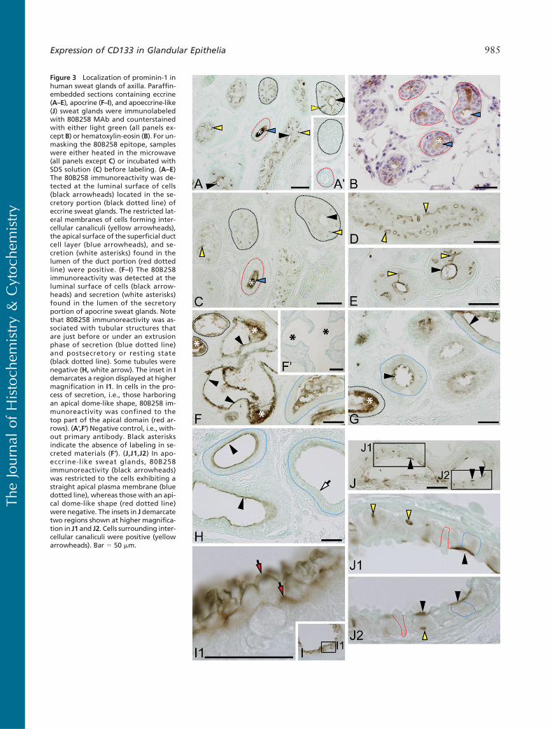
Figure 3 Localization of prominin-1 inhuman sweat glands of axilla. Paraffin-embedded sections containing eccrine(A–E), apocrine (F–I), and apoeccrine-like(J) sweat glands were immunolabeledwith 80B258 MAb and counterstainedwith either light green (all panels ex-cept B) or hematoxylin-eosin (B). For un-masking the 80B258 epitope, sampleswere either heated in the microwave(all panels except C) or incubated withSDS solution (C) before labeling. (A–E)The 80B258 immunoreactivity was de-tected at the luminal surface of cells(black arrowheads) located in the se-cretory portion (black dotted line) ofeccrine sweat glands. The restricted lat-eral membranes of cells forming inter-cellular canaliculi (yellow arrowheads),the apical surface of the superficial ductcell layer (blue arrowheads), and se-cretion (white asterisks) found in thelumen of the duct portion (red dottedline) were positive. (F–I) The 80B258immunoreactivity was detected at theluminal surface of cells (black arrow-heads) and secretion (white asterisks)found in the lumen of the secretoryportion of apocrine sweat glands. Notethat 80B258 immunoreactivity was as-sociated with tubular structures thatare just before or under an extrusionphase of secretion (blue dotted line)and postsecretory or resting state(black dotted line). Some tubules werenegative (H,white arrow). The inset in Idemarcates a region displayed at highermagnification in I1. In cells in the pro-cess of secretion, i.e., those harboringan apical dome-like shape, 80B258 im-munoreactivity was confined to thetop part of the apical domain (red ar-rows). (A′,F′) Negative control, i.e., with-out primary antibody. Black asterisksindicate the absence of labeling in se-creted materials (F′). (J,J1,J2) In apo-eccrine-like sweat glands, 80B258immunoreactivity (black arrowheads)was restricted to the cells exhibiting astraight apical plasma membrane (bluedotted line), whereas those with an api-cal dome-like shape (red dotted line)were negative. The insets in J demarcatetwo regions shown at higher magnifica-tion in J1 and J2. Cells surrounding inter-cellular canaliculi were positive (yellowarrowheads). Bar 5 50 mm.
Expression of CD133 in Glandular Epithelia 985
TheJourna
lof
Histoch
emistry&
Cytoc
hemistry

Figure 4 Localization of prominin-1 in the human lacrimal gland and the gland of Moll of eyelid. Paraffin-embedded sections containing theaccessory lacrimal gland of Wolfring (A–D), the gland of Moll (E), and conjunctival epithelium (F) were immunolabeled with 80B258 MAb andcounterstained with light green. For unmasking the 80B258 epitope, samples were either heated in the microwave (A,B) or incubated withSDS solution (C–F) before labeling. (A–D) The 80B258 immunoreactivity was observed at the apical and the top part of lateral membranes ofserous secretory cells (black and white arrowheads, respectively), and at the widened endings of intercellular canaliculi (yellow arrowheads)of the lacrimal gland. The luminal surface of the intercalated ducts was labeled (blue arrowheads). (C′) Negative control, i.e., without primaryantibody. (E,F) The 80B258 immunoreactivity was observed at the apical membrane (black arrowheads) of either columnar cells lining secre-tory tubule of the apocrine gland of Moll (E) or epithelial cells located at the superficial layer of conjunctiva (F). The inset in E demarcates aregion displayed at higher magnification in E1. Bar 5 50 mm.
986 Karbanová, Missol-Kolka, Fonseca, Lorra, Janich, Hollerová, Jászai, Ehrmann, Kolář, Liebers, Arl,Subrtová, Freund, Mokrý, Huttner, Corbeil
TheJourna
lof
Histoch
emistry&
Cytoc
hemistry

reminiscent structurally to apocrine glands of the axilla.The lipid secreting holocrine Meibomian sebaceousglands were negative (data not shown).
Finally, some regions of palpebral conjunctival strat-ified columnar epithelium in the vicinity of the fornixexhibited 80B258 immunoreactivity (Figure 4F, blackarrowhead). Neither the MAb AC133 nor isotype IgG1control gave any immunoreactivity (see online Supple-mental Figures SF3F and SF3G, respectively).
Within all sweat gland tissues examined, the AC133MAb weakly labeled the secreted materials (see onlineSupplemental Figures SF2I, SF2L, and SF3C, white as-terisks) and rarely the apical membrane of apocrine se-cretory tubules (see online Supplemental Figure SF2L1,black arrowhead), whereas the isotype IgG1 controlgave no signal (see online Supplemental Figures SF2J,SF2M, and SF3D).
Expression of Prominin-1 in the Uterus, Liver,and Pancreas
Uterus. Bioptic samples of normal endometrium in theproliferative phase of the menstrual cycle were exam-ined for prominin-1 expression. In deep portions ofthe endometrium (pars spongiosa), uterine glands ex-hibited strong 80B258 immunoreactivity at the apicalsurface of the epithelium (Figure 5A, black arrow-heads). Interestingly, higher-magnification viewsincluding a tangential section showed that the im-munoreactivity was associated with motile cilia (Fig-ures 5B, 5B1, 5B2, and 5C, blue arrowheads) andother small plasma membrane–associated structures,e.g., microvilli (Figure 5B2, red arrowhead). The toppart of the endometrium (pars compacta) containingstraight narrow glands and some segments of epithe-lium lining the uterine cavity were either negative orweakly positive (data not shown).
In the cervix, i.e., the lower and narrow portion ofthe uterus, 80B258 immunoreactivity was observed atthe dome-shaped apical domain (Figures 5D, 5D1, and5D2, arrowheads) of epithelial cells of the mucous cer-vical glands (dotted line). Neither the MAb AC133 norisotype IgG1 control gave any immunoreactivity (datanot shown).
Liver (Hepar). In the liver, 80B258 immunoreactivitywas detected at the apical membrane of cells formingcanals of Hering, which are located at the peripheryof classic hepatic lobule (Figures 6A and 6B, white ar-rowheads). In the portal space/tract (red dotted line),intraportal bile ductules lined by a simple cuboidal epi-thelium (Figures 6A–6E, black dotted line) and smallinterlobular bile ducts lined by a cuboidal or low co-lumnar epithelium (Figure 6F) were positive at their lu-minal surface (Figures 6A–6F, blue arrowheads). Largebile ducts lined with simple columnar epithelium (Fig-
ures 6G and 6H), connective tissue, blood vessels lo-cated in the portal spaces, the apical bile canalicularmembrane of hepatocytes, and gall bladder were neg-ative (data not shown). Neither the MAb AC133 norisotype IgG1 control gave any immunoreactivity (seeonline Supplemental Figures SF4A–SF4C).
Pancreas. Recently, we as well as others have shownthat prominin-1 is expressed in the apical membraneof cells present in intercalated ducts of the pancreas(Immervoll et al. 2008; Lardon et al. 2008). We couldreproduce these data with MAb 80B258 (Figures 7Aand 7C, blue arrowheads). Interestingly, we also de-tected 80B258 immunoreactivity in the apical domainof centroacinar cells (Figures 7B and 7D, black arrow-heads), i.e., pale-stained cells lining the inner part ofacini (dotted line) of pancreas secretory portion. Inter-lobular ducts in the connective tissue septa and isletsof Langerhans were negative in agreement with previousreports (data not shown). On microwave pretreatment,but not SDS, MAb AC133 gave a weak and occasionalstaining at the apical membrane of cells present in inter-calated ducts (see online Supplemental Figures SF4E–SF4G, blue arrowheads) as previously reported usingparaffin sections (Lardon et al. 2008). Rare cells locatedin the interstitium were found to be positive for AC133(see online Supplemental Figure SF4H, black arrow-head). The nature of these cells is unknown. No labelingwas observed with the isotype IgG1 control (see onlineSupplemental Figures SF4I and SF4J).
DiscussionThis study showed that prominin-1 is widely expressedin healthy adult human tissues, which is in agreementwith the distribution of its transcript (Miraglia et al.1997; Florek et al. 2005; see online Supplemental Ta-ble ST1), thus mimicking the situation previously ob-served in the mouse (Weigmann et al. 1997). Thelack of its detection observed in previous studies—based on MAb AC133 (Miraglia et al. 1997; for re-view see Fargeas et al. 2006; see online SupplementalFigures SF2–SF4)—might be explained by the depen-dence of the corresponding AC133 epitope on confor-mational and/or glycosylation modifications.
Technically, the AC133 epitope might be sensitive tothe paraffin embedding procedure. Indeed, we havepreviously shown that MAb AC133 failed to give re-producible immunostaining on paraffin sections ofhealthy human pancreatic samples, although it workedwell on cryosections (Lardon et al. 2008). Likewise,Immervoll et al. (2008) have suggested very recentlythat the prominin-1 AC133 epitope might also be ex-pressed in various adult epithelia, but its detection onparaffin sections needed very sensitive methods includingharsh conditions for antigen retrieval. Physiologically,
Expression of CD133 in Glandular Epithelia 987
TheJourna
lof
Histoch
emistry&
Cytoc
hemistry
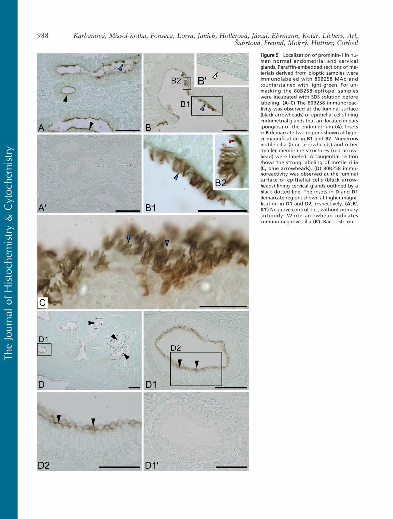
Figure 5 Localization of prominin-1 in hu-man normal endometrial and cervicalglands. Paraffin-embedded sections of ma-terials derived from bioptic samples wereimmunolabeled with 80B258 MAb andcounterstained with light green. For un-masking the 80B258 epitope, sampleswere incubated with SDS solution beforelabeling. (A–C) The 80B258 immunoreac-tivity was observed at the luminal surface(black arrowheads) of epithelial cells liningendometrial glands that are located in parsspongiosa of the endometrium (A). Insetsin B demarcate two regions shown at high-er magnification in B1 and B2. Numerousmotile cilia (blue arrowheads) and othersmaller membrane structures (red arrow-head) were labeled. A tangential sectionshows the strong labeling of motile cilia(C, blue arrowheads). (D) 80B258 immu-noreactivity was observed at the luminalsurface of epithelial cells (black arrow-heads) lining cervical glands outlined by ablack dotted line. The insets in D and D1demarcate regions shown at higher magni-fication in D1 and D2, respectively. (A′,B′,D1′) Negative control, i.e., without primaryantibody. White arrowhead indicatesimmuno-negative cilia (B′). Bar 5 50 mm.
988 Karbanová, Missol-Kolka, Fonseca, Lorra, Janich, Hollerová, Jászai, Ehrmann, Kolář, Liebers, Arl,Subrtová, Freund, Mokrý, Huttner, Corbeil
TheJourna
lof
Histoch
emistry&
Cytoc
hemistry
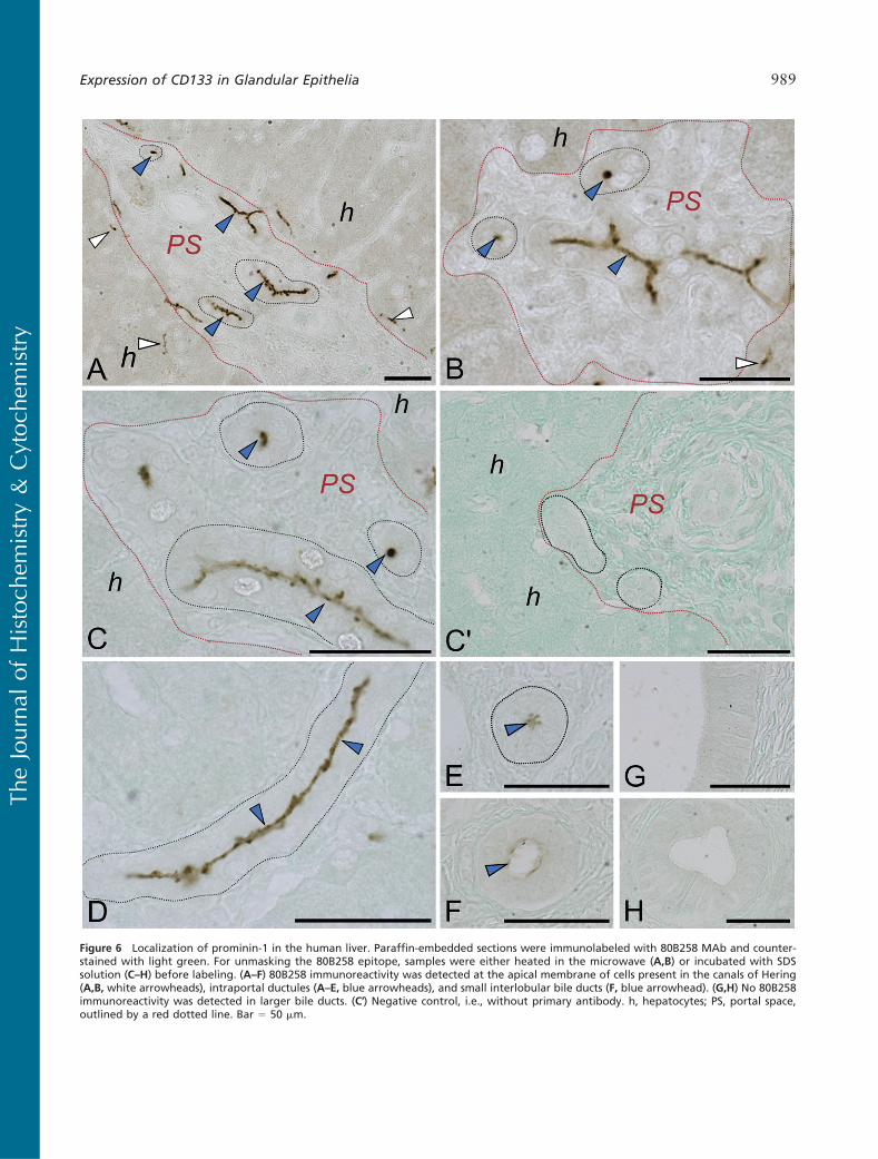
Figure 6 Localization of prominin-1 in the human liver. Paraffin-embedded sections were immunolabeled with 80B258 MAb and counter-stained with light green. For unmasking the 80B258 epitope, samples were either heated in the microwave (A,B) or incubated with SDSsolution (C–H) before labeling. (A–F) 80B258 immunoreactivity was detected at the apical membrane of cells present in the canals of Hering(A,B, white arrowheads), intraportal ductules (A–E, blue arrowheads), and small interlobular bile ducts (F, blue arrowhead). (G,H) No 80B258immunoreactivity was detected in larger bile ducts. (C′) Negative control, i.e., without primary antibody. h, hepatocytes; PS, portal space,outlined by a red dotted line. Bar 5 50 mm.
Expression of CD133 in Glandular Epithelia 989
TheJourna
lof
Histoch
emistry&
Cytoc
hemistry

we cannot exclude that the AC133 epitope is down-regulated upon cellular differentiation, with the resultthat only a small subset of prominin-1 molecules car-ried the AC133 epitope, particularly those expressedby stem and progenitor cells (see below; Bhatia 2001;Fargeas et al. 2006). Our data with salivary glands andthe pancreas, for instance, are consistent with this hy-pothesis (see online Supplemental Figures SF2 andSF4). Given the postulated dependence of the AC133epitope on a glycosylated structure (Miraglia et al.1997), the general glycosylation profile of cells mightchange on differentiation, as suggested in epithelialand non-epithelial cell models (Ogier-Denis et al. 1988;Hemmoranta et al. 2007), leading to the lack of theAC133 epitope but not the prominin-1 molecule as such(Corbeil et al. 2000; Florek et al. 2005). The glycosyla-
tion of prominin-1 molecules could also be altered incells that have undergone malignant transformation.Such alteration may explain its apparently discordantexpression in patients with acute myelogenous leukemiaand myelodysplastic syndrome that have been observedwith two MAbs recognizing distinct glycosylation-dependent epitopes (MAbs AC133 and AC141) ofprominin-1 (Green et al. 2000) and could reflect thedifferent stages of differentiation of these malignantcells. A similar conclusion was drawn on solid cancer(Florek et al. 2005). Alternatively, a switch during thecellular development of a particular prominin-1 splicevariant carrying the AC133 epitope to another onelacking it might occur as well. Up to 12 splice variantsof prominin-1 have been described so far (Fargeas et al.2003a,2007). Finally, it is important to point out that
Figure 7 Localization of prominin-1 in the human pancreas. Paraffin-embedded sections were immunolabeled with 80B258 MAb and coun-terstained with light green (A,B) or hematoxylin-eosin (C,D). For unmasking the 80B258 epitope, samples were incubated with SDS solutionbefore labeling. (A–D) 80B258 immunoreactivity was detected at the luminal surface of cells located in intercalated ducts (blue arrowheads)and the apical membrane of centroacinar cells (black arrowheads) of the secretory portion outlined by a dotted line. (A′) Negative control, i.e.,without primary antibody. Bar 5 50 mm.
990 Karbanová, Missol-Kolka, Fonseca, Lorra, Janich, Hollerová, Jászai, Ehrmann, Kolář, Liebers, Arl,Subrtová, Freund, Mokrý, Huttner, Corbeil
TheJourna
lof
Histoch
emistry&
Cytoc
hemistry
the special molecular environment of prominin-1 withinthe plasma membrane might also influence the detec-tion of certain epitope(s). For instance, its incorporationinto a densely packed cholesterol-based lipid micro-domain might mask some of them, particularly thosein the vicinity of the membrane (Röper et al. 2000;Janich and Corbeil 2007). Thus, numerous parametersmight interfere with the IHC detection of the prominin-1 AC133 epitope, and importantly, the lack of its detec-tion needs to be evaluated with some caution. On amore general note, the term “AC133” shall be used todescribe human prominin-1 bearing the AC133 epitopeas previously proposed (Fargeas et al. 2003a), andthus the AC133 antigen is not necessary synonymouswith prominin-1.
Our study showed that prominin-1 is expressed invarious glandular epithelia (see online SupplementalTable ST1), where it is confined to the apical or apico-lateral membranes of polarized cells that were fully dif-ferentiated and adapted to their function, i.e., secretion(in the case of secretory cells) or modification and con-ducting the secretion (in the case of duct cells). The freesurface of these cells is covered with numerous micro-villi, for which prominin-1 has its own tendency to beconcentrated, and it might be involved in their orga-nization (for review, see Corbeil et al. 2001b). Thepresence of prominin-1 in motile cilia found in the en-dometrium (Figure 5) is another example of its selectivelocalization in plasma membrane protrusions. The lat-ter observation together with the fact that prominin-1binds to the plasma membrane cholesterol within aspecific lipid microdomain (Röper et al. 2000) suggestthat the lipid composition and/or organization of themembrane of the motile cilium is distinct to that foundin the planar portion, as recently shown for the pri-mary cilium in MDCK cells (Janich and Corbeil 2007).
The overall expression of prominin-1 based on MAb80B258 is generally consistent with previous RNA dataobtained from Northern and dot blot analyses (for de-tails, see online Supplemental Table ST1). However,some discrepancy might be observed given that the ex-pression of prominin-1 is often confined to a particularstructure, e.g., cilium, which is not abundant regardingthe whole tissue that is normally used to prepare themRNA samples. The uterus is a good example.
Our data concerning salivary and lacrimal glands(Figures 2 and 4, respectively) document for the firsttime the source of small membrane vesicles contain-ing prominin-1 found in the human saliva and tears(Marzesco et al. 2005; Jászai et al. 2007). Moreover,the expression of prominin-1 by sweat glands and80B258 immunoreactivity associated with the secre-tion therein (Figure 3) indicate that prominin-1 mightbe found also in sweat. Although the physiologicalfunction of these extracellular membrane vesicles is
currently unknown, they might offer an interesting toolfor diagnostic purposes of certain solid cancers, e.g.,salivary gland cancer, as recently proposed for centralnervous system diseases (Huttner et al. 2008). Like-wise, analyzing the prominin-1 content, in conjunctionwith other constituents of the saliva, might be usefulfor monitoring functional recovery of salivary glandson tissue engineering/replacement. The physiologicalfunction of prominin-1 is still not clarified as well.However, its concentration in plasma membrane pro-trusions (microvillus and motile cilium) present withinthe apical membrane of glandular (this study; Jászaiet al. 2007) and other epithelial cells (Weigmann et al.1997; Marzesco et al. 2005; Dubreuil et al. 2007) andthe occurrence in extracellular membrane vesicles(Marzesco et al. 2005; Florek et al. 2007) potentiallyderived from these plasmalemma protrusions (Dubreuilet al. 2007; for review, see Fargeas et al. 2006) suggestthat this cholesterol-binding glycoprotein might play arole in the secretion process and/or its regulation.
The general expression of prominin-1 in adult humantissues, like in the mouse, is clearly beyond the stem andprogenitor cells. However, within the prominin-1–positive cell population might reside a minute popula-tion of cells harboring stem cell properties, which areplaying a role in vivo in the replacement of damagedand/or aging cells. Such rare primitive cells could beimmuno-isolated on tissue dissociation with MAbAC133 as reported previously (Uchida et al. 2000;Tamaki et al. 2002; Yu et al. 2002; Schwartz et al.2003; Alessandri et al. 2004; Belicchi et al. 2004;Richardson et al. 2004; Bussolati et al. 2005). Alterna-tively, prominin-1–positive cells (particularly those as-sociated with glandular epithelium) might have a highpropensity to dedifferentiate on presentation of appro-priate cues. In both scenarios, which are not mutuallyexclusive, prominin-1 emerges as an important markerwith significant clinical value for somatic stem cell iso-lation (Bornhäuser et al. 2005; Torrente et al. 2007).
Acknowledgments
This study was supported by Ministry of Education,Youth and Sport of the Czech Republic (MSM 0021620820)to J. K. and by Sächsisches Ministerium für Wissenschaft undKunst–Europäischer Fond für Regionale Entwicklung (4212/05-16) to D.C.
The authors thank Prof. Dr. Med. Martin Bornhäuserfor the primary hematopoietic and mesenchymal stem cells;Sylvi Graupner for technical assistance; and Dr. ChristineA. Fargeas for critical review of the article.
Literature Cited
Alessandri G, Pagano S, Bez A, Benetti A, Pozzi S, Iannolo G, BaronioM, et al. (2004) Isolation and culture of human muscle-derivedstem cells able to differentiate into myogenic and neurogenic celllineages. Lancet 364:1872–1883
Expression of CD133 in Glandular Epithelia 991
TheJourna
lof
Histoch
emistry&
Cytoc
hemistry

Bauer N, Fonseca AV, Florek M, Freund D, Jászai J, Bornhäuser M,Fargeas CA, et al. (2008) New insights into the cell biology ofhematopoietic progenitors by studying prominin-1 (CD133). CellsTissues Organs 188:127–138
Belicchi M, Pisati F, Lopa R, Porretti L, Fortunato F, Sironi M,Scalamogna M, et al. (2004) Human skin-derived stem cells mi-grate throughout forebrain and differentiate into astrocytes afterinjection into adult mouse brain. J Neurosci Res 77:475–486
Bhatia M (2001) AC133 expression in human stem cells. Leukemia15:1685–1688
Bornhäuser M, Eger L, Oelschlaegel U, Auffermann-Gretzinger S,Kiani A, Schetelig J, Illmer T, et al. (2005) Rapid reconstitutionof dendritic cells after allogeneic transplantation of CD1331 se-lected hematopoietic stem cells. Leukemia 19:161–165
Bussolati B, Bruno S, Grange C, Buttiglieri S, Deregibus MC, CantinoD, Camussi G (2005) Isolation of renal progenitor cells from adulthuman kidney. Am J Pathol 166:545–555
Corbeil D, Fargeas CA, Huttner WB (2001a) Rat prominin, like itsmouse and human orthologues, is a pentaspan membrane glyco-protein. Biochem Biophys Res Commun 285:939–944
Corbeil D, Röper K, Fargeas CA, Joester A, Huttner WB (2001b)Prominin: a story of cholesterol, plasma membrane protrusionsand human pathology. Traffic 2:82–91
Corbeil D, Röper K, Hannah MJ, Hellwig A, Huttner WB (1999)Selective localization of the polytopic membrane protein promininin microvilli of epithelial cells - a combination of apical sorting andretention in plasma membrane protrusions. J Cell Sci 112:1023–1033
Corbeil D, Röper K, Hellwig A, Tavian M, Miraglia S, Watt SM,Simmons PJ, et al. (2000) The human AC133 hematopoietic stemcell antigen is also expressed in epithelial cells and targeted toplasma membrane protrusions. J Biol Chem 275:5512–5520
Coskun V, Wu H, Blanchi B, Tsao S, Kim K, Zhao J, Biancotti JC,et al. (2008) CD1331 neural stem cells in the ependyma ofmammalian postnatal forebrain. Proc Natl Acad Sci USA 105:1026–1031
Dubreuil V, Marzesco AM, Corbeil D, Huttner WB, Wilsch-Bräuninger M (2007) Midbody and primary cilium of neuralprogenitors release extracellular membrane particles enriched inthe stem cell marker prominin-1. J Cell Biol 176:483–495
Fargeas CA, Corbeil D, Huttner WB (2003a) AC133 antigen,CD133, prominin-1, prominin-2, etc.: prominin family gene prod-ucts in need of a rational nomenclature. Stem Cells 21:506–508
Fargeas CA, Florek M, Huttner WB, Corbeil D (2003b) Character-ization of prominin-2, a new member of the prominin family ofpentaspan membrane glycoproteins. J Biol Chem 278:8586–8596
Fargeas CA, Fonseca AV, Huttner WB, Corbeil D (2006) Prominin-1(CD133): from progenitor cells to human diseases. Future Lipid-ology 1:213–225
Fargeas CA, Huttner WB, Corbeil D (2007) Nomenclature ofprominin-1 (CD133) splice variants: an update. Tissue Antigens69:602–606
Fargeas CA, Joester A, Missol-Kolka E, Hellwig A, Huttner WB,Corbeil D (2004) Identification of novel prominin-1/CD133 splicevariants with alternative C-termini and their expression in epidid-ymis and testis. J Cell Sci 117:4301–4311
Florek M, Bauer N, Janich P, Wilsch-Bräuninger M, FargeasCA, Marzesco AM, Ehninger G, et al. (2007) Prominin-2 is acholesterol-binding protein associated with apical and basolat-eral plasmalemmal protrusions in polarized epithelial cells andreleased into urine. Cell Tissue Res 328:31–47
Florek M, Haase M, Marzesco AM, Freund D, Ehninger G, HuttnerWB, Corbeil D (2005) Prominin-1/CD133, a neural and hemato-poietic stem cell marker, is expressed in adult human differen-tiated cells and certain types of kidney cancer. Cell Tissue Res319:15–26
Freund D, Bauer N, Boxberger S, Feldmann S, Streller U, EhningerG, Werner C, et al. (2006a) Polarization of human hematopoieticprogenitors during contact with multipotent mesenchymal stro-mal cells: effects on proliferation and clonogenicity. Stem CellsDev 15:815–829
Freund D, Oswald J, Feldmann S, Ehninger G, Corbeil D, BornhäuserM (2006b) Comparative analysis of proliferative potential andclonogenicity of MACS-immunomagnetic isolated CD341 andCD1331 blood stem cells derived from a single donor. Cell Prolif39:325–332
Green CL, Loken M, Buck D, Deeg HJ (2000) Discordant expressionof AC133 and AC141 in patients with myelodysplastic syndrome(MDS) and acute myelogeneous leukemia (AML). Leukemia14:770–772
Harlow E, Lane D (1988) Antibodies: A Laboratory Manual. ColdSpring Harbor, NY, Cold Spring Harbor Laboratory Press
Hemmoranta H, Satomaa T, Blomqvist M, Heiskanen A, Aitio O,Saarinen J, Natunen J, et al. (2007) N-glycan structures and asso-ciated gene expression reflect the characteristic N-glycosylationpattern of human hematopoietic stem and progenitor cells. ExpHematol 35:1279–1292
Huttner HB, Janich P, KöhrmannM, Jászai J, Siebzehnrubl F, BlümckeI, Suttorp M, et al. (2008) The stem cell marker prominin-1/CD133on membrane particles in human cerebrospinal fluid offers novelapproaches for studying central nervous system disease. StemCells 26:698–705
Immervoll H, Hoem D, Sakariassen PO, Steffensen OJ, Molven A(2008) Expression of the “stem cell marker” CD133 in pancreasand pancreatic ductal adenocarcinomas. BMC Cancer 8:48
Ito Y, Hamazaki TS, Ohnuma K, Tamaki K, Asashima M, Okochi H(2007) Isolation of murine hair-inducing cells using the cellsurface marker prominin-1/CD133. J Invest Dermatol 127:1052–1060
Janich P, Corbeil D (2007) GM1 and GM3 gangliosides highlightdistinct lipid microdomains within the apical domain of epithelialcells. FEBS Lett 581:1783–1787
Jászai J, Janich P, Farkas LM, Fargeas CA, Huttner WB, CorbeilD (2007) Differential expression of Prominin-1 (CD133) andProminin-2 in major cephalic exocrine glands of adult mice.Histochem Cell Biol 128:409–419
Lardon J, Corbeil D, Huttner WB, Ling Z, Bouwens L (2008) Stemcell marker prominin-1/AC133 is expressed in duct cells of theadult human pancreas. Pancreas 36:e1–e6
Marzesco AM, Janich P, Wilsch-Bräuninger M, Dubreuil V,Langenfeld K, Corbeil D, Huttner WB (2005) Release of extracel-lular membrane particles carrying the stem cell marker prominin-1 (CD133) from neural progenitors and other epithelial cells.J Cell Sci 118:2849–2858
Maw MA, Corbeil D, Koch J, Hellwig A, Wilson-Wheeler JC,Bridges RJ, Kumaramanickavel G, et al. (2000) A frameshiftmutation in prominin (mouse)-like 1 causes human retinal degen-eration. Hum Mol Genet 9:27–34
Miraglia S, Godfrey W, Yin AH, Atkins K, Warnke R, Holden JT,Bray RA, et al. (1997) A novel five-transmembrane hematopoieticstem cell antigen: isolation, characterization, and molecular clon-ing. Blood 90:5013–5021
Ogier-Denis E, Codogno P, Chantret I, Trugnan G (1988) The pro-cessing of asparagine-linked oligosaccharides in HT-29 cells is afunction of their state of enterocytic differentiation. An accumula-tion of Man9,8-GlcNAc2-Asn species is indicative of an impairedN-glycan trimming in undifferentiated cells. J Biol Chem 263:6031–6037
Richardson GD, Robson CN, Lang SH, Neal DE, Maitland NJ,Collins AT (2004) CD133, a novel marker for human prostaticepithelial stem cells. J Cell Sci 117:3539–3545
Röper K, Corbeil D, Huttner WB (2000) Retention of prominin inmicrovilli reveals distinct cholesterol-based lipid micro-domainsin the apical plasma membrane. Nat Cell Biol 2:582–592
Saga K (2001) Histochemical and immunohistochemical markers forhuman eccrine and apocrine sweat glands: an aid for histopatho-logic differentiation of sweat gland tumors. J Investig DermatolSymp Proc 6:49–53
Sato K, Kang WH, Saga K, Sato KT (1989) Biology of sweat glandsand their disorders. I. Normal sweat gland function. J Am AcadDermatol 20:537–563
992 Karbanová, Missol-Kolka, Fonseca, Lorra, Janich, Hollerová, Jászai, Ehrmann, Kolář, Liebers, Arl,Subrtová, Freund, Mokrý, Huttner, Corbeil
TheJourna
lof
Histoch
emistry&
Cytoc
hemistry

Sato K, Leidal R, Sato F (1987) Morphology and development ofan apoeccrine sweat gland in human axillae. Am J Physiol 252:R166–180
Sato K, Sato F (1987) Sweat secretion by human axillary apoeccrinesweat gland in vitro. Am J Physiol 252:R181–187
Schwartz PH, Bryant PJ, Fuja TJ, Su H, O’Dowd DK, Klassen H(2003) Isolation and characterization of neural progenitor cellsfrom post-mortem human cortex. J Neurosci Res 74:838–851
Tamaki S, Eckert K, He D, Sutton R, Doshe M, Jain G, TushinskiR, et al. (2002) Engraftment of sorted/expanded human centralnervous system stem cells from fetal brain. J Neurosci Res 69:976–986
Torrente Y, Belicchi M, Marchesi C, Dantona G, Cogiamanian F,Pisati F, Gavina M, et al. (2007) Autologous transplantation ofmuscle-derived CD1331 stem cells in Duchenne muscle patients.Cell Transplant 16:563–577
Uchida N, Buck DW, He D, Reitsma MJ, Masek M, PhanTV, Tsukamoto AS, et al. (2000) Direct isolation of humancentral nervous system stem cells. Proc Natl Acad Sci USA 97:14720–14725
Weigmann A, Corbeil D, Hellwig A, Huttner WB (1997) Prominin,a novel microvilli-specific polytopic membrane protein of theapical surface of epithelial cells, is targeted to plasmalemmalprotrusions of non-epithelial cells. Proc Natl Acad Sci USA 94:12425–12430
Yin AH, Miraglia S, Zanjani ED, Almeida-Porada G, Ogawa M,Leary AG, Olweus J, et al. (1997) AC133, a novel marker forhuman hematopoietic stem and progenitor cells. Blood 90:5002–5012
Yu Y, Flint A, Dvorin EL, Bischoff J (2002) AC133-2, a novel iso-form of human AC133 stem cell antigen. J Biol Chem 277:20711–20716
Expression of CD133 in Glandular Epithelia 993
TheJourna
lof
Histoch
emistry&
Cytoc
hemistry