CD133 expression is not restricted to stem cells, and both CD133+ and CD133– metastatic colon...
10
Research article The Journal of Clinical Investigation http://www.jci.org Volume 118 Number 6 June 2008 2111 CD133 expression is not restricted to stem cells, and both CD133 + and CD133 – metastatic colon cancer cells initiate tumors Sergey V. Shmelkov, 1 Jason M. Butler, 1 Andrea T. Hooper, 1 Adilia Hormigo, 1 Jared Kushner, 1 Till Milde, 1 Ryan St. Clair, 1 Muhamed Baljevic, 1 Ian White, 1 David K. Jin, 1 Amy Chadburn, 1 Andrew J. Murphy, 2 David M. Valenzuela, 2 Nicholas W. Gale, 2 Gavin Thurston, 2 George D. Yancopoulos, 2 Michael D’Angelica, 3 Nancy Kemeny, 3 David Lyden, 1 and Shahin Rafii 1 1 Howard Hughes Medical Institute, Ansary Center for Stem Cell Therapeutics, and Department of Genetic Medicine, Weill Medical College of Cornell University, New York, New York, USA. 2 Regeneron Pharmaceuticals Inc., Tarrytown, New York, USA. 3 Sloan-Kettering Cancer Center, New York, New York, USA. Colon cancer stem cells are believed to originate from a rare population of putative CD133 + intestinal stem cells. Recent publications suggest that a small subset of colon cancer cells expresses CD133, and that only these CD133 + cancer cells are capable of tumor initiation. However, the precise contribution of CD133 + tumor-ini- tiating cells in mediating colon cancer metastasis remains unknown. Therefore, to temporally and spatially track the expression of CD133 in adult mice and during tumorigenesis, we generated a knockin lacZ reporter mouse (CD133 lacZ/+ ), in which the expression of lacZ is driven by the endogenous CD133 promoters. Using this model and immunostaining, we discovered that CD133 expression in colon is not restricted to stem cells; on the contrary, CD133 is ubiquitously expressed on differentiated colonic epithelium in both adult mice and humans. Using Il10 –/– CD133 lacZ mice, in which chronic inflammation in colon leads to adenocarcinomas, we demonstrated that CD133 is expressed on a full gamut of colonic tumor cells, which express epithelial cell adhesion molecule (EpCAM). Similarly, CD133 is widely expressed by human primary colon cancer epithelial cells, whereas the CD133 – population is composed mostly of stromal and inflammatory cells. Conversely, CD133 expression does not identify the entire population of epithelial and tumor-initiating cells in human metastatic colon cancer. Indeed, both CD133 + and CD133 – metastatic tumor subpopulations formed colono- spheres in in vitro cultures and were capable of long-term tumorigenesis in a NOD/SCID serial xenotransplan- tation model. Moreover, metastatic CD133 – cells form more aggressive tumors and express typical phenotypic markers of cancer-initiating cells, including CD44 (CD44 + CD24 – ), whereas the CD133 + fraction is composed of CD44 low CD24 + cells. Collectively, our data suggest that CD133 expression is not restricted to intestinal stem or cancer-initiating cells, and during the metastatic transition, CD133 + tumor cells might give rise to the more aggressive CD133 – subset, which is also capable of tumor initiation in NOD/SCID mice. Introduction CD133 (also known as Prominin-1 or AC133), a transmembrane pentaspan protein, was initially described as a surface antigen specific for human hematopoietic stem cells (1, 2) and as a mark- er for murine neuroepithelial cells and several other embryonic epithelia (3). Later, it was found on endothelial (4), lymphan- giogenic (5), and myoangiogenic (6) progenitors. Although the biological function of CD133 remains unknown, CD133 is rec- ognized as a stem cell marker for normal and cancerous tissues. Indeed, CD133 alone or in a combination with other markers is currently used for the isolation of stem cells from numerous tis- sues, such as bone marrow (1, 2), brain (7, 8), kidney (9), prostate (10), liver (11), pancreas (12, 13), and skin (14). Furthermore, in a number of recent studies, investigators have used monoclonal antibodies to CD133 for the identification and isolation of a putative cancer stem cell population from malignant tumors of brain (15, 16), prostate (17), liver (18, 19), pancreas (20), lung (21), and colon (22–24). However, several studies have suggested that CD133 expres- sion might not be limited to organ-specific stem cells (3, 25, 26). Using a monoclonal antibody (13A4), mouse CD133 was detected in tubular epithelium of the kidney and in the ependymal epithe- lium in the brain (3, 26). In humans, using a monoclonal anti- body (clone AC133), CD133 expression initially was found only in hematopoietic stem cells, while adult epithelial tissues, includ- ing liver, pancreas, lung, and colon, were found to be negative for CD133 in both mouse and human (1, 2, 27). The other clone (αhE2) detected CD133 immunoreactivity in adult human kidney and mammary gland (25). The difference in organ-specific rec- ognition pattern of these antibodies has been explained by their differential affinity to various glycosylated forms of CD133. How- ever, recently, rare CD133 + cells were found in most adult human organs using the original AC133 monoclonal antibody, and it was suggested that they represent organ-specific stem cells (7, 9, 10, 23). These contradictory data have generated confusion regarding the precise expression pattern of CD133 in the adult tissues and the hierarchy of tumor-initiating cells. To resolve the uncertainties associated with immunodetec- tion of CD133, we have generated a transgenic mouse model, in which endogenous promoters of CD133 drive the expression of the reporter gene lacZ. Using this genetic model and complemen- Nonstandard abbreviations used: EpCAM, epithelial cell adhesion molecule; UTR, untranslated region. Conflict of interest: The authors have declared that no conflict of interest exists. Citation for this article: J. Clin. Invest. 118:2111–2120 (2008). doi:10.1172/JCI34401. Related Commentary, page 2021
-
Upload
uni-freiburg -
Category
Documents
-
view
1 -
download
0
Transcript of CD133 expression is not restricted to stem cells, and both CD133+ and CD133– metastatic colon...

Research article
TheJournalofClinicalInvestigation http://www.jci.org Volume 118 Number 6 June 2008 2111
CD133 expression is not restricted to stem cells, and both CD133+ and CD133–
metastatic colon cancer cells initiate tumorsSergey V. Shmelkov,1 Jason M. Butler,1 Andrea T. Hooper,1 Adilia Hormigo,1 Jared Kushner,1
Till Milde,1 Ryan St. Clair,1 Muhamed Baljevic,1 Ian White,1 David K. Jin,1 Amy Chadburn,1 Andrew J. Murphy,2 David M. Valenzuela,2 Nicholas W. Gale,2 Gavin Thurston,2
George D. Yancopoulos,2 Michael D’Angelica,3 Nancy Kemeny,3 David Lyden,1 and Shahin Rafii1
1Howard Hughes Medical Institute, Ansary Center for Stem Cell Therapeutics, and Department of Genetic Medicine, Weill Medical College of Cornell University, New York, New York, USA. 2Regeneron Pharmaceuticals Inc., Tarrytown, New York, USA. 3Sloan-Kettering Cancer Center, New York, New York, USA.
ColoncancerstemcellsarebelievedtooriginatefromararepopulationofputativeCD133+intestinalstemcells.RecentpublicationssuggestthatasmallsubsetofcoloncancercellsexpressesCD133,andthatonlytheseCD133+cancercellsarecapableoftumorinitiation.However,theprecisecontributionofCD133+tumor-ini-tiatingcellsinmediatingcoloncancermetastasisremainsunknown.Therefore,totemporallyandspatiallytracktheexpressionofCD133inadultmiceandduringtumorigenesis,wegeneratedaknockinlacZreportermouse(CD133lacZ/+),inwhichtheexpressionoflacZisdrivenbytheendogenousCD133promoters.Usingthismodelandimmunostaining,wediscoveredthatCD133expressionincolonisnotrestrictedtostemcells;onthecontrary,CD133isubiquitouslyexpressedondifferentiatedcolonicepitheliuminbothadultmiceandhumans.UsingIl10–/–CD133lacZmice,inwhichchronicinflammationincolonleadstoadenocarcinomas,wedemonstratedthatCD133isexpressedonafullgamutofcolonictumorcells,whichexpressepithelialcelladhesionmolecule(EpCAM).Similarly,CD133iswidelyexpressedbyhumanprimarycoloncancerepithelialcells,whereastheCD133–populationiscomposedmostlyofstromalandinflammatorycells.Conversely,CD133expressiondoesnotidentifytheentirepopulationofepithelialandtumor-initiatingcellsinhumanmetastaticcoloncancer.Indeed,bothCD133+andCD133–metastatictumorsubpopulationsformedcolono-spheresininvitro culturesandwerecapableoflong-termtumorigenesisinaNOD/SCIDserialxenotransplan-tationmodel.Moreover,metastaticCD133–cellsformmoreaggressivetumorsandexpresstypicalphenotypicmarkersofcancer-initiatingcells,includingCD44(CD44+CD24–),whereastheCD133+fractioniscomposedofCD44lowCD24+cells.Collectively,ourdatasuggestthatCD133expressionisnotrestrictedtointestinalstemorcancer-initiatingcells,andduringthemetastatictransition,CD133+tumorcellsmightgiverisetothemoreaggressiveCD133– subset,whichisalsocapableoftumorinitiationinNOD/SCIDmice.
IntroductionCD133 (also known as Prominin-1 or AC133), a transmembrane pentaspan protein, was initially described as a surface antigen specific for human hematopoietic stem cells (1, 2) and as a mark-er for murine neuroepithelial cells and several other embryonic epithelia (3). Later, it was found on endothelial (4), lymphan-giogenic (5), and myoangiogenic (6) progenitors. Although the biological function of CD133 remains unknown, CD133 is rec-ognized as a stem cell marker for normal and cancerous tissues. Indeed, CD133 alone or in a combination with other markers is currently used for the isolation of stem cells from numerous tis-sues, such as bone marrow (1, 2), brain (7, 8), kidney (9), prostate (10), liver (11), pancreas (12, 13), and skin (14). Furthermore, in a number of recent studies, investigators have used monoclonal antibodies to CD133 for the identification and isolation of a putative cancer stem cell population from malignant tumors of brain (15, 16), prostate (17), liver (18, 19), pancreas (20), lung (21), and colon (22–24).
However, several studies have suggested that CD133 expres-sion might not be limited to organ-specific stem cells (3, 25, 26). Using a monoclonal antibody (13A4), mouse CD133 was detected in tubular epithelium of the kidney and in the ependymal epithe-lium in the brain (3, 26). In humans, using a monoclonal anti-body (clone AC133), CD133 expression initially was found only in hematopoietic stem cells, while adult epithelial tissues, includ-ing liver, pancreas, lung, and colon, were found to be negative for CD133 in both mouse and human (1, 2, 27). The other clone (αhE2) detected CD133 immunoreactivity in adult human kidney and mammary gland (25). The difference in organ-specific rec-ognition pattern of these antibodies has been explained by their differential affinity to various glycosylated forms of CD133. How-ever, recently, rare CD133+ cells were found in most adult human organs using the original AC133 monoclonal antibody, and it was suggested that they represent organ-specific stem cells (7, 9, 10, 23). These contradictory data have generated confusion regarding the precise expression pattern of CD133 in the adult tissues and the hierarchy of tumor-initiating cells.
To resolve the uncertainties associated with immunodetec-tion of CD133, we have generated a transgenic mouse model, in which endogenous promoters of CD133 drive the expression of the reporter gene lacZ. Using this genetic model and complemen-
Nonstandardabbreviationsused: EpCAM, epithelial cell adhesion molecule; UTR, untranslated region.
Conflictofinterest: The authors have declared that no conflict of interest exists.
Citationforthisarticle: J. Clin. Invest. 118:2111–2120 (2008). doi:10.1172/JCI34401.
Related Commentary, page 2021

research article
2112 TheJournalofClinicalInvestigation http://www.jci.org Volume 118 Number 6 June 2008
tary immunodetection studies, we demonstrated that CD133 is expressed by mature luminal ductal epithelial cells in adult organs, including the full spectrum of undifferentiated and differentiated colonic epithelial cells, suggesting that CD133 is not a specific marker of organ-specific stem and progenitor cells. Strikingly, we also show that virtually all tumor cells within primary human colon cancers as well as in spontaneous colon tumors in Il10–/–CD133lacZ mice are CD133+, while the CD133– population consists primarily of nontumorigenic stromal and inflammatory cells. In contrast, metastatic colon cancers consist of CD133+ and CD133– tumor epithelium, and both populations are capable of tumor initiation. Our data suggest that CD133 is widely expressed by differentiated epithelium and that the downregulation of its expression in cancer cells may indicate a transformation of primary CD133+ cancer into more aggressive CD133– metastatic tumors.
ResultsMultiple alternative promoters regulate the expression of mouse CD133 gene. In order to genetically track and formally delineate the expression of CD133, we sought to generate a knockin mouse model (CD133lacZ/+) of the lacZ reporter controlled by the endogenous CD133 promoter. We have previously described the human CD133 promoter region and found that the expression of CD133 is driven by multiple pro-moters in a tissue specific manner, mediated by alternative splicing in the 5′ untranslated region (5′-UTR) of the CD133 gene (28). Here, we found that the mouse CD133 promoter region has the same level of complexity as its human counterpart and that mouse CD133 gene consists of at least four 5′-UTR exons. Upon RNA processing, each of these exons (Ex1A, Ex1B, Ex1C, and Ex1D) is spliced to a com-mon exon 2 in a tissue specific manner (Figure 1A). This alternative splicing results in the synthesis of transcripts, which differ in the 5′-UTR, although they encode the same protein.
LacZ distribution in the kidney of adult CD133lacZ/+ mice matches the previously reported CD133 expression profile. Based on the above find-ings, we designed a lacZ reporter construct, which allows tracking of the expression of all alternatively spliced 5′-UTR forms of CD133 mRNA, reflecting the cumulative activity of the endogenous CD133 promoters. Accordingly, the portion of the CD133 gene between exons 3 and 8, located downstream of the exons affected by 5′-UTR alternative splicing, was replaced with the lacZ gene (Figure 1A). To validate our model, the CD133lacZ/+ knockin mice were examined for expression of the lacZ reporter in kidney, an organ reported to have large numbers of CD133+ cells (3). As anticipated, lacZ expression was detected on the parietal layer of the Bowman’s capsule and on the epithelium of proximal, but not distal, tubules (Figure 1B).
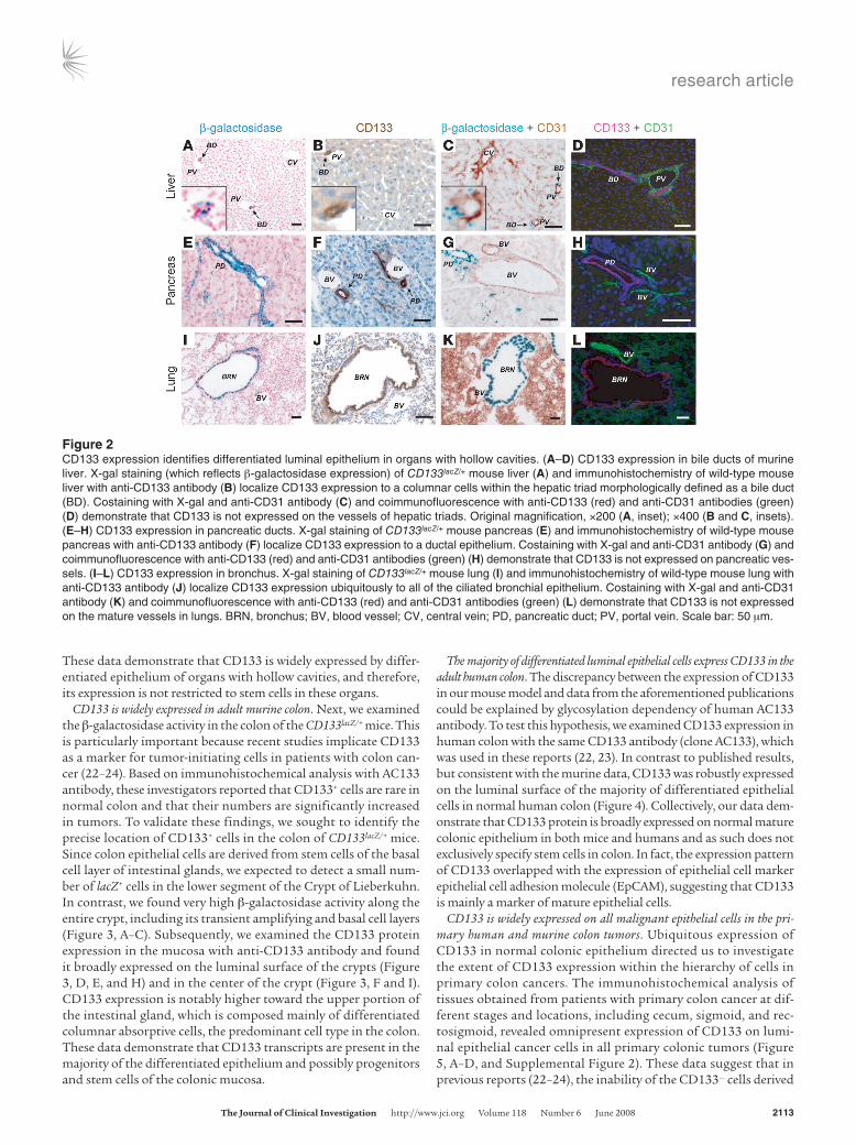
CD133 is expressed by luminal differentiated epithelial cells in adult mice. Further analysis of CD133lacZ/+ knockin mice revealed the expres-sion of CD133 in epithelial tissues of other adult organs. Inspec-tion of the hepatic tissue by X-gal staining revealed that CD133+ cells are localized to portal canals but not central veins (Figure 2A). Histologically, these CD133+ structures within portal triads were identified as bile ducts. Staining with anti-CD133 antibody further confirmed this finding (Figure 2B). To determine whether CD133 expression is restricted to the epithelium, we costained X-gal–treat-ed tissues with antibody against the endothelial cell marker CD31 (Figure 2C) and also performed double staining with anti-CD133 and anti-CD31 antibodies (Figure 2D). We could not detect any expression of CD133 in hepatic vascular cells. Similarly, we dem-onstrated that CD133 is ubiquitously expressed on the mature luminal epithelium of normal pancreatic ducts (Figure 2, E–H) and bronchial epithelium (Figure 2, I–L). X-gal and CD133 antibody staining on serial sections confirms their colocalization to the afore-mentioned structures (Supplemental Figure 1; supplemental mate-rial available online with this article; doi:10.1172/JCI34401DS1).
Figure 1Design and validation of CD133lacZ/+ mouse model for tracking CD133+ cells. (A) Structure of murine CD133 promoter region, genomic structure of murine CD133 gene, and the lacZ knockin strategy. AUG, start codon; Ex1A, exon 1A. (B) X-gal staining of the adult mouse kidney shows high β-galactosidase activ-ity in the proximal tubule (PT), but not distal tubule (DT). Staining with anti-CD133 antibody localizes CD133 protein specifically to the brush border of PT. Coimmunofluorescence of anti-CD133 with anti-CD31 antibodies shows CD133 expression on the parietal layer of Bowman’s capsule, but not the glomeruli (GL). Scale bar: 50 μm.
research article
TheJournalofClinicalInvestigation http://www.jci.org Volume 118 Number 6 June 2008 2113
These data demonstrate that CD133 is widely expressed by differ-entiated epithelium of organs with hollow cavities, and therefore, its expression is not restricted to stem cells in these organs.
CD133 is widely expressed in adult murine colon. Next, we examined the β-galactosidase activity in the colon of the CD133lacZ/+ mice. This is particularly important because recent studies implicate CD133 as a marker for tumor-initiating cells in patients with colon can-cer (22–24). Based on immunohistochemical analysis with AC133 antibody, these investigators reported that CD133+ cells are rare in normal colon and that their numbers are significantly increased in tumors. To validate these findings, we sought to identify the precise location of CD133+ cells in the colon of CD133lacZ/+ mice. Since colon epithelial cells are derived from stem cells of the basal cell layer of intestinal glands, we expected to detect a small num-ber of lacZ+ cells in the lower segment of the Crypt of Lieberkuhn. In contrast, we found very high β-galactosidase activity along the entire crypt, including its transient amplifying and basal cell layers (Figure 3, A–C). Subsequently, we examined the CD133 protein expression in the mucosa with anti-CD133 antibody and found it broadly expressed on the luminal surface of the crypts (Figure 3, D, E, and H) and in the center of the crypt (Figure 3, F and I). CD133 expression is notably higher toward the upper portion of the intestinal gland, which is composed mainly of differentiated columnar absorptive cells, the predominant cell type in the colon. These data demonstrate that CD133 transcripts are present in the majority of the differentiated epithelium and possibly progenitors and stem cells of the colonic mucosa.
The majority of differentiated luminal epithelial cells express CD133 in the adult human colon. The discrepancy between the expression of CD133 in our mouse model and data from the aforementioned publications could be explained by glycosylation dependency of human AC133 antibody. To test this hypothesis, we examined CD133 expression in human colon with the same CD133 antibody (clone AC133), which was used in these reports (22, 23). In contrast to published results, but consistent with the murine data, CD133 was robustly expressed on the luminal surface of the majority of differentiated epithelial cells in normal human colon (Figure 4). Collectively, our data dem-onstrate that CD133 protein is broadly expressed on normal mature colonic epithelium in both mice and humans and as such does not exclusively specify stem cells in colon. In fact, the expression pattern of CD133 overlapped with the expression of epithelial cell marker epithelial cell adhesion molecule (EpCAM), suggesting that CD133 is mainly a marker of mature epithelial cells.
CD133 is widely expressed on all malignant epithelial cells in the pri-mary human and murine colon tumors. Ubiquitous expression of CD133 in normal colonic epithelium directed us to investigate the extent of CD133 expression within the hierarchy of cells in primary colon cancers. The immunohistochemical analysis of tissues obtained from patients with primary colon cancer at dif-ferent stages and locations, including cecum, sigmoid, and rec-tosigmoid, revealed omnipresent expression of CD133 on lumi-nal epithelial cancer cells in all primary colonic tumors (Figure 5, A–D, and Supplemental Figure 2). These data suggest that in previous reports (22–24), the inability of the CD133– cells derived
Figure 2CD133 expression identifies differentiated luminal epithelium in organs with hollow cavities. (A–D) CD133 expression in bile ducts of murine liver. X-gal staining (which reflects β-galactosidase expression) of CD133lacZ/+ mouse liver (A) and immunohistochemistry of wild-type mouse liver with anti-CD133 antibody (B) localize CD133 expression to a columnar cells within the hepatic triad morphologically defined as a bile duct (BD). Costaining with X-gal and anti-CD31 antibody (C) and coimmunofluorescence with anti-CD133 (red) and anti-CD31 antibodies (green) (D) demonstrate that CD133 is not expressed on the vessels of hepatic triads. Original magnification, ×200 (A, inset); ×400 (B and C, insets). (E–H) CD133 expression in pancreatic ducts. X-gal staining of CD133lacZ/+ mouse pancreas (E) and immunohistochemistry of wild-type mouse pancreas with anti-CD133 antibody (F) localize CD133 expression to a ductal epithelium. Costaining with X-gal and anti-CD31 antibody (G) and coimmunofluorescence with anti-CD133 (red) and anti-CD31 antibodies (green) (H) demonstrate that CD133 is not expressed on pancreatic ves-sels. (I–L) CD133 expression in bronchus. X-gal staining of CD133lacZ/+ mouse lung (I) and immunohistochemistry of wild-type mouse lung with anti-CD133 antibody (J) localize CD133 expression ubiquitously to all of the ciliated bronchial epithelium. Costaining with X-gal and anti-CD31 antibody (K) and coimmunofluorescence with anti-CD133 (red) and anti-CD31 antibodies (green) (L) demonstrate that CD133 is not expressed on the mature vessels in lungs. BRN, bronchus; BV, blood vessel; CV, central vein; PD, pancreatic duct; PV, portal vein. Scale bar: 50 μm.

research article
2114 TheJournalofClinicalInvestigation http://www.jci.org Volume 118 Number 6 June 2008
from primary colonic tumors to generate tumors in NOD/SCID mice may be due to the possibility that CD133– cells primarily consist of nontumorigenic stromal and inflammatory cells inca-pable of tumor initiation.
To validate these results in a de novo colon cancer mouse model and to genetically track the expression of CD133 in primary colon-ic tumors, we generated Il10–/–CD133lacZ mice. In IL-10–knockout mice, chronic inflammation results in spontaneous tumor forma-tion in the colon (29). Using this model, we demonstrated that virtually all of the EpCAM+ tumor cells in Il10–/–CD133lacZ mice were β-galactosidase+ and therefore expressed CD133 (Figure 5, E–H). Therefore, similar to CD133 expression in human primary tumors, CD133 expression in Il10–/–CD133lacZ intestinal adenocar-cinomas was not restricted to intestinal or cancer-initiating cells but rather is ubiquitously distributed throughout the full range of tumor epithelial cells in the colon.
Metastatic colon tumors comprise both CD133+ and CD133– epithelial cell populations. Our data suggest that in primary human and mouse colon tumors, the omnipresence of CD133+ cells diminished the utility of this marker to identify true tumor-initiating cells. However, as metastatic lesions have a genetic repertoire that could be distinct from the primary tumors, the contribution of CD133+ tumor cells to the initiation of metastasis could differ from their contribution to primary tumors. In order to explore the role of CD133 as a marker of metastatic colon cancer–initiating cells, we examined tumors from 19 patients with colon cancer metastatic to the liver. Eleven out of nineteen patients had CD133+ tumors (CD133 expression ranged from 1.9% to 30.0%), whereas the other 8 patients had tumors with no detectable level of CD133 protein as seen by FACS analysis. Immunostaining of the tumors confirmed these results (Figure 6 and Supplemental Figure 3) and revealed that CD133+ cells are located at the tumor leading edge, localizing to the liver-tumor interface.
Both CD133+ and CD133– subsets of CD133+ human metastatic colon cancer are capable of initiating tumors in immunodeficient mice. The exis-tence of CD133-negative tumors led us to speculate that metastatic colon cancer–initiating cells may not be demarcated solely by the expression of CD133. We tested this hypothesis by taking advantage of a serial tumor xenograft model (Figure 7A). CD133+ liver metas-tases from 9 patients were separated into CD133+ and CD133– sub-
populations. The purity of each fraction was assessed by FACS analysis (Figure 7B). The minimal numbers of CD133– and CD133+ colon cancer cells capable of engraftment were injected subcuta-neously in NOD/SCID mice to determine tumor growth rate over time (Figure 7D). To explore the long-term tumorigenic potential of both fractions, newly formed CD133+ and CD133– tumors were transplanted into secondary NOD/SCID recipients, which ulti-mately gave rise to a new source of CD133+ and CD133– tumors that generated tertiary and subsequently quaternary tumors. In all 9 patient samples, the CD133– population led to emergence of adenocarcinomas, superseding the size of tumors derived from
Figure 3CD133 is widely expressed in murine colonic epithelial lining. (A–C) X-gal staining shows the distribution of β-galacto-sidase activity in the colon of CD133lacZ/+ mice (A), and in sagittal (B) and transverse (C) sections of the Lieberkuhn’s crypt. (D–F) Corresponding immunostaining with anti-CD133 antibody confirms the X-gal staining and demonstrates that CD133 protein is localized to the apical surface of mouse colon epithelium (indicated with the arrows). (G) X-Gal stain-ing of a wild-type mouse colon. (H) Magnification of rectangle in E showing the brush border localization of CD133 protein. The inset shows a negative control staining, where arrows point to brush borders on both sides of the lumen. Original magnification, ×200 (inset). (I) Coimmunofluorescence with anti-CD31 antibody (green) and anti-CD133 antibody (red) shows the apical localization of CD133 protein in mouse colon (indicated with arrows). Scale bar: 50 μm.
Figure 4CD133 is expressed in differentiated epithelium of normal human colon. (A and B) Immunostaining with CD133 (AC133) antibody (red) demonstrates the luminal localization (indicated with the arrows) of CD133 in normal human colon. (C and D) Coimmunofluorescence with CD133 (AC133) antibody (red) and EpCAM (a marker of epithelial cells) antibody (green) shows wide distribution of CD133 expression on differentiated epithelium in normal human colon, demonstrating consistency with the murine model. C shows a transverse section of the crypt. D shows a saggital section of the crypt. Scale bar: 50 μm.

research article
TheJournalofClinicalInvestigation http://www.jci.org Volume 118 Number 6 June 2008 2115
CD133+ cells (Figure 7C). The CD133– population always initiated tumor growth earlier than the CD133+ population and sustained a faster growth rate (Figure 7D).
The CD133– subset of colon cancer exhibits phenotypic characteristics of tumor-initiating stem cells. Sorted CD133+ and CD133– subpopu-lations of human colon cancer were plated as single cells in the presence of the appropriate growth medium. Subsequently, the number of cells forming typical round colonospheres was quanti-fied by phase contrast microscopy. Both CD133+ and CD133– sub-populations were capable of forming colonospheres in vitro at a similar rate (Figure 8A). Phenotypic analysis of individual colo-nies revealed that CD133– fraction–derived clones were CD133–
CD44+CD24–, whereas CD133+ fraction–derived colonies were CD133+CD44lowCD24+ (Figure 8B). Additionally, both CD133+ (51.3%) and CD133– (60.9%) cells coexpressed carcinoembryonic antigen (CEA), the expression level of which reflects the aggressive-ness of tumors (30). Cumulatively, these data support our find-ing that the CD133– subset of metastatic tumor exhibits a higher growth rate in vivo than the CD133+ subpopulation and that in metastatic colon cancers both CD133+ as well as CD133– subsets contain tumor-initiating cells.
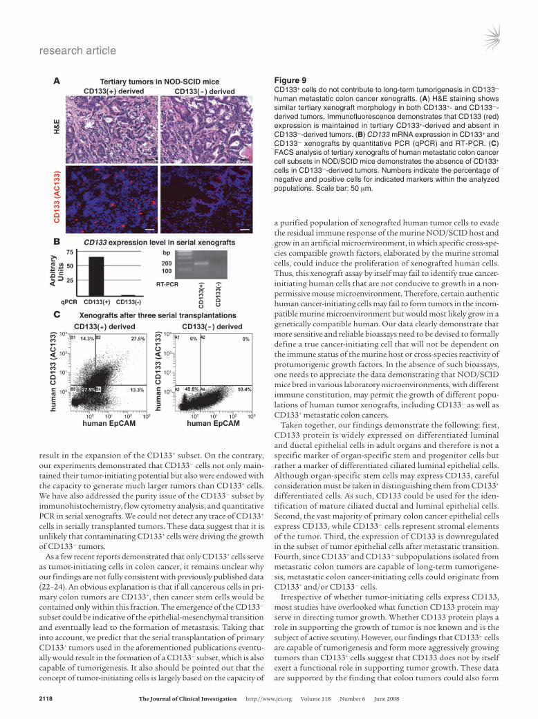
Tumorigenic potential of CD133– human metastatic colon cancer cells is not a result of their contamination by CD133+ cells. Histological analysis showed that both CD133+ and CD133– are similar in their mor-phology. Immunofluorescence demonstrated that after 3 serial transplantations, CD133+ cells were not found in the CD133– sub-set–derived tumors, while tumors derived from the CD133+ frac-tion consistently maintained CD133 expression (Figure 9A). The latter results were also confirmed by comparing CD133 mRNA levels (Figure 9B) and FACS analysis (Figure 9C) of the xenografts.
Thus, both CD133+ and CD133– subsets of human metastatic colon cancer exhibit long-term tumorigenic potential, and there-fore CD133 expression in metastatic colon cancer does not distin-guish a population of cancer stem cells.
DiscussionCD133 is considered a universal marker of organ-specific stem cells and tumor-initiating cells. As previous studies were based on the detection of CD133 by using commercially available anti-bodies, which may not recognize the full gamut of CD133 expres-sion pattern, we sought to design a genetic model to monitor the expression of CD133 in normal adult murine epithelium and in colon cancers, using protumorigenic Il10–/– deficient mice. In the present study, we have generated such a mouse model using the technology to introduce the reporter gene lacZ in the CD133 locus, in which the endogenous alternative CD133 promot-ers (28) control the expression of lacZ. Unexpectedly, we discov-ered that CD133+ cells are not exclusively localized to stem cell
Figure 5CD133 is expressed ubiquitously in all malignant cells of both human and mouse primary colon tumors. (A–D) CD133 expression on human primary colon cancer epithelium is shown by anti-CD133 antibody (A and B) and anti-EpCAM antibody (D); the negative IgG control is shown in C. Note the broad expression of CD133 in all malignant epithelial cells. CD133 protein expression is localized to the apical region of the columnar colonic cancer cells in primary human tumors. (E–H) CD133 expression in primary colon tumor of Il10–/–CD133lacZ mice. H&E stain-ing (E and F) shows the site of the primary tumor in the murine colon; X-gal (G) and anti-EpCAM (H) antibody immunostaining corroborate the human primary colon cancer data. Note the broad expression of CD133 in all transformed epithelial cells in the Il10–/–CD133lacZ mice as shown by the lacZ reporter. Scale bar: 50 μm.
Figure 6Metastatic colon cancer comprises both EpCAM+CD133+ and EpCAM+CD133– tumor cells. Costaining of colon cancer metastases to the liver with anti-EpCAM (green) and anti-CD133 (red) antibodies. (A and C) Typical examples of metastatic colon cancer harboring 2 subpopulations of tumor cells, EpCAM+CD133+ and EpCAM+CD133–. (B and D) Metastatic colon cancer completely lacking expression of CD133. The arrows indicate CD133 expression. Scale bar: 50 μm.

research article
2116 TheJournalofClinicalInvestigation http://www.jci.org Volume 118 Number 6 June 2008
niches. On the contrary, we detected CD133 in the majority, if not in all, of differentiated epithelial cells in organs with hollow ductal and nonductal luminal cavities, including the colon. We demonstrated that CD133 is expressed ubiquitously in the major-ity of the human primary colon tumor cells and in colon tumors emerging in Il10–/–CD133lacZ mice. Notably, the majority of stromal and inflammatory cells were CD133– in primary human tumors or murine Il10–/–CD133lacZ-derived colonic tumors. In contrast, CD133+ and CD133– cells coexpressing EpCAM were detected in human metastatic colon cancer, and both populations were capa-ble of tumor initiation. Although both CD133+ and CD133– cells isolated from metastatic colon adenocarcinomas can perform as cancer-initiating cells, the CD133– subset results in the formation of more aggressively growing tumors. Cumulatively, our data show that CD133 expression is not restricted to organ-specific stem cells or to cancer-initiating cells in metastatic adenocarcinomas.
CD133 was originally shown to be primarily expressed on CD34+ hematopoietic stem and progenitor cells (1, 2). Subsequently, it was shown to be expressed on subsets of endothelial progenitor cells (4). Using both the genetic model and immunohistochemical staining, we have discovered that in contrast to the hemangiogenic cells, CD133 expression in epithelial tissues is not restricted to stem or progenitor cells, but it is broadly distributed through the mature ciliated epithelial lining of luminal cavities. This is a sur-prising finding in relation to previously published data, which were obtained using immunohistochemical methods with commer-cially available antibodies. One explanation for this inconsistency could be hidden in the potential difference between the presence of mRNA and the CD133 protein in epithelial cells. Alternatively, this discrepancy may be due to the antibody affinity and different glycosylation and/or splice variants of CD133. However, our data demonstrate that CD133 immunohistochemical staining in both
Figure 7Both CD133+ and CD133–
subsets of human meta-static colon cancer exhibit long-term tumorigenic potential. (A) Serial xeno-graft strategy. (B) FACS analysis of human primary metastatic colon cancer cell subsets demonstrates their purity after separation with CD133 (AC133) anti-body. Numbers indicate the percentage of negative and positive cells within the ana-lyzed populations. (C) Sub-cutaneous tumors derived from the tertiary injection of the CD133– subset of human colon cancer grow faster in NOD/SCID mice than tumors which were derived from the CD133+ subset. Original magnifica-tion, ×0.25 (left panel). The units for the right panel of C are centimeters. (D) Growth curve of xenograft tumors in NOD/SCID mice shows that even after 3 serial transplantations CD133– cancer cells are capable of tumorigenesis; they initiate tumor formation earlier and exhibit faster growth rate than the CD133+ fraction. Data are shown as mean ± SEM; n = 3.

research article
TheJournalofClinicalInvestigation http://www.jci.org Volume 118 Number 6 June 2008 2117
human and mouse tissues phenocopied the ubiquitous expression of CD133 in the transgenic reporter mouse model, strongly sug-gesting that previous studies might have underestimated the true spectrum of CD133 expression pattern in the adult tissues. These data suggest that CD133 expression is not restricted to organ-spe-cific epithelial stem cells, but on the contrary, CD133 is expressed on differentiated epithelium. As such, CD133 mostly qualifies as a marker for identification of ductal and ciliated luminal epithelium but not as a specific marker of organ-specific stem cells.
Several studies have shown that CD133+ cells, but not CD133– cells, derived from human colon carcinomas were the only cells that could initiate tumors in immunodeficient mice (22–24). However, in our initial assessment, we found that 40% of the metastatic tumors were negative for CD133 expression, suggesting that CD133+ cells are not responsible for the growth of these tumors. On the contrary, in human primary colon cancer samples, we could not detect the EpCAM+CD133– tumor cells. In fact, based on immunohistochemical analysis, the majority of primary colon cancer cells were CD133+. This observation is also supported by the analysis of murine pri-mary tumors in Il10–/–CD133lacZ mice, in which all of the EpCAM+ tumor cells were also CD133+. Most importantly, in both primary human colon tumors and colon tumors in Il10–/–CD133lacZ mice, we were not able to detect a tumor epithelium that was CD133–. Thus, the CD133– population in primary tumors most likely consists of stromal, endothelial, and inflammatory cells but not tumorigenic cells of epithelial origin. Therefore, it is not surprising that in previ-ous studies on primary colon tumors, only the CD133+ subset was shown to contain cancer stem cells. On the contrary, after metastatic
transition, we identified a subset of EpCAM+CD133– cells, suggest-ing that some epithelial cells within the primary tumor may down-regulate the expression of CD133. Intriguingly, the CD133– cells iso-lated from metastatic colon tumors were also capable of long-term tumor growth in a serial xenograft model. Of note, the injection of the CD133– subset into immunodeficient mice resulted in more aggressive tumor growth, compared with growth of tumors that were initiated by the CD133+ fraction. These data suggest that CD133– colon tumors might either be more genetically unstable malignant derivatives of CD133+ primary colon cancer cells or originate from an as of yet unrecognized CD133– population of intestinal cells.
Further analysis revealed that CD133– cells display other pheno-typical characteristics of cancer stem cells. Metastatic CD133– tumor cells are CD44+CD24–, whereas CD133+ cells are CD44lowCD24+. The CD44+CD24– cells were previously identified as cancer stem cells in human breast cancer (31), and later CD44 was also suggested as a marker of colon cancer stem cells (32). Therefore, our data indi-cate that the CD133– fraction of colon cancer is more enriched for colon cancer-initiating cells. It is also remarkable that faster growing CD133– metastatic colon cancer cells express CEA at a higher level, which is indicative of aggressiveness in colorectal cancer (30). This also may explain high growth rate of CD133– cells in vivo.
Cumulatively, our data show that in metastatic colon adenocarci-nomas, tumor-initiating cells could be found in both CD133+ and CD133– fractions. Could this finding be due to the contribution of contaminating CD133+ cells to the growth of CD133– tumors? If this was true, then most likely, (a) CD133+ xenografts would grow faster and (b) serial transplantation of CD133– cells would
Figure 8Both CD133+ and CD133– xenografts form colonospheres, while CD133– cells display the molecular signature of cancer stem cells. (A) CD133+ and CD133– human colon cancer xenografts dissociated to a suspension of single cells are capable of forming colonospheres. Original magni-fication, ×200. (B) Colonospheres were subject to FACS analysis with CD133 antibody to confirm that the expression pattern was maintained in both CD133+ and CD133– colonies after in vitro culture. Both fractions were analyzed for the expression of CD44, CD24, and carcinoembryonic antigen (CEA). The expression pattern of these stem cell markers indicates the lesser level of maturity (CD44+CD24–) and higher level of aggres-siveness (CEA+) of the CD133– fraction of human colon cancer. Numbers indicate the percentage of positive cells within each population. Arrows refer to the stained population within the selected gates with the indicated markers.
research article
2118 TheJournalofClinicalInvestigation http://www.jci.org Volume 118 Number 6 June 2008
result in the expansion of the CD133+ subset. On the contrary, our experiments demonstrated that CD133– cells not only main-tained their tumor-initiating potential but also were endowed with the capacity to generate much larger tumors than CD133+ cells. We have also addressed the purity issue of the CD133– subset by immunohistochemistry, flow cytometry analysis, and quantitative PCR in serial xenografts. We could not detect any trace of CD133+ cells in serially transplanted tumors. These data suggest that it is unlikely that contaminating CD133+ cells were driving the growth of CD133– tumors.
As a few recent reports demonstrated that only CD133+ cells serve as tumor-initiating cells in colon cancer, it remains unclear why our findings are not fully consistent with previously published data (22–24). An obvious explanation is that if all cancerous cells in pri-mary colon tumors are CD133+, then cancer stem cells would be contained only within this fraction. The emergence of the CD133– subset could be indicative of the epithelial-mesenchymal transition and eventually lead to the formation of metastasis. Taking that into account, we predict that the serial transplantation of primary CD133+ tumors used in the aforementioned publications eventu-ally would result in the formation of a CD133– subset, which is also capable of tumorigenesis. It also should be pointed out that the concept of tumor-initiating cells is largely based on the capacity of
a purified population of xenografted human tumor cells to evade the residual immune response of the murine NOD/SCID host and grow in an artificial microenvironment, in which specific cross-spe-cies compatible growth factors, elaborated by the murine stromal cells, could induce the proliferation of xenografted human cells. Thus, this xenograft assay by itself may fail to identify true cancer-initiating human cells that are not conducive to growth in a non-permissive mouse microenvironment. Therefore, certain authentic human cancer-initiating cells may fail to form tumors in the incom-patible murine microenvironment but would most likely grow in a genetically compatible human. Our data clearly demonstrate that more sensitive and reliable bioassays need to be devised to formally define a true cancer-initiating cell that will not be dependent on the immune status of the murine host or cross-species reactivity of protumorigenic growth factors. In the absence of such bioassays, one needs to appreciate the data demonstrating that NOD/SCID mice bred in various laboratory microenvironments, with different immune constitution, may permit the growth of different popu-lations of human tumor xenografts, including CD133– as well as CD133+ metastatic colon cancers.
Taken together, our findings demonstrate the following: first, CD133 protein is widely expressed on differentiated luminal and ductal epithelial cells in adult organs and therefore is not a specific marker of organ-specific stem and progenitor cells but rather a marker of differentiated ciliated luminal epithelial cells. Although organ-specific stem cells may express CD133, careful consideration must be taken in distinguishing them from CD133+ differentiated cells. As such, CD133 could be used for the iden-tification of mature ciliated ductal and luminal epithelial cells. Second, the vast majority of primary colon cancer epithelial cells express CD133, while CD133– cells represent stromal elements of the tumor. Third, the expression of CD133 is downregulated in the subset of tumor epithelial cells after metastatic transition. Fourth, since CD133+ and CD133– subpopulations isolated from metastatic colon tumors are capable of long-term tumorigene-sis, metastatic colon cancer-initiating cells could originate from CD133+ and/or CD133– cells.
Irrespective of whether tumor-initiating cells express CD133, most studies have overlooked what function CD133 protein may serve in directing tumor growth. Whether CD133 protein plays a role in supporting the growth of tumor is not known and is the subject of active scrutiny. However, our findings that CD133– cells are capable of tumorigenesis and form more aggressively growing tumors than CD133+ cells suggest that CD133 does not by itself exert a functional role in supporting tumor growth. These data are supported by the finding that colon tumors could also form
Figure 9CD133+ cells do not contribute to long-term tumorigenesis in CD133– human metastatic colon cancer xenografts. (A) H&E staining shows similar tertiary xenograft morphology in both CD133+- and CD133–-derived tumors. Immunofluorescence demonstrates that CD133 (red) expression is maintained in tertiary CD133+-derived and absent in CD133–-derived tumors. (B) CD133 mRNA expression in CD133+ and CD133– xenografts by quantitative PCR (qPCR) and RT-PCR. (C) FACS analysis of tertiary xenografts of human metastatic colon cancer cell subsets in NOD/SCID mice demonstrates the absence of CD133+ cells in CD133–-derived tumors. Numbers indicate the percentage of negative and positive cells for indicated markers within the analyzed populations. Scale bar: 50 μm.

research article
TheJournalofClinicalInvestigation http://www.jci.org Volume 118 Number 6 June 2008 2119
in Il10–/–CD133–/– mice, again indicating that CD133 by itself may not be critical for tumor initiation. Nonetheless, identification of the function of CD133 may increase our understanding of its role in tumorigenesis.
MethodsDetermination of the 5′-UTR structure of mouse CD133 and generation of the CD133lacZ/+ knockin mice. Alternatively spliced exons were identified by using database analysis and 5′-RACE, using mouse brain and kidney cDNA as previously described (28). CD133lacZ/+ knockin mice were generated by the previously described high-throughput VelociGene (33).
Generation of Il10–/–CD133lacZ mouse model of colon cancer. Il10–/– mice were obtained from The Jackson Laboratory and were crossed with CD133lacZ mice at the age of 8 weeks to generate double heterozygous mice, which underwent subsequent breeding for generation of Il10–/– in CD133lacZ back-ground. Il10–/–CD133lacZ mice were analyzed for tumor formation in the colon at the age of 6 months or at the first sign of tumor progression.
Detection of β-galactosidase activity in mouse tissues. Fresh-frozen tissues of the CD133lacZ/+ mice were sectioned, subsequently incubated for 4–16 hours with X-gal (1 mg/ml; Calbiochem), and then counterstained with Nuclear Fast Red (Vector Laboratories).
Immunohistochemistry. Immunohistochemistry was performed on fresh-frozen tissues postfixed in paraformaldehyde. The slides were stained with anti-mouse Prominin-1 antibody clone 13A4 (5 μg/ml; eBioscience) or anti-human CD133/1 clone AC133 (5 μg/ml; Miltenyi Biotec) for the detection of mouse or human CD133, respectively. Coimmunofluorescence was performed using FITC-conjugated anti-mouse CD31 clone Mec13.3 (BD Biosciences — Pharmingen), and anti-mouse Prominin-1 antibody was detected with Cy3-conjugated anti-rat IgG (Jackson ImmunoResearch Lab-oratories Inc.). X-gal–stained tissues were subsequently immunostained using anti-CD31 as above. Frozen samples of primary colon cancer samples were obtained in accordance with the protocol approved by the Institu-tional Review Board of Weill Medical College of Cornell University.
Imaging. Bright-field micrographs were captured on an Olympus micro-scope using AxioCam (Zeiss). Confocal images were captured on an LSM 510 META confocal microscope (Zeiss).
Tumor cell preparation. Resected colon cancer specimens, primary or meta-static to the liver, were obtained from patients that provided informed con-sent, as approved by the Institutional Review Board of Weill Medical Col-lege and Memorial Sloan-Kettering Cancer Center. The patients studied in this protocol were not treated with any form of neoadjuvant therapy prior to surgery. However, all of these patients were treated at the time of diag-nosis of advanced disease with 5FU-based chemotherapy. Tumor tissue was mechanically dissociated with sterile scalpel blades until tumor was minced into approximately 1 mm in size. Tumor tissue was then enzymatically dis-sociated by 1-hour incubation with Collagenase Type IV (Sigma-Aldrich) at 37°C into single-cell suspensions. Human colon cancer cells were mag-netically labeled and separated by triple passage using a CD133 Cell Isola-tion kit (Miltenyi Biotec). Before separation, samples were assessed using a FACSCalibur flow cytometer (BD Biosciences), with mouse IgG1 antibody conjugated to phycoerythrin were used as isotype controls (BD Biosci-ences). CD133 expression was assessed using anti-CD133/1 phycoerythrin (Miltenyi Biotec). At least 10,000 events were acquired for each sample, and all cells positive for propidium iodide were gated out. After magnetic bead separation, samples were assessed by flow cytometry for purity. Purity for CD133+ colon cancer cells from all tumor samples was greater than 97%.
Subcutaneous transplantation of sorted metastatic human colon cancer cells into NOD/SCID mice. Xenograft in vivo tumor models were approved by the Institutional Review Board of Weill Medical College. NOD.CB17-Prkdcscid/J (NOD/SCID) mice were bred and maintained under specific pathogen-
free conditions with the approval of the Institutional Animal Care and Use Committee Animal Care Committee of Weill Cornell Medical Col-lege. Colon cancer cells were suspended in 100 μl of AMEM and 100 μl of matrigel (BD Biosciences), making a 1:1 mixture. Colon cancer cells (2 × 10⁶) from each human tumor were injected subcutaneously in the left flank of 3 mice (8 weeks of age). To determine the minimal amount of cells capable of engraftment, we performed limiting dilution experiments. After different times (see Figure 7D), tumor appearance was evaluated using a cali-per, and tumor volume was calculated using the formula ;pi;/6(w1 × w2 × w2), where w1 represents the largest tumor diameter, and w2 represents the smallest tumor diameter. After 8 to 10 weeks of tumor growth, ani-mals were sacrificed, and tumors were removed and then subjected to immunohistochemical analysis or injected into secondary, tertiary, or quaternary recipients. Some tumors were embedded in Tissue-Tek OCT compound (VWR International) and immediately frozen in liquid nitro-gen; others were embedded in paraffin. Paraffin-embedded tumors were serially sectioned and stained with H&E for histological analysis.
Generation of colonospheres. Tumors were excised from NOD/SCID mice and were mechanically dissociated using a MediMachine (BD Biosciences) with 50 μm Medicons (BD Biosciences). The dissociated tumor cells were washed in 12 ml of DMEM, supplemented with 1% penicillin/streptomy-cin and 10% fetal bovine serum and centrifuged at 300 g for 5 minutes at 4°C. The cell pellet was resuspended at a concentration of 107 cells/300 μl, using DMEM supplemented with 1% penicillin/streptomycin and 10% fetal bovine serum. Resuspended cell pellets (300 μl) were placed in each well of a 24-well plate. Media was replaced every third day.
FACS analysis of colonospheres. Colonospheres were removed by a 5-minute trypsinization, washed in 12 ml of DMEM, supplemented with 1% penicil-lin/streptomycin and 10% fetal bovine serum, and centrifuged at 1,200 rpm for 5 minutes at 4°C. The colonospheres were resuspended in ice-cold PBS (Gibco), supplemented with 5% BSA (Sigma-Aldrich), and divided into the appropriate amount of FACS tubes (BD Biosciences). The cells were stained with IgG-PE (BD Biosciences — Pharmingen) and IgG-FITC anti-bodies (BD Biosciences — Pharmingen) for detection of unspecific binding of the antibodies. The primary antibodies used were AC133/1 PE (Miltenyi Biotec), CD44 FITC (Novocastra), CD24 FITC (BD Biosciences — Pharmin-gen), and Carcino Embryonic Antigen FITC (Abcam).
Quantification of CD133 expression by quantitative PCR. Total RNA was prepared from cultured cells using the RNeasy extraction kit (QIAGEN) and reverse transcribed using SuperScript II Reverse Transcriptase (Invitrogen) according to the manufacturer’s instructions. Relative quantitative PCR was performed on a 7500 Fast Real-Time PCR System (Applied Biosystems) using SYBR Green PCR mix (Applied Biosystems). The following human-specific intron-spanning primer pairs for CD133 were used: forward, 5′-GGACCCATTG-GCATTCTC-′3; and reverse, 5′-CAGGACACAGCATAGAATAATC-′3. Cycle conditions were as follows: 1 cycle at 50°C for 2 minutes, followed by 1 cycle at 95°C for 10 minutes, followed by 40 cycles at 95°C for 15 seconds and 60°C for 1 minute. Specificity of PCR products was tested by dissociation curves. Threshold cycles of primer probes were normalized to Actb. Relative values of transcripts were calculated using the equation, 2–ΔΔCt, where ΔCt is equal to the difference in threshold cycles for target and reference.
AcknowledgmentsThis work was supported by the Howard Hughes Medical Insti-tute, Ansary Center for Stem Cell Therapeutics, and National Heart, Lung and Blood Institute grants HL075234, HL059312, and HL084936 (to Shahin Rafii). Till Milde was supported by a grant from the Mildred-Scheel-Stiftung, Deutsche Krebshilfe, Bonn, Germany. We thank Andrew J. Dannenberg for providing resected human primary colon cancer specimens.
research article
2120 TheJournalofClinicalInvestigation http://www.jci.org Volume 118 Number 6 June 2008
Received for publication November 2, 2007, and accepted in revised form April 9, 2008.
Address correspondence to: Shahin Rafii, Weill Medical College of Cornell University, 1300 York Avenue, Room A-869A, New York,
New York 10021, USA. Phone: (212) 746-2070; Fax: (212) 746-2286; E-mail: [email protected].
Sergey V. Shmelkov and Jason M. Butler contributed equally to this work.
1. Miraglia, S., et al. 1997. A novel five-transmem-brane hematopoietic stem cell antigen: isolation, characterization, and molecular cloning. Blood. 90:5013–5021.
2. Yin, A.H., et al. 1997. AC133, a novel marker for human hematopoietic stem and progenitor cells. Blood. 90:5002–5012.
3. Weigmann, A., Corbeil, D., Hellwig, A., and Huttner, W.B. 1997. Prominin, a novel microvilli-specific polytopic membrane protein of the apical surface of epithelial cells, is targeted to plasmalem-mal protrusions of non-epithelial cells. Proc. Natl. Acad. Sci. U. S. A. 94:12425–12430.
4. Peichev, M., et al. 2000. Expression of VEGFR-2 and AC133 by circulating human CD34(+) cells identifies a population of functional endothelial precursors. Blood. 95:952–958.
5. Salven, P., Mustjoki, S., Alitalo, R., Alitalo, K., and Rafii, S. 2003. VEGFR-3 and CD133 identify a pop-ulation of CD34+ lymphatic/vascular endothelial precursor cells. Blood. 101:168–172.
6. Shmelkov, S.V., et al. 2005. Cytokine precondition-ing promotes codifferentiation of human fetal liver CD133+ stem cells into angiomyogenic tissue. Circulation. 111:1175–1183.
7. Uchida, N., et al. 2000. Direct isolation of human central nervous system stem cells. Proc. Natl. Acad. Sci. U. S. A. 97:14720–14725.
8. Lee, A., et al. 2005. Isolation of neural stem cells from the postnatal cerebellum. Nat. Neurosci. 8:723–729.
9. Sagrinati, C., et al. 2006. Isolation and character-ization of multipotent progenitor cells from the Bowman’s capsule of adult human kidneys. J. Am. Soc. Nephrol. 17:2443–2456.
10. Richardson, G.D., et al. 2004. CD133, a novel mark-er for human prostatic epithelial stem cells. J. Cell Sci. 117:3539–3545.
11. Kordes, C., et al. 2007. CD133+ hepatic stellate cells are progenitor cells. Biochem. Biophys. Res. Commun. 352:410–417.
12. Oshima, Y., et al. 2007. Isolation of mouse pancreat-ic ductal progenitor cells expressing CD133 and c-Met by flow cytometric cell sorting. Gastroenterology. 132:720–732.
13. Sugiyama, T., Rodriguez, R.T., McLean, G.W., and Kim, S.K. 2007. Conserved markers of fetal pancre-atic epithelium permit prospective isolation of islet progenitor cells by FACS. Proc. Natl. Acad. Sci. U. S. A. 104:175–180.
14. Ito, Y., et al. 2006. Isolation of murine hair-induc-ing cells using the cell surface marker prominin-1/CD133. J. Invest. Dermatol. 127:1052–1060.
15. Singh, S.K., et al. 2003. Identification of a can-cer stem cell in human brain tumors. Cancer Res. 63:5821–5828.
16. Singh, S.K., et al. 2004. Identification of human brain tumor initiating cells. Nature. 432:396–401.
17. Collins, A.T., Berry, P.A., Hyde, C., Stower, M.J., and Maitland, N.J. 2005. Prospective identification of tumorigenic prostate cancer stem cells. Cancer Res. 65:10946–10951.
18. Suetsugu, A., et al. 2006. Characterization of CD133+ hepatocellular carcinoma cells as cancer stem/progenitor cells. Biochem. Biophys. Res. Commun. 351:820–824.
19. Yin, S., et al. 2007. CD133 positive hepatocellular carcinoma cells possess high capacity for tumori-genicity. Int. J. Cancer. 120:1444–1450.
20. Hermann, P.C., et al. 2007. Distinct populations of cancer stem cells determine tumor growth and metastatic activity in human pancreatic cancer. Cell Stem Cell. 1:313–323.
21. Eramo, A., et al. 2008. Identification and expansion of the tumorigenic lung cancer stem cell population. Cell Death Differ. 15:504–514.
22. O’Brien, C.A., Pollett, A., Gallinger, S., and Dick, J.E. 2007. A human colon cancer cell capable of initiating tumor growth in immunodeficient mice. Nature. 445:106–110.
23. Ricci-Vitiani, L., et al. 2007. Identification and
expansion of human colon-cancer-initiating cells. Nature. 445:111–115.
24. Todaro, M., et al. 2007. Colon cancer stem cells dic-tate tumor growth and resist cell death by produc-tion of interleukin-4. Cell Stem Cell. 1:389–402.
25. Florek, M., et al. 2005. Prominin-1/CD133, a neural and hematopoietic stem cell marker, is expressed in adult human differentiated cells and certain types of kidney cancer. Cell Tissue Res. 319:15–26.
26. Pfenninger, C.V., et al. 2007. CD133 is not pres-ent on neurogenic astrocytes in the adult subven-tricular zone, but on embryonic neural stem cells, ependymal cells, and glioblastoma cells. Cancer Res. 67:5727–5736.
27. Corbeil, D., Roper, K., Fargeas, C.A., Joester, A., and Huttner, W.B. 2001. Prominin: a story of choles-terol, plasma membrane protrusions and human pathology. Traffic. 2:82–91.
28. Shmelkov, S.V., et al. 2004. Alternative promoters regulate transcription of the gene that encodes stem cell surface protein AC133. Blood. 103:2055–2061.
29. Berg, D.J., et al. 1996. Enterocolitis and colon can-cer in interleukin-10-deficient mice are associated with aberrant cytokine production and CD4(+) TH1-like responses. J. Clin. Invest. 98:1010–1020.
30. Goldstein, M.J., and Mitchell, E.P. 2005. Carcino-embryonic antigen in the staging and follow-up of patients with colorectal cancer. Cancer Invest. 23:338–351.
31. Al-Hajj, M., Wicha, M.S., Benito-Hernandez, A., Morrison, S.J., and Clarke, M.F. 2003. Prospective identification of tumorigenic breast cancer cells. Proc. Natl. Acad. Sci. U. S. A. 100:3983–3988.
32. Dalerba, P., et al. 2007. Phenotypic characterization of human colorectal cancer stem cells. Proc. Natl. Acad. Sci. U. S. A. 104:10158–10163.
33. Valenzuela, D.M., et al. 2003. High-throughput engineering of the mouse genome coupled with high-resolution expression analysis. Nat. Biotechnol. 21:652–659.