Are purified or expanded cord blood-derived CD133+ cells better at improving cardiac function
11
Original Research Are purified or expanded cord blood-derived CD133 1 cells better at improving cardiac function? Alexandra C Senegaglia 1 , Laura A Barboza 1 , Bruno Dallagiovanna 2 , Carlos A M Aita 1 , Paula Hansen 1 , Carmen L K Rebelatto 1 , Alessandra M Aguiar 2 , Nelson I Miyague 1 , Patrı´cia Shigunov 2 , Fabiane Barchiki 1 , Alejandro Correa 2 , Marcia Olandoski 1 , Marco A Krieger 2 and Paulo R S Brofman 1 1 Pontifı´cia Universidade Cato ´ lica do Parana ´ , Institute for Health and Biological Sciences, Rua Imaculada Conceic ¸a ˜ o, 1155 Curitiba, Parana ´ , 80215901; 2 Instituto Carlos Chagas, Fundac ¸a ˜o Oswaldo Cruz. Rua Prof. Algacyr Munhoz Maeder, 3775 Curitiba, Parana ´ 81350010, Brazil Corresponding author: Alexandra C Senegaglia, Rua Guilherme Pugsley 1760, Apartment 501, Curitiba, Parana ´ , 80620000, Brazil. Emails: [email protected]; [email protected] Abstract Endothelial progenitor cells (EPCs), which express the CD133 marker, can differentiate into mature endothelial cells (ECs) and create new blood vessels. Normal angiogenesis is unable to repair the injured tissues that result from myocardial infarction (MI). Patients who have high cardiovascular risks have fewer EPCs and their EPCs exhibit greater in vitro senescence. Human umbilical cord blood (HUCB)-derived EPCs could be an alternative to rescue impaired stem cell function in the sick and elderly. The aim of this study was to purify HUCB-derived CD133 þ cells, expand them in vitro and evaluate the efficacy of the purified and expanded cells in treating MI in rats. CD133 þ cells were selected for using CD133-coupled magnetic microbeads. Purified cells stained positive for EPC markers. The cells were expanded and differentiated in media supplemented with fetal calf serum and basic fibroblast growth factor, insulin-like growth factor-I and vascular endothelial growth factor (VEGF). Differentiation was confirmed by lack of staining for EPC markers. These expanded cells exhibited increased expression of mature EC markers and formed tubule-like structures in vitro. Only the expanded cells expressed VEGF mRNA. Cells were expanded up to 70-fold during 60 days of culture, and they retained their functional activity. Finally, we evaluated the therapeutic potential of purified and expanded CD133 þ cells in treating MI by intramyocardially injecting them into a rat model of MI. Rats were divided into three groups: A (purified CD133 þ cells-injected); B (expanded CD133 þ cells-injected) and C (saline buffer-injected). We observed a significant improvement in left ventricular ejection fraction for groups A and B. In summary, CD133 þ cells can be purified from HUCB, expanded in vitro without loosing their biological activity, and both purified and expanded cells show promising results for use in cellular cardiomyoplasty. However, further pre-clinical testing should be performed to determine whether expanded CD133 þ cells have any clinical advantages over purified CD133 þ cells. Keywords: umbilical cord blood, endothelial progenitor cells, transplantation, myocardial infarction Experimental Biology and Medicine 2010; 235: 119–129. DOI: 10.1258/ebm.2009.009194 Introduction Reperfusion therapy is associated with significantly reduced mortality rates and improved clinical outcomes in patients with acute myocardial infarction (MI). 1 However, normal angiogenesis is usually unable to supply the greater demand for oxygen and nutrients required following MI. In addition, it is unable to prevent hypertrophied cardiomyo- cyte apoptosis and ventricular remodeling. 2 Cell therapy may provide a novel therapeutic strategy to modify left ventricular remodeling processes and prevent postinfarction heart failure 3 Different cell populations have been tested in this regard in both preclinical and clinical settings. Recently, isolated CD34 þ cells demonstrated increased potency and safety for therapeutic neovascularization after MI, as compared with total mononuclear cells (MNCs). 4 Endothelial progenitor cells (EPCs) are precursor cells that can differentiate into mature endothelial cells (ECs) and create new blood vessels. 5 Generally, EPCs can be identified based ISSN: 1535-3702 Copyright # 2010 by the Society for Experimental Biology and Medicine Experimental Biology and Medicine 2010; 235: 119–129
-
Upload
independent -
Category
Documents
-
view
0 -
download
0
Transcript of Are purified or expanded cord blood-derived CD133+ cells better at improving cardiac function

Original Research
Are purified or expanded cord blood-derived CD1331 cells
better at improving cardiac function?
Alexandra C Senegaglia1, Laura A Barboza1, Bruno Dallagiovanna2, Carlos A M Aita1,
Paula Hansen1, Carmen L K Rebelatto1, Alessandra M Aguiar2, Nelson I Miyague1,
Patrıcia Shigunov2, Fabiane Barchiki1, Alejandro Correa2, Marcia Olandoski1, Marco A Krieger2
and Paulo R S Brofman1
1Pontifıcia Universidade Catolica do Parana, Institute for Health and Biological Sciences, Rua Imaculada Conceicao, 1155 Curitiba,
Parana, 80215901; 2Instituto Carlos Chagas, Fundacao Oswaldo Cruz. Rua Prof. Algacyr Munhoz Maeder, 3775 Curitiba, Parana
81350010, Brazil
Corresponding author: Alexandra C Senegaglia, Rua Guilherme Pugsley 1760, Apartment 501, Curitiba, Parana, 80620000, Brazil.
Emails: [email protected]; [email protected]
AbstractEndothelial progenitor cells (EPCs), which express the CD133 marker, can differentiate into mature endothelial cells (ECs) and
create new blood vessels. Normal angiogenesis is unable to repair the injured tissues that result from myocardial infarction
(MI). Patients who have high cardiovascular risks have fewer EPCs and their EPCs exhibit greater in vitro senescence.
Human umbilical cord blood (HUCB)-derived EPCs could be an alternative to rescue impaired stem cell function in the sick
and elderly. The aim of this study was to purify HUCB-derived CD133þ cells, expand them in vitro and evaluate the
efficacy of the purified and expanded cells in treating MI in rats. CD133þ cells were selected for using CD133-coupled
magnetic microbeads. Purified cells stained positive for EPC markers. The cells were expanded and differentiated in media
supplemented with fetal calf serum and basic fibroblast growth factor, insulin-like growth factor-I and vascular endothelial
growth factor (VEGF). Differentiation was confirmed by lack of staining for EPC markers. These expanded cells exhibited
increased expression of mature EC markers and formed tubule-like structures in vitro. Only the expanded cells expressed
VEGF mRNA. Cells were expanded up to 70-fold during 60 days of culture, and they retained their functional activity.
Finally, we evaluated the therapeutic potential of purified and expanded CD133þ cells in treating MI by intramyocardially
injecting them into a rat model of MI. Rats were divided into three groups: A (purified CD133þ cells-injected); B (expanded
CD133þ cells-injected) and C (saline buffer-injected). We observed a significant improvement in left ventricular ejection
fraction for groups A and B. In summary, CD133þ cells can be purified from HUCB, expanded in vitro without loosing their
biological activity, and both purified and expanded cells show promising results for use in cellular cardiomyoplasty.
However, further pre-clinical testing should be performed to determine whether expanded CD133þ cells have any clinical
advantages over purified CD133þ cells.
Keywords: umbilical cord blood, endothelial progenitor cells, transplantation, myocardial infarction
Experimental Biology and Medicine 2010; 235: 119–129. DOI: 10.1258/ebm.2009.009194
Introduction
Reperfusion therapy is associated with significantly reducedmortality rates and improved clinical outcomes in patientswith acute myocardial infarction (MI).1 However, normalangiogenesis is usually unable to supply the greaterdemand for oxygen and nutrients required following MI. Inaddition, it is unable to prevent hypertrophied cardiomyo-cyte apoptosis and ventricular remodeling.2 Cell therapymay provide a novel therapeutic strategy to modify left
ventricular remodeling processes and prevent postinfarctionheart failure3 Different cell populations have been testedin this regard in both preclinical and clinical settings.Recently, isolated CD34þ cells demonstrated increasedpotency and safety for therapeutic neovascularization afterMI, as compared with total mononuclear cells (MNCs).4
Endothelial progenitor cells (EPCs) are precursor cells thatcan differentiate into mature endothelial cells (ECs) and createnew blood vessels.5 Generally, EPCs can be identified based
ISSN: 1535-3702
Copyright # 2010 by the Society for Experimental Biology and Medicine
Experimental Biology and Medicine 2010; 235: 119–129

on their expression of CD133, CD34, KDR and/or VE-cadherincellular markers.6 The similar phenotype of hematopoietic andEPCs strongly supports the existence of a common precursorcell, which has been called the hemangioblast. This cell hasbeen identified in in vitro studies using embryonic stem cellsfrom postnatal bone marrow and cord blood.7
EPCs represent less than 1% of all bone marrow cells andless than 0.01% of peripheral blood MNCs.8 Human umbi-lical cord blood (HUCB) is a viable source of stem cellsdue to its large CD34 and CD133-expressing cell popu-lations. These cells exhibit robust proliferative capacity,low immunogenicity and low infection contamination(including virions). Furthermore, the diverse representationof human leukocyte antigen (HLA) genotypes in unrelated,banked HUCB allows the use of HUCB-derived cells in allo-geneic transplantations.9 Promising results were obtainedusing purified HUCB stem cells as cellular therapies inanimal models, and these results support their potentialapplication in cardiovascular medicine.10,11 Independentlyor in combination with biomaterials, such as collagenmatrix, HUCB-derived stem cells seem to improve theefficiency of cellular cardiomyoplasty in mice. Thus, thisstrategy has emerged as a novel therapy.12
Results from preclinical studies have suggested that itwould take more than 10 L of autologous peripheral bloodto produce a sufficient number of EPCs to induce angiogen-esis in one patient.13 Furthermore, patients with increasedcardiovascular risk have fewer EPCs, as compared withhealthy subjects, and their EPCs demonstrate increasedin vitro senescence.14 The use of CD133þ cells derivedfrom purified and expanded HUCB could be used torescue stem cell function in the sick and elderly. EPCs canbe expanded in vitro to increase their potential therapeuticuse. However, all current protocols involving short-termculture would not yield sufficient numbers of cells for sys-temic therapy in humans. On the other hand, long-termculture may introduce changes in the EPC’s phenotype,which could reduce their therapeutic efficacy.15
The aim of this study was to isolate and purify CD133þ
cells from HUCB and to expand the cells in vitro by exposingthem to an appropriate growth factor cocktail. We evaluatedthe effectiveness of the purified and expanded CD133þ cellsin improving the cardiac function in a rat model of MI.
Materials and methods
This study was reviewed and approved by the Local EthicsCommittee (Pontifıcia Universidade Catolica do Parana:numbers 1366 and 180). Signed informed consent wasobtained from each mother prior to HUCB collection. Allanimal experiments were carried out in accordance with:‘The Guide for the Care and Use of Laboratory Animals’published by the National Institute of Health (NIH publi-cation 85-23, 7th edn, revised 1996).
Purification of CD1331 cells
MNCs were isolated from HUCB. Thirty-seven HUCBsamples (approximately 78.53+ 4.63 mL each) were
collected from fresh placentas with the umbilical cord stillattached, and acid-citrate-dextrose was used as anticoagu-lant. MNCs were isolated by centrifugation for 30 min at400 g using Ficoll density gradients (Sigma-Aldrich, SaintLouis, MO, USA) and washed three times in phosphate-buffered saline (PBS) (Invitrogen, Grand Island, NY, USA).CD133þ cells were selected using CD133-coupled magneticmicrobeads (Miltenyi Biotech, Bergisch-Gladbach, Germany),following the manufacturer’s instructions. Briefly, MNCswere incubated with the CD133/1 monoclonal antibody,which was directly conjugated to microbeads, for 30 min. Thesamples were washed, filtered through a 50-mm nylon meshto remove clumps and added to a column in the midi MACScell separator (Miltenyi Biotec). The labeled cells were separ-ated using a high-gradient magnetic field and eluted fromthe column after being separated from the magnet. At the endof the separation, the cell purity and viability were determinedusing an anti-CD133 antibody (PE conjugated; Miltenyi Biotec)and 7AAD (BD Pharmingen, San Diego, CA, USA), respect-ively. The samples were analyzed using a FACSCalibur flowcytometer (Becton Dickinson, San Jose, CA, USA).
Proliferation assay on CD1331 cells
A cell proliferation assay was performed to establish theoptimal culture conditions for CD133þ cells. A cell suspen-sion of 50 mL (1 � 105 cells/mL) in Iscove’s ModifiedDulbecco’s Media (IMDM) (Invitrogen), supplementedwith 10% fetal bovine serum (FBS) (Invitrogen) and 1%penicillin–streptomycin (Invitrogen), was seeded into96-well plates (Corning, Oneonta, NY, USA). Various con-centrations of growth factors were added: 0.5, 1.0 and2.0 ng/mL of basic fibroblast growth factor (b-FGF)(Invitrogen); 1.0, 2.0 and 3.0 ng/mL of insulin-like growthfactor-I (IGF-I) (Sigma-Aldrich); and 25, 50 and 75 ng/mLof vascular endothelial growth factor (VEGF) (Sigma-Aldrich). Media with a constant amount of 1 ng/mLb-FGF and 2 ng/mL IGF-I with increasing VEGF concen-trations (25, 50 and 75 ng/mL) was used. The cells wereincubated at 378C with 5% CO2 in a humidified atmosphere.After five days, 1 mCi of [methyl-3H] thymidine (5.0 Ci/mmol) (GE Healthcare, Buckinghamshire, UK) was addedto each well. Six hours later, the culture media and cellswere removed, and the cell-associated radioactivity wasmeasured using a scintillation counter (Beckman Coulterw,Fullerton, CA, USA). Responses to each concentrationwere assayed in triplicate, and results were expressed asthe mean of 12 independent experiments.
Culture and expansion of CD1331 cells
After establishing the optimal culture conditions, 2 � 105
CD133þ cells in 50 mL were plated into 12-well culture platescoated with human fibronectin (Becton Dickinson) andcultured in IMDM supplemented with 50 ng/mL VEGF,1 ng/mL b-FGF, 2 ng/mL IGF-I, 10% FBS and 1% penicillin–streptomycin. All cultures were maintained at 378C with 5%CO2 in a humidified atmosphere. Additional feeding wasperformed depending on the rate of cell proliferation; thesupernatant was removed by gentle pipetting and fresh
................................................................................................................................................120 Experimental Biology and Medicine Volume 235 January 2010
medium was added. Cells were cultured to approximately80% confluence, detached using Accutase (US Bio-Technologies, Pottstown, PA, USA) digestion and replated inT-25 tissue culture flasks for 60 d. On day 60, the cells wereassessed for viability, rate of expansion and phenotype.
Phenotypic characterization of cells
Immunophenotypic analysis was performed by staining 2 �105 of MNCs, purified and expanded CD133þ cells. MNCsand purified cells were analyzed after isolation, whereasexpanded cells were analyzed after 60 days of culture. Thecells were incubated with various conjugated and unconju-gated monoclonal antibodies against the following humanantigens: CD133 (PE conjugated; Miltenyi Biotec), CD34(APC conjugated; BD Pharmingen), CD45 (PerCP conju-gated; BD Pharmingen), CD14 (fluorescein isothiocynate[FITC] conjugated; BD Pharmingen), CD31 (BD Pharmingen),CD105 (BD Pharmingen) and von Willebrand Factor (vWF;Dako, Glostrup, Denmark). All incubations were performedat 48C for 30 min. For CD31 and CD105 detection, cellswere further incubated with an isotype-specific FITC-conjugated goat anti-mouse antibody (Caltag, Burlingame,CA, USA). For intracellular detection of vWF, cells werepermeabilized using FIX & PERM cell permeabilizationreagents (Caltag, Carlsbad, CA, USA), and further incubatedwith an isotype-specific FITC-conjugated goat anti-rabbitantibody (Sigma-Aldrich). Isotype-identical antibodies (BDPharmingen) were served as controls. After incubation, thecells were washed with PBS containing 2% FBS and fixedwith PBS containing 1% paraformaldehyde. Quantitative ana-lyses were performed using a FACSCalibur flow cytometer andFlowJo software (Flowjo, Ashland, OR, USA).
Immunofluorescence microscopy
Expanded CD133þ cells were cultured on chamber slides (ErieScientific Company, Portsmouth, NH, USA) to confluence.Cells were washed twice with PBS and fixed for 20 min withcold methanol. Slides were incubated for 30 min with a poly-clonal rabbit anti-human vWF antibody (1:200) (Caltag),followed by an isotype-specific FITC-conjugated goat anti-rabbit antibody (1:80) (Sigma-Aldrich). Cells were washedwith PBS and then stained with 40,6-diamidino-2-phenylindole dihydrochloride (1:5) (Sigma-Aldrich). Thenegative control was obtained by omitting the primary anti-body incubation. Slides were examined and imaged using afluorescent microscope (Olympus, Tokyo, Japan).
Reverse transcription-polymerase chain reaction
To evaluate VEGF expression, total RNA was extracted frompurified and expanded CD133þ cells using the RNeasy kit(QIAGEN, Valencia, CA, USA). RNA was treated withDNAse I (QIAGEN) in a column, according to the manufac-turer’s instructions. The total RNA (1 mg) was converted tocDNA by adding 10 nmol/L of oligo-dT primer (USBCorporation, Cleveland, OH, USA) and 1 mL of reverse tran-scriptase (RT) (IMPROM II, Promega, Fitchburg, WI, USA),according to the manufacturer’s instructions. Polymerase
chain reaction (PCR) reactions contained 20 ng of cDNAas a template with 20 mmol/L Tris-HCl (pH 8.4),50 mmol/L KCl, 10 pmol of forward and reverse primersfor VEGF and 5 pmol for glyceraldehyde-3-phosphatedehydrogenase (GAPDH) primers, 2.5 mmol/L MgCl2,62.5 mmol/L dNTPs (deoxyribonucleotide triphosphates)and 1 U Taq polymerase (Invitrogen). The oligonucleotideprimers used for PCR (Midland Certified ReagentCompany, Midland, TX, USA) were as follows: VEGF(Accession Number: M27281) forward (50 CTACCTCCACCATGCCAAGTG 30) and reverse (50 TGCGCTGATAGACATCCATGA 30), which yielded a 101-base pair (bp)product; and GAPDH (Accession Number 2597) forward(50 GGCGATGCTGGCGCTGAGTAC 30) and reverse (50
TGGTTCACACCCATGACGA 30), which yielded a 150-bpproduct. PCR conditions were: 948C for two minutes, fol-lowed by 30 cycles of 948C for 15 s, 628C for VEGF and55ºC for GAPDH for 30 s, and 72ºC for 40 s followed by afinal extension step of 728C for three minutes. All reactionswere performed in a Bio-Cycler II thermocycler (Bio-Rad,Hercules, CA, USA). Amplified PCR products were separ-ated by electrophoresis on 2% agarose gels and visualizedby ethidium bromide staining. Bands were imaged usingultraviolet illumination (UV White Darkroom, UVPBioimaging Systems, Upland, CA, USA). Relative geneexpression was normalized to GAPDH.
Capillary-like tubule formation assay
The Matrigelw basement membrane matrix (BectonDickinson) was used to assess EC tube formation.Matrigel was thawed overnight at 48C and added (250 mL)to each well of a 24-well plate. The Matrigel matrix wasallowed to polymerize at room temperature for 20 min. Asuspension of 2 � 104 CD133þ cells/well in 250 mL ofIMDM containing 10% FBS and 1% penicillin–streptomy-cin, and a suspension of expanded CD133þ cells/well in250 mL of IMDM containing 50 ng/mL VEGF, 1 ng/mLb-FGF, 2 ng/mL IGF-I, 10% FBS and 1% penicillin–strepto-mycin were added to the wells. All assays were performedin triplicate. Cells were incubated for 24 h at 378C in ahumidified 5% CO2 incubator and observed at 2, 6, 12 and24 h using an IX70 Olympus microscope.
Rat model of MI
Seventy male Wistar rats (200–400 g) were subjected to MI,as previously described.16 Briefly, rats were anesthetized byintramuscularly injecting ketamine (50 mg/kg) and xylazine(10 mg/kg). The animals were intubated with a 14-gauge,2.54-cm angiocatheter under direct vision and ventilatedwith a Harvard rodent ventilator model 683 (HarvardApparatus, Holliston, MA, USA) with minute ventilationsof 150 mL/min. The heart was exposed via a left anterolat-eral thoracotomy incision at the fourth intercostal space.The ribs were retracted to open the chest cavity. Afterremoving the pericardium, the left anterior descending(LAD) branch of the left coronary artery was identifiedand then permanently ligated by passing a 7-0 polypropy-lene suture (Premiow Peters Surgical, Bobgny, France)
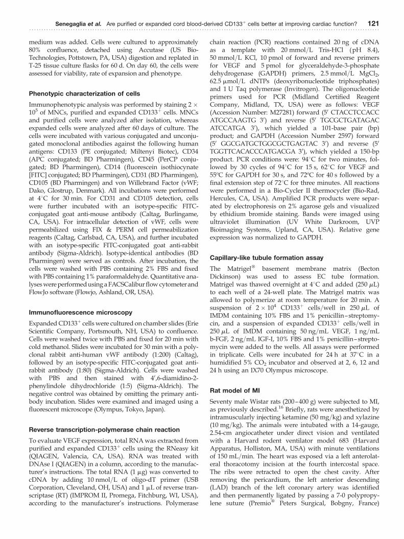
................................................................................................................................................Senegaglia et al. Are purified or expanded cord blood-derived CD133þ cells better at improving cardiac function? 121

under the LAD at the level of the distal margin of theretracted left atrial appendage. The chest wall, musclelayers and skin were closed with interrupted 3-0 silk sutures.
Cell transplantation
Rats with ejection fractions (EF) lower than 40% were ran-domly assigned to three groups: purified CD133þ cellsgroup A; expanded CD133þ cells group B and controlgroup C. Seven days after the MI, the animals were anesthe-tized and ventilated as described above. Groups A and Bwere treated with 2 � 105 cells in 0.3 mL of PBS injectedusing a tuberculin syringe with a 13 � 0.38 needle intoone point of the myocardium (left ventricle [LV]), specifi-cally in the center of the scar covering the whole infarctedand peri-infarcted area. An equal volume of PBS wasinjected into the heart of control animals.
Echocardiography analysis
Transthoracic echocardiography was performed in allanimals five days after MI (baseline echocardiogram) and30 days after the transplant. The evaluated variables wereLV ejection fraction (LVEF), LV end-systolic volume andLV end-diastolic volume. Echocardiograms were performedusing a commercially available echocardiography systemequipped with a 12-MHz phased-array transducer(Hewlett-Packard Sonos 5500; Andover, MA, USA; http://www.hp.com). Under sedation, the transducer was posi-tioned in the left anterolateral portion of the thorax, andthe heart was visualized using the two-dimensional modewith the axial view of the LV, with the mitral and aorticvalves and the apex within the same image. The LV end-diastolic and LV end-systolic volumes were calculatedusing Simpson’s rule. All measurements were averagedover three consecutive cardiac cycles and were determinedby an experienced cardiologist who was blinded to the treat-ment group. Only animals with left ventricular dysfunction(i.e. LVEF lower than 40% during the first echocardio-graphic evaluation) were included in the study. Animalswere euthanized after the second echocardiographicevaluation.
Statistical analysis
Continuous variables were presented as mean+ standarddeviation, and categorical variables were presented as fre-quency and percentage. After statistical evaluation, the out-liers, which were samples with values greater than 1.5 timesthe interquartile interval, were removed from the finalanalysis. Cell proliferation and immunophenotyping datawere both evaluated using Friedman’s non-parametrictest. The normality distribution of the data was evaluatedusing the Shapiro–Wilks test, and the homegeneity con-dition was assessed by the Levene test. Multiple compari-sons were analyzed using the LSD test, and comparisonsbetween two evaluations were assessed by the Student’st-test for paired samples. Values of P , 0.05 were consideredto be statistically significant. Analysis was performed usingthe SPSS V.14 software (SPSS, Chicago, IL, USA).
Results
Purification and phenotypical characterizationof HUCB-derived CD1331 cells
CD133þ cells were positively selected using anti-CD133-coupled magnetic microbeads. The mean number of cellsobtained after magnetic CD133þ cell sorting was 0.55 �106+ 0.04, corresponding to 0.64% of the MNC population.As determined by flow cytometry, the mean purity ofselected CD133þ cells was 74.8+ 15.82%, and the viabilitywas 80.87+ 10.55%. As shown by fluorescence-activatedcell sorting (FACS) analyses, the major contaminants inthe CD133-enriched population were cells of the hemato-poietic (CD45þ) and monocytic (CD14þ) lineages.
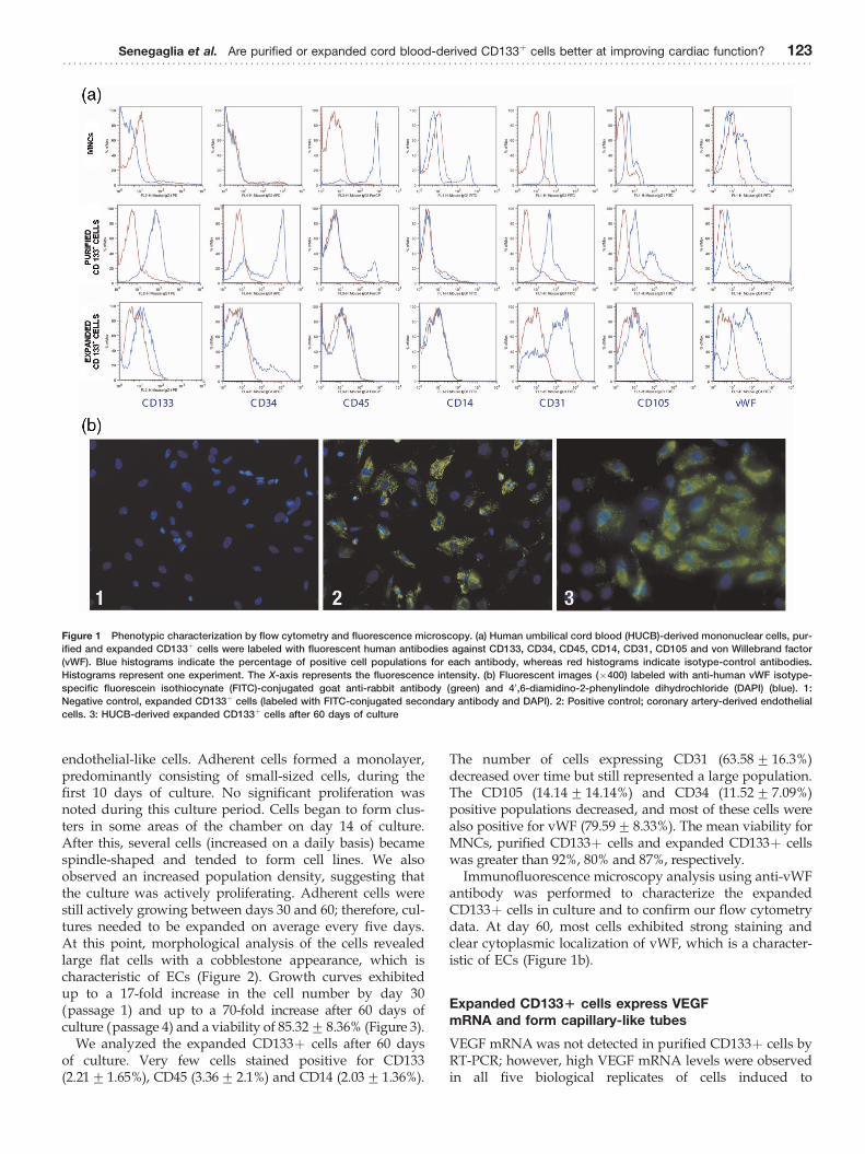
Flow cytometry analysis was performed to characterizethe cell populations contributing to the MNCs, as well asthe purified and expanded CD133þ cells. The staining pat-terns of cells for specific markers are provided in Figure 1a.The cell population profiles showed that most of the MNCsstained positive for CD45 (88.69+ 10.19%). Some cellsstained positive for CD14 (19.49+ 10.4%), CD31 (26.09+6.22%), CD105 (33.97+ 5.88%) and vWF (34.25+ 24.12%).A small number of MNCs stained positive for CD133(3.37+ 2.3%) and CD34 (3.25+ 1.97%). After the CD133purification, a large number of cells stained positive forCD133 (74.8%+ 15.82) and CD34 (62.15+ 16.09%). Agreater number of purified CD133þ cells co-stained forCD31 (86.27+ 18.37%) and CD105 (69.16+ 31.95%). Asimilar number of cells stained positive for vWF (35.24+9.78%), and a considerably reduced number were positivefor CD45 (9.03+ 5.07%) and CD14 (3.18+ 4.1%).
Proliferation assays on growthfactor-treated CD1331 cells
We evaluated the effects of using different concentrations ofthree growth factors (b-FGF, IGF-I and VEGF) on CD133þ
cell proliferation. Proliferation was assessed after five daysof treatment, which included thymidine incorporation forsix hours. Three concentrations of each growth factor wereindependently used, in addition to media with a constantconcentration of b-FGF and IGF-I and varying VEGFconcentrations. The proliferation rate was calculated bydividing the mean cell count at each growth factor concen-tration by the mean cell count of the control. The only stat-istically relevant effect for b-FGF was observed when1.0 ng/mL was added to the cultures (P ¼ 0.04). Therewere no significant differences in proliferation observedwhen various concentrations of IGF-I or VEGF were inde-pendently used. Constant concentrations of b-FGF (1.0 ng/mL) and IGF-I (2.0 ng/mL) were added along withvarying concentrations of VEGF. Incubating the cells with50 ng/mL VEGF increased their proliferation (P ¼ 0.004).
CD1331 cells actively proliferate anddifferentiate into endothelial-like cells
CD133þ cells were grown on fibronectin-coated chamberslides in the presence of VEGF, b-FGF and IGF-I to inducethe differentiation of putative purified EPCs into
................................................................................................................................................122 Experimental Biology and Medicine Volume 235 January 2010
endothelial-like cells. Adherent cells formed a monolayer,predominantly consisting of small-sized cells, during thefirst 10 days of culture. No significant proliferation wasnoted during this culture period. Cells began to form clus-ters in some areas of the chamber on day 14 of culture.After this, several cells (increased on a daily basis) becamespindle-shaped and tended to form cell lines. We alsoobserved an increased population density, suggesting thatthe culture was actively proliferating. Adherent cells werestill actively growing between days 30 and 60; therefore, cul-tures needed to be expanded on average every five days.At this point, morphological analysis of the cells revealedlarge flat cells with a cobblestone appearance, which ischaracteristic of ECs (Figure 2). Growth curves exhibitedup to a 17-fold increase in the cell number by day 30(passage 1) and up to a 70-fold increase after 60 days ofculture (passage 4) and a viability of 85.32+8.36% (Figure 3).
We analyzed the expanded CD133þ cells after 60 daysof culture. Very few cells stained positive for CD133(2.21+1.65%), CD45 (3.36+2.1%) and CD14 (2.03+1.36%).
The number of cells expressing CD31 (63.58+ 16.3%)decreased over time but still represented a large population.The CD105 (14.14+ 14.14%) and CD34 (11.52+ 7.09%)positive populations decreased, and most of these cells werealso positive for vWF (79.59+8.33%). The mean viability forMNCs, purified CD133þ cells and expanded CD133þ cellswas greater than 92%, 80% and 87%, respectively.
Immunofluorescence microscopy analysis using anti-vWFantibody was performed to characterize the expandedCD133þ cells in culture and to confirm our flow cytometrydata. At day 60, most cells exhibited strong staining andclear cytoplasmic localization of vWF, which is a character-istic of ECs (Figure 1b).
Expanded CD1331 cells express VEGFmRNA and form capillary-like tubes
VEGF mRNA was not detected in purified CD133þ cells byRT-PCR; however, high VEGF mRNA levels were observedin all five biological replicates of cells induced to
Figure 1 Phenotypic characterization by flow cytometry and fluorescence microscopy. (a) Human umbilical cord blood (HUCB)-derived mononuclear cells, pur-
ified and expanded CD133þ cells were labeled with fluorescent human antibodies against CD133, CD34, CD45, CD14, CD31, CD105 and von Willebrand factor
(vWF). Blue histograms indicate the percentage of positive cell populations for each antibody, whereas red histograms indicate isotype-control antibodies.
Histograms represent one experiment. The X-axis represents the fluorescence intensity. (b) Fluorescent images (�400) labeled with anti-human vWF isotype-
specific fluorescein isothiocynate (FITC)-conjugated goat anti-rabbit antibody (green) and 40,6-diamidino-2-phenylindole dihydrochloride (DAPI) (blue). 1:
Negative control, expanded CD133þ cells (labeled with FITC-conjugated secondary antibody and DAPI). 2: Positive control; coronary artery-derived endothelial
cells. 3: HUCB-derived expanded CD133þ cells after 60 days of culture
................................................................................................................................................Senegaglia et al. Are purified or expanded cord blood-derived CD133þ cells better at improving cardiac function? 123

differentiate. Thus far, the data suggest that CD133þ cellsare indeed EPCs, and expanded CD133þ cells areendothelial-like cells. Based on these conclusions, wecharacterized the function of expanded CD133þ cells bystudying their ability to form tubule-like structures on aMatrigel layer. Only expanded CD133þ cells, not purifiedCD133þ cells, formed a branched network of tubule-likestructures after 24 h of culture on Matrigel (Figure 4).Thus, the in vitro data indicated that expanded CD133þcells clearly exhibit EC-like characteristics.
Purified and in vitro expanded CD1331
cells improved LVEF in a rat model of MI
To confirm the in vivo functional activity of purified andexpanded HUCB-derived CD133þ cells and their potentialuse as a cell therapy, the cells were implanted in a rat modelof MI. Seventy rats were infarcted and 11 animals died afterthis procedure. Twenty-seven rats showed left ventricular dys-function (EF below 40%) in the first echocardiographic evalu-ation. Four rats died during or immediately following celltransplantation. The remaining 23 rats were divided into
Figure 2 CD133þ cell culture in the presence of growth factors (basic fibroblast growth factor, insulin-like growth factor-I and vascular endothelial growth
factor). (1) Small-sized round cells at day 5 (�100). (2) Small-sized round and flat elongated cells at day 14 (�200). (3) Cluster formation at day 15 (�200). (4)
Cell lines at day 21 (�100). (5) Proliferation enhanced at day 21 (�200). (6) Confluent cells with cobblestone appearance at day 27 (�200)
Figure 3 Proliferation rates. Eleven samples were used to evaluate the increase in cell number at three time points. P0, day 0: purified CD133þ cells were grown
on fibronectin-coated chamber slides (mean ¼ 0.19 � 106). P1, day 30: first passage; cells reached 80% confluence, and the morphological analysis showed
spindle-shaped cells without a differentiated phenotype. The cell number increased up to 17-fold (mean ¼ 1 � 106). P4, day 60: fourth passage; the morphological
analysis revealed large flat cells with a cobblestone appearance, which is characteristic of expanded endothelial cells, and the cell number increased up to 70-fold
(mean ¼ 6.23 � 106)
................................................................................................................................................124 Experimental Biology and Medicine Volume 235 January 2010

three groups: group A, purified CD133þ cells-injected rats (7rats); group B, expanded CD133þ cells-injected rats (6 rats),and group C, saline buffer-injected rats (10 rats). Three rats ingroup C died after surgery (12, 16 and 24 days). There wereno deaths in groups A and B. After statistical evaluation, theoutliers in each group were discarded. There was one outlierin group A, one in group B and two in group C.
Echocardiography measurements were applied to evaluatewhether purified and/or in vitro expanded HUCB-derivedCD133þ cells could restore cardiac function in a rat model ofMI 30 days after cell injection. Five days after MI, the LVEFmeasurements for the groups A, B and C were 22.51+7.48%, 20.75+5.42% and 25.99+5.03%, respectively. In com-parison to the second echocardiography (30 days after cellinjection), the LVEF had increased by 10.16% for group Aand by 6.8% for group B. The control group (group C) exhib-ited a 7.09% reduction in the LVEF. However, these datawere not statistically significant. At this point, the LVEFvalues for groups A, B and C were 32.67+14.37%, 27.55+5.42% and 20.46+4.05%, respectively. When we comparedthe difference between the first and second echocardiographicevaluations, we observed an improvement in the LVEF for thecell injection groups (A and B). The LVEF for group A (purifiedCD133þ cell injection) was improved, as compared with thecontrol group (P ¼ 0.001). Group B (expanded CD133þ cellinjection) also exhibited a significant improvement in theLVEF, as compared with the control group (P ¼ 0,011). Therewas no statistical difference observed between groups A andB (Figure 5).
Discussion
Neoangiogenesis within the infarcted tissue is an integralcomponent of the remodeling process; however, the capil-lary network is unable to support the increased
requirements of the hypertrophied myocardium, resultingin a progressive loss of viable tissue, infarct extension andfibrous replacement.17 Limitations of the current strategyfor inducing angiogenesis in ischemic tissues has led tothe exploration of cell therapy as a means of stimulatingneovascularization.18 – 20 Most clinical assays use autologousbone marrow-derived MNCs for revascularization celltherapy.21 – 24 In spite of bone marrow-derived MNCsshowing positive effects in the treatment of heart diseasein humans,2,25 it has been suggested that using a specificfraction of progenitor cells (such as EPCs) could be moreeffective in rescuing the ischemic-induced injured areas ofthe heart.26,27 Overall, the available clinical data indicatethat cell therapy with EPCs is safe, feasible and associatedwith improved heart function.28,29 The outcomes of clinicaltrials have been mixed and not as robust as they were inthe animal models, partly because of the conflicts regardingthe definition of human EPCs and the heterogeneity of thecell populations used in the treatments.30
However, EPCs isolated from patients with increased cardio-vascular risk factors such as hypertension, atherosclerosis,smoking, family history and diabetes mellitus, as comparedwith those isolated from healthy subjects, are reduced innumber and demonstrate increased in vitro senescence.14,31–33
In patients either at high risk for future cardiovascular eventsor who have documented coronary disease, the body’sendogenous stem cell supply and bone marrow response maybe inadequate.9 Impairment may be due to age-related dimin-ution of vascular EC number and function. This may limit theefficacy of patient-derived progenitor cells in mediating neovas-cularization;14 therefore, the use of stem cells for myocardialtissue repair might be limited among the elderly and sickbecause their cell supply is depleted and/or exhausted.34
Cord blood-derived EPCs may have a potentially power-ful therapeutic advantage in ischemic diseases, especially
Figure 4 Capillary-like tubule formation assay of human umbilical cord blood-derived CD133þ cells cultured for 24 h on a Matrigelw layer. (1) Purified cells after
24 h of culture (�40). Expanded CD133þ cells formed tubules in response to basic fibroblast growth factor, insulin-like growth factor-I and vascular endothelial
growth factor after (2) 2 h (�40); (3) 6 h (�200); (4) 12 h (�200); (5) 24 h (�40) and (6) 24 h (�100)
................................................................................................................................................Senegaglia et al. Are purified or expanded cord blood-derived CD133þ cells better at improving cardiac function? 125

MI and stroke.35 HUCB has been previously described as apotential source of EPCs,36 but little is known about theoptimal culture conditions and differentiation of thesecells.15 Previous studies have demonstrated that circulatingCD34þ cells co-expressing the CD133 maker can be isolatedfrom MNCs.6,37 Using a similar methodology, we purifiedCD133þ cells from HUCB, resulting in a cellular fractionof approximately 75% purity. The frequency of EPCs inbone marrow-derived MNCs is 0.01%.38 In this study, weobtained approximately 0.64% EPCs from HUCB-derivedMNCs, confirming that HUCB is a better source for EPCs.15
After deciding on an EPC source, we attempted to estab-lish the optimal conditions for cell expansion. VEGF wasone of the growth factors chosen for cultivation, because ithas been established to be involved in the first steps ofhemoangioblast differentiation and is associated withfaster endothelium growth.20 VEGF could also be importantin stimulating growth in relation to neovascularization,39,40
as it is considered to be an important mitogen for ECs.41
FGF is a regulator of proliferation; it influences ECgrowth, differentiation and migration both in vivo andin vitro.39 ECs cultivated without b-FGF undergo apopto-sis.42 In clinical trials, co-administration of VEGF andb-FGF led to a faster and more efficient neovascularizationthan if either growth factor was independently used. Ithas been proposed that these growth factors promote thedifferentiation of EPCs into EC-like cells in vitro. IGF-I wasalso chosen, as it is a mitogen that regulates cellular prolifer-ation and differentiation.43
There is no consensus regarding the optimal dose for stemcell administration.44 Extrapolating from the data availableon the injected EPC number required to obtain therapeuticefficacy in mice,37 the number of EPCs required for clinicaltherapy in humans would exceed the output of most pub-lished protocols.15 We expanded CD133þ cells in the pres-ence of three growth factors, b-FGF, IGF-I and VEGF.
Cellular proliferation assays obtained with each growthfactor showed that the optimal and most statistically signifi-cant concentration of b-FGF was 1.0 ng/mL. Different IGF-Iand VEGF concentrations were not statistically significantwhen individually used. When VEGF was analyzed withco-administration of b-FGF and IGF-I (at a constant concen-tration), a VEGF dose of 50 ng/mL was found to be the mosteffective and statistically significant. Eighty milliliters ofcord blood, a volume that can be routinely extracted fromone cord, yields about 6.23 � 106 EPCs after expansion.According to Forraz et al.,45 HUCB-derived EPCs exhibit ahigh proliferative capacity and also demonstrate rapid self-renewal. We found this same result in our study. Growthcurves showed up to a 17-fold increase on day 30, andcells had the typical morphology of undifferentiated EPCs.After this point and until the fourth passage (approximately60 days), the cell population increased up to 70-fold, andthey acquired similar morphological and functional pheno-types to ECs, suggesting that the cells lost expression of EPCmarkers. At this point, the cells continued to proliferate atleast until the fourth passage, suggesting that a few undif-ferentiated cells were still present. The cell numberincreased by 17-fold at P1, so at this point, these cellscould be clinically used, since there is a reasonablenumber of undifferentiated EPCs that have been passagedfor a relatively short period of time.
EPCs are subdivided into two groups: early and lateEPCs. Both can induce neovascularization in animals andcan synergistically interact with each other.46 Early EPCsappear within 4–7 days of in vitro culture; they are spindle-shaped and express CD14.14,47 Late EPCs develop after 2–3weeks of culture; they have a cobblestone pattern, are veryproliferative and do not express CD14.48 Our culture wasmaintained for 60 days, and the cells continued to signifi-cantly expand without any signs of senescence. EC-likecolonies appeared around the third week, and they had
Figure 5 Echocardiographic evaluation. (a) Comparison of the left ventricle ejection fraction (LVEF) in two evaluations: echo 1, five days after myocardial infarc-
tion (white box); and echo 2, 30 d after cell transplantation (striped box). (b) Differences in the LVEF between echocardiography evaluations 1 and 2, comparing
the groups in pairs and all together
................................................................................................................................................126 Experimental Biology and Medicine Volume 235 January 2010

the appearance of a cobblestone monolayer. These resultswere consistent with those reported by Ingram and col-leagues,49 i.e. the expanded cell population was character-istic of late EPCs.
We used flow cytometry to characterize the cell pheno-type. Immunophenotyping of MNCs showed the typicalphenotype for the hematopoietic lineage, i.e. cells werepositive for CD4550 and CD14, which is characteristic ofmonocytes.51 There were practically no cells positive forCD13352 – 54 and CD34.55 Some cells expressed both endo-thelial and hematopoietic markers56 and were CD31þ .57
We also observed the presence of CD105-positive cells,which is characteristic of HUCB-derived cells and proliferat-ing ECs.58 Purified EPCs expressed predominantly CD133,CD34, CD31 and CD105. There was a small percentage ofcontamination from hematopoietic lineage cells (CD45þ).The cells reached confluence after four weeks of cultureand displayed a typical EC phenotype, including vWFexpression59 and CD31 enhancement. We observed areduction in CD34þ and CD105þ cells in the population;however, some cells remained, which was consistent withthe previous study by Gross and Herbrig.38 Our results pre-sented a similar loss of CD133 cells, as previously describedby Shmelkov and colleagues.60 EPCs adopted a moremature endothelial phenotype during their in vitro cultureand expansion, which was consistent with a previousstudy.15
We evaluated the functional phenotype of CD133þ cells.One important finding is that CD133þ cells only form capil-lary tubes and express VEGF after induction of differen-tiation, suggesting that only these cells have the potentialto induce vascular regeneration. Samadja and colleagues61
have demonstrated that bone morphogenetic proteins 2and 4, which play a key role in endothelial colony-formingcell commitment and outgrowth during neovascularization,were selectively expressed by late EPCs but not by early andcirculating EPCs.
During in vitro expansion, stem cells experience a longreplicative history; thus, they are subject to damage fromintracellular and extracellular insults.62 Therefore, expandedand purified CD133þ cells could show different biologicalproperties when transplanted in vivo. We decided to testboth populations in terms of their ability to repair myo-cardial tissue after inducing MI in a rat model.
The rats that received purified CD133þ cells (Group A)and expanded CD133þ cells (Group B) showed a significantimprovement in their LVEF, as compared with the controlgroup. The smaller improvement observed for Group B, ascompared with Group A, may be related to the lower pro-liferation rate of these differentiated cells. The high prolifer-ation rate of purified CD133þ cells suggests that these cellswould continue to proliferate after injection into the heart.Despite the ventricular enlargement, the results suggestthat LV systolic dilation is reduced. Leor and colleagues11
obtained similar results using 1.2–2 � 106 purifiedHUCB-derived CD133þ cells intravenously injected sevendays after permanent coronary artery ligation in athymicnude rats. The cells showed functional recovery as therewas abrogation of scar thinning and LV systolic dilation.We used approximately 6–10 times less cells in our
experiments and obtained the same improvement. Thedirect infusion of the cells into the myocardial scar couldexplain our results.
The therapeutic effects of EPCs have been recently demon-strated in preclinical studies of hindlimb ischemia,15,63
strokes64 and MI.11 One disadvantage of using HUCB-derived EPCs in clinical trials is the inherent risk of rejection.Previous studies suggest that HUCB-derived cells are notvery immunogenic,65–67 so these cells may be a useful sourcefor cell therapy. In addition, a previous Eurocord studydemonstrated that unrelated HLA-mismatched umbilicalcord blood transplantation in children with acute leukemiaproduces comparable results to HLA-matched transplantationwith other stem cell sources.68 However, the reduced immuno-genicity of HUCB cells is unproven and warrants further inves-tigation. According to Cho and colleagues,69 umbilical cordtissue derived cells are major histocompatibility complex(MHC) class I dim and negative for MHC class II. A singleinjection of MHC-mismatched UTC-derived cells did notinduce a detectable immune response. However, when thesecells were injected into an inflamed region, repeatedly injectedinto the same region, or stimulated with interferon g prior toinjection, they were immunogenic. The reported successes ofCD133þ and mesenchymal cell allograft-xenografts in animalmodels encouraged further experiments to test the efficacyof clinical-grade HUCB CD133þ cells in promoting cardiacregeneration using more accurate immunogenicity studies.70
In summary, the cells from HUCB-compatible MHC donorsin cord blood banks have the potential for therapeuticapplication.
In conclusion, we purified and expanded CD133þ cellsin vitro up to 70-fold. After 60 days of cultivation, the cellswere functionally active in vitro and expressed cellulardifferentiation markers. We observed a significant improve-ment in the LVEF and attenuation of ventricular remodelingafter transplantation of the purified and expanded HUCB-derived CD133þ cells, suggesting that both purified andexpanded cells are promising candidates for use in cellularcardiomyoplasty. However, further pre-clinical trialsshould be performed to elucidate whether expandedCD133þ cells have any clinical advantages over purifiedCD133þ cells.
ACKNOWLEDGEMENTS
This work was supported by grants from the Ministry ofHealth of Brazil and CNPq (552233/2005-6). We wouldlike to thank Marco Stimamiglio for assisting us with thestatistical analysis and Dr Vivian Ferreira do Amaral forsupervising the harvesting of human umbilical cordbloods. The authors have no competing financial interests.
REFERENCES
1 Pitt B, Remme W, Zannad F, Neaton J, Martinez F, Roniker B, Bittman R,Hurley S, Kleiman J, Gatlin M. Eplerenone, a selective aldosteroneblocker, in patients with left ventricular dysfunction after myocardialinfarction. N Engl J Med 2003;348:1309–21
2 Lee MS, Lill M, Makkar RR. Stem cell transplantation in myocardialinfarction. Rev Cardiovasc Med 2004;5:82–98
................................................................................................................................................Senegaglia et al. Are purified or expanded cord blood-derived CD133þ cells better at improving cardiac function? 127

3 Dimmeler S, Zeiher AM, Schneider MD. Unchain my heart: the scientificfoundations of cardiac repair. J Clin Invest 2005;115:572–83
4 Kawamoto A, Iwasaki H, Kusano K, Murayama T, Oyamada A, Silver M,Hulbert C, Gavin M, Hanley A, Ma H, Kearney M, Zak V, Asahara T,Losordo DW. CD34-positive cells exhibit increased potency and safetyfor therapeutic neovascularization after myocardial infarction comparedwith total mononuclear cells. Circulation 2006;114:2163–9
5 Asahara T, Murohara T, Sullivan A, Silver M, van der ZR, Li T,Witzenbichler B, Schatteman G, Isner JM. Isolation of putative progenitorendothelial cells for angiogenesis. Science 1997;275:964–7
6 Peichev M, Naiyer AJ, Pereira D, Zhu Z, Lane WJ, Williams M, Oz MC,Hicklin DJ, Witte L, Moore MA, Rafii S. Expression of VEGFR-2 andAC133 by circulating human CD34(þ) cells identifies a population offunctional endothelial precursors. Blood 2000;95:952–8
7 Orlic D, Kajstura J, Chimenti S, Jakoniuk I, Anderson SM, Li B, Pickel J,McKay R, Nadal-Ginard B, Bodine DM, Leri A, Anversa P. Bone marrowcells regenerate infarcted myocardium. Nature 2001;410:701–5
8 Smadja DM, Cornet A, Emmerich J, Aiach M, Gaussem P. Endothelialprogenitor cells: characterization, in vitro expansion, and prospects forautologous cell therapy. Cell Biol Toxicol 2007;23:223–39
9 Goldberg JL, Laughlin MJ, Pompili VJ. Umbilical cord blood stem cells:implications for cardiovascular regenerative medicine. J Mol Cell Cardiol2007;42:912–20
10 Hu CH, Li ZM, DU ZM, Zhang AX, Yang DY, Wu GF. Human umbilicalcord-derived endothelial progenitor cells promote growthcytokines-mediated neorevascularization in rat myocardial infarction.Chin Med J (Engl) 2009;122:548–55
11 Leor J, Guetta E, Feinberg MS, Galski H, Bar I, Holbova R, Miller L, ZarinP, Castel D, Barbash IM, Nagler A. Human umbilical cord blood-derivedCD133þ cells enhance function and repair of the infarcted myocardium.Stem Cells 2006;24:772–80
12 Cortes-Morichetti M, Frati G, Schussler O, Van Huyen JP, Lauret E,Genovese JA, Carpentier AF, Chachques JC. Association between acell-seeded collagen matrix and cellular cardiomyoplasty for myocardialsupport and regeneration. Tissue Eng 2007;13:2681–7
13 Iwaguro H, Asahara T. Endothelial progenitor cell culture and genetransfer. Methods Mol Med 2005;112:239–47
14 Hill JM, Finkel T, Quyyumi AA. Endothelial progenitor cells andendothelial dysfunction. Vox Sang 2004;87(Suppl. 2):31–7
15 Eggermann J, Kliche S, Jarmy G, Hoffmann K, Mayr-Beyrle U, DebatinKM, Waltenberger J, Beltinger C. Endothelial progenitor cell culture anddifferentiation in vitro: a methodological comparison using humanumbilical cord blood. Cardiovasc Res 2003;58:478–86
16 Etzion S, Battler A, Barbash IM, Cagnano E, Zarin P, Granot Y, KedesLH, Kloner RA, Leor J. Influence of embryonic cardiomyocytetransplantation on the progression of heart failure in a rat model ofextensive myocardial infarction. J Mol Cell Cardiol 2001;33:1321–30
17 Kocher AA, Schuster MD, Szabolcs MJ, Takuma S, Burkhoff D, Wang J,Homma S, Edwards NM, Itescu S. Neovascularization of ischemicmyocardium by human bone-marrow-derived angioblasts preventscardiomyocyte apoptosis, reduces remodeling and improves cardiacfunction. Nat Med 2001;7:430–6
18 Li TS, Hamano K, Hirata K, Kobayashi T, Nishida M. The safety andfeasibility of the local implantation of autologous bone marrow cells forischemic heart disease. J Card Surg 2003;18(Suppl. 2):S69–75
19 Masuda H, Asahara T. Post-natal endothelial progenitor cellsfor neovascularization in tissue regeneration. Cardiovasc Res 2003;58:390–8
20 Menasche P. Cell transplantation in myocardium. Ann Thorac Surg2003;75:S20–8
21 Britten MB, Abolmaali ND, Assmus B, Lehmann R, Honold J, Schmitt J,Vogl TJ, Martin H, Schachinger V, Dimmeler S, Zeiher AM. Infarctremodeling after intracoronary progenitor cell treatment in patients withacute myocardial infarction (TOPCARE-AMI): mechanistic insights fromserial contrast-enhanced magnetic resonance imaging. Circulation2003;108:2212–8
22 Meyer GP, Wollert KC, Lotz J, Steffens J, Lippolt P, Fichtner S, Hecker H,Schaefer A, Arseniev L, Hertenstein B, Ganser A, Drexler H.Intracoronary bone marrow cell transfer after myocardial infarction:eighteen months’ follow-up data from the randomized, controlledBOOST (BOne marrOw transfer to enhance ST-elevation infarctregeneration) trial. Circulation 2006;113:1287–94
23 Stamm C, Westphal B, Kleine HD, Petzsch M, Kittner C, Klinge H,Schumichen C, Nienaber CA, Freund M, Steinhoff G. Autologousbone-marrow stem-cell transplantation for myocardial regeneration.Lancet 2003;361:45–6
24 Strauer BE, Brehm M, Zeus T, Kostering M, Hernandez A, Sorg RV,Kogler G, Wernet P. Repair of infarcted myocardium by autologousintracoronary mononuclear bone marrow cell transplantation in humans.Circulation 2002;106:1913–8
25 Kim EJ, Li RK, Weisel RD, Mickle DA, Jia ZQ, Tomita S, Sakai T, YauTM. Angiogenesis by endothelial cell transplantation. J Thorac CardiovascSurg 2001;122:963–71
26 Iwami Y, Masuda H, Asahara T. Endothelial progenitor cells: past, stateof the art, and future. J Cell Mol Med 2004;8:488–97
27 Kalka C, Masuda H, Takahashi T, Kalka-Moll WM, Silver M, Kearney M,Li T, Isner JM, Asahara T. Transplantation of ex vivo expandedendothelial progenitor cells for therapeutic neovascularization. Proc NatlAcad Sci USA 2000;97:3422–7
28 Bartunek J, Vanderheyden M, Vandekerckhove B, Mansour S, De BruyneB, De Bondt P, Van Haute I, Lootens N, Heyndrickx G, Wijns W.Intracoronary injection of CD133-positive enriched bone marrowprogenitor cells promotes cardiac recovery after recent myocardialinfarction: feasibility and safety. Circulation 2005;112:I178–83
29 Losordo DW, Schatz RA, White CJ, Udelson JE, Veereshwarayya V,Durgin M, Poh KK, Weinstein R, Kearney M, Chaudhry M, Burg A,Eaton L, Heyd L, Thorne T, Shturman L, Hoffmeister P, Story K, Zak V,Dowling D, Traverse JH, Olson RE, Flanagan J, Sodano D, Murayama T,Kawamoto A, Kusano KF, Wollins J, Welt F, Shah P, Soukas P, Asahara T,Henry TD. Intramyocardial transplantation of autologous CD34þ stemcells for intractable angina: a phase I/IIa double-blind, randomizedcontrolled trial. Circulation 2007;115:3165–72
30 Mund JA, Ingram DA, Yoder MC, Case J. Endothelial progenitorcells and cardiovascular cell-based therapies. Cytotherapy 2009;11:103–13
31 Hristov M, Weber C. Endothelial progenitor cells: characterization,pathophysiology, and possible clinical relevance. J Cell Mol Med2004;8:498–508
32 Vasa M, Fichtlscherer S, Aicher A, Adler K, Urbich C, Martin H, ZeiherAM, Dimmeler S. Number and migratory activity of circulatingendothelial progenitor cells inversely correlate with risk factors forcoronary artery disease. Circ Res 2001;89:E1–7
33 Fritzenwanger M, Lorenz F, Jung C, Fabris M, Thude H, Barz D, FigullaHR. Differential number of CD34 þ , CD133þ and CD34 þ /CD133þcells in peripheral blood of patients with congestive heart failure. Eur JMed Res 2009;14:113–7
34 Scheubel RJ, Zorn H, Silber RE, Kuss O, Morawietz H, Holtz J, Simm A.Age-dependent depression in circulating endothelial progenitor cells inpatients undergoing coronary artery bypass grafting. J Am Coll Cardiol2003;42:2073–80
35 El-Badri NS. Endothelial progenitor cells from cord blood: a newtherapeutic promise? Stem Cells Dev 2005;14:237–8
36 Murohara T, Ikeda H, Duan J, Shintani S, Sasaki K, Eguchi H, Onitsuka I,Matsui K, Imaizumi T. Transplanted cord blood-derived endothelialprecursor cells augment postnatal neovascularization. J Clin Invest2000;105:1527–36
37 Gehling UM, Ergun S, Schumacher U, Wagener C, Pantel K, Otte M,Schuch G, Schafhausen P, Mende T, Kilic N, Kluge K, Schafer B, HossfeldDK, Fiedler W. In vitro differentiation of endothelial cells fromAC133-positive progenitor cells. Blood 2000;95:3106–12
38 Gross P, Herbrig K. Role of endothelial progenitor cells in cardiovascularpathology. Rocz Akad Med Bialymst 2004;49:174–7
39 Peters KG, Werner S, Chen G, Williams LT. Two FGF receptor genes aredifferentially expressed in epithelial and mesenchymal tissues duringlimb formation and organogenesis in the mouse. Development1992;114:233–43
40 Rivard A, Silver M, Chen D, Kearney M, Magner M, Annex B, Peters K,Isner JM. Rescue of diabetes-related impairment of angiogenesis byintramuscular gene therapy with adeno-VEGF. Am J Pathol1999;154:355–63
41 Shen BQ, Lee DY, Cortopassi KM, Damico LA, Zioncheck TF. Vascularendothelial growth factor KDR receptor signaling potentiates tumornecrosis factor-induced tissue factor expression in endothelial cells. J BiolChem 2001;276:5281–6
................................................................................................................................................128 Experimental Biology and Medicine Volume 235 January 2010

42 Hartnett ME, Garcia CM, D’Amore PA. Release of bFGF, an endothelialcell survival factor, by osmotic shock. Invest Ophthalmol Vis Sci1999;40:2945–51
43 Zumstein P, Stiles CD. Molecular cloning of gene sequences thatare regulated by insulin-like growth factor I. J Biol Chem 1987;262:11252–60
44 Henning RJ, bu-Ali H, Balis JU, Morgan MB, Willing AE, Sanberg PR.Human umbilical cord blood mononuclear cells for the treatment ofacute myocardial infarction. Cell Transplant 2004;13:729–39
45 Forraz N, Pettengell R, Deglesne PA, McGuckin CP. AC133þ umbilicalcord blood progenitors demonstrate rapid self-renewal and lowapoptosis. Br J Haematol 2002;119:516–24
46 Yoon CH, Hur J, Park KW, Kim JH, Lee CS, Oh IY, Kim TY, Cho HJ,Kang HJ, Chae IH, Yang HK, Oh BH, Park YB, Kim HS. Synergisticneovascularization by mixed transplantation of early endothelialprogenitor cells and late outgrowth endothelial cells: the role ofangiogenic cytokines and matrix metalloproteinases. Circulation2005;112:1618–27
47 Hur J, Yoon CH, Kim HS, Choi JH, Kang HJ, Hwang KK, Oh BH, LeeMM, Park YB. Characterization of two types of endothelial progenitorcells and their different contributions to neovasculogenesis. ArteriosclerThromb Vasc Biol 2004;24:288–93
48 Ingram DA, Mead LE, Tanaka H, Meade V, Fenoglio A, Mortell K, PollokK, Ferkowicz MJ, Gilley D, Yoder MC. Identification of a novel hierarchyof endothelial progenitor cells using human peripheral and umbilicalcord blood. Blood 2004;104:2752–60
49 Ingram DA, Mead LE, Moore DB, Woodard W, Fenoglio A, Yoder MC.Vessel wall-derived endothelial cells rapidly proliferate because theycontain a complete hierarchy of endothelial progenitor cells. Blood2005;105:2783–6
50 Trowbridge IS, Thomas ML. CD45: an emerging role as a proteintyrosine phosphatase required for lymphocyte activation anddevelopment. Annu Rev Immunol 1994;12:85–116
51 Goyert SM, Ferrero E, Rettig WJ, Yenamandra AK, Obata F, Le BeauMM. The CD14 monocyte differentiation antigen maps to a regionencoding growth factors and receptors. Science 1988;239:497–500
52 Miraglia S, Godfrey W, Yin AH, Atkins K, Warnke R, Holden JT, BrayRA, Waller EK, Buck DW. A novel five-transmembrane hematopoieticstem cell antigen: isolation, characterization, and molecular cloning.Blood 1997;90:5013–21
53 Rehman J, Li J, Orschell CM, March KL. Peripheral blood ‘endothelialprogenitor cells’ are derived from monocyte/macrophages and secreteangiogenic growth factors. Circulation 2003;107:1164–9
54 Ribatti D. The involvement of endothelial progenitor cells in tumorangiogenesis. J Cell Mol Med 2004;8:294–300
55 Sutherland DR, Keating A. The CD34 antigen: structure, biology, andpotential clinical applications. J Hematother 1992;1:115–29
56 Garcia-Porrero JA, Manaia A, Jimeno J, Lasky LL, eterlen-Lievre F,Godin IE. Antigenic profiles of endothelial and hemopoietic lineages inmurine intraembryonic hemogenic sites. Dev Comp Immunol1998;22:303–19
57 Newman PJ. The biology of PECAM-1. J Clin Invest 1997;100:S25–9
58 Duff SE, Li C, Garland JM, Kumar S. CD105 is important forangiogenesis: evidence and potential applications. FASEB J2003;17:984–92
59 Hoyer LW. The factor VIII complex: structure and function. Blood1981;58:1–13
60 Shmelkov SV, Jun L, St CR, McGarrigle D, Derderian CA, Usenko JK,Costa C, Zhang F, Guo X, Rafii S. Alternative promoters regulatetranscription of the gene that encodes stem cell surface protein AC133.Blood 2004;103:2055–61
61 Smadja DM, Bieche I, Silvestre JS, Germain S, Cornet A, Laurendeau I,Duong-Van-Huyen JP, Emmerich J, Vidaud M, Aiach M, Gaussem P.Bone morphogenetic proteins 2 and 4 are selectively expressed by lateoutgrowth endothelial progenitor cells and promote neoangiogenesis.Arterioscler Thromb Vasc Biol 2008;28:2137–43
62 Lepperdinger G. Aging stem cells and regenerative biomedicine:concepts, opportunities and technological advances. Exp Gerontol2008;43:967
63 Yamaguchi J, Kusano KF, Masuo O, Kawamoto A, Silver M, MurasawaS, Bosch-Marce M, Masuda H, Losordo DW, Isner JM, Asahara T.Stromal cell-derived factor-1 effects on ex vivo expanded endothelialprogenitor cell recruitment for ischemic neovascularization. Circulation2003;107:1322–8
64 Borlongan CV, Hadman M, Sanberg CD, Sanberg PR. Central nervoussystem entry of peripherally injected umbilical cord blood cells is notrequired for neuroprotection in stroke. Stroke 2004;35:2385–9
65 Goldstein G, Toren A, Nagler A. Transplantation and other uses ofhuman umbilical cord blood and stem cells. Curr Pharm Des2007;13:1363–73
66 Laham RJ, Oettgen P. Bone marrow transplantation for the heart: fact orfiction? Lancet 2003;361:11–2
67 Rocha V, Wagner JE Jr, Sobocinski KA, Klein JP, Zhang MJ, HorowitzMM, Gluckman E. Graft-versus-host disease in children who havereceived a cord-blood or bone marrow transplant from an HLA-identicalsibling. Eurocord and International Bone Marrow Transplant RegistryWorking Committee on Alternative Donor and Stem Cell Sources. N EnglJ Med 2000;342:1846–54
68 Locatelli F, Rocha V, Chastang C, Arcese W, Michel G, Abecasis M,Messina C, Ortega J, Badell-Serra I, Plouvier E, Souillet G, Jouet JP,Pasquini R, Ferreira E, Garnier F, Gluckman E. Factors associated withoutcome after cord blood transplantation in children with acuteleukemia. Eurocord-Cord Blood Transplant Group. Blood1999;93:3662–71
69 Cho PS, Messina DJ, Hirsh EL, Chi N, Goldman SN, Lo DP, Harris IR,Popma SH, Sachs DH, Huang CA. Immunogenicity of umbilical cordtissue derived cells. Blood 2008;111:430–8
70 Bonanno G, Mariotti A, Procoli A, Corallo M, Rutella S, Pessina G,Scambia G, Mancuso S, Pierelli L. Human cord blood CD133þ cellsimmunoselected by a clinical-grade apparatus differentiate in vitro intoendothelial- and cardiomyocyte-like cells. Transfusion 2007;47:280–9
(Received June 24, 2009, Accepted November 3, 2009)
................................................................................................................................................Senegaglia et al. Are purified or expanded cord blood-derived CD133þ cells better at improving cardiac function? 129