Comportamento termo-higro-mecânico de elementos em betão: análise experimental e simulação numérica
Upload
khangminh22Category
view
0download
0
1
Resumo
O termo meningioma foi utilizado pela primeira vez por Harvey Cushing em 1922, para
descrever tumores com origem nas meninges do cérebro e da coluna vertebral. O vasto
espectro de apresentação clínica e do prognóstico deste tipo de tumores é reconhecido
desde 1938, quando foram descritas variantes associadas a sobrevivências inferiores a 2.5
anos.1,2 Apesar do progresso alcançado com novas técnicas de diagnóstico, classificação
histológica, técnicas cirúrgicas melhoradas e terapêuticas adjuvantes, o tratamento
contemporâneo do meningioma ainda não inclui linhas orientadoras reconhecidas a nível
internacional. De facto, as opções terapêuticas para este tipo de doentes, especialmente a
recomendação de radioterapia adjuvante no meningioma atípico é um dos tópicos mais
discutidos desta área.3 Por este motivo, a análise de fatores prognósticos associados a uma
maior taxa de recorrência do tumor, morbilidade e mortalidade é vital para selecionar os
doentes que poderão beneficiar de um plano de tratamento mais agressivo. Este estudo
teve como objetivo realizar uma análise descritiva do tratamento de meningiomas atípicos
num centro neurocirúrgico terciário. Adicionalmente, analisou o impacto dos fatores
relacionados com a cirurgia, terapêutica adjuvante, histologia e com a história do doente
na sobrevivência livre de doença (SLD) e na sobrevivência total (ST). Os resultados
obtidos estão de acordo com a literatura existente e confirmam a importância da resseção
total (RT) na SLD. Aos 5 anos após a cirurgia, os doentes que foram submetidos a
resseção subtotal (RS) registavam uma SLD de 35.7%, aproximadamente metade da
registada pelos doentes com RT (SLD de 68.8%, log-rank:0.047, Breslow:0.033). Parece
existir um efeito benéfico associado à radioterapia adjuvante nos doentes com RS, tendo
estes registado uma SLD aos 5 anos de 66.7%, comparada com valores de 29.3% nos
doentes que não receberam terapêutica adjuvante (log-rank:0.262, Breslow: 0.122). Esta
associação não foi observada para o grupo de doentes submetidos a RT. A análise do
grupo que manifestou recidiva tumoral identificou como fatores de risco a idade
(p=0.033), história de meningioma atípico prévio (p=0.012), cirurgia cerebral prévia
(p=0.014), invasão dos seios venosos e do córtex (p=0.018 e p=0.002), RS (p=0.009) e
graus elevados de edema (p=0.041). Os resultados deste estudo corroboram a abordagem
cirúrgica com o intuito de RT como objetivo primário no tratamento do meningioma
atípico. Caso a resseção tumoral máxima não seja possível deverá ser considerado o uso
de radioterapia adjuvante. O papel desta terapêutica adjuvante após RT mantem-se
controverso.
Palavras-chave: meningioma atípico, extensão da resseção, radioterapia adjuvante.

2
Abstract
The term meningioma was coined in 1922 by Harvey Cushing to describe masses arising
in the meninges of the brain and spinal cord. The wide spectrum of presentation and
clinical outcome of these tumours has been recognized from as early as 1938, when
variants associated with survival rates of 2.5 years were described.1,2 Despite the progress
in diagnostic techniques, histological classification, new surgical approaches and
adjuvant therapies, the contemporary management of meningioma patients still lacks
clear and internationally validated guidelines. In fact the treatment options for these
patients, particularly the use of adjuvant radiotherapy in atypical meningioma (AM),
remains one of the most discussed topics in this area.3 For this reason, the study of
prognostic factors associated with increased tumour recurrence, morbidity and mortality
is vital for selecting patients who may benefit from an aggressive treatment plan from
those who are not likely to. This study aimed to provide a descriptive analysis of the
treatment of atypical meningioma at a tertiary neurosurgical centre. Furthermore, it
evaluated the impact of the surgical outcome and adjuvant treatment as well as the
histological and patients’ related factors in progression free survival (PFS) and overall
survival (OS). The results obtained are consistent with the existing literature and confirm
the importance of gross total resection (GTR) for improved progression free survival. The
5 year PFS for the patients who received subtotal resection (STR) was 35.7%, which was
approximately half of the values registered for patients who received GTR surgeries (5
year PFS of 68.8%; log-rank of 0.047 and Breslow of 0.033). There seems to be a benefit
in recommending adjuvant radiotherapy in patients who underwent STR, with a 5 year
PFS of 66.7% in this group compared with 29.3% in the STR only group (log-rank of
0.262 and Breslow of 0.122). This association was not seen for GTR patients. The analysis
of the recurrence group identified older age (p=0.033), previous grade II meningioma
(p=0.012), previous brain surgery (p=0.014), venous sinus and cortex invasion (p=0.018
and p=0.002), STR (p=0.009) and higher grades of oedema (p=0.041) as recurrence risk
factors. The evidence from this study supports GTR as a primary goal in the management
of atypical meningioma patients. If safe maximal resection in not possible adjuvant
radiotherapy should be considered. The role of this adjuvant treatment following GTR
remains controversial.
Key words: atypical meningioma, extent of resection, adjuvant radiotherapy

3
Contents
List of abbreviations and units.......................................................................................... 4
I. Introduction ............................................................................................................... 5
II. Aims of the Study ...................................................................................................... 7
III. Methods ................................................................................................................. 8
IV. Results ................................................................................................................. 11
V. Discussion ............................................................................................................... 23
VI. Conclusions ......................................................................................................... 32
VII. Acknowledgments ............................................................................................... 33
VIII. References ........................................................................................................... 34
Appendix ........................................................................................................................ 40

4
List of abbreviations and units
AM – atypical meningioma
cm – centimetre
CNS – central nervous system
CSF – cerebralspinal fluid
CT – computerized tomography
EBRT – external beam radiation therapy
e.g. – exempli gratia
EOR – extent of resection
EORTC - European Organisation for Research and Treatment of Cancer
GTR – gross total resection
Gy - Gray
HPF – high power fields
MRI – magnetic resonance imaging
OS – overall survival
PFS – progression free survival
ROAM - Radiotherapy versus Observation following surgical resection of Atypical
Meningioma
SRS – stereotactic radiosurgery
STR – subtotal resection
UK – United Kingdom
WHO – World Health Organization

5
I. Introduction
Meningiomas are the most frequently diagnosed primary central nervous system (CNS)
tumours in adults, comprising approximately one third of all cases.4 They are more
common in women than in men and they particularly affect middle aged and elderly
patients.5 Even though the majority of the cases are sporadic, meningiomas may also be
present in hereditary syndromes, from which the best documented is neurofibromatosis
type 2. Recognized and suggested risk factors include radiation exposure, sexual
hormones, head trauma, familiar history of benign brain tumours and occupational and
dietary causes.6–8 These tumours originate in the arachnoid cap cells that assemble the
outer layer of the arachnoid mater and the arachnoid villi, being in 90% of the cases
intracranial. The vast majority are located in the falx and parasagittal region (25%), in the
convexity (19%) and along the sphenoid ridge (17%). Other common locations are the
suprasellar region (9%), the olfactory groove (8%), the posterior fossa (8%) and the
middle fossa (4%). Less frequently meningiomas can be found in the peri-torcular region,
in the lateral ventricles, in the foramen magnum and in the orbit or optic nerve sheath.9
Most symptoms are insidious, depending on the tumour location and resulting from the
compression of adjacent structures. Thus, the presentation can range from easily
recognizable signs and symptoms such as headache, seizure, paresis or visual field
deficits to more subjective alterations, for instance personality disorders.10,11 Magnetic
Resonance Imaging (MRI) and Computer Tomography (CT) with the application of
contrast medium are the methods of choice for the neuroradiological evaluation of the
tumour (Figure 1).12
As a group, meningiomas show more histological variants than any other tumour and
although its classification is complex, it assumes a pivotal role in the disease management.
The World Health Organization (WHO) classification of central nervous system tumours,
divides meningiomas into three grades: grade I (benign), grade II (atypical) and grade III
(malignant)/(Figure 2).13,14 Atypical meningiomas represent 5 to 15% of all meningiomas,
with the histological subgroups “atypical”, “chordoid” and “clear cell” in descending
frequency. In general, atypical meningiomas are associated with higher recurrence rates
(30-40%) and increased morbidity and mortality compared to benign meningiomas.15
Currently the treatment options for atypical meningioma remain controversial. The range
includes watchful waiting, surgery, stereotactic radiosurgery (SRS), radiotherapy (more
recently ion beam radiotherapy) and combinations thereof. Chemotherapy and other

6
biologic therapies are reserved for selected cases.1 Total surgical resection of the tumour
and invaded structures is the current gold standard, however, this is not always
anatomically possible, especially for tumours located in the skull base.16 The extent of
the surgical resection can be classified according to the Simpson grading system,
proposed by Donald Simpson in 1957. Similarly to histological grading, this classification
is also a risk stratification method that correlates closely with recurrence.17
Regarding radiotherapy, there is no consensus on recommendation as a standard adjuvant
therapy to surgery in atypical meningioma, particularly after GTR. Authors have
suggested that the use of radiotherapy immediately after surgical resection could decrease
the recurrence rate and thus result in a better outcome. Recent meta-analysis and
systematic reviews have revealed that this might be correct for the local control of the
disease, particularly in cases of subtotal resection, however there is no impact in overall
survival.1,18–20 Although the side-effects of radiotherapy have decreased in the last
decades, they are still responsible for considerable morbidity (3.4-16.7% of AM patients)
and consequently the risk-benefit ratio should be carefully considered.20
a. b. c.
d. e. f.
Figure 1 –Meningioma imaging with CT scan and MRI. a. sphenoid ridge meningioma on CT scan
(axial); b. same lesion on T2 MRI (axial); c. cystic meningioma with dura tail (black arrow) on T1 MRI
(axial); d. meningioma surrounding the right optic nerve on T1 MRI (axial) ; e. en plaque meningioma
with hyperostotic focus on T1 MRI (sagittal); f. spinal cord meningioma at the T5 level narrowing the
spinal canal on T2 MRI (sagittal) / (images from12,21)

7
Figure 2: WHO 2007 Meningioma grading system. The histological images correspond to meningothelial
(grade I), atypical (grade II) and anaplastic tumours (grade III). In immunochemistry analysis all grades
of meningioma are positive for vimentin stain, grade I meningiomas are more commonly positive for
epithelial membrane antigen (EMA) stain and secretory meningiomas are positive for carcinoembryonic
antigen (CEA) stain (adapted from8,22,23).
II. Aims of the Study
The main problem for the current management of meningioma, particularly atypical
meningioma, is to predict which patients are more prone to recurrence.24 The
identification of prognostic factors is of vital importance since these allow recognizing
which patients benefit from a more aggressive treatment plan or require a closer follow
up. One of the main discussed topics in meningioma management is related to the
recommendation of radiotherapy in atypical meningioma following GTR or STR and its
role in prevention of recurrence.
This study aims to retrospectively analyse the management of patients with atypical
meningiomas at a tertiary neurosurgical centre and to determine the impact of surgical,
histological, adjuvant treatment and patient related factors in the progression of the
disease.

8
III. Methods
Patient selection
This retrospective cohort study includes the analysis of all grade II meningioma patients
who underwent surgery at the Neurosurgery Department of the General Hospital of
Vienna in a period of 12 years (January 2002 to December 2013). To evaluate the impact
of previous treatments on tumour progression and clinical outcome, we included primary
and recurrent atypical meningiomas, as well as recurrent grade II meningiomas after grade
I meningioma. Other inclusion criteria were the patients’ age at the time of the surgery
(≥18 years) and the existence of a neuropathology report confirming the grade II histology
according to the WHO 2000 or 2007 diagnostic criteria.
Source of information and study approval
All required information was retrieved from the patients’ clinical files, surgical reports,
histological reports, discharge letters and neuroradiology images stored at the
Neurosurgery Department at the Medical University of Vienna. For confidentiality
purposes, patient data was anonymized and only authorized personnel had access to the
original files. The ethics committee from this institution approved this study, which was
performed according to the standards of the Declaration of Helsinki.
Variables assessed
The data gathered for each patient included age, gender, previous grade I or grade II
meningioma, previous brain surgery, neurofibromatosis type 2 diagnosis, radiation
exposure history and existing comorbidities. Due to the highly variable location of
meningiomas and their broad range of clinical manifestations, several features were
considered for the presenting signs and symptoms, notably: headache, pain, vertigo,
paresis, sensibility disorder, visual impairment, hearing deficit, disturbance of taste or
smell, exophthalmus, swelling, seizure, aphasia, change of personality, disturbance of
consciousness and neurogenic bowel and/or bladder dysfunction.
The location, number, diameter (in cm) of the tumour and the presence of perifocal
oedema was obtained from the neuroradiology images and/or from the surgical report.
The exact location of the tumours was categorized into the following groups: spinal,

9
convexity, falx, tentorium, sphenoid or sphenoorbital, orbital, olfactory, frontobasal,
parasagittal, petroclival or clival, falcotentorial, intraventricular, middle fossa floor,
cerebellar falx or multiple. In order to facilitate the statistical analysis, these locations
were further grouped into convexity/falx/tentorium, skull base, spinal, intraventricular
and multiple. In cases of multiple tumours, the size of the biggest tumour was preferred.
The presence of oedema was semiquantitatively classified into grades: mild, moderate
and severe.
The analysis of the surgical treatment included the assessment of invasion of adjacent
structures such as bone, venous sinus, arachnoid layer and/or cortex. The Simpson
classification was also reassessed and subdivided into gross total resection (GTR –
Simpson grades I and II) and subtotal resection (STR – Simpson III and IV) / (Figure 3).25
For the histological analysis, tumours were subcategorized according to the WHO 2000
and 2007 classification into atypical, chordoid and clear cell. From these histological
reports the MIB-1 number (Ki-67 index) and mitotic index (number of mitosis per 10
high power fields) was also recorded when present.
The length of hospitalization and the peri-operative complications were also studied. The
latter were subclassified into infection, CNS haemorrhage, non-CNS haemorrhage, CNS
ischaemia, non-CNS ischaemia, ventricular system disorder, neurologic deficit,
psychiatric disorder and death. The Clavien Dindo complication scale was also used.26
The adjuvant therapies after primary surgery included external beam radiation therapy
(EBRT) and chemotherapy and for these we noted the start date, the radiation dose and
the chemotherapy scheme.
During the follow up we noted tumour recurrence with the following characteristics:
clinical presentation, location, size, type of meningioma spread (local and cerebrospinal
fluid (CSF) spread), treatment type and histological grade. Recurrence was defined as the
date at which the patient received treatment for the recurrent tumour or in cases of
watchful waiting the date of the neuroradiology diagnosis of the recurrent tumour.
Treatment options for the recurrent meningioma included surgery, SRS, EBRT,
chemotherapy (with Imatinib, Glivec®) or any combination of these. Finally, a status
variable was assigned to each patient as stable, progressing, meningioma related death,
non-meningioma related death or unknown cause of death.

10
Figure 3 – Simpson resection grades and division according to gross total resection (Simpson I and II) and
subtotal resection (Simpson III and IV)
Statistical analysis
The primary question of this study was to test if there was a difference in overall and
progression-free survival grouped by specific parameters. Kaplan-Meier curves based on
the extent of resection were used for this purpose together with log rank and Breslow
tests. Additionally, the same calculations were done for GTR and STR according to the
fact whether patients received postoperative radiotherapy or not. Quantitative variables
were assessed for normality with Shapiro-Wilk tests. Comparisons between groups were
then performed with Student test and Mann Whitney test, as applicable. The association
between categorical variables was assessed resorting to Chi-square-tests or Fischer exact
tests, resorting to Monte Carlo simulations when needed.27 The statistical analysis was
performed using IBM® SPSS® Software version 22. The level of significance adopted
was α=0.05.

11
IV. Results
Patient and tumour characteristics
Of 140 patients that met the inclusion criteria of this study, 62% were female and 38%
were male, with a mean age at the surgical intervention of 56 years (range 18 to 82 years).
Of those, 18.7% harboured recurrent meningiomas (10.1% had previous grade I
meningioma, 7.2% had previous grade II meningioma and 1.4% had both previous grade
I and II meningiomas). Overall, 22.1% received previous brain surgery at some point of
their life, 7.1% had a history of radiation exposure and only one patient had a confirmed
diagnosis of neurofibromatosis type 2. The most frequently encountered comorbidities
were cardiovascular and metabolic (e.g. diabetes and dyslipidaemia), followed by
thrombotic, oncologic, psychiatric and neurologic disorders (Table 1 appendix).
The majority of patients was symptomatic (88.6%) and the most common symptoms were
headache (39%), visual impairment (29%), paresis (22%) and seizures (22%) / (Table 2
appendix). The mean diameter of the meningiomas was 4.18±1.92 cm (range 1.00 to 9.00
cm). Preferential locations were sphenoid or sphenoorbital (24.5%), convexity (21.6%),
parasagittal (17.3%) and falx (8.6%). Multiple meningiomas were found in 10% of cases
and the presence of oedema was verified in 50% of the cases.
The main histological subgroup was atypical (84.3%), followed by the chordoid and clear
cell variants in 12.9% and 2.9% of the cases respectively. The mitotic index, when
available, was ≥5 in 83% of the cases and it was ≥8 in 10% of the cases. The MIB-1
number was obtained for 40 cases, with a mean value of 13.14±5.42% (range 4.2 to
28.0%) / (Table 3 appendix).
Treatment characteristics
Gross total resection (Simpson grades I and II) was achieved in 78% of the cases.
Adjacent structure invasion was a frequent finding with 59.3% of the tumours invading
the arachnoid, 28.6% the cortex, 25.0% the bone and 17.9% a venous sinus.
The mean hospital stay was 15±13.27 days (range 4 - 138 days), with a complication rate
of 21%, the majority being related to neurological disorders (e.g. hemiparesis, aphasia,
vision-field deficits, Jacksonian seizures), ventricular system disorders (e.g. CSF fistula
and hydrocephalus) and CNS haemorrhage. During the hospital stay two of these patients
died due to severe brain oedema and haemorrhage (Table 4 appendix).

12
External beam radiation therapy (EBRT) was given to 10% of the patients with a mean
dose of 55±9.59 Gy (range 24.0-60.0 Gy). One patient received adjuvant chemotherapy
with Imatinib (Glivec®). No complications following adjuvant treatment were noted.
Recurrence and follow up characteristics
For this part of the study, 24 patients with no follow up were excluded, as well as the 2
patients who died during the primary hospital stay. From these patients 26.3% had tumour
recurrence during the follow up, with 16.7% registering one tumour recurrence and 9.6%
presenting more than one tumour recurrence. At recurrence, the majority of the cases
were asymptomatic (60%) and the mean tumour diameter was 2.82±1.36 cm (range 0.70-
6.60). In one third of the cases there were multiple tumours. The most common locations
were the convexity (13.3%) and the sphenoid or sphenoorbital regions (13.3%). In half
of the cases the recurrence resulted from local spread of initial tumour, while the other
half occurred due to CSF spread. Treatments of recurrent tumours included surgery, SRS,
EBRT, chemotherapy, watchful waiting and combined treatment strategies (Table 5
appendix, Figure 4).
Figure 4 – Pattern of recurrence and treatment options

13
Regarding the patients’ status at the end of the follow up period, 86.9% were stable, 3.5%
were progressing and 9.6% were deceased. The cause of death was related to the
meningioma in 3.5% of the cases, it was attributable to other causes in 2.6% of the cases
and it was unknown in the remaining 3.5% of the patients. Three patients (10% of the
recurrent tumours) underwent malignant transformation to grade III meningioma (Table
6 appendix).
Tumour precursors: primary grade II tumours and recurrent grade II tumours
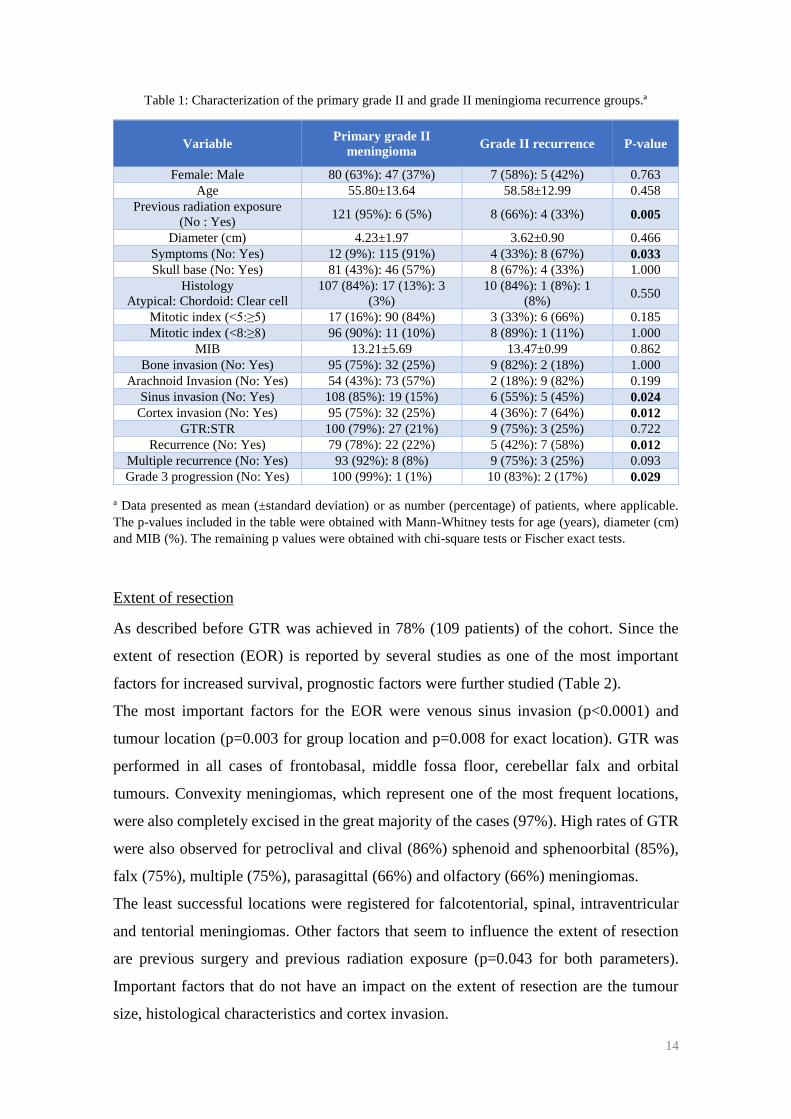
We further analysed cases depending on primary grade II tumour or grade II meningioma
recurrence. This analysis revealed that the recurrence group had significantly more
commonly previous radiation exposure (p=0.005), was less symptomatic at presentation
(p=0.033) and had more sinus and cortex invasion (p=0.024 and p=0.012). They were
also more prone to recurrence (p=0.012) and they showed a higher progression rate to
grade III meningiomas (p=0.029). They also seem to have a greater probability of
multiple recurrence, even though this parameter did not reach statistical significance
(Figure 5 and Table 1).
Figure 5: Comparison between primary grade II meningioma and grade II recurrence groups
0%
20%
40%
60%
80%Sinus invasion
Cortex invasion
Recurrence
Multiple recurrence
Grade III progression
Radiation exposure
Primary grade II meningioma Grade II recurrence
14
Table 1: Characterization of the primary grade II and grade II meningioma recurrence groups.ª
Variable Primary grade II
meningioma Grade II recurrence P-value
Female: Male 80 (63%): 47 (37%) 7 (58%): 5 (42%) 0.763
Age 55.80±13.64 58.58±12.99 0.458
Previous radiation exposure
(No : Yes) 121 (95%): 6 (5%) 8 (66%): 4 (33%) 0.005
Diameter (cm) 4.23±1.97 3.62±0.90 0.466
Symptoms (No: Yes) 12 (9%): 115 (91%) 4 (33%): 8 (67%) 0.033
Skull base (No: Yes) 81 (43%): 46 (57%) 8 (67%): 4 (33%) 1.000
Histology
Atypical: Chordoid: Clear cell
107 (84%): 17 (13%): 3
(3%)
10 (84%): 1 (8%): 1
(8%) 0.550
Mitotic index (<5:≥5) 17 (16%): 90 (84%) 3 (33%): 6 (66%) 0.185
Mitotic index (<8:≥8) 96 (90%): 11 (10%) 8 (89%): 1 (11%) 1.000
MIB 13.21±5.69 13.47±0.99 0.862
Bone invasion (No: Yes) 95 (75%): 32 (25%) 9 (82%): 2 (18%) 1.000
Arachnoid Invasion (No: Yes) 54 (43%): 73 (57%) 2 (18%): 9 (82%) 0.199
Sinus invasion (No: Yes) 108 (85%): 19 (15%) 6 (55%): 5 (45%) 0.024
Cortex invasion (No: Yes) 95 (75%): 32 (25%) 4 (36%): 7 (64%) 0.012
GTR:STR 100 (79%): 27 (21%) 9 (75%): 3 (25%) 0.722
Recurrence (No: Yes) 79 (78%): 22 (22%) 5 (42%): 7 (58%) 0.012
Multiple recurrence (No: Yes) 93 (92%): 8 (8%) 9 (75%): 3 (25%) 0.093
Grade 3 progression (No: Yes) 100 (99%): 1 (1%) 10 (83%): 2 (17%) 0.029
a Data presented as mean (±standard deviation) or as number (percentage) of patients, where applicable.
The p-values included in the table were obtained with Mann-Whitney tests for age (years), diameter (cm)
and MIB (%). The remaining p values were obtained with chi-square tests or Fischer exact tests.
Extent of resection
As described before GTR was achieved in 78% (109 patients) of the cohort. Since the
extent of resection (EOR) is reported by several studies as one of the most important
factors for increased survival, prognostic factors were further studied (Table 2).
The most important factors for the EOR were venous sinus invasion (p<0.0001) and
tumour location (p=0.003 for group location and p=0.008 for exact location). GTR was
performed in all cases of frontobasal, middle fossa floor, cerebellar falx and orbital
tumours. Convexity meningiomas, which represent one of the most frequent locations,
were also completely excised in the great majority of the cases (97%). High rates of GTR
were also observed for petroclival and clival (86%) sphenoid and sphenoorbital (85%),
falx (75%), multiple (75%), parasagittal (66%) and olfactory (66%) meningiomas.
The least successful locations were registered for falcotentorial, spinal, intraventricular
and tentorial meningiomas. Other factors that seem to influence the extent of resection
are previous surgery and previous radiation exposure (p=0.043 for both parameters).
Important factors that do not have an impact on the extent of resection are the tumour
size, histological characteristics and cortex invasion.

15
Table 2: Analysis of factors influencing the extent of resection and its effect in the postoperative period ª
Variable GTR STR P-value
Female : Male 71 (65%): 38 (35%) 16 (52%): 15 (48%) 0.171
Age 56.08±13.31 56.14±14.53 0.802
Previous grade I meningioma (No: Yes) 98 (90%); 11 (10%) 25 (83%): 5 (17%) 0.338
Previous grade II meningioma (No: Yes) 100 (92%): 9 (8%) 27 (90%): 3 (10%) 0.722
Previous brain surgery (No : Yes) 89 (82%): 20 (18%) 20(%): 11 (%) 0.043
Previous radiation exposure (No : Yes) 104 (95%): 5(5%) 25(83%): 5 (17%) 0.043
Falx/Convexity/Tentorium : Skull base :
Other
58 (53%): 43 (40%):
8 (7%)
15(48%): 7 (23%): 9
(29%) 0.003
Skull base (No: Yes) 66 (61%): 43 (39%) 24 (77%): 7 (23%) 0.084
Location exact
Convexity 29 (97%) 1 (3%)
0.008
Sphenoid/
Sphenoorbital 29 (85%) 5 (15%)
Petroclival/clival 6 (86%) 1 (14%)
Falx 9 (75%) 3 (25%)
Multiple 8 (75%) 6 (25%)
Parasagital 16 (66%) 8 (33%)
Olfactory 2 (66%) 1 (33%)
Frontobasal 3 (100 %) 0 (0%)
Middle fossa floor 2 (100%) 0 (0%)
Cerebellum falx 1 (100%) 0 (0%)
Orbital 1 (100%) 0 (0%)
Tentorium 1 (33%) 2 (66%)
Intraventricular 1 (33%) 2 (66%)
Spinal 0 (0%) 1 (100%)
Falcotentorial 0 (0%) 1 (100%)
Diameter (cm) 4.18±1.98 4.16±1.70 0.771
Oedema (No: Yes) 45 (43%): 59 (57%) 16 (59%): 11 (41%) 0.138
Oedema
No oedema 45 (74%) 16 (26%)
0.149
Mild 39 (87%) 6 (13%)
Moderate 17 (85%) 3 (15%)
Severe 3 (60%) 2 (40%)
No info 5 (56%) 4 (44%)
Bone invasion (No: Yes) 80 (74%): 28 (26%) 24 (77%): 7 (23%) 0.705
Arachnoid Invasion (No: Yes) 46 (43%): 62 (57%) 10 (32%): 21 (68%) 0.301
Sinus invasions (No: Yes) 99 (92%): 9 (8%) 15 (48%): 16 (52%) <0.0001
Cortex invasion (No: Yes) 79 (73%): 29 (27%) 20 (65%): 11 (35%) 0.349
Histology:
Atypical: Chordoid: Clear Cell
91 (83%): 16 (15%):
2 (2%)
27 (87%): 2 (6%): 2
(6%) 0.192
MIB 12.94±5.12 13.65±6.40 0.858
Mitotic index (<5 : ≥5) 19 (21%): 72 (79%) 1 (4%): 25 (96%) 0.043
Mitotic index (<8 : ≥8) 81 (89%): 10 (11%) 24 (92%): 2 (8%) 1.000
Surgical complications (No: Yes) 85 (78%): 24 (22%) 26 (84%): 5 (16%) 0.475
Hospital stay 15.34±14.47 16.13±7.89 0.176
a Data presented as mean (±standard deviation) or as number (percentage) of patients, where applicable.
The p-values included in the table were obtained with Mann-Whitney tests for age (years), diameter (cm),
MIB (%) and radiation dose (Gy). The remaining p values were obtained with chi-square tests or Fischer
exact tests.

16
The analysis of the incidence of surgical complications following GTR revealed that the
EOR is not associated with additional complications or a longer hospital stay (p=0.475
and 0.176 respectively). Nevertheless, more serious complications such as death, CNS
ischaemia and CNS haemorrhage were exclusively found in this group (Figure 6).
Figure 6 – Surgical complications according to the extent of resection (GTR and STR). ªx axis: number of
patients; y axis: complication types
Adjuvant Radiotherapy
Here we seek to assess which factors influenced the decision of recommending adjuvant
radiotherapy after surgical resection. The ensuing analysis included age, gender, previous
meningioma and irradiation history, tumour size, location, presence of oedema, adjacent
structure invasion, histological characteristics and its resection outcome. Some
differences can be observed between the patients undergoing adjuvant radiotherapy and
those without it in terms of previous grade II meningioma, cortex and venous sinus
invasion, Simpson’s grades III and IV and a mitotic index ≥5, even though statistical
significance was not attained (Figure 7 and Table 3).
Patients receiving adjuvant radiotherapy had both similar recurrence and multiple
recurrence rates when compared to patients who did not receive this treatment. Of note,
all patients who have progressed to grade III meningioma did not receive adjuvant
radiotherapy, however this parameter did not reach statistical significance (Table 4).
0 1 2 3 4 5 6 7
non-CNS hemorrhage
psychiatric disorder
death
CNS ischaemia
non-CNS ischaemia
CNS haemorrhage
ventricular system disorder
neurological disorder
GTR STR

17
Figure 7 – Factors related with the recommendation of adjuvant radiotherapy
Table 3: Factors related with the recommendation of adjuvant radiotherapy ª
Variable Adjuvant radiotherapy No adjuvant
radiotherapy P-value
Female: Male 9 (64%): 5 (36%) 78 (62%): 48 (38%) 0.862
Age 61.53±13.31 55.49±13.48 0.096
Previous grade II meningioma
(No:Yes) 11 (79%): 3 (21%) 116 (93%): 9 (7%) 0.104
Previous radiation exposure 12 (86%): 2 (14%) 118 (94%): 8 (6%) 0.262
Diameter (cm) 3.99±1.60 4.20±1.95 0.850
Falx/Convexity/Tentorium : Skull
base : Other 6 (43%): 6 (43%): 2 (14%)
67 (53%): 44 (35%):
15 (12%) 0.322
Skull base (No: Yes) 8 (57%): 6 (43%) 82 (65%): 44 (35%) 0.557
Oedema (No: Yes) 5 (42%): 7 (58%) 56 (47%): 63 (53%) 0.721
Histology - Atypical: Chordoid:
Clear cell 14 (100%): 0 (0%): 0 (0%)
104 (83%): 18 (14%):
4 (3%) 0.245
Mitotic index (<5 : ≥5) 0 (0%): 14(100%) 20 (19%): 83 (81%) 0.124
Mitotic index (<8 : ≥8) 13 (93%): 1 (7%) 92 (89%): 11 (11%) 1.000
MIB 15.07±3.72 12.98±5.55 0.492
Bone invasion (No: Yes) 9 (69%):4 (31%) 95 (75%): 31 (25%) 0.738
Arachnoid Invasion (No: Yes) 4 (31%): 9 (69%) 52 (41%): 74 (59%) 0.561
Sinus invasions (No: Yes) 13 (100%): 0 (0%) 101 (80%): 25 (20%) 0.067
Cortex invasion (No: Yes) 6 (46%): 7 (54%) 93 (74%): 33 (26%) 0.052
GTR:STR 9 (64%): 5 (36%) 100 (80%): 26 (20%) 0.194 a Data presented as mean (±standard deviation) or as number (percentage) of patients, where applicable.
The p-values included in the table were obtained with Mann-Whitney tests for age (years), diameter (cm)
and MIB (%). The remaining p values were obtained with chi-square tests or Fischer exact tests.
0%
20%
40%
60%
80%
100%Simpson III and IV
Cortex invasion
Mitotic index ≥5
OedemaSkull base
Previous grade II
meningioma
Previous radiation
exposure
Adjuvant radiotherapy No adjuvant radiotherapy

18
Table 4: Adjuvant radiotherapy and meningioma recurrence / progression ª
Variable Adjuvant radiotherapy No adjuvant
radiotherapy P-value
Recurrence (No: Yes) 10 (71%): 4 (29%) 74 (74%): 26 (26%) 1.000
Multiple recurrence (No: Yes) 13 (93%): 1 (7%) 90 (90%): 10 (10%) 1.000
Grade III progression 14 (100%): 0 (0%) 97 (97%): 3 (3%) 1.000
a Data presented as number (percentage) of patients, where applicable. The p-values were obtained with
chi-square tests or Fischer exact tests.
Recurrence analysis
This analysis aimed to identify factors associated with higher recurrence rates. Therefore,
parameters related to the patients’ history, tumour and surgical procedure were
considered. The most important were venous sinus and cortex invasion (p=0.018 and
p=0.002), STR (p=0.009) and the grades of oedema (p=0.041). From the patients’ history,
older age (p=0.033), previous grade II meningioma (p=0.012) and previous brain surgery
(p=0.014) were statistically significant. Previous radiation exposure (p=0.052) presented
borderline results (Figure 8 and Table 5).
Figure 8: Risk factors distribution in recurrence group and no recurrence group
0%
20%
40%
60%
80%Simpson III and IV
Previous brain surgery
Previous grade II
meningioma
Cortex invasionSinus invasion
Previous radiation
exposure
Oedema
Recurrence No recurrence

19
Table 5: Analysis of risk factors for recurrence ª
Variable Recurrence No Recurrence P-value
Female : Male 17 (57%): 13 (43%) 52 (62%): 32(38%) 0.614
Age 59.03±12.56 54.07±13.27 0.033
Previous grade I meningioma
(No: Yes) 27 (93%); 2 (7%) 71 (85%): 13 (15%) 0.347
Previous grade II meningioma
(No: Yes) 22 (76%): 7 (24%) 79 (94%): 5 (6%) 0.012
Previous brain surgery (No : Yes) 18 (60%): 12 (40%) 69 (82%): 15 (18%) 0.014
Previous radiation exposure
(No : Yes) 25 (83%): 5 (17%) 80 (95%): 4 (5%) 0.052
Falx/Convexity/Tentorium :
Skull base : Other
13 (43%): 10 (33%): 7
(23%)
43 (51%): 32 (38%): 9
(11%) 0.232
Skull base (No: Yes) 20 (66%): 10 (33%) 52 (62%): 32 (38%) 0.643
Diameter (cm) 4.83±2.24 4.03±1.75 0.116
Oedema (No: Yes) 7 (28%): 18 (72%) 40 (49%): 41 (51%) 0.060
Oedema
No oedema 7 (23%) 40 (48%)
0.041
Mild 11 (37%) 28 (33%)
Moderate 6 (20%) 10 (12%)
Severe 1 (3%) 3 (4%)
No info 5 (17%) 3 (4%)
Bone invasion (No: Yes) 21 (72%): 8 (28%) 60 (71%): 24 (29%) 0.919
Arachnoid Invasion (No: Yes) 10 (33%): 19 (66%) 35 (42%): 49 (58%) 0.496
Sinus invasions (No: Yes) 19 (66%): 10 (34%) 72 (86%): 12 (14%) 0.018
Cortex invasion (No: Yes) 14 (48%): 15 (52%) 66 (79%): 18 (21%) 0.002
Simpson
I 12 (40%) 45 (54%)
0.050 II 6 (20%) 25 (30%)
III 7 (23%) 6 (7%)
IV 5 (17%) 8 (9%)
GTR:STR 18 (60%): 12 (40%) 70 (83%): 14 (17%) 0.009
Histology:
Atypical: Chordoid: Clear Cell
26 (87%): 3 (10%): 1
(3%)
71 (85%): 12 (14%): 1
(1%) 0.779
MIB 13.36±5.74 13.30±5.34 0.606
Mitotic index (<5 : ≥5) 5 (%): 21 (%) 10 (15%): 58 (85%) 0.753
Mitotic index (<8 : ≥8) 24 (92%): 2 (8%) 60 (88%): 8 (12%) 0.721
Adjuvant radiotherapy (No: Yes) 26 (87%): 4 (13%) 74 (88%): 10 (12%) 1.000
Radiation dose (Gy) 48.40±16.99 57.94±2.79 0.539
Surgical complications (No: Yes) 22 (73%): 8 (27%) 68 (81%): 16 (19%) 0.436
a Data presented as mean (±standard deviation) or as number (percentage) of patients, where applicable.
The p-values included in the table were obtained with Mann-Whitney tests for age (years), diameter (cm),
MIB (%) and radiation dose (Gy). The remaining p values were obtained with chi-square tests or Fischer
exact test.

20
Survival analysis: extent of resection and adjuvant radiotherapy
The cohort mean progression free survival was 77.66±6.29 months (median: 81.00; 95%
confidence interval 65.32-89.93) and the mean overall survival was 123.67±5.62 months
(median: 138.00; 95% confidence interval: 112.66-134.68)/(Figure 9). The mean follow
up was 46.12±37.87 months (median: 38.00; range 1 to 144 months).
Kaplan Maier analysis demonstrated an increased PFS in GTR compared to STR, with
statistically significant log rank and Breslow tests (p=0.047 and p=0.033). The PFS for
GTR at 1, 3 and 5 years was 92.0% (±3.1), 80.6% (±5.2) and 68.8% (±6.6) respectively,
while for STR these values decreased to 84.0% (±7.3), 53.6% (±11.2) and 35.7% (±12.7)
for the same time intervals. This association was not verified for the OS in which GTR
and STR showed similar values (Figure 10 and Table 6).
In order to reduce potential bias, the analysis for the effect of adjuvant EBRT was divided
according to the EOR. For GTR, this adjuvant treatment did not show any survival
benefit. 5-year PFS and OS were 71.6% and 93.5% respectively for GTR alone and 32.8%
and 65.6% for GTR with EBRT (Figure 11 and Table 7).
In STR, the opposite association was obtained, with patients who received adjuvant
EBRT showing longer PFS and OS rates when compared to the STR alone group, even
though this did not reach statistical significance. The 5-year PFS was 66.7% and 29.3%
respectively for the STR with adjuvant EBRT group and for the STR only group. The OS
was 100.0% and 84.7% for the same groups (Figure 12 and Table 8).
Figure 9 – Cohort progression free survival and overall survival. A: progression free survival, B: overall
survival. x axis: time in months, y axis: proportion of patients.

21
Figure 10 – Effect of extent of resection in PFS and OS. A: progression free survival, B: overall survival.
x axis: time in months, y axis: proportion of patients.
Table 6: Effect of extent of resection in PFS and OS
Survival GTR
(n=88)
STR
(n=26)
PFS
1 year 92.0% ±3.1 84.0% ±7.3
3 year 80.6% ±5.2 53.6% ±11.2
5 year 68.8% ±6.6 35.7% ±12.7
Log-rank 0.047
Breslow 0.033
OS
1 year 96.2% ±2.1 96.2% ±3.8
3 year 90.9%±3.6 96.2% ±3.8
5 year 90.9% ±3.6 88.1% ±8.4
Log-rank 0.473
Breslow 0.775
Figure 11: Effect of adjuvant EBRT in patients with GTR. A: progression free survival, B: overall
survival. x axis: time in months, y axis: proportion of patients.

22
Table 7: Graph 5: Effect of adjuvant EBRT in patients with GTR
Survival Adjuvant EBRT
(n=79)
No adjuvant EBRT
(n=9)
PFS
1 year 65.6% ±20.9 94.2% ±2.8
3 year 65.6% ±20.9 81.8% ±5.4
5 year 32.8% ±25.4 71.6% ±6.7
Log-rank 0.059
Breslow 0.042
OS
1 year 87.5% ±11.7 97.2% ±1.9
3 year 65.6% ±20.9 93.5% ±3.2
5 year 65.6% ±20.9 93.5% ±3.2
Log-rank 0.081
Breslow 0.037
Figure 12: Effect of adjuvant EBRT in patients with STR. A: progression free survival, B: overall
survival. x axis: time in months, y axis: proportion of patients.
Table 8: Effect of adjuvant EBRT in patients with STR
Survival Adjuvant EBRT
(n=5)
No adjuvant EBRT
(n=21)
PFS
1 year 100.0% 81.0% ±8.6
3 year 66.7% ±27.2 43.9% ±12.4
5 year 66.7% ±27.2 29.3% ±12.5
Log-rank 0.262
Breslow 0.122
OS
1 year 100.0% 95.2% ±4.6
3 year 100.0% 95.2% ±4.6
5 year 100.0% 84.7% ±10.8
Log-rank 0.450
Breslow 0.486

23
V. Discussion
Summary of the study
This study analysed a cohort of 140 patients with histologically confirmed WHO grade II
meningioma. Mean age at surgical intervention was 56 years and 62% of these patients
were female. This data is congruent with the epidemiological description of such tumours
although some series report a male gender predominance for non-benign meningiomas.28
History of previous grade I and/or II meningiomas was present in 18.7% of the patients,
previous brain surgery in 22.1% and history of radiation exposure in 7.1%. The fact that
some patients (8.6%) had previous grade II meningioma allowed the differentiation of
primary and recurrent grade II meningioma subgroups. This analysis revealed that
recurrence patients had more history of previous irradiation (p=0.005) were less
symptomatic (p=0.033) and had more adjacent structure invasion (p=0.024 for venous
sinus and p=0.012 for cortex). They also registered increased recurrence rates (p=0.012)
when compared with primary tumours.
For the primary surgical intervention, the vast majority of patients were symptomatic and
presented with a wide range of symptoms, being the most frequent headache, visual
impairment and seizures. This is corroborated by the variable tumour location, with the
sphenoid or sphenoorbital, convexity and falx regions affected in more than half of the
cases. Multiple meningiomas were present in 10% of the cases.
In 78% of the patients GTR was attained even though adjacent structure invasion was
rather frequent. The analysis of factors limiting the EOR revealed that venous sinus
invasion (p<0.0001) and tumour location (p=0.003) were the most important parameters.
The mean tumour diameter was 4.18±1.92 cm and this was not associated with decreased
EOR. Mean hospital stay and complication rates were not affected by an aggressive
operative strategy, with similar values reported in GTR and STR approaches. The overall
intra-hospital mortality rate was 1.4%. The Kaplan Meier analysis reiterated the
importance of complete resection in progression-free survival, with 5 year PFS of 68.8%
for GTR and 35.7% for STR with a statistical significance for the log-rank and Breslow
tests (p=0.047 and p=0.033).
Adjuvant therapy options at this stage were EBRT (for 14 patients) and chemotherapy
with Imatinib (for 1 patient), with no associated complications. The recommendation of
adjuvant EBRT improved both PFS and OS for the STR group without reaching statistical
significance, most probably due to the reduced number of patients in this arm of the study.

24
In contrast, adjuvant radiotherapy did not show an improvement in PFS or OS in GTR
patients. In fact, an inverse relation was seen with shorter PFS and OS in the group
receiving adjuvant radiotherapy. Since the patients were not randomized, further analysis
was performed in order to determine if there were additional factors contributing to this
restriction. All GTR patients who received radiotherapy had meningiomas of the atypical
histological subgroup and they did not show features of increased malignity such as
higher MIB-1 or mitotic index. They also did not show any statistically significant
differences regarding the patients’ profile or tumour size or location. However, this group
of patients had considerably more history of previous grade II meningioma (p=0.026) and
also more cortex invasion (p=0.031) when compared to the non-irradiated GTR group.
Also, there was an increase in post-surgical complications in this group, with
approximately one half of the patients presenting some sort of complication (4 out of 9
patients: 2 with ventricular system disorders (hydrocephalus and CSF fistula), 1 with
neurological disorder (motor aphasia) and 1 with intracranial abscess). These factors
along with the group’s small numbers might have contributed to this result. When the
equivalent analysis was performed for the STR group, there were no such differences.
In general, more than one quarter of the patients had tumour recurrence during follow up
(16.7% with one recurrence and 9.6% with multiple recurrence). At recurrence, 60% of
the patients were asymptomatic and the mean tumour diameter was smaller comparatively
to the previous episode (2.82±1.36 cm), most probably due to neuroradiological
surveillance. Half of the cases resulted from local spread while the other half occurred
due to CSF spread. In one third of the cases there were multiple locations. More complex
treatment strategies were adopted for recurrent tumours and they included surgery, SRS,
radiotherapy, chemotherapy, watchful waiting and combined treatment strategies. The
overall recurrence risk factors were also studied and from these the most important were
older age (p=0.033), previous grade II meningioma (p=0.012), previous brain surgery
(p=0.014), sinus and cortex invasion (p=0.018 and p=0.002), STR (p=0.009) and higher
grades of oedema (p=0.041).
The cohort mean progression free survival was 77.66±6.29 months (median: 81.00; 95%
confidence interval 65.32-89.93) and the mean overall survival was 123.67±5.62 months
(median: 138.00; 95% confidence interval: 112.66-134.68). The mean follow up was
46.12±37.87 months (median: 38.00; range 1 to 144 months). At the end of the follow up
period 86.9% of the patients were stable, 3.5% were progressing and 9.6% were deceased,

25
with 3.5% of the deaths attributable to meningioma. Three patients (10% of the recurrent
tumours) underwent malignant transformation to grade III meningioma.
Comparison with the existing literature
For this comparison, a Pubmed search of the English literature was performed combining
the terms “atypical meningioma”, “grade II meningioma” and “radiotherapy” (Table 9
and 10). This search only included studies with histological grading according to the
WHO 2000 or 2007 classification system and if applicable with separate analysis of
atypical and malignant meningiomas. These criteria were included because the previous
classification system from 1993 was much less specific and also because in the present
study grade III tumours were excluded to prevent bias from evaluating two types of
tumours with a very different biological behaviour.
With reference to the extent of resection, the benefit of GTR in PFS and even in OS has
been widely confirmed by several studies, regardless of the criteria used to define GTR
(from Simpson grade I only to Simpson grade I-III) / (Tables 9 and 10). This factor seems
to be the most important prognostic factor for survival.
The evidence obtained supports the idea that the role of adjuvant radiotherapy in atypical
meningioma is still not fully understood. In fact, the great majority of the existing studies
have not been able to demonstrate a statistically significant advantage in recommending
adjuvant radiotherapy as part of the standard treatment, particularly for GTR. The meta-
analysis from Hasan et al,18 evaluating the role of radiotherapy following GTR included
14 studies with an overall number of 757 atypical meningioma patients. They have
described a 5 year PFS of 62% for the GTR only group and 73% for the GTR and adjuvant
radiotherapy group (p=0.057), as well as an OS of 85% and 88% for the same groups
(p=0.95). Another meta-analysis from Fam and Eljamel29, with 23 studies and 1575
patients, concluded that there was a benefit in adjuvant radiotherapy following GTR with
a PFS of 73% for the GTR only group and 84% for the GTR and adjuvant radiotherapy
group (p=0.036). Because of the timeline, both meta-analysis did not took into account
one of the largest single centre studies from Sun et al,10,30 which involved 151 GTR
patients and established that there was no difference in PFS and OS in these groups of
patients (p=0.83 and p>0.99). Despite the different outcomes, these two recent meta-
analysis confirm that there is an enormous divergence between studies and that

26
prospective studies are required to elucidate this matter, expressly for adjuvant
radiotherapy after GTR.
The evaluation of the effect of adjuvant radiotherapy following STR, appears to be less
challenging and it presents more uniform results. Several studies have revealed that there
is an advantage for PFS, however this does not seem to be the case for OS. For example,
Park et al have reported a 5-year PFS of 0% for the STR only group (18 patients) and
68% for STR plus adjuvant radiotherapy group (7 patients)/(p<0.001).31 Sun et al, who
have conducted one of the largest studies evaluating the role of adjuvant radiotherapy in
STR also concluded that there was a benefit for these patients. The 5 year PFS was 30%
for the STR only group (34 patients) and 65% for the STR and adjuvant radiotherapy
group (25 patients)/(p=0.03).10 Despite this impact on PFS these studies could not show
significant improvement in OS.
In general, several authors report only the p value for these groups and do not give
information about the actuarial survival rates. This limits the comparison between studies,
even though some have reached significant p values (e.g. Champeaux and Dunn32). In
addition, other studies have assessed the effect of adjuvant radiotherapy on the overall
cohort, without further division according to the extent of resection. These studies have
reached inconsistent results with some reporting a substantial effect of adjuvant
radiotherapy on PFS and others reporting no effect at all (e.g. Zaher et al33 and Zhao et
al2).

27
Table 9 – Effect of the extent of resection and adjuvant radiotherapy in the existing literature (part 1)
Authors
No of
cases
(years)
GTR
GTR
cases
Follow
up
GTR
Follow
up
STR
Benefit
of GTR
ERBT
after
GTR
Follow
up:
GTR
only
Follow
up:
GTR+
EBRT
Effect
of
ERBT
STR
cases
EBRT
after
STR
Follow
up:
STR
only
Follow
up
STR+
EBRT
Effect
of
ERBT
Present
study
114
(02/13) I-II 88
69% 5
yrs PFS
36% 5
yrs PFS p=0.047
p=0.033 9
72% 5
yrs PFS
33% 5
yrs PFS
p=
0.059 26 5
29% 5
yrs PFS
67% 5
yrs PFS
p=
0.262
Champeaux
and Dunn,
201632
178
(00/15) I-III 142 - - p=0.01 24 - -
p<
0.001 35 12 - -
p<
0.001
Jenkinson et
al, 201634
133
(01/10) I-III 113
81% 5
yrs PFS
40% 5
yrs PFS p=0.001 32
82% 5
yrs PFS
80% 5
yrs PFS
p=
0.808 19 4 - - -
bChoy et al,
201635
221
(NA) I-II 172 - - p<0.0001 16 - -
p=
0.633c 48 14 - - p=
0.633c
Cao et al,
201636
41
(00/13) I-II 28
56% 3
yrs PFS
29% 3
yrs PFS p=0.007
Patients receiving RT: 21
PFS: p=0.427; OS: p=0.169c
ªHasan et al,
201518
757
(NA) I-III 549 - - - 208
62% 5
yrs LC
73% 5
yrs LC
p=
0.057 - - - - -
Wang et al,
201516
28
(01/09) - 14
71% 3
yr FS
36% 3
yr PFS p=0.011 3
87% 3
yrs PFS
100% 3
yrs PFS p=0.18 14 9
0% 5
yrs PFS
49% 5
yrs PFS
p=
0.074
Zhao et al,
20152
89
(01/11) I-II 72 - -
p=0.021
(OS)
Patients receiving RT: 40
PFS: p=0.442; OS: p=0.896c
Aizer, et al
201537
575
(04/09) I-III 303
91% 5
yrs OS
78% 5
yrs OS p<0.001 73 - -
p=
0.320c 272 56 - - p=
0.320c
Nowak et al,
201538
44
(00/09) I-II 44 - - - 11 - -
p=
0.079 - - - - -
ªFam and
Eljamel,
201529
1575
(NA) - 297 - - - 86
73% 5
yrs PFS
84% 5
yrs PFS p=
0.036 80 63
47% 5
yrs PFS
47% 5
yrs PFS
p=
0.966
LC: local control, PFS: progression fee survival; OS: overall survival, reported p values apply for PFS unless stated otherwise; ª Meta-analysis results; b Meta-analysis for
chordoid meningiomas; c GTR and STR results combined.

28
Table 10 -Effect of the extent of resection and adjuvant radiotherapy in the existing literature (part 2)
Authors No of
cases GTR
GTR
cases
Follow
up
GTR
Follow
up
STR
Benefit
of GTR
ERBT
after
GTR
Follow
up:
GTR
only
Follow
up:
GTR+
EBRT
Effect
of
ERBT
STR
cases
EBRT
after
STR
Follow
up:
STR
only
Follow
up
STR+
EBRT
Effect
of
ERBT
Sun et al,
201410,30
210
(93/12) I-III 151
89% 5
yr PFS
48% 5
yr PFS p<0.001 37
91% 5
yrs LC
100% 5
yrs LC p=0.53 59 25
30% 5
yrs PFS
65% 5
yrs PFS p=0.03
Hammouche
et al, 20143
79
(96/09) I 34
74% 5
yrs PFS
32% 5
yrs PFS p=0.005 9
68% 5
yrs PFS
100% 5
yrs PFS p=0.13
45
27
- - None§
Choi et al,
201439
72
(95/13) I-II 53
92% 5
yrs OS
61% 5
yrs OS p<0.001¥ 42 - -
p=0.28
(LC) 19
13
- -
p=
0.070
Aboukais et
al, 201340
167
(94/11) I-II
96
- - - 27c - -
p=
0.039c
71
27c - -
p=
0.039c
Hardesty et
al, 201341
258
(92/11) I-II 149
85% 5
yrs PFS
54% 5
yrs PFS p<0.0001 15
96% 5
yrs PFS
100% 5
yrs PFS None§ 79 20
60% 5
yrs PFS
80%5
yrs PFS p=0.55
Park et al,
201331
83
(97/11) I-II 55
59% 5
yrs PFS
30% 5
yrs PFS p=0.002 17
65% 5
yrs PFS
52% 5
yrs PFS p=0.86 25 7
0% 5
yrs PFS
68% 5
yrs PFS p
<0.001
Lee et al,
201342
90
(99/09) I-III 71
85% 5
yrs PFS
70% 5
yrs PFS p=0.007 17
65% 5
yrs PFS
74% 5
yrs PFS p=1.00 19 17
20% 5
yrs PFS
91% 5
yrs PFS p=
0.002
Zaher et al,
201333
44
(09/12) I-II
16
- - p<0.0001
Patients receiving RT: 26
PFS: p=0.007c
Mair et al.
201143
114
(01/10) I-II 66
59% 5
yrs PFS
32% 5
yrs PFS p=0.018 15 - - None§ 48 15
14% 5
yrs PFS
43% 5
yrs PFS p=0.04
Jo et al,
201044
35
(97/08) I 11 - - - 5
100% 3
yrs PFS
100% 3
yrs PFS None§ 23 16
34% 5
yrs PFS
63% 5
yrs PFS p=
0.011
LC: local control, PFS: progression fee survival; OS: overall survival, reported p values apply for PFS unless stated otherwise; c GTR and STR results combined; § exact value
was not reported; ¥Atypical and malignant meningioma combined

29
Atypical Meningioma Management – Vienna’s General Hospital Strategy
The attempt of recognizing the most common indications for recommending adjuvant
radiotherapy in this centre could not find any statistically significant factors.
Nevertheless, the tendency for this decision appeared to be based on factors such as
previous grade II meningioma, cortex invasion and extent of resection (STR). These
results reflect the lack of clear indications or protocols for this treatment option and the
fact that this decision is the result of the healthcare team’s best knowledge.
The first step of the management strategy is to achieve GTR and particularly Simpson
grade I resections. Nevertheless, as mentioned before, this is not always possible due to
anatomic limitations. If the tumour is accessible and can be removed in its vast majority,
the localized remnants (e.g. adjacent to a venous sinus) can be managed with SRS,
available in this centre as GammaKnife®. This option consists of a localised radiation
therapy that damages tumour cells located in the irradiated area and spares the
surrounding tissues. The target population for this technique are patients with small or
residual tumours (maximum diameter of 3.5 cm) with a security distance of at least 3 mm
from critical structures (e.g. the brainstem and the optic chiasm). In these cases the
success rate of this treatment option is approximately 90%. Consequently patients can be
managed as if GTR was accomplished and serial MRI should be performed during follow
up in order to evaluate tumour recurrence.45,46 On the other hand, STR patients who are
not candidates for SRS should be seriously considered for adjuvant radiotherapy because
of their increased risk for recurrence.
The MIB-1 parameter closely correlates with the tumour aggressiveness and in fact,
studies indicate that atypical meningiomas with MIB-1 above 20% present mortality rates
similar to malignant meningiomas.8 For this reason, patients with high MIB-1 values
(≥10%) or other histological characteristics of increased malignancy should also be
considered for adjuvant radiotherapy even when GTR is achieved.
The same strategy should be applied to recurrent atypical meningioma, with the
particularity that surgically untreatable recurrences or radio-resistant tumours are more
likely to occur. In such cases, other treatment options should be considered, including
chemotherapy, hormonal therapy, immunotherapy, therapy with radiopeptides, ion beam
therapy or brachytherapy. Chemotherapy currently plays a minor role in the adjuvant
treatment of meningiomas. Classic compounds, for instance hydroxyurea, have been
included in this type of treatment, as well as new chemotherapy agents such as inhibitors
of vascular endothelial growth factor receptor (VEGFR - Bevacizumab, Sutinib), platelet

30
derived growth factor receptor (PDGFR - Imatinib), epidermal growth factor receptor
(EGFR - Erlotinib) and agonists of the somatostatin receptor (Sandostatin).
Chemotherapy with Imatinib is the most commonly adopted scheme in this centre. The
expression of sexual hormone receptors in some meningiomas advocates the use of
antiprogesterone and antioestrogen agents such as Mifepristone and Tamoxifen, however
their efficacy in atypical meningiomas is not certain. Immunotherapy with Interferon-
alpha has shown promising results with the stabilization and remission of rapid growing
and recurrent atypical meningiomas.47,48 Radiopeptides targeting the somatostatin
receptors are currently in phase II clinical trials and show potential as a treatment option
for tumours with aggressive biological behaviour.49 Ion beam radiotherapy is one of the
most promising therapies for both atypical and anaplastic meningiomas. Proton and
carbon ion therapies are thought to be the most valuable options and hopefully with the
opening of the third European synchrotron research centre in Austria this treatment will
become more available to atypical meningioma patients.50 In cases of salvage therapy it
is possible to consider brachytherapy. It consists in the implantation of radioactive
“beads” containing iodine-125 in the tumour bed at the time of the operation. There is a
very limited number of studies evaluating this treatment option, reporting small gains in
OS with a noteworthy percentage of radiation necrosis.51
With this strategy in mind the following algorithm was developed (Figure 13).
Figure 13 – Proposed algorithm for the management of atypical meningiomas

31
Limitations of the study
The most important limitations of this study are its retrospective nature and the fact that
patients were not randomized for adjuvant radiotherapy. The irradiated group represented
10% of all patients and even though it was rather small it was consistent with the existing
literature (range 7.4-59.1%).32 These numbers are also supported by the work of Simon
et al and Marcus et al who have conducted several surveys in Neurosurgery departments
in Germany and the United Kingdom (UK).52,53 They have concluded that the great
majority of the centres would not recommend adjuvant radiotherapy following GTR
surgeries (84% in Germany and 80% in the UK) and that only 26 to 41% of them would
recommend it for STR patients.1,52,53 Other important limitations of the study are the
cohort heterogeneity (analysis of primary and secondary grade II meningiomas), the fact
that Simpson grades were grouped into GTR and STR divisions in order to facilitate
statistical analysis and also the relatively short follow up. In addition, due to the small
number of recurrence patients receiving SRS and their diversity as a group, the effect of
this treatment option was not further studied for this cohort. In reference to future studies,
more histological characterization could provide important information about the
variability of atypical meningiomas as a group. Despite these limitations, this is one of
the largest single institution series considering WHO grade II meningiomas only.
Outlook
The upcoming “Radiotherapy versus Observation following surgical resection of
Atypical Meningioma” (ROAM) trial from the European Organisation for Research and
Treatment of Cancer (EORTC) will be the first prospective randomized study assessing
the role of adjuvant radiotherapy in atypical meningioma. This will involve 190 patients
in Europe and it aims to compare watchful waiting (with serial MRI) and EBRT (with 60
Gy in 30 fractions) as management options after GTR.54 The results from this study are
much expected as they may help to resolve one of the most discussed topics in
Neurosurgery and shift the paradigm in the treatment of these patients.

32
VI. Conclusions
Although the registration of malignant tumours is mandatory in most countries, the same
does not apply to benign or borderline tumours. For this reason, there is no long-term
register for atypical meningioma and thus the exact incidence, prevalence, risk factor
analysis and its natural history are not totally available at a global scale.4 The aim of the
present study was to provide a descriptive analysis of the cohort of grade II meningioma
patients treated at the Vienna’s General Hospital during the 12 year time period (2002-
2013). In addition, the risk factors for recurrence, the role of the extent of resection and
the effect of adjuvant radiotherapy were also evaluated.
Even though atypical meningiomas are not a malignant tumour they can assume a
malignant course, with a much higher recurrence rate than WHO grade I meningiomas (8
fold higher).1 Gross total resection is the gold standard treatment and it is associated with
a longer PFS and consequently OS. Adjuvant EBRT is a reasonable option for STR
patients, however this is still debatable for GTR patients in which watchful waiting might
be as equally useful.
33
VII. Acknowledgments
I owe my gratitude to all those people who have made this study possible. My deepest
gratitude is to my advisors, Professor Dr Marcos Barbosa, Professor Dr Stefan
Wolfsberger, Dr Matthias Millesi and Dr Miguel Patricio. I have been remarkably
fortunate to spend a year in the Medizinische Universität Wien (MUW) as an Erasmus
student and to be able to develop my final year thesis in one of the largest and most
recognized neurosurgery centres in Europe. To my tutor, Professor Dr Marcos Barbosa,
thank you for introducing me to Neurosurgery and for your guidance throughout this
journey. To Professor Dr Wolfsberger, thank you for sharing with me your knowledge
and passion for Neurosurgery and for allowing me to be part of such a great project. Dr
Millesi, I truly appreciated your insightful and constructive comments and your
continuous help along all stages of this study. Dr Miguel Patricio, thank you for setting
such high standards and for encouraging me to explore my ideas.
I am also thankful to Dr Alexander Micko for letting me experience the NeuroTouch to
simulate meningioma surgeries and understand “real-life” difficulties in Neurosurgery.
I must also acknowledge the Österreichische Gesellschaft für Neurochirurgie for allowing
me to participate on their 50th annual meeting devoted to Meningiomas.
Finally, I would like to extend my gratitude to my family, friends and to João Barros for
their love, support, patience and concern.

34
VIII. References
1. Rogers, L., Barani, I., Chamberlain, M., Kaley, T.J., Mcdermott, M., Raizer, J.,
Schiff, D., Weber, D.C., Wen, P.Y. and Vogelbaum MA. Meningiomas:
knowledge base, treatment outcomes, and uncertainties. A RANO review. J
Neurosurg. 2015;122(1):4-23. doi:10.3171/2014.7.JNS131644.Disclosure.
2. Zhao, P., Hu, M., Zhao, M., Ren, X. and Jiang Z. Prognostic factors for patients
with atypical or malignant meningiomas treated at a single center. Neurosurg
Rev. 2015;38(1):101-107. doi:10.1007/s10143-014-0558-2.
3. Hammouche, S., Clark, S., Wong AHL, Eldridge, P. and Farah JO. Long-term
survival analysis of atypical meningiomas: Survival rates, prognostic factors,
operative and radiotherapy treatment. Acta Neurochir (Wien). 2014;156:1475-
1481. doi:10.1007/s00701-014-2156-z.
4. Wöhrer, A., Waldhör, T., Heinzl, H., Hackl, M., Feichtinger, J., Gruber-
Mösenbacher, U., Kiefer, A., Maier, H., Motz, R., Reiner-Concin, A., Richling,
B., Idriceanu, C., Scarpatetti, M., Sedivy, R., Bankl, H.C., Stiglbauer, W.,
Preusser, M., Rössler, K. a JA. The Austrian Brain Tumour Registry: A
cooperative way to establish a population-based brain tumour registry. J
Neurooncol. 2009;95:401-411. doi:10.1007/s11060-009-9938-9.
5. Jenkinson, M.D., Weber, D.C., Haylock, B.J., Mallucci, C.L., Zakaria, R. and
Javadpour M. Atypical meningoma: current management dilemmas and
prospective clinical trials. J Neurooncol. 2015;121(1):1-7. doi:10.1007/s11060-
014-1620-1.
6. Wiemels, J., Wrensch, M. and Claus EB. Epidemiology and etiology of
meningioma. J Neurooncol. 2010;99:307-314. doi:10.1007/s11060-010-0386-3.
7. Baldi, I., Engelhardt, J., Bonnet, C., Bauchet, L., Berteaud, E., Grüber, A. and
Loiseau H. Epidemiology of meningiomas. Neurochirurgie. 2014;Epub:pii:
S0028-S3770(14)00112 - X. doi:10.1016/j.neuchi.2014.05.006.
8. Louis, D.N., Ohgaki, H. IA for R on C. Meningeal tumours - Meningiomas. In:
WHO Classification of Tumours of the Central Nervous System. 4th editio.
International Agency for Research on Cancer; 2007:164-172.
doi:10.1007/s00401-007-0243-4.
9. Lee JH. Meningiomas: Diagnosis, Treatment, and Outcome. Springer Sci Bus
Media. 2008:639.

35
10. Sun, S.Q., Cai, C., Murphy, R.K.J., DeWees, T., Dacey, R.G., Grubb, R.L., Rich,
K.M., Zipfel, G.J., Dowling, J.L., Leuthardt, E.C., Leonard, J.R., Evans, J.,
Simpson, J.R., Robinson, C.G., Perrin, R.J., Huang, J., Chicoine MR, Kim AH.
Management of atypical cranial meningiomas, part 2: Predictors of progression
and the role of adjuvant radiation after subtotal resection. Neurosurgery.
2014;75(4):356-363. doi:10.1227/NEU.0000000000000462.
11. Brady, L W., Heilmann, H. P., Molls, M. and Nieder C. Radiation Oncology: An
Evidence Based Approach. (Lu JJ, Brady LW, eds.). Springer; 2008.
12. Hallinan, J.T.P.D, Hegde, A.N. and Lim WE. Dilemmas and diagnostic
difficulties in meningioma. Clin Radiol. 2013;68:837-844.
doi:10.1016/j.crad.2013.03.007.
13. Scheithauer BW. Development of the WHO classification of tumors of the
central nervous system: a historical perspective. Brain Pathol. 2009;19(3):551-
564. doi:10.1111/j.1750-3639.2008.00192.x.
14. Backer-Grøndahl, T., Bjørnar H.M. and Torp SH. The histopathological spectrum
of human meningiomas. Int J Clin Exp Pathol. 2012;5(3):231-242.
15. Cain, S..A., Smoll, N.R., Van Heerden, J., Tsui, A. and Drummond KJ. Atypical
and malignant meningiomas: Considerations for treatment and efficacy of
radiotherapy. J Clin Neurosci. 2015;22(11):1742-1748.
doi:10.1016/j.jocn.2015.03.054.
16. Wang, Y., Chuang, C., Wei, K., Hsu, Y., Hsu, P., Lee, S., Wu, C., Tseng, C.,
Wang, C., Chen, Y., Jung, S. and Chen P. Skull base atypical meningioma : Long
term surgical outcome and prognostic factors. Clin Neurol Neurosurg.
2015;128:112-116. doi:10.1016/j.clineuro.2014.11.009.
17. Hasseleid, B.F., Meling, T.R., Rønning, P., Scheie, D. and Helseth E. Surgery for
convexity meningioma: Simpson Grade I resection as the goal. J Neurosurg.
2012;117(December):999-1006. doi:10.3171/2012.9.JNS12294.
18. Hasan, S., Young, M., Albert, T., Shah, A.H., Okoye, C., Bregy, A., Lo, S.S.,
Ishkanian, F. and Komotar RJ. The role of adjuvant radiotherapy after gross total
resection of atypical meningiomas. World Neurosurg. 2015;83(5):808-815.
doi:10.1016/j.wneu.2014.12.037.
19. Veeravagu, A., Azad, T.D. and Chang SD. Perspective on “The Role of Adjuvant
Radiotherapy After Gross Total Resection of Atypical Meningiomas.” World
Neurosurg. 2015;83(5):737-738. doi:10.1016/j.wneu.2015.01.047.

36
20. Kaur, G., Sayegh, E.T., Larson, A., Bloch, O., Madden, M., Sun, M.Z., Barani,
I.J., James, C.D. and Parsa AT. Adjuvant radiotherapy for atypical and malignant
meningiomas: A systematic review. Neuro Oncol. 2014;16(5):628-636.
doi:10.1093/neuonc/nou025.
21. Watts, J., Box, G., Galvin, A., Brotchie P, Trost, N. and Sutherland T. Magnetic
resonance imaging of meningiomas: A pictorial review. Insights Imaging.
2014;5(1):113-122. doi:10.1007/s13244-013-0302-4.
22. Riemenschneider, M.J, Perry, A. and Reifenberger G. Histological classifi cation
and molecular genetics of meningiomas. Lancet Neurol. 2006;5(12):1045-1054.
doi:10.1016/S1474-4422(06)70625-1.
23. Klinger, D.R., Flores, B.C., Lewis, J.J., Hatanpaa, K., Choe, K., Mickey, B. and
Barnett S. Atypical Meningiomas: Recurrence, Reoperation, and Radiotherapy.
World Neurosurg. 2015;84(3):839-845. doi:10.1016/j.wneu.2015.04.033.
24. Aizer, A.A., Arvold, N.D., Catalano, P., Claus, E.B., Golby, A.J., Johnson, M.D.,
Al-Mefty, O. Wen, P.Y., Reardon, D.A., Lee, E.Q., Nayak, L., Rinne ML,
Beroukhim, R., Weiss, S.E. and Ramkissoon SH, Abedalthagafi M, Santagata S,
Dunn IF, Alexander BM. Adjuvant radiation therapy, local recurrence, and the
need for salvage therapy in atypical meningioma. Neuro Oncol.
2014;16(11):1547-1553. doi:10.1093/neuonc/nou098.
25. Heald, J.B., Carroll, T.A. and Mair RJ. Simpson grade : an opportunity to
reassess the need for complete resection of meningiomas. Acta Neurochir (Wien).
2014;156:383-388. doi:10.1007/s00701-013-1923-6.
26. Dindo D, Demartines N, Clavien P-A. Classification of Surgical Complications.
Ann Surg. 2004;240(2):205-213. doi:10.1097/01.sla.0000133083.54934.ae.
27. Cochran WG. The Chi-square Test of Goodness of Fit. Ann Math Stat.
1952;23(3):315-345.
28. Buttrick, S., Shah, A.H., Komotar, R.J and Ivan ME. Management of Atypical
and Anaplastic Meningiomas. Neurosurg Clin N Am. 2016;27(2):239-247.
doi:10.1016/B978-1-4160-5654-6.00059-3.
29. Fam, M. and Eljamel S. The Role of Adjuvant Radiotherapy in Atypical (Who
Grade II) Meningiomas: Meta-Analysis and Systematic Review of Literature. J
Neurol Surg Part B Skull Base. 2015;76(S 01):5-6. doi:10.1055/s-0035-1546700.
30. Sun, S.Q., Kim, A.H., Cai, C., Murphy, R.K.J., DeWees, T., Sylvester, P., Dacey,
R.G., Grubb, R.L., Rich KM, Zipfel, G.J., Dowling, J.L., Leuthardt, E.C.,

37
Leonard, J.R., Evans, J., Simpson, J.R., Robinson, C.G., Perrin, R.J., Huang, J.
and Chicoine MR. Management of atypical cranial meningiomas, part 1:
Predictors of recurrence and the role of adjuvant radiation after gross total
resection. Neurosurgery. 2014;75(4):6-11.
doi:10.1227/NEU.0000000000000461.
31. Park, H.J., Kang, H.C., Kim, I.H., Park, S.H., Kim, D.G., Park, C.K., Paek, S.H.
and Jung HW. The role of adjuvant radiotherapy in atypical meningioma. J
Neurooncol. 2013;115(2):241-247. doi:10.1007/s11060-013-1219-y.
32. Champeaux, C. and Dunn L. World Health Organization Grade II Meningioma:
A 10-Year Retrospective Study for Recurrence and Prognostic Factor
Assessment. World Neurosurg. 2016;89:180-186.
doi:10.1016/j.wneu.2016.01.055.
33. Zaher, A., Mattar, M.A., Zayed, D.H., Ellatif, R.A., and Ashamallah SA.
Atypical meningioma: A study of prognostic factors. World Neurosurg.
2013;80(5):549-553. doi:10.1016/j.wneu.2013.07.001.
34. Jenkinson, M.D., Waqar, M., Farah, J.O., Farrell, M., Barbagallo, G.M.V.,
McManus, R., Looby, S., Hussey, D., Fitzpatrick, D., Certo, F. and Javadpour M.
Early adjuvant radiotherapy in the treatment of atypical meningioma. J Clin
Neurosci. 2016;Epub:pii: S0967-S5868(15)00663-3.
doi:10.1016/j.jocn.2015.09.021.
35. Choy, W., Ampie, L., Lamano, J.B., Kesavabhotla, K., Mao, Q., Parsa, A.T. and
Bloch O. Predictors of recurrence in the management of chordoid meningioma. J
Neurooncol. 2016;126(1):107-116. doi:10.1007/s11060-015-1940-9.
36. Cao, X., Hao, S., Wu, Z., Wang, L., Jia, G., Zhang, L. and Zhang J. Treatment
Response and Prognosis after Recurrence of Atypical Meningiomas. World
Neurosurg. 2015;84(4):1014-1019. doi:10.1016/j.wneu.2015.05.032.
37. Aizer, A.A., Bi, W.L., Kandola, M.S., Lee, E.Q., Nayak, L., Rinne, M.L.,
Norden, A.D., Beroukhim, R., Reardon DA, Wen, P.Y., Al-Mefty, O., Arvold,
N.D., Dunn, I.F. A and BM. Extent of resection and overall survival for patients
with atypical and malignant meningioma. Cancer. 2015;121(24):4376-4381.
doi:10.1002/cncr.29639.
38. Nowak, A., Dziedzic, T., Krych, P., Czernicki, T., Kunert, P., and Marchel A.
Benign versus atypical meningiomas: Risk factors predicting recurrence. Neurol
Neurochir Pol. 2015;49(1):1-10. doi:10.1016/j.pjnns.2014.11.003.

38
39. Choi, Y., Lim, D.H., Kyungil, J., Nam, D., Seol, H.J. and Lee J. Efficacy of
postoperative radiotherapy for high grade meningiomas. J Neurooncol.
2014;119:405-412. doi:10.1007/s11060-014-1507-1.
40. Aboukais, R., Baroncini, M., Zairi, F., Reyns, N. and Lejeune JP. Early
postoperative radiotherapy improves progression free survival in patients with
grade 2 meningioma. Acta Neurochir (Wien). 2013;155(8):1385-1390.
doi:10.1007/s00701-013-1775-0.
41. Hardesty, D.A., Wolf, A.B., Brachman, D.G., McBride, H.L., Youssef, E.,
Nakaji, P., Porter, R.W., Smith, K.A., Spetzler, R.F. and Sanai N. The impact of
adjuvant stereotactic radiosurgery on atypical meningioma recurrence following
aggressive microsurgical resection. J Neurosurg. 2013;119(August):475-481.
42. Lee, K.D., DePowell, J.J., Air, E.L., Dwivedi, A.K., Kendler, A. and McPherson
CM. Atypical meningiomas: is postoperative radiotherapy indicated? Neurosurg
Focus. 2013;35(December):E15. doi:10.3171/2013.9.FOCUS13325.
43. Mair, R., Morris, K., Scott, I. and Carroll TA. Radiotherapy for atypical
meningiomas. J Neurosurg. 2011;115(October):811-819.
doi:10.3171/2011.5.JNS11112.
44. Jo, K., Park, H.J., Nam, D.H., Lee, J.I., Kong, D.S., Park, K. and Kim JH.
Treatment of atypical meningioma. J Clin Neurosci. 2010;17(11):1362-1366.
doi:10.1016/j.jocn.2010.03.036.
45. Bulthuis, V.J., Hanssens, P.E.J., Lie, S.T. and Overbeeke JJV. Gamma Knife
radiosurgery for intracranial meningiomas : Do we need to treat the dural tail ? A
single ‑ center retrospective analysis and an overview of the literature. Surg
Neurol Int. 2014;5(8):391-395. doi:10.4103/2152-7806.140192.
46. Minniti, G., Amichetti, M. and Enrici RM. Radiotherapy and radiosurgery for
benign skull base meningiomas. Radiat Oncol. 2009;4(42):1-11.
doi:10.1186/1748-717X-4-42.
47. Sioka, C. and Kyritsis AP. Chemotherapy, hormonal therapy, and
immunotherapy for recurrent meningiomas. J Neurooncol. 2009;92:1-6.
doi:10.1007/s11060-008-9734-y.
48. Sherman, W.J. and Raizer JJ. Chemotherapy: what is its role in meningioma?
Expert Rev Neurother. 2012;12(10):1189-1196. doi:10.1586/ern.12.108.
49. Marincek, N., Radojewski, P., Dumont, R.A., Brunner, P., Muller-Brand, J.,
Maecke, H.R., Briel, M. and Walter MA. Somatostatin receptor-targeted

39
radiopeptide therapy with 90Y-DOTATOC and 177Lu-DOTATOC in
progressive meningioma: long-term results of a phase II clinical trial. J Nucl
Med. 2015;56(2):171-176. doi:10.2967/jnumed.114.147256.
50. Mock, U., Georg, D., Sölkner, L., Suppan, C., Vatnitsky, S.M., Flechl, B.,
Mayer, R., Dieckmann, K. and Knäusl B. Assessment of improved organ at risk
sparing for meningioma: Light ion beam therapy as boost versus sole treatment
option. Radiother Oncol. 2014;111(3):451-456.
doi:10.1016/j.radonc.2014.05.018.
51. Walcott, B.P., Nahed, B.V. B, K.P. and Loeffler JS. Radiation Treatment for
WHO Grade II and III Meningiomas. Front Oncol. 2013;3(227):1-6.
doi:10.3389/fonc.2013.00227.
52. Marcus, H.J., Price, S.J., Wilby, M., Santarius, T. and Kirollos RW.
Radiotherapy as an adjuvant in the management of intracranial meningiomas: are
we practising evidence-based medicine? Br J Neurosurg. 2008;22(4):520-528.
doi:10.1080/02688690802308687.
53. Simon, M., Boström, J., Koch, P. and Schramm J. Interinstitutional variance of
postoperative radiotherapy and follow up for meningiomas in Germany: impact
of changes of the WHO classification. J Neurol Neurosurg Psychiatry.
2006;77(6):767-773. doi:10.1136/jnnp.2005.077974.
54. Jenkinson, M.D., Javadpour, M., Haylock, B.J., Young, B., Gillard, H., Vinten,
J., Bulbeck, H., Das, K., Farrell, M., Looby, S., Hickey, H., Preusser, M.,
Mallucci, C.L., Hughes, D., Gamble, C. and Weber DC. The ROAM/EORTC-
1308 trial: Radiation versus Observation following surgical resection of Atypical
Meningioma: study protocol for a randomised controlled trial. Trials.
2015;16(1):519. doi:10.1186/s13063-015-1040-3.

40
Appendix
Table 1 – Patients’ characteristics
Patients’ characteristics Number %
Gender
Male 53 37.9
Female 87 62.1
Age:
Mean: 56.10±13.54; Median: 56.04; Min: 18.66; Max: 82.54
Comorbidities
Cardiovascular 62 44.3
Metabolic 49 35.0
Thrombotic 21 15.0
Oncologic 17 12.1
Psychiatric 17 12.1
Neurologic 15 10.7
Infectious 8 5.7
Autoimmune 4 2.9
Risk factors
Previous brain surgery 31 22.1
Irradiation 10 7.1
Neurofibromatosis type 2 1 0.7
Previous meningiomas
Previous grade 1 meningioma 16 11.4
Previous grade 2 meningioma 12 8.6
Previous grade 1 and 2 meningioma 2 1.4
Previous meningiomas of unknown
histology 1 0.7
Table 2 – Patients’ presentation symptoms
Presentation Number %
Symptomatic 124 88.6
Incidental 16 11.4
Presentation symptoms
Headache 39 27.9
Visual impairment 29 20.7
Paresis 22 15.7
Seizure 22 15.7
Sensibility disorder 17 12.1
Personality disorder 17 12.1
Others* 14 10.0
Vertigo 10 7.1
Hearing disorder 10 7.1
Swelling 10 7.1
Aphasia 10 7.1
Ataxia 8 5.7
Disturbance of
consciousness 8 5.7
Smelling disorders 7 5
Pain 6 4.3
Exophthalmos 2 1.4
Vesicolorectal disorder 2 1.4
*Others include nausea, vomiting, tremor, fine motor skills disorder, amnesia, asthenia and dysphagia

41
Table 3 – Tumours’ characteristics: location, size, oedema and histological features
Tumours’ characteristics Number %
Tumour location – exact
Sphenoid/Sphenoorbital 34 24.5
Convexity 30 21.6
Parasagital 24 17.3
Multiple 14 10.1
Falx 12 8.6
Petroclival/clival 7 5
Olfactory 3 2.2
Frontobasal 3 2.2
Tentorium 3 2.2
Intraventricular 3 2.2
Middle fossa floor 2 1.4
Spinal 1 0.7
Falcotentorial 1 0.7
Cerebellum falx 1 0.7
Orbital 1 07
Tumour location – groups
Falx/Convexity/Tentorium 73 52.1
Skull base 50 35.7
Intraventricular 3 2.1
Spinal 1 0.7
Multiple 13 9.3
Diameter (cm)
Mean: 4.18±1.92; Median: 3.85; Min: 1.00; Max: 9.00
Oedema
No oedema 61 43.6
Mild 45 32.1
Moderate 20 14.3
Severe 5 3.6
Not available 9 6.4
Histology
Atypic 118 84.3
Chordoid 18 12.9
Clear cell 4 2.9
Mitotic figures
Mitotic index <5 : ≥5 20 : 97 17 : 83
Mitotic index <8 : ≥8 105 : 12 90 : 10
MIB (n=40 patients)
Mean: 13.14±5.42; Median: 12.90; Min: 4.2; Max: 28.00

42
Table 4 – Surgical outcome data: extent of resection, adjacent structure invasion, surgical complications,
hospitalization length and adjuvant therapy
Surgical parameter Number %
Simpson
I 72 51.4
II 37 26.4
III 17 12.1
IV 14 10
Invasion of adjacent structures
Bone 35 25.0
Sinus 25 17.9
Arachnoid 83 59.3
Cortex 40 28.6
Complications (No: Yes) 111:29 79 : 21
Complications scale (Clavien Dindo )
I. Minor pharmacological
intervention 14 48.3
II. Major pharmacological
intervention 6 20.7
III. Surgery, radio or endoscopic
intervention 6 20.7
IV. Life threatening
complication (Organ failure) 1 3.4
V. Death 2 6.9
Complication types
Neurological disorder (e.g.
hemiparesis, visual-field
deficits, Jacksonian seizures)
7 24.1
Ventricular system disorder (e.g
fistula and hydrocephalus) 6 20.7
CNS Haemorrhage 4 13.8
Ischaemic (non-CNS) (e.g.
Pulmonary thromboembolism
and deep vein thrombosis )
3 10.3
Ischaemic CNS 2 6.9
Psychiatric disorder 2 6.9
Infections 2 6.9
Haemorrhage (non-CNS) 1 3.5
Death 2 6.9
Hospital stay:
Mean: 15,51±13.27; Median: 12,50; Min: 4; Max: 138
Adjuvant treatment
No adjuvant 125 89.3
Adjuvant radiation 14 10.0
Adjuvant chemo 1 0.7
Radiotherapy (Gy)
Mean: 55,21±9.59; Median: 59,7 ; Min: 24.0 ; Max: 60.0

43
Table 5 – Recurrence descriptive statistics
Recurrence Number %
Recurrence 30 26.3
Multiple recurrence 11 9.6
Symptoms
Control scan 18 60.0
Multiple 4 13.3
Headache 2 6.7
Visual impairment 2 6.7
Exophthalmus 1 3.3
Seizure 1 3.3
Paresis 1 3.3
Aphasia 1 3.3
Location
Multiple 10 33.3
Convexity 4 13.3
Sphenoid/sphenoorbital 4 13.3
Frontobasal 3 10
Parasagital 3 10
Falx 2 6.7
Olfactory 1 3.3
Tentorium 1 3.3
Petroclival/clival 1 3.3
Mid fossa floor 1 3.3
Spread type
Local spread 16 53.3
CSF spread 14 46.7
Diameter (cm)
Mean: 2.82±1.36; Median: 3.00 ; Min: 0.70; Max: 6.60
Treatment
GKN 10 33.3
Surgery and GKN 6 20.0
Surgery 4 13.3
Surgery and Radiation 3 10.0
Watchful waiting 3 10.0
Surgery, GKN and radiation 2 6.7
Radiation 1 3.3
Surgery, GKN; radiation and
chemotherapy 1 3.3
Table 6– Follow up data: patients’ status, grade III progression, PFS and OS
Status Number %
Stable 99 86.9
Meningioma related death 4 3.5
Progressing 4 3.5
Unknown cause of death 4 3.5
Non meningioma related death 3 2.6
Progression
Progression to grade III 3 2.6
Progression free survival
Mean: 35.82±31.01; Median: 27.00; Min: 1; Max: 133
Overall survival
Mean: 46.12±37.87; Median: 38.00; Min: 1; Max: 144
Copyright © 2022 FDOKUMEN