Progesterone-dependent decidualization of the human endometrium is mediated by cAMP
7
Endocrine, vol. 6, no. 3, 301-307, June 1997 0969-71 lX/97/6:301-307/$9.75 1997 by Humana PressInc. All rights of any nature whatsoever reserved. Progesterone-Dependent Decidualization of the Human Endometrium is Mediated by cAMP Anoop K. Brar, 1 Graeme R. Frank, 1 Cherie A. Kessler, ~ Marcelle I. Cedars/and S. Handwerger ~ ~Department of Pediatric Endocrinology, Children's Hospital Medical Center, Cincinnati, OH," and 2Department of Obstetrics and Gynecology, University of Cincinnati Medical Center, Cincinnati, OH Progesterone is a key factor in regulating endometrial cell decidualization, but the signal transduction path- ways involved in mediating the effects of progesterone are not known. A role of the cAMP pathway in decid- ualization has been suggested by in vitro studies demonstrating that cAMP agonists can stimulate decidualization, in the absence of sex steroids. In this article, we have used an in vitro culture model of progesterone-dependent decidualization of human endometrial stromal cells to examine whether proges- terone-induced decidualization is associated with activation of the cAMP signal transduction pathway in which the prolactin gene expression is a marker of decidualization. Following a lag period of approx 3 d, progesterone induced prolactin secretion and elevated intracellular cAMP levels. By d 15, cAMP and prolactin levels were approx 10- and 60-fold greater, respectively, than those on d 3. Changes in cAMP levels showed a posi- tive correlation with prolactin secretion. Prostaglan- din E 2(PGE2) , which enhances progesterone-dependent decidualization, also increased both prolactin secre- tion and cAMP levels approx two- to fourfold on d 15 compared with d 3, whereas PGE~alone, which does not induce decidualization, did not stimulate prolac- tin secretion or intracellular cAMP accumulation. Conversely, all-trans retinoic acid, which attenuates progesterone-dependent decidualization, significantly (p < 0.05) decreased both prolactin secretion and cAMP levels. Furthermore, the protein kinase A (PKA) inhibitor, 8-bromoadenosine-3',5'-cyclic monophos- phorothioate, significantly (p< 0.05) suppressed progest- erone-dependent prolactin expression. Since activation of the PGE 2 receptor subtype EP 2 stimulates adenylate cyclase, reverse transcription-polymerase chain reac- tion (RT-PCR) analysis of endometrial cells was under- Received January 8, 1997; Revised March 20, 1997; Accepted March 20, 1997. Author to whom all correspondence and reprint requests should be addressed: Anoop K. Brar, PhD, Division of Endocrinology, Children's Hospital Medical Center, 3333 Burnet Avenue, Cincinati, OH 45229-3039. E-mail: [email protected] taken. Expression of EP 2 mRNA was induced in cells treated with progesterone and estradiol alone or with PGE2, compared with untreated controls. The data suggest that the cAMP signal transduction cas- cade is activated during progesterone-dependent decidualization. Key Words: Decidualization; endometrium; cAMP; human; progesterone. Introduction The process of decidualization is crucial for favorable uterine receptivity and implantation of the blastocyst. During the progesterone-dominant secretory phase of the menstrual cycle, endometrial stromal cells undergo decidualization during which the prolactin gene and other genes are induced. Although several studies have estab- lished a critical role for progesterone in decidualization, relatively little is known about the mechanisms by which progesterone responsiveness is mediated. Several lines of evidence suggest that the cAMP pathway mediates hormonal signals in the human endometrium. First of all, transcription of the decidual prolactin gene, which is induced by progesterone during endometrial decid- ualization, can be induced in vitro by dibutyryl cAMP in the absence of hormones and growth factors (1,2). Second, cAMP-dependent protein kinases in the endometrium have also been demonstrated to be regulated by progesterone and estrogen (3). During the menstrual cycle, endometrial cAMP concentrations and prostaglandin-stimulated ade- nylate cyclase activity are higher during the secretory phase when decidualization occurs than in the prolifera- tive phase (3,4). Third, in cultured human endometrial stromal cells, prostaglandin-stimulated adenylate cyclase activity has been shown to be enhanced by progesterone (5). In this study, we examined whether the cAMP signal transduction pathway is activated during progesterone- dependent decidualization. Postglandin E 2 (PGE2) medi- ates its biological effects through interaction with at least four specific membrane-bound receptor subtypes, which 301
Transcript of Progesterone-dependent decidualization of the human endometrium is mediated by cAMP

Endocrine, vol. 6, no. 3, 301-307, June 1997 0969-71 lX/97/6:301-307/$9.75 �9 1997 by Humana Press Inc. All rights of any nature whatsoever reserved.
Progesterone-Dependent Decidualization of the Human Endometrium is Mediated by cAMP
Anoop K. Brar, 1 Graeme R. Frank, 1 Cherie A. Kessler, ~ Marcelle I. Cedars /and S. Handwerger ~
~Department of Pediatric Endocrinology, Children's Hospital Medical Center, Cincinnati, OH," and 2Department of Obstetrics and Gynecology, University of Cincinnati Medical Center, Cincinnati, OH
Progesterone is a key factor in regulating endometrial cell decidualization, but the signal transduction path- ways involved in mediating the effects of progesterone are not known. A role of the cAMP pathway in decid- ualization has been suggested by in vitro studies demonstrating that cAMP agonists can stimulate decidualization, in the absence of sex steroids. In this article, we have used an in vitro culture model of progesterone-dependent decidualization of human endometrial stromal cells to examine whether proges- terone-induced decidualization is associated with activation of the cAMP signal transduction pathway in which the prolactin gene expression is a marker of decidualization. Following a lag period of approx 3 d, progesterone induced prolactin secretion and elevated intracellular cAMP levels. By d 15, cAMP and prolactin levels were approx 10- and 60-fold greater, respectively, than those on d 3. Changes in cAMP levels showed a posi- tive correlation with prolactin secretion. Prostaglan- din E 2 (PGE2) , which enhances progesterone-dependent decidualization, also increased both prolactin secre- tion and cAMP levels approx two- to fourfold on d 15 compared with d 3, whereas PGE~ alone, which does not induce decidualization, did not stimulate prolac- tin secretion or intracellular cAMP accumulation. Conversely, all-trans retinoic acid, which attenuates progesterone-dependent decidualization, significantly (p < 0.05) decreased both prolactin secretion and cAMP levels. Furthermore, the protein kinase A (PKA) inhibitor, 8-bromoadenosine-3',5'-cyclic monophos- phorothioate, significantly (p< 0.05) suppressed progest- erone-dependent prolactin expression. Since activation of the PGE 2 receptor subtype EP 2 stimulates adenylate cyclase, reverse transcription-polymerase chain reac- tion (RT-PCR) analysis of endometrial cells was under-
Received January 8, 1997; Revised March 20, 1997; Accepted March 20, 1997.
Author to whom all correspondence and reprint requests should be addressed: Anoop K. Brar, PhD, Division of Endocrinology, Children's Hospital Medical Center, 3333 Burnet Avenue, Cincinati, OH 45229-3039. E-mail: [email protected]
taken. Expression of EP 2 mRNA was induced in cells treated with progesterone and estradiol alone or with PGE2, compared with untreated controls. The data suggest that the cAMP signal transduction cas- cade is activated during progesterone-dependent decidualization.
Key Words: Decidualization; endometrium; cAMP; human; progesterone.
Introduction
The process of decidualization is crucial for favorable uterine receptivity and implantation of the blastocyst. During the progesterone-dominant secretory phase of the menstrual cycle, endometrial stromal cells undergo decidualization during which the prolactin gene and other genes are induced. Although several studies have estab- lished a critical role for progesterone in decidualization, relatively little is known about the mechanisms by which progesterone responsiveness is mediated. Several lines of evidence suggest that the cAMP pathway mediates hormonal signals in the human endometrium. First of all, transcription of the decidual prolactin gene, which is induced by progesterone during endometrial decid- ualization, can be induced in vitro by dibutyryl cAMP in the absence of hormones and growth factors (1,2). Second, cAMP-dependent protein kinases in the endometrium have also been demonstrated to be regulated by progesterone and estrogen (3). During the menstrual cycle, endometrial cAMP concentrations and prostaglandin-stimulated ade- nylate cyclase activity are higher during the secretory phase when decidualization occurs than in the prolifera- tive phase (3,4). Third, in cultured human endometrial stromal cells, prostaglandin-stimulated adenylate cyclase activity has been shown to be enhanced by progesterone (5). In this study, we examined whether the cAMP signal transduction pathway is activated during progesterone- dependent decidualization. Postglandin E 2 (PGE2) medi- ates its biological effects through interaction with at least four specific membrane-bound receptor subtypes, which
301
302 cAMP Deactivation During Decidualization/Brar et al. Endocrine
have been defined on the basis of their different pharma- cological profiles and signal transduction pathways (6). Since activation of the EP 2 subtype leads to stimulation of adenylate cyclase, we examined whether EP 2 receptor mRNA is expressed during progesterone-induced decid- ualization of endometrial stromal cells.
Primary cultures of human endometrial stromal cells treated with progesterone have been demonstrated to be an excellent in vitro model of progesterone-dependent decidualization (7,8). In this model of decidualization, morphological changes characteristic of decidualization and expression of several decidual proteins, such as pro- lactin and insulin-like growth factor binding protein- 1, are induced by progesterone in the presence of estradiol. Using this model, we and others have demonstrated that progesterone-dependent prolactin protein and mRNA expression, which can be used as a marker of deciduali- zation, are stimulated or inhibited by a number of agents, including PGE2, interleukins, retinoic acid, relaxin, and insulin-like growth factors (8-12). The purpose of this study was to determine whether intracellular cAMP levels are increased on the induction of prolactin in endometrial stromal cells treated with progesterone and estradiol. We also examined whether the cAMP signaling cascade is activated by agents known to regulate progesterone- dependent decidual prolactin gene expression, such as PGE 2 and all-trans retinoic acid.
Results
Progesterone Stimulates Prolactin Secretion and lntracellular cAMP
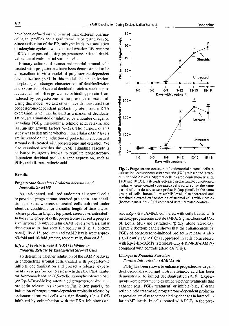
As anticipated, cultured endometrial stromal cells exposed to progesterone secreted prolactin into condi- tioned media, whereas untreated cells cultured under identical conditions for a similar length of time did not release prolactin (Fig. 1, top panel, steroids vs untreated). In the same group of cells, progesterone caused a progres- sive increase in intracellular cAMP levels with a similar time-course to that seen for prolactin (Fig. l, bottom panel). By d 15, prolactin and cAMP levels were approx 60-fold and lO-fold greater, respectively, than on d 3.
Effect of Protein Kinase A (PKA) Inhibitor on Prolactin Release by Endometrial Stromal Cells
To determine whether inhibition of the cAMP pathway in endometrial stromal cells treated with progesterone inhibits decidualization and prolactin release, experi- ments were performed to assess whether the PKA inhibi- tor 8-bromoadenosine-3',5-cyclic monophosphorothioate (or Rp-8-Br-cAMPs) attenuated progesterone-induced prolactin release. As shown in Fig. 2 (top panel), the induction of progesterone-dependent prolactin release by endometrial stromal cells was significantly (*p < 0.05) inhibited by coincubation with the PKA inhibitor (ste-
80-
"6 Q .
v
e t
_= m
m
e= 40. ._ Steroids
.c 20.
. J D : o. / Untreated
o . = - - - \ - 1 v w w
I I I I I I
1-3 3-6 6-9 9-12 12-15 15-18 Days with treatment
300 -
/ s t " J_ E eroids
100.
J Untreated \
o = O,
t - I I I I I I
1-3 3-6 6-9 9-12 12-15 15-18 Days with treatment
Fig. 1. Progesterone treatment of endometrial stromal cells in culture induced an increase in prolactin (PRL) release and intrac- ellular cAMP levels. Stromal cells treated continuously with 1 gM and 10 nM E 2 (steroids) released prolactin into conditioned media, whereas control (untreated) cells cultured for the same period of time do not release prolactin (top panel). In the same group of cells, intracellular cAMP levels also increased and remained elevated on incubation of stromal cells with steroids (bottom panel). *p < 0.05 compared with untreated controls.
roids/Rp-8-Br-cAMPs), compared with cells treated with medroxyprogesterone acetate (MPA; Sigma Chemical Co., St. Louis, MO) and estradiol-17[3 (E2) alone (steroids). Figure 2 (bottom panel) shows that the enhancement by PGE 2 of progesterone-induced prolactin release is also significantly (*p < 0.05) suppressed in cells coincubated with Rp-8-Br-cAMPs (steroids/PGE 2 + RP-8-Br-cAMPs) compared with controls (steroids/PGE2).
Changes in Prolactin Secretion Parallel Intraeellular cAMP Levels
PGE 2 has been shown to enhance progesterone-depen- dent decidualization and all-trans retinoic acid has been demonstrated to inhibit decidualization (9,10). Experi- ments were performed to examine whether treatments that enhance (e.g., PGE 2 treatment) or inhibit (e.g., all-trans retinoic acid treatment) progesterone-dependent prolactin expression are also accompanied by changes in intracellu- lar cAMP levels. In cells treated with PGE 2 in the pres-

Vol. 6, No. 3 cAMP Deactivation During Decidualization/Brar et al. 303
125
~1oo
~ 75
~ 5o E .~_
~, 25
200 -
"~150,
r
El00,
50, .E
n 0
"k
-8-Br-cAMPs
1-3 I I
3-6 6-9 9-12 Days with treatment
S t e ~
§
I I I I
1-3 3-6 6-9 9-12 Days with treatment
Fig. 2. A PKA inhibitor suppressed the release of progesterone- induced prolactin release. Coincubation of endometrial stromal cells in culture with 1 I.tM MPA and 10 nME 2, and the PKA inhibitor Rp-8-Br-cAMPs (steroids + Rp-8-Br-cAMPs) signifi- cantly (p < 0.05) suppressed the release of prolactin into condi- tioned media compared with controls (steroids) at each of the time-points examined (top panel). Rp-8-Br-cAMPs also signifi- cantly (p < 0.05) suppressed PGE 2 stimulation of progesterone- dependent prolactin release from endometrial stromal cells (steroids/PGE 2 + Rp-8-Br-cAMPs) compared with controls (ste- roids/PGE2; bottom panel). *p < 0.05 compared with controls.
ence of MPA and E 2 for 15 d, both prolactin release and intracellular cAMP levels were significantly (p < 0.05) increa- sed compared with cells cultured with MPA and E 2 alone (Fig. 3). Conversely, intracellular cAMP levels were signifi- cantly (p < 0.05) decreased by all-trans retinoic acid, which suppresses progesterone-dependent prolactin release (Fig. 4). Treatment of stromal cells with PGE 2 or retinoic acid alone, in the absence of steroids, does not induce changes in prolactin (9,10) or cAMP levels (PGEz-treated cells 17.9 + 1.2 pmol/(/.tg protein; retinoic acid-treated cells 15.75 _+ 1.4 pmol/gg protein; untreated cells 19.27 + 1.2 pmol/gg protein).
Activation of the PGE 2 Receptor Subtype EP 2 During Decidualization Using reverse transcription-polymerase chain reaction
(RT-PCR) analysis, we next examined whether EP 2 recep- tor mRNA is expressed during progesterone-induced
decidualization of endometrial stromal cells and in decidual cells from term placenta (Fig. 5, upper panel). Expression of the E P 2 receptor was undetectable or low in untreated endometrial stromal cells (Fig. 5, [-] lane), but is induced on treatment with progesterone and estradiol for 15 d (Fig. 5, [+] lane). In addition, expression of EP z mRNA in cells treated with PGE z in combination with progester- one and estradiol was twofold greater than cells treated with progesterone and estradiol alone (Fig. 5, [+/PGE 2] vs [+] lanes). High levels of EP 2 receptor mRNA expres- sion are maintained in differentiated term decidual cells (Fig. 5, D lane). No significant changes occurred in (GAPDH) mRNA expression in endometrial stromal cells with steroid and PGE2 treatments Fig. 5, bottom panel). Similar results were obtained in three separate experiments using endometrial stromal cells and decidua from different individuals.
D i s c u s s i o n
This study demonstrates that the cAMP signaling sys- tem is activated by progesterone during in vitro decidual- ization in human endometrial stromal cells. There is a sustained increase in intracellular cAMP levels during progesterone-dependent induction of decidual prolactin expression. Moreover, there is a positive correlation between intracellular cAMP levels and prolactin release under conditions that enhance (PGE 2 treatment) or suppress (all-trans retinoic acid treatment) progesterone-dependent prolactin expression. Additional evidence that activation of cAMP-dependent PKA is required for progesterone- dependent induction of decidual prolactin expression in human endometrial stromal cells is provided by studies showing that coincubation of cells with PKA inhibitor blocks the induction of prolactin by progesterone. The induction of prolactin during decidualization by perma- nently elevated intracellular cAMP is accompanied by downregulation of PKA regulatory subunit RIc~ with no significant changes in the catalytic subunits (13). There- fore, this study supports the hypothesis that sustained acti- vation of the cAMP signaling cascade occurs during progesterone-dependent decidualization.
There is increasing evidence that crosstalk between progesterone and cAMP systems regulates gene expression in human breast cancer and rat and porcine granulosa cells (14-16). An increase in cAMP accumulation following progesterone treatment has recently been reported in epi- thelial cells of the guinea pig endometrium (17). In those cells, progesterone, which increases sulfate uptake, also increased intracellular cAMP accumulation in epithelial cells treated with estradiol. Additionally, cAMP or forskolin elicited the same increase in sulfate uptake as that observed with progesterone. This interaction between progesterone and cAMP is analogous to that reported here in the human endometrial stromal cells in which the

304 cAMP Deactivation During Decidualization/[3rar et a[. Endocrine
'I0
03
t" v
._m "I0 (D
E t-
n- O_
200 -
150 -
100-
50-
800 -
S
600,
@ 0
E
400-
200-
m
0 0-
Fig. 3. Stimulation of progesterone-dependent prolactin release by PGE 2 parallels an increase in intracellular cAMP levels in cultured endometrial stromal cells. In cells treated with PGE 2 in the presence of MPA and E 2 for 15 d (light-gray bars), both prolactin release (left panel) and intracellular cAMP levels (right panel) were significantly higher (p < 0.05) compared with cells treated with MPA and E 2 alone (black bars). *p < 0.05 compared with controls.
200- 400-
150. "10
o '3
E t -
.~_ 100. " 0
E e - '
, . J n~ o_ 50.
" k
o r
o~ 300" -'1
0
E
a. 200.
' - I
100. t ,-
m
0 ~ 0
Fig. 4. Suppression of progesterone-dependent prolactin release and intracellular cAMP levels by all-trans retinoic acid. Endometrial stromal cells treated with all-trans retinoic acid in the presence of MPA and E 2 for 15 d (light-gray bars) showed a suppression of both prolactin release (left panel) and intracellular cAMP levels (right panel), which were significantly (p < 0.05) lower than cells treated with MPA and E 2 alone (black bars). *p < 0.05 compared with controls.
induction of prolactin is the response to progesterone treat- ment and is mediated via increased intracellular cAMP accumulation.
Our data indicate that PGE 2 enhances cAMP formation in progesterone-treated endometrial stromal cells. Despite compelling evidence in rat and rabbit endometrial stromal cells that PGE 2 induces cAMP production and stimulates adenylate cyclase (18,19) respectively, our studies indicate
that PGE 2 treatment alone, in the absence of progesterone, does not increase cAMP in human endometrial stromal cells. Signal transduction via PGE 2 receptor subtype EP2 involves the activation of adenylate cyclase, resulting in an increase in cAMP formation (20). We therefore exam- ined EP 2 mRNA levels during decidualization. Our study suggests that effects of progesterone may be partially mediated by activation of the cAMP signaling cascade

Vol. 6, No. 3 cAMP Deactivation During Decidualization/Brar et al. 305
(-) (+) (+P) D
EP2 rt
GAPDH
Fig. 5. Expression of the EP2 receptor mRNA in human endo- metrial stromal cells by RT-PCR analysis followed by Southern blot analysis of the products (EP 2 receptor 368 and 193 bp for GAPDH) . Expression of the EP 2 receptor (rt) in endometrial stromal cells is induced in cells treated for 15 d with MPA and E 2 (+) compared with untreated cells (-) (upper panel). Expres- sion of EP 2 mRNA in cells treated with PGE 2 as well as MPA and E 2 (+/PGE2) was fourfold greater than steroid-treated cells (+). High levels of EP2 receptor mRNA expression were detected in differentiated term decidual cells (D). There was no significant change in GAPDH levels in the different samples (bottom panel).
because mRNA levels of EP 2 are significantly increased with progesterone treatment of human endometrial stro- real cells. In addition, high levels of EP 2 mRNA are also expressed in fully differentiated decidual cells from term placenta. A role for the PGE 2 receptor subtype EP 4, which like EP 2 is coupled to adenylate cyclase (21), in proges- terone-dependent endometrial stromal decidualization remains to be determined. Our observations are in agree- ment with the early studies suggesting that endometrial PGE binding sites are progesterone-dependent (19,22) and explain the inability of PGE2 alone, in the absence of progesterone and estradiol, to induce cAMP or prolactin expression in human endometrial stromal cells. Finally, this finding suggests that the synergistic effect of PGE2 on progesterone induction of decidual prolactin is likely to be owing to an enhancement of cAMP production greater than that produced with progesterone alone (9).
The mechanisms by which cAMP induces decidualiza- tion and prolactin gene expression are unknown, cAMP may have a direct effect on the decidual prolactin pro- moter, since the decidual prolactin 5'-flanking region con- tains a sequence TGACG at -25 bp that is part of the canonical CRE octanucleotide, TGACGTCA (2). Although it is as yet unresolved whether this imperfect CRE in the 5'-region of the decidual prolactin gene is the cAMP reg- ulatory region, partial CRE motifs can be as effective as or even more effective than perfect consensus CRE sequen- ces in directing cAMP transcription (23). The CRE and
related sequence motifs are recognized by a number of DNA-binding proteins that belong to the cAMP-regulated enhancer binding/activating transcription factor (CREB/ ATF) family of proteins (24). The presence of a CRE and CCAAT site juxtaposed in the decidual prolactin 5'-flank- ing region (2) suggests that an interaction between more than one site could mediate cAMP responsiveness, i.e., a CRE site may be necessary, but not sufficient to mediate cAMP responsiveness, cAMP responsiveness in the human fibronectin and G-protein O~i_ 2 subunit genes requires cooperative interaction between CRE and a CCAAT box (25,26). Supershift analysis of decidualized endometrial stromal cell extracts using antibodies against human CREB-1 (unpublished data) suggests that complexes formed by binding of CREB protein to the putative CRE in the decidual prolactin 5'-region may be partially medi- ated by CREB proteins. Finally, individual segments of the decidual prolactin gene may not function as autono- mous cAMP response regions, but may require synergistic action of several cAMP enhancer sequences and/or other sequences within the gene, as demonstrated in the transcriptional regulation of calcitonin, gonadotrophin o~-subunit, and human tryptophan hydroxylase genes by cAMP (22,27,28).
In summary, this study provides evidence that the cAMP signaling cascade is activated during progester-one-depen- dent decidualization of human endometrial stromal cells.
Mater ia l s and M e t h o d s
Tissue and Cell Preparation
Decidual tissue was obtained at term and used after dis- section away from amniotic and chorionic membranes as previously described (29). Uterine endometrial tissue was obtained from women with normal menstrual cycles at the time of elective tubal ligation. Informed consent was obtained from patients, and the study was approved by the Institutional Review Boards of Children's Hospital Medi- cal Center and the University of Cincinnati. Proliferative or secretory phase endometrium was removed by suction biopsy and stromal cell cultures were prepared as previ- ously described (8). Enriched stromal cells cultures were obtained using a method of selective attachment to plastic and brief trypsinization of sub-cultures. This method yields a >95% pure stromal cell population. Stromal cells were cultured in Dulbecco's Modified Medium (DMEM) containing 2% fetal bovine serum, 25 U/mL penicillin G, 25 ~tg/mL streptomycin, and 2.5 ~tg/mL amphotericin B.
In Vitro Decidualization of Human Endometrial Stromal Cells
Decidualization of endometrial stromal cells was induced by incubating subconfluent cells in media con- taining 1 ~tM MPA and 10 nM E2 (Sigma) as previously described (8). These steroids were dissolved in ethanol

306 cAMP Deactivation During Decidualization/13rar et al. Endocrine
and added to the media before use. The final concentration of ethanol in the media never exceeded 0.09% (v/v). Phase-contrast microscopy was used to verify morpholog- ical changes associated with differentiation in vitro in response to MPA and E 2 (7,30). In some experiments, cells were treated with 1 gM PGE2; or 1 I.tM all-trans retinoic acid in combination with MPA and E2. The PKA inhibitor Rp-8-Br-cAMPs purchased from BioLog Life Science Institute, La Jolla, CA was used at a concentration of 0.8 x 10⁻⁴ M.
Cyclic Nucleotide Extraction and Assay
Following removal of the media, cAMP was extracted from cultured cells using ice-cold acidified ethanol (0.1 N HC1/95% ethanol) and freeze-thawing. Ethanol extracts from three six-well clusters were pooled and evaporated using a Savant Automatic SpeedVac (Forma Scientific Inc., Marietta, OH), and stored at -20~ prior to being recon- stituted in 250 gl sodium acetate buffer (Rainen Assay System, Dupont, NEN Life Sciences, Boston, MA). cAMP concentrations were determined using a kit (Rainen Assay System), and the results are expressed as pmol/gg protein. The protein concentration in samples was determined using the micro BCA protein assay reagent kit (Pierce, Rockford, IL) according to manufacturer's instructions.
Prolactin Assay
Prolactin was measured in conditioned media by homolo- gous radioimmunoassay, as previously described (31) using materials provided by the National Hormone and Pituitary Program, NIDDK, Bethesda, MD. Conditioned media was stored at -20~ until assayed. Interassay varia- tion (SEM at EDs0) was + 4% of the mean.
Analysis of RNA by RT-PCR
Total RNA from cells and tissue was isolated by the guanidium thiocyanate-phenol-chloroform extraction method of Chomczynski and Sacci (32) using Tri-reagent (Molecular Research Center Inc., Cincinnati, OH). The RNA (200 ng) was reverse-transcribed using M-MLV reverse transcriptase (Gibco-BRL) and oligo (dT)l 6 prim- ers. The reverse-transcribed RNA was then amplified by PCR for 35 cycles using specific primers corresponding to the second extracellular loop (nucleotides 740-757) and to transmembrane region 7 (nucleotides 1090-1107) of the human PGE2 receptor EP 2 subtype as previously described (20). For each sample, primers for GAPDH were added to the same reaction tube as those used to amplify the EP2 as previously described (10). The denaturing temperature was 94~ for 1 min, annealing temperature was 50~ for 2 min and extending temperature was 72~ for 3 min. The PCR products obtained were electrophoresed on a 1% agarose gel, stained with ethidium bromide, and then transferred to a nylon membrane (Nytran, Schleicher and Schnell, Keene, NH). Southern hybridization was per- formed with 32p-end labeled oligonucleotide sequences,
which are complementary to a specific region within each of the predicted PCR products for the EP 2 receptor or GAPDH. Signals on autoradiographs were quantified by densitometric analysis. The signal generated by GAPDH, which was not changed by any of the experimental treat- ments studied, was used as an internal positive control to minimize intersample variations.
Statistical Analysis
The data are presented as the mean + SEM. Statistical differences between group means was determined by analysis of variance with Bonferroni adjustment or a Student's t-test, depending on design. Differences are con- sidered significant when p < 0.05.
References
1. Tang, B., Guller, S., and Gurpide, E. (1993). Endocrinology 133, 2197-2203.
2. Gellersen, B., Kempf, R., Telgmann, R., and DiMattia, G. E. (1994). Mol. Endocrinol. 8, 356-373.
3. Tanaka, N., Miyazaki, K., Tashiro, H., Mizutani, H., and Okamura, H. (1993). J. Reprod. Fertil. 97, 33-39.
4. Bergamini, C. M., Pansini, F., Bettocchi, S. Jr., Segala, V., Dallocchio, F., Bagni, B., and Mollica, G. (1985). J. Steroid Biochem. 22, 299-303.
5. Houserman, V. L., Todd, H. and Hertelendy, F. (1989). J. Reprod. Fert. 85, 195-202.
6. Coleman, R. A., Smith, W. L., and Narumiya, S. (1994). Pharmacol. Rev. 46, 205-229.
7. Irwin, J. C., Utian, W. H., and Eckert, R. L. (1991 ). Endocrinol- ogy 129, 2385-2392.
8. Brar, A. K., Frank, G. R., Richards, R. R., Meyer, A. J., Kessler, C. A., Cedars, M. I., Klein, D. J., and Handwerger, S. (1995). J. Cell. Physiol. 163, 30-37.
9. Frank, G. R., Brar, A. K., Cedars, M. I., and Handwerger, S. (1993). Endocrinology 134, 258-263.
10. Brar, A. K., Kessler, C. A., Meyer, A. J., Cedars, M. I., and Jikihara, H. (1996). Mol. Human Reproduction 2, 185-193.
11. Frank, G. R., Brar, A. K., Jikihara, H., Cedars, M. I., and Handwerger, S. (1995). Biol. Reprod. 52, 184-191.
12. Zhou, J., Dsupin, B. A., Giudice, L. A., and Bondy, C. A. (1994). J. Clin. Endocrinol. Metab. 79, 1723-1734.
13. Telgmann, R., Maronde, E, Tasken, K., and Gellersen, B. (1997). Endocrinology 138, 929-937.
14. Cho, H., Aronica, S. M., and Katzenellenbogen, B. S. (1994). Endocrinology 134, 658-664.
15. Park-Sarge, O.-K. and Sarge, K. D. (1995). Endocrinology 136, 5430-5437.
16. Sirotkin, A. V. and Nitray, J. (1993). J. Steroid Biochem. MoL Biol. 46, 573-577.
17. Beck, L., Mularoni, A., Cardis, P., Adessi, G. L., and Nicollier, M. (1995). Endocrinology 136, 1737-1743.
18. Yee, G. M. and Kennedy, T. G. (1991). Biol. Reprod. 45, 163-171.
19. Fortier, M. A., Boulanger, M., Boulet, A. P., and Lambert, R. D. (1987). Biol. Reprod. 36, 1025-1033.
20. Regan, J. W., Bailey, T. J., Pepperl, D. J., Pierce, K. L., Bogardus, A. M., Donello, J. E., Fairbairn, C. E., Kedzie, K. M., Woodward, D. F., and Gil, D. W. (1994). Prostaglandins 47, 151-168.
21. Negishi, M., Sugimoto, Y., and Ichikawa, A. (1995). J. Lipid Mediators Cell Signalling 12, 379-391.
22. Kennedy, T. G., Martel, D., and Psychoyos, A. (1983). Biol. Reprod. 29, 556-564.

Vol. 6, No. 3 cAMP Deactivation During Decidualization/Brar et al. 307
23. de Bustros, A., Ball, D. W., Peters, R., Compton, D., and Nelkin, B. D. (t 992). Biochem. Biophys. Res. Commun. 189, 1157-1164.
24. Vallejo, M. (1994). J. Neuroendocrinol. 6, 587-596. 25. Muro, A. F., Bernath, V. A., and Kornblihtt, A. R. (1992).
J. Biol. Chem. 267, 12,767-12,774. 26. Kinane, T. B., Shang, C., Finder, J. D., and Ercolani, L. (1993).
J. Biol. Chem. 268, 24,669-24,676. 27. Andersen, B., Kennedy, G. C., Hamernik, D. L., Bokar, J. A.,
Bohinski, R., and Nilson, J. H. (1990). Mol. Endocrinol. 4, 573-582.
28. Boularand, S., Darmon, M. C., Ravassard, P., and Mallet, J. (1995). J. Biol. Chem. 270, 3757-3764.
29. Markoff, E., Zeitler, P., Peleg, S., and Handwerger S. (1983). J. Clin. Endocrinol. Metab. 56, 962-968.
30. Irwin, J. C., Kirk, D., King, R. J., Quigley, M. M. and Gwatkin, R. B. (1989). Fertil. Steril. 52, 761-768.
31. Golander, A., Hurley, T., and Handwerger, S. (1979). J. Endo- crinot. 82, 263-267.
32. Chomczynski, P. and Sacchi, N. (1987). Anal. Biochem. 162, 156-159.