Expression of Transcription Factors in Endometrium During Natural Cycles
8
Journal of Assisted Reproduction and Genetics, Vol. 20, No. 11, November 2003 ( C 2003) Endometrium & Endometriosis Expression of Transcription Factors in Endometrium During Natural Cycles Vicente Maldonado, 1 Jos ´ e Antonio Castilla, 1,4 Luis Mart´ ınez, 1 Alfonso Herruzo, 2 ´ Angel Concha, 3 Juan Fontes, 1 Nicol ´ as Mendoza, 1 Mar´ ıa Luisa Garc´ ıa-Pe ˜ na, 1 Jos ´ e Luis Mendoza, 1 Rosa Mag ´ an, 1 ´ Agueda Ortiz, 1 and Esther Gonz ´ alez 1 Submitted April 9, 2003; accepted October 2, 2003 Purpose : The sex steroid control of the endometrial cycle is mediated by transcription factors, four of which are the estrogen and progesterone receptors, c-jun and c-fos, all expressed by the endometrium. The aim of this study was to analyze the distribution of the transcription factors in the different endometrial compartments during natural cycles. Methods : We studied 53 reproductively-normal women, of whom 26 were in the proliferative phase and 27 in the secretory phase. An endometrial biopsy was performed and serum values of LH, FSH, estradiol, and progesterone were determined. We studied the expression of transcription factors using monoclonal antibodies. Results : A correlation between estrogen receptor and c-jun and c-fos expression was observed in stroma and epithelia, and progesterone receptor expression correlated with c-jun expression in epithelia. C-jun and c-fos presented greater expression in the proliferative phase than in the secretory phase, in the stroma and in both epithelia. No relation was found between estradiol serum levels and any transcription factor, but progesterone serum levels correlated significantly with most such factors. Conclusion : The two proto-oncogenes could play a decisive role in regulating the endometrial cycle; they could mediate the effects induced by sex steroid, and could be related to other transcription factors. KEY WORDS: C-fos; c-jun; endometrium; steroid receptor; transcription factor. INTRODUCTION The human endometrium is subject to strict hormonal control during the menstrual cycle. Its maximum pro- liferation, stimulated by 17β -estradiol, occurs with ovulation. Progesterone inhibits the proliferation 1 Unidad de Reproducci ´ on, Hospital Virgen de las Nieves, Granada, Spain. 2 Departamento de Especialidades M ´ edico Quir ´ urgicas, Facultad de Medicina y Odontolog´ıa, Universidad del Pa´ıs Vasco, Leioa, Vizcaya, Spain. 3 Servicio de Anatom´ ıa Patol ´ ogica, Hospital Virgen de las Nieves, Granada, Spain. 4 To whom correspondence should be addressed at Unidad de Reproducci ´ on, Hospital Universitario “Virgen de las Nieves,” Avda Coronel Mu˜ noz s/n, Granada E-18014, Spain; e-mail: [email protected]. of the endometrial epithelium stimulated by 17β - estradiol after ovulation and induces secretory dif- ferentiation (1,2). The reduction in the presence of ovarian steroids at the end of the luteal phase in- duces a chain of events, with lysosomal autodiges- tion and cell involution (2), followed by vascular deterioration, thrombosis, and interstitial bleeding. During menstruation, a variable quantity of the func- tional layer is lost (3,4), and the remaining mu- cosa enters an early proliferative phase. Foresee- able cyclic changes, together with the possibility of obtaining biopsies, make it possible to study the human endometrium in relation to the hormonal changes taking place at the end of the menstrual cycle, with regard to proliferation, differentiation, and cell death. 1058-0468/03/1100-0474/0 C 2003 Plenum Publishing Corporation 474
-
Upload
independent -
Category
Documents
-
view
4 -
download
0
Transcript of Expression of Transcription Factors in Endometrium During Natural Cycles
P1: ZBU
Journal of Assisted Reproduction and Genetics pp1064-jarg-477342 November 25, 2003 13:45 Style file version June 3rd, 2002
Journal of Assisted Reproduction and Genetics, Vol. 20, No. 11, November 2003 ( C© 2003)
Endometrium & Endometriosis
Expression of Transcription Factors in EndometriumDuring Natural Cycles
Vicente Maldonado,1 Jose Antonio Castilla,1,4 Luis Martınez,1 Alfonso Herruzo,2 Angel Concha,3
Juan Fontes,1 Nicolas Mendoza,1 Marıa Luisa Garcıa-Pena,1 Jose Luis Mendoza,1 Rosa Magan,1
Agueda Ortiz,1 and Esther Gonzalez1
Submitted April 9, 2003; accepted October 2, 2003
Purpose : The sex steroid control of the endometrial cycle is mediated by transcription factors,four of which are the estrogen and progesterone receptors, c-jun and c-fos, all expressed bythe endometrium. The aim of this study was to analyze the distribution of the transcriptionfactors in the different endometrial compartments during natural cycles.Methods : We studied 53 reproductively-normal women, of whom 26 were in the proliferativephase and 27 in the secretory phase. An endometrial biopsy was performed and serum valuesof LH, FSH, estradiol, and progesterone were determined. We studied the expression oftranscription factors using monoclonal antibodies.Results : A correlation between estrogen receptor and c-jun and c-fos expression was observedin stroma and epithelia, and progesterone receptor expression correlated with c-jun expressionin epithelia. C-jun and c-fos presented greater expression in the proliferative phase than inthe secretory phase, in the stroma and in both epithelia. No relation was found betweenestradiol serum levels and any transcription factor, but progesterone serum levels correlatedsignificantly with most such factors.Conclusion : The two proto-oncogenes could play a decisive role in regulating the endometrialcycle; they could mediate the effects induced by sex steroid, and could be related to othertranscription factors.
KEY WORDS: C-fos; c-jun; endometrium; steroid receptor; transcription factor.
INTRODUCTION
The human endometrium is subject to strict hormonalcontrol during the menstrual cycle. Its maximum pro-liferation, stimulated by 17β-estradiol, occurs withovulation. Progesterone inhibits the proliferation
1 Unidad de Reproduccion, Hospital Virgen de las Nieves,Granada, Spain.
2 Departamento de Especialidades Medico Quirurgicas, Facultadde Medicina y Odontologıa, Universidad del Paıs Vasco, Leioa,Vizcaya, Spain.
3 Servicio de Anatomıa Patologica, Hospital Virgen de las Nieves,Granada, Spain.
4 To whom correspondence should be addressed at Unidad deReproduccion, Hospital Universitario “Virgen de las Nieves,”Avda Coronel Munoz s/n, Granada E-18014, Spain; e-mail:[email protected].
of the endometrial epithelium stimulated by 17β-estradiol after ovulation and induces secretory dif-ferentiation (1,2). The reduction in the presence ofovarian steroids at the end of the luteal phase in-duces a chain of events, with lysosomal autodiges-tion and cell involution (2), followed by vasculardeterioration, thrombosis, and interstitial bleeding.During menstruation, a variable quantity of the func-tional layer is lost (3,4), and the remaining mu-cosa enters an early proliferative phase. Foresee-able cyclic changes, together with the possibility ofobtaining biopsies, make it possible to study thehuman endometrium in relation to the hormonalchanges taking place at the end of the menstrual cycle,with regard to proliferation, differentiation, and celldeath.
1058-0468/03/1100-0474/0 C© 2003 Plenum Publishing Corporation 474
P1: ZBU
Journal of Assisted Reproduction and Genetics pp1064-jarg-477342 November 25, 2003 13:45 Style file version June 3rd, 2002
Expression of Transcription Factors in Endometrium 475
Growth regulation and sexual differentiation dur-ing the menstrual cycle by the sex steroids is achievedby hormones produced locally, by cytokines andproto-oncogenes, whose interactions are as yet in-completely understood. The steroid receptors are in-tracellular proteins that join in a specific way withsteroid hormones, provoking phenomena that in turnlead to cellular change. The steroid receptor/steroidhormone complex regulates the process of transcrip-tion of specific genes. One of the possible agents ofestrogen-regulated endometrial proliferation is theproto-oncogene c-fos, a protein that amplifies the es-trogenic message and activates the transcription ofother genes that control multiple cellular functionssuch as division. The transcription of c-fos is rein-forced by estrogens in the uterus of ovariectomizedrats, and its effect is reversed by progesterone (5),which suggests that inhibiting the expression of c-foscould be an important mechanism involved in the an-tiproliferative effect of progestagens (6). The pres-ence of m-RNA c-fos in the human endometrium hasbeen established, with the highest levels being foundduring the proliferative phase of the menstrual cycle(7,8). These findings suggest that the expression ofc-fos in the human endometrium could also be mod-ulated by sex steroid hormones.
The proto-oncogene c-jun is one of the genes whoseactivation has been related to proliferation, differen-tiation, and apoptosis, depending on the tissue beingstudied (9,10). This protein is part of the activating fac-tor (AP-1) that participates in the induction of tran-scription genes (11,12). This protein AP-1 is consti-tuted with the union of c-fos to c-jun. The c-jun geneis strongly expressed from the proliferative phase tothe secretory medium, but is virtually absent in the de-cidua (8). No cyclical changes have been detected inthe endometrial expression of c-jun during the normalmenstrual cycle, although the significant differencesfound between gestating and nongestating womensupport the hypothesis of hormonal regulation (7).
This study proposes an analysis of the distributionof the transcription factors between the different en-dometrial compartments, both in the proliferative andin the secretory phase, in natural cycles. We also ana-lyze the relation of these factors with the serum valuesof circulating hormones.
MATERIALS AND METHODS
Subjects
A study was made of 53 reproductively-normalwomen who attended the Family Planning clinic at
the “Virgen de las Nieves” University Hospital inGranada. All had requested contraception by meansof tubaric electrocoagulation, and fulfilled the requi-sites of fertile age, absence of any history of sterility,or disorders in their menstrual cycle, having becomepregnant spontaneously during the previous 3 yearsand, during the last 6 months, having received no hor-monal or IUD contraception.
Of these patients, 26 were in the proliferative phase(between days 9 and 12 of the menstrual cycle) and 27were in the secretory phase (between days 20 and 22).On the appropriate days, blood samples were takenand an endometrial biopsy performed by aspiration,using a Cournier cannula. The biopsies were datedaccording to the Noyes method (13).
Estradiol, progesterone, LH, FSH, prolactin, andtestosterone serum concentrations were analyzedwith commercial kits (VIDAS, Biomerieux, France).
Tissue Processing
The biopsy material was fixed in tamponaded for-malin pH 7.2 for 24 h and then embedded in paraffin.Sections were made with a rotary-blade microtomeand then stained with hematoxylin–eosin and muci-carmin.
As part of the biopsy, a standard histologic studywas made of the three components of the en-dometrium (epithelium, stroma, and vessels) in or-der to discount the possibility of any kind of mor-phologic disorder (irregular maduration, maturativeglandular–stromal dissociation, etc.), to determine thepresence of specific pathologic signs (endometritis,hyperplasias, etc.), and to attempt to date, as accu-rately as possible, the functional evolutive moment ofthe endometrium.
Immunohistochemistry
A streptavidin–biotin peroxidase detection systemspecific for the TechMate automated immunostainer(Dakocytomation AS, Denmark) was used for the im-munohistochemical technique, on 5 µm paraffin sec-tions. For antigen retrieval, the slides were placed in200 mL of retrieval solution (pH 6.8) and boiled for2 min. The slides were cooled at room temperaturefor 20 min before processing. The following primarymonoclonal antibodies were used: Estrogen receptor:monoclonal antibody, clone 1D5 (Dakopatts); Pro-gesterone receptor: monoclonal antibody, clone 1A6(Dakopatts); C-fos: monoclonal antibody, clone c-fos 88 (Biogenex); C-jun: monoclonal antibody, clonec-jun 88 (Biogenex).
Journal of Assisted Reproduction and Genetics, Vol. 20, No. 11, November 2003
P1: ZBU
Journal of Assisted Reproduction and Genetics pp1064-jarg-477342 November 25, 2003 13:45 Style file version June 3rd, 2002
476 Maldonado et al.
The chromogen substrate used was 3′3′-diaminobencidine tetrahydrochloride. The slideswere counterstained with Harris’s hematoxilin andmounted with synthetic mounting medium. A semi-quantitative method was performed for evaluation ofimmunohistochemical assays, as follows: 0: negative;1: positive at<25%; 2: positive at 25–50%; 3: positiveat 50–75%; 4: positive at >75%.
The positivity of the expression of the differenttranscription factors studied, within the differenttissue compartments and cycle phases, was scoredbetween 0 and 4. Interpretation of immunohisto-chemical techniques was performed blind withoutknowledge of clinical data.
Statistical Analysis
The Student’s t test was used to compare hormonalserum levels between phases of the menstrual cycle.The Mann–Whitney U test was used for the statis-tical study of the values of the receptors in the en-dometrium. Spearman correlation was used to showthe degree of association between hormone serum,and transcription factor expressions. Values were con-sidered to be statistically significant for p values lessthan 0.05.
RESULTS
Significant differences (p < 0.001) were only foundin the serum levels of progesterone (0.97± 0.9 ng/mLvs. 12.27± 3.52 ng/mL) between the proliferative andthe secretory phase (Table I).
In every case, the endometrial morphology and tis-sue dating were adequate, and considered to be “inphase,” following Noyes’ criteria (1950). The speci-mens in the proliferative phase presented intense pro-liferative activity, particularly so in the glands, in thesurface epithelium, in the stroma, and in the vascular
Table I. Hormone Serum Values Obtained for the TwoEndometrial Phases
Follicular phase Secretory phase(n = 26) (n = 27)
Estradiol (pg/mL) 134.41± 98.69a 114.75± 76.63Progesterone (ng/mL) 0.97± 0.79 12.27± 3.52∗∗∗FSH (mU/mL) 6.27± 1.85 4.82± 3.42LH (mU/mL) 4.63± 2.44 3 ± 2.72Testosterone (ng/mL) 0.60± 0.45 0.46± 0.23Prolactin (ng/mL) 14.74± 6.82 12.25± 5.13
a Mean ± SD.∗∗∗p < 0.0001.
endothelium of the upper third of the endometrium.The glands appeared to be straight or slightly curled,narrow, unbranched, and uniformly dispersed withinan edematous stromal matrix. The tissue samples inthe secretory phase presented the characteristic ab-sence of proliferation of the stroma cells. In the glan-dular epithelium, the distal edge of the cells presenteda curled appearance due to the numerous projectionsof apical cytoplasm filled with secretion material.
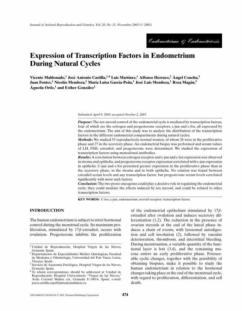
The estrogen receptor presented intense expressionin the four compartments studied during the prolifer-ative phase of the endometrial cycle but the epithe-lium in the secretory phase showed minimal levelsof expression (Figs. 1 and 8(a)). The progesterone re-ceptor, however, remained unchanged throughout theendometrial cycle in the stromal and endothelial com-partments. In the glandular and surface epithelilia,
Fig. 1. Expression of the estrogen receptor in the different tissuecompartments.
Journal of Assisted Reproduction and Genetics, Vol. 20, No. 11, November 2003
P1: ZBU
Journal of Assisted Reproduction and Genetics pp1064-jarg-477342 November 25, 2003 13:45 Style file version June 3rd, 2002
Expression of Transcription Factors in Endometrium 477
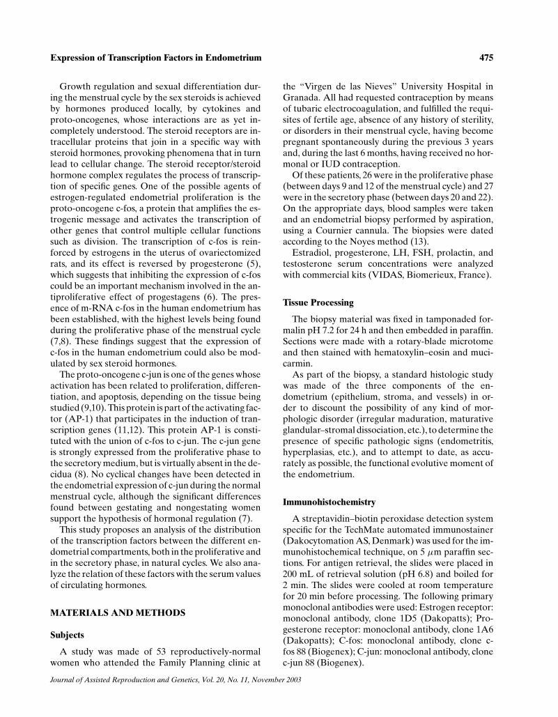
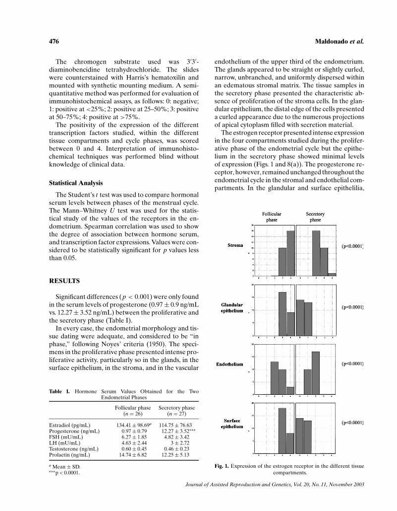
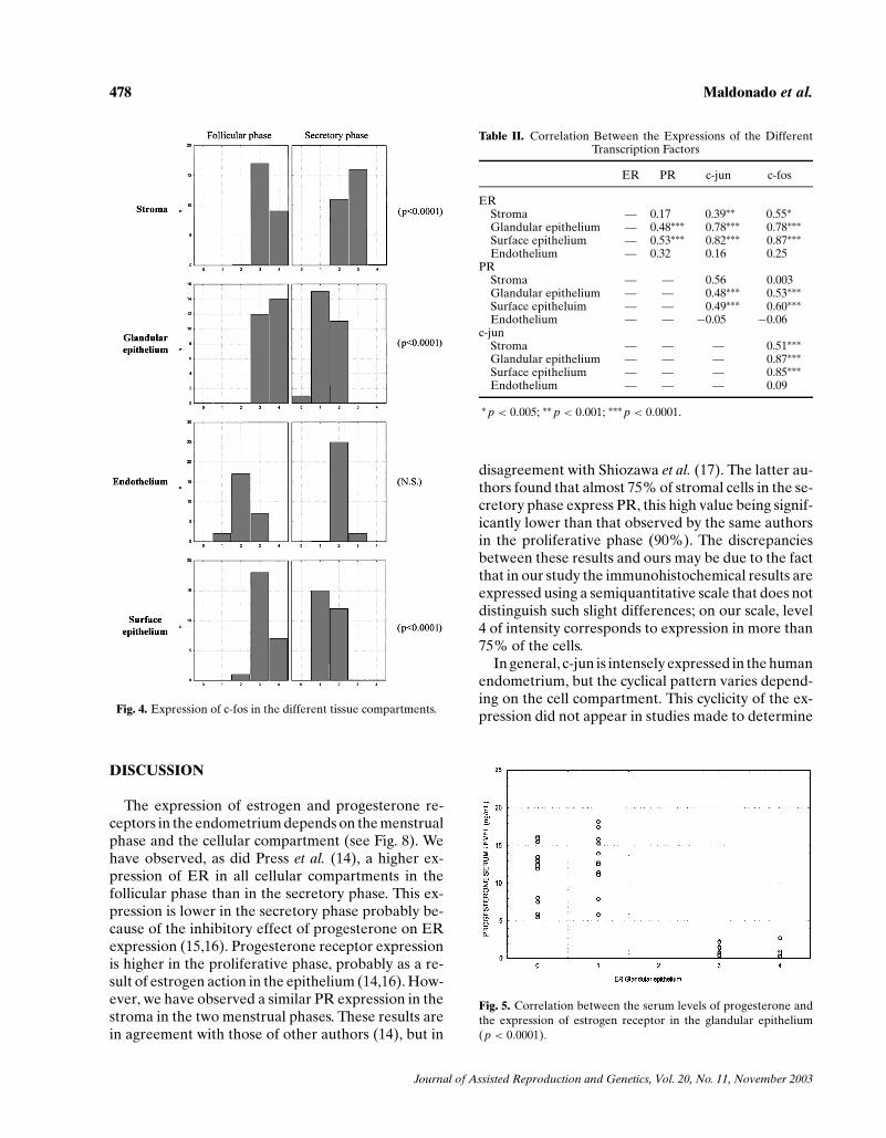
though, expression was greater during the prolifer-ative phase and decreased in the secretory phase(Figs. 2 and 8(b)). C-jun presented greater expres-sion in the proliferative phase than in the secretoryphase, in the stroma, in the glandular epithelium, andin the surface epithelium, but there were no significantcyclical changes in the vascular endothelium (Figs. 3and 8(c)). C-fos presented a very similar pattern ofexpression to c-jun (Figs. 4 and 8(d)).
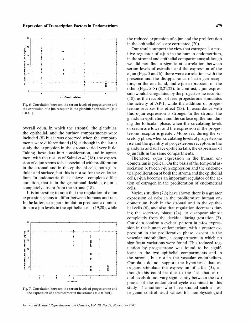
We found a correlation between the expression ofthe estrogen receptor and that of c-jun in the stroma(p < 0.005) and in both epithelia (p < 0.0001), butnot in the vascular endothelium. With respect to thecorrelation of the estrogen receptor with that of c-fos,it was found to exist in the stroma and in both epithelia
Fig. 2. Expression of the progesterone receptor in the differenttissue compartments.
(p < 0.0001 in all three cases), but not in the vascu-lar endothelium. A statistical correlation between PRand c-jun was only found in the glandular and in thesurface epithelia (p < 0.001 and p < 0.005, respec-tively). There was a correlation between PR and c-fosonly in the two epithelia (p < 0.001) (Table II).
No statistical correlation was found between theserum levels of estradiol and the expression of thereceptors in the different tissue compartments. How-ever, the serum levels of progesterone were found tobe significantly correlated with the expression of allthe receptors studied, in the different compartments,with the exceptions of the progesterone receptor inthe stroma and in the endothelium, and the c-fos onco-gene receptor in the vascular endothelium (Figs. 5–7).
Fig. 3. Expression of c-jun in the different tissue compartments.
Journal of Assisted Reproduction and Genetics, Vol. 20, No. 11, November 2003
P1: ZBU
Journal of Assisted Reproduction and Genetics pp1064-jarg-477342 November 25, 2003 13:45 Style file version June 3rd, 2002
478 Maldonado et al.
Fig. 4. Expression of c-fos in the different tissue compartments.
DISCUSSION
The expression of estrogen and progesterone re-ceptors in the endometrium depends on the menstrualphase and the cellular compartment (see Fig. 8). Wehave observed, as did Press et al. (14), a higher ex-pression of ER in all cellular compartments in thefollicular phase than in the secretory phase. This ex-pression is lower in the secretory phase probably be-cause of the inhibitory effect of progesterone on ERexpression (15,16). Progesterone receptor expressionis higher in the proliferative phase, probably as a re-sult of estrogen action in the epithelium (14,16). How-ever, we have observed a similar PR expression in thestroma in the two menstrual phases. These results arein agreement with those of other authors (14), but in
Table II. Correlation Between the Expressions of the DifferentTranscription Factors
ER PR c-jun c-fos
ERStroma — 0.17 0.39∗∗ 0.55∗Glandular epithelium — 0.48∗∗∗ 0.78∗∗∗ 0.78∗∗∗Surface epithelium — 0.53∗∗∗ 0.82∗∗∗ 0.87∗∗∗Endothelium — 0.32 0.16 0.25
PRStroma — — 0.56 0.003Glandular epithelium — — 0.48∗∗∗ 0.53∗∗∗Surface epitheluim — — 0.49∗∗∗ 0.60∗∗∗Endothelium — — −0.05 −0.06
c-junStroma — — — 0.51∗∗∗Glandular epithelium — — — 0.87∗∗∗Surface epithelium — — — 0.85∗∗∗Endothelium — — — 0.09
∗ p < 0.005; ∗∗ p < 0.001; ∗∗∗ p < 0.0001.
disagreement with Shiozawa et al. (17). The latter au-thors found that almost 75% of stromal cells in the se-cretory phase express PR, this high value being signif-icantly lower than that observed by the same authorsin the proliferative phase (90%). The discrepanciesbetween these results and ours may be due to the factthat in our study the immunohistochemical results areexpressed using a semiquantitative scale that does notdistinguish such slight differences; on our scale, level4 of intensity corresponds to expression in more than75% of the cells.
In general, c-jun is intensely expressed in the humanendometrium, but the cyclical pattern varies depend-ing on the cell compartment. This cyclicity of the ex-pression did not appear in studies made to determine
Fig. 5. Correlation between the serum levels of progesterone andthe expression of estrogen receptor in the glandular epithelium(p < 0.0001).
Journal of Assisted Reproduction and Genetics, Vol. 20, No. 11, November 2003
P1: ZBU
Journal of Assisted Reproduction and Genetics pp1064-jarg-477342 November 25, 2003 13:45 Style file version June 3rd, 2002
Expression of Transcription Factors in Endometrium 479
Fig. 6. Correlation between the serum levels of progesterone andthe expression of c-jun receptor in the glandular epithelium (p <0.0001).
overall c-jun, in which the stromal, the glandular,the epithelial, and the surface compartments wereincluded (8) but it was observed when the compart-ments were differentiated (18), although in the latterstudy the expression in the stroma varied very little.Taking these data into consideration, and in agree-ment with the results of Salmi et al. (18), the expres-sion of c-jun seems to be associated with proliferationin the stromal and in the epithelial cells, both glan-dular and surface, but this is not so for the endothe-lium. In endometria that achieve a complete differ-entiation, that is, in the gestational decidua, c-jun iscompletely absent from the stroma (18).
It is interesting to note that the regulation of c-junexpression seems to differ between humans and rats.In the latter, estrogen stimulation produces a diminu-tion in c-jun levels in the epithelial cells (19,20), while
Fig. 7. Correlation between the serum levels of progesterone andthe expression of c-fos receptor in the stroma (p < 0.0001).
the reduced expression of c-jun and the proliferationin the epithelial cells are correlated (20).
Our results support the view that estrogen is a pos-itive regulator of c-jun in the human endometrium,in the stromal and epithelial compartments; althoughwe did not find a significant correlation betweenserum levels of estradiol and the expression of thec-jun (Figs. 5 and 6), there were correlations with thepresence and the disappearance of estrogen recep-tors, on the one hand, and c-jun expression, on theother (Figs. 5–8) (8,21,22). In contrast, c-jun expres-sion would be regulated by the progesterone receptor(18), as the receptor of free progesterone stimulatesthe activity of AP-1, while the addition of proges-terone reverses this effect (23). In accordance withthis, c-jun expression is stronger in the stroma, theglandular epithelium and the surface epithelium dur-ing the follicular phase, when the circulating levelsof serum are lower and the expression of the proges-terone receptor is greater. Moreover, during the se-cretory phase, when circulating levels of progesteronerise and the quantity of progesterone receptors in theglandular and surface epithelia falls, the expression ofc-jun falls in the same compartments.
Therefore, c-jun expression in the human en-dometrium is cyclical. On the basis of the temporal as-sociation between c-jun expression and the endome-trial proliferation of both the stroma and the epithelialcells, c-jun becomes an important regulator of the ac-tion of estrogen in the proliferation of endometrialcells.
Various studies (7,8) have shown there is a greaterexpression of c-fos in the proliferative human en-dometrium, both in the stromal and in the epithe-lial cells (6), and also that regulation decreases dur-ing the secretory phase (24), to disappear almostcompletely from the decidua during gestation (7).Our data confirm a cyclical pattern in c-fos expres-sion in the human endometrium, with a greater ex-pression in the proliferative phase, except in thevascular endothelium, a compartment in which nosignificant variations were found. This reduced reg-ulation by progesterone was found to be signif-icant in the two epithelial compartments and inthe stroma, but not in the vascular endothelium.Our data do not support the hypothesis that es-trogens stimulate the expression of c-fos (5), al-though this could be due to the fact that estra-diol levels do not vary significantly between the twophases of the endometrial cycle examined in thisstudy. The authors who have studied such an es-trogenic control used values for nonphysiological
Journal of Assisted Reproduction and Genetics, Vol. 20, No. 11, November 2003
P1: ZBU
Journal of Assisted Reproduction and Genetics pp1064-jarg-477342 November 25, 2003 13:45 Style file version June 3rd, 2002
480 Maldonado et al.
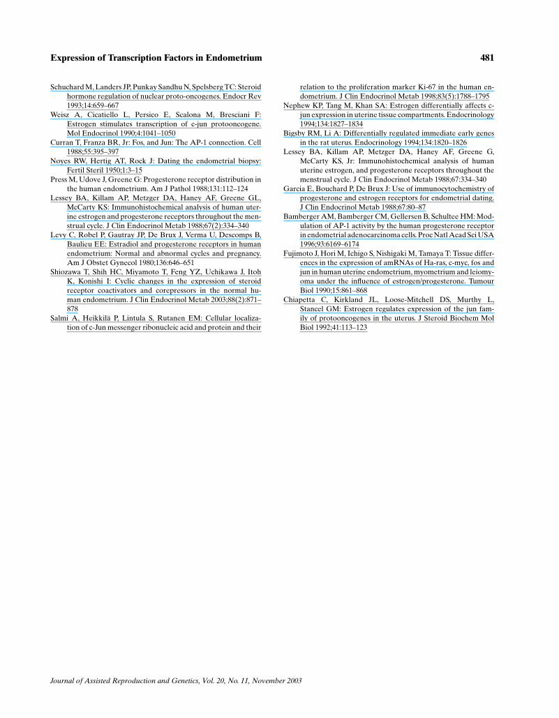
Fig. 8. Immunohistochemical study of normal cycling endometrium (see text). Evident stromal immunoreactivity of estrogenreceptor with negativity in the epithelium (a) and important nuclear positivity of progesterone receptor (b) both in stromaland epithelial cells in secretory phase. Strong expression of c-jun (c) and c-fos (d) in proliferative phase.
estradiol (5,25). We found, thus, not only that theexpression of c-fos during the endometrial cycleis controlled by hormonal serum levels, but alsothat this control varies between different tissuecompartments.
In conclusion, both proto-oncogenes could play adecisive role in regulating endometrial growth andin differentiation; they could modulate the effects ofestradiol and progesterone and could be closely re-lated to other transcription factors such as the steroidreceptors.
ACKNOWLEDGMENTS
The authors’ experimental data reviewed in this pa-per were supported by Instituto de Salud Carlos IIIde Espana (FIS 97/1175).
REFERENCES
Clarke C, Sutherland R: Progestin regulation of cellular prolifera-tion. Endocr Rev 1990;11:266–272
Ferenzy A, Guralnic M: Endometrial microstructure: Structure–function relationship throughout the menstrual cycle. SeminReprod Endocrinol 1983;1:205–219
Bartelmez GW: The phases of the menstrual cycle and their in-terpretations in terms of the pregnancy cycle. Am J ObstetGynecol 1957;74:931–955
Nogales-Ortiz F, Puerta J: The normal menstrual cycle. Chronol-ogy and mechanism of endometrial desquamation. ObstetGynecol 1978;51:259–264
Kirkland JL, Muthy L, Stancel GM: Progesterone inhibits theestrogen-induced expression of c-fos messenger ribonu-cleic acid in the uterus. Endocrinology 1992;130:3223–3230
Reis FM, Maia AL, Ribeiro MF, Spritzer PM: Progestin modu-lation of c-fos and prolactin gene expression in the humanendometrium. Fertil Steril 1999;71:1125–1132
Salmi A, Ammala M, Rutanen EM: Proto-oncogenes c-jun and c-fos are down-regulated in human endometrium during preg-nancy: Relationship to oestrogen receptor status. Mol HumReprod 1996;2:979–984
Salmi A, Rutanen EM: C-fos and c-jun expression in hu-man endometrium and myometrium. Mol Cell Endocrinol1996;117:233–240
Landers JP, Spelsberg TC: New concepts in steroid hormone ac-tion: Transcription factors, proto-oncogenes, and the cascademodel for steroid regulation of gene expression. Crit RevEucaryot Gene Expr 1992;2:19–63
Journal of Assisted Reproduction and Genetics, Vol. 20, No. 11, November 2003
P1: ZBU
Journal of Assisted Reproduction and Genetics pp1064-jarg-477342 November 25, 2003 13:45 Style file version June 3rd, 2002
Expression of Transcription Factors in Endometrium 481
Schuchard M, Landers JP, Punkay Sandhu N, Spelsberg TC: Steroidhormone regulation of nuclear proto-oncogenes. Endocr Rev1993;14:659–667
Weisz A, Cicatiello L, Persico E, Scalona M, Bresciani F:Estrogen stimulates transcription of c-jun protooncogene.Mol Endocrinol 1990;4:1041–1050
Curran T, Franza BR, Jr: Fos, and Jun: The AP-1 connection. Cell1988;55:395–397
Noyes RW, Hertig AT, Rock J: Dating the endometrial biopsy:Fertil Steril 1950;1:3–15
Press M, Udove J, Greene G: Progesterone receptor distribution inthe human endometrium. Am J Pathol 1988;131:112–124
Lessey BA, Killam AP, Metzger DA, Haney AF, Greene GL,McCarty KS: Immunohistochemical analysis of human uter-ine estrogen and progesterone receptors throughout the men-strual cycle. J Clin Endocrinol Metab 1988;67(2):334–340
Levy C, Robel P, Gautray JP, De Brux J, Verma U, Descomps B,Baulieu EE: Estradiol and progesterone receptors in humanendometrium: Normal and abnormal cycles and pregnancy.Am J Obstet Gynecol 1980;136:646–651
Shiozawa T, Shih HC, Miyamoto T, Feng YZ, Uchikawa J, ItohK, Konishi I: Cyclic changes in the expression of steroidreceptor coactivators and corepressors in the normal hu-man endometrium. J Clin Endocrinol Metab 2003;88(2):871–878
Salmi A, Heikkila P, Lintula S, Rutanen EM: Cellular localiza-tion of c-Jun messenger ribonucleic acid and protein and their
relation to the proliferation marker Ki-67 in the human en-dometrium. J Clin Endocrinol Metab 1998;83(5):1788–1795
Nephew KP, Tang M, Khan SA: Estrogen differentially affects c-jun expression in uterine tissue compartments. Endocrinology1994;134:1827–1834
Bigsby RM, Li A: Differentially regulated immediate early genesin the rat uterus. Endocrinology 1994;134:1820–1826
Lessey BA, Killam AP, Metzger DA, Haney AF, Greene G,McCarty KS, Jr: Immunohistochemical analysis of humanuterine estrogen, and progesterone receptors throughout themenstrual cycle. J Clin Endocrinol Metab 1988;67:334–340
Garcia E, Bouchard P, De Brux J: Use of immunocytochemistry ofprogesterone and estrogen receptors for endometrial dating.J Clin Endocrinol Metab 1988;67:80–87
Bamberger AM, Bamberger CM, Gellersen B, Schultee HM: Mod-ulation of AP-1 activity by the human progesterone receptorin endometrial adenocarcinoma cells. Proc Natl Acad Sci USA1996;93:6169–6174
Fujimoto J, Hori M, Ichigo S, Nishigaki M, Tamaya T: Tissue differ-ences in the expression of amRNAs of Ha-ras, c-myc, fos andjun in human uterine endometrium, myometrium and leiomy-oma under the influence of estrogen/progesterone. TumourBiol 1990;15:861–868
Chiapetta C, Kirkland JL, Loose-Mitchell DS, Murthy L,Stancel GM: Estrogen regulates expression of the jun fam-ily of protooncogenes in the uterus. J Steroid Biochem MolBiol 1992;41:113–123
Journal of Assisted Reproduction and Genetics, Vol. 20, No. 11, November 2003