Progesterone receptors: Form and function in brain
27
Review Progesterone receptors: Form and function in brain Roberta Diaz Brinton a,b, * , Richard F. Thompson b,c,d , Michael R. Foy h , Michel Baudry b,d , JunMing Wang a , Caleb E. Finch b,d,e , Todd E. Morgan e , Christian J. Pike b,e , Wendy J. Mack g , Frank Z. Stanczyk f , Jon Nilsen a a Department of Pharmacology and Pharmaceutical Sciences, University of Southern California, School of Pharmacy, 1985 Zonal Avenue, Los Angeles, CA 90089, USA b Neuroscience Program, University of Southern California, Los Angeles, CA 90033, USA c Psychology Department, University of Southern California, Los Angeles, CA 90033, USA d Department of Biological Sciences, University of Southern California, Los Angeles, CA 90033, USA e Davis School of Gerontology, University of Southern California, Los Angeles, CA 90033, USA f Department of Obstetrics/Gynecology, University of Southern California, Los Angeles, CA 90033, USA g Department of Preventive Medicine, University of Southern California, Los Angeles, CA 90033, USA h Loyola Marymount College, Los Angeles, CA 90045-8405, USA Available online 23 February 2008 Abstract Emerging data indicate that progesterone has multiple non-reproductive functions in the central nervous system to regulate cognition, mood, inflammation, mitochondrial function, neurogenesis and regeneration, myelination and recovery from traumatic brain injury. Progesterone-regulated neural responses are mediated by an array of progesterone receptors (PR) that include the classic nuclear PRA and PRB receptors and splice variants of each, the seven transmembrane domain 7TMPRb and the membrane-associated 25- Dx PR (PGRMC1). These PRs induce classic regulation of gene expression while also transducing signaling cascades that originate at the cell membrane and ultimately activate transcription factors. Remarkably, PRs are broadly expressed throughout the brain and can be detected in every neural cell type. The distribution of PRs beyond hypothalamic borders, suggests a much broader role of pro- gesterone in regulating neural function. Despite the large body of evidence regarding progesterone regulation of reproductive behaviors and estrogen-inducible responses as well as effects of progesterone metabolite neurosteroids, much remains to be discovered regarding the functional outcomes resulting from activation of the complex array of PRs in brain by gonadally and/or glial derived progesterone. Moreover, the impact of clinically used progestogens and developing selective PR modulators for targeted outcomes in brain is a critical avenue of investigation as the non-reproductive functions of PRs have far-reaching implications for hormone therapy to maintain neu- rological health and function throughout menopausal aging. Ó 2008 Elsevier Inc. All rights reserved. Keywords: Progesterone; PRA; PRB; 7TMPR; 25-Dx PR; PGRMC1; Neurogenesis; Inflammation; Alzheimer’s disease; Hormone therapy 1. Introduction It has become increasingly evident that the functions of gonadal steroid hormones, such as progesterone (P 4 ), extend well beyond reproduction. Multiple regions within the central nervous system (CNS) beyond the hypothala- mus are targeted by P 4 , including the hippocampus and cortex. In recent years, both of these extrahypothalamic sites have garnered increasing interest from endocrinologists based on accumulating evidence that P 4 has potent and direct neuroprotective and neuroregenerative effects in these brain regions while also regulating estrogen action [20,44,91,94, 102,127,135,172,182,201,202,214,231,232,243,244,246,295, 299]. The non-reproductive neural effects of P 4 have 0091-3022/$ - see front matter Ó 2008 Elsevier Inc. All rights reserved. doi:10.1016/j.yfrne.2008.02.001 * Corresponding author. Address: Department of Pharmacology and Pharmaceutical Sciences, University of Southern California, School of Pharmacy, 1985 Zonal Avenue, Los Angeles, CA 90089, USA. Fax: +1 323 442 1489. E-mail address: [email protected] (R.D. Brinton). www.elsevier.com/locate/yfrne Frontiers in Neuroendocrinology Available online at www.sciencedirect.com Frontiers in Neuroendocrinology 29 (2008) 313–339
-
Upload
westernu-us -
Category
Documents
-
view
3 -
download
0
Transcript of Progesterone receptors: Form and function in brain
Available online at www.sciencedirect.com
www.elsevier.com/locate/yfrne
Frontiers inNeuroendocrinology
Frontiers in Neuroendocrinology 29 (2008) 313–339
Review
Progesterone receptors: Form and function in brain
Roberta Diaz Brinton a,b,*, Richard F. Thompson b,c,d, Michael R. Foy h, Michel Baudry b,d,JunMing Wang a, Caleb E. Finch b,d,e, Todd E. Morgan e, Christian J. Pike b,e,
Wendy J. Mack g, Frank Z. Stanczyk f, Jon Nilsen a
a Department of Pharmacology and Pharmaceutical Sciences, University of Southern California, School of Pharmacy,
1985 Zonal Avenue, Los Angeles, CA 90089, USAb Neuroscience Program, University of Southern California, Los Angeles, CA 90033, USA
c Psychology Department, University of Southern California, Los Angeles, CA 90033, USAd Department of Biological Sciences, University of Southern California, Los Angeles, CA 90033, USA
e Davis School of Gerontology, University of Southern California, Los Angeles, CA 90033, USAf Department of Obstetrics/Gynecology, University of Southern California, Los Angeles, CA 90033, USAg Department of Preventive Medicine, University of Southern California, Los Angeles, CA 90033, USA
h Loyola Marymount College, Los Angeles, CA 90045-8405, USA
Available online 23 February 2008
Abstract
Emerging data indicate that progesterone has multiple non-reproductive functions in the central nervous system to regulate cognition,mood, inflammation, mitochondrial function, neurogenesis and regeneration, myelination and recovery from traumatic brain injury.Progesterone-regulated neural responses are mediated by an array of progesterone receptors (PR) that include the classic nuclearPRA and PRB receptors and splice variants of each, the seven transmembrane domain 7TMPRb and the membrane-associated 25-Dx PR (PGRMC1). These PRs induce classic regulation of gene expression while also transducing signaling cascades that originateat the cell membrane and ultimately activate transcription factors. Remarkably, PRs are broadly expressed throughout the brain andcan be detected in every neural cell type. The distribution of PRs beyond hypothalamic borders, suggests a much broader role of pro-gesterone in regulating neural function. Despite the large body of evidence regarding progesterone regulation of reproductive behaviorsand estrogen-inducible responses as well as effects of progesterone metabolite neurosteroids, much remains to be discovered regarding thefunctional outcomes resulting from activation of the complex array of PRs in brain by gonadally and/or glial derived progesterone.Moreover, the impact of clinically used progestogens and developing selective PR modulators for targeted outcomes in brain is a criticalavenue of investigation as the non-reproductive functions of PRs have far-reaching implications for hormone therapy to maintain neu-rological health and function throughout menopausal aging.� 2008 Elsevier Inc. All rights reserved.
Keywords: Progesterone; PRA; PRB; 7TMPR; 25-Dx PR; PGRMC1; Neurogenesis; Inflammation; Alzheimer’s disease; Hormone therapy
1. Introduction
It has become increasingly evident that the functions ofgonadal steroid hormones, such as progesterone (P4),
0091-3022/$ - see front matter � 2008 Elsevier Inc. All rights reserved.
doi:10.1016/j.yfrne.2008.02.001
* Corresponding author. Address: Department of Pharmacology andPharmaceutical Sciences, University of Southern California, School ofPharmacy, 1985 Zonal Avenue, Los Angeles, CA 90089, USA. Fax: +1323 442 1489.
E-mail address: [email protected] (R.D. Brinton).
extend well beyond reproduction. Multiple regions withinthe central nervous system (CNS) beyond the hypothala-mus are targeted by P4, including the hippocampus andcortex. In recent years, both of these extrahypothalamic siteshave garnered increasing interest from endocrinologistsbased on accumulating evidence that P4 has potent and directneuroprotective and neuroregenerative effects in these brainregions while also regulating estrogen action [20,44,91,94,102,127,135,172,182,201,202,214,231,232,243,244,246,295,299]. The non-reproductive neural effects of P4 have

314 R.D. Brinton et al. / Frontiers in Neuroendocrinology 29 (2008) 313–339
substantial clinical significance, as progestogens areadministered in conjunction with estrogens in hormone ther-apy to counter the proliferative effect of estrogen on the uter-ine epithelium. Estrogen, 17b-estradiol (E2), acts in concertwith progesterone to regulate multiple non-reproductivebrain functions, such as cognition and neuroprotection[201, 42,99,100,126,161,203,228,242,254,93]. Perhaps thebest-known neural effect of estrogen is its ability to protectneurons against a wide variety of insults including glutamateexcitotoxicity, amyloid beta (Ab), and oxidative stress[201,202,42,99,100,126,161,228,242,254,39,38,64,253,292,306]. On the other hand, the neuroprotective role of P4
[42,99,100,126,161,228,242,101,162,156,98] is just emerg-ing. From a reproductive gonadal hormone perspective,progesterone always acts in concert with E2. However, thisis not the case for glial derived progesterone [244,18] or forcurrent and future therapeutic uses of progesterone[244,18,258,259]. Here, we discuss the non-reproductiveneural functions of P4 as well as the possible mechanismsby which P4 achieves these effects, including ‘classical’progesterone receptor-mediated pathways and alternate‘non-genomic’ mechanisms.
2. Progesterone receptors from membrane to nucleus
The classical nuclear progesterone receptor (cPR) wasfirst characterized in the 1970s and since this time, has beenlocalized to many regions of the CNS, including the hippo-campus, cortex, hypothalamus, and cerebellum [117,116,115,114,43,147,120,121,145,146]. Like most steroids, P4
exerts its effects by binding and activating specific cellularreceptors. According to the common theory of steroidaction, P4 effects are mediated by binding to its cognatereceptors (PR), classically defined as ligand-activated tran-scription factors. In the absence of hormone, PRs are com-plexed with several chaperone molecules, including heatshock protein (hsp) 90, hsp70, and hsp40. The interactionof PRs with the chaperones is a prerequisite for hormonebinding [218,219]. The chaperones also serve to link thePR with protein trafficking systems. Upon P4 binding, PRundergoes conformational changes, dissociates from thechaperone proteins, dimerizes, and directly interacts withspecific response elements (PREs) in the promoters of targetgenes [173,59,66,3]. When bound to PREs, PRs interactwith components of the basal transcription machinery bybinding to steroid receptor co-activators. These co-activa-tors bind to PR via a conserved LXXLL amphipathic helixor nuclear-receptor box motifs, which make initial contactswith several helices in the AF-2 (activation function) regionof the PR ligand-binding domain [192,289]. Classical PREsare not required for P4-induced gene upregulation, as P4 hasbeen shown to increase expression of genes lacking theseelements [110,226,208]. These non-classical responses mayoccur through alternative genomic mechanisms, such asPR tethering to the SP1 transcription factor [208] or non-genomic mechanisms, such as activation of second messen-ger signaling cascades [201,202,166].
3. Progesterone receptors in the brain: the case for multiple
isoforms
Two major isoforms of cPR are known to exist, the full-length B isoform (PRB) and the N-terminal-truncated Aisoform (PRA) [55]. Both isoforms are encoded by a singlegene (with 8 exons), with translation being initiated fromseparate start codons (Fig. 1). Like other steroid nuclearreceptors, cPR is composed of a variable N-terminal region(encoded by exon 1), a conserved DNA-binding domain(encoded by exons 2 and 3), a variable hinge region(encoded by part of exon 4), and a conserved ligand-bind-ing domain (encoded by exons 4–8). The N-terminal 164-amino acids of PRB, known as B-upstream segment orBUS (absent in PRA), constitute an additional activationfunction (AF-3), which is made up of two LXXLL motifsand a conserved tryptophan residue. Although the role ofcPRs in reproductive function has been extensively studied,the receptors that mediate the neuroprotective and neuro-trophic effects of P4 have yet to be identified.
Splice variants other than the classical PRA and PRBvariants have been identified [130,129]. These include vari-ants with insertions of ‘intronic’ exons and exon-skippedvariants (Fig. 2). The intronic exons T [129] and S can beinserted between exons 3 and 4, while exons i45a andi45b can be inserted between exons 4 and 5 (Fig. 2A).Exon-skipped variants include PR-c, PR-s, and PR-t.These variants are generated through omission of exon 1(PR-c) or exons 1–3 (PR-s and PR-t). Both PR-s andPR-t also retain 50 untranslated exons (Fig. 2B). Otherexon-skipped mRNA variants include del 2, del 4, del 6,del 5+6, del 4+6, del 4+5+6, del 3+4, and del 3+4+5+6.Interestingly, some of these variants have a defectiveDNA-binding domain and also lack the nuclear localiza-tion signal (NLS). PR splice variants lacking NLS, whichwould be expected to result in cytoplasmic localization,and contain the intact proline rich (PXXP) domain, wouldbe expected to displace the intramolecular occupation ofSH3 in Src, enabling them to activate Src and mitogen-acti-vated protein kinases (MAPK) in the cytosol. PRs lackinga functional DNA-binding domain and a NLS could serveas a membrane-associated PR.
Although cPR is expressed in the hippocampus andfrontal cortex [117,116,115,114,43], P4 effects have beenreported in the CNS of PR knockout mice, indicating thatreceptors other than cPR may mediate P4 signaling in thebrain [157]. A novel P4-binding protein that is distinct fromthe cPR has been identified as a membrane protein, knownas 7TMPR for its seven transmembrane domains [312,313],has characteristics of a G protein-coupled receptor. Whenbound to progestin, this receptor blocks the activity ofadenylyl cyclase, the enzyme that catalyzes production ofthe intracellular second-messenger cAMP [313]. Three iso-forms of 7TMPR have been identified—7TMPRa, b, andc. The open reading frames (ORFs) of 7TMPRa fromhuman, pig, and mouse are 1038–1053 nucleotides in lengthand encode peptides 346–350 amino acids in length. These

Fig. 1. Gene structure and functional domains of rat cPRA and cPRB. In rat, the classical progesterone receptor is composed of 8 exons with a 3100-bpcoding region and 50- and 30-untranslated region. Both cPRB and cPRA are transcribed from this gene, but use alternative initiation codons (redhorizontal arrows) driven by different promoters. The cPRs have a highly conserved DNA-binding domain (DBD), an activation function1 (AF1) domainimmediately upstream of the DBD, a hinge region downstream of the DBD, as well as a ligand-binding domain (LBD) and a C-terminal AF2 domain. Aninhibition factor (IF) is present upstream of AF1. The N-terminus of cPRB contains an AF3 domain, which acts in synergy with AF1 and AF2.
Fig. 2. Splice variants of cPR. (A) Variants can be generated through insertion of T or S between exon 3 and 4 as well as through insertion of a or bbetween exon 4 and 5. (B) Alternatively, variants can be generated through exon skipping. In PR-c, exon 1 is omitted. In PR-S and PR-T, exons 1–3 areomitted, but the 50-untranslated exons S and T are retained.
R.D. Brinton et al. / Frontiers in Neuroendocrinology 29 (2008) 313–339 315
peptides are similar in size to 7TMPR of spotted seatrout(352 amino acids) and zebrafish (354 amino acids). TheORFs of 7TMPRb from mammals, zebrafish, and Xenopus
laevis are 1064/1065 nucleotides in length and encode 352–354-amino acid peptides. The 7TMPRc are slightly shorter,at approximately 330 amino acids.
An additional putative membrane-bound PR has beencloned in multiple different species. The initial isolationand cloning of this protein from pig and rat revealed thepresence of a single transmembrane domain. Humans con-tain two orthologous genes, hpr6.6 (chromosome X, 195amino acids) and Dg6 (chromosome 4, 223 amino acids).The rat homologue is a 25 kDa, 223-amino acid protein(25-Dx) possessing a hydrophobic domain of 14 residuesand a proline-rich domain in the N-terminal region. Inde-pendent isolation of rat adrenocortical innerzone-specificantigen (IZAg) by affinity chromatography revealed this
protein to be identical to 25-Dx. Overexpression of 25-Dxin CHO cells has been shown to increase P4 binding tothe microsomal fraction [70]. This 25-Dx-enriched micro-somal fraction has moderate affinity for testosterone andweak affinity for corticosterone and cortisol. In contrast,it does not bind to either estradiol (E2) or aldosterone. Inhuman sperm, blockade of 25-Dx with a specific antibodyinhibits P4-induced increases in intracellular Ca2+, provid-ing further support that this protein serves as a membranePR [70].
4. Localization of progesterone receptors in the brain
Progesterone receptors are broadly expressed through-out the brain, with no apparent restriction to specific celltypes. Nevertheless, PR expression may vary dependingon the brain region, cell type, or hormonal status
316 R.D. Brinton et al. / Frontiers in Neuroendocrinology 29 (2008) 313–339
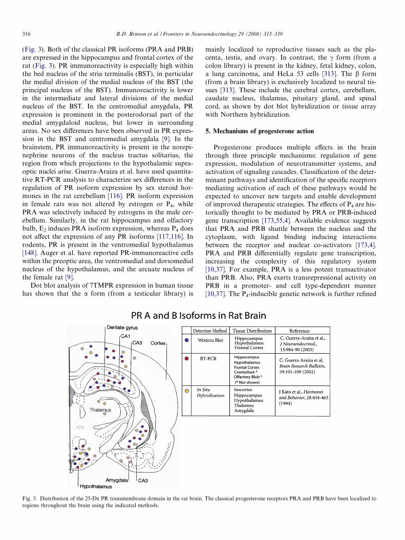
(Fig. 3). Both of the classical PR isoforms (PRA and PRB)are expressed in the hippocampus and frontal cortex of therat (Fig. 3). PR immunoreactivity is especially high withinthe bed nucleus of the stria terminalis (BST), in particularthe medial division of the medial nucleus of the BST (theprincipal nucleus of the BST). Immunoreactivity is lowerin the intermediate and lateral divisions of the medialnucleus of the BST. In the centromedial amygdala, PRexpression is prominent in the posterodorsal part of themedial amygdaloid nucleus, but lower in surroundingareas. No sex differences have been observed in PR expres-sion in the BST and centromedial amygdala [9]. In thebrainstem, PR immunoreactivity is present in the norepi-nephrine neurons of the nucleus tractus solitarius, theregion from which projections to the hypothalamic supra-optic nuclei arise. Guerra-Araiza et al. have used quantita-tive RT-PCR analysis to characterize sex differences in theregulation of PR isoform expression by sex steroid hor-mones in the rat cerebellum [116]. PR isoform expressionin female rats was not altered by estrogen or P4, whilePRA was selectively induced by estrogens in the male cer-ebellum. Similarly, in the rat hippocampus and olfactorybulb, E2 induces PRA isoform expression, whereas P4 doesnot affect the expression of any PR isoforms [117,116]. Inrodents, PR is present in the ventromedial hypothalamus[148]. Auger et al. have reported PR-immunoreactive cellswithin the preoptic area, the ventromedial and dorsomedialnucleus of the hypothalamus, and the arcuate nucleus ofthe female rat [9].
Dot blot analysis of 7TMPR expression in human tissuehas shown that the a form (from a testicular library) is
Fig. 3. Distribution of the 25-Dx PR transmembrane domain in the rat brain.regions throughout the brain using the indicated methods.
mainly localized to reproductive tissues such as the pla-centa, testis, and ovary. In contrast, the c form (from acolon library) is present in the kidney, fetal kidney, colon,a lung carcinoma, and HeLa 53 cells [313]. The b form(from a brain library) is exclusively localized to neural tis-sues [313]. These include the cerebral cortex, cerebellum,caudate nucleus, thalamus, pituitary gland, and spinalcord, as shown by dot blot hybridization or tissue arraywith Northern hybridization.
5. Mechanisms of progesterone action
Progesterone produces multiple effects in the brainthrough three principle mechanisms: regulation of geneexpression, modulation of neurotransmitter systems, andactivation of signaling cascades. Classification of the deter-minant pathways and identification of the specific receptorsmediating activation of each of these pathways would beexpected to uncover new targets and enable developmentof improved therapeutic strategies. The effects of P4 are his-torically thought to be mediated by PRA or PRB-inducedgene transcription [173,55,4]. Available evidence suggeststhat PRA and PRB shuttle between the nucleus and thecytoplasm, with ligand binding inducing interactionsbetween the receptor and nuclear co-activators [173,4].PRA and PRB differentially regulate gene transcription,increasing the complexity of this regulatory system[10,37]. For example, PRA is a less potent transactivatorthan PRB. Also, PRA exerts transrepressional activity onPRB in a promoter- and cell type-dependent manner[10,37]. The P4-inducible genetic network is further refined
The classical progesterone receptors PRA and PRB have been localized to

R.D. Brinton et al. / Frontiers in Neuroendocrinology 29 (2008) 313–339 317
by the expression of PR splice variants with variable ligandaffinities and transactivational activities [187].
The effects of P4 may be attributed to mechanisms apartfrom the ‘classical’ gene transcription mediated by PRAand PRB. Recently, P4 binding sites have been detectedat the surface of hypothalamic and spinal neurons[157,237]. These binding sites, identified as 25-Dx (alsoknown as PGRMC1), mediate the antiapoptotic actionsof P4 in granulose and luteal cells [212,211]. In the ovary,25-Dx complexes with the plasminogen activator inhibitorRNA-binding protein 1 (25-Dx/SERBP1) to activatecGMP-dependent protein kinase [68]. P4 signaling may alsobe mediated by several putative Src homology domainspresent in 25-Dx [210]. However, the P4-activated signalingpathways that are mediated by 25-Dx have yet to be deter-mined in neurons. Like 25-Dx, the seven transmembraneputative progesterone receptor 7TMPR may regulate P4
signaling. The 7TMPR has been shown to activate a per-tussis toxin-sensitive inhibitory G protein, resulting in acti-vation of the MAPK pathway through inhibition of cAMPproduction [271,312].
Progestereone and its 5a-reduced derivatives dihydropro-gesterone (DHP) and tetrahydroprogesterone (THP or allo-pregnanolone), can promote Schwann cell proliferation andactivation of the myelinating program of these cells [183].Melcangi and colleagues demonstrated that P4 and its deriv-atives increased expression of the transcription factors Sox-
Fig. 4. Model of progesterone-induced neuroprotective signaling. Progestogeeration through (1) ligand activation of a progesterone receptor, (2) which initiaThese signaling cascades converge on mitochondrial function to protect againstdirect protection signaling pathway. In the indirect prevention signaling pathwawhich (4) enhances mitochondrial function and enables neurons to better withstAb-induced JNK activation and mitochondrial dysfunction.
10 and Krox-20, both of which play a key role in Schwanncell physiology and in their myelinating program. Westernblot analyses indicated that Krox-20 was increased after3 h of treatment with P4, dihydroprogesterone, or tetrahy-droprogesterone, whereas P4 or dihydroprogesterone stimu-lated expression of Sox-10 after 6 h of exposure. Analysis ofrat and human promoters for these two transcription factorsindicated that putative P4-response elements are present inthe Krox-20 gene but not in Sox-10. These findings suggestthat P4 and its neuroactive derivatives could coordinate theSchwann cell-myelinating program utilizing different intra-cellular pathways [183].
Although mPR binds P4 with high selectivity and affin-ity, it does not bind many of the synthetic progestinsincluding norethisterone, norgestrel, promesgestrone, anddemegestone [271]. This differential binding may underliethe distinct differences in neuroprotection and MAPK acti-vation elicited by P4 and synthetic progestins [201,202].However, the neuronal expression of mPR has not yet beendetermined. The various progestin-activated signalingpathways can be combined synergistically or antagonisti-cally in an intriguing number of ways to regulate develop-ment, survival, and electrical activity in the CNS. Each stepof this signaling network can be influenced in a cell type- orbrain region-specific manner through alterations in recep-tor expression or ligand structure, opening up a wide arrayof therapeutic possibilities.
n prevents synaptic dysfunction associated with aging and neurodegen-tes second messenger signaling cascades, (3) to promote neuronal survival.toxic insults and include the indirect prevention signaling pathway and they, both ERK/CREB/bcl-2 and Akt pathways are simultaneously activated,and neurodegenerative insults. The direct protection pathway acts to block

318 R.D. Brinton et al. / Frontiers in Neuroendocrinology 29 (2008) 313–339
The MAPKs modulate cellular differentiation, prolifera-tion, survival, and death. Activation of the MAPK,extracellular signal regulated kinase (ERK), is requiredfor E2-induced neuroprotection (Fig. 4) [253]. We and oth-ers have demonstrated that both E2 and P4 activate theERK signaling pathway [201,202,254,253]. However,nuclear activation of ERK is not induced by medroxypro-gesterone acetate (MPA), a progestin that lacks neuropro-tective effects [202]. Phosphorylation of the MAPKsubstrate, cAMP response element binding protein(CREB), is associated with increased resistance to ischemicinjury [72,77], and CREB is activated in response to ovar-ian hormones [118,168,281,301,309,311]. CREB, in turn,can upregulate bcl-2 expression [77,301]. Accordingly, E2
and P4 upregulate bcl-2 in hippocampal neurons [201]. Incontrast to P4, MPA does not activate CREB, nor does itincrease bcl-2 expression [201]. On the contrary, MPAblocked E2-induced CREB activation and bcl-2 upregula-tion in primary hippocampal neurons.
Estrogen and P4 simultaneously activate the MAPK/ERK pathway as well as an alternate pro-survival path-way, the Akt pathway [254]. . Activation of Akt by E2
and P4 in cortical slice cultures is associated with increaseneuronal survival [254]. However, use of mixed cell typesin these slice cultures precludes differentiation betweendirect and indirect effects of the steroids on neural cells.In primary hippocampal neuron cultures, E2 and P4
directly activate Akt in neurons. Western blot analysis ofwhole-cell lysates revealed that E2 (10 ng/mL) and P4
(10 ng/mL), either alone or in combination, significantlyincreased Akt phosphorylation within 20 min of treatment.Treatment of primary hippocampal neurons with MPA didnot alter Akt phosphorylation, but blocked E2-inducedAkt phosphorylation.
More recently, it has been recognized that these steroidsalso regulate metabolic functions sustaining the energeticdemands of this neuronal activation [205,204,206,200,283,34,252,227]. Recent findings from Nilsen and colleaguesindicate that P4 significantly increased mitochondrial respi-ration 24 h following a single in vivo exposure at a magni-tude comparable to E2 [135]. Consistent with an increase inoxidative respiration, P4 and E2 significantly increasedCOXIV enzyme activity and expression of COXIV mRNA.Both P4 and E2 reduced free radical leak indicating greaterefficiency of electron transport, which was evidence in areduced generation of free radicals, P4 and E2 induced asignificant reduction in mitochondrial lipid peroxidation.The reduction in lipid peroxidation suggests the activationof mechanisms beyond solely mitochondrial efficiency. P4
induced a significant increase in MnSOD expression asdid E2 and E2/P4. In contrast, the expression of peroxire-doxin V was only increased by E2 but not in the P4- andP4 blocked the E2 induction of peroxiredoxin V. Theseresults indicate that both P4 and E2 can promote dismuta-tion of the superoxide anion O�2 by increasing MnSOD toform H2O2 whereas only E2 induces peroxiredoxin V thatpromotes clearance of H2O2 and prevention of oxidative
damage. Further, P4 and E2 directly regulate mitochondrialfunction and are not due to an increase in the number ofmitochondria as neither P4 nor E2 nor their combinationinduced evidence for mitochondrial biogenesis. While P4
was as efficacious as E2, the combination of P4 and E2
led to reduced efficacy. On all outcome measures, the com-bination of P4 and E2 resulted in a substantial decrement inresponse magnitude.
6. Neuroprotective actions of progesterone and progestin in
the CNS
P4 has established neuroprotective actions that likelyinvolve several different mechanisms. Anxiolytic effectsare one way by which P4 can reduce neural injury. Diversestimuli including kainate [25], pilocarpine [275], and penty-lenetetrazole [143] elicit stereotypic seizure behaviors andwithin several hours to a few days, significant neuronal lossin select brain regions such as the hippocampus. In theseparadigms, P4 treatment attenuates not only seizure behav-iors ([131], Rhodes, 2004 #2538; [225]), but also neuronalinjury [52]. The primary mechanism of neuroprotection inthese models appears to involve the P4 metabolite, allo-pregnanolone (APa, also known as 5a-pregnan-3a-ol-20-one and 3a,5a-tetrahydroprogesterone). P4 is metabolizedto APa following the sequential action of 5a-reductaseand 3a-hydroxysteroid dehydrogenase. APa acts as potentmodulator of c-aminobutyric acid subtype A (GABAA)receptors, increasing chloride conductance evoked byGABA [20,174]. This serves to decrease excitatory signal-ing and thus, antagonize seizure activity. Support for anAPa-mediated mechanism of P4 neuroprotection is pro-vided by the finding that (i) APa is as effective as P4 in sei-zure paradigms [52,224,27,81,78,80] and (ii) molecular[82,221] or pharmacological [224,80,155,276] inhibition ofP4 metabolism to APa blocks the protective actions ofP4. Interestingly, APa has also been implicated in the neu-roprotective effects of P4 following oxygen–glucose depri-vation of rat Purkinje cells [7] and traumatic injury[126,61,60,230].
P4 likely triggers multiple neuroprotective mechanisms.For example, in neuronal cultures, P4 activates MAPK/ERK [201,202,254] and Akt [254] signaling pathways, bothof which are associated with neuroprotection [254,253,314].Recent evidence suggests that, in spinal cord injury models,P4 neuroprotection is associated with upregulation ofbrain-derived neurotrophic factor (BDNF) [283,34,252]increased levels and activity of choline acetyltransferase[252], and a reduction in mitochondrial dysfunction [227].In cerebral ischemia models, the protective effects of P4
are attributed, in part, to suppression of inflammatoryresponses and nitric oxide synthase-2 expression [95]. Inaddition to its direct effects on neurons, P4 may exert indi-rect neuroprotective effects by acting on non-neuronal tar-get cell populations. For example, P4 reduces blood–brainbarrier leakage [232], decreases glial activation [111], andincreases myelination [12,154].

R.D. Brinton et al. / Frontiers in Neuroendocrinology 29 (2008) 313–339 319
P4 and E2 are known to modulate the activity of oneanother, sometimes antagonistically. Thus, it is of interestto determine the effect of P4 and progestogens on the neu-roprotective effects of E2. The antagonistic relationshipbetween P4 and E2 is illustrated by the finding that P4
can block E2-induced increases in spine density in the hip-pocampus [295,197]. P4 can also attenuate E2-inducedupregulation of BDNF, neurotrophin 3, and nerve growthfactor in the entorhinal cortex, but not in hippocampus, offemale rats [30]. P4 reverses estrogen-induced enhancementof spatial memory in ovariectomized female rodents [315].Recent results from our group demonstrate that both P4
and MPA block the neuroprotective effect of E2 in the hip-pocampus following kainate lesion in young female rodents[233] and reproductively senescent female rodents (Carrolland Pike, unpublished observations). P4 treatmentdecreases the estrogen receptor hybridization signal inmonkey brain [119], indicating that P4 may limit E2 signal-ing. However, in both a systemic kainate lesion model [11]and an ischemic stroke model [272], acute P4 treatment wasnot neuroprotective and did not significantly affect E2
neuroprotection.The neuroprotective action of P4 in traumatic brain
injury has been extensively studied by Stein and col-leagues. Results of their extensive body of work indicatethat a single injection of P4 attenuated cerebral edemawhen administered during the first 24 h after traumaticbrain injury (TBI) in rats whereas 5 days of P4 injectionresulted in improved spatial learning performance andreduced sensory neglect [246]. In subsequent analyses,Grossman et al. [111] found that P4 reduced edema levels,as in previous studies, while increasing the accumulationof activated microglia in traumatic brain injured rat brain[127]. However, a parallel analysis indicated that P4 andallopregnanolone reduced both IL-1b and TNF-a 3 hpost-traumatic brain injury, when the expression of thesecytokines peaked in the untreated animals [127]. Proges-terone-induced reduction in inflammatory cytokines wasalso observed in a medial frontal cortex model of trau-matic brain injury [214]. Progesterone inhibited theinjury-induced rise in complement factor C3, GFAP,and nuclear factor kappa beta (NFkappaB) [214]. Theparadox of the dual effect of P4 to induce accumulationof activated microglia and reduce inflammatory immunecytokines remains unresolved.
The contributions of gender and gonadal hormones inthe cascade of events following brain injury were investi-gated and revealed that normally cycling females exhibitedsignificantly less edema than males following traumaticbrain injury and that pseudopregnant females were virtu-ally spared from postinjury edema. Subsequent studies ofovariectomized females, with or without hormone treat-ment, indicated that the reduction of cerebral edema wasassociated primarily with the presence of circulatingprogesterone [229]. The Stein group then went onto todetermine whether P4 metabolite neurosteroids mediatedthe neuroprotection of exogenous or endogenous P4.
One day after traumatic brain injury, both P4-treated(16 mg/kg) and allopregnanolone (8 or 16 mg/kg)-treatedrats showed less caspase-3 activity, and rats treated withallopregnanolone (16 mg/kg) showed less DNA fragmen-tation in the lesion area, indicating reduced apoptosis.Nineteen days after the injury, rats treated with P4 or allo-pregnanolone (8 or 16 mg/kg) showed no difference innecrotic cavity size but had less cell loss in the medio-dor-sal nucleus of the thalamus and less learning and memoryimpairments compared with the injured vehicle-treatedrats. The results from their analyses indicated that P4
and allopregnanolone had similar neuroprotective efficacyafter traumatic brain injury, but that allopregnanoloneappeared to be more potent than P4 in promoting CNSrepair [60]. A follow-up study compared the effects of P4
and its metabolite, allopregnanolone, on the early injurycascade (apoptosis) and long-term functional deficits aftertraumatic brain injury [61]. Progesterone (16 mg/kg) orallopregnanolone (4, 8, or 16 mg/kg) were injected at 1,6 h, and then for five consecutive days after bilateral con-tusions of the frontal cortex in adult male rats. Within1 day after injury, P4 and allopregnanolone reducedexpression of pro-apoptotic proteins caspase-3 and Bax,and apoptotic DNA fragmentation. Progesterone andallopregnanolone also reduced the size of glial fibrillaryacid protein (GFAP)-positive astrocytes at the lesion site24 h after injury. At 19 days postinjury, rats given P4 orallopregnanolone (8 mg/kg) showed improved perfor-mance in a spatial learning task compared to injured ratsgiven only the vehicle [61].
The extensive body of basic science in vivo evidenceindicating that P4 was highly efficacious in reducing orpreventing the neurological consequences of traumaticbrain injury [259], led Stein and colleagues to conducta clinical trial of P4 in human victims of moderate tosevere coma associated traumatic brain injury [299]. Ina phase II, randomized, double-blind, placebo-controlledtrial conducted at an urban Level I trauma center with100 adult trauma patients who arrived within 11 h ofinjury with a postresuscitation Glasgow Coma Scalescore of 4–12 (scores associated with moderate to severedegrees of coma) were enrolled with proxy consent. Neu-rologic outcome was assessed 30 days postinjury. Sev-enty-seven patients received progesterone, 23 receivedplacebo. No serious adverse events were attributed toprogesterone. Adverse and serious adverse event rateswere similar in both groups, except that patients random-ized to P4 had a lower 30-day mortality rate than con-trols. Thirty days postinjury, the majority of severetraumatic brain injury survivors in both groups had rel-atively poor Glasgow Outcome Scale-Extended and Dis-ability Rating Scale scores. Moderate traumatic braininjury survivors who received P4 were more likely to havea moderate to good outcome than those randomized toplacebo. The authors concluded that results of this smallstudy, P4 caused no discernible harm and showed poten-tial benefit [299].

320 R.D. Brinton et al. / Frontiers in Neuroendocrinology 29 (2008) 313–339
7. Progesterone regulation of memory and neuronal
excitability
After more than three decades of research, it is now wellestablished that the ovarian hormone, E2, exerts a widevariety of effects on neural structures and function, partic-ularly within the hippocampus [191,298,296]. Electrophys-iological studies have shown that E2 enhances hippocampalCA1 synaptic transmission and plasticity by increasingNMDA and AMPA receptor activity, which results in neu-ronal excitation [268,75,293,294]. While the above studieshave focused exclusively on the effect of E2 on brain struc-ture and function, more recent studies have investigated theeffect of P4 and its neuroactive metabolites APa and preg-nanolone (PREG) on cognitive function and neuralexcitability.
Long-term potentiation (LTP) is considered to be thebest cellular model of memory trace formation in the brain,at least for certain forms of memory in the hippocampusand neocortex [16,33,165]. The phenomenon opposite toLTP is long-term depression (LTD), which was first dem-onstrated in cerebellar cortex by Ito in 1982 [136]. LTDhas also been demonstrated in the hippocampus and neo-cortex and like LTP, is considered a mechanism for mem-ory storage [22,65]. Although the molecular mechanismsunderlying LTP (and LTD) have been extensively investi-gated, there is a relative paucity of studies demonstratingthe critical role of LTP in behavioral learning and memory[248]. Nonetheless, LTP (and LTD) are the best currentmodels of synaptic plasticity, which may underlie memorystorage [29]. In the CA1, the most widely studied form ofLTP involves glutamate activation of NMDA receptors,which augments AMPA receptor function for the expres-sion and maintenance of LTP. However, this is not the soleform of LTP in the CA1, as Teyler and associates havedemonstrated a form of tetanus-induced LTP in the CA1that is independent of NMDA receptors and involves volt-age-dependent calcium channels [112].
Few studies have examined the acute effects of P4 onsynaptic transmission and plasticity, with the results ofthese studies being mostly contradictory. P4 (10 lM)reportedly has no effect on LTP (CA1 slices from 4-week-old rats), but no non-drug control was used in this study[137]. In another study, P4 (8–10 M, in CA1 slices) signifi-cantly enhanced synaptic transmission, as seen by anincreased field potential and population spike amplitude;however, following a seizure-induced tetanus, P4 decreasedthe field potential, the population spike responses, and theduration of after-discharges [67]. In whole cell patch clampof pyramidal neurons from slices of prelimbic cortex, P4
(100 lM) had no effect on the frequency of excitatorypostsynaptic currents (EPSCs), but inhibited dopamine-induced increases in EPSCs [71]. P4 dose–responsefunctions were not obtained in any of these studies. Inprimary hippocampal neurons, P4 as well as E2 enhancesglutamate-mediated increases in intracellular calcium, withE2 having a greater effect. P4 appears to interfere with E2
enhancement of synaptic transmission, as seen by the find-ing that co-treatment with P4 and E2 enhances glutamate-mediated increases in intracellular calcium to the samedegree as P4 alone [201].
Clinical investigations have been performed to under-stand the effect of P4 on moods associated with premen-strual dysphoric disorder (PMDD) [290]. PMDD isdefined in the Diagnostic and Statistical Manual of MentalDisorders (DSM-IV, APA 1994) as a cluster of both nega-tive mood symptoms and physical symptoms that occurduring the luteal phase of the menstrual cycle (when P4
and APa levels are high) and disappear several days follow-ing the onset of menstruation (when P4 and APa levels arelow). The increase in P4 (and APa) levels that occurs duringthe luteal phase of the menstrual cycle is considered to be atleast partly responsible for the negative mood changesassociated with PMDD [290,13]. Although ovarian steroidsare required for onset of premenstrual symptoms, womenwith PMDD are thought to exhibit an altered GABAreceptor sensitivity [264]. Support for the association ofP4 with negative mood symptoms comes from the findingthat postmenopausal women exhibiting intermediate APaplasma concentrations subjectively rated themselves ashaving significantly more negative mood symptoms duringP4 treatment than during treatment with unopposed E2 orplacebo [6]. P4 concentrations (measured via radioimmuno-assay) in the amygdala, cerebellum, and hypothalamus aresignificantly higher in fertile women in the luteal phase ofmenstruation than in postmenopausal controls [31]. More-over, in fertile women, APa concentrations are highest insubstantia nigra and basal hypothalamus, suggesting thatthe pattern of steroid secretion during the menstrual cycleis reflected in specific brain tissues [31].
P4 and APa are known to regulate cognitive function,particularly those functions related to mood and/orassociated with changes in the menstrual cycle (e.g., postpar-tum depression, major depression, epilepsy) [14]. TheGABAergic system also participates in major depression(for review, see [35]). The GABAA receptor mediates themajority of rapid (1–100 ms) synaptic inhibition in the mam-malian brain, and APa and PREG exert both anxiolytic andanesthetic effects by enhancing GABA-stimulated chlorideconductance. This enhanced conductance serves to hyperpo-larize postsynaptic membranes and results in neuronalinhibition [186,185]. Recent evidence suggests that specificneurosteroids ‘fine-tune’ neural inhibition via the GABAer-gic system [195]. In another recent study, the ability of P4 toinfluence cognition and memory of biologically salient stim-uli was investigated in healthy young women [278]. Here, asingle dose of P4 was orally administered to women whowere then asked to memorize and recognize faces whileundergoing functional magnetic resonance imaging. Theresults revealed that P4 decreases recognition accuracy with-out affecting reaction times. P4 also decreased amygdala andfusiform gyrus activity elicited by faces during memoryencoding, supporting the conclusion that P4 alters memoryfunction by influencing amygdala activity [278,279].
R.D. Brinton et al. / Frontiers in Neuroendocrinology 29 (2008) 313–339 321
In animals, P4 and its metabolites severely impair learn-ing and memory performance immediately followingadministration in the Morris water maze test [142,250].Although the mechanism underlying this impairment isunknown, a recent study demonstrated that pretreatmentof rats with APa induces a partial tolerance against theacute effects of APa in the Morris water maze test [274].These authors suggest that prolonged exposure to APa inwomen (e.g., pregnancy, postmenopausal hormonereplacement therapy, menstrual cycle) may alter cognitivebehavior such as learning and memory, possibly througha GABAergic-dependent mechanism.
Excitatory synapses, which can be approximated bydendritic spine density, serve as the substrate for learningand memory. A significant amount of literature describesthe effects of estrogen and P4 on dendritic spine formationin hippocampal pyramidal neurons [104]. In rats, ovariec-tomy results in a decrease (over 6 days) in the density ofdendritic spines, which can be prevented and reversed byE2 treatment [295]. Administration of P4 after estrogentreatment initially augments the effects of E2 (and sexualbehavior such as lordosis). However, 6 h later, P4 rapidlydecreases spine density to the very low levels, which areequivalent to levels seen 18 h after ovariectomization.Indeed, the number of dendritic spines in specific brainregions fluctuates over the 4–5 day estrous cycle in accor-dance with estrogen and P4 levels [76]. The down regulationof dendritic spines by P4 is blocked by the P4 antagonist,RU-38486, consistent with the presence of intracellularPRs in the hippocampus. Electron microscopic studieshave demonstrated the presence of non-nuclear PRs in gliaand dendritic spines in the hippocampus [191]. Also, P4 actsdirectly on GABAergic receptors to enhance GABA inhibi-tion and thus, counter the effects of E2 [197,291].
During the ovulatory cycle, extensive tissue remodelingoccurs throughout the body, including certain brainregions. Some of the most profound changes occur in uter-ine tissue, which undergoes extensive angiogenesis and cellproliferation during the follicular phase. In the absence of ablastocyst, the uterine growth phase is terminated byresorption or exfoliation of epithelial and vascular cells.Numerous inflammatory mediators are cyclically regulatedduring both the growth and exfoliative phase [73,158,180].These cyclically-regulated genes encode diverse proteins,many of which function in apoptosis such as Fas, cas-pase-3, M30 [8], complement C3 [180,178], and secretoryleukocyte proteinase inhibitor (SLPI) [149]. Progestins reg-ulate the expression of many of these genes. For example,progestins antagonize estrogens in regulation of C3[180,178], whereas P4 acts in synergy with the proinflamma-tory cytokine, IL-1, to induce expression of SLPI, an anti-microbial peptide important in host defense [149].
In contrast to uterine remodeling during the estrouscycle, brain ‘remodeling’ is more modest and does notinvolve major changes in the proportions of cell popula-tions. In the rodent hippocampus, certain synaptic bedsimplicated in declarative memory undergo striking, tran-
sient changes during the estrous cycle. The dendritic spinedensity of CA1 pyramidal cells undergoes cyclic changes,which are strongly correlated with sensitivity to NMDAreceptor-mediated synaptic responses [296,298]. There isalso increased aggregation of MAP2 in apical dendrites[223] as well as an increased expression of syntaxin, synap-tophysin (presynaptic), and spinophilin (postsynaptic) [51].The increase in dendritic spines is driven by elevations in E2
during the preovulatory follicular phase, whereas the rapidregression of these spines after ovulation depends upon ele-vation of P4 from the corpus luteum [295]. Administrationof the PR antagonist, RU 486, during proestrus inhibits thedecrease in spine density after proestrus [295]. Like theCA1, the hypothalamic arcuate nucleus undergoes remod-eling in response to the preovulatory luteinizing hormonesurge. In this region, astrocyte volume changes have beenassociated with altered GABAergic contacts on gonadotro-pin-releasing hormone axons [134,198]. There is now a sig-nificant literature concerning the effects of estrogen andprogesterone on dendritic spine formation in hippocampalpyramidal neurons [104]. In brief, ovariectomy (in rats)causes a decrease (over 6 days) in the density of dendriticspines, which can be prevented and reversed by estradioltreatment [295]. Progesterone treatment subsequent toestrogen treatment initially augments the effects of estra-diol (and sexual behavior, e.g., lordosis) and then (after6 h) results in a rapid decrease in spine density to the verylow values seen in ovariectomized animals by 18 h. Indeed,the number of dendritic spines in specific brain regions fluc-tuates over the 4–5 day estrous cycle in accordance withestrogen and progesterone levels [297]. The down regula-tion of dendritic spines by progesterone is blocked by theprogesterone antagonist, RU-38486, consistent with thepresence of intracellular progesterone receptors (PR). Elec-tron microscopic studies also indicated the presence of non-nuclear PRs in glia and dendritic spines in hippocampus[191]. Progesterone can also act directly on GABAergicreceptors to enhance GABA inhibition, thus counteringthe effects of E2 [197,291].
8. Progesterone regulation of glial cell function and response
Progesterone regulates responses in each of the majorglial cell types, astrocytes, microglia, oligodendrocytesand Schwann cells. During the estrous cycle astrocyte sizevaries with CA1 astrocytes shrinking immediately beforeincreases in spine density [153]. Astrocytes also decreasein size in the rostral preoptic location of gonadotropin-releasing hormone cell bodies [45]. Astrocyte size isstrongly associated with the expression of glial fibrillaryacidic protein (GFAP), which varies during the estrouscycle in the dentate gyrus [262]. Progesterone, and its neu-rosteroid metabolite dihydroprogesterone induced a signif-icant elevation of GFAP mRNA levels in type 1 astrocyteswithin hours of exposure with direct administration ofdihydroprogesterone inducing an increase within 2 hwhereas P4 required 6 h for increased GFAP expression.

322 R.D. Brinton et al. / Frontiers in Neuroendocrinology 29 (2008) 313–339
These findings suggest that the effect of P4 is likely due tometabolism to DHP. The requirement for conversion ofP4 to increase GFAP protein level was confirmed throughthe addition of finasteride (a specific blocker of the 5a-reductase) which completely abolished the effect of P4 [92].
In astrocytes, P4 regulates production of multiple pro-teins including those shown to be involved in regulatingsynaptic plasticity such as ApoE which is secreted by astro-cytes may be an important player in synaptic remodeling,since this protein transports cholesterol and other lipidsto outgrowing neurites [184]. Glial apoE mRNA changescyclically in the CA1 and arcuate nucleus [261,199], sup-porting a role for this protein in synaptic remodeling. Dur-ing pregnancy, twofold increases in uterine apoE levels areassociated with increased import of maternal lipids [277].Also, estrogen-dependent sprouting of perforant pathfibers to the hippocampus is absent in apoE-knockout mice[263,199,267]. Complex heterotypic cellular interactionsthat occur in response to ovarian steroids extend beyondastrocytic–neuronal interactions. Although ApoE issecreted by astrocytes, astrocytic responses to estrogenrequire interactions with microglia, as evidenced in mono-typic astrocyte cultures which are much less sensitive toestrogen than mixed glial cultures containing microglia[262]. Besides E2, Premarin� also induced ApoE expressionin mixed glia [235]. P4 increased ApoE secretion in macro-phages (microglia also express apoE mRNA) by acting onthe C-terminal lipid-binding domain of ApoE to block itsintracellular degradation [63]. The effect of other clinicalprogestins on glial apoE expression remains to bedetermined.
In many organ systems, estrogen actions are attenuatedor antagonized by progestins. As discussed above, E2
drives increases in CA1 spine density (growth phase), whileP4 promotes the regression of dendritic spines (during theproestrus to estrus transition). In mouse hippocampal slicecultures, mossy fiber sprouting into the molecular layer ofthe dentate gyrus is induced by deafferentation of theentorhinal cortex [267]. In this system, E2 (100 pM)increases sprouting by 75%. This concentration of E2 alsoinduces maximal apoE induction in mixed glia [236]. E2-dependent sprouting can be blocked by P4 or tamoxifen[267]. P4 appears to differ from MPA in its ability to regu-late synaptic plasticity. Preliminary results from our labo-ratory show that MPA, but not P4, inhibits E2-mediatedsprouting. This difference is consistent with recent findingsby Nilsen and Brinton that P4, but not MPA, is neuropro-tective against excitotoxicity and stimulates nuclear activa-tion of ERK. Further, MPA, but not P4, blocked theneuroprotective effects of E2 [202]. Additional studies willbe necessary to evaluate the differences and similarities ofMPA and P4.
In some models, P4 has been reported to have anti-inflammatory activities. After stab wounds, P4 decreasedreactive astrocytes to a greater extent than E2, but less thanpregnenolone[88,87]. In a model of cerebral concussion,male rats given an i.p. injection of P4 of had less edema
[111] and lipid peroxidation [232] than their untreatedcounterparts. On the other hand, P4 had a negligible effecton glial activation or neuronal loss in this model [111]. Inaccord with findings from the cerebral concussion model,P4 inhibited the level of clinical neurogenic edema, possiblyby reducing meningeal release of substance P [113]. Ofcourse, P4 has many complex interactions with neurotrans-mitters, which may underlie this and other anti-inflamma-tory effects (e.g., activation of the GABA receptor by P4
and other C21 steroids [79]).Progesterone regulation of myelination is now well doc-
umented and is a compelling example of the profounddirect effects of P4. The sciatic nerve, and Schwann cellsin particular, are capable of synthesizing P4 and possessthe enzymes necessary to convert P4 to the 5a-reducedand the 3a–5a-reduced derivatives of P4: dihydroprogester-one and tetrahydroprogesterone [188]. Progesterone recep-tor has been detected in both sciatic nerve and in Schwanncell cultures. P4 and its metabolite neurosteroids regulateexpression of two major proteins of the peripheral nervoussystem (PNS): the glycoprotein Po (Po) and peripheralmyelin protein 22 (PMP22). Melcangi and colleagues haveshown that: (a) dihydroprogesterone enhanced the lowmRNA levels of Po in the sciatic nerve of aged male rats;(b) P4 and its derivatives stimulates the gene expressionof Po in the sciatic nerve of adult rats and in Schwann cellcultures; (c) tetrahydroprogesterone increased PMP22 geneexpression in the sciatic nerve of adult rats and in Schwanncell cultures. They further demonstrated that P4 and itsderivatives control Po gene expression via the PR, whiletetrahydroprogesterone modulated expression of PMP22through the GABAA receptor[188]. Melcangi and cowork-ers went on to demonstrate that P4 and its derivatives reg-ulate other myelin proteins [i.e., myelin-associatedglycoprotein (MAG) and myelin and lymphocyte protein(MAL)] in sex-specific cultures of rat Schwann cells [182].Progesterone or dihydroprogesterone induced a stimula-tory effect on P0 mRNA levels in male but not in femaleSchwann cells. In contrast, treatment with tetrahydropro-gesterone increased gene expression of P0 in female derivedSchwann cells. A similar sex-difference was also evident forother myelin proteins. PMP22 expression was increased byP4 in male derived Schwann cell cultures, whereas tetrahy-droprogesterone induced an increase of mRNA levels infemale derived cells. Moreover, MAG was stimulated bytetrahydroprogesterone treatment in male cultures only,whereas MAL expression was unaffected by neuroactivesteroid treatment in both male and female cultures. Collec-tively these findings indicate that P4 and its metabolite neu-roactive steroids on regulate myelin protein expression in asexually dimorphic manner. This finding might representan important background for sex-specific therapies ofacquired and inherited peripheral neuropathies [182,188].Melcangi and colleagues pursued the relevance of thesefindings for age-associated myelin loss and morphologicalalterations of myelinated fibers in the sciatic nerve of 22–24-month-old male rats. The sciatic nerves of untreated

R.D. Brinton et al. / Frontiers in Neuroendocrinology 29 (2008) 313–339 323
old male rats, showed a general disorganization and a sig-nificant reduction in the density of myelinated fibers, com-pared to nerves from 3-month-old male rats. The effect ofaging was particularly evident in myelinated fibers of smallcaliber (<5 lm in diameter). In addition, the sciatic nervesof old rats showed a significant increase in the number offibers with myelin infoldings in the axoplasm and in thenumber of fibers with irregular shapes. Treatments of oldrats with P, DHP and THP resulted in a significant increasein the number of myelinated fibers of small caliber, a signif-icant reduction in the frequency of myelin abnormalitiesand a significant increase in the g ratio of small myelinatedfibers. Furthermore, P4 significantly reduced the frequencyof myelinated fibers with irregular shapes. Results of thesein vivo animal studies indicate that in the aged male rat P4
and its neuroactive metabolites reduced aging-associatedmorphological abnormalities of myelin and aging-associ-ated myelin fiber loss in the sciatic nerve [12].
In the central nervous system P4 and its neurosteroidmetabolites were found to promote glial functions, suchas the synthesis of myelin proteins. In glial cell cultures pre-pared from neonatal rat brain, P4 increased the number ofoligodendrocytes expressing myelin basic protein and the20,30-cyclic nucleotide-30-phosphodiesterase (CNPase), thethird most abundant myelin protein in the CNS [18]. Therole of P4 in myelination is extensively covered in severalexcellent reviews by Schumacher and colleagues and thereader is referred to these for further reading [243,244,18].
9. Progesterone regulation of meiosis and mitosis
During development of both vertebrates and inverte-brates, P4 promotes meiosis to generate germ cells[19,47,240]. P4 induced re-entry into the cell cycle at medi-ated by a membrane-bound PR [19,17,21,270,123].
P4 promotion of meiosis is mediated by a rise in intracel-lular Ca2+ [24,196,286]. In Xenopus oocytes, P4 induces theresumption of meiosis (maturation) through a non-geno-mic mechanism involving inhibition of adenylyl cyclaseand reduction of intracellular cAMP. However, P4 actionin Xenopus oocytes is not blocked by pertussis toxin, indi-cating that inhibition of the oocyte adenylyl cyclase is notmediated by the a subunit of classical Gi-type G proteins[181]. Subsequent analyses indicate that P4 is likely induc-ing maturation by antagonizing constitutive Gbc-mediatedinhibition of cell cycle progression [181].
Intracellular Ca2+ influx and inhibition of Gbc are notthe sole requirements for P4 regulation of meiosis. Multiplelaboratories have demonstrated that P4 activates MAPKsignaling pathway in oocytes and that this pathway isrequired for promotion of meiosis by P4 [69,144,74]. P4
activation of MAPK leads to the formation of the M-phasepromoting protein complex (CDC2 and cyclin B), whichpromotes G2 to M phase transition [171,189].
Mitotically, P4 has a complex function in the uterus andcan be both inhibitory and stimulatory for proliferationdepending upon cell type (endometrial or stromal) the reg-
imen of treatment, the type of PR that is activated (PRAversus PRB), the dose of E2 and P4 , and when in the cycleP4 is administered [56–58,96,133,150]. In the endometrium,P4 inhibits proliferation of endometrial cells whereas P4 is aproliferative agent in the stromal cells of the uterus. Fur-ther, P4 inhibited proliferation in E2 primed uterus butnot when administered alone or with low dose E2. A prog-estinal agent in hormone therapy is added to antagonizeendometrial cell proliferation in the uterus [249]. In addi-tion to P4 action in the uterus, P4 can promote proliferationin the breast [151,215,26,50].
In the Women’s Health Initiative, in which medroxypro-gesterone acetate (MPA) was used as the progestinal agent,an increased risk of breast cancer in the hormone therapy(HT) trial was observed which did not occur in the estrogenonly therapy (ET) [50,300,288,36,269,86]. The WHI HTtrial also revealed an increased risk of invasive ovarian can-cer and a reduced risk of endometrial cancer [5,234]. Theuse of the progestin, MPA, in the WHI HT trials has beenproposed to be a major factor contributing to the increasedcancer risk seen in this trial. However, the tumorgenicproperties of different progestogens have not been system-atically studied, and not all progestogen molecules have thesame antagonistic or agonistic profiles [256,257,15]. Theimpact of different progestogens on the proliferation ofneural stem cells or neural progenitors is currentlyunknown.
10. Progesterone and estrogen regulation of neurogenesis and
neural progenitor proliferation
As in the uterus [56,105], P4 regulation of mitosis of neu-ral progenitors in brain has a complex profile. Tanapatet al. have shown that ovariectomized rats treated with ahigh level of E2 have enhanced hippocampal cell prolifera-tion, whereas subsequent exposure to P4 resulted in block-ade of the E2-induced enhancement of cell proliferation[266]. In contrast to P4 regulation of E2-induced neurogen-esis in vivo, we have demonstrated that P4 alone enhancescell proliferation in vitro [284]. More recent results indicatethat P4 induced a dose dependent significant increase in ratneural progenitor cells (rNPC) proliferation as measuredby BrdU incorporation over a 24-h exposure period withan EC100 value of 100 pM. Unlike its metabolite steroidAPa, the dose–response curve for P4 was shallow but linearup to 1 lM whereas the dose–response for APa was steeplylinear followed by an inverse function at 1 lM. Further,when compared to the dose–response for E2-induced rNPCproliferation (EC100 of 250 nM), P4 induced a more con-sistent enhancement of rNPC proliferation. The timecourse of P4-induced rNPC proliferation generated 2important outcomes. First, P4-induced DNA synthesisoccurred rapidly within the first 1–4 h of P4 exposure. Sec-ond, it appears that P4-induced DNA synthesis does notpersist beyond 6 h and that by 8 h, P4 no longer inducesDNA synthesis. These data suggest that P4 is not drivingrNPCs into prolonged or uncontrolled proliferation.

324 R.D. Brinton et al. / Frontiers in Neuroendocrinology 29 (2008) 313–339
Lastly, we conducted a steroid specificity analysis whichindicates that P4 and its metabolite APa are both prolifer-ative agents [40].
Our own work has demonstrated that the neurosteroidP4 metabolite, APa is a potent, stereoisomer-specific pro-moter of neurogenesis of both rat hippocampal neural pro-genitor cells and human cortical neural stem cells [284].Allopregnanolone-induced proliferation was isomer andsteroid specific, in that the stereoisomer 3b-hydroxy-5b-pregnan-20-one and related steroids did not increase 3H-thymidine uptake. Immunofluorescent analyses for theneural progenitor markers, nestin and Tuj1, indicated thatnewly formed cells were of neuronal lineage. Furthermore,microarray analysis of cell cycle genes and real time RT-PCR and western blot validation revealed that allopreg-nanolone increased the expression of genes, which promotemitosis and inhibited the expression of genes that represscell proliferation. Allopregnanolone-induced proliferationwas antagonized by the voltage gated L-type calcium chan-nel blocker nifedipine consistent with the finding that allo-pregnanolone induces a rapid increase in intracellularcalcium in hippocampal neurons via a GABA type Areceptor activated voltage gated L-type calcium channel[285]. These data demonstrate that APa significantlyincreased rNPC and hNSM proliferation with concomitantregulation in mitotic cell cycle genes via a voltage gated L-type calcium channel mechanism.
APa-induced neurogenesis is a dose-dependent process,with concentrations in the low to mid nanomolar rangepromoting proliferation and concentrations exceeding1 lM significantly inhibiting neurogenesis. The biphasicdose–response profile of APa-induced neurogenesis couldaccount for the disparity between our in vitro data andreports that APa decreases neurogenesis in the rat dentategyrus in vivo. In these in vivo studies, APa inhibited neuro-genesis of rat SVG cells following intracerebral ventricularinjection of 7.8 mmol of APa [91,92]. Considering that theinjected concentration is diluted into the cerebrospinal fluidand the volume of the cerebrospinal fluid in a 300-g rat is�580 ll [163], the final concentration of APa would bemore than 50 lM. Thus, inhibition of neurogenesis atmicromolar concentrations of APa in this study is consis-tent with our dose–response data, which demonstrates inhi-bition of neural progenitor cells proliferation atmicromolar concentrations and promotion of neurogenesisat nanomolar concentrations [284]. Griffin and Mellonfound that early administration of APa substantially delaysprogression and severity of symptoms in a transgenicmouse model of Niemann-Pick Type C, a disease charac-terized by disrupted neurosteroidogenesis [109].
While production of new neurons from proliferatingstem/progenitor cells in the SGZ of the dentate gyrus ismaintained throughout life in multiple species includinghumans [103,83], the magnitude of neurogenesis declineswith age. Age-associated decline in neurogenic potentialin the dentate gyrus has been observed as early as middleage [159,220] and has been proposed to contribute to
age-related learning and memory impairments[62,84,89,1]. The mechanism underlying age-associateddecline in neurogenesis remains to be fully determined.However, loss of the growth factors FGF-2, IGF-1, andVEGF in the microenvironment of the SGZ is a prime con-tributor to the reduced neurogenic potential of the SGZ[122]. Recent studies have demonstrated that the levels ofthese three multiple stem/progenitor cell proliferation fac-tors decline early on during the course of aging in the hip-pocampus [28,247]. Hippocampal levels of FGF-2, IGF-1,and VEGF are more than 50–60% decline lower in adultrats than in young rats [247]. These findings suggest thatthe dramatic decline in dentate neurogenesis could belinked to reduced concentrations of FGF-2, IGF-1, andVEGF in the hippocampus, as each of these factors canindividually influence the proliferation of stem/progenitorcells in the SGZ of the dentate gyrus. For example, FGF-2 enhances dentate neurogenesis in both neonatal and adultbrain [247,209,222,273,48,49,282,85], and intracerebroven-tricular (ICV) infusions of FGF-2 upregulate dentate neu-rogenesis in the aged brain [159,85,132,141,160]. Likewise,ICV administration of IGF-1 increased dentate neurogene-sis in the adult and aged brain [1,2,176]. VEGF can pro-mote dentate neurogenesis in both the intact and theinjured adult brain following ICV administration[265,139,140]. During neurogenesis, VEGF may act as achemoattractant that specifically targets FGF2-stimulatedneural progenitors [308].
Growth factors regulate a myriad of cellular processesaside from neurogenesis. For example, EGF has well-char-acterized proliferative effects on the endometrium. EGFgene expression dramatically increases in the endometrialglands of pregnant mares at approximately 40 days afterovulation [260]. This upregulation is maintained until atleast day 250 of gestation and is associated with an increasein EGF receptor binding sites in the endometrium [170].The expression of both IGF and TGFb1 is also upregu-lated during this time [169]. Administration of varyingdoses and combinations of P4 and estrogen for 35 daysyields negative or only weakly positive EGF expression,whereas administration of only P4 for 40 days stronglyupregulated EGF expression irrespective of additionaltreatment with estrogen [90]. These findings underscorethe importance of P4 in regulation of growth factors andreveal a mechanism for P4-associated cell proliferationin vivo.
Similar to EGF, IGF, and TGF, expression of brainderived neurotrophic factor (BDNF) is positively corre-lated with E2 and P4 levels and negatively correlated withmenopausal age [23]. Hormone replacement therapyrestored plasma BDNF levels to levels seen in fertilewomen during the follicular phase [23]. Circulating plasmalevels of BDNF change during the menstrual cycle, sug-gesting that P4 may regulate neurotrophin expression[23]. Modifications in BDNF circulating levels during themenstrual cycle suggest a potential role for gonadal sexhormones in regulating neurotrophin expression, which

R.D. Brinton et al. / Frontiers in Neuroendocrinology 29 (2008) 313–339 325
has implications for sustaining the regenerative milieu ofthe brain during the menopausal years.
Regulation of cell cycle entry by progesterone and itsneurosteroid metabolites as therapeutic regenerative agentswill require careful analysis prior to development for resto-ration of neurons lost due to neurodegenerative disease. InAlzheimer’s disease (AD), cell cycle-specific gene expres-sion is upregulated [32] and, evidence indicates that mitoticsignaling is dysregulated [305]. In the aged and AD brain,both the pool of neural stem cells and their proliferativepotential are markedly diminished [159,177]. In addition,the level of potential regenerative factors is reduced inthe brains of AD patients compared to age-matched con-trols [287]. Herrup and colleagues have found that ectopicexpression of cell cycle proteins predicts the site of neuro-nal cell death in the AD brain [41], leading these investiga-tors to propose that dysregulation of various elements ofthe cell cycle contributes to regionally specific neuronaldeath in AD. They also found that DNA replication pre-cedes neuronal death in AD brain [304]. More disturbing,cell cycle events precede neuronal death at all stages ofAD, from mild cognitive impairment to advanced AD[305]. This finding has important implications for strategiestargeting neurogenesis in the AD brain. Specifically, it sug-gests that promoting entry into the cell cycle could poten-tially be a double-edged sword, with benefit to healthybrains but with exacerbation of ectopic mitosis in brainsdestined to develop AD or with existing AD.
11. Progesterone and regulation Alzheimer’s diseasepathology
A key hypothesis linking P4 to AD posits that P4 acts asan endogenous regulator of b-amyloid (Ab) metabolism.According to the widely but not universally embraced‘amyloid cascade hypothesis,’ AD pathogenesis is triggeredby any of a number of events that have the final commonendpoint of increasing the pool of soluble Ab[125,124,255]. In turn, elevated soluble Ab leads to the for-mation of an array of soluble oligomeric, minimally solubleaggregated, and eventually insoluble fibrillar Ab species, allof which are linked in a variety of ways to neurodegenera-tive cascades [53,217,216,245,179,190,251,164]. Thus,factors that have the net effect of reducing the pool ofsoluble Ab are thought to represent potentially powerfulstrategies for preventing the development of AD[97,152].
Neurosteroids have recently been measured in variousbrain regions of aged Alzheimer’s disease patients and agednon-demented controls by GC/MS [241]. In Alzheimer’spatients, there was a general trend toward lower levels ofneurosteroids in different brain regions, and neurosteroidlevels were negatively correlated with two biochemicalmarkers of Alzheimer’s disease, the phosphorylated tauprotein and the b-amyloid peptides [241]. The formationof these metabolites within distinct brain regions negativelycorrelated with the density of b-amyloid deposits.
Available evidence suggests that E2, like P4, may influ-ence Ab levels. In women, E2 status has been linked tothe development of AD [15]. Schonknecht and colleaguesfound that levels of E2 in cerebral spinal fluid are lowerin AD patients than in non-AD controls and that, in theAD group, E2 levels are inversely correlated with Ab1–42
levels [239]. In accord with this finding, several cell culturestudies have shown that E2 decreases Ab production, pre-sumably increasing the non-amyloidogenic cleavage ofamyloid precursor protein (APP) via a-secretase[46,138,302,303,280,107]. The physiological significance ofthis effect has been confirmed by in vivo studies investigat-ing the relationship between circulating E2 concentrationsand brain levels of Ab. Depletion of endogenous E2 viaovariectomy results in a significantly increases levels of sol-uble Ab in brains of mice [238] and guinea pigs [213].Importantly, this effect is partially reversed by E2 replace-ment. Furthermore, E2 reduces pools of soluble [175,310]and deposited Ab [310] in mouse models of AD, stronglyimplicating E2 as a regulator of AD pathogenesis. Unex-pectedly, two recent studies using transgenic mouse modelsof AD have found weak [106] or absent evidence [128] thatE2 reduces insoluble pools of Ab. Several factors may havecontributed to these negative results, including the trans-genic strains, the relevant pool of Ab, and the differencebetween estrogen levels in circulation and in the brain[307]. Prior investigations of Ab regulation by ovariansex steroids have used ovariectomy models (which depletesendogenous E2 and P4) combined with E2, but not P4,replacement [213,175,310,106,128].
While E2 replacement is beneficial in regulation of Abmetabolism, the interaction between E2 with P4 is more clin-ically relevant. That is, how are Ab levels affected when P4
acts in concert with E2? Our group has recently addressedthis question in the 3xTg-AD mouse model of AD. Ourresults suggest that continuous E2, but not P4 treatmentattenuates the acceleration of Ab accumulation and memorydeficits observed in ovariectomized mice. More importantly,in animals receiving both hormones, P4 blocked the benefi-cial effect of E2 on Ab accumulation [44].
12. Progestogens, progestins and metabolism
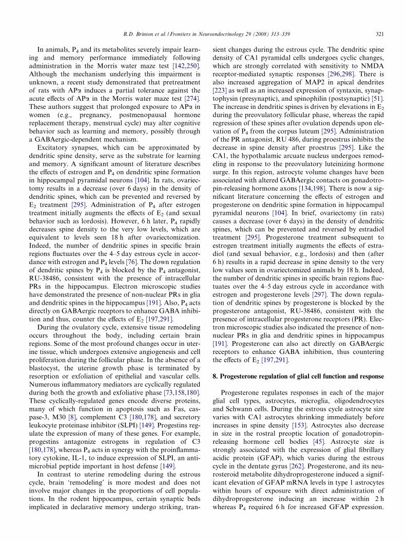
Many progestogens are used therapeutically amongthese, P4 is the only naturally occurring progestogen (seeTable 1). The remainder, which are synthetic, are referredto as progestins (see Table 1) [257]. Progestins are classifiedon the basis of their chemical structure, since the structuresof these molecules vary widely. Progestins can be dividedinto those related in chemical structure to P4 and thoserelated chemically to testosterone. The classificationscheme and the names of progestins in each of the catego-ries are summarized in Table 1. This classification schemedoes not denote the chemical source of the compounds.
Progestins related to P4 are characterized by the pres-ence or absence of a methyl group on carbon 10, and aresubdivided into pregnanes (21 carbons) and 19-norpregn-
Table 1Classification of progestogens
I. Natural II. Synthetic progestins
Structurally related to progesterone Structurally related to androgen
Progesterone 1. Pregnane derivativesa. Acetylated: medroxyprogesterone acetate, megestrol acetate,
cyproterone acetate, chlormadinone acetate, medrogestoneb. Non-acetylated: dydrogesterone
2. 19- Norpregnane derivativesa. Acetylated: nomegestrol acetateb. Non-acetylated: demegestone, trimesgestone, promgeestone, nesterone
1. Ethinylateda. Estranes: norethindrone, norethynodrel, lynestrenol, nor-
ethindrone acetate, ethynodiol diacetate, tiboloneb. Gonanes: levonorgestrel, desogestrel, norgestimate,
gestodene
2. Non-ethinylated: dienogest, drospirenone
326 R.D. Brinton et al. / Frontiers in Neuroendocrinology 29 (2008) 313–339
anes (20 carbons) (see Tables 1 and 2) [258]. The pregnanesand 19-norpregnanes can be further separated into com-pounds with and without an acetyl group. One of thebest-known and most widely used of these progestins isMPA, which is classified as an acetylated pregnane. Allof the 19-norpregnane progestins have been used primarilyin Europe and not in the United States.
Unlike the progestins related to P4, which are first sub-divided on the basis of the number of carbons (21 versus20), those related to testosterone are first subdivided onthe basis of whether or not they contain an ethinyl group.The ethinylated progestins are subdivided further intothose related to the parent steroid, estrane, and thoserelated to 13-ethylgonane. Both estranes and 13-ethylgon-anes lack a methyl group at carbon 10. The estrane groupof progestins consists of norethindrone and its prodrugs,namely norethindrone acetate, ethynodiol diacetate, nore-thynodrel, and lynestrenol. These prodrugs, which are con-sidered part of the norethindrone family, have been widelyused for hormone therapy and/or contraception. Althoughtibolone is also a prodrug and is listed in the estrane cate-gory, it is not converted to norethindrone. Instead, it istransformed to other active metabolites.
The 13-ethylgonanes contain an ethyl group on carbon13 of the basic steroid nucleus (gonane). This category ofprogestins, sometimes referred to as the levonorgestrel fam-ily, consists of levonorgestrel and the levonorgestrel deriv-atives desogestrel, norgestimate, and gestodene. The latterthree progestins are often referred to as the new progestins,as they have been marketed relatively recently. In contrast,levonorgestrel has been used for many years.
Norgestimate and desogestrel, but not gestodene, areprodrugs. However, only norgestimate is converted to lev-onorgestrel. Norgestimate is also converted to deacetylatednorgestimate (levonorgestrel-3-oxime), which has progesta-tional activity. Desogestrel is converted to its active form,3-ketodesogestrel. Gestodene has inherent progestationalactivity, but is not approved for use in the United States.
13. Progestogen metabolism
With the exception of P4, little is known about themetabolism of most progestogens. Baulieu first discovered
that P4 is converted to neuroactive metabolites in the brain[20,243,242]. This has now been well established by manylaboratories and documented in multiple species includingthe rodent and human. Neurosteroids such as APa are syn-thesized in the central and peripheral nervous system, pri-marily by myelinating glial cells, but also by astrocytesand several neuron types [243,193,194,54]. A region-spe-cific expression pattern of P4-converting enzymes in brainis evident in both the hippocampus and cortex of rodentand human brain [193,194,54,108]. The P4-convertingenzymes 5a-reductase and 3a-hydroxysteroid dehydroge-nase are expressed in the hippocampus of both the rodentand human brain and convert P4 to its 5a, 3a-reducedmetabolites (e.g., APa). Remarkably, these enzymes arealso present and functional in pluripotential progenitors[167]. The conversion of P4 to its 5a, 3a-reduced metabo-lites can be blocked by 5a-reductase inhibitors, such asfinasteride.
Of the progestins, the metabolism of norethindrone andlevonorgestrel is the best characterized (see Table 2). Thesecompounds undergo extensive reduction at the doublebond between carbons 5 and 6 and the carbonyl group atcarbon 3, resulting in the formation of dihydro- and tetra-hydroderivatives. They can also undergo hydroxylation.Surprisingly little is known about the metabolism of themost widely used progestin, MPA. This compound, likeother progestins structurally related to P4 or testosterone,contains a double bond between carbons 5 and 6 as wellas a ketone group at carbon 3. Therefore, it is likely toundergo extensive reduction at these functional groups.Unlike the neuroactive metabolites of P4 (e.g., APa),exceedingly little is known about the neuroactive propertiesof progestin metabolites.
14. Progesterone and PRs: translational and therapeutic
challenges
Relative to estrogen neurobiology, the non-reproduc-tive neural functions of P4 and the basic genomic, signal-ing and cell biology of these processes are just emerging.Progesterone and its neuroactive metabolites can pro-mote the viability of neurons and function of glial cellswithin both the central and peripheral nervous system.

Table 2Structures of selected progestogens and list of their active metabolites
Category Progestogens Structure Active compound of prodrug
Naturalprogestogens
Progesterone
O
O
Pregnane(Acetylated)
Medroxyprogesterone acetate
O
O
CH3
O O
Megestrol acetate
O
O
CH3
O O
Chlormadionone acetate
O
O
Cl
O O
Cyproterone acetate
O
O
Cl
O O
(continued on next page)
R.D. Brinton et al. / Frontiers in Neuroendocrinology 29 (2008) 313–339 327

Table 2 (continued)
Category Progestogens Structure Active compound of prodrug
Pregnane(Non-acetylated)
Dydrogesterone
O
O
Medrogestone
O
O
CH3
CH3
19-Norpregnane(Acetylated)
Nomegestrol acetate
O
O
CH3
O O
Nesterone
O
O
OO
CH2
Norpregnane(Non-acetylated)
Trimegestone
O
OOH
Demegestone
O
O
CH3
328 R.D. Brinton et al. / Frontiers in Neuroendocrinology 29 (2008) 313–339

Table 2 (continued)
Category Progestogens Structure Active compound of prodrug
Promegestone
O
O
CH3CH3
Tesosterone derivative(Estranes)
Norethindrone
O
HO
CCH
Noethindrone acetate
O
O
CCH
O
Norethindrone
Ethynodiol diacetate
O
CCH
O
O
O
Norethindrone
Norethynodrel
HO
CCH
O
Norethindrone
Lynestrenol
HO
CCH
Norethindrone
(continued on next page)
R.D. Brinton et al. / Frontiers in Neuroendocrinology 29 (2008) 313–339 329

Table 2 (continued)
Category Progestogens Structure Active compound of prodrug
Tibolone
O
HO
CCH
D4-Tibolone3a-OH-Tibolone3b-OH-Tibolone
Testosterone derivatives(Gonanes)
Levonorgestrel
O
C
HO CH
Desogestrel
H2C
OH
C CH
3-Ketodesogestrel
Norgestimate
N
O
H
HO
O
C CH
Levonorgestrel-3-oximeLevonorgestrel
Gestodene
OH
C CH
O
Testosterone derivatives(Non-ethinylated)
Drospirenone
O
O
O
Dienogest
CH3OH
H2C
O
C
N
330 R.D. Brinton et al. / Frontiers in Neuroendocrinology 29 (2008) 313–339

R.D. Brinton et al. / Frontiers in Neuroendocrinology 29 (2008) 313–339 331
While there is a substantial body of evidence regardingthe pleiotrophic actions of P4 [244], much remains tobe determined regarding the specific PR required andthe associated effector mechanism(s) of action. The useof P4 as a myelinating agent and as a treatment for trau-matic brain injury highlight the basic science and clinicalimportance of understanding the direct effects of P4 andother progestogens. It is interesting to note that womenhave a greater risk of developing the demylinating dis-ease multiple sclerosis with a frequent onset at meno-pause [207]. Given the significant impact of P4 and itsmetabolite, allopregnanolone (tetrahydroprogesterone)on remylination following injury and preventing age-associated myelin loss, the potential therapeutic use ofP4 for remylination is substantial in both men andwomen. Of particular concern, given the results of theWHI and WHIMS trials, is the impact of clinically usedprogestins on neurological function. While progestogensin hormone therapy are given to reduce the risk of uter-ine cancer, these agents potentially exert effects in brain.Collectively, we know little regarding the impact of dif-ferent progestins on neural function either acutely orchronically. The wide distribution of progesterone recep-tors in brain suggests that this gonadally and brainderived steroid plays a significant role in neural function,which awaits continued discovery.
Acknowledgments
Development of this work was supported by the Na-tional Institute of Aging P01AG026572 on Progesteronein Brain Aging and Alzheimer’s Disease to R.D.B.
References
[1] M.A. Aberg, N.D. Aberg, H. Hedbacker, J. Oscarsson, P.S.Eriksson, Peripheral infusion of IGF-I selectively induces neuro-genesis in the adult rat hippocampus, J. Neurosci. 20 (2000) 2896–2903.
[2] M.A. Aberg, N.D. Aberg, T.D. Palmer, A.M. Alborn, C. Carlsson-Skwirut, P. Bang, L.E. Rosengren, T. Olsson, F.H. Gage, P.S.Eriksson, IGF-I has a direct proliferative effect in adult hippocampalprogenitor cells, Mol. Cell Neurosci. 24 (2003) 23–40.
[3] G.F. Allan, X. Leng, S.Y. Tsai, N.L. Weigel, D.P. Edwards, M.J.Tsai, B.W. O’Malley, Hormone and antihormone induce distinctconformational changes which are central to steroid receptoractivation, J. Biol. Chem. 267 (1992) 19513–19520.
[4] G.F. Allan, S.Y. Tsai, M.J. Tsai, B.W. O’Malley, Ligand-dependentconformational changes in the progesterone receptor are necessaryfor events that follow DNA binding, Proc. Natl. Acad. Sci. USA 89(1992) 11750–11754.
[5] G.L. Anderson, H.L. Judd, A.M. Kaunitz, D.H. Barad, S.A.Beresford, M. Pettinger, J. Liu, S.G. McNeeley, A.M. Lopez, Effectsof estrogen plus progestin on gynecologic cancers and associateddiagnostic procedures: the Women’s Health Initiative randomizedtrial, JAMA 290 (2003) 1739–1748.
[6] L. Andreen, I. Sundstrom-Poromaa, M. Bixo, A. Andersson, S.Nyberg, T. Backstrom, Relationship between allopregnanolone andnegative mood in postmenopausal women taking sequential hor-mone replacement therapy with vaginal progesterone, Psychoneu-roendocrinology 30 (2005) 212–224.
[7] A. Ardeshiri, M.H. Kelley, I.P. Korner, P.D. Hurn, P.S. Herson,Mechanism of progesterone neuroprotection of rat cerebellarPurkinje cells following oxygen–glucose deprivation, Eur. J. Neuro-sci. 24 (2006) 2567–2574.
[8] P. Atasoy, O. Bozdogan, S. Erekul, N. Bozdogan, M. Bayram, Fas-mediated pathway and apoptosis in normal, hyperplastic, andneoplastic endometrium, Gynecol. Oncol. 91 (2003) 309–317.
[9] C.J. Auger, G.J. De Vries, Progestin receptor immunoreactivitywithin steroid-responsive vasopressin-immunoreactive cells in themale and female rat brain, J. Neuroendocrinol. 14 (2002) 561–567.
[10] M.D. Aupperlee, S.Z. Haslam, Differential hormonal regulation andfunction of progesterone receptor isoforms in normal adult mousemammary gland, Endocrinology 148 (2007) 2290–2300.
[11] I. Azcoitia, C. Fernandez-Galaz, A. Sierra, L.M. Garcia-Segura,Gonadal hormones affect neuronal vulnerability to excitotoxin-induced degeneration, J. Neurocytol. 28 (1999) 699–710.
[12] I. Azcoitia, E. Leonelli, V. Magnaghi, S. Veiga, L.M. Garcia-Segura,R.C. Melcangi, Progesterone and its derivatives dihydroprogester-one and tetrahydroprogesterone reduce myelin fiber morphologicalabnormalities and myelin fiber loss in the sciatic nerve of aged rats,Neurobiol. Aging 24 (2003) 853–860.
[13] T. Backstrom, D. Sanders, R. Leask, D. Davidson, P. Warner, J.Bancroft, Mood, sexuality, hormones, and the menstrual cycle. II.Hormone levels and their relationship to the premenstrual syn-drome, Psychosom. Med. 45 (1983) 503–507.
[14] T. Backstrom, L. Andreen, V. Birzniece, I. Bjorn, I.M. Johansson,M. Nordenstam-Haghjo, S. Nyberg, I. Sundstrom-Poromaa, G.Wahlstrom, M. Wang, D. Zhu, The role of hormones and hormonaltreatments in premenstrual syndrome, CNS Drugs 17 (2003) 325–342.
[15] L.D. Baker, K. Sambamurti, S. Craft, M. Cherrier, M.A. Raskind,F.Z. Stanczyk, S.R. Plymate, S. Asthana, 17b-Estradiol reducesplasma Ab40 for HRT-naive postmenopausal women with Alzhei-mer disease: a preliminary study, Am. J. Geriatr. Psychiatry 11(2003) 239–244.
[16] M. Baudry, J.L. Davis, R.F. Thompson, Advances in SynapticPlasticity, The MIT Press, Cambridge, MA, 2000.
[17] E.E. Baulieu, Cell membrane, a target for steroid hormones, Mol.Cell Endocrinol. 12 (1978) 247–254.
[18] E. Baulieu, M. Schumacher, Progesterone as a neuroactive neuros-teroid, with special reference to the effect of progesterone onmyelination, Steroids 65 (2000) 605–612.
[19] E.E. Baulieu, F. Godeau, M. Schorderet, S. Schorderet-Slatkine,Steroid-induced meiotic division in Xenopus laevis oocytes: surfaceand calcium, Nature 275 (1978) 593–598.
[20] E.E. Baulieu, M. Schumacher, H. Koenig, I. Jung-Testas, Y. Akwa,Progesterone as a neurosteroid: actions within the nervous system,Cell Mol. Neurobiol. 16 (1996) 143–154.
[21] M. Bayaa, R.A. Booth, Y. Sheng, X.J. Liu, The classical proges-terone receptor mediates Xenopus oocyte maturation through anongenomic mechanism, Proc. Natl. Acad. Sci. USA 97 (2000)12607–12612.
[22] M.F. Bear, R.C. Malenka, Synaptic plasticity: LTP and LTD, Curr.Opin. Neurobiol. 4 (1994) 389–399.
[23] S. Begliuomini, E. Casarosa, N. Pluchino, E. Lenzi, M. Centofanti,L. Freschi, M. Pieri, A.D. Genazzani, S. Luisi, A.R. Genazzani,Influence of endogenous and exogenous sex hormones on plasmabrain-derived neurotrophic factor, Hum. Reprod. 22 (2007) 995–1002.
[24] W.M. Bement, D.G. Capco, Intracellular signals trigger ultrastruc-tural events characteristic of meiotic maturation in oocytes ofXenopus laevis, Cell Tissue Res. 255 (1989) 183–191.
[25] Y. Ben-Ari, Limbic seizure and brain damage produced by kainicacid: mechanisms and relevance to human temporal lobe epilepsy,Neuroscience 14 (1985) 375–403.
[26] L. Bernstein, Epidemiology of endocrine-related risk factors forbreast cancer, J. Mammary Gland Biol. Neoplasia 7 (2002) 3–15.

332 R.D. Brinton et al. / Frontiers in Neuroendocrinology 29 (2008) 313–339
[27] S. Beyenburg, B. Stoffel-Wagner, J. Bauer, M. Watzka, I. Blumcke,F. Bidlingmaier, C.E. Elger, Neuroactive steroids and seizuresusceptibility, Epilepsy Res. 44 (2001) 141–153.
[28] M. Bhatnagar, A. Cintra, G. Chadi, J. Lindberg, M. Oitzl, E.R. DeKloet, A. Moller, L.F. Agnati, K. Fuxe, Neurochemical changes inthe hippocampus of the brown Norway rat during aging, Neurobiol.Aging 18 (1997) 319–327.
[29] G.Q. Bi, M.M. Poo, Synaptic modification by correlated activity:Hebb’s postulate revisited, Annu. Rev. Neurosci. 24 (2001) 139–166.
[30] H.A. Bimonte-Nelson, M.E. Nelson, A.C. Granholm, Progesteronecounteracts estrogen-induced increases in neurotrophins in the agedfemale rat brain, Neuroreport 15 (2004) 2659–2663.
[31] M. Bixo, A. Andersson, B. Winblad, R.H. Purdy, T. Backstrom,Progesterone, 5a-pregnane-3,20-dione and 3a-hydroxy-5a-pregnane-20-one in specific regions of the human female brain in differentendocrine states, Brain Res. 764 (1997) 173–178.
[32] E.M. Blalock, J.W. Geddes, K.C. Chen, N.M. Porter, W.R. Markes-bery, P.W. Landfield, Incipient Alzheimer’s disease: microarraycorrelation analyses reveal major transcriptional and tumor suppres-sor responses, Proc. Natl. Acad. Sci. USA 101 (2004) 2173–2178.
[33] T.V.P. Bliss, G.L. Collingridge, A synaptic model of memory: long-term potentiation in the hippocampus, Nature 361 (1993) 31–39.
[34] C. Borras, J. Sastre, D. Garcia-Sala, A. Lloret, F.V. Pallardo, J.Vina, Mitochondria from females exhibit higher antioxidant geneexpression and lower oxidative damage than males, Free Radic. Biol.Med. 34 (2003) 546–552.
[35] P. Brambilla, J. Perez, F. Barale, G. Schettini, J.C. Soares,GABAergic dysfunction in mood disorders, Mol. Psychiatry 8(2003), 721–737, 715.
[36] H. Braunsberg, N.G. Coldham, R.E. Leake, S.K. Cowan, W. Wong,Actions of a progestogen on human breast cancer cells: mechanismsof growth stimulation and inhibition, Eur. J. Cancer Clin. Oncol. 23(1987) 563–571.
[37] M.J. Brayman, J. Julian, B. Mulac-Jericevic, O.M. Conneely, D.P.Edwards, D.D. Carson, Progesterone receptor isoforms A and Bdifferentially regulate MUC1 expression in uterine epithelial cells,Mol. Endocrinol. 20 (2006) 2278–2291.
[38] R.D. Brinton, Cellular and molecular mechanisms of estrogenregulation of memory formation and neuroprotection againstAlzheimer’s disease: recent insights and remaining challenges, Learn.Mem. 8 (2001) 121–133.
[39] R.D. Brinton, S. Chen, M. Montoya, D. Hsieh, J. Minaya, Theestrogen replacement therapy of the Women’s Health Initiativepromotes the cellular mechanisms of memory and neuronal survivalin neurons vulnerable to Alzheimer’s disease, Maturitas 34 (Suppl 2)(2000) S35–S52.
[40] R.D. Brinton, J. Wang, L. Liu, Progesterone regulation of neuralprogenitor proliferation, Society for Neuroscience Annual Meeting,San Diego, CA, 2007, pp. 56.16.
[41] J. Busser, D.S. Geldmacher, K. Herrup, Ectopic cell cycle proteinspredict the sites of neuronal cell death in Alzheimer’s disease brain,J. Neurosci. 18 (1998) 2801–2807.
[42] S. Callier, M. Morissette, M. Grandbois, D. Pelaprat, T. Di Paolo,Neuroprotective properties of 17b-estradiol, progesterone, andraloxifene in MPTP C57Bl/6 mice, Synapse 41 (2001) 131–138.
[43] I. Camacho-Arroyo, C. Guerra-Araiza, M.A. Cerbon, Progesteronereceptor isoforms are differentially regulated by sex steroids in therat forebrain, Neuroreport 9 (1998) 3993–3996.
[44] J.C. Carroll, E.R. Rosario, L. Chang, F.Z. Stanczyk, S. Oddo, F.M.LaFerla, C.J. Pike, Progesterone and estrogen regulate Alzheimer-like neuropathology in female 3xTg-AD mice, J. Neurosci. 27 (2007)13357–13365.
[45] A.B. Cashion, M.J. Smith, P.M. Wise, The morphometry ofastrocytes in the rostral preoptic area exhibits a diurnal rhythm onproestrus: relationship to the luteinizing hormone surge and effectsof age, Endocrinology 144 (2003) 274–280.
[46] D. Chang, J. Kwan, P.S. Timiras, Estrogens influence growth,maturation, and amyloid b-peptide production in neuroblastoma
cells and in a b-APP transfected kidney 293 cell line, Adv. Exp. Med.Biol. 429 (1997) 261–271.
[47] C.P. Channing, T. Hillensjo, F.W. Schaerf, Hormonal control ofoocyte meiosis, ovulation and luteinization in mammals, Clin.Endocrinol. Metab. 7 (1978) 601–624.
[48] Y. Cheng, Y. Tao, I.B. Black, E. DiCicco-Bloom, A singleperipheral injection of basic fibroblast growth factor (bFGF)stimulates granule cell production and increases cerebellar growthin newborn rats, J. Neurobiol. 46 (2001) 220–229.
[49] Y. Cheng, I.B. Black, E. DiCicco-Bloom, Hippocampal granuleneuron production and population size are regulated by levels ofbFGF, Eur. J. Neurosci. 15 (2002) 3–12.
[50] R.T. Chlebowski, S.L. Hendrix, R.D. Langer, M.L. Stefanick, M.Gass, D. Lane, R.J. Rodabough, M.A. Gilligan, M.G. Cyr, C.A.Thomson, J. Khandekar, H. Petrovitch, A. McTiernan, Influence ofestrogen plus progestin on breast cancer and mammography inhealthy postmenopausal women: the Women’s Health Initiativerandomized trial, JAMA 289 (2003) 3243–3253.
[51] J.M. Choi, R.D. Romeo, W.G. Brake, C.L. Bethea, Z. Rosenwaks,B.S. McEwen, Estradiol increases pre- and post-synaptic proteins inthe CA1 region of the hippocampus in female rhesus macaques(Macaca mulatta), Endocrinology 144 (2003) 4734–4738.
[52] I. Ciriza, I. Azcoitia, L.M. Garcia-Segura, Reduced progesteronemetabolites protect rat hippocampal neurones from kainic acidexcitotoxicity in vivo, J. Neuroendocrinol. 16 (2004) 58–63.
[53] J.P. Cleary, D.M. Walsh, J.J. Hofmeister, G.M. Shankar, M.A.Kuskowski, D.J. Selkoe, K.H. Ashe, Natural oligomers of theamyloid-b protein specifically disrupt cognitive function, Nat.Neurosci. 8 (2005) 79–84.
[54] N.A. Compagnone, S.H. Mellon, Neurosteroids: biosynthesis andfunction of these novel neuromodulators, Front. Neuroendocrinol.21 (2000) 1–56.
[55] O.M. Conneely, B.L. Maxwell, D.O. Toft, W.T. Schrader, B.W.O’Malley, The A and B forms of the chicken progesterone receptorarise by alternate initiation of translation of a unique mRNA,Biochem. Biophys. Res. Commun. 149 (1987) 493–501.
[56] O.M. Conneely, B. Mulac-Jericevic, F. DeMayo, J.P. Lydon, B.W.O’Malley, Reproductive functions of progesterone receptors, RecentProg. Horm. Res. 57 (2002) 339–355.
[57] S.W. Curtis, J. Clark, P. Myers, K.S. Korach, Disruption of estrogensignaling does not prevent progesterone action in the estrogen receptora knockout mouse uterus, Proc. Natl. Acad. Sci. USA 96 (1999) 3646–3651.
[58] D. Dai, D.M. Wolf, E.S. Litman, M.J. White, K.K. Leslie,Progesterone inhibits human endometrial cancer cell growth andinvasiveness: down-regulation of cellular adhesion moleculesthrough progesterone B receptors, Cancer Res. 62 (2002) 881–886.
[59] A.M. DeMarzo, C.A. Beck, S.A. Onate, D.P. Edwards, Dimeriza-tion of mammalian progesterone receptors occurs in the absence ofDNA and is related to the release of the 90-kDa heat shock protein,Proc. Natl. Acad. Sci. USA 88 (1991) 72–76.
[60] M. Djebaili, S.W. Hoffman, D.G. Stein, Allopregnanolone andprogesterone decrease cell death and cognitive deficits after acontusion of the rat pre-frontal cortex, Neuroscience 123 (2004)349–359.
[61] M. Djebaili, Q. Guo, E.H. Pettus, S.W. Hoffman, D.G. Stein, Theneurosteroids progesterone and allopregnanolone reduce cell death,gliosis, and functional deficits after traumatic brain injury in rats, J.Neurotrauma 22 (2005) 106–118.
[62] M.D. Dobrossy, E. Drapeau, C. Aurousseau, M. Le Moal, P.V.Piazza, D.N. Abrous, Differential effects of learning on neurogenesis:learning increases or decreases the number of newly born cellsdepending on their birth date, Mol. Psychiatry 8 (2003) 974–982.
[63] H. Duan, D. Gu, T. Mazzone, Sterols and inhibitors of steroltransport modulate the degradation and secretion of macrophageApoE: requirement for the C-terminal domain, Biochim. Biophys.Acta 1484 (2000) 142–150.
R.D. Brinton et al. / Frontiers in Neuroendocrinology 29 (2008) 313–339 333
[64] D.B. Dubal, P.J. Shughrue, M.E. Wilson, I. Merchenthaler, P.M.Wise, Estradiol modulates bcl-2 in cerebral ischemia: a potential rolefor estrogen receptors, J. Neurosci. 19 (1999) 6385–6393.
[65] S.M. Dudek, M.F. Bear, Homosynaptic long-term depression inarea CA1 of hippocampus and effects of N-methyl-D-aspartatereceptor blockade, Proc. Natl. Acad. Sci. USA 89 (1992) 4363–4367.
[66] D.P. Edwards, A.M. DeMarzo, S.A. Onate, C.A. Beck, P.A. Estes,S.K. Nordeen, Mechanisms controlling steroid receptor binding tospecific DNA sequences, Steroids 56 (1991) 271–278.
[67] H.E. Edwards, T. Errps, P.L.J. Carlen, N. MacLusky, Progestinreceptors mediate progesterone suppression of epileptiform activityin tetanized hippocampal slices in vitro, Neuroscience 101 (2000)895–906.
[68] L. Engmann, R. Losel, M. Wehling, J.J. Peluso, Progesteroneregulation of human granulosa/luteal cell viability by an RU486-independent mechanism, J. Clin. Endocrinol. Metab. 91 (2006)4962–4968.
[69] J.R. Fabian, D.K. Morrison, I.O. Daar, Requirement for Raf andMAP kinase function during the meiotic maturation of Xenopus
oocytes, J. Cell Biol. 122 (1993) 645–652.[70] E. Falkenstein, M. Heck, D. Gerdes, D. Grube, M. Christ, M.
Weigel, M. Buddhikot, S. Meizel, M. Wehling, Specific progesteronebinding to a membrane protein and related nongenomic effects onCa2+-fluxes in sperm, Endocrinology 140 (1999) 5999–6002.
[71] X.Q. Feng, Y. Dong, Y.M. Fu, Y.H. Zhu, J.L. Sun, Progesteroneinhibition of dopamine-induced increase in frequency of spontane-ous excitatory postsynaptic currents in rat prelimbic corticalneurons, Neuropharmacology 46 (2004) 211–222.
[72] S. Finkbeiner, CREB couples neurotrophin signals to survivalmessages, Neuron 25 (2000) 11–14.
[73] C.A. Finn, Menstruation: a nonadaptive consequence of uterineevolution, Q. Rev. Biol. 73 (1998) 163–173.
[74] D.L. Fisher, T. Brassac, S. Galas, M. Doree, Dissociation ofMAP kinase activation and MPF activation in hormone-stimu-lated maturation of Xenopus oocytes, Development 126 (1999)4537–4546.
[75] M.R. Foy, J. Xu, X. Xie, R.D. Brinton, R.F. Thompson, T.W.Berger, 17b-Estradiol enhances NMDA receptor-mediated EPSPsand long-term potentiation, J. Neurophysiol. 81 (1999) 925–929.
[76] M. Frankfurt, E. Gould, C.S. Woolley, B.S. McEwen, Gonadalsteroids modify dendritic spine density in ventromedial hypotha-lamic neurons: a Golgi study in the adult rat, Neuroendocrinology51 (1990) 530–535.
[77] K. Freeland, L.M. Boxer, D.S. Latchman, The cyclic AMP responseelement in the Bcl-2 promoter confers inducibility by hypoxia inneuronal cells, Brain Res. Mol. Brain Res. 92 (2001) 98–106.
[78] C.A. Frye, The neurosteroid 3a, 5a-THP has antiseizure andpossible neuroprotective effects in an animal model of epilepsy,Brain Res. 696 (1995) 113–120.
[79] C.A. Frye, The role of neurosteroids and non-genomic effects ofprogestins and androgens in mediating sexual receptivity of rodents,Brain Res. Brain Res. Rev. 37 (2001) 201–222.
[80] C.A. Frye, L.E. Bayon, Seizure activity is increased in endocrinestates characterized by decline in endogenous levels of the neuros-teroid 3a, 5a-THP, Neuroendocrinology 68 (1998) 272–280.
[81] C.A. Frye, T.J. Scalise, Anti-seizure effects of progesterone and 3a,5a-THP in kainic acid and perforant pathway models of epilepsy,Psychoneuroendocrinology 25 (2000) 407–420.
[82] C.A. Frye, M.E. Rhodes, A. Walf, J. Harney, Progesterone reducespentylenetetrazol-induced ictal activity of wild-type mice but not thosedeficient in type I 5a-reductase, Epilepsia 43 (Suppl 5) (2002) 14–17.
[83] F.H. Gage, Neurogenesis in the adult brain, J. Neurosci. 22 (2002)612–613.
[84] F.H. Gage, Brain, repair yourself, Sci. Am. 289 (2003) 46–53.[85] F.H. Gage, G. Kempermann, T.D. Palmer, D.A. Peterson, J. Ray,
Multipotent progenitor cells in the adult dentate gyrus, J. Neurobiol.36 (1998) 249–266.
[86] P.H. Gann, M. Morrow, Combined hormone therapy and breastcancer: a single-edged sword, JAMA 289 (2003) 3304–3306.
[87] J. Garcia-Estrada, J.A. Del Rio, S. Luquin, E. Soriano, L.M.Garcia-Segura, Gonadal hormones down-regulate reactive gliosisand astrocyte proliferation after a penetrating brain injury, BrainRes. 628 (1993) 271–278.
[88] J. Garcia-Estrada, S. Luquin, A.M. Fernandez, L.M. Garcia-Segura, Dehydroepiandrosterone, pregnenolone and sex steroidsdown-regulate reactive astroglia in the male rat brain after apenetrating brain injury, Int. J. Dev. Neurosci. 17 (1999) 145–151.
[89] Y. Geinisman, O. Ganeshina, R. Yoshida, R.W. Berry, J.F. Disterh-oft, M. Gallagher, Aging, spatial learning, and total synapse numberin the rat CA1 stratum radiatum, Neurobiol. Aging 25 (2004) 407–416.
[90] C. Gerstenberg, W.R. Allen, F. Stewart, Factors controllingepidermal growth factor (EGF) gene expression in the endometriumof the mare, Mol. Reprod. Dev. 53 (1999) 255–265.
[91] C. Giachino, M. Galbiati, A. Fasolo, P. Peretto, R. Melcangi,Neurogenesis in the subependymal layer of the adult rat: a role forneuroactive derivatives of progesterone, Ann. NY Acad. Sci. 1007(2003) 335–339.
[92] C. Giachino, M. Galbiati, A. Fasolo, P. Peretto, R.C. Melcangi,Effects of progesterone derivatives, dihydroprogesterone and tetra-hydroprogesterone, on the subependymal layer of the adult rat, J.Neurobiol. 58 (2004) 493–502.
[93] R.B. Gibbs, Oestrogen and the cholinergic hypothesis: implicationsfor oestrogen replacement therapy in postmenopausal women,Novartis Found. Symp. 230 (2000) 94–107, discussion 107–111.
[94] R.B. Gibbs, Long-term treatment with estrogen and progesteroneenhances acquisition of a spatial memory task by ovariectomizedaged rats, Neurobiol. Aging 21 (2000) 106–116.
[95] C.L. Gibson, D. Constantin, M.J. Prior, P.M. Bath, S.P. Murphy,Progesterone suppresses the inflammatory response and nitric oxidesynthase-2 expression following cerebral ischemia, Exp. Neurol. 193(2005) 522–530.
[96] S.R. Glasser, J.H. Clark, A determinant role for progesterone in thedevelopment of uterine sensitivity to decidualization and ovo-implantation, Symp. Soc. Dev. Biol. (1975) 311–345.
[97] T.E. Golde, The Ab hypothesis: leading us to rationally-designedtherapeutic strategies for the treatment or prevention of Alzheimerdisease, Brain Pathol. 15 (2005) 84–87.
[98] M.C. Gonzalez Deniselle, S.L. Gonzalez, A.F. De Nicola, Cellularbasis of steroid neuroprotection in the wobbler mouse, a genetic modelof motoneuron disease, Cell Mol. Neurobiol. 21 (2001) 237–254.
[99] M.C. Gonzalez Deniselle, J.J. Lopez Costa, S.L. Gonzales, F.Labombada, L. Garay, Basis of progesterone protection in spinalcord neurodegeneration, J. Steroid Biochem. Mol. Biol. 83 (2002) 199–209.
[100] M.C. Gonzalez Deniselle, J.J. Lopez-Costa, J.P. Saavedra, L.Pietranera, S.L. Gonzalez, L. Garay, R. Guennoun, M. Schumacher,A.F. De Nicola, Progesterone neuroprotection in the Wobblermouse, a genetic model of spinal cord motor neuron disease,Neurobiol. Dis. 11 (2002) 457–468.
[101] M.C. Gonzalez Deniselle, L. Garay, J.J. Lopez-Costa, S. Gonzalez,A. Mougel, R. Guennoun, M. Schumacher, A.F. De Nicola,Progesterone treatment reduces NADPH-diaphorase/nitric oxidesynthase in Wobbler mouse motoneuron disease, Brain Res. 1014(2004) 71–79.
[102] M.C. Gonzalez Deniselle, L. Garay, S. Gonzalez, F. Saravia, F.Labombarda, R. Guennoun, M. Schumacher, A.F. De Nicola,Progesterone modulates brain-derived neurotrophic factor andcholine acetyltransferase in degenerating Wobbler motoneurons,Exp. Neurol. 203 (2007) 406–414.
[103] E. Gould, C.G. Gross, Neurogenesis in adult mammals: someprogress and problems, J. Neurosci. 22 (2002) 619–623.
[104] E. Gould, C.S. Woolley, M. Frankfurt, B.S. McEwen, Gonadalsteroids regulate dendritic spine density in hippocampal pyramidalcells in adulthood, J. Neurosci. 10 (1990) 1286–1291.

334 R.D. Brinton et al. / Frontiers in Neuroendocrinology 29 (2008) 313–339
[105] J.D. Graham, C.L. Clarke, Physiological action of progesterone intarget tissues, Endocr. Rev. 18 (1997) 502–519.
[106] P.S. Green, K. Bales, S. Paul, G. Bu, Estrogen therapy fails to alteramyloid deposition in the PDAPP model of Alzheimer’s disease,Endocrinology 146 (2005) 2774–2781.
[107] J.P. Greenfield, L.W. Leung, D. Cai, K. Kaasik, R.S. Gross, E.Rodriguez-Boulan, P. Greengard, H. Xu, Estrogen lowers Alzheimerb-amyloid generation by stimulating trans-Golgi network vesiclebiogenesis, J. Biol. Chem. 277 (2002) 12128–12136.
[108] L.D. Griffin, S.H. Mellon, Biosynthesis of the neurosteroid 3a-hydroxy-4-pregnen-20-one (3a hp), a specific inhibitor of FSHrelease, Endocrinology 142 (2001) 4617–4622.
[109] L.D. Griffin, W. Gong, L. Verot, S.H. Mellon, Niemann-Pick type Cdisease involves disrupted neurosteroidogenesis and responds toallopregnanolone, Nat. Med. 10 (2004) 704–711.
[110] S.D. Groshong, G.I. Owen, B. Grimison, I.E. Schauer, M.C. Todd,T.A. Langan, R.A. Sclafani, C.A. Lange, K.B. Horwitz, Biphasicregulation of breast cancer cell growth by progesterone: role of thecyclin-dependent kinase inhibitors, p21 and p27(Kip1), Mol. Endo-crinol. 11 (1997) 1593–1607.
[111] K.J. Grossman, C.W. Goss, D.G. Stein, Effects of progesterone onthe inflammatory response to brain injury in the rat, Brain Res. 1008(2004) 29–39.
[112] L.M. Grover, T.J. Teyler, Two components of long-term potenti-ation induced by different patterns of afferent activation, Nature 347(1990) 477–479.
[113] C.J. Gruber, J.C. Huber, Differential effects of progestins on thebrain, Maturitas 46 (Suppl 1) (2003) S71–S75.
[114] C. Guerra-Araiza, M.A. Cerbon, S. Morimoto, I. Camacho-Arroyo,Progesterone receptor isoforms expression pattern in the rat brainduring the estrous cycle, Life Sci. 66 (2000) 1743–1752.
[115] C. Guerra-Araiza, A. Reyna-Neyra, A.M. Salazar, M.A. Cerbon, S.Morimoto, I. Camacho-Arroyo, Progesterone receptor isoformsexpression in the prepuberal and adult male rat brain, Brain Res.Bull. 54 (2001) 13–17.
[116] C. Guerra-Araiza, A. Coyoy-Salgado, I. Camacho-Arroyo, Sexdifferences in the regulation of progesterone receptor isoformsexpression in the rat brain, Brain Res. Bull. 59 (2002) 105–109.
[117] C. Guerra-Araiza, O. Villamar-Cruz, A. Gonzalez-Arenas, R. Cha-vira, I. Camacho-Arroyo, Changes in progesterone receptor isoformscontent in the rat brain during the oestrous cycle and after oestradioland progesterone treatments, J. Neuroendocrinol. 15 (2003) 984–990.
[118] G. Gu, A.A. Rojo, M.C. Zee, J. Yu, R.B. Simerly, Hormonalregulation of CREB phosphorylation in the anteroventral periven-tricular nucleus, J. Neurosci. 16 (1996) 3035–3044.
[119] C. Gundlah, S.G. Kohama, S.J. Mirkes, V.T. Garyfallou, H.F.Urbanski, C.L. Bethea, Distribution of estrogen receptor b (ERb)mRNA in hypothalamus, midbrain and temporal lobe of spayedmacaque: continued expression with hormone replacement, BrainRes. Mol. Brain Res. 76 (2000) 191–204.
[120] K. Hagihara, S. Hirata, T. Osada, M. Hirai, J. Kato, Distribution ofcells containing progesterone receptor mRNA in the female rat di-and telencephalon: an in situ hybridization study, Brain Res. Mol.Brain Res. 14 (1992) 239–249.
[121] K. Hagihara, S. Hirata, T. Osada, M. Hirai, J. Kato, Expression ofprogesterone receptor in the neonatal rat brain cortex: detection ofits mRNA using reverse transcription-polymerase chain reaction, J.Steroid Biochem. Mol. Biol. 41 (1992) 637–640.
[122] A.F. Hallbergson, C. Gnatenco, D.A. Peterson, Neurogenesis andbrain injury: managing a renewable resource for repair, J. Clin.Invest. 112 (2003) 1128–1133.
[123] S.R. Hammes, E.R. Levin, Extranuclear steroid receptors: natureand actions, Endocr. Rev. 28 (2007) 726–741.
[124] J.A. Hardy, G.A. Higgins, Alzheimer’s disease: the amyloid cascadehypothesis, Science 256 (1992) 184–185.
[125] J. Hardy, K. Duff, K.G. Hardy, J. Perez-Tur, M. Hutton, Geneticdissection of Alzheimer’s disease and related dementias: amyloid andits relationship to tau, Nat. Neurosci. 1 (1998) 355–358.
[126] J. He, S.W. Hoffman, D.G. Stein, Allopregnanolone, a progesteronemetabolite, enhances behavioral recovery and decreases neuronalloss after traumatic brain injury, Restor. Neurol. Neurosci. 22 (2004)19–31.
[127] J. He, C.O. Evans, S.W. Hoffman, N.M. Oyesiku, D.G. Stein,Progesterone and allopregnanolone reduce inflammatory cyto-kines after traumatic brain injury, Exp. Neurol. 189 (2004) 404–412.
[128] T. Heikkinen, G. Kalesnykas, A. Rissanen, T. Tapiola, S. Iivonen, J.Wang, J. Chaudhuri, H. Tanila, R. Miettinen, J. Puolivali, Estrogentreatment improves spatial learning in APP + PS1 mice but does notaffect b amyloid accumulation and plaque formation, Exp. Neurol.187 (2004) 105–117.
[129] S. Hirata, T. Shoda, J. Kato, K. Hoshi, Novel isoforms of themRNA for human female sex steroid hormone receptors, J. SteroidBiochem. Mol. Biol. 83 (2002) 25–30.
[130] S. Hirata, T. Shoda, J. Kato, K. Hoshi, Isoform/variant mRNAs forsex steroid hormone receptors in humans, Trends Endocrinol.Metab. 14 (2003) 124–129.
[131] G.E. Hoffman, N. Moore, G. Fiskum, A.Z. Murphy, Ovariansteroid modulation of seizure severity and hippocampal cell deathafter kainic acid treatment, Exp. Neurol. 182 (2003) 124–134.
[132] P.J. Horner, F.H. Gage, Regeneration in the adult and aging brain,Arch. Neurol. 59 (2002) 1717–1720.
[133] A.J. Hsueh, E.J. Peck Jr., J.H. clark, Progesterone antagonism of theoestrogen receptor and oestrogen-induced uterine growth, Nature254 (1975) 337–339.
[134] A.J. Hung, M.G. Stanbury, M. Shanabrough, T.L. Horvath, L.M.Garcia-Segura, F. Naftolin, Estrogen, synaptic plasticity and hypo-thalamic reproductive aging, Exp. Gerontol. 38 (2003) 53–59.
[135] R.W. Irwin, J. Yao, R. Hamilton, E. Cadenas, R.D. Brinton, J.Nilsen, Progesterone and estradil regulate oxidative metabolism inbrain mitochondria, Endocrinology, in press.
[136] M. Ito, The Cerebellum and Neural Control, Raven Press, NewYork, 1984.
[137] K.I. Ito, K.L. Skinkle, T.P. Hicks, Age-dependent, steroid-specificeffects of oestrogen on long-term potentiation in rat hippocampalslices, J. Physiol. 515 (1999) 209–220.
[138] A.B. Jaffe, C.D. Toran-Allerand, P. Greengard, S.E. Gandy,Estrogen regulates metabolism of Alzheimer amyloid b precursorprotein, J. Biol. Chem. 269 (1994) 13065–13068.
[139] K. Jin, X.O. Mao, Y. Sun, L. Xie, L. Jin, E. Nishi, M. Klagsbrun, D.A.Greenberg, Heparin-binding epidermal growth factor-like growthfactor: hypoxia-inducible expression in vitro and stimulation ofneurogenesis in vitro and in vivo, J. Neurosci. 22 (2002) 5365–5373.
[140] K. Jin, Y. Zhu, Y. Sun, X.O. Mao, L. Xie, D.A. Greenberg,Vascular endothelial growth factor (VEGF) stimulates neurogenesisin vitro and in vivo, Proc. Natl. Acad. Sci. USA 99 (2002) 11946–11950.
[141] K. Jin, Y. Sun, L. Xie, S. Batteur, X.O. Mao, C. Smelick, A.Logvinova, D.A. Greenberg, Neurogenesis and aging: FGF-2 andHB-EGF restore neurogenesis in hippocampus and subventricularzone of aged mice, Aging Cell 2 (2003) 175–183.
[142] I.M. Johansson, V. Birzniece, C. Lindblad, T. Olsson, T. Backstrom,Allopregnanolone inhibits learning in the Morris water maze, BrainRes. 934 (2002) 125–131.
[143] M.E. Jung, H. Lal, M.B. Gatch, The discriminative stimulus effectsof pentylenetetrazol as a model of anxiety: recent developments,Neurosci. Biobehav. Rev. 26 (2002) 429–439.
[144] A. Karaiskou, A. Dupre, O. Haccard, C. Jessus, From progesteroneto active Cdc2 in Xenopus oocytes: a puzzling signalling pathway,Biol. Cell 93 (2001) 35–46.
[145] J. Kato, T. Onouchi, Progestin receptors in female rat brain andhypophysis in the development from fetal to postnatal stages,Endocrinology 113 (1983) 29–36.
[146] J. Kato, T. Onouchi, M. Takamatsu, Decreased progestin receptorsin the cerebral cortex of hypothyroid postnatal rats, J. SteroidBiochem. 20 (1984) 817–819.

R.D. Brinton et al. / Frontiers in Neuroendocrinology 29 (2008) 313–339 335
[147] J. Kato, S. Hirata, A. Nozawa, N. Mouri, The ontogeny of geneexpression of progestin receptors in the female rat brain, J. SteroidBiochem. Mol. Biol. 47 (1993) 173–182.
[148] J. Kato, S. Hirata, A. Nozawa, N. Yamada-Mouri, Gene expressionof progesterone receptor isoforms in the rat brain, Horm. Behav. 28(1994) 454–463.
[149] A.E. King, K. Morgan, J.M. Sallenave, R.W. Kelly, Differentialregulation of secretory leukocyte protease inhibitor and elafin byprogesterone, Biochem. Biophys. Res. Commun. 310 (2003) 594–599.
[150] J.L. Kirkland, L. Murthy, G.M. Stancel, Progesterone inhibits theestrogen-induced expression of c-fos messenger ribonucleic acid inthe uterus, Endocrinology 130 (1992) 3223–3230.
[151] R. Kiss, R.J. Paridaens, J.C. Heuson, A.J. Danguy, Effect ofprogesterone on cell proliferation in the MXT mouse hormone-sensitive mammary neoplasm, J. Natl. Cancer Inst. 77 (1986) 173–178.
[152] W.L. Klein, G.A. Krafft, C.E. Finch, Targeting small Ab oligomers:the solution to an Alzheimer’s disease conundrum? Trends. Neuro-sci. 24 (2001) 219–224.
[153] A. Klintsova, W.B. Levy, N.L. Desmond, Astrocytic volumefluctuates in the hippocampal CA1 region across the estrous cycle,Brain Res. 690 (1995) 269–274.
[154] H.L. Koenig, M. Schumacher, B. Ferzaz, A.N. Thi, A. Ressouches,R. Guennoun, I. Jung-Testas, P. Robel, Y. Akwa, E.E. Baulieu,Progesterone synthesis and myelin formation by Schwann cells,Science 268 (1995) 1500–1503.
[155] T.G. Kokate, M.K. Banks, T. Magee, S. Yamaguchi, M.A.Rogawski, Finasteride, a 5a-reductase inhibitor, blocks the anticon-vulsant activity of progesterone in mice, J. Pharmacol. Exp. Ther.288 (1999) 679–684.
[156] C.L. Koski, S. Hila, G.E. Hoffman, Regulation of cytokine-inducedneuron death by ovarian hormones: involvement of antiapoptoticprotein expression and c-JUN N-terminal kinase-mediated proa-poptotic signaling, Endocrinology 145 (2004) 95–103.
[157] C.J. Krebs, E.D. Jarvis, J. Chan, J.P. Lydon, S. Ogawa, D.W. Pfaff, Amembrane-associated progesterone-binding protein, 25-Dx, is regu-lated by progesterone in brain regions involved in female reproductivebehaviors, Proc. Natl. Acad. Sci. USA 97 (2000) 12816–12821.
[158] T. Krzymowski, S. Stefanczyk-Krzymowska, Uterine blood supplyas a main factor involved in the regulation of the estrous cycle—anew theory, Reprod. Biol. 2 (2002) 93–114.
[159] H.G. Kuhn, H. Dickinson-Anson, F.H. Gage, Neurogenesis in thedentate gyrus of the adult rat: age-related decrease of neuronalprogenitor proliferation, J. Neurosci. 16 (1996) 2027–2033.
[160] H.G. Kuhn, J. Winkler, G. Kempermann, L.J. Thal, F.H. Gage,Epidermal growth factor and fibroblast growth factor-2 havedifferent effects on neural progenitors in the adult rat brain, J.Neurosci. 17 (1997) 5820–5829.
[161] F. Labombarda, S.L. Gonzalez, D.M. Gonzalez, R. Guennoun, M.Schumacher, A.F. de Nicola, Cellular basis for progesteroneneuroprotection in the injured spinal cord, J. Neurotrauma 19(2002) 343–355.
[162] F. Labombarda, S.L. Gonzalez, M.C. Deniselle, G.P. Vinson, M.Schumacher, A.F. De Nicola, R. Guennoun, Effects of injury andprogesterone treatment on progesterone receptor and progesteronebinding protein 25-Dx expression in the rat spinal cord, J.Neurochem. 87 (2003) 902–913.
[163] Y.L. Lai, P.M. Smith, W.J. Lamm, J. Hildebrandt, Sampling andanalysis of cerebrospinal fluid for chronic studies in awake rats, J.Appl. Physiol. 54 (1983) 1754–1757.
[164] M.P. Lambert, A.K. Barlow, B.A. Chromy, C. Edwards, R. Freed,M. Liosatos, T.E. Morgan, I. Rozovsky, B. Trommer, K.L. Viola, P.Wals, C. Zhang, C.E. Finch, G.A. Krafft, W.L. Klein, Diffusible,nonfibrillar ligands derived from Ab1–42 are potent central nervoussystem neurotoxins, Proc. Natl. Acad. Sci. USA 95 (1998) 6448–6453.
[165] P.W. Landfield, S.A. Deadwyler, Long-term Potentiation: FromBiophysics to Behavior, Alan R. Liss, Inc., New York, 1988.
[166] C.A. Lange, J.K. Richer, T. Shen, K.B. Horwitz, Convergence ofprogesterone and epidermal growth factor signaling in breast cancer.Potentiation of mitogen-activated protein kinase pathways, J. Biol.Chem. 273 (1998) 31308–31316.
[167] M.E. Lauber, W. Lichtensteiger, Ontogeny of 5a-reductase (type 1)messenger ribonucleic acid expression in rat brain: early presence ingerminal zones, Endocrinology 137 (1996) 2718–2730.
[168] S.J. Lee, C.R. Campomanes, P.T. Sikat, A.T. Greenfield, P.B. Allen,B.S. McEwen, Estrogen induces phosphorylation of cyclic AMPresponse element binding (pCREB) in primary hippocampal cells ina time-dependent manner, Neuroscience 124 (2004) 549–560.
[169] S.N. Lennard, F. Stewart, W.R. Allen, Insulin-like growth factor IIgene expression in the fetus and placenta of the horse during the firsthalf of gestation, J. Reprod. Fertil. 103 (1995) 169–179.
[170] S.N. Lennard, C. Gerstenberg, W.R. Allen, F. Stewart, Expressionof epidermal growth factor and its receptor in equine placentaltissues, J. Reprod. Fertil. 112 (1998) 49–57.
[171] J.L. Lenormand, R.W. Dellinger, K.E. Knudsen, S. Subramani, D.J.Donoghue, Speedy: a novel cell cycle regulator of the G2/Mtransition, EMBO J. 18 (1999) 1869–1877.
[172] E. Leonelli, R. Bianchi, G. Cavaletti, D. Caruso, D. Crippa, L.M.Garcia-Segura, G. Lauria, V. Magnaghi, I. Roglio, R.C. Melcangi,Progesterone and its derivatives are neuroprotective agents inexperimental diabetic neuropathy: a multimodal analysis, Neurosci-ence 144 (2007) 1293–1304.
[173] S.A. Leonhardt, V. Boonyaratanakornkit, D.P. Edwards, Proges-terone receptor transcription and non-transcription signaling mech-anisms, Steroids 68 (2003) 761–770.
[174] E.D. Lephart, D.A. Husmann, Altered brain and pituitary androgenmetabolism by prenatal, perinatal or pre- and postnatal finasteride,flutamide or dihydrotestosterone treatment in juvenile male rats,Prog. Neuropsychopharmacol. Biol. Psychiatry 17 (1993) 991–1003.
[175] J.A. Levin-Allerhand, C.E. Lominska, J. Wang, J.D. Smith, 17a-Estradiol and 17b-estradiol treatments are effective in loweringcerebral amyloid-b levels in AbPPSWE transgenic mice, J. Alzhei-mers Dis. 4 (2002) 449–457.
[176] R.J. Lichtenwalner, M.E. Forbes, S.A. Bennett, C.D. Lynch, W.E.Sonntag, D.R. Riddle, Intracerebroventricular infusion of insulin-like growth factor-I ameliorates the age-related decline in hippo-campal neurogenesis, Neuroscience 107 (2001) 603–613.
[177] D.C. Lie, H. Song, S.A. Colamarino, G.L. Ming, F.H. Gage,Neurogenesis in the adult brain: new strategies for central nervoussystem diseases, Annu. Rev. Pharmacol. Toxicol. 44 (2004) 399–421.
[178] S.H. Li, H.L. Huang, Y.H. Chen, Ovarian steroid-regulatedsynthesis and secretion of complement C3 and factor B in mouseendometrium during the natural estrous cycle and pregnancy period,Biol. Reprod. 66 (2002) 322–332.
[179] A. Lorenzo, B.A. Yankner, b-Amyloid neurotoxicity requires fibrilformation and is inhibited by congo red, Proc. Natl. Acad. Sci. USA91 (1994) 12243–12247.
[180] S.G. Lundeen, Z. Zhang, Y. Zhu, J.M. Carver, R.C. Winneker, Ratuterine complement C3 expression as a model for progesteronereceptor modulators: characterization of the new progestin trimege-stone, J. Steroid Biochem. Mol. Biol. 78 (2001) 137–143.
[181] L.B. Lutz, B. Kim, D. Jahani, S.R. Hammes, G protein b c subunitsinhibit nongenomic progesterone-induced signaling and maturationin Xenopus laevis oocytes. Evidence for a release of inhibitionmechanism for cell cycle progression, J. Biol. Chem. 275 (2000)41512–41520.
[182] V. Magnaghi, S. Veiga, M. Ballabio, L.C. Gonzalez, L.M. Garcia-Segura, R.C. Melcangi, Sex-dimorphic effects of progesterone and itsreduced metabolites on gene expression of myelin proteins by ratSchwann cells, J. Peripher. Nerv. Syst. 11 (2006) 111–118.
[183] V. Magnaghi, M. Ballabio, I. Roglio, R.C. Melcangi, Progesteronederivatives increase expression of Krox-20 and Sox-10 in ratSchwann cells, J. Mol. Neurosci. 31 (2007) 149–157.
[184] R.W. Mahley, S.C. Rall Jr., Apolipoprotein E: far more than a lipidtransport protein, Annu. Rev. Genomics Hum. Genet. 1 (2000) 507–537.
336 R.D. Brinton et al. / Frontiers in Neuroendocrinology 29 (2008) 313–339
[185] M.D. Majewska, Neurosteroids: endogenous bimodal modulators ofthe GABAA receptor. Mechanism of action and physiologicalsignificance, Prog. Neurobiol. 38 (1992) 379–395.
[186] M.D. Majewska, N.L. Harrison, R.D. Schwartz, J.L. Barker, S.M.Paul, Steroid hormone metabolites are barbiturate-like modulatorsof the GABA receptor, Science 232 (1986) 1004–1007.
[187] P.B. Marshburn, J. Zhang, Z. Bahrani-Mostafavi, M.L. Matthews,J. White, B.S. Hurst, Variant progesterone receptor mRNAs are co-expressed with the wild-type progesterone receptor mRNA in humanendometrium during all phases of the menstrual cycle, Mol. Hum.Reprod. 11 (2005) 809–815.
[188] L. Martini, V. Magnaghi, R.C. Melcangi, Actions of progesteroneand its 5a-reduced metabolites on the major proteins of the myelinof the peripheral nervous system, Steroids 68 (2003) 825–829.
[189] Y. Masui, From oocyte maturation to the in vitro cell cycle: thehistory of discoveries of Maturation-Promoting Factor (MPF) andCytostatic Factor (CSF), Differentiation 69 (2001) 1–17.
[190] M.P. Mattson, S.W. Barger, B. Cheng, I. Lieberburg, V.L. Smith-Swintosky, R.E. Rydel, b-Amyloid precursor protein metabolitesand loss of neuronal Ca2+ homeostasis in Alzheimer’s disease,Trends Neurosci. 16 (1993) 409–414.
[191] B. McEwen, K. Akama, S. Alves, W.G. Brake, K. Bulloch, Trackingthe estrogen receptor in neurons: implications for estrogen-inducedsynapse formation, Proc. Natl. Acad. Sci. USA 98 (2001) 7093–7100.
[192] N.J. McKenna, B.W. O’Malley, Minireview: nuclear receptorcoactivators—an update, Endocrinology 143 (2002) 2461–2465.
[193] S.H. Mellon, L.D. Griffin, Neurosteroids: biochemistry and clinicalsignificance, Trends Endocrinol. Metab. 13 (2002) 35–43.
[194] S.H. Mellon, L.D. Griffin, Synthesis, regulation, and function ofneurosteroids, Endocr. Res. 28 (2002) 463.
[195] E.A. Mitchell, M.B. Herd, B.G. Gunn, J.J. Lambert, D. Belelli,Neurosteroid modulation of GABA(A) receptors: molecular deter-minants and significance in health and disease, Neurochem. Int.(2007).
[196] M. Moreau, J.P. Vilain, P. Guerrier, Free calcium changes associ-ated with hormone action in amphibian oocytes, Dev. Biol. 78 (1980)201–214.
[197] D.D. Murphy, M. Segal, Progesterone prevents estradiol-induceddendritic spine formation in cultured hippocampal neurons, Neuro-endocrinology 72 (2000) 133–143.
[198] M.B. Mydlarski, A. Liberman, H.M. Schipper, Estrogen inductionof glial heat shock proteins: implications for hypothalamic aging,Neurobiol. Aging 16 (1995) 977–981.
[199] B.P. Nathan, A.G. Barsukova, F. Shen, M. McAsey, R.G. Struble,Estrogen facilitates neurite extension via apolipoprotein E incultured adult mouse cortical neurons, Endocrinology 145 (2004)3065–3073.
[200] J. Nilsen, R.D. Brinton, Mitochondria as therapeutic targets ofestrogen action in the central nervous system, Curr. Drug TargetsCNS Neurol. Disord. 3 (2004) 61–72.
[201] J. Nilsen, R.D. Brinton, Impact of progestins on estrogen-inducedneuroprotection: synergy by progesterone and 19-norprogesteroneand antagonism by medroxyprogesterone acetate, Endocrinology143 (2002) 205–212.
[202] J. Nilsen, R.D. Brinton, Divergent impact of progesterone andmedroxyprogesterone acetate (Provera) on nuclear mitogen-acti-vated protein kinase signaling, Proc. Natl. Acad. Sci. USA 100(2003) 10506–10511.
[203] J. Nilsen, S. Chen, R.D. Brinton, Dual action of estrogen onglutamate-induced calcium signaling: mechanisms requiring interac-tion between estrogen receptors and src/mitogen activated proteinkinase pathway, Brain Res. 930 (2002) 216–234.
[204] J. Nilsen, R.W. Irwin, R. Masri, R.D. Brinton, Hormone therapyenhances functional efficiency of brain mitochondria, EndocrineSociety Annual Meeting, Boston, MA, 2006.
[205] J. Nilsen, R.W. Irwin, T.K. Gallaher, R.D. Brinton, Estradiolin vivo regulation of brain mitochondrial proteome, J. Neurosci. 27(2007) 14069–14077.
[206] J. Nilsen, R.W. Irwin, J. Yao, R.D. Brinton, Nuclear receptorsand brain metabolism: regulation of mitochondrial energetics byovarian hormones, Keystone Symposia: Nuclear ReceptorPathways to Metabolic Regulation, Steamboat Springs, CO,2007.
[207] J.H. Noseworthy, C. Luchinetti, M. Rodriguez, B.G. Weinshenker,Multiple sclerosis, N. Engl. J. Med. 342 (2000) 938–952.
[208] G.I. Owen, J.K. Richer, L. Tung, G. Takimoto, K.B. Horwitz,Progesterone regulates transcription of the p21(WAF1) cyclin-dependent kinase inhibitor gene through Sp1 and CBP/p300, J.Biol. Chem. 273 (1998) 10696–10701.
[209] T.D. Palmer, E.A. Markakis, A.R. Willhoite, F. Safar, F.H. Gage,Fibroblast growth factor-2 activates a latent neurogenic program inneural stem cells from diverse regions of the adult CNS, J. Neurosci.19 (1999) 8487–8497.
[210] J.J. Peluso, Multiplicity of progesterone’s actions and receptors inthe mammalian ovary, Biol. Reprod. 75 (2006) 2–8.
[211] J.J. Peluso, A. Pappalardo, R. Losel, M. Wehling, Progesteronemembrane receptor component 1 expression in the immature ratovary and its role in mediating progesterone’s antiapoptotic action,Endocrinology 147 (2006) 3133–3140.
[212] J.J. Peluso, J. Romak, X. Liu, Progesterone receptor membranecomponent-1 (PGRMC1) is the mediator of progesterone’s anti-apoptotic action in spontaneously immortalized granulosa cells asrevealed by PGRMC1 siRNA treatment and functional analysis ofPGRMC1 mutations, Endocrinology (2007).
[213] S.S. Petanceska, V. Nagy, D. Frail, S. Gandy, Ovariectomy and 17b-estradiol modulate the levels of Alzheimer’s amyloid b peptides inbrain, Exp. Gerontol. 35 (2000) 1317–1325.
[214] E.H. Pettus, D.W. Wright, D.G. Stein, S.W. Hoffman, Progesteronetreatment inhibits the inflammatory agents that accompany trau-matic brain injury, Brain Res. 1049 (2005) 112–119.
[215] M.C. Pike, R.K. Ross, Progestins and menopause: epidemiologicalstudies of risks of endometrial and breast cancer, Steroids 65 (2000)659–664.
[216] C.J. Pike, A.J. Walencewicz, C.G. Glabe, C.W. Cotman, Aggrega-tion-related toxicity of synthetic b-amyloid protein in hippocampalcultures, Euro. J. Pharm. 207 (1991) 367–368.
[217] C.J. Pike, D. Burdick, A.J. Walencewicz, C.G. Glabe, C.W. Cotman,Neurodegeneration induced by b-amyloid peptides in vitro: the role ofpeptide assembly state, J. Neurosci. 13 (1993) 1676–1687.
[218] W.B. Pratt, The hsp90-based chaperone system: involvement insignal transduction from a variety of hormone and growth factorreceptors, Proc. Soc. Exp. Biol. Med. 217 (1998) 420–434.
[219] W.B. Pratt, D.O. Toft, Regulation of signaling protein function andtrafficking by the hsp90/hsp70-based chaperone machinery, Exp.Biol. Med. (Maywood) 228 (2003) 111–133.
[220] M.S. Rao, B. Hattiangady, A. Abdel-Rahman, D.P. Stanley, A.K.Shetty, Newly born cells in the ageing dentate gyrus display normalmigration, survival and neuronal fate choice but endure retardedearly maturation, Eur. J. Neurosci. 21 (2005) 464–476.
[221] D.S. Reddy, D.C. Castaneda, B.W. O’Malley, M.A. Rogawski,Anticonvulsant activity of progesterone and neurosteroids in pro-gesterone receptor knockout mice, J. Pharmacol. Exp. Ther. 310(2004) 230–239.
[222] B. Reuss, O. von Bohlen und Halbach, Fibroblast growth factorsand their receptors in the central nervous system, Cell Tissue Res.313 (2003) 139–157.
[223] A. Reyna-Neyra, C. Arias, P. Ferrera, S. Morimoto, I. Camacho-Arroyo, Changes in the content and distribution of microtubuleassociated protein 2 in the hippocampus of the rat during the estrouscycle, J. Neurobiol. 60 (2004) 473–480.
[224] M.E. Rhodes, C.A. Frye, Progestins in the hippocampus of femalerats have antiseizure effects in a pentylenetetrazole seizure model,Epilepsia 45 (2004) 1531–1538.
[225] M.E. Rhodes, C.A. Frye, Actions at GABA(A) receptors in thehippocampus may mediate some antiseizure effects of progestins,Epilepsy Behav. 6 (2005) 320–327.

R.D. Brinton et al. / Frontiers in Neuroendocrinology 29 (2008) 313–339 337
[226] J.K. Richer, C.A. Lange, N.G. Manning, G. Owen, R. Powell, K.B.Horwitz, Convergence of progesterone with growth factor andcytokine signaling in breast cancer. Progesterone receptors regulatesignal transducers and activators of transcription expression andactivity, J. Biol. Chem. 273 (1998) 31317–31326.
[227] C.L. Robertson, A. Puskar, G.E. Hoffman, A.Z. Murphy, M.Saraswati, G. Fiskum, Physiologic progesterone reduces mitochon-drial dysfunction and hippocampal cell loss after traumatic braininjury in female rats, Exp. Neurol. 197 (2006) 235–243.
[228] R.L. Roof, E.D. Hall, Gender differences in acute CNS trauma andstroke: neuroprotective effects of estrogen and progesterone, J.Neurotrauma 17 (2000) 367–388.
[229] R.L. Roof, R. Duvdevani, D.G. Stein, Gender influences outcome ofbrain injury: progesterone plays a protective role, Brain Res. 607(1993) 333–336.
[230] R.L. Roof, R. Duvdevani, L. Braswell, D.G. Stein, Progesteronefacilitates cognitive recovery and reduces secondary neuronal losscaused by cortical contusion injury in male rats, Exp. Neurol. 129(1994) 64–69.
[231] R.L. Roof, R. Duvdevani, J.W. Heyburn, D.G. Stein, Progesteronerapidly decreases brain edema: treatment delayed up to 24 hours isstill effective, Exp. Neurol. 138 (1996) 246–251.
[232] R.L. Roof, S.W. Hoffman, D.G. Stein, Progesterone protects againstlipid peroxidation following traumatic brain injury in rats, Mol.Chem. Neuropathol. 31 (1997) 1–11.
[233] E.R. Rosario, M. Ramsden, C.J. Pike, Progestins inhibit theneuroprotective effects of estrogen in rat hippocampus, Brain Res.1099 (2006) 206–210.
[234] J.E. Rossouw, G.L. Anderson, R.L. Prentice, A.Z. LaCroix, C.Kooperberg, M.L. Stefanick, R.D. Jackson, S.A. Beresford, B.V.Howard, K.C. Johnson, J.M. Kotchen, J. Ockene, Risks andbenefits of estrogen plus progestin in healthy postmenopausalwomen: principal results from the Women’s Health Initiativerandomized controlled trial, JAMA 288 (2002) 321–333.
[235] I. Rozovsky, M. Wei, D.J. Stone, H. Zanjani, C.P. Anderson, T.E.Morgan, C.E. Finch, Estradiol (E2) enhances neurite outgrowth byrepressing glial fibrillary acidic protein expression and reorganizinglaminin, Endocrinology 143 (2002) 636–646.
[236] I. Rozovsky, S. Hoving, C.P. Anderson, J. O’Callaghan, C.E. Finch,Equine estrogens induce apolipoprotein E and glial fibrillary acidicprotein in mixed glial cultures, Neurosci. Lett. 323 (2002) 191–194.
[237] H. Sakamoto, K. Ukena, H. Takemori, M. Okamoto, M. Kawata,K. Tsutsui, Expression and localization of 25-Dx, a membrane-associated putative progesterone-binding protein, in the developingPurkinje cell, Neuroscience 126 (2004) 325–334.
[238] M.J. Savage, S.P. Trusko, D.S. Howland, L.R. Pinsker, S. Mistretta,A.G. Reaume, B.D. Greenberg, R. Siman, R.W. Scott, Turnover ofamyloid b-protein in mouse brain and acute reduction of its level byphorbol ester, J. Neurosci. 18 (1998) 1743–1752.
[239] P. Schonknecht, J. Pantel, K. Klinga, M. Jensen, T. Hartmann, B.Salbach, J. Schroder, Reduced cerebrospinal fluid estradiol levels areassociated with increased b-amyloid levels in female patients withAlzheimer’s disease, Neurosci. Lett. 307 (2001) 122–124.
[240] S. Schorderet-Slatkine, M. Schorderet, E.E. Baulieu, Progesterone-induced meiotic reinitiation in vitro in Xenopus laevis oocytes: a rolefor the displacement of membrane-bound calcium, Differentiation 9(1977) 67–76.
[241] M. Schumacher, S. Weill-Engerer, P. Liere, F. Robert, R.J.Franklin, L.M. Garcia-Segura, J.J. Lambert, W. Mayo, R.C.Melcangi, A. Parducz, U. Suter, C. Carelli, E.E. Baulieu, Y. Akwa,Steroid hormones and neurosteroids in normal and pathologicalaging of the nervous system, Prog. Neurobiol. 71 (2003) 3–29.
[242] M. Schumacher, R. Guennoun, F. Robert, C. Carelli, N. Gago, A.Ghoumari, M.C. Gonzalez Deniselle, S.L. Gonzalez, C. Ibanez, F.Labombarda, H. Coirini, E.E. Baulieu, A.F. De Nicola, Localsynthesis and dual actions of progesterone in the nervous system:neuroprotection and myelination, Growth Horm. IGF Res. 14(Suppl A) (2004) S18–S33.
[243] M. Schumacher, R. Guennoun, A. Ghoumari, C. Massaad, F. Robert,M. El-Etr, Y. Akwa, K. Rajkowski, E.E. Baulieu, Novel perspectivesfor progesterone in hormone replacement therapy, with specialreference to the nervous system, Endocr. Rev. 28 (2007) 387–439.
[244] M. Schumacher, R. Guennoun, D.G. Stein, A.F. De Nicola,Progesterone: therapeutic opportunities for neuroprotection andmyelin repair, Pharmacol. Ther. 116 (2007) 77–106.
[245] D.J. Selkoe, Amyloid b-protein and the genetics of Alzheimer’sdisease, J. Biol. Chem. 271 (1996) 18295–18298.
[246] D.A. Shear, R. Galani, S.W. Hoffman, D.G. Stein, Progesteroneprotects against necrotic damage and behavioral abnormalitiescaused by traumatic brain injury, Exp. Neurol. 178 (2002) 59–67.
[247] A.K. Shetty, B. Hattiangady, G.A. Shetty, Stem/progenitor cellproliferation factors FGF-2, IGF-1, and VEGF exhibit early declineduring the course of aging in the hippocampus: role of astrocytes,Glia (2005).
[248] T.J. Shors, L.D. Matzel, Long-term potentiation: what’s learninggot to do with it? Brain Behav. Sci. 20 (1997) 597–614.
[249] D. Shoupe, D. Meme, G. Mezrow, R.A. Lobo, Prevention ofendometrial hyperplasia in postmenopausal women with intrauter-ine progesterone, N. Engl. J. Med. 325 (1991) 1811–1812.
[250] J.M. Silvers, S. Tokunaga, R.B. Berry, A.M. White, D.B. Matthews,Impairments in spatial learning and memory: ethanol, allopreg-nanolone, and the hippocampus, Brain Res. Brain Res. Rev. 43(2003) 275–284.
[251] L.K. Simmons, P.C. May, K.J. Tomaselli, R.E. Rydel, K.S. Fuson,E.F. Brigham, S. Wright, I. Lieberburg, G.W. Becker, D.N. Brems,W.Y. Li, Secondary structure of amyloid b peptide correlates withneurotoxic activity in vitro, Mol. Pharmacol. 45 (1994) 373–379.
[252] J.W. Simpkins, J.A. Dykens, Mitochondrial mechanisms of estrogenneuroprotection, Brain Res. Rev. (2007).
[253] C.A. Singer, X.A. Figueroa-Masot, R.H. Batchelor, D.M. Dorsa,The mitogen-activated protein kinase pathway mediates estrogenneuroprotection after glutamate toxicity in primary cortical neurons,J. Neurosci. 19 (1999) 2455–2463.
[254] M. Singh, Ovarian hormones elicit phosphorylation of Akt andextracellular-signal regulated kinase in explants of the cerebralcortex, Endocrine 14 (2001) 407–415.
[255] B. Sommer, Alzheimer’s disease and the amyloid cascade hypothesis:ten years on, Curr. Opin. Pharmacol. 2 (2002) 87–92.
[256] F.Z. Stanczyk, Pharmacokinetics of progesterone administered bythe oral and parenteral routes, J. Reprod. Med. 44 (1999) 141–147.
[257] F.Z. Stanczyk, Pharmacokinetics and potency of progestins used forhormone replacement therapy and contraception, Rev. Endocr.Metab. Disord. 3 (2002) 211–224.
[258] F.Z. Stanczyk, All progestins are not created equal, Steroids 68(2003) 879–890.
[259] D.G. Stein, Brain damage, sex hormones and recovery: a new rolefor progesterone and estrogen? Trends Neurosci. 24 (2001) 386–391.
[260] F. Stewart, C.A. Power, S.N. Lennard, W.R. Allen, L. Amet, R.M.Edwards, Identification of the horse epidermal growth factor (EGF)coding sequence and its use in monitoring EGF gene expression in theendometrium of the pregnant mare, J. Mol. Endocrinol. 12 (1994)341–350.
[261] D.J. Stone, I. Rozovsky, T.E. Morgan, C.P. Anderson, H. Hajian,C.E. Finch, Astrocytes and microglia respond to estrogen withincreased apoE mRNA in vivo and in vitro, Exp. Neurol. 143 (1997)313–318.
[262] D.J. Stone, Y. Song, C.P. Anderson, K.K. Krohn, C.E. Finch, I.Rozovsky, Bidirectional transcription regulation of glial fibrillaryacidic protein by estradiol in vivo and in vitro, Endocrinology 139(1998) 3202–3209.
[263] D.J. Stone, I. Rozovsky, T.E. Morgan, C.P. Anderson, C.E. Finch,Increased synaptic sprouting in response to estrogen via anapolipoprotein E-dependent mechanism: implications for Alzhei-mer’s disease, J. Neurosci. 18 (1998) 3180–3185.
[264] I. Sundstrom, A. Andersson, S. Nyberg, D. Ashbrook, R.H. Purdy, T.Backstrom, Patients with premenstrual syndrome have a different

338 R.D. Brinton et al. / Frontiers in Neuroendocrinology 29 (2008) 313–339
sensitivity to a neuroactive steroid during the menstrual cyclecompared to control subjects, Neuroendocrinology 67 (1998) 126–138.
[265] Y. Sun, K. Jin, L. Xie, J. Childs, X.O. Mao, A. Logvinova, D.A.Greenberg, VEGF-induced neuroprotection, neurogenesis, andangiogenesis after focal cerebral ischemia, J. Clin. Invest. 111(2003) 1843–1851.
[266] P. Tanapat, N.B. Hastings, E. Gould, Ovarian steroids influencecell proliferation in the dentate gyrus of the adult female rat in adose- and time-dependent manner, J. Comp. Neurol. 481 (2004)252–265.
[267] B. Teter, M.E. Harris-White, S.A. Frautschy, G.M. Cole, Role ofapolipoprotein E and estrogen in mossy fiber sprouting in hippo-campal slice cultures, Neuroscience 91 (1999) 1009–1016.
[268] T.J. Teyler, R.M. Vardaris, D. Lewis, A.B. Rawitch, Gonadalsteroids: effect on excitability of hippocampal pyramidal cells,Science 209 (1980) 1017–1019.
[269] The Women’s Health Initiative Steering Committee, Effects ofconjugated equine estrogen in postmenopausal women with hyster-ectomy: the Women’s Health Initiative randomized controlled trial,JAMA 291 (2004) 1701–1712.
[270] P. Thomas, Y. Zhu, M. Pace, Progestin membrane receptorsinvolved in the meiotic maturation of teleost oocytes: a review withsome new findings, Steroids 67 (2002) 511–517.
[271] P. Thomas, Y. Pang, J. Dong, P. Groenen, J. Kelder, J. de Vlieg, Y.Zhu, C. Tubbs, Steroid and G protein binding characteristics of theseatrout and human progestin membrane receptor a subtypes andtheir evolutionary origins, Endocrinology 148 (2007) 705–718.
[272] T.J. Toung, T.Y. Chen, M.T. Littleton-Kearney, P.D. Hurn, S.J.Murphy, Effects of combined estrogen and progesterone on braininfarction in reproductively senescent female rats, J. Cereb. BloodFlow Metab. 24 (2004) 1160–1166.
[273] V. Tropepe, M. Sibilia, B.G. Ciruna, J. Rossant, E.F. Wagner, D.van der Kooy, Distinct neural stem cells proliferate in response toEGF and FGF in the developing mouse telencephalon, Dev. Biol.208 (1999) 166–188.
[274] S. Turkmen, M. Lofgren, V. Birzniece, T. Backstrom, I.M. Johansson,Tolerance development to Morris water maze test impairmentsinduced by acute allopregnanolone, Neuroscience 139 (2006) 651–659.
[275] L. Turski, C. Ikonomidou, W.A. Turski, Z.A. Bortolotto, E.A.Cavalheiro, Review: cholinergic mechanisms and epileptogenesis.The seizures induced by pilocarpine: a novel experimental model ofintractable epilepsy, Synapse 3 (1989) 154–171.
[276] R.R. Ugale, N. Mittal, K. Hirani, C.T. Chopde, Essentiality ofcentral GABAergic neuroactive steroid allopregnanolone for anti-convulsant action of fluoxetine against pentylenetetrazole-inducedseizures in mice, Brain Res. 1023 (2004) 102–111.
[277] L. Umans, L. Overbergh, L. Serneels, I. Tesseur, F. Van Leuven,Analysis of expression of genes involved in apolipoprotein E-basedlipoprotein metabolism in pregnant mice deficient in the receptor-associated protein, the low density lipoprotein receptor, or apolipo-protein E, Biol. Reprod. 61 (1999) 1216–1225.
[278] G. van Wingen, F. van Broekhoven, R.J. Verkes, K.M. Petersson, T.Backstrom, J. Buitelaar, G. Fernandez, How progesterone impairsmemory for biologically salient stimuli in healthy young women, J.Neurosci. 27 (2007) 11416–11423.
[279] G. van Wingen, F. van Broekhoven, R.J. Verkes, K.M. Petersson, T.Backstrom, J.K. Buitelaar, G. Fernandez, Progesterone selectivelyincreases amygdala reactivity in women, Mol. Psychiatry (2007).
[280] B. Vincent, J.D. Smith, Effect of estradiol on neuronal Swedish-mutated b-amyloid precursor protein metabolism: reversal byastrocytic cells, Biochem. Biophys. Res. Commun. 271 (2000) 82–85.
[281] C.B. Wade, D.M. Dorsa, Estrogen activation of cyclic adenosine 50-monophosphate response element-mediated transcription requiresthe extracellularly regulated kinase/mitogen-activated protein kinasepathway, Endocrinology 144 (2003) 832–838.
[282] J.P. Wagner, I.B. Black, E. DiCicco-Bloom, Stimulation of neonataland adult brain neurogenesis by subcutaneous injection of basicfibroblast growth factor, J. Neurosci. 19 (1999) 6006–6016.
[283] J. Wang, P.S. Green, J.W. Simpkins, Estradiol protects against ATPdepletion, mitochondrial membrane potential decline and thegeneration of reactive oxygen species induced by 3-nitroproprionicacid in SK-N-SH human neuroblastoma cells, J. Neurochem. 77(2001) 804–811.
[284] J.M. Wang, P. Johnston, B. Ball, R.D. Brinton, The neurosteroidallopregnanolone promotes proliferation of rodent and humanneural progenitor and regulates cell-cycle gene and protein expres-sion, J. Neurosci. 25 (2005) 4706–4718.
[285] J.A.B. Wang, R.D., Allopregnanolone induces a rapid transient risein intracellular calcium in embryonic hippocampal neurons via L-type calcium channel dependent mechanism, Curr. Alzheimer Res.,in press.
[286] W.J. Wasserman, L.H. Pinto, C.M. O’Connor, L.D. Smith, Proges-terone induces a rapid increase in [Ca2+] in of Xenopus laevis
oocytes, Proc. Natl. Acad. Sci. USA 77 (1980) 1534–1536.[287] S. Weill-Engerer, J.P. David, V. Sazdovitch, P. Liere, B. Eychenne,
A. Pianos, M. Schumacher, A. Delacourte, E.E. Baulieu, Y. Akwa,Neurosteroid quantification in human brain regions: comparisonbetween Alzheimer’s and nondemented patients, J. Clin. Endocrinol.Metab. 87 (2002) 5138–5143.
[288] L.K. Weiss, R.T. Burkman, K.L. Cushing-Haugen, L.F. Voigt, M.S.Simon, J.R. Daling, S.A. Norman, L. Bernstein, G. Ursin, P.A.Marchbanks, B.L. Strom, J.A. Berlin, A.L. Weber, D.R. Doody,P.A. Wingo, J.A. McDonald, K.E. Malone, S.G. Folger, R. Spirtas,Hormone replacement therapy regimens and breast cancer risk (1),Obstet. Gynecol. 100 (2002) 1148–1158.
[289] S. Westin, M.G. Rosenfeld, C.K. Glass, Nuclear receptor coactiva-tors, Adv. Pharmacol. 47 (2000) 89–112.
[290] A. Wihlback, I. Sundstrom-Poromaa, T. Backstrom, Action by andsensitivity to neuroactive steroids in menstrual cycle related CNSdisorders, Psychopharmacology (Berl) 186 (2006) 388–401.
[291] M.A. Wilson, GABA physiology: modulation by benzodiazepinesand hormones, Crit. Rev. Neurobiol. 10 (1996) 1–37.
[292] P.M. Wise, Estrogens and neuroprotection, Trends Endocrinol.Metab. 13 (2002) 229–230.
[293] M. Wong, R.L. Moss, Electrophysiological evidence for a rapidmembrane action of the gonadal steroid, 17b-estradiol, on CA1pyramidal neurons of the rat hippocampus, Brain Res. Bull. 543(1991) 148–152.
[294] M. Wong, R.L. Moss, Long-term and short-term electrophysiolog-ical effects of estrogen on the synaptic properties of hippocampalCA1 neurons, J. Neurosci. 12 (1992) 3217–3225.
[295] C.S. Woolley, B.S. McEwen, Roles of estradiol and progesterone inregulation of hippocampal dendritic spine density during the estrouscycle in the rat, J. Comp. Neurol. 336 (1993) 293–306.
[296] C.S. Woolley, B.S. McEwen, Estradiol regulates hippocampaldendritic spine density via an N-methyl-D-aspartate receptor-depen-dent mechanism, J. Neurosci. 14 (1994) 7680–7687.
[297] C.S. Woolley, E. Gould, M. Frankfurt, B.S. McEwen, Naturallyoccurring fluctuation in dendritic spine density on adult hippocam-pal pyramidal neurons, J. Neurosci. 10 (1990) 4035–4039.
[298] C.S. Woolley, N.G. Weiland, B.S. McEwen, P.A. Schwartzkroin,Estradiol increases the sensitivity of hippocampal CA1 pyramidalcells to NMDA receptor-mediated synaptic input: correlation withdendritic spine density, J. Neurosci. 17 (1997) 1848–1859.
[299] D.W. Wright, A.L. Kellermann, V.S. Hertzberg, P.L. Clark, M.Frankel, F.C. Goldstein, J.P. Salomone, L.L. Dent, O.A. Harris, D.S.Ander, D.W. Lowery, M.M. Patel, D.D. Denson, A.B. Gordon, M.M.Wald, S. Gupta, S.W. Hoffman, D.G. Stein, ProTECT: a randomizedclinical trial of progesterone for acute traumatic brain injury, Ann.Emerg. Med. 49 (2007) 391–402, 402 e1-2.
[300] Writing Group for the Women’s Health Initiative Investigators,Risks and benefits of estrogen plus progestin in healthy postmen-

R.D. Brinton et al. / Frontiers in Neuroendocrinology 29 (2008) 313–339 339
opausal women: principal results from the Women’s Health Initia-tive randomized controlled trial, JAMA 288 (2002) 321–333.
[301] T.W. Wu, J.M. Wang, S. Chen, R.D. Brinton, 17b-Estradiol inducedCa2+ influx via L-type calcium channels activates the Src/Erk/CREBsignal pathway and bcl-2 expression in rat hippocampal neurons: apotential initiation mechanism for estrogen-induced neuroprotec-tion, Neuroscience, submitted for publication.
[302] H. Xu, G.K. Gouras, J.P. Greenfield, B. Vincent, J. Naslund, L.Mazzarelli, G. Fried, J.N. Jovanovic, M. Seeger, N.R. Relkin, F. Liao,F. Checler, J.D. Buxbaum, B.T. Chait, G. Thinakaran, S.S. Sisodia, R.Wang, P. Greengard, S. Gandy, Estrogen reduces neuronal generationof Alzheimer b-amyloid peptides, Nat. Med. 4 (1998) 447–451.
[303] K. Yaffe, G. Sawaya, I. Lieberburg, D. Grady, Estrogen therapy inpostmenopausal women: effects on cognitive function and dementia,JAMA 279 (1998) 688–695.
[304] Y. Yang, D.S. Geldmacher, K. Herrup, DNA replication precedesneuronal cell death in Alzheimer’s disease, J. Neurosci. 21 (2001)2661–2668.
[305] Y. Yang, E.J. Mufson, K. Herrup, Neuronal cell death is precededby cell cycle events at all stages of Alzheimer’s disease, J. Neurosci.23 (2003) 2557–2563.
[306] S.H. Yang, R. Liu, S.S. Wu, J.W. Simpkins, The use of estrogensand related compounds in the treatment of damage from cerebralischemia, Ann. NY Acad. Sci. 1007 (2003) 101–107.
[307] X. Yue, M. Lu, T. Lancaster, P. Cao, S. Honda, M. Staufenbiel, N.Harada, Z. Zhong, Y. Shen, R. Li, Brain estrogendeficiency accelerates Ab plaque formation in an Alzheimer’s diseaseanimal model, Proc. Natl. Acad. Sci. USA 102 (2005) 19198–19203.
[308] H. Zhang, L. Vutskits, M.S. Pepper, J.Z. Kiss, VEGF is achemoattractant for FGF-2-stimulated neural progenitors, J. CellBiol. 163 (2003) 1375–1384.
[309] L. Zhao, S. Chen, J.M. Wang, R.D. Brinton, 17b-Estradiol inducesCa2+ influx, dendritic and nuclear Ca2+ rise and subsequent CREBactivation in hippocampal neurons: a potential initiation mechanismfor estrogen neurotrophism, Neuroscience, in press.
[310] H. Zheng, H. Xu, S.N. Uljon, R. Gross, K. Hardy, J. Gaynor, J.Lafrancois, J. Simpkins, L.M. Refolo, S. Petanceska, R. Wang, K.Duff, Modulation of A(b) peptides by estrogen in mouse models, J.Neurochem. 80 (2002) 191–196.
[311] Y. Zhou, J.J. Watters, D.M. Dorsa, Estrogen rapidly induces thephosphorylation of the cAMP response element binding protein inrat brain, Endocrinology 137 (1996) 2163–2166.
[312] Y. Zhu, C.D. Rice, Y. Pang, M. Pace, P. Thomas, Cloning,expression, and characterization of a membrane progestinreceptor and evidence it is an intermediary in meiotic matu-ration of fish oocytes, Proc. Natl. Acad. Sci. USA 100 (2003)2231–2236.
[313] Y. Zhu, J. Bond, P. Thomas, Identification, classification, andpartial characterization of genes in humans and other vertebrateshomologous to a fish membrane progestin receptor, Proc. Natl.Acad. Sci. USA 100 (2003) 2237–2242.
[314] Y. Zhu, C. Culmsee, S. Klumpp, J. Krieglstein, Neuroprotectionby transforming growth factor-b1 involves activation of nuclearfactor-kappaB through phosphatidylinositol-3-OH kinase/Aktand mitogen-activated protein kinase-extracellular-signal regu-lated kinase1,2 signaling pathways, Neuroscience 123 (2004)897–906.
[315] H.A. Bimonte-Nelson, K.R. Franccis, C.D. Umphlet, A.-C.Granholm, Progesterone reverses the spatial memory enhance-ments initiated by tonic and cyclic oestrogen therapy in middle-aged ovariectomized female rats, Eur. J. Neurosci. 24 (2006) 229–242.