Three dimensional culture of HepG2 liver cells on a rat decellularized liver matrix for...
11
Three dimensional culture of HepG2 liver cells on a rat decellularized liver matrix for pharmacological studies Kamal H. Hussein, 1,2 Kyung M. Park, 1,2 Jinn H. Ghim, 1 Se R. Yang, 1,3 Heung M. Woo 1,2,4 1 Stem Cell Institute, Kangwon National University, Chuncheon, Gangwon 200-701, Korea 2 Department of Veterinary Surgical Sciences, College of Veterinary Medicine, Kangwon National University, Chuncheon, Gangwon 200-701, Korea 3 Department of Thoracic and Cardiovascular Surgery, School of Medicine, Kangwon National University, Chuncheon, Gangwon 200-701, Korea 4 Harvard Stem Cell Institute, Renal Division, Brigham and Women’s Hospital, Harvard Medical School, Massachusetts 02115 Received 22 April 2014; revised 15 December 2014; accepted 9 January 2015 Published online 00 Month 2015 in Wiley Online Library (wileyonlinelibrary.com). DOI: 10.1002/jbm.b.33384 Abstract: Three-dimensional in vitro tumor models are needed to obtain more information about drug behavior in tumors. The aim of this study is to establish a new model for hepato- cellular carcinoma (HCC) using decellularized rat livers. After generating the rat liver scaffolds, HepG2 liver cancer cells were perfused via the portal vein and placed in a bioreactor for 10 days. Histology was performed to analyze cell distribu- tion within the scaffolds. Function and tumor-related gene expression were examined by polymerase chain reaction (PCR). We evaluated the function of HepG2 cells grown on scaffolds in the presence of a well-known anti-cancer drug to investigate the potential application of our system for drug screening. The scaffolds were devoid of cellular materials and preserved extracellular matrix components. HepG2 cells grew well on the scaffolds. The PCR results showed that the cells maintained function and invasion ability at significantly higher levels than cells grown on two-dimensional (2-D) dishes or spheroids on Matrigel. Unlike the 2-D cultures, albumin secre- tion and alpha-fetoprotein expression in three-dimensional cul- tures were less susceptible to lower concentrations of the drug. Cells grown in scaffolds seemed to respond to the drug in an analogous manner to its known activity in vivo. These findings strengthen the potential use of rat liver scaffolds for screening HCC drugs. V C 2015 Wiley Periodicals, Inc. J Biomed Mater Res Part B: Appl Biomater 00B: 000–000, 2015. Key Words: in vitro 3-D culture, drug screening, liver scaffold, tumor How to cite this article: Hussein KH, Park KM, Ghim JH, Yang SR, Woo HM. 2015. Three dimensional culture of HepG2 liver cells on a rat decellularized liver matrix for pharmacological studies. J Biomed Mater Res Part B 2015:00B:000–000. INTRODUCTION Hepatocellular carcinoma (HCC) is one of the most common malignant hepatobiliary diseases. It is also the third leading cause of cancer-related deaths, with 1 million deaths per year particularly in Southeast Asia and Sub-Saharan Africa. 1 HCC is commonly diagnosed in 50–60 year old people and represents the fifth most frequent neoplastic disease with a mortality rate of 94%. 2 Many researchers believe that this high mortality rate is related to the current limitations of in vitro and in vivo studies, which fail to mimic the human dis- ease condition. 3 Preclinical studies are widely performed in vitro using human cancer cells grown in two-dimensional (2-D) systems to predict the efficacy of potential anticancer drugs; how- ever, a significant discrepancy exists in their efficacy when these drugs are evaluated in phase-III human clinical trials. 4 This discrepancy and lack of success could be due to the three dimensional (3-D) nature of tumors compared to the 2-D nature of monolayer cultures. In contrast, the response to drug screening using human cancer cells grown in animal models does not always correlate with findings from human clinical trials. 5 Thus, 3-D models that mimic the 3-D tumor environment are required to address some of the limitations of 2-D cancer cell line cultures and animal models. 6,7 Three-dimensional culture of human cancer cells has been frequently used as a surrogate model to evaluate dif- ferent anticancer drugs. 8 Cancer cells grown in 3-D culture with extracellular matrix microenvironment are more resist- ant to cytotoxic agents than cells in 2-D culture. 9 Matrigel, commercially available as a soluble extract with different Additional Supporting Information may be found in the online version of this article. Part of this study was presented in the 13th congress of Asian Society of Transplantation (CAST) in Kyoto, Japan (September 2013). Competing interests: The authors declare that they have no competing interests. Correspondence to: H.-M. Woo; e-mail: [email protected] Contract grant sponsor: iPET, Ministry of Food, Agriculture, Forestry, and Fisheries Contract grant sponsor: Next-Generation BioGreen 21 program, Rural Development Administration, Korea; contract grant number: PJ009627 Contract grant sponsor: Animal and Plant Quarantine Agency, Ministry of Agriculture, Food and Rural Affairs (MAFRA), Korea; contract grant numbers: Z-1541745–2013-14-01 and Z-1541745-2013-14-02 V C 2015 WILEY PERIODICALS, INC. 1
Transcript of Three dimensional culture of HepG2 liver cells on a rat decellularized liver matrix for...
Three dimensional culture of HepG2 liver cells on a rat decellularizedliver matrix for pharmacological studies
Kamal H. Hussein,1,2 Kyung M. Park,1,2 Jinn H. Ghim,1 Se R. Yang,1,3 Heung M. Woo1,2,4
1Stem Cell Institute, Kangwon National University, Chuncheon, Gangwon 200-701, Korea2Department of Veterinary Surgical Sciences, College of Veterinary Medicine, Kangwon National University, Chuncheon,
Gangwon 200-701, Korea3Department of Thoracic and Cardiovascular Surgery, School of Medicine, Kangwon National University, Chuncheon,
Gangwon 200-701, Korea4Harvard Stem Cell Institute, Renal Division, Brigham and Women’s Hospital, Harvard Medical School, Massachusetts 02115
Received 22 April 2014; revised 15 December 2014; accepted 9 January 2015
Published online 00 Month 2015 in Wiley Online Library (wileyonlinelibrary.com). DOI: 10.1002/jbm.b.33384
Abstract: Three-dimensional in vitro tumor models are needed
to obtain more information about drug behavior in tumors.
The aim of this study is to establish a new model for hepato-
cellular carcinoma (HCC) using decellularized rat livers. After
generating the rat liver scaffolds, HepG2 liver cancer cells
were perfused via the portal vein and placed in a bioreactor
for 10 days. Histology was performed to analyze cell distribu-
tion within the scaffolds. Function and tumor-related gene
expression were examined by polymerase chain reaction
(PCR). We evaluated the function of HepG2 cells grown on
scaffolds in the presence of a well-known anti-cancer drug to
investigate the potential application of our system for drug
screening. The scaffolds were devoid of cellular materials and
preserved extracellular matrix components. HepG2 cells grew
well on the scaffolds. The PCR results showed that the cells
maintained function and invasion ability at significantly higher
levels than cells grown on two-dimensional (2-D) dishes or
spheroids on Matrigel. Unlike the 2-D cultures, albumin secre-
tion and alpha-fetoprotein expression in three-dimensional cul-
tures were less susceptible to lower concentrations of the
drug. Cells grown in scaffolds seemed to respond to the drug
in an analogous manner to its known activity in vivo. These
findings strengthen the potential use of rat liver scaffolds for
screening HCC drugs. VC 2015 Wiley Periodicals, Inc. J Biomed Mater
Res Part B: Appl Biomater 00B: 000–000, 2015.
Key Words: in vitro 3-D culture, drug screening, liver scaffold,
tumor
How to cite this article: Hussein KH, Park KM, Ghim JH, Yang SR, Woo HM. 2015. Three dimensional culture of HepG2 livercells on a rat decellularized liver matrix for pharmacological studies. J Biomed Mater Res Part B 2015:00B:000–000.
INTRODUCTION
Hepatocellular carcinoma (HCC) is one of the most commonmalignant hepatobiliary diseases. It is also the third leadingcause of cancer-related deaths, with �1 million deaths peryear particularly in Southeast Asia and Sub-Saharan Africa.1
HCC is commonly diagnosed in 50–60 year old people andrepresents the fifth most frequent neoplastic disease with amortality rate of 94%.2 Many researchers believe that thishigh mortality rate is related to the current limitations of invitro and in vivo studies, which fail to mimic the human dis-ease condition.3
Preclinical studies are widely performed in vitro usinghuman cancer cells grown in two-dimensional (2-D) systemsto predict the efficacy of potential anticancer drugs; how-ever, a significant discrepancy exists in their efficacy when
these drugs are evaluated in phase-III human clinical trials.4
This discrepancy and lack of success could be due to thethree dimensional (3-D) nature of tumors compared to the2-D nature of monolayer cultures. In contrast, the responseto drug screening using human cancer cells grown in animalmodels does not always correlate with findings from humanclinical trials.5 Thus, 3-D models that mimic the 3-D tumorenvironment are required to address some of the limitationsof 2-D cancer cell line cultures and animal models.6,7
Three-dimensional culture of human cancer cells hasbeen frequently used as a surrogate model to evaluate dif-ferent anticancer drugs.8 Cancer cells grown in 3-D culturewith extracellular matrix microenvironment are more resist-ant to cytotoxic agents than cells in 2-D culture.9 Matrigel,commercially available as a soluble extract with different
Additional Supporting Information may be found in the online version of this article.
Part of this study was presented in the 13th congress of Asian Society of Transplantation (CAST) in Kyoto, Japan (September 2013).
Competing interests: The authors declare that they have no competing interests.Correspondence to: H.-M. Woo; e-mail: [email protected] grant sponsor: iPET, Ministry of Food, Agriculture, Forestry, and Fisheries
Contract grant sponsor: Next-Generation BioGreen 21 program, Rural Development Administration, Korea; contract grant number: PJ009627
Contract grant sponsor: Animal and Plant Quarantine Agency, Ministry of Agriculture, Food and Rural Affairs (MAFRA), Korea; contract grant
numbers: Z-1541745–2013-14-01 and Z-1541745-2013-14-02
VC 2015 WILEY PERIODICALS, INC. 1
compositions, is commonly used with tumor cells as a toolin modeling of the tumor microenvironment for testingpotential cancer therapeutics.10
Here, we hypothesized that hepatocarcinoma cells(HepG2 cell line) cultured in a decellularized rat liver scaf-fold would maintain their function and oncogenicity. Thisnatural scaffold is the ideal structural component of the cellmicroenvironment.11 It is composed of a sophisticatedassembly of collagens, proteoglycans, laminins, elastin, andgrowth factors that represent the basic substances neededfor cell growth attachment, growth, and proliferation.12
Therefore, our aim in this study is to create a new HCCmodel using a native rat liver matrix for drug screening anddiscovery as well as studying disease pathogenesis to meetthe emerging demand.
MATERIALS AND METHODS
Harvest of rat liversAll animal procedures were conducted in compliance withthe guidelines approved by our Institutional Animal Careand Use Committee (Kangwon National University, SouthKorea). Adult Sprague–Dawley rats (weight, 170–250 g,Samtako BIO Korea, Seoul, South Korea) were anesthetizedwith an intraperitoneal injection of ketamine (100 mg/kg)and xylazine (10 mg/kg) followed by systemic administra-tion of 200 U of heparin (Chungwae Pharma, Seoul, SouthKorea). After inserting a 24 or 26 gauge cannula into theportal vein, 30 mL of heparinized phosphate buffered saline(PBS) was infused to flush out the blood from the organ.The inferior vena cava and superior vena cava were severed,and the liver was excised from the rat, and reinfused on thebackbench with 70 mL of heparinized PBS to remove theremaining blood.
Preparation of the decellularized liver scaffoldThe liver was infused with 1.5–2 L of 0.1% sodium dodecylsulfate (SDS; Sigma-Aldrich, St. Louis, MO) solution preparedin distilled water using a roller pump at a flow rate of25 mL/min for 60–80 min. Then, the liver was perfusedwith PBS for 20 min to remove the SDS residue. A 0.1%peracetic acid solution was perfused for 2 h to sterilize thetissue. Finally, the liver scaffold was perfused with PBS for120 min to remove any chemical residue.
Culture of HepG2 cellsHuman HepG2 hepatic carcinoma cells were cultured as amonolayer (2-D) in Dulbecco’s modified Eagle’s medium(Invitrogen, Carlsbad, CA) supplemented with 10% (w/v)fetal calf serum (Hyclone, Logan, UT) and 1% (w/v) penicil-lin/streptomycin (Gibco, Grand Island, NY). The cells wereincubated in a humidified 37 �C incubator with 5% CO2 andwere passaged every 7–10 days. Suspensions of HepG2 cellswere obtained from confluent culture dishes using trypsin/EDTA solution followed by determining the cell concentra-tion using a hemocytometer.
For comparative studies, spheroids formation wasinduced by culturing HepG2 onto Matrigel as a 3-D cell cul-ture system.13–15 Briefly, precoating of 24-well plates was
done with 100 mL of Matrigel-DMEM mixture (1:20) for 2 hto let the Matrigel solidify. Cells from 2-D cultures weretrypsinized, washed, resuspended in medium containing 2%Matrigel, and added at a density of 80 3 103 on top of theMatrigel. Plates were kept at 37 �C with 5% CO2 for 10days. Samples from the cultured media were collected onthe days of 3, 5, 7, 8, and 10 to quantify albumin. For obser-vation of morphogenesis, photos were taken by invert phasecontrast microscope (Olympus, Tokyo, Japan).
Bioreactor design and whole-organ cultureWe designed a simplified, small, closed-perfusion system tobe used as a liver bioreactor in an incubator after steriliza-tion with ethylene oxide gas. We used a custom-designed500-mL glass bottle with three holes in the cap fitted with afemale Luer thread-style panel, (one for the portal vein can-nula, other for air filtration, and another for recirculatingmedium from the bottle), which was considered as an organchamber. Medium was circulated and perfused through theportal vein to the hanged scaffold using a peristaltic pumpat a flow rate of 3 mL per minute. The outlet was allowedto drain passively into the organ chamber. After a 30-minperfusion with medium, a total of 50 3 106 HepG2 cellswere infused into the circuit in three steps at 15-min inter-vals and were recirculated at a flow rate of 1 mL/min withthe medium at 25 �C. At the end of the 60 min, the perfus-ate was collected, and the number of cells not retained inthe liver was determined by a hemocytometer. The totalnumber of cells engrafted in the decellularized liver was cal-culated as the difference between the initial number ofseeded cells and the number of cells found in the perfusateafter reseeding.
The bioreactor was placed in the incubator for 1 h toallow the cells to attach. After 1 h, we circulated culturemedium in the scaffold at a flow rate of 3–4 mL/min. Thebioreactor was kept in the incubator for 10 days. Then, theliver matrix was removed from the perfusion system for fur-ther analysis. Samples of recellularized liver were kept forhistological examination. The experiments were repeated atleast three times. Samples from the perfusate were collectedon the days of 3, 5, 7, 8, and 10 to quantify albumin.
Histological characterizationLiver samples collected before, during, and after decellulari-zation in addition to the recellularized scaffolds were fixedin 10% neutral buffered formalin for hematoxylin and eosin(H&E) staining. Decellularized liver samples were alsostained for glycosaminoglycans (GAGs) using Alcian blue/periodic acid Schiff (PAS), whereas staining of collagen andelastin was done with Verhoeff-Van Gieson (VVG) stainaccording to the standard protocols.
Scanning electron microscopy (SEM)Decellularized and native matrices were washed in distilledwater, fixed in cold 2.5% glutaraldehyde for 2 h at 4 �C, andwashed twice in PBS and dehydrated in an ascending seriesof ethanol (30% ethanol, 50% ethanol, 70% ethanol, 90%ethanol, and 100% ethanol) for 20–30 min per concentration
2 HUSSEIN ET AL. 3-D MODEL FOR PHARMACOLOGICAL STUDIES
at room temperature. The samples were transferred to thecritical-point dryer and dried with CO2. Then, the sampleswere gold sputtered and mounted for imaging on a scanningelectron microscope (Carl Zeiss, Germany).
Microvasculature integrityTo evaluate the vasculature and surface capsule integrity,the decellularized livers was perfused with diluted trypanblue perfusion of liver via the portal vein.
DNA quantificationDNA was extracted from native and decellularized liversusing the DNeasy Blood and Tissue Kit (Qiagen, Hilden,Germany), according to the manufacturer’s instructions.DNA concentrations were quantified with a NanoDrop spec-trophotometer ND-1000 (PeqLab, Erlangen, Germany).
Biochemical analysis of the ECM componentsIn consideration of the importance of the ECM componentsfor cell attachment and proliferation, total sulfated GAGs,collagen, and elastin contents were quantified in lyophilizeddecellularized livers after digestion with papain, pepsin, andoxalic acid, respectively. GAGs, collagen, and elastin wereextracted using the Blyscan, Sircol, and Fastin assay kits(Biocolor, Ltd., Newtownabbey, UK), respectively. Theamounts of these components were compared with those ofnative rat livers as a control group.
ImmunohistochemistryFor immunohistochemical analysis, the DAKO system (DakoLSAB1 System-HRP, DAKO, Tokyo, Japan) was used. Briefly,the tissue sections were deparaffinized and rehydrated.Peroxidase blocking was done at room temperature with3% H2O2 for 20 min, and then all sections were furtherincubated in 1% bovine serum albumin (Sigma Aldrich) for60 min. Next, the sections were incubated with a primaryantibody against either albumin (1:500, Abcam, Cambridge,UK), or alpha-fetoprotein (a-FP) antibody (1:400, Sigma-Aldrich) for 30 min at room temperature. Then, the sectionswere incubated for 30 min with the biotinylated link anti-body and peroxidase-labeled streptavidin. For signal detec-tion, the sections were incubated with the DAB substrate-chromogen solution for 1 min according to the manufac-turer’s protocol. Finally, sections were counterstained withhematoxylin, dehydrated, and mounted. Photographs weretaken with a microscope (Olympus) at 400-fold magnification.
For Immunofluroscence analysis, sections of native anddecellularized livers were placed on glass slides. The sam-ples were rehydrated, permeabilized, and labeled with anti-actin antibody as a marker for cytoplasmic remnants. Inaddition, sections of recellularized scaffolds were immuno-stained against Ki-67 (Abcam) or beta-catenin (Sigma-Aldrich). Texas Red goat anti-rabbit IgG (Invitrogen) andgoat anti-rabbit IgG FITC (Abcam) were used as secondaryantibodies. Nuclei were counterstained using a mountingmedium containing DAPI (Vector Laboratories, Burlingame,CA). Stained sections were examined under an immunoflur-oscent microscope (Olympus).
Western blot analysisCells were lysed in PRO-PREP protein extraction solution con-taining protease inhibitor cocktail (Intron Biotechnology,Sungnam, South Korea). Protein concentrations were measuredusing the Bio-Rad protein assay (Bio-Rad Laboratories,Hercules, CA). Equal amount of cellular proteins (30 mg/well)were loaded onto 10% SDS-PAGE for electrophoresis. The sepa-rated proteins were transferred to polyvinylidene difluoridemembranes (Pall Co., Ann Arbor, MI) and blots were probedwith anti-albumin antibody (1:4000), anti-Ki-67 antibody(1:2000), anti-a-fetoprotein antibody (1:2000), anti-beta-catenin antibody (1:4000), or anti-c-Fos antibody (1:2000,Sigma Aldrich) as primary antibodies. b-actin was used asinternal control and the blots were probed with mouse anti-actin antibody (1:5000, Sigma Aldrich). Membranes were incu-bated with the species specific horseradish peroxidase conju-gated secondary antibody (1:4000, Santa Cruz Biotechnology,Santa Cruz, CA). Finally, the membranes were incubated withECL plus western blotting detection kit (GE Healthcare,Pittsburgh, PA) and the bands were visualized by exposure toX-ray film (GE Healthcare, Waukesha, WI).
Methotrexate (MTX) toxicity studiesThree different concentrations of MTX (Sigma-Aldrich; 8, 31,and 125 mM) were added to the culture medium in the per-fusion system on the seventh day of perfusion. Samples ofperfusate were collected on the days of 8 and 10 to quantifyalbumin. The same concentrations were also added toHepG2 cell culture medium in monolayers. The experimentswere repeated at least three times.
Albumin quantificationMetabolic and synthetic functions of HepG2 cells wereassessed by measuring the production of albumin as a rep-resentative liver-specific marker. Albumin secreted into cul-ture medium was measured using an enzyme linkedimmunosorbent assay kit against human albumin content asdescribed previously (Bethyl Laboratories, Montgomery, TX)and normalized against total DNA extracted from the seededliver bioscaffold, and from the HepG2 cells that werecultured as a monolayer or on Matrigel.
Polymerase chain reaction (PCR) analysisTotal RNA was extracted from recellularized scaffolds, decellu-larized livers and cells cultured in a monolayer or on Matrigelusing TRIzol solution (Invitrogen), according to manufacturer’sinstructions. One mg of total RNA was used to synthesizecDNA using random primers and GoScript reverse transcrip-tase (Promega, Seoul, South Korea). Fifty ng of extracted cDNAwas used for PCR analysis using a TProfessional standard 96gradient machine (Biometra). The primers sequences are sum-marized in Table I. The PCR conditions were as follows: 94 �Cfor 3 min, followed by 34 cycles of 94 �C for 30 s, annealingtemperature for 30 s and 72 �C for 45 s, and a final extensionat 72 �C for 10 min. PCR products were analyzed on 1.5%agarose gels stained with ethidium bromide. Relative geneexpression was quantified with Image J software and GAPDHwas the endogenous control.
ORIGINAL RESEARCH REPORT
JOURNAL OF BIOMEDICAL MATERIALS RESEARCH B: APPLIED BIOMATERIALS | MONTH 2015 VOL 00B, ISSUE 00 3
Statistical analysisResults are presented as mean6 standard deviation.Statistical analysis of the data was performed with Student’sunpaired t test using SPSS ver. 19.0 software (IBM Corp.,Armonk, NY). A p values< 0.05 was considered significant.
RESULTS
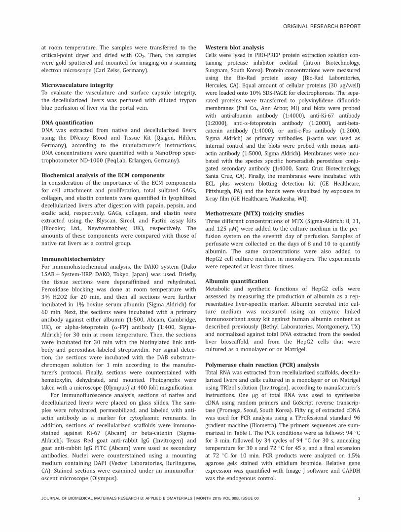
Decellularized livers with well-preserved ECMcomponents and a vascular tree were successfullydevelopedTransparent white-colored constructs that retained similarshape and dimensions of native liver were generated afterperfusion of rat liver with 0.1% SDS for 60–80 min [Figure1(Aa–Ac)]. The vascular network was easily visualizedgrossly because of the transparency of the decellularizedliver. The efficiency of decellularization was observed by theabsence of H&E [Figure 1(Ad–Af)], Alcian blue/PAS and VVGdarkly stained nuclei [Supporting Information FigureS1(Aa,Ab)] and no cytoplasmic remnants were detected insections stained with antiactin antibody [SupportingInformation Figure S1(B)] compared with native and incom-pletely decellularized livers. VVG staining revealed the pres-ence of darkly stained elastic fibers in the perivascularareas in addition to the well preserved pink-stained collagenfibers that outlined the eradicated hepatocytes. Alcian blue/PAS staining demonstrated that GAGs was homogenouslydistributed within the decellularized liver.
The SEM analysis conducted on native and decellularizedlivers showed a porous network of woven collagen fibers that
did not contain any cellular materials [Figure 1(Bb)] com-pared to native liver tissue [Figure 1(Ba)] in addition to thepreservation of the large blood vessels within the scaffold.During the perfusion of trypan blue dye [Figure 1(C)] throughthe portal vein, the dye flowed from larger blood vesselstoward smaller capillaries, suggesting preservation of the vas-cular tree, and no leakage was observed from the surface cap-sule of the decellularized livers.
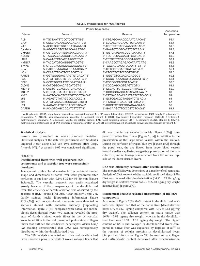
DNA was efficiently removed after decellularizationThe amount of DNAwas determined as a marker of cell remnants.Analysis of DNA content within scaffolds confirmed that> 99%DNA was removed after decellularization (34.516 13.56 ng/mgdry weight in scaffolds versus 6616.6627.83 ng/mg dry weightin native liver) [Figure 2(A)].
Biochemical analysis revealed preservation of the ECMcomponentsAs shown in Figure 2(B), GAG content in decellularized scaf-folds was higher than that of the native liver (decellularizedliver: 5.776 0.69 mg/mg compared with 3.996 0.13 mg/mgdry weight). The collagen content in native tissue was16.566 0.83 mg/mg dry weight, whereas in the decellular-ized liver was 19.3461.33 mg/mg dry weight. The highercontent of GAGs and collagen in decellularized livers com-pared to native liver was explained by Baptista et al.12 asthe removal of cellular proteins in decellularized livers(Supporting Information Figure S2). In contrast to collagenand GAGs, elastin content decreased after decellularization
TABLE I. Primers used for PCR Analysis
Primer
Primer SequencesAnnealing
TemperatureForward Reverse
ALB 5’-TGCTAATTTCCCTCCGTTTG-3’ 5’-CTGAGCAAAGGCAATCAACA-3’ 56.4GPX1 5’-CGCCAAGAACGAAGAGATTC-3’ 5’-CCCACCAGGAACTTCTCAAA-3’ 58.4a-FP 5’-AGCTTGGTGGTGGATGAAAC-3’ 5’-CCCTCTTCAGCAAAGCAGAC-3’ 59.5Catalase 5’-AGGCCAGTCCTGACAAAATG-3’ 5’-GAATCTCCGCACTTCTCCAG-3’ 59.8CYP2E1 5’-CGTGGAAATGGAGAAGGAAA-3’ 5’-GGTGATGAACCGCTGAATCT-3’ 57.6ASGR2 5’-TAGGAGCCAAGCTGGAGAAA-3’ 5’-TCCTCCCAGGAGTTGATGAC-3’ 60.0LDLR 5’-CAATGTCTCACCAAGCTCT-3’ 5’-TCTGTCTCGAGGGGTAGCT-3’ 58.1HMGCR 5’-TACCATGTCAGGGGTACGT-3’ 5’-CAAGCCTAGAGACATAATCAT-3’ 56.8c-MYC 5’-CTGCGACGAGGAGGAGGACT-3’ 5’- GGCAGCAGCTCGAATTTCTT-3’ 61.5c-Jun 5’-GACTGCAAAGATGGAAACGA-3’ 5’-GTTGCTGGACTGATTATCA-3’ 55.1c-fos 5’-CCAACTTCATTCCCACGTC-3’ 5’-CTCCCTCCTCCGGTTGC-3’ 58.5RAB3B 5’-GGTGGGGAACAAGTGTGACAT-3’ 5’-GGGTGTCCGAGAGACGC-3’ 60.5FAK 5’-ATGTTCTGGTGTCCTCAAATG-3’ 5’-GAGGTAAAACGTCGAAAATTG-3’ 58.4CDH1 5’-GCCCTGCCAATCCCGATGAA-3’ 5’-CGCCGCCTCCGTACAT-3’ 56.6CLDN6 5’-CATCGGCAACAGCATCGT-3’ 5’-CGCCAGCAGTGAGTCGT-3’ 59.1MMP-9 5’-CACTGTCCACCCCTCAGAGC-3’ 5’-GCCACTTGTCGGCGATAAGG-3’ 60.2MRP-2 5’-CTGGAGGAGATTTGGCTGAG-3’ 5’-GGCGGGAGGTAGACACATAA-3’ 60Ki-67 5’-AATTCAGACTCCATGTGCCTGAG-30 50-CTTGACACACACATTGTCCTCAGC-30 59p53 5’-AGAGTCTATAGGCCCACCCC-3’ 5’-GCTCGACGCTAGGATCTG AC-3’ 64p27 5’-ATGTCAAACGTGCGAGTGTCT-3’ 5’-TTACGTTTGACGTCTTCTGA-3’ 57p21 5’-AAGACCATGTGGACCTGTCA-3’ 5’-GGCTTCCTCTTGGAGAAGAT-3’ 55GAPDH 5’-ACAGTCAGCCGCATCTTCTT-3’ 5’-GACAAGCTTCCCGTTCTCAG-3’ 59.7
ALB, albumin; GPX1, glutathione peroxidase transcript variant 1; a-FP, alpha-fetoprotein; CYP2E1, cytochrome P450 family 2 subfamily E,
polypeptide 1; ASGR2, asialoglycoprotein receptor 2 transcript variant 1; LDLR, low-density lipoprotein receptor; HMGCR, 3-hydroxy-3-
methylglutaryl coenzyme A reductase; RAB3B, ras-related protein; FAK, focal adhesion kinase; CDH1, E-cadherin; CLDN6, claudin 6; MMP-9,
matrix metalloproteinases-9; MRP-2, multidrug resistance protein 2; GAPDH, glyceraldehyde-3-phosphate dehydrogenase transcript variant 1.
4 HUSSEIN ET AL. 3-D MODEL FOR PHARMACOLOGICAL STUDIES
(decellularized liver: 205.246 19.36 mg/mg dry vs. nativeliver tissue: 381.946 16.42 mg/mg dry weight).
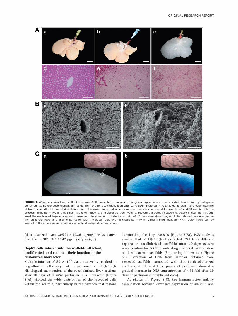
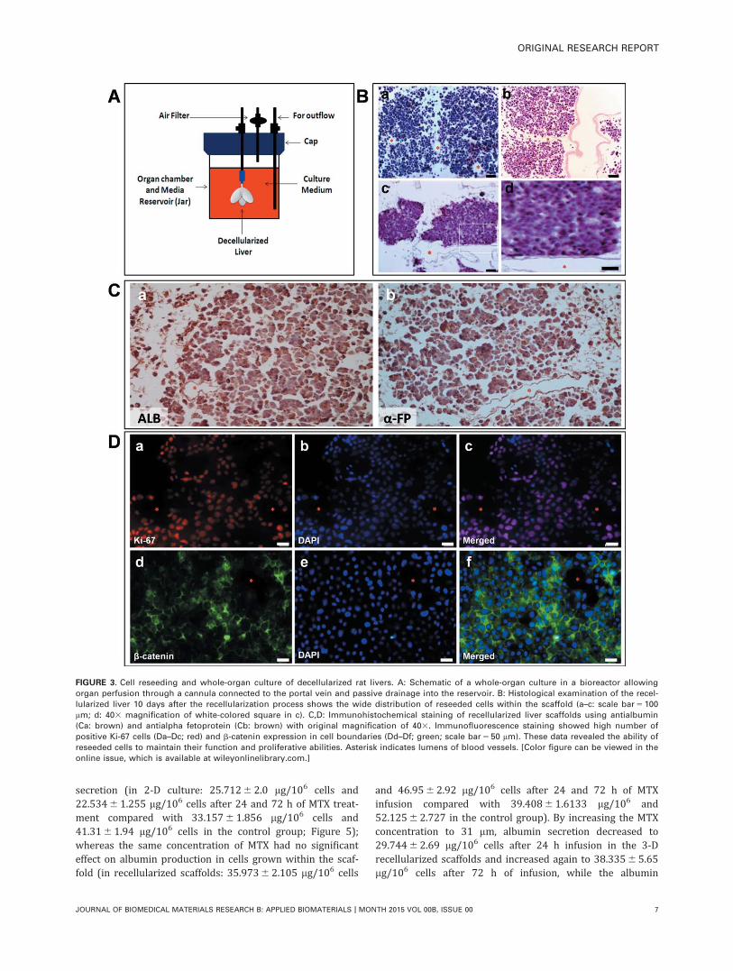
HepG2 cells infused into the scaffolds attached,proliferated, and retained their function in thecustomized bioreactorMultiple-infusion of 50 3 106 via portal veins resulted inengraftment efficiency of approximately 88%6 7%.Histological examination of the recellularized liver sectionsafter 10 days of in vitro perfusion in a bioreactor [Figure3(A)] showed the wide distribution of the reseeded cellswithin the scaffold, particularly in the parenchymal regions
surrounding the large vessels [Figure 2(B)]. PCR analysisshowed that �91%6 6% of extracted RNA from differentregions in recellularized scaffolds after 10-days culturewere positive for GAPDH, indicating the good repopulationof decellularized scaffolds (Supporting Information FigureS3). Extraction of DNA from samples obtained fromreseeded scaffolds, compared with that in decellularizedscaffolds, at different time points of perfusion showed agradual increase in DNA concentration of �84-fold after 10days of perfusion (unpublished data).
As shown in Figure 3(C), the immunohistochemistryexamination revealed extensive expression of albumin and
FIGURE 1. Whole acellular liver scaffold structure. A: Representative images of the gross appearance of the liver decellularization by antegrade
perfusion. (a) Before decellularization, (b) during, (c) after decellularization with 0.1% SDS (Scale bar 5 10 mm). Hematoxylin and eosin staining
of liver tissue after 80 min of decellularization (f) showed no cytoplasmic or nuclear materials compared to prior to (d) and 30 min (e) into the
process. Scale bar 5 400 lm. B: SEM images of native (a) and decellularized livers (b) revealing a porous network structure in scaffold that out-
lined the eradicated hepatocytes with preserved blood vessels (Scale bar 5 100 lm). C: Representative images of the retained vascular bed in
the left lateral lobe (a) and after perfusion with the trypan blue dye (b) (Scale bar 5 10 mm, insets magnification 5 43). [Color figure can be
viewed in the online issue, which is available at wileyonlinelibrary.com.]
ORIGINAL RESEARCH REPORT
JOURNAL OF BIOMEDICAL MATERIALS RESEARCH B: APPLIED BIOMATERIALS | MONTH 2015 VOL 00B, ISSUE 00 5
a-FP, confirming the ability of the reseeded cells to retaintheir hepatic function by secreting albumin and a-FP withinthe scaffolds. Immunofluroscence analysis showed a highnumber of positive Ki-67 cells [Figure 3(Da–Dc)] withinscaffolds compared with that of spheroids [SupportingInformation Figure S4(C)]. In addition, beta-catenin, a spe-cific indicator of cell–cell interaction, was highly expressedat the cell–cell boundary [Figure 3(Dd–Df)].
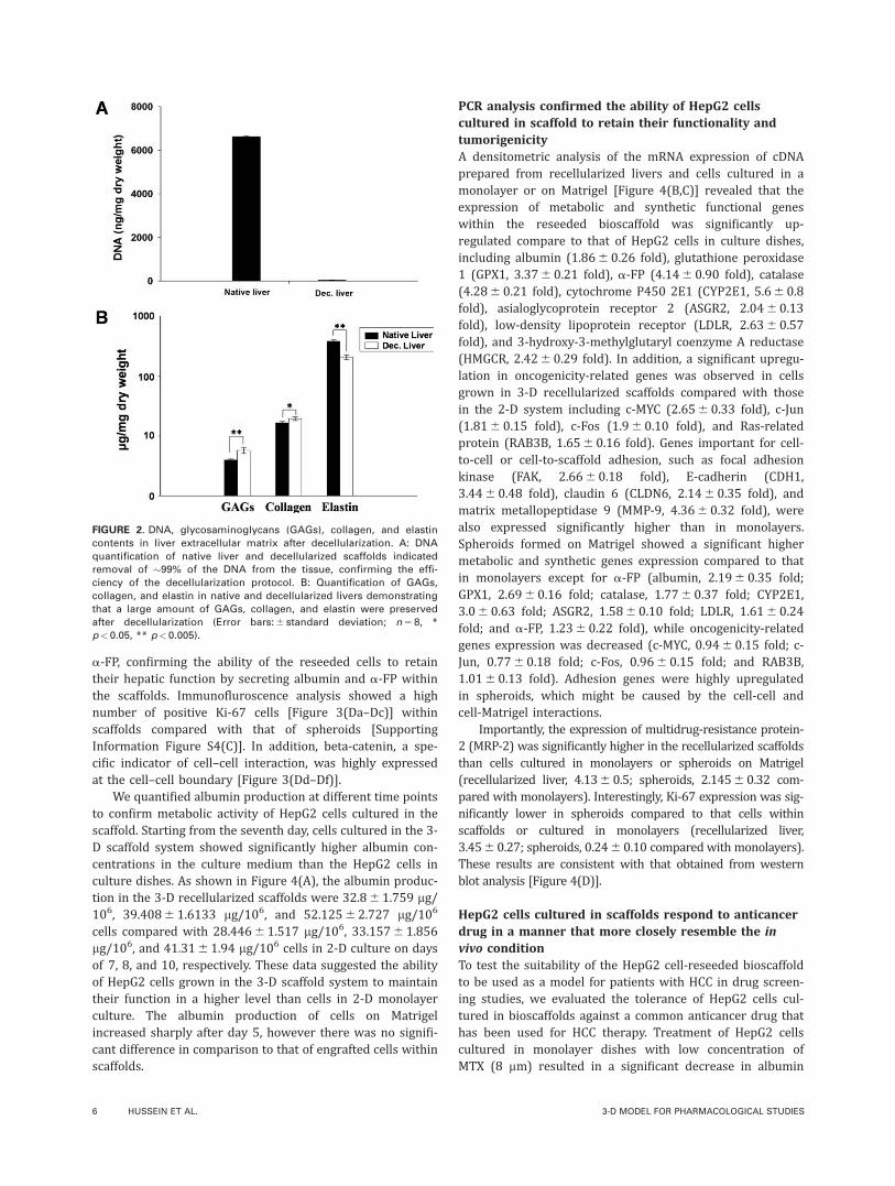
We quantified albumin production at different time pointsto confirm metabolic activity of HepG2 cells cultured in thescaffold. Starting from the seventh day, cells cultured in the 3-D scaffold system showed significantly higher albumin con-centrations in the culture medium than the HepG2 cells inculture dishes. As shown in Figure 4(A), the albumin produc-tion in the 3-D recellularized scaffolds were 32.861.759 lg/106, 39.4086 1.6133 lg/106, and 52.12562.727 lg/106
cells compared with 28.4466 1.517 lg/106, 33.15761.856lg/106, and 41.3161.94 lg/106 cells in 2-D culture on daysof 7, 8, and 10, respectively. These data suggested the abilityof HepG2 cells grown in the 3-D scaffold system to maintaintheir function in a higher level than cells in 2-D monolayerculture. The albumin production of cells on Matrigelincreased sharply after day 5, however there was no signifi-cant difference in comparison to that of engrafted cells withinscaffolds.
PCR analysis confirmed the ability of HepG2 cellscultured in scaffold to retain their functionality andtumorigenicityA densitometric analysis of the mRNA expression of cDNAprepared from recellularized livers and cells cultured in amonolayer or on Matrigel [Figure 4(B,C)] revealed that theexpression of metabolic and synthetic functional geneswithin the reseeded bioscaffold was significantly up-regulated compare to that of HepG2 cells in culture dishes,including albumin (1.866 0.26 fold), glutathione peroxidase1 (GPX1, 3.376 0.21 fold), a-FP (4.146 0.90 fold), catalase(4.286 0.21 fold), cytochrome P450 2E1 (CYP2E1, 5.66 0.8fold), asialoglycoprotein receptor 2 (ASGR2, 2.0460.13fold), low-density lipoprotein receptor (LDLR, 2.636 0.57fold), and 3-hydroxy-3-methylglutaryl coenzyme A reductase(HMGCR, 2.426 0.29 fold). In addition, a significant upregu-lation in oncogenicity-related genes was observed in cellsgrown in 3-D recellularized scaffolds compared with thosein the 2-D system including c-MYC (2.656 0.33 fold), c-Jun(1.816 0.15 fold), c-Fos (1.960.10 fold), and Ras-relatedprotein (RAB3B, 1.656 0.16 fold). Genes important for cell-to-cell or cell-to-scaffold adhesion, such as focal adhesionkinase (FAK, 2.6660.18 fold), E-cadherin (CDH1,3.4460.48 fold), claudin 6 (CLDN6, 2.146 0.35 fold), andmatrix metallopeptidase 9 (MMP-9, 4.366 0.32 fold), werealso expressed significantly higher than in monolayers.Spheroids formed on Matrigel showed a significant highermetabolic and synthetic genes expression compared to thatin monolayers except for a-FP (albumin, 2.196 0.35 fold;GPX1, 2.696 0.16 fold; catalase, 1.776 0.37 fold; CYP2E1,3.06 0.63 fold; ASGR2, 1.586 0.10 fold; LDLR, 1.6160.24fold; and a-FP, 1.2360.22 fold), while oncogenicity-relatedgenes expression was decreased (c-MYC, 0.9460.15 fold; c-Jun, 0.7760.18 fold; c-Fos, 0.966 0.15 fold; and RAB3B,1.0160.13 fold). Adhesion genes were highly upregulatedin spheroids, which might be caused by the cell-cell andcell-Matrigel interactions.
Importantly, the expression of multidrug-resistance protein-2 (MRP-2) was significantly higher in the recellularized scaffoldsthan cells cultured in monolayers or spheroids on Matrigel(recellularized liver, 4.1360.5; spheroids, 2.14560.32 com-pared with monolayers). Interestingly, Ki-67 expression was sig-nificantly lower in spheroids compared to that cells withinscaffolds or cultured in monolayers (recellularized liver,3.4560.27; spheroids, 0.246 0.10 compared with monolayers).These results are consistent with that obtained from westernblot analysis [Figure 4(D)].
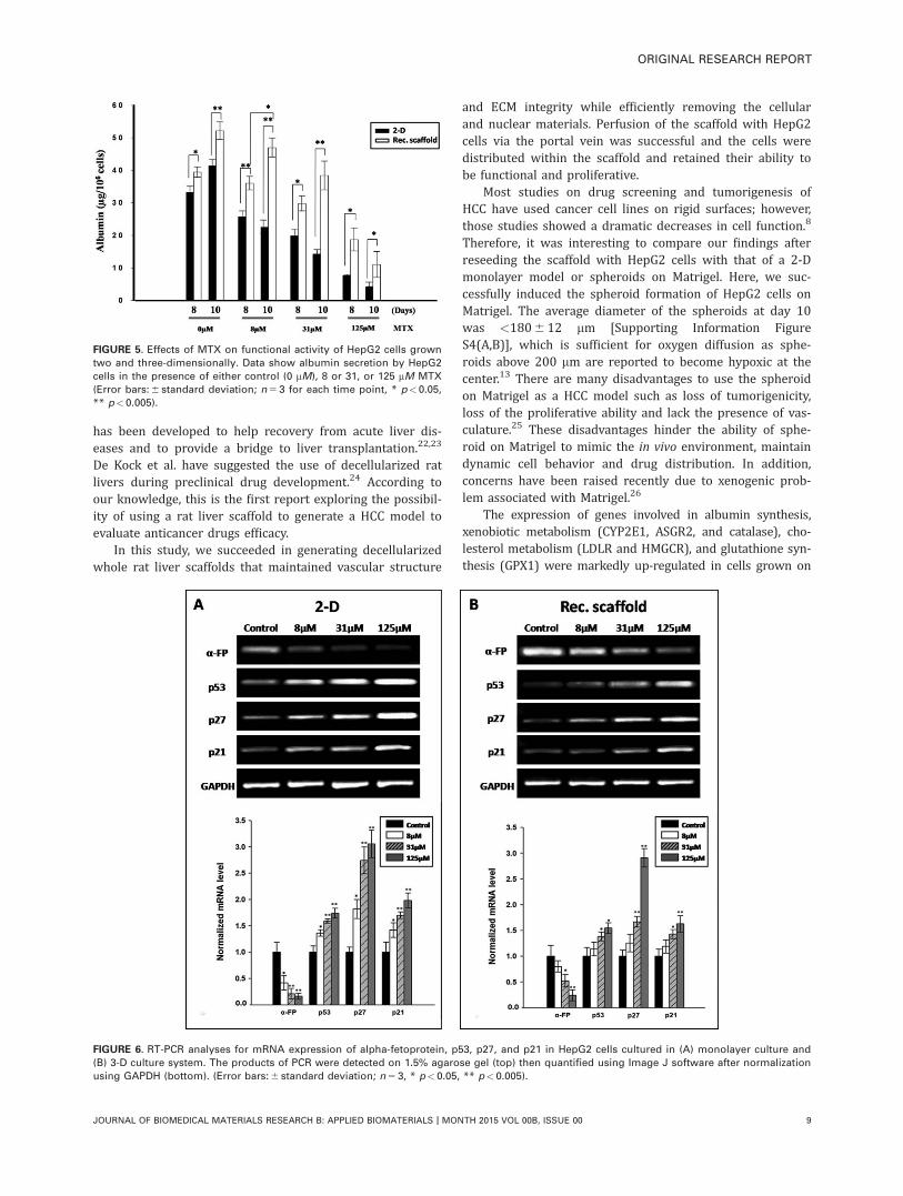
HepG2 cells cultured in scaffolds respond to anticancerdrug in a manner that more closely resemble the invivo conditionTo test the suitability of the HepG2 cell-reseeded bioscaffoldto be used as a model for patients with HCC in drug screen-ing studies, we evaluated the tolerance of HepG2 cells cul-tured in bioscaffolds against a common anticancer drug thathas been used for HCC therapy. Treatment of HepG2 cellscultured in monolayer dishes with low concentration ofMTX (8 mm) resulted in a significant decrease in albumin
FIGURE 2. DNA, glycosaminoglycans (GAGs), collagen, and elastin
contents in liver extracellular matrix after decellularization. A: DNA
quantification of native liver and decellularized scaffolds indicated
removal of �99% of the DNA from the tissue, confirming the effi-
ciency of the decellularization protocol. B: Quantification of GAGs,
collagen, and elastin in native and decellularized livers demonstrating
that a large amount of GAGs, collagen, and elastin were preserved
after decellularization (Error bars: 6 standard deviation; n 5 8, *
p< 0.05, ** p<0.005).
6 HUSSEIN ET AL. 3-D MODEL FOR PHARMACOLOGICAL STUDIES
secretion (in 2-D culture: 25.7126 2.0 lg/106 cells and22.5346 1.255 lg/106 cells after 24 and 72 h of MTX treat-ment compared with 33.1576 1.856 lg/106 cells and41.316 1.94 lg/106 cells in the control group; Figure 5);whereas the same concentration of MTX had no significanteffect on albumin production in cells grown within the scaf-fold (in recellularized scaffolds: 35.9736 2.105 lg/106 cells
and 46.956 2.92 lg/106 cells after 24 and 72 h of MTXinfusion compared with 39.40861.6133 lg/106 and52.12562.727 in the control group). By increasing the MTXconcentration to 31 mm, albumin secretion decreased to29.74462.69 lg/106 cells after 24 h infusion in the 3-Drecellularized scaffolds and increased again to 38.33565.65lg/106 cells after 72 h of infusion, while the albumin
FIGURE 3. Cell reseeding and whole-organ culture of decellularized rat livers. A: Schematic of a whole-organ culture in a bioreactor allowing
organ perfusion through a cannula connected to the portal vein and passive drainage into the reservoir. B: Histological examination of the recel-
lularized liver 10 days after the recellularization process shows the wide distribution of reseeded cells within the scaffold (a–c: scale bar 5 100
lm; d: 403 magnification of white-colored square in c). C,D: Immunohistochemical staining of recellularized liver scaffolds using antialbumin
(Ca: brown) and antialpha fetoprotein (Cb: brown) with original magnification of 403. Immunofluorescence staining showed high number of
positive Ki-67 cells (Da–Dc; red) and b-catenin expression in cell boundaries (Dd–Df; green; scale bar 5 50 lm). These data revealed the ability of
reseeded cells to maintain their function and proliferative abilities. Asterisk indicates lumens of blood vessels. [Color figure can be viewed in the
online issue, which is available at wileyonlinelibrary.com.]
ORIGINAL RESEARCH REPORT
JOURNAL OF BIOMEDICAL MATERIALS RESEARCH B: APPLIED BIOMATERIALS | MONTH 2015 VOL 00B, ISSUE 00 7
secretion decreased in 2-D system to 19.86562.95 lg/106
cells and 14.30761.24 lg/106 cells after 24 and 72 h,respectively.
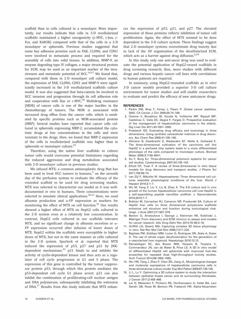
In the presence of higher concentrations of MTX, boththe 2-D monolayers and 3-D recellularized scaffold systemsshowed a severe reduction in albumin secretion (in 2-D:7.5676 1.127 lg/106 cells and 3.966 2.316 lg/106 cells;in recellularized scaffold: 18.7146 5.2 lg/106 cells and11.0196 3.443 lg/106 cells after 24 and 72 h of MTX infu-sion, respectively). These data clearly showed that cell func-tion was impaired after infusion of MTX in a dose-dependent manner and that cells grown in recellularizedscaffold appeared more robust and resistant to anticancerdrug treatments than cells cultured in 2-D monolayer cul-ture. We also evaluated the effect of MTX on HepG2 cellscultured in the 2-D monolayers and 3-D recellularized scaf-fold culture systems at the gene level. As shown in Figure 6,the expression of a-FP; as a tumor marker,16 decreasedeven by low dose of MTX in 2-D system, while HepG2 cellsin the 3-D recellularized scaffold system appeared to be
more resistant to the same dose. Therapeutic efficacy ofMTX may arise, in part, via its ability to restore expressionlevels of key proteins required for cell cycle checkpointarrest leading to apoptosis of HCC.17 Therefore, we figuredout the effect of MTX on the expression of p53, p21, andp27. All doses of MTX significantly induced mRNA expres-sion of p53, p21 and p27 in the 2-D system, whereas thelow concentration of MTX (8 mM) demonstrated no signifi-cant effect on the gene expression in the recellularized scaf-fold culture system.
DISCUSSION
Many drugs have been withdrawn from the market duringthe past decade because of inefficiency and hepatotoxicity,which may be explained by poor correlation with the corre-sponding in vitro and animal models.18–20 Approximatelyhalf a billion dollars are lost after withdrawal of a drug can-didate because of liver toxicity.21 The field of liver tissueengineering, particularly that focusing on decellularization
FIGURE 4. A: Albumin production in culture supernatants by enzyme-linked immunosorbent assay greater in HepG2-reseeded scaffolds culture
and spheroids than in cells grown in a static two-dimensional (2-D) monolayer culture (n 5 3 for each time point). Gel electrophoresis of the PCR
products on 1.5% agarose (B) followed by the analysis of the bands density using Image J software (C) revealed a significantly higher upregulation
of the metabolic and synthetic genes in addition to oncogenicity-related genes in HepG2-reseeded scaffolds compared with those in the monolayer
culture. Genes abbreviations: ALB, albumin; GPX 1, glutathione peroxidase 1; a-FP, alpha-fetoprotein; CYP 2E1, cytochrome P450 2E1; ASGR2, asia-
loglycoprotein receptor 2; LDLR, low-density lipoprotein receptor; HMGCR, 3-hydroxy-3-methylglutaryl coenzyme A reductase; RAB3B, Ras-related
protein; FAK, focal adhesion kinase; CDH1, E-cadherin; CLDN6, claudin 6; MMP-9, matrix metalloproteinases-9; MRP-2, multidrug resistance protein
2; GAPDH, glyceraldehyde-3-phosphate dehydrogenase transcript variant 1. (Error bars: 6 standard deviation; n 5 3, * p< 0.05, ** p<0.005). D:
Western blot analysis of albumin, a-FP, c-Fos, b-catenin, and Ki-67 in HepG2 cells in monolayer culture, HepG2 cell in the recellularized scaffolds,
and HepG2 cells in spheroid culture. [Color figure can be viewed in the online issue, which is available at wileyonlinelibrary.com.]
8 HUSSEIN ET AL. 3-D MODEL FOR PHARMACOLOGICAL STUDIES
has been developed to help recovery from acute liver dis-eases and to provide a bridge to liver transplantation.22,23
De Kock et al. have suggested the use of decellularized ratlivers during preclinical drug development.24 According toour knowledge, this is the first report exploring the possibil-ity of using a rat liver scaffold to generate a HCC model toevaluate anticancer drugs efficacy.
In this study, we succeeded in generating decellularizedwhole rat liver scaffolds that maintained vascular structure
and ECM integrity while efficiently removing the cellularand nuclear materials. Perfusion of the scaffold with HepG2cells via the portal vein was successful and the cells weredistributed within the scaffold and retained their ability tobe functional and proliferative.
Most studies on drug screening and tumorigenesis ofHCC have used cancer cell lines on rigid surfaces; however,those studies showed a dramatic decreases in cell function.8
Therefore, it was interesting to compare our findings afterreseeding the scaffold with HepG2 cells with that of a 2-Dmonolayer model or spheroids on Matrigel. Here, we suc-cessfully induced the spheroid formation of HepG2 cells onMatrigel. The average diameter of the spheroids at day 10was <1806 12 lm [Supporting Information FigureS4(A,B)], which is sufficient for oxygen diffusion as sphe-roids above 200 lm are reported to become hypoxic at thecenter.13 There are many disadvantages to use the spheroidon Matrigel as a HCC model such as loss of tumorigenicity,loss of the proliferative ability and lack the presence of vas-culature.25 These disadvantages hinder the ability of sphe-roid on Matrigel to mimic the in vivo environment, maintaindynamic cell behavior and drug distribution. In addition,concerns have been raised recently due to xenogenic prob-lem associated with Matrigel.26
The expression of genes involved in albumin synthesis,xenobiotic metabolism (CYP2E1, ASGR2, and catalase), cho-lesterol metabolism (LDLR and HMGCR), and glutathione syn-thesis (GPX1) were markedly up-regulated in cells grown on
FIGURE 5. Effects of MTX on functional activity of HepG2 cells grown
two and three-dimensionally. Data show albumin secretion by HepG2
cells in the presence of either control (0 mM), 8 or 31, or 125 mM MTX
(Error bars: 6 standard deviation; n 5 3 for each time point, * p< 0.05,
** p<0.005).
FIGURE 6. RT-PCR analyses for mRNA expression of alpha-fetoprotein, p53, p27, and p21 in HepG2 cells cultured in (A) monolayer culture and
(B) 3-D culture system. The products of PCR were detected on 1.5% agarose gel (top) then quantified using Image J software after normalization
using GAPDH (bottom). (Error bars: 6 standard deviation; n 5 3, * p< 0.05, ** p< 0.005).
ORIGINAL RESEARCH REPORT
JOURNAL OF BIOMEDICAL MATERIALS RESEARCH B: APPLIED BIOMATERIALS | MONTH 2015 VOL 00B, ISSUE 00 9
scaffold than in cells cultured in a monolayer. More impor-tantly, our results indicate that cells in 3-D recellularizedscaffolds maintained a higher oncogenicity (c-MYC, c-Jun, c-Fos, and RAB3B) compared with that of the cells in a 2-Dmonolayer or spheroids. Previous studies suggested thatsome key adhesion proteins such as FAK, CLDN6, and CDH1were involved in metastatic process and required for theassembly of cells into solid tissues. In addition, MMP-9, anenzyme degrading type IV collagen, a major structural proteinfor ECM, may be used as an important marker of the inva-siveness and metastatic potential of HCC.14,27 We found that,compared with those in 2-D monolayer cell culture model,the expression of FAK, CLDN6, CDH1 and MMP-9 were signif-icantly increased in the 3-D recellularized scaffolds culturemodel. It was also suggested that beta-catenin be involved inHCC invasion and progression through its cell adhesion roleand cooperation with Ras or c-MYC.28 Multidrug resistance(MDR) of cancer cells is one of the major hurdles in thechemotherapy of tumors. The MDR is associated withincreased drug efflux from the cancer cells, which is medi-ated by specific proteins such as MDR-associated protein(MRP). Several studies have reported that cancer cells cul-tured in spheroids expressing MRP-2, accumulated the cyto-toxic drugs at low concentrations in the cells and wereresistant to the drugs. Here, we found that MRP-2 expressionof the cells in recellularized scaffolds was higher than inspheroids or monolayer culture.29
Therefore, using the native liver scaffolds to culturetumor cells would overcome potential limitations regardingthe reduced aggression and drug metabolism associatedwith 2-D monolayer culture in previous studies.
We infused MTX; a common antineoplastic drug that hasbeen used to treat HCC tumors in humans,9 on the seventhday of the perfusion system to evaluate the efficacy of thereseeded scaffold to be used for drug screening studies.MTX was selected to characterize our model as it was well-documented in vivo in humans. Three concentrations wereselected to simulate clinical applications.30,31 Here, we usedalbumin production and a-FP expression as markers formonitoring the effect of MTX on cell function.32 Our resultsshowed a higher effect of MTX on HepG2 cells cultured inthe 2-D system even at a relatively low concentration. Incontrast, HepG2 cells cultured in our scaffolds toleratesMTX, and no significant change in albumin secretion or a-FP expression occurred after infusion of lower doses ofMTX. HepG2 within the scaffolds were susceptible to higherdoses of MTX, but not in the same manner as cells culturedin the 2-D system. Spurlock et al. reported that MTXinduced the expression of p53, p27 and p21 by JNK-dependent mechanisms.33 p21 binds to and inhibits theactivity of cyclin-dependent kinase and thus acts as a regu-lator of cell cycle progression at G1 and S phase. Theexpression of this gene is controlled by the tumor suppres-sor protein p53, through which this protein mediates thep53-dependent cell cycle G1 phase arrest. p21 can alsoinhibit the combination of proliferating-cell nuclear antigenand DNA polymerase, subsequently inhibiting the extensionof DNA.17 Results from this study indicate that MTX enhan-
ces the expression of p53, p21, and p27. The elevatedexpression of these proteins reflects inhibition of tumor cellproliferation. Again, the effect of MTX seemed to be dosedependent in the 3-D culture system. These findings suggestthat 2-D monolayer systems overestimate drug toxicity dueto lack of the 3D organization of the decellularized ECM,which acts as a barrier against drug diffusion.6,34
In this study, only one anti-ancer drug was used to eval-uate the potential application of HepG2-reseed scaffolds indrug screening research; thus, more studies with differentdrugs and various hepatic cancer cell lines with correlationsto human patients are required.
In summary, using HepG2-reseeded scaffolds as in vitro3-D cancer models provided a superior 3-D cell cultureenvironment for tumor studies and will enable researchersto evaluate and predict the efficacy of new anticancer drugs.
REFERENCES1. Parkin DM, Bray F, Ferlay J, Pisani P. Global cancer statistics,
2002. CA Cancer J Clin 2005;55:74–108.
2. Ozenne V, Bouattour M, Goutte N, Vullierme MP, Ripault MP,
Castelnau C, Valla DC, Degos F, Farges O. Prospective evaluation
of the management of hepatocellular carcinoma in the elderly.
Dig Liver Dis 2011;43:1001–1005.
3. Prestwich GD. Evaluating drug efficacy and toxicology in three
dimensions: Using synthetic extracellular matrices in drug discov-
ery. Acc Chem Res 2008;41:139–148.
4. Altmann B, Giselbrecht S, Weibezahn KF, Welle A, Gottwald E.
The three-dimensional cultivation of the carcinoma cell line
HepG2 in a perfused chip system leads to a more differentiated
phenotype of the cells compared to monolayer culture. Biomed
Mater 2008;3:1748–6041.
5. Xu F, Burg KJ. Three-dimensional polymeric systems for cancer
cell studies. Cytotechnology 2007;54:135–143.
6. Elliott NT, Yuan F. A review of three-dimensional in vitro tissue
models for drug discovery and transport studies. J Pharm Sci
2011;100:59–74.
7. van Zijl F, Mikulits W. Hepatospheres: Three dimensional cell cul-
tures resemble physiological conditions of the liver. World J
Hepatol 2010;2:1–7.
8. Wu M, Yang Z, Liu Y, Liu B, Zhao X. The 3-D culture and in vivo
growth of the human hepatocellular carcinoma cell Line HepG2 in
a self-assembling peptide nanofiber scaffold. J. Nanomaterials
2010;2010:1–7.
9. Bokhari M, Carnachan RJ, Cameron NR, Przyborski SA. Culture of
HepG2 liver cells on three dimensional polystyrene scaffolds
enhances cell structure and function during toxicological chal-
lenge. J Anat 2007;211:567–576.
10. Benton G, Arnaoutova I, George J, Kleinman HK, Koblinski J.
Matrigel: From discovery and ECM mimicry to assays and models
for cancer research. Adv Drug Deliv Rev 2014;79–80:3–18.
11. Griffith LG, Swartz MA. Capturing complex 3D tissue physiology
in vitro. Nat Rev Mol Cell Biol 2006;7:211–224.
12. Baptista PM, Siddiqui MM, Lozier G, Rodriguez SR, Atala A, Soker
S. The use of whole organ decellularization for the generation of
a vascularized liver organoid. Hepatology 2010;12:12.
13. Ramaiahgari SC, den Braver MW, Herpers B, Terpstra V,
Commandeur JN, van de Water B, Price LS. A 3D in vitro model
of differentiated HepG2 cell spheroids with improved liver-like
properties for repeated dose high-throughput toxicity studies.
Arch Toxicol 2014;88:1083–1095.
14. Wu YM, Tang J, Zhao P, Chen ZN, Jiang JL. Morphological changes
and molecular expressions of hepatocellular carcinoma cells in
three-dimensional culture model. Exp Mol Pathol 2009;87:133-140.
15. Li L, Lu Y. Optimizing a 3D culture system to study the interaction
between epithelial breast cancer and its surrounding fibroblasts.
J Cancer 2011;2:458–466.
16. Lai Q, Melandro F, Pinheiro RS, Donfrancesco A, Fadel BA, Levi
Sandri GB, Rossi M, Berloco PB, Frattaroli FM. Alpha-fetoprotein
10 HUSSEIN ET AL. 3-D MODEL FOR PHARMACOLOGICAL STUDIES
and novel tumor biomarkers as predictors of hepatocellular carci-
noma recurrence after surgery: A brilliant star raises again. Int J
Hepatol 2012;893103:27.
17. Zhu Z, Jia J, Lu R, Lu Y, Fu Z, Zhao L, Wang L, Jin M, Zhao L,
Gao W, Yao Z. Expression of PTEN, p27, p21 and AKT mRNA and
protein in human BEL-7402 hepatocarcinoma cells in transplanted
tumors of nude mice treated with the tripeptide tyroservatide
(YSV). Int J Cancer 2006;118:1539–1544.
18. Guillouzo A, Corlu A, Aninat C, Glaise D, Morel F, Guguen-
Guillouzo C. The human hepatoma HepaRG cells: A highly differ-
entiated model for studies of liver metabolism and toxicity of
xenobiotics. Chem Biol Interact 2007;168:66–73.
19. Kaneko S, Furuse J, Kudo M, Ikeda K, Honda M, Nakamoto Y,
Onchi M, Shiota G, Yokosuka O, Sakaida I, Takehara T, Ueno Y,
Hiroishi K, Nishiguchi S, Moriwaki H, Yamamoto K, Sata M, Obi
S, Miyayama S, Imai Y. Guideline on the use of new anticancer
drugs for the treatment of Hepatocellular Carcinoma 2010 update.
Hepatol Res 2012;42:523–542.
20. Prestwich GD, Liu Y, Yu B, Shu XZ, Scott A. 3-D culture in synthetic
extracellular matrices: New tissue models for drug toxicology and
cancer drug discovery. Adv Enzyme Regul 2007;47:196–207.
21. DiMasi JA, Hansen RW, Grabowski HG. The price of innovation:
New estimates of drug development costs. J Health Econ 2003;
22:151–185.
22. Uygun BE, Soto-Gutierrez A, Yagi H, Izamis ML, Guzzardi MA,
Shulman C, Milwid J, Kobayashi N, Tilles A, Berthiaume F, Hertl
M, Nahmias Y, Yarmush ML, Uygun K. Organ reengineering
through development of a transplantable recellularized liver graft
using decellularized liver matrix. Nat Med 2010;16:814-820.
23. Booth C, Soker T, Baptista P, Ross CL, Soker S, Farooq U, Stratta
RJ, Orlando G. Liver bioengineering: current status and future
perspectives. World J Gastroenterol 2012;18:6926–6934.
24. De Kock J, Ceelen L, De Spiegelaere W, Casteleyn C, Claes P,
Vanhaecke T, Rogiers V. Simple and quick method for whole-liver
decellularization: A novel in vitro three-dimensional bioengineer-
ing tool? Arch Toxicol 2011;85:607–612.
25. Mishra DK, Sakamoto JH, Thrall MJ, Baird BN, Blackmon SH,
Ferrari M, Kurie JM, Kim MP. Human lung cancer cells grown in
an ex vivo 3D lung model produce matrix metalloproteinases not
produced in 2D culture. PLoS One 2012;7:17.
26. Phelps EA, Garc�ıa AJ. Engineering more than a cell:
Vascularization strategies in tissue engineering. Curr Opin
Biotechnol 2010;21:704–709.
27. Cheng JC, Chou CH, Kuo ML, Hsieh CY. Radiation-enhanced
hepatocellular carcinoma cell invasion with MMP-9 expression
through PI3K/Akt/NF-kappaB signal transduction pathway.
Oncogene 2006;25:7009–7018.
28. Morin PJ. beta-catenin signaling and cancer. Bioessays 1999;21:
1021–1030.
29. Oshikata A, Matsushita T, Ueoka R. Enhancement of drug efflux
activity via MDR1 protein by spheroid culture of human hepatic
cancer cells. J Biosci Bioeng 2011;111:590–593.
30. Aquerreta I, Aldaz A, Giraldez J, Sierrasesumaga L. Pharmacodynamics
of high-dose methotrexate in pediatric patients. Ann Pharmacother
2002;36:1344–1350.
31. Walker TM, Rhodes PC, Westmoreland C. The differential cytotox-
icity of methotrexate in rat hepatocyte monolayer and spheroid
cultures. Toxicol In Vitro 2000;14:475–485.
32. Kane BJ, Zinner MJ, Yarmush ML, Toner M. Liver-specific func-
tional studies in a microfluidic array of primary mammalian hepa-
tocytes. Anal Chem 2006;78:4291–4298.
33. Spurlock CF, 3rd, Tossberg JT, Fuchs HA, Olsen NJ, Aune
TM. Methotrexate increases expression of cell cycle check-
point genes via JNK activation. Arthritis Rheum 2012;64:1780–
1789.
34. Battle T, Maguire T, Moulsdale H, Doyle A. Progressive matura-
tion resistance to microcystin-LR cytotoxicity in two different hep-
atospheroidal models. Cell Biol Toxicol 1999;15:3–12.
ORIGINAL RESEARCH REPORT
JOURNAL OF BIOMEDICAL MATERIALS RESEARCH B: APPLIED BIOMATERIALS | MONTH 2015 VOL 00B, ISSUE 00 11