Asymmetric Chromatid Segregation in Cardiac Progenitor Cells Is Enhanced by Pim-1 Kinase
11
Asymmetric chromatid segregation in cardiac progenitor cells is enhanced by Pim-1 kinase Balaji Sundararaman, B.Tech. 1 , Daniele Avitabile, Ph.D. 1 , Mathias H Konstandin, M.D. 1 , Christopher T Cottage, M.S. 1 , Natalie Gude, Ph.D. 1 , and Mark A Sussman, Ph.D. 1,* 1 SDSU Heart Institute and Biology Department, San Diego State University, 5500 Campanile Drive, San Diego, CA 92182, USA Abstract Rationale—Cardiac progenitor cells (CPCs) in the adult heart are used for cell-based treatment of myocardial damage but factors determining stemness, self-renewal and lineage commitment are poorly understood. Immortal DNA strands inherited through asymmetric chromatid segregation correlate with self-renewal of adult stem cells, but CPCs capacity for asymmetric segregation to retain immortal strands is unknown. Cardioprotective kinase Pim-1 increases asymmetric cell division in vivo but the ability of Pim-1 to enhance asymmetric chromatid segregation is unknown. Objective—Demonstrate immortal strand segregation in CPCs and the enhancement of asymmetric chromatid distribution by Pim-1 kinase. Methods and Results—Asymmetric segregation is tracked by incorporation of bromodeoxyuridine (BrdU). CPC DNA was labeled for several generations and then blocked in second cytokinesis during chase to determine distribution of immortal versus newly synthesized strands. Binucleated CPCs with BrdU intensity ratio of 70:30 or more between daughter nuclei indicative of asymmetric chromatid segregation occur with a frequency of 4.57% and asymmetric chromatid segregation is demonstrated at late mitotic phases. Asymmetric chromatid segregation is significantly enhanced by Pim-1 overexpression in CPCs (9.19% vs 4.79% in eGFP expressing cells, p=0.006). Conclusions—Asymmetric segregation of chromatids in CPCs is increased nearly twofold Pim-1 kinase overexpression, indicating Pim-1 promotes self renewal of stem cells. Keywords immortal DNA strand hypothesis; asymmetric chromatid segregation; asymmetric cell division; BrdU; cytochalasin B Introduction The immortal strand hypothesis states that adult stem cells selectively retain chromatids with old DNA strands (the mother strand) whereas all differentiating progeny acquire newly synthesized daughter to protect stem cells from DNA replication errors 1 and aging 2 . The silent sister hypothesis presumes template strand segregation of all or some chromatids to regulate gene expression to determine cell fate 3, 4 . The immortal strand and silent sister * To whom correspondence should be addressed: SDSU Heart Institute and Biology Department, San Diego State University, 5500 Campanile Drive, San Diego, CA 92182, 619) 594-2983 (voice), (619) 594-2610 (fax), [email protected] (email). Disclosures: None. NIH Public Access Author Manuscript Circ Res. Author manuscript; available in PMC 2013 April 27. Published in final edited form as: Circ Res. 2012 April 27; 110(9): 1169–1173. doi:10.1161/CIRCRESAHA.112.267716. NIH-PA Author Manuscript NIH-PA Author Manuscript NIH-PA Author Manuscript
-
Upload
independent -
Category
Documents
-
view
3 -
download
0
Transcript of Asymmetric Chromatid Segregation in Cardiac Progenitor Cells Is Enhanced by Pim-1 Kinase

Asymmetric chromatid segregation in cardiac progenitor cells isenhanced by Pim-1 kinase
Balaji Sundararaman, B.Tech.1, Daniele Avitabile, Ph.D.1, Mathias H Konstandin, M.D.1,Christopher T Cottage, M.S.1, Natalie Gude, Ph.D.1, and Mark A Sussman, Ph.D.1,*
1SDSU Heart Institute and Biology Department, San Diego State University, 5500 CampanileDrive, San Diego, CA 92182, USA
AbstractRationale—Cardiac progenitor cells (CPCs) in the adult heart are used for cell-based treatmentof myocardial damage but factors determining stemness, self-renewal and lineage commitment arepoorly understood. Immortal DNA strands inherited through asymmetric chromatid segregationcorrelate with self-renewal of adult stem cells, but CPCs capacity for asymmetric segregation toretain immortal strands is unknown. Cardioprotective kinase Pim-1 increases asymmetric celldivision in vivo but the ability of Pim-1 to enhance asymmetric chromatid segregation isunknown.
Objective—Demonstrate immortal strand segregation in CPCs and the enhancement ofasymmetric chromatid distribution by Pim-1 kinase.
Methods and Results—Asymmetric segregation is tracked by incorporation ofbromodeoxyuridine (BrdU). CPC DNA was labeled for several generations and then blocked insecond cytokinesis during chase to determine distribution of immortal versus newly synthesizedstrands. Binucleated CPCs with BrdU intensity ratio of 70:30 or more between daughter nucleiindicative of asymmetric chromatid segregation occur with a frequency of 4.57% and asymmetricchromatid segregation is demonstrated at late mitotic phases. Asymmetric chromatid segregationis significantly enhanced by Pim-1 overexpression in CPCs (9.19% vs 4.79% in eGFP expressingcells, p=0.006).
Conclusions—Asymmetric segregation of chromatids in CPCs is increased nearly twofoldPim-1 kinase overexpression, indicating Pim-1 promotes self renewal of stem cells.
Keywordsimmortal DNA strand hypothesis; asymmetric chromatid segregation; asymmetric cell division;BrdU; cytochalasin B
IntroductionThe immortal strand hypothesis states that adult stem cells selectively retain chromatids withold DNA strands (the mother strand) whereas all differentiating progeny acquire newlysynthesized daughter to protect stem cells from DNA replication errors1 and aging2. Thesilent sister hypothesis presumes template strand segregation of all or some chromatids toregulate gene expression to determine cell fate3, 4. The immortal strand and silent sister
*To whom correspondence should be addressed: SDSU Heart Institute and Biology Department, San Diego State University, 5500Campanile Drive, San Diego, CA 92182, 619) 594-2983 (voice), (619) 594-2610 (fax), [email protected] (email).
Disclosures: None.
NIH Public AccessAuthor ManuscriptCirc Res. Author manuscript; available in PMC 2013 April 27.
Published in final edited form as:Circ Res. 2012 April 27; 110(9): 1169–1173. doi:10.1161/CIRCRESAHA.112.267716.
NIH
-PA Author Manuscript
NIH
-PA Author Manuscript
NIH
-PA Author Manuscript

hypotheses are supported by observations of absolute to partial asymmetric chromatidsegregation (ACS) in adult stem cells like mouse intestinal and colon epithelial stemcells5, 6, neural stem cells7, satellite cells8, 9 and many cancer stem cells10, 11. Stem cellsmay also undergo symmetric segregation on an intermittent basis and switch back toasymmetric segregation7, 8, 10, 11. The silent sister hypothesis is also supported by thecorrelation of ACS with asymmetric cell division (ACD), self-renewal and fatedetermination7–10. Discovery of resident adult stem cells specific to the heart termed cardiacprogenitor cells (CPCs)12 has opened new possibilities for cell-based treatment of heartdisease which could be mitigated by improved myocardial regeneration13. CPCs found incardiac-niches are c-kit positive, clonogenic, multipotent, self-renewing, and capable ofasymmetric division12, 14. Results of a phase I clinical trial with CPCs are promising15, butclinical potential of CPCs is limited by donor age and disease state16, 17. CPCs in agedmyocardium are senescent with limited replication potential17. Therefore, a subset of CPCscapable of ACS would be desirable to preserve youthful self-renewal of the stem cellphenotype. ACS in CPCs is enhanced two fold in our study by overexpression of Pim-1, akinase that enhances cardiac repair18, increases asymmetric cell division19, and promotesCPC self-renewal.
MethodsCell culture, treatments, BrdU immuno-staining and image quantification are explained inonline supplementary methods. Experimental design for label release and retention assay areexplained below and depicted in Online Figure I.
Label Release Assay‘Immortal’ strands are never labeled as per Carins initial postulate1. Short time BrdUlabeling for a single S phase results in CPCs with one labeled and one unlabeled DNAstrand (Online Figure IA). After completion of mitosis when cells are chased in BrdU freemedia, chromatids with unlabeled stands segregate from chromatids with newly made BrdU-labeled strands. Therefore stem cells release the label due to retention of the oldest immortalstrands not being labeled throughout the assay7, 11.
Label Retention AssayLong term BrdU exposure of CPCs allows for labeling of both DNA strands (Online FigureIB) assuming switching occurs between symmetric and asymmetric segregation aspreviously published6–8, 10, 11. Following completion of a single mitotic division, chasingcells in BrdU free medium results in CPCs with one labeled and one unlabeled strand in achromatid. Following a second round of mitosis, labeled immortal strands will segregatefrom the unlabeled new strands, therefore label retention will occur for CPCs continuing toasymmetrically segregate their chromatids6, 7, 10.
ResultsCell cycle distribution of CPCs after drug-induced mitotic synchronization of the cellpopulation is shown in Online Figure II. Use of a label release assay (Online Figure IA) wasdevised to demonstrate presence of unlabeled ‘immortal stands’. Asymmetric segregation ofunlabeled immortal strands from BrdU labeled newly synthesized strands is revealed by rarebinucleated CPCs with one daughter nucleus having little or no BrdU staining while theother nucleus is positive for BrdU (Fig. 1A). CPCs exhibiting daughter nuclei with equalBrdU intensity indicate symmetric segregation (Fig. 1B).
Sundararaman et al. Page 2
Circ Res. Author manuscript; available in PMC 2013 April 27.
NIH
-PA Author Manuscript
NIH
-PA Author Manuscript
NIH
-PA Author Manuscript

Label retention assay (Online Figure IB) validates results of the release assay that is limitedto CPCs undergoing DNA synthesis at the time of labeling. Both strands are BrdU labeled inthe label retention assay, in which CPCs are continuously labeled and passaged at least fourtimes to dilute slowly cycling and exclusively asymmetrically segregating CPCs6–8, 10.Daughter nuclei with approximately 50:50 BrdU intensity ratio (only symmetricsegregation) are observed in CPCs blocked at first mitosis during chase (Fig. 2A), consistentwith model predictions (Online Figure IB). Both symmetric and asymmetric BrdU stainingis observed at second mitosis (Fig. 2B-D). Asymmetric segregation of BrdU labeledchromatids is demonstrated by binucleated cells having daughter nuclei withdisproportionate BrdU intensity (Fig. 2B and 2D). CPCs never exhibited a completelynegative paired with positive BrdU daughter nuclei pair, consistent with previous reports ofpunctate BrdU staining10, 20. Therefore ACS is defined as daughter nuclei pair having BrdUratio of 70:30 or higher similar to chromosome in-situ hybridization experiment5 instead ofabsolute ratio (100:0) confirming that not all chromatids are asymmetricallysegregated4, 21, 22. CPCs with asymmetric segregation of chromatids with BrdU intensityratio of 70:30 or higher between the daughter nuclei are found in 4.59% of the population(n=4, 53–194 binucleated cells per experiment). CPCs at late stage mitosis collected throughmitotic shake off exhibit asymmetric (Fig. 3A) as well as symmetric BrdU intensity (Fig.3B) demonstrating that ACS occurs without requiring drug-induced cell cycle arrest.Intensity differences between daughter nuclei are not due to differences in focal plane asconfirmed by confocal optical sectioning (Fig. 3C), validating asymmetric segregation ofBrdU labeled chromatids.
CPCs over expressing Pim-1 and eGFP (CPCeP) or eGFP alone (CPCe) were used to testthe capacity of Pim-1 to enhance ACS by label retention assay. Morphological differenceswere not observed between CPCs, CPCe, or CPCeP indicating that the genetic modificationdoes not significantly affect cell function. Binucleated CPCs with daughter nuclei exhibitingmarked differential BrdU intensity are observed in both CPCe and CPCeP (Fig. 4A).Asymmetric segregation of chromatids is increased nearly two-fold in CPCeP (9.19%, n=5,72–135 binucleated cells per experiment) compared to CPCe (4.79%, p=0.006, Fig. 4B–D)with no significant difference between non-transduced control versus CPCe (4.59% vs4.79%, p=0.375).
DiscussionAutologous cell therapy with CPC is an efficacious treatment for heart failure15. Beneficialeffects of naïve cells are improved by modification and selection to promote reparativepotential in the harsh milieu of a post-infarct heart.16 Modification with Pim-1 facilitatesCPC survival and proliferation after infarction injury18, 23 in addition to numerous othersignaling molecules and pathways that facilitate stem cell mediated repair (reviewed in16).However, CPC-mediated regeneration could be further augmented by identification of a cellsubset that possesses enhanced potential for self-renewal as evidenced by ACS. Therefore,there is a need to identify signal transduction cascades that enhance ACS to confer superiorself-renewal properties.
The presence of immortal/template strands and ACS in adult stem cells remainscontroversial2–4. The immortal strand hypothesis assumes ability of stem cells to retain oldstrands to minimize transformational risk associated with DNA replication errors1. Ourfindings of predominant rather than total ACS are consistent with direct observation ofchromatid segregation5 and punctate BrdU retention in non-myocardial stem cellstudies10, 20 indicating partial ACS might be stem cell-type specific. The silent sisterhypothesis presumes ACS to regulate differential gene expression profile to determine cellfate and self-renewal3, 4, 24, which is strengthened by the observation of co-segregation of
Sundararaman et al. Page 3
Circ Res. Author manuscript; available in PMC 2013 April 27.
NIH
-PA Author Manuscript
NIH
-PA Author Manuscript
NIH
-PA Author Manuscript

template strands with self-renewal markers in stem cells7–10 and of conformational changesin histone variant H2A.Z on template strands20. Functional significance of ACS throughstudy of live cells is technically challenging due to protocols of fixation and immunolabelingrequired for detection by fluorescent cell sorting to isolate asymmetrically segregating cells,although utilization of directly labeled thymidine analogues has recently been used in cancerstem cells to circumvent fixation requirements25, 26. Future studies will capitalize upon thefunctional characterization of asymmetrically segregating CPCs to determine theirregenerative potential relative to comparable CPCs undergoing symmetric segregation, withthe expectation that asymmetric segregation correlates with enhanced myocardial repair.
Previous research in our lab established multiple beneficial roles for Pim-1 includingenhancement of proliferation, survival and commitment of CPCs without causing tumorformation after transplantation18, 19. Pim-1 mediated increase in ACS can be explained bythe multifaceted participation of Pim-1 in mitosis and transcriptional regulation. Pim-1 co-localizes at spindle poles during mitosis to phosphorylate NuMA and promote complexformation with HP1β, dynein and dynactin during chromosome segregation27. The left-rightdynein motor is necessary for selective segregation of mouse chromosome 722. Pim-1 alsobinds and phosphorylates HP1γ on serine rich clusters, modulating its transcriptionalrepression activity28. Pim-1 mediated phosphorylation of HP1 proteins, which are involvedin chromatin structure, indicates Pim-1 likely modulates chromatin dynamics. Chromatindynamic regulation by Pim-1 could explain the functional significance of ACS inconjunction with the silent sister hypothesis and stem cell self-renewal. Pim-1 increasesasymmetrically dividing CPCs in the heart after pathological injury to balance self-renewingversus differentiated cell populations19. Pim-1 decreases spontaneous differentiation of EScells29 and is highly expressed in long term populating HSC30, supporting the premise thatPim-1 regulates stemness. Future studies are warranted to address the direct role of Pim-1 inACS and stem cell self-renewal to further enhance the regenerative potential of CPCs andother stem cell types.
Supplementary MaterialRefer to Web version on PubMed Central for supplementary material.
AcknowledgmentsWe thank the Sussman lab members for their critical review of the manuscript and Brett Hilton of SDSU FACSCore facility for his help in flow cytometry analysis.
Sources of Funding:
M.A.S. was supported by National Heart, Lung, and Blood Institute Grants 1R21HL102714-01, 2 R01 HL067245,IR37 HL091102-01, 5 P01HL085577-05, RC1HL100891-02, 1 R21 HL102613-02, 1 R21 HL102714-02, 1 R21HL104544-02 and R01 HL105759-01. C.T.C. is supported by the Rees-Stealy Research Foundation, theAchievement Rewards for Colleges Scientists Foundation, American Heart Association Predoctoral Fellowship10PRE3060046, and an Inamori Foundation Fellowship. M.H.K is supported by Deutsche Forschungsgemeinschaft(DFG) Grant KO 3900/1-1.
Non standard Abbreviations and Acronyms
ACD asymmetric cell division
ACS asymmetric chromatid segregation
BrdU 5-bromo-2′-deoxyuridine
CPC Cardiac Progenitor Cell
Sundararaman et al. Page 4
Circ Res. Author manuscript; available in PMC 2013 April 27.
NIH
-PA Author Manuscript
NIH
-PA Author Manuscript
NIH
-PA Author Manuscript

CPCe CPC expressing eGFP
CPCeP CPC expressing eGFP and Pim-1
References1. Cairns J. Mutation selection and the natural history of cancer. Nature. 1975; 255:197–200.
[PubMed: 1143315]
2. Charville GW, Rando TA. Stem cell ageing and non-random chromosome segregation. Philos TransR Soc Lond B Biol Sci. 2011; 366:85–93. [PubMed: 21115534]
3. Lansdorp PM. Immortal strands? Give me a break. Cell. 2007; 129:1244–1247. [PubMed:17604711]
4. Tajbakhsh S. Stem cell identity and template DNA strand segregation. Curr Opin Cell Biol. 2008;20:716–722. [PubMed: 18996191]
5. Falconer E, Chavez EA, Henderson A, Poon SS, McKinney S, Brown L, Huntsman DG, LansdorpPM. Identification of sister chromatids by DNA template strand sequences. Nature. 2010; 463:93–97. [PubMed: 20016487]
6. Potten CS, Owen G, Booth D. Intestinal stem cells protect their genome by selective segregation oftemplate DNA strands. J Cell Sci. 2002; 115:2381–2388. [PubMed: 12006622]
7. Karpowicz P, Morshead C, Kam A, Jervis E, Ramunas J, Cheng V, van der Kooy D. Support for theimmortal strand hypothesis: Neural stem cells partition DNA asymmetrically in vitro. J Cell Biol.2005; 170:721–732. [PubMed: 16115957]
8. Shinin V, Gayraud-Morel B, Gomes D, Tajbakhsh S. Asymmetric division and cosegregation oftemplate DNA strands in adult muscle satellite cells. Nat Cell Biol. 2006; 8:677–687. [PubMed:16799552]
9. Conboy MJ, Karasov AO, Rando TA. High incidence of non-random template strand segregationand asymmetric fate determination in dividing stem cells and their progeny. PLoS Biol. 2007;5:e102. [PubMed: 17439301]
10. Pine SR, Ryan BM, Varticovski L, Robles AI, Harris CC. Microenvironmental modulation ofasymmetric cell division in human lung cancer cells. Proc Natl Acad Sci U S A. 2010; 107:2195–2200. [PubMed: 20080668]
11. Merok JR, Lansita JA, Tunstead JR, Sherley JL. Cosegregation of chromosomes containingimmortal DNA strands in cells that cycle with asymmetric stem cell kinetics. Cancer Res. 2002;62:6791–6795. [PubMed: 12460886]
12. Beltrami AP, Barlucchi L, Torella D, Baker M, Limana F, Chimenti S, Kasahara H, Rota M,Musso E, Urbanek K, Leri A, Kajstura J, Nadal-Ginard B, Anversa P. Adult cardiac stem cells aremultipotent and support myocardial regeneration. Cell. 2003; 114:763–776. [PubMed: 14505575]
13. Anversa P, Kajstura J, Leri A, Bolli R. Life and death of cardiac stem cells: A paradigm shift incardiac biology. Circulation. 2006; 113:1451–1463. [PubMed: 16549650]
14. Urbanek K, Cesselli D, Rota M, Nascimbene A, De Angelis A, Hosoda T, Bearzi C, Boni A, BolliR, Kajstura J, Anversa P, Leri A. Stem cell niches in the adult mouse heart. Proc Natl Acad Sci US A. 2006; 103:9226–9231. [PubMed: 16754876]
15. Bolli R, Chugh AR, D’Amario D, Loughran JH, Stoddard MF, Ikram S, Beache GM, Wagner SG,Leri A, Hosoda T, Sanada F, Elmore JB, Goichberg P, Cappetta D, Solankhi NK, Fahsah I,Rokosh DG, Slaughter MS, Kajstura J, Anversa P. Cardiac stem cells in patients with ischaemiccardiomyopathy (scipio): Initial results of a randomised phase 1 trial. Lancet. 2011; 378:1847–1857. [PubMed: 22088800]
16. Mohsin S, Siddiqi S, Collins B, Sussman MA. Empowering adult stem cells for myocardialregeneration. Circ Res. 2011; 109:1415–1428. [PubMed: 22158649]
17. Cesselli D, Beltrami AP, D’Aurizio F, Marcon P, Bergamin N, Toffoletto B, Pandolfi M, PuppatoE, Marino L, Signore S, Livi U, Verardo R, Piazza S, Marchionni L, Fiorini C, Schneider C,Hosoda T, Rota M, Kajstura J, Anversa P, Beltrami CA, Leri A. Effects of age and heart failure onhuman cardiac stem cell function. Am J Pathol. 2011; 179:349–366. [PubMed: 21703415]
Sundararaman et al. Page 5
Circ Res. Author manuscript; available in PMC 2013 April 27.
NIH
-PA Author Manuscript
NIH
-PA Author Manuscript
NIH
-PA Author Manuscript

18. Fischer KM, Cottage CT, Wu W, Din S, Gude NA, Avitabile D, Quijada P, Collins BL, Fransioli J,Sussman MA. Enhancement of myocardial regeneration through genetic engineering of cardiacprogenitor cells expressing pim-1 kinase. Circulation. 2009; 120:2077–2087. [PubMed: 19901187]
19. Cottage CT, Bailey B, Fischer KM, Avitable D, Collins B, Tuck S, Quijada P, Gude N, Alvarez R,Muraski J, Sussman MA. Cardiac progenitor cell cycling stimulated by pim-1 kinase. Circ Res.2010; 106:891–901. [PubMed: 20075333]
20. Huh YH, Sherley JL. Molecular cloaking of h2a.Z on mortal DNA chromosomes duringnonrandom segregation. Stem Cells. 2011; 29:1620–1627. [PubMed: 21905168]
21. Armakolas A, Klar AJ. Cell type regulates selective segregation of mouse chromosome 7 DNAstrands in mitosis. Science. 2006; 311:1146–1149. [PubMed: 16497932]
22. Armakolas A, Klar AJ. Left-right dynein motor implicated in selective chromatid segregation inmouse cells. Science. 2007; 315:100–101. [PubMed: 17204651]
23. Mohsin S, Khan M, Toko H, Bailey B, Cottage CT, Wallach K, Nag D, Lee A, Siddiqi S, Lan F,Fischer KM, Gude N, Quijada Q, Avitabile D, Truffa S, Dembitsky W, Wu JC, Sussman MA.Human cardiac progenitor cells engineered with pim-1 kinase enhance myocardial repair. J AmColl Cardiol. 2012 In Press.
24. Tajbakhsh S, Gonzalez C. Biased segregation of DNA and centrosomes: Moving together ordrifting apart? Nat Rev Mol Cell Biol. 2009; 10:804–810. [PubMed: 19851338]
25. Hari D, Xin HW, Jaiswal K, Wiegand G, Kim BK, Ambe C, Burka D, Koizumi T, Ray S, GarfieldS, Thorgeirsson S, Avital I. Isolation of live label-retaining cells and cells undergoing asymmetriccell division via nonrandom chromosomal cosegregation from human cancers. Stem Cells Dev.2011; 20:1649–1658. [PubMed: 21294632]
26. Xin HW, Hari DM, Mullinax JE, Ambe CM, Koizumi T, Ray S, Anderson AJ, Wiegand GW,Garfield SH, Thorgiersson SS, Avital I. Tumor initiating label-retaining-cancer-cells in humangastrointestinal cancers undergo asymmetric cell division. Stem Cells. 2012
27. Bhattacharya N, Wang Z, Davitt C, McKenzie IF, Xing PX, Magnuson NS. Pim-1 associates withprotein complexes necessary for mitosis. Chromosoma. 2002; 111:80–95. [PubMed: 12111331]
28. Koike N, Maita H, Taira T, Ariga H, Iguchi-Ariga SM. Identification of heterochromatin protein 1(hp1) as a phosphorylation target by pim-1 kinase and the effect of phosphorylation on thetranscriptional repression function of hp1(1). FEBS Lett. 2000; 467:17–21. [PubMed: 10664448]
29. Aksoy I, Sakabedoyan C, Bourillot PY, Malashicheva AB, Mancip J, Knoblauch K, Afanassieff M,Savatier P. Self-renewal of murine embryonic stem cells is supported by the serine/threoninekinases pim-1 and pim-3. Stem Cells. 2007; 25:2996–3004. [PubMed: 17717068]
30. Hu YL, Passegue E, Fong S, Largman C, Lawrence HJ. Evidence that the pim1 kinase gene is adirect target of hoxa9. Blood. 2007; 109:4732–4738. [PubMed: 17327400]
Sundararaman et al. Page 6
Circ Res. Author manuscript; available in PMC 2013 April 27.
NIH
-PA Author Manuscript
NIH
-PA Author Manuscript
NIH
-PA Author Manuscript

Novelty and Significance
What is known?
• Preferential partitioning of old versus newly synthesized DNA strands known asasymmetric segregation is hypothesized to regulate self-renewal and aging inseveral cell types.
• The cardioprotective kinase Pim-1 increases proliferation, asymmetric celldivision, and regenerative potential of cardiac progenitor cells (CPCs)
What new information does this article contribute?
• Asymmetric partitioning of old versus newly synthesized DNA formed duringmitosis occurs in a small subpopulation of CPCs.
• Asymmetric segregation of chromatids into daughter CPCs is enhanced byPim-1 kinase overexpression.
CPCs are effective as agents of cell-based regenerative treatment for myocardial damage,but factors determining self-renewal and lineage commitment of CPCs are poorlyunderstood. So too, the regenerative potential of aged CPCs may be compromised ineldery patients suffering from chronic heart disease. Subsets of CPCs exhibitingpreservation of proliferative potential and enhanced self-renewal will be valuable for usein the clinical setting to augment stem cell-based repair. Superior self-renewal propertiesand protection from phenotypic characteristics of aging are associated with asymmetricchromatid segregation wherein old DNA strands preferentially distribute to a singledaughter cell during mitosis. This study confirms that asymmetric DNA segregationoccurs in CPCs at a low frequency consistent with rates observed for other adult stem celltypes. Moreover, the frequency of asymmetric DNA segregation in CPCs can besignificantly increased by genetic modification to overexpress Pim-1 kinase. Theobservation of increased asymmetric segregation with Pim-1 overexpression may helpexplain the enhanced regenerative properties of CPCs engineered to overexpress Pim-1and provide another mechanistic basis for increasing self-renewal of CPCs that isessential for effective myocardial repair.
Sundararaman et al. Page 7
Circ Res. Author manuscript; available in PMC 2013 April 27.
NIH
-PA Author Manuscript
NIH
-PA Author Manuscript
NIH
-PA Author Manuscript

Figure 1. CPCs retain unlabeled immortal strands in label release assayA, Binucleated cell with one daughter nuclei exhibiting low BrdU immuno-staining (redarrowhead) and another showing intense BrdU signal. B, Binucleated cell with daughternuclei exhibiting symmetric BrdU immunostaining. Binucleated CPCs are confirmed byreflection scanning (Cell). Scale bar = 10 μm.
Sundararaman et al. Page 8
Circ Res. Author manuscript; available in PMC 2013 April 27.
NIH
-PA Author Manuscript
NIH
-PA Author Manuscript
NIH
-PA Author Manuscript

Figure 2. CPCs asymmetrically segregate BrdU labeled chromatidsA and B, BrdU intensity distribution among daughter nuclei in binucleated cells at first (A)and second (B) mitosis during chase in label retention assay. 70:30 distribution threshold forBrdU intensity ratio is shown as dotted box in panel B and events above this threshold areconsidered asymmetrically segregating cells. C and D, Representative binucleated cells withsymmetric (C) and asymmetric (D) BrdU immuno-stain.
Sundararaman et al. Page 9
Circ Res. Author manuscript; available in PMC 2013 April 27.
NIH
-PA Author Manuscript
NIH
-PA Author Manuscript
NIH
-PA Author Manuscript
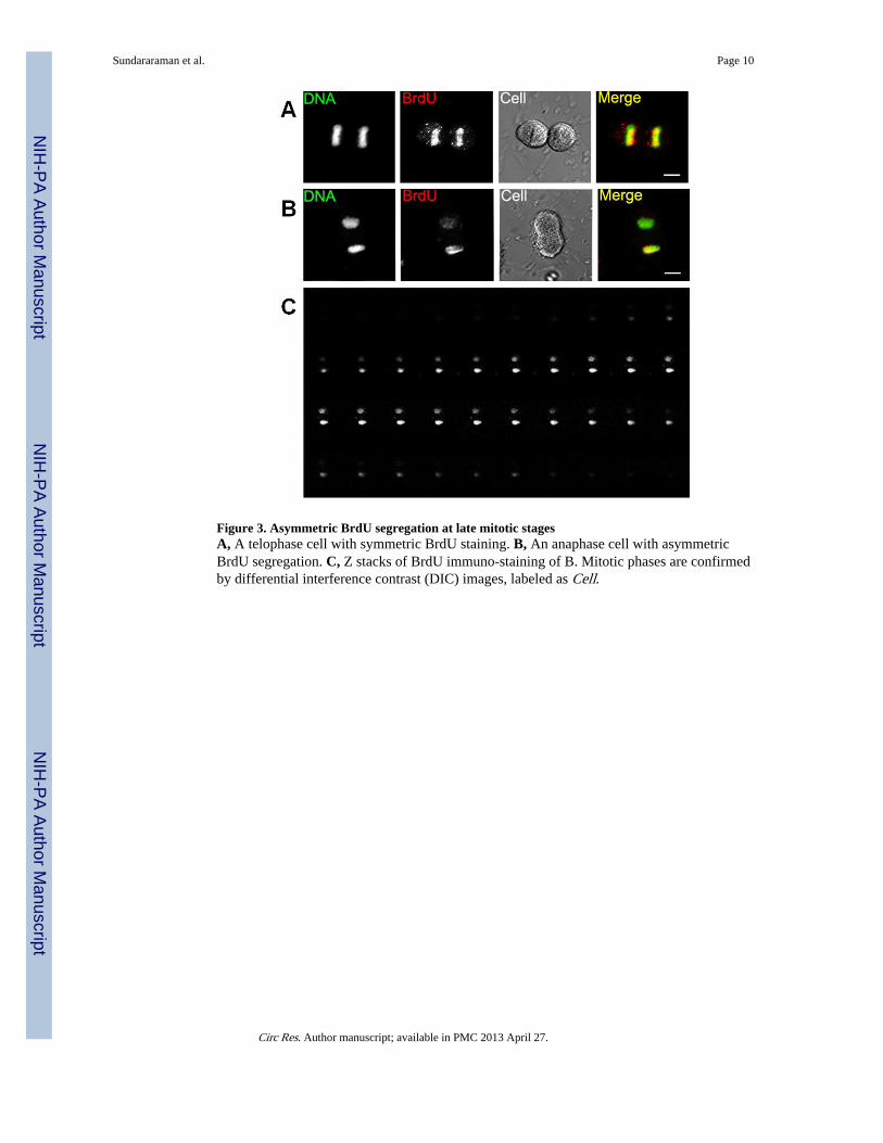
Figure 3. Asymmetric BrdU segregation at late mitotic stagesA, A telophase cell with symmetric BrdU staining. B, An anaphase cell with asymmetricBrdU segregation. C, Z stacks of BrdU immuno-staining of B. Mitotic phases are confirmedby differential interference contrast (DIC) images, labeled as Cell.
Sundararaman et al. Page 10
Circ Res. Author manuscript; available in PMC 2013 April 27.
NIH
-PA Author Manuscript
NIH
-PA Author Manuscript
NIH
-PA Author Manuscript

Figure 4. Pim-1 kinase increases asymmetric DNA segregationA, Representative binucleated cell at second mitosis for CPCe (upper) and CPCeP (lower)with asymmetric BrdU staining indicated by red arrow head. B and C, BrdU distribution atsecond mitosis between daughter nuclei of CPCe and CPCeP, respectively. Dotted boxrepresents 70:30 threshold of BrdU intensity ratio. D, Percentage of asymmetricallysegregating cells from five independent experiments. Asterisk indicates p=0.006 comparedto CPCe.
Sundararaman et al. Page 11
Circ Res. Author manuscript; available in PMC 2013 April 27.
NIH
-PA Author Manuscript
NIH
-PA Author Manuscript
NIH
-PA Author Manuscript