Norovirus Proteinase-Polymerase and Polymerase Are Both Active Forms of RNA-Dependent RNA Polymerase
Norovirus Regulation of the Innate Immune Responseand Apoptosis Occurs via the Product of the AlternativeOpen Reading Frame 4Nora McFadden1., Dalan Bailey1., Guia Carrara1¤, Alicia Benson2, Yasmin Chaudhry1, Amita Shortland3,
Jonathan Heeney3, Felix Yarovinsky2, Peter Simmonds4, Andrew Macdonald5, Ian Goodfellow1*
1 Section of Virology, Department of Medicine, Imperial College London, London, United Kingdom, 2 University of Texas Southwestern Medical Center at Dallas, Dallas,
Texas, United States of America, 3 Laboratory of Viral Zoonotics, Department of Veterinary Medicine, University of Cambridge, Cambridge, United Kingdom, 4 Centre for
Immunology, Infection and Evolution, University of Edinburgh, Edinburgh, United Kingdom, 5 Institute of Molecular and Cellular Biology, Faculty of Biological Sciences,
University of Leeds, Leeds, United Kingdom
Abstract
Small RNA viruses have evolved many mechanisms to increase the capacity of their short genomes. Here we describe theidentification and characterization of a novel open reading frame (ORF4) encoded by the murine norovirus (MNV)subgenomic RNA, in an alternative reading frame overlapping the VP1 coding region. ORF4 is translated during virusinfection and the resultant protein localizes predominantly to the mitochondria. Using reverse genetics we demonstratedthat expression of ORF4 is not required for virus replication in tissue culture but its loss results in a fitness cost since viruseslacking the ability to express ORF4 restore expression upon repeated passage in tissue culture. Functional analysis indicatedthat the protein produced from ORF4 antagonizes the innate immune response to infection by delaying the upregulation ofa number of cellular genes activated by the innate pathway, including IFN-Beta. Apoptosis in the RAW264.7 macrophagecell line was also increased during virus infection in the absence of ORF4 expression. In vivo analysis of the WT and mutantvirus lacking the ability to express ORF4 demonstrated an important role for ORF4 expression in infection and virulence.STAT1-/- mice infected with a virus lacking the ability to express ORF4 showed a delay in the onset of clinical signs whencompared to mice infected with WT virus. Quantitative PCR and histopathological analysis of samples from these infectedmice demonstrated that infection with a virus not expressing ORF4 results in a delayed infection in this system. In light ofthese findings we propose the name virulence factor 1, VF1 for this protein. The identification of VF1 represents the firstcharacterization of an alternative open reading frame protein for the calicivirus family. The immune regulatory function ofthe MNV VF1 protein provide important perspectives for future research into norovirus biology and pathogenesis.
Citation: McFadden N, Bailey D, Carrara G, Benson A, Chaudhry Y, et al. (2011) Norovirus Regulation of the Innate Immune Response and Apoptosis Occurs viathe Product of the Alternative Open Reading Frame 4. PLoS Pathog 7(12): e1002413. doi:10.1371/journal.ppat.1002413
Editor: Christopher F. Basler, Mount Sinai School of Medicine, United States of America
Received May 2, 2011; Accepted October 12, 2011; Published December 8, 2011
Copyright: � 2011 McFadden et al. This is an open-access article distributed under the terms of the Creative Commons Attribution License, which permitsunrestricted use, distribution, and reproduction in any medium, provided the original author and source are credited.
Funding: IG is a Wellcome Trust Senior fellow. This work was supported by a Wellcome Trust Senior Fellowship to IG (WT081624MA) as well as funding from theSociety for General Microbiology and Imperial College London. AM is an RCUK Academic Fellow and research in his group is funded by Yorkshire Cancer Research(LPP041, PP015 and L33) and the Royal Society. The funders had no role in study design, data collection and analysis, decision to publish, or preparation of themanuscript.
Competing Interests: The authors have declared that no competing interests exist.
* E-mail: [email protected]
¤ Current address: Department of Pathology, University of Cambridge, Cambridge, United Kingdom
. These authors contributed equally to this work.
Introduction
Collectively, the innate and adaptive immune systems result in a
strong evolutionary pressure on pathogens to develop counter-
measures to allow their continued existence. Therefore pathogens,
including viruses, have evolved a multitude of mechanisms for
evading the host response to infection, often by the expression of
proteins that interfere with cellular antimicrobial response
mechanisms [1]. The size of RNA virus genomes is thought to
be limited by the error prone nature of the viral polymerase. As a
likely direct consequence, RNA viruses have evolved a variety of
mechanisms to increase the coding capacity of their genomes [2].
These include the use of ribosomal frameshifting where a
proportion of translating ribosomes change the reading frame to
produce proteins with common N-terminal but a different C-
terminal from the read-through sequence [3]. Many viruses have
also evolved to use a mechanism that creates overlapping reading
frames through the use of two or more transcription initiation sites
or translation start codons within the same RNA sequence [4,5].
Murine norovirus (MNV) was identified in 2003 as a virus that
caused a lethal infection in immunocompromised mice [6].
However, MNV is now known to be a widespread infectious
agent of laboratory mice with a reported seroprevalence of 20-
64% [7,8]. MNV is currently the only norovirus which replicates
efficiently in tissue culture, where it has a tropism for dendritic and
macrophage cells [9]. The availability of immortalized macro-
phage cell lines such as the murine macrophage RAW264.7, has
allowed significant advances to be made in understanding the life
cycle of this virus. For the first time critical processes in the
norovirus life cycle have been dissected e.g. the mechanism of
PLoS Pathogens | www.plospathogens.org 1 December 2011 | Volume 7 | Issue 12 | e1002413
tissue culture mediated attenuation of MNV-1 [10] , the
requirement for dynamin II and cholesterol during virus entry
[11,12], the identification and functional requirement for RNA
secondary structures in virus replication [13] and pathogenesis
[14] as well as the induction of apoptosis during infection [15,16].
In addition, MNV has allowed an unprecedented analysis of the
immune response to norovirus infection [17],[18–21]. This
broadening in understanding of norovirus replication has been
facilitated greatly by the development of murine norovirus reverse
genetics [22,23] and its recent optimisation [24]. The role of
murine norovirus in potential exacerbation or complication of
other diseases, especially murine models of infection, has also been
investigated. This is certainly warranted given the seroprevalence
of MNV in animal houses. Studies with models of Crohn’s disease
[25] or bacterial induced inflammatory bowel disease [26] showed
a significant impact of MNV and MNV infection prolongs the
shedding of mouse parvovirus [27]. In contrast, MNV co-infection
had little or no impact on murine CMV [28], Friend retrovirus
infection [29] or models of diet induced obesity and insulin
resistance [30]. These contrasts warrant further studies into the
nature and mechanisms of interference observed in mouse models
of disease. MNV has also provided a useful experimental system in
determining the immune responses required for efficient norovirus
vaccination [18],[31]. Collectively, this highlights both the
relevance of MNV as a significant infectious agent in its own
right and also the utility of MNV as a model for human norovirus.
Continued research into what differentiates murine and human
noroviruses and how norovirus infection affects the host cell is
therefore of upmost importance to both fields of research.
Unlike other members of the Caliciviridae, which typically encode
three open reading frames [6], our analysis and that presented
during large scale sequencing of many MNV genomes [32]
indicates the presence of a fourth potential ORF in the MNV
genome (Figure 1A) In this study we demonstrate that the protein
encoded by ORF4 is expressed during virus infection, is not
essential for virus replication in tissue culture but plays a role in
viral virulence and therefore represents a novel viral virulence
factor. Based on the findings that it possesses anti-innate immune
activity, contribute towards the regulation of virus induced
apoptosis during infection and modulates the outcome of
experimental infection of mice, we have described the ORF4
gene product as virulence factor 1 (VF1). The study provides
important insights into the mechanisms of norovirus avoidance of
the innate immune response and norovirus pathobiology.
Results
Bioinformatic prediction and analysis of ORF4/VF1The region of the genome encoding VF1 contains an intact
reading frame in all available MNV sequences derived from
different isolates or strains (Figure 1A, 1B and data not shown). In
contrast to the typical 8–15% sequence divergence seen between
MNV variants in the amino acid sequences of ORF1, ORF3 and
the predicted single coding region of ORF2, variability is markedly
suppressed in the predicted ORF4 and double coding region of
ORF2 (3%; Table 1; Figure 1C). Almost all sequence variability
between MNV variants in the single coding regions (ORF1 and
ORF3) occurs at synonymous sites. dN/dS ratios, namely the
substitution rates at non-synonymous and synonymous sites,
ranging between 0.03–0.10 are indicative of strong negative
selection. In the double coding region of ORF2 (i.e. the region
which codes for both VP1 and VF1), the restricted variability that
is observed occurs at synonymous sites in the ORF2 reading frame
(dN/dS: 0.044) consistent with stronger sequence constraints in
the conventional reading frame encoding the MNV structural
protein than in the ORF4 gene (dN/dS < 2). However, the
elevated ratio relative to that of ORF2 arose through greater
suppression of synonymous variability in this reading frame, rather
than increased amino acid sequence variability. dN values were
0.04 and 0.03 in ORF2 and ORF4 respectively.
As well as suppressing variability, the existence of a second
reading frame in ORF2 leads to altered codon usage by the
ORF4/VF1 coding sequence. For example, there was a significant
overrepresentation of the UUG triplet coding for Leu in ORF4 (15
from 33, compared to 17 from 128 in ORF1; p,0.001 in a 662
contingency table for the 6 synonymous Leu codons), whereas
there were no differences in Leu codon usage between ORFs 1, 2
(single coding region) and ORF3. The program MLOGD
identifies overlapping coding sequences by specific codon usage
signatures arising from mutational constraints consequent to the
requirement to maintain protein function in two putative genes
[33]. The relative likelihood that a given sequence region is single-
coding or double-coding was calculated using a codon usage table
and nucleotide mutation and amino acid substitution matrices
(Figure 1C). This analysis provides independent support for the
existence of ORF4/VF1, independent of its effect on sequence
variability and evolutionary conservation. The first methionine
codon in ORF4 at 5069 lies two residues downstream from a stop
codon in that reading frame, and is 2 and 4 residues away from
Met codons in ORF1 (including the -1 frameshift). The ORF4
start codon is in a strong Kozak context (G at +4 and -3) and likely
represents the translation start site of VF1.
In vitro translation of the MNV-1 subgenomic RNAproduces three proteins, VP1, VP2 and VF1
The ability of the MNV-1 subgenomic RNA (sgRNA) to
produce a protein from the open reading frame predicted to
encode VF1 was examined by in vitro translation of a plasmid
containing the entire MNV-1 sgRNA under control of a T7 RNA
polymerase promoter (Figure 2A). A coupled transcription and
translation reaction of the MNV-1 sgRNA produced three
proteins and the identity of the major (VP1) and minor (VP2)
capsid proteins were confirmed using immunoprecipitation
Author Summary
This report describes the identification and characteriza-tion of a novel protein of unknown function encoded by amouse virus genetically similar to human noroviruses. Thisgene is unique to the mouse virus and occupies the samepart of the genome that codes for the major capsidprotein. The protein that we have described as virulencefactor 1 (VF1) is found in all murine norovirus isolates,absent in all human strains but is indeed expressed duringinfection. Its expression enables MNV-1 to establishefficient infection of its natural host through interferencewith interferon-mediated response pathways and apopto-sis. Our data would indicate that the VF1 protein is multi-functional with an ability to modulate the host’s responseto infection. Murine noroviruses are frequently used firstlyas a model to study human norovirus replication andpathogenesis, studies hampered by their inability toreplicate in cell culture. Secondly, persistent infection oflaboratory animals with murine norovirus may affect othermodels of disease using experimental mice. The role of VF1in infection and pathology in the differential outcome ofinfection is the source of continued research in ourlaboratory.
Norovirus Control of the Innate Immune Response
PLoS Pathogens | www.plospathogens.org 2 December 2011 | Volume 7 | Issue 12 | e1002413
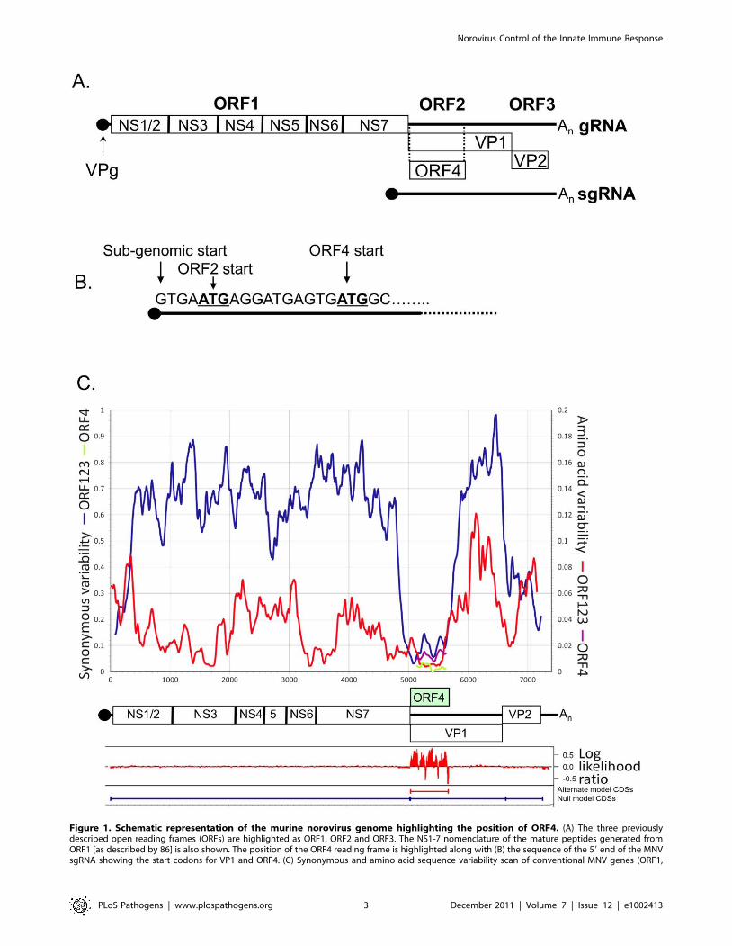
Figure 1. Schematic representation of the murine norovirus genome highlighting the position of ORF4. (A) The three previouslydescribed open reading frames (ORFs) are highlighted as ORF1, ORF2 and ORF3. The NS1-7 nomenclature of the mature peptides generated fromORF1 [as described by 86] is also shown. The position of the ORF4 reading frame is highlighted along with (B) the sequence of the 59 end of the MNVsgRNA showing the start codons for VP1 and ORF4. (C) Synonymous and amino acid sequence variability scan of conventional MNV genes (ORF1,
Norovirus Control of the Innate Immune Response
PLoS Pathogens | www.plospathogens.org 3 December 2011 | Volume 7 | Issue 12 | e1002413
(Figure 2A). Polyclonal antisera to a peptide from MNV-1 VF1
was generated in rabbits and used to confirm the identity of the
VF1 protein product by immunoprecipitation (Figure 2A). Full
length his-tagged VF1, purified from E.coli was poorly immuno-
genic, hence a modified immunization protocol that used a variety
of forms of VF1 (described in Materials and Methods), followed by
affinity purification was required in order to obtain reactive
antisera. Immune sera from MNV-1 infected mice did not contain
antibodies to VF1 as determined by western blot using
recombinant his tagged VF1 (data not shown).
VF1 is produced during MNV-1 replication in tissueculture
To examine the expression of VF1 during MNV-1 replication in
tissue culture, the well established RAW264.7 cell culture system
for MNV [9] was used and the production of VF1 analyzed by
western blot (Figure 2B). Using a high multiplicity of infection
(MOI of 5 TCID50/cell) infection, VF1 was readily detected as
early as 9 hours post infection, appearing at the same time as the
minor capsid protein VP2 (Figure 2B). In contrast, the viral RNA
polymerase NS7 was detected as early as 6 hours post infection
(Figure 2B). Whilst we were unable to detect VF1 and VP2 prior to
9 hours, this may simply be a reflection of the sensitivity of the
antisera used in the assay, but may also reflect the kinetics of viral
sgRNA synthesis, as this is likely to occur after the initial rounds of
viral genomic RNA synthesis. VF1 and VP2 expression levels
observed over the course of the infection were also significantly
different, with VF1 being expressed to a higher degree than VP2.
Whilst this may be a reflection of the differences in the ability of
the antisera to detect both proteins, it is known that VP2 synthesis
requires translation re-initiation at the end of VP1 [34] which is
likely to produce reduced levels of VP2 relative to the other
proteins expressed from the viral sgRNA.
VF1 is not essential for MNV-1 replication in tissue cultureTo determine if VF1 was required for MNV-1 replication in
tissue culture we used a recently developed reverse genetics system
[22] to truncate the VF1 coding region at various positions. Three
mutants were created containing single nucleotide changes that
lead to the introduction of a stop codon in the VF1 coding region
but which did not alter the VP1 coding sequence (Figure 3A): M1
containing the mutation T5118A truncating the VF1 protein at
amino acid 16; M10 containing the mutation T5364A, truncating
VF1 at amino acid 98; M20 containing the mutation G5655A,
truncating VF1 at amino acid 195. All mutations were introduced
at positions where it was possible to change the VF1 coding
sequence without affecting the major capsid protein VP1. Single
nucleotide substitutions were used due to the nature of the
overlapping coding regions. The interruption of VF1/ORF4 was
confirmed by in vitro coupled transcription and translation of a
PCR product encompassing the sgRNA of each mutant compared
to wild-type MNV-1 (Figure 3B). VF1 was readily detected after in
vitro translation of the wild type sgRNA product as well as the
sgRNA from the M20 mutant that encodes a C-terminally
truncated form of VF1. VF1 was not detected after in vitro
translation of the sgRNA from either the M1 or M10 VF1
truncations as expected (Figure 3B). Recovery of wild-type and
VF1 mutant viruses was performed using fowlpox mediated
expression of T7 RNA polymerase to drive the synthesis of MNV-
1 RNA in cells transfected with full length cDNA constructs of
MNV-1 as described [22]. As we have previously reported, the
BHK cell line used during virus recovery, although permissive to
virus replication, cannot be infected with MNV due to the lack of a
suitable receptor [22], therefore the yield of virus from this system
represents a single round of virus replication only. The initial
yields of VF1 knockout or truncation viruses were comparable to
that derived from wild-type cDNA (,1–56104 TCID50 per 35mm
dish, data not shown), indicative that VF1 was not required for
virus replication in tissue culture. Western blot analysis of cells
infected with the sequence verified M1, M10, M20 viruses
confirmed that VF1 was not expressed in cells infected with either
M1 or M10, but low levels of VF1 were observed in M20 infected
cells (Figure 3C). The levels of VP2 produced by the VF1
knockout viruses were comparable to the wild-type MNV-1
derived from cDNA, confirming comparative levels of infection
(Figure 3C). It is possible that the truncation of VF1 in the mutant
M20 results in some protein misfolding, decreasing the half-life of
the resulting truncated protein. The growth kinetics of low
passage, sequence verified M1, M10 and M20 viruses was
examined by both single-step (data not shown) and multi-step
growth curve analysis and were indistinguishable from that of the
wild-type parental MNV-1 derived from cDNA (Figure 3D),
indicating that VF1 is not required for MNV-1 replication in tissue
culture.
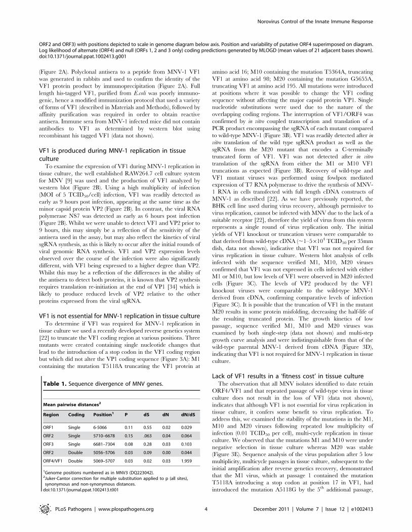
Lack of VF1 results in a ‘fitness cost’ in tissue cultureThe observation that all MNV isolates identified to date retain
ORF4/VF1 and that repeated passage of wild-type virus in tissue
culture does not result in the loss of VF1 (data not shown),
indicates that although VF1 is not essential for virus replication in
tissue culture, it confers some benefit to virus replication. To
address this, we examined the stability of the mutations in the M1,
M10 and M20 viruses following repeated low multiplicity of
infection (0.01 TCID50 per cell), multi-cycle replication in tissue
culture. We observed that the mutations M1 and M10 were under
negative selection in tissue culture whereas M20 was stable
(Figure 3E). Sequence analysis of the virus population after 5 low
multiplicity, multicycle passages in tissue culture, subsequent to the
initial amplification after reverse genetics recovery, demonstrated
that the M1 virus, which at passage 1 contained the mutation
T5118A introducing a stop codon at position 17 in VF1, had
introduced the mutation A5118G by the 5th additional passage,
Table 1. Sequence divergence of MNV genes.
Mean pairwise distances2
Region Coding Position1 P dS dN dN/dS
ORF1 Single 6-5066 0.11 0.55 0.02 0.029
ORF2 Single 5710–6678 0.15 .063 0.04 0.064
ORF3 Single 6681–7304 0.08 0.28 0.03 0.103
ORF2 Double 5056–5706 0.03 0.09 0.00 0.044
ORF4/VF1 Double 5069–5707 0.03 0.02 0.03 1.959
1Genome positions numbered as in MNV3 (DQ223042).2Juker-Cantor correction for multiple substitution applied to p (all sites),synonymous and non-synonymous distances.
doi:10.1371/journal.ppat.1002413.t001
ORF2 and ORF3) with positions depicted to scale in genome diagram below axis. Position and variability of putative ORF4 superimposed on diagram.Log likelihood of alternate (ORF4) and null (ORFs 1, 2 and 3 only) coding predictions generated by MLOGD (mean values of 21 adjacent bases shown).doi:10.1371/journal.ppat.1002413.g001
Norovirus Control of the Innate Immune Response
PLoS Pathogens | www.plospathogens.org 4 December 2011 | Volume 7 | Issue 12 | e1002413
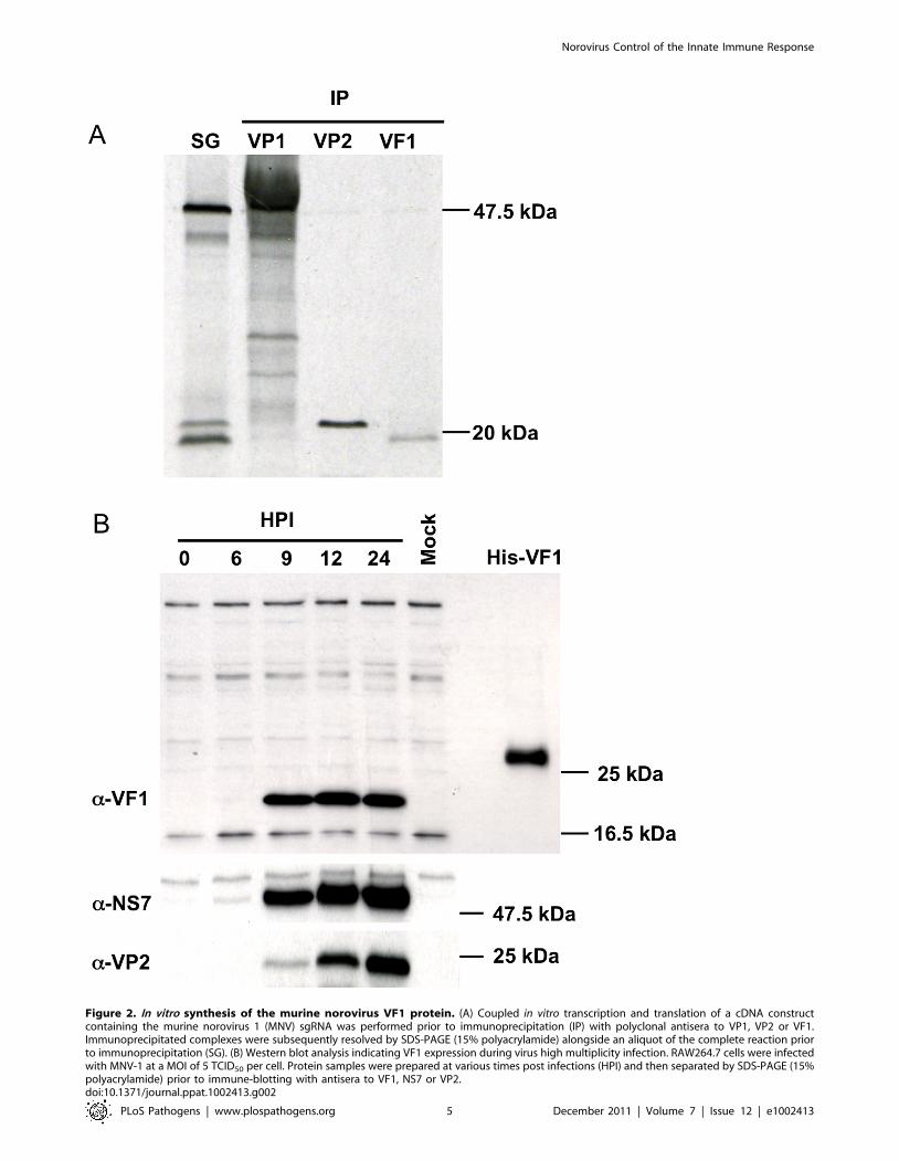
Figure 2. In vitro synthesis of the murine norovirus VF1 protein. (A) Coupled in vitro transcription and translation of a cDNA constructcontaining the murine norovirus 1 (MNV) sgRNA was performed prior to immunoprecipitation (IP) with polyclonal antisera to VP1, VP2 or VF1.Immunoprecipitated complexes were subsequently resolved by SDS-PAGE (15% polyacrylamide) alongside an aliquot of the complete reaction priorto immunoprecipitation (SG). (B) Western blot analysis indicating VF1 expression during virus high multiplicity infection. RAW264.7 cells were infectedwith MNV-1 at a MOI of 5 TCID50 per cell. Protein samples were prepared at various times post infections (HPI) and then separated by SDS-PAGE (15%polyacrylamide) prior to immune-blotting with antisera to VF1, NS7 or VP2.doi:10.1371/journal.ppat.1002413.g002
Norovirus Control of the Innate Immune Response
PLoS Pathogens | www.plospathogens.org 5 December 2011 | Volume 7 | Issue 12 | e1002413
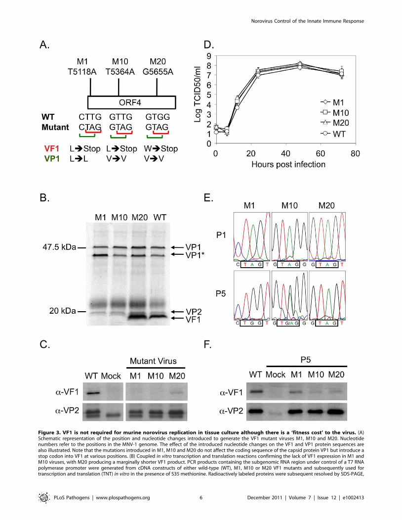
Figure 3. VF1 is not required for murine norovirus replication in tissue culture although there is a ‘fitness cost’ to the virus. (A)Schematic representation of the position and nucleotide changes introduced to generate the VF1 mutant viruses M1, M10 and M20. Nucleotidenumbers refer to the positions in the MNV-1 genome. The effect of the introduced nucleotide changes on the VF1 and VP1 protein sequences arealso illustrated. Note that the mutations introduced in M1, M10 and M20 do not affect the coding sequence of the capsid protein VP1 but introduce astop codon into VF1 at various positions. (B) Coupled in vitro transcription and translation reactions confirming the lack of VF1 expression in M1 andM10 viruses, with M20 producing a marginally shorter VF1 product. PCR products containing the subgenomic RNA region under control of a T7 RNApolymerase promoter were generated from cDNA constructs of either wild-type (WT), M1, M10 or M20 VF1 mutants and subsequently used fortranscription and translation (TNT) in vitro in the presence of S35 methionine. Radioactively labeled proteins were subsequent resolved by SDS-PAGE,
Norovirus Control of the Innate Immune Response
PLoS Pathogens | www.plospathogens.org 6 December 2011 | Volume 7 | Issue 12 | e1002413
restoring full-length VF1 production by the insertion of a
tryptophan residue. Analysis of the M10 virus population, which
had the mutation T5364A at the first passage, also indicated that
the population was heterogeneous and that in a proportion the
VF1 open reading frame was restored by the introduction of the
mutation A5364G. As with the M1 virus, this mutation is
predicted to result in the introduction of a tryptophan at position
99. In contrast however, sequence analysis of the M20 virus after
repeated multicycle passage in tissue culture demonstrated that the
introduced mutation (G5655A) was in fact stable (Figure 3E),
which may indicate that the major functional domain lay within
the 195 amino acids. Western blot analysis of cells infected with
‘passage 5’ stocks of M1 and M10 viruses indicated that, as
expected from sequence analysis, VF1 expression was detectable
(Figure 3F), although the levels were notably lower than observed
in WT infected cells. This reduced level may be in part due to the
effect of the amino acid change on VF1 protein stability, but
clearly for the M10 virus population the heterogeneous nature of
the M10 virus stock (Figure 3E) is likely a contributing factor. M20
virus stocks maintained the ability to express low levels VF1 as
previously seen using the initial virus stocks (Figure 3C and 3F). In
all cases, the level of virus replication was similar as determined by
the expression of the minor capsid protein VP2 (Figure 3F) and
virus titre (data not shown).
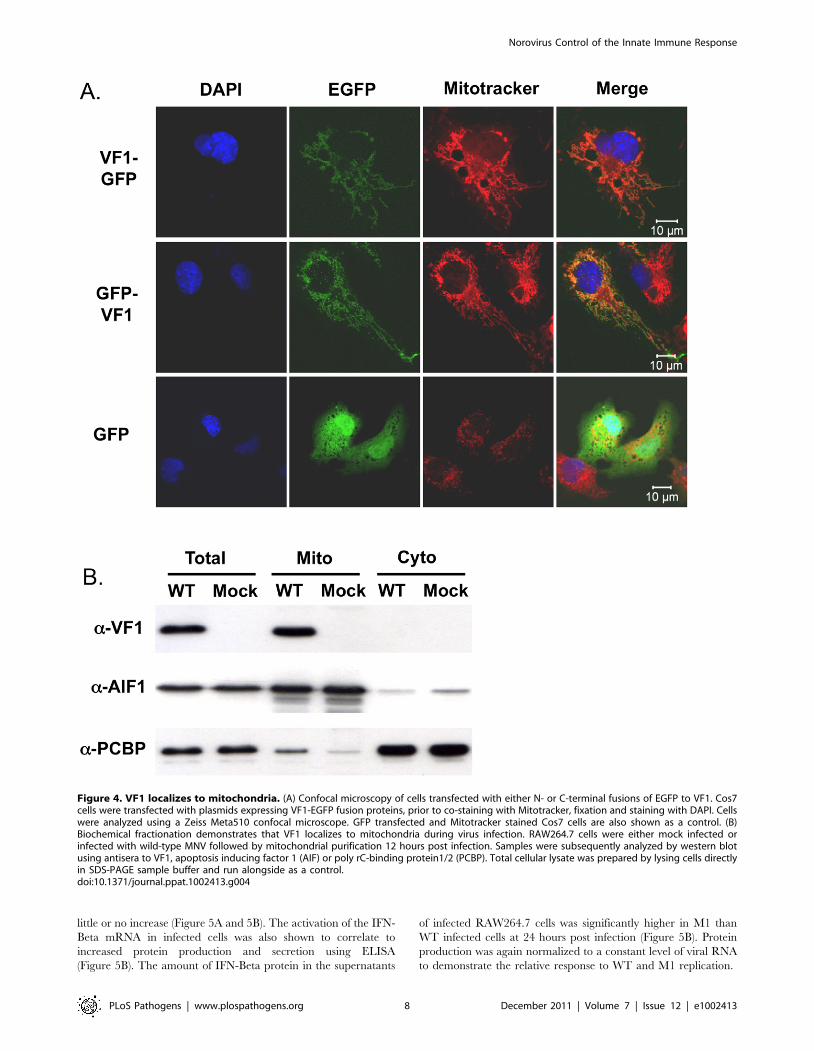
VF1 localizes to mitochondriaTo gain further insights in the potential function of VF1, the
localization of VF1 was examined by confocal microscopy. Due to
the high degree of cross-reactivity of the VF1 antisera with
endogenous host cell proteins (Figure 2B), fusions of MNV-1 VF1
to EGFP were used to examine VF1 localization in cells.
Transfection of COS7 cells with cDNA constructs expressing
either N or C-terminal fusions of MNV-1 VF1 with EGFP
demonstrated a pattern of EGFP expression characteristic of
mitochondrial localization (Figure 4A). This was confirmed via co-
staining of cells with the mitochondrial vital stain Mitotracker
(Invitrogen) (Figure 4A). Similar co-localization of VF1-GFP and
mitochondria was observed in BHK and 293 cells (data not
shown). The expression levels observed in cells transfected with the
VF1-GFP fusion proteins were substantially lower than those
observed in infected cells as expression was not detectable by
western blot analysis with either a-VF1 or a-GFP antisera (data
not shown).
To confirm the mitochondrial localization of VF1 during virus
infection, mitochondria were purified from infected RAW264.7
cells at 15 hours post infection and analyzed for the presence of
VF1 by western blot (Figure 4B). Whereas the well characterized
host cell nucleic acid binding proteins PCBP1/2 were shown to be
predominantly cytoplasmic as expected [35], VF1 was only
detected in the mitochondrial fraction (Figure 4B). Apoptosis
inducing factor 1, a predominantly mitochondrial protein was
enriched in the mitochondrial fraction, confirming the validity of
the purification procedure (Figure 4B).
VF1 production affects mitochondrial-dependent innateimmune signalling
RNA viruses frequently encode proteins that antagonize the
innate immune response to infection. Mitochondria play a
significant role in signaling innate immune responses through
the well characterized mitochondrial antiviral signaling protein
(MAVS), an integral membrane protein found in the outer
mitochondrial membrane [36–39]. MAVS is a key adapter protein
in the sensing of viral RNA by RIG-I and MDA5 that, in part,
leads to IRF3 and NFKB activation and the upregulation of
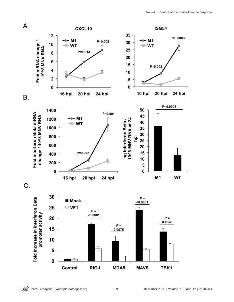
antiviral genes such as IFN-Beta, CXCL10 and ISG54 [37]. Given
the mitochondrial localization of VF1 in infected cells we assessed
the activation of this sub-section of the innate immune response in
both M1 and WT infected cells. RAW264.7 cells infected at a low
MOI (0.1 TCID50 per cell) with the M1 VF1 knockout virus
exhibited a greater induction of antiviral genes such as ISG54,
CXCL10 and IFN-Beta in response to viral infection than those
infected with the WT virus (Figure 5A and Figure 5B). Alterations
to the levels of mRNA were calculated relative to uninfected cells
using the standard DDCT method with hypoxanthine phosphor-
ibosyltransferase 1 (HPRT) (Figure 5A and Figure 5B) or actin
(data not shown) mRNA levels used as endogenous controls.
CXCL10, ISG54 and IFN-Beta mRNA levels were then
normalized to the amount of viral RNA present in each sample
in order to calculate the rate of induction of the innate immune
response over time. This method of data normalization was also
used to overcome variations often observed in the rate of virus
replication seen in a variety of RAW264.7 cell clones (not shown).
Normalizing the mRNA fold change to a constant amount of
MNV RNA established that in all cases examined (CXCL10,
ISG54 and IFN-Beta) the M1 infection causes a much more rapid
induction of the innate immune response. For instance CXCL10
in M1 infected cells is induced 15.5 fold more quickly in response
to the same amount of viral RNA than in WT infected cells. This
value is calculated by comparing the slope/gradient for M1 and
WT (Figure 5A) which represents the rate of induction of each
gene. Significantly, the IFN-Beta and ISG54 mRNAs are also
activated more quickly, 4.6 and 8.3 fold respectively, in M1
infected cells compared to WT equivalents. Of note, the total fold
increase in CXCL10, ISG54 and IFN-Beta mRNA induced in M1
infected cells was significantly higher than that observed in WT
cells at both 20 and 24 hours post infection (Figure 5A). These
time points, and the eight hour window from 16 to 24 hpi, reflect
the period of amplification for innate immune related gene
activation following low MOI MNV-1 infection of RAW264.7
cells, since quantification of mRNA fold change at 16 hpi showed
prior to exposure to film. VP1* represents a potential shorter VP1 product generated by translation initiation from an AUG initiation codon in-frameyet downstream from the authentic VP1 AUG. (C) Western blot analysis of RAW264.7 cells infected with low passage, sequence verified stocks ofeither wild-type (WT), M1, M10 or M20 VF1 mutant viruses. RAW264.7 cells were infected at a MOI 10 TCID50 per cell and harvested 12 hours postinfection, prior to separation by SDS-PAGE and western blot using either anti-VF1 or anti-VP2 antisera. (D) Multi-cycle growth kinetics analysis of VF1mutant viruses M1, M10 and M20 in RAW264.7 cells. Cells were infected with a MOI of 0.01 TCID50 per cell and samples harvested at various timespost infection prior to titration on RAW264.7 cells. Virus yield is expressed as TCID50/ml. Infections were performed in triplicate, with the average virustiter and standard deviation plotted. (E) Sequence chromatograms of M1, M10 and M20 VF1 mutant viruses after passage 1 or 5 in RAW264.7 cells.Viruses obtained from passage 1 and 5 low multiplicity infections (MOI) of RAW264.7 cells were used to infect a subsequent monolayer at high MOIprior to RNA isolation, RT-PCR amplification of the region encompassing ORF4 and sequence analysis. The positions of the introduced stop codons inthe mutants M1, M10 and M20 are boxed as are the sequences after 5 repeated passages in cell culture. (F) Western blot analysis of VF1 and VP2expression in cells infected with either wild-type MNV or passage 5 VF1 mutant viruses M1, M10 and M20. 18 hours post infection at a high MOI (4TCID50 per cell) cells were harvested, separated by SDS-PAGE prior to western blot analysis using antisera to either VF1 or VP2. Note that batch-to-batch variation in the quality of the anti-VP2 antisera accounts for the variations in the levels of non-specific proteins detected in panels C and F.doi:10.1371/journal.ppat.1002413.g003
Norovirus Control of the Innate Immune Response
PLoS Pathogens | www.plospathogens.org 7 December 2011 | Volume 7 | Issue 12 | e1002413
little or no increase (Figure 5A and 5B). The activation of the IFN-
Beta mRNA in infected cells was also shown to correlate to
increased protein production and secretion using ELISA
(Figure 5B). The amount of IFN-Beta protein in the supernatants
of infected RAW264.7 cells was significantly higher in M1 than
WT infected cells at 24 hours post infection (Figure 5B). Protein
production was again normalized to a constant level of viral RNA
to demonstrate the relative response to WT and M1 replication.
Figure 4. VF1 localizes to mitochondria. (A) Confocal microscopy of cells transfected with either N- or C-terminal fusions of EGFP to VF1. Cos7cells were transfected with plasmids expressing VF1-EGFP fusion proteins, prior to co-staining with Mitotracker, fixation and staining with DAPI. Cellswere analyzed using a Zeiss Meta510 confocal microscope. GFP transfected and Mitotracker stained Cos7 cells are also shown as a control. (B)Biochemical fractionation demonstrates that VF1 localizes to mitochondria during virus infection. RAW264.7 cells were either mock infected orinfected with wild-type MNV followed by mitochondrial purification 12 hours post infection. Samples were subsequently analyzed by western blotusing antisera to VF1, apoptosis inducing factor 1 (AIF) or poly rC-binding protein1/2 (PCBP). Total cellular lysate was prepared by lysing cells directlyin SDS-PAGE sample buffer and run alongside as a control.doi:10.1371/journal.ppat.1002413.g004
Norovirus Control of the Innate Immune Response
PLoS Pathogens | www.plospathogens.org 8 December 2011 | Volume 7 | Issue 12 | e1002413
Norovirus Control of the Innate Immune Response
PLoS Pathogens | www.plospathogens.org 9 December 2011 | Volume 7 | Issue 12 | e1002413
Treating cells with poly (I:C), and analysis of gene expression,
confirmed the sensitivity of RAW264.7 cells to dsRNA over an
equivalent time course. Induction of ISG54, CXCL10 and IFN-
Beta was demonstrated in poly (I:C) treated cells confirming their
suitability for the investigation of innate immune responses to
RNA stimuli (Figure S1). In addition, UV inactivated M1 and WT
virus showed no significant induction of ISG54, CXCL10 and
IFN-Beta when used in equivalent experiments and compared to
mock infected cells (Figure S1). The IFN-Beta protein secretion in
response to poly (I:C) and UV inactivated viruses was equivalent to
that seen for the mRNA (Figure S1).
This ability of VF1 to antagonize the innate immune response
was confirmed independently of infection using an IFN-Beta
promoter driven luciferase assay. Murine embryonic fibroblast
(MEF) cells were co-transfected with plasmid DNA expressing
firefly luciferase under the control of an IFN-Beta promoter as well
as expression constructs for RIGI, MDA5, MAVS or TBK1 whose
ectopic over-expression has been shown to drive IFN-Beta
production [40]. In addition these cells were co-transfected with
either the empty vector or a plasmid expressing the MNV-1 VF1
protein. Over-expression of RIG1, MDA5, MAVS and TBK1 in
cells transfected with the IFN-Beta promoter driven reporter
resulted in an expected increase in luciferase production in all
cases. However, this induction was significantly reduced in all
instances where VF1 was co-transfected in comparison to the
empty vector (Figure 5C). This indicates that VF1 in some way
antagonizes the induction of IFN-Beta, correlating with the results
observed in infected RAW264.7 cells.
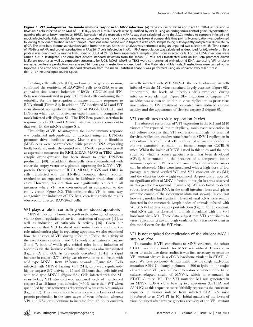
VF1 plays a role in controlling virus-induced apoptosisMNV-1 infection is known to result in the induction of apoptosis
via the down regulation of survivin, activation of caspases [41], as
well as induction of cathepsin B activity [16]. Given our
observation that VF1 localized with mitochondria and the key
role mitochondria play in regulating apoptosis, we also examined
how the absence of VF1 during infection affected the activity of
the executioner caspases 3 and 7. Proteolytic activation of caspase
3 and 7, both of which play critical roles in the induction of
apoptosis via the intrinsic cellular pathway, was also investigated
(Figure 6A and 6C). As previously described [16,41], a rapid
increase in caspase 3/7 activity was observed in cells infected with
wild type MNV-1 from 12 hours onwards (Figure 6A). Cells
infected with MNV-1 lacking VF1 (M1), displayed significantly
higher caspase 3/7 activity at 15 and 18 hours than cells infected
with wild type MNV-1 (Figure 6A). Cells infected with the M1
virus lacking VF1 also displayed increased levels of the cleaved
caspase 3 at 16 hours post infection (,50% more than WT when
quantified by densitometry) as determined by western blot analysis
(Figure 6C). There was a notable alteration to the kinetics of viral
protein production in the later stages of virus infection; whereas
VP1 and NS7 levels continue to increase from 15 hours onwards
in cells infected with WT MNV-1, the levels observed in cells
infected with the M1 virus remained largely constant (Figure 6B).
Importantly, the levels of infectious virus produced during
infection were identical (Figure 3D). Induction of caspase 3/7
activities was shown to be due to virus replication as prior virus
inactivation by UV treatment prevented virus induced caspase
activity and the appearance of cleaved caspase 3 (Figure 6C).
VF1 contributes to virus replication in vivoThe observed restoration of VF1 expression in the M1 and M10
viruses after repeated low multiplicity, multi-cycle replication in
cell culture indicates that VF1 expression, although not essential
for virus replication, confers some benefit to MNV-1 replication in
cell culture. To examine if VF1 contributed to virus replication in
vivo we examined replication in immunocompetent C57BL/6
mice. Whilst the isolate of MNV-1 used in this study and the only
strain for which a reverse genetics system has been developed
(CW1), is attenuated in the presence of a competent innate
immune response [6,42], low level virus replication in some tissues
can be observed. Mice were inoculated with a high dose of low
passage, sequenced verified WT and VF1 knockout viruses (M1)
and the effect on body weight examined. As previously reported,
no significant effect of MNV infection on weight loss was observed
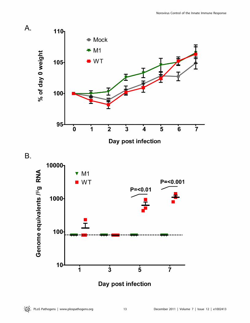
in this genetic background (Figure 7A). We also failed to detect
robust levels of viral RNA in the small intestine, feces and spleen
over the course of the experiment (data not shown). In contrast
however, modest but significant levels of viral RNA were readily
detected in the mesenteric lymph nodes of animals infected with
WT MNV-1 at days 5 and 7 post infection (Figure 7B). In contrast,
viral RNA was not detected in animals inoculated with the VF1
knockout virus M1. These data suggest that VF1 contributes to
virus replication in vivo although virulence per se was not evident in
this model even for the WT virus.
VF1 is not required for replication of the virulent MNV-1strain in vitro
To examine if VF1 contributes to MNV virulence, the robust
STAT1 -/- mouse model for MNV was utilized. However, in
order to undertake these studies it was first necessary to generate
VF1 mutant viruses in a cDNA backbone virulent in STAT1-/-
mice. We have previously demonstrated that the single nucleotide
mutation A5941G, changing glutamate 296 to lysine in the major
capsid protein VP1, was sufficient to restore virulence to the tissue
culture adapted strain of MNV-1, which is attenuated in
STAT1-/- mice [10]. The VF1 mutation M1 was generated in
an MNV-1 cDNA clone bearing two mutations (G2151A and
A5941G) as this sequence more faithfully represents the consensus
sequence in viruses isolated from infected STAT1-/- mice
[6,referred to as CW1.P1 in 10]. Initial analysis of the levels of
virus obtained after reverse genetics recovery of the VF1 mutant
Figure 5. VF1 antagonizes the innate immune response to MNV infection. (A) Time course of ISG54 and CXCL10 mRNA expression inRAW264.7 cells infected at an MOI of 0.1 TCID50 per cell. mRNA levels were quantified by qPCR using an endogenous control gene (Hypoxanthine-guanine phosphoribosyltransferase, HPRT). Expression of the respective mRNAs was then calculated using the DDCt method to compare infected andmock infected cells. Relative fold change was calculated using mock infected samples taken at comparable time points. Normalization was performedfollowing MNV quantification in each sample. Infections were carried out in triplicate with each sample being subsequently analyzed in duplicate byqPCR. The error bars denote standard deviation from the mean. Statistical analysis was performed using an unpaired two tailed t test. (B) Time courseof IFN-Beta mRNA and protein production in RAW264.7 cells infected as in (A). mRNA upregulation was calculated as described for (A). Interferon Betaprotein was quantified by murine IFN-B specific ELISA at 24 hpi from supernatant samples taken from infected cells. For the ELISA infections werecarried out in sextuplate. The error bars denote standard deviation from the mean. (C) MEF cells transfected with an IFN-Beta promoter drivenluciferase reporter as well as expression constructs for RIG1, MDA5, MAVS or TBK1 were co-transfected with plasmid DNA expressing VF1 or blankmessage. Luciferase production was assayed 24 hours post transfection as described in the Materials and Methods. Transfections were carried out intriplicate. The error bars denote standard deviation from the mean. Statistical analysis was performed using an unpaired two tailed t test.doi:10.1371/journal.ppat.1002413.g005
Norovirus Control of the Innate Immune Response
PLoS Pathogens | www.plospathogens.org 10 December 2011 | Volume 7 | Issue 12 | e1002413
Norovirus Control of the Innate Immune Response
PLoS Pathogens | www.plospathogens.org 11 December 2011 | Volume 7 | Issue 12 | e1002413
virus M1 in the virulent backbone (referred to herein as M1-v),
demonstrated identical levels to the wild-type virulent virus (WT-v)
of approximately 1-56103 TCID50/ml (data not shown). This
suggests that, as observed in the attenuated background, VF1
expression is not essential for MNV-1 replication in the STAT1-/-
virulent backbone. To further verify this, multi-cycle growth
kinetics analysis of low passage, sequence verified, WT-v and M1-v
viruses in RAW264.7 cells was performed confirming equivalent
growth kinetics (data not shown).
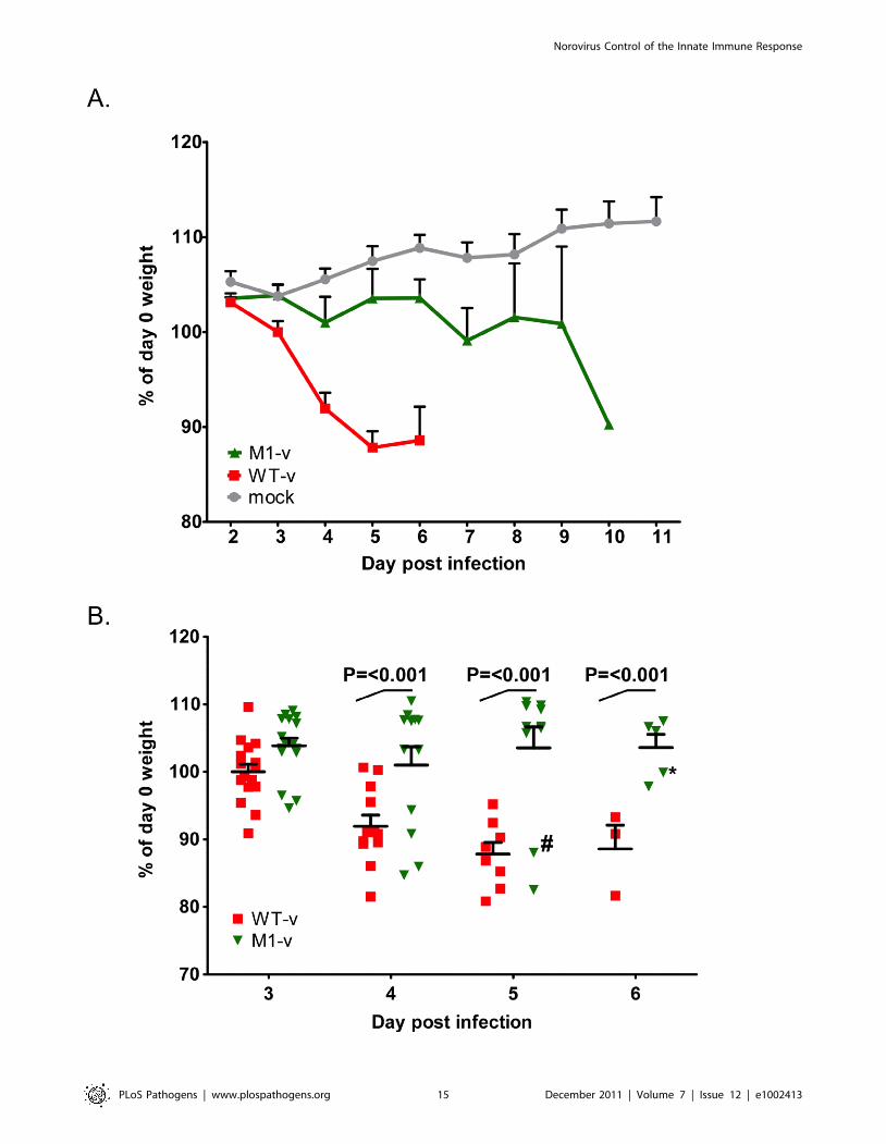
MNV-1 viruses lacking VF1 are partially attenuated inSTAT1-/- mice
The ability of WT-v and M1-v viruses to infect and cause
disease in the STAT1-/- mouse model was then examined by oral
infection of age and sex matched mice. Oral inoculation of
STAT1-/- mice with 1000 TCID50 units of low passage, sequence
verified, wild-type virulent MNV-1 derived from cDNA (WT-v)
resulted in the appearance of clinical signs (sunken eyes, reduced
appetite, hunched inactivity and piloerection) as early as three
days post inoculation. This was followed by a rapid and statistically
significant (P,0.001) weight loss, when compared to animals
inoculated with mock RAW264.7 cell lysate (day 4 onwards), and
the development of more severe clinical signs culminating in
significant weight loss (Figure 8). All WT-v infected mice
succumbed to infection or were euthanized (because of disease
severity limits being surpassed) by day 7 post infection. In stark
contrast mice inoculated with the VF1 mutant virus (M1-v)
showed a delayed onset of clinical signs. A statistically significant
weight loss, compared to the mock-inoculated control group, was
not observed until 6 days post infection (P,0.05, Figure 8). Of
note, although the onset of M1-v associated disease was
significantly delayed, all M1-v infected animals eventually
succumbed to the infection or surpassed the severity limits of
our trial. Experiments performed using a 10 fold higher dose
(10,000 TCID50) also demonstrated that M1-v inoculated mice
displayed a delayed onset of clinical signs including a statistically
significant variation in body weight loss (two-way ANOVA).
However this variation was markedly less than that observed at the
lower infectious dose of 1000 TCID50 (Figure S2).
Viral RNA replication is reduced in animals infected withthe M1-v VF1 knockout virus
To examine if the distribution of virus replication differed
between animals inoculated with WT-v or M1-v viruses, viral
genome copies were quantified in various tissues at 3 and 5 days
post infection by quantitative real-time reverse transcription PCR
(qRT-PCR, Figure 9). Whilst .106 genome equivalents (gEq) per
mg of total RNA could be readily detected in samples from mice
infected with WT-v 3 days post infection, viral genome levels in
M1-v infected animals were typically 104–105 fold lower
(Figure 9A). For example, average levels in the spleen for WT-v
infected animals were 1.76109 gEq/mg of total RNA whereas M1-
v inoculated animals showed an average of 2.66104 qEq/mg of
total RNA. Increased viral RNA replication was detected in all
WT-v infected mice at day 5 post infection but only in a subset of
the M1-v infected mice (Figure 9B). This subset correlated with
those animals that had developed more significant clinical signs
and had lost body weight at day 5 post infection.
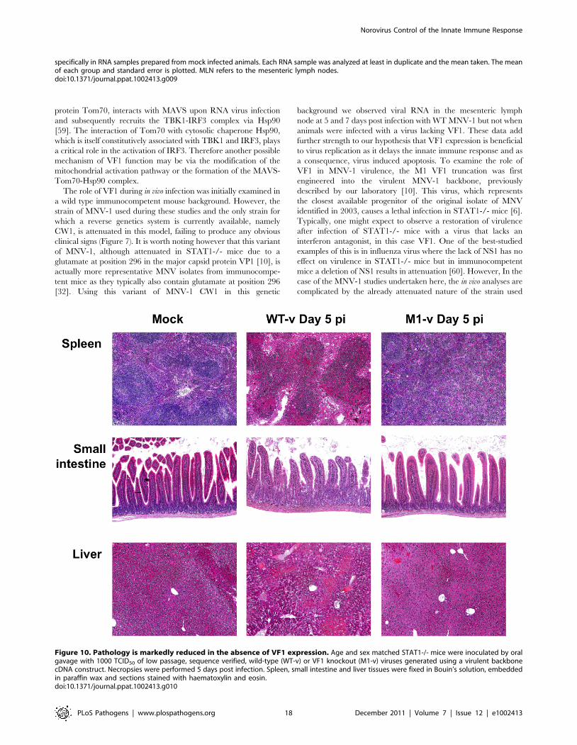
M1-v infected mice show reduced histopathology at day5 post infection
Tissue samples from the spleen, small intestine and liver of
mock, WT-v and M1-v infected mice were harvested at day 5 post
infection for histopathological analysis on hematoxylin and eosin
stained sections (Figure 10). Tissues from mock-infected mice were
relatively normal for STAT1-/- mice (Figure 10). In contrast the
WT-v infected tissues demonstrated reduced cellularity in spleen
and liver, with foci of marked necrosis and apoptosis. Necrosis was
evidenced by eosinophilia (dead cells staining bright pink) with
pyknosis (nuclear condensation) and karryorrhexis (pyknotic nuclei
become fragmented into several particles). Apoptosis was evi-
denced by cell rounding, a shrunken nucleus and in some cases cell
fragmentation with some of the fragments containing apoptotic
bodies. Blunting of the intestinal villi, as determined by measuring
the villous height on the digital images taken at the same
magnification and a comparison carried out on the mean of 8 villi,
was apparent only in sections of the small intestine from animals
infected with WT-v (Figure 10). Although the spleen from animals
infected with M1-v appeared activated with partial paracortical
hyperplasia, it was otherwise normal with little evidence of the
necrosis or apoptosis evident in WT-v tissues (Figure 10). The liver
revealed a partial loss of cellularity; however, evidence of apoptosis
was again absent (Figure 10). In conclusion the lack of substantial
pathology in M1-v infected mice at 5 days post infection correlated
with our previous observations for differential viral RNA
replication and weight loss in WT-v and M1-v infected mice.
The M1-v virus does not revert during infection ofSTAT1-/- mice
The delayed virulence of VF1 knockout viruses in STAT1-/-
mice was suggestive of reversion during replication in vivo. To
examine this possibility further, sequence analysis of the viral
population from all tissues in two animals displaying the highest
degree of clinical signs (and viral genomes determined by qRT-
PCR) was undertaken. The animals analyzed are highlighted in
Figure 8B with a hash and asterisk indicating animals culled on
days 5 and 7 respective. The animal culled on day 5 displayed
similar disease onset to that of WT-v inoculated animals and had
lost ,12% of its initial body weight. The animal culled on day 7
for sequence analysis was removed from the study due to human
end points being exceeded and had lost ,7.6% of its initial body
weight. Consensus sequence analysis, which under the conditions
Figure 6. Viruses lacking VF1 are altered in their ability to induce apoptosis. (A) Time course of caspase 3/7 activity in RAW264.7 cellstreated with Staurosporine or infected with wild-type MNV-1 (WT) or a VF1 knockout virus (M1). Cells were infected at a MOI of 5 TCID50 per cell andsamples harvested at various times post infection. Caspase 3/7 activity was determined using a commercial assay from Promega (Glo 3/7) and theactivity represented as relative light units (RLU). Each assay was performed in triplicate with the average and standard error within a singleexperiment plotted. Statistical analysis was performed using 2 way ANOVA with Bonfferoni post tests (B) Western blot analysis of extracts preparedfrom cells infected as detailed in (A). Samples were prepared at various times post infection, separated by SDS-PAGE and subsequently immuneblotted using antisera to GAPDH, NS7, VP1 or VF1. Non-infected (Mock) cells were included as controls. (C). Caspase 3/7 activity assay in RAW 264.7cells infected with M1 or WT MNV-1 or UV-inactivated M1 and WT viruses. Samples were analyzed at 16 hours post infection with triplicate samplestaken. The mean and standard error within a single experiment are plotted. Statistical analysis was performed using an unpaired two tailed t-test (WTversus M1). P,0.01 is illustrated as ** and P,0.001 is shown as ***. Western blot analysis of GAPDH, NS7 and cleaved caspase 3 for the same samplesis also shown.doi:10.1371/journal.ppat.1002413.g006
Norovirus Control of the Innate Immune Response
PLoS Pathogens | www.plospathogens.org 12 December 2011 | Volume 7 | Issue 12 | e1002413
Norovirus Control of the Innate Immune Response
PLoS Pathogens | www.plospathogens.org 13 December 2011 | Volume 7 | Issue 12 | e1002413
used could reproducibly detect reversion when ,25% of the
population had restored VF1, failed to detect any reversion in any
of the tissues (Figure S3). In addition, tissues/samples containing
the highest viral loads (46106 to 46107 copies per mg of RNA),
namely the spleen and feces from the day 5 animal and the spleen
from the day 7 animal, were PCR amplified and 10 individual
clones sequenced. Of the 30 clones sequenced, none contained a
mutation in VF1 that would lead to restoration of VF1 expression,
further confirming the lack of detectable reversion upon
replication a single pass in Stat1-/- mice in vivo (data not shown).
Discussion
Studies on numerous RNA viruses have identified the use of
overlapping open reading frames to maximize the coding capacity
of their small RNA genomes [2]. These ORFs and their proteins
typically play accessory roles in the viral life cycle such as
modulating the host immune response to infection [43-45].
Frequently they dispensable for viral replication in immortalized
cell lines; however, it is the in vivo setting that the true requirement
for these proteins in the viral life cycle is apparent. This study
indicates that MNV should now be added to the list of viruses that
have adopted this strategy to maximize the coding potential of
their genome.
Initial bioinformatic investigation of MNV complete genome
sequences identified a conserved ORF overlapping with ORF2
(Figure 1), potentially translated from the sgRNA produced during
infection. Traditionally the sgRNA is thought to encode only the
major and minor capsid proteins, VP1 and VP2. However
suppression of variability in this region and conservation of the
alternate ORF was shown to be absolute in all available MNV
sequences (Table 1). Although the resultant full length protein,
VF1, was recalcitrant to high level expression and poorly
immunogenic, polyclonal antibody specific to this protein was
generated and used to confirm expression during infection
(Figure 2). The efficient translation of this protein was confirmed
by immmunoprecipitation following translation of the sgRNA in
vitro (Figure 2).
This is the first confirmation of the expression of an internal
open reading frame protein for any member of the Caliciviridae.
The internal open reading frame encoding VF1 can be found in all
currently published MNV genome sequences, highlighting the
requirement for this feature in the MNV genome. The
evolutionary conservation of ORF4 coding sequences and the
marked suppression of sequence variability localising specifically to
the area of overlap (Figure 1C) provides evidence independent of
the in vitro data for a functional requirement to maintain an intact
ORF4 reading frame. As indicated by the analysis of leucine and
other synonymous codon usage, this selection pressure was
sufficiently strong to drive unfavoured codons into the ORF4
coding sequence (Figure 1C), a feature exploited by MLOGD [33]
to detect regions of multiple coding.
There were considerable similarities in the arrangement and
translation contexts of the ORF2 and ORF4 genes of MNV with
documented regions of multiple coding in other viruses. The
MNV ORF4 has an initiating AUG triplet at position 5069 in a
strong Kozak context (G at -3 and +4 [46]. It is positioned 13 bases
downstream from the first AUG triplet of ORF2 (weak context; U
at -3, A at +4) and 7 from the second (adequate context; A at -3
and +4). This arrangement of initiating codons in the MNV
sgRNA transcripts is similar to viral [47] and eukaryotic [48]
dicistronic mRNAs in which alternative weak context initiating
codons around an initiating codon in a strong context (ORF4 in
MNV) can be accessed by random forwards and backwards
movements of the ribosome from its initial binding site, termed
"leaky scanning". In this case, this would require a backwards
movement to the second AUG triplet of ORF2, remarkably
similar to the documented dicistronic expression of p206 (strong
context) and p69 (weak context 7 bases upstream) from genomic
RNA of turnip yellow mosaic virus [47]. This hypothesis is
supported by the observation that noroviruses that lack ORF4
(genogroups 1–4) show a strong Kozak context around the second
AUG triplet in ORF2 (A at -3, G at +4). The evolutionarily
conserved nucleotide difference at position 5065 (+4) between
MNV (A) and other noroviruses (G) may thus play a key enabling
role in the hypothesised dicistronic expression of ORF4 and ORF2
by MNV.
The juxtapositioning of ORF4 at the start of the sgRNA gives
an indication of the additional evolutionary constraints that this
ORF, and the respective protein, must be under. This region of
the genome contains multiple conserved cis-acting RNA elements
that play an important role in the viral life cycle (unpublished
observations). It is important to note however that the single
nucleotide mutation introduced in this region to generate the M1
virus, did not affect the structure of these RNA elements as we
have determined biochemically that nucleotide 5118 is positioned
within a single-stranded region (data not shown). ORF4 also
overlaps with the region of ORF2 that encodes the shell (S)
domain of the major capsid protein, VP1. Dimerization of the S
domain is thought to be integral for the development of the
icosahedral core of the virus particle and is consequently the most
conserved domain in VP1. As the S domain is buried inside the
virus particle, it is unlikely to be under strong antibody selection
pressure, unlike the more variable C terminus of VP1 which
contains the protruding (P) domain. The contribution of all these
factors is likely responsible for the low divergence observed
between MNV VF1 sequences (Table 1).
Within the norovirus genus, ORF4/VF1 appears to be a feature
unique to MNV as other noroviruses appear not to encode an
equivalent open reading frame. The human noroviruses, which
represent a significant cause of viral gastroenteritis in man, do not
share the extensive suppression of synonymous site variability at
the start of ORF2 that first indicated the presence of ORF4
(Figure 1C) [13]. Direct analysis of the available human norovirus
sequences confirms that no such ORF exists (data not shown). A
broader analysis of the Caliciviridae family indicates the presence of
an equivalent open reading frame in some strains of human
sapoviruses (data not shown) [49]. Although there is low sequence
homology between the respective proteins (25% similarity, 18%
identity) the presence of this alternative ORF indicates a potential
Figure 7. VF1 expression is required for virus efficient replication in vivo. Age and sex matched C57BL/6 mice were inoculated by oralgavage with 16107 TCID50 of low passage, sequence verified, wild-type (WT) or VF1 knockout (M1) viruses. Body weight was measured on a dailybasis and expressed as a percentage of the weight on day 0 prior to inoculation. The mean weight and the standard error are plotted as a line graphcovering the duration of the experiment (A). Quantitative real-time RT-PCR quantification of viral genome copies in the mesenteric lymph nodeisolated from mice at various times post inoculation (B). The mean and standard error are plotted. Statistical analysis was performed using two-wayANOVA and Bonferroni post tests (WT versus M1). The blue line represents the detection limit as determined by the sensitivity of qPCR to detectstandards, equating to 80 copies per mg of RNA. Samples that were below the detection limit were set as 80 copies per mg of RNA.doi:10.1371/journal.ppat.1002413.g007
Norovirus Control of the Innate Immune Response
PLoS Pathogens | www.plospathogens.org 14 December 2011 | Volume 7 | Issue 12 | e1002413
Norovirus Control of the Innate Immune Response
PLoS Pathogens | www.plospathogens.org 15 December 2011 | Volume 7 | Issue 12 | e1002413
conserved mechanism for maximizing coding potential. It is also
possible that a common ancestor of all caliciviruses possessed an
equivalent ORF, which has subsequently been lost in the case of
the majority of caliciviruses. Although human noroviruses, as well
as other members of the Caliciviridae, lack an equivalent ORF4
within the VP1 coding region of the sgRNA, we cannot at this
point rule out functional duplication i.e. that the functions of
MNV VF1 have been duplicated in human noroviruses by one of
the other viral proteins. Further studies are therefore warranted to
determine if human noroviruses and other members of the
Caliciviridae also possess the ability to modulate the innate immune
response.
The role of the VF1 protein in MNV-1 replication was
examined using the permissive macrophage RAW264.7 cell line
and the reverse genetics system developed previously in our
laboratory [22]. A series of VF1 truncations, generated by
inserting stop codons into ORF4, which importantly left the
VP1 coding sequence unaltered, confirmed this protein was a
classical viral accessory protein not required for replication.
However, repetitive multicycle, low multiplicity of infection
passage in the permissive RAW264.7 cell line resulted in a
phenotypic reversion for the more severe truncations (M1 and
M10) (Figure 3), demonstrating that VF1 expression and function
benefits virus replication in cell culture. Whilst this observation
was reproducible, it was clear that rapid phenotypic reversion did
not occur as virus stocks generated by reverse genetics and
subsequently amplified in cell culture by a single passage
maintained the introduced mutations. The phenotypic reversion
observed was the likely result of the multicycle nature of the
infections as very low multiplicity of infections were used, resulting
in multiple rounds of virus replication to occur in each pass in cell
culture.
As the ‘powerhouses’ of the eukaryotic cell, viruses often
modulate the function of mitochondria to maintain an intracellular
environment beneficial for viral replication. The mitochondrial
localization of VF1 (Figure 4) together with its apparent
modulation of innate immune signaling and apoptosis (Figure 5
and 6) indicates that this protein may function, like so many other
viral proteins, to facilitate viral replication and antagonize anti-
viral mechanisms adopted by the cell. The beneficial functions of
VF1 expression, e.g. delayed apoptosis and innate immune
responses in infected cells, are apparent since analysis of VP1
protein levels produced during infection are clearly reduced at the
later stages of infection in the absence of VF1 (Figure 6B).
Surprisingly this does not appear to affect the final yield of virus
(Figure 3D) or the levels of viral RNA produced during replication
in RAW264.7 cells (data not shown). The RAW264.7 murine
macrophage cell line is extremely permissive to infection and it is
likely that the total pool of available VP1 protein in the M1
infected cells later in infection does not limit virion production. Of
relevance is our observation that the IRF3 modulated gene ISG54,
also known as IFIT2 or p54, is significantly upregulated in cells
infected with a virus lacking VF1 (Figure 5A). The ISG54 protein
(p54 or IFIT2) functions to repress cellular translation by binding
and inhibiting the cellular eIF3c protein [50]. Previous work has
indicated that norovirus translation initiation may require the eIF3
complex via a direct interaction of VPg [51]. The expression of the
MNV VF1 protein may therefore delay or block the ISG54
mediated inhibition of cellular and viral VPg-dependent transla-
tion by preventing the induction of ISG54 mRNA at the point of
mitochondrial mediated activation of the innate immune response
[51–53]. ISG54 expression has also been linked to apoptosis
induced via the mitochondrial pathway [54], again in agreement
with our observed increase in apoptosis in the absence of VF1
expression (Figure 6). It is possible therefore that the increased
apoptosis observed during virus replication in the absence of VF1
expression is as a result of the increased induction of the innate
immune response.
It is well established that MNV is sensitive to an effective innate
immune response: type I and II interferon are known to inhibit
viral translation [20], a fact supported by the observed sensitivity
of STAT1-/- mice to infection [6]. The role of STAT1 mediated,
interferon-based, innate immune signaling in combating MNV-1
infection has been well characterized to prevent the progression of
MNV-1 infection and dissemination to peripheral tissues [42]. We
also observed this effect in our studies with immunocompetent
C56BL/6 mice as only low levels of viral RNA were detected in
the MLN (Figure 7). Previous work has also highlighted that at
least part of the innate sensing of MNV infection by the host cell
can be attributed to the MDA5 protein [17]. When activated,
MDA5 signals the detection of viral RNA through the mitochon-
drial antiviral signaling (MAVS) adapter protein (also known as
IPS-1, VISA or Cardif) embedded in the outer membrane of this
organelle [36–38]. One downstream consequence of these
signaling events at the mitochondria is the dimerisation and
subsequent nuclear shuttling of IRF3. This activation of IRF3
results in the trans-activation of genes responsible for combating
viral infection including interferon beta. In our studies, the
regulation of genes specifically stimulated by virus infection was
monitored by qPCR and ELISA and shown to be elevated in cells
infected with a virus lacking the VF1 accessory protein (Figure 5A
and Figure 5B). This potential role for VF1 as an antagonist of the
innate immune response was investigated using co-expression
studies with various auto-stimulatory components of this pathway
which, after transient over-expression, are known to trigger
interferon production (such as RIG-I, MAVS) (Figure 5C). In this
instance, VF1 was shown to reduce the expression of a reporter
protein under the direct transcriptional control of the interferon
beta promoter. This occurred at the level of, or subsequent to
TBK1 activation, since its stimulation of the IFN promoter was
also inhibited by VF1 expression. The mechanism of action of
VF1 therefore potentially involves modulation of the interaction of
TBK1 with IRF3, or directly acts on IRF3 itself. Inhibition or
degradation of IRF-3 is a frequent target of viral evasion strategies
among both RNA and DNA viruses, including the Npro protein of
pestviruses [55,56], the P protein of rabies virus [57] and the G1
protein of hantaviruses [58]. Mitochondria also serve as a platform
for the activation of IRF3 via TBK1 as the mitochondrial import
Figure 8. VF1 plays a role in viral virulence. Age and sex matched STAT1-/- mice were inoculated by oral gavage with 1000 TCID50 of lowpassage, sequence verified, wild-type (WT-v) or VF1 knockout (M1-v) viruses generated using a virulent backbone cDNA construct. As a measure ofthe severity of clinical disease, body weight was measured on a daily basis and expressed as percentage of the weight on day 0 prior to inoculation.Mock infected animals were orally inoculated with a control lysate preparation, generated as described in the materials and methods. For clarity themean weight and the standard error are plotted as both a line graph covering the duration of the experiment (A) and as individual animal weightsduring days 3-6 (B). Statistical analysis was performed using two-way ANOVA with Bonferroni post-tests (WT-v versus M1-v). The hash and asteriskhighlight animal number 776 and 751 used in the sequence analysis on days 5 and 7 respectively as described in the text. Note that animal 751 wasremoved from the study on day 7 (not shown on the plot) due to humane endpoints.doi:10.1371/journal.ppat.1002413.g008
Norovirus Control of the Innate Immune Response
PLoS Pathogens | www.plospathogens.org 16 December 2011 | Volume 7 | Issue 12 | e1002413
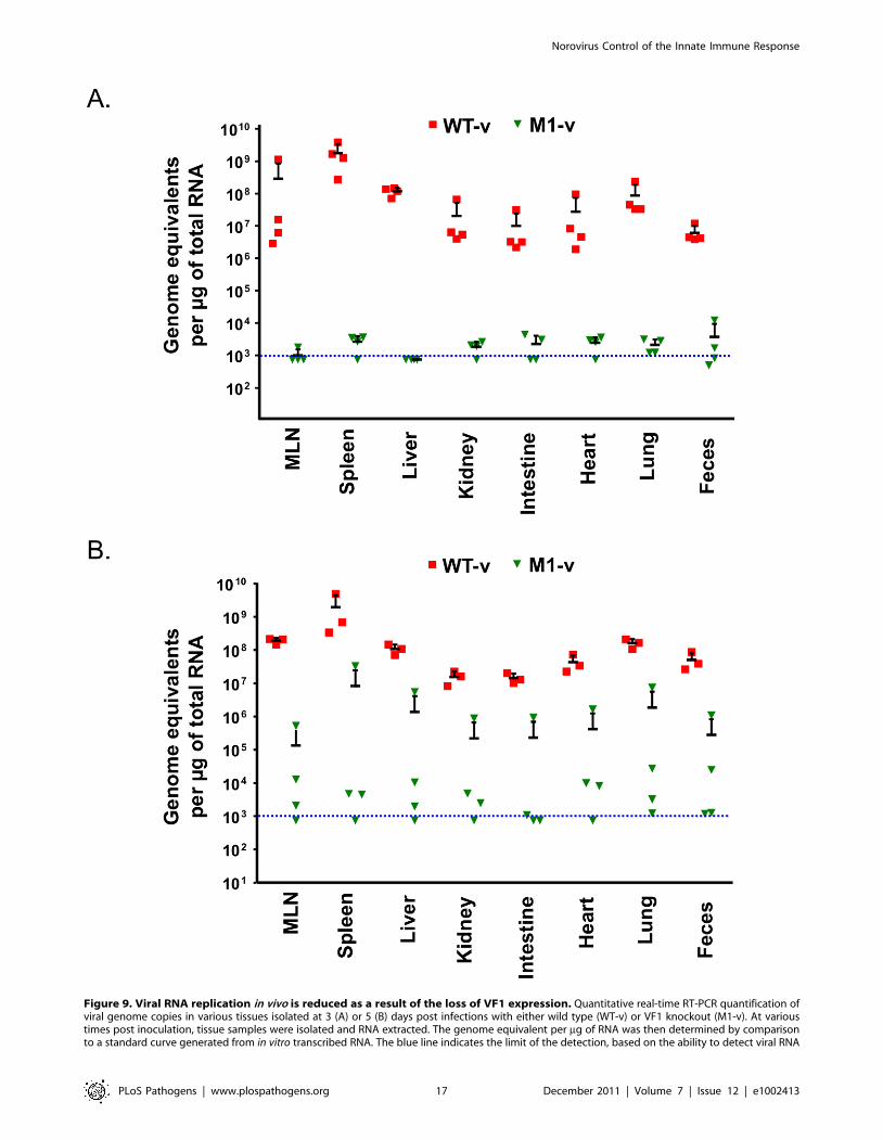
Figure 9. Viral RNA replication in vivo is reduced as a result of the loss of VF1 expression. Quantitative real-time RT-PCR quantification ofviral genome copies in various tissues isolated at 3 (A) or 5 (B) days post infections with either wild type (WT-v) or VF1 knockout (M1-v). At varioustimes post inoculation, tissue samples were isolated and RNA extracted. The genome equivalent per mg of RNA was then determined by comparisonto a standard curve generated from in vitro transcribed RNA. The blue line indicates the limit of the detection, based on the ability to detect viral RNA
Norovirus Control of the Innate Immune Response
PLoS Pathogens | www.plospathogens.org 17 December 2011 | Volume 7 | Issue 12 | e1002413
protein Tom70, interacts with MAVS upon RNA virus infection
and subsequently recruits the TBK1-IRF3 complex via Hsp90
[59]. The interaction of Tom70 with cytosolic chaperone Hsp90,
which is itself constitutively associated with TBK1 and IRF3, plays
a critical role in the activation of IRF3. Therefore another possible
mechanism of VF1 function may be via the modification of the
mitochondrial activation pathway or the formation of the MAVS-
Tom70-Hsp90 complex.
The role of VF1 during in vivo infection was initially examined in
a wild type immunocompetent mouse background. However, the
strain of MNV-1 used during these studies and the only strain for
which a reverse genetics system is currently available, namely
CW1, is attenuated in this model, failing to produce any obvious
clinical signs (Figure 7). It is worth noting however that this variant
of MNV-1, although attenuated in STAT1-/- mice due to a
glutamate at position 296 in the major capsid protein VP1 [10], is
actually more representative MNV isolates from immunocompe-
tent mice as they typically also contain glutamate at position 296
[32]. Using this variant of MNV-1 CW1 in this genetic
background we observed viral RNA in the mesenteric lymph
node at 5 and 7 days post infection with WT MNV-1 but not when
animals were infected with a virus lacking VF1. These data add
further strength to our hypothesis that VF1 expression is beneficial
to virus replication as it delays the innate immune response and as
a consequence, virus induced apoptosis. To examine the role of
VF1 in MNV-1 virulence, the M1 VF1 truncation was first
engineered into the virulent MNV-1 backbone, previously
described by our laboratory [10]. This virus, which represents
the closest available progenitor of the original isolate of MNV
identified in 2003, causes a lethal infection in STAT1-/- mice [6].
Typically, one might expect to observe a restoration of virulence
after infection of STAT1-/- mice with a virus that lacks an
interferon antagonist, in this case VF1. One of the best-studied
examples of this is in influenza virus where the lack of NS1 has no
effect on virulence in STAT1-/- mice but in immunocompetent
mice a deletion of NS1 results in attenuation [60]. However, In the
case of the MNV-1 studies undertaken here, the in vivo analyses are
complicated by the already attenuated nature of the strain used
specifically in RNA samples prepared from mock infected animals. Each RNA sample was analyzed at least in duplicate and the mean taken. The meanof each group and standard error is plotted. MLN refers to the mesenteric lymph nodes.doi:10.1371/journal.ppat.1002413.g009
Figure 10. Pathology is markedly reduced in the absence of VF1 expression. Age and sex matched STAT1-/- mice were inoculated by oralgavage with 1000 TCID50 of low passage, sequence verified, wild-type (WT-v) or VF1 knockout (M1-v) viruses generated using a virulent backbonecDNA construct. Necropsies were performed 5 days post infection. Spleen, small intestine and liver tissues were fixed in Bouin’s solution, embeddedin paraffin wax and sections stained with haematoxylin and eosin.doi:10.1371/journal.ppat.1002413.g010
Norovirus Control of the Innate Immune Response
PLoS Pathogens | www.plospathogens.org 18 December 2011 | Volume 7 | Issue 12 | e1002413
(CW1) in an immunocompetent host. Infection of STAT1-/- mice
with either 104 or 103 TCID50 of WT-v or M1v demonstrated that
MNV-1 lacking VF1 was partially attenuated in this system
exhibiting delayed replication kinetics in the murine host (Figure 7,
8, 9 and 10). This manifested as a delay in both the onset of typical
MNV-1 disease and the associated presentation of symptoms
(weight loss, piloerection, anorexia, eye discharge that subsequent-
ly develop to ataxia, moribundity and death). Quantification of the
viral RNA genomes in infected tissues at days 3 and 5 post
infection, as well as the gross differences in MNV-1 related
pathology at day 5 are testament to the debilitated replicative
ability of this virus in vivo even in the absence of STAT1. The exact
mechanism of this attenuation is unclear since all the inoculated
animals (M1-v or WT-v) eventually developed disease and either
succumbed to infection or had to be euthanized due to the
established humane end points being surpassed. Detailed analysis
of the function of VF1 in the avoidance of the innate immune
response in vivo is likely to require the development of a reverse
genetics system for a MNV variant capable of infecting
immunocompetent mice. In the absence of this however, we are
able to offer at least one possible explanation as to why we
observed clinical disease in the STAT1-/- model even in the
absence of VF1. In STAT1-/- mice, the absence of an intact
STAT1-dependent interferon response pathway prevents the
generation of robust autocrine and paracrine interferon responses.
There are however many examples of virus infection leading to the
induction of host genes classically defined as interferon stimulated
genes (ISGs) in the absence of interferon and/or STAT1 mediated
signalling; examples include LCMV [61] where the induction of
ISG-49, ISG-54, and ISG-56 was observed in the absence of
STAT1, and also HSV-1 which elicits an IRF3-dependent, but
IFN-independent cellular antiviral response [62–64]. Direct IRF3
mediated responses are also known to protect against West Nile
virus infection in both interferon dependent and independent
mechanisms [65]. In addition, recent studies have highlighted that
STAT2-mediated signalling may stimulate the expression of a
subset of ISGs in the absence of STAT1 [66]. Therefore we would
propose that during our studies in the STAT1-/- mouse model, it
is likely that infection with the virus lacking VF1 leads to the
induction of a subset of ISGs during virus replication at the
primary site of infection, either directly via an unknown
mechanism, or via STAT2. This limited response may slow virus
replication, resulting in the delayed virus replication at the initial
site of infection, reduced virus production and delayed onset of
disease, all consistent with our observations. We would predict
however that this limited response is not sufficient to clear virus
after multiple rounds of infection. Our preliminary analysis would
confirm that infection of STAT1-/- mice with virulent WT MNV-
1, can result in the induction of ISGs, even in the absence of
STAT1, as we observed increased levels of CXCL10 and ISG54 at
3 days post infection (Figure S4). The mechanism of ISG induction
in the absence of STAT1-mediated signalling and how VF1
contributes to virulence in the absence of STAT1 will require
further studies.
Expression of accessory proteins from alternate open reading
frames can be found in many RNA viruses, many of which parallel
the ability of VF1 to antagonize the innate immune system
(discussed in more detail below). Many negative strand RNA
viruses from the Paramyxovirus genus encode alternative proteins
from internal ORFs in the phosphoprotein mRNA. These
proteins, denoted C, play multiple roles in the viral life cycle that
include the facilitation of RNA replication and control of the
innate-immune response [67]. Sendai and measles virus mutants
lacking C are viable in tissue culture but partially attenuated in vivo
[68,69]. This inability to replicate as efficiently as the wild-type
virus in vivo is comparable to the observed results in this study for
MNV-1 VF1 (Figure 7, 8, 9 and 10).
The influenza protein PB1-F2, another viral protein produced
from an alternate open reading frame, provides additional
evidence for the role of these accessory proteins in disease
[43,44,70]. PB1-F2 is a recently discovered virulence factor,
encoded by the PB1 gene segment, which interacts with
mitochondria and stimulates apoptosis by facilitating cytochrome
c release via interactions with ANT3 and VDAC1 [71,72]. In
addition PB1-F2 has been shown to affect influenza polymerase
activity in the nucleus, modulate interferon responses during
infection and, interestingly, to exacerbate secondary bacterial
infections in vivo [71]. Despite the mitochondrial interactions of
PB1-F2, it is unlikely that VF1 functions in an analogous manner
since apoptosis was exacerbated in cells infected with a virus
lacking VF1 (Figure 6). A recent report demonstrates a link
between the innate immune response and apoptosis suggesting
that both MAVS and IRF3 may play direct roles in stimulating
apoptosis [73,74]. ISG54 expression is also known to induce
apoptosis [54]. It is possible that this exacerbation of apoptosis, in
the absence of VF1, is a by-product of enhanced activation of the
innate immune response in cells infected with the M1 virus
(Figure 5 and 6) or an as yet uncharacterized direct or indirect
modification of MAVS function by VF1. Interestingly, the HCV
NS3/4A [75] and SARS-CoV NSP15 protein have been shown to
be inhibitors of MAVS mediated apoptosis, identifying these
proteins as potential orthologs of VF1 [73].
Other potential ARFP proteins have also recently been shown
to associate with the mitochondria [76] as has the L* protein of
Theiler’s murine encephalomyelitis virus (TMEV) [77]. L* is only
encoded by the TO subgroup of TMEV viruses where it is
required for growth in macrophages and has been implicated in
the establishment of persistence and the demyelination associated
with TMEV [78]. This macrophage specific requirement for L* is
paralleled, albeit to a lesser degree, for MNV VF1. In our study
the M1 and M10 knockout viruses phenotypically reverted upon
repeated, low multiplicity, multy cycle replication in the
RAW264.7 murine macrophage cell line, highlighting that VF1
expression and function confers some benefit to MNV growth in
this cell line (Figure 3E and 3F). This benefit is the likely
combination of the observed increase in apoptosis and innate
immune signalling observed in cells infected with a virus lacking
the VF1 protein.
Numerous other examples of viral proteins that interact with the
mitochondria during infection include the HBV X gene protein
whose interactions are thought to stimulate apoptosis and play a
role in the development of cancer in affected individuals [79], the
HCMV UL37 protein which is thought to modulate Ca2+
signaling and apoptosis at the mitochondrial ER synapse [80], as
well as three hepatitis virus proteases that have been shown to
cleave MAVS (Hepatitis A, B and C), the adapter for RIG1 and
MDA5 signaling, thereby antagonizing the innate immune
response to infection [81]. Given the critical role that mitochon-
dria play in cellular responses to infection and stress e.g. innate
immune signaling, calcium homeostasis and regulation of
apoptosis, it is maybe not surprising that so many viruses target
this organelle.
Our studies have demonstrated that MNV-1 encodes a novel
virulence factor from an alternate open reading frame in the
sgRNA. This protein localizes to the mitochondria during
infection and apparently inhibits the signaling events that take
place in and around this organelle during infection of the host cell.
This appears to affect the downstream activation of genes
Norovirus Control of the Innate Immune Response
PLoS Pathogens | www.plospathogens.org 19 December 2011 | Volume 7 | Issue 12 | e1002413
regulated by mitochondrial arm of the innate immune response
and also the development of apoptosis in response to infection. A
mutant virus lacking the ability to express VF1 does not replicate
as efficiently in immuncompetent or STAT1-/- mice, which
manifests as a delayed onset in the development of disease.
However this does not protect the mice from developing serious
disease highlighting the sensitivity of this specific model. The role
of VF1 in the establishment of persistent MNV infection as well as
the exact nature of VF1 interaction with the mitochondria and the
mechanism by which this interaction modulates the function of
this organelle is the subject of continued research in our
laboratory. The identification and preliminary characterization
of the MNV-1 VF1 protein provides a unique perspective on this
widely used model pathogen and may provide additional insights
into the mechanism of norovirus evasion of the immune system.
Materials and Methods
Ethics statementAll of the STAT1-/- animals used in this study were maintained
at an American Association of Laboratory Animal Care-accredited
animal facility at UTSW Medical Center and the protocol was
approved by the IACUC at UT Southwestern Medical Center
(Permit number: 1151). Animal use adhered to applicable
requirements such as the Animal Welfare Act (AWA), the Guide
for the Care and Use of Laboratory Animals (Guide), the Public
Health Service Policy, and the U.S. Government Principles
Regarding the Care and Use of Animals. Studies involving
C57BL/6 mice were performed at Imperial College London St
Mary’s Campus (PCD 70/2727) after ethical review by the
Imperial College Ethical Review Panel and subsequent approval
of the British Home Office (PPL 70/6838). All animal procedures
and care in the UK conformed strictly to the United Kingdom
Home Office Guidelines under The Animals (Scientific Proce-
dures) Act 1986.
Cell culture and cell linesMNV-1 was propagated in the murine leukaemia macrophage
cell line RAW264.7 using Dulbecco modified Eagle medium
(DMEM) with 10% fetal calf serum (FCS), penicillin (100 U/ml),
streptomycin (100 mg/ml) and 10 mM HEPES (pH7.6). COS7
and MEF cells were cultured in DMEM (with FCS and pen/step
as above). Baby-hamster kidney cells expressing T7 DNA
polymerase (BSRT7 cells) used during reverse genetics recovery
of MNV-1 from cDNA clones, were obtained from Klaus
Conzelmann (Ludwig-Maximilians-University Munich) [82] and
cultured in DMEM (+FCS and pen/step as above) containing
G418 at a concentration of 1 mg/ml. All cells were maintained at
37uC with 10% CO2.
AntibodiesRepeated attempts to immunize rabbit with full length his-
tagged MNV-1 VF1, purified from E.coli, failed to illicit a robust
immune response. Therefore a modified immunization regime
that used a combination of peptide immunization followed by
booster injections with various recombinant proteins was required.
Full details are available upon request, but briefly animals were
immunized with the peptide (PGKLTKLTPGSSKIL), represent-
ing amino acids 42–56 of VF1 conjugated to KLH, then boosted
with the same peptide. This was followed by two subsequent
booster injections using full length recombinant his-tagged VF1
expressed in and purified from E.coli. One booster injection with
amino acids 42–70 (PGKLTKLTPGSSKILSSAPLVSFPSRLET)
fused to a Cherry/his tag (Cherry-VF1-his), expressed and purified
from E.coli using the Cherry express system (Eurogentec) was also
performed. Animals were subsequently boosted again with the
primary peptide immunogen conjugated to KLH, followed by a
final boost with Cherry-VF1-his. VF1 specific antiserum was then
affinity purified from sera on a column generated using the initial
peptide immunogen. Antisera to the MNV-1 VP2 protein, the
product of ORF3, was generated by immunization of rabbits with
full-length recombinant his-tagged protein expressed and purified
from E.coli. Note that some batch-to-batch variation of the anti-
VP2 antisera was observed resulting in minor differences in the
staining intensity of background non-specific bands. In some cases
antisera was pre-adsorbed by prior incubation with membranes on
which uninfected samples has been run to remove the non-specific
reactivity. Antisera to the MNV-1 VP1 protein was kindly supplied
by Skip Virgin (Washington University in St Louis) and was used
as previously described [6].
ORF4 bioinformatic predictionThe following 28 full length genomic sequences were down-
loaded from GenBank and used for bioinformatic prediction of
open reading frames: DQ223042, EU854589, EU004665,
FJ446720, FJ446719, AB435514, EU004660, EU004672,
EU004679, EU004681, EU004682, EU004674, EF531291,
EU004673, EF531290, DQ911368, EU004683, EU004670,
EU004668, EU004663, EU004671, EU004664, EU004677,
EU004676, DQ223041, DQ223043, EU004678 and EU004680.
Sequences were selected based on showing .1% sequence
divergence from all other sequences and thus representing
different MNV isolates.
Synonymous and amino acid variability for each ORF coding
sequence were calculated using the program Sequence distance in
the Simmonic sequence editor as previously described [13].
Variability at each position was averaged over 11 adjacent
windows of 50 codons incrementing by 3 bases/window.
MLOGD [33,83] was used through the web interface (http://
guinevere.otago.ac.nz/aef/MLOGD/). The MNV phylogeny
used to generate the Pairs files was created by PHYLIP version
3.62 [84] using DNADIST (Jukes-Cantor corrected distances)
and NEIGHBOR programs. Likelihood scores were calculated
for the existence of ORF4 in addition to ORFs 1, 2 and 3 (gene
positions 6-5066, 5056-6678 and 6681-7307 added as annota-
tion).
Mitochondrial isolation26107 RAW264.7 cells were infected with MNV-1 at a MOI
of 5 TCID50 per cell. After 12 h at 37uC, total cell lysates were
prepared by washing in PBS and lysing directly into reducing
SDS sample buffer. The mitochondria and cytosol of infected
cells were separated using a mitochondria isolation kit for
mammalian cells (Thermo scientific). The isolated mitochondria
were directly suspended into reducing SDS sample buffer whilst
due to the high volume the cytosolic fraction was concentrated
using the UPPA-protein concentration reagent (G-Biosciences).
Fractions were separated on a 15% SDS PAGE gel and analyzed
by western blot using antibodies against VF1. A rabbit antibody
against poly rC-binding protein1 and 2 (PCBP) acted as a control
for the cytosolic fraction and the commercial goat anti-apoptosis
inducing factor (AIF) antibody (D-20 from Santa Cruz Biotech-
nology) acted as a control for the mitochondrial fraction. VF1
and PCBP were detected using secondary HRPO conjugated
anti-rabbit antibodies, whilst AIF was detected using a secondary
HRPO conjugated donkey anti-goat antibody (Santa Cruz
Biotechnology).
Norovirus Control of the Innate Immune Response
PLoS Pathogens | www.plospathogens.org 20 December 2011 | Volume 7 | Issue 12 | e1002413
Plasmid constructionVF1 mutant viruses were generated by the insertion of stop
codons at various positions within ORF4, which disrupted VF1
production without affecting the amino acid coding sequence of
the major capsid protein VP1. The mutations were generated in
the previously described MNV-1 cDNA clone pT7:MNV 3’Rz
[22] by PCR mutagenesis (primer details available upon request)
using KOD hot start DNA polymerase (Novagen). The VF1
mutant virus M1 contains a stop codon through mutation of T to
A at genome position 5118 and hence translation of VF1
terminates after 16 amino acids. The VF1 truncated virus M10
contains a T to A mutation at genome position 5364 terminating
VF1 translation after 98 amino acids, and the truncated M20
contains a G to A mutation at genome position 5655 terminating
VF1 translation after 195 amino acids.
The VF1 expression plasmid pcDNA3.1+MNV-1 VF1 was
generated by cloning the VF1 encoding sequence into the
expression plasmid pcDNA3.1+, which contains a CMV and T7
promoter (primer details available upon request). GFP fusions of
VF1 were generated by cloning the VF1 encoding sequence into
the GFP plasmids pEGFP-N1 and pEGFP-C1 (Clontech).
Reverse genetics recovery of ORF4 mutant virusesORF4 mutant viruses were recovered using reverse genetics as
previously described [22]. Briefly, BHK cells expressing T7
polymerase (BSRT-7) were infected with FPV expressing T7
polymerase and were subsequently transfected with the MNV-1
full length clones (pT7 :MNV 3’Rz) containing the VF1 mutations
M1, M10 or M20 (described above). 24 h post transfection cells
were frozen and the clarified lysates were used to generate passage
1 and 2 stocks by infecting RAW264.7 cells at low MOI and
freezing 48 hours post infection. The virulent WT and VF1
knockout viruses used in vivo were generated by the same means,
although the virus underwent only a single pass in tissue culture to
prevent the appearance of the tissue culture adapted mutations at
genome positions 2151 and 5941 as described previously [10].
Viral titres were determined by TCID50 titration in RAW264.7
cells. Prior to use, all viruses were sequenced to ensure they
contained the relevant mutations.
Growth kinetic analysisRAW264.7 cells were seeded at 3.26105 cells per well in a 24-
well plate and subsequently infected with the VF1 knockout (or
truncated) viruses M1, M10 or M20 at an MOI of 0.01 TCID50
per cell. The assay was performed in triplicate for each virus. At
given time points (0, 6, 12, 24, 48, 72 hours post-infection) the
infections were frozen and upon thawing the viral titers were
determined by TCID50. Protein samples over the given time
course were also taken to analyze the kinetics of viral protein
expression (data not shown).
Analysis of ORF4 mutant virus stabilityRAW264.7 cells were seeded at 3.756106 cells per well of a 6-
well dish and were subsequently infected with ORF4 mutant
viruses M1, M10 or M20 at a MOI of 0.01 TCID50 per cell. Note
that the initial virus stock used for the infections were generated by
reverse genetics recovery followed by a single passage in cell
culture, so were effectively passage 1. The ORF4 region of the
input viruses was sequenced prior to use. After 48 h the resulting
‘pass 1’ cultures were freeze-thawed and used to set up the
subsequent low MOI (,0.01 TCID50 per cell) infections. Virus
passage was continued for 5 cycles after which cells were infected
at high MOI and RNA was isolated 12 h post infection. RT-PCR
reactions were subsequently used to sequence the region of the
genome encompassing ORF4 (primer details available upon
request).
Reporter assays of the effect of VF1 expression in theinnate immune response
MEFs were seeded into 12 well dishes and transfected the
following day using Lipofectamine 2000 (Invitrogen) with a
reporter plasmid expressing firefly luciferase under the control of
the complete IFN beta promoter. Where appropriate, cells were
co-transfected with plasmids expressing VF1 or empty vector
(pcDNA3.1) along with plasmids expressing cellular proteins (e.g.
RIG-I etc. [85]). Empty plasmid was added to ensure each
transfection received the same amount of total DNA. To
normalize for transfection efficiency and to ensure lack of protein
toxicity, the pRLTK Renilla luciferase reporter plasmid was added
to each transfection. Samples were lysed 24 hours post transfection
in passive lysis buffer (Promega) and activity measured using a dual
luciferase reporter assay system (Promega) as described [85].
In vivo virulence analysisIn order to analyze the virulence of wild type and VF1 mutant
M1 viruses in mice, age (six to eight weeks, up to16 animals per
group) and sex matched animals were orally inoculated (oral
gavage) with the WT or the VF1 knockout viruses (diluted in
DMEM to a total volume of 100 ml). Control mice (a group of 6)
were inoculated with non-infected cell lysates prepared in an
identical manner to the virus stocks. Mice were sourced from
Taconic (STAT1 -/-) or Harlan (C57BL/6) and verified as MNV
free at the beginning of the study. Control mice were verified as
MNV free at the end of the study confirming barrier controls were
effective. At various times post infection post infection tissue
samples were taken post mortem. In addition a fecal sample was
taken post mortem. All samples were snap-frozen and either stored
in Trizol solution (Invitrogen) or RNA later (Ambion) before the
RNA was extracted, as per the manufacturer’s instructions.
Remaining mice were left for the duration of the experiment or
euthanized based on the following humane end points: a 20% loss
of bodyweight, the development of severe symptoms (ataxia,
moribundity), or the presence of moderate MNV-1 specific
symptoms (continued weight loss, discharge from the eye) for 3
consecutive days. These limits were set in order to prevent undue
suffering to the animal. In accordance with funding regulations
and to minimize the number of animals used in these studies, the
data for the control and WT-v inoculated groups of animals in
Figure 7 has been reported in a previous study [14]. Note that
both the previous experiment and that illustrated in Figure 8 were
performed side by side.
q-RT-PCR quantification of viral loadTissue samples were stored in Trizol reagent (Invitrogen) or
RNa Later (Ambion) and after homogenization RNA was
extracted according to the manufacturer instructions. Upon
quantification, an aliquot of total RNA from each tissue sample
was used for reverse transcription using AMV RT enzyme
(Promega) and a primer specific for the genomic RNA of MNV-1
(IC464, CAAACATCTTTCCCTTGTTC). qPCR reactions were
prepared using the MESA Blue qPCR MasterMix Plus for SYBR
Assay (Eurogentech). Briefly, cDNA was mixed with 2X buffer and
primers IC464 and IC465 (TGGACAACGTGGTGAAGGAT)
prior to activation by incubation at 95uC for 10 min. Reactions
were then subjected to 40 cycles of 94uC, 15 sec; 58uC, 30 sec;
72uC, 30 sec. Viral genome copy number was calculated by
Norovirus Control of the Innate Immune Response
PLoS Pathogens | www.plospathogens.org 21 December 2011 | Volume 7 | Issue 12 | e1002413
interpolation from a standard curve generated using serial
dilutions of standard RNA representing nucleotides 1085 to
1986 generated by in vitro transcription and extrapolated back to
per mg of input RNA. The limit of detection was determined either
by the lowest dilution of control standard RNA reproducibly
detected in the assay (Figure 7) or by incubating serial dilutions of
standard RNA in RNA extracted from the tissues of mock infected
animals (Figure 9). An equivalent protocol was used to determine
the genome copy number in RNA samples extracted from infected
RAW264.7 cells, using 500 ng of input RNA was used.
Analysis of CXCL10, ISG54 and IFN-Beta levels in infectedRAW264.7 cells
Analysis of mRNA levels in RAW264.7 cells was performed at
low MOI (0.1 TCID50 units/cell). Cells were seeded at 26105 cells
per well in a 24 well dish, grown overnight at 37uC before being
infected. RNA was harvested from infected cells at 16, 20 and
24 hours post infection using the GenElute RNA extraction kit
(Sigma) as detailed by the manufacturer. RNA was quantified and
diluted to a standard concentration before being reverse
transcribed using MuMLV RT enzyme (Promega) using an oligo
dT primer. SYBR green based qPCR was performed using an ABI
7900 HT real time PCR machine. The MESA Blue (Eurogentec)
SYBR master mix was combined with sample cDNAs and
optimised high efficiency mouse specific primers for the following
genes: HPRT, CXCL10, ISG54 and Interferon Beta (primer
details available upon request). Relative mRNA fold change was
calculated using the DDCt method e.g. normalising target mRNA
levels based on an endogenous control (HPRT) before comparison
with mock infected cells at equivalent time points. Where
appropriate this fold change was then normalised to the level of
MNV RNA detected in each sample in order to provide statistical
information on mRNA or protein fold induction relative to MNV
RNA and allow for the variance in the susceptibility of RAW264.7
cell clones to MNV infection (data not shown). Data handling and
fold change was calculated using the SDS 2.3 and RQ programs
from ABI. For the UV inactivation experiments high titre stocks of
MNV were cross-linked for 15 minutes under high intensity UV
light using a Spectrolinker XL-1500 (Spectronics Corporation).
The artificial viral dsRNA analogue poly IC was used to confirm
the suitability of RAW264.7 as a model for innate immunity,
specifically their capacity to sense dsRNA. polyIC was added
directly to the media at a final concentration of 25 mg/ml. RNA
was harvested at 24 hours post addition and analysed by qPCR as
detailed above. Analysis of IFN-Beta protein levels was performed
using a murine IFN-B specific ELISA (PBL Interferon Source) as
per manufacturer’s instructions. Supernatants from infected or
treated cells were centrifuged for 5 minutes at 2,000 rcf before
analysis to remove any cellular debris.
Analysis of the role of ORF4 in control of MNV inducedapoptosis
For apoptosis assays, RAW264.7 cells were seeded at 56105
cells per well of a 24-well plate and grown overnight at 37uC. Cells
were infected with an MOI of 5 TCID50 per cell or treated with
5 mM staurosporine (Sigma) and at given time points (9, 12, 15, 18
and 21 h post infection) cells were PBS washed and lysed in 1 ml
of 1 x cell culture lysis reagent (Promega). 100 ml of lysate was then
incubated with 100 ml of Glo3/7 assay reagent (Promega) and
after incubating at room temperature for 40 minutes, lumines-
cence was read using a TD20/20 luminometer (Turner Designs).
Samples were subsequently analyzed for protein content and the
luminescence signal normalised to account for variations in the
efficiency of cell lysis. Where UV-inactivated virus was used, virus
stocks were UV-inactivated on ice for 20 minutes using a UV-
crosslinker at 254 nm (Stratagene). Mock treated stocks were used
as controls and were simply kept on ice for the same period of time
prior to dilution and use in virus infections.
Levels of cleaved caspase 3, were compared by western blot
analysis using antibodies from Cell Signalling Technology. Rabbit
polyclonal antibodies generated against the viral polymerase (NS7)
and major capsid protein (VP1) were used to control for equal
amounts of virus, whereas a mouse monoclonal antibody to
GAPDH (Ambion) was used to ensure equal protein loading.
Supporting Information
Figure S1 The response of RAW264.7 cells to polyIC andUV inactivated viruses. IFN-Beta, CXCL10 and ISG54
mRNA and IFN-Beta protein analysis of RAW264.7 cells (A)
infected with an equivalent MOI of 0.1 of UV inactivated M1 and
WT viruses at 24hpi or (B) treated with 25 mg/ml polyIC for
24 hours. mRNA levels were quantified by qPCR using an
endogenous control gene (Hypoxanthine-guanine phosphoribosyl-
transferase, HPRT). Expression of the respective mRNAs was then
calculated using the DDCt method to compare infected and mock
infected cells. IFN-Beta protein secretion was quantified by murine
IFN-B specific ELISA in the supernatants of treated/infected cells.
Relative fold change was calculated using mock infected samples
taken at comparable time points.
(TIF)
Figure S2 VF1 expression contributes to viral virulence.Age and sex matched STAT1-/- mice were inoculated by oral
gavage with 10,000 TCID50 of low passage, sequence verified,
wild-type (WT-v) or VF1 knockout (M1-v) viruses generated using
a virulent backbone cDNA construct. As a measure of the severity
of clinical disease, body weight was measured on a daily basis and
expressed as percentage of the weight on day 0 prior to
inoculation. Mock infected animals were orally inoculated with a
control lysate preparation, generated as described in the materials
and methods. The error bars represent the mean and standard
error for each group. Statistical analysis was performed using two-
way ANOVA and Bonferroni post tests (WT-v versus M1-v).
(TIF)
Figure S3 Reversion of VF1 expression does not readilyoccur during replication in vivo. Sequence analysis of viral
RNA extracted from tissues isolated from M1-v infected moribund
animals on day 5 (A) and day 7 (B). The identity and weight loss of
the animals is also illustrated in Figure 7. RNA was extracted from
the relevant tissues and subjected to RT-PCR amplification of the
region encoding the mutated sequence of VF1. MLN and SI refer
to the mesenteric lymph node and small intestine respectively. (C)
The sensitivity of consensus sequencing of the VF1 region was
confirmed by combining cDNA constructs of with the WT or M1
viruses at various combinations prior to PCR and sequencing. The
positions of the mutation site is boxed. Consensus sequencing
could readily detect reversion in 25% of the population.
(TIF)
Figure S4 CXCL10 and ISG54 responses in MNVinfected STAT-/- mice at 3 days post infection. Tissues
were harvested from STAT1-/- mice infected with 1000 TCID-50
of MNV-1 (CW1.P1) at 3 days post infection. Relative fold
induction of the CXCL10 and ISG54 mRNAs was calculated for
each tissue separately, through comparison with mRNA levels in
tissues from a mock/uninfected mouse (represented by the green
line). This analysis was performed using the DDCt qPCR method
Norovirus Control of the Innate Immune Response
PLoS Pathogens | www.plospathogens.org 22 December 2011 | Volume 7 | Issue 12 | e1002413

with the cellular gene HPRT used as an endogenous control. The
data shown is from two separate animals, with the bar referring to
the mean fold induction detected in each instance.
(TIF)
Acknowledgments
The authors would like to thank Skip Virgin and Larissa Thackray
(Washington University, St Luis) as well as Geoffrey Smith and Brian
Ferguson (University of Cambridge) for reagents and helpful suggestions.
Author Contributions
Conceived and designed the experiments: NMcF DB GC FY PS AM IG.
Performed the experiments: NMcF DB GC AB YC AS FY PS AM IG.
Analyzed the data: NMcF DB GC AS JH FY PS AM IG. Wrote the paper:
NMcF DB GC AS JH PS AM IG.
References
1. Richards KH, Macdonald A (2011) Putting the brakes on the anti-viral response:
negative regulators of type I interferon (IFN) production. Microbes Infect 13:
291–302.
2. Belshaw R, Pybus OG, Rambaut A (2007) The evolution of genome
compression and genomic novelty in RNA viruses. Genome Res 17: 1496–1504.
3. Brierley I (1995) Ribosomal frameshifting viral RNAs. J Gen Virol 76(Pt 8, . pp
1885–1892.
4. Huang YW, Opriessnig T, Halbur PG, Meng XJ (2007) Initiation at the third in-
frame AUG codon of open reading frame 3 of the hepatitis E virus is essential for
viral infectivity in vivo. J Virol 81: 3018–3026.
5. van Eyll O, Michiels T (2002) Non-AUG-initiated internal translation of the L*
protein of Theiler’s virus and importance of this protein for viral persistence.
J Virol 76: 10665–10673.
6. Karst SM, Wobus CE, Lay M, Davidson J, Virgin HW (2003) STAT1-
dependent innate immunity to a Norwalk-like virus. Science 299: 1575–1578.
7. Hsu CC, Wobus CE, Steffen EK, Riley LK, Livingston RS (2005) Development
of a microsphere-based serologic multiplexed fluorescent immunoassay and a
reverse transcriptase PCR assay to detect murine norovirus 1 infection in mice.
Clin Diagn Lab Immunol 12: 1145–1151.
8. Muller B, Klemm U, Mas Marques A, Schreier E (2007) Genetic diversity and
recombination of murine noroviruses in immunocompromised mice. Arch Virol
152: 1709–1719.
9. Wobus CE, Karst SM, Thackray LB, Chang K, Sosnovtsev SV, et al. (2004)
Replication of Norovirus in cell culture reveals a tropism for dendritic cells and
macrophages. PLoS Biol 2: e432.
10. Bailey D, Thackray LB, Goodfellow IG (2008) A single amino acid substitution
in the murine norovirus capsid protein is sufficient for attenuation in vivo. J Virol
82: 7725–7728.
11. Gerondopoulos A, Jackson T, Monaghan P, Doyle N, Roberts LO (2010) Murine
norovirus-1 cell entry is mediated through a non-clathrin-, non-caveolae-, dynamin-
and cholesterol-dependent pathway. J Gen Virol 91: 1428–1438.
12. Perry JW, Wobus CE (2010) Endocytosis of murine norovirus 1 into murine
macrophages is dependent on dynamin II and cholesterol. J Virol 84:
6163–6176.
13. Simmonds P, Karakasiliotis I, Bailey D, Chaudhry Y, Evans DJ, et al. (2008)
Bioinformatic and functional analysis of RNA secondary structure elements
among different genera of human and animal caliciviruses. Nucleic Acids Res
36: 2530–2546.
14. Bailey D, Karakasiliotis I, Vashist S, Chung LM, Rees J, et al. (2010) Functional
analysis of RNA structures present at the 3’ extremity of the murine norovirus
genome: the variable polypyrimidine tract plays a role in viral virulence. J Virol
84: 2859–2870.
15. Bok K, Prikhodko VG, Green KY, Sosnovtsev SV (2009) Apoptosis in murine
norovirus-infected RAW264.7 cells is associated with downregulation of
survivin. J Virol 83: 3647–3656.
16. Furman LM, Maaty WS, Petersen LK, Ettayebi K, Hardy ME, et al. (2009)
Cysteine protease activation and apoptosis in Murine norovirus infection. Virol J
6: 139.
17. McCartney SA, Thackray LB, Gitlin L, Gilfillan S, Virgin HW, et al. (2008)
MDA-5 recognition of a murine norovirus. PLoS Pathog 4: e1000108.
18. Chachu KA, LoBue AD, Strong DW, Baric RS, Virgin HW (2008) Immune
mechanisms responsible for vaccination against and clearance of mucosal and
lymphatic norovirus infection. PLoS Pathog 4: e1000236.
19. Chachu KA, Strong DW, LoBue AD, Wobus CE, Baric RS, et al. (2008)
Antibody is critical for the clearance of murine norovirus infection. J Virol 82:
6610–6617.
20. Changotra H, Jia Y, Moore TN, Liu G, Kahan SM, et al. (2009) Type I and
type II interferons inhibit the translation of murine norovirus proteins. J Virol
83: 5683–5692.
21. Mumphrey SM, Changotra H, Moore TN, Heimann-Nichols ER, Wobus CE,
et al. (2007) Murine norovirus 1 infection is associated with histopathological
changes in immunocompetent hosts but clinical disease is prevented by STAT1-
dependent interferon responses. J Virol 81: 3251–3263.
22. Chaudhry Y, Skinner MA, Goodfellow IG (2007) Recovery of genetically
defined murine norovirus in tissue culture by using a fowlpox virus expressing T7
RNA polymerase. J Gen Virol 88: 2091–2100.
23. Ward VK, McCormick CJ, Clarke IN, Salim O, Wobus CE, et al. (2007)
Recovery of infectious murine norovirus using pol II-driven expression of full-
length cDNA. Proc Natl Acad Sci U S A 104: 11050–11055.
24. Yunus MA, Chung LM, Chaudhry Y, Bailey D, Goodfellow I (2010)
Development of an optimized RNA-based murine norovirus reverse genetics
system. J Virol Meth 169: 112–118.
25. Cadwell K, Patel KK, Maloney NS, Liu TC, Ng AC, et al. (2010) Virus-plus-
susceptibility gene interaction determines Crohn’s disease gene Atg16L1
phenotypes in intestine. Cell 141: 1135–1145.
26. Lencioni KC, Seamons A, Treuting PM, Maggio-Price L, Brabb T (2008)
Murine norovirus: an intercurrent variable in a mouse model of bacteria-
induced inflammatory bowel disease. Comp Med 58: 522–533.
27. Compton SR, Paturzo FX, Macy JD (2010) Effect of murine norovirus infection
on mouse parvovirus infection. J Am Assoc Lab Anim Sci 49: 11–21.
28. Doom CM, Turula HM, Hill AB (2009) Investigation of the impact of the
common animal facility contaminant murine norovirus on experimental murine
cytomegalovirus infection. Virology 392: 153–161.
29. Ammann CG, Messer RJ, Varvel K, Debuysscher BL, Lacasse RA, et al. (2009)
Effects of acute and chronic murine norovirus infections on immune responses
and recovery from Friend retrovirus infection. J Virol 83: 13037–13041.
30. Paik J, Fierce Y, Drivdahl R, Treuting PM, Seamons A, et al. (2010) Effects of
murine norovirus infection on a mouse model of diet-induced obesity and insulin
resistance. Comp Med 60: 189–195.
31. Liu G, Kahan SM, Jia Y, Karst SM (2009) Primary high-dose murine norovirus
1 infection fails to protect from secondary challenge with homologous virus.
J Virol 83: 6963–6968.
32. Thackray LB, Wobus CE, Chachu KA, Liu B, Alegre ER, et al. (2007) Murine
noroviruses comprising a single genogroup exhibit biological diversity despite
limited sequence divergence. J Virol 81: 10460–10473.
33. Firth AE, Brown CM (2005) Detecting overlapping coding sequences with
pairwise alignments. Bioinformatics 21: 282–292.
34. Napthine S, Lever RA, Powell ML, Jackson RJ, Brown TD, et al. (2009)
Expression of the VP2 protein of murine norovirus by a translation termination-
reinitiation strategy. PLoS One 4: e8390.
35. Berry AM, Flock KE, Loh HH, Ko JL (2006) Molecular basis of cellular
localization of poly C binding protein 1 in neuronal cells. Biochem Biophys Res
Commun 349: 1378–1386.
36. Seth RB, Sun L, Ea CK, Chen ZJ (2005) Identification and characterization of
MAVS, a mitochondrial antiviral signaling protein that activates NF-kappaB
and IRF 3. Cell 122: 669–682.
37. Kawai T, Takahashi K, Sato S, Coban C, Kumar H, et al. (2005) IPS-1, an
adaptor triggering RIG-I- and Mda5-mediated type I interferon induction. Nat
Immunol 6: 981–988.
38. Xu LG, Wang YY, Han KJ, Li LY, Zhai Z, et al. (2005) VISA is an adapter
protein required for virus-triggered IFN-beta signaling. Mol Cell 19: 727–740.
39. Meylan E, Curran J, Hofmann K, Moradpour D, Binder M, et al. (2005) Cardif
is an adaptor protein in the RIG-I antiviral pathway and is targeted by hepatitis
C virus. Nature 437: 1167–1172.
40. Mankouri J, Fragkoudis R, Richards KH, Wetherill LF, Harris M, et al. (2010)
Optineurin negatively regulates the induction of IFNbeta in response to RNA
virus infection. PLoS Pathog 6: e1000778.
41. Bok K, Prikhodko VG, Green KY, Sosnovtsev SV (2009) Apoptosis in Murine
Norovirus-Infected RAW264.7 Cells Is Associated with Downregulation of
Survivin. J Virol 83: 3647–3656.
42. Mumphrey SM, Changotra H, Moore TN, Heimann-Nichols ER, Wobus CE,
et al. (2007) Murine norovirus 1 infection is associated with histopathological
changes in immunocompetent hosts, but clinical disease is prevented by STAT1-
dependent interferon responses. J Virol 81: 3251–3263.
43. Chen W, Calvo PA, Malide D, Gibbs J, Schubert U, et al. (2001) A novel
influenza A virus mitochondrial protein that induces cell death. Nat Med 7:
1306–1312.
44. Coleman JR (2007) The PB1-F2 protein of Influenza A virus: increasing
pathogenicity by disrupting alveolar macrophages. Virol J 4: 9.
45. Conenello GM, Zamarin D, Perrone LA, Tumpey T, Palese P (2007) A single
mutation in the PB1-F2 of H5N1 (HK/97) and 1918 influenza A viruses
contributes to increased virulence. PLoS Pathog 3: 1414–1421.
Norovirus Control of the Innate Immune Response
PLoS Pathogens | www.plospathogens.org 23 December 2011 | Volume 7 | Issue 12 | e1002413

46. Kozak M (1984) Selection of initiation sites by eucaryotic ribosomes: effect of
inserting AUG triplets upstream from the coding sequence for preproinsulin.Nucleic Acids Res 12: 3873–3893.
47. Matsuda D, Dreher TW (2006) Close spacing of AUG initiation codons confers
dicistronic character on a eukaryotic mRNA. RNA 12: 1338–1349.48. Smith E, Meyerrose TE, Kohler T, Namdar-Attar M, Bab N, et al. (2005) Leaky
ribosomal scanning in mammalian genomes: significance of histone H4alternative translation in vivo. Nucleic Acids Res 33: 1298–1308.
49. Liu BL, Clarke IN, Caul EO, Lambden PR (1995) Human enteric caliciviruses
have a unique genome structure and are distinct from the Norwalk-like viruses.Arch Virol 140: 1345–1356.
50. Terenzi F, Pal S, Sen GC (2005) Induction and mode of action of the viral stress-inducible murine proteins, P56 and P54. Virology 340: 116–124.
51. Daughenbaugh KF, Fraser CS, Hershey JW, Hardy ME (2003) The genome-linked protein VPg of the Norwalk virus binds eIF3, suggesting its role in
translation initiation complex recruitment. Embo J 22: 2852–2859.
52. Chaudhry Y, Nayak A, Bordeleau M-E, Tanaka J, Pelletier J, et al. (2006)Caliciviruses differ in their functional requirements for eIF4F components. J Biol
Chem 281: 25315–25325.53. Goodfellow I, Chaudhry Y, Gioldasi I, Gerondopoulos A, Natoni A, et al. (2005)
Calicivirus translation initiation requires an interaction between VPg and eIF 4
E. EMBO Rep 6: 968–972.54. Stawowczyk M, Van Scoy S, Kumar KP, Reich NC (2011) The interferon
stimulated gene 54 promotes apoptosis. J Biol Chem 286: 7257–7266.55. Hilton L, Moganeradj K, Zhang G, Chen YH, Randall RE, et al. (2006) The
NPro product of bovine viral diarrhea virus inhibits DNA binding by interferonregulatory factor 3 and targets it for proteasomal degradation. J Virol 80:
11723–11732.
56. Ruggli N, Bird BH, Liu L, Bauhofer O, Tratschin JD, et al. (2005) N(pro) ofclassical swine fever virus is an antagonist of double-stranded RNA-mediated
apoptosis and IFN-alpha/beta induction. Virology 340: 265–276.57. Brzozka K, Finke S, Conzelmann KK (2005) Identification of the rabies virus
alpha/beta interferon antagonist: phosphoprotein P interferes with phosphor-
ylation of interferon regulatory factor 3. J Virol 79: 7673–7681.58. Alff PJ, Gavrilovskaya IN, Gorbunova E, Endriss K, Chong Y, et al. (2006) The
pathogenic NY-1 hantavirus G1 cytoplasmic tail inhibits RIG-I- and TBK-1-directed interferon responses. J Virol 80: 9676–9686.
59. Liu XY, Wei B, Shi HX, Shan YF, Wang C (2010) Tom70 mediates activationof interferon regulatory factor 3 on mitochondria. Cell Res 20: 994–1011.
60. Garcia-Sastre A, Egorov A, Matassov D, Brandt S, Levy DE, et al. (1998)
Influenza A virus lacking the NS1 gene replicates in interferon-deficient systems.Virology 252: 324–330.
61. Wacher C, Muller M, Hofer MJ, Getts DR, Zabaras R, et al. (2007)Coordinated regulation and widespread cellular expression of interferon-
stimulated genes (ISG) ISG-49, ISG-54, and ISG-56 in the central nervous
system after infection with distinct viruses. J Virol 81: 860–871.62. Paladino P, Collins SE, Mossman KL (2010) Cellular localization of the herpes
simplex virus ICP0 protein dictates its ability to block IRF3-mediated innateimmune responses. PLoS One 5: e10428.
63. Mossman KL, Ashkar AA (2005) Herpesviruses and the innate immuneresponse. Viral Immunol 18: 267–281.
64. Collins SE, Noyce RS, Mossman KL (2004) Innate cellular response to virus
particle entry requires IRF3 but not virus replication. J Virol 78: 1706–1717.65. Daffis S, Samuel MA, Keller BC, Gale M, Jr., Diamond MS (2007) Cell-specific
IRF-3 responses protect against West Nile virus infection by interferon-dependent and -independent mechanisms. PLoS Pathog 3: e106.
66. Perry ST, Buck MD, Lada SM, Schindler C, Shresta S (2011) STAT2 mediates
innate immunity to Dengue virus in the absence of STAT1 via the type Iinterferon receptor. PLoS Pathog 7: e1001297.
67. Ramachandran A, Horvath CM (2009) Paramyxovirus disruption of interferonsignal transduction: STATus report. J Interferon Cytokine Res 29: 531–537.
68. Patterson JB, Thomas D, Lewicki H, Billeter MA, Oldstone MB (2000) V and C
proteins of measles virus function as virulence factors in vivo. Virology 267:80–89.
69. Garcin D, Itoh M, Kolakofsky D (1997) A point mutation in the Sendai virus
accessory C proteins attenuates virulence for mice, but not virus growth in cellculture. Virology 238: 424–431.
70. Zamarin D, Garcia-Sastre A, Xiao X, Wang R, Palese P (2005) Influenza virus
PB1-F2 protein induces cell death through mitochondrial ANT3 and VDAC1.PLoS Pathog 1: e4.
71. Conenello GM, Palese P (2007) Influenza A virus PB1-F2: a small protein with a
big punch. Cell Host Microbe 2: 207–209.
72. McAuley JL, Hornung F, Boyd KL, Smith AM, McKeon R, et al. (2007)
Expression of the 1918 influenza A virus PB1-F2 enhances the pathogenesis of
viral and secondary bacterial pneumonia. Cell Host Microbe 2: 240–249.
73. Lei Y, Moore CB, Liesman RM, O’Connor BP, Bergstralh DT, et al. (2009)
MAVS-mediated apoptosis and its inhibition by viral proteins. PLoS One 4:
e5466.
74. Chattopadhyay S, Marques JT, Yamashita M, Peters KL, Smith K, et al. (2010)
Viral apoptosis is induced by IRF-3-mediated activation of Bax. EMBO J 29:
1762–1773.
75. Foy E, Li K, Wang C, Sumpter R, Jr., Ikeda M, et al. (2003) Regulation of
interferon regulatory factor-3 by the hepatitis C virus serine protease. Science300: 1145–1148.
76. Ratinier M, Boulant S, Crussard S, McLauchlan J, Lavergne JP (2009)
Subcellular localizations of the hepatitis C virus alternate reading frame proteins.Virus Res 139: 106–110.
77. Sorgeloos F, Vertommen D, Rider MH, Michiels T (2011) Theiler’s Virus L*
Protein Is Targeted to the Mitochondrial Outer Membrane. J Virol 85:3690–3694.
78. Takano-Maruyama M, Ohara Y, Asakura K, Okuwa T (2006) Leader (L) and
L* proteins of Theiler’s murine encephalomyelitis virus (TMEV) and theirregulation of the virus’ biological activities. J Neuroinflammation 3: 19.
79. Koike K (2009) Hepatitis B virus X gene is implicated in liver carcinogenesis.
Cancer Lett 286: 60–68.
80. Williamson CD, Colberg-Poley AM (2009) Access of viral proteins to
mitochondria via mitochondria-associated membranes. Rev Med Virol 19:
147–164.
81. Bellecave P, Sarasin-Filipowicz M, Donze O, Kennel A, Gouttenoire J, et al.
(2010) Cleavage of mitochondrial antiviral signaling protein in the liver of
patients with chronic hepatitis C correlates with a reduced activation of theendogenous interferon system. Hepatology 51: 1127–1136.
82. Buchholz UJ, Finke S, Conzelmann K (1999) Generation of Bovine Respiratory
Syncytial Virus (BRSV) from cDNA: BRSV NS2 Is Not Essential for VirusReplication in Tissue Culture, and the Human RSV Leader Region Acts as a
Functional BRSV Genome Promoter. J Virol 73: 251–259.
83. Firth AE, Brown CM (2006) Detecting overlapping coding sequences in virusgenomes. BMC Bioinformatics 7: 75.
84. Felsenstein J (2002) PHYLIP (phylogeny inference package), version 3.6a.Available: http://evolution.genetics.washington.edu/phylip.html.
85. Gamlen T, Richards KH, Mankouri J, Hudson L, McCauley J, et al. (2010)
Expression of the NS3 protease of cytopathogenic bovine viral diarrhea virusresults in the induction of apoptosis but does not block activation of the beta
interferon promoter. J Gen Virol 91: 133–144.
86. Sosnovtsev SV, Belliot G, Chang K, Prikhodko VG, Thackray LB, et al. (2006)Cleavage Map and Proteolytic Processing of the Murine Norovirus Nonstruc-
tural Polyprotein in Infected Cells. J Virol 80: 7816–7831.
Norovirus Control of the Innate Immune Response
PLoS Pathogens | www.plospathogens.org 24 December 2011 | Volume 7 | Issue 12 | e1002413
Copyright © 2022 FDOKUMEN