Neural Correlates of Emotion: Acquisition versus Innate View Point
10
12/18/2014 Neural Correlates of Emotion: Acquisition versus Innate View Point http://www.ncbi.nlm.nih.gov/pmc/articles/PMC4201790/?report=printable 1/10 Indian J Psychol Med. 2014 Oct-Dec; 36(4): 385–391. doi: 10.4103/0253-7176.140720 PMCID: PMC4201790 Neural Correlates of Emotion: Acquisition versus Innate View Point Rajakumari P. Reddy , Sakshi P. Korde , Silpa Kanungo , A. Thamodharan , Jamuna Rajeswaran , Rose Dawn Bharath , Neeraj Upadhya , Rajanikanth Panda , and Shobini L. Rao Department of Clinical Psychology, Cognitive Neuroscience Centre, National Institute of Mental Health and Neurosciences, Bengaluru, Karnataka, India Department of NeuroImaging and Intervention Radiology, Cognitive Neuroscience Centre, National Institute of Mental Health and Neurosciences, Bengaluru, Karnataka, India Address for correspondence: Dr. Jamuna Rajesw aran Department of Clinical Psychology, Dr. M. V. Govindasw amy Centre, 3 Floor, National Institute of Mental Health and Neurosciences, Hosur Road, Bengaluru - 560 029, Karnataka, India. E-mail: [email protected] Copyright : © Indian Journal of Psychological Medicine This is an open-access article distributed under the terms of the Creative Commons Attribution-Noncommercial-Share Alike 3.0 Unported, w hich permits unrestricted use, distribution, and reproduction in any medium, provided the original w ork is properly cited. Abstract Background: Emotion entails cognitive processes that may either be conscious or unconscious. Emotions influence all aspects of cognition. Aim: The aim of the following study was to study the effect of education on neural correlates of emotions in healthy normal volunteers. Materials and Methods: Sample consisted total of 61 healthy young educated adults in the age range of 18-40 years. The volunteers were asked to view neutral, pleasant and unpleasant pictures from international affective picture system in a magnetic resonance imaging (MRI) scanner. Statistics Analysis: Rest-active block design paradigm, functional MRI results analyzed in statistical parametric mapping 8. Results and Conclusion: Activations associated with emotions were present in cerebral and cerebellar regions. Education influences emotion. Keywords: Education, emotion, functional magnetic resonance imaging INTRODUCTION Emotion is an expression of a basic mechanism of life regulation developed in evolution and is indispensable for survival. It consists of behaviors, physiologic changes and subjective experiences as evoked by thoughts or external events, particularly those that are perceived as important.[1 ] Theories describe, emotion as a complex 1 1 1 1 1 rd
Transcript of Neural Correlates of Emotion: Acquisition versus Innate View Point

12/18/2014 Neural Correlates of Emotion: Acquisition versus Innate View Point
http://www.ncbi.nlm.nih.gov/pmc/articles/PMC4201790/?report=printable 1/10
Indian J Psychol Med. 2014 Oct-Dec; 36(4): 385–391.
doi: 10.4103/0253-7176.140720
PMCID: PMC4201790
Neural Correlates of Emotion: Acquisition versus Innate View Point
Rajakumari P. Reddy, Sakshi P. Korde, Silpa Kanungo, A. Thamodharan, Jamuna Rajeswaran, Rose Dawn
Bharath, Neeraj Upadhya, Rajanikanth Panda, and Shobini L. Rao
Department of Clinical Psychology, Cognitive Neuroscience Centre, National Institute of Mental Health and Neurosciences, Bengaluru,
Karnataka, India
Department of NeuroImaging and Intervention Radiology, Cognitive Neuroscience Centre, National Institute of Mental Health and
Neurosciences, Bengaluru, Karnataka, India
Address for correspondence: Dr. Jamuna Rajesw aran Department of Clinical Psychology, Dr. M. V. Govindasw amy Centre, 3 Floor,
National Institute of Mental Health and Neurosciences, Hosur Road, Bengaluru - 560 029, Karnataka, India. E-mail: [email protected]
Copyright : © Indian Journal of Psychological Medicine
This is an open-access article distributed under the terms of the Creative Commons Attribution-Noncommercial-Share Alike 3.0 Unported,
w hich permits unrestricted use, distribution, and reproduction in any medium, provided the original w ork is properly cited.
Abstract
Background:
Emotion entails cognitive processes that may either be conscious or unconscious. Emotions influence all aspects of
cognition.
Aim:
The aim of the following study was to study the effect of education on neural correlates of emotions in healthy
normal volunteers.
Materials and Methods:
Sample consisted total of 61 healthy young educated adults in the age range of 18-40 years. The volunteers were
asked to view neutral, pleasant and unpleasant pictures from international affective picture system in a magnetic
resonance imaging (MRI) scanner.
Statistics Analysis:
Rest-active block design paradigm, functional MRI results analyzed in statistical parametric mapping 8.
Results and Conclusion:
Activations associated with emotions were present in cerebral and cerebellar regions. Education influences
emotion.
Keywords: Education, emotion, functional magnetic resonance imaging
INTRODUCTION
Emotion is an expression of a basic mechanism of life regulation developed in evolution and is indispensable for
survival. It consists of behaviors, physiologic changes and subjective experiences as evoked by thoughts or
external events, particularly those that are perceived as important.[1] Theories describe, emotion as a complex
1 1
1 1
1
rd

12/18/2014 Neural Correlates of Emotion: Acquisition versus Innate View Point
http://www.ncbi.nlm.nih.gov/pmc/articles/PMC4201790/?report=printable 2/10
psychophysiological experience of an individual's state of mind as interacting with biochemical (internal) and
environmental (external) influences. It is described that in humans, emotion fundamentally involves “physiological
arousal, expressive behaviors and conscious experience.”[2] There are four important theories of emotion. They
are called the James-Lange theory, Cannon-Bard theory, Schachter-Singer theory and opponent process theory
of emotion. According to James-Lange theory emotion is experienced when the organism becomes aware of
visceral and somatic changes induced by some event.[3] Few years later, the Cannon-Bard thalamic theory
proposed that emotions result from concurrent brainstem and cortical events, in which the impulses that are
released to the autonomic nervous system produce the emotional behavior[4] Following which the Schachter and
Singer[5] theory came. They proposed that emotions and emotional behavior is produced as a result of
information from two systems: The internal state regulated by the hypothalamus and the limbic system and the
external environment or context in which the internal state occurs. The opponent-process theory of emotion,[6]
suggest that the experience of an emotion disrupts the body's state of balance and that our basic emotions typically
have their opposing counterparts. Contemporary theories of emotion emphasis on cognitive processing,[7]
cognitive appraisal.[8] Proponents of understanding the stimuli of the emotions has proposed multi-level theories.
The thalamus-amygdala circuit has a role in to respond rapidly in threatening situations and thus can be valuable in
ensuring the survival. In contrast, the cortical circuit has been implicated in producing a detailed evaluation of the
emotional significance of the situation which allows responding to situations in the most appropriate fashion.[9]
Power et al.[10] put forward a Schematic Propositional Associative and Analogical Representational Systems
(Power et al., 2000) approach. According to which there are two main ways in which emotion can occur as a
result of thorough cognitive processing when the schematic system is involved or it occurs automatically and
without the involvement of conscious processing when the associative system is involved. The network theory[11]
is based on the assumptions that the emotions are units or nodes in a semantic network, with numerous
connections to related ideas, to physiological systems, to events and to muscular and expressive patterns. Zinck
and Newen, in 2008[12] have described four classes of emotions.
1. Pre-emotions i.e., unfocused expressions like distress and comfort.
2. Basic emotions like joy, fear, sadness etc.
3. Primary cognitive emotions which are extensions of basic emotions and
4. Secondary cognitive emotions which are complex emotions constituting of multiple external or internal
influencing factors also referred as emotion schema Izard.[13]
In sum, theories of emotion have come a long way from experience of emotion to processing of emotion and liner
relationship to network theory. The mechanisms through which the theories have been tested include observation,
laboratory experiments and imaging studies. Pleasantness or unpleasantness emotions are related to contentment
and joy. It is known to be executed and regulated by appetitive motivational system in the brain which is
modulated by amygdala, striatum and hypothalamus.[14] The current imaging studies propose that emotion
involves neuroanatomical structures such as the limbic system, containing the amygdala, hippocampus,
hypothalamus, cingulate cortex.[15] The prefrontal cortex and the posterior areas, such as the retrosplenial cortex,
posterior cingulate and parietal lobes are implicated in emotions.[16]
Experiences of pleasantness or unpleasantness are dependent on satisfaction of appetive needs such as hunger,
acculturation with the socio cultural environment and mastery over one's environment. Education is a variable
which can influence these processes. Higher education gives increased job opportunities thereby increased access
to the satisfaction of one's appetitive needs. It also gives access to larger economic and social resources there by
facilitating acculturation and environmental mastery. It follows that the experience of pleasantness could be
influenced by the educational level of the person. In the society where monetary gains through higher education
have become a basic measure of success and happiness, very little knowledge regarding how education influences
experience of pleasantness and unpleasantness is available. The present study aims at exploring the association
between the emotion of pleasantness and unpleasantness with education.

12/18/2014 Neural Correlates of Emotion: Acquisition versus Innate View Point
http://www.ncbi.nlm.nih.gov/pmc/articles/PMC4201790/?report=printable 3/10
METHODOLOGY
The aim was to study the effect of education on neural correlates of emotions.
Sample
The study recruited 61 right handed educated young normal adults belonging to both gender in the age range of
18-40 years. Sample was drawn from the staff and students of the hospital. There were 7 females and 23 males in
the school educated (SE) and 10 females and 21 males in the college educated (CE) groups. Volunteers were
categorized into the SE and CE groups based on years of formal education which they had undergone. In the SE
group, the mean age was 28.46 (±6.03) years and mean education was 8.36 (±2.07) years. In the CE group, the
mean age was 25.09 (±3.51) years and mean number of years of education was 15.48 (±2.48) years. All
volunteers were screened. On a screening interview none of the volunteers reported clinically significant anxiety or
depression as assessed by Psychologist prior to scan. The study was approved by the Ethics Committee of
National Institute of Mental Health and Neurosciences. Written informed consent was obtained from the
volunteers.
Functional magnetic resonance imaging (fMRI) paradigm
Pleasantness was studied by imaging the brain activations associated with pleasant pictures. Pictures were chosen
from International Affective Picture System (IAPS, 2006) with positive valence in range of 6-8.5 (mean valence-
7.35) on the scale of 10, with 10 being most pleasant while 1 being most unpleasant. Arousal for pleasant pictures
was maintained in the range of 3.5-6 (mean-4.51) and for neutral pictures it was 3-5.5 (mean-3.96). Similarly 40
unpleasant and forty neutral pictures were chosen from the IAPS. Unpleasant pictures had a mean valence of 2.72
(±0.52) and mean arousal of 4.74 (±0.58). Neutral pictures had a mean valence of 4.54 (±0.33) and mean
arousal of 4.12 (±0.44). Block design paradigm were employed to capture blood oxygenated level dependent
signals with maximum efficiency. Three block design paradigms were used, with 4 rest and 4 active blocks and 10
dynamics per block totaling to 80 dynamics. In the active block of the first paradigm pleasant pictures were
displayed followed by neutral pictures and unpleasant pictures consecutively. The rest block in all the paradigms
displayed a cross hair. All the paradigms subjects were asked to merely view the stimuli. Total duration of the task
was 20.48 ± 2 min including the positioning of volunteers [Figure 1].
fMRI scanning
MRI scanning was conducted in a 3 Tesla Siemens Magnetom skyra scanner. Anatomical scan was acquired with
a T1 magnetization-prepared rapid acquisition gradient echo sequence. The field of view (FOV) was 240 mm,
slice thickness was 0.9 mm and the number of slices per slab was 176, voxel size was 0.9 mm × 0.9 mm × 0.9
mm. fMRI was acquired with an echo planar imaging (EPI) sequence. The FOV was 192 mm, slice thickness was
4 mm, number of slices obtained was 36, voxel size was 3 mm × 3 mm × 4 mm and the matrix was 64 × 64, TR
was 4 s, TE.03 s.
fMRI analysis was performed using the statistical parametric mapping 8. The data from the first five dynamics
were discarded. Preprocessing consisted of Realignment, Normalization and Smoothing. Realignment removed
movement artifacts in fMRI time-series. The translation and rotation did not exceed ±3 mm in any subject. The
data were resliced after movement correction with an estimation quality of 1 and separation of 4. 2 degree B
Spline interpolation with registration to the first image was used. Normalization was done to the EPI.nii.1 template.
Bounding Box parameters were x of-85-85; y of-120-90 and z of-80-96. Again 2 degree B Spline
interpolation was used. The source image was the mean image after realignment. The Images to write included
both mean and realignment images. The smoothing at full-width half maximum was 8 mm. Masking and wrapping
were not used in any step. The first level analysis was done with the general linear model with family-wise error
(FWE), P < 0.05 significance were applied. Two sample t-test was used in the 2 level analysis without FWE
nd
nd
nd

12/18/2014 Neural Correlates of Emotion: Acquisition versus Innate View Point
http://www.ncbi.nlm.nih.gov/pmc/articles/PMC4201790/?report=printable 4/10
and P < 0.001. Activations associated with neutral pictures were subtracted from that of pleasant and unpleasant
pictures separately for SE and CE.
RESULTS
The results of the emotions were divided pleasant, neutral and unpleasant for SE and CE. The rest and active
condition of the emotional task was subtracted independently and later neutral task was subtracted for both
pleasant and unpleasant emotions in both the groups. The pleasant emotional task in which the healthy volunteers
viewed it activated bilateral cerebellum (left declive, uvula right culmen) bilateral occipital (Brodmann area [BA]
18 and 19), left putamen and right thalamus in SE. Right cerebellum (inferior semi lunar lobule, culmen), bilateral
occipital lobe (BA 17, 19), bilateral middle frontal lobe (BA 6, 8 and 47) fusiform gyrus (BA 19) and superior
frontal gyrus (BA 8) were activated in the CE for pleasant emotions. Neutral pictures activated left cerebellum
(declive), right thalamus, left limbic lobe (posterior cingulate BA 30), right parietal lobe (precuneus BA 7) bilateral
frontal lobe (precentral gyrus BA 4, 8 and middle frontal gyrus BA 9) in SE. CE activated left cerebellum
(culmen), bilateral parietal lobe (precuneus BA 7), bilateral thalamus, bilateral frontal lobe (BA 6, 9, 46) and right
occipital lobe (BA 17). Unpleasant pictures activated bilateral cerebellum (left declive, uvula and right culmen) left
putamen, right thalamus along with bilateral occipital lobe (BA 18 and 19) for SE. Left declive, bilateral pulvinar
and medial dorsal nucleus of thalamus, bilateral precentral gyrus (BA 6) right middle frontal gyrus (BA 9, 46)
including left lateral geniculum body and right lateral globus pallidus was found to be activated in CE [Figure 2].
Using subtraction method, (Pleasant-Neutral) both groups were analyzed. The common areas activated between
SE and CE for pleasant minus neutral was right insula (BA 13), bilateral superior temporal gyrus (BA 22, 38).
However the coordinates were not identical. Left medial pre frontal gyrus (BA 9) was activated uniquely by SE
group. CE activated right inferior (BA 47), middle frontal gyrus (BA 8), left middle (BA 19), inferior temporal
gyrus (BA 37) and bilateral parietal lobe (Precuneus, BA 7). The common areas for unpleasant – neutral was right
superior temporal gyrus (BA 22, 38). The unique area for SE was left superior temporal gyrus (BA 39) and right
supramarginal gyrus (BA 40) along with thalamus. The CE on the other hand had greater activity in the following
areas (right middle gyrus and left inferior gyrus BA 37), right parietal lobe (precuneus 7), left caudate and right
amygdala (uncus and parahippocampal gyrus) [Tables 1–4].
DISCUSSION
The pleasant emotional pictures activated bilateral cerebellum, occipital, putamen and thalamus in SE Cerebellum,
occipital lobe, frontal lobe and fusiform gyrus were activated in the CE for pleasant emotions. Neutral pictures
activated cerebellum, thalamus, limbic lobe, parietal lobe and frontal lobe in SE CE activated cerebellum, parietal
lobe, thalamus, frontal lobe and occipital lobe. Unpleasant pictures activated cerebellum, putamen and thalamus
along with occipital lobe for SE Cerebellum, thalamus, frontal gyrus and basal ganglia were found to be activated
in CE. There were both common and unique areas activated in both the groups.
On subtraction of activations in neutral task from pleasant task activations (Pleasant-Neutral), significant difference
in the number of activations was observed between the two groups. The SE group activated very few areas when
compared to CE group. Conjunction analysis was done to find the commonality and uniqueness of brain areas
activated in the two groups. If in both groups the same gyrus with the same laterality, lobe and BA were activated
then it was termed as a common area otherwise it was termed as a unique area. The common areas activated
between SE and CE for pleasant minus neutral was right insula, bilateral superior temporal gyrus. The study has
focused on education as a factor impacting experience of pleasantness.
Left Medial pre frontal gyrus was activated uniquely by SE group which is in accordance with the literature.
Medial prefrontal cortex (MPFC) is sensitive to both emotional tasks with and without cognitive demand.[17]
According to Lane, MPFC was activated when subjects internally-attended to their emotional state rather than
non affective characteristics of a picture stimulus. Furthermore[18] indicated the role of MPFC in detecting

12/18/2014 Neural Correlates of Emotion: Acquisition versus Innate View Point
http://www.ncbi.nlm.nih.gov/pmc/articles/PMC4201790/?report=printable 5/10
emotional signals from both exteroceptive and interoceptive cues. MPFC is thus involved in the cognitive aspects
(attention to emotion, appraisal/identification of emotion) of emotional processing.[19] SE overall activated insula
and temporal lobe [Table 1]. CE activated right inferior, middle frontal gyrus, left middle, inferior temporal gyrus
and bilateral parietal lobe [Table 2]. Precuneus which has been activated strongly in CE is reported by Sabatinelli
et al. in 2007.[20] Pleasant pictures were associated with activations of right insula, bilateral superior temporal
gyrus in both groups. The profile of activated lobes differed between the two groups. The study has focused on
education as a factor impacting experience of pleasantness. The SE group activated left medial pre frontal gyrus
indicating that the SE paid attention to emotion, appraisal/identification of emotion while processing the pleasant
emotions. While CE activated right inferior, middle frontal gyrus, left middle, inferior temporal gyrus and bilateral
parietal lobe indicating that they have used different areas in processing of emotions (parietal) and in modulation of
emotions (frontal). Not only did the CE group activate a diversity of lobes, the number of brain regions activated
within these lobes was also many. The common areas in SE and CE for unpleasant — neutral were right superior
temporal gyrus. The unique area for SE was left superior temporal gyrus and right supramarginal gyrus along with
thalamus. The CE activated the right middle temporal gyrus and left inferior temporal gyrus, right parietal lobe, left
caudate and right amygdala (uncus and parahippocampal gyrus). In earlier positron emission tomography study by
Lane[21] using IAPS (n = 12), pleasant, neutral and unpleasant emotions were explored. Their findings suggest
that there was a significant difference between the pleasant, neutral and unpleasant emotions. There was increased
blood flow in the MPFC, thalamus, hypothalamas and midbrain for pleasant emotions, bilateral occipito temporal
cortex, cerebellum, hippocampal, parahippocampal and amygdala were activated in unpleasant emotions. Interms
of processing of emotional information, two distinct neural systems was identified: One involved in the processing
of emotions located in the parietal lobes and the other involved in the modulation of emotions located in the frontal
lobes.[22] Categorization of emotions was explored for implicit and explicit aspect, in a study thalami,
hippocampi, frontal inferior gyri and right middle temporal region were observed in implicit processing of emotional
paradigm; caudate nucleus, cingulum and right prefrontal cortex for explicit condition. Explicit trials showed
increased inferior, superior and middle frontal gyri, middle cingulum and left parietal regions along with signal
increases detected in occipital regions, cerebellum and right angular and lingual gyrus.[23] Does judging one's
own, as compared to another individual's emotional state was of interest to,[24] subjects (n = 13) viewed IAPS
while whole-brain fMRI data were collected. Self-judgments activated the MPFC, the superior temporal gyrus
and the posterior cingulate/precuneus, other judgments activated the left lateral prefrontal cortex and the medial
occipital cortex.
The findings of our study indicate that there is a difference in the areas activated by both SE and CE for pleasant
and unpleasant emotions which is substantiated by many neuroimaging studies. The findings in our study indicate
there was in-depth visual processing (superior temporal gyrus), cognitive processing (attention and
appraisal/identification of emotion) MPFC and insula in preferential response process for pleasant emotions in SE
group.[19] The brain areas for unpleasant emotion in SE group indicate processing of movement in the unpleasant
pictures through superior temporal gyrus and cognitive awareness, heightened perception of heightened emotional
arousal from thalamus. The CE group has utilized wider areas of brain functioning and more networks while
processing the emotion. CE group processed the unpleasant pictures in depth as seen by activations in structures
mediating visual attention (Precuneus BA 7), visual scanning (BA 8), coding of biological relevance of unpleasant
stimuli (Amygdala). Caudate is associated with the valence of emotional processing of motor memory, reward and
encoding of fear-related stimuli.[25]
The networks indicate deeper levels of processing (frontal, temporal and parietal lobes). Emotional signals of
biological relevance were processed in both cortical and subcortical networks.
Long years of formal education help us train our mind for clear and structured thinking which is essential for critical
analysis of the stimuli. SE group activated very few brain processes, which points to relative absence of such
structured thinking process. It is noteworthy that structured thinking plays a role even in the perception of a basic
12/18/2014 Neural Correlates of Emotion: Acquisition versus Innate View Point
http://www.ncbi.nlm.nih.gov/pmc/articles/PMC4201790/?report=printable 6/10
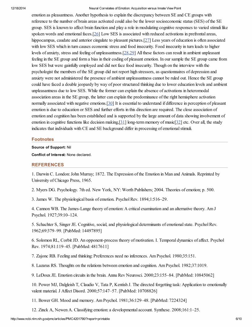
emotion as pleasantness. Another hypothesis to explain the discrepancy between SE and CE groups with
reference to the number of brain areas activated could also be the lower socioeconomic status (SES) of the SE
group. SES is known to affect brain function and play a role in modulating cognitive responses to varied stimuli like
spoken words and emotional faces.[26] Low SES is associated with reduced activations in prefrontal areas,
hippocampus, caudate and anterior cingulate to pleasant pictures.[27] Less years of education is often associated
with low SES which in turn causes economic stress and food insecurity. Food insecurity in turn leads to higher
levels of anxiety, stress and feeling of unpleasantness.[28,29] All these factors can result in ambient unpleasant
feeling in the SE group and form a bias in their coding of pleasant emotion. In our sample the SE group came from
low SES but were gainfully employed and did not face food insecurity. Though on the interview with the
psychologist the members of the SE group did not report high stressors, as questionnaires of depression and
anxiety were not administered the presence of ambient unpleasantness cannot be ruled out. Hence the SE group
could have faced a double jeopardy by way of poor structured thinking due to lower education levels and ambient
unpleasantness due to low SES. While the former can explain the absence of activations in heteromodal
association areas in the SE group, the latter can explain the predominance of the right hemisphere activation
normally associated with negative emotions.[30] It is essential to understand if difference in perception of pleasant
emotion is due to education or SES and further efforts in this direction are required. The close association of
emotion and cognition has been established and is supported by the large amount of data showing involvement of
emotion in cognitive functions like decision making,[31] long-term memory of music[32] etc. Over all, the study
indicates that individuals with CE and SE background differ in processing of emotional stimuli.
Footnotes
Source of Support: Nil
Conflict of Interest: None declared.
REFERENCES
1. Darwin C. London: John Murray; 1872. The Expression of the Emotion in Man and Animals. Reprinted by
University of Chicago Press, 1965.
2. Myers DG. Psychology. 7th ed. New York, NY: Worth Publishers; 2004. Theories of emotion; p. 500.
3. James W. The physiological basis of emotion. Psychol Rev. 1894;1:516–29.
4. Cannon WB. The James-Lange theory of emotion: A critical examination and an alternative theory. Am J
Psychol. 1927;39:10–124.
5. Schachter S, Singer JE. Cognitive, social, and physiological determinants of emotional state. Psychol Rev.
1962;69:379–99. [PubMed: 14497895]
6. Solomon RL, Corbit JD. An opponent-process theory of motivation. I. Temporal dynamics of affect. Psychol
Rev. 1974;81:119–45. [PubMed: 4817611]
7. Zajonc RB. Feeling and thinking: Preferences need no inferences. Am Psychol. 1980;35:151.
8. Lazarus RS. Thoughts on the relations between emotion and cognition. Am Psychol. 1982;37:1019.
9. LeDoux JE. Emotion circuits in the brain. Annu Rev Neurosci. 2000;23:155–84. [PubMed: 10845062]
10. Power MJ, Dalgleish T, Claudio V, Tata P, Kentish J. The directed forgetting task: Application to emotionally
valent material. J Affect Disord. 2000;57:147–57. [PubMed: 10708826]
11. Bower GH. Mood and memory. Am Psychol. 1981;36:129–48. [PubMed: 7224324]
12. Zinck A, Newen A. Classifying emotion: a developmental account. Synthese. 2008;161:1–25.

12/18/2014 Neural Correlates of Emotion: Acquisition versus Innate View Point
http://www.ncbi.nlm.nih.gov/pmc/articles/PMC4201790/?report=printable 7/10
13. Izard C. Many meanings/aspects of emotion: Definitions, functions, activation, and regulation. Emot Rev.
2010;2:363.
14. Herbert C, Ethofer T, Anders S, Junghofer M, Wildgruber D, Grodd W, et al. Amygdala activation during
reading of emotional adjectives – An advantage for pleasant content. Soc Cogn Affect Neurosci. 2009;4:35–49.
[PMCID: PMC2656883] [PubMed: 19015080]
15. Patterson DW, Schmidt LA. Neuroanatomy of the human affective system. Brain Cogn. 2003;52:24–6.
[PubMed: 12812801]
16. Banich MT. Houghton: Mifflin College Division; 2004. Cognitive Neuroscience and Neuropsychology.
17. Phan KL, Taylor SF, Welsh RC, Ho SH, Britton JC, Liberzon I. Neural correlates of individual ratings of
emotional salience: A trial-related fMRI study. Neuroimage. 2004;21:768–80. [PubMed: 14980580]
18. Lane RD, Reiman EM, Axelrod B, Yun LS, Holmes A, Schwartz GE. Neural correlates of levels of emotional
awareness. Evidence of an interaction between emotion and attention in the anterior cingulate cortex. J Cogn
Neurosci. 1998;10:525–35. [PubMed: 9712681]
19. Drevets WC, Raichle ME. Suppression of regional cerebral blood during emotional versus higher cognitive
implications for interactions between emotion and cognition. Cogn Emot. 1998;12:353–85.
20. Sabatinelli D, Bradley MM, Lang PJ, Costa VD, Versace F. Pleasure rather than salience activates human
nucleus accumbens and medial prefrontal cortex. J Neurophysiol. 2007;98:1374–9. [PubMed: 17596422]
21. Lane RD, Reiman EM, Bradley MM, Lang PJ, Ahern GL, Davidson RJ, et al. Neuroanatomical correlates of
pleasant and unpleasant emotion. Neuropsychologia. 1997;35:1437–44. [PubMed: 9352521]
22. Heller W, Nitschke JB, Miller GA. Lateralization in emotion and emotional disorders. Curr Dir Psychol Sci.
1998;7:26–32.
23. Scheuerecker J, Frodl T, Koutsouleris N, Zetzsche T, Wiesmann M, Kleemann AM, et al. Cerebral
differences in explicit and implicit emotional processing – An fMRI study. Neuropsychobiology. 2007;56:32–9.
[PubMed: 17986835]
24. Ochsner KN, Gross JJ. The cognitive control of emotion. Trends Cogn Sci. 2005;9:242–9.
[PubMed: 15866151]
25. Badgaiyan RD. Dopamine is released in the striatum during human emotional processing. Neuroreport.
2010;21:1172–6. [PMCID: PMC2997433] [PubMed: 21057339]
26. Hackman DA, Farah MJ. Socioeconomic status and the developing brain. Trends Cogn Sci. 2009;13:65–73.
[PMCID: PMC3575682] [PubMed: 19135405]
27. Silverman ME, Muennig P, Liu X, Rosen Z, Goldstein MA. The impact of socioeconomic status on the neural
substrates associated with pleasure. Open Neuroimag J. 2009;3:58–63. [PMCID: PMC2731107]
[PubMed: 19718457]
28. Cohen S, Doyle WJ, Baum A. Socioeconomic status is associated with stress hormones. Psychosom Med.
2006;68:414–20. [PubMed: 16738073]
29. Evans GW, English K. The environment of poverty: Multiple stressor exposure, psychophysiological stress,
and socioemotional adjustment. Child Dev. 2002;73:1238–48. [PubMed: 12146745]
30. Flores-Gutiérrez E, Díaz J, Barrios F, Favila-Humara R, Guevara M, Río-Portilla Y, et al. Metabolic and
electric brain patterns during pleasant and unpleasant emotions induced by music masterpieces. Int J

12/18/2014 Neural Correlates of Emotion: Acquisition versus Innate View Point
http://www.ncbi.nlm.nih.gov/pmc/articles/PMC4201790/?report=printable 8/10
Psychophysiol. 2007;65:69–84. [PubMed: 17466401]
31. Naqvi N, Shiv B, Bechara A. The role of emotion in decision making a cognitive neuroscience perspective.
Curr Dir Psychol Sci. 2006;15:5260–4.
32. Eschrich S, Münte TF, Altenmüller EO. Unforgettable film music: The role of emotion in episodic long-term
memory for music. BMC Neurosci. 2008;9:48. [PMCID: PMC2430709] [PubMed: 18505596]
Figures and Tables
Figure 1
International affective picture system block design paradigm
Figure 2

12/18/2014 Neural Correlates of Emotion: Acquisition versus Innate View Point
http://www.ncbi.nlm.nih.gov/pmc/articles/PMC4201790/?report=printable 9/10
Render images of unpleasant– neutral stimuli in college educated
Table 1
Pleasant – neutral emotions for school educated FWE unc, P = 0.0001
Table 2

12/18/2014 Neural Correlates of Emotion: Acquisition versus Innate View Point
http://www.ncbi.nlm.nih.gov/pmc/articles/PMC4201790/?report=printable 10/10
Pleasant – neutral emotions for college educated FWE unc, P = 0.0001
Table 3
Unpleasant – neutral emotions for school educated (FWE unc, P = 0.0001)
Table 4
Unpleasant– neutral emotions for college educated (FWE unc, P = 0.0001)
Articles from Indian Journal of Psychological Medicine are provided here courtesy of Medknow
Publications