The Interplay between Dengue Virus and the Human Innate ...
21
Review The Interplay between Dengue Virus and the Human Innate Immune System: A Game of Hide and Seek Nicolas Tremblay , Wesley Freppel , Aïssatou Aïcha Sow and Laurent Chatel-Chaix * Institut national de la recherche scientifique, Centre Armand-Frappier Santé Biotechnologie, Laval, QC H7V 1B4, Canada; [email protected] (N.T.); [email protected] (W.F.); [email protected] (A.A.S.) * Correspondence: [email protected] Received: 31 July 2019; Accepted: 8 October 2019; Published: 10 October 2019 Abstract: With 40% of the world population at risk, infections with dengue virus (DENV) constitute a serious threat to public health. While there is no antiviral therapy available against this potentially lethal disease, the efficacy of the only approved vaccine is not optimal and its safety has been recently questioned. In order to develop better vaccines based on attenuated and/or chimeric viruses, one must consider how the human immune system is engaged during DENV infection. The activation of the innate immunity through the detection of viruses by cellular sensors is the first line of defence against those pathogens. This triggers a cascade of events which establishes an antiviral state at the cell level and leads to a global immunological response. However, DENV has evolved to interfere with the innate immune signalling at multiple levels, hence dampening antiviral responses and favouring viral replication and dissemination. This review elaborates on the interplay between DENV and the innate immune system. A special focus is given on the viral countermeasure mechanisms reported over the last decade which should be taken into consideration during vaccine development. Keywords: dengue virus; innate immunity; viral evasion; interferon; MAVS; RIG-I; mitochondria 1. Introduction 1.1. Disease Burden Dengue is a neglected tropical disease caused by the dengue virus (DENV) that is transmitted to humans by Aedes aegypti or Aedes albopictus mosquitoes. In the recent years, it has become a public health concern, given that its global incidence has dramatically increased over the last 20 years and that no effective antiviral therapies are currently available. A dengue vaccine developed by Sanofi Pasteur, Dengvaxia, has been recently approved in over 20 countries around the globe. However, while the efficacy of this vaccine is not optimal for all DENV serotypes, its safety has been seriously questioned, especially for seronegative individuals. Epidemiological models estimate that about 390 million out of the 3.9 billion people living in endemic or epidemic areas contract the disease each year, making it the most prevalent arbovirus infection [1,2]. About 60 million infected individuals per year will develop symptomatic dengue fever, resulting in 10 to 20 thousand fatalities and 1.14 million Disability-Adjusted Life Years (DALY) [3]. This increase in the disease burden is not evenly distributed across the globe, with Latin America and the Caribbean ending up since 1990 with an increase in dengue-related DALY of about 250% or 4-fold above the global average and with Southeast Asia accounting for 596,000 DALY. This might be due to the simultaneous occurrence of multiple factors in these low-to-middle income emerging countries which affect the complex balance of DENV transmission dynamics, leading to a rapid endemic–epidemic cycle. For instance, this includes an optimal climate for Aedes life cycle, a rapid increase in population density in urban centres, and the concomitant circulation of all DENV Vaccines 2019, 7, 145; doi:10.3390/vaccines7040145 www.mdpi.com/journal/vaccines
-
Upload
khangminh22 -
Category
Documents
-
view
1 -
download
0
Transcript of The Interplay between Dengue Virus and the Human Innate ...

Review
The Interplay between Dengue Virus and the HumanInnate Immune System: A Game of Hide and Seek
Nicolas Tremblay , Wesley Freppel , Aïssatou Aïcha Sow and Laurent Chatel-Chaix *
Institut national de la recherche scientifique, Centre Armand-Frappier Santé Biotechnologie,Laval, QC H7V 1B4, Canada; [email protected] (N.T.); [email protected] (W.F.);[email protected] (A.A.S.)* Correspondence: [email protected]
Received: 31 July 2019; Accepted: 8 October 2019; Published: 10 October 2019�����������������
Abstract: With 40% of the world population at risk, infections with dengue virus (DENV) constitutea serious threat to public health. While there is no antiviral therapy available against this potentiallylethal disease, the efficacy of the only approved vaccine is not optimal and its safety has been recentlyquestioned. In order to develop better vaccines based on attenuated and/or chimeric viruses, one mustconsider how the human immune system is engaged during DENV infection. The activation of theinnate immunity through the detection of viruses by cellular sensors is the first line of defence againstthose pathogens. This triggers a cascade of events which establishes an antiviral state at the cell leveland leads to a global immunological response. However, DENV has evolved to interfere with theinnate immune signalling at multiple levels, hence dampening antiviral responses and favouringviral replication and dissemination. This review elaborates on the interplay between DENV and theinnate immune system. A special focus is given on the viral countermeasure mechanisms reportedover the last decade which should be taken into consideration during vaccine development.
Keywords: dengue virus; innate immunity; viral evasion; interferon; MAVS; RIG-I; mitochondria
1. Introduction
1.1. Disease Burden
Dengue is a neglected tropical disease caused by the dengue virus (DENV) that is transmitted tohumans by Aedes aegypti or Aedes albopictus mosquitoes. In the recent years, it has become a publichealth concern, given that its global incidence has dramatically increased over the last 20 years and thatno effective antiviral therapies are currently available. A dengue vaccine developed by Sanofi Pasteur,Dengvaxia, has been recently approved in over 20 countries around the globe. However, while theefficacy of this vaccine is not optimal for all DENV serotypes, its safety has been seriously questioned,especially for seronegative individuals. Epidemiological models estimate that about 390 million out ofthe 3.9 billion people living in endemic or epidemic areas contract the disease each year, making it themost prevalent arbovirus infection [1,2]. About 60 million infected individuals per year will developsymptomatic dengue fever, resulting in 10 to 20 thousand fatalities and 1.14 million Disability-AdjustedLife Years (DALY) [3]. This increase in the disease burden is not evenly distributed across the globe,with Latin America and the Caribbean ending up since 1990 with an increase in dengue-related DALYof about 250% or 4-fold above the global average and with Southeast Asia accounting for 596,000 DALY.This might be due to the simultaneous occurrence of multiple factors in these low-to-middle incomeemerging countries which affect the complex balance of DENV transmission dynamics, leading to arapid endemic–epidemic cycle. For instance, this includes an optimal climate for Aedes life cycle, arapid increase in population density in urban centres, and the concomitant circulation of all DENV
Vaccines 2019, 7, 145; doi:10.3390/vaccines7040145 www.mdpi.com/journal/vaccines

Vaccines 2019, 7, 145 2 of 21
serotypes (1–4) in one given restricted geographic area [4,5]. While there is an unmet medical needregarding strategies against dengue, the incidence of this disease is expected to geographically expandin the future considering that the arthropod vector is colonizing northern European and Americanterritories with temperate climates.
1.2. Clinical Manifestation
DENV infection is characterized by a mixed clinical presentation that ranges from an asymptomaticdisease to a mild febrile prodrome all the way to a severe hemorrhagic fever and shock syndrome.All infected individuals, asymptomatic or not, can transmit DENV to Aedes mosquitoes during ablood meal, making it difficult to precisely estimate the actual size of the reservoir at any given time.DENV infection severity is classified according to the World Health Organization (WHO) 1997 and 2009guidelines [6]. Dengue “without warning signs” (or dengue fever) regroups at-risk individuals withfever and at least two of the following signs and symptoms: nausea/vomiting, rash, headaches, eyepain, muscle aches, joint pain, leukopenia, or positivity at the tourniquet test. Dengue “with warningsigns of severe infection” (or dengue hemorrhagic fever) includes in addition to the previous signs andsymptoms abdominal pain, persistent vomiting, ascites, pleural effusion, mucosal bleeding, lethargyor restlessness, hepatomegaly, and an increase in hematocrit paired with rapid decrease in plateletcount. Lastly, severe dengue (or dengue shock) occurs when the infection leads to severe plasmaleakage, massive bleeding, and multiple organ failures. As of now, the therapeutic arsenal againstDENV infection is fairly limited and only consists of supportive care and intravenous fluid therapy.
From a host–pathogen interaction point of view, it is interesting to note that, in the majorityof DENV-infected individuals, viremia is controlled by the innate and adaptive immune systemswithin three to seven days, independently of the clinical manifestation [7]. However, in some cases,the infection is not properly handled and symptoms aggravate. A higher peak in viral titres seemsto be predictive of disease severity [8,9]. While the pathophysiology behind severe dengue is notfully understood, it is accepted that dengue hemorrhagic fever and severe dengue result from acytokine-mediated pathology (cytokine storm) that occurs because of the unbalanced production ofvarious soluble and short-lived immune factors, such as TNF-α, VEGF-A, IL-6, IL-8, IL-10, CCL2, andCXCL10, that are produced by immune cells [10].
1.3. Dengue Virus Life Cycle
DENV is an enveloped, positive-stranded RNA virus and belongs to the Flavivirus genus withinthe Flaviviridae family. Flaviviruses also include genetically related and/or medically relevantarthropod-borne pathogens such as yellow fever virus (YFV), Zika virus (ZIKV), West Nile virus(WNV), and Japanese encephalitis virus (JEV). The enveloped virion is constituted of prM/M (precursormembrane/membrane) and E (envelope) structural proteins at the surface while C nucleocapsid proteinsurrounds the 11 kilobase-long non-segmented viral RNA genome (vRNA). Following virus entryinto the target cell, vRNA is translated into one polyprotein, which is subsequently processed by hostand viral proteases to generate the structural proteins C, prM, and E and the seven nonstructural(NS) proteins NS1, NS2A, NS2B, NS3, NS4A, NS4B, and NS5. NS proteins are responsible for vRNAreplication that occurs exclusively in the cytoplasm of infected cells. This involves the specific enzymaticactivity of several NS proteins. The NS5 RNA-dependent RNA polymerase is responsible for vRNAsynthesis and possesses a methyltransferase activity which is absolutely required for vRNA 5′ cappingand 2′-O-methylation. The processing of the DENV polyprotein involves the serine protease activity ofthe NS2B/NS3 complex (or NS2B3). NS3 is also essential for vRNA synthesis and capping throughits helicase, NTPase, and triphosphatase activities. Assembled virions bud into the endoplasmicreticulum (ER) and are taken in charge by the cellular secretion machinery. In the Golgi apparatus,they undergo a final round of furin-mediated maturation and acquire infectivity before their releasevia exocytosis [11–13]. In order to create a cytoplasmic environment that can sustain an optimal lifecycle, DENV, much like other flaviviruses, induces within the infected cell the biogenesis of specific

Vaccines 2019, 7, 145 3 of 21
membranous replication organelles or replication factories (RF) with unique morphologies [14,15].These RFs are formed by altering the curvature and composition of cellular endomembranes. They havebeen characterized as ER-derived ultrastructures that include (1) vesicle packets (VP) which result frommembrane invaginations and contain the vRNA replication components; (2) convoluted membranes(CM) which are enriched in NS3, NS4A, and NS4B and of which the exact role(s) remain poorly defined;and (3) virus bags which contain immature virions organized in regular arrays [16–18]. In addition,DENV also manipulates the architecture of other organelles (such as mitochondria or peroxisomes) tomodulate specific cellular functions, including innate immunity, in favour of replication (see below).While DENV is genetically simple with only 10 viral protein expressed, its life cycle relies on highlycoordinated machineries that offer various points of intervention and research interests.
Following the entry of the virus inside the cell, it is rapidly sensed as a foreign “intruder” by thecell surveillance machinery, which triggers innate immunity. Strikingly, in response to these defencemechanisms, DENV has evolved to efficiently hide and mask its foreign molecular signatures, hence,avoiding the establishment of an antiviral environment that is detrimental for replication. In additionto these “cloak-and-dagger” operations, many viral proteins are able to directly hit the host antiviralmachinery to shutdown innate immune signalling. Thus, antiviral immunity and DENV life cycle aredynamically interconnected events which balance each other; on one hand, the virus is trying to hijackthe functions of the cellular machinery to its own benefit, while, on the other hand, the host factors aretrying to control the viral infection while minimizing damages to cell homeostasis.
In this review, we elaborate on the interplay between DENV and innate immunity. More precisely,it brings into focus how DENV is detected inside an infected cell as well as how it can hide to avoiddetection. A better understanding of how DENV circumvents and/or attenuates innate immunity canprovide critical information for rational vaccine development.
2. Recognition of DENV by Pathogen Recognition Receptors
Innate antiviral immunity is the first line of defence against viral pathogens and participates inthe establishment of the adaptive immune response. It engages complex networks of proteins thatare able to detect, control, and defeat viral threats. Virus detection is done by specialized proteinscalled Pattern Recognition Receptors (PRR), which can recognize various virus-specific conservedmolecular signatures known as Pathogen-Associated Molecular Patterns (PAMP). This detection signalis transduced by adaptor proteins to ultimately activate various transcription factors (e.g., InterferonRegulatory Factors (IRF) 3 and 7) that drive the production of antiviral proteins including type I andtype III interferons (IFN) (Figure 1, upper panel) [19–22]. Once secreted, these cytokines activatethe Janus Kinase-Signal Transducer and Activator of Transcription (JAK-STAT) pathway, leading tothe production of many Interferon-Stimulated Genes (ISG), which contribute to sustained paracrineand autocrine antiviral responses. Briefly, following the binding of type I IFN to the Interferon-α/β
Receptors (IFNAR) 1/2, signalling through the JAK-STAT pathway results in the amplification of theantiviral response via the activation of STAT1, STAT2, and IRF9, a transcription factor complex knownas IFN-stimulated gene factor 3 (ISGF3) [23,24]. All in all, innate antiviral immunity is a crucial cellularsystem that provides a quick reaction force against a given pathogen infection and can eventually leadto the establishment of a more focused yet costly effector response [25,26].

Vaccines 2019, 7, 145 4 of 21
Vaccines 2019, 7, x FOR PEER REVIEW 4 of 21
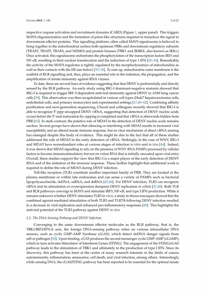
Figure 1. The sensing of Dengue virus by the innate immune responses and multiple evasion strategies to hinder the production of type I interferons (IFN).The upper panel shows the sensing of viral infection by RIG-I and cGAS. DENV-derived PAMPs are recognized by various pathogen recognition receptors like RIG-I. Upon recognition of pathogenic RNA, RIG-I is translocated to the mitochondria-associated MAVS adaptor protein with the help of the 14-3-3 and TRIM25 proteins. This induces the formation of MAVS aggregates that serve as an immune signalosome to activate through phosphorylation transcription factors IRF3 and NF-κB. In turn, these transcription factors translocate to the nucleus and elicit the transactivation of type I IFN. Once produced, type I IFN is secreted into the extracellular space and activates, in paracrine and autocrine fashions, the JAK-STAT pathway, leading to the amplification of the antiviral response via the activation of STAT1, STAT2, and IRF9, a transcription factor complex known as ISGF3. Infection also leads to mitochondrial stress and the release of mtDNA into the cytosol. mtDNA is recognized by cGAS, which activates STING, leading to the activation of IRF3/NF-κB and IFN production. DENV has developed various countermeasures to evade and/or engage the RLR and cGAS/STING pathways and to hinder innate immune signalling. These evasion mechanisms include (1) posttranscriptional modification of vRNA; (2) inhibition of 14-3-3ε or TRIM25 to hinder RIG-I activation; (3) MAVS aggregation; (4) alterations in endomembrane architecture to disrupt the MAVS signalosome; (5) the inhibition of TBK1 and IKBKE kinase activation, preventing the activation of transcription factors like IRF3; (6) the disruption of cGAS/STING signalling; and (7) the dampening of ISGF3 complex activation that is required for late innate antiviral responses.
2.1. DENV vRNA-Recognition
The RIG-I-Like Receptor (RLR) pathway has been classically studied for its role in the detection of viral ribonucleic acids and involves Retinoic Acid-Inducible gene I (RIG-I or DDX58), Melanoma Differentiation-Associated protein 5 (MDA5), and Laboratory of Genetics and Physiology 2 (LGP2/DHX58) as cytosolic sensors of an infection. They are ubiquitously expressed and are able to sense specific viral RNA moieties such as short uncapped 5′ triphosphorylated single-stranded (ss) RNA, short double-stranded (ds) RNA, U/A-rich 3′ regions of viral RNA, or long dsRNA [27]. Upon binding to vRNA, RIG-I and MDA5 are activated and translocated to the surface of the mitochondria where they interact with an adaptor protein, Mitochondrial Antiviral-Signalling Protein (MAVS), via their respective caspase activation and recruitment domains (CARD) (Figure 1, upper panel). This
Figure 1. The sensing of Dengue virus by the innate immune responses and multiple evasion strategiesto hinder the production of type I interferons (IFN).The upper panel shows the sensing of viral infectionby RIG-I and cGAS. DENV-derived PAMPs are recognized by various pathogen recognition receptorslike RIG-I. Upon recognition of pathogenic RNA, RIG-I is translocated to the mitochondria-associatedMAVS adaptor protein with the help of the 14-3-3 and TRIM25 proteins. This induces the formationof MAVS aggregates that serve as an immune signalosome to activate through phosphorylationtranscription factors IRF3 and NF-κB. In turn, these transcription factors translocate to the nucleus andelicit the transactivation of type I IFN. Once produced, type I IFN is secreted into the extracellular spaceand activates, in paracrine and autocrine fashions, the JAK-STAT pathway, leading to the amplificationof the antiviral response via the activation of STAT1, STAT2, and IRF9, a transcription factor complexknown as ISGF3. Infection also leads to mitochondrial stress and the release of mtDNA into the cytosol.mtDNA is recognized by cGAS, which activates STING, leading to the activation of IRF3/NF-κB andIFN production. DENV has developed various countermeasures to evade and/or engage the RLRand cGAS/STING pathways and to hinder innate immune signalling. These evasion mechanismsinclude (1) posttranscriptional modification of vRNA; (2) inhibition of 14-3-3ε or TRIM25 to hinderRIG-I activation; (3) MAVS aggregation; (4) alterations in endomembrane architecture to disrupt theMAVS signalosome; (5) the inhibition of TBK1 and IKBKE kinase activation, preventing the activationof transcription factors like IRF3; (6) the disruption of cGAS/STING signalling; and (7) the dampeningof ISGF3 complex activation that is required for late innate antiviral responses.
2.1. DENV vRNA-Recognition
The RIG-I-Like Receptor (RLR) pathway has been classically studied for its role in thedetection of viral ribonucleic acids and involves Retinoic Acid-Inducible gene I (RIG-I or DDX58),Melanoma Differentiation-Associated protein 5 (MDA5), and Laboratory of Genetics and Physiology 2(LGP2/DHX58) as cytosolic sensors of an infection. They are ubiquitously expressed and are able tosense specific viral RNA moieties such as short uncapped 5′ triphosphorylated single-stranded (ss) RNA,short double-stranded (ds) RNA, U/A-rich 3′ regions of viral RNA, or long dsRNA [27]. Upon bindingto vRNA, RIG-I and MDA5 are activated and translocated to the surface of the mitochondria wherethey interact with an adaptor protein, Mitochondrial Antiviral-Signalling Protein (MAVS), via their
Vaccines 2019, 7, 145 5 of 21
respective caspase activation and recruitment domains (CARD) (Figure 1, upper panel). This triggersMAVS oligomerization and the formation of prion-like structures required to transduce the signal todownstream effector proteins. This signalling platform, often called MAVS signalosome is believed tobring together to the mitochondrial surface both upstream PRRs and downstream regulatory subunits(TRAF2, TRAF5, TRAF6, and NEMO) and protein kinases (TBK1 and IKBKE, also known as IKKε).Once activated, this signalosome orchestrates the phosphorylation of the transcription factors IRF3 andNF-κB, resulting in their nuclear translocation and the induction of type 1 IFN [28–34]. Remarkably,the activity of the MAVS regulome is tightly regulated by the morphodynamics of mitochondria aswell as their contacts with the ER (see below) [35–38]. To sum up, mitochondria outer membrane is thescaffold of RLR signalling and, thus, plays an essential role in the initiation, the propagation, and theamplification of innate immunity against RNA viruses.
To date, there are several lines of evidence suggesting that that DENV is preferentially and directlysensed by the RLR pathway. An early study using RIG-I dominant-negative mutants showed thatRIG-I is required to trigger IRF-3-dependent antiviral immunity against DENV in A549 lung cancercells [39]. This observation was then recapitulated in various cell types (Huh7 hepatocarcinoma cells,endothelial cells, and primary monocytes) and experimental settings [17,40–42]. Combining affinitypurification and next-generation sequencing, Chazal and colleagues recently showed that RIG-I isable to recognize 5′-ppp uncapped DENV vRNA, suggesting that detection of DENV nucleic acidsoccurs before the 5′-end maturation by capping is completed and that vRNA is afterwards hidden fromPRR [43]. In stark contrast, the putative role of MDA5 in the detection of DENV nucleic acids remainsunclear. Several groups have shown that silencing or interfering with MDA5 results in increased viralsusceptibility and an altered innate immune response, but no clear mechanism of direct vRNA sensinghas emerged despite this body of evidence. This might be due to the fact that all of these studiesaddressed the role of MDA5 in the early detection of vRNA. Strikingly, in the case of WNV, RIG-Iand MDA5 have nonredundant roles at various stages of infection in vitro and in vivo [44]. Indeed,it was shown that MDA5 signalling to rely on the presence of WNV RNA PAMPs processed by cellularfactors to become immunostimulatory but not on virion RNA that is initially uncoated upon viral entry.Overall, these studies support the view that RIG-I is a major player of the early detection of DENVRNA and of the initiation of the immune response. These further highlight that additional work isrequired to define the role of MDA5 during DENV infection.
Toll-like receptors (TLR) constitute another important family of PRR. They are located at theplasma membrane or within late endosomes and can sense a variety of PAMPs such as bacteriallipopolysaccharide, dsDNA, ssRNA, and dsRNA [45,46]. For DENV infection, TLR3 can recognizevRNA and its stimulation or overexpression dampens DENV replication in cellulo [47,48]. Both TLRand RLR pathways converge to MAVS and stimulate IRF3, NF-κB, and type I IFN production. While itremains unknown whether DENV stimulates TLR3 in vivo, a study in rhesus macaques showed that thecombined agonist-mediated stimulation of both TLR3 and TLR7/8 following DENV infection resultedin a decrease in viral replication and enhanced pro-inflammatory responses [49]. This highlights theantiviral potential of the TLR3 pathway against DENV in vivo.
2.2. The DNA Sensing Pathway and DENV Infection
Converging to the same downstream effector molecules as the RLR pathway, that is, theTBK1/IRF3/IFN-β axis, the foreign DNA-sensing pathway relies on various intracellular DNAsensors, such as cyclic GMP-AMP Synthase (cGAS), which detect dsDNA danger signals fromself or pathogen [50]. Upon binding, cGAS produces the second messenger cyclic GMP-AMP (cGAMP),which in turn activates Stimulator of Interferon Genes (STING). The engagement of the STING/cGASpathway leads to the stimulation of TBK1 and ultimately to the production of type I IFN. Since itsdiscovery, this pathway has been at the centre of many research interests in the fields of cancer,autoimmunity, inflammation, senescence, cell death, and viral infection, among others. Interestingly,while sensing DNA, the cGAS/STING pathway has been reported to be essential for the optimal innate

Vaccines 2019, 7, 145 6 of 21
immune response against RNA viruses [51–53]. Not surprisingly, several viruses such as influenza virus,hepatitis C virus, and coronavirus [52,54–56] have developed targeted evasion mechanisms to interferewith this pathway. The involvement of cGAS and STING in the innate immune response againstDENV has become evident over the last few years, with several reports showing that DENV actuallymodulates this pathway (see below) [54,57–59]. While it remains unclear how a DNA-binding protein,like cGAS, can detect viral RNA, it was recently suggested that the sensing of DENV infection ratheroccurs indirectly. Indeed, virus-induced mitochondrial stress leads to the leakage of mitochondrialDNA (mtDNA) into the cytosol. It is subsequently sensed by cGAS, which in turn activates the typeI IFN response [60]. More recently, it was shown that following inflammasome activation, secretedInterleukin (IL)-1β can induce mtDNA release, leading to the stimulation of the cGAS pathway andIFN production [61]. Considering that DENV infection triggers the activation of the inflammasomein human platelets and macrophages [62,63], it is plausible that this indirectly contributes to themtDNA-dependent induction of early innate immunity.
Lastly, the emerging concept of a cross talk between the RIG-I/MAVS and cGAS/STING pathways issurely interesting to understand DENV biology. Indeed, there is an accumulating literature that showsthat the RLR pathway is able to potentiate the cGAS/STING pathway and vice versa during RNA virusinfection [64]. This seems to occur through the physical connection between RIG, MAVS, and STINGduring viral infection [51,65,66], the coactivation of both pathways [52,53,67], or a transcriptionalfeedback loops [66,68,69]. For flaviviral infections, a study has shown that STING potentiates the RLRsignalling via the assembly of a RIG-I/MAVS/STING complex following JEV nucleic acid recognition [66].In addition, another study in mice has shown that cGAS is required for the optimal RLR-dependent IFNresponse against WNV. Indeed, cGAS mice had a higher viral load and lower ISG expression, resultingin a higher mortality when compared to wild-type controls [53]. Future studies will be required toassess the role of the cross talk between the RIG-I/MAVS and cGAS/STING during DENV infection.
More generally, it will be required to investigate the exact nature, role, and distribution of theimmunomodulatory molecules and signalling adaptors that are produced and expressed as a result ofthe pathogenesis of DENV infection. This will ultimately provide mechanistic insights on the exactcontribution of the cGAS/STING pathway during DENV infection.
3. Viral Countermeasures
3.1. Interference with RLR-Dependent Signalling
DENV RNA synthesis and capping are believed to occur within the VPs since the viral replicationdsRNA intermediate localizes within this ER-derived ultrastructure as shown by imaging studies usingimmunogold labelling and electron microscopy [16]. This confined environment is most probablyimportant not only to concentrate the metabolites and proteins required for replication but alsoto exclude potentially inhibitory host factors. Considering this, it is tempting to speculate thatneo-synthesized uncapped DENV RNAs “hide” from RIG-I in VPs although this model remains to beexperimentally validated.
The replicated DENV RNA molecule possesses several intrinsic features that allow its evasionfrom detection by the innate immune system. These sensing interference strategies rely on two specificmodifications of vRNA: its 2′-O-methylation and its partial degradation by host nucleases (Figure 1,bottom left panel). In addition to its RNA polymerase function, DENV NS5 has a methyltransferase(MTase) activity that generates the 5′ 7-methyl-guanosine cap and also methylates the 2′-OH positionon the first nucleotide, forming a type 1 cap (m7GpppNm) structure [70,71]. The 2′-O-methylatedvRNAs mimic cellular mRNAs, thus evading the host immune system [72–76] (Figure 1). Surprisingly,this mechanism is not necessarily strictly dependent on the capping of the vRNA since internal2′-O-methylation can occur on vRNA lacking the 5′ cap structure during infections with DENVand other flavivirus such as WNV [72]. The 2′-O-methylation has been shown to be important toavoid coronavirus infection recognition by MDA5 in mouse and human cells. Indeed, a DENV2

Vaccines 2019, 7, 145 7 of 21
mutant deficient in the 2′-O MTase activity (NS5 E217A mutation) leads to an attenuated viral spreadin IFN-competent cells [73,74]. Consistent with this, it was later shown that the replication of the2′-O-MTase mutant is attenuated at the very early stages of the life cycle, i.e. during the first 16 h ofinfection. As shown by a transcriptome kinetic analysis, this resulted in an increase in ISG transcriptionat the early time points postinfection with an enrichment in PRR genes (RIG-I, MDA5, and IFIT1).Altogether, these studies strongly support that DENV 2′-O-methylation aims to hide viral nucleic acidsfrom early recognition by RLRs [76]. Importantly, a 2′-O-MTase mutant DENV2 strain was identifiedas a vaccine candidate since immunization with it conferred protection against subsequent DENVinfection in mice and rhesus macaques [75]. However, it was proposed that exacerbated immuneresponse might be due rather to a 2′-O-methylation-mediated evasion of the antiviral action of specificISGs that enact downstream RLR and IFN signalling (see below).
Another vRNA-related evasion mechanism relies on the partial degradation of vRNA by hostfactors, resulting in the generation of the subgenomic flavivirus RNA (sfRNA), a small noncodingRNA (Figure 1). sfRNAs are produced as byproducts of an incomplete degradation of flaviviralRNA due to unique secondary structures in the 3′ UTR that causes XRN1, a 5′–3′ exoribonuclease, tostall and cut the vRNA prematurely [77,78]. Produced by all tested flaviviruses, the accumulation ofbiologically active sfRNA can contribute to virus-induced cytopathic effects [78,79]; to hijack mRNAprocessing machinery [80,81]; to promote optimal viral replication [82]; and most relevant here, to helpthe virus to circumvent antiviral signalling [83–85]. Indeed, in the case of DENV, one study showedthat DENV sfRNAs are able to inhibit TRIM25 deubiquitylation by USP15 [86]. This prevents the K63polyubiquitylation of RIG-I, its dimerization via its CARD domains; and its subsequent interactionwith downstream adaptor protein MAVS, resulting in hindered interferon signalling [31,87,88]. On thewhole, vRNA methylation and degradation into sfRNA consist of two evolutionarily conservedflaviviral strategies to counteract innate antiviral immunity.
In addition to interfering with RLR sensing of vRNA, DENV has developed other evasionstrategies to inhibit the signalling cascade following RLR activation. Several DENV proteins modulatethe pathway both upstream and downstream from MAVS. Notably, DENV NS3, through a conservedphosphomimetic RxEP motif, is able to prevent RIG-I translocation to the mitochondria by sequestering14-3-3ε [89] (Figure 1). 14-3-3ε is required for efficient RIG-I/TRIM25 association and subsequentrecruitment to mitochondria and interaction with MAVS [90]. Importantly, viruses expressing a mutatedNS3 unable to associate with 14-3-3ε elicited a stronger immune response and replicated less efficientlyin immune-competent cells [89]. Moreover, DENV NS4A is able to bind to MAVS CARD domains andto effectively prevent RIG-I/MAVS interaction through an unknown mechanism [91].
In order to sustain and maintain the immune synapse, MAVS needs to be physically accessible andat the proper position on the surface of the mitochondria; otherwise, antiviral signalling and responsemay not be optimal. Targeting this aspect of RLR-dependent innate immune signalling, DENV is alsoable to change the architecture of mitochondria in terms of morphodynamics and contacts with theendoplasmic reticulum (Figure 1). First, mitochondria show an elongated morphology in DENV-infectedcells and make contacts with DENV convoluted membranes [17,92]. Interestingly, mitochondrialmorphodynamics were reported to modulate antiviral signalling [35–37]. The morphology ofmitochondria relies on an equilibrium between their fusion and fission regulated by dynamin-likeGTPases. Briefly, these key players are mitofusins (MFN1 and MFN2) and Optic Atrophy 1 (OPA1),which mediate fusion (leading to elongation), and Dynamin-Related Protein 1 (DRP1), whichmediates mitochondrial fission [93]. DENV-induced mitochondria elongation was attributed toNS4B since its overexpression alone recapitulated the phenotype. Furthermore, an inhibition of thephosphorylation-dependent activation of DRP1 was observed in both infected and NS4B-expressingcells, thus favouring mitochondrial elongation over fission [17].
Second, DENV infection resulted in a drastic disruption of the contacts between mitochondriaand the ER (ERMC) [17]. Interestingly, the residual Mitochondria-Associated Membranes (MAM)appeared to be connected to CMs, suggesting that the biogenesis of this DENV RF substructure was

Vaccines 2019, 7, 145 8 of 21
responsible for ERMC disruption. Notably, MAMs are important for normal RLR signalling since theyfavour the tethering of MAVS required for optimal antiviral signalling [35–38]. This suggests that thealteration of the reticulo-mitochondrial interface by DENV would contribute to the dampening of RLRsignalling. In strong support to this, upon enforced mitochondria elongation via DRP1 expressionknockdown in DENV-infected Huh7 cells, RIG-I was barely recruited to the MAMs, correlating with adampened type I and III IFN mRNA transcription and an increased viral replication. This highlightsthe functional interplay between mitochondria morphodynamics and contact with ER. In another study,Yu and colleagues showed that DENV NS2B3 protease cleaves MFN1 and MFN2 [94]. Interestingly,MFN1 overexpression-mediated hyperfusion of mitochondria in the perinuclear region resulted in adecreased viral replication. Moreover, independent expression knockdown of MFN1 or MFN2 led todifferent phenotypes in viral replication, IFN-β induction, and cell death, illustrating the divergentroles of mitofusins despite their similarity. This is consistent with the different assumed functions ofMFN1 in docking and fusion of the mitochondria and of MFN2 in the stabilization of the interactionsbetween mitochondria [95,96]. Furthermore, MFN2 localizes to MAMs and as an ER-resident proteintethers mitochondria to ER through homodimerization and heterotypic interactions with MFN1 [97].This suggests that DENV might alter ERMCs by directly targeting MFN2 through NS2B3 proteaseactivity without necessarily impacting on mitochondria fusion and elongation. Overall, these studiessupport the model that antiviral signalling is dependent on various spatiotemporal events and thatalterations of the mitochondrial morphology and cytosolic interface regulate MAVS signalosomefunction. This concept of viral subversion of mitochondrial morphodynamics is not unique to DENVsince related strategies have been observed for other viruses such as hepatitis B virus, hepatitis C virus,Severe acute respiratory syndrome-related coronavirus (SARS–CoV), and alphaherpesvirus [98–101].
In addition to ER and mitochondria, DENV impairs the morphology of another membranousorganelle functionally linked to MAVS-related signalling. Indeed, DENV as well as WNV and ZIKVinduce a loss of cellular peroxisomes [102,103], which contain MAVS at their surface and constitutea transduction platform for innate signalling [104–106]. Notably, the expression knockdown of theperoxin Pex19, which is essential for peroxisome biogenesis [107], led to a significant decrease in typeIII IFN gene transcription following the activation of RLR signalling with a synthetic dsRNA [102].This highlights the importance of peroxisome integrity in innate immune signalling and the benefit forDENV of depleting them upon infection. Overall, these changes in cellular endomembrane morphologyare believed to create a cytoplasmic microenvironment in which viral proteins are able to engagecellular host factors that are crucial to mount an adequate antiviral response.
Finally, downstream of MAVS, DENV is also able to evade RLR signalling by interfering withthe activity of effector proteins (Figure 1). Indeed, NS2A, NS2B3, and NS4B are able to block IRF3phosphorylation by interfering with serine kinase activity of TBK1 and IKBKE, which leads to asuboptimal type I IFN response [108,109].
All these evasion strategies are consistent with the idea that DENV physically targets the MAVSsignalosome in its entirety when it brings together upstream and downstream factors to the cytosolicside of the mitochondria. Interestingly, several DENV proteins involved in this co-opting, namelyNS2B3, NS4A, and NS4B, all interact and partly localize within CMs [16–18,110–113]. Furthermore,CMs make with elongated mitochondria and there is strong evidence that they originate from MAMs,an important compartment for MAVS-dependent signalling [17]. Altogether, this supports a modelin which CMs constitute a “hijacking” unit, which targets the whole MAVS signalosome at multiplelevels. Rather than directly regulating vRNA replication, this viral substructure would instead“shutoff” potentially antiviral responses to infection, including early innate immunity, hence creating acytoplasmic environment favourable to viral replication. The close spatial relationship between CMsand mitochondria would help to “trap” host targets inside CMs or to concentrate them near viralproteins in order to very efficiently downregulate the MAVS signalosome and maximize viral evasion.Nevertheless, this model requires experimental challenging.

Vaccines 2019, 7, 145 9 of 21
3.2. Interference with the cGAS/STING Pathway
DENV also has the capacity to directly interfere with IFN induction that is triggered by theactivated cGAS/STING pathway as a result of the release of mtDNA into the cytoplasm following DENVinfection [114] (Figure 1). Indeed, DENV NS2B marks cGAS for lysosomal degradation and DENVNS2B-NS3 protease cleaves STING to suppress type I IFN induction [54,58,114,115]. Interestingly,DENV NS2B3 cannot process either mouse or nonhuman primate STING orthologs, suggesting thatthis pathogen has evolved towards an optimal pathogenicity in its natural hosts [54,59]. Very recently,it has been reported that DRP1-mediated mitochondrial fission results in mitochondrial stress andthe release of mtDNA in the cytosol [116]. Considering this, it will be interesting to investigate in thefuture whether DENV-induced mitochondrial elongation through DRP1 inhibition actually dampensnot only RIG-I signal but also the activation of the STING/cGAS pathway and/or its cross talk with RLRsignalling. In the same line of ideas, STING is a resident protein of MAMs [117], and the alteration ofthese structures during DENV infection might contribute to targeting STING to NS2B3-rich CMs fordegradation or spatiotemporal sequestration.
3.3. Interference with the IFN Signalling
In addition to the primary virus sensing pathway, DENV is also able to target several componentsof the downstream interferon-induced JAK-STAT signalling cascade that normally leads to the sustainedexpression of various ISGs following the secretion of IFN-α and -β (Figure 1). Early in vitro assaysdemonstrated that DENV NS4B is able to prevent STAT1 activation by a mechanism conserved inWNV and YFV that is yet to be elucidated [118,119]. However, it can be rationalized that STAT1inhibition most likely occurs either through prevention of its activation or by its dephosphorylation orby degradation of activated STAT1. On the matter of another STAT protein, DENV NS5 is able to bindSTAT2 and to promote its E3 ubiquitin ligase UBR4-dependent degradation. This results in a drasticdecrease of STAT2 basal expression level and, hence, to a hindered JAK-STAT signalling [120–122].Interestingly, this STAT2-related evasion strategy is also conserved in other flaviviruses, such asZIKV. Indeed, ZIKV is able to bind to STAT2 and to promote its proteasomal degradation but in anUBR4-independent manner [123,124].
3.4. Interference with Other Mechanisms
In addition to its role in STAT2 degradation, DENV NS5 protein is also able to interfere withISG expression. First, both DENV and ZIKV NS5 selectively inhibit ISG expression by binding toand antagonizing the transcription factor PAF1C [125]. Second, DENV NS5 hijacks core componentsof the spliceosomal machinery, namely CD2BP2 and DDX23, resulting in an alteration of isoformabundance and stoichiometry of many antiviral factors such as RIG-I, ISG15, or IL-8, hence contributingto an environment favourable to viral replication [126]. At the posttranscriptional level, sfRNA hasbeen shown to interfere with the translation of ISGs. Indeed, DENV sfRNA associates with the hostRNA-binding proteins G3BP1, G3BP2, and CAPRIN1, which, as a result, are unable to stimulate asefficiently the translation of many ISGs such as PKR and IFITM2 for instance [127]. Consistently,sfRNA binding to these host factors protects DENV replication from IFN-β treatment. Interestingly, the2′-O-methylation mechanism described above as a way to downregulate PRR early IFN response is alsoable to interfere with the functions of specific ISGs. Indeed, the replication of the NS5 E217A 2′-O-MTaseDENV2 mutant is significantly more sensitive to IFN-β treatment than wt virus [75], demonstrating thatthis modification may contribute to immunity evasion downstream of PRR signalling. More specificallyfor DENV but also for WNV, poxvirus, coronavirus, and JEV, the 2′-O-methylation of vRNA capenables the viral nucleic acids to be marked as “self-RNA” and to evade from the antiviral functionof IFN-Induced Protein with Tetratricopeptide Repeat (IFIT) proteins [128–131]. Notably, IFIT1 wasshown to associate with non-methylated RNAs with a higher affinity than the other IFITs. It wasproposed that translation initiation factor eIF4E and IFIT1 compete for cap binding [129]. Hence,

Vaccines 2019, 7, 145 10 of 21
IFIT1 senses virus infection through the binding to foreign RNAs lacking 2′-O-methyls and inhibitsthe translation of viral RNA via the displacement of eIF4E. Consistently, IFIT1 overexpression inHEK293-DC-SIGN cells significantly decreases DENV2 replication in contrast to other IFITs [75]. In thecase of WNV, the pathogenesis of the E218A MTase mutant is greatly attenuated in wt mice. Veryinterestingly, intracranial infection of Ifit1 knockout C57BL/6 mice with the E218A MTase mutant WNVrestored lethality, highlighting the critical role of this IFIT in the sensing of the “2′-O-methyl-free”vRNA in vivo [128]. Considering the genetic and biological proximity with WNV, it is tantalizing tospeculate that DENV RNA is able to evade IFIT antiviral activity in vivo by mimicking cellular mRNAsthrough the addition of 2′-O-methyls, one of the most common posttranscriptional modifications ofRNA allowing vRNA to hide in plain sight among host mRNAs.
As explained above, DENV sfRNA targets innate immune signalling in human cells at multiplelevels (i.e. RIG-I activation and ISG expression). Of significant interest, several recent studieshave highlighted the important role of sfRNA in the dissemination of DENV among infectedinsects [81,132–134]. In fact, the DENV and YFV subgenomic RNAs are able to interfere withthe Aedes mosquito innate system as well by targeting both Toll receptor and RNA interference (RNAi)pathways to favour optimal replication. In the mosquitoes, viral double-stranded RNA is processedinto small siRNAs by RNase III DICER and is processed into viral siRNAs (vsiRNAs) by AGO2, whichare able to target viral RNA for silencing [135,136]. This shows that sfRNA plays two distinct roles,albeit using the same cellular factors, in the evasion of the innate immune response in mammalianhosts or arthropod vectors. This concept of different roles of host factors between the mammalianhosts or arthropod vectors will most likely be explored in depth in the future considering that a recentsystem biology study identified more than 45 shared pathway interactions between DENV–human andDENV–mosquito interactomes in the context of innate immune evasion and viral pathogenesis [125].
4. Importance for Vaccine Development
Multiple tetravalent vaccine candidates are being developed. These include live-attenuatedvaccines, whole virus inactivated vaccines, protein-based vaccines, chimeric vaccines, and mRNA-basedand synthetic virus-like particle vaccines [137–142]. Among these vaccines, Dengvaxia (also calledCYD-TDV), developed by Sanofi Pasteur, is the only one licensed for use in about 20 dengue-endemiccountries in Asia, Latin America, and Oceania as well as in Europe. CYD-TDV is a tetravalent denguechimeric live-attenuated vaccine which is based on the YFV 17D strain as a backbone. This vaccine istypically indicated for individuals aged from 9 to 45 years living in an endemic country and, hence, isnot accessible to the population that is the most at risk to develop severe dengue-related symptoms.Unfortunately, the vaccine confers less protection against serotypes 1 and 2 than with serotypes 3and 4 [143,144]. Importantly, CYD-TDV efficacy was higher than pooled estimates in seropositiveindividuals but extremely low for seronegative children with risks of severe complications due tothe vaccine. As a probable consequence of this, a global vaccination campaign in schools of thePhilippines has resulted in the death of many children and the suspension of the program by theDepartment of Health in late 2017 [145]. As such, a pre-vaccination screening of previous exposureis now recommended prior to vaccination [146]. Lastly, a study shows that the CYD-TDV-elicitedmemory response decreases over time, as measured by low antibody titres in blood samples 5 yearsfollowing an initial three-dose vaccine regimen. This suggests that a booster dose might be needed forsustained long-term immunological protection [147].
The development of new dengue vaccines which are safe and highly efficacious against all serotypesfaces many challenges, including some involving the interplay between the virus and the immunesystem. Seminal studies by Albert Sabin, dating from the WWII era [148], have demonstrated thatvolunteers exposed to DENV showed long-lived homotypic immunity but short-lived cross-protectionagainst viruses of a different serotype. However, the mechanisms behind these observations are stillnot fully understood and prospective studies at the population level show that these conclusionscannot be generalized to inform rational vaccine design [149]. While a majority of DENV infections

Vaccines 2019, 7, 145 11 of 21
go unnoticed, some infected individuals progress along the disease spectrum and develop denguehemorrhagic fever or dengue shock. Although no consensus exists about the pathophysiology behindthis progression, it is most likely due to host-specific factors related to the presence of preexistingimmunity such as antibody-dependent enhancements (ADE) and immune-mediated cytokine stormrelated to the antigenic sin phenomenon that occurs even past the peak in viremia in symptomaticindividuals [10,150]. Indeed, a second heterologous DENV infection is expected to elicit a strongmemory recall of DENV-specific T and B cells that will produce an enhanced level of cytokines andneutralizing antibodies. However, this adaptive immune response might not lead to the control ofviremia but rather quite the opposite, resulting in an antibody-dependent enhanced infection (virusopsonization and increased replication), mast cell activation (vascular permeability), and cellularcytotoxicity (cytokine storm) [151]. With this in mind, an effective antiviral therapy would need to betaken as a prophylactic treatment and would most likely be highly expensive given the number ofindividuals living in endemic areas. Thus, the only way to eradicate dengue worldwide would be todevelop a safe, effective, and pan-serotypic vaccine that confers long-term sterilizing immunity.
In summary, while CYD-TDV constitutes a safe and efficacious tool in the therapeutic arsenalagainst DENV when properly administered, it is plagued by shortcomings and controversies and doesnot completely meet the need for an effective dengue vaccine, especially in seronegative individuals.Thus, many challenges remain to be addressed to develop the “holy grail” of DENV vaccines and abetter understanding of host–pathogen interactions is warranted to set up new vaccinal strategies orrelevant animal models. In the following section, we discuss three examples illustrating how innateimmunity research can inform vaccine design.
4.1. DENV∆30 Recombinant Dengue Virus
The DENV∆30 is a live attenuated vaccine candidate that was engineered by deleting 30 nucleotidesof the 3′UTR vRNA using reverse genetics and by subsequently creating modified dengue viruses forall four serotypes [152,153]. Proof-of-concept studies showed that, despite attenuation, the DENV∆30is highly immunogenic in both humans and rhesus monkeys for all tested serotypes and confersprotection upon homologous rechallenge [153–161]. In addition, DENV∆30 appears to be less virulentin mosquitoes, limiting the risk of transmission from a vaccinated human to the Aedes vector [162].For long, the molecular mechanism conferring attenuation of this vaccine candidate was largelyunknown. However, it was shown that DENV4∆30-infected cells accumulated less sfRNA, resulting inan increased sensitivity to type I IFN [163]. Further studies will be required to assess if other evasionmechanisms are in play in DENV∆30 attenuation to fully understand how this strain is able to create abalanced environment that is favourable to antigen presentation and cytokine production. However,given the reported roles of sfRNA in both IFN production and response (see above), it is likely that theattenuation of DENV∆30 results from an increased innate immune response.
4.2. 2′-O-methyltransferase-Deficient Dengue Vaccine
As explained above, 2′-O-methylation is a modification that allows the vRNA to mimic cellularmRNAs and to evade the host innate immune system. Using reverse genetics, Züst and colleagueshave generated DENV clones from serotypes 1 and 2 that harbour a single point mutation in theconserved KDKE motif of the NS5 methyltransferase catalytic site. These mutants (E216A for DENV1or E217A for DENV2) are impaired in 2′-O-methylation but are still able to perform the N7-methylationof the cap. Importantly, they are more susceptible to IFN treatment or human IFIT1 overexpressionthan wild-type DENV, which is consistent with the idea that they are impaired in IFIT inhibition.Importantly, treatment of mice competent for T-cell responses with these viruses alone or in serotypecombination conferred protection against a subsequent infection with a lethal mouse-adapted DENVstrain. Remarkably, a single low dose of the DENV2 E217A mutant in monkeys was enough fora complete seroconversion and protection [75]. This approach was also applied to develop a JEVvaccine candidate. A E218A mutant JEV was attenuated and more sensitive to a type I IFN treatment

Vaccines 2019, 7, 145 12 of 21
as well [164]. A single dose of this live attenuated vaccine was sufficient to elicit a strong humoralresponse that conferred protection and survival following heterologous rechallenge in BALB/c mice.Ongoing studies are now trying to implement this vaccine rational design towards the development ofa tetravalent, non-chimeric vaccine with therapeutic relevance.
4.3. Immunocompetent Mouse Model for Denv Vaccine research
Vaccine development studies usually rely on large animal models during nonclinical and preclinicalstudies [165]. The use of small animal models such as mice usually requires genetic modificationsthat dampen their immune system in order to increase viral permissiveness and, thus, do not reflectthe reality of an infection in the natural host with an intact immune response. In most of the cases,in vivo DENV studies classically involve immunodeficient mouse strains such as AG129 which donot express IFN-α/β and IFN-γ receptors. These mice are highly susceptible to DENV infection andgenerally die within two weeks after injection [166–168]. Following advancement of the knowledgeabout host–pathogen interactions and the advent of novel technologies (e.g., by CRISPR/Cas9 geneediting), a paradigm shift can be anticipated. As an example of this, using a gene knock-in approachand a mouse-adapted virus strain, Gorman and colleagues recently developed an immunocompetenttransgenic mouse model of ZIVK infection by replacing the mouse STAT2 by the human STAT2 [169].This genetic engineering was conceptually made possible thanks to previous studies which showedthat ZIKV induces the degradation of human STAT2 but not of the mouse ortholog [123,124,170].While it will be interesting to see if the use of humanized immunocompetent mouse model can beexpanded to DENV studies, it definitely opens up many future research opportunities.
5. Conclusions and Perspectives
DENV research will most likely remain a challenging and exciting field in times to come. In recentyears, DENV infection has taken a front seat in the realm of global health concerns as it spreads outsidethe Western Pacific region and is now threatening more than 4 billion individuals. With no effectiveantiviral therapies or prophylactic vaccine, DENV is expected to cause disease and to inflict harmthat amounts to more than 1.14 million DALY. As extensively discussed in this review, DENV is wellequipped to evade the innate antiviral immune system through various antagonizing functions ofviral NS proteins and RNA. From being able to hide in plain sight due to the 2′-O-methylation ofthe viral genome and to the disruption of MAVS signalosome activity and of the IFN signalling, itcan be acknowledged that, despite a simple viral genome organization, DENV pathogenesis relieson a complex network of host–pathogen interactions. It is conceivable that better understanding thiscomplexity will contribute to solving the puzzle of dengue control and/or eradication. Indeed, theinterplay between DENV and the immune system can lead to rational vaccine design by providingattractive targets for antigen and adjuvant development and by filling the gaps in knowledge regardinghow the innate immune system can be harnessed to support a long-lasting protective immune response.
Author Contributions: Conceptualization and supervision: N.T. and L.C.-C.; original draft preparation: N.T.,A.A.S. and W.F.; review and editing: N.T. and L.C.-C.
Funding: This work was supported by a postdoctoral scholarship to N.T. and a research scholarship to L.C.-Cfrom Fonds de la Recherche du Québec-Santé. W.F. and A.A.S. salaries are funded by Institut National de laRecherche Scientifique and the Canadian Institutes of Health Research.
Acknowledgments: We are grateful to Karine Boulay (Université de Montréal, Canada) for critical reading of themanuscript and helpful comments.
Conflicts of Interest: The authors declare no conflict of interest. The funders had no role in the writing of themanuscript or in the decision to publish this review.

Vaccines 2019, 7, 145 13 of 21
References
1. Bhatt, S.; Gething, P.W.; Brady, O.J.; Messina, J.P.; Farlow, A.W.; Moyes, C.L.; Drake, J.M.; Brownstein, J.S.;Hoen, A.G.; Sankoh, O.; et al. The global distribution and burden of dengue. Nature 2013, 496, 504–507.[CrossRef] [PubMed]
2. Brady, O.J.; Gething, P.W.; Bhatt, S.; Messina, J.P.; Brownstein, J.S.; Hoen, A.G.; Moyes, C.L.; Farlow, A.W.;Scott, T.W.; Hay, S.I. Refining the Global Spatial Limits of Dengue Virus Transmission by Evidence-BasedConsensus. PLoS Negl. Trop. Dis. 2012, 6, e1760. [CrossRef] [PubMed]
3. Stanaway, J.D.; Shepard, D.S.; Undurraga, E.A.; Halasa, Y.A.; Coffeng, L.E.; Brady, O.J.; Hay, S.I.; Bedi, N.;Bensenor, I.M.; Castañeda-Orjuela, C.A.; et al. The global burden of dengue: An analysis from the GlobalBurden of Disease Study 2013. Lancet Infect. Dis. 2016, 16, 712–723. [PubMed]
4. Ramos-Castañeda, J.; Barreto Dos Santos, F.; Martínez-Vega, R.; Galvão de Araujo, J.M.; Joint, G.; Sarti, E.Dengue in Latin America: Systematic Review of Molecular Epidemiological Trends. PLoS Negl. Trop. Dis.2017, 11, e0005224. [CrossRef] [PubMed]
5. San Martín, J.L.; Brathwaite, O.; Zambrano, B.; Solórzano, J.O.; Bouckenooghe, A.; Dayan, G.H.; Guzmán, M.G.The epidemiology of dengue in the americas over the last three decades: A worrisome reality. Am. J. Trop.Med. Hyg. 2010, 82, 128–135. [CrossRef] [PubMed]
6. World Health Organization. Dengue: Guidelines for Diagnosis, Treatment, Prevention and Control; World HealthOrganization: Geneva, Switzerland, 2009; ISBN 9789241547871.
7. Gubler, D.J.; Suharyono, W.; Tan, R.; Abidin, M.; Sie, A. Viraemia in patients with naturally acquired dengueinfection. Bull. World Health Organ. 1981, 59, 623–630. [PubMed]
8. Gómez-Ochoa, S.A. Viremia en plasma como factor asociado a gravedad en la infección por el virus deldengue: Revisión sistemática de la literatura. Revista Chilena de Infectología 2018, 35, 176–183. [PubMed]
9. Vaughn, D.W.; Green, S.; Kalayanarooj, S.; Innis, B.L.; Nimmannitya, S.; Suntayakorn, S.; Endy, T.P.;Raengsakulrach, B.; Rothman, A.L.; Ennis, F.A.; et al. Dengue viremia titer, antibody response pattern, andvirus serotype correlate with disease severity. J. Infect. Dis. 2000, 181, 2–9. [CrossRef] [PubMed]
10. Srikiatkhachorn, A.; Mathew, A.; Rothman, A.L. Immune-mediated cytokine storm and its role in severedengue. Semin. Immunopathol. 2017, 39, 563–574. [CrossRef]
11. Apte-Sengupta, S.; Sirohi, D.; Kuhn, R.J. Coupling of replication and assembly in flaviviruses. Curr. Opin. Virol.2014, 9, 134–142. [CrossRef]
12. Neufeldt, C.J.; Cortese, M.; Acosta, E.G.; Bartenschlager, R. Rewiring cellular networks by members of theFlaviviridae family. Nat. Rev. Microbiol. 2018, 16, 125–142. [CrossRef] [PubMed]
13. Mazeaud, C.; Freppel, W.; Chatel-Chaix, L. The Multiples Fates of the Flavivirus RNA Genome DuringPathogenesis. Front. Genet. 2018, 9, 595. [CrossRef] [PubMed]
14. Paul, D.; Bartenschlager, R. Flaviviridae Replication Organelles: Oh, What a Tangled Web We Weave.Annu. Rev. Virol. 2015, 2, 289–310. [CrossRef] [PubMed]
15. Chatel-Chaix, L.; Bartenschlager, R. Dengue virus- and hepatitis C virus-induced replication and assemblycompartments: The enemy inside–caught in the web. J. Virol. 2014, 88, 5907–5911. [CrossRef] [PubMed]
16. Welsch, S.; Miller, S.; Romero-Brey, I.; Merz, A.; Bleck, C.K.E.; Walther, P.; Fuller, S.D.; Antony, C.;Krijnse-Locker, J.; Bartenschlager, R. Composition and three-dimensional architecture of the dengue virusreplication and assembly sites. Cell Host Microbe 2009, 5, 365–375. [CrossRef] [PubMed]
17. Chatel-Chaix, L.; Cortese, M.; Romero-Brey, I.; Bender, S.; Neufeldt, C.J.; Fischl, W.; Scaturro, P.; Schieber, N.;Schwab, Y.; Fischer, B.; et al. Dengue Virus Perturbs Mitochondrial Morphodynamics to Dampen InnateImmune Responses. Cell Host Microbe 2016, 20, 342–356. [CrossRef] [PubMed]
18. Miller, S.; Kastner, S.; Krijnse-Locker, J.; Bühler, S.; Bartenschlager, R. The non-structural protein 4A of denguevirus is an integral membrane protein inducing membrane alterations in a 2K-regulated manner. J. Biol. Chem.2007, 282, 8873–8882. [CrossRef] [PubMed]
19. Yoneyama, M.; Suhara, W.; Fukuhara, Y.; Fukuda, M.; Nishida, E.; Fujita, T. Direct triggering of the typeI interferon system by virus infection: Activation of a transcription factor complex containing IRF-3 andCBP/p300. EMBO J. 1998, 17, 1087–1095. [CrossRef]
20. Wathelet, M.G.; Lin, C.H.; Parekh, B.S.; Ronco, L.V.; Howley, P.M.; Maniatis, T. Virus infection induces theassembly of coordinately activated transcription factors on the IFN-beta enhancer in vivo. Mol. Cell 1998, 1,507–518. [CrossRef]

Vaccines 2019, 7, 145 14 of 21
21. Lin, R.; Heylbroeck, C.; Pitha, P.M.; Hiscott, J. Virus-dependent phosphorylation of the IRF-3 transcriptionfactor regulates nuclear translocation, transactivation potential, and proteasome-mediated degradation.Mol. Cell. Biol. 1998, 18, 2986–2996. [CrossRef]
22. Yang, H.; Lin, C.H.; Ma, G.; Baffi, M.O.; Wathelet, M.G. Interferon regulatory factor-7 synergizes with othertranscription factors through multiple interactions with p300/CBP coactivators. J. Biol. Chem. 2003, 278,15495–15504. [CrossRef] [PubMed]
23. Darnell, J.E., Jr.; Kerr, I.M.; Stark, G.R. Jak-STAT pathways and transcriptional activation in response to IFNsand other extracellular signaling proteins. Science 1994, 264, 1415–1421. [CrossRef] [PubMed]
24. Kessler, D.S.; Veals, S.A.; Fu, X.Y.; Levy, D.E. Interferon-alpha regulates nuclear translocation andDNA-binding affinity of ISGF3, a multimeric transcriptional activator. Genes Dev. 1990, 4, 1753–1765.[CrossRef] [PubMed]
25. McDonald, D.R.; Levy, O. 3—Innate immunity. In Clinical Immunology, 4th ed.; Rich, R.R., Fleisher, T.A.,Shearer, W.T., Schroeder, H.W., Frew, A.J., Weyand, C.M., Eds.; Content Repository Only: London, UK, 2013;pp. 35–46. ISBN 9780723436911.
26. Chow, K.T.; Gale, M., Jr.; Loo, Y.-M. RIG-I and Other RNA Sensors in Antiviral Immunity. Annu. Rev. Immunol.2018, 36, 667–694. [CrossRef] [PubMed]
27. Said, E.A.; Tremblay, N.; Al-Balushi, M.S.; Al-Jabri, A.A.; Lamarre, D. Viruses Seen by Our Cells: The Role ofViral RNA Sensors. J. Immunol. Res. 2018, 2018, 9480497. [CrossRef] [PubMed]
28. Hou, F.; Sun, L.; Zheng, H.; Skaug, B.; Jiang, Q.-X.; Chen, Z.J. MAVS forms functional prion-like aggregatesto activate and propagate antiviral innate immune response. Cell 2011, 146, 448–461. [CrossRef] [PubMed]
29. Kawai, T.; Takahashi, K.; Sato, S.; Coban, C.; Kumar, H.; Kato, H.; Ishii, K.J.; Takeuchi, O.; Akira, S. IPS-1, anadaptor triggering RIG-I- and Mda5-mediated type I interferon induction. Nat. Immunol. 2005, 6, 981–988.[CrossRef]
30. Meylan, E.; Curran, J.; Hofmann, K.; Moradpour, D.; Binder, M.; Bartenschlager, R.; Tschopp, J. Cardif isan adaptor protein in the RIG-I antiviral pathway and is targeted by hepatitis C virus. Nature 2005, 437,1167–1172. [CrossRef]
31. Seth, R.B.; Sun, L.; Ea, C.-K.; Chen, Z.J. Identification and characterization of MAVS, a mitochondrial antiviralsignaling protein that activates NF-kappaB and IRF 3. Cell 2005, 122, 669–682. [CrossRef]
32. Xu, H.; He, X.; Zheng, H.; Huang, L.J.; Hou, F.; Yu, Z.; de la Cruz, M.J.; Borkowski, B.; Zhang, X.; Chen, Z.J.; et al.Correction: Structural basis for the prion-like MAVS filaments in antiviral innate immunity. Elife 2015, 4.[CrossRef]
33. Xu, L.-G.; Wang, Y.-Y.; Han, K.-J.; Li, L.-Y.; Zhai, Z.; Shu, H.-B. VISA is an adapter protein required forvirus-triggered IFN-beta signaling. Mol. Cell 2005, 19, 727–740. [CrossRef] [PubMed]
34. Liu, S.; Cai, X.; Wu, J.; Cong, Q.; Chen, X.; Li, T.; Du, F.; Ren, J.; Wu, Y.T.; Grishin, N.V.; et al. Phosphorylation ofinnate immune adaptor proteins MAVS, STING, and TRIF induces IRF3 activation. Science 2015, 347, aaa2630.[CrossRef] [PubMed]
35. Castanier, C.; Garcin, D.; Vazquez, A.; Arnoult, D. Mitochondrial dynamics regulate the RIG-I-like receptorantiviral pathway. EMBO Rep. 2010, 11, 133–138. [CrossRef] [PubMed]
36. Jacobs, J.L.; Coyne, C.B. Mechanisms of MAVS regulation at the mitochondrial membrane. J. Mol. Biol. 2013,425, 5009–5019. [CrossRef]
37. Onoguchi, K.; Onomoto, K.; Takamatsu, S.; Jogi, M.; Takemura, A.; Morimoto, S.; Julkunen, I.; Namiki, H.;Yoneyama, M.; Fujita, T. Virus-Infection or 5′ppp-RNA Activates Antiviral Signal through Redistribution ofIPS-1 Mediated by MFN1. PLoS Pathog. 2010, 6, e1001012. [CrossRef] [PubMed]
38. Horner, S.M.; Liu, H.M.; Park, H.S.; Briley, J.; Gale, M., Jr. Mitochondrial-associated endoplasmic reticulummembranes (MAM) form innate immune synapses and are targeted by hepatitis C virus. Proc. Natl. Acad.Sci. USA 2011, 108, 14590–14595. [CrossRef] [PubMed]
39. Chang, T.-H.; Liao, C.-L.; Lin, Y.-L. Flavivirus induces interferon-beta gene expression through a pathwayinvolving RIG-I-dependent IRF-3 and PI3K-dependent NF-kappaB activation. Microbes Infect. 2006, 8,157–171. [CrossRef] [PubMed]
40. Nasirudeen, A.M.A.; Wong, H.H.; Thien, P.; Xu, S.; Lam, K.-P.; Liu, D.X. RIG-I, MDA5 and TLR3 synergisticallyplay an important role in restriction of dengue virus infection. PLoS Negl. Trop. Dis. 2011, 5, e926. [CrossRef][PubMed]

Vaccines 2019, 7, 145 15 of 21
41. da Conceição, T.M.; Rust, N.M.; Berbel, A.C.E.R.; Martins, N.B.; do Nascimento Santos, C.A.; Da Poian, A.T.;de Arruda, L.B. Essential role of RIG-I in the activation of endothelial cells by dengue virus. Virology 2013,435, 281–292. [CrossRef] [PubMed]
42. Olagnier, D.; Scholte, F.E.M.; Chiang, C.; Albulescu, I.C.; Nichols, C.; He, Z.; Lin, R.; Snijder, E.J.;van Hemert, M.J.; Hiscott, J. Inhibition of dengue and chikungunya virus infections by RIG-I-mediated type Iinterferon-independent stimulation of the innate antiviral response. J. Virol. 2014, 88, 4180–4194. [CrossRef][PubMed]
43. Chazal, M.; Beauclair, G.; Gracias, S.; Najburg, V.; Simon-Lorière, E.; Tangy, F.; Komarova, A.V.; Jouvenet, N.RIG-I Recognizes the 5′ Region of Dengue and Zika Virus Genomes. Cell Rep. 2018, 24, 320–328. [CrossRef]
44. Errett, J.S.; Suthar, M.S.; McMillan, A.; Diamond, M.S.; Gale, M. The Essential, Nonredundant Roles of RIG-Iand MDA5 in Detecting and Controlling West Nile Virus Infection. J. Virol. 2013, 87, 11416–11425. [CrossRef]
45. Leifer, C.A.; Medvedev, A.E. Molecular mechanisms of regulation of Toll-like receptor signaling. J. Leukoc. Biol.2016, 100, 927–941. [CrossRef]
46. Gao, D.; Li, W. Structures and recognition modes of toll-like receptors. Proteins 2017, 85, 3–9. [CrossRef]47. Tsai, Y.-T.; Chang, S.-Y.; Lee, C.-N.; Kao, C.-L. Human TLR3 recognizes dengue virus and modulates viral
replication in vitro. Cell. Microbiol. 2009, 11, 604–615. [CrossRef]48. Liang, Z.; Wu, S.; Li, Y.; He, L.; Wu, M.; Jiang, L.; Feng, L.; Zhang, P.; Huang, X. Activation of Toll-like
receptor 3 impairs the dengue virus serotype 2 replication through induction of IFN-β in cultured hepatomacells. PLoS ONE 2011, 6, e23346. [CrossRef]
49. Sariol, C.A.; Martínez, M.I.; Rivera, F.; Rodríguez, I.V.; Pantoja, P.; Abel, K.; Arana, T.; Giavedoni, L.;Hodara, V.; White, L.J.; et al. Decreased dengue replication and an increased anti-viral humoral responsewith the use of combined Toll-like receptor 3 and 7/8 agonists in macaques. PLoS ONE 2011, 6, e19323.[CrossRef]
50. Dhanwani, R.; Takahashi, M.; Sharma, S. Cytosolic sensing of immuno-stimulatory DNA, the enemy within.Curr. Opin. Immunol. 2018, 50, 82–87. [CrossRef]
51. Ishikawa, H.; Barber, G.N. STING is an endoplasmic reticulum adaptor that facilitates innate immunesignalling. Nature 2008, 455, 674–678. [CrossRef]
52. Holm, C.K.; Rahbek, S.H.; Gad, H.H.; Bak, R.O.; Jakobsen, M.R.; Jiang, Z.; Hansen, A.L.; Jensen, S.K.; Sun, C.;Thomsen, M.K.; et al. Influenza A virus targets a cGAS-independent STING pathway that controls envelopedRNA viruses. Nat. Commun. 2016, 7, 10680. [CrossRef]
53. Schoggins, J.W.; MacDuff, D.A.; Imanaka, N.; Gainey, M.D.; Shrestha, B.; Eitson, J.L.; Mar, K.B.;Richardson, R.B.; Ratushny, A.V.; Litvak, V.; et al. Pan-viral specificity of IFN-induced genes revealsnew roles for cGAS in innate immunity. Nature 2014, 505, 691–695. [CrossRef]
54. Aguirre, S.; Maestre, A.M.; Pagni, S.; Patel, J.R.; Savage, T.; Gutman, D.; Maringer, K.; Bernal-Rubio, D.;Shabman, R.S.; Simon, V.; et al. DENV inhibits type I IFN production in infected cells by cleaving humanSTING. PLoS Pathog. 2012, 8, e1002934. [CrossRef]
55. Sun, L.; Xing, Y.; Chen, X.; Zheng, Y.; Yang, Y.; Nichols, D.B.; Clementz, M.A.; Banach, B.S.; Li, K.;Baker, S.C.; et al. Coronavirus papain-like proteases negatively regulate antiviral innate immune responsethrough disruption of STING-mediated signaling. PLoS ONE 2012, 7, e30802. [CrossRef]
56. Ding, Q.; Cao, X.; Lu, J.; Huang, B.; Liu, Y.-J.; Kato, N.; Shu, H.-B.; Zhong, J. Hepatitis C virus NS4B blocksthe interaction of STING and TBK1 to evade host innate immunity. J. Hepatol. 2013, 59, 52–58. [CrossRef]
57. Yu, C.-Y.; Chang, T.-H.; Liang, J.-J.; Chiang, R.-L.; Lee, Y.-L.; Liao, C.-L.; Lin, Y.-L. Dengue virus targets theadaptor protein MITA to subvert host innate immunity. PLoS Pathog. 2012, 8, e1002780. [CrossRef]
58. Aguirre, S.; Luthra, P.; Sanchez-Aparicio, M.T.; Maestre, A.M.; Patel, J.; Lamothe, F.; Fredericks, A.C.;Tripathi, S.; Zhu, T.; Pintado-Silva, J.; et al. Dengue virus NS2B protein targets cGAS for degradation andprevents mitochondrial DNA sensing during infection. Nat. Microbiol. 2017, 2, 17037. [CrossRef]
59. Stabell, A.C.; Meyerson, N.R.; Gullberg, R.C.; Gilchrist, A.R.; Webb, K.J.; Old, W.M.; Perera, R.; Sawyer, S.L.Dengue viruses cleave STING in humans but not in nonhuman primates, their presumed natural reservoir.Elife 2018, 7, e31919. [CrossRef]
60. West, A.P.; Khoury-Hanold, W.; Staron, M.; Tal, M.C.; Pineda, C.M.; Lang, S.M.; Bestwick, M.; Duguay, B.A.;Raimundo, N.; MacDuff, D.A.; et al. Mitochondrial DNA stress primes the antiviral innate immune response.Nature 2015, 520, 553–557. [CrossRef]

Vaccines 2019, 7, 145 16 of 21
61. Aarreberg, L.D.; Esser-Nobis, K.; Driscoll, C.; Shuvarikov, A.; Roby, J.A.; Gale, M., Jr. Interleukin-1β InducesmtDNA Release to Activate Innate Immune Signaling via cGAS-STING. Mol. Cell 2019, 74, 801–815.e6.[CrossRef]
62. Wu, M.-F.; Chen, S.-T.; Yang, A.-H.; Lin, W.-W.; Lin, Y.-L.; Chen, N.-J.; Tsai, I.-S.; Li, L.; Hsieh, S.-L. CLEC5A iscritical for dengue virus-induced inflammasome activation in human macrophages. Blood 2013, 121, 95–106.[CrossRef]
63. Hottz, E.D.; Lopes, J.F.; Freitas, C.; Valls-de-Souza, R.; Oliveira, M.F.; Bozza, M.T.; Da Poian, A.T.; Weyrich, A.S.;Zimmerman, G.A.; Bozza, F.A.; et al. Platelets mediate increased endothelium permeability in denguethrough NLRP3-inflammasome activation. Blood 2013, 122, 3405–3414. [CrossRef]
64. Zevini, A.; Olagnier, D.; Hiscott, J. Crosstalk between Cytoplasmic RIG-I and STING Sensing Pathways.Trends Immunol. 2017, 38, 194–205. [CrossRef]
65. Zhong, B.; Yang, Y.; Li, S.; Wang, Y.-Y.; Li, Y.; Diao, F.; Lei, C.; He, X.; Zhang, L.; Tien, P.; et al. The adaptorprotein MITA links virus-sensing receptors to IRF3 transcription factor activation. Immunity 2008, 29, 538–550.[CrossRef]
66. Nazmi, A.; Mukhopadhyay, R.; Dutta, K.; Basu, A. STING mediates neuronal innate immune responsefollowing Japanese encephalitis virus infection. Sci. Rep. 2012, 2, 347. [CrossRef]
67. Thompson, M.R.; Sharma, S.; Atianand, M.; Jensen, S.B.; Carpenter, S.; Knipe, D.M.; Fitzgerald, K.A.;Kurt-Jones, E.A. Interferon γ-inducible Protein (IFI) 16 Transcriptionally Regulates Type I Interferons andOther Interferon-stimulated Genes and Controls the Interferon Response to both DNA and RNA Viruses.J. Biol. Chem. 2014, 289, 23568–23581. [CrossRef]
68. Härtlova, A.; Erttmann, S.F.; Raffi, F.A.; Schmalz, A.M.; Resch, U.; Anugula, S.; Lienenklaus, S.; Nilsson, L.M.;Kröger, A.; Nilsson, J.A.; et al. DNA damage primes the type I interferon system via the cytosolic DNAsensor STING to promote anti-microbial innate immunity. Immunity 2015, 42, 332–343. [CrossRef]
69. Liu, Y.; Goulet, M.-L.; Sze, A.; Hadj, S.B.; Belgnaoui, S.M.; Lababidi, R.R.; Zheng, C.; Fritz, J.H.; Olagnier, D.;Lin, R. RIG-I-Mediated STING Upregulation Restricts Herpes Simplex Virus 1 Infection. J. Virol. 2016, 90,9406–9419. [CrossRef]
70. Bradrick, S.S. Causes and Consequences of Flavivirus RNA Methylation. Front. Microbiol. 2017, 8, 2374.[CrossRef]
71. Furuichi, Y.; Shatkin, A.J. Viral and cellular mRNA capping: Past and prospects. Adv. Virus Res. 2000, 55,135–184.
72. Dong, H.; Chang, D.C.; Hua, M.H.C.; Lim, S.P.; Chionh, Y.H.; Hia, F.; Lee, Y.H.; Kukkaro, P.; Lok, S.-M.;Dedon, P.C.; et al. 2′-O methylation of internal adenosine by flavivirus NS5 methyltransferase. PLoS Pathog.2012, 8, e1002642. [CrossRef]
73. Schmid, B.; Rinas, M.; Ruggieri, A.; Acosta, E.G.; Bartenschlager, M.; Reuter, A.; Fischl, W.; Harder, N.;Bergeest, J.-P.; Flossdorf, M.; et al. Live Cell Analysis and Mathematical Modeling Identify Determinants ofAttenuation of Dengue Virus 2′-O-Methylation Mutant. PLoS Pathog. 2015, 11, e1005345. [CrossRef]
74. Züst, R.; Cervantes-Barragan, L.; Habjan, M.; Maier, R.; Neuman, B.W.; Ziebuhr, J.; Szretter, K.J.; Baker, S.C.;Barchet, W.; Diamond, M.S.; et al. Ribose 2′-O-methylation provides a molecular signature for the distinctionof self and non-self mRNA dependent on the RNA sensor Mda5. Nat. Immunol. 2011, 12, 137–143. [CrossRef]
75. Züst, R.; Dong, H.; Li, X.-F.; Chang, D.C.; Zhang, B.; Balakrishnan, T.; Toh, Y.-X.; Jiang, T.; Li, S.-H.; Deng, Y.-Q.;et al. Rational Design of a Live Attenuated Dengue Vaccine: 2′-O-Methyltransferase Mutants Are HighlyAttenuated and Immunogenic in Mice and Macaques. PLoS Pathog. 2013, 9, e1003521. [CrossRef]
76. Chang, D.C.; Hoang, L.T.; Naim, A.N.M.; Dong, H.; Schreiber, M.J.; Hibberd, M.L.; Tan, M.J.A.; Shi, P.-Y.Evasion of early innate immune response by 2′- O -methylation of dengue genomic RNA. Virology 2016, 499,259–266. [CrossRef]
77. Clarke, B.D.; Roby, J.A.; Slonchak, A.; Khromykh, A.A. Functional non-coding RNAs derived from theflavivirus 3′ untranslated region. Virus Res. 2015, 206, 53–61. [CrossRef]
78. Pijlman, G.P.; Funk, A.; Kondratieva, N.; Leung, J.; Torres, S.; van der Aa, L.; Liu, W.J.; Palmenberg, A.C.;Shi, P.-Y.; Hall, R.A.; et al. A highly structured, nuclease-resistant, noncoding RNA produced by flavivirusesis required for pathogenicity. Cell Host Microbe 2008, 4, 579–591. [CrossRef]
79. Liu, Y.; Liu, H.; Zou, J.; Zhang, B.; Yuan, Z. Dengue virus subgenomic RNA induces apoptosis through theBcl-2-mediated PI3k/Akt signaling pathway. Virology 2014, 448, 15–25. [CrossRef]

Vaccines 2019, 7, 145 17 of 21
80. Moon, S.L.; Anderson, J.R.; Kumagai, Y.; Wilusz, C.J.; Akira, S.; Khromykh, A.A.; Wilusz, J. A noncodingRNA produced by arthropod-borne flaviviruses inhibits the cellular exoribonuclease XRN1 and alters hostmRNA stability. RNA 2012, 18, 2029–2040. [CrossRef]
81. Schnettler, E.; Sterken, M.G.; Leung, J.Y.; Metz, S.W.; Geertsema, C.; Goldbach, R.W.; Vlak, J.M.; Kohl, A.;Khromykh, A.A.; Pijlman, G.P. Noncoding flavivirus RNA displays RNA interference suppressor activity ininsect and Mammalian cells. J. Virol. 2012, 86, 13486–13500. [CrossRef]
82. Fan, Y.-H.; Nadar, M.; Chen, C.-C.; Weng, C.-C.; Lin, Y.-T.; Chang, R.-Y. Small noncoding RNA modulatesJapanese encephalitis virus replication and translation in trans. Virol. J. 2011, 8, 492. [CrossRef]
83. Chang, R.-Y.; Hsu, T.-W.; Chen, Y.-L.; Liu, S.-F.; Tsai, Y.-J.; Lin, Y.-T.; Chen, Y.-S.; Fan, Y.-H. Japanese encephalitisvirus non-coding RNA inhibits activation of interferon by blocking nuclear translocation of interferonregulatory factor 3. Vet. Microbiol. 2013, 166, 11–21. [CrossRef] [PubMed]
84. Schuessler, A.; Funk, A.; Lazear, H.M.; Cooper, D.A.; Torres, S.; Daffis, S.; Jha, B.K.; Kumagai, Y.; Takeuchi, O.;Hertzog, P.; et al. West Nile virus noncoding subgenomic RNA contributes to viral evasion of the type Iinterferon-mediated antiviral response. J. Virol. 2012, 86, 5708–5718. [CrossRef] [PubMed]
85. Donald, C.L.; Brennan, B.; Cumberworth, S.L.; Rezelj, V.V.; Clark, J.J.; Cordeiro, M.T.; Freitas de OliveiraFrança, R.; Pena, L.J.; Wilkie, G.S.; Da Silva Filipe, A.; et al. Full Genome Sequence and sfRNA InterferonAntagonist Activity of Zika Virus from Recife, Brazil. PLoS Negl. Trop. Dis. 2016, 10, e0005048. [CrossRef][PubMed]
86. Manokaran, G.; Finol, E.; Wang, C.; Gunaratne, J.; Bahl, J.; Ong, E.Z.; Tan, H.C.; Sessions, O.M.; Ward, A.M.;Gubler, D.J.; et al. Dengue subgenomic RNA binds TRIM25 to inhibit interferon expression for epidemiologicalfitness. Science 2015, 350, 217–221. [CrossRef] [PubMed]
87. Gack, M.U.; Albrecht, R.A.; Urano, T.; Inn, K.-S.; Huang, I.-C.; Carnero, E.; Farzan, M.; Inoue, S.; Jung, J.U.;García-Sastre, A. Influenza A virus NS1 targets the ubiquitin ligase TRIM25 to evade recognition by the hostviral RNA sensor RIG-I. Cell Host Microbe 2009, 5, 439–449. [CrossRef] [PubMed]
88. Gack, M.U.; Shin, Y.C.; Joo, C.-H.; Urano, T.; Liang, C.; Sun, L.; Takeuchi, O.; Akira, S.; Chen, Z.; Inoue, S.; et al.TRIM25 RING-finger E3 ubiquitin ligase is essential for RIG-I-mediated antiviral activity. Nature 2007, 446,916–920. [CrossRef] [PubMed]
89. Chan, Y.K.; Gack, M.U. A phosphomimetic-based mechanism of dengue virus to antagonize innate immunity.Nat. Immunol. 2016, 17, 523–530. [CrossRef] [PubMed]
90. Liu, H.M.; Loo, Y.-M.; Horner, S.M.; Zornetzer, G.A.; Katze, M.G.; Gale, M., Jr. The mitochondrial targetingchaperone 14-3-3ε regulates a RIG-I translocon that mediates membrane association and innate antiviralimmunity. Cell Host Microbe 2012, 11, 528–537. [CrossRef] [PubMed]
91. He, Z.; Zhu, X.; Wen, W.; Yuan, J.; Hu, Y.; Chen, J.; An, S.; Dong, X.; Lin, C.; Yu, J.; et al. Dengue VirusSubverts Host Innate Immunity by Targeting Adaptor Protein MAVS. J. Virol. 2016, 90, 7219–7230. [CrossRef]
92. Barbier, V.; Lang, D.; Valois, S.; Rothman, A.L.; Medin, C.L. Dengue virus induces mitochondrial elongationthrough impairment of Drp1-triggered mitochondrial fission. Virology 2017, 500, 149–160. [CrossRef]
93. Lee, H.; Yoon, Y. Mitochondrial fission: Regulation and ER connection. Mol. Cells 2014, 37, 89–94. [CrossRef][PubMed]
94. Yu, C.-Y.; Liang, J.-J.; Li, J.-K.; Lee, Y.-L.; Chang, B.-L.; Su, C.-I.; Huang, W.-J.; Lai, M.M.C.; Lin, Y.-L.Dengue Virus Impairs Mitochondrial Fusion by Cleaving Mitofusins. PLoS Pathog. 2015, 11, e1005350.[CrossRef] [PubMed]
95. Ishihara, N.; Eura, Y.; Mihara, K. Mitofusin 1 and 2 play distinct roles in mitochondrial fusion reactions viaGTPase activity. J. Cell Sci. 2004, 117, 6535–6546. [CrossRef] [PubMed]
96. Koshiba, T.; Detmer, S.A.; Kaiser, J.T.; Chen, H.; McCaffery, J.M.; Chan, D.C. Structural basis of mitochondrialtethering by mitofusin complexes. Science 2004, 305, 858–862. [CrossRef] [PubMed]
97. de Brito, O.M.; Scorrano, L. Mitofusin 2 tethers endoplasmic reticulum to mitochondria. Nature 2008, 456,605–610. [CrossRef] [PubMed]
98. Kim, S.-J.; Khan, M.; Quan, J.; Till, A.; Subramani, S.; Siddiqui, A. Hepatitis B virus disrupts mitochondrialdynamics: Induces fission and mitophagy to attenuate apoptosis. PLoS Pathog. 2013, 9, e1003722. [CrossRef][PubMed]
99. Kim, S.-J.; Syed, G.H.; Khan, M.; Chiu, W.-W.; Sohail, M.A.; Gish, R.G.; Siddiqui, A. Hepatitis C virus triggersmitochondrial fission and attenuates apoptosis to promote viral persistence. Proc. Natl. Acad. Sci. USA 2014,111, 6413–6418. [CrossRef] [PubMed]

Vaccines 2019, 7, 145 18 of 21
100. Kramer, T.; Enquist, L.W. Alphaherpesvirus infection disrupts mitochondrial transport in neurons.Cell Host Microbe 2012, 11, 504–514. [CrossRef] [PubMed]
101. Shi, C.-S.; Qi, H.-Y.; Boularan, C.; Huang, N.-N.; Abu-Asab, M.; Shelhamer, J.H.; Kehrl, J.H. SARS-coronavirusopen reading frame-9b suppresses innate immunity by targeting mitochondria and the MAVS/TRAF3/TRAF6signalosome. J. Immunol. 2014, 193, 3080–3089. [CrossRef] [PubMed]
102. You, J.; Hou, S.; Malik-Soni, N.; Xu, Z.; Kumar, A.; Rachubinski, R.A.; Frappier, L.; Hobman, T.C.Flavivirus Infection Impairs Peroxisome Biogenesis and Early Antiviral Signaling. J. Virol. 2015, 89,12349–12361. [CrossRef]
103. Wong, C.P.; Xu, Z.; Hou, S.; Limonta, D.; Kumar, A.; Power, C.; Hobman, T.C. Interplay between Zika Virusand Peroxisomes during Infection. Cells 2019, 8, 725. [CrossRef] [PubMed]
104. Dixit, E.; Boulant, S.; Zhang, Y.; Lee, A.S.Y.; Odendall, C.; Shum, B.; Hacohen, N.; Chen, Z.J.; Whelan, S.P.;Fransen, M.; et al. Peroxisomes are signaling platforms for antiviral innate immunity. Cell 2010, 141, 668–681.[CrossRef] [PubMed]
105. Odendall, C.; Dixit, E.; Stavru, F.; Bierne, H.; Franz, K.M.; Durbin, A.F.; Boulant, S.; Gehrke, L.; Cossart, P.;Kagan, J.C. Diverse intracellular pathogens activate type III interferon expression from peroxisomes.Nat. Immunol. 2014, 15, 717–726. [CrossRef] [PubMed]
106. Bender, S.; Reuter, A.; Eberle, F.; Einhorn, E.; Binder, M.; Bartenschlager, R. Activation of Type I andIII Interferon Response by Mitochondrial and Peroxisomal MAVS and Inhibition by Hepatitis C Virus.PLoS Pathog. 2015, 11, e1005264. [CrossRef] [PubMed]
107. Götte, K.; Girzalsky, W.; Linkert, M.; Baumgart, E.; Kammerer, S.; Kunau, W.H.; Erdmann, R. Pex19p, afarnesylated protein essential for peroxisome biogenesis. Mol. Cell. Biol. 1998, 18, 616–628. [CrossRef][PubMed]
108. Angleró-Rodríguez, Y.I.; Pantoja, P.; Sariol, C.A. Dengue virus subverts the interferon induction pathway viaNS2B/3 protease-IκB kinase epsilon interaction. Clin. Vaccine Immunol. 2014, 21, 29–38. [CrossRef] [PubMed]
109. Dalrymple, N.A.; Cimica, V.; Mackow, E.R. Dengue Virus NS Proteins Inhibit RIG-I/MAVS Signaling byBlocking TBK1/IRF3 Phosphorylation: Dengue Virus Serotype 1 NS4A Is a Unique Interferon-RegulatingVirulence Determinant. mBio 2015, 6. [CrossRef]
110. Chatel-Chaix, L.; Fischl, W.; Scaturro, P.; Cortese, M.; Kallis, S.; Bartenschlager, M.; Fischer, B.; Bartenschlager, R.A Combined Genetic-Proteomic Approach Identifies Residues within Dengue Virus NS4B Critical forInteraction with NS3 and Viral Replication. J. Virol. 2015, 89, 7170–7186. [CrossRef]
111. Zou, J.; Lee, L.T.; Wang, Q.Y.; Xie, X.; Lu, S.; Yau, Y.H.; Yuan, Z.; Geifman Shochat, S.; Kang, C.; Lescar, J.; et al.Mapping the Interactions between the NS4B and NS3 proteins of dengue virus. J. Virol. 2015, 89, 3471–3483.[CrossRef] [PubMed]
112. Zou, J.; Xie, X.; Wang, Q.-Y.; Dong, H.; Lee, M.Y.; Kang, C.; Yuan, Z.; Shi, P.-Y. Characterization of denguevirus NS4A and NS4B protein interaction. J. Virol. 2015, 89, 3455–3470. [CrossRef] [PubMed]
113. Umareddy, I.; Chao, A.; Sampath, A.; Gu, F.; Vasudevan, S.G. Dengue virus NS4B interacts with NS3 anddissociates it from single-stranded RNA. J. Gen. Virol. 2006, 87, 2605–2614. [CrossRef] [PubMed]
114. Sun, B.; Sundström, K.B.; Chew, J.J.; Bist, P.; Gan, E.S.; Tan, H.C.; Goh, K.C.; Chawla, T.; Tang, C.K.; Ooi, E.E.Dengue virus activates cGAS through the release of mitochondrial DNA. Sci. Rep. 2017, 7, 3594. [CrossRef][PubMed]
115. Yu, C.-Y.; Chang, T.-H.; Liang, J.-J.; Chiang, R.-L.; Lee, Y.-L.; Liao, C.-L.; Lin, Y.-L. P122 Dengue virus targetsthe adaptor protein MITA to subvert host innate immunity. Cytokine 2012, 59, 558. [CrossRef]
116. Bao, D.; Zhao, J.; Zhou, X.; Yang, Q.; Chen, Y.; Zhu, J.; Yuan, P.; Yang, J.; Qin, T.; Wan, S.; et al.Mitochondrial fission-induced mtDNA stress promotes tumor-associated macrophage infiltration andHCC progression. Oncogene 2019, 38, 5007–5020. [CrossRef] [PubMed]
117. Ishikawa, H.; Ma, Z.; Barber, G.N. STING regulates intracellular DNA-mediated, type I interferon-dependentinnate immunity. Nature 2009, 461, 788–792. [CrossRef]
118. Muñoz-Jordan, J.L.; Sánchez-Burgos, G.G.; Laurent-Rolle, M.; García-Sastre, A. Inhibition of interferonsignaling by dengue virus. Proc. Natl. Acad. Sci. USA 2003, 100, 14333–14338. [CrossRef]
119. Muñoz-Jordán, J.L.; Laurent-Rolle, M.; Ashour, J.; Martínez-Sobrido, L.; Ashok, M.; Lipkin, W.I.;García-Sastre, A. Inhibition of alpha/beta interferon signaling by the NS4B protein of flaviviruses. J. Virol.2005, 79, 8004–8013. [CrossRef]

Vaccines 2019, 7, 145 19 of 21
120. Morrison, J.; Laurent-Rolle, M.; Maestre, A.M.; Rajsbaum, R.; Pisanelli, G.; Simon, V.; Mulder, L.C.F.;Fernandez-Sesma, A.; García-Sastre, A. Dengue virus co-opts UBR4 to degrade STAT2 and antagonize type Iinterferon signaling. PLoS Pathog. 2013, 9, e1003265. [CrossRef]
121. Ashour, J.; Laurent-Rolle, M.; Shi, P.-Y.; García-Sastre, A. NS5 of dengue virus mediates STAT2 binding anddegradation. J. Virol. 2009, 83, 5408–5418. [CrossRef]
122. Jones, M.; Davidson, A.; Hibbert, L.; Gruenwald, P.; Schlaak, J.; Ball, S.; Foster, G.R.; Jacobs, M. Dengue virusinhibits alpha interferon signaling by reducing STAT2 expression. J. Virol. 2005, 79, 5414–5420. [CrossRef]
123. Kumar, A.; Hou, S.; Airo, A.M.; Limonta, D.; Mancinelli, V.; Branton, W.; Power, C.; Hobman, T.C. Zika virusinhibits type-I interferon production and downstream signaling. EMBO Rep. 2016, 17, 1766–1775. [CrossRef][PubMed]
124. Grant, A.; Ponia, S.S.; Tripathi, S.; Balasubramaniam, V.; Miorin, L.; Sourisseau, M.; Schwarz, M.C.;Sánchez-Seco, M.P.; Evans, M.J.; Best, S.M.; et al. Zika Virus Targets Human STAT2 to Inhibit Type I InterferonSignaling. Cell Host Microbe 2016, 19, 882–890. [CrossRef] [PubMed]
125. Shah, P.S.; Link, N.; Jang, G.M.; Sharp, P.P.; Zhu, T.; Swaney, D.L.; Johnson, J.R.; Von Dollen, J.; Ramage, H.R.;Satkamp, L.; et al. Comparative Flavivirus-Host Protein Interaction Mapping Reveals Mechanisms of Dengueand Zika Virus Pathogenesis. Cell 2018, 175, 1931–1945.e18. [CrossRef] [PubMed]
126. De Maio, F.A.; Risso, G.; Iglesias, N.G.; Shah, P.; Pozzi, B.; Gebhard, L.G.; Mammi, P.; Mancini, E.;Yanovsky, M.J.; Andino, R.; et al. The Dengue Virus NS5 Protein Intrudes in the Cellular Spliceosome andModulates Splicing. PLoS Pathog. 2016, 12, e1005841. [CrossRef] [PubMed]
127. Bidet, K.; Dadlani, D.; Garcia-Blanco, M.A. G3BP1, G3BP2 and CAPRIN1 Are Required for Translationof Interferon Stimulated mRNAs and Are Targeted by a Dengue Virus Non-coding RNA. PLoS Pathog.2014, 10, e1004242. [CrossRef]
128. Daffis, S.; Szretter, K.J.; Schriewer, J.; Li, J.; Youn, S.; Errett, J.; Lin, T.-Y.; Schneller, S.; Zust, R.; Dong, H.; et al.2′-O methylation of the viral mRNA cap evades host restriction by IFIT family members. Nature 2010, 468,452. [CrossRef]
129. Habjan, M.; Hubel, P.; Lacerda, L.; Benda, C.; Holze, C.; Eberl, C.H.; Mann, A.; Kindler, E.; Gil-Cruz, C.;Ziebuhr, J.; et al. Sequestration by IFIT1 impairs translation of 2′ O-unmethylated capped RNA. PLoS Pathog.2013, 9, e1003663. [CrossRef]
130. Kimura, T.; Katoh, H.; Kayama, H.; Saiga, H.; Okuyama, M.; Okamoto, T.; Umemoto, E.; Matsuura, Y.;Yamamoto, M.; Takeda, K. Ifit1 Inhibits Japanese Encephalitis Virus Replication through Binding to 5′ Capped2′-O Unmethylated RNA. J. Virol. 2013, 87, 9997–10003. [CrossRef]
131. Kumar, P.; Sweeney, T.R.; Skabkin, M.A.; Skabkina, O.V.; Hellen, C.U.T.; Pestova, T.V. Inhibition of translationby IFIT family members is determined by their ability to interact selectively with the 5′-terminal regions ofcap0-, cap1- and 5′ppp- mRNAs. Nucleic Acids Res. 2014, 42, 3228–3245. [CrossRef]
132. Pompon, J.; Manuel, M.; Ng, G.K.; Wong, B.; Shan, C.; Manokaran, G.; Soto-Acosta, R.; Bradrick, S.S.;Ooi, E.E.; Missé, D.; et al. Dengue subgenomic flaviviral RNA disrupts immunity in mosquito salivaryglands to increase virus transmission. PLoS Pathog. 2017, 13, e1006535. [CrossRef]
133. Moon, S.L.; Dodd, B.J.T.; Brackney, D.E.; Wilusz, C.J.; Ebel, G.D.; Wilusz, J. Flavivirus sfRNA suppressesantiviral RNA interference in cultured cells and mosquitoes and directly interacts with the RNAi machinery.Virology 2015, 485, 322–329. [CrossRef] [PubMed]
134. Göertz, G.P.; Fros, J.J.; Miesen, P.; Vogels, C.B.F.; van der Bent, M.L.; Geertsema, C.; Koenraadt, C.J.M.;van Rij, R.P.; van Oers, M.M.; Pijlman, G.P. Noncoding Subgenomic Flavivirus RNA Is Processed by theMosquito RNA Interference Machinery and Determines West Nile Virus Transmission by Culex pipiensMosquitoes. J. Virol. 2016, 90, 10145–10159. [CrossRef] [PubMed]
135. Gammon, D.B.; Mello, C.C. RNA interference-mediated antiviral defense in insects. Curr Opin Insect Sci2015, 8, 111–120. [CrossRef]
136. Siomi, H.; Siomi, M.C. On the road to reading the RNA-interference code. Nature 2009, 457, 396–404.[CrossRef] [PubMed]
137. Hu, H.-M.; Chen, H.-W.; Hsiao, Y.-J.; Wu, S.-H.; Chung, H.-H.; Hsieh, C.-H.; Chong, P.; Leng, C.-H.; Pan, C.-H.The successful induction of T-cell and antibody responses by a recombinant measles virus-vectored tetravalentdengue vaccine provides partial protection against dengue-2 infection. Hum. Vaccin. Immunother. 2016, 12,1678–1689. [CrossRef] [PubMed]

Vaccines 2019, 7, 145 20 of 21
138. Tripathi, N.K.; Shrivastava, A. Recent Developments in Recombinant Protein-Based Dengue Vaccines.Front. Immunol. 2018, 9, 1919. [CrossRef]
139. McArthur, M.A.; Sztein, M.B.; Edelman, R. Dengue vaccines: Recent developments, ongoing challenges andcurrent candidates. Expert Rev. Vaccines 2013, 12, 933–953. [CrossRef]
140. Shukla, R.; Ramasamy, V.; Rajpoot, R.K.; Arora, U.; Poddar, A.; Ahuja, R.; Beesetti, H.; Swaminathan, S.;Khanna, N. Next generation designer virus-like particle vaccines for dengue. Expert Rev. Vaccines 2019, 18,105–117. [CrossRef]
141. Fahimi, H.; Mohammadipour, M.; Haddad Kashani, H.; Parvini, F.; Sadeghizadeh, M. Dengue viruses andpromising envelope protein domain III-based vaccines. Appl. Microbiol. Biotechnol. 2018, 102, 2977–2996.[CrossRef]
142. Lazo, L.; Valdes, I.; Guillén, G.; Hermida, L.; Gil, L. Aiming at the heart: The capsid protein of dengue virusas a vaccine candidate. Expert Rev. Vaccines 2019, 18, 161–173. [CrossRef]
143. Hadinegoro, S.R.; Arredondo-García, J.L.; Capeding, M.R.; Deseda, C.; Chotpitayasunondh, T.; Dietze, R.;Muhammad Ismail, H.I.H.; Reynales, H.; Limkittikul, K.; Rivera-Medina, D.M.; et al. Efficacy and Long-TermSafety of a Dengue Vaccine in Regions of Endemic Disease. N. Engl. J. Med. 2015, 373, 1195–1206. [CrossRef][PubMed]
144. Malisheni, M.; Khaiboullina, S.F.; Rizvanov, A.A.; Takah, N.; Murewanhema, G.; Bates, M. Clinical Efficacy,Safety, and Immunogenicity of a Live Attenuated Tetravalent Dengue Vaccine (CYD-TDV) in Children:A Systematic Review with Meta-analysis. Front. Immunol. 2017, 8, 863. [CrossRef] [PubMed]
145. Diseases, T.L.I. The Lancet Infectious Diseases The dengue vaccine dilemma. Lancet Infect. Dis. 2018, 18, 123.[CrossRef]
146. Dengue vaccine: WHO position paper, September 2018—Recommendations. Vaccine 2019, 37, 4848–4849.[CrossRef] [PubMed]
147. Velumani, S.; Toh, Y.X.; Balasingam, S.; Archuleta, S.; Leo, Y.S.; Gan, V.C.; Thein, T.L.; Wilder-Smith, A.;Fink, K. Low antibody titers 5 years after vaccination with the CYD-TDV dengue vaccine in both pre-immuneand naïve vaccinees. Hum. Vaccines Immunother. 2016, 12, 1265–1273. [CrossRef] [PubMed]
148. Sabin, A.B. Research on dengue during World War II. Am. J. Trop. Med. Hyg. 1952, 1, 30–50. [CrossRef]149. Snow, G.E.; Haaland, B.; Ooi, E.E.; Gubler, D.J. Review article: Research on dengue during World War II
revisited. Am. J. Trop. Med. Hyg. 2014, 91, 1203–1217. [CrossRef] [PubMed]150. Kuczera, D.; Assolini, J.P.; Tomiotto-Pellissier, F.; Pavanelli, W.R.; Silveira, G.F. Highlights for Dengue
Immunopathogenesis: Antibody-Dependent Enhancement, Cytokine Storm, and Beyond. J. InterferonCytokine Res. 2018, 38, 69–80. [CrossRef]
151. St John, A.L.; Rathore, A.P.S. Adaptive immune responses to primary and secondary dengue virus infections.Nat. Rev. Immunol. 2019, 19, 218–230. [CrossRef]
152. Blaney, J.E., Jr.; Durbin, A.P.; Murphy, B.R.; Whitehead, S.S. Development of a live attenuated dengue virusvaccine using reverse genetics. Viral Immunol. 2006, 19, 10–32. [CrossRef]
153. Durbin, A.P.; Karron, R.A.; Sun, W.; Vaughn, D.W.; Reynolds, M.J.; Perreault, J.R.; Thumar, B.; Men, R.;Lai, C.J.; Elkins, W.R.; et al. Attenuation and immunogenicity in humans of a live dengue virus type-4vaccine candidate with a 30 nucleotide deletion in its 3′-untranslated region. Am. J. Trop. Med. Hyg. 2001, 65,405–413. [CrossRef] [PubMed]
154. Blaney, J.E.; Sathe, N.S.; Goddard, L.; Hanson, C.T.; Romero, T.A.; Hanley, K.A.; Murphy, B.R.; Whitehead, S.S.Dengue virus type 3 vaccine candidates generated by introduction of deletions in the 3′ untranslated region(3′-UTR) or by exchange of the DENV-3 3′-UTR with that of DENV-4. Vaccine 2008, 26, 817–828. [CrossRef][PubMed]
155. Durbin, A.P.; Whitehead, S.S.; Shaffer, D.; Elwood, D.; Wanionek, K.; Thumar, B.; Blaney, J.E.; Murphy, B.R.;Schmidt, A.C. A single dose of the DENV-1 candidate vaccine rDEN1∆30 is strongly immunogenic andinduces resistance to a second dose in a randomized trial. PLoS Negl. Trop. Dis. 2011, 5, e1267. [CrossRef][PubMed]
156. Blaney, J.E., Jr.; Hanson, C.T.; Firestone, C.-Y.; Hanley, K.A.; Murphy, B.R.; Whitehead, S.S.Genetically modified, live attenuated dengue virus type 3 vaccine candidates. Am. J. Trop. Med. Hyg. 2004,71, 811–821. [CrossRef] [PubMed]

Vaccines 2019, 7, 145 21 of 21
157. Durbin, A.P.; McArthur, J.; Marron, J.A.; Blaney, J.E., Jr.; Thumar, B.; Wanionek, K.; Murphy, B.R.;Whitehead, S.S. The Live Attenuated Dengue Serotype 1 Vaccine rDEN1∆30 is Safe and Highly Immunogenicin Healthy Adult Volunteers. Hum. Vaccin. 2006, 2, 167–173. [CrossRef] [PubMed]
158. Durbin, A.P.; McArthur, J.H.; Marron, J.A.; Blaney, J.E.; Thumar, B.; Wanionek, K.; Murphy, B.R. rDEN2/4∆30(ME), a live attenuated chimeric dengue serotype 2 vaccine, is safe and highly immunogenic in healthydengue-naïve adults. Hum. Vaccin. 2006, 2, 255–260. [CrossRef] [PubMed]
159. Durbin, A.P.; Whitehead, S.S.; McArthur, J.; Perreault, J.R.; Blaney, J.E., Jr.; Thumar, B.; Murphy, B.R.;Karron, R.A. rDEN4delta30, a live attenuated dengue virus type 4 vaccine candidate, is safe, immunogenic,and highly infectious in healthy adult volunteers. J. Infect. Dis. 2005, 191, 710–718. [CrossRef] [PubMed]
160. Blaney, J.E.; Matro, J.M.; Murphy, B.R.; Whitehead, S.S. Recombinant, Live-Attenuated Tetravalent DengueVirus Vaccine Formulations Induce a Balanced, Broad, and Protective Neutralizing Antibody Responseagainst Each of the Four Serotypes in Rhesus Monkeys. J. Virol. 2005, 79, 5516–5528. [CrossRef]
161. Kirkpatrick, B.D.; Whitehead, S.S.; Pierce, K.K.; Tibery, C.M.; Grier, P.L.; Hynes, N.A.; Larsson, C.J.;Sabundayo, B.P.; Talaat, K.R.; Janiak, A.; et al. The live attenuated dengue vaccine TV003 elicits completeprotection against dengue in a human challenge model. Sci. Transl. Med. 2016, 8, 330ra36. [CrossRef]
162. Troyer, J.M.; Durbin, A.P.; Strickman, D.; Hanley, K.A.; Karron, R.A.; Murphy, B.R.; Whitehead, S.S. A liveattenuated recombinant dengue-4 virus vaccine candidate with restricted capacity for dissemination inmosquitoes and lack of transmission from vaccinees to mosquitoes. Am. J. Trop. Med. Hyg. 2001, 65, 414–419.[CrossRef]
163. Bustos-Arriaga, J.; Gromowski, G.D.; Tsetsarkin, K.A.; Firestone, C.-Y.; Castro-Jiménez, T.; Pletnev, A.G.;Cedillo-Barrón, L.; Whitehead, S.S. Decreased accumulation of subgenomic RNA in human cells infected withvaccine candidate DEN4∆30 increases viral susceptibility to type I interferon. Vaccine 2018, 36, 3460–3467.[CrossRef]
164. Li, S.-H.; Dong, H.; Li, X.-F.; Xie, X.; Zhao, H.; Deng, Y.-Q.; Wang, X.-Y.; Ye, Q.; Zhu, S.-Y.; Wang, H.-J.; et al.Rational design of a flavivirus vaccine by abolishing viral RNA 2′-O methylation. J. Virol. 2013, 87, 5812–5819.[CrossRef] [PubMed]
165. Gerdts, V.; Wilson, H.L.; Meurens, F.; van Drunen Littel-van den Hurk, S.; Wilson, D.; Walker, S.; Wheler, C.;Townsend, H.; Potter, A.A. Large animal models for vaccine development and testing. ILAR J. 2015, 56,53–62. [CrossRef] [PubMed]
166. Johnson, A.J.; Roehrig, J.T. New mouse model for dengue virus vaccine testing. J. Virol. 1999, 73, 783–786.167. Sarathy, V.V.; White, M.; Li, L.; Gorder, S.R.; Pyles, R.B.; Campbell, G.A.; Milligan, G.N.; Bourne, N.;
Barrett, A.D.T. A lethal murine infection model for dengue virus 3 in AG129 mice deficient in type I and IIinterferon receptors leads to systemic disease. J. Virol. 2015, 89, 1254–1266. [CrossRef] [PubMed]
168. Shresta, S.; Kyle, J.L.; Snider, H.M.; Basavapatna, M.; Beatty, P.R.; Harris, E. Interferon-Dependent ImmunityIs Essential for Resistance to Primary Dengue Virus Infection in Mice, Whereas T- and B-Cell-DependentImmunity Are Less Critical. J. Virol. 2004, 78, 2701–2710. [CrossRef]
169. Gorman, M.J.; Caine, E.A.; Zaitsev, K.; Begley, M.C.; Weger-Lucarelli, J.; Uccellini, M.B.; Tripathi, S.;Morrison, J.; Yount, B.L.; Dinnon, K.H., 3rd; et al. An Immunocompetent Mouse Model of Zika VirusInfection. Cell Host Microbe 2018, 23, 672–685.e6. [CrossRef]
170. Bowen, J.R.; Quicke, K.M.; Maddur, M.S.; O’Neal, J.T.; McDonald, C.E.; Fedorova, N.B.; Puri, V.; Shabman, R.S.;Pulendran, B.; Suthar, M.S. Zika Virus Antagonizes Type I Interferon Responses during Infection of HumanDendritic Cells. PLoS Pathog. 2017, 13, e1006164. [CrossRef]
© 2019 by the authors. Licensee MDPI, Basel, Switzerland. This article is an open accessarticle distributed under the terms and conditions of the Creative Commons Attribution(CC BY) license (http://creativecommons.org/licenses/by/4.0/).