Detection of Saint Louis Encephalitis Virus in Dengue-Suspected Cases During a Dengue 3 Outbreak
10
Detection of Saint Louis Encephalitis Virus in Dengue-Suspected Cases During a Dengue 3 Outbreak Ana Carolina Bernardes Terzian, 1 Adriano Mondini, 1,2 Roberta Vieira de Moraes Bronzoni, 1 Beta ˆ nia Paiva Drumond, 3 Bianca Piovezan Ferro, 1 Eliana Ma ´ rcia Sotello Cabrera, 4 Luis Tadeu Moraes Figueiredo, 5 Francisco Chiaravalloti-Neto, 2,6 and Maurı ´cio Lacerda Nogueira 1 Abstract Arboviruses are frequently associated with outbreaks in humans and represent a serious public health problem. Among the Brazilian arboviruses, Mayaro virus, Dengue virus (DENV), Yellow Fever virus, Rocio virus, Saint Louis Encephalitis virus (SLEV), and Oropouche virus are responsible for most of human cases. All these arboviruses usually produce undistinguishable acute febrile illness, especially in the acute phase of infection. In this study we investigated the presence of arboviruses in sera of 519 patients presenting acute febrile illness, during a dengue outbreak in Sa ˜o Jose ´ do Rio Preto City (Sa ˜ o Paulo, Brazil). A multiplex-nested RT-polymerase chain reaction assay was applied to detect and identify the main Brazilian arboviruses (Flavivirus, Alphavirus, and Orthobunyavirus genera). The molecular analysis showed that 365 samples were positive to DENV-3, 5 to DENV-2, and 8 to SLEV. Among the positive samples, one coinfection was detected between DENV-2 and DENV-3. The phylogenetic analysis of the SLEV envelope gene indicated that the virus circulating in city is related to lineage V strains. These results indicated that during that large DENV-3 outbreak in 2006, different arboviruses cocirculated causing human disease. Thus, it is necessary to have an efficient surveillance system to control the dissemination of these arboviruses in the population. Key Words: Acute febrile illness—Arbovirus—Dengue virus—Molecular diagnostic—SLEV envelope gene. Introduction A rboviruses are frequently associated with human outbreaks and represent a serious public health problem, with economic and social impacts. Most of these arboviruses belong to three virus families: Togaviridae (Alphavirus genus), Flaviviridae (Flavivirus genus), and Bunyaviridae (Orthobunya- virus genus). Mayaro virus (MAYV), Dengue virus (DENV), Yellow Fever virus, Rocio virus (ROCV), Saint Louis en- cephalitis virus (SLEV), and Oropouche virus (OROV) are responsible for more than 95% of the human arbovirus disease cases in Brazil and they cause human disease in sporadic, endemic, and=or epidemic ways (Vasconcelos et al. 2005, Figueiredo 2007, Mondini et al. 2007a, Terzian et al. 2009). Arbovirus infections are commonly associated with three different clinical syndromes: acute febrile illness, hemorrhagic fever, and encephalitis. Moreover, more than one clinical syndrome is observed in infections caused by a given arbo- virus (Vasconcelos et al. 2005). DENV is considered the most important arbovirus that in- fects humans. DENV, serotypes 1–4, are responsible for large urban outbreaks, especially when the cocirculation of different serotypes is observed or when a new serotype is introduced (Figueiredo 2000, Gubler 2002, De Simone et al. 2004, Fonseca and Figueiredo 2005). Since 1986, large dengue epidemics have been reported in Brazil, initially after introduction of DENV-1 in 1985, followed by introduction of DENV-2 in 1990. In 2000, DENV-3 was introduced in Rio de Janeiro State. DENV-3 and DENV-2 have been responsible for most cases of dengue fever (DF) and dengue hemorrhagic fever and=or dengue shock syndrome in Brazil (Nogueira et al. 2005, Vasconcelos et al. 2005, 2009). Recently, the reintroduction of Laborato ´rio de 1 Pesquisa em Virologia and 2 Vetores, Faculdade de Medicina de Sa ˜o Jose ´ do Rio Preto (FAMERP), Sa ˜o Jose ´ do Rio Preto, Brazil. 3 Centro de Pesquisa Rene ´ Rachou—FIOCRUZ-MG, Belo Horizonte, Brazil. 4 Departamento de Sau ´ de Coletiva, Faculdade de Medicina de Sa ˜o Jose ´ do Rio Preto (FAMERP), Sa ˜o Jose ´ do Rio Preto, Brazil. 5 Centro de Pesquisa em Virologia, Faculdade de Medicina de Ribeira ˜ o Preto, Ribeira ˜ o Preto, Brazil. 6 Superintende ˆncia de Controle de Endemias—SUCEN, Sa ˜o Jose ´ do Rio Preto, Brazil. VECTOR-BORNE AND ZOONOTIC DISEASES Volume 11, Number 3, 2011 ª Mary Ann Liebert, Inc. DOI: 10.1089=vbz.2009.0200 291
-
Upload
independent -
Category
Documents
-
view
1 -
download
0
Transcript of Detection of Saint Louis Encephalitis Virus in Dengue-Suspected Cases During a Dengue 3 Outbreak
Detection of Saint Louis Encephalitis Virusin Dengue-Suspected Cases During a Dengue 3 Outbreak
Ana Carolina Bernardes Terzian,1 Adriano Mondini,1,2 Roberta Vieira de Moraes Bronzoni,1
Betania Paiva Drumond,3 Bianca Piovezan Ferro,1 Eliana Marcia Sotello Cabrera,4
Luis Tadeu Moraes Figueiredo,5 Francisco Chiaravalloti-Neto,2,6 and Maurıcio Lacerda Nogueira1
Abstract
Arboviruses are frequently associated with outbreaks in humans and represent a serious public health problem.Among the Brazilian arboviruses, Mayaro virus, Dengue virus (DENV), Yellow Fever virus, Rocio virus, SaintLouis Encephalitis virus (SLEV), and Oropouche virus are responsible for most of human cases. All thesearboviruses usually produce undistinguishable acute febrile illness, especially in the acute phase of infection. Inthis study we investigated the presence of arboviruses in sera of 519 patients presenting acute febrile illness,during a dengue outbreak in Sao Jose do Rio Preto City (Sao Paulo, Brazil). A multiplex-nested RT-polymerasechain reaction assay was applied to detect and identify the main Brazilian arboviruses (Flavivirus, Alphavirus,and Orthobunyavirus genera). The molecular analysis showed that 365 samples were positive to DENV-3, 5 toDENV-2, and 8 to SLEV. Among the positive samples, one coinfection was detected between DENV-2 andDENV-3. The phylogenetic analysis of the SLEV envelope gene indicated that the virus circulating in city isrelated to lineage V strains. These results indicated that during that large DENV-3 outbreak in 2006, differentarboviruses cocirculated causing human disease. Thus, it is necessary to have an efficient surveillance system tocontrol the dissemination of these arboviruses in the population.
Key Words: Acute febrile illness—Arbovirus—Dengue virus—Molecular diagnostic—SLEV envelope gene.
Introduction
Arboviruses are frequently associated with humanoutbreaks and represent a serious public health problem,
with economic and social impacts. Most of these arbovirusesbelong to three virus families: Togaviridae (Alphavirus genus),Flaviviridae (Flavivirus genus), and Bunyaviridae (Orthobunya-virus genus). Mayaro virus (MAYV), Dengue virus (DENV),Yellow Fever virus, Rocio virus (ROCV), Saint Louis en-cephalitis virus (SLEV), and Oropouche virus (OROV) areresponsible for more than 95% of the human arbovirus diseasecases in Brazil and they cause human disease in sporadic,endemic, and=or epidemic ways (Vasconcelos et al. 2005,Figueiredo 2007, Mondini et al. 2007a, Terzian et al. 2009).Arbovirus infections are commonly associated with threedifferent clinical syndromes: acute febrile illness, hemorrhagic
fever, and encephalitis. Moreover, more than one clinicalsyndrome is observed in infections caused by a given arbo-virus (Vasconcelos et al. 2005).
DENV is considered the most important arbovirus that in-fects humans. DENV, serotypes 1–4, are responsible for largeurban outbreaks, especially when the cocirculation of differentserotypes is observed or when a new serotype is introduced(Figueiredo 2000, Gubler 2002, De Simone et al. 2004, Fonsecaand Figueiredo 2005). Since 1986, large dengue epidemicshave been reported in Brazil, initially after introduction ofDENV-1 in 1985, followed by introduction of DENV-2 in 1990.In 2000, DENV-3 was introduced in Rio de Janeiro State.DENV-3 and DENV-2 have been responsible for most cases ofdengue fever (DF) and dengue hemorrhagic fever and=ordengue shock syndrome in Brazil (Nogueira et al. 2005,Vasconcelos et al. 2005, 2009). Recently, the reintroduction of
Laboratorio de 1Pesquisa em Virologia and 2Vetores, Faculdade de Medicina de Sao Jose do Rio Preto (FAMERP), Sao Jose do Rio Preto,Brazil.
3Centro de Pesquisa Rene Rachou—FIOCRUZ-MG, Belo Horizonte, Brazil.4Departamento de Saude Coletiva, Faculdade de Medicina de Sao Jose do Rio Preto (FAMERP), Sao Jose do Rio Preto, Brazil.5Centro de Pesquisa em Virologia, Faculdade de Medicina de Ribeirao Preto, Ribeirao Preto, Brazil.6Superintendencia de Controle de Endemias—SUCEN, Sao Jose do Rio Preto, Brazil.
VECTOR-BORNE AND ZOONOTIC DISEASESVolume 11, Number 3, 2011ª Mary Ann Liebert, Inc.DOI: 10.1089=vbz.2009.0200
291
DENV-4 was reported in Manaus, north region of Brazil(Figueiredo et al. 2008).
DF is characterized initially by abrupt high fever (39–408C)followed by headache, myalgia, arthralgia, prostration, eyeand abdominal pain, rash, and positive tourniquet test duringillness for 5–7 days basically (Brazilian Ministry of Health2005). World Health Organization defines dengue hemor-rhagic fever as the manifestation of high fever, hemorrhagicphenomena with hepatomegaly, and signs of circulatoryfailure. The evolution to hypovolemic shock and plasmaleakage is characterized as dengue shock syndrome and canbe fatal to patient (WHO 1997)
In Sao Jose do Rio Preto City (northwest region of Sao PauloState, Brazil), cases of DENV-1 were initially identified after1990 through 1995 (Mondini et al. 2005). In 1996, DENV-2became the most important predominant agent of dengue(Brazilian Ministry of Health 2008), and in 2005 and 2006, SaoJose do Rio Preto City suffered from a large DENV-3 outbreakwith more than 15,000 reported cases (Mondini et al. 2009).
In this way, the objective of this study was to identifyBrazilian arboviruses that caused acute febrile illness in pa-tients from Sao Jose do Rio Preto (SP), which were consideredpositive for DENV-3 during dengue outbreak according toepidemiological criteria (legality of epidemics and consistentclinical symptoms) by the Municipal Office of Health andHygiene and by the Center of Epidemiological Surveillance ofthe city.
Materials and Methods
Clinical samples
We analyzed 519 clinical samples, collected consecutivelyfrom May to July, 2006, from patients who presented acute
febrile disease for�5 days. They were attended by the Sao Josedo Rio Preto Health Service and the illness was diagnosed asdengue based only on clinical–epidemiological data (clinicalsigns compatible with dengue and occurrence of an outbreak)according to Brazilian Ministry of Health (2009). These pa-tients were attended in Basic Unities of Health and Hospitals,where information on clinical presentation of the disease wascollected and later registered in the National System of Injuryof Notifications—SINAN. All patients were investigated byPublic Health Office and they were considered autochthonescases. Blood samples were collected; the sera were separatedand stored at�808C. Viral RNA was extracted from 140 mL ofeach serum with the QIAamp Viral RNA Mini kit (Qiagen)as described by the manufacturer and RNAs were submittedto polymerase chain reaction (PCR) test. This study was ap-proved by the Internal Ethical Review Board and all sampleswere in storage (at �808C) when the study was initiated.
Study strategy
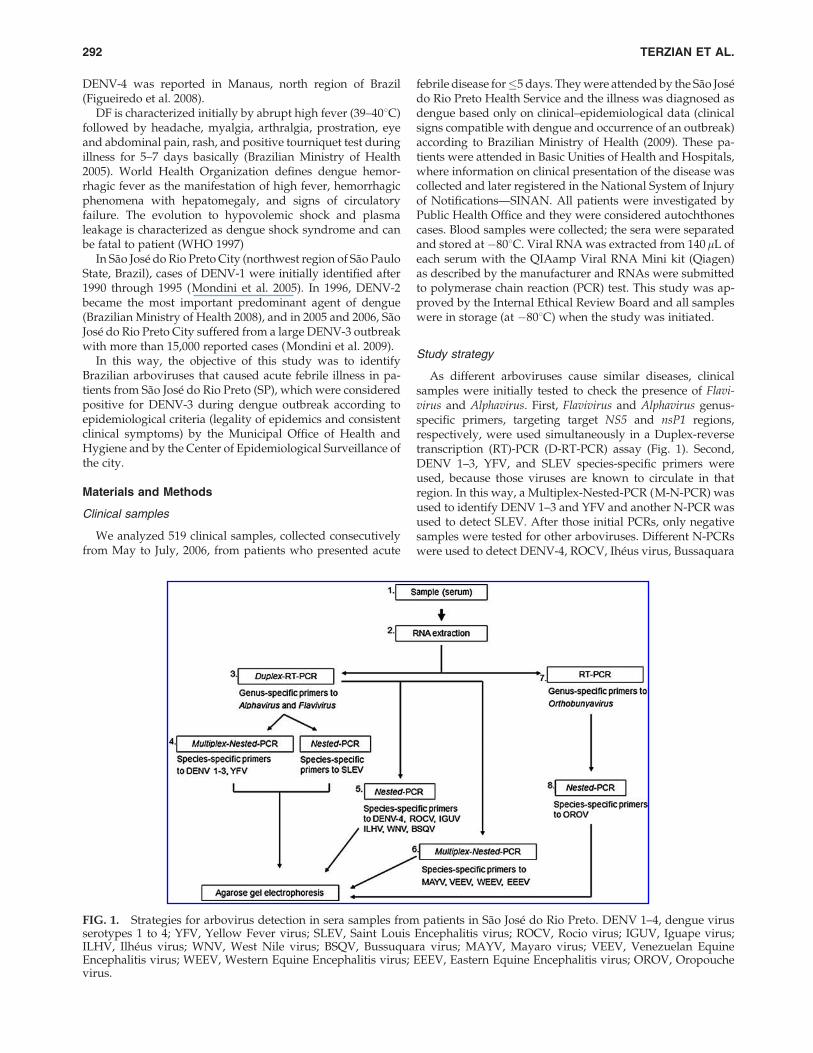
As different arboviruses cause similar diseases, clinicalsamples were initially tested to check the presence of Flavi-virus and Alphavirus. First, Flavivirus and Alphavirus genus-specific primers, targeting target NS5 and nsP1 regions,respectively, were used simultaneously in a Duplex-reversetranscription (RT)-PCR (D-RT-PCR) assay (Fig. 1). Second,DENV 1–3, YFV, and SLEV species-specific primers wereused, because those viruses are known to circulate in thatregion. In this way, a Multiplex-Nested-PCR (M-N-PCR) wasused to identify DENV 1–3 and YFV and another N-PCR wasused to detect SLEV. After those initial PCRs, only negativesamples were tested for other arboviruses. Different N-PCRswere used to detect DENV-4, ROCV, Iheus virus, Bussaquara
FIG. 1. Strategies for arbovirus detection in sera samples from patients in Sao Jose do Rio Preto. DENV 1–4, dengue virusserotypes 1 to 4; YFV, Yellow Fever virus; SLEV, Saint Louis Encephalitis virus; ROCV, Rocio virus; IGUV, Iguape virus;ILHV, Ilheus virus; WNV, West Nile virus; BSQV, Bussuquara virus; MAYV, Mayaro virus; VEEV, Venezuelan EquineEncephalitis virus; WEEV, Western Equine Encephalitis virus; EEEV, Eastern Equine Encephalitis virus; OROV, Oropouchevirus.
292 TERZIAN ET AL.
virus, Iguape virus, and West Nile virus (WNV). Another M-N-PCR was used to detect Venezuelan Equine Encephalitisvirus, Eastern Equine Encephalitis virus, Western EquineEncephalitis virus, and MAYV. Finally, a RT-PCR followed bya semi-nested assay (SN-PCR), using specific primers to Ssegment of OROV, was used. All primers used in this studyare listed in Table 1. Precautions to avoid contamination werefollowed, positive and negative controls were used in all re-actions, and the procedure was reproduced several times(Borst et al. 2004).
Virus and RNA extraction
The viral strains used as positive controls were MAYVBeAr20290, DENV-1 Mochizuki, DENV-2 SpH 125367,DENV-3 RPDen06=41, DENV-4 Boa Vista, SLEV BeH 355964,Ilheus virus BeH7445, ROCV SpH34675, West Nile NY99,YFV 17DD (vaccine), and OROV BeAn 19991. They werepropagated by intracerebral inoculation into 2-day-old suck-ling mice or in C6=36 Aedes albopictus cell cultures as previ-ously described (Shope and Sather 1979, Figueiredo 1990).Viral RNAs were extracted from 140mL of a 1=20 dilution ofmouse brain tissue macerated suspensions or from cell culturesupernatants with the QIAamp Viral RNA Mini kit (Qiagen)according to the manufacturer’s instructions.
PCR assays for Alphavirus and Flavivirus detection
D-RT-PCR, M-N-PCR, and N-PCR were performed as pre-viously described (de Morais Bronzoni et al. 2005). For theD-RT-PCR, the RT mixture was incubated at 508C for 50 minand at 708C for 15 min to inactivate the reverse transcrip-tase. The thermal cycling was performed with termocyclerGeneAmp� PCR System 9700 (Applied Biosystems). Accord-ing to the authors, M-N-PCR presented 99% of sensitivity and83% of specificity for the arbovirus.
SN-RT-PCR assays for OROV identification
The RT-PCR and SN-PCR were performed as described(Terzian et al. 2009). Primers used were c2ORO (RT-PCR) ands1ORO (PCR). Primers s1ORO and c1ORO were used togetherin the SN-PCR. The PCR mixture and cycling conditions wereperformed as for M-N-PCR Flavivirus assays (‘‘PCR assays forAlphavirus and Flavivirus detection’’ section).
Viral isolation
Viral isolation was performed in C6=36 cell culture aspreviously described (Figueiredo 1990) in selected samples toisolate a new SLEV strain. C6=36 cell culture confluentmonolayer was infected, for 60 min, with 100mL of serasamples diluted 1=10 in culture medium. After incubation,5 mL of Leibovitz medium L-15 (Cultilab, Campinas, Brazil)with 1% bovine fetal serum (Cultilab) was added to cellmonolayer. After 7 days, the cell culture was frozen at �808Cand thawed and the supernatant was analyzed for viral RNApresence by the specific PCR assay described earlier (Bronzoniet al. 2004, de Morais Bronzoni et al. 2005).
Nucleotide sequence
Amplicons obtained by (M)-N-PCR were purified withethanol as previously described (Sambrook 2001) and then
sequenced by dideoxynucleotide method (Sanger et al. 1977).Sequencing was realized using Big Dye Terminator Kit v3.1(Applied Biosystems) according to the manufacturer’s in-structions. In the sequencing PCR reactions, species-specificprimers (3.2 mM) were used. DNA obtained was centrifugedfor 20 min (48C, full speed), precipitated with 80mL of 75%isopropanol, and homogenized with 2.0 mL of 0.5 M ethyle-nediaminetetraacetic acid (pH 8.0) buffer and Bleu Dextran.DNA was denaturated at 958C for 2 min and applied onsequencing gel. Electrophoresis was realized on ABI PRISM377 DNA Sequencer (Applied Biosystems). Sequences wereanalyzed using DS Gene 2.0 (Accelrys) and BLAST (www.ncbi.nlm.nih.gov=blast=Blast.cgi) programs.
Analysis of partial sequence of envelope genefrom a SLEV isolate
Attempting to identify the genotype of SLEV isolate, theORF that codes for the envelope (E) protein was partiallyamplified and then sequenced. RNA obtained from theSLEV-isolated sample was submitted to RT-PCR and N-PCRassays using the primers previously described (Chandlerand Nordoff 1999, Kramer and Chandler 2001) and listed inTable 1. The amplicon was cloned into pCR2.1 vector (TACloning Kit; Invitrogen) and then several clones were se-quenced using Big Dye Terminator Kit v3.1 (Applied Bio-systems), according to manufacturer’s instructions. Sequenceswere analyzed using DS Gene 2.0 (Accelrys). Nucleotide anddeduced amino acid sequences were used to perform a simi-larity search in sequence databases, using FASTA (www.ebi.ac.uk=Tools=fasta=). The partial nucleotide sequence ofE gene from SLEV strain SJRP06=155 was deposited in Gen-Bank (accession no. GQ281267) and was aligned with otherSLEV sequences from different genotypes using Clustal X(Thompson et al. 1997). Before phylogenetic tree reconstruction by Maximum Likelihood (PAUP*4.0b10), the nucleotide substitution model GTRþIþG was selected using Modeltest(Posada and Crandall 2001). A phylogenetic tree was also reconstructed by neighbor-joining method, using p-distance(Mega3) (Kumar et al. 2004). Sequences of WNV, JapaneseEncephalitis virus, and Murray Valley Encephalitis virus wereused as outgroup. Bootstrapping analysis was performed toassess the robustness of tree topologies. Sequences used in thephylogenetic analysis are given in Table 2.
Statistical analysis
For statistical analysis, t, F, and w2 (comparison of propor-tion) test using Epi Info� 6.4 from the Center of DiseaseControl and Yates test were used. Standard deviations wereused for mean, variance, and deviation standard.
Results
Arbovirus detection by PCR
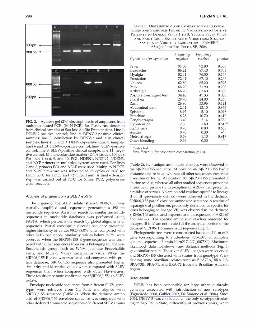
From 519 clinical samples that were analyzed, arbovirusesgenomes were detected in 374 samples (72.07%). DENV-3genome was detected in 365 samples and SLEV and DENV-2 genomes were detected in eight and five samples, respec-tively. Among these, coinfection of DENV-3 and DENV-2 wasdetected in one sample (Fig. 2). The remaining 145 samples offebrile acute illness patients (27. 93%) were all negative fortested arboviruses. All positive amplicons were sequenced to
ARBOVIRUSES IN DENGUE-SUSPECTED CASES 293
Ta
bl
e1.
Pr
im
er
sU
se
din
Th
is
St
ud
y
Tar
get
vir
us
Pri
mer
sS
equ
ence
(50 –
30 )
Am
pli
con
(pb)
Nu
cleo
tid
ep
osit
ion
Ref
eren
ce
Gen
us-
spec
ific
pri
mer
sto
Alp
hav
iru
san
dF
lav
ivir
us
Alp
hav
iru
sM
2W(þ
)Y
AG
AG
CD
TT
TT
CG
CA
YS
TR
GC
HW
a43
416
4–18
6P
feff
eret
al.
(199
7)cM
3W(�
)A
CA
TR
AA
NK
GN
GT
NG
TR
TC
RA
AN
CC
DA
YC
Ca
568–
597
Fla
viv
iru
sF
G1
(þ)
TC
AA
GG
AA
CT
CC
AC
AC
AT
GA
GA
TG
TA
CT
1000
8270
–829
7F
ulo
pet
al.
(199
3)F
G2
(�)
GT
GT
CC
CA
TC
CT
GC
TG
TG
TC
AT
CA
GC
AT
AC
A92
28–9
258
Sp
ecie
s-sp
ecifi
cp
rim
ers
toA
lph
avir
us
Ven
ezu
elan
Eq
uin
eE
nce
ph
alit
isv
iru
sN
VE
E(þ
)A
CG
GA
GG
TA
GA
CC
CA
TC
CG
A39
8b19
9–21
8B
ron
zon
iet
al.
(200
4)E
aste
rnE
qu
ine
En
cep
hal
itis
vir
us
NE
EE
(þ)
CC
AC
GG
TA
CC
GT
TG
CC
128b
469–
484
Wes
tern
Eq
uin
eE
nce
ph
alit
isv
iru
sN
WE
E(þ
)G
GC
GG
CA
GA
CC
TG
CT
GG
AA
208b
,c36
3–38
1M
ayar
ov
iru
sN
MA
Y(þ
)G
GA
AG
TT
GG
CC
AA
GG
C27
0b,c
164–
189
Sp
ecie
s-sp
ecifi
cp
rim
ers
toF
lav
ivir
us
Den
gu
e1
ND
EN
1(�
)C
GT
TT
TG
CT
CT
TG
TG
TG
CG
C47
2d,e
8653
–867
3d
eM
ora
isB
ron
zon
iet
al.
(200
5)D
eng
ue
2N
DE
N2
(�)
GA
AC
CA
GT
TT
GT
TT
DR
TT
TC
AT
AG
CT
GC
Ca
316d
,e84
88–8
516
Den
gu
e3
ND
EN
3(�
)C
CC
AT
TG
GT
TC
TC
CT
CT
GT
G62
8d,e
8800
–881
9D
eng
ue
4N
DE
N4
(�)
GC
AA
TC
GC
TG
AA
GC
CT
TC
TC
CC
222d
,e83
94–8
415
St.
Lo
uis
En
cep
hal
itis
vir
us
(SL
EV
)N
SL
E(�
)A
TT
CT
TC
TC
TC
AA
TC
TC
CG
T23
2d84
83–8
502
Bu
ssu
qu
ara
vir
us
NB
SQ
(�)
AA
GT
GA
CA
CC
TG
TT
CA
GG
GT
A38
8d86
38–8
658
Ilh
eus
vir
us
NIL
H(�
)T
CC
AC
CG
CT
GA
TC
TG
AG
CC
CG
TG
A47
4d87
21–8
744
Ro
cio
vir
us
NR
OC
(�)
TC
AC
TC
TT
CA
GC
CT
TT
CG
230d
8483
–850
0Y
ello
wF
ever
vir
us
NY
F(�
)T
CA
GA
AG
AC
CA
AG
AG
GT
CA
TG
T25
3d85
02–8
523
Igu
ape
vir
us
NIG
U(�
)C
CA
CG
AA
CC
AA
CT
TG
AA
G25
4d85
07–8
524
Wes
tN
ile
vir
us
NW
N(�
)T
CC
CC
CC
GC
AG
GT
GT
GC
CT
CG
717d
,e89
80–9
000
Sp
ecifi
cp
rim
ers
toO
rop
ouch
ev
iru
s(O
RO
V)
f
OR
OV
S1O
RO
(þ)
GA
AG
CT
AG
AT
AC
GG
AC
AA
GT
GC
TC
AA
TG
C50
079
–107
Ter
zian
etal
.(2
009)
C1O
RO
(�)
CT
CT
TA
CA
GC
AA
CT
AT
CT
CT
TC
AC
G57
7–60
1C
2OR
O(�
)C
CA
GA
GT
GC
CT
AC
AG
CC
TC
610–
628
Sp
ecifi
cp
rim
ers
toS
LE
Ven
vel
ope
gen
eS
LE
VF
880
(þ)
CG
AT
TG
GA
TG
GA
TG
CT
AG
GT
AG
477
880–
901
Kra
mer
and
Ch
and
ler
(200
1)B
2586
(�)
CA
GT
TG
GA
GT
CA
GA
GG
GA
AA
TA
CT
T25
86–2
562
B13
57(�
)G
CA
AC
CT
CA
TA
CT
TG
AT
GT
TT
TC
TC
1357
–138
1
(þ),
gen
om
icse
nse
pri
mer
;(�
),p
rim
eran
tise
nse
.aD
egen
erat
edp
rim
ers.
Sin
gle
lett
erco
de:
Y(C
or
T);
S(C
or
G);
R(A
or
G);
H(A
or
Co
rT
);W
(Ao
rT
);N
(Ao
rC
or
Go
rT
);K
(Go
rT
);D
(Ao
rG
or
T).
bW
ith
pri
mer
cM3W
.c W
ith
pri
mer
cM3W
bas
edo
nv
iru
sse
qu
ence
fro
mG
enB
ank
(NW
EE
AF
2140
40;
NM
AY
U94
602)
.dW
ith
pri
mer
FG
1.eW
ith
pri
mer
FG
1b
ased
on
vir
us
seq
uen
cefr
om
Gen
Ban
k(N
DE
NV
1A
B07
4760
;N
DE
NV
2A
F38
403;
ND
EN
V3
AY
9238
65;
ND
EN
4N
C_0
0264
0;N
WN
DQ
4110
34).
f Pri
mer
sto
Sse
gm
ent
of
OR
OV
.P
b,
pai
ro
fb
ases
.
294
confirm the specificity of the amplification. Viral isolation wasperformed in SLEV-positive samples using the same serasamples from PCR assay. As result, one SLEV strain wasisolated (SJRP06=155).
Epidemiological and clinical findings
This study was realized with 519 clinical samples collectedat the beginning of an outbreak indicating high positivity toFlaviviruses. DENV-2, DENV-3, and SLEV were detected in374 samples.
The epidemiological and clinical data showed no statisti-cally significant difference between the Flavivirus-positivepatients by PCR and the negatives, with the exception on thetourniquet test (Table 3), which was found more frequently inFlavivirus-positive patients as expected ( p> 0.005). Only onecase presenting a positive tourniquet test was not dengue butit was a SLEV infection. Pleural and pericardic hemorrhage,
hepatomegaly, cardiomyopathy, and shock were not referredby nonarbovirus cases. No other considerable symptomswere referred. One case of nasal bleeding (0.68%) and one caseof pain on inferior limb and lumbar region (0.68%) fromnegative cases were referred. Among negative patients forFlavivirus, 6.90% of them were hospitalized; however, nostatistically significant difference was found compared withpositive cases ( p-value: 0.666461). No significant difference( p-value: 0.77) was observed on fever duration (3.125 daysmean) in arbovirus-infected (3.125 days mean) and uninfectedindividuals (3.054 days mean).
Based on RT-PCR assay, 145 individuals were negativefor arboviruses; however, based on Ministry of Healthclinical criteria (SMSH=VE=SEI=SINAN) in this samegroup, 101 cases (69.65%) were classified as having DFand 1 case was classified as having atypical severedengue.
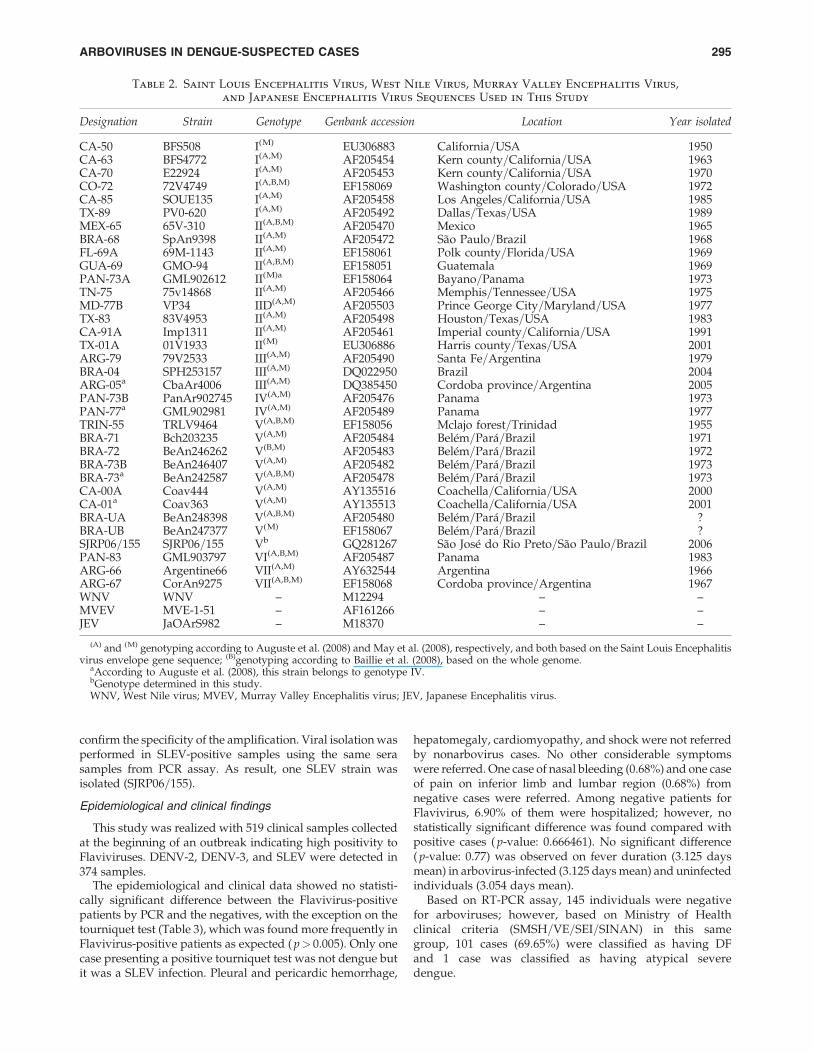
Table 2. Saint Louis Encephalitis Virus, West Nile Virus, Murray Valley Encephalitis Virus,
and Japanese Encephalitis Virus Sequences Used in This Study
Designation Strain Genotype Genbank accession Location Year isolated
CA-50 BFS508 I(M) EU306883 California=USA 1950CA-63 BFS4772 I(A,M) AF205454 Kern county=California=USA 1963CA-70 E22924 I(A,M) AF205453 Kern county=California=USA 1970CO-72 72V4749 I(A,B,M) EF158069 Washington county=Colorado=USA 1972CA-85 SOUE135 I(A,M) AF205458 Los Angeles=California=USA 1985TX-89 PV0-620 I(A,M) AF205492 Dallas=Texas=USA 1989MEX-65 65V-310 II(A,B,M) AF205470 Mexico 1965BRA-68 SpAn9398 II(A,M) AF205472 Sao Paulo=Brazil 1968FL-69A 69M-1143 II(A,M) EF158061 Polk county=Florida=USA 1969GUA-69 GMO-94 II(A,B,M) EF158051 Guatemala 1969PAN-73A GML902612 II(M)a EF158064 Bayano=Panama 1973TN-75 75v14868 II(A,M) AF205466 Memphis=Tennessee=USA 1975MD-77B VP34 IID(A,M) AF205503 Prince George City=Maryland=USA 1977TX-83 83V4953 II(A,M) AF205498 Houston=Texas=USA 1983CA-91A Imp1311 II(A,M) AF205461 Imperial county=California=USA 1991TX-01A 01V1933 II(M) EU306886 Harris county=Texas=USA 2001ARG-79 79V2533 III(A,M) AF205490 Santa Fe=Argentina 1979BRA-04 SPH253157 III(A,M) DQ022950 Brazil 2004ARG-05a CbaAr4006 III(A,M) DQ385450 Cordoba province=Argentina 2005PAN-73B PanAr902745 IV(A,M) AF205476 Panama 1973PAN-77a GML902981 IV(A,M) AF205489 Panama 1977TRIN-55 TRLV9464 V(A,B,M) EF158056 Mclajo forest=Trinidad 1955BRA-71 Bch203235 V(A,M) AF205484 Belem=Para=Brazil 1971BRA-72 BeAn246262 V(B,M) AF205483 Belem=Para=Brazil 1972BRA-73B BeAn246407 V(A,M) AF205482 Belem=Para=Brazil 1973BRA-73a BeAn242587 V(A,B,M) AF205478 Belem=Para=Brazil 1973CA-00A Coav444 V(A,M) AY135516 Coachella=California=USA 2000CA-01a Coav363 V(A,M) AY135513 Coachella=California=USA 2001BRA-UA BeAn248398 V(A,B,M) AF205480 Belem=Para=Brazil ?BRA-UB BeAn247377 V(M) EF158067 Belem=Para=Brazil ?SJRP06=155 SJRP06=155 Vb GQ281267 Sao Jose do Rio Preto=Sao Paulo=Brazil 2006PAN-83 GML903797 VI(A,B,M) AF205487 Panama 1983ARG-66 Argentine66 VII(A,M) AY632544 Argentina 1966ARG-67 CorAn9275 VII(A,B,M) EF158068 Cordoba province=Argentina 1967WNV WNV – M12294 – –MVEV MVE-1-51 – AF161266 – –JEV JaOArS982 – M18370 – –
(A) and (M) genotyping according to Auguste et al. (2008) and May et al. (2008), respectively, and both based on the Saint Louis Encephalitisvirus envelope gene sequence; (B)genotyping according to Baillie et al. (2008), based on the whole genome.
aAccording to Auguste et al. (2008), this strain belongs to genotype IV.bGenotype determined in this study.WNV, West Nile virus; MVEV, Murray Valley Encephalitis virus; JEV, Japanese Encephalitis virus.
ARBOVIRUSES IN DENGUE-SUSPECTED CASES 295
Analysis of E gene from a SLEV isolate
The E gene of the SLEV isolate (strain SJRP06=155) waspartially amplified and sequenced generating a 492 pbnucleotide sequence. An initial search for similar nucleotidesequences in nucleotide databases was performed usingFASTA, which performs the search by global alignments ofsequences. Partial envelope nucleotide sequence presentedhigher similarity of values 90.2–98.0% when compared withother SLEV sequences. Similarity values below 69.7% wereobserved when the SJRP06=155 E gene sequence was com-pared with other sequences from virus belonging to JapaneseEncephalitis group, such as WNV, Japanese Encephalitisvirus, and Murray Valley Encephalitis virus. When theSJRP06=155 E gene was translated and compared with pro-tein database, SJRP06=155 sequence also presented highersimilarity and identities values when compared with SLEVsequences than when compared with other Flaviviruses.These results once more confirmed that SJRP06=155 is a SLEVisolate.
Envelope nucleotide sequences from different SLEV geno-types were retrieved from GenBank and aligned withSJRP06=155 sequence (Table 2). When the deduced aminoacid of SJRP06=155 envelope sequence was compared withother deduced amino acid sequences of different SLEV strains
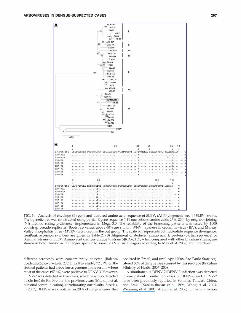
(Table 2), two unique amino acid changes were observed inthe SJRP06=155 sequence. At position 44, SJRP06=155 had aglutamic acid residue, whereas all other sequences presenteda residue of lysine. At position 88, SJRP06=155 presented aleucine residue, whereas all other studied sequences presenteda residue of proline (with exception of ARG79 that presenteda residue of serine). No amino acid residues specific to lineageII or III (previously defined) were observed in the deducedSPJR06=155 partial envelope amino acid sequence. A residue ofasparagine at position 66, previously described as specific forvirus belonging to lineage VII, was observed in the deducedSJRP06=155 amino acid sequence and in sequences of ARG-67and ARG-66. The specific amino acid residues observed forlineages III to V are not located at the analyzed portion of thededuced SJRP06=155 amino acid sequence (Fig. 3).
Phylogenetic trees were reconstructed based on 411 nt of Egene (corresponding to nucleotides 963–1373 of completegenome sequence of strain Kern217, NC_007580). Maximumlikelihood (data not shown) and distance methods (Fig. 3)gave similar results. The seven SLEV lineages were observedand SJRP06=155 clustered with strains from genotype V, in-cluding some Brazilian isolates such as BRA73A, BRA-UB,BRA-73B, BRA-71, and BRA-72 from the Brazilian Amazonregion.
Discussion
DENV has been responsible for large urban outbreaksgenerally associated with introduction of new serotypes(Figueiredo 2000, Gubler 2002, De Simone et al. 2004). Since2004, DENV-3 was considered as the only serotype circulat-ing in Sao Paulo State, differently of previous years, when
FIG. 2. Agarose gel (2%) electrophoresis of amplicons frommultiplex-nested-PCR (M-N-PCR) for Flavivirus detectionfrom clinical samples of Sao Jose do Rio Preto patient. Line 1:DENV-2-positive control; line 2: DENV-2-positive clinicalsamples; line 3: coinfection by DENV-2 and 3 in clinicalsamples; lines 4, 5, and 9: DENV-3-positive clinical samples;lines 6 and 10: DENV-3-positive control; line7: SLEV-positivecontrol; line 8: SLEV-positive clinical sample; line 11: nega-tive control. M, molecular size marker (DNA ladder, 100 pb).For lines 1 to 6, 9, and 10, FG1, NDEN1, NDEN2, NDEN3,and NYF primers in multiplex system were used. For lines7 and 8, primers FG1 and NSLE were used. Multiplex N-PCRand N-PCR mixture was subjected to 25 cycles of 948C for1 min, 538C for 1 min, and 728C for 2 min. A final extensionstep was carried out at 728C for 5 min. PCR, polymerasechain reaction.
Table 3. Distribution and Comparison of Clinical
Signs and Symptoms Found in Negative and Positive
Patients to Dengue Virus 1 to 3, Yellow Fever Virus,
and Saint Louis Encephalitis Virus from Studied
Samples in Virology Laboratory—FAMERP,Sao Jose do Rio Preto, SP, 2006
Signals and=or symptomsFrequencynegative
Frequencypositive p-value
Fever 91.00 92.80 0.503Headache 86.21 87.40 0.708Myalgia 82.41 76.50 0.166Prostation 72.41 67.40 0.266Nausea 62.80 65.20 0.595Pain 66.20 71.90 0.200Arthralgia 66.20 63.60 0.583Positive tourniquet test 34.48 47.33 0.008Diarrhea 29.70 24.90 0.265Rash 26.90 33.96 0.121Abdominal pain 12.41 13.10 0.833Epistaxis 8.97 5.10 0.098Petechiae 8.28 10.70 0.410Gengivorragia 3.40 2.14 0.586Hypotension 2.8 1.60 0.615a
Hematuria 0.70 0.80 0.668Ascites 0.70 0.30 –b
Metrorrhagia 0.69 1.10 0.917Other bleeding 0.69 0.30 –b
aYates test.bInsufficient n for proportion comparation (n< 5).
296 TERZIAN ET AL.
different serotypes were concomitantly detected (BoletimEpidemiologico Paulista 2005). In this study, 72.07% of thestudied patients had arboviruses genome in the serum, wheremost of the cases (97.6%) were positive to DENV-3. However,DENV-2 was detected in five cases, which was also detectedin Sao Jose do Rio Preto in the previous years (Mondini et al.personal communication), corroborating our results. Besides,in 2007, DENV-2 was isolated in 20% of dengue cases that
occurred in Brazil, and until April 2008, Sao Paulo State reg-istered 60% of dengue cases caused by this serotype (BrazilianMinistry of Health 2007, 2008).
A simultaneous DENV-2=DENV-3 infection was detectedin one patient. Coinfection cases of DENV-3 and DENV-2have been previously reported in Somalia, Taiwan, China,and Brazil (Kanesa-thasan et al. 1994, Wang et al. 2003,Wenming et al. 2005, Araujo et al. 2006). Other coinfection
FIG. 3. Analysis of envelope (E) gene and deduced amino acid sequence of SLEV. (A) Phylogenetic tree of SLEV strains.Phylogenetic tree was constructed using partial E gene sequence (411 nucleotides, amino acids 27 to 200), by neighbor-joining(NJ) method (using p-distance) implemented in Mega 3.0. The reliability of the branching patterns was tested by 1000bootstrap pseudo replicates. Bootstrap values above 60% are shown. WNV, Japanese Encephalitis virus ( JEV), and MurrayValley Encephalitis virus (MVEV) were used as the out group. The scale bar represents 5% nucleotide sequence divergence.GenBank accession numbers are given in Table 2. (B) Alignment of deduced amino acid E protein (partial sequence) ofBrazilian strains of SLEV. Amino acid changes unique to strain SJRP06=155, when compared with other Brazilian strains, areshown in bold. Amino acid changes specific to some SLEV virus lineages (according to May et al. 2008) are underlined.
ARBOVIRUSES IN DENGUE-SUSPECTED CASES 297
cases had also been reported among DENV serotypes (Gubleret al. 1985, Laille et al. 1991, Wang et al. 2003, Araujo et al.2006, Bharaj et al. 2008). It is believed that these coinfectionsonly occur during outbreaks with circulation of multiple se-rotypes and where there is high prevalence of the urbanvector, being capable to transmit more than one virus at thesame time (Wenming et al. 2005, Bharaj et al. 2008).
All studied patients had been previously considered ashaving dengue based on clinical and epidemiological criteria.However, from 519 samples tested by RT-PCR, 27.93% werenegative to dengue as well as other tested arboviruses. Manyother infectious agents, such as poliomyelitis, rabies, measles,rubella, hepatitis, malaria, or leptospirosis, can induce similarsymptomatology (Fonseca and Figueiredo 2005, Vasconceloset al. 2005), and the laboratory diagnosis is fundamental inthis context. This work shows that the utilization of clinicaland epidemiological criteria for dengue diagnosis can be amajor problem, even in the presence of a major outbreak. Thesamples collected for this study were all collected in less than5 days of the onset of the disease, suggesting that a falsenegative due to absence of viruses circulating in the blood isnot the point here.
SLEV is widely distributed in the Americas, from Canadato Argentina (Sabattini et al. 1998, Burke and Monath 2001).SLEV infections have been reported in the United States andless frequently in Central and South America (Sabattini et al.1998). However, 5% of population in north and southeast ofBrazil seemed to have antibodies against the virus. Never-theless, these data must be carefully analyzed, because it maybe due to cross-reaction between antibodies of differentFlavivirus, which are induced especially by Yellow Fevervaccination or exposure to DENV. In this way, SLEV may becirculating in these areas and causing human infections,although most of them are undiagnosed (Lopes et al. 1979,Vasconcelos et al. 1998, Figueiredo 2000).
In Brazil, in 2004, SLEV was isolated from an acute febrileillness patient living in Sao Pedro county, Sao Paulo State(Rocco et al. 2005). In 2006, a serological study in birds carriedout in the southern coast of Sao Paulo State showed a highincrease of the prevalence of antibodies against SLEV (Suzukiet al. 2006). Finally, also in 2006, concomitant to a DENV-3outbreak in Sao Jose do Rio Preto, 12 dengue suspected caseshad SLEV genome detected in clinical samples and it has beenconsidered the first SLEV outbreak registered in Brazil(Mondini et al. 2007b). Serology is an important tool in SLEVmonitoring, especially for bird reservoirs and horses. It isknown that horses can be infected but they rarely developsymptoms. Studies with infected horses can indicate viruscirculating among animals, and in the presence of mosquitoes(Culex vectors), they can be a potential source for humaninfection. In 2009, researches of Sao Paulo State, using aSLEV IgG-ELISA, detected a 31.11% of positivity among 180studied horses. This indicated that human infections can beoccurring in locations where these animals live (Silva et al.2009).
The fact that SLEV was the agent of human disease servesas advice to health professionals about the need of a completeclinical and epidemiological investigation about febrile dis-eases, especially during outbreaks, as SLEV infections can beunrecognized or be mistaken with dengue because of similarclinical manifestations. Epidemiological investigation asso-ciated with the methodology applied in this study, using
FG1=FG2 and NSLE primers in a multiplex system withdengue primers and associated alphaviruses PCR, can be animportant tool in a trustworthy and fast diagnosis, not onlyfor SLEV but also for other arboviruses. In this study, it wasdemonstrated that SLEV circulation in Sao Jose do Rio Pretowas more important than previously reported because eightdengue-suspected cases were diagnosed in the laboratory ashaving SLEV.
Phylogenetic analysis of different SLEV isolates suggeststhat this virus is mainly maintained locally, although migra-tion between different areas also occurs (Kramer and Chandler2001, Twiddy and Holmes 2003). This analysis showed thatSLEV isolates are clustered in seven different lineages basedon E gene or the genome sequences (Kramer and Chandler2001, Auguste et al. 2008, Baillie et al. 2008) and it is mainlycorrelated with the geographical characteristics of thoseviruses and not phenotypic characteristics such as viru-lence (Trent et al. 1980, Kramer et al. 1997). Lineages I and IIare predominantly from North America, whereas lineages IIIto VII belong to South and Central Americas. The BrazilianSLEV strains described so far belong to genotypes II (BRA-04),III (BRA-68), and V (BRA-72, BRA-71, BRA-73A, BRA-73B,BRA-73C, BRA-73D, BRA-UA, BRA-UB, and BRA-60) (Augusteet al. 2008, Baillie et al. 2008, May et al. 2008).
Although the clustering of SRJP06=155 within lineage V(also including BRA72, BRA71, BRA73A, BRA73B, BRA-UA,BRA-UB, TRIN-55, PER-75, CA00A, and CA01A) was notsupported by high bootstrap values (as also observed byMay et al. 2008), similar results were observed by otherauthors when analyzing bigger sequences of E gene (Au-guste et al. 2008, May et al. 2008), where the same strainswere clustered within lineage V. In the same way, thestrains BRA-73A, BRA-73B, PER-75, and TRI-55 were alsoshown to belong to lineage V, based on the analysis of thewhole genome of SLEV (Baillie et al. 2008). Analysis of otherregions of SJRP06=155 genome could help elucidating thegenotyping of SJRP06=155.
May et al. (2008) defined some specific amino acidchanges that are observed in the E protein sequence forindividual lineages. Although it was not possible to checkfor amino acid residues specific to lineages III to V, noamino acid change specific to lineage II or III was observedin the deduced SPJR06=155 partial envelope amino acidsequence. An amino acid residue specific to lineage VII (atposition 66) was observed in SJRP06=155 E protein se-quence; however, the residue of glycine observed at position29 in isolates from genotype VII was not observed inSJRP06=155 sequence.
In summary, we reported here a dengue outbreak whereDENV-3 infections predominated, consistent with the intro-duction of this new serotype in the region. Some infectionswere caused by DENV-2, the previous causative agent ofoutbreaks in the region. However, the unexpected presence ofSLEV causing acute febrile illness was also detected by anunusual diagnostic approach looking for arboviruses insteadof dengue only. These results showed the real situation ofcocirculation of some arboviruses during an outbreak, ini-tially caused only for DENV-3, which warns health profes-sionals and requires the inclusion of infection caused by SLEVin the differential diagnosis of acute febrile illness and en-cephalitis. In Brazil, SLEV situation is worrisome becauseprobably the disease has been unrecognized or mistaken
298 TERZIAN ET AL.
with dengue and SLEV human cases and is occurring inother places of Brazil. Sao Jose do Rio Preto is an importantgeographic area for arboviruses surveillance (especiallyFlaviviruses) because of outbreaks caused by DENV-1, �2,and �3, as well as identification of SLEV and the risk SylvaticYellow Fever in the region. In this way, it is necessary toimplement an active surveillance, based on prevention andcontrol of patients with acute febrile illness, reservoirs, do-mestics’ animals, and mosquito vectors, to avoid introductionor reintroduction of these viruses. We also believe that the useof clinical and epidemiological criteria without laboratoryconfirmation should not be done even in the presence of alarge DENV outbreak.
Author’s Contributions
A.C.B.T., L.T.M.F., F.C.N., and M.L.N. conceptualized thestudy. A.C.B.T. and B.P.F. processed and performed themanipulation of samples. A.M. collected samples and clus-tered patients’ clinical information. R.V.M.B. contributed totechnique used for this study and intellectual and technicalknowledge. B.P.D. contributed to sequence and phyloge-netic analysis. E.M.S.C. contributed to statistical analysis.L.T.M.F. contributed with viral control samples. M.L.N.critically appraised the manuscript and contributed to theintellectual content. All authors helped in drafting and ap-proved the final manuscript. M.L.N. is guarantor of themanuscript.
Acknowledgments
The authors thank all staff of the County Office of Healthand Hygiene, Basic Unities of Health, Emergency Unities andHospitals from the Sao Jose do Rio Preto City. They also thankMilena Polloto for sample sequencing. This work was sup-ported by grants from Fundacao de Amparo a Pesquisa doEstado de Sao Paulo (FAPESP) (grant no. 04=11098-2) andConselho Nacional de Desenvolvimento Cientıfico e Tecno-logico (grant no. 401396=2004-5). This work was also partiallysupported by the Viral Genetic Diversity Network (VGDN–FAPESP–Brazil).
Disclosure Statement
No competing financial interests exist
References
Araujo, FM, Nogueira, RM, de Araujo, JM, Ramalho, IL, et al.Concurrent infection with dengue virus type-2 and DENV-3 ina patient from Ceara, Brazil. Mem Inst Oswaldo Cruz 2006;101:925–928.
Auguste, AJ, Pybus, OG, Carrington, CV. Evolution and dis-persal of St. Louis encephalitis virus in the Americas. InfectGenet Evol 2009; 9:409–415.
Baillie, GJ, Kolokotronis, SO, Waltari, E, Maffei, JG, et al.Phylogenetic and evolutionary analyses of St. Louis en-cephalitis virus genomes. Mol Phylogenet Evol 2008; 47:717–728.
Bharaj, P, Chahar, HS, Pandey, A, Diddi, K, et al. Concurrentinfections by all four dengue virus serotypes during an out-break of dengue in 2006 in Delhi, India. Virol J 2008; 5:1.
Borst, A, Box, AT, Fluit, AC. False-positive results and contam-ination in nucleic acid amplification assays: suggestions for a
prevent and destroy strategy. Eur J Clin Microbiol Infect Dis2004; 23:289–299.
Boletim Epidemiologico Paulista. Dengue: Epidemia ou en-demia? Boletim Epidemiologico Paulista. 2005; 2:20.
Brazilian Ministry of Health. Dengue no Brasil. Informe Epide-miologico 12=2009. Monitoramento CGPNCD. Available athttp:==portal.saude.gov.br=portal=arquivos=pdf=inform_dengue_1905200922.pdf2009.
Brazilian Ministry of Health. Secretaria de Vigilancia em Saude.Guia de Vigilancia Epidemiologica=Ministerio da Saude, Secre-taria de Vigilancia em Saude; Brasılia: Ministerio da Saude, 2005.
Brazilian Ministry of Health. Informe epidemiologico de dengue,Janeiro a Dezembro de 2007. http:==portal.saude.gov.br=portal=arquivos=pdf=boletim dengue 010208.pdf.2007
Brazilian Ministry of Health. Informe epidemiologico da den-gue. Janeiro a Abrll de 2008. http:==portal.saude.gov.br=portal=arquivos=pdf=boletim dengue maio2008.pdf.2008
Brazilian Ministry of Health. Dengue no Brasil. Informe epide-miologico 12=2009. Monitoramento CGPNCD. http:==portal.saude.gov.br=portal=arquivos=pdf=inform dengue 1905200922.pdf.2009.
Bronzoni, RV, Moreli, ML, Cruz, AC, Figueiredo, LT. Multi-plexnested PCR for Brazilian Alphavirus diagnosis. Trans Rsoc Trop Med Hyg 2004; 98:456–461.
Burke, DS, Monath, TP. Flaviviruses FIELDS’ Virology. Philadel-phia: LWW, 2001.
Chandler, LJ, Nordoff, NG. Identification of genetic variationamong St. Louis encephalitis virus isolates, using single-strandconformation polymorphism analysis. J Virol Methods 1999;80:169–178.
de Morais Bronzoni, RV, Baleotti, FG, Ribeiro Nogueira, RM,Nunes, M, et al. Duplex reverse transcription-PCR followedby nested PCR assays for detection and identification of Bra-zilian alphaviruses and flaviviruses. J Clin Microbiol 2005;43:696–702.
De Simone, TS, Nogueira, RM, Araujo, ES, Guimaraes, FR, et al.Dengue virus surveillance: the co-circulation of DENV-1,DENV-2 and DENV-3 in the State of Rio de Janeiro, Brazil.Trans R Soc Trop Med Hyg 2004; 98:553–562.
Figueiredo, LT. Emergent arboviruses in Brazil. Rev Soc BrasMed Trop 2007; 40:224–229.
Figueiredo, LT. [The use of Aedes albopictus C6=36 cells in thepropagation and classification of arbovirus of the Togaviridae,Flaviviridae, Bunyaviridae and Rhabdoviridae families]. RevSoc Bras Med Trop 1990; 23:13–18.
Figueiredo, LTM. The Brazilian flaviviruses. Microbes Infect2000; 2:1643–1649.
Figueiredo, RMP, Naveca, FG, Bastos, MS, Melo, MN, et al.Dengue virus type 4, Manaus, Brazil. Emerg Infect Dis 2008;14:667–669.
Fonseca, BAL, Figueiredo, LTM. Dengue VERONESI: Tratado deInfectologia. Sao Paulo: Atheneu, 2005:343–356.
Fulop, L, Barrett, AD, Phillpotts, R, Martin, K, et al. Rapididentification of flaviviruses based on conserved NS5 genesequences. J Virol Methods 1993; 44:179–188.
Gubler, DJ. Epidemic dengue=dengue hemorrhagic fever as apublic health, social and economic problem in the 21st cen-tury. Trends Microbiol 2002; 10:100–103.
Gubler, DJ, Kuno, G, Sather, GE, Waterman, SH. A case ofnatural concurrent human infection with two dengue viruses.Am J Trop Med Hyg 1985; 34:170–173.
Kanesa-thasan, N, Iacono-Connors, L, Magill, A, Smoak, B, et al.Dengue serotypes 2 and 3 in US forces in Somalia. Lancet1994; 343:678.
ARBOVIRUSES IN DENGUE-SUSPECTED CASES 299
Kramer, LD, Chandler, LJ. Phylogenetic analysis of the envelopegene of St. Louis encephalitis virus. Arch Virol 2001; 146:2341–2355.
Kramer, LD, Presser, SB, Hardy, JL, Jackson, AO. Genotypic andphenotypic variation of selected Saint Louis encephalitis viralstrains isolated in California. Am J Trop Med Hyg 1997; 57:222–229.
Kumar, S, Tamura, K, Nei, M. MEGA3: Integrated software formolecular evolutionary genetics analysis and sequence align-ment. Brief Bioinform 2004; 5:150–163.
Laille, M, Deubel, V, Sainte-Marie, FF. Demonstration of con-current dengue 1 and dengue 3 infection in six patients by thepolymerase chain reaction. J Med Virol 1991; 34:51–54.
Lopes, OS, Sacchetta, LA, Coimbra, TL, Pereira, LE. Isolation ofSt. Louis encephalitis virus in south Brazil. Am J Trop MedHyg 1979; 28:583–585.
May, FJ, Li, L, Zhang, S, Guzman, H, et al. Genetic variation ofSt. Louis encephalitis virus. J Gen Virol 2008; 89:1901–1910.
Mondini, A, Bronzoni, RV, Cardeal, IL, dos Santos, TM, et al.Simultaneous infection by DENV-3 and SLEV in Brazil. J ClinVirol 2007a; 40:84–86.
Mondini, A, Bronzoni, RVM, Nunes, SHP, Chiaravalloti Neto, F,et al. Spatio-temporal tracking and phylodynamics of anurban dengue 3 outbreak in Sao Paulo, Brazil. PLoS Negl TropDis 2009; 3:e448.
Mondini, A, Cardeal, IL, Lazaro, E, Nunes, SH, et al. Saint Louisencephalitis virus, Brazil. Emerg Infect Dis 2007b; 13:176–178.
Mondini, A, Chiaravalloti, Neto, F, Gallo y Sanches, M, Lopes,JC. [Spatial analysis of dengue transmission in a medium-sized city in Brazil]. Rev Saude Publica 2005; 39:444–451.
Nogueira, RM, Schatzmayr, HG, de Filippis, AM, dos Santos,FB, et al. Dengue virus type 3, Brazil, 2002. Emerg Infect Dis2005; 11:1376–1381.
Pfeffer, M, Proebster, B, Kinney, RM, Kaaden, OR. Genus-specific detection of alphaviruses by a semi-nested reversetranscription-polymerase chain reaction. Am J Trop Med Hyg1997; 57:709–718.
Posada, D, Crandall, KA. Selecting the best-fit model of nucle-otide substitution. Syst Biol 2001; 50:580–601.
Rocco, IM, Santos, CL, Bisordi, I, Petrella, SM, et al. St. Louisencephalitis virus: first isolation from a human in Sao PauloState, Brazil. Rev Inst Med Trop Sao Paulo 2005; 47:281–285.
Sabattini, M, Aviles, G, Monath, TP. Historical, epidemiologicaland ecological aspects of arbovirus in Argentina. Flaviviridae,Bunyaviridae, Rhabdoviridae. In: Travassos da Rosa, A, Vas-concelos, PFC, Travassos da Rosa, JFS, eds. An overview ofArbovirology in Brasil and Neighboring Countries. Belem, Brazil:Instituto Evandro Chagas, 1998:114–134.
Sambrook, DW, Jr. In vitro amplification of DNA by polymerasechain reaction. In: Molecular Cloning: A Laboratory Manual.New York: Cold Spring Harbor Laboratory Press, 2001; Sec. 8.
Sanger, F, Nicklen, S, Coulson, AR. DNA sequencing withchain-terminating inhibitors. Proc Natl Acad Sci USA 1977;74:5463–5467.
Shope, RE, Sather, GE. Arboviruses. In: Lennet, FH, Schimidt,NJ, eds. Diagnostic Procedures for Viral, Rickettsial and Clamydial
Infections. Washington: American Public Health Association,1979:767–814.
Silva, JR, Chavez, JH, Figueiredo, LTM. A serologic survey forSaint Louis Encephalitis virus in horses from Sao Paulo State,Brazil. Virus: Rev Res 2009; 14:115.
Suzuki, A, Bisordi, I, Lima, LBQ, Pereira, LE, et al. Saint LouisEncephalitis virus: high prevalence of antibodies in wild birds,Iguape—Sao Paulo State, Brazil, 2005–2006 (abstract). J BrazSoc Virol 2006; 11:129.
Terzian, ACB, Bronzoni, RVM, Drumond, BP, Silva-Nunes, M,et al. Sporadic Oropouche virus infection, Acre, Brazil. EmergInfect Dis 2009; 15:348–350.
Thompson, JD, Gibson, TJ, Plewniak, F, Jeanmougin, F, Higgins,DG. The CLUSTAL_X windows interface: flexible strategiesfor multiple sequence alignment aided by quality analysistools. Nucleic Acids Res 1997; 25:4876–4882.
Trent, DW, Monath, TP, Bowen, GS, Vorndam, AV, et al. Var-iation among strains of St. Louis encephalitis virus: basis for agenetic, pathogenetic, and epidemiologic classification. AnnNY Acad Sci 1980; 354:219–237.
Twiddy, SS, Holmes, EC. The extent of homologous recombi-nation in members of the genus Flavivirus. J Gen Virol 2003;84:429–440.
Vasconcelos, PFC, Travassos da Rosa, APA, Pinheiro, FP,Shope, RE, et al. Arboviruses pathogenic for man in Brazil.In: Travassos da Rosa, APA, Vasconcelos, PFC, Travassos daRosa, JFS, eds. An Overview of Arbovirology in Brazil andNeighbouring Countries. Belem: Evandro Chagas Institute,1998:72–99.
Vasconcelos, PFC, Travassos da Rosa, APA, Pinheiro, FP,Travassos da Rosa, JFS. Arboviroses. VERONESI: Tratado deInfectologia. Sao Paulo: Atheneu, 2005:289–302.
Wang, WK, Chao, DY, Lin, SR, King, CC, Chang, SC. Con-current infections by two dengue virus serotypes amongdengue patients in Taiwan. J Microbiol Immunol Infect 2003;36:89–95.
Wenming, P, Man, Y, Baochang, F, Yongqiang, D, et al.Simultaneous infection with dengue 2 and 3 viruses in aChinese patient return from Sri Lanka. J Clin Virol 2005; 32:194–198.
World Health Organization (WHO). Dengue and dengue hae-morrhagic fever: Diagnosis, treatment, prevention and control.Geneva: World Health Organization, 1997.
Address correspondence to:Maurıcio Lacerda Nogueira
Laboratorio de Pesquisa em VirologiaFaculdade de Medicina de Sao Jose do Rio Preto (FAMERP)
Av. Brigadeiro Faria Lima5416-Vila Sao Pedro
15090-000, Sao Jose do Rio PretoSao Paulo
Brazil
E-mail: [email protected]
300 TERZIAN ET AL.