Survival, Recovery, and Inactivation of Human Norovirus ...
182
Clemson University TigerPrints All Dissertations Dissertations 8-2017 Survival, Recovery, and Inactivation of Human Norovirus Surrogates, Feline Calicivirus and Murine Norovirus, on Carpets David Buckley Clemson University, [email protected] Follow this and additional works at: hps://tigerprints.clemson.edu/all_dissertations is Dissertation is brought to you for free and open access by the Dissertations at TigerPrints. It has been accepted for inclusion in All Dissertations by an authorized administrator of TigerPrints. For more information, please contact [email protected]. Recommended Citation Buckley, David, "Survival, Recovery, and Inactivation of Human Norovirus Surrogates, Feline Calicivirus and Murine Norovirus, on Carpets" (2017). All Dissertations. 1979. hps://tigerprints.clemson.edu/all_dissertations/1979
-
Upload
khangminh22 -
Category
Documents
-
view
0 -
download
0
Transcript of Survival, Recovery, and Inactivation of Human Norovirus ...

Clemson UniversityTigerPrints
All Dissertations Dissertations
8-2017
Survival, Recovery, and Inactivation of HumanNorovirus Surrogates, Feline Calicivirus andMurine Norovirus, on CarpetsDavid BuckleyClemson University, [email protected]
Follow this and additional works at: https://tigerprints.clemson.edu/all_dissertations
This Dissertation is brought to you for free and open access by the Dissertations at TigerPrints. It has been accepted for inclusion in All Dissertations byan authorized administrator of TigerPrints. For more information, please contact [email protected].
Recommended CitationBuckley, David, "Survival, Recovery, and Inactivation of Human Norovirus Surrogates, Feline Calicivirus and Murine Norovirus, onCarpets" (2017). All Dissertations. 1979.https://tigerprints.clemson.edu/all_dissertations/1979

SURVIVAL, RECOVERY, AND INACTIVATION OF HUMAN NOROVIRUS SURROGATES, FELINE CALICIVIRUS AND MURINE NOROVIRUS, ON
CARPETS
A Dissertation Presented to
the Graduate School of Clemson University
In Partial Fulfillment of the Requirements for the Degree
Doctor of Philosophy Microbiology
by David Buckley August 2017
Accepted by: Xiuping Jiang, Committee Chair
Simon Scott, Co-Chair Angela Fraser
Charles Pettigrew
ii
ABSTRACT
Worldwide, enteric viruses are the main cause of acute gastroenteritis (AGE).
Among these viruses, human noroviruses (HuNoV) are leading cause of AGE and
account for ca. 20% of all diarrheal cases, a top-five cause of death worldwide. In
humans, these viruses spread via person-to-person contact, food, water, and/or the
environment. Person-to-person contact is the most common mode of HuNoV
transmission. Yet, environmental transmission has been linked to several outbreaks and
prolonged others. HuNoV survival and inactivation on hard environmental surfaces have
been extensively studied. However, nonlaunderable soft surfaces, such as carpet, have
received little attention despite epidemiological evidence suggesting their role in transfer
and transmission of HuNoV. Currently there are no commercially available products for
sanitizing these surface after a contamination event. Documenting the efficacy of
sanitizers intended for virally contaminated soft surfaces is also compounded by no
standardized method for the recovery of viruses. Therefore our aims for this study, using
the environmentally relevant soft surface, carpet, were to (i) determine factors that
influence the survival and inactivation of enteric viruses on nonlaunderable soft surfaces
(ii) determine survival of HuNoV surrogates on an carpet, (iii) compare sampling
methods to determine their ability to recover HuNoV surrogates from carpet, and (iv) to
assess two sanitizing technologies, silver dihydrogen citrate (SDC) and steam vapor,
against a HuNoV surrogate, FCV, on carpet.
A systematic review of the literature was conducted to determine factors that
influence the survival and inactivation of enteric viruses on nonlaunderable soft surfaces.

iii
EBSCO and Web of Science were searched for experimental studies published between
1965 and 2015 using Preferred Reporting Items for Systematic Reviews and Meta-
Analyses methods. Titles and abstracts were screened using 3 eligibility criteria. The
quality of all study methods was also assessed. Our search yielded 12 articles. Viruses
survived between 0 hours and 140 days depending on surface and environment
conditions. Virus survival was influenced by temperature, relative humidity, organic
content, and deposition method. A variety of chemistries were tested across studies and
were shown to have a varied effect on enteric viruses. Chlorine, glutaraldehyde, vaporous
ozone, and hydrogen peroxide were the most efficacious against enteric viruses (> 3-log
reduction). The efficacy of liquid and vaporous chemistries are associated with surface
and virus type
The survival profile of HuNoV surrogates, FCV and murine norovirus (MNV), as
studied on carpet. First, we measured the zeta potential and absorption capacity of wool
and nylon carpet fibers, developed a mini-spin column elution method (MSC), and
characterized the survival of HuNoV surrogates, FCV and MNV over 60 days under 30
and 70% relative humidity (RH) on carpets and a glass surface. Carpet surface charge
was negative at a typical buffer pH while wool could absorb ca. 2X more liquid than
nylon. Percent recovery efficiency with the MSC ranged from 4.34 to 20.89% and 30.71
to 54.14% for FCV and MNV on carpet fibers, respectively. Moreover, elution buffer
type did not significantly affect recovery of either surrogate virus. Infectious FCV or
MNV survived between <1 and 15 or 3 and 15 days, respectively. However, MNV
survived longer under some conditions and at significantly higher titers compared to

iv
FCV. Albeit, surrogates followed similar survival trends, i.e. both survived longest on
wool followed by nylon and glass while 30% RH provided a more hospitable
environment compared to 70% RH. qRT-PCR signals for both surrogates were detectable
for the entire study but FCV genomic copies experienced significantly higher reductions
(<3.80 log10 copies) on all surfaces compared to MNV (<1.10 log10 copies).
Virus recovery methods were compared to evaluate their ability to recover FCV
and MNV from carpet. Specifically, we assessed and compared three recovery methods,
i.e. bottle extraction (BE), macrofoam-tipped swabbing (MS), and the microbial vacuum
(MVAC), using HuNoV surrogates, FCV and MNV, inoculated on wool and nylon. We
also investigated detection issues for FCV after environmental recovery, i.e. inhibition.
Infectious FCV and MNV percent recovery efficiency (% RE) of BE ranged from 0.44 to
48.44 and 40.77 to 68.83%, respectively, compared to MS % RE, which was 0.02 to
0.82% and 1.54 to 2.87%, respectively. The MVAC % RE of infectious FCV and MNV
ranged from 7.30 to 18.29% and 52.58 to 74.67%, respectively. Percent RE of genomic
copies of FCV and MNV with BE ranged from 0.36 to 2.53% and 3.34 to 14.97%,
respectively, while MS % RE ranged from 1.03 to 2.24 and 2.02 to 4.25%, respectively.
The MVAC % RE of genomic copies of FCV and MNV ranged from 2.49 to 23.72% and
28.78 to 79.15%, respectively. Significantly more plaque-forming units and genomic
copies were recovered using BE and MVAC compared to MS, while buffer type played a
significant role in recovery of infectious FCV. Additionally, qRT-PCR analysis indicated
recovery from tested carpet types inhibited amplification of FCV RNA and required
dilution after nucleic acid extraction

v
Two sanitizing technologies, SDC and steam vapor, were evaluated against FCV
on wool and nylon carpet carriers. First, we evaluated both technologies effect on
aesthetic appearance on carpet, developed a neutralizer for SDC, evaluated SDC’s
efficacy in suspension with and without 5% fetal bovine serum (FBS), SDC and steam
vapor’s efficacy on glass, each with and without 5% FBS, and finally tested both
sanitizers on carpets. Wool and nylon carpet carriers exhibit no obvious color changes or
abrasions after both treatments, however SDC treatment left a residue while steam left
minor abrasions to the surface fibers. A sodium thioglycolate-based solution was found to
adequately neutralize and eliminate SDC cytotoxicity. SDC in suspension and on glass
reduced FCV by 4.65 and >4.66 log10 pfu, respectively, but demonstrated reduced
efficacy in the presence of serum. However, SDC was only efficacious against FCV on
nylon (3.62 log10 pfu reduction). Steam vapor reduced FCV by >4.93 log10 pfu on glass
in 10 sec, with no observed difference among serum treatments, and >3.68 log10 pfu on
wool and nylon carpet carriers in 90 sec. There was limited reduction to FCV RNA under
both sanitizer treatments, but RNA reductions were higher in treatments with serum.
In this Ph.D. dissertation, we characterized wool and nylon carpet fibers based on
their absorptive capacity and zeta potential while demonstrating that HuNoV surrogates,
feline calicivirus (FCV) and murine norovirus (MNV), can survive for at least 15 days on
carpets under some conditions. Additionally, we evaluated three methods’ recovery
efficiency with FCV and MNV on wool and nylon carpets that provides key data and
analysis of methods intended for efficacy testing and environmental monitoring. Finally,
we assessed two sanitizing technologies, silver dihydrogen citrate (SDC) and steam-

vi
vapor with thermo-accelerated nano-crystal sanitation (TANCS) technology, against
FCV, in suspension, glass, and wool and nylon carpet carriers of an experimental design
for assessing efficacy of sanitizer intended for viruses on carpets. Results suggest SDC
and steam-vapor with TANCS are efficacious against FCV but steam-vapor provides the
highest level of inactivation. Ultimately, this is the first comprehensive study of HuNoV
on carpet, an understudied fomite. Specifically, these studies estimate the survival
characteristic of HuNoV on carpet, provide a comprehensive comparison of potential
virus recovery methods from carpet, demonstrate the efficacy of two acceptable and
reasonable virucidal sanitizers on carpet, and establish a much-needed experimental
design for assessing virucidal sanitizers on carpets.

vii
DEDICATION
I dedicate this work to my father, Michael Buckley, my grandparents, Jim and
Shirley Norman, and my wife, Anna Buckley. I believe this dissertation would not have
been possible without your love and support. Dad, thank you for sacrificing your job and
time to provide a better life for your sons. Your love and commitment to me has always
made me aim higher. Grandma and Grandpa, thank you for your constant encouragement
and believing in me when others did not. And to Anna, your unconditional love, patience,
and support throughout my education has been a blessing. I cannot thank you enough for
your sacrifices and understanding while I pursued one my most important dreams.
viii
ACKNOWLEDGMENTS
First, I would like to thank my advisor Dr. Xiuping Jiang for her guidance
throughout my graduate career. As a master’s student, Dr. Jiang saw my scientific
potential and fought to convert me to the Ph.D. route, an undertaking not easily done nor
forgotten. Secondly, I would like to thank my committee members, Drs. Simon Scott,
Angela Fraser, and Charles Pettigrew for their support and guidance. Dr. Scott, thank you
for your willingness to serve on my committee given the circumstances. Dr. Fraser, thank
you for all the academic training you have provided, which has transformed me into a
better scientist. Dr. Pettigrew, thank you for providing several opportunities to advance
my scientific knowledge outside the walls of traditional learning, you are a true role
model. Finally, I would like to thank my lab mates, James, Jack, Katherine, Maple,
Muthu, and Thomas for their patience with my endless questions and assistance with my
work. My research is based upon work supported by the ASTM International, the Procter
and Gamble Company, and the National Institute of Food and Agriculture, USDA, under
Agreement No. 2011-68003-30395.

ix
TABLE OF CONTENTS
Page
TITLE PAGE .................................................................................................................... i
ABSTRACT ..................................................................................................................... ii
DEDICATION ............................................................................................................... vii
ACKNOWLEDGMENTS ............................................................................................ viii
LIST OF TABLES ..........................................................................................................xi
LIST OF FIGURES ......................................................................................................xiii
CHAPTER
I. LITERATURE REVIEW, AN OVERIVEW OF NOROVIRUS, ENTRICVIRUS RECOVERY METHODS, AND SOFT SURFACES ...................... 1
Introduction ............................................................................................. 1 Pathogenesis ............................................................................................ 3 Human norovirus surrogates ................................................................... 4
Transfer and transmission ....................................................................... 8 Detection ............................................................................................... 14 Recovery methods ................................................................................. 18 Soft surface properties .......................................................................... 27 Carpets .................................................................................................. 28 Conclusions ........................................................................................... 30 References ............................................................................................. 30
II. THE SURVIVAL AND INACTIVATION OF ENTERIC VIRUSES ONSOFT SURFACE – A SYSTEMATIC LITERATURE REVIEW ............. 44
Abstract .................................................................................................. 44 Introduction ............................................................................................ 45 Methods.................................................................................................. 47 Results .................................................................................................... 49 Discussion .............................................................................................. 54
Future research ....................................................................................... 63 Conclusion ............................................................................................. 64

x
References .............................................................................................. 64
III. RECOVERY OPTIMIZATION AND SURVIVAL OF HUMANNOROVIRUS SURROGATES, FELINE CALICIVIRUS AND MURINENOROIRUS, ON CARPET. ........................................................................ 85
Abstract .................................................................................................. 78 Introduction ............................................................................................ 79 Materials and methods ........................................................................... 81
Results .................................................................................................... 88 Discussion .............................................................................................. 90 Conclusion ............................................................................................. 96 References .............................................................................................. 96
IV. COMPARATIVE RECOVERY OF HUMAN NOROVIRUSSURROGATES, FELINE CALICIVIRUS AND MURINE NOROVIRUS,FROM CARPET WITH THREE RECOVERY METHODS .................... 114
Abstract ................................................................................................ 107 Introduction .......................................................................................... 108 Materials and methods ......................................................................... 110 Results .................................................................................................. 116 Discussion ............................................................................................ 118 Conclusion ........................................................................................... 124 References ............................................................................................ 125
V. EFFICACY OF SILVER DIHYDROGEN CITRATE AND STEAM-VAPORAGAINST A HUMAN NOROVIRUS SURROGATE, FELINECALICIVIRUS, IN SUSPENSION, ON GLASS, AND CARPET .......... 132
Abstract ................................................................................................ 133 Introduction .......................................................................................... 134 Materials and methods ......................................................................... 137 Results .................................................................................................. 144 Discussion ............................................................................................ 147 Conclusion ........................................................................................... 154 References ............................................................................................ 155

xi
LIST OF TABLES
Table Page
1.1 Overview of human norovirus surrogates ...................................................... 7
1.2 Suspected soft surface mediated transmission of human noroviruses ......... 11
1.3 Transfer of enteric viruses and surrogates from soft surfaces ..................... 12
1.4 Overview of laboratory assays for detection of norovirus .......................... 17
1.5 Recovery of enteric viruses from environmental surfaces ........................... 22
1.6 Enteric virus soft surface recovery methods ................................................ 25
2.1 Search strings used for online databases ...................................................... 72
2.2 Quality assessment of eligible articles (n=12) based on 5 criteria .............. 73
2.3 Survival of enteric viruses on soft surfaces ................................................. 74
2.4 Inactivation of enteric viruses on soft surfaces ............................................ 76
3.1 Absorptive capacity of carpet fibers ........................................................... 102
3.2 Recovery efficiency of FCV and MNV from wool and nylon fibers using a mini-spin column extraction method with 4 buffer types ........................... 103
4.1 Recovery efficiency percentage of FCV and MNV via plaque assay from carpet ........................................................................................................... 130
4.3 Recovery efficiency percentage of FCV and MNV via qRT-PCR from carpet ..................................................................................................................... 131
4.3 Recovery of FCV from wool carpet with bottle extraction method after sample clarification .................................................................................................. 132
5.1 Virucidal efficacy of silver dihydrogen citrate against FCV in suspension 160
5.2 Virucidal efficacy of silver dihydrogen citrate against FCV on a glass surface ..................................................................................................................... 161

xii
List of Tables (Continued)
Table Page
5.3 Virucidal efficacy of steam-vapor with TANCS against FCV on a glass surface ........................................................................................................ 162
5.4 Virucidal efficacy of silver dihydrogen citrate and steam-vapor with TANCS against FCV on wool and nylon carpet carriers ......................................... 163

xiii
LIST OF FIGURES
Figure Page
1.1 Norovirus classification tree for nomenclature .............................................. 2
1.2 Overview of known and hypothetical human norovirus transmission ......... 10
1.3 Genomic regions targeted by reverse transcription-polymerase chain reaction (RT-PCR) assays used for norovirus detection ............................................ 18
2.1 Preferred Reporting Items for systematic Review and Meta-Analysis flow Chart describing the literature search procedure ......................................... 77
3.1 Electrokinetic potential analysis of wool and nylon fibers via SurPASS titrations ..................................................................................................... 104
3.2 Survival analysis of FCV and MNV assessed via plaque assay on nylon, wool, and glass surfaces at 30 and 70% RH ........................................................ 105
3.3 Survival analysis of FCV and MNV assessed via qRT-PCR on nylon, wool, and glass surfaces at 30 and 70% RH ........................................................ 106
5.1 Flow chart for performing sanitizer efficacy testing .................................. 164
5.3 Effect of SDC and steam-vapor on appear of wool and nylon carpet carriers between 0 and 24 hr ................................................................................... 165

1
CHAPTER ONE
LITERATURE REVIEW – A BREIF OVERVIEW OF NOROVIRUS, ENTERIC VIRUS RECOVERY METHODS, AND SOFT SURFACES
INTRODUCTION
Norovirus (NoV) is a large and diverse genus of icosahedral enteric viruses
belonging to the Caliciviridae (1). Human noroviruses (HuNoV), a subgroup of
norovirus, are associated with acute gastroenteritis (AGE). AGE is a top-5 cause of death
among humans while HuNoV account for ca. 20% of all diarrheal cases worldwide (2, 3).
Additionally, HuNoV’s economic burden worldwide is estimated to be $4.2 billion in
health care costs and $60.3 billion in societal costs annually. In 2011, the Centers for
Disease Control and Prevention (CDC), estimated that HuNoV accounted for 58% of the
population afflicted by known foodborne disease in the United States (4, 5).
HuNoV were first discovered in 1972 using immune electron microscopy (IEM)
to examine isolates from an elementary school outbreak that occurred 4 years earlier in
Norwalk, Ohio (6). Over the years, HuNoV have been colloquially known as: winter
vomiting disease, hyperemesis emesis, the stomach flu, stomach bug, and Norwalk virus
(6). Symptoms of HuNoV infection include diarrhea, vomiting, nausea, dehydration, low-
grade fever, muscle pains, and malaise. HuNoV are known to be shed in both vomit and
diarrhea of infected patients. Diarrhea is due, in part, to malabsorption of carbohydrates
and histological changes to the intestine, whereas delayed gastric emptying and motility
are likely responsible for nausea and vomiting (2).
Culturing of NoV is not routinely available, except for murine strains precluding
classification of NoV into serogroup or types. As such, NoV are classified into six

2
distinct genogroups (GI-GVI) consisting of 40 known genotypes based on genetic
analysis (7). However, a new genogroup (GVII) has been adopted based on the genetic
analysis of new 9 genotypes (7). HuNoV are located within GI, GII, and GIV, but the
majority of HuNoV outbreaks are associated with GII.4 (Figure 1.1) (8).
Figure 1.1 Norovirus classification tree for nomenclature
Source: figure from Vinjé, Jan. "Advances in laboratory methods for detection and typing of norovirus." Journal of clinical microbiology 53.2 (2015): 373-381. (7).

3
PATHOGENESIS
Until recently, HuNoV were not cultureable, which hindered our ability to answer
key questions surrounding their pathogenesis. The use of HuNoV-like particles have
assisted with modeling infection. Working with these particles has demonstrated that
HuNoV’s capsid binding motifs likely attach to human blood group antigens prior to cell
entry and replication within the intestinal milieu (9). In 2016, an ex vivo human intestinal
enteroid (HIE) culturing system was shown to support HuNoV replication (10). Results
of culturing HuNoV with the HIE system suggests HuNoV are capable of replicating in
enterocytes from different segments of the small intestine. Furthermore, results also
indicate that some strains require bile, whereas bile only enhances replication of other
strains. With further work, this system may answer several key questions regarding
intracellular replication and dissemination. At present the system is technically
demanding and only available in two laboratories. But, additional studies are needed to
make this culturing system more widely. Historically, human volunteer studies were
conducted to answer key questions, such as infectious dose, viral shedding, and titer. The
estimated infectious dose of HuNoV is 10 – 100 virions, which may be affected by strain
type and host susceptibly (11). NoV’s non-enveloped structure allows for its passage
through harsh environments, such as the gastrointestinal track of animals, ambient
environmental conditions, and a variety of disinfecting chemistries.

4
HUMAN NOROVIRUS SURROGATES
The lack of a routine culturing method has hampered our ability to study HuNoV.
Even with the new HIE culture system, HuNoV research remains at a disadvantaged
compared to other groups studying enteric viruses with adaptable cell culture models, e.g.
rotavirus and hepatitis A virus (10). Some studies, e.g. disinfection and survival, require
the use of infectious viruses because molecular assays, such as qRT-PCR, cannot
differentiate between infectious and non-infectious particles. Accordingly, the
investigators are forced to use infectious surrogates that mimic HuNoV both structurally
and genetically. Common HuNoV surrogate viruses not infectious to humans are feline
calicivirus (FCV), murine norovirus (MNV), tulane virus (TV), and porcine sapovirus
(PSaV), among others (Table 1.1) (12).
FCV is the most recognizable surrogate used in HuNoV studies and has been
selected based on its genetic similarities to HuNoV. Although FCV belongs to the
Caliciviridae family, widely known for enteric viruses, it infects the upper respiratory
track of cats (13). Many laboratories elect to use FCV because it demonstrates tropism
for Crandell Rees kidney cells, and some strains, such as F9, have been cell culture
adapted to form plaques. While there have been several recent studies that demonstrated
FCV’s susceptibility to some environmental factors and disinfectants, such as low pH and
moderate levels of chlorine, FCV strain F9 remains the U.S. Environmental Protection
Agency (EPA) designated surrogate for disinfectant efficacy studies (12).
MNV was discovered in 2003 within immunocompromised mice deficient in
recombination-activating gene 2 (RAG2) and signal transducer and activator of

5
transcription 1 (STAT-1) and successfully cultured in a murine macrophage cell line
(RAW 264.7) (14). Further work demonstrated murine microglial cells are susceptible to
MNV infection. This marked the first successful cultivation of a NoV. Comparative
studies using MNV illustrated similar qualities to HuNoV, such as size, shape, buoyant
density, and biochemical features (15). For instance, MNV has a similar response to pH,
temperature, and chlorine (12). An observed chemical response difference is MNV’s
susceptibly to ethanol and isopropanol treatments compared to HuNoV and FCV (12).
Moreover, these viruses differ both clinically and genetically. Although found in stool,
MNV does not cause AGE or vomiting in mice. In addition, HuNoV are known to bind
with human blood group antigens (HBGA) and infect enterocytes, whereas MNV binds
with sialic acid to infect macrophages and dendritic cells (14). Regardless of these
differences, MNV is a more suitable surrogate compared to FCV for environmental and
disinfectant studies.
Tulane virus (TV) is the newest virus to be used as a surrogate for the study of
HuNoV and still not recognized by International Committee of Taxonomy of Viruses
(ICTV). Discovered in 2008 in the stools of rhesus macaques, TV belongs to the
Caliciviridae family and is included in a unique own genus: Recovirus (16). Benefits for
using TV, include similarities to HuNoV, such as size, shape, buoyant density, and
biochemical features (16). TV was successfully cell culture adapted and readily infects
African monkey kidney cells in a plaque assay format. Like HuNoV, symptoms of TV
infection include diarrhea and TV also recognizes HBGA antigens (15). TV is a

6
promising surrogate for HuNoV, but because of its novelty, more studies are needed as
many technologies have not been extensively evaluated against TV.
PSaV, a surrogate commonly used for the uncultureable human sapovirus, has
been proposed as a surrogate for HuNoV. PSaV was first cultured in 1980 within pig
kidney cells (17). As a calicivirus, PSaV shares similar structural and genetic features to
HuNoV, FCV, MNV, and TV, while also sharing similar biochemical features to MNV
and TV (15). Furthermore, and like the HuNoV HIE, PSaV infects intestinal cells of pigs
and requires additional supplements for infection, such as bile. The downsides to this
surrogate are the low titer in cell culture compared to other surrogates and its inability to
form plaques in culture (17).
Overall, these 4 surrogates (FCV, MNV, TV, and PSaV) have features that allow
us, in the absence of a reliable culturing system, to estimate the effect of interventions
and measure environmental stability of HuNoV. But each has drawbacks that limit our
ability to make informed decisions regarding prevention and control strategies. At
present, the best method for the study of HuNoV is human challenge studies, but these
studies are expensive and not warranted in many cases. It is imperative that investigators
select the most resilient surrogates for their application, e.g. do not select MNV for
ethanol sanitizer tests, and, if possible, test multiple surrogates within the same study.
Until, HIE or another culturing method is developed, infectious surrogates are the safest
options for evaluating HuNoV.

7
Table 1.1 Overview of human norovirus surrogates
FCV MNV TV PSaV NoV VLP AiV
Host Feline Murine Primate Porcine None Human
Family Caliciviridae Caliciviridae Caliciviridae Caliciviridae None Picornaviridae
Genus Vesivirus Norovirus Recovirus Sapovirus None Kobuvirus
Symptoms No diarrhea No diarrhea* Diarrhea Diarrhea None Diarrhea
Virus titer 106-108 106-108 106-107 105-106 >108 106-108
Cell line CRFK RAW 264.7 LLC-MK2 LLC-PK2 None Vero
Assay Plaque Assay Plaque Assay Plaque Assay TCID 50 -- Plaque Assay
*virus shed in stool but not apparent AGE symptoms.
Source: Table composed of information from (7, 9, 14, 16–18)

8
TRANSFER AND TRANSMISSION OF HUMAN NOROVIRUSES
HuNoV are transmitted via the fecal oral route or vomitus oral route. The most
common exposure pathways are person-to-person contact (66%), contaminated foods
(25%), water (0.2%) and environmental transmission (0.3%) (Figure 1.2) (19, 20).
Symptoms of HuNoV include involuntary vomiting and diarrhea that can harbor 105-109
HuNoV particles/g and 107 particles/30 ml, respectively (21, 22). These symptoms,
among other reasons, contribute to the spread of HuNoV. HuNoV from both vomit and
diarrhea can be found on hands and deposited onto surrounding surfaces, such as food
and both hard and soft surface fomites, where they can survive for extended periods of
time (23–25).
Transmission infers the passage of an infectious pathogen to a competent host
causing disease, whereas transfer means the movement of a pathogen from one surface to
another. Episodes of transmission are often based on epidemiological reports. Person-to-
person and foodborne transmission are estimated to be the top causes of HuNoV
transmission (19). Although there are several epidemiological investigations suggesting
environmental transmission of HuNoV (Table 1.2). For instance, two carpet fitters
exhibited HuNoV-like symptoms after removing carpet from a room used to cohort
patients with HuNoV-like symptoms 16 days after the room had been vacated and
decontaminated (25). Authors highlighted that the only intervention used on the carpet
was a dry vacuum and the two carpet fitters had no other contact while working in the
hospital ward. Another retrospective study found that 300 students visiting a concert hall
were infected with HuNoV (26). A guest from the previous night experienced HuNoV

9
infection symptoms in the carpeted hallway. The highest reported attack rates (75%) were
observed in students seated near the guest’s seat and students using the same hallway (30-
50%). Based on the negative results from food and water, and infection onset,
investigators believed environmental transmission played a critical role in the outbreak,
which was compounded by ineffective decontamination strategies and the environmental
stability of the virus.
Evidence of virus transfer has been evaluated under controlled conditions. Several
laboratory-based studies have documented the transfer of enteric viruses between
surfaces (Table 1.3). For example, poliovirus is capable of being transferred between
both natural, e.g. wool, and synthetic, e.g. nylon, surfaces (27). By the same token, and in
separate studies, poliovirus, rotavirus, human adenovirus, and MS2 phage were found to
be transferable between a variety of both natural and synthetic soft surfaces when
washing (28–30). More importantly, investigators have demonstrated that contact
between some soft surfaces can transfer a HuNoV surrogate, MS2 phage, to other
surfaces, including hands (31). Taken together, both epidemiological reports of
transmission and controlled transfer studies with soft surfaces suggest they may be
important and overlooked fomites contributing to HuNoV outbreaks.

10
Figure 1.2 Overview of known and hypothetical human norovirus transmission routes
Source: Figure from Mathijs, E., et al. "A review of known and hypothetical transmission routes for noroviruses. Food Environ Virol 4: 131–152." (2012). (32).

11
Table 1.2 Suspected soft surface-mediated transmission of human noroviruses
Setting Surface Cases Duration of Outbreak
Disinfection methods
Outcomes Reference
Concert Hall
Carpet >300 5 days “emergency spillage compound”, vacuuming
High attack rate associated with seating near patient-zero seat (75%) and with patrons using carpeted corridor (30-50%)
Evans, M.R. et al. (26)
Airplane Carpet, Upholstered seats, curtains
27 5 days Soft surfaces within 3 rows of incident removed. Other carpeted areas received steam cleaning
All hard surface negative for HuNoV, suggesting survival within carpets after treatment
Thornley, C. et al. (33)
Hotel Carpet 942 5 months Vacuuming, water, detergents
62% samples from carpets positive after cleaning
Cheesbrough, J. et al. (34)
Hospital Carpet 2 N/A Vacuuming HuNoV likely transmitted while removing carpets 13 days outbreak cleared and 12 days after cleaning.
Cheesbrough, J. et al. (25)
Soccer Tournament
Reusable grocery bag
10 N/A N/A Soft surface contaminated via aerosolized HuNoV transferred to other surfaces
Repp and Keene (35)
Source: Yeargin, Thomas, "The role of human norovirus surrogates, feline calicivirus and murine norovirus, on non-porous and soft porous surfaces" (2014). All Theses. 1882. http://tigerprints.clemson.edu/all_theses/1882. (36)

12
Table 1.3 Transfer of enteric viruses and surrogates from soft surfaces
Surfaces Virusa Study designb Significant Results Reference Cotton and wool fabrics: Dull nylon jersey Dacron/Cotton Shirting
PV Time: 16 h DM: direct contact and aerosol Temp: 25˚C RH: 35% RH Treatment: inoculated carriers tumbled dried with sterile carriers.
Infectious PV transferable (3 log10) between surface. Wool showed highest transfer rate. poliovirus within 10 min.
Sidwell et al. (27)
Cotton and wool fabrics: Dull nylon jersey Dacron/Cotton Shirting
PV Time: N/A DM: direct contact and aerosol Temp: 21-27, 38-43, and 54-60°C RH: N/A Treatment: Inoculated carriers washed with sterile carriers
PV reduced by washing treatment but transfer did not differ significantly between treatments and surface type
Sidwell et al. (28)
Cotton RV HAV ADV
Time: 12/3 min cycle Temp: 20-23°C, 55°C RH: N/A Treatment: Washed and rinsed with detergent
RV, HAV, and ADV transferred 3.54, 3.18, and 3.4 log10 pfu/carrier, respectively, to sterile carrier. Transfer after drying: RV, HAV, and ADV 3.35, 3.43, and 3.4 log pfu/carrier, respectively
Gerba and Kennedy (29)

13
Hands, cotton/polyester blended knit weave, cotton toweling, cotton knit weave
MS2 Time: 16/10 min rinse/spin cycle Temp: N/A RH: N/A Treatment: Carriers washed in 69 L with sterile carriers. After, transferred to finger pad.
Up to 3.77 log10 pfu/carrier MS2 transferred between carriers. MS2 could be transferred to finger pad after washing with average transfer rate of 0.19%
O’toole et al. (30)
Cellulose/cotton cloths, microfiber cloth, nonwoven cloth, cotton terry towel
FCV PRD1 MS2
Time: 12/3 min cycle Temp: RT RH: N/A Treatment: sterile surfaces wiped contaminated cloths
Surrogates transferred between 0.41 and 2.91 log10 pfu/ml to hard surfaces. Nonwoven and terry cloth transferred more virus to hard surfaces.
Gibson et al. (37)
Cotton, polyester, paper currency
MS2 Time: 10s Temp: RT RH: 15-32%, 40-65% Treatment: Index, middle, and ring finger pressed against inoculated carriers
% MS2 transfer efficiency ranged from 0.03 to 0.4% under low RH and 0.3 to 2.3% high RH
Lopez et al. (31)
aPV: poliovirus; RV: rotavirus; HAV: hepatitis A virus; ADV: human adenovirus; MS2: MS2 bacteriophage; FCV: feline calicivirus; PRD1: PRD1 bacteriophage. bDM: deposition method; RH: relative humidity

14
DETECTION METHODS
A variety of tools have been used for qualitative and quantitative detection of
HuNoV and their surrogates: visualization, i.e. microscopy, cell culture-based,
immunological-based, and molecular-based (Table 1.3) (7). Electron microscopy (EM)
was a popular tool in the 1970s through 1980s for visualization and confirmation (via
immune EM). Currently, EM is a popular tool for visualizing the effect of disinfectant
treatments against HuNoV’s capsid. Since the sequencing of HuNoV in 1990, real-time
polymerase chain reaction (RT-PCR) has replaced EM as a diagnostic tool because EM
requires expensive equipment and training with low throughput and sensitivity (2, 6).
Cell culturing is the most desired method for observation and characterization of
enteric viruses. The most common cell culture formats for quantifying enteric viruses are
the plaque assay and 50% tissue culture infectious dose (TCID50). Plaque assays are the
gold standard for infectious viral detection (15). Another infection detection assay is
TCID50 that is typically used for high throughput analysis but results develop slower and
in some cases not as sensitive as plaque assays (38). Both techniques rely upon the use of
a virally competent cell line. But TCID50 is considered an endpoint dilution assay that
provides qualitative results per well, which collectively can be used for quantification. On
the other hand, plaque assays provide quantitative results per well by the development of
individual plaques. These plaques theoretically represent a single virus. With this,
investigators can isolate and purify clonal population, unlike the TCID50 assay. The
downsides to both plaque assays and TCID50 are time, skills, and cost. Plaque assays
also take 24-72 hours for completion compared to up to 1 week for TCID50 not including

15
the time it takes to prepare the assay. For example, propagating cells may take up to 1.5
weeks before there are an adequate number of cells for an assay. Some cells are also
delicate, heterogeneous, or may activate after numerous passages, which may create
batch-to-batch variation. Furthermore, special care is needed when passaging the cells
and proper neutralizers are needed when conducting disinfection studies to achieve a
successful infection and avoid erroneous results.
Currently, only murine stains are routinely available for cell culture. On the other
hand, the HIE system for culturing HuNoV strains can be completed but HuNoV
replication requires three separate media types for passage and differentiation of HIEs.
Not to mention, the assay for cultivation of HuNoV appears to be particularly sensitive to
bile type for the replication of some strains. HIEs are a promising culture-based method
for studying HuNoV, but this method was only recently published and has yet to be
replicated. Future studies should attempt to replicate this work by improving its limit of
detection, ease of use, and burden of cost.
Molecular-based technologies, in part, have sustained HuNoV research. Some
common molecular tools for analyses of HuNoV include: RT-PCR, sodium dodecyl
sulfate-polyacrylamide gel electrophoresis (SDS-PAGE), western blotting, and HBGA
assays (39). RT-PCR, focused on here, is the most commonly used tool for HuNoV
detection. To detect and differentiate HuNoV genotypes, investigators rely on differences
between their plus sense, single stranded RNA genome, which is divided into 3 open
reading frames (ORF). Primers used to detect HuNoV focus on the ORF 1 and 2 junction
(Figure 1.3) (11). The 3’ end of ORF 1 contains a gene sequence for the highly-
16
conserved RNA-dependent RNA polymerase among the family Norovirus, which is
required for replication. ORF 2 is considered the hyper-variable region as it codes for the
capsid protein which frequently changes due to antigenic drift (40). Investigators used
ORF 2 regions C and D to differentiate genotype and strains, respectively. Drawbacks to
this method include a post-amplification step for confirmation of amplification unless
quantitative RT-PCR (qRT-PCR) is used. This technique allows for real-time
amplification via fluorescent dyes or probes and allows for quantification, if desired.
Downsides to this method are the inability to distinguish between viable and non-viable
nucleic acid, although enzyme-based protocols can be used to lyse unstable capsids and
cleave exogenous RNA to amplify stable and presumably intact and infectious virions
(41, 42). Moreover, other disadvantages of PCR are false-positive and false-negative
results. This can be attributed to non-specific amplification and PCR inhibitors. The
overriding issue with PCR, regardless of treatment, is its inability to differentiate between
infectious and non-infectious virus.

17
Table 1.4 Overview of laboratory assays for norovirus detection
Source: Data from Vinjé, Jan. "Advances in laboratory methods for detection and typing
of norovirus." Journal of clinical microbiology 53.2 (2015): 373-381. (7).

18
Figure 1.3 Genomic regions targeted by reverse transcription-polymerase chain reaction
(RT-PCR) assays used for norovirus detection and genotyping
Source: Hall, Aron J., et al. "Updated norovirus outbreak management and disease
prevention guidelines." Morbidity and Mortality Weekly Report: Recommendations and
Reports 60.3 (2011): 1-15. (11). Data adapted from (43–45)
RECOVERY METHODS
Environmental transmission of HuNoV is estimated to be low (19). However,
ample epidemiological evidence suggests transmission from fomites with one controlled
laboratory study documenting the transfer of HuNoV between surfaces and skin (31).
Many disinfection processes have been evaluated in several studies to prevent and control
outbreaks. However, as stated previously, there is a knowledge gap regarding the
correlation between risk of infection and level of contamination that may influence the

19
efficacy of these disinfection processes (46). To elucidate this correlation comprehensive
comparative sampling studies are needed.
The bedrock of pathogen detection methods are effective sampling methods. Viral
recovery from surfaces is dependent on a variety of factors, such as virus type, surface
type, implement type, and eluent type. Traditionally, the implement type used for
detection of HuNoV on environmental surfaces is swabs. Typical methods include the
swab rinse methods, antistatic wipes, or cotton swabs as recommend by ISO 15216 (47)
for both hard and soft surface. In fact, Julian et al. (48) completed a meta-analysis of
recovery methods used to elute viruses from environmental samples. A subset, focusing
on enteric viruses only, are annotated in Table 1.5. Consistent with ISO 15216, the
majority (n=12) of studies used cotton-tipped swabs, while some (n=5) used other swab
materials, i.e. antistatic cloth and polyester swabs. The investigators followed up their
meta-analysis with a controlled study evaluating antistatic cloth and cotton and polyester-
tipped swabs with MS2 phage, a HuNoV surrogate. Their results indicate that polyester-
tipped swabs perform better than antistatic and cotton when assessed via infectivity
assay. This is likely due to the irregular shaped fibers of cotton and inhibitory effect
associated with the antistatic cloth (48). Although it should be mentioned that higher
amounts of MS2 RNA were recovered from the antistatic cloth compared to polyester
and cotton-tipped swabs.
Controlled laboratory studies measuring survival or inactivation of enteric viruses
on hard and soft surfaces can vary. An ASTM International standard mandates the use of
cell scrapers and a neutralizing/recovery broth to elute enteric viruses from hard surfaces

20
(49). Although, there are published variations of this method including vigorous pipetting
or vortexing the virus film for desorption and resuspension (50, 51). Correspondingly,
these methods are also used to recover HuNoV from foods. Conversely, comparisons of
methods used for recovery of enteric viruses from soft surfaces varied widely (Table
1.6). But, generally, these studies used a mixture of destructive sampling methods, e.g.
agitating, vortexing, sonicating, macerating, and stomaching. For instance, one study
found ca. 3.5 to 6 log10 pfu/ml of FCV and MNV could be recovered from soft surfaces
when using a combination of sonication and stomaching depending upon the surface type
(52).
Eluent type is another critical factor to consider when developing and evaluating
recovery methods intended for enteric viruses. Julian et al. (48)’s combined meta-analysis
and controlled laboratory study suggests that eluent type did not play a significant role in
the recovery of enteric viruses. To the contrary, other studies have indicated that eluent
type could play a critical role in the improvement of viral recovery and stabilization (53,
54). For example, Taku et al. (54) suggested that the eluent type significantly influenced
recovery of FCV, indicating that using solutions with low ionic strength and a pH above a
virus’s isoelectric point (pI) recover more non-enveloped viruses. Another study supports
claims made by Taku et al. (54) by suggesting that important components of a recovery
buffer are: pH, ionic strength, and amino acids (55). Moreover, Fowler (56) found that a
larger foam-topped swab could recover significantly more HuNoV compared to smaller
swabs. This suggests that surface size could influence the recovery rate based on
adsorption capacity (56). Unfortunately, some studies do not invest time in elution buffer

21
development and simply use a phosphate buffered-saline (PBS) solution, modified PBS
solutions, or a complex cell culture media (37, 53).
Overall, more advanced methods, such as destructive sampling, can be
implemented to increase the amount of virus recovered (57). These methods may
improve recovery but are not feasible when sampling from immovable soft surfaces, such
as carpet and upholstery in the natural environment (52, 58, 59). The two overriding
issues surrounding virus recovery from soft surfaces are the lack of consistency between
studies and the lack of internationally recognized recovery methods. Furthermore, to our
knowledge, there are no comparative studies aimed at investigating recovery methods
intended for relevant soft surfaces i.e. carpets (58), contaminated with viruses. Therefore,
we suggest, based on scant literature, a comprehensive comparative study should
investigate methods intended for recovery of HuNoV from soft surfaces.

22
Table 1.5 Recovery of enteric viruses from environmental surfaces
Author Virus Assay Implement Eluent Surface Positive Total Location Boxman et al. (60)
HuNoV qRT-PCR Antistatic Ringer’s Toilet seat Knife grips
3 6 Ship
Boxman et al. (61)
HuNoV qRT-PCR Antistatic Ringer’s Cash desk Telephone Handrail Elevator button Door
48 119 Ship
Bright et al. (62)
HuNoV RT-PCR Rayon Amies Desks Computers Doorknobs Handles Counters Towel dispensers
9 55 Class
Butz et al. (63)
RoV RT-PCR Cotton PBS Telephone Fountain Toilet handle Sink handle Plastic toys
14 91 DCC
Carducci et al. (64)
HuNoV RoV HCV AdV
qRT-PCR PCR
Cotton BE General surgery
1 0 0 1
114 Hospital Hospital Hospital Hospital
Cheesbrough etal. (34)
HuNoV qRT-PCR Cotton VTM Carpets Toilets
61 144 Hotel

23
Tables Phones Cushions
Gallimore et al. (65)
AsV HuNoV RoV
qRT-PCR Cotton Saline Game Console Toilet taps Phone Medical equip
6 28 24
154 154 155
Hospital Hospital Hospital
Gallimore et al. (66)
AsV HuNoV RoV
qRT-PCR Cotton Saline Toilet tap Light switch
3 12 28
242 242 242
Hospital
Green et al. (67)
HuNoV qRT-PCR Cotton VTM Lockers Curtains Commodes
11 36 Ward
Jones et al. (68)
HuNoV qRT-PCR Rayon Amies Gel
Bathroom surfaces Kitchen surfaces Doorknobs
11 14 Boat
Keswick et al. (69)
HuNoV Antigen Cotton MEM Diaper pail Doorknob Sink Hands
4 25 DCC
Kuusi et al. (70)
HuNoV qRT-PCR Cotton PBS Ultrasound handle Bathroom door handle, toilet seat
4 30 Hotel
Lyman et al. AdV qRT-PCR NR NR NR 16 27 DCC

24
(71) AsV HuNoV RoV
9 11 38
45 40 38
DCC DCC DCC
Morter et al. (72)
HuNoV qRT-PCR Cotton Water Blood pressure machine Computer Hand rails Lockers Soap dispenser
75 239 Hospital
Ramani et al. (73)
RoV RoV
qRT-PCR Cotton MEM Bedclothes Cradle Toys
30 28
30 30
Hospital Hospital
Sandora et al. (74)
HuNoV qRT-PCR Polyester VTM Computer mouse Desk Water fountain
59 294 DCC
Soule et al. (75)
RoV qRT-PCR Cotton MEM Handles Playmats Cleaning cloths Tables Medical equip Washbasins
22 45 Hospital
Source: data adapted from Julian, Timothy R., et al. "Comparison of surface sampling methods for virus recovery from
fomites." Applied and environmental microbiology 77.19 (2011): 6918-6925. (47).

25
Table 1.6 Enteric virus soft surface recovery methods
Author Virusa Assayb Implement Eluentc Surfaced Dixon et al. (76)
PV CCID50 Maceration BME Fabric: WB, WG, CS, CTC, CJK
Sidwell et al. (77)
PV CCID50 Maceration BME Fabric: Cotton “wash-and wear” with Triazone resin
Sattar et al. (78)
RoV PA 10 min Sonication
NR CM: Poster card, Paper currency, Paper Fabric: Cotton-polyester
Abad et al. (79)
ADV B40-8 HAV PV
RoV
MPNCU Vigorous pipetting
20X
3% BE
CM: Paper Fabric: Cotton
Abad et al. (80)
ADV B40-8 HAV PV
RoV
IFT MPNCU RT-PCR
Vigorous pipetting
20X
3% BE
CM: Paper
Malik et al. (81)
FCV TCID50 Agitation with rotary shaker at 150 rpm
3% BE-0.05M
glycine pH 8.5
Fabric: cotton, polyester, cotton polyester blend. Carpets: olefin, polyester, nylon/olefn blend
Hudson et al. (82)
FCV HuNoV
PA qRT-PCR TCID50
NR NR Fabric-- Cotton, fabric (undefined) Carpet-- Undefined
Fijan et al. (83)
RoV RT-PCR Swab MEM w/ supplements Fabric: cotton textile fabric
Lee et al. (84)
MNV PA qRT-PCR
Sonication Vortexing
0.3% BE CM: Diapers Fabrics: Gauze
Fisher et MS2 PA Vortexing 271 CM: FFR

26
al. (85) medium Tuladhar et al. (86)
PV RoV MNV
PA TCID50
Rayon Swab
DMEM Fabric: Gauze
Yeargin et al. (52)
FCV MNV
PA qRT-PCR
Sonication Stomaching
0.01M PBS w/ 0.02% Tween 80
Fabric: Cotton, Polyester
a: FCV: feline calicivirus, HuNoV: human norovirus, PV: poliovirus, RoV: rotavirus, MNV: murine norovirus b: CCID50: 50% cell culture infectious dose, TCID50: 50% tissue culture infectious dose; PA: plaque assay: qRT-PCR: quantitative polymerase chain reaction b: BME: Eagle’s basal medium; NR: not reported; BE: beef extract; MEM: minimal essential medium; d: WB: wool blanket, WG: wool gabardine, CS: cotton sheeting, CTC: cotton, terry cloth, CJK: cotton jersey knit, CM: complex matrix, FFR: filter face piece respirator

27
SOFT SURFACE PROPERTIES
Soft and hard surfaces are differentiated based on porosity of the surface.
Generally, soft surfaces are porous, whereas hard surfaces are not. Although, there are
exceptions, such as wood and some polymers, which can be categorized as hard-porous
surfaces. Furthermore, soft surfaces, for sanitizing or disinfecting purposes, can be
separated into two categories: launderable and non-launderable (58). Launderable
surfaces include linens and textiles, whereas non-launderable surfaces are immovable,
such as carpets and upholstery. Moisture retention, moisture regain, and wettability can
be used to characterize a soft surface’s interaction with aqueous liquids (87, 88).
Moisture retention, i.e. absorptive capacity, is described as the volume of liquid that a
specific weight of fiber can retain. However, moisture regain is determined by the fiber’s
ability to absorb air moisture under ambient conditions. Finally, wettability is defined by
the time required for a surface to absorb and wick a liquid. These factors can change
depending upon the surface type and construction. For instance, a single fiber may
perform differently than a woven fabric of the sample material due to differences in
geometry created by fabrication (88).
Identifying a soft surface’s characteristics is an important step to characterizing
the overall relationship with viruses (58). For instance, hydrophobic surfaces absorb
aqueous solutions poorly compared to hydrophilic surfaces. The increased absorption
observed in hydrophilic surfaces will theoretically provide more moisture retention,
regain, and wettability. This may positively affect the virus survival during the
desiccation process and survival thereafter based on data that suggests adsorbed viruses

28
survive longer as compared to free, unbound viruses (89). An additional surface
characteristic is the electrokinetic potential, i.e. zeta potential (90). This intermediate
value can be used to estimate a surface charge under various solution characteristics, e.g.
pH and ionic strength. Because non-enveloped viruses behave like zwitterions they
possess a pI (91), which may change based on the solution pH and ionic strength.
Consequently, the zeta potential of the surface under a given condition, in addition to
knowledge of a virus’s pI, may assist with explaining surface interactions and difference
in recovery of viruses. Unfortunately, investigators studying the relationship between
enteric virus and soft surfaces often fail to characterize the surface, making comparisons
between studies difficult (58). Future studies should incorporate detailed descriptions of
the surfaces and seek surfaces previously used.
CARPETS
Carpets can be found within homes, businesses, and most importantly, long-term
care facilities where over 60% of HuNoV outbreaks occur in the United States annually
(92). Additionally, epidemiological reports have suggested soft surfaces, such as carpets,
may harbor and transmit HuNoV. As early as 1850, the risks associated with carpets were
understood. As Florence Nightingale once wrote, “For a sick room a carpet is perhaps
the worst expedient could by any possibility have been invented…A dirt carpet literally
infects the room” (93). Carpets can harbor a variety of unwanted contaminants including
allergens, mites, bedbugs, mold, bacteria, and viruses harmful to human health (94).
Because of this association, the carpet industry has estimated a $2 million annual loss in

29
revenue from schools and hospitals between 1999 and 2003 (94). Regardless of the
contaminants carpets may hold, they remain commonplace in a variety of other settings.
To combat these contaminants, a variety of technologies have been used to clean
and sanitize the surfaces. Popular interventions include steam-cleaning, stain-resistant
finishes, and antimicrobial finishes. Recently, ASTM International developed a standard
method for evaluating the efficacy of liquid sanitizers intended for carpets (59). However,
this method is only recognized for bacterial use, not viruses. Currently, the Occupational
Safety and Hazard Administration (OSHA) and Centers for Disease Control and
Prevention (CDC) recommend steam-cleaning carpets for 5 min at 70˚C or 1 min at
100˚C after a suspected HuNoV contamination event. However, efficacy and
effectiveness of steam-cleaning has not been validated against viruses on soft surfaces.
Furthermore, there is a lack of standards for assessing the efficacy of disinfection
interventions against viruses contaminated on carpets.
Carpets can be difficult to assess compared to hard surfaces. In addition to what is
stated above in “soft surface properties”, carpets can be characterized based on their
construction gauge/pitch, pile height, stitches/wires, face weight, finish, backing, yarn
type, ply, material, and fiber twist, all of which contribute to their complexity. Secondly,
carpets can also be divided into natural, e.g. wool, and synthetic, e.g. nylon, and blended,
categories which may affect performance in the presence of soils. Bradbury et al. (95)
underscores issues surrounding some textiles, such as wool, and our lack of knowledge
regarding modification performed during processing. Sanitizers used on soft surfaces
have also demonstrated limited efficacy based on the fiber’s absorptive capacity. For

30
example, gauze can remove 21.1 mg/ml of a quaternary ammonium compound (QUAT)
while wool has been shown to remove up to 98% of an 800 ppm chlorine solution (96,
97). The complexity of these surfaces, lack of knowledge regarding processing, and
number of structural facets of carpets makes comparisons between studies difficult.
CONCLUSION
In summary, results from both epidemiological investigation and controlled
studies have demonstrated that HuNoV (i) can be transferred between surfaces and hands
and (iii) soft surfaces, such as carpets, may be a route of transmission for HuNoV.
Furthermore, literature on HuNoV recovery and recovery efficiency is scant and presents
a knowledge gap, which suggests the need for a comprehensive comparative study
investigating a variety of methods for recovery of HuNoV from relevant soft surface.
REFERENCES
1. Zheng DP, Ando T, Fankhauser RL, Beard RS, Glass RI, Monroe SS. 2006.
Norovirus classification and proposed strain nomenclature. Virology 346:312–323.
2. Glass RI, Parashar UD, Estes MK. 2009. Norovirus gastroenteritis. N Engl J Med
361:1776–1785.
3. Ahmed SM, Hall AJ, Robinson AE, Verhoef L, Premkumar P, Parashar UD,
Koopmans M, Lopman BA. 2014. Global prevalence of norovirus in cases of
gastroenteritis: a systematic review and meta-analysis. Lancet Infect Dis 14:725–
730.
31
4. Bartsch SM, Lopman BA, Ozawa S, Hall AJ, Lee BY. 2016. Global Economic
Burden of Norovirus Gastroenteritis. PLoS One 11:e0151219.
5. Hall AJ, Lopman B, Payne DC, Patel MM, Gastañaduy P a., Vinjé J, Parashar UD.
2013. Norovirus disease in the united states. Emerg Infect Dis 19:1198–1205.
6. Kapikian AZ. 2000. The discovery of the 27-nm Norwalk virus: an historic
perspective. J Infect Dis 181 Suppl:S295–S302.
7. Vinjé J. 2015. Advances in Laboratory Methods for Detection and Typing of
Norovirus. J Clin Microbiol 53:373–381.
8. Hall AJ, Wikswo ME, Manikonda K, Roberts VA, Yoder JS, Gould LH. 2013.
Acute Gastroenteritis Surveillance through the National Outbreak Reporting
System, United States. Emerg Infect Dis 19.
9. Hutson AM, Atmar RL, Marcus DM, Estes MK. 2003. Norwalk virus-like particle
hemagglutination by binding to h histo-blood group antigens. J Virol 77:405–15.
10. Ettayebi K, Crawford SE, Murakami K, Broughman JR, Karandikar U, Tenge VR,
Neill FH, Blutt SE, Zeng X-L, Qu L, Kou B, Opekun AR, Burrin D, Graham DY,
Ramani S, Atmar RL, Estes MK. 2016. Replication of human noroviruses in stem
cell-derived human enteroids. Science (80- ) 353:1387–1393.
11. Hall AJ, Vinjé J, Lopman B, Park GW, Yen C, Gregoricus N, Parashar UD,
Diseases R. 2011. Updated norovirus outbreak management and disease
prevention guidelines. Morb Mortal Wkly Rep 60:1–18.
12. Cromeans T, Park GW, Costantini V, Lee D, Wang Q, Farkas T, Lee A, Vinjé J.
2014. Comprehensive Comparison of Cultivable Norovirus Surrogates in

32
Response to Different Inactivation and Disinfection Treatments. Appl Environ
Microbiol.
13. Doultree JC, Druce JD, Birch CJ, Bowden DS, Marshall J a. 1999. Inactivation of
feline calicivirus, a Norwalk virus surrogate. J Hosp Infect 41:51–57.
14. Wobus CE, Thackray LB, Virgin HW. 2006. Murine norovirus: a model system to
study norovirus biology and pathogenesis. J Virol 80:5104–5112.
15. Li J, Predmore A, Divers E, Lou F. 2012. New Interventions Against Human
Norovirus: Progress, Opportunities, and Challenges. Annu Rev Food Sci Technol
3:331–352.
16. Farkas T, Sestak K, Wei C, Jiang X. 2008. Characterization of a Rhesus Monkey
Calicivirus Representing a New Genus of Caliciviridae. J Virol 82:5408–5416.
17. Wang Q-H, Han MG, Cheetham S, Souza M, Funk JA, Saif LJ. 2005. Porcine
Noroviruses Related to Human Noroviruses. Emerg Infect Dis 11:1874–1881.
18. Bidawid S, Malik N, Adegbunrin O, Sattar SA, Farber JM. 2003. A feline kidney
cell line-based plaque assay for feline calicivirus, a surrogate for Norwalk virus. J
Virol Methods 107:163–167.
19. Kosa KM, Cates SC, Hall AJ, Brophy JE, Fraser A. 2014. Knowledge of norovirus
prevention and control among infection preventionists. Am J Infect Control
42:676–678.
20. Zheng DP, Widdowson MA, Glass RI, Vinjé J. 2010. Molecular epidemiology of
genogroup II-genotype 4 noroviruses in the United States between 1994 and 2006.
J Clin Microbiol 48:168–177.

33
21. Atmar RL, Opekun AR, Gilger MA, Estes MK, Crawford SE, Neill FH, Graham
DY. 2008. Norwalk virus shedding after experimental human infection. Emerg
Infect Dis 14:1553–1557.
22. Makison, Booth C, Booth C. 2014. Vomiting Larry: a simulated vomiting system
for assessing environmental contamination from projectile vomiting related to
norovirus infection. J Infect Prev 15:176–180.
23. Escudero BI, Rawsthorne H, Gensel C, Jaykus LA. 2012. Persistence and
transferability of noroviruses on and between common surfaces and foods. J Food
Prot 75:927–935.
24. Marks PJ, Vipond IB, Regan FM, Wedgwood K, Fey RE, Caul EO. 2003. A
school outbreak of Norwalk-like virus: evidence for airborne transmission.
Epidemiol Infect 131:727–736.
25. Cheesbrough JS, Barkess-Jones L, Brown DW. 1997. Possible prolonged
environmental survival of small round structured viruses. J Hosp Infect 35:325–
326.
26. Evans MR, Meldrum R, Lane W, Gardner D, Ribeiro CD, Gallimore CI,
Westmoreland D. 2002. An outbreak of viral gastroenteritis following
environmental contamination at a concert hall. Epidemiol Infect 129:355–360.
27. Sidwell RW, Dixon GJ, Westbrook L, Forziati FH. 1970. Quantitative studies on
fabrics as disseminators of viruses. IV. Virus transmission by dry contact of
fabrics. Appl Microbiol 19:950–954.
28. Sidwell RW, Dixon GJ, Westbrook L, Forziati FH. 1971. Effect of Laundering on

34
Poliovius-Contaminted Fabric 21:227–234.
29. Gerba CP, Kennedy D. 2007. Enteric virus survival during household laundering
and impact of disinfection with sodium hypochlorite. Appl Environ Microbiol
73:4425–4428.
30. O ’toole J, Sinclair M, Leder K. 2009. Transfer Rates of Enteric Microorganisms
in Recycled Water during Machine Clothes Washing. Appl Environ Microbiol
75:1256–1263.
31. Lopez GU, Gerba CP, Tamimi AH, Kitajima M, Maxwell SL, Rose JB. 2013.
Transfer efficiency of bacteria and viruses from porous and nonporous fomites to
fingers under different relative humidity conditions. Appl Environ Microbiol
79:5728–5734.
32. Mathijs E, Stals A, Baert L, Botteldoorn N, Denayer S, Mauroy A, Scipioni A,
Daube G, Dierick K, Herman L, Van Coillie E, Uyttendaele M, Thiry E. 2012. A
Review of Known and Hypothetical Transmission Routes for Noroviruses. Food
Environ Virol 4:131–152.
33. Thornley CN, Emslie NA, Sprott TW, Greening GE, Rapana JP. 2011. Recurring
norovirus transmission on an airplane. Clin Infect Dis 53:515–520.
34. Cheesbrough JS, Green J, Gallimore CI, Wright PA, Brown DW. 2000.
Widespread environmental contamination with Norwalk-like viruses (NLV)
detected in a prolonged hotel outbreak of gastroenteritis. Epidemiol Infect 125:93–
98.
35. Repp KK, Keene WE. 2012. A point-source norovirus outbreak caused by

35
exposure to fomites. J Infect Dis 205:1639–1641.
36. Yeargin T. 2014. The role of human norovirus surrogates, feline calicivirus and
murine norovirus, on non-porous and soft porous surfaces. All Theses.
37. Gibson KE, Crandall PG, Ricke SC. 2012. Removal and transfer of viruses on
food contact surfaces by cleaning cloths. Appl Environ Microbiol 78:3037–3044.
38. Gonzalez-Hernandez MB, Bragazzi Cunha J, Wobus CE. 2012. Plaque assay for
murine norovirus. J Vis experiements.
39. Manuel CS, Moore MD, Jaykus LA. 2015. Destruction of the capsid and genome
of GII.4 human norovirus occurs during exposure to metal alloys containing
copper. Appl Environ Microbiol 81:4940–6.
40. Donaldson EF, Lindesmith LC, Lobue AD, Baric RS. 2010. Viral shape-shifting:
norovirus evasion of the human immune system. Nat Rev Microbiol 8:231–241.
41. Lamhoujeb S, Fliss I, Ngazoa SE, Jean J. 2008. Evaluation of the persistence of
infectious human noroviruses on food surfaces by using real-time nucleic acid
sequence-based amplification. Appl Environ Microbiol 74:3349–3355.
42. Mormann S, Dabisch M, Becker B. 2010. Effects of technological processes on the
tenacity and inactivation of Norovirus genogroup II in experimentally
contaminated foods. Appl Environ Microbiol 76:536–545.
43. Trujillo AA, McCaustland KA, Zheng D-P, Hadley LA, Vaughn G, Adams SM,
Ando T, Glass RI, Monroe SS. 2006. Use of TaqMan Real-Time Reverse
Transcription-PCR for Rapid Detection, Quantification, and Typing of Norovirus.
J Clin Microbiol 44:1405–1412.
36
44. Kageyama T, Kojima S, Shinohara M, Uchida K, Fukushi S, Hoshino FB, Takeda
N, Katayama K. 2003. Broadly reactive and highly sensitive assay for Norwalk-
like viruses based on real-time quantitative reverse transcription-PCR. J Clin
Microbiol 41:1548–1557.
45. Vinjé J, Hamidjaja RA, Sobsey MD. 2004. Development and application of a
capsid VP1 (region D) based reverse transcription PCR assay for genotyping of
genogroup I and II noroviruses. J Virol Methods 116:109–17.
46. Park GW, Lee D, Treffiletti A, Hrsak M, Shugart J, Vinjé J. 2015. Evaluation of a
New Environmental Sampling Protocol for Detection of Human Norovirus on
Inanimate Surfaces. Appl Environ Microbiol 81:5987–92.
47. ISO/TS 15216-1:2013 - Microbiology of food and animal feed -- Horizontal
method for determination of hepatitis A virus and norovirus in food using real-time
RT-PCR -- Part 1: Method for quantification.
48. Julian TR, Tamayo FJ, Leckie JO, Boehm AB. 2011. Comparison of surface
sampling methods for virus recovery from fomites. Appl Environ Microbiol
77:6918–6925.
49. ASTM International. 2012. Standard Test Method to Assess Virucidal Activity of
Chemicals Intended for Disinfection of Inanimate , Nonporous Environmental 9–
12.
50. Manuel CS, Moore MD, Jaykus LA. 2017. Efficacy of a disinfectant containing
silver dihydrogen citrate against GI.6 and GII.4 human norovirus. J Appl
Microbiol 122:78–86.

37
51. Cannon JL, Papafragkou E, Park GW, Osborne J, Jaykus LA, Vinjé J. 2006.
Surrogates for the study of norovirus stability and inactivation in the environment:
A comparison of murine norovirus and feline calicivirus. J Food Prot 69:2761–
2765.
52. Yeargin T, Fraser A, Guohui H, Jiang X. 2015. Recovery and disinfection of two
human norovirus surrogates, feline calicivirus and murine norovirus, from hard
nonporous and soft porous surfaces. J Food Prot 78:1842–1850.
53. Zuo Z, de Abin M, Chander Y, Kuehn TH, Goyal SM, Pui DYH. 2013.
Comparison of spike and aerosol challenge tests for the recovery of viable
influenza virus from non-woven fabrics. Influenza Other Respi Viruses 7:637–644.
54. Taku A, Gulati BR, Allwood PB, Palazzi K, Hedberg CW, Goyal SM. 2002.
Concentration and detection of caliciviruses from food contact surfaces. J Food
Prot 65:999–1004.
55. Gerba CP. 1984. Applied and theoretical aspects of virus adsorption to surfaces.
Adv Appl Microbiol 30:133–168.
56. Fowler J. 2012. Environmental sampling for detection of norovirus using a real-
time rt-pcr assay: A tool for foorborne outbreak investigations.
57. Lukasik J, Bradley ML, Scott TM, Hsu WY, Farrah SR, Tamplin ML. 2001.
Elution, detection, and quantification of polio I, bacteriophages, Salmonella
montevideo, and Escherichia coli O157:H7 from seeded strawberries and
tomatoes. J Food Prot 64:292–297.
58. Yeargin T, Buckley D, Fraser A, Jiang X. 2016. The survival and inactivation of

38
enteric viruses on soft surfaces: A systematic review of the literature. Am J Infect
Control 44:1365–1373.
59. ASTM International. 2015. Standard Test Method for Quantitative Assessment of
Sanitizing Solutions for Carpet. American Society for Testing and Materials.
60. Boxman I, Dijkman R, Verhoef L, Maat A, Dijk G Van, Vennema H, Koopmans
M. 2009. Norovirus on Swabs Taken from Hands Illustrate Route of Transmission:
A Case Study. J Food Prot 72:1753–1755.
61. Boxman ILA, Dijkman R, te Loeke NAJM, Hägele G, Tilburg JJHC, Vennema H,
Koopmans M. 2009. Environmental swabs as a tool in norovirus outbreak
investigation, including outbreaks on cruise ships. J Food Prot 72:111–9.
62. Bright KR, Boone SA, Gerba CP. 2010. Occurrence of bacteria and viruses on
elementary classroom surfaces and the potential role of classroom hygiene in the
spread of infectious diseases. 26:33–41.
63. Butz a M, Fosarelli P, Dick J, Cusack T, Yolken R. 1993. Prevalence of rotavirus
on high-risk fomites in day-care facilities. Pediatrics 92:202–205.
64. Carducci A, Verani M, Lombardi R, Casini B, Privitera G. 2011. Environmental
survey to assess viral contamination of air and surfaces in hospital settings. J Hosp
Infect 77:242–7.
65. Gallimore CI, Taylor C, Gennery AR, Cant AJ, Galloway A, Iturriza-gomara M,
Gray JJ. 2006. Environmental Monitoring for Gastroenteric Viruses in a Pediatric
Primary Immunodeficiency Unit. J Clin Microbiol 44:395–399.
66. Gallimore CI, Taylor C, Gennery AR, Cant AJ, Galloway A, Xerry J, Adigwe J,

39
Gray JJ. 2008. Contamination of the hospital environment with gastroenteric
viruses: comparison of two pediatric wards over a winter season. J Clin Microbiol
46:3112–5.
67. Green J, Wright PA, Gallimore CI, Mitchell O, Morgan-Capner P, Brown DW,
Ashley C, Caul E. 1998. The role of environmental contamination with small
round structured viruses in a hospital outbreak investigated by reverse-
transcriptase polymerase chain reaction assay. J Hosp Infect 39:39–45.
68. Jones EL, Kramer A, Gaither M, Gerba CP. 2007. Role of fomite contamination
during an outbreak of norovirus on houseboats. Int J Environ Health Res 17:123–
31.
69. Keswick BH, Pickering LK, DuPont HL, Woodward WE. 1983. Survival and
detection of rotaviruses on environmental surfaces in day care centers. Appl
Environ Microbiol 46:813–816.
70. Kuusi M, Nuorti JP, Maunula L, Minh Tran NN, Ratia M, Karlsson J, von
Bonsdorff CH. 2002. A prolonged outbreak of Norwalk-like calicivirus (NLV)
gastroenteritis in a rehabilitation centre due to environmental contamination.
Epidemiol Infect 129:133–8.
71. Lyman WH, Walsh JF, Kotch JB, Weber DJ, Gunn E, Vinjé J. 2009. Prospective
study of etiologic agents of acute gastroenteritis outbreaks in child care centers. J
Pediatr 154:253–7.
72. Morter S, Bennet G, Fish J, Richards J, Allen DJ, Nawaz S, Iturriza-Gómara M,
Brolly S, Gray J. 2011. Norovirus in the hospital setting: virus introduction and

40
spread within the hospital environment. J Hosp Infect 77:106–12.
73. Ramani S, Arumugam R, Gopalarathinam N, Mohanty I, Mathew S, Gladstone
BP, Jana AK, Kuruvilla KA, Kang G. 2008. Investigation of the environment and
of mothers in transmission of rotavirus infections in the neonatal nursery. J Med
Virol 80:1099–105.
74. Sandora TJ, Shih M-C, Goldmann DA. 2008. Reducing absenteeism from
gastrointestinal and respiratory illness in elementary school students: a
randomized, controlled trial of an infection-control intervention. Pediatrics
121:e1555-62.
75. Soule H, Genoulaz O, Gratacap-Cavallier B, Mallaret MR, Morand P, François P,
Luu Duc Bin D, Charvier A, Bost-Bru C, Seigneurin JM. 1999. Monitoring
rotavirus environmental contamination in a pediatric unit using polymerase chain
reaction. Infect Control Hosp Epidemiol 20:432–4.
76. Dixon GJ, Sidwell RW, Mcneil E. 1966. Quantitative Studies on Fabrics of
Viruses Disseminators. II. Persistence of poliomyelitis virus on cotton and wool
fabrics. Appl Microbiol 14:183–188.
77. Sidwell RW, Dixon GJ, McNeil E. 1967. Quantitative studies on fabrics as
disseminators of viruses. III. Persistence of vaccinia virus on Fabrics Impregnated
with a Virucidal Agent. Appl Microbiol 15:921–927.
78. Sattar SA, Lloyd-Evans N, Springthorpe VS, Nair R. 1986. Institutional outbreaks
of rotavirus diarrhoea: potential role of fomites and environmental surfaces as
vehicles for virus transmission. J Hyg (Lond) 96:277–289.

41
79. Abad FX, Pintó RM, Bosch A. 1994. Survival of enteric viruses on environmental
fomites. Appl Environ Microbiol 60:3704–3710.
80. Abad FX, Villena C, Guix S, Caballero S, Pintó RM, Bosch A. 2001. Potential
Role of Fomites in the Vehicular Transmission of Human Astroviruses. Appl
Environ Microbiol 67:3904–3907.
81. Malik YS, Allwood PB, Hedberg CW, Goyal SM. 2006. Disinfection of fabrics
and carpets artificially contaminated with calicivirus: relevance in institutional and
healthcare centres. J Hosp Infect 63:205–210.
82. Hudson JB, Sharma M, Petric M. 2007. Inactivation of Norovirus by ozone gas in
conditions relevant to healthcare. J Hosp Infect 66:40–45.
83. Fijan S, Steyer A, Poljšak-Prijatelj M, Cencič A, Šostar-Turk S, Koren S. 2008.
Rotaviral RNA found on various surfaces in a hospital laundry. J Virol Methods
148:66–73.
84. Lee JE, Zoh KD, Ko GP. 2008. Inactivation and UV disinfection of murine
norovirus with TiO2 under various environmental conditions. Appl Environ
Microbiol 74:2111–2117.
85. Fisher E, Shaffer R. 2010. Survival of bacteriophage MS2 on filtering facepiece
respirator coupons. J Am Biol Saf Assoc 15:72–76.
86. Tuladhar E, Terpstra P, Koopmans M, Duizer E. 2012. Virucidal efficacy of
hydrogen peroxide vapour disinfection. J Hosp Infect 80:110–115.
87. Hsieh Y-L, Timm D. 1988. Relationship of substratum wettability measurements
and initial Staphylococcus aureau adhesion to films and fabrics. J Colloid Interface

42
Sci 123:275–286.
88. Hsieh Y-L, Yu B. 1992. Liquid Wetting, Transport, and Retention Properties of
Fibrous Assemblies: Part I: Water Wetting Properties of Woven Fabrics and Their
Constituent Single Fibers. Text Res J 62:677–685.
89. Gerba CP. 1984. Applied and theoretical aspects of virus adsorption to surfaces.
Adv Appl Microbiol 30:133–168.
90. Grancaric AM, Tarbuk A, Pusic T. 2005. Electrokinetic properties of textile
fabrics. Color Technol 121:221–227.
91. Michen B, Graule T. 2010. Isoelectric points of viruses. J Appl Microbiol
109:388–397.
92. Yen C, Wikswo ME, Lopman BA, Vinje J, Parashar UD, Hall AJ, Vinjé J,
Parashar UD, Hall AJ. 2011. Impact of an Emergent Norovirus Variant in 2009 on
Norovirus Outbreak Activity in the United States. Clin Infect Dis 53:568–571.
93. Nightengale F. 1969. Notes on nursing - what it is, and what it is not. Dover
Publication, New York.
94. Karen Leonas. 2003. Miccroorganisms in carpet 1–16.
95. Bradbury JH, Chapman G V., King NL. 1965. The Chemical composition of
Wool. II. Analysis of the major histological components produced by ultrasonic
disintegration. Aust J Biol Sci 18:353–364.
96. McNeil E, Greenstein M, Stuart LS, Goldsmith MT. 1960. Some Problems
Involved in the Use of Quaternary Ammonium Compounds as Fabric
Disinfectants. Appl Microbiol 8:156–159.

43
97. Goldsmith MT, Latlief M a, Friedl JL, Stuart LS. 1954. Adsorption of available
chlorine and quaternary by cotton and wool fabrics from disinfecting solutions.
Appl Microbiol 2:360–364.

44
CHAPTER TWO
THE SURVIVAL AND INACTIVATION OF ENTERIC VIRUSES ON SOFT SURFACES: A SYSTEMATIC REVIEW OF THE LITERATURE
ABSTRACT
Worldwide, enteric viruses are the main cause of acute gastroenteritis. In humans,
these viruses spread via person-to-person contact, food, water, and/or the environment.
Their survival and inactivation on hard surfaces has been extensively studied; however,
nonlaunderable soft surfaces, such as upholstery and carpet, have received little attention.
The aim of this systematic review was to determine factors that influence the survival and
inactivation of enteric viruses on nonlaunderable soft surfaces. EBSCO and Web of
Science were searched for experimental studies published between 1965 and 2015 using
Preferred Reporting Items for Systematic Reviews and Meta-Analyses methods. Titles
and abstracts were screened using 3 eligibility criteria. The quality of all study methods
was also assessed. Our search yielded 12 articles. Viruses survived between 0 hours and
140 days depending on surface and environment conditions. Virus survival was
influenced by temperature, relative humidity, organic content, and deposition method. A
variety of chemistries were tested across studies and were shown to have a varied effect
on enteric viruses. Chlorine, glutaraldehyde, vaporous ozone, and hydrogen peroxide
were the most efficacious against enteric viruses (> 3-log reduction). Environmental
factors, such as temperature and relative humidity, can influence survival of enteric
viruses on nonlaunderable soft surfaces. The efficacy of liquid and vaporous chemistries
are associated with surface and virus type

45
INTRODUCTION
Acute gastroenteritis (AGE) is among the top-5 causes of death worldwide (1).
Most cases occur in young children in resource-poor countries, whereas in industrialized
countries, such as the United States and in Europe, AGE sickens individuals across all
age groups with the most common symptoms being vomiting and diarrhea. AGE
etiologies include both microorganisms (bacteria, viruses, and parasites) and chemical
compounds (toxins and pharmaceutical drugs). Enteric viruses are the most common
etiology with caliciviruses, such as human noroviruses, and rotaviruses, causing most
cases of illness (2–4).
The primary mode of transmission (direct or indirect) of all enterics, including
viruses, is the fecal-oral or vomitus-oral route, which occurs via person-to-person contact,
food, water, and/or environmental surfaces (5). Although most cases of illness are due to
person-to-person transmission, a growing body of evidence suggests contaminated
environmental surfaces play an important role in spreading viruses (6–8). Surfaces in the
environment can become contaminated by direct contact with vomit or feces, soiled
hands, aerosolized virus generated by vomiting, or airborne virus that settles after
disturbance of a contaminated surface (e.g., walking on contaminated carpeting).
However, just because a surface becomes contaminated with an enteric pathogen does not
mean the pathogen survives, illustrating the importance of examining their survival and
persistence on surfaces. Survival of enteric bacteria and viruses has been studied on hard
surfaces but little attention has been given to studying enterics, particularly viruses, on
soft surfaces. It is presumed that intrinsic factors, such as surface properties or virus

46
characteristics, and extrinsic factors, including temperature and relative humidity of the
environment, influence survival just as they do with enterics on hard surfaces.
Studying survival of enteric viruses on soft surfaces, particularly nonlaunderable
soft surfaces, is essential for 2 reasons. First, semienclosed environments, such as long-
term-care facilities, restaurants, and schools are common settings for AGE outbreaks,
particularly norovirus outbreaks. These settings often include many soft surfaces (eg,
carpeted floors, upholstered furniture, and draperies), which epidemiologic evidence
suggests the surfaces may play a role in transmission of enteric viruses (9–13). Secondly,
laboratory studies investigating transfer efficiency have demonstrated the transfer of
infectious viral surrogates from soft surfaces to hands and inanimate objects (14, 15).
These studies conclude that virus surface interactions influencing survival and
transmission may be much more complex on soft surfaces than hard surfaces, presumably
because of the porous and 3-dimensional nature of soft surfaces, so knowledge regarding
influencing factors on hard surfaces cannot necessarily be used in relation to soft surfaces
(16, 17).
Identifying factors associated with survival on soft surfaces is the underpinning of
the design of inactivation treatments, which are essential to prevent as well as control the
spread of enteric viruses. Published literature reviews have focused primarily on the
environmental contamination of hard surfaces by enteric viruses with limited attention
given to soft surfaces (18–20).
Although validated procedures have been developed to inactivate enteric viruses,
such as human noroviruses (HuNoV), on hard surfaces, such procedures are not available

47
for nonlaunderable soft surfaces. Understanding which inactivation chemistries are
efficacious (> 3-log reduction) against enteric viruses is also critical to understanding
survival and persistence. To our knowledge, this has not been reviewed in detail. Our aim
was to review published studies to answer the following 2 research questions: What
factors influence the survival of enteric viruses on nonlaunderable soft surfaces? and,
What chemistries are associated with the inactivation of enteric viruses on
nonlaunderable soft surfaces?
METHODS
We used the Preferred Reporting Items for Systematic Reviews and Meta-Analyses
principles to create a transparent (Preferred Reporting Items for Systematic Reviews and
Meta-Analyses) PRISMA, valid review (Figure 2.1). The PRISMA statement, an
evidence-based set of 27 items used to conduct systematic reviews and meta-analyses, is
an international standard. To be included in our analysis, each article had to meet 3
eligibility criteria: peer-reviewed in a scientific journal, published in English, and used an
experimental study design that examined survival and/or inactivation of AGE-associated
enteric viruses on nonlaunderable soft surfaces. A nonlaunderable soft surface material
was defined as a porous material that could absorb and wick liquid. Hereafter these
surfaces are referred to as soft surfaces. Please note that raw soft surface materials, in
general, can be used to create both launderable and nonlaunderable surfaces. Studies in
which filters, membranes, and laundry practices were analyzed for their intended purpose

48
were excluded. Moreover, studies that examined both hard and soft surfaces were
included but only the soft surface findings were used in our analysis.
Two online databases—EBSCO and Web of Science—were searched for articles
published between 1965 and 2015. EBSCO and Web of Science were selected as
searches of multiple databases; for example, Academic Search Complete and MEDLINE
could be simultaneously performed. Each database was searched using the search string
presented in Table 1.2.
Study selection. After the initial search, duplicates were removed then titles and abstracts
screened to determine eligibility. A full-text article was retrieved if the title or abstract
met our 3 eligibility criteria, as described above. Two trained reviewers analyzed all full-
text articles to determine eligibility. Articles meeting the 3 eligibility criteria were then
analyzed and summarized by surface type, country, inoculation concentration, detection
method, virus, treatment, and significant findings.
Quality assessment. To our knowledge no quality assessment tool exists to evaluate the
quality of laboratory-based microbiologic studies. Therefore, we identified 5 criteria to
assess the quality of study methods and reporting: methods clearly described, adequate
controls, studies performed at least in duplicate, appropriate detection methods, and
statistical analysis of data (Table 2.2). Two reviewers rated each article using a pass or
fail scoring system for each of the 5 criteria. Articles that met the criteria received a 1,
whereas those that did not received a 0. The maximum points per article was 5.

49
RESULTS
Literature search strategy and selection. The 2 electronic databases yielded 860
articles (Figure 2.1). Results were cross-referenced and 261 duplicates were removed.
The title and abstracts of the remaining articles (n = 601) were then screened. After
screening, 526 articles were excluded based on title and abstract alone—8 were not
published in English and 7 full-text articles were not available. Two reviewers screened
full-text versions of the 73 remaining articles. In addition, the reference lists of these 73
articles were checked for related articles and yielded 2 new articles. A total of 61 full-text
articles were excluded: nonenteric virus (n = 24), hard surfaces (n = 15), laundering (n =
4), related to food (n = 5), review articles (n = 5), transmission not fomite-mediated (n =
3), membrane filters (n = 3), and transmission (n = 2). A total of 12 articles were eligible
for our review.
Study characteristics. The 12 eligible studies were divided into 2 groups to help us
answer our 2 research questions about survival (n = 7) and inactivation (n = 5). All
studies were published between 1966 and 2015. Eligible studies were conducted in 6
countries, with most conducted in the United States (n = 5) followed by Spain (n = 2) and
Canada (n = 2). Five virus families of importance to human health were represented in
the studies we reviewed: Calicivirdae (n = 3), Picornaviridae (n = 2), Reoviridae (n = 1),
Adenoviridae (n = 1), and Astroviridae (n = 1). In addition, 2 bacteriophage families,
frequently used as surrogates when studying human enteric viruses, were also included:
Leviviridae (n = 1) and Siphoviridae (n = 1). A variety of methods were used to detect
viruses with most investigators using cell culture methods (n = 5) followed by molecular

50
based methods (n = 2). Type of soft surface varied greatly across studies. To simplify the
presentation of our findings, we assigned the type of material examined in each study to 1
of 3 categories: fabric (n = 8), complex-matrix (n = 6), and carpet (n = 4). Materials were
classified as complex if the authors did not identify the composition of the material, such
as diapers.
Quality assessment. All articles had sound research methods (Table 2.1). Nearly all
used appropriate controls, quantified viruses appropriately (given the time published),
and completed ≥ 2 experiments in duplicate. One deficiency across studies was that 4 of
10 studies did not perform statistical analysis of data.
Significant findings
Factors associated with survival of enteric viruses. Thirteen types of material and 8
types of viruses spanning 7 families were evaluated across the studies (Table 2.3). To
further complicate our analysis, no standard protocol was used across the studies. Surface
size, study duration, inoculum volume and concentration, and recovery method varied
widely. As such, many results could not be aggregated for comparison. Those factors that
could be compared because of homogeneity include temperature, relative humidity (RH),
organic content, and deposition method.
Virus survival was assessed at low (−20°C – 4°C), ambient (18°C – 27°C), and/or
high (≥30°C) temperatures but only 4 studies modified temperatures to compare its
effects on survival (21–24). In addition to measuring temperature, RH was also

51
measured. The RH ranged from 25%-90%, and its effect on survival was only measured
in 3 studies (21, 22, 25) (Table 2.3). All enteric viruses, except human rotavirus (RoV),
tested in the 7 studies, followed a temperature trend of low > ambient > high on survival.
Survival of RoV contradicted this temperature trend. The findings in 2 studies published
by Abad et al (22, 23) showed that RoV titers at a low temperature (4°C) were similar to
those at an ambient temperature (20°C). RoV survival also differed from other enteric
viruses in the relationship between RH and enteric survival (22). For example, viruses,
such as poliovirus (PV), adenovirus (ADV), and hepatitis A virus (HAV), had similar
survival results at 50% and 85% RH, whereas RoV did not survive as well (~1.0-log
reduction) at 50% RH compared with 85% RH. These findings are in conflict with RoV
results from Sattar et al (21) and PV results from Dixon et al. (25) Sattar et al. (21)
reported that RoV survival was lower at higher RH (85%) compared with low RH (25%
and 50%). Dixon et al. (25) reported survival of PV was lower at 78% compared with
35% RH. The data from Dixon et al. (25) also demonstrated PV titers decreased less
rapidly at higher RH. Deposition method (ie, direct contact, droplet nuclei formation via
aerosolization, and virus-containing dust) was studied with 2 viruses (PV and MS2). The
level of survival across studies was direct contact > aerosolization > virus-containing dust
(25, 26). Dixon et al. (25) showed that PV survived < 7 days on wool fabric compared
with ~84 and ~140 days when deposited via aerosol and direct contact, respectively,
whereas Fisher and Shaffer (26) reported infectious MS2 bacteriophage deposited on to
filter face piece respirators material via direct contact survived with a higher titer (~2.5
log) compared with aerosolization.
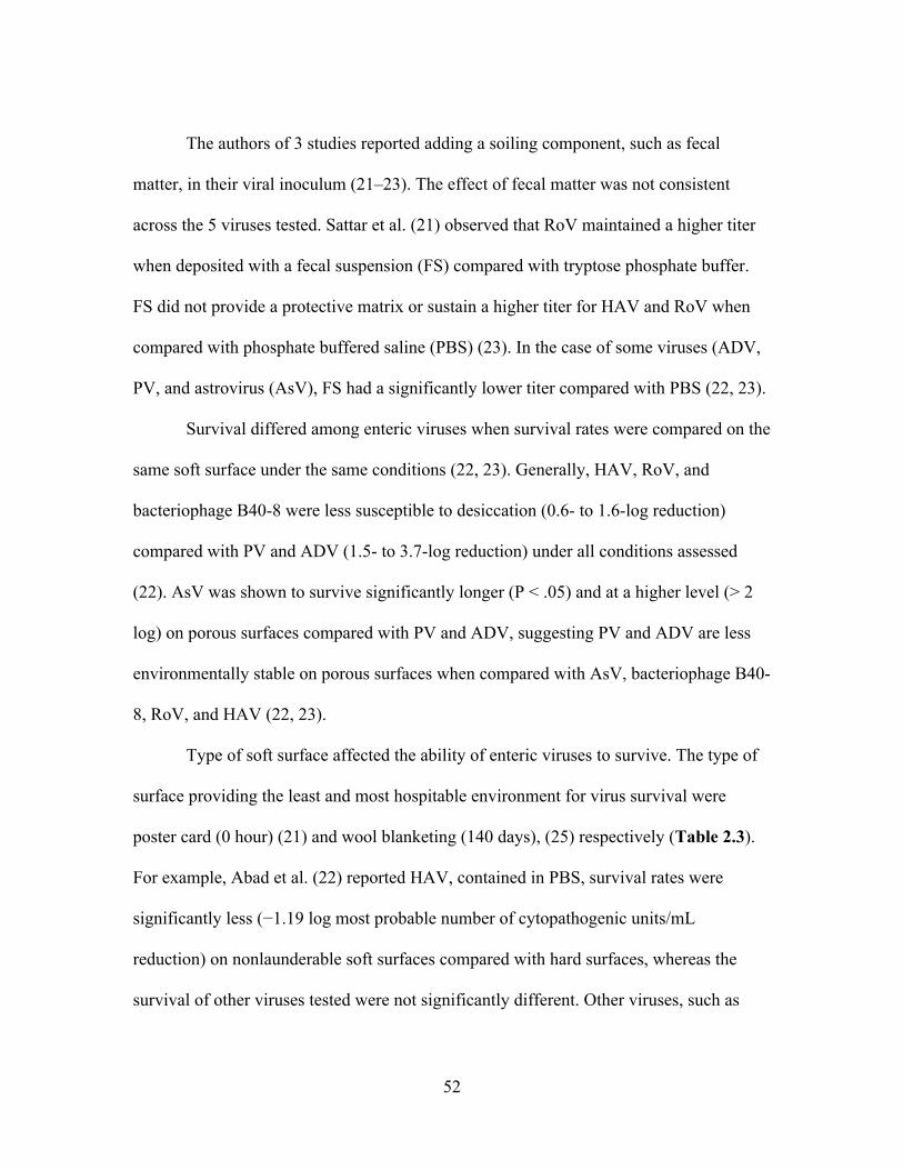
52
The authors of 3 studies reported adding a soiling component, such as fecal
matter, in their viral inoculum (21–23). The effect of fecal matter was not consistent
across the 5 viruses tested. Sattar et al. (21) observed that RoV maintained a higher titer
when deposited with a fecal suspension (FS) compared with tryptose phosphate buffer.
FS did not provide a protective matrix or sustain a higher titer for HAV and RoV when
compared with phosphate buffered saline (PBS) (23). In the case of some viruses (ADV,
PV, and astrovirus (AsV), FS had a significantly lower titer compared with PBS (22, 23).
Survival differed among enteric viruses when survival rates were compared on the
same soft surface under the same conditions (22, 23). Generally, HAV, RoV, and
bacteriophage B40-8 were less susceptible to desiccation (0.6- to 1.6-log reduction)
compared with PV and ADV (1.5- to 3.7-log reduction) under all conditions assessed
(22). AsV was shown to survive significantly longer (P < .05) and at a higher level (> 2
log) on porous surfaces compared with PV and ADV, suggesting PV and ADV are less
environmentally stable on porous surfaces when compared with AsV, bacteriophage B40-
8, RoV, and HAV (22, 23).
Type of soft surface affected the ability of enteric viruses to survive. The type of
surface providing the least and most hospitable environment for virus survival were
poster card (0 hour) (21) and wool blanketing (140 days), (25) respectively (Table 2.3).
For example, Abad et al. (22) reported HAV, contained in PBS, survival rates were
significantly less (−1.19 log most probable number of cytopathogenic units/mL
reduction) on nonlaunderable soft surfaces compared with hard surfaces, whereas the
survival of other viruses tested were not significantly different. Other viruses, such as
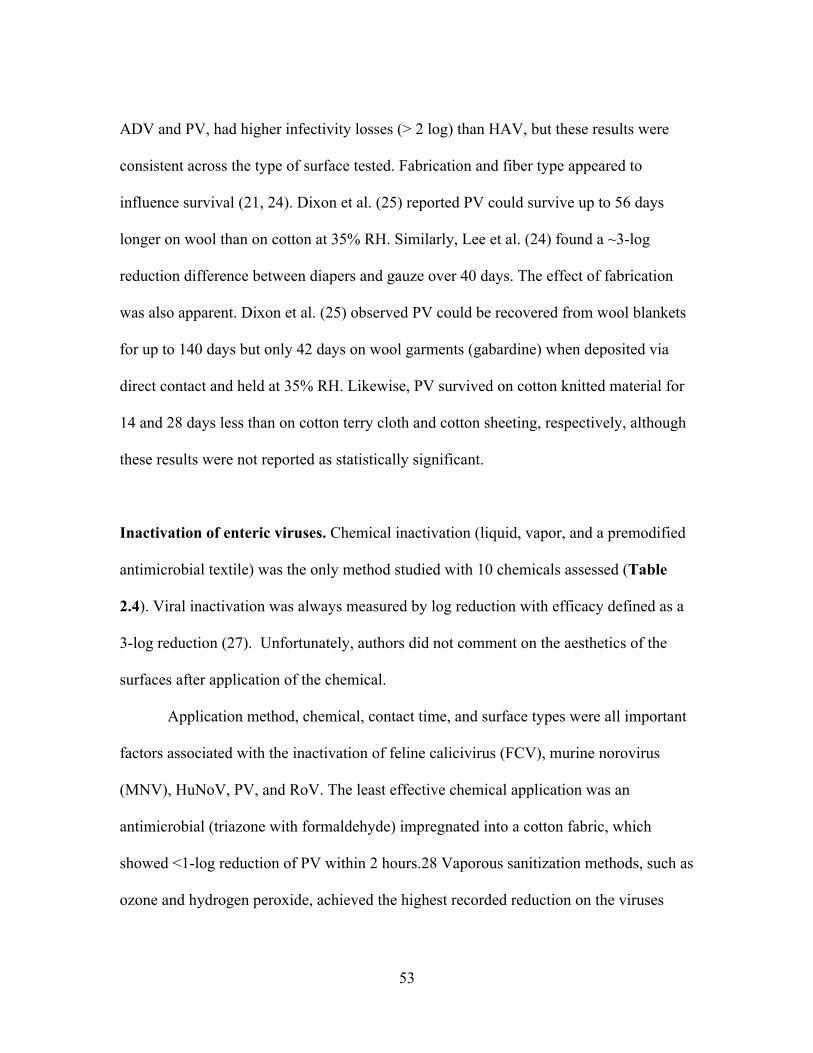
53
ADV and PV, had higher infectivity losses (> 2 log) than HAV, but these results were
consistent across the type of surface tested. Fabrication and fiber type appeared to
influence survival (21, 24). Dixon et al. (25) reported PV could survive up to 56 days
longer on wool than on cotton at 35% RH. Similarly, Lee et al. (24) found a ~3-log
reduction difference between diapers and gauze over 40 days. The effect of fabrication
was also apparent. Dixon et al. (25) observed PV could be recovered from wool blankets
for up to 140 days but only 42 days on wool garments (gabardine) when deposited via
direct contact and held at 35% RH. Likewise, PV survived on cotton knitted material for
14 and 28 days less than on cotton terry cloth and cotton sheeting, respectively, although
these results were not reported as statistically significant.
Inactivation of enteric viruses. Chemical inactivation (liquid, vapor, and a premodified
antimicrobial textile) was the only method studied with 10 chemicals assessed (Table
2.4). Viral inactivation was always measured by log reduction with efficacy defined as a
3-log reduction (27). Unfortunately, authors did not comment on the aesthetics of the
surfaces after application of the chemical.
Application method, chemical, contact time, and surface types were all important
factors associated with the inactivation of feline calicivirus (FCV), murine norovirus
(MNV), HuNoV, PV, and RoV. The least effective chemical application was an
antimicrobial (triazone with formaldehyde) impregnated into a cotton fabric, which
showed <1-log reduction of PV within 2 hours.28 Vaporous sanitization methods, such as
ozone and hydrogen peroxide, achieved the highest recorded reduction on the viruses
54
they assessed (3 log->5 log) (28, 29). RoV was the only virus tested that did not achieve a
3-log reduction when using a vaporous chemicals (hydrogen peroxide > 2 log). A liquid
version of this chemistry, accelerated hydrogen peroxide, had an effect on FCV (>3 log)
but a limited effect on MNV (0.17 and 0.57 log) on cotton and polyester, respectively
(30). This same study found that sodium hypochlorite (5,000 ppm) was effective at
reducing infectious FCV and MNV by > 3 log on both surfaces within 5 minutes. Malik
et al. (31) extensively investigated the effect of 5 liquid chemicals against FCV on a
variety of fabrics and carpets. Only 1 liquid chemical (activated 2.6% glutaraldehyde)
achieved a >3-log reduction of FCV on all test surfaces, excluding blended carpet. FCV
was easier to inactivate on fabrics than carpets with the exception of 100% polyester
fabric. Their data demonstrated a positive linear relationship between contact time and
inactivation.
DISCUSSION
The aim of this systematic review was to determine factors that influence the
survival and inactivation of enteric viruses on nonlaunderable soft surfaces.
Findings from the articles presented in our review were derived from well-
designed research studies as determined by our quality assessment tool. Our analysis
suggests temperature and RH are important extrinsic factors associated with virus
survival in a manner similar to that of bacterial survival. We also determined, from the
current literature, that deposition method and organic content are contributing factors to
enteric virus survival on nonlaunderable soft surfaces. Furthermore, enteric viruses were
55
resistant, under controlled conditions, to a variety of chemistries that are otherwise
efficacious against other microorganisms, such as enveloped viruses and vegetative
bacteria (32, 33). Other published reviews reported similar conclusions for survival of
viruses on a variety of surfaces, such as porous, hard nonporous surfaces, and produce
(18–20). However, these reviews did not address the collective knowledge regarding
enteric virus survival and inactivation on nonlaunderable soft surfaces. Controlling
transmission through the elimination of enteric viruses on environmental surfaces,
including nonlaunderable soft surfaces, is an important (and overlooked) step in reducing
the burden of illness, especially in semienclosed indoor environments, a common setting
for outbreaks of AGE. Results must be interpreted with caution because the duration of
survival studies varied and viruses may survive longer than the
reported time. For example, the longest low-temperature survival study was conducted
for 90 days compared with 140 days for ambient temperature (23, 25). As such, the
authors of ambient temperature studies reported longer survival times.
Factors associated with survival. As expected, temperature is, generally, inversely
related to enteric virus survival on nonlaunderable soft surfaces. This survival trend is
similar to results reported about survival on hard nonporous surfaces, (18–20) which
might explain the seasonality of outbreaks attributed to enteric viruses. Lower
temperatures, a common method of preservation for enteric viruses and other
microorganisms, can slow or halt biological and chemical activities in the liquid in which
they are contained. However, nonlaunderable surfaces of concern, such as mattresses or

56
carpets, are not typically found at low temperatures (−20°C – 4°C). The most alarming
result was how long enteric viruses survived (60-140 days) at ambient temperatures. This
is of concern given that nonlaunderable soft surfaces are commonly found at ambient
temperatures and in indoor settings so individuals can come in contact with contaminated
surfaces repeatedly. Some viruses, such as RoV, demonstrated enhanced survival at
ambient temperatures. One plausible explanation for the increased survival of RoV is that
their genome and structure differ from other common enteric viruses because it is
composed of an intermediate and inner capsid containing a double-stranded RNA
segmented genome (34). These structural features may explain the ability of RoV to have
higher survival rates at ambient temperatures
compared with other enteric viruses. Although this trait is worrisome this should not
lessen our concern of other enterics.
The effect of RH on virus survival varied, particularly by virus type and
temperature, based on the literature included in our literature analysis. As described
elsewhere, (35) nonenveloped viruses survive better at higher RH compared with
enveloped viruses that favor lower RH. Typically, indoor environments range from 40%
to 70% RH, which was measured in several studies in this review (36). Theoretically,
high RH could limit evaporation and desiccation and positively influence the survival of
nonenveloped viruses. Evaporation of water molecules from airborne bacteria has been
shown to decrease survival by removing essential water molecules (37). The studies
investigating deposition method support this idea (25, 26). Moreover, this trend is
directly related to the amount of liquid present during deposition. Aerosolized particles

57
can form droplet nuclei that stay suspended for extended periods of time so have less
liquid than
do droplets or virus directly applied to a surface (26). The reduced liquid content
associated with droplet nuclei also makes the particles more susceptible to desiccation.
As postulated previously (38), the displacement of water essential to the viral capsid
could increase inactivation.
These forms of deposition can be represented by vomiting, a common method of soft
surface contamination by enteric viruses (9–12). The act of vomiting has been shown to
deposit enteric viruses via direct contact and aerosolization at distances > 3 meters (39).
The hypothesis
that higher liquid volume content equals longer survival rate could explain the increased
survival at higher RH. However, there are exceptions to this trend. The discrepancies in
survival across studies within this review (21, 23, 25) may be attributed to experimental
factors, such as low RH percentages ranging from 35% to ~50% RH, inoculation
composition, surface type, and strain type. Thus, it is difficult to state a common trend for
RH effects on enteric viruses
contaminated on soft surfaces with the current literature.
Mixing viruses with solutions containing organic matter, such as fetal bovine
serum or FS, is a common practice because it can mimic the fecal-oral or vomitus-oral
route. Therefore, it is assumed that soft surfaces contaminated with an enteric virus will
also be contaminated with organic matter (typically from vomitus or diarrhea). Organic
matter provides a protective matrix for bacterial and viral pathogens, making it more

58
difficult to eliminate them (18, 24, 40, 41). The varied effect of organic content presented
in this review is unknown. However, speculations point to the adsorption state of the
virus on a particular surface. When viruses are deposited onto a soft surface they are
subjected to a variety of factors, such as adsorption ability of the material, electrostatic
interactions, and can be complicated by the solution characteristics; that is, ionic strength
(41, 42). Outside of their host, adsorbed enteric viruses are more stable under
environmental conditions than free, unbound, virus (42). Gerba (42) stated that due to the
low isoelectric point, organic matter may compete with viruses for available adsorption
sites and as a result the adsorption ability of a particular virus can aid in predicting its
inactivation. The organic component of the FS used by Abad et al (22, 23) may have
competed for adsorption sites, resulting in shorter survival. Moreover, it is conceivable
that fecal constituents may have interfered with the cell culture system, inhibited elution
for AsV, PV, and ADV, or had a synergistic effect across fecal components, virus, and
the soft surface, although no data support these hypotheses (22, 23). Nevertheless, these
results are in contrast to the accepted theory that organic material-containing solutions
provide a protective environment for enteric viruses. Because the sources of enteric
viruses contaminated on soft surfaces are usually vomitus or diarrhea, this subject
warrants further investigation.
The relationship between surface type and virus is inconclusive because too many
surfaces were used to aggregate the findings. Inferences could only be made in regard to
some specific surface types and the survival of some enteric viruses. Results were as
expected for the natural hygroscopic fibers, wool, and cotton. Wool retained PV up to 56
59
days longer than cotton, presumably because of its ability to absorb more liquid than
cotton (43), a factor not studied when assessing hard nonporous surfaces. Materials that
have a higher adsorption capacity have been shown to positively influence virus survival
compared to surfaces with low absorption capacity as seen by the differences in MNV
and RoV survival (21, 24).
Interestingly, the higher PV survival rate (98 days) on wool blanketing compared
with wool garments suggests the construction of a material influences virus survival (25).
To explain this phenomenon, the authors of a single study (14) found that loose fabrics
transfer more virus than denser fabrics and speculate that loose fabrics may not adsorb
virus as readily compared to denser fabrics which creates higher amounts of free,
unbound virus susceptible to desiccation.
As stated previously (14, 20, 41, 44), viruses are more likely to remain infectious if
adsorbed. This is due to viruses being immobilized and protected from environmental
factors such as temperature and solution characteristics (eg pH, ionic strength) that would
otherwise inactivate the virus (45).
The use of different elution buffers was also mentioned by Dixon et al (25) and
may explain the higher survival rates between surface types. Recovery efficiencies may
decrease depending upon the eluent characteristics (eg, pH). Buffer type and the method
used for viral recovery were shown to produce different elution efficiencies as they relate
to MNV (24). For example, Abad et al (22) reported their elution buffer to be pH ~7.4.
As a result, under those conditions, RoV would theoretically be positively charged (pI,
~8.0), whereas HAV would be negatively charged (pI, ~ 2.8) (45) These differences

60
could explain why some enteric viruses survived longer or at higher levels on surfaces,
such as the ability of HAV to survive significantly longer on hard nonporous surfaces
than on soft surfaces while other viruses, in the same study, survived longer and at higher
titers on soft surfaces compared to hard nonporous surfaces. To support this hypothesis, a
more recent study (30) reported a significantly different recovery efficiency of enteric
viruses from cotton compared with other surfaces (ie, glass and polyester fabric), even
when destructive sampling methods were used, such as sonication and stomaching. One
important factor not mentioned in the studies reviewed was the material’s surface charge.
Knowing the charge of a virus and the surface are mutually important for understanding
elution patterns. It is possible that these differences could affect elution and adsorption to
the soft surface and may account for the difference in reported survival times. Overall,
the results of this review were based on contact time and did not account for all human
enteric viruses discussed in this review. Moreover, we do not believe these results can be
generalized to all enteric viruses because the resiliency differs among them. The current
literature underscores the need to study survival profiles of multiple enteric viruses under
the same conditions to determine environmental resilience. Furthermore, investigating
multiple enteric viruses can aid in determining potential surrogates for nonculturable
viruses, such as human noroviruses. Lastly, knowledge of survival time can improve
retrospective epidemiologic investigations and aid in selection of better chemistries to
remove enteric viruses from nonlaunderable soft surfaces.

61
Inactivation. To reduce the burden of illness associated with enteric viruses we must
understand the risks associated with contaminated surfaces in the environment by
establishing survival profiles. The next logical step is to develop an effective pathogen
inactivation treatment that could reduce or eliminate the risks. Enteric viruses of public
health importance, such as HuNoV, HAV, RoV, PV, and AsV, lack an envelope, making
them environmentally stable and resistant to much of the common chemistry used for
inactivation (46, 47). For instance, triazone resin containing formaldehyde, the least
efficacious reduction method tested on porous surfaces, was designed to make fabrics
quick-dry so in theory the resin could lead to greater inactivation during viral attachment
(48). The resin demonstrated some virucidal activity against PV when compared with a
control. However, the reduction of PV was too slow to be considered as a sanitizer or
disinfectant (<99% in 1 day). These data suggest that active ingredients, if any, present in
the resin are not efficacious against PV or concentrations were too low to have a quicker
effect.
Other chemicals tested, like those from Malik et al. (31) did not reduce the
surrogate for HuNoV adequately due to their lipophilic mode of action. The most
efficacious chemicals tested were liquid and vaporous hydrogen peroxide, vaporous
ozone, and sodium hypochlorite. These compounds are strong oxidizing agents that
damage the viral capsid, nucleic acid, or a combination of both resulting in virucidal
efficacy (49). Liquid hydrogen peroxide, although efficacious against FCV, may cause
superficial damage to soft surfaces and would not be an appropriate treatment. In
addition, hydrogen peroxide was shown to be ineffective against MNV depending on
62
concentration and contact time, which may discredit its efficacy against HuNoV (30).
Unlike liquids, vaporous chemicals, such as ozone, may be an important tool for
sanitizing soft surfaces. Vaporous chemicals can penetrate deeper into a surface
compared with a liquid surfactant product alone. These products are also ideal for large
institutional settings, such as long-term-care facilities and hospital wards, but are not
recommended for household use due to chemical hazard concerns. Manufacturers and
consumers should not consider only the pathogen-chemical interaction, but also the soft
surface-chemical interaction.
An observation noted by Malik et al. (31) was the difference in inactivation when
using the same chemical compound on different types of surfaces. The limited efficacy of
these products may be linked to the surface to which they are applied due to chemical
adsorption. Some studies have characterized the degree to which soft surfaces interact
with the active ingredient of a disinfectant solution (50–52). For example, cotton, wool,
and polyester were shown to remove quaternary ammonium compounds, sodium
hypochlorite, and benzalkonium chloride, respectively, from solutions until the
adsorptive capacity of the fibers was met (50–52). Due to the large variety of both soft
surfaces and disinfectants, these relationships are not universally defined. However, they
are an essential aspect in regard to sanitization of soft surfaces because efficacy can vary
based on the soft surface. The most effective liquid chemical in this study, tested by
Malik et al. (31) was 2.6% activated glutaraldehyde, a widely used disinfectant in
hospitals that has been shown to induce alkylation of key functional groups and amino
acids on a variety of bacteria (49). These reactions are capable of damaging the integrity

63
of the viral capsid and genome. Glutaraldehyde has also been shown to resist adsorption
by fibers, which may be another reason for its broad effectiveness (52).
At present, bleach is the only chemical for removal of enteric viruses of
importance, such as HuNoV, recommended by the Centers for Disease Control and
Prevention. Bleach has been shown to be effective at reducing infectious MNV and FCV,
2 surrogates for HuNoV, on both hard and soft surfaces and is the current recommended
tool for eliminating HuNoV from hard nonporous surfaces (30, 53). However, bleach
cannot be used for contaminated nonlaunderable soft surfaces as bleach will adversely
affect the surfaces aesthetic appearance by, oxidizing the surface and damaging the
fibers. These same adverse effects are also true for other chemistries, such as hydrogen
peroxide, tested on soft surfaces. Another commonly recommended method is steam
cleaning at a minimum of 70°C or 100°C for 5 minutes or 1 minute, respectively, but the
efficacy of this method has not been determined under controlled conditions (54).
Therefore, a limited number of tools are available to inactivate enteric viruses on
nonlaunderable soft surfaces.
FUTURE RESEARCH
Because experimental evidence is limited in regard to survival and inactivation of
enteric viruses on soft surfaces, this review suggests the need for additional studies.
Future research should consult previously used methods and experimental designs.
Investigators should also assess ≥ 2 enteric viruses under environmentally relevant
conditions with surfaces that can easily be characterized and procured. Inactivation
64
studies should investigate chemistries that are efficacious and known to not affect the
aesthetic appearance.
CONCLUSIONS
The literature shows that several key factors influence survival: temperature, RH,
organic content, deposition method, and virus type. Of the studies that evaluated different
chemistries, it was shown that chlorine, glutaraldehyde, and oxidizing vaporous products
demonstrated the best efficacy against enteric viruses on soft surfaces. Because the EPA
currently does not have a standard method for assessing virucidal activity of product on
nonlaunderable soft surfaces, there are no registered products that can be reported to
inactivate enteric viruses contaminated on nonlaunderable soft surfaces.
REFERENCES
1. Chow CM, Leung AKC, Hon KL. 2010. Acute gastroenteritis: From guidelines to
real life. Clin Exp Gastroenterol 3:97–112.
2. Hall AJ, Lopman B, Payne DC, Patel MM, Gastañaduy P a., Vinjé J, Parashar UD.
2013. Norovirus disease in the united states. Emerg Infect Dis 19:1198–1205.
3. Payne DC, Vinjé J, Szilagyi PG, Edwards KM, Staat MA, Weinberg GA, Hall CB,
Chappell J, Bernstein DI, Curns AT, Wikswo ME, Shirley SH, Hall AJ, Lopman
B, Parashar UD. 2013. Norovirus and Medically Attended Gastroenteritis in U.S.
Children. N Engl J Med 368:1121–1130.
4. Kowalzik F, Riera-Montes M, Verstraeten T, Zepp F. 2015. The burden of

65
norovirus disease in children in the European union. Pediatr Infect Dis J 34:229–
34.
5. Bosch A. 1998. Human enteric viruses in the water environment: a minireview. Int
Microbiol 1:191–196.
6. Barker J, Stevens D, Bloomfield SF. 2001. Spread and prevention of some
common viral infections in community facilities and domestic homes. J Appl
Microbiol 91:7–21.
7. Hota B. 2004. Contamination, disinfection, and cross-colonization: are hospital
surfaces reservoirs for nosocomial infection? Clin Infect Dis 39:1182–1189.
8. Springthorpe VS, Sattar SA. 1990. Chemical disinfection of virus-contaminted
surfaces. Crit Rev Environ Control 20:169–229.
9. Evans MR, Meldrum R, Lane W, Gardner D, Ribeiro CD, Gallimore CI,
Westmoreland D. 2002. An outbreak of viral gastroenteritis following
environmental contamination at a concert hall. Epidemiol Infect 129:355–360.
10. Thornley CN, Emslie NA, Sprott TW, Greening GE, Rapana JP. 2011. Recurring
norovirus transmission on an airplane. Clin Infect Dis 53:515–520.
11. Cheesbrough JS, Barkess-Jones L, Brown DW. 1997. Possible prolonged
environmental survival of small round structured viruses. J Hosp Infect 35:325–
326.
12. Cheesbrough JS, Green J, Gallimore CI, Wright PA, Brown DW. 2000.
Widespread environmental contamination with Norwalk-like viruses (NLV)
detected in a prolonged hotel outbreak of gastroenteritis. Epidemiol Infect 125:93–

66
98.
13. Repp KK, Keene WE. 2012. A point-source norovirus outbreak caused by
exposure to fomites. J Infect Dis 205:1639–1641.
14. Gibson KE, Crandall PG, Ricke SC. 2012. Removal and transfer of viruses on
food contact surfaces by cleaning cloths. Appl Environ Microbiol 78:3037–3044.
15. Lopez GU, Gerba CP, Tamimi AH, Kitajima M, Maxwell SL, Rose JB. 2013.
Transfer efficiency of bacteria and viruses from porous and nonporous fomites to
fingers under different relative humidity conditions. Appl Environ Microbiol
79:5728–5734.
16. Hsieh Y-L, Timm D. 1988. Relationship of substratum wettability measurements
and initial Staphylococcus aureau adhesion to films and fabrics. J Colloid Interface
Sci 123:275–286.
17. Hsieh Y-L, Yu B. 1992. Liquid Wetting, Transport, and Retention Properties of
Fibrous Assemblies: Part I: Water Wetting Properties of Woven Fabrics and Their
Constituent Single Fibers. Text Res J 62:677–685.
18. Boone SA, Gerba CP. 2007. Significance of fomites in the spread of respiratory
and enteric viral disease. Appl Environ Microbiol 73:1687–1696.
19. Rzezutka A, Cook N. 2004. Survival of human enteric viruses in the environment
and food. FEMS Microbiol Rev 28:441–453.
20. Vasickova P, Pavlik I, Verani M, Carducci a. 2010. Issues concerning survival of
viruses on surfaces. Food Environ Virol 2:24–34.
21. Sattar SA, Lloyd-Evans N, Springthorpe VS, Nair R. 1986. Institutional outbreaks

67
of rotavirus diarrhoea: potential role of fomites and environmental surfaces as
vehicles for virus transmission. J Hyg (Lond) 96:277–289.
22. Abad FX, Pintó RM, Bosch A. 1994. Survival of enteric viruses on environmental
fomites. Appl Environ Microbiol 60:3704–3710.
23. Abad FX, Villena C, Guix S, Caballero S, Pintó RM, Bosch A. 2001. Potential
Role of Fomites in the Vehicular Transmission of Human Astroviruses. Appl
Environ Microbiol 67:3904–3907.
24. Lee JE, Zoh KD, Ko GP. 2008. Inactivation and UV disinfection of murine
norovirus with TiO2 under various environmental conditions. Appl Environ
Microbiol 74:2111–2117.
25. Dixon GJ, Sidwell RW, Mcneil E. 1966. Quantitative Studies on Fabrics of
Viruses Disseminators. II. Persistence of poliomyelitis virus on cotton and wool
fabrics. Appl Microbiol 14:183–188.
26. Fisher E, Shaffer R. 2010. Survival of bacteriophage MS2 on filtering facepiece
respirator coupons. J Am Biol Saf Assoc 15:72–76.
27. EPA. 2012. Product Performance Test Guidelines OCSPP 810.2400: Disinfectants
and Sanitizer for Use on Fabrics and Textiles.
28. Hudson JB, Sharma M, Petric M. 2007. Inactivation of Norovirus by ozone gas in
conditions relevant to healthcare. J Hosp Infect 66:40–45.
29. Tuladhar E, Terpstra P, Koopmans M, Duizer E. 2012. Virucidal efficacy of
hydrogen peroxide vapour disinfection. J Hosp Infect 80:110–115.
30. Yeargin T, Fraser A, Guohui H, Jiang X. 2015. Recovery and disinfection of two

68
human norovirus surrogates, feline calicivirus and murine norovirus, from hard
nonporous and soft porous surfaces. J Food Prot 78:1842–1850.
31. Malik YS, Allwood PB, Hedberg CW, Goyal SM. 2006. Disinfection of fabrics
and carpets artificially contaminated with calicivirus: relevance in institutional and
healthcare centres. J Hosp Infect 63:205–210.
32. Shirai J, Kanno T, Tsuchiya Y, Mitsubayashi S, Seki R. 2000. Effects of chlorine,
iodine, and quaternary ammonium compound disinfectants on several exotic
disease viruses. J Vet Med Sci 62:85–92.
33. Ioannou CJ, Hanlon GW, Denyer SP. 2007. Action of disinfectant quaternary
ammonium compounds against Staphylococcus aureus. Antimicrob Agents
Chemother 51:296–306.
34. Pesavento JB, Crawford SE, Estes MK, Prasad B V. 2006. Rotavirus proteins:
Structure and assembly. Reoviruses Entry, Assem Morphog 309:189–219.
35. Sobsey MD, Meschke JS. 2003. Virus survival in the environment with special
attention to survival in sewage droplets and other environmental media of fecal or
respiratory origin. Rep World Heal Organ Geneva, Switz 70 pages.
36. Arundel A V, Sterling EM, Biggin JH, Sterling TD. 1986. Indirect Health Effects
of Relative Humidity in Indoor Environments 65:351–361.
37. Webb SJ. 1960. Factors affecting the viability of air-borne bacteria. III. the role of
bonded water and protein structure in the death of air-borne cells. Can J Microbiol.
38. Yang W, Marr LC. 2012. Mechanisms by which ambient humidity may affect
viruses in aerosols. Appl Environ Microbiol 78:6781–8.

69
39. Makison, Booth C, Booth C. 2014. Vomiting Larry: a simulated vomiting system
for assessing environmental contamination from projectile vomiting related to
norovirus infection. J Infect Prev 15:176–180.
40. Kiseleva LF. 1971. Survival of poliomyelitis, ECHO and coxsackie viruses in
some food products. Vopr Pitan 30:58–61.
41. Hurst CJ, Gerba CP, Chech I. 1980. Effects of environmental variables and soil
characteristics on virus survival in soil 40:1067–1079.
42. Gerba CP, Goyal SM, Cech I, Bogdan GF. 1981. Quantitative assessment of the
adsorptive behavior of viruses to soils. Environ Sci Technol 15:940–944.
43. Hailwood AJ, Horrobin S. 1946. Absorption of water by polymers: analysis in
terms of a simple model. Trans Faraday Soc 84–92.
44. Da Silva AK, Kavanagh O V., Estes MK, Elimelech M. 2011. Adsorption and
aggregation properties of norovirus GI and GII virus-like particles demonstrate
differing responses to solution chemistry. Environ Sci Technol 45:520–526.
45. Michen B, Graule T. 2010. Isoelectric points of viruses. J Appl Microbiol
109:388–397.
46. McDonnell G, Russell AD. 1999. Antiseptics and Disinfectants: Activity, Action,
and Resistance. Clin Microbiol Rev 12:147–179.
47. Masri NM, Hanbali LB, Kamar AH, Kanafani LMS, Hanbali MB, Haddad JJ.
2013. The Immunomodulatory, Antimicrobial and Bactericidal Efficacy of
Commonly Used Commercial Household Disinfectants, Sterilizers and Antiseptics
in Vitro: Putative Anti-Inflammatory Infection Control Mechanisms and

70
Comparative Biochemic. Am J Med Biol Res 1:103–133.
48. Sidwell RW, Dixon GJ, McNeil E. 1967. Quantitative studies on fabrics as
disseminators of viruses. III. Persistence of vaccinia virus on Fabrics Impregnated
with a Virucidal Agent. Appl Microbiol 15:921–927.
49. Rutala W, Weber DJ. 2008. Guideline for disinfection and sterilization in
healthcare facilities.
50. Goldsmith MT, Latlief M a, Friedl JL, Stuart LS. 1954. Adsorption of available
chlorine and quaternary by cotton and wool fabrics from disinfecting solutions.
Appl Microbiol 2:360–364.
51. McNeil E, Greenstein M, Stuart LS, Goldsmith MT. 1960. Some Problems
Involved in the Use of Quaternary Ammonium Compounds as Fabric
Disinfectants. Appl Microbiol 8:156–159.
52. Bloß R, Meyer S, Kampf G. 2010. Adsorption of active ingredients of surface
disinfectants depends on the type of fabric used for surface treatment. J Hosp
Infect 75:56–61.
53. Hall AJ, Vinjé J, Lopman B, Park GW, Yen C, Gregoricus N, Parashar UD,
Diseases R. 2011. Updated norovirus outbreak management and disease
prevention guidelines. Morb Mortal Wkly Rep 60:1–18.
54. OSHA. 2008. OSHA Fact Sheet - Noroviruses.
55. Fijan S, Steyer A, Poljšak-Prijatelj M, Cencič A, Šostar-Turk S, Koren S. 2008.
Rotaviral RNA found on various surfaces in a hospital laundry. J Virol Methods
148:66–73.

71

72
Table 2.1 Search strings used for online databases
Research questions
Virus Surface Role
Research question 1
Virus AND Fabric Or Textile OR soft surface OR porous surface OR fomite OR upholstery OR carpet OR rug OR
draper
AND Persist OR surviv OR recover OR transmission
Research question 2
Virus AND Fabric OR textile OR soft surface OR porous surface OR fomite OR upholstery OR carpet OR rug OR
draper
AND disinfect OR sanitize OR clean

73
Table 2.2 Quality assessment of eligible articles (n=12) based on 5 criteria. Quality Assessment Criteria Pass Fail %Pass
Used adequate controls 12 0 100% Clear experimental design 10 2 83% Appropriate quantification technique employed 10 2 83% Experiment done in duplicate and more than 1 trial completed 10 2 83% Statistical analysis completed 8 4 67% Total 50 10 Avg: 83%

74
Table 2.3 Survival of enteric viruses on soft surfaces.
SurfacesA Inoculation
LevelB Detection MethodC VirusD TreatmentF Significant findingsF Reference
Fabric: WB, WG, CS, CTC, CJK
~5~10 log CCID50
•CCID50 PV Temp: 25 °C RH: 35 and 78% DM: Aerosol, direct, VCD SD: 140 d
35% RH: Survived on wool for 140 d & on cotton 84 d. 78% RH: Survived on wool 84 d & on cotton 42 d. Rapid decrease in titer on cotton under both 35 and 78% RH. Better survival via direct contact > aerosol > VCD. Better survival on WG > CTC > CKJ > CS
Dixon et al. (25)
CM: Poster card, Paper currency, Paper Fabric: Cotton-polyester
~5.3 pfu •Plaque assay
RoV Temp: 4 and 22˚C RH: 25, 50 and 85% IC: FS SD: 12 d
RoV survived for 2 and 10 d at 22 and 4˚C on cotton-polyester. No RoV detected on poster card due to inhibition. Titers too low & variable to include for paper currency results. 50-80% recovery on paper after 3 h & no long-term samples taken.
Sattar et al. (21)
CM: FFR 7 log pfu •Plaque assay
MS2 Temp: 22˚C RH: 30% DM: Aerosol & direct SD: 10 d
MS2 detected up to 10 d. Direct contact showed higher (5.7 log pfu) terminal survival than aerosol (3.2 log).
Fisher and Shaffer (26)
CM: Paper Fabric: Cotton
Not described •MPNCU ADV B40-8 HAV PV
RoV
Temp: 4 and 20˚C RH: 90 (-4˚C), 85 and 50% (20˚C) IC: PBS or 20% FS SD: 60 d
RoV>HAV>B40-8>PV>ADV. PV/ADV sig. inactivated by FS. Excluding RoV, all survival sig. enhanced at 4˚C. Viruses’ survived for 60 d in all conditions, except when suspended in FS (7-60 d). RoV survived longer in high RH. HAV and B40-8 decreased survival on soft surfaces compare to hard surfaces.
Abad et al. (22)
CM: Paper 5~5.7 log MPN
•IFT •MPNCU •RT-PCR
AsV
Temp: 4 and 20˚C RH: 90% IC: PBS or 20% FS SD: 90 d
4°C: AsV survived up to 90 d in PBS & FS. 20°C: AsV survived up to 60 & 7 d in PBS & FS.
Abad et al. (23)
ADV AsV
HAV PV
RoV
Temp: 4 and 20˚C at RH: 90% IC: 20% FS or PBS SD: 7 d
RoV/HAV > AsV > PV/ADV. AsV not sig. inactivated by FS at 4 & 20˚C. No sig increase for RoV survival at 4˚C. RoV/HAV similar decay rates. PV & ADV decreased survival at 20˚C in FS.
CM: Diapers Fabrics: Gauze
8 log pfu •Plaque assay •qRT-PCR
MNV Temp: -20, 4, 18 and 30 °C SD: 40 d
-20°C: <2 & <1 log reduction on gauze & diapers after 40 d, respectively. 4°C: 2 & <2 log reduction on gauze & diapers after 30 & 40 d, respectively. 18 and 30°C: 3 logs after 1 d on both surfaces.
Lee et al. (24)
Fabric: cotton textile fabric
~7~4 log RT-PCR units
•RT-PCR RoV Temp: Room temp SD: 24 h
After 24 h RoV RNA detected on cotton after traditional nested PCR for all inoculation levels, except disinfection control.
Fijan et al. (55)
A: WB: wool blanket, WG: wool gabardine, CS: cotton sheeting, CTC: cotton, terry cloth, CJK: cotton jersey knit, CM: complex matrix, FFR: filter face piece respirator

75
B: pfu: plaque-forming units, CCID50: 50% cell culture infectious dose, RT-PCR: reverse transcriptase-polymerase chain reaction. C: TCID50: 50% tissue culture infectious dose, MPNCU: most probable number cytopathogenic units, IFT: indirect immunofluorescences test, qRT-PCR: quantitative reverse transcriptase-polymerase chain reaction D: PV: poliovirus, RoV: human rotavirus, MNV: murine norovirus, HAV: hepatitis A virus, ADV: enteric adenovirus, AsV: astrovirus E: Non human enteric viruses: MS2, B40-8, and MNV F: RH: relative humidity, DM: deposition method, SD: study duration, IC: inoculum composition, FS: fecal suspension

76
Table 2.4. Inactivation of enteric viruses on soft surfaces.
Surface
Inoculum levelA
Detection methodB VirusC TreatmentD
Significant Results Reference
Fabric: Cotton “wash-and wear” with Triazone resin
10 log CCID50
•CCID50 PV Chemical: Material impregnated with Triazone resin Contact time: 10 d RH: 35 and 78%
PV survived up to 7 d on impregnated cotton.
Sidwell et al. (48)
Fabric: cotton, polyester, cotton polyester blend. Carpets: olefin, polyester, nylon/olefn blend
8 log TCID50
•TCID50 FCV Chemical: Activated 2.6% glutaraldehyde, 4.75% o-benzyl p-chlorophenol + 4.75% o-phenylphenol, 10% NaHCO3 + 10% quaternary ammonium compound, 70% isopropanol, 2.5% NaHCO3 + 1.3% glutaraldehyde Contact time—1, 5, and 10 min.
Activated dialdehyde product achieved 99.9% reduction on all surfaces, except blended carpet. All other products <99.86. Polyester least amenable fabric.
Malik et al. (31)
Fabric-- Cotton, fabric (undefined) Carpet-- Undefined
FCV: 5.7~6.3 log pfu HuNoV: Not described
•Plaque assay, •qRT-PCR •TCID50
FCV HuNoV
Chemical: 20-25 ppm ozone with 5 min vapor burst Contact time: 20 min
>3 log reduction in infectivity for FCV and RNA for HuNoV.
Hudson et al. (28)
Fabric: Gauze
PV: 7.7 TCID50
RoV: 5.5 TCID50
MNV: 6 log pfu
•Plaque assay •TCID50
PV RoV
MNV
Chemical: 127 ppm hydrogen peroxide vapor Contact time: 1 h
>5, >3, >2 log reduction of PV, MNV, RoV
Tuladhar et al. (29)
Fabric: Cotton, Polyester
FCV: ~6.6 log pfu MNV: ~6.3 log pfu
•Plaque Assay •qRT-PCR
FCV MNV
Chemical: 5000 ppm NaOCl, 2656 ppm AHP Contact time: 5 min
NaOCl >3 log reduction of FCV, MNV on both surfaces. AHP 0.57, 0.17 log reduction of MNV on polyester & cotton. >3 & >2 log reduction of FCV on polyester & cotton
Yeargin et al. (30)
A: CCID50: 50% cell culture infectious dose, TCID50: 50% tissue culture infectious dose, pfu: plaque forming units, FCV: feline calicivirus, HuNoV: human norovirus, PV: poliovirus, RoV: rotavirus, MNV: murine norovirus B: qRT-PCR: quantitative reverse transcriptase-polymerase chain reaction C: non-human enteric viruses: FCV and MNV D: AHP: activated hydrogen peroxide, RH: relative humidity, ppm: parts per million

77
Figure 2.1 Preferred Reporting Items for Systematic Reviews and Meta-Analysis flow
Chart describing the literature search procedure

78
CHAPTER THREE
RECOVERY OPTIMIZATION AND SURVIVAL OF HUMAN NOROVIRUS SURROGATES, FELINE CALICIVIRUS AND MURINE NOROVIRUS ON CARPET
ABSTRACT
Human noroviruses (HuNoV) remain the leading cause of acute gastroenteritis
worldwide, in part, because of their ability to survive for extended periods of time on
environmental surfaces. Furthermore, viral recovery from environmental surfaces, e.g.
soft surfaces, remains undeveloped. Our aim was to determine survival of HuNoV
surrogates on an understudied environmental surface, carpet. First, we measured the zeta
potential and absorption capacity of wool and nylon carpet fibers, developed a mini-spin
column elution method (MSC), and characterized the survival of HuNoV surrogates,
feline calicivirus (FCV) and murine norovirus (MNV), over 60 days under 30 and 70%
relative humidity (RH) on carpets and a glass surface. Carpet surface charge was negative
at a typical buffer pH while wool can absorb ca. 2X more liquid than nylon. Percent
recovery efficiency with the MSC ranged from 4.34 to 20.89% and 30.71 to 54.14% for
FCV and MNV on carpet fibers, respectively. Moreover, elution buffer type did not
significantly affect recovery of either surrogate virus. Infectious FCV or MNV survived
between <1 and 15 or 3 and 15 days, respectively. However, MNV survived longer under
some conditions and at significantly higher titers compared to FCV. Albeit, surrogates
followed similar survival trends, i.e. both survived longest on wool then nylon and glass
while 30% RH provided a more hospitable environment compared to 70% RH. qRT-PCR
signals for both surrogates were detectable for the entire study but FCV genomic copies

79
experienced significantly higher reductions (<3.80 log10 copies) on all surfaces compared
to MNV (<1.10 log10 copies).
INTRODUCTION
Human noroviruses (HuNoV) are recognized as the leading cause of acute
gastroenteritis worldwide as well as the most common cause of foodborne disease in the
United States (1). Symptoms may include both diarrhea and vomit, which can contain up
to 1011 viruses/g and 107 viruses/30 mL, respectively (2, 3). This, coupled with their
environmental stability and low infectious dose, makes HuNoV highly contagious.
Primary modes of transmission are fecal-oral or vomitus-oral and can occur via person-
to-person or spread via food, water, and environmental surfaces (4). Although
environmental transmission of HuNoV is estimated to be low, environmental surfaces
may serve as a temporary reservoir and act as a secondary source of transmission (5).
These temporary reservoirs allow a person to become infected without direct contact with
the primary source of infection, leading to prolonged and reoccurring outbreaks. This
thinking is supported by epidemiological investigations attributing HuNoV outbreaks (6,
7) to contaminated environmental fomites and laboratory-based studies that documented
transfer between surfaces and hands (8). For example, a hotel in England experienced a
5-month HuNoV outbreak (6). Outbreak investigators attributed this to the ability of the
virus to survive for prolonged periods on both hard and soft surface along with
ineffective decontamination strategies. Additionally, another research group
demonstrated that enteric viruses and infectious HuNoV surrogates, MS2 coliphage, are

80
transferable between environmental surfaces, such as glass, cotton, and polyester, and
hands (8).
Understanding survival profiles under various conditions could improve
epidemiologic investigations and microbial risk assessment in addition to answering key
questions surrounding environmental stability, decontamination strategies, and
seasonality of HuNoV. Most studies investigating enteric virus survival used hard
surfaces, whereas few used soft surfaces (9, 10). These hard surface studies demonstrated
the resiliency of HuNoV, especially under low temperature conditions, i.e. 4˚C. For
example, Escudero et al. (11) detected HuNoV via qRT-PCR on 3 hard surfaces for up to
42 days and infectious murine norovirus (MNV), a surrogate for HuNoV, was detectable
for at least 14 days. Likewise, Lamhoujeb et al. (12) demonstrated a GII HuNoV genome
was detectable for up to 56 days on PVC and stainless steel. In the absence of laboratory-
based evidence, ample epidemiological evidence suggests non-launderable, soft surfaces,
such as carpet, may also be a HuNoV fomite. Currently, only 2 laboratory-based studies
have documented HuNoV and their surrogate survival on soft surfaces (13, 14).
Moreover, no Environmental Protection Agency (EPA)-registered commercial products
are available to sanitize these fomites (15). Taken together, these findings illustrate a
significant public health concern, especially in settings where HuNoV outbreaks and soft
surfaces are common, such as long-term care facilities and daycare facilities.
To our knowledge, no published studies have reported the survival of HuNoV, or
their surrogates, on carpet. Therefore, our three specific aims were: (i) study the carpet
characteristics, i.e. zeta potential and absorption capacity, of wool and nylon carpet

81
fibers, (ii) develop and assess the recovery efficiency of a mini-spin column based virus
elution method, and (iii) determine the survival of HuNoV surrogates, feline calicivirus
(FCV) and MNV, on wool and nylon carpet fibers and a glass surface (as a hard surface
control) over 60 days under 2 relative humidities (RH), 30 and 70%, at 25˚C. As a first-
generation study, the intent was to develop an experimental model to produce infectious
estimates for microbial risk assessments while providing an analysis of these complex
surfaces.
MATERIALS AND METHODS
Virus propagation, cells, and plaque assay. A stock of murine norovirus (MNV) strain
CW3 (kindly provided by Dr. Herbert Virgin at the University of Washington, St. Louis)
was propagated by infecting 60 – 80% confluent monolayers of RAW 264.7 cells (ATCC
TIB-71. American Type Culture Collection, Manassas, VA) at a multiplicity of infection
(MOI) of 0.05 in a supplemented medium described elsewhere (16). Feline calicivirus
(FCV) strain F9 (kindly provided by Dr. Jan Vinje at the Centers for Disease Control and
Prevention, Atlanta, GA) was propagated by infecting 90% confluent monolayers of
Crandell-Rees kidney cell (CRFK) (ATCC CCL-94, American Type Culture Collection,
Manassas, VA) at a MOI of 0.01 in Complete Eagles Modified Essential Media (Corning,
Corning, NY) supplemented with 10% low-endotoxin heat inactivated fetal bovine serum
(FBS) (Seradigm, VWR International, Randor, PA), 100 U/liter penicillin (HyClone, GE,
Boston, MA), and 100 µg/liter streptomycin (HyClone, GE, Boston, MA). Both cell lines
were incubated at 37˚C and 5% CO2 (Symphony, VWR International, Randor, PA) until

82
complete cytopathic effect was observed (1 – 3 days). Both surrogate viruses were
harvested from cell lysates by three cycles of freeze-thawing followed by centrifugation
for 10 min at 5,000 x g and 4˚C then extracted with chloroform as previously described
(16). MNV (ca. 7 log PFU/ml) and FCV (ca. 8 log PFU/ml) stocks were aliquoted and
stored at -80˚C.
Infectious MNV and FCV were quantified by standard plaque assays as
previously described with modifications (16, 17). Briefly, MNV plaque assays were
completed by seeding 6-well dishes with RAW 264.7 cells at 1 x 10⁶ viable cells/well and
incubated until 60 – 80% confluent (4 – 8 hr). MNV experimental samples were diluted,
if needed, in MNV infection medium, described elsewhere, containing 5% FBS
(CDMEM-5) to improve plaque formation (16). FCV plaque assays were based on
previous work with significant modifications (17). CRFK cells were seeded in 6-well
dishes at 2.5 x 10⁵ viable cells/well and incubated until ca. 90% confluent (2 days). FCV
samples were serially diluted in 1X phosphate buffered saline (PBS) if needed. After a 1
hr absorption phase, 2 ml of 1:1 mixtures of 3% seaplaque agarose (Lonza, Switzerland)
and 2X Temin’s Modified Eagle Medium (MEM) were added to each well incubated
until visible plaque formation (1 – 3 days). The 2X MEM was supplemented with 10%
low-endotoxin heat inactivated FBS, 100 U/liter penicillin, 100 µg/liter streptomycin,
10mM HEPES (HyClone, GE, Boston, MA), and 1 mM non-essential amino acids
(HyClone, GE, Boston, MA). MNV and FCV plaques were visualized by staining
agarose plugs with a 0.03% neutral red solution (Carolina Biological, Burlington, NC)
mixed with 1X PBS and enumerated on a light box (Futura light box, Logan Electric,

83
Bartlett, IL). Plaque assays for both MNV and FCV contained a stock suspension of virus
and CDMEM-5 or PBS as a positive and negative control, respectively, to test for cell
line permissiveness and contamination. Cell lines were not passaged >25 times.
RNA extraction and qRT-PCR. Viral extraction was performed as previously described
with minor modifications (18). Viral RNA was extracted from 0.15 ml of a sample or
virus stock with E.N.Z.A Viral RNA Kit (OMEGA Bio-Tek, Norcross, GA) per
manufacturer instructions. Viral RNA was extracted on the day of recovery experiments
and stored at -80˚C prior to use. qRT-PCR for FCV and MNV was completed with
KAPA SYBR Fast Universal One-Step qRT-PCR Kit (Kapa Biosystems, Wilmington,
MA) on a Realplex2 Mastercycler platform (Eppendorf, Hauppauge, NY). Forward and
reverse primer sequences for FCV qRT-PCR analysis were
GCCATTCAGCATGTGGTAGTAACC and GCACATCATATGCGGCTCTG,
respectively whereas MNV qRT-PCR forward and reverse primer sequences were
TGATCGTGCCAGCATCGA and GTTGGGAGGGTCTCTGAGCAT, respectively
(19). The standard curves for both viruses were prepared by performing an 8-step 10-fold
dilution of virus stocks. Log reductions (Equation 1) of virus RNA were performed as
previously described (19).
!"# $%& '()*+,-%. = (12,4512,6)8 (Equation 1)
where CT,t is the cycle threshold (CT) for the experiment group, CT,c is the cycle threshold
for the control recovered at time 0, and k is the slope obtained from plotting the CT values
vs. the log10 of the RNA copy number used for presenting the standard curve (19).

84
Carpet and carpet fiber preparation. Wool and nylon carpet panels (SDL-ATLAS,
Rock Hill, SC) were selected from ASTM standard F655-13 (20). Carpet materials had
no finishes, e.g. antimicrobial or stain-resistant finishes. Carpet fibers were prepared by
shaving nylon and wool fibers from their polypropylene backings with a 22-blade scalpel
and autoclaving prior to use in all experiments. Carpet fibers were prepared from the
same carpet panel for the entire study, autoclaved on a 30-min dry cycle, then used for
the entire study.
Electrokinetic potential. The zeta potential (ζ) of sterile wool and nylon fibers was
measured as previously described with modifications to surface only (21). Wool and
nylon fibers (1 g) were packed into a SurPASS electrokinetic analyzer cylinder (Anton
Paar GMBH, Graz, Austria). The ζ was calculated using VisioLab software from
Streaming Potential Measurements (SPM) using the Fairbrother-Mastin equation
(Equation 2). Flow of the electrolyte (0.001 M KCl) was directed through cells by
linearly ramping pressure from 0 to 300 mbar in both directions. Electrodes on either side
measured the streaming current. Two cycles of pressure ramping in each direction were
performed and the average ζ reported. An HCl (0.1 M) and NaOH (0.1 M) titration were
used to measure the ζ between pH 2 and 9. The pH conditions were adjusted by
increments of 0.2 with an auto titration unit. The system was rinsed with nanopure water
between titrations and between each trial (n=3) to reduce ionic strength build up.
9 = ;<;= ×?
@×@A×BC×! (Equation 2)
85
where dU/dp is the slope of streaming potential versus pressure, η is the electrolyte
viscosity, ε is the dielectric constant of elect, ε0 this vacuum permittivity, κΒ is the
electrical conductivity of electrolyte outside the capillary cell, and R is the Ohm
resistance inside the measuring cell (21).
Carpet absorption capacity. To test the absorption capacity of the carpet, wool and
nylon fibers (0.1 g) were packed into 2 ml microcentrifuge tubes (VWR International,
Randor, PA) and autoclaved on a 30-min dry cycle. Wool and nylon fibers were saturated
with 0.1% safranin and 0.2% crystal violet solution, respectively, in increments of 0.05
ml up to 1 ml. After application of the indicator liquid, samples were vortexed for 30 sec,
carpet fibers were removed, and empty microcentrifuge tubes were weighed (X564,
Mettler-Toledo, Switzerland) for residual liquid. Absorption capacities were tested in
triplicate in 3 separate experiments at room temperature.
Recovery efficiency. Four elution buffers were assessed for their ability to elute FCV
and MNV from wool and nylon carpet fibers using a newly designed MSC method. Virus
samples were recovered after desiccation, i.e. 6 and 12 h for saturated nylon and wool,
respectively, at room temperature. The buffers assessed were deionized water (DI)
(buffer 1), Phosphate buffer (Butterfield’s buffer) (buffer 2), DI water + 0.01 M sodium
bicarbonate + 0.02% Tween 20 (buffer 3), and 0.01 M phosphate buffered saline + 0.02%
Tween 80 (buffer 4).

86
Wool and nylon carpet fibers were prepared, packed, and autoclaved as described
above. Aliquots of FCV and MNV containing a known concentration of virus were
thawed in a 37˚C water bath (IR35 New Brunswick Scientific, New Brunswick, NJ) and
diluted in CDMEM-5 cell culture media. Separate virus inocula were prepared for each
carpet type based on their absorption capacity, i.e. 0.8 ml/0.1 g wool carpet and 0.4
ml/0.1 g nylon carpet. Samples were vortexed for 30 sec followed by the removal of
carpet fibers and placement into 60 mm dishes (Corning, Corning, NY). Residual liquid,
if any, was pipetted back onto the carpet fibers. Samples were then placed into an open
chamber (480 HP, VWR International, Randor, PA), with lids ajar to expose samples to
the atmosphere.
To recover viruses, carpet fibers were packed into empty mini spin columns (USA
Scientific, Orlando, FL) with sterile forceps and eluted 2 times by applying 0.5 ml
aliquots of respective elution buffers and centrifuged (Model 5424, Eppendorf, Germany)
at 2,000 x g for 1 min at room temperature. Each 0.5 ml fraction was combined into a
microcentrifuge tube, vortexed, weighed, and stored at -80˚C. All samples were assayed
via plaque assay. Recovery efficiency (RE) was tested in duplicate in 3 separate
experiments. RE is defined as the number of pfu recovered and divided by the number of
pfu initially seeded (22).
Survival study design. Prior to characterizing the survival of surrogate viruses
artificially contaminated on carpet fibers and a glass surface, environmental chambers
(480 HP, VWR International, Randor, PA) were established with 30 and 70% RH

87
chambers relative humidity (RH) by maintaining with a saturated MgCl2 and 1:1 mixture
of NaCl + KCl solution, respectively (23).
Carpets fibers and virus inocula were prepared as described above. Glass cover
slips (25 mm2) (VWR International, Randor, PA), included as a hard surface control,
were rinsed in sterile DI water, 100% ethanol, and sterile DI again prior to autoclaving on
a 30-min dry cycle. Carpet fibers and glass samples were stored at room temperature
under ambient conditions until used.
Wool and nylon fibers were prepared and inoculated as described above. Glass
coverslips were inoculated with the same inoculum as nylon, i.e. 0.4 ml/25mm2. Samples
were recovered on day 0, 1, 2, 3, 7, 15, 30, and 60. Viruses contaminated on carpets were
recovered with the MSC method as described above using 0.01 M PBS + 0.02% Tween
80 and 4 separate washes. Using the sample recovery buffer, glass coverslips were
recovered as previously described (24). After elution fractions were collected,
microcentrifuge tubes were vortexed, weighed, and stored at -80˚C. Prior to storage, an
aliquot was removed for qRT-PCR analysis. All samples were assayed via plaque assay
and qRT-PCR.
Statistical analysis. Statistical analysis was performed using one-way multiple
comparisons t-test. A power analysis was performed prior to the survival study with a
95% confidence interval. All results are expressed as mean ± standard deviation.
Statistical significance was defined as P ≤ 0.05. Statistical analyses were conducted using
JMP (JMP 11.2.1, SAS Inc., Cary, NC).

88
RESULTS
Electrokinetic potential. Both wool and nylon fibers were negatively charged and the
values inversely proportional to pH between pH 2.7 and 9 based on their electrokinetic
potential analysis (Figure 3.1). Between pH 2.7 and 9, nylon fiber’s ζ ranged from -0.33
to -68.20 mV, whereas wool fiber’s ζ ranged from -27.55 to -58.78 between pH 2.7 and
8.3. Comparatively, nylon fibers maintained a higher ζ than wool fibers until pH 8.5
when the nylon fiber’s ζ decreased to ca. -68 mV. During the HCl titration, wool and
nylon fibers differed by ca. 25 mV between pH 2.7 and 4 then progressively grew closer.
However, between pH 7.5 and 8.2, both fiber types showed little difference in ζ.
Carpet absorption capacity. Wool absorbed up to 0.8 ml/0.1 g of saffarin solution
whereas nylon only absorbed up 0.4 ml/0.1 g of crystal violet solution (Table 3.1).
Although not compared statistically, because of different indicator liquids were used,
wool fibers were capable of absorbing 2 times more liquid than nylon fibers.
Recovery efficiency. Table 3.2 shows the RE percentage of FCV and MNV after
desiccation when recovered using the MSC method with 4 different elution buffers.
Desiccation of wool took 12 hr compared to 6 hr for nylon. For FCV, RE percentages
from wool and nylon carpet fibers ranged from 11.44 to 20.89% and 4.34 to 8.05%,
respectively. The recovery of MNV from wool and nylon carpet fibers ranged from 30.71
to 38.34% and 40.89 to 54.14%, respectively. Elution buffers did not significantly affect
the recovery of FCV and MNV when using the MSC method under ambient conditions.

89
Statistical comparisons were not conducted between carpet types due to the difference in
absorption capacity and drying time. Though not shown, more infectious FCV and MNV
were recovered from wool fibers compared to nylon when both surrogates were
recovered from both fiber types at 6 hr of drying.
Survival of surrogate viruses. Figure 3.2 shows the survival characteristics of FCV and
MNV inoculated onto carpet fibers and glass under 30 and 70% RH at 25˚C over a 60-
day period. Infectious FCV survived up to 15, 3, and 3 days at 30% RH, whereas FCV
only survived for 7, 1, and <1 days at 70% RH on wool, nylon, and glass, respectively.
Infectious MNV survived for up to 15, 7, and 7 days at 30% RH while MNV held at 70%
RH survived for 7, 3, and 3 days on wool, nylon, and glass, respectively. Overall, FCV
and MNV survived longer and at significantly higher infectious levels when held at 30%
RH compared to 70% RH. Additionally, surface type played a significantly role in the
survival of both surrogates with wool providing a more hospitable environment.
Generally, under each RH condition survival for both surrogate viruses was
wool>nylon>glass. Comparatively, MNV survived longer and at a significantly higher
titer on each surface after the first day compared to FCV.
Figure 3.3 illustrates the reduction of FCV and MNV genomic copies
contaminated on wool, nylon, and glass under 30 and 70% RH at 25˚C over a 60-day
period. FCV and MNV were detected for up to 60 days on all surfaces. The maximum
log10 copy reductions for FCV after 60 days were <1.30, <3.10, and <3.80 log10 copies,
whereas MNV exhibited a maximum log10 copy reduction of <0.70, <1.10, and <0.80 on

90
wool, nylon, and glass, respectively, all at 70% RH. FCV genomic copies recovered from
all surfaces were significantly different, whereas no significant difference was observed
between MNV genomic copies among surfaces tested. Comparatively, after day 3,
significantly more MNV genomic copies were detected compared to FCV.
DISCUSSION
Historically, FCV and MNV have been used as surrogates to study HuNoV on
both hard and soft surfaces (24). To date, no studies have investigated the survival of
HuNoV or their surrogates on carpet despite ample epidemiological evidence suggesting
soft surfaces, such as carpet, may be a mode of transmission for HuNoV (6, 7). In this
study, we characterized the carpet fiber’s ζ and absorption capacities, developed and
assessed a new virus elution method for carpet fibers, and provided evidence that
infectious HuNoV surrogates, FCV and MNV, can survive for at least 15 days, depending
on environmental conditions and on type of carpet fiber. These findings provide
laboratory-based evidence to support published epidemiological evidence regarding the
prolonged survival of viruses on soft surfaces, e.g. carpet.
It is important to note that soft surface studies can be challenging, especially when
drawing conclusions between studies with limited information regarding surface
characteristics (10). ζ is a useful intermediate value for estimating surface charge, which
knowing this information may aid in a better understanding of the virus-soft surface
interaction. Our ζ results support previous findings that suggest an inverse relationship
with pH (25, 26). ζ is considered pH dependent because functional groups at the surface

91
can become ionized under varying pH conditions. For example, carboxylate groups,
commonly found on wool and nylon fibers, can contribute to an increased negative ζ
when the pH of a solution is increased (26). The difference can be attributed to absorption
qualities of the fibers. Due to limited absorption, synthetic fibers, such as nylon, have a
higher ζ compared to wool. Furthermore, swelling of fibers can affect a surface’s ζ (25,
26). The SurPASS electrokinetic analyzer, used here, conducts the acid titration
immediately after saturation, whereas the base titration is completed ca. 30 min after
saturation. This may further explain the ζ differences between these fibers observed
between pH 3 and 5. Carpet ζ’s reported here differ in the range of ca. 5 and 30 mV
between pH 3 and 7 from previous findings with wool and nylon surfaces (26). Although
values were distinct between studies, the wool and nylon trends reported were similar.
Difference are expected because ζ measurements can be affected by many experimental
factors, such as surface type, fiber aging and processing, porosity, dyes, electrolyte
solution, surface treatments, and cleaning procedures (25, 26).
Fibers used in this study were autoclaved prior to use. Common laboratory
procedures for cleaning fibers prior to ζ measurements include scouring by washing with
detergents, petroleum ethers, or via Soxhlet extraction (25). Our intent was to measure
the behavior of these fibers under their natural conditions and how they interact with
FCV and MNV, not to assess the ζ of pure wool or nylon. Taken together, these results
suggest that buffers, intended for the elution of HuNoV or their surrogates from wool and
nylon fibers, should be > pH 7.25 because of reported isoelectric points of FCV (4.9) and

92
MNV (5.0 – 6.0) and fiber charge. Under pH conditions >7.25 both viruses and fibers
would be negatively charged leading to increased repulsion and better virus recovery.
As expected, wool and nylon fibers are capable of absorbing different amounts of
liquid. Saffarin and crystal violet solutions were selected over traditional inocula to
enable direct observation of liquid coverage, in addition to weighing residual liquid.
Unfortunately, these characteristics were not reported in studies investigating the enteric
virus-soft surface relationship (10). Like all condense-phase material, wool and nylon are
hydrophilic in nature, but, the magnitude of hydrophilicity can vary between surfaces
(27). By the same token, each surface’s magnitude of hydrophilicity is directly related to
its absorption capacity. Our results are supported by observed and calculated sorption
isotherms previously investigated (28). Hailwood and Horrobin (28) demonstrated that
wool’s percent regain could be >4 times that of nylon, depending on the RH. The
absorption capacity of a soft surface is a critical factor to consider. Higher absorption
capacities may allow for higher adsorption of viruses, and, as stated previously, viruses in
the environment are less susceptible to desiccation and inactivation when adsorbed to a
surface (29). These relationships may explain the longer virus survival observed on wool
compared to nylon fibers and glass surface. Previous work has demonstrated that natural
fibers provide a more protective environment for enteric viruses, such as poliovirus, when
inoculated at the same volume (30, 31). Because these fibers absorb different amounts of
liquid, we chose to inoculate the carpet fibers based on their maximum absorptive
capacity to mimic a natural contamination event but maintained the same level of

93
inoculation of each virus. Therefore, natural surfaces with high absorption capacity may
facilitate longer survival times compared to synthetic, low absorbing surfaces.
The above discussions highlight the importance of assessing the characteristics of
a soft surface, such as ζ and absorption capacity. These characteristics can be used in
microbial risk assessments to assist in predicting the fate of a virus along with developing
more efficient recovery buffers. As previously stated, the lack of consistency between
soft surface studies and limited descriptions of surfaces leaves little room for adequate
comparisons (10). Further, studies should consider characterizing soft surfaces prior to
analysis or use the same materials from previous studies to broaden our knowledge
regarding virus-soft surface interactions.
To evaluate the survival of a virus inoculated onto carpet, improved recovery
methods and buffer optimization were needed. Previous recovery methods designed for
carpet are time-consuming and resource intense (32, 33). Some methods, e.g. orbital
shaking and bottle extraction, require high buffer volumes that may lower the RE and
increase detection limits. The MSC method, developed in this study, allows for a
simplistic, volume adaptable, and resource light approach to assess survival of non-
enveloped viruses on soft surfaces. Our results indicate that the MSC method is efficient
at eluting both FCV and MNV from wool and nylon carpet fibers. Additionally, buffer
type does not significantly influence % RE when using the MSC approach for both
viruses and carpet fiber type, among tested buffers. The mechanism of recovery is likely
a result of fiber rehydration that changes the surface charge and assists with resuspending
the virus in solution while the centrifugal force pulls the solution into the collect tube.

94
Ultimately, the Tween 80-based solution was selected for follow-up experiments, i.e.
virus survival assessment, as it has been used previously and is a safe storage medium for
both virus surrogates (18).
Virus survival can be affected by many factors, such as temperature, RH, organic
content, deposition method, and adsorption (10). Temperature remains the most
important environmental factor affecting virus survival. Typically, virus’s survival is
inversely proportional to temperature on both hard and soft surfaces (10, 34). For
instance, Lee et al. (14) reported that MNV survived longer and at higher titers on cotton
gauze and diapers at low (4˚C) temperatures compared to higher (18 and 30˚C)
temperatures. However, carpet is generally found indoors with climate control where
temperature varies little. In contrast, RH can vary indoors (40 – 70%) and has been
shown to significantly affect the survival of enteric viruses (10). Equally important are
the discrepancies among studies investigating effect of RH on virus survival. As
described previously, non-enveloped viruses tend to survive longer under high RH
conditions (34). However, other studies countered this trend by demonstrating that enteric
viruses and their surrogates, such as rotavirus, poliovirus, MNV, and MS2 phage, favor
low RH (10). Reasons behind these conflicting results are unclear, but can be attributed to
difference in interactions of the follow factors: temperature, surface type, virus type,
experimental design. In our study, FCV and MNV favored low RH on all surfaces types.
Ideally, low RH provides a quicker time to desiccation and adsorption. Moreover, an
adsorbed non-enveloped virion is more stable compared to a free, unbound, virion. This
protection is thought to be garnered by aggregation and reduced accessibility (35).

95
Longer times to desiccation observed under high RH conditions permits virions to stay
free and unbound where they are more vulnerable to environmental conditions and
solution characteristics, e.g. ionic strength and pH (36).
The difference between survival profiles of FCV and MNV were not surprising as
previous studies have demonstrated that FCV has a higher susceptibility to pH,
temperature, and some environmental conditions compared to other HuNoV surrogates,
such as MNV (18, 24, 37). For instance, D’Souza et al. (37) found that FCV could
survive on 3 hard surfaces for 7 days but experienced up to 4 and 7 log pfu reduction
after 2 and 7 days, respectively. Similarly, when compared under wet conditions, MNV’s
survival was significantly enhanced compared to FCV inoculated onto hard surfaces (24).
Our results, in conjunction with previously published findings, suggest that FCV may not
be a suitable surrogate for estimating the survival of HuNoV on carpet with MNV being
the more appropriate option.
Previous studies have documented the divergence among infectious and
molecular data, i.e. qRT-PCR (11, 14). qRT-PCR should still be used in surrogate studies
as HuNoV culturing is not routinely available. Additionally, qRT-PCR can be useful in
determining the mode of inactivation of a virus along with assessing the fidelity of a
recovery method after infectious virus falls below the limit of detection. Our results assist
with confirming this trend that the presence of a viral genome does not signify infectious
virus. Furthermore, our MNV qRT-PCR results demonstrated a limited reduction of
genomic copies suggesting that our MSC recovery method is not affected by wetting or
study duration. However, the reduction of FCV genomic copies over 60 days suggest that

96
capsid integrity and binding motifs may be more susceptible to environmental factors
compared to MNV.
CONCLUSION
In summary, results presented here demonstrated that characterizing a soft surface
can improve our understanding of virus-soft surface interactions. Furthermore, infectious
HuNoV surrogates, FCV and MNV, can survive for extended periods of time on carpet
fibers. This survival can be affected by at least 2 factors: RH and surface type.
Specifically, low RH favors FCV and MNV survival while natural fibers, such as wool,
may provide a more protective environment compared to synthetic fibers and hard
surfaces.
ACKNOWLEDGEMENTS
We thank Dr. David Lander for his assistance with zeta potential measurements
and Dr. William Bridges for his help with the statistical analysis and power analysis. This
research was supported by the National Institute of Food and Agriculture, U.S.
Department of Agriculture (USDA; agreement 2011-68003-30395). Any opinions,
findings, conclusions, or recommendations expressed in this publication are those of the
author(s) and do not necessarily reflect the view of the USDA.
REFERENCES
1. Glass RI, Parashar UD, Estes MK. 2009. Norovirus gastroenteritis. N Engl J Med

97
361:1776–1785.
2. Atmar RL, Opekun AR, Gilger MA, Estes MK, Crawford SE, Neill FH, Graham
DY. 2008. Norwalk virus shedding after experimental human infection. Emerg
Infect Dis 14:1553–1557.
3. Caul EO. 1994. Small round structured viruses: airborne transmission and hospital
control. Lancet (London, England) 343:1240–2.
4. Hall AJ, Vinjé J, Lopman B, Park GW, Yen C, Gregoricus N, Parashar UD,
Diseases R. 2011. Updated norovirus outbreak management and disease
prevention guidelines. Morb Mortal Wkly Rep 60:1–18.
5. Kosa KM, Cates SC, Hall AJ, Brophy JE, Fraser A. 2014. Knowledge of norovirus
prevention and control among infection preventionists. Am J Infect Control
42:676–678.
6. Cheesbrough JS, Green J, Gallimore CI, Wright PA, Brown DW. 2000.
Widespread environmental contamination with Norwalk-like viruses (NLV)
detected in a prolonged hotel outbreak of gastroenteritis. Epidemiol Infect 125:93–
98.
7. Repp KK, Keene WE. 2012. A point-source norovirus outbreak caused by
exposure to fomites. J Infect Dis 205:1639–1641.
8. Lopez GU, Gerba CP, Tamimi AH, Kitajima M, Maxwell SL, Rose JB. 2013.
Transfer efficiency of bacteria and viruses from porous and nonporous fomites to
fingers under different relative humidity conditions. Appl Environ Microbiol
79:5728–5734.

98
9. Kotwal G, Cannon JL. 2014. Environmental persistence and transfer of enteric
viruses. Curr Opin Virol 4:37–43.
10. Yeargin T, Buckley D, Fraser A, Jiang X. 2016. The survival and inactivation of
enteric viruses on soft surfaces: A systematic review of the literature. Am J Infect
Control 44:1365–1373.
11. Escudero BI, Rawsthorne H, Gensel C, Jaykus LA. 2012. Persistence and
transferability of noroviruses on and between common surfaces and foods. J Food
Prot 75:927–935.
12. Lamhoujeb S, Fliss I, Ngazoa SE, Jean J. 2008. Evaluation of the persistence of
infectious human noroviruses on food surfaces by using real-time nucleic acid
sequence-based amplification. Appl Environ Microbiol 74:3349–3355.
13. Fisher E, Shaffer R. 2010. Survival of bacteriophage MS2 on filtering facepiece
respirator coupons. J Am Biol Saf Assoc 15:72–76.
14. Lee JE, Zoh KD, Ko GP. 2008. Inactivation and UV disinfection of murine
norovirus with TiO2 under various environmental conditions. Appl Environ
Microbiol 74:2111–2117.
15. EPA. 2012. Product Performance Test Guidelines OCSPP 810.2400: Disinfectants
and Sanitizer for Use on Fabrics and Textiles.
16. Hwang S, Alhatlani B, Arias A, Caddy SL, Christodoulou C, Cunha JB, Emmott
E, Gonzalez-Hernandez MB, Kolawole A, Lu J, Rippinger C, Sorgeloos F, Thorne
L, Vashist S, Goodfellow IG, Wobus CE. 2014. Murine norovirus: propagation,
quantification, and genetic manipulationCurrent Protocols in Microbiology.

99
17. Bidawid S, Malik N, Adegbunrin O, Sattar SA, Farber JM. 2003. A feline kidney
cell line-based plaque assay for feline calicivirus, a surrogate for Norwalk virus. J
Virol Methods 107:163–167.
18. Yeargin T, Fraser A, Guohui H, Jiang X. 2015. Recovery and disinfection of two
human norovirus surrogates, feline calicivirus and murine norovirus, from hard
nonporous and soft porous surfaces. J Food Prot 78:1842–1850.
19. Park GW, Barclay L, Macinga D, Charbonneau D, Pettigrew CA, Vinjé J. 2010.
Comparative efficacy of seven hand sanitizers against murine norovirus, feline
calicivirus, and GII.4 norovirus. J Food Prot 73:2232–2238.
20. ASTM International. 2015. Standard Specification for Test Carpets and Pads for
Vacuum Cleaner Testing 1–3.
21. Ladner DA, Steele M, Weir A, Hristovski K, Westerhoff P. 2012. Functionalized
nanoparticle interactions with polymeric membranes. J Hazard Mater 211–
212:288–295.
22. Julian TR, Tamayo FJ, Leckie JO, Boehm AB. 2011. Comparison of surface
sampling methods for virus recovery from fomites. Appl Environ Microbiol
77:6918–6925.
23. Greenspan L. 1976. Humdity fixed points of binary saturated aqueous solutions. J
Res Natl Bur Stand (1934) 81:89–96.
24. Cannon JL, Papafragkou E, Park GW, Osborne J, Jaykus LA, Vinjé J. 2006.
Surrogates for the study of norovirus stability and inactivation in the environment:
A comparison of murine norovirus and feline calicivirus. J Food Prot 69:2761–

100
2765.
25. Capablanca JS, Watt IC. 1986. Factors affecting the zeta potential at wool fiber
surfaces. Text Res J.
26. Grancaric AM, Tarbuk A, Pusic T. 2005. Electrokinetic properties of textile
fabrics. Color Technol 121:221–227.
27. Van Oss CJ, Giese RF. 1995. The hydrophilicity and hydrophobicity of clay
minerals. Clays Clay Miner 43:474–477.
28. Hailwood AJ, Horrobin S. 1946. Absorption of water by polymers: analysis in
terms of a simple model. Trans Faraday Soc 84–92.
29. Gerba CP. 1984. Applied and theoretical aspects of virus adsorption to surfaces.
Adv Appl Microbiol 30:133–168.
30. Sidwell RW, Dixon GJ, Westbrook L, Forziati FH. 1970. Quantitative studies on
fabrics as disseminators of viruses. IV. Virus transmission by dry contact of
fabrics. Appl Microbiol 19:950–954.
31. Dixon GJ, Sidwell RW, Mcneil E. 1966. Quantitative Studies on Fabrics of
Viruses Disseminators. II. Persistence of poliomyelitis virus on cotton and wool
fabrics. Appl Microbiol 14:183–188.
32. ASTM International. 2015. Standard Test Method for Quantitative Assessment of
Sanitizing Solutions for Carpet. American Society for Testing and Materials.
33. Malik YS, Allwood PB, Hedberg CW, Goyal SM. 2006. Disinfection of fabrics
and carpets artificially contaminated with calicivirus: relevance in institutional and
healthcare centres. J Hosp Infect 63:205–210.

101
34. Sobsey MD, Meschke JS. 2003. Virus survival in the environment with special
attention to survival in sewage droplets and other environmental media of fecal or
respiratory origin. Rep World Heal Organ Geneva, Switz 70 pages.
35. Park GW, Boston DM, Kase JA, Sampson MN, Sobsey MD. 2007. Evaluation of
liquid- and fog-based application of Sterilox hypochlorous acid solution for
surface inactivation of human norovirus. Appl Environ Microbiol 73:4463–4468.
36. Gerba CP, Goyal SM, Cech I, Bogdan GF. 1981. Quantitative assessment of the
adsorptive behavior of viruses to soils. Environ Sci Technol 15:940–944.
37. D’Souza DH, Sair A, Williams K, Papafragkou E, Jean J, Moore C, Jaykus LA.
2006. Persistence of caliciviruses on environmental surfaces and their transfer to
food. Int J Food Microbiol 108:84–91.

102
Table 3.1 Absorptive capacity of carpet fibers Samplea Volume Added (ml) Residual Weightb (g)
Wool 0.650 0.009 ± 0.004 Ac
0.700 0.015 ± 0.003 AB
0.750 0.018 ± 0.003 BC 0.800 0.021 ± 0.003 C
0.850 0.051 ± 0.013 D
Nylon 0.300 0.003 ± 0.002 A
0.350 0.003 ± 0.003 A
0.400 0.005 ± 0.002 A 0.450 0.019 ± 0.005 B
a Carpet fiber samples were 0.1 grams each. b Residual weight based on remaining liquid after carpet fiber removal. c Data are expressed as log means ± standard deviation. Means with different letters in the same column and surface type are significantly different (P<0.05).
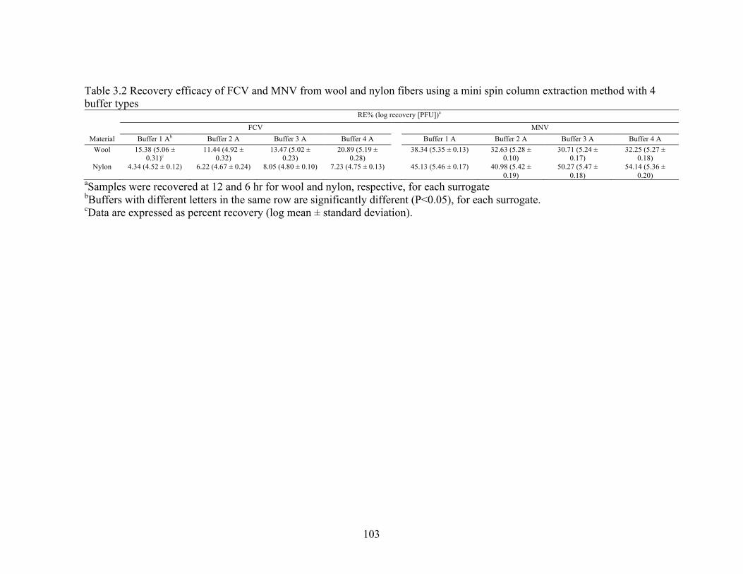
103
Table 3.2 Recovery efficacy of FCV and MNV from wool and nylon fibers using a mini spin column extraction method with 4 buffer types
RE% (log recovery [PFU])a
FCV MNV Material Buffer 1 Ab Buffer 2 A Buffer 3 A Buffer 4 A Buffer 1 A Buffer 2 A Buffer 3 A Buffer 4 A
Wool 15.38 (5.06 ± 0.31)c
11.44 (4.92 ± 0.32)
13.47 (5.02 ± 0.23)
20.89 (5.19 ± 0.28)
38.34 (5.35 ± 0.13) 32.63 (5.28 ± 0.10)
30.71 (5.24 ± 0.17)
32.25 (5.27 ± 0.18)
Nylon 4.34 (4.52 ± 0.12) 6.22 (4.67 ± 0.24) 8.05 (4.80 ± 0.10) 7.23 (4.75 ± 0.13) 45.13 (5.46 ± 0.17) 40.98 (5.42 ± 0.19)
50.27 (5.47 ± 0.18)
54.14 (5.36 ± 0.20)
aSamples were recovered at 12 and 6 hr for wool and nylon, respective, for each surrogate bBuffers with different letters in the same row are significantly different (P<0.05), for each surrogate. cData are expressed as percent recovery (log mean ± standard deviation).

104
Figure 3.1 Electrokinetic potential analysis of wool and nylon via SurPASS titration. One
gram of each surface, wool and nylon, was packed into a cylindrical cell to estimate each
surface’s electrokinetic potential. Hydrochloric acid (£) titrations for nylon were
followed by a NaOH (¯) titration. Similarly, HCl (r) titrations for wool were followed
by a NaOH (Ð) titration. Parameters were set to assess electrokinetic potential between
pH 2 – 9. Error bars indicated standard deviation among 12 replicates from 3 independent
experiments.

105
Figure 3.2 Survival analysis of FCV and MNV assessed via plaque assay on nylon, wool,
and glass surfaces at 30% RH (£) and 70% RH (r). The dotted line indicates detection
limits. Surfaces were individually inoculated with ca. 6 log PFU of FCV and MNV and
recovered with the MSC method using 4 washes with five hundred microliters of buffer
on days 0, 1, 2, 3, 7, 15, 30, and 60. Data were expressed as the mean ± standard
deviation of 6 replicates from two independent experiments
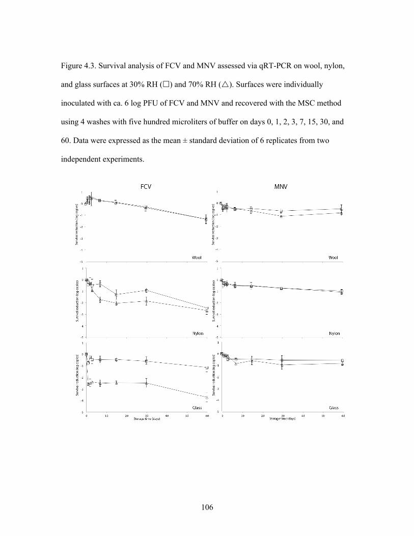
106
Figure 4.3. Survival analysis of FCV and MNV assessed via qRT-PCR on wool, nylon,
and glass surfaces at 30% RH (£) and 70% RH (r). Surfaces were individually
inoculated with ca. 6 log PFU of FCV and MNV and recovered with the MSC method
using 4 washes with five hundred microliters of buffer on days 0, 1, 2, 3, 7, 15, 30, and
60. Data were expressed as the mean ± standard deviation of 6 replicates from two
independent experiments.

107
CHAPTER FOUR
COMPARATIVE RECOVERY OF HUMAN NOROVIRUS SURROGATES, FELINE CALICIVIRUS AND MURINE NOROVIRUS, FROM CARPET WITH THREE
RECOVERY METHODS
ABSTRACT
Human noroviruses (HuNoV) are the leading cause of acute gastroenteritis
worldwide as well as the most common cause of foodborne disease in the United States.
Prevention and control of HuNoV contaminated on soft surfaces, such as carpet, remains
a challenge due, in part, to a lack of robust recovery methods. Our aim was to compare
recovery methods to determine their ability to recover HuNoV surrogates from carpet.
Specifically, we assessed and compared three recovery methods, i.e. bottle extraction
(BE), macrofoam-tipped swabbing (MS), and the microbial vacuum (MVAC), using
HuNoV surrogates, feline calicivirus (FCV) and murine norovirus (MNV), inoculated on
wool and nylon carpet carriers. We also investigated qRT-PCR detection issues for FCV
after environmental recovery, i.e. inhibition. Infectious FCV and MNV percent recovery
efficiency (% RE) of BE ranged from 0.44 to 48.44% and 40.77 to 68.83%, respectively,
compared to % RE of MS, which was 0.02 to 0.82% and 1.54 to 2.87%, respectively. The
% RE of MVAC for infectious FCV and MNV ranged from 7.30 to 18.29% and 52.58 to
74.67%, respectively. Percent RE of genomic copies of FCV and MNV with BE ranged
from 0.36 to 2.53% and 3.34 to 14.97%, respectively, while % RE of MS ranged from
1.03 to 2.24 and 2.02 to 4.25%, respectively. Percent RE of genomic copies of FCV and
MNV with MVAC ranged from 2.49 to 23.72% and 28.78 to 79.15%, respectively.
Significantly more plaque-forming units and genomic copies were recovered using BE
108
and MVAC compared to MS, while buffer type played a significant role in recovery of
infectious FCV. Additionally, qRT-PCR analysis indicated recovery from tested carpet
types inhibited amplification of FCV RNA and required dilution after nucleic acid
extraction.
INTRODUCTION
Worldwide, human noroviruses (HuNoV) account for ca. 20% of all diarrheal
cases (1,2). Its economic burden is estimated to be $4.2 billion in direct health care costs
and $60.3 billion in societal costs per year. Control of HuNoV within the environment
remains a challenge due to their stability and low infectious dose.
Environmental transmission and transfer of viral pathogens has been documented
in both epidemiological and laboratory-based studies on hard and soft surfaces (1–3). For
example, two carpet fitters exhibited HuNoV-like symptoms after removing carpet from a
room used to cohort patients with HuNoV-like symptoms 16 days after the room had
been vacated and decontaminated (1). Likewise, carpet samples from a hotel, associated
with a 5-month HuNoV outbreak, had the highest swab positivity rates (2). These high
positivity rates were attributed to the environmental stability of HuNoV along with the
use of ineffective decontamination strategies. However, as stated previously, there is a
knowledge gap regarding the correlation between risk of infection and level of
contamination that may affect the effectiveness of these disinfection processes (4). To
elucidate this correlation comprehensive comparative sampling studies are needed.

109
The efficacy of sanitizers and disinfectants against HuNoV on hard surfaces is
well documented (5). However, only 3 studies have tested soft surfaces, such as carpet
(6). Currently, the U.S. Environmental Protection Agency (EPA) does not recognize a
standard test method for evaluating the efficacy of an antiviral sanitizer intended for
carpets. But, the EPA does have methods for other types of soft surfaces in order to make
claims against bacteria (7). This is, in part, because of inadequate recovery methods for
carpets, making it difficult to quantify the effect of control strategies.
Adequate recovery methods are the underpinning of environmental detection
methods. Improving and characterizing these methods would provide higher recoveries of
pathogens that would increase their detection and lead to higher positivity rates. To
improve our understanding and better detect and quantify enteric viruses, such as
HuNoV, deposited on soft surfaces, current recovery methods should be assessed and
compared. A meta-analysis of the literature conducted by Julian et al. (8) showed that
only 2 of 59 studies sampled soft surfaces for HuNoV with both using cotton-tipped
swabs (2, 9). Moreover, the laboratory-based studies investigating HuNoV and soft
surfaces relied on destructive recovery methods, e.g. sonication, stomaching, vortexing,
and orbital shaking (10–12), which may not be appropriate for some types of carpet.
Common methods to recover pathogens from complex surfaces, such as carpet,
include bottle extraction (BE), microbial vacuum (MVAC), and swabbing. BE uses both
sonication and handshaking to recover microorganisms of interest and is the American
Society of Testing and Materials (ASTM) recommended method for recovery of bacteria
from carpet carriers (13). On the other hand, the MVAC, which was designed to recover
110
pathogens from food matrices, has recently been used in the forensic science field to
recover a variety of body fluids from complex surfaces, e.g. soft surfaces (14). Even so,
swabbing is still the most frequently used method based on a previous review of the
literature and ISO 15216’s recommendation to use cotton-tipped swabs for recovery of
hepatitis A virus and noroviruses from foodstuffs and food surfaces (8, 15).
This study’s aim was to compare viral recovery methods and improve detection of
viruses contaminated on carpet. To our knowledge, no published studies have assessed
recovery methods for any enteric viruses, including HuNoV, from carpet. Therefore, our
objectives were: (i) compare applicable surface recover methods for two HuNoV
surrogates, feline calicivirus (FCV) and murine norovirus (MNV), on wool and nylon
carpets, with BE, MVAC, and a macrofoam-tipped swab (MS), and (ii) improve detection
via qRT-PCR by clarifying samples prior to analysis.
MATERIALS AND METHODS
Virus propagation and cell culture. Feline calicivirus (FCV) strain F9 (kindly provided
by Dr. Jan Vinje at the Centers for Disease Control and Prevention, Atlanta, GA) stocks
were made by infecting 90% confluent monolayers of Crandell Rees feline kidney
(CRFK) (ATTC CCL-94, American Type Culture Collection, Manassas, VA) at a
multiplicity of infection of 0.01 in Complete Eagles Modified Essential Media (Corning,
Corning, NY) supplemented with 10% low-endotoxin heat inactivated fetal bovine serum
(FBS) (Seradigm, VWR International, Randor, PA), 100 U/liter penicillin (HyClone, GE,
Boston, MA), and 100 µg/liter streptomycin (HyClone, GE, Boston, MA). Murine

111
norovirus (MNV) CW3 (kindly provided by Dr. Herbert Virgin at the University of
Washington, St. Louis) stocks were made by infecting 60 – 80% confluent monolayers of
RAW 264.7 cell (ATCC TIB-71. American Type Culture Collection, Manassas, VA) at a
multiplicity of infection (MOI) of 0.05. The medium used to grow RAW 264.7 cells and
propagate MNV was previously described (16). CRFK and RAW 264.7 cells were grown
at 37˚C and 5% CO2 (Symphony, VWR International, Randor, PA) until complete
cytopathic effect (CPE) was observed (24 – 72 hr). After CPE, both viruses were
harvested by three cycles of freeze-thawing followed by centrifugation for 10 min at
5,000 x g and 4˚C. Finally, viral suspensions were chloroform extracted based on
previous methods (16). Stocks of FCV (ca. 9 log PFU/ml) and MNV (ca. 8 log PFU/ml)
were aliquoted and stored at -80˚C.
Plaque assays. Infectious MNV and FCV were quantified by standard plaque assay as
previously described with modifications (16, 17). Briefly, MNV plaque assay was
completed by seeding 6-well dishes with RAW 264.7 cells at 1 x 10⁶ viable cells/well and
incubated until 60 – 80% confluent (4 – 8 hr). MNV experimental samples were diluted,
if needed, in MNV infection medium, described elsewhere (16), containing 5% FBS
(complete Eagles Modified Essential medium) to improve plaque formation. FCV plaque
assay was based on previous work with significant modifications (17). CRFK cells were
seeded in 6-well dishes at 2.5 x 10⁵ viable cells/well and incubated until ca. 90%
confluent (48 hr). FCV samples were serially diluted in CDMEM-5 if needed. After a 1
hr absorption phase, 2 ml of 1:1 mixtures of 3% sea plaque agarose (Lonza, Switzerland)

112
and 2X Temin’s Modified Eagle Medium (MEM) were added to each well and incubated
until visible plaque formation (24 – 72 hr). The 2X MEM was supplemented with 10%
low-endotoxin heat inactivated FBS, 100 U/liter penicillin, 100 µg/liter streptomycin,
10mM HEPES (HyClone, GE, Boston, MA), and 1 mM NEAA (HyClone, GE, Boston,
MA). MNV and FCV plaques were visualized by staining agarose plugs with a 0.03%
neutral red solution (Carolina Biological, Burlington, NC) mixed with 1X PBS and
enumerated on a light box (Futura light box, Logan Electric, Bartlett, IL). Plaque assays
for both MNV and FCV contained a stock suspension of virus and CDMEM-5 as a
positive and negative control, respectively, to test for cell line permissiveness and
contamination. Cell lines were not passaged >25 times.
Preparation of carpet carriers. Wool level loop and nylon multi-level loop carpets
(SDL-ATLAS, Rock Hill, SC) were selected from a list provided in ASTM standard
F655-13 ((18). The carpet fiber characteristics, e.g. absorption capacity and zeta
potential, are described elsewhere (Buckley et al. unpublished). Carpet samples contained
no finishes, e.g. antimicrobial or soil retardant, and were cut into 5 x 5 cm carriers with a
mechanical cutting die (Model #1500, Freeman Schwabe, Batavia, OH) (courtesy of Dr.
Daniel Price, Interface Inc., Atlanta, GA). After cutting, carpets were shaken by hand to
remove loose fibers, wrapped in aluminum foil, and autoclaved on a 30-min dry cycle.
Sampling experiments. Wool and nylon carpet carriers (5 x 5 cm), each contained in a
petri dish, were individually inoculated with 0.1 ml of FCV or MNV. Inocula were

113
diluted in CDMEM-5 and contained ca. 7 log PFU/ml of FCV or MNV. Carpet carriers
were dried for 1 hr in a 30% relative humidity (RH) chamber (480 HP, VWR
International, Randor, PA) maintained with a saturated MgCl2 solution. After drying,
three recovery procedures, i.e. BE, MS, and MVAC, were assessed for their ability to
elute FCV and MNV from wool and nylon carpets. Additionally, two separate elution
buffers, 0.01 M phosphate buffered saline (PBS) + 0.02% Tween 80 (T80) and ¼
strength Ringer’s solution (RS) were tested using the BE and MS. Due to design
limitations, only one buffer was used for the MVAC, i.e. sterile collection solution (SRS)
(SRS 1000, M-VAC Systems, Sandy, UT).
The MVAC was used in accordance with required training provide by M-Vac
Systems, Incorporated, Sandy, UT. To recover viruses from carpet carriers using the
MVAC, carriers were moved to a 150 mm Kirby Bauer dish (VWR International,
Randor, PA) to adequately collect residual liquid. The M-VAC head has two functions:
dry vacuum and wet vacuum extraction. During dry vacuum extraction, the edge of the
circular head aspirates whereas during wet vacuum extraction the head aspirates while
simultaneously spraying SRS from the head’s center in a horizontal plane. During
recovery, the MVAC head was passed over three separate areas of the carrier three times,
both horizontally and vertically. On each initial pass, the wet vacuum was on, whereas
during the remaining two passes, in each area, only the dry vacuum was on.
Recovery of FCV and MNV from carpet carriers were consistent with BE
methods outlined in ASTM 2966-14 (13). Dried inoculated carpet carriers were each
placed in separate 500 ml wide mouth polypropylene bottles (Fisher Scientific)

114
containing 100 ml of buffer. Bottles were then sonicated (FS110, Fisher Scientific
International, Pittsburg, PA) for 1 min at 40 kHz and hand shaken for l min. After
shaking, carriers were removed with sterile forceps.
Due to high sample volumes associated with BE and MVAC, a concentration step
was applied. Immediately after recovery, BE and MVAC samples were concentrated via
ultrafiltration (Amicon Ultra-15 30K, Millipore, Billerica, MA) at 4,000 x g for 15 min at
4˚C (Allegra X-30R, Beckman Coulter, Brea, CA). Supernatants were pooled, vortexed,
weighed, and stored at -80˚C prior to infectivity and qRT-PCR analysis.
The MS was designed by Puritan Medical Supply (NC1213065 ENVIROMAX
PROTOTYPE, Medical Supply, Guilford, ME) and purchased dry. Swabs were pre-
moistened by filling sleeves with sterile buffers, mentioned above, then gently rolled for
3 minutes. Carpet carriers were swabbed horizontally, vertically, and diagonally, twice.
Next, the swab head was pressed and twisted within its sleeve to elute any remain liquid.
After eluting, the swab head was discarded. Samples were vortexed, weighed, aliquoted,
and stored at -80˚C prior to infectivity and qRT-PCR analysis.
Sample clarification. To elucidate issues surrounding recovery and detection with qRT-
PCR recovery of FCV from wool carpet with BE and 0.01 M + 0.02% Tween 80 was
used. Virus, recovery method, and carpet were selected based on the least efficacious
RNA recovery rates reported in the comparison of sample recovery experiments (Table
2). Carpet samples and viral inocula were prepared and executed as described above.
After recovery samples were clarified by either filtering with 0.2 or 0.4 µm syringe filters

115
(Sterile Syringe Filter, VWR International, Randor, PA), centrifuging at 4,000 x g for 15
mins at 4˚C (Allegra X-30R, Beckman Coulter, Brea, CA), 10-1 dilution before FCV
RNA extraction in diethyl pyrocarbonate (DEPC) water (OMEGA Bio-Tek, Norcross,
GA), or 10-1 dilution after FCV RNA extraction in DEPC water. The control samples
were recovered and concentrated as stated above with no additional treatments.
Real time qRT-PCR. Viral extraction was performed as previously described with minor
modifications (11). QRT-PCR assays were completed with the E.N.Z.A Viral RNA Kit
(OMEGA Bio-Tek, Norcross, GA), per manufacturer instructions, by extracting viral
RNA from 0.15 ml of sample or stock. MS samples were extracted directly, whereas BE
and MVAC samples were extracted after concentration via ultrafiltration and stored at -
80˚C prior to assessment. qRT-PCR of both viruses were completed with KAPA SYBR
Fast Universal One-Step qRT-PCR Kit (Kapa Biosystems, Wilmington, MA) on a
Realplex2 Mastercycler platform (Eppendorf, Hauppauge, NY). Forward and reverse
primer sequences for FCV analysis were GCCATTCAGCATGTGGTAGTAACC and
GCACATCATATGCGGCTCTG, respectively, whereas MNV forward and reverse
primer sequences were TGATCGTGCCAGCATCGA and
GTTGGGAGGGTCTCTGAGCAT, respectively (19). A standard curve was generated
for both viruses by performing a 7-step 10-fold diluting of a previously titered and RNA
extracted virus stock. Each dilution’s threshold cycle (CT) values was used to plot against
its corresponding plaque forming unit (PFU) and calculated as previously described (19).

116
Data Analysis. All experiments were performed in triplicate with three replicates per
experiment. Percent recovery efficiency (% RE) was defined as the number of PFU or
genomic copies recovered divided by the number of PFU or genomic copies initially
seeded (8). One-way multiple comparisons to determine significance among treatments.
All results were expressed as mean ± standard deviation. Statistical significance was
defined as P ≤ 0.05. Statistical analyses were conducted using JMP (JMP Pro 12.2.0, SAS
Inc., Cary, NC).
RESULTS
Virus recovery efficiency via plaque assay. Table 4.1 shows the % RE and log10 PFU
recovery of FCV and MNV from wool and nylon carpet carriers as measured by plaque
assay. Percent RE and log10 PFU recovery of infectious FCV ranged from 0.37 to 48.44%
and 3.24 to 5.51 log10 PFU on wool carrier, respectively, compared to 0.02 to 16.24% and
2.10 to 5.03 log10 PFU on nylon carriers, respectively. The % RE and log10 recovery for
infectious MNV ranged from 1.87 to 68.83% and 4.48 to 6.10 log10 PFU, respectively, on
wool, compared to 1.54 to 74.67% and 4.30 to 6.13 log10 PFU, respectively, on nylon
carpet carriers. The recovery method and eluent used significantly affected the recovery
of infectious FCV whereas only the recovery method affected the recovery of MNV. BE
with T80 and RS, and the MVAC achieved significantly higher recovery rates for FCV
and MNV than did MS, excluding recovery of FCV using BE with RS from nylon carpet
carriers. Carpet type significantly affected recovery of FCV when using BE and MS with
the elution buffer RS with more FCV was recovered from wool than from nylon.

117
Comparatively, significantly more infectious MNV was recovered from both carpet types
than was FCV, regardless of method and eluent.
Virus recovery efficiency via qRT-PCR. Table 4.2 shows the % RE and log10 genomic
copy recovery of FCV and MNV from wool and nylon carpet carriers measured with
qRT-PCR. Percent RE and log10 genomic copy recovery of FCV ranged from 0.36 to
4.95% and 2.63 to 3.81 log10 genomic copies, respectively, on wool carpet carriers
compared to 0.69 to 23.72% and 2.78 to 4.49 log10 genomic copies, respectively, on
nylon carpet carriers. To the contrary, % RE and log10 genomic copy recovery of MNV
ranged from 2.42 to 38.78% and 4.36 to 5.40 log10 genomic copies, respectively, on wool
carpet carriers compared to 2.02 to 79.15% and 4.13 to 5.68 log10 genomic copies.
Recovery method and eluent type significantly affected the recovery of both FCV and
MNV on both carpet types when assessed via qRT-PCR. MVAC exhibited significantly
higher recoveries of genomic copies for FCV and MNV compared to all other methods,
excluding FCV recovery using MS with RS on nylon and MNV recovery using BE with
T80 and RS. Carpet type did not play a significant role in the recovery of FCV and MNV
genomic copies, excluding recovery of MNV using BE T80 with RS. Overall,
significantly more MNV genomic copies were detected compared to FCV, regardless of
method and eluent. Additionally, visible wool and nylon carpet debris was accumulated
when using BE in both the recovery suspension and within the RNA extraction columns,
whereas with the MVAC, only wool debris appears in the recovery suspension and RNA
extraction columns.

118
FCV detection after sample clarification. Sample clarification results are presented in
Table 4.3. Due to low RNA % RE from wool, FCV was further tested for optimization of
recovery and detection. Syringe filtering with 0.2 and 0.4 µm resulted in volumes too low
for plaque assay and qRT-PCR analysis, as such no analysis was completed for this
treatment. Recovery of FCV genomic copies from the control treatments and centrifuged
treatments ranged from 4.14 to 4.89 log10 copies and 3.88 to 4.90 log10 copies,
respectively. In contrast, recovered infectious FCV from the control was 5.76 log10 PFU,
whereas, centrifugation yielded 5.61 log10 PFU. Recovery of infectious FCV and genomic
copies were significantly reduced with centrifugation alone compared other clarification
methods. At the same time, amplification of the PCR target from the 10-1 dilution of both
control and centrifuged samples after RNA extraction was significantly higher compared
to all other clarification processes.
DISCUSSION
FCV and MNV are commonly used as surrogates to study the behavior of HuNoV
in the environment (20). Recovery methods are the underpinning of environmental
detection procedures. However, there is scant literature regarding recovery efficiency of
methods used to detect HuNoV as well as other enteric viruses on complex soft surfaces,
such as carpet, and of those studies, many use different recovery procedures. In this
study, we evaluated three recovery methods’ ability to recover two HuNoV surrogates,
FCV and MNV, from wool and nylon carpet carriers. Additionally, we attempted to
improve detection via qRT-PCR by clarifying samples prior to analysis. Our findings

119
demonstrate the efficiency and limitations these three methods may have when used to
recover and detect HuNoV surrogates from complex soft surfaces.
The findings from previously published studies indicated that both recovery
method and eluent type could significantly affect the recovery of viruses with some study
authors suggesting that optimization of methods may improve recovery (21, 22). In our
study, data from plaque assays, i.e. infectious data, indicated that recovery method and
eluent type significantly affected the recovery of HuNoV surrogates, FCV and MNV.
Among the carpet samples tested, BE and MVAC were the most efficacious methods for
recovering infectious FCV and MNV from wool and nylon carpets. The mode of action
for each recovery method tested relied upon fiber rehydration, which changes the zeta
potential of the fiber and the virus surface charge. However, each recovery method
differed in the physical removal and recovery of the virus after the elution buffer
application. For instance, the mechanism of action for MVAC relies upon the dual use of
a spray, which assists in desorption of virus from the fibers, while simultaneously
aspirating the virus containing suspension. In contrast, BE uses sonication which
weakens virus surface attachment, providing higher recovery than traditional
handshaking (11). However, both methods are expensive and rely upon the use of high
volumes to recover virus, which lowers the detection limit and necessitates the use of a
concentration step. Swabs are the most frequently used tool for environmental recovery
method because they are simple to use, economical, and low-volume, i.e. no addition
concentration step. Nevertheless, the problem with swabbing is that it is usually
accompanied by a significant loss in recovery efficiency. Ultimately, each method may

120
be useful based on – time, funding, technical expertise, and study type, i.e. efficacy study
or environmental monitoring.
Recovery of infectious virus from environmental samples can vary based on
surface type. The surface hydrophilicity, absorption capacity, zeta potential, and chemical
surface interactions in addition to a virus’s characteristics, i.e. isoelectric point, can
influence both survival and recovery of the virus. For example, Yeargin et al. (11) found
that after drying, recovery of both FCV and MNV was significantly different across
cotton, polyester, and glass when using the same recovery method. Another study
demonstrated higher recovery efficiency of MNV from inoculated diapers compared to
cotton gauze (10). Unpublished data from M-Vac Systems, Incorporated, suggested some
biological material, such as DNA, has higher recovery rates on natural surfaces, such as
wool and cotton, compared to synthetic material, nylon and rayon (14). Consistent with
these findings, we found that higher titers of FCV and MNV were recoverable from wool
compared to nylon. This is likely a result of wool’s higher absorption capacity and ability
to provide a more protective environment during desiccation (Buckley et al. chapter 3).
This assertion is supported by a published study that examined the same fibers,
suggesting both FCV and MNV can survive longer on wool carpet fibers than on nylon
carpet fibers. Fabrication of soft surfaces has also been shown to influence the recovery
and survival of enteric viruses, such as poliovirus (23). The additional fiber twist that
accompanies a tuft carpet construction, observed in nylon carpet but not in wool carpets,
may further prevent elution of viruses.

121
When selecting a recovery method, eluent type is a critical factor to consider
based on recovery type, downstream assays, and storage conditions, especially when a
concentration step is required for detection. Concentration methods can also concentrate
inhibitors cytotoxic to cell culture systems or may interfere with PCR, e.g. degradation of
target nucleic acid or reduced extraction efficiency from heterogeneous samples (5, 24).
However, a meta-analysis of virus surface sampling literature suggests eluent type does
not significantly influence recovery of viruses (8). Although, the meta-analysis conducted
in that study did not specifically target non-enveloped viruses and most recovery methods
used swabs. Ultimately, the authors suggested using ¼ strength Ringer’s solution with a
polyester-tipped swab. Our results indicated eluent type can significantly influence virus
recovery of FCV, when using BE and MS methods. Typically, eluents range from pH 6 –
9 to buffer the solution, reduce cytotoxicity, and create a negative viral surface charge
(25). Often, surfactants are included to assist with recovery via reduced surface tension
and micelles formation. In this study, the addition of Tween 80 to a simple, low ionic
strength buffer, significantly improved FCV recovery compared to using ¼ strength
Ringer’s solution, which contains no surfactants. On the other hand, another high
efficiency recovery method for soft surfaces using a mini-spin column-based approach
had similar recovery rates of FCV and MNV as the BE and MVAC methods from the
same carpet fibers but did not require high volumes or a concentration step (Buckley et
al. chapter 3). Additionally, and consistent with the findings of Julian et al. (8), the mini-
spin column method results indicated eluent type did not significantly affect recovery of
FCV and MNV (Buckley et al. chapter 3). Albeit, this method was not incorporated to

122
our study because it requires packable fibers, e.g. shaved carpet fibers or swatches, and
does not mimic the scale or surface complexity needed to assess virus recovery of intact
carpet carriers. Collectively, these findings suggest that eluent type may affect recovery
of some viruses and is dependent upon both recovery method and virus type.
Several published papers have detailed the inactivation profiles of HuNoV
surrogates (6, 26). Not surprisingly and consistent across previous studies, significantly
more infectious and genomic copies of MNV were detected compared to FCV. MNV has
been demonstrated to be more environmentally resilient on both hard and soft porous
surfaces and is more resistant to some chemical inactivation treatments compared to FCV
(20, 27). Although a calicivirus, FCV is also classified as a respiratory pathogen and
cannot survive harsh climates like the gastrointestinal system. We believe the difference
is primarily due to capsid integrity and higher receptor sensitivity to environmental
factors, although other structural or solution characteristics cannot be eliminated.
The divergence between infectivity assays and molecular data, i.e. qRT-PCR, has
been previously documented (10, 28). Traditionally, unless samples are enzymatically
pre-treated, concentrations of RNA as measured by qRT-PCR, are higher compared to
infectious counts. This is due to capsids becoming inactivated but not lysed, allowing for
temporary preservation of its genome. However, there is evidence to dispute this. For
instance, an extensive recovery comparison study using another HuNoV surrogate, MS2
phage, showed that fractions of MS2 phage RNA recovered from environmental surfaces
were lower than recovered infectious MS2 phage (8). Consistent with those findings, we
found that recovery of FCV and MNV genomic copies were less than the recovery of

123
infectious FCV and MNV among undiluted, RNA extracted samples. We believe an
inhibitor impacted our qRT-PCR amplification and/or extraction efficiency, whereas
Julian et al. (8) suggested the lower fractional recovery may be a result of exogenously
seeded RNA or viral aggregates (8). Degradation or lysis of capsids can occur leading to
higher exogenous genomic copies compared to estimates of infectious counts in stocks
and inocula. Commonly, when seeded, the exogenous RNA would degrade more quickly
than capsid-protected RNA, leading to an inflated initial copy count, and resulting in
lower calculated RE (28). Be that as it may, viral aggregates, may also reduce the
recovery of RNA as they are not distinguishable within a plaque assay and may decrease
extraction efficiency.
In our study, the sample matrices were wool and nylon, and the fibers in
recovered samples may have affected qRT-PCR. Therefore, sample clarification was
studied to improve detection by removing contaminants that may interfere with the assay.
Attempts to elucidate possible interferences with qRT-PCR via sample clarification both
positively and negatively affected genomic copy recovery. We believed carpet fibers may
reduce extraction efficiency based on accumulated carpet debris observed within RNA
extraction columns. Our attempts to remove the carpet fibers via syringe filtering (data
not shown) and centrifugation significantly lowered volume and both PFU and copy
number, respectively. This suggests some viruses may be attached or trapped within
pelleted fibers. At any rate, both syringe filtering and centrifuging are not recommended
as sample clarification techniques for recovery of FCV from carpets. Dilution is also a
simple but effective method for removing inhibitors. Our qRT-PCR results of samples

124
diluted prior to RNA extraction suggested a filterable PCR inhibitor was concentrated
with FCV genomic RNA, prompting us to dilute the extracted RNA samples. Under these
conditions, the inhibitor was reduced, which allowed for significantly more target
amplification. The specific inhibitor is unknown but, as stated previously, PCR can be
inhibited by numerous factors, such as complex polysaccharides, organic material, and
metal ions (29). The inhibitor could have originated from a variety of sources but likely is
due to the carpet or extraction column. Future studies designing or optimizing recovery of
viruses from environmental samples are encouraged test for qRT-PCR for inhibition with
specificity, i.e. fluorogenic probe, and incorporate an external standard.
CONCLUSIONS
The assessment of recovery methods is a critical first step to improve the
detection and quantification of viruses. Here, we tested 3 mechanistically different
recovery methods intended for HuNoV recovery from carpets. BE and MVAC exhibited
higher recovery efficiencies compared to MS. Furthermore, detection of FCV and MNV
via qRT-PCR can be inhibited when recovered from wool and nylon carpets. Ultimately,
each method demonstrates merit for use under different conditions, e.g. standard testing
and environmental monitoring.
ACKNOWLEDGEMENTS
This research was supported by the National Institute of Food and Agriculture,
U.S. Department of Agriculture (USDA; agreement 2011-68003-30395), and The Procter

125
and Gamble Company. Any opinions, findings, conclusions, or recommendations
expressed in this publication are those of the author(s) and do not necessarily reflect the
view of the USDA or The Procter and Gamble Company.
REFERENCES
1. Cheesbrough JS, Barkess-Jones L, Brown DW. 1997. Possible prolonged
environmental survival of small round structured viruses. J Hosp Infect 35:325–
326.
2. Cheesbrough JS, Green J, Gallimore CI, Wright PA, Brown DW. 2000.
Widespread environmental contamination with Norwalk-like viruses (NLV)
detected in a prolonged hotel outbreak of gastroenteritis. Epidemiol Infect 125:93–
98.
3. Lopez GU, Gerba CP, Tamimi AH, Kitajima M, Maxwell SL, Rose JB. 2013.
Transfer efficiency of bacteria and viruses from porous and nonporous fomites to
fingers under different relative humidity conditions. Appl Environ Microbiol
79:5728–5734.
4. Park GW, Lee D, Treffiletti A, Hrsak M, Shugart J, Vinjé J. 2015. Evaluation of a
New Environmental Sampling Protocol for Detection of Human Norovirus on
Inanimate Surfaces. Appl Environ Microbiol 81:5987–92.
5. Bright KR, Boone SA, Gerba CP. 2010. Occurrence of bacteria and viruses on
elementary classroom surfaces and the potential role of classroom hygiene in the
spread of infectious diseases. 26:33–41.

126
6. Yeargin T, Buckley D, Fraser A, Jiang X. 2016. The survival and inactivation of
enteric viruses on soft surfaces: A systematic review of the literature. Am J Infect
Control 44:1365–1373.
7. EPA. 2012. Product Performance Test Guidelines OCSPP 810.2400: Disinfectants
and Sanitizer for Use on Fabrics and Textiles. https://www.epa.gov/test-
guidelines-pesticides-and-toxic-substances/series-810-product-performance-test-
guidelines
8. Julian TR, Tamayo FJ, Leckie JO, Boehm AB. 2011. Comparison of surface
sampling methods for virus recovery from fomites. Appl Environ Microbiol
77:6918–6925.
9. Green J, Wright PA, Gallimore CI, Mitchell O, Morgan-Capner P, Brown DW,
Ashley C, Caul E. 1998. The role of environmental contamination with small
round structured viruses in a hospital outbreak investigated by reverse-
transcriptase polymerase chain reaction assay. J Hosp Infect 39:39–45.
10. Lee JE, Zoh KD, Ko GP. 2008. Inactivation and UV disinfection of murine
norovirus with TiO2 under various environmental conditions. Appl Environ
Microbiol 74:2111–2117.
11. Yeargin T, Fraser A, Guohui H, Jiang X. 2015. Recovery and disinfection of two
human norovirus surrogates, feline calicivirus and murine norovirus, from hard
nonporous and soft porous surfaces. J Food Prot 78:1842–1850.
12. Malik YS, Allwood PB, Hedberg CW, Goyal SM. 2006. Disinfection of fabrics
and carpets artificially contaminated with calicivirus: relevance in institutional and

127
healthcare centres. J Hosp Infect 63:205–210.
13. ASTM International. 2015. Standard Test Method for Quantitative Assessment of
Sanitizing Solutions for Carpet. American Society for Testing and Materials.
14. M-Vac Systems, Inc. Food Safety. http://www.m-vac.com/food-saftey
15. ISO/TS 15216-1:2013 - Microbiology of food and animal feed - Horizontal
method for determination of hepatitis A virus and norovirus in food using real-time
RT-PCR -- Part 1: Method for quantification. Accessed July 5, 2017.
https://www.iso.org/standard/55382.html
16. Hwang S, Alhatlani B, Arias A, Caddy SL, Christodoulou C, Cunha JB, Emmott
E, Gonzalez-Hernandez MB, Kolawole A, Lu J, Rippinger C, Sorgeloos F, Thorne
L, Vashist S, Goodfellow IG, Wobus CE. 2014. Murine norovirus: propagation,
quantification, and genetic manipulation Current Protocols in Microbiology.
17. Bidawid S, Malik N, Adegbunrin O, Sattar SA, Farber JM. 2003. A feline kidney
cell line-based plaque assay for feline calicivirus, a surrogate for Norwalk virus. J
Virol Methods 107:163–167.
18. ASTM International. 2015. F655 - 13. Standard Specification for Test Carpets and
Pads for Vacuum Cleaner Testing 1–3. West Conshohocken, PA.
19. Park GW, Barclay L, Macinga D, Charbonneau D, Pettigrew CA, Vinjé J. 2010.
Comparative efficacy of seven hand sanitizers against murine norovirus, feline
calicivirus, and GII.4 norovirus. J Food Prot 73:2232–2238.
20. Cromeans T, Park GW, Costantini V, Lee D, Wang Q, Farkas T, Lee A, Vinjé J.
2014. Comprehensive Comparison of Cultivable Norovirus Surrogates in
128
Response to Different Inactivation and Disinfection Treatments. Appl Environ
Microbiol. 90:5143-5751
21. Scherer K, Mäde D, Ellerbroek L, Schulenburg J, Johne R, Klein G. 2009.
Application of a Swab Sampling Method for the Detection of Norovirus and
Rotavirus on Artificially Contaminated Food and Environmental Surfaces. Food
Environ Virol 1:42–49.
22. Taku A, Gulati BR, Allwood PB, Palazzi K, Hedberg CW, Goyal SM. 2002.
Concentration and detection of caliciviruses from food contact surfaces. J Food
Prot 65:999–1004.
23. Dixon GJ, Sidwell RW, Mcneil E. 1966. Quantitative Studies on Fabrics of
Viruses Disseminators. II. Persistence of poliomyelitis virus on cotton and wool
fabrics. Appl Microbiol 14:183–188.
24. Boxman ILA, Dijkman R, te Loeke NAJM, Hägele G, Tilburg JJHC, Vennema H,
Koopmans M. 2009. Environmental swabs as a tool in norovirus outbreak
investigation, including outbreaks on cruise ships. J Food Prot 72:111–9.
25. Farrah SR, Gerba CP, Wallis C, Melnick JL. 1976. Concentration of Viruses from
Large Volumes of Tap Water Using Pleated Membrane Filters. Appl Environ
Microbiol 31:221–226.
26. Bolton SL, Kotwal G, Harrison M a., Law SE, Harrison J a., Cannon JL. 2013.
Sanitizer efficacy against murine norovirus, a surrogate for human norovirus, on
stainless steel surfaces when using three application methods. Appl Environ
Microbiol 79:1368–1377.
129
27. Cannon JL, Papafragkou E, Park GW, Osborne J, Jaykus LA, Vinjé J. 2006.
Surrogates for the study of norovirus stability and inactivation in the environment:
A comparison of murine norovirus and feline calicivirus. J Food Prot 69:2761–
2765.
28. Escudero BI, Rawsthorne H, Gensel C, Jaykus LA. 2012. Persistence and
transferability of noroviruses on and between common surfaces and foods. J Food
Prot 75:927–935.
29. Julian TR, Schwab KJ. 2012. Challenges in environmental detection of human
viral pathogens. Curr Opin Virol 2:78–83.

130
Table 4.1 Recovery efficiency percentages of FCV and MNV via plaque assay from carpet %RE (log recovery [PFU])a
FCV MNV Recovery Methodb Bufferc Wool Nylon Wool Nylon BE T80 48.44 (5.51 ± 0.13)A 16.24 (5.03 ± 0.16)BC 68.83 (6.10 ± 0.09)A 60.47 (6.07 ± 0.06)A
BE RS 10.41 (4.88 ± 0.07)BC 0.44 (3.42 ± 0.30)DE 38.87 (5.88 ± 0.09)A 40.77 (5.91 ± 0.10)A
MS T80 0.82 (3.76 ± 0.16)D 0.54 (3.62 ± 0.01)D 1.87 (4.50 ± 0.17)B 2.87 (4.58 ± 0.29)B
MS RS 0.37 (3.24 ± 0.34)E 0.02 (2.10 ± 0.22)F 2.16 (4.48 ± 0.26)B 1.54 (4.30 ± 0.29)B
MVAC SRS 18.29 (5.16 ± 0.04)AB 7.30 (4.75 ± 0.01)C 52.58 (5.99 ± 0.08)A 74.67 (6.13 ± 0.15)A
aData are expressed as percent recovery (log means ± standard deviation). Within each surrogate, log10 values with different letters are significantly different (P<0.05), among both surfaces. bBE: bottle extraction; MS: macrofoam-tipped swab; MVAC: microbial vacuum; cT80: 0.01 M phosphate buffered saline + 0.02% Tween 80; RS; ¼ strength Ringer’s solution; SRS: sterile rinse solution

131
Table 4.2. Recovery efficiency percentage of FCV and MNV via qRT-PCR from carpet %RE (log recovery [genomic copies])a
FCV MNV Recovery Methodb
Bufferc Wool Nylon Wool Nylon
BE T80 0.81 (2.95 ± 0.19)DE 2.53 (3.46 ± 0.21)CD 3.37 (4.44 ± 0.19)CD 8.04 (4.83 ± 0.15)ABC
BE RS 0.36 (2.63 ± 0.19)F 0.69 (2.78 ± 0.36)EF 3.34 (4.47 ± 0.12)CD 14.97 (5.15 ± 0.06)AB
MS T80 1.03 (2.95 ± 0.33)DE 1.31 (3.18 ± 0.25)CDE 4.25 (4.59 ± 0.07)BCD 3.08 (4.34 ± 0.25)D
MS RS 1.70 (3.23 ± 0.24)CDE 2.24 (3.45 ± 0.12)BC 2.42 (4.36 ± 0.03)CD 2.02 (4.13 ± 0.29)D
MVAC SRS 4.95 (3.81 ± 0.05)AB 23.72 (4.49 ± 0.09)A 38.78 (5.40 ± 0.36)A 79.15 (5.68 ± 0.27)A
aData are expressed as percent recovery (log means ± standard deviation). Within each surrogate, log10 values with different letters are significantly different (P<0.05), among both surfaces. bBE: bottle extraction; MS: macrofoam-tipped swab; MVAC: microbial vacuum; cT80: 0.01 M phosphate buffered saline + 0.02% Tween 80; RS; ¼ strength Ringer’s solution; SRS: sterile rinse solution

132
Table 4.3. Recovery of FCV from wool carpet with bottle extraction method after sample clarification
Recoverya
Clarification Processb
qRT-PCR log copies ±
SD
Plaque Assay
log PFU ± SD
Control 4.14 ± 0.29B 5.76 ± 0.10A 10-1 control before RNA extraction 4.36 ± 0.08B - 10-1 control after RNA extraction 4.89 ± 0.12A -
Centrifugation treatment 3.88 ± 0.17C 5.61 ± 0.10B 10-1 before RNA extraction 4.27 ± 0.21B - 10-1 after RNA extraction 4.90 ± 0.12A -
aLog10 values with different letters in the same column are significantly different (P<0.05). bControl: unclarified FCV recovery from wool carpet; 10-1 dilution before FCV RNA extraction in diethyl pyrocarbonate (DEPC) water; 10-1 dilution after FCV RNA extraction in DEPC water; Centrifugation at 4,000 x g for 15 mins; 10-1 dilution of centrifuged samples before FCV RNA extraction in DEPC water; 10-1 dilution of centrifuged samples after FCV RNA extraction in DEPC water. c – not tested as there was no suspected interference.

133
CHAPTER FIVE
EFFICACY OF SILVER DIHYDROGEN CITRATE AND STEAM VAPOR AGAINST A HUMAN NOROVIRUS SURROGATE, FELINE CALICIVIRUS, IN SUSPENSION,
ON GLASS, AND CARPET
ABSTRACT
Human noroviruses (HuNoV) are responsible for 19-21 million illnesses each
year in the United States while also accounting for ca. 20% of all diarrheal cases
worldwide. Effective environmental hygiene programs are important to prevent and
control of HuNoV outbreaks. However, despite our increasing knowledge of the role of
soft surfaces in the transmission of HuNoV, no commercially available disinfection
technologies have been evaluated on carpets. Our aim was to assess two disinfection
technologies, silver dihydrogen citrate (SDC) and steam vapor, against one HuNoV
surrogate, feline calicivirus (FCV), on wool and nylon carpet. First, we evaluated the
effect of both technologies on the aesthetic appearance of carpet. After developing a
neutralizer for SDC, we evaluated the efficacy of SDC in suspension with and without
5% fetal bovine serum (FBS) and the efficacy of SDC and steam vapor on glass, each
with and without 5% FBS. Lastly, we tested both technologies on carpets. Wool and
nylon carpet carriers exhibited no obvious color changes after both treatments, however,
SDC treatment left a residue while steam resulted in minor abrasions to surface fibers. A
sodium thioglycolate-based solution was found to adequately neutralize and eliminate
SDC cytotoxicity. SDC in suspension and on glass reduced FCV by 4.65 and >4.66 log10
pfu with in 30 mins, respectively, but demonstrated reduced efficacy in the presence of
serum. However, SDC was only efficacious against FCV on nylon (3.62 log10 pfu

134
reduction) as compared with 1.82 log10 reduction on wool carpet. Steam vapor reduced
FCV by >4.93 log10 pfu on glass in 10 sec, with no observed difference among serum
treatments, and >3.68 log10 pfu on wool and nylon carpet carriers in 90 sec. There was
limited reduction to FCV RNA under both treatments regardless of treatment compared
to infectivity assays but RNA reductions were higher in sample that contained 5% serum.
INTRODUCTION
Human noroviruses (HuNoV) are the leading causes of acute gastroenteritis
worldwide as well as the most common cause of foodborne disease in the United States.
Worldwide, its economic burden is estimated to be US$4.2 billion in health care costs
alone illustrating its significance as a public health problem (1, 2). Common symptoms
include both diarrhea and vomiting, which aid in its spread. Transmission occurs two
ways -- the fecal-oral or vomitus-oral route through person-to-person contact, food,
water, or environmental surfaces (1).
Environmental transmission of HuNoV is estimated to be low (3). Even so,
several epidemiological investigations and laboratory-based studies suggest the
environment plays an important role in transmission as both hard and soft surfaces may
initiate and prolong HuNoV outbreaks (4). For example, a hotel in the United Kingdom
experienced a 5-month long HuNoV outbreak (5). Outbreaks investigators suggested, in
addition to HuNoV environmental stability, ineffective decontamination of soft surfaces
assisted in prolonging the outbreak.

135
Focusing on environmental sanitation is a recommended strategy to prevent and
control HuNoV outbreaks. The challenge to effective environmental sanitation is that
HuNoV can be shed in high titers from infected individuals. This, coupled with its
environmental stability, low infectious dose, and resistance to many commonly used
disinfectants, e.g. phenolic and quaternary ammonium compounds, makes HuNoV
outbreaks difficult to control. The current recommendation for HuNoV clean-up includes
using1,000–5,000 ppm bleach or an Environmental Protection Agency (EPA)-registered
disinfectant (6). However, these were validated for hard surfaces not soft surfaces.
Moreover, their use may damage the appearance of soft surfaces, such as carpet and soft
furnishings. Furthermore, carpet and other soft furnishings can absorb toxic active
ingredients causing irritation of the skin, eyes, and respiratory tract (7). Fogging with
certain chemistries, e.g. ozone and H2O2, has demonstrated efficacy against some enteric
viruses but is impractical in some settings, e.g. long-term care facilities and residential
homes, due to temporary removal of residents and cost (8, 9). Taken together, the
disinfectant shortcomings of current disinfectant procedures present a gap in evidence-
based control strategies for disinfecting soft surfaces contaminated with viruses
suggesting the need to evaluate safe and practical technologies for use on soft surfaces.
To our knowledge, only one previous study has investigated the use of
disinfection technologies against viruses on carpets, all of which are liquid-based (10).
Investigators found that a glutaraldehyde-based solution could achieve EPA efficacy
standards for disinfection on soft surfaces against a HuNoV surrogate, feline calicivirus
(FCV) (3.0 log10 reduction) but was linked to negative health effects, such as skin rashes

136
and respiratory irritation (11). Other chemistries tested were salt and quaternary
ammonium-based products, which have shown little efficacy against HuNoV (10).
Recently, transitional metal ions, such as silver, have demonstrated broad-range efficacy
against microorganisms although they are more stable when generated in the presence of
citric acid to form silver dihydrogen citrate (SDC). The benefit of SDC is that it is less
toxic compared to other chemistries, i.e. glutaraldehyde, and may be more gentle on
delicate soft surfaces. Previous studies with SDC have demonstrated its efficacy against
HuNoV but these studies lack infectious data required by the EPA to be registered (12,
13).
The Centers for Disease Control and Prevention (CDC) and the Occupational
Safety and Hazard Administration (OSHA) have both recommended steam cleaning for
carpets after a suspected HuNoV contamination event (14). Steam has been shown to be
efficacious against non-sporeforming bacteria on hard surfaces, but there is scant
literature of its efficacy against viruses. A dry-steam vapor system with thermos
accelerated nano-crystal sanitation (TANCS) technology (Advanced Vapor Technologies,
Seattle, WA) is a promising tool for disinfecting virally contaminated soft surfaces.
Previous work with MS2 phage and FCV on hard surfaces demonstrated a >6 log and >4
log reduction in less than 5 and 10 sec, respectively (15, 16). However, the efficacy of
steam or moist heat against virus contamination on soft surfaces has not been measured
under controlled conditions (17). This is due, in part, to a lack of standardized test
methods for quantitative assessment of disinfecting solutions for carpets contaminated
with viruses.

137
The gap in knowledge about the efficacy of soft surface disinfection technologies
and the lack of standardized test methods for carpets warrants further exploration. To our
knowledge, no published studies have investigated either technology against infectious
HuNoV surrogates on carpet. The aim of this study was to assess these two disinfection
technologies against FCV on wool and nylon carpet. Specific objectives were to: (i)
assess these technologies in suspension, (ii) on a hard surface with current American
Society for Testing and Materials (ASTM) International standards, and (iii) on carpets by
adapting a current ASTM International standard.
MATERIALS AND METHODS
Virus propagation, cell culture, and plaque assay. A stock of feline calicivirus (FCV)
strain F9 (kindly provided by Dr. Jan Vinje at the Centers for Disease Control and
Prevention, Atlanta, GA) was propagated by infecting 90% confluent monolayers of
Crandell-Rees kidney cell (CRFK) (ATCC CCL-94, American Type Culture Collection,
Manassas, VA) at a multiplicity of infection (MOI) of 0.01 in Complete Eagles Modified
Essential Media (Corning, Corning, NY) supplemented with 10% low-endotoxin heat
inactivated fetal bovine serum (FBS) (Seradigm, VWR International, Randor, PA), 100
U/liter penicillin (HyClone, GE, Boston, MA), and 100 µg/liter streptomycin (HyClone,
GE, Boston, MA). CRFK was incubated at 37˚C and 5% CO2 (Symphony, VWR
International, Randor, PA) until complete cytopathic effect was observed (1 – 3 days).
FCV was harvested from cell lysates by three cycles of freeze-thawing followed by
centrifugation for 10 min at 5,000 x g and 4˚C then extracted with chloroform as

138
previously described (18). FCV (ca. 9 log PFU/ml) stocks were aliquoted and stored at -
80˚C.
Infectious FCV was quantified by standard plaque assays as previously described
with modifications (19). Briefly, CRFK cells were seeded in 6-well dishes at 2.5 x 10⁵
viable cells/well and incubated until ca. 90% confluent (2 days). FCV samples were
serially diluted in an infection medium described elsewhere, containing 5% FBS
(CDMEM-5) if needed. During the plaque assay 0.2 ml of sample was added to each well
containing 0.3 ml of CDMEM-5 and immediately swirled. After a 1 hr absorption phase,
2 ml of 1:1 mixtures of 3% seaplaque agarose (Lonza, Switzerland) and 2X Temin’s
Modified Eagle Medium (MEM) were added to each well incubated until visible plaque
formation (1 – 3 days). The 2X MEM was supplemented with 10% low-endotoxin heat
inactivated FBS, 100 U/liter penicillin, 100 µg/liter streptomycin, 10 mM HEPES
(HyClone, GE, Boston, MA), and 1 mM NEAA (HyClone, GE, Boston, MA). FCV
plaques were visualized by staining agarose plugs with a 0.03% neutral red solution
(Carolina Biological, Burlington, NC) mixed with 1X PBS and enumerated on a light box
(Futura light box, Logan Electric, Bartlett, IL). FCV plaque assays contained a stock
suspension of virus and CDMEM-5 as a positive and negative control, respectively, to
test for cell line permissiveness and contamination. CRFK was passaged fewer than 25
times.
RNA extraction and qRT-PCR. Viral extraction was performed as previously described
with minor modifications (20). Viral RNA was extracted from 0.15 ml of a sample or

139
virus stock with ENZA Viral RNA Kit (OMEGA Bio-Tek, Norcross, GA) per
manufacturer instructions. Viral RNA was extracted on the day of recovery experiments
and stored at -80˚C prior to use. KAPA SYBR Fast Universal One-Step qRT-PCR Kit
(Kapa Biosystems, Wilmington, MA) was used to detect FCV on a Realplex2
Mastercycler platform (Eppendorf, Hauppauge, NY). Forward and reverse primer
sequences for FCV qRT-PCR analysis were GCCATTCAGCATGTGGTAGTAACC and
GCACATCATATGCGGCTCTG, respectively (21). The standard curve for FCV was
prepared by performing an 7-step 10-fold dilution of virus stocks. Log reductions
(Equation 1) of virus RNA were performed as previously described (21).
!"# $%& '()*+,-%. = (12,4512,6)8 (Equation 1)
where CT,t is the cycle threshold (CT) for the experimental group, CT,c is the cycle
threshold for the control recovered at time 0, and k is the slope obtained from plotting the
CT values vs. the log10 of the RNA copy number used for presenting the standard curve
(21).
Preparation of surface samples. Wool-level loop and nylon multi-level loop carpets
(SDL-ATLAS, Rock Hill, SC) were selected according to ASTM standard F655-13 (22).
The carpet fiber characteristics, e.g. absorption capacity and zeta potential, are described
elsewhere (Buckley et al. Chapter 3). Carpets contained no finishes, e.g. antimicrobial or
soil retardant, and were cut into 5 x 5 cm carriers with a mechanical cutting die (Model
#1500, Freeman Schwabe, Batavia, OH) (courtesy of Dr. Daniel Price, Interface Inc.,

140
Atlanta, GA). After cutting, carpets were dusted by hand to remove loose fibers, wrapped
in aluminum foil, and autoclaved on a 30-min dry cycle.
Disinfection technologies. We tested two disinfection technologies: SDC and
steam-vapor (2300SB with TANCS, Advanced Vapor Technologies, Seattle, WA). SDC
contained 0.003% silver ion stabilized in 4.846% citric acid to form SDC. The steam-
vapor device with a TANCS processer was filled with tap water and connected to a hose
with a ca. 2.5 cm diameter cleaning head. A standard cotton terry cloth was autoclaved,
folded to yield 4 layers, wrapped around the cleaning head, held with a rubber band, and
changed between each sample. Before each use the system was pre-heated by saturating
the hose line and cleaning head with steam (ca. 20 sec). During application, the cleaning
head was vertically guided across a surface while providing a temperature between 85 to
104˚C and a pressure of 0.83 to 1.38 bar per the manufactures specifications.
Cytotoxicity and neutralization testing. SDC cytotoxicity testing, validation of
neutralization, and assessment of neutralized test substance interference with FCV
infectivity were conducted in accordance with methods outlined in ASTM 2197-11 (23).
Two SDC neutralizers were tested: neutralizer 1: 2.2 g/l NaHCO3, 1 g/l Na2S2O3, 0.01 M
PBS, and 0.02% Tween 80, pH = 8.5; and neutralizer 2: 4.4 g/l NaHCO3, 3 g/l Na2S2O3,
10 mM HEPES, 0.01 M PBS, and 0.02% Tween 80, pH = 8.5. Briefly, CRFK
monolayers were observed under a microscope (ACCU-SCOPE, Commack, NY) for
apparent cytotoxicity after a 1 hr contact time with dilutions (1:10 and 1:20) of SDC.
Next, dilution with no cytotoxicity and a control, CDMEM-5, were spiked with 10 to 100

141
PFU of FCV and measured for infectivity with plaque assay. Infectivity interference was
measured by incubating the dilutions of the SDC with CRFK monolayers for 1 hr
followed by a standard plaque assay with similarly spiked samples and controls
mentioned above. Neutralizers were considered suitable if 80% of the spiked FCV titer
was recovered (23).
Quantitative suspension test. Efficacy of SDC, with and without 5% FBS, was
tested in accordance with ASTM standard E1052-11 with minor modifications (24).
Briefly, a FCV stock was diluted in CDMEM-5 or 1X phosphate buffered saline (PBS) to
yield a ca. 1 x 10⁷ pfu/ml concentration. A 100 µl volume of each inocula were combined
with 900 µl of SDC for 1, 5, 10, and 30 min contact times. Samples were neutralized by
mixing 100 µl of sample with 900 µl of a neutralizer containing 4.4 g/l NaHCO3 + 3 g/l
Na2S2O3 + 10 mM HEPES + 0.01 M PBS + 0.02% Tween 80. To verify neutralization,
neutralization and cytotoxicity controls were incorporated. Separate aliquots for each
sample were prepared for infectivity and qRT-PCR analysis and frozen at -80˚C.
Quantitative disk carrier test. The efficacy of SDC and steam-vapor against
FCV on a hard surface, with and without 5% FBS, were performed in accordance with
ASTM standard E1053-11 with modifications (25). Briefly, glass coverslips (25 mm x 25
mm) contained in a glass petri dish (Corning, Corning, NY) were inoculated with 25 µl
(ca. 1 x 10⁷ pfu/sample) and dried for 1 hr in a 30% relative humidity (RH) chamber (480
HP, VWR International, Randor, PA) then maintained with a saturated MgCl2 solution.

142
After drying, virus films were inoculated with 200 µl of SDC for 1, 5, 10, and 30 min
contact times. At each time point, 1.8 ml of a neutralizing broth, mentioned above, were
pipetted on the cover slip. On the other hand, steam-vapor was applied for 10, 30, 60, and
90 sec. Samples treated with steam-vapor were neutralized by applying 2 ml of a 4˚C
chilled neutralizer (0.01 M PBS + 0.02% Tween 80) to the glass surface. To verify
neutralization, virus, neutralization, and cytotoxicity controls were incorporated. In both
experiments, samples were recovered as previously described, aliquoted for infectivity
and qRT-PCR analysis, and frozen at -80˚C (26).
Quantitative carpet carrier test. The efficacy of SDC and steam-vapor against
FCV inoculated onto carpets was performed in accordance with ASTM E2966-14 with
modifications (Figure 5.1) (27). Wool and nylon carpet carriers (5 x 5 cm), each
contained in a petri dish, were inoculated with 100 µl of FCV, and dried for 1 hr at 30%
RH. Inocula were diluted in CDMEM-5, contained ca. 7 log pfu/ml of FCV. For SDC
treatment, carpet carriers were sprayed 5 times (6.85 ± 0.21 ml) and scrubbed clockwise
and counter-clockwise for 30 sec each with a saturated surgical scrub brush (1.23 ± 0.41
ml) (Becton Dickinson, Franklin Lakes, NJ) without detergent and left at ambient
conditions for a 1 hr contact time. In contrast, steam-vapor was applied to the carpet
carriers for a 90 sec contact time.
To recover FCV, dried inoculated carpet carriers were aseptically transferred to a
500 ml bottle (Thermo Fisher Scientific, Waltham, MA) containing 100 ± 1 ml of their
respective neutralizing broth, mentioned above, sonicated for 1 min at 40 KHz (FS110,
Fisher Scientific International, Pittsburg, PA), and hand shaken for 1 min. Next, carpet

143
carriers were aseptically removed, and samples frozen at -80˚C. On a separate day,
samples were thawed in a water bath at 37˚C (IR35 New Brunswick Scientific, New
Brunswick, NJ), transferred to 50 ml conical tubes (VWR International, Randor, PA),
centrifuged at 4,000 x g for 15 min at 4˚C (Allegra X-30R, Beckman Coulter, Brea, CA),
and concentrated via ultrafiltration (Amicon Ultra-15 30K, Millipore, Billerica, MA) at
4,000 x g for 15 min at 4˚C. Supernatants were pooled, vortexed, weighed, aliquoted, and
stored at -80˚C prior to infectivity and qRT-PCR analysis.
Qualitative appearance test. Wool and nylon carpet carriers were treated with SDC and
steam vapor as described above. Carriers were photographed with a camera (AX53,
Sony, Minato, Tokyo, Japan) at time 0, 60 min, and 24 hr.
Statistical analysis. All experiments were performed in triplicate with three replicates
per experiment except for the carpet experiment, which had 5 replicates in 3 independent
experiments. Log reductions were calculated by Log N/N0 where N is the average of
treatment samples and N0 is the average of control samples. Statistical analysis was
performed using one-way multiple comparisons. All results were expressed as mean ±
standard deviation. Statistical significance was defined as P ≤ 0.05. Statistical analyses
were conducted using JMP (JMP Pro 12.2.0, SAS Inc., Cary, NC).

144
RESULTS
Cytotoxicity and neutralization of SDC. Neutralizers 1 and 2 were evaluated based on
their ability to prevent cytotoxicity and neutralize SDC. SDC showed no apparent
cytotoxicity toward CRFK cells after 1 hr incubation and at the 1:10 and 1:20 dilutions in
both neutralizers. Furthermore, during validation of neutralization both neutralizers
achieved >80% recovery of FCV compared to the controls. However, FCV was unable to
form plaques when using neutralizer 1 as all results indicated 100% cytopathic effect at
both 1:10 and 1:20 dilutions of SDC during the assessment of neutralized SDC
interference with infectivity testing, whereas neutralizer 2 did not show signs of
cytotoxicity and yielded >84% recovery of FCV with a 1:10 and 1:20 dilution of SDC.
As a results neutralizer 2 was used in the following studies.
Efficacy of SDC in suspension. Table 5.1 shows the efficacy of SDC against infectious
FCV with and without 5% serum between a 1 and 30 min contact time in suspension.
With the addition of serum, infectious FCV was reduced by 4.29 log10 pfu within 1 min
and continued to inactivate FCV up to 4.65 log10 pfu after 30 min. Conversely, SDC
treatments of FCV with no serum were reduced by 4.51 log10 pfu within 1 min but no
additional inactivation was observed after 5 min. Comparatively, between 1 and 10 min
serum significantly reduced SDC’s ability to inactivate FCV compared serum-free
treatments. Likewise, contact time significantly affected SDC in the presence of serum
but not for treatments without serum. However, overall there was no significant
difference observed between serum treatments after a 30 min contact time.

145
Table 5.1 shows the efficacy of SDC against FCV evaluated via qRT-PCR. Log10
reduction of FCV RNA ranged between 1.85 to 1.93 log10 copies and 1.72 to 1.84 log10
copies for treatments with and without serum, respectively. Contact time did not
significantly affect FCV’s RNA, regardless of serum presence. However, there was a
significant difference observed among samples treated with and without serum after 5
mins of SDC exposure, albeit, the difference was only 0.12 log10 copies. Similarly, other
serum-free treatments exhibited lower copy reductions compared to treatments with
serum.
Efficacy of SDC and dry-steam on glass carriers. Table 5.2 shows the efficacy of SDC
against FCV on a glass surface with and without serum between 1 and 30 min of contact
time. On glass the initial level of FCV recovered from controls during SDC testing for
samples with and without serum were 5.68 ± 0.24 log10 pfu and 4.50 ± 0.04 log10 pfu,
respectively. SDC reduced FCV by >4.66 and >3.46 log10 pfu within 30 and 10 min with
and without serum, respectively. Inactivation of FCV by SDC was significantly affected
by time. The presence of serum demonstrated a higher log reduction compared to serum-
free treatments. However, there was significant difference (1.18 log10 pfu) among
recovered control samples from serum and serum-free carriers. SDC qRT-PCR results
exhibit a maximum reduction of 1.01 log10 copies over 30 min. And, like suspension
tests, serum-free samples exhibited a lower reduction in RNA compared to serum treated
samples.
Table 5.3 shows the efficacy of dry-steam vapor with TANCS against FCV on a

146
glass surface with and without serum between a 10 and 90 sec contact time. On glass the
initial level of FCV recovered from controls during dry-steam vapor testing for samples
with and without serum were 5.99 ± 0.20 log10 pfu and 5.17 ± 0.16 log10 pfu,
respectively. Dry-steam reduced infectious FCV by >4.93 and >4.11 log10 pfu within 10
sec, both with and without serum, respectively. These values represent the method’s limit
of detection for each treatment. As such, no further inactivation of FCV was detected
beyond the 10 sec treatment. There was no observed time effect between 10 and 90 sec.
However, treatments with serum exhibited significantly higher reduction compared to
serum-free samples. However, this was due to a difference in recoverable FCV from
controls after drying. Analysis via qRT-PCR shows steam-vapor could reduce FCV’s
RNA between 1.92 and 2.31 log10 copies. Unlike samples treated with serum, serum-free
samples were significantly affected by time. Furthermore, qRT-PCR analysis shows a
similar serum trend to infectious FCV treated with steam-vapor, i.e. lower reductions
among serum-free samples.
Efficacy of SDC and steam-vapor on carpets. Table 5.4 shows the efficacy of SDC
and steam-vapor with TANCS against FCV on wool and nylon carpet carriers treated for
60 min and 90 sec, respectively. On carpet, initial levels of FCV recovered from wool
and nylon carpet during SDC testing were 5.11 ± 0.06 and 5.20 ± 0.22 log10 pfu. SDC
reduced FCV by 1.82 and 3.62 log10 pfu on wool and nylon carpet carriers within 60 min,
respectively. On the other hand, initial levels of FCV recovered from wool and nylon
carpet during dry-steam testing were 5.38 ± 0.19 and 5.26 ± 0.07 log10 pfu. Steam-vapor

147
reduced FCV by 3.80 and 3.68 log10 pfu on wool and nylon carpet carriers, respectively.
Mixed carpet type effects were observed between treatments assessed via plaque assay.
The efficacy of SDC significantly affected by the carpet type, whereas no significant
surface effect was observed across steam-vapor treatments. Analysis by qRT-PCR
demonstrated little reduction among SDC and steam-vapor treatments. However, there
was a surface type effect among both treatments. Specially, significantly more log10 copy
reductions were observed across nylon carpet carriers compared to wool carpet carriers.
SDC and steam-vapor effect on carpet appearance. Figure 5.2 illustrates the effects of
both SDC and steam vapor immediately after application and after 60 min and 24 hr of
drying. After application of SDC and scrubbing, a white film appeared over wool and
nylon carriers but dissipated within 60 min. After 24 hr no visual effects were observed
although the carriers had a sticky residue. After 90 sec of steam treatment carriers
appeared wet with minor abrasion to the carriers. After 60 mins, wool and nylon carriers
were dry but surface still appeared to have minor abrasion to the surface fibers.
DISCUSSION
An effective environmental hygiene program is important for the prevention and
control of HuNoV outbreaks (28). The efficacy of a variety of technologies and
chemistries have been tested against HuNoV and their surrogates (29). However, a
limited number of these interventions have been evaluated for their efficacy on carpets. In
this study, we demonstrated the efficacy of a novel liquid disinfectant, SDC in

148
suspension, on glass, and carpets as well as the efficacy of steam-vapor on glass and
carpets against the HuNoV surrogate FCV.
Cytotoxicity and neutralization tests are critical steps for the successful evaluation
of chemistries intended for virucidal efficacy testing (23). A previous study investigating
SDC against HuNoV used Dey/Engley neutralizing broth to quench silver ions and
increase the solution pH (13). However, Dey/Engley broth is a complex medium and
considered a universal neutralizing broth that contains a variety of neutralizers, such as
sodium thiosulfate, sodium bisulfite, and sodium thioglycolate. Because this broth
contains a variety of complex compounds and molecules this broth was not filterable via
ultrafiltration, a tool essential for detection of viruses on carpet with our method, and
necessitated the development of a targeted, filterable neutralizer. We successfully
developed filterable sodium thioglycolate-based neutralizer based on work by Liau et al
(30) who demonstrated silver ion’s affinity for thiol-containing groups. While the sodium
thioglycolate component was necessary for neutralization of silver ions, sodium
bicarbonate and HEPES buffer were added to eliminate cytotoxicity caused by low pH,
whereas the non-ionic surfactant, Tween 80, was used to assist with recovery of viruses.
In suspension, SDC could reach the EPA standard for antiviral efficacy against
HuNoV (4 log10 reduction) within 1 min both with and without 5% serum present. This
contrasts with previous results with SDC against HuNoV (31). Manuel et al. (13)’s
results suggested 5 mins are needed to reach a 4 log10 reduction of HuNoV RNA under
pristine condition whereas a 5% soil load only reduced HuNoV RNA copy number by ca.

149
2.5 log10 in 30 mins. This discrepancy can be attributed to FCV’s documented
susceptibility to low pH solutions compared to HuNoV (29).
During suspension testing, the efficacy of SDC was significantly lower in
treatments with serum compared treatments without serum between 1 and 10 min. The
reduced efficacy of liquid chemistries in the presence organic soil has been documented
(13, 32, 33). For instance, Manuel et al. (13) while studying SDC’s efficacy against
HuNoV, found similar results with and without a soil component. Furthermore, the CDC
recommends higher concentrations of bleach, i.e. 5,000 ppm, for soiled surfaces
compared to 1,000 ppm for pre-cleaned surface and is why the EPA requires one-step
cleaning products to incorporate a 5% soil load (6). In this study, the lower efficacy under
5% serum conditions is not surprising as SDC has an affinity for thiol-containing groups,
which are present in FBS, i.e. amino acids cysteine and methionine.
On the other hand, SDC demonstrated little efficacy against FCV’s RNA (<2
log10 copies) in 30 mins. However, unlike Manuel et al. (13), we chose to not treat
samples with RNase, a method commonly used in studies that evaluate strains of HuNoV
not cultureable. This method assist with remove exogenous nucleic acid that may inflate
the copy number. By not applying RNase and measuring infectivity of surrogates we can
gain a better understanding of the disinfectant technology’s mode of action under certain
conditions. The difference in log10 reduction between infectious and molecular data
suggests the SDC primarily works against FCV’s capsid compared to the RNA genome
and is supported by previous work (13). Counter to the trend observed in infectious data,
lower FCV RNA log10 reductions were reported in serum-free samples compared to

150
samples with serum. The serum-free samples were diluted in inert PBS, whereas serum-
treated samples contain FBS and a variety complex molecules and compounds that may
have reduced amplification or degraded exogenous genomic RNA but this has not been
confirmed. However, this contrary to trends observed in infectious estimates where serum
provides a protection against biocides.
Results for our glass carrier test with SDC demonstrated its efficacy against
HuNoV, albeit SDC’s efficacy is reduced compared to it efficacy in suspension.
Generally, suspension tests overestimate the efficacy of a technology compared to hard
surface testing. As postulated previously, this likely due to adsorption and aggregation of
virions on the surface which provides less accessibility compared to free, unbound
viruses in suspension with more exposure (34, 35). Regardless, hard surface testing
simulates in-use conditions better than in suspension testing. Consistent with suspension
tests, FCV inactivation showed a time-dependent response to SDC and limited FCV RNA
reduction. However, inactivation was not as immediate as suspension testing because
SDC tests took up to 30 min to achieve a >4 log10 reduction in the presence of serum.
Nevertheless, serum-free samples treated with SDC met our limit of detection within 10
mins due to lower recovery after desiccation.
In contrast to SDC, steam-vapor demonstrated rapid inactivation of FCV by
achieving a >4.11 log10 reduction in 10-sec on glass carriers. The inactivation time is a
critical factor because typical contact times for liquid disinfectants are between 1 and 10
mins and surfaces must be thoroughly wet. Steam vapor with TANCS technology appears
to provide synergism between heat and municipal tap water containing natural impurities,

151
such as calcium carbonate, that can be crystalized with heat (36). In addition to the steam
vapor, these crystals are thought to provide an additional hurdle for microorganisms by
interacting with cell membranes, although this has yet to be confirmed. Regardless, our
results support previous finding that steam vapor can reduce FCV and MS2 phage
beyond the EPA standard in short contact times, i.e. <10 sec (15, 16).
Currently, there are no standardized methods for evaluation of disinfectants
intended for viruses contaminated on carpet but the EPA requires soft surface
disinfectants to meet a minimum of a 3 log10 reduction (37). SDC could meet this
requirement in 60 min on nylon but not wool carpet carriers with significantly higher
reduction found on nylon carpets. To our knowledge, only one other study has evaluated
liquid chemistries against viruses inoculated on carpet. Malik et al. (10) indicated that of
the disinfectants tested only 2.6% activated glutaraldehyde was effective on synthetic
carpets, i.e. olefin, polyester, nylon, and blended carpets. Although this may be true,
activated glutaraldehyde, at that level, may not be safe for application on soft surfaces
because glutaraldehyde is listed as a Category I and III for primary eye irritation and
acute dermal exposure, respectively, according to the EPA’s toxicity rating, whereas SDC
falls within Category IV, EPA lowest toxicity rating (38).
It is especially important to consider toxicity of chemicals being applied to soft
surface that can absorb liquids. Previous work with quaternary ammonium compounds
(QUAT) and chlorine on soft surfaces demonstrated some soft surface can adsorb and
sequester active ingredients (38). In addition to toxic residues, this ability may reduce the
efficacy of some disinfectants. For instance, McNeil et al. (39) found that some soft

152
surfaces, e.g. gauze and yarn, could adsorb up to >40% of a QUAT while a similar study
using chlorine determined another surface, e.g. cotton, could adsorb up to 98% of a 800
ppm chlorine solution. Taken together, these results may explain the difference in SDC’s
efficacy against FCV on wool and nylon carpets as wool has been demonstrated to absorb
ca. 2 time more liquid than nylon (Buckley et al. from chapter 3). Because of this, it is
likely more silver ions were sequestered by wool fibers than nylon, which decreased the
availability of free silver ions.
Heat treatments are effective at inactivating viruses both in suspension and on
hard surfaces and have no toxicity or residues. This may be why steam treatments have
been recommended by multiple government agencies. Be that as it may, their efficacy has
never been demonstrated on carpets. In this study, steam vapor with TANCS met the
EPA’s 3 log10 reduction standard for soft surfaces in 90 sec (>3.68 log10 pfu), although
FCV was not inactivated below the limit of detection with dry-steam as it was when dried
on a glass surfaces. Depending upon the environment, FCV has been shown to survive
significantly better on both wool and nylon carpet fibers compared to glass (Buckley et
al. chapter 3). It is possible these fibers do not transfer heat as efficiently as glass but
more importantly FCV is adsorbed and trapped within the substrata of these multilayered
fibers, which occludes the capsid and may prevent inactivation.
Consistent with suspension tests, both SDC and steam vapor had a limited effect
on FCV RNA when tested on glass and wool and nylon carpets. This provides more
evidence that SDC and steam vapor primarily target the capsid. More importantly, similar
trends between serum and serum-free treatments were found among these testes, which

153
support our hypothesis that serum presence may reduce amplification or degrade
exogenous genomic RNA compared to samples that did not contain 5% serum.
Determining a disinfection technologies mode of action is a basic component of
any characterization study. Although not our study’s aim, our results provide evidence of
SDC and steam vapor’s mode of action against FCV. The thermal heat provided by the
steam vapor presumably denatures the capsid and eventually begins to degrade FCV
RNA. Different from dry-steam vapor, the citrate in SDC may change the particles
morphology while the silver ions attack cysteine residues important for capsid
stabilization and formation. A previous study using a similar disinfecting technology
found that citrate altered HuNoV virus-like particle’s morphology and their ability to
bind to human blood group antigens (HBGA) (12). But a follow-up study only found a
25% reduction in capsid protein and suggests silver ion either potentiates or
synergistically impacts the efficacy of citrate (13).
Appearance is also a critical factor to consider when developing disinfectants
intended for soft surface. Although some may be efficacious, many chemistries can be
damaging to soft surface. Our qualitative data of treatments using both technologies
demonstrated little effect to the aesthetic appearance of the carpet. This suggests, if
efficacious, these technologies can be applied with limited damage to wool and nylon
surfaces.
A limitation of our study is the recovery of FCV from control glass carriers.
Regardless of disinfection technology, lower FCV titers were recovered from serum-free
samples (ca. 1 log10 pfu and copies). Carriers for both treatments were dried, recovered,

154
and disinfected under the same conditions. But, it is commonly known that organic soils
can provide protection to enteric viruses. It is likely the reduced organic load and 30%
RH contributed to higher inactivation during desiccation compared to treatments
deposited with serum. Although serum-free samples treated with both SDC and steam
vapor were inactivated below our limit of detection, the difference between recovered
controls represents a limitation to our study as we cannot statistically compare serum and
serum-free treatments after they fallen below the limit of detection.
CONCLUSION
In summary, SDC was found to be efficacious against FCV in suspension, on
glass, and nylon carpets. However, SDC is less efficacious against FCV in the presence
of serum and wool. On the other hand, steam vapor with TANCS was efficacious on all
surfaces tested and exhibited no loss to its efficacy in the presence of serum.
Furthermore, treatments with these technologies do not affect the aesthetic appearance of
the carpets. Taken together, these results suggest surfaces should be thoroughly pre-
cleaned for SDC to become efficacious while steam vapor with TANCS demonstrated
rapid inactivation and could be an appropriate disinfection technology for virally
contaminated natural and synthetic carpets.
ACKNOWLEDGEMENTS
This research was supported by the National Institute of Food and Agriculture,
U.S. Department of Agriculture (USDA; agreement 2011-68003-30395). Any opinions,

155
findings, conclusions, or recommendations expressed in this publication are those of the
author(s) and do not necessarily reflect the view of the USDA.
REFERENCES
1. Glass RI, Parashar UD, Estes MK. 2009. Norovirus gastroenteritis. N Engl J Med
361:1776–1785.
2. Bartsch SM, Lopman BA, Ozawa S, Hall AJ, Lee BY. 2016. Global Economic
Burden of Norovirus Gastroenteritis. PLoS One 11:e0151219.
3. Kosa KM, Cates SC, Hall AJ, Brophy JE, Fraser A. 2014. Knowledge of norovirus
prevention and control among infection preventionists. Am J Infect Control
42:676–678.
4. Lopman B, Gastañaduy P, Park GW, Hall AJ, Parashar UD, Vinjé J. 2012.
Environmental transmission of norovirus gastroenteritis. Curr Opin Virol 2:96–
102.
5. Cheesbrough JS, Green J, Gallimore CI, Wright PA, Brown DW. 2000.
Widespread environmental contamination with Norwalk-like viruses (NLV)
detected in a prolonged hotel outbreak of gastroenteritis. Epidemiol Infect 125:93–
98.
6. Hall AJ, Vinjé J, Lopman B, Park GW, Yen C, Gregoricus N, Parashar UD,
Diseases R. 2011. Updated norovirus outbreak management and disease
prevention guidelines. Morb Mortal Wkly Rep 60:1–18.
7. Quinn MM, Henneberger PK, Braun B, Delclos GL, Fagan K, Huang V, Knaack

156
JLS, Kusek L, Lee SJ, Le Moual N, Maher KAE, McCrone SH, Mitchell AH,
Pechter E, Rosenman K, Sehulster L, Stephens AC, Wilburn S, Zock JP. 2015.
Cleaning and disinfecting environmental surfaces in health care: Toward an
integrated framework for infection and occupational illness prevention. Am J
Infect Control 43:424–434.
8. Hudson JB, Sharma M, Petric M. 2007. Inactivation of Norovirus by ozone gas in
conditions relevant to healthcare. J Hosp Infect 66:40–45.
9. Tuladhar E, Terpstra P, Koopmans M, Duizer E. 2012. Virucidal efficacy of
hydrogen peroxide vapour disinfection. J Hosp Infect 80:110–115.
10. Malik YS, Allwood PB, Hedberg CW, Goyal SM. 2006. Disinfection of fabrics
and carpets artificially contaminated with calicivirus: relevance in institutional and
healthcare centres. J Hosp Infect 63:205–210.
11. Ocupational and Health Administration. 2006. Best Practices for the Safe Use of
Glutaraldehyde in Health Care. OSHA 3258-08N.
12. Koromyslova AD, White PA, Hansman GS. 2015. Treatment of norovirus particles
with citrate. Virology 485:199–204.
13. Manuel CS, Moore MD, Jaykus LA. 2017. Efficacy of a disinfectant containing
silver dihydrogen citrate against GI.6 and GII.4 human norovirus. J Appl
Microbiol 122:78–86.
14. OSHA. 2008. OSHA Fact Sheet - Noroviruses.
15. Tanner BD, Rock R. 2009. Reduction in infection risk through treatment of
microbially contaminated surfaces with a novel, portable, saturated steam vapor

157
disinfection system. Am J Infect Control 37:20–27.
16. Lundberg L. 2005. Virucidal Effectiveness Test – Feline calicivirus (Surrogate for
Norwalk and Norwalk- like viruses) on Unglazed, Clay Tiles. MICROTEST, INC.
17. Yeargin T, Buckley D, Fraser A, Jiang X. 2016. The survival and inactivation of
enteric viruses on soft surfaces: A systematic review of the literature. Am J Infect
Control 44:1365–1373.
18. Hwang S, Alhatlani B, Arias A, Caddy SL, Christodoulou C, Cunha JB, Emmott
E, Gonzalez-Hernandez MB, Kolawole A, Lu J, Rippinger C, Sorgeloos F, Thorne
L, Vashist S, Goodfellow IG, Wobus CE. 2014. Murine norovirus: propagation,
quantification, and genetic manipulation. Current Protocols in Microbiology. 15K-
2
19. Bidawid S, Malik N, Adegbunrin O, Sattar SA, Farber JM. 2003. A feline kidney
cell line-based plaque assay for feline calicivirus, a surrogate for Norwalk virus. J
Virol Methods 107:163–167.
20. Yeargin T, Fraser A, Guohui H, Jiang X. 2015. Recovery and disinfection of two
human norovirus surrogates, feline calicivirus and murine norovirus, from hard
nonporous and soft porous surfaces. J Food Prot 78:1842–1850.
21. Park GW, Barclay L, Macinga D, Charbonneau D, Pettigrew CA, Vinjé J. 2010.
Comparative efficacy of seven hand sanitizers against murine norovirus, feline
calicivirus, and GII.4 norovirus. J Food Prot 73:2232–2238.
22. ASTM International. 2015. F655-13 Standard Specification for Test Carpets and
Pads for Vacuum Cleaner Testing 1–3. West Conshohocken, PA.

158
23. ASTM International. 2011. E2197, Standard Quantitative Disk Carrier Test
Method for Determining Bactericidal, Virucidal , Fungicidal, Mycobactericidal,
and Sporicidal Activities of Chemicals 1–13. West Conshohocken, PA.
24. ASTM International. 2011. E1053, Standard Test Method to Assess the Activity of
Microbicides against Viruses in 11–13. West Conshohocken, PA.
25. ASTM International. 2011. E1053-11, Standard Test Method to Assess Virucidal
Activity of Chemicals Intended for Disinfection of Inanimate , Nonporous
Environmental 9–12. West Conshohocken, PA.
26. Cannon JL, Papafragkou E, Park GW, Osborne J, Jaykus LA, Vinjé J. 2006.
Surrogates for the study of norovirus stability and inactivation in the environment:
A comparison of murine norovirus and feline calicivirus. J Food Prot 69:2761–
2765.
27. ASTM International. 2014. E2966, Standard Test Method for Quantitative
Assessment of Sanitizing Solutions for Carpet 1–4.
28. Barclay L, Park GW, Vega E, Hall AJ, Parashar U, Vinjé J, Lopman B. 2014.
Infection control for norovirus. Clin Microbiol Infect 20:731–740.
29. Cromeans T, Park GW, Costantini V, Lee D, Wang Q, Farkas T, Lee A, Vinjé J.
2014. Comprehensive comparison of cultivable norovirus surrogates in response to
different inactivation and disinfection treatments. Appl Environ Microbiol
80:5743–5751.
30. Liau SY, Read DC, Pugh WJ, Furr JR, Russell AD. 1997. Interaction of silver
nitrate with readily identifiable groups: relationship to the antibacterial action of

159
silver ions. Lett Appl Microbiol 25:279–83.
31. EPA ADU. 2006. Initial virucidal effectivenes test, Using feline calicivirus as
surrogate for norovirus. 1–10. https://www.epa.gov/sites/production/files/2015-
09/documents/fcv2_confirm_surf_pcol.pdf. Assessed July 14, 2017
32. Chiu S, Skura B, Petric M, McIntyre L, Gamage B, Isaac-Renton J. 2015. Efficacy
of common disinfectant/cleaning agents in inactivating murine norovirus and
feline calicivirus as surrogate viruses for human norovirus. Am J Infect Control
43:1208–1212.
33. Pinto P, Wang Q, Chen N, Dubovi EJ, Daniels JB, Millward LM, Buonavoglia C,
Martella V, Saif LJ. 2012. Discovery and genomic characterization of noroviruses
from a gastroenteritis outbreak in domestic cats in the us. PLoS One 7.
34. Gerba CP. 1984. Applied and theoretical aspects of virus adsorption to surfaces.
Adv Appl Microbiol 133–168.
35. Park GW, Boston DM, Kase JA, Sampson MN, Sobsey MD. 2007. Evaluation of
liquid- and fog-based application of Sterilox hypochlorous acid solution for
surface inactivation of human norovirus. Appl Environ Microbiol 73:4463–4468.
36. Bauer W. 2006. Systems and methods for disinfecting and sterilizing by applying
steam vapor containing low zeta potential mineral crystals. 7,547,413. United
States.
37. EPA. 2012. Product Performance Test Guidelines OCSPP 810.2400: Disinfectants
and Sanitizer for Use on Fabrics and Textiles. https://www.epa.gov/test-
guidelines-pesticides-and-toxic-substances/series-810-product-performance-test-

160
guidelines. Assessed July 5, 2017.
38. Enviromental Protection Agency. 2007. Reregistration Eligibility Decision for
Glutaraldehyde. Assessed July 18, 2017
https://www3.epa.gov/pesticides/chem_search/reg_actions/reregistration/red_PC-
043901_28-Sep-07.pdf
39. McNeil E, Greenstein M, Stuart LS, Goldsmith MT. 1960. Some Problems
Involved in the Use of Quaternary Ammonium Compounds as Fabric
Disinfectants. Appl Microbiol 8:156–159.

161
Table 5.1 Virucidal efficacy of silver dihydrogen citrate against FCV in suspension measured by plaque assay and qRT-PCR Reduction*
Plaque assay (log PFU) qRT-PCR (log copies)
Contact time (min) With 5% serum† Without 5% serum control‡ With 5% serum† Without 5% serum control‡
1 4.29 ± 0.12Aa 4.51 ± 0.09Ab 1.93 ± 0.07Aa 1.84 ± 0.19Aa 5 4.37 ± 0.06ABa 4.69 ± 0.28Ab 1.85 ± 0.05Aa 1.72 ± 0.17Ab 10 4.41 ± 0.08Ba 4.67 ± 0.01Ab 1.89 ± 0.04Aa 1.84 ± 0.10Aa 30 4.65 ± 0.11Ca 4.65 ± 0.05Aa 1.88 ± 0.07Aa 1.78 ± 0.08Aa
*The plaque assay and qRT-PCR data are expressed as mean ± deviation of 9 replicates from 3 independent experiment.Values with different Letters in the same column are significantly different (P<0.05), whereas values with different superscriptletters in the same row for each detection method are significantly different.†FCV stocks concentrated via ultrafiltration were diluted in complete Dulbecco's modified eagle's medium with 5% lowendotoxin (<10 EU/ml) fetal bovine serum.‡FCV stocks concentrated via ultrafiltration were diluted in 1X phosphate buffered saline.

162
Table 5.2 Virucidal efficacy of silver dihydrogen citrate against FCV on glass measured by plaque assay and qRT-PCR Reduction*
Plaque assay (log PFU) qRT-PCR (log copies) Contact time
(min) With 5% serum† Without 5% serum control‡ With 5% serum† Without 5% serum
control‡ 1 1.17 ± 0.30Aa 0.77 ± 0.41Aa 0.12 ± 0.25Aa -0.85 ± 0.09Ab
5 3.71 ± 0.35Ba 2.49 ± 0.08Bb 0.53 ± 0.07ABa -0.58 ± 0.26Ab
10 3.84 ± 0.45B >3.46A 0.45 ± 0.06Ba -0.66 ± 0.10Ab
30 >4.66C >3.46A 1.01 ± 0.28Ca -0.45 ± 0.38Ab
*The plaque assay and qRT-PCR data are expressed as mean ± deviation of 9 replicates from 3 independent experiment.Values with different letters in the same column are significantly different (P<0.05) whereas values with different superscriptletters in the same row for each detection method are significantly different.†FCV stocks concentrated via ultrafiltration were diluted in complete Dulbecco's modified eagle's medium with 5% lowendotoxin (<10 endotoxin units/ml) fetal bovine serum. Recovered control was 5.68 ± 0.24 log10 pfu‡FCV stocks concentrated via ultrafiltration were diluted in 1X phosphate buffered saline. Recovered control was 4.50 ± 0.04log10 pfu
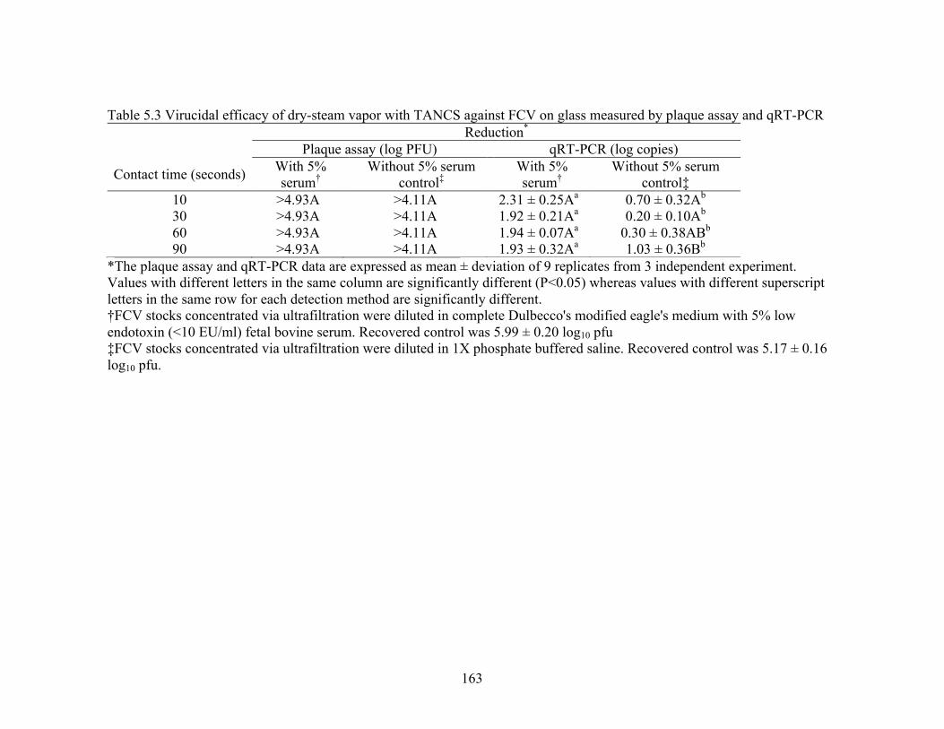
163
Table 5.3 Virucidal efficacy of dry-steam vapor with TANCS against FCV on glass measured by plaque assay and qRT-PCR Reduction* Plaque assay (log PFU) qRT-PCR (log copies)
Contact time (seconds) With 5% serum†
Without 5% serum control‡
With 5% serum†
Without 5% serum control‡
10 >4.93A >4.11A 2.31 ± 0.25Aa 0.70 ± 0.32Ab 30 >4.93A >4.11A 1.92 ± 0.21Aa 0.20 ± 0.10Ab 60 >4.93A >4.11A 1.94 ± 0.07Aa 0.30 ± 0.38ABb 90 >4.93A >4.11A 1.93 ± 0.32Aa 1.03 ± 0.36Bb
*The plaque assay and qRT-PCR data are expressed as mean ± deviation of 9 replicates from 3 independent experiment. Values with different letters in the same column are significantly different (P<0.05) whereas values with different superscript letters in the same row for each detection method are significantly different. †FCV stocks concentrated via ultrafiltration were diluted in complete Dulbecco's modified eagle's medium with 5% low endotoxin (<10 EU/ml) fetal bovine serum. Recovered control was 5.99 ± 0.20 log10 pfu ‡FCV stocks concentrated via ultrafiltration were diluted in 1X phosphate buffered saline. Recovered control was 5.17 ± 0.16 log10 pfu.

164
Table 5.4 Virucidal efficacy of silver dihydrogen citrate and dry-steam vapor with TANCS against FCV on wool and nylon carpet measured by plaque assay and qRT-PCR
Reduction* Treatment† Contact time Surface Plaque assay (log PFU) qRT-PCR (log copies)
SDC 60 minutes Wool 1.82 ± 0.19A -0.06 ± 0.26ANylon 3.62 ± 0.32B 0.49 ± 0.27B
Dry-steam 90 seconds Wool 3.80 ± 0.16B 0.03 ± 0.17ANylon 3.68 ± 0.09B 0.39 ± 0.23B
*The plaque assay and qRT-PCR data are expressed as mean ± deviation of 15 replicates from 3 independent experiment.Values with different letters in the same column are significantly different (P<0.05).†FCV stocks concentrated via ultrafiltration were diluted in complete Dulbecco's modified eagle's medium with 5% lowendotoxin (<10 EU/ml) fetal bovine serum.

165
Figure 5.1 Flow chart for performing disinfection efficacy testing

166
Figure 5.2. Effect of silver dihydrogen citrate on appear of wool and nylon carpet carriers between 0 and 24 hr (A – P)

167
CONCLUSIONS
HuNoV remain the leading cause of acute gastroenteritis worldwide, which can be
transmitted via person-to-person, food, water, or the environment. Current literature
shows that several key factors influence virus survival: temperature, RH, organic content,
deposition method, and virus type. Of the studies that evaluated different chemistries, it
was shown that chlorine, glutaraldehyde, and oxidizing vaporous products demonstrated
the best efficacy against enteric viruses on soft surfaces. Be that as it may, some of these
technologies may not be safe or practical on carpets. Furthermore, because the EPA
currently does not have a standard method for assessing virucidal activity of product on
nonlaunderable soft surfaces, to include recovery methods, there are no registered
products that can be reported to inactivate enteric viruses contaminated on
nonlaunderable soft surfaces.
Results presented here demonstrated that characterizing a soft surface can
improve our understanding of virus-soft surface interactions. Furthermore, infectious
HuNoV surrogates, FCV and MNV, can survive for extended periods of time on carpet
fibers. This survival can be affected by at least 2 factors: RH and surface type.
Specifically, low RH favors FCV and MNV survival while natural fibers, such as wool,
may provide a more protective environment compared to synthetic fibers and hard
surfaces. Additionally, the assessment of recovery methods is a critical first step to
improve the detection and quantification of viruses. Here, we tested 3 mechanistically
different recovery methods intended for HuNoV recovery from carpets. Bottle extraction
and the microbial vacuum exhibited higher recovery efficiencies compared to

168
macrofoam-tipped swabs. Moreover, detection of FCV and MNV via qRT-PCR can be
inhibited when recovered from wool and nylon carpets. Ultimately, each method
demonstrates merit for use under different conditions, e.g. efficacy testing and
environmental monitoring. Sanitization programs are essential for prevention and control
measures against HuNoV. Our efficacy testing found that silver dihydrogen citrate (SDC)
was efficacious against FCV in suspension, on glass, and nylon carpet. However, SDC is
sensitive to serum. On the other hand, steam vapor with TANCS technology was
efficacious on all surfaces tested and exhibited no loss to its efficacy in the presence of
serum. Furthermore, treatments with these technologies do not affect the aesthetic
appears of the carpets. Taken together, these results suggest that SDC, to remain
efficacious, should be used as a two-step cleaner while steam vapor with TANCS
demonstrated rapid inactivation and could be an appropriate sanitizing technology for
virally contaminated natural and synthetic carpets.
Although our findings suggest HuNoV can survive for extended periods of time
on carpet, future studies are still needed to validate the survival profiles of HuNoV. Also,
to usher in a standard for assessing the efficacy of sanitizers against viruses intended for
carpets, our ASTM International-adapted standard should be used as a platform in future
studies. While surrogate testing is an acceptable alternative for the study of HuNoV, there
are significant drawbacks. Based on SDC and steam-vapor’s efficacy demonstrated here,
these technologies warrant further testing with the human enteroid system that support
HuNoV propagation.