Innate immune response to tissue-specific infection: notochord ...
166
HAL Id: tel-01559163 https://tel.archives-ouvertes.fr/tel-01559163 Submitted on 10 Jul 2017 HAL is a multi-disciplinary open access archive for the deposit and dissemination of sci- entific research documents, whether they are pub- lished or not. The documents may come from teaching and research institutions in France or abroad, or from public or private research centers. L’archive ouverte pluridisciplinaire HAL, est destinée au dépôt et à la diffusion de documents scientifiques de niveau recherche, publiés ou non, émanant des établissements d’enseignement et de recherche français ou étrangers, des laboratoires publics ou privés. Innate immune response to tissue-specific infection : notochord infection in the zebrafish embryo Quang Tien Phan To cite this version: Quang Tien Phan. Innate immune response to tissue-specific infection : notochord infection in the zebrafish embryo. Human health and pathology. Université Montpellier, 2016. English. NNT : 2016MONTT082. tel-01559163
-
Upload
khangminh22 -
Category
Documents
-
view
0 -
download
0
Transcript of Innate immune response to tissue-specific infection: notochord ...

HAL Id: tel-01559163https://tel.archives-ouvertes.fr/tel-01559163
Submitted on 10 Jul 2017
HAL is a multi-disciplinary open accessarchive for the deposit and dissemination of sci-entific research documents, whether they are pub-lished or not. The documents may come fromteaching and research institutions in France orabroad, or from public or private research centers.
L’archive ouverte pluridisciplinaire HAL, estdestinée au dépôt et à la diffusion de documentsscientifiques de niveau recherche, publiés ou non,émanant des établissements d’enseignement et derecherche français ou étrangers, des laboratoirespublics ou privés.
Innate immune response to tissue-specific infection :notochord infection in the zebrafish embryo
Quang Tien Phan
To cite this version:Quang Tien Phan. Innate immune response to tissue-specific infection : notochord infection in thezebrafish embryo. Human health and pathology. Université Montpellier, 2016. English. �NNT :2016MONTT082�. �tel-01559163�

Délivré par UNIVERSITÉ MONTPELLIER
Préparée au sein de l’école doctorale CBS2 Et de l’unité de recherche UMR 5235-DIMNP
Spécialité : Microbiologie et Immunologie
Présentée par PHAN Quang Tiến
Soutenue le 22 Mars 2016 devant le jury composé de
Mme Emma COLUCCI-GUYON, Dr, Institut Pasteur Rapporteur
M Serge MOSTOWY, Dr, Imperial College London Rapporteur
Mme Annemarie H. MEIJER, Prof, Universiteit Leiden Examinateur
M Victoriano MULERO, Prof, Universidad de Murcia
Mme Mai NGUYEN-CHI, Dr, Université Montpellier
M Georges LUTFALLA, Dr, CNRS Montpellier
Examinateur
Examinateur
Directeur de thèse
Innate immune response to tissue-specific infection: Notochord infection in the
zebrafish embryo

Page 1
Résumé
Lors des infections bactériennes, selon les tissus infectés, et selon la nature
des pathogènes, l’organisme répond en mobilisant différents acteurs. Nous
avons décidé d’utiliser le modèle du zebrafish ou Danio rério pour étudier la
réponse immunitaire innée dans les situations d’infection bactérienne où les
phagocytes professionnels ne peuvent pas venir au contact direct des
bactéries. Pour cela, j’ai développé un modèle d’infection de la notochorde de
l’embryon de zebrafish. Lors de l’injection des bactéries dans ce
compartiment, les bactéries se retrouvent protégées par une épaisse gaine de
collag agocytes ne peuvent pas pénétrer. Alors que les mycobactéries,
protégées par la gaine de collagène ne sont pas détectées par les
phagocytes, les bactéries E. coli sont immédiatement détectées ce qui
déclenche une importante inflammation locale autour de la notochorde. Alors
que les bactéries E. coli, bien qu’inaccessibles à la phagocytose sont
éliminées dans les première 24 heures qui suivent l’injection, l’inflammation
dure plusieurs jours.
J’ai étudié les mécanismes qui conduisent à cette inflammation
persistante et ses conséquences à long terme sur le développement du
poisson. J’ai montré le rôle central de la cytokine IL1b dans ce processus, et
j’ai développé une lignée transgénique qui permet d’étudier l’induction de cette
cytokine in vivo chez le poisson.
J’ai ensuite étudié le rôle des deux principales populations de
phagocytes dans l’élimination des bactéries E coli. J’ai montré que les
macrophages ne sont pas impliqués dans la disparition des bactéries alors
que les neutrophiles, bien qu’incapable de pénétrer à l’intérieur de la gaine de
collagène sont nécessaires à l’élimination des bactéries.
J’ai ensuite montré que la myelopéroxidase et le monoxyde d’azote ne
sont pas impliqués dans l’élimination des bactéries alors que les espèces
réactives de l’oxygène produites par les neutrophiles sont nécessaires pour
éradiquer l’infection.

Page 2
Summary
In bacterial infections, according to the infected tissue and the nature of
pathogens, the body responds by mobilizing various actors. I decided to use
zebrafish or Danio rerio model to study the innate immune response to
bacterial infection in the situations that professional phagocytes cannot come
in direct contact with the bacteria. For this, I developed a model of infection in
the notochord of zebrafish embryo. Upon injection of bacteria in this
compartment, the microbes find themselves protected by the thick collagen
sheath where the phagocytes cannot penetrate. While mycobacteria are not
detected by phagocytes; E. coli bacteria are sensed and a significant local
inflammation around the notochord is mounted. The E. coli, although
inaccessible to phagocytosis are eliminated within the first 24 hours after
injection, the inflammation lasts several days.
I studied the mechanisms that lead to this persistent inflammation and its long
term consequences on the development of the fish. I showed the central role
of the cytokine IL1B in this process, and I developed a transgenic line that
allows studying in vivo the induction of this cytokine in fish.
I then studied the roles of the two main populations of phagocytes in the
elimination of E. coli. I revealed that macrophages are not involved in the
removal of bacteria but neutrophils, although unable to penetrate inside the
collagen casing, are necessary for the bacterial elimination.
I also confirmed that myeloperoxidase and nitrogen monoxide are not involved
in the removal of bacteria, rather the reactive oxygen species produced by
neutrophils are needed to eradicate the infection.

Page 3
ACKNOWLEDGEMENTS
The weather looks good for today and I am very pleased to take this
chance to express my gratefulness to all people who are always
supportive to help create this thesis!
First and foremost I thankfully acknowledge my supervisor, Georges
LUTFALLA and my mentor, Mai NGUYEN-CHI for offering me this
fantastic opportunity to pursue my PhD thesis at University of
Montpellier, and for all their advices during my lab time. It is my huge
pleasure working with them!
I would also like to thank all people from the FishForPharma Network
who are so friendly, funny and always available to share their
facilities and opinions at any time. I have special thanks to Annemarie
H. Meijer and other PIs for their giant efforts setting up this network
for me and other students to learn about the zebrafish model in an
international and cooperative environment!
This work could never be accomplished without advices, discussions
and supports from people at DIMNP, especially Etienne LELIEVRE,
Jana TRAVNICKOVA, Paul GUGLIELMI, Suma CHOORAPOIKAYIL,
Karima KISSA. Also, I would like to give my gratitude to Annette
VERGUNST and Jean-Pierre LEVRAUD for their fabulous advices and
for being members of my Comité de thèse, thanks to all!
I am joyful to mention Catherine GONZALEZ, Richard CASTEL,
Stéphane CASTEL who are not only extremely supportive to my lab
work and the “French” administrative paper work. These amazing
people always make my life in France just like home!
Being away is not easy. Fortunately thanks to all friends at the
Association of Vietnamese Students in Montpellier, we have got lot of
fun together!
Lastly, the deepest gratitude goes to my parents, my brothers and
sisters and my wife, Thanh Trúc. Thanks all for treating me with love,
always!

Page 4
Contents Résumé .................................................................................................................................. 1
Summary ............................................................................................................................... 2
ACKNOWLEDGEMENTS .......................................................................................................... 3
CHAPTER I: INTRODUCTION ................................................................................................... 7
I. Innate immune system in human .................................................................................... 7
I.1. General introduction ................................................................................................ 7
I.2. Innate immune response to bacterial infection ......................................................... 7
I.3. ROS productions and functions ............................................................................... 22
I.4. Cytokines and immunoregulation ......................................................................... 27
I.4.1. Resources and functions of cytokines .................................................................. 27
I.5. Characteristics and functions of inflammation ........................................................ 34
II. Zebrafish model for study of host-pathogen interaction ............................................... 36
II.1. Zebrafish biology and experimental advantages .................................................... 36
II.2. Zebrafish immune system ...................................................................................... 37
II.3. Innate immune response to bacterial infection ...................................................... 39
II.4. The zebrafish notochord ........................................................................................ 45
AIM OF THE THESIS .............................................................................................................. 47
OUTLINE OF THE RESULTS .................................................................................................... 49
Part I: Transient infection of zebrafish notochord with E. coli induces chronic inflammation
........................................................................................................................................ 49
Part II: Neutrophils control notochord infection at a distance with superoxide ................. 49
Part III: Identification of polarized macrophage subsets in zebrafish ................................. 50
Part IV: Rearing zebrafish larvae with Tetrahymenas ........................................................ 50
PART I .............................................................................................................................. 51
PART II ............................................................................................................................. 52
Abstract ....................................................................................................................... 53
Introduction ................................................................................................................. 54
Materials and Methods ................................................................................................ 56
Discussion .................................................................................................................... 63
Acknowledgements ...................................................................................................... 66
FIGURES AND LEGENDS ................................................................................................ 68
References ................................................................................................................... 82
PART III ............................................................................................................................ 85

Page 5
PART IV ............................................................................................................................ 86
Abstract ....................................................................................................................... 87
Introduction ................................................................................................................. 88
Materials and Methods ................................................................................................ 89
Results ......................................................................................................................... 91
Discussion .................................................................................................................... 95
Acknowledgements ...................................................................................................... 97
References ................................................................................................................... 97
CHAPTER III: DISCUSSION AND PERSPECTIVES ...................................................................... 99
1. Innate immune response to tissue-specific infection .................................................... 99
2. Roles of cytokines in chronic inflammation and bone diseases ................................... 100
2.1. Interleukin 1β ...................................................................................................... 100
2.2. TNFα ................................................................................................................... 102
3. Combat of neutrophils and E. coli, victory for the crowd ............................................. 103
General conclusion ..................................................................................................... 106
ANNEXES ........................................................................................................................... 107
1. Only live bacteria could induce chronic inflammation ................................................. 107
2. Chronic inflammation affects vertebral column ossification ........................................ 109
LIST OF TABLES ............................................................................................................... 110
ABBREVIATIONS ............................................................................................................. 110
LISTS OF FIGURES ........................................................................................................... 111
REFERENCES ................................................................................................................... 112

Page 6
CHAPTER I
INTRODUCTION
Zebrafish cycle
Memorial University

Page 7
CHAPTER I: INTRODUCTION
I. Innate immune system in human
I.1. General introduction
The human innate immunity is made up with layer-to-layer of defensive
structures ranging from physical and cellular to molecular entities which can be
recognized as followed: 1) The first barrier is the epithelium which is highly
structured with keratin. 2) The less structured mucosal membranes of the
respiratory and gastrointestinal tracts yet are equipped with thick protective
mucus. 3) The phagocyte population consists of neutrophils, macrophages,
dendritic cells, mast cells, eosinophils, basophils and NK cells which are able
to engulf microbes and destroy them with lysosomal and granular toxic
components. 4) The intracellular or secretory proteins including: a)
complements synthesized in the liver and in other local tissues, and b)
antimicrobial compounds mostly produced by epithelia and by phagocytes are
among the most important artillery of the innate immune system (Klebanoff et
al., 2013; Li et al., 2007; Paiva and Bozza, 2014). The common function of all
the aforementioned defensive layers is to deal with attacks or invasions of any
harmful foreign agents such as microbes, toxins or dangerous molecules in the
unspecific manners. The innate immunity also signals to activate the adaptive
immune system, the other arm of the host immune system (Charles A
Janeway et al., 2001).
I.2. Innate immune response to bacterial infection
The innate immunity plays crucial role in the control of wide range of microbial
infections. Dysfunction of this arm of the host defense results in sepsis and
other fatal symptoms (Albiger et al., 2007; Kobayashi and DeLeo). This thesis
focuses more on the functions of two most important cellular actors of the
innate immune system, neutrophils and macrophages.

Page 8
I.2.1. Neutrophils
I.2.1.1. Biology and function of neutrophils
The polymorphonuclear neutrophil is a member of granulocyte family which
includes also eosinophil and basophil. After being generated and matured in
the bone marrow neutrophils possessing pro-inflammatory molecules such as
cytokines, chemokines, antimicrobial and lytic compounds (depicted in figure
1) are released into the circulation where they join the innate immunity and
perform protective function (Rosenbauer and Tenen, 2007). Neutrophils are
the most abundant population of leukocytes in the blood (ATHENS et al.,
1961; Dancey et al., 1976). The normal number of neutrophils in the body is
actively regulated by hematopoietic organ and is known as steady-state
density. Upon infection neutrophils are massively produced in response to the
demand of fighting infection, this process is described as emergency
granulopoiesis (Manz and Boettcher, 2014; Panopoulos and Watowich, 2008).
In human, although having short-live characteristics, neutrophils are produced
massively at daily basis of approximately 100 billion cells.
The correct population of neutrophils is critical for host protection from
infections (Courtney et al., 2007; Donadieu et al., 2009). Patients with
neutropenia or impaired neutrophil function suffer severe viral, bacterial and
fungal infections which result in high rate of morbidity and mortality (Geiszt et
al., 2001; Newburger, 2006).

Page 9
Figure 1: Neutrophils deliver multiple anti-microbial molecules
Microbicidal products arise from most compartments of the neutrophil: azurophilic
granules (also known as primary granules), specific granules (also known as
secondary granules) and tertiary granules, plasma and phagosomal membranes,
the nucleus and the cytosol. BPI, bactericidal permeability increasing protein;
H2O2, hydrogen peroxide; HOBr,hypobromous acid; HOCl, hypochlorous acid;
HOI, hypoiodous acid; MMP, matrix metalloproteinase; 1O2, singlet oxygen; O2–,
superoxide; O3, ozone; •OH, hydroxyl radical; phox, phagocyte oxidase. Adapted
from (Nathan, 2006).

Page 10
I.2.1.2. Mechanism of bacterial killing by neutrophils
Neutrophils have been shown to use different mechanisms to control infections
(depicted in figure 3). The primary mean is professional phagocytosis in which
neutrophils engulf microbes into their phagosomes. Following phagocytosis,
infecting microorganisms are destroyed in the phagosomes by abundant and
diverse sets of antimicrobial compounds including Reactive Oxygen Species
(ROS), mainly produced by nadph oxidase complexes, and other toxic
molecules such as myeloperoxidase, neutrophil elastase, defensins,
azurocidin, protease 3, cathepsin G, lysozyme, lipocalin delivered from
granules in the cytoplasm. These granular antimicrobial molecules, mostly
from specific granules, could also be released to the extracellular environment
to cope with invading agents as well as to facilitate the extracellular matrix
degradations (Abdel-Latif et al., 2005; Fang, 2011; Kolaczkowska and Kubes,
2013a). The massive production of ROS, especially the superoxide, in the
phagosome is suggested to cause damage and make the ingested bacteria
susceptible to other granular antimicrobial effectors (Slauch, 2011). In addition
to the intracellular decomposition by phagocytosis, neutrophils were also
described by Brinkmann in 2004 to perform the extracellular killing mechanism
named Neutrophil Extracellular Traps (NETs).
In this process neutrophils usefully suicide themselves to trap the spreading of
microbes by expelling their chromatin associated with range of antimicrobial
molecules (Brinkmann et al., 2004; Halverson et al., 2015; Papayannopoulos
and Zychlinsky, 2009). Recently the growing data have demonstrated that
NETs are used in response to varieties of stimuli, such as: PMA, LPS, fungi,
bacteria and protozoan (Fuchs et al., 2007; Guimarães-Costa et al., 2009;
Remijsen et al., 2011c). Interestingly, neutrophils have been shown to release
NETs in response to large pathogens which could not be engulfed by
conventional phagocytosis such as C. albicans hyphae (Branzk et al., 2014).

Page 11
Figure 2: Killing mechanisms of neutrophil
Neutrophils can eliminate pathogens by multiple means, both intra- and
extracellular. When neutrophils encounter microorganisms, they phagocytose
them. After being encapsulated in phagosomes, the pathogens are killed by
NADPH oxygenase-dependent pathway through reactive oxygen species or by
antibacterial proteins such as cathepsins, defensins, lactoferrin and lysozyme.
These molecules are released from the neutrophil granules either into
phagosomes or into the extracellular milieu, thus acting on either intra- or
extracellular pathogens, respectively. Highly activated neutrophils can stop the
spreading and eliminate extracellular microorganisms by releasing neutrophil
extracellular traps (NETs). NETs are composed of a core DNA element to
which histones, antimicrobial proteins (for example, lactoferrin and cathepsins)
and enzymes (for example, MPO and neutrophil elastase) that are released
from neutrophil granules are attached. Adapted from (Kolaczkowska and
Kubes, 2013a).

Page 12
The formation of NETs need stimulation and correct contribution of key cellular
components. ROS is one of the important players and its role was shown in
patients with Chronic Granulomatous Disease whose neutrophils failed to
produce ROS due to mutation in the gene encoding nadph oxidase complex
(Fuchs et al., 2007). The nadph oxidase-derived superoxide, a major member
of ROS, was also shown to involve in the activation of neutrophil elastase (NE)
another actor of NETs forming process (Reeves et al., 2002). Upon stimulation
NE and Myeloperoxidase are translocated from cytosolic granules into the
nucleus where NE promotes fragmentation of chromatin and NETs expulsion
(Metzler et al., 2011; Papayannopoulos et al., 2010).
For all three neutrophil killing mechanisms, neutrophil granules with their
components exhibit central regulatory function. Generally the neutrophil
granules are classified into three groups: the primary or azurophilic granule
contains BPI, neutrophil elastase, cathepsin G, protease 3, azurocidin,
myeloperoxidase; the secondary or specific granule and the tertiary or
gelatinase granule contain Lactoferrin, lipocalin, lysozyme, matrix
metalloproteinase (MMP8-9). Since these granular microbicides are also
devastating to host cells, it is crucial that their deliveries into phagosomes
(depicted in figure 3) are correctly regulated to avoid host tissue damages
(Nathan, 2006).
Although facing different neutrophil effectors, many microbes have evolved
variety of mechanisms to survive neutrophil elimination; some of which include
turning on stress response, avoiding contact with neutrophils, inhibiting
phagocytosis, surviving intracellular, inducing cell death or destroying NETs
(Corleis et al., 2012; de Haas et al., 2004; Fradin et al., 2005; Rubin-Bejerano
et al., 2003; Urban et al., 2006).

Page 13
Figure 3: Killing of bacteria in neutrophil phagosome
Upon engulfment of bacteria into the neutrophil phagosome, granules fuse with
the phagosome and the phagocytic NADPH oxidase is assembled and activated
on the phagosomal membrane. By transporting electrons from cytosolic NADPH,
the phagosomal membrane depolarizes as superoxide is produced in the
phagosome lumen. Superoxide gives rise to the whole spectrum of reactive
oxygen species that are highly reactive and attack bacteria. The Nox2-generated
depolarization drives protons and potassium ions into the phagosome. The
protons maintain a neutral pH in the phagosome required for optimal protease
activity and sustained oxidase function. Potassium ions liberate and activate latent
proteases from the granule matrix, allowing them to attack and destroy bacteria.
Adapted from (Rada and Leto, 2008).

Page 14
I.2.2. Macrophages
I.2.2.1. Biology and function of macrophages
The mononuclear phagocyte, macrophage, was originally discovered by
Metchnikoff to have the ability to phagocytose. Macrophages are found in
many different tissues throughout the body with diverse appearances and
functions and known as resident macrophages (Wynn et al., 2013). They are
produced in the yolk sac, the fetal liver and then by the bone marrow and
distributed towards specific tissues during embryonic development and
persisted in tissues until adulthood partly by self-renewal mechanism (depicted
in figure 4-5) (Gautier et al., 2012; Ginhoux and Jung, 2014). From the
functional aspect, macrophages are categorized in two main phenotypes M1
and M2 with different profiles of gene expressions, functions and general
behaviors in response to infections or host homeostasis. Upon infections with
gram-negative or gram-positive bacteria M1 macrophages express relatively
similar transcriptional profiles of genes involved in pro-inflammation process,
while M2 macrophages are reported to play important roles in the inflammation
resolution, tissues repair and homeostasis by expressing anti-inflammatory
genes (Benoit et al., 2008). Although having different mechanisms of activity,
both functional M1 and M2 macrophages are crucial for the clearance of
cellular debris and the control of inflammation and infections with bacteria or
viruses. Macrophages perform their protective function through diverse
modulations of sets of cytokines, chemokines and receptors. Mice deficient for
TNF or IFN-γ, two of the important cytokines produced by M1 macrophages, or
their respective receptors are susceptible to Listeria monocytogenes infection
(Pfeffer et al., 1993). The intense inflammatory reaction promoted by
uncontrolled expression of cytokines in macrophages is also fatal in certain
conditions. In severe sepsis patients macrophages produce high level of type-
1 cytokines which in turn induce death through heart arrest and block of
perfusion (Bozza et al., 2007; Smith et al., 2007). As professional phagocytes,
macrophages also act as antigen presenting cells (Hume, 2008;
Koppensteiner et al., 2012).

Page 15
Figure 4: Embryonic macrophage development
Primitive haematopoietic progenitors generated in the yolk sac blood islands gives rise to
primitive macrophages from embryonic day 8.5–9.0 (E8.5–E9.0). They populate embryos
upon the formation of the blood circulatory system and colonize the whole embryo from
E9.0–E10.0 to develop into fetal primitive macrophages. In parallel, definitive
haematopoiesis occurs in the aorta–gonads– mesonephros (AGM) from E10.5 and is
responsible for the generation of haematopoietic stem cells (HSCs) with multi-lineage
haematopoietic potential, that give rise to progenitors that colonize the fetal liver.
Haematopoiesis in the fetal liver generates monocytes around E11.5–E12.5 that migrate
into the blood (E12.5–E13.5) and then invade embryonic tissues around E13.5–E14.5, a
few days after yolk sac-derived macrophage colonization. Of note, yolk sac progenitors can
also contribute to fetal liver haematopoiesis during the middle phase of embryonic
development (from E8.5) and might contribute to the generation of fetal liver monocytes
(dashed arrow). Once inside the tissues, fetal liver-derived monocytes proliferate and
differentiate into macrophages and markedly dilute the population of yolk sac-derived
macrophages as shown in the lungs and in the heart. Exceptions are the microglia, which
arise predominantly from yolk sac-derived macrophages; and Langerhans cells, which
mainly arise from fetal liver-derived monocytes but retain a detectable yolk sac-derived
macrophage component. These recent findings reveal that the origin of adult
macrophages in the steady state can vary considerably between tissues. Moreover, in the
steady state, particular tissues — such as the skin and gut — contain adult
monocyte-derived macrophages. CNS, central nervous system; CSF1R,
colony-stimulating factor 1 receptor. Adapted from (Ginhoux and Jung, 2014).

Page 16
Figure 5: Monocyte-derived macrophage development
Monocytes are continuously generated in the bone marrow from haematopoietic
stem cells (HSCs) via macrophage and dendritic cell (DC) precursor (MDP) and
common monocyte progenitor (cMoP) intermediates. In the steady state, LY6Chi
and LY6Clow monocyte subsets in the circulation form a developmental
continuum but are functionally distinct. Macrophage-like LY6Clow cells in the
blood vessel lumen patrol the endothelial surface and coordinate its repair by
recruiting neutrophils as required. LY6Chi monocytes are rapidly recruited to
sites of inflammation and sites of tissue remodeling, where they extravasate and
can give rise to monocyte-derived DCs and monocyte-derived macrophages.
Mouse LY6Chi and LY6Clow cells are the equivalent of human CD14+ and
CD14lowCD16+ monocyte subsets, respectively. CCR2, CC-chemokine
receptor2; CSF1R, colony-stimulating factor 1 receptor; CX3CR1,
CX3C-chemokine receptor1; GMP, granulocyte–macrophage progenitor.
Adapted from (Ginhoux and Jung, 2014).

Page 17
I.2.2.2. Mechanism of bacterial killing by macrophages
Besides the roles in tissue repair and cellular debris cleaning, tissue-resident
macrophages guard the epithelial layer and actively join other professional
phagocytes of the innate immune system to fight invading pathogens at the
very early phase of attack (depicted in figure 6). To support phagocytosis,
macrophages possess classes of pattern recognition receptors (PRRs) located
on the extracellular- or intracellular- or vesical surface which help sense
directly the pathogens through their pathogen-associated molecular patterns
(PAMPS) on the bacterial surface such as peptidoglycans or lipoproteins and
promote internalization of these bacteria into phagosomes. In some cases the
foreign objects or the altered-self are opsonized by soluble PRRs and
components of the complement system so that they would be recognized by
other types of phagocytic receptors, for example Fcγ receptor (FcγR),
complement C3 receptor (CR3), and internalized by the phagocytes (depicted
in figure 7) (Litvack and Palaniyar, 2010; Weiss and Schaible, 2015). Upon
contact with microbes, cascades of pro-inflammatory signals linked to PRRs
are activated to promote production of other alarming molecules such as
cytokines, chemokines and antimicrobial compounds. These signaling
molecules such as interleukin-1b (IL1b), interleukin-6 (IL6), interleukin-8 (IL8),
tumor necrosis factor alpha (TNFa) activate other phagocytes including
neutrophils and promote recruitment of these leukocytes to join the fight at the
site of infection (Arango Duque and Descoteaux, 2014; Kapetanovic et al.,
2007). It is noted that the downstream signaling cascades of each PRR is
activated differently for that particular PRR-mediated internalization. For Fcγ
receptor for example, the pseudopod growing requires actin polymerization
and is controlled by Rac1/2 plus the cell division control protein 42 (Cdc42)
(Caron and Hall, 1998). In contrast, the CR3-mediated phagocytosis is
regulated by interaction of diaphanous-related formin Dia1 and Ras GTPase-
Activating-Like Protein IQGAP1(Brandt et al., 2007).
Page 18
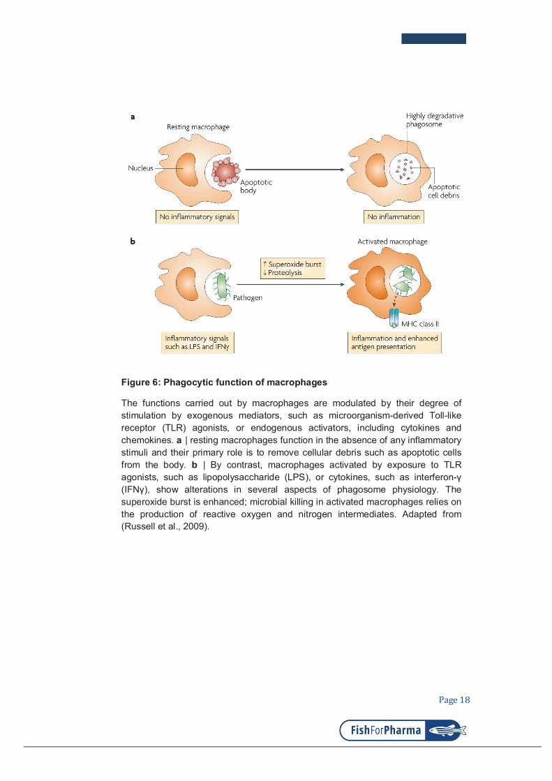
Figure 6: Phagocytic function of macrophages
The functions carried out by macrophages are modulated by their degree of stimulation by exogenous mediators, such as microorganism-derived Toll-like receptor (TLR) agonists, or endogenous activators, including cytokines and chemokines. a | resting macrophages function in the absence of any inflammatory stimuli and their primary role is to remove cellular debris such as apoptotic cells from the body. b | By contrast, macrophages activated by exposure to TLR agonists, such as lipopolysaccharide (LPS), or cytokines, such as interferon-γ
(IFNγ), show alterations in several aspects of phagosome physiology. The superoxide burst is enhanced; microbial killing in activated macrophages relies on the production of reactive oxygen and nitrogen intermediates. Adapted from (Russell et al., 2009).

Page 19
Following the phagocytosis, a phagosome carrying bacteria is translocated
into the cytoplasm and matures through multiple interactions and fusions with
endosomal and other vesical body, and eventually fuse with lysosomes for the
degradation of the engulfed items (Zimmerli et al., 1996).
The interactions and fusions of the phagosome with cytoplasmic vesical
compartments require involvements of different factors that first bring the
fusing bodies close to each other and then bridge and join the compartments.
This process is regulated by cytoplasmic effectors such as the family of Rab
GTPases proteins and soluble N-ethylmaleimide-sensitive factor attachment
protein receptor (SNARE) (Becken et al., 2010), During this process
phagosomes become acidified to the low pH of 4-4.5, which is usually optimal
for the activities of most lysosomal proteases.
Continuing accumulation and alteration the intracellular components result in
maturation of phagosome which possessing a diverse sets of lytic oxidative
compounds that are highly toxic and degradative to the trapped agents (Weiss
and Schaible, 2015). However, some intracellular bacteria have developed
strategies to survive in the phagosome of macrophages and eventually spread
their infections systemically. Being engulfed in the macrophage Salmonella
Typhimurium can modify its outer membrane to become more resistant to
antimicrobial peptides and evade recognition by host (Ernst et al., 2001). The
intracellular bacteria Francisella tularensis is able to survive macrophage
killing by altering the maturation of its phagosome and accessing the host cell
cytoplasm (Clemens et al., 2004).

Page 20
Table 1: Receptors involved in the phagocytosis
Type of
phagocytosis Receptors Ligands
Opsonic
phagocytosis
Fc receptor family (FcγRI,
FcγRIIA and FcγRIIA) Antibody-opsonized targets
Complement receptors (CR1,
CR3 and CR4) Complement-opsonized targets
α5β1 integrin Fibronectin
Non-opsonic
phagocytosis
Dectin 1 β-glucan
Macrophage receptor MARCO Bacteria (undefined specific
ligand)
Scavenger receptor A Bacteria (diverse charged
molecules)
αVβ5 integrin Apoptotic cells
Triggered
(nonspecific)
phagocytosis
Toll-like receptors
Various, including
lipopolysaccharides
and lipopeptides
FcγR, Fc receptor for IgG (Underhill and Goodridge, 2012)

Page 21
Figure 7: Process of the phagocytosis
As phagocytosis proceeds from the initial binding of a target to actin-dependent
internalization and ultimately to degradation of the target in the phagolysosome,
myeloid cells acquire information about the target through a variety of mechanisms.
At the cell surface, receptors sample the chemical constituents of the particle and
membrane dynamics facilitate an assessment of its physical properties. Additional
information is gathered as the phagosome pinches off from the plasma membrane
and as it matures through interactions with other intracellular compartments. Finally,
the degradation of the target exposes ligands that were not previously accessible
and releases ligands into the cytosol for detection by intracellular receptors. The
information gathered by all of these processes is integrated to shape the ensuing
immune response. MHC: major histocompatibility complex. Adapted from (Underhill
and Goodridge, 2012).

Page 22
I.3. ROS productions and functions
ROS are highly reactive oxygen-derived molecules produced by
transmembrane multiprotein NADPH Oxidase complexes (NOXs) and DUOX
enzymes that are present in many different cell types and tissues throughout
the body. The NOX family comprises five isoforms named NOX1-NOX5 which
work as electron transporters once assembled with of other particular units
such as p40, p47, p22phox and Racs (depicted in figure 8). The DUOX enzymes
encoded by duox1 and duox2 genes which found to be mostly expressed in
thyroid (Bedard and Krause, 2007). While located on different chromosomes
Duox genes share similar functional domain with Noxs in the conversion of
oxygen into superoxide. While both Duox1 and Duox2 are highly expressed in
thyroid, Duox1 expression is also detected in respiratory epithelia and
prostate, and Duox2 found in gastrointestinal tract and rectal mucosa (De
Deken et al., 2000; Dupuy et al., 1999; Forteza et al., 2005; Geiszt et al., 2003;
Schwarzer et al., 2004). In recent years the most intensively studied species of
ROS include superoxide anion, hydrogen peroxide, singlet oxygen and
peroxynitrite, whose half-lives vary from milliseconds to seconds and hours in
most biological processes with diverse and complicated functions (Bedard
and Krause, 2007; Lushchak, 2014). Through NOX complexes, oxygen is
converted into superoxide which subsequently reduced to hydrogen peroxide
and other halide radicals by superoxide dismutase and peroxidase
respectively (Fridovich, 1997). It is suggested that based on the concentrations
in their microenvironments ROS have distinct biological functions in signaling,
cell death, tissue homeostasis, inflammation and control of infections (Dupré-
Crochet et al., 2013; Henrotin et al., 2003).
Hundreds of publications have revealed the wide spectrum of ROS
productions and functions in many cell types and tissues. Their correct
balances affect critically the regular function and physiology of the adipose
tissue in response to insulin; the maturation of and function of spermatocytes;
cardiovascular system, central nervous system, endocrine organs,
gastrointestinal system, liver, kidney, urinary tract, lung and airways (Bedard
and Krause, 2007; Mikkelsen and Wardman, 2003).

Page 23
Figure 8: NADPH oxidase and DUOX isoforms
Despite their similar structure and enzymatic function, NOX family enzymes differ
in their mechanism of activation. NOX1 activity requires p22phox, NOXO1 (or
possibly p47phox in some cases) and NOXA1, and the small GTPase Rac.
NOX2 requires p22phox, p47phox, p67phox, and Rac; p47phox phosphorylation is
required for NOX2 activation. Although not absolutely required, p40phox also
associates with this complex and may contribute to activation. NOX3 requires
p22phox and NOXO1; the requirement for NOXA1 may be species dependent, and
the requirement of Rac is still debated. NOX4 requires p22phox, but in reconstitute
systems it is constitutively active without the requirement for other subunits.
However, in native NOX4-expressing cells, activation, possibly including Rac, has
been described. NOX5, DUOX1, and DUOX2 are activated by Ca2+ and do not
appear to require subunits. Adapted from (Bedard and Krause, 2007).

Page 24
Table 2: NOX enzymes expression in different tissues
(adapted from (Bedard and Krause, 2007)
In the context of the bone, ROS are produced in osteoclasts, macrophage-like
cells which function in bone resorption, and they play roles in osteoclast
differentiation and activity (Lee et al., 2005; Yang et al., 2001). ROS
production has also been found to cause matrix remodeling, cartilage
degradation and inflammation of the articular joint tissues usually seen in
osteoarthritis and rheumatoid arthritis (Henrotin et al., 2003).
In phagocytes ROS are generated, especially by NOX2, in response to
infections. Patients with severe defect in the control of infection in chronic
granuloma disease were found to lose oxidative burst activity due to mutations
effecting the functional assemble of NOX complexes (Klebanoff, 2005; Stasia
and Li, 2008). With the above observation, ROS were previously mentioned as
effectors of bacterial killing. However, researchers also found that the
neutrophils from CGD patients did not need NOX2 to clear many types of other
bacteria (Winkelstein et al., 2000). While the presence of ROS is important for
the control of many types of infection, their chemical interactions with diverse
sets of granular compounds in phagocytes have been shown to contribute
critically to the clearance of bacterial infections.

Page 25
Upon bacterial infection, phagocytes engulf microbes into their phagosomes.
During the phagosomal maturation, together with the delivery of other
antimicrobial compounds, superoxide is massively produced by NADPH
Oxidase complexes into the bacterial containing phagosomes. Superoxide
together with H2O2, another ROS, are also the inputs of productions of other
ROS such as singlet oxygen, ozone, hydroxyl radical which are essential for
the bacterial killing and degradation in the direct manner (Rada and Leto,
2008; Robinson, 2008). Alternatively, Segal proposed that the high quantity of
negatively charged superoxide released into the phagosome by NADPH
Oxidase complexes must be compensated by ions; proton transferring into
phagosome would in turn creates conductive electrophysiology which is
deleterious to microbes (Segal, 2006). However, other hypotheses have
suggested that the cooperation between superoxide and antimicrobial peptides
seems instrumental for the bacterial killing (Slauch, 2011).
Recent studies have pointed out that, as the requirement of autophagy, ROS
also have a function in the formation of NETs promote the translocations of
Neutrophil Elastase (NE) and Myeloperoxydase (MPO) into the nucleus, these
components subsequently induce de-condensation and fragmentation of the
chromatin, a critical step in NETosis (Björnsdottir et al., 2015;
Papayannopoulos et al., 2010; Remijsen et al., 2011a; Remijsen et al., 2011b;
Stoiber et al., 2015). Action mechanism of ROS are depicted in figure 9.

Page 26
Figure 9: Distribution of ROS and their interactions with NETs formation
Arrows indicate directions of effects. Note that only some of
the processes shown can co-occur. CatS cathepsin S; citH3 citrullinated histone
H3; C5aR complement component 5a receptor; DOCK dedicator of cytokinesis
proteins; HOCl hypochlorous acid; HOCSN hypothiocyanous acid; MASPK
mitogen-activated protein kinases, ERK extracellular signal-regulated kinases,
positive regulators of autophagy; PI3K phosphoinositide-3-kinase, mTOR
mammalian target of rapamycin, negative regulators of autophagy and apoptosis;
MPO myeloperoxidase; NE neutrophil elastase; NFkB nuclear factor kappa-light-
chain-enhancer of activated B cells; OXPHOS oxidative phosphorylation; PAD4
peptidylarginine deiminase 4; PKC protein kinase C; SK3 small conductance
calcium-activated potassium channel 3; SOD superoxide dismutase; TLR4 toll-
like receptor 4. Adapted from (Stoiber et al., 2015).

Page 27
I.4. Cytokines and immunoregulation
I.4.1. Resources and functions of cytokines
Cytokines are the extremely diverse group of small soluble proteins and
glycoproteins whose functions are to transfer signaling messages from cells to
cells throughout the body, which help regulate the host immune activity, tissue
homeostasis and hematopoiesis in both physiological and pathological
conditions (Foster, 2001).
The general acting mechanism of cytokines is their pleiotropic bindings to
transmembrane receptors which in turn trigger cascades of intracellular
reactions leading to downstream modifications in gene expression patterns.
Cytokines can act autonomously (autocrine), on the neighboring cells
(paracrine), or on the cells at a distance (endocrine). Functionally, depending
on particular stimulating signals different cytokines might have stimulatory or
inhibitory effects and synergistic or antagonistic effects (Matsumoto and
Kanmatsuse, 2000).
Many types of cells may produce cytokines including immune cells, kidney
cells, liver cells and fibroblasts. However, the main sources of cytokines are
epithelial cells, leukocytes and lymphocytes. According to their functions and
origins cytokines are known by other names such as lymphokine, which is
produced by lymphocytes; monokine, produced by monocytes; interleukin,
produced by leukocytes; chemokine, having chemotactic property. Their
expressions are strictly and dynamically regulated when host encounters
stimulating signals from for example infection, trauma, injury, cancer and
physiological disorder (Hanada and Yoshimura; Orman et al., 2011).
Accumulating evidence has disclosed the functions of cytokines in many
different pathological conditions such as neurodegeneration, cancer,
hepatocellular injury, cirrhosis, neoplasia, hematopoiesis disorders, infectious
and inflammatory diseases (Elenkov et al., 2005; Lucey et al., 1996; Martins et
al., 2010; O’Shea et al., 2002). Function, production, classification and
receptors of some known cytokines are depicted in figure 11, table 3 and table
4.

Page 28
Table 3: Cytokines production and function
Cytokines Main sources Target cell Major function
Interleukins
IL-1 Macrophages, B cells, DCs
B cells, NK cells, T-cells
Pyrogenic, proinflammatory, proliferation, differentiation, BM cell proliferation
IL-2 T cells Activated B-and T-cells, NK cells
Proliferation and activation
IL-3 T cells, NK cells Stem cells Hematopoietic precursor proliferation and
differentiation
IL-4 Th cells B cells, T cells, macrophages
Proliferation of B and cytotoxic T cells, enhances MHC class II expression, stimulates
IgG and IgE production
IL-5 Th cells Eosinophils, B-
cells
Proliferation and maturation, stimulates IgA and IgM production
IL-6 Th cells,
macrophages, fibroblasts
Activated B-cells, plasma cells
Differentiation into plasma cells, IgG production
IL-7 BM stromal cells,
epithelial cells Stem cells B and T cell growth factor
IL-8 Macrophages Neutrophils Chemotaxis, pro-inflammatory
IL-9 T cell T cell Growth and proliferation
IL-10 T cell B cells,
macrophages Inhibits cytokine production and mononuclear
cell function, anti-inflammatory
IL-11 BM stromal cells B cells Differentiation, induces acute phase proteins
IL-12 T cells NK cells Activates NK cells
TNFs
TNF-α Macrophages Macrophages Phagocyte cell activation, endotoxic shock
TNF-β Monocytes, T-cells Tumour cells, Phagocytes
Tumour cytotoxicity, cachexia, Chemotactic, phagocytosis, oncostatic, induces other
cytokines
interferons
IFN-α Leukocytes Various Anti-viral
IFN-β Fibroblasts Various Anti-viral, anti-proliferative
IFN-γ T-cells Various
Anti-viral, macrophage activation, increases neutrophil and monocyte function, MHC-I and -
II expression on cells
CSF
G-CSF Fibroblasts, endothelium
Stem cells in BM Granulocyte production
GM-CSF
T cells, macrophages, fibroblasts
Stem cells Granulocyte, monocyte, eosinophil production
M-CSF Fibroblast,
endothelium Stem cells
Monocyte production and activation
Erythropoietin
Endothelium Stem cells Red blood cell production
Others TGF-β T cells and B cells Activated T and
B cells
Inhibit T and B cell proliferation, inhibit haematopoiesis, promote wound healing
BM, bone marrow; DCs, dendritic cells; G-CSF, granulocyte-colony stimulating factors; M-CSF, macrophage colony stimulating factor; Th, T helper cells; TFNs, Tumour Necrosis Factors. Adapted from (Turner et al., 2014).

Page 29
Figure 10: Some cytokines and their receptors
Cytokines signal via oligomers of single-pass, type I transmembrane receptors,
with distinct extracellular domains for ligand binding and intracellular domains that
allow signal transduction. The receptor for IL-1 is a complex of IL-1R1 and the IL-1
receptor accessory protein (IL-1RAcP) formed following ligand binding to the
extracellular immunoglobulin domains (IGDs); intracellular signaling is mediated
via the Toll/IL-1R (TIR) domain. Trimeric TNFα binds to the cysteine-rich domains
(CRDs) of the pre-assembled trimmers of the TNF receptor (TNFR) and signaling
is mediated via the receptor's death domain (DD). Finally, the IL-6R is a multimeric
structure with IL-6R chains complexed with gp130. The ligand-binding region is
within the fibronectin III (FIII) modules of the IL-6R chains but signal transduction
is via the associated gp130 molecules. Adapted from (Turner et al., 2014).

Page 30
I.4.2. Roles of cytokines during infection and inflammation
Cytokines are dynamically expressed by variety of immune cells, and also
epithelial cells in response to infectious agents or inflammatory stimuli. Very
quickly following the onsets of infection or inflammation a primary set of
cytokines is transcribed to mount the inflammation called pro-inflammatory
cytokines. At the later phase another set of cytokines is produced to resolve
the inflammation known as anti-inflammatory cytokines.
The best characterized pro-inflammatory cytokines, including interleukin-1
family (IL-1), interleukin-6 family (IL-6), interleukin-17 (IL-17), interleukin-18
(IL18), Tumor Necrosis Factor alpha (TNFα), interferon gamma (IFNγ) and set
of chemokines such as G-CSF, GM-CSF, Eotaxin, MCP-1 (CCL2), MIP-1
(CCL3) etc., promote inflammation by sending out signals to stimulated and/or
guide the recruitments of immune cells and other effectors to the site of
infection to help control the infection. In the case of severe infections, the
recruited cells then also actively produce large amount of pro-inflammatory
cytokines to attract more innate immune components to the inflammatory foci.
After clearing the infecting agents or danger signals immune cells at the
inflammatory foci change their programs to produce anti-inflammatory
cytokines including interleukin-4 (IL-4), interleukin-10 (IL-10), interleukin-13
(IL-13) etc., to stop the recruitment of immune cells, promote inflammation
resolution and tissue healing. The modulations of pro-inflammatory and anti-
inflammatory cytokines are strictly controlled. However, very often they have
time-overlapping of their expressions; also some cytokines have both pro- and
anti-inflammatory effects such as interleukin-4, interleukin-6 (Scheller et al.,
2011; Seruga et al., 2008; Van Kampen et al., 2005).
The correct productions of cytokines are critical in many infectious and
inflammatory diseases. Patients with Inflammatory Bowel Disease (IBD) or
Rheumatoid arthritis (RA) who have imbalance between pro-inflammatory and
anti-inflammatory cytokines usually experience serious damages caused by
chronic inflammations. In case of RA the uncontrolled prolonged expression of
pro-inflammatory cytokines especially TNFα and interleukin-1β leads to severe

Page 31
destruction of cartilage and loss of the bone (Baum and Gravallese, 2014;
Neurath, 2014; Schett, 2011; Schuerwegh et al., 2003) .
Upon infections with pneumococcus or influenza virus cytokines appear to
have important role for the outcome or infections. First they are produced with
other chemokines as a network to regulate the inflammation by activating the
recruited neutrophils. The activated neutrophils then produce microbicides
such as reactive oxygen species, defensins, proteolytic enzymes,
myeloperoxidase etc., to control the infections. It is observed in many cases
that the above cytotoxic agents from neutrophils might be massively released
into the extracellular microenvironment and cause devastating damages to
host tissues by the overstimulation of pro-inflammatory cytokines (Barnes,
2008; Bordon et al., 2013; Goodman et al., 2003).

Page 32
Table 4: Classification of cytokines by immune responses
TSLP: Thymic Stromal Lymphopoietin; CNTF: ciliary neurotrophic factor; CT-1:
cardiotrophin-1; GM-CSF: granulocyte macrophage-colony stimulating factor; IFN:
interferon; LIF: leukaemia inhibitory factor; OPN: osteopontin; OSM: oncostatin M;
TSLP, thymic stromal lymphopoietin; BAFF: B cell activating factor; APRIL : a
proliferation inducing ligand. Adapted from (Turner et al., 2014).
Function Family Members
Adaptive
Immunity
Common γ chain
receptor ligands IL-2, IL-4, IL-7, IL-9, IL-15, IL21
Common β chain (CD131) receptor ligands
IL-3, IL-5, GM-CSF
Shared IL-2β chain (CD122)
IL-2, IL-15
Shared receptors IL-13, IL-13R-IL-4R complex
TSLP (TSLPR-IL-7R complex)
Pro-inflammatory singaling
IL-1 IL-1α, IL-1β, IL-1RA, IL-18, IL-33, IL-36α, IL-36β, IL-36γ, IL-36RA,
IL-37, IL-1Hy2
IL-6 IL-6, IL-11, IL-31, CNTF, CT-1, LIF, OPN, OSM
TNFα TNFα, TNFβ , BAFF, APRIL
IL-17 IL-17A-F, IL-25 (IL-17E)
Type I IFN IFNα, IFNβ, IFNω, IFNk, Limitin
Type II IFN IFNγ
Type III IFN IFNλ1 (IL-29), IFNλ2 (IL-28A), IFNλ3 (IL-28B)
Anti-inflammatory singaling
IL-12 IL-12, IL-23, IL-27, IL-35
IL-10 IL-10, IL-19, IL-20, IL-22,
IL-24, IL-26, IL-28, IL-29

Page 33
I.4.3. Targeting cytokines as therapeutics of inflammatory diseases
Thanks to the understandings of cytokine-receptor interaction mechanisms
many cytokine-based therapeutics have been developed rapidly over the past
decades. The treatment formulation is based on the seemingly simple
strategies which are either to suppress or induce the ligand or/and receptor
expressions, or to inhibit the interaction between them by antagonistic
molecules or antibodies.
Since the onset of this application in 1960s recombinant cytokines have been
successfully produced and used in clinics; exemplified as the utilizations of
colony stimulating factors to induce granulopoiesis in patients suffering
neutropenia during the course of chemotherapy, or of recombinant interferons
as treatments for chronic hepatitis and osteosarcoma (Garcia-Carbonero et al.,
2001; Gresser et al., 1969; Müller-Redetzky et al., 2015; Vilcek and Feldmann,
2004).
Treatment of Rheumatoid arthritis (RA) by blocking TNF activity with
monoclonal antibodies and recombinant TNF receptor has been demonstrated
to be effective. However, the recent data have marked the inefficacy and
failure in response to certain TFN antagonists (Emery, 2012; Rubbert-Roth
and Finckh, 2009; Solau-Gervais et al., 2006). Interleukin-1 and Interleukin-6
antagonists have also been developed for treatments of RA and other
autoimmune and chronic inflammatory diseases (Hennigan and Kavanaugh,
2008; Jones and Moreland, 1999; Krishnan, 1999; Tanaka and Kishimoto,
2012). The combination therapy using antagonists of both cytokines TFN and
IL-1 appeared to have higher curing efficacy by preventing bone erosion and
cartilage damage (Zwerina et al., 2004).

Page 34
I.5. Characteristics and functions of inflammation
In mammal the immunity plays indispensable function in orchestrating host
adaptation to harmful stimuli such as infection and tissue injury, by employing
a fundamental mechanism called inflammation. The inflammatory response is
usually ignited when components of the immune system encounter
immunogenic signals from exogenous origins such as microbes, microbial
remnants, allergens, irritants, toxic compounds; or signals from endogenous
physiopathological disorders like cancer metastasis, tissue injury, trauma, cell
death. However, the current knowledge reveals some inflammations occur with
unknown causes like the case of Rheumatoid arthritis (Barton, 2008; Dinh et
al., 2014; Medzhitov, 2008; Rock and Kono, 2008; Rodrigues Viana et al.,
2015). Few characteristics of inflammation are simplified below in figure 11.
Figure 11: Causes, physiopathology and outcomes of inflammation
Depending on the triggers, the inflammatory response has a different
physiological purpose and pathological consequences. Of the three possible
initiating stimuli, only infection induced inflammation is coupled with the
induction of an immune response. Adapted from (Medzhitov, 2008).

Page 35
During the course of inflammation many different types of effectors of
immunity, including neutrophils, macrophages, eosinophils, basophils, T cells,
B cells platelets, and components of the complement system are recruited,
activated, and produced to support the host protection and destruction of
harmful interferences as well as the tissue healing (Hamann, 2000;
Kolaczkowska and Kubes, 2013a; Lalor et al., 2002; Shenoy et al., 2012).
Granulocytes such as neutrophils and eosinophils are reported to come first to
the sites of inflammation for dealing with threatening agents. After cleaning the
risks these leukocytes subsequently shutdown the alarm signals in a tightly
controlled manner to stop recruitment of other effectors and undergo apoptosis
and eventually promote resolution. Although being recruited later
macrophages also crawl to the inflammation site and contribute important
aspects of inflammatory resolution and tissue homeostasis normalization
(Aggarwal et al., 2014; Maskrey et al., 2011; Serhan et al., 2007). This
regulated program shortens the acute inflammation leading to less damage to
the inflamed tissues. On the other hand, many serious diseases such as
pulmonary fibrosis, Crohn’s disease, and rheumatoid arthritis have been
documented to be the results of uncontrolled prolonged inflammatory
responses and denoted as chronic inflammation.
The causes of chronic inflammation in some cases are unknown. However,
besides the massive recruitments of leukocytes, it includes the participation of
other cell populations such as B cells and T cells. All these cells together are
reservoir of uncontrolled productions of pro-inflammatory cytokines and
chemokines which continuously promote inflammation and cause severe
defects to host tissue at the site of inflammation (Chimen et al., 2015;
Karmakar et al., 2010; Lalor et al., 2002; McInnes and Schett, 2007; Pène et
al., 2008).
Upon both acute and chronic inflammations the host experiences some
common symptoms such as fever, redness, swelling, pain, destruction and
loss of functions of the inflamed tissues. Acute inflammations associated with
infections have also been reported in number of patients with compromised
immunities (Karin et al., 2006; Lawrence et al., 2002; Wright et al., 2001).

Page 36
II. Zebrafish model for study of host-pathogen interaction
II.1. Zebrafish biology and experimental advantages
Danio rerio (zebrafish) is a small tropical freshwater fish native to Southeast
Asia. Zebrafish was first used by Dr Georges Streisinger at University of
Oregon in the late 1960s as he was trying to establish new vertebrate model
for laboratory research. With many dominant advantages such as extremely
rapid development, cost-effective husbandry, optical transparency of young
embryo, easy genetic manipulation, zebrafish has been widely exploited by
researchers worldwide as an excellent model to study vertebrate development.
Its advantages have now been recognized in a wide variety of scientific fields,
and it is used to establish research models of many human diseases (e.g.
infections, immune responses, cancers, physiopathology, genetic disorders)
and for high throughput drug screenings, (Dooley, 2000; Kar, 2013; Lieschke
and Currie, 2007; Patton et al., 2014; Renshaw et al., 2006; Torraca et al.,
2014; Ablain and Zon, 2013; Patton et al., 2014). However, some limitations
are worth considering when using this model to
Figure 12: Zebrafish. Most organs are functionally formed at larval stage
A: Larva at 3-5 days post fertilization; B: Adult. Adapted from (Santoriello and Zon, 2012).

Page 37
mimic human infectious diseases. One important example is the difference between fish housing temperature and the optimal temperature for human bacteria.
II.2. Zebrafish immune system
Protection from foreign invasion by the immune system is crucial for host
survival and homeostasis. The immune system is remarkably well conserved
in vertebrates from bony fish to human with the innate branch including the
complement system and the adaptive branch (Amatruda and Zon, 1999; Hu et
al., 2010; Trede et al., 2004).
At the early phase of embryonic development, like in mammal, zebrafish blood
cells including erythrocytes and macrophages are produced by a primitive
wave that is followed by a definitive wave of hematopoiesis (Davidson and
Zon, 2004; Lieschke, 2001). The main difference between mammals and the
zebrafish is the absence of lymphoid organs in the later. The primary lymphoid
organ of mammals, the bone marrow is absent in fish. In fish, the primary
lymphoid organ is the kidney (Ma et al., 2011; Murayama et al., 2006; Song et
al., 2004).
Cells of the innate immune system such as macrophages and neutrophils are
produced very early at the first day after fertilization by the primitive wave of
hematopoiesis. Definitive hematopoiesis is initiated at about 30hpf by the
emergence of hematopoietic stem cells from the ventral wall of the aorta.
These stem cells migrate to the Caudal Hematopoietic Tissue (transient
hematopoietic tissue) before to colonize the definitive hematopoietic tissue, the
kidney. This definitive hematopoiesis will give raise to all blood cells including
erythrocytes, lymphocytes and all myeloid cells (macrophages, neutrophils,
dendritic cells, eosinophils, mast cells) (Balla et al., 2010; Herbomel et al.,
1999; Lieschke, 2001; Lugo-Villarino et al., 2010). At 4 days post fertilization
(dpf) components of the adaptive system could be detected: precursor T-cells
expressing recombinant activating gene 1 (rag1) and ikaros. However, the full
function of adaptive immunity has been reported at two to four weeks after the
fertilization (Georgopoulos et al., 1997; Kissa et al., 2008; Langenau et al.,
2004; Schorpp et al., 2006).

Page 38
Figure 13: Scheme of primitive and definitive hematopoiesis during zebrafish
embryogenesis
After gastrulation, hemangioblasts in the intermediate cell mass differentiate into
either primitive hematopoietic stem cells (HSCs) and hematopoietic progenitor cells
(HPCs) or vascular endothelial cells. Primitive HSCs and HPCs give rise to
erythrocytes, granulocytes, and macrophages. For definitive hematopoiesis, HSCs
and HPCs emerge from hemogenic endothelial cells lining the ventral wall of dorsal
aorta in the aorta-gonad-mesonephros (AGM)-like region. These stem cells then
enter the circulation and colonize to the caudal hematopoietic tissue, thymus, and
pronephros (kidney), where they differentiate into erythroid/myeloid progenitors or
lymphoid progenitor, and subsequently further differentiate into the indicated cell
types. Transcription factors critical for HPCs and erythroid/myeloid progenitor cells
are shown in blue in italics. cmyb, transcription factor cmyb; csf1ra, colony-
stimulating factor 1 receptor a; gata1a, GATA-binding protein 1a; gata2a, GATA-
binding protein 2a; runx1, runt-related transcription factor 1; spi1, spleen focus-
forming virus proviral integration oncogene spi1; tal1, T-cell acute lymphocytic
leukemia. Adapted from (Kulkeaw and Sugiyama, 2012).

Page 39
II.3. Innate immune response to bacterial infection
II.3.1. Innate immune response
During the first week of embryonic development the adaptive arm of the
immunity is not yet active, and the young larva relies solely on the innate
immune system. Zebrafish larva possesses highly conserved innate immunity
which comprises of different cell types as found in mammals such as
neutrophils, eosinophils, macrophages, dendritic cells. These granulocytes
and phagocytes express classes of membrane and soluble receptors which
recognize microbial-associated molecular patterns (MAMPs) from microbes,
and in turn trigger cascades of signaling and cytoskeleton modification to
facilitate the engulfment of infectious agents (Levraud et al., 2007; Mostowy
and Shenoy, 2015; van der Vaart et al., 2012; Varela et al., 2012).
The pattern recognition receptors (PRRs) of zebrafish phagocytes are
diversely present on both cellular and phagosomal membranes including Toll-
like receptor family (TLRs), Nod-like receptors (NLDs) respectively, and other
soluble receptors like that of mammalian counterpart. In particular, some other
non-mammalian TLRs have also been discovered in fish for example the
soluble forms of TLR4 and TLR5 (sTLR4, sTLR5), TLR19, TLR20, TLR21,
TLR22 and TLR23 (Jault et al., 2004; Palti, 2011; Quiniou et al., 2013).
Although most TLRs are known for their ligands and functions, these novel
receptors as well as the extra copies of the mammalian orthologs are
somehow unclear for their interacting partners or signaling transduction
mechanisms.
Upon infection leukocytes of the innate immunity also express extracellular
messenger molecules especially the cytokines, chemokines and reactive
oxygen species to mount the inflammation, a crucial process that facilitates the
bacterial clearance and tissue homeostasis (Henrotin et al., 2003; Nguyen-Chi
et al., 2014; Olavarría et al., 2010). At the site of inflammation neutrophils and
macrophages are abundantly recruited and they phagocytose the bacteria.
However, in some particular locations of infection these two phagocytes
behave differently. Studies have shown that in the case of E. coli infection
neutrophils are very efficient at clearing surface associated microbes in a

Page 40
“vacuum-cleaner”-like behavior while macrophages are very efficient at
engulfing microbes in the body fluids context using a ”flypaper” strategy
(Colucci-Guyon et al., 2011; Le Guyader et al., 2008; Nguyen-Chi et al., 2014).
While being instrumental in bacterial clearance and tissue physiological
control, inflammatory responses in many cases are found destructive when
recruited leukocytes, neutrophils in particular, become highly and
uncontrolledly activated. Devastating symptoms are often seen in chronic
inflammatory diseases of the lung or bone and joints of patients with
pulmonary fibrosis or arthritis respectively (Karmakar et al., 2010; Wright et al.,
2001). Furthermore, some pathogens use phagocytes as reservoirs and
replicative niches for multiplication and dissemination (Mastroeni et al., 2009;
Suzuki and Sasakawa, 2001; Vergunst et al., 2010).
II.3.2. Main effectors of zebrafish innate immunity
Zebrafish has a blood cell production mechanism similar to that of mammal
which includes both primitive and definitive wave hematopoiesis (Paik and
Zon, 2010; Warga et al., 2009). The professional phagocytes of the innate
immune system, mainly neutrophils and macrophages, possess highly
conserved functions and antimicrobial arsenals. These characteristics are
important to the effectiveness of the innate immune responses (Elks et al.,
2014; Nguyen-Chi et al., 2014; Palić et al., 2007).
II.3.2.1. Neutrophils
Zebrafish neutrophils have similar morphology and behavior to their human
counterparts (Harvie and Huttenlocher, 2015; Lieschke et al., 2001). Early in
the first day of fertilization neutrophils are produced from the primitive
hematopoiesis in the yolk sac. They have the same origin as that of primitive
macrophages (Le Guyader et al., 2008). During the later stage of development
neutrophils are produced by definitive hematopoiesis that is initiated by the
emergence of Hematopoietic Stem Cells in a region called the Aorta Gonad
Mesonephros (AGM), their migration to the Caudal Hematopoietic Tissue
(CHT) for maturation and then their colonization of Thymus and Kidney
(Davidson and Zon, 2004; Murayama et al., 2006).

Page 41
Like human granulocytes, zebrafish neutrophils are abundant and crucial for
fighting infections by using wide range of conventional granular microbicides
such as myeoloperoxidase (Mpo), proteases, antimicrobial peptides (Henry et
al., 2013; Leischke 2001). They also contain the NADPH Oxidase machinery
that helps generate reactive oxygen species (Brothers et al., 2011). While
being capable of sensing and engulfing pathogens, neutrophils isolated from
kidney are also found to attack microbes extracellularly by releasing neutrophil
extra cellular traps (NETs) (Palić et al., 2007). Transgenic reporter lines such
as tg(mpx:GFP) and tg(lyz:DsRed) with fluorescently labelled neutrophils are
helpful tools to study the in vivo behavior of these leukocytes in real time upon
infection (Hall et al., 2007; Renshaw et al., 2006). In humans, neutrophils often
become apoptotic after being activated at the inflammation site. Interestingly,
most neutrophils of zebrafish were witnessed to skip apoptosis after the
resolution of tail wound inflammation; instead, they reversely migrate away
from the injured tail fin while maintaining their activated states and are able to
phagocytose the Staphylococcus aureus infected at the other site of the larva
(Ellett et al., 2015; Robertson et al., 2014). This highlights that zebrafish
neutrophils do not behave exactly like human neutrophils; probably their life
span is a major difference. Chronic inflammations with prolonged recruitment
and activation of neutrophils, as seen in models of inflammatory bowel
diseases and notochord infection, cause serious damages to the local tissues
(Fleming et al., 2010; Nguyen-Chi et al., 2014).
II.3.2.2. Macrophages
In zebrafish macrophages are the first immune cells of the primitive
hematopoiesis which arise in the yolk sac as early as 16 hours post
fertilization. These cells then migrate to colonize specific tissues where part of
them becomes highly differentiated into tissue resident macrophages with
distinct functions as seen in mammal resident macrophages (Herbomel et al.,
1999; Le Guyader et al., 2008). The other portion of macrophage remains less
differentiated but functionally mature. These cells scatter to patrol throughout
the body of embryo. The primarily produced macrophages at the early
embryonic stage migrate and set their positions in brain and retina and further
differentiate into resident macrophages called microglial cells. Zebrafish

Page 42
microglial cells have highly ramified morphology as seen in the microglia of
mammal (Morsch et al., 2015; Svahn et al., 2013). After populating brain and
retina other macrophages set their fates to other tissues. Osteoclast-like cells
which function as regulator of the bone development have also been described
in zebrafish embryos (Chatani et al., 2011; Sharif et al., 2014).
The inflammatory macrophages with active transcription of macrophage
expressed gene 1 (mpeg1) are most abundant among macrophage population
in zebrafish embryo. These cells together with neutrophils are the main
phagocytes to come to the infection and inflammation sites. The dynamic role
and behavior of macrophages during infections have been widely investigated
in vivo thanks to the release of mpeg1-driven fluorescent reporter transgenic
lines (Ellett et al., 2011; Torraca et al., 2014). Upon notochord infection with E.
coli macrophages are massively recruited to the site of infection. Interestingly,
they remain persistently up to five or seven days post infection while the
bacteria have been eliminated within the first day after the bacterial injection.
In another study using double transgenic embryos
tg(mpeg1:mCherry/tnfa:GFP) macrophages were shown to switch their stages
in a timely manner from M1-like to M2-like macrophages when being recruited
to the tail fin inflammation model (Nguyen-Chi et al., 2015). Revelation of
zebrafish macrophages polarity vividly confirmed the previously described
phenomenon found in mouse and human (Italiani and Boraschi, 2014; Mills
and Ley, 2014; Stöger et al., 2012).
Phagocytosis of apoptotic cells and promotion of inflammation resolution are
also featured in zebrafish macrophages. Likewise, macrophages have critical
functions in tissue repair and regeneration. Depletion of macrophages was
found to affect the resolution of inflammation and tail fin regeneration of
zebrafish (Li et al., 2012; Petrie et al., 2014; van Ham et al., 2012).
Like other immune cells macrophages are susceptible to certain highly
pathogenic bacteria. They are for example hijacked and killed by
Pseudomonas aeruginosa and Burkholderia cenocepacia. These bacteria
eventually disseminate to the whole body after escaping the infected
macrophages (Belon et al., 2015; Vergunst et al., 2010).

Page 43
II.3.3. Signalling modulations of innate immune response
Sensing and migrating to the sites of infection or injury of phagocytes need
signaling messages. In most cases the information is transferred by diffusible
cytokines, chemokines and reactive oxygen species.
II.3.3.1. Cytokines
Cytokines in fish are also small secreted proteins which act mainly as guiders
for cell migrations in both normal physiological and pathological conditions.
Accumulating data on cytokines and chemokines have been documented for
the last two decades with differences when compare fish cytokines to those of
human and even within fish species. Most cytokines have not been well
characterized in zebrafish (Bird and Tafalla, 2015; DeVries et al., 2005).
However, number of them present conserved functional properties as well as
related signaling pathways (van der Vaart et al., 2012; Yeh et al., 2013).
While the main functions of most cytokines are to orchestrate the host immune
responses to the variety of microbes, which are omnipresent in the aqua
environment, some cytokines play important roles in the embryonic
development (Candel et al., 2014; Harrison et al., 2015a; Ogryzko et al., 2014;
Pozzoli et al., 2011). Chemokine receptor Cxcr4a together with myocardium-
expressed ligand Cxcl12b are essential for coronary vessel formation and
heart tissue regeneration, while Cxcl12a-Cxcr4b signaling functions as guider
of lateral line primordium (Ghysen and Dambly-Chaudière, 2007; Harrison et
al., 2015b). The known pro-inflammatory cytokine interleukin-1b (IL1b) of
zebrafish is strongly expressed from 17-somite stage when the functional
immunity is still not yet formed. This expression must facilitate the tissue
homeostasis during embryonic development (Nguyen-Chi et al., 2014). In the
inflammatory responses to gram-negative microbes or their bacterial
components like LPS and DNA, a cascades of signaling transduction under toll
like receptors (TLRs) recognition are activated and subsequently inducing
transcriptions and activation of many cytokines, remarkably the interleukin-1
family, tumor necrosis factor alpha (tnfα) and other members of pro-
inflammatory cytokines (H. Meijer and P. Spaink, 2011; Harvie et al., 2013;
Pressley et al., 2005; Schett, 2011). Study of notochord infection has shown

Page 44
that il1b and tnfα expressions are highly upregulated. During the notochord
inflammation il1b is dynamically expressed first in the recruited macrophages
and later in neutrophils. This cytokine prolongs the recruitment of neutrophils
and contributes to the severe damage of the infected notochord (Nguyen-Chi
et al., 2014). Tnfα has recently been recorded in vivo in M2-like macrophages
at the tail wound of zebrafish (Laplace-Builhe et al., 2015).
II.3.3.2. Reactive oxygen species
Reactive oxygen species are produced by NADPH Oxidase (NOX) and Dual
Oxidase (DUOX) complexes which locate on the cellular and cytosolic
membranes of variety of cell types. Zebrafish possesses all NOX members
except for NOX3 gene, and there is one copy of Duox found on its genome
(Kawahara et al., 2007; Sumimoto, 2008).The correct regulation of ROS
concentration is crucial in maintaining normal physiology during embryonic
development (Covarrubias et al., 2008; Hagedorn et al., 2012).
Hydrogen peroxide (H2O2), one of the main functional components of ROS,
appears to play important roles in signaling transduction in infection and tissue
injury. In the tail fin amputation experiment a tissue-scale gradient of hydrogen
peroxide is produced by cells proximal to the wound margin. This production of
H2O2 acts as a signal to recruit phagocytes to the injured site. The recruited
neutrophils then use their myeloperoxidase to downregulate the concentration
of H2O2. Both signaling function of H2O2 and the recruitments of phagocytes
help promote the epidermal proliferation which is essential for tissue
regeneration (Gauron et al., 2013; Niethammer et al., 2009b; Pase et al.,
2012). Other studies in cells transplantation also revealed the regulatory
function of H2O2 while the DOUX-mediated H2O2 is produced by transformed
cells and their neighboring cells (Feng et al., 2010). Superoxide and other
halide radicals are also important members of ROS in the control of infection
and inflammation. However, they remain to be better characterized in
zebrafish.
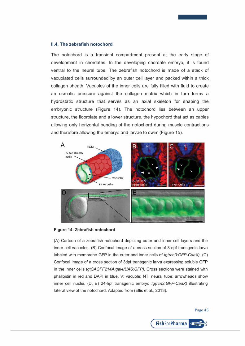
Page 45
II.4. The zebrafish notochord
The notochord is a transient compartment present at the early stage of
development in chordates. In the developing chordate embryo, it is found
ventral to the neural tube. The zebrafish notochord is made of a stack of
vacuolated cells surrounded by an outer cell layer and packed within a thick
collagen sheath. Vacuoles of the inner cells are fully filled with fluid to create
an osmotic pressure against the collagen matrix which in turn forms a
hydrostatic structure that serves as an axial skeleton for shaping the
embryonic structure (Figure 14). The notochord lies between an upper
structure, the floorplate and a lower structure, the hypochord that act as cables
allowing only horizontal bending of the notochord during muscle contractions
and therefore allowing the embryo and larvae to swim (Figure 15).
Figure 14: Zebrafish notochord
(A) Cartoon of a zebrafish notochord depicting outer and inner cell layers and the
inner cell vacuoles. (B) Confocal image of a cross section of 3-dpf transgenic larva
labeled with membrane GFP in the outer and inner cells of tg(rcn3:GFP-CaaX). (C)
Confocal image of a cross section of 3dpf transgenic larva expressing soluble GFP
in the inner cells tg(SAGFF214A:gal4/UAS:GFP). Cross sections were stained with
phalloidin in red and DAPI in blue. V: vacuole; NT: neural tube; arrowheads show
inner cell nuclei. (D, E) 24-hpf transgenic embryo tg(rcn3:GFP-CaaX) illustrating
lateral view of the notochord. Adapted from (Ellis et al., 2013).

Page 46
The notochord also gives developmental signals to pattern the neighboring
tissue such as somites, neural tube and blood vessel. During larval
development the notochordal cells undergo differentiation and segmentation
and eventually transforms into nucleus pulposus of the intervertebral discs in
adult fish (Ellis et al., 2013; Stemple, 2005; Glickman, 2003).
Figure 15: Structural aspects of the notochord
(A) A lateral view of a living zebrafish tail at 24 hpf, showing the main features of
the notochord. Dorsal to the notochord is the floor plate, in the ventral-most part of
the forming spinal cord. Ventral to the notochord is the hypochord.
(B) A schematic diagram of lateral and cross-sections of the notochord, showing
the floor plate and hypochord acting as cables running along the top and bottom
of the notochord. (C) As well as the notochord, the floor plate and hypochord
express type II collagen (Yan et al., 1995). cc, central canal; fp, floor plate; hy,
hypochord; no, notochord; nt, neural tube. Adapted from (Stemple, 2005).

Page 47
AIM OF THE THESIS
Outcome of a bacterial infection event is determined by nature of the microbe,
multiplicity of bacterial cells, physiology of host and location of the infection.
The main aim of this thesis is to understand how the innate immune system
responses to bacterial infection in an organ where the professional phagocytes
cannot come in direct contact with the bacteria to perform their conventional
phagocytic activity.
Since zebrafish is a powerful vertebrate model for study of infectious diseases,
and its notochord, which comprises vacuolated cells encased in the thick
collagen sheath, is inaccessible to phagocytes like neutrophils and
macrophages; I developed an infection model in zebrafish embryo notochord
and investigated the fate of the infected bacteria and the response of host
innate immunity to this tissue-specific infection.

Page 48
CHAPTER II
RESULTS
Page 49
CHAPTER II: RESULTS
OUTLINE OF THE RESULTS
Part I: Transient infection of zebrafish notochord with E. coli
induces chronic inflammation
Question: How the innate immune system responses to the bacterial infection
in a specific tissue where the microbes could not be phagocytosed?
Strategy: Injection of non-pathogenic E. coli in the notochord of transgenic
zebrafish embryo and investigate in vivo the behaviors of fluorescently labeled
phagocytes and the modulation of cytokines using microscopy techniques.
Results:
- The injected E.coli is eliminated within 24 hours after the infection.
- Although neutrophils and macrophages could not access the
notochord to phagocytose the injected bacteria, these leukocytes are
massively recruited and persist for long period after the infection.
- Interleukin-1β is dynamically expressed first by macrophage and later
by neutrophils. This cytokine is crucial for neutrophils recruitment.
- Chronic inflammation causes severe defect on the notochord and
subsequently malformation of the vertebral column.
Participation: I fully involved in the project.
Part II: Neutrophils control notochord infection at a distance with
superoxide
Question: How is E. coli cleared in the notochord without phagocytosis event?
Strategy: Test the roles of neutrophils and macrophages in the bacterial killing
by depletion each population at a time. Investigate the involvements of
antimicrobial arsenals present in these leukocytes.
Results:
- Neutrophils but not macrophages are necessary for the E. coli killing.
- Neutrophils control E. coli at a distance through superoxide production.

Page 50
- Neutrophils need proper cellular density to fight different levels of
notochord infection with E. coli.
Participation: I fully involved in the project.
Part III: Identification of polarized macrophage subsets in zebrafish
Aim: Investigate the polarity of zebrafish macrophage during infection and
inflammation.
Strategy: Induction of inflammation by tail fin amputation and by E. coli
injection in double transgenic embryo tg(mpeg1:mCherry-F/tnfa:GFP-F).
Follow the behavior of fluorescently labelled macrophages and genetic
markers for M1 and M2 subtypes macrophages using microscopy and FACS.
Results:
- Generation of transgenic line tg(tnfα: GFP-F).
- Macrophages switch their phenotypes between M1 and M2 subtypes
during the onset and the resolution of inflammation as seen in
mammalian counterpart.
Participation: I partly involved in the generation of transgenic reporter line
tg(tnfa:GFP-F).
Part IV: Rearing zebrafish larvae with Tetrahymenas
Question: Could Tetrahymenas be an alternative food for young zebrafish embryo?
Strategy: 24-hour Tetrahymena culture in enriched medium is washes and the clean Tetrahymenas are used to feed zebrafish embryos starting from day 5 to day 10 after fertilization for one month. Survival and developmental characteristics of larvae are recorded.
Results
- Feeding the zebrafish larvae with Tetrahymenas at their early phase of embryonic development help increase fish survivals.
- Tetrahymenas help maintain good quality of larval medium.
Participation: Fully involved in the project

Page 51
PART I
Transient infection of zebrafish notochord with E. coli
induces chronic inflammation
Authors:
NGUYEN-CHI Mai*, PHAN Quang Tien*, GONZALEZ Catherine,
DUBREMETZ Jean-François, LEVRAUD Jean-Pierre, and LUTFALLA
Georges
Disease models & Mechanisms
2014

© 2014. Published by The Company of Biologists Ltd | Disease Models & Mechanisms (2014) 7, 871-882 doi:10.1242/dmm.014498
871
ABSTRACT
Zebrafish embryos and larvae are now well-established models in
which to study infectious diseases. Infections with non-pathogenic
Gram-negative Escherichia coli induce a strong and reproducible
inflammatory response. Here, we study the cellular response of
zebrafish larvae when E. coli bacteria are injected into the notochord
and describe the effects. First, we provide direct evidence that the
notochord is a unique organ that is inaccessible to leukocytes
(macrophages and neutrophils) during the early stages of
inflammation. Second, we show that notochord infection induces a
host response that is characterised by rapid clearance of the bacteria,
strong leukocyte recruitment around the notochord and prolonged
inflammation that lasts several days after bacteria clearance. During
this inflammatory response, il1b is first expressed in macrophages
and subsequently at high levels in neutrophils. Moreover, knock down
of il1b alters the recruitment of neutrophils to the notochord,
demonstrating the important role of this cytokine in the maintenance
of inflammation in the notochord. Eventually, infection of the
notochord induces severe defects of the notochord that correlate with
neutrophil degranulation occurring around this tissue. This is the first
in vivo evidence that neutrophils can degranulate in the absence of
a direct encounter with a pathogen. Persistent inflammation,
neutrophil infiltration and restructuring of the extracellular matrix are
defects that resemble those seen in bone infection and in some
chondropathies. As the notochord is a transient embryonic structure
that is closely related to cartilage and bone and that contributes to
vertebral column formation, we propose infection of the notochord in
zebrafish larvae as a new model to study the cellular and molecular
mechanisms underlying cartilage and bone inflammation.
KEY WORDS: Zebrafish, Neutrophils, Inflammation, Interleukin-1
INTRODUCTION
Osteomyelitis, chondritis and septic arthritis are inflammatory
diseases of bones, cartilage and joints, respectively, that are often
caused by bacterial infections. These diseases are caused by local or
systemic immune responses that lead to leukocyte infiltration,
RESEARCH ARTICLE
1Dynamique des Interactions Membranaires Normales et Pathologiques,
Université Montpellier 2, UMR 5235, case 107, Place Eugène Bataillon, 34095
Montpellier Cedex 05, France. 2CNRS; UMR 5235, Place Eugène Bataillon, 34095
Montpellier Cedex 05, France. 3Macrophages et Développement de l’Immunité,
Institut Pasteur, Paris F-75015, France. 4CNRS URA2578, Paris F-75015, France.
*These authors contributed equally to this work
‡Authors for correspondence ([email protected];
This is an Open Access article distributed under the terms of the Creative CommonsAttribution License (http://creativecommons.org/licenses/by/3.0), which permits unrestricteduse, distribution and reproduction in any medium provided that the original work is properlyattributed.
Received 11 October 2013; Accepted 1 April 2014
damage to extracellular matrix, compression of vasculature and the
progressive destruction of bone and joints (Wright and Nair, 2010).
The bacteria that are involved are predominantly Staphylococcus
aureus, Streptococcus spp. and members of Gram-negative
Enterobacteriaceae, including Escherichia coli (Gutierrez, 2005;
Lew and Waldvogel, 2004). Although the pathogen is often present
within the infected site, it is occasionally undetectable, despite the
persistence of immunological symptoms. Substantial evidence
supports the importance of innate immunity in the physiopathology
of these diseases; however, its exact role is still poorly understood
(Lew and Waldvogel, 2004; Wright et al., 2010).
The innate immune response is the first defence of the host in the
case of infection. Macrophages and neutrophils are two major
effectors of innate immunity against microbes. Neutrophils are the
first to be recruited to the infected tissue; they migrate rapidly by
responding to gradients of chemoattractant molecules – such as the
contents of damaged cells, bacterial products and host chemokines
(Deng et al., 2013; Renshaw and Trede, 2012). Macrophages are
recruited later in order to aid the removal of dead cells and
microbes; they play a role in remodelling the injured tissue and
activating adaptive immunity. Neutrophils and macrophages are both
professional phagocytes, but the efficiency of phagocytosis depends
on the site at which they encounter bacteria (Colucci-Guyon et al.,
2011).
Although leukocytes can migrate rapidly and specifically to an
infection site, most tissues have specific resident macrophages
permanently patrolling the tissue, circulating through tissues to
eliminate cellular debris and microorganisms. From this
immunological perspective, bone and cartilage are very specific
tissues in which, owing to their highly structured and resistant
extracellular matrix, no specialised phagocytes patrol. The best-
documented example is that of bones. Resident bone macrophages,
OsteoMacs, are not present within the bone matrix but, with their
stellate morphology, do extensively cover bone surfaces (Pettit et al.,
2008). They probably act as guards to prevent microbes from
entering or attacking the bone. By contrast, osteoblasts (that make
bone) and osteoclasts (monocyte-macrophage lineage cells that
resorb bone) are capable of phagocytosis, probably to compensate
for the absence of professional phagocytes within the tissue. Some
pathogens have even developed specific strategies to colonise these
cells (Wright and Nair, 2010). Although documented to a lesser
extent, the situation in cartilage is similar, with chondrocytes
displaying phagocytic activity and a highly phagocytic cell
population that specifically expresses the macrophage marker
CD163, present below the superior zone at the periphery of cartilage
(Castillo and Kourí, 2004; Jiao et al., 2013).
To address the complexity of the chronic inflammation of bones
and joints, a number of mammalian infection models have been
developed (Patel et al., 2009). Most of them rely on the injection of
Staphylococcus aureus, or other bacteria, directly into the bone in
Transient infection of the zebrafish notochord with E. coli induces
chronic inflammationMai Nguyen-Chi1,2,*,‡, Quang Tien Phan1,2,*, Catherine Gonzalez1,2, Jean-François Dubremetz1,2,
Jean-Pierre Levraud3,4 and Georges Lutfalla1,2,‡
Dis
ea
se
Mo
de
ls &
Me
ch
an
ism
s

872
combination with an adjuvant or a trauma. Owing to these models,
advances have been made in understanding the disease and in tuning
antibiotic therapies; however, to understand the complexity of
dynamic interactions during inflammation, non-invasive and
tractable models in which pathogens and immune cells can be
imaged in the context of three-dimensional tissue architecture in the
host are required. Owing to its exceptional transparency, the
zebrafish embryo is an exquisite vertebrate model for the study of
tissue-specific infections and the inflammatory consequences within
a homologous tissue.
At the embryonic and early-larval stages, zebrafish do not possess
bone. However, the notochord, a transient embryonic structure that
is present in all chordates, in addition to its essential roles in
vertebrate patterning during development, serves as an axial
skeleton of the embryo until other structures, such as the vertebrae,
form (Stemple, 2005). The notochord comprises vacuolated cells
that are encased in a sheath of collagen. Osmotic pressure within
the vacuoles exerts such a pressure on the thick collagenous sheath
that it forms a hydrostatic skeleton in the embryo and young larva.
Notochordal cells that express many cartilage-specific genes are
most closely related to chondrocytes, but as development
proceeds, they become ossified in regions of developing vertebrae
and contribute to the formation of the centre of the intervertebral
discs in a structure called the nucleus pulposis (Stemple, 2005).
The notochord can be studied to dissect the molecular mechanisms
that are involved in the immune response to infection in a
chrondrocyte-like tissue. Previous results from our laboratory have
shown that the injection of an attenuated mutant of Mycobacterium
marinum, TesA:Tn, into the notochord allows active replication of
the bacteria, whereas the infection is easily controlled by
professional phagocytes when injected at other locations (Alibaud
et al., 2011). The drawback of this infection model is that the
active replication of the mycobacteria within the notochord
ultimately leads to the death of the host. As neither macrophage
nor neutrophil infiltration is observed (by using electron
microscopy), we hypothesised that the notochord is an organ that
is inaccessible to leukocytes and that this organ could be a good
model in which to study the outcome of an infection in a tissue
where leukocytes cannot reach the pathogen.
Here, we show that the injection of non-pathogenic Gram-
negative bacteria Escherichia coli K12 into the notochord of
zebrafish embryos induces a strong immune response without
compromising the survival of the host. Using a combination of
imaging and genetic techniques, we investigate the fate of the
bacteria in this organ, analyse the interaction between the bacteria
and the leukocytes, describe the inflammation induced by the
infection and analyse the consequences of this inflammation.
RESULTS
Zebrafish larvae rapidly clear E. coli infection in the
notochord
To characterise the immune response to notochord infection, we first
injected 3×10³ bacteria of an Escherichia coli K12 strain that
expressed the fluorescent protein DsRed (E. coli-DsRed) into the
notochord of zebrafish embryos at 45 hours post-fertilisation (hpf)
(Fig. 1A). Injection through the collagen sheath, just posterior to the
yolk extension, leads to the anterior sliding of the bacteria around
the notochordal cells inside of the collagen sheath. Fifteen minutes
after injection, the embryos were fixed, and the localisation of the
bacteria was checked by using transmission electron microscopy
(TEM). Bacteria were found inside the notochord, mainly at the
periphery, pinned against the collagen sheath by the turgescent
notochordal cells (Fig. 1B,C). The fate of the bacteria in the
notochord was monitored by live-imaging microscopy and
compared to the fate of bacteria that were injected inside muscles.
When injected into muscles, bacteria were restricted to the injected
somite for the first hours, but after 24 hours, their fluorescent
remnants were observed within motile cells that migrated away from
the site of injection (Fig. 1D). By contrast, in most cases (73%, 57
out of 78), the day following injection into the notochord,
fluorescent bacteria were not observed in the notochord, suggesting
that bacteria had been removed (Fig. 1E). In the other 27% (21 out
of 78) – presumably those embryos that received the higher bacterial
load – bacteria proliferated within the notochord (Fig. 1F-I) and led
to larval death within 3 days, as illustrated by the reduced survival
of the embryos in which bacteria had been injected into the
notochord (Fig. 1J). Further experiments were performed using
lower bacterial loads (2×10³); at this load, all embryos survived and
bacterial division was not observed (by using TEM; data not
shown). The clearance of bacteria from the notochord of most of the
infected fishes was confirmed by counting colony-forming units
RESEARCH ARTICLE Disease Models & Mechanisms (2014) doi:10.1242/dmm.014498
TRANSLATIONAL IMPACT
Clinical issue
Osteomyelitis, chondritis and septic arthritis are inflammatory diseases
of bone, cartilage and joints that are often caused by bacterial infections.
In these diseases, local or systemic immune responses lead to leukocyte
infiltration and the progressive destruction of bone and joints. Special
culture cell systems and several mammalian infection models have been
developed to understand these diseases and to tune antibiotic therapies.
However, the study of the complex dynamic interactions that occur
during inflammation requires non-invasive models in which pathogens
and immune cells can be imaged in the context of three-dimensional
tissue architecture in the host. In the last decade, the zebrafish, owing
to the transparency of its embryo and the ease with which it can be
genetically manipulated, has emerged as an exquisite system for
visualising the immune response to pathogen infections in vivo and in
real time.
Results
Here, the authors develop a zebrafish model to investigate the
inflammatory response that is induced by infection of the notochord, a
cartilaginous-like embryonic tissue that participates in vertebral column
formation and shares many similarities with chondrocytes. Using multi-
dimensional confocal imaging, the authors show that, during the early
stages of inflammation, infected zebrafish notochord is inaccessible to
leukocytes, even though these professional phagocytes engulf microbes
that had been injected into other tissues of the zebrafish embryo. Using
Escherichia coli, a pathogen that cannot develop within the notochord,
the authors show that a transient infection in this tissue triggers a severe
and persistent inflammation that is characterised by a massive
recruitment of leukocytes, extensive degranulation of neutrophils and
leukocyte infiltration into the notochord. Notably, this non-resolved
inflammation is accompanied by extracellular matrix remodelling and by
the development of notochord lesions and dysmorphic vertebrae
development. Finally, the authors report that an Il1b cytokine burst occurs
during notochord inflammation that is specifically required for the
establishment of the inflammatory episode and of notochord lesions.
Implications and future directions
These findings establish this in vivo approach as a relevant new
vertebrate model of chronic inflammation in matrix-enriched tissues, such
as bones and cartilage. Indeed, the symptoms exhibited by the
notochord-infected larvae are reminiscent of those seen in bone and
cartilage infection in mammals. Furthermore, these findings provide the
first in vivo evidence that neutrophils can degranulate in the absence of
a direct encounter with a pathogen. Thus, transient infection of the
zebrafish notochord provides a unique model in which to visualise at high
resolution the cellular events underlying pathological inflammation.
Dis
ea
se
Mo
de
ls &
Me
ch
an
ism
s

(CFUs) (Fig. 1K), indicating that bacteria, if not too numerous, are
efficiently cleared from the notochord within the first 24 hours of
infection.
Neutrophils and macrophages fail to reach, and engulf,
bacteria in the notochord
During the first weeks of development, the zebrafish larva has no
functional adaptive immune system, relying mostly on macrophages
and neutrophils, to fight infections (Lieschke et al., 2001; Meijer and
Spaink, 2011; Renshaw and Trede, 2012). Therefore, we determined
whether these cells were able to reach the bacteria in the notochord
in order to phagocytose and/or kill them. To study neutrophil-
microbe interactions, we injected fluorescent E. coli into
Tg(mpx:GFP) embryos, which expressed green fluorescent protein
(GFP) under the control of the neutrophil-specific promoter of
myeloperoxidase (mpx) (Renshaw et al., 2006), and we then imaged
neutrophil behaviour by using confocal time-lapse microscopy from
2 hours post-infection (hpi). In contrast to PBS-injected controls
(data not shown), neutrophils were primarily attracted to the
injection site, then, arriving from the ventral side of the larvae
(Fig. 2A,B; supplementary material Movies 1, 2), they crawled
along the outside of the infected notochord, spreading towards the
anterior region (Fig. 2B; supplementary material Movie 1).
Although neutrophils appeared to be attempting to try to
phagocytose bacteria (i.e. crawling forward and backward in the
vicinity of bacteria, emitting cytoplasmic protrusions), they failed to
engulf them (supplementary material Movie 3). Only a few
neutrophils contained engulfed bacteria, but these leukocytes were
carefully, and retrospectively, traced from the injection site,
suggesting that they phagocytosed a few E.coli that were deposited
at the injury site of the notochord (Fig. 2B; supplementary material
Movie 1). This behaviour is in stark contrast with that observed in
muscles, where many neutrophils displayed phagosomes that were
full of bacteria at 2 hpi and still efficiently engulfed bacteria (data
not shown; Colucci-Guyon et al., 2011).
To analyse macrophage-bacteria interactions, we constructed the
Tg(mpeg1:mCherryF) line that expressed red fluorescent
macrophages. Transgenic larvae were infected either in the
bloodstream or in the notochord, and macrophage behaviour was
recorded at 1 hpi. Similar to neutrophils, although less mobile,
macrophages were recruited to the infected notochord and extended
pseudopodal protrusions in attempts to engulf bacteria (Fig. 2A,C;
supplementary material Movies 4, 5). In contrast to the numerous
macrophages that were laden with E. coli after bloodstream infection
(supplementary material Movie 6), macrophages were inefficient at
engulfing the bacteria present in the notochord. It is noteworthy that
no significant difference in the time to the first recruitment was
detected between neutrophils and macrophages (data not shown).
The fact that leukocytes easily engulfed bacteria at the injection
site, where the notochordal collagen sheath had been damaged by
the injection, but not in the undamaged upstream region suggests
that bacteria might not be accessible to leukocytes because of the
physical properties of the notochord (data not shown). We tested this
hypothesis by performing three-dimensional reconstruction of
confocal acquisitions. From 2 hpi onwards, bacteria were found in
the phagosomes of neutrophils and macrophages after injection into
873
RESEARCH ARTICLE Disease Models & Mechanisms (2014) doi:10.1242/dmm.014498
Fig. 1. Injection of Escherichia coli into the notochord of
zebrafish larvae. (A) Embryos at 48 hpf. Bacteria were injected
(elongated arrowhead) into the notochord (red) dorsal to the uro-
genital opening. The dashed line with arrowheads shows the region
where cross-sections have been performed for electron microscopy.
(B,C) Transmission electron microscopy analysis of a larva 15
minutes after injection of bacteria into the notochord; C is a higher
magnification of the boxed region in B. Arrowheads indicate bacteria.
Nc, notochordal cell; v, vacuole; pbm, peri-notochordal basement
membrane that comprises three layers – o, outer; m, medium
(collagen); I, inner. (D-F) The fate and distribution of DsRed-
expressing E. coli (red) after infection of muscle with 3000 CFU (D)
or notochord with 2000 CFU (E) and 3000 CFU (F). Each larva was
followed and imaged at 3 (top row) and 24 (bottom row) hpi.
Arrowheads indicate clusters of bacteria. (G-I) Transmission electron
microscopy analysis of the notochord of larvae that had been
infected in the notochord (11 hpi); H,I show magnifications of regions
of G. (G) Numerous bacteria (bracket) are found between the peri-
notochordal basement membrane (pbm, arrowheads) and notochord
cells (nc). (H) Collagen (col) disorganisation is observed next to
bacteria. (I) Extracellularly replicating bacterium (arrowhead).
(J) Survival was scored of uninfected larvae and those that were
infected in the muscle or notochord (n>24 each). These results are
representative of three independent experiments, P<0.0005 for
notochord injection versus uninjected. (K) Whole embryo bacterial
counts of E. coli when injected into the muscle or notochord. Results
are expressed as the mean number of CFU per larvae±s.e.m.
(n>5 larvae per timepoint). NS: not significant. Scale bars: 5 µm
(B,G); 1 µm (C,H,I).
Dis
ea
se
Mo
de
ls &
Me
ch
an
ism
s

874
muscle (Fig. 3A) and the bloodstream, respectively (Fig. 3C);
however, they remained next to, but outside of, immune cells after
notochord infection (Fig. 3B,D). Indeed, many leukocytes
accumulated around the notochord but could not infiltrate it, even at
24 hpi (Fig. 3E-G).
To confirm further that leukocytes could not reach the bacteria, and
engulf them, in the notochord, we analysed the ultrastructure of the
notochord of infected larvae by using TEM at different timepoints
following infection. At 15 minutes post-infection, bacteria were found
in the periphery of the notochord, between notochordal cells and the
collagen sheath as mentioned above (supplementary material Fig.
S1A-C). At 2 hpi and 4 hpi, bacteria remained in the same location –
in the extracellular space – although they were less numerous at 4 hpi
(supplementary material Fig. S1D-F; data not shown for the 4 hpi
RESEARCH ARTICLE Disease Models & Mechanisms (2014) doi:10.1242/dmm.014498
Fig. 2. Behaviour of neutrophils and macrophages
following notochord infection. (A) Diagram showing
the regions of the larvae imaged in B and C (dashed
boxes). (B) Tg(mpx:GFP) larvae that had been infected
with crimson-expressing E. coli (magenta) in the
notochord at 48 hpf. The general behaviour of neutrophils
(green) was imaged using four-dimensional confocal
microscopy, starting at 2 hpi for a duration of 3 hours (the
time post-infection is shown in the top right corner).
*Neutrophils loaded with bacteria, the arrowhead
indicates the injection site. (C) Tg(mpeg1:mCherryF)
larvae that had been infected with GFP-expressing E. coli
(green) at 48 hpf in the notochord were imaged in the
region represented in A to visualise the behaviour of
macrophages (red) using four-dimensional confocal
microscopy at 1 hpi for a duration of 3 hours and 20
minutes. Scale bars: 20 m.
Fig. 3. Leukocytes do not enter the notochord during the early phase of infection. (A,B) Multiscan confocal analysis at 2 hpi of neutrophils (green) in
Tg(mpx:GFP) larvae after muscle (A) or notochord infection (B) with DsRed-expressing E. coli. (C,D) Multiscan confocal analysis of macrophages (red) in
Tg(mpeg1:mCherryF) larvae after intravenous (C) or notochord (D) infection with E. coli-GFP (green) at 2 hpi. In A-D, the left panels are xy maximum
projections and the right panels are yz cross-section views at the position of the stippled line. Arrowheads show bacteria-containing leukocytes, *Leukocytes
that were located next to bacteria but did not contain any. (E-G) Confocal analysis at 24 hpi of the notochord with fluorescent E. coli. Bacteria were no longer
visible at 24 hpi. (E,E') Tg(mpx:GFP) larva showing neutrophils (green), (F,F') Tg(mpeg1:mCherryF) showing macrophages, (E,F) maximum projection, (E',F')
single confocal scan. (G) Confocal analysis of a 60- m cross-section from a double-transgenic larva – neutrophils (green) and macrophages (red) accumulated
around the notochord but did not enter this structure, as shown. Scale bars: 10 m (A-D); 20 m (E-G). Dis
ea
se
Mo
de
ls &
Me
ch
an
ism
s

timepoint). Replicating bacteria were never observed under these
conditions. This correlated with the observation that DsRed
fluorescence in E. coli remained constant in the notochord for a few
hours following injection before diminishing completely. Although a
few leukocytes were detected outside of the notochord (data not
shown), none were found inside, confirming that leukocytes cannot
phagocytose bacteria in this structure (supplementary material Fig.
S1). Strikingly, numerous E. coli displayed an altered morphology
(supplementary material Fig. S1B,C,F), suggesting they are eliminated
by a humoral lysis mechanism. No altered morphologies were
observed when higher doses of bacteria were used, suggesting that the
humoral lysis mechanism is overpowered when the notochord is
injected with higher doses of E. coli.
Notochord infection induces a strong and persistent
inflammatory reaction
We analysed the kinetics of immune cell recruitment using
Tg(mpx:GFP) and Tg(mpeg1:mCherryF) lines by counting the
leukocytes that accumulated around the infected notochord. As for
muscle infections (Colucci-Guyon et al., 2011), notochord infections
led to neutrophil recruitment within the very first hours following
the injection. After injection of bacteria into the muscle, the number
of neutrophils that had been recruited started decreasing at 24 hpi
(Fig. 4A,C), whereas, in the notochord, neutrophil recruitment
continued to increase at 48 hpi and remained high up to 6 days post-
infection (dpi), forming one to four clusters that were towards the
anterior of the injection site (20 out of 26 fish; Fig. 4A,C).
Macrophages migrated to both infected muscle and notochord in a
similar manner (Fig. 4B) – in both cases, the recruitment of
macrophages lasted for 6 days, but more macrophages were
recruited to the infected notochord (23 out of 23 fish; Fig. 4B,D).
The observed immune response was not a consequence of the
injection per se in the notochord, because the injection of PBS or
fluogreen Dextran caused neither neutrophil nor macrophage
recruitment (Fig. 4C,D; supplementary material Fig. S2).
We conclude that infection of the notochord induces a massive
recruitment of macrophages and neutrophils to its periphery;
remarkably, this inflammatory response is not properly resolved, as
leukocyte accumulation persists for days, despite the clearance of
bacteria in a matter of hours.
Notochord inflammation induces notochord damage
Inflammation is fundamental in order to control bacterial infections.
However, it is crucial that this inflammation resolves in a timely
manner in order to prevent damage to the surrounding tissues. In
order to analyse the consequences of notochord inflammation, we
imaged larvae, in which bacteria had been injected into the
notochord, for 10 days. At 5 dpi, most of the infected larvae
875
RESEARCH ARTICLE Disease Models & Mechanisms (2014) doi:10.1242/dmm.014498
Fig. 4. Neutrophils and macrophages
are recruited around the infected
notochord. (A) Tg(mpx:GFP) larvae were
either left uninfected or infected with
DsRed-expressing E. coli (red) in the
muscle or notochord at 48 hpf. Neutrophil
recruitment (green) was imaged repeatedly
in individual larvae at the indicated
timepoints. (B) A similar experiment to
that shown in A was performed in
Tg(mpeg1:mCherryF) larvae that were
either uninfected or infected with GFP-
expressing E. coli (green) in muscle or the
notochord at 48 hpf. (C,D) Corresponding
counts of neutrophils (C) and
macrophages (D) over the course of 1
week post-infection. Results are expressed
as the mean number of cells±s.e.m., five to
nine fish were examined per timepoint,
*P<0.05, **P<0.005 and ***P<0.001.
(E) The dashed boxes in the diagram
represent the zone where counting was
performed.D
ise
ase
Mo
de
ls &
Me
ch
an
ism
s

876
presented curved malformation and lesions of the notochord,
upstream of the injection site (43 out of 49 fish; supplementary
material Fig. S3A,B). Although the defects were lessened, we still
observed this malformation at 10 dpi (supplementary material Fig.
S3C). Histological analysis of PBS-injected larvae at 5 dpi revealed
normal architecture with highly vacuolated cells that were
surrounded by a regular collagen sheath. By contrast, the infected
larvae displayed swelling of the notochord at various locations along
the antero-posterior axis (Fig. 5A,B; supplementary material Fig.
S3D-K). At these spots, we observed an ectopic collagen-rich
extracellular matrix, and an increased number of cells and vacuoles
inside the notochord, suggesting the presence of supernumerary
notochordal cells, rather than a phenomenon of fragmented vacuoles.
Cells of a smaller size were also found clustered either outside or
inside of the notochord; they were often trapped within the matrix,
suggesting that they are infiltrating leukocytes (supplementary
material Fig. S3J-K). Analysis by using confocal microscopy
revealed that, although leukocytes were positioned at the margin of
the myotome in PBS-injected larvae, they started infiltrating the
notochord in E. coli-injected larvae from 2 dpi (Fig. 5C,D shows the
5 dpi timepoint; the data at 2 dpi is not shown), a timepoint that is
long after bacterial clearance. Because notochord defects colocalised
with neutrophil clusters (Fig. 5E,F), we asked whether neutrophil
infiltration contributes to the destruction of host tissues. Indeed, this
infiltration was concomitant with neutrophil degranulation – 35%
of neutrophils in the inflammation region harboured no
myeloperoxidase-containing granules, whereas all neutrophils in the
control retained them (Fig. 5G-J). Taken together, these results show
that infection of the notochord eventually leads to severe damage to
the notochord tissue, which correlates with late leukocytic infiltration
and neutrophil degranulation.
Notochord inflammation triggers vertebral column defects
As the notochord participates to vertebral column formation we
asked whether the inflammation of the notochord could trigger
vertebral column defects. We thus examine the axial skeleton of
juvenile fishes at 60 days post-fertilization (dpf) using Alizarin red
staining. Zebrafish that had been previously injected with E. coli in
the notochord developed scoliosis and dysmorphic vertebral column
in their anterior region (upstream the injection site), characterised
by fused and misshaped vertebral bodies and absence of
intervertebral disc (11 out of 11 fish; Fig. 5K). No defects were
observed in the PBS injected controls, excepting a single vertebral
body that appeared more calcified at the injection site in few fishes
(two out of 19 fish; Fig. 5K; data not shown).
Neutrophils but not macrophages expressed high levels of
il1b in notochord infected embryos.
In order to decipher the molecular mechanisms that orchestrate the
inflammatory response to notochord infection, we analyzed the
expression of many cytokines and chemokines in larvae that had
been infected in the notochord (supplementary material Table S1).
The expression of most of the chemokines that we examined was
unchanged, such as that of the chemokine Cxcl-c5c; however,
transcripts of the proinflammatory cytokine Il1b were strongly
upregulated from 1 dpi onwards (Fig. 6A,B).
To study the spatiotemporal behaviour of the cells that produce
the il1b transcripts, we established a transgenic line that expressed
RESEARCH ARTICLE Disease Models & Mechanisms (2014) doi:10.1242/dmm.014498
Fig. 5. Late consequences of notochord infection.
(A,B) Histological analysis at 5 dpi of larvae that had been
injected in the notochord with PBS (A) or E. coli (B). Cross-
sections (4 m) stained with Masson’s Trichrome. In the infected
larvae, collagen-enriched matrixes (blue) and supernumerary
vacuoles (V) were seen. Cells inside the notochord (*) and
ectopic matrix deposition (arrowhead) can be seen in the
sections. (C,D) Neutrophil (green) and macrophage (red)
infiltration in the notochord, multiscan confocal microscopy.
Cross-sections (60 m) of Tg(mpx:GFP; mpeg1:mCherryF)
larvae at 5 days following injection of the notochord with PBS
(C) or with E. coli (D). *Nonspecific staining. (E,F) Notochord of
Tg(mpx:GFP) larvae at 3 dpi. (E) Transmitted-light image,
overlaid in F with a maximal projection of confocal fluorescence
images that show neutrophils (green) clustered at the damaged
zone. (G-I) Sudan-black (SB) staining (black) and GFP
expression (green) in Tg(mpx:GFP) larvae at 48 hours post-
injection of either PBS (G) or E. coli (H,I) into the notochord.
Arrowheads indicate neutrophils that have degranulated.
(J) Graphic representation of the percentage of GFP+SB– cells
under the indicated conditions, mean±s.e.m., five or six fish per
condition were analysed. *P<0.05 compared with the PBS
control. (K) Infected larvae developed scoliosis and dysmorphic
vertebrae. Larvae were injected with PBS or E. coli into the
notochord at 48 hpf, grown to 60 dpf and fixed for analysis.
Bones were stained with Alizarin red. Lateral and dorsal views of
the anterior part of the vertebral column are shown. Arrowheads
indicate mis-shaped and fused vertebral bodies. Scale bars: 20
m (A-D,G-I); 50 m (E,F).
Dis
ea
se
Mo
de
ls &
Me
ch
an
ism
s

farnesylated GFP (membrane-targeted GFP; GFP-F) under the
control of the il1b promoter Tg(il1b:GFP-F). Uninfected
Tg(il1b:GFP-F) developing embryos expressed GFP-F during the
segmentation period in the elongating tail bud (supplementary
material Fig S4). At 24 hpf, GFP expression appeared in tail
keratinocytes, the olfactory epithelium and individual cells on the
yolk sac that behave as primitive leukocytes. At 35 hpf, when
leukocytes appear in the caudal hematopoietic tissue (CHT),
scattered GFP+ cells were also found in the CHT region. From 50
hpf, GFP was expressed in keratinocytes at the tip of the caudal fin,
in fin buds, retina, neuromasts, gills and thymus (supplementary
material Fig. S4; data not shown). Whole-mount in situ
hybridisation analysis confirmed the expression of endogenous il1b
mRNA in the different organs that are cited above, showing that
Tg(il1b:GFP-F) recapitulates il1b transcriptional expression
(supplementary material Fig. S5A,C). Furthermore, upon
stimulation by ‘danger signals’, both the neutrophils and
macrophages of Tg(il1b:GFP-F) larvae strongly expressed GFP,
correlating with the upregulation of the endogenous il1b mRNA that
was observed in both leukocyte populations (supplementary material
Fig. S6).
To study il1b transcription upon infection, we injected fluorescent
bacteria (E. coli-DsRed) into the notochord of Tg(il1b:GFP-F)
embryos. Specific expression of GFP was detected along the
notochord as early as 18 hpi, which increased significantly by 48 hpi
and was maintained up to 5 to 6 dpi before decreasing (24 out of 36
fish; Fig. 6D; supplementary material Fig. S5D). The induction of
GFP upon infection correlated with the upregulation of il1b mRNA
(supplementary material Fig. S5A-C). Conversely, control PBS and
muscle injections, respectively, induced no and low expression of
GFP around the injection site at 24 hpi and 48 hpi (Fig. 6C,E;
supplementary material Fig. S5D). The analysis of larvae that had
been injected in the notochord revealed that GFP was expressed by
leukocytes that were clustered around the notochord but not by
notochordal cells (Fig. 6F). Additional GFP expression was detected
in leukocytes that were scattered throughout the body and in the
877
RESEARCH ARTICLE Disease Models & Mechanisms (2014) doi:10.1242/dmm.014498
Fig. 6. GFP expression in Tg(il1b:GFP-F) infected
larvae. (A,B) The relative expression of il1b (A) and of
cxcl-c5c (B) at different timepoints post-infection, under
three different conditions – PBS injected into the
notochord, E. coli injected into muscle and E. coli
injected into the notochord. Quantitative real-time RT-
PCR on whole embryo RNA extracts using ef1a as a
reference gene was performed; light grey bars
represent the 95% confidence intervals (Student’s t-
test). Non-overlapping light grey bars indicate P<0.05.
These results are representative of three independent
experiments. (C,D) GFP expression at 24 hpi in
Tg(il1b:GFP-F) larvae that had been injected with E.
coli (red) into muscle (C) or the notochord (D). Arrows
indicate expression next to inflammation sites. N,
notochord; DA, dorsal aorta. (E,F) Multiscan confocal
analysis of GFP expression in Tg(il1b:GFP-F) larvae in
the notochord region 48 hours following the injection
of PBS or E. coli. (E-F') Maximum projections,
(F) longitudinal- and (F ) cross-section views. The
dashed line indicates the position of cross-section F
relative to F. (G,H) The expression of mCherry (red)
and GFP (green) in Tg(mpeg1:mCherryF ; il1b:GFP-F)
double transgenic larvae at 18 (G) and 48 hpi (H) in
the notochord region was analysed by using confocal
microscopy. Arrowheads indicate macrophages
expressing GFP. (I,J) Sudan-black staining and GFP
expression in Tg(il1b:GFP-F) larvae was analysed by
using confocal microscopy at 48 hpi. The images show
a GFP+ neutrophil underneath GFP+ keratinocytes.
(K-K ) The expression of DsRed (red) and GFP (green)
in Tg(Lyz:DsRed ; il1b:GFP-F) double transgenic
larvae at 48 hpi was analysed by using confocal
microscopy. A merged image is shown on the right.
(L) Quantification of il1b+; mpeg1+ and il1b+; Lyz+ cells
in the inflammation region. Counts are expressed as
the percentage of cells within the population
expressing GFP (il1b+, il1blow or il1bhigh). *P<0.05,
**P<0.005 and ***P<0.001. Scale bars: 20 m.
Dis
ea
se
Mo
de
ls &
Me
ch
an
ism
s

878
keratinocytes surrounding the inflammation site (supplementary
material Fig. S5E,F; Fig. 6J).
By early timepoints (18 hpi) 30% of GFP-positive cells (il1b+)
also expressed the macrophage marker mpeg1 (mpeg1+), whereas
only 10% expressed the neutrophil marker lysosymeC (lyz+)
(Fig. 6G,L). During the late stages of inflammation (48 hpi), two
distinct populations of GFP-expressing cells were observed around
the inflamed notochord – cells that expressed a high level of GFP
[il1bhigh, 60±6.5% (mean±s.e.m.)] and those that expressed a low
level of GFP (il1blow, 40±6.5%). At 48 hpi, no macrophages were
il1bhigh, whereas neutrophils (lyz+) comprised 68% of these il1bhigh
cells. The other 32% of il1bhigh cells were mpeg1 and Lyz and
unidentifiable. The remaining il1blow cell population comprised 35%
neutrophils (il1blow Lyz+) and 27% macrophages (il1blow mpeg1+)
(Fig. 6H-L).
In conclusion, our data suggest that Il1b production dynamically
shifts from early timepoints of the inflammatory response – at which
both macrophages and neutrophils contribute to Il1b production – to
late phases, when neutrophils are the main source of Il1b.
Il1b is required for the early and late recruitment of
neutrophils but not macrophages
Morpholino-mediated gene knockdown was used to investigate the
role of zebrafish Il1b during severe notochord inflammation.
Reverse transcription (RT)-PCR analysis, performed using primers
on both sides of the splice site that is targeted by the morpholino,
provided evidence of strong blocking of splicing, whereas overall
morphology was not noticeably affected (Fig. 7A; the general
morphology of the morphants is not shown). The il1b morpholino
was injected into Tg(mpx:GFP ; mpeg1:mCherryF) embryos, and
the morphants were infected by the injection of E. coli into the
notochord. In embryos that had not been injected with morpholino,
or those that had been injected with control scrambled morpholino,
both neutrophils and macrophages were recruited to the inflamed
notochord in a normal manner. In il1b morphants, neutrophil
recruitment was slightly reduced at 4 hpi and strongly reduced at 48
hpi compared with controls (Fig. 7C,E-G); only macrophages
localised around the notochord of il1b morphants at 4 and 48 hpi
(Fig. 7G; supplementary material Fig. S7). The decreased neutrophil
recruitment was not caused by a general reduction of the neutrophil
population under steady-state conditions because the total number
of neutrophils in il1b morphants was similar to that in uninjected
embryos or embryos that had been injected with the control
scrambled morpholino at 4 hpi (Fig. 7D for il1b morpholino
compared to scrambled morpholino; data not shown for il1b
morphants compared to uninjected larvae). Additionally, a reduction
of the overall inflammatory response was excluded because
macrophage recruitment was unaffected (Fig. 7G). Long-term
examination of the infected il1b morphants revealed that loss of il1b
prevented formation of the lesions that were often detected in the
rostral part of the notochord after infection (Fig. 7H-J, lesions were
found in one out of 12 il1b morphant fish and seven out of 11
scrambled morpholino fish). Taken together, these results establish
an important and specific role for Il1b in the recruitment of
neutrophils during chronic inflammation of the notochord and
subsequent sequelae.
RESEARCH ARTICLE Disease Models & Mechanisms (2014) doi:10.1242/dmm.014498
Fig. 7. Il1b is required for the early and late recruitment of neutrophils but not macrophages. PCR amplification of il1b cDNA from the 48 hpf
intravenous-infected larvae, which had previously been injected with either scambled morpholino (scramMO) or with a morpholino antisense oligonucleotide
that blocked the splicing of intron 2 (il1bMO). (B) The diagram shows the region that is imaged in C (dashed box). (C) Recruitment of neutrophils (green) in
Tg(mpx:GFP) larvae at 4 hours and 48 hours following the injection of DsRed-expressing E. coli (red) into the notochord. These larvae had been previously
injected with scrambled morpholinos (scramMO) or il1b MO. N, notochord. At 48 hpi, many neutrophils were found in the AGM region, in between the dorsal
aorta (DA) and the posterior caudal vein (pcv); arrows indicate the direction of blood flow. (D-G) Plots show the number of total neutrophils at 4 hpi (D) and the
number of recruited neutrophils at 4 hpi (E) and 48 hpi (F). (G) The number of recruited macrophages at 48 hpi in similar experiments that were performed
using Tg(mpeg1:mCherryF) larvae. Each point represents an individual larva, the bar represents the mean value. **P<0.005, ***P<0.001. (H-J) Bright-field
images of control (scramMO) and il1b morphants 3 days after injection of either PBS or E. coli into the notochord. Lateral views of the trunk region are shown
in C,H,I,J. Dis
ea
se
Mo
de
ls &
Me
ch
an
ism
s

DISCUSSION
This article describes a model of zebrafish notochord infection with
E. coli that recapitulates several of the inflammatory aspects of bone
and cartilage infection in human. We thus propose that the specific
advantages of the larval zebrafish model system – transparency,
genetic tractability – should facilitate the study of the early cellular
and molecular inflammatory mechanisms that are related to matrix-
tough tissue infections.
Notochord infection of the zebrafish larvae as a model to
study inflammatory aspects of bone and cartilage infections
The similarity of the notochord with cartilage (in terms of
transcriptional profile) and vertebral bone (in terms of structure and
function) (Fleming et al., 2001; Stemple, 2005) prompted us to
establish a model of the infection of this organ in order to mimic the
inflammatory aspects of human osteomyelitis and osteoarthritis. As
in some infections of human bones and joints, infection of the
zebrafish notochord induces a severe inflammation that greatly
outlasts the presence of live microbes and thus can be considered
‘chronic’ – taking into account due allowances. Indeed, injection of
E. coli into the notochord leads to the rapid recruitment of both
neutrophils and macrophages to the infected notochord within the
first hour. The injection needle locally disrupts the collagen sheath,
creating an opening for the leukocytes to phagocytose some bacteria
in the notochord. However, probably because of the turgidity of the
notochordal cells within the thick collagen sheath, neutrophils and
macrophages fail to enter the notochord at sites distant from the
injection site. This finding suggests that, in contrast to what is
observed for transendothelial migration, leukocytes do not easily
pass through all matrixes. Even though the injected bacteria are
cleared in less than one day, neutrophil and macrophage recruitment
along the notochord persists for days, preventing the resolution of
inflammation. Although they, initially, randomly spread around the
notochord at 2 to 24 hpi, leukocytes accumulate in patches in the
later stages of the response (from 48 hpi), suggesting that they are
involved in the activation and recruitment of more leukocytes.
Granules, the hallmarks of granulocytes, are stores of enzymes that
participate in killing microbes and digesting tissues. We found that,
from 2 dpi, despite the fact that injected microbes have been cleared
by this point, neutrophils degranulate next to the notochord. Thus,
the release and activation of proteolytic enzymes that are able to
disassemble the collagen fibrils that impede neutrophil-bacteria
contacts might explain the infiltration of neutrophils into the
notochord, which is observed in the late phases of inflammation
(Nathan, 2006). This neutrophil degranulation might be responsible
for the tissue damage that is observed in notochord-infected larvae
– including perturbation of extracellular matrix deposition, swelling
and curvature of the notochord, and cellular clusters within the
notochord that are reminiscent of the abscesses observed in human
osteomyelitis (Lew and Waldvogel, 2004; Wright and Nair, 2010).
The notochord lesion, which is induced by the severe inflammatory
episode might contribute to the dismorphic vertebrae that were
observed during adulthood. Although extracellular matrix
deposition, neutrophil infiltration and tissue damage are the
hallmarks of chronic infection, which occur in some osteomyelitis
and septic arthritis individuals, curvature of the axial skeleton is
reminiscent of that of observed in individuals that have bone
tuberculosis. Indeed, also known as Pott disease, which accounts for
1–2% of overall tuberculosis cases, it is usually characterised by the
spread of tuberculosis bacilli into the disc space from the vascular
system, resulting in the destruction of the disc and progression to the
bone of adjoining vertebral bodies. As the vertebrae degenerate and
collapse, a kyphotic deformity results (Moon, 1997). The chronic
inflammation of the zebrafish notochord after bacterial clearance
described here is also reminiscent of autoinflammatory disorders –
such as chronic non-bacterial osteomyelitis, deficiency of IL-1
receptor antagonist or neonatal-onset multisystem inflammatory
disease. Although the etiology of chronic non-bacterial osteomyelitis
is still unknown, deficiency of IL-1 receptor antagonist and
neonatal-onset multisystem inflammatory disease are caused by
mutations in the IL1RN and NLRP3 genes, respectively, and result
in over-activation of the IL-1 pathway (Morbach et al., 2013).
Accordingly, neutrophil recruitment to the infected zebrafish
notochord was found to be dependent on Il1b.
Elimination mechanism of E. coli in the notochord
Many bacteria avoid the immune response by hiding inside of cells of
their host. By doing so, they are out of reach of the immune system
and multiply within these cells before further invasion of the body. We
have previously shown that, when injected into the notochord, non-
pathogenic Mycobacterium marinum Tes:A mutant bacteria are
internalised by notochordal cells as revealed, using TEM, by the
presence of the bacteria in endosomes (Alibaud et al., 2011).
Interestingly, similar bacterial internalisation has previously been
reported for osteoblasts and chondrocytes (Diaz-Rodriguez et al.,
2009; Wright and Nair, 2010; Jiao et al., 2013, Castillo and Kourí,
2004). All of these observations suggest that osteoblasts, chondrocytes
and notochordal cells are non-professional phagocytes that engulf
invader microbes. However, their phagocytic ability depends on the
nature of these microbes. We observe here that notochordal cells do
not phagocytose E. coli; bacteria remain extracellular for a few hours
before being eliminated, as demonstrated by CFU assays, and
fluorescence and electron microscopy. How are they killed? As
leukocytes cannot phagocytose them, another mechanism must be
used. The ‘unhealthy’ shape of some E. coli within the notochord, as
observed by electron microscopy, suggests death by a humoral lysis
mechanism that remains to be identified. The complement system is
an obvious candidate, and we are currently studying the contribution
of complement genes to the observed bacterial clearance.
Putative functions of Il1b in zebrafish
IL-1 is a critical pro-inflammatory mediator of the host response
to microbial infection. It acts on almost all cell types and has many
functions – including growth and proliferation, the induction of
adhesion molecules in epithelial cells, the activation of phagocytosis
and release of proteases in macrophages and neutrophils, and the
activation of B and T cells (Dinarello, 2009). Using transgenic
zebrafish larvae that expressed farnesylated GFP under the control
of the il1b promoter, we show here, for the first time, the dynamic
transcriptional activity of il1b in a whole organism. Its expression
during zebrafish development, especially in the keratinocytes of the
caudal and pectoral fin bud, correlates with the regions of the body
where active keratinocyte proliferation occurs. This is in agreement
with findings in mammals where keratinocytes express IL-1
(Kupper et al., 1986). However, resting keratinocytes express IL-1
in its unprocessed biologically inactive form – pro-IL-1 . Indeed, to
be secreted as an active cytokine, pro-IL-1 must be cleaved by
caspases (caspase-1, caspase-11 and others), which are themselves
activated through inflammasome assembly (Kayagaki et al., 2011).
Although il1b transcription is low in most cell types and strongly
inducible following the detection of microbial molecules – notably
in myeloid cells – constitutive expression of pro-IL-1 by some
tissues should allow for a faster response to external injuries. This
is logical for cells that are directly exposed to the environment –
879
RESEARCH ARTICLE Disease Models & Mechanisms (2014) doi:10.1242/dmm.014498
Dis
ea
se
Mo
de
ls &
Me
ch
an
ism
s

880
such as keratinocytes, olfactory epithelial cells, enterocytes or
neuromast cells – all cell types that were seen to spontaneously
express endogenous il1b mRNA and GFP in the Tg(il1b:GFP-F)
line. In zebrafish, Il1b has been shown to be structurally conserved
compared with its human counterpart, and the Il1b pathway is also
dependent on caspases (Ogryzko et al., 2014). Whether the active
form of Il1b is produced in these zebrafish organs remains to be
determined. Notably, spontaneous GFP expression in some cell
types, such as those of neuromasts and the gut, mimics the
spontaneous expression of GFP in the Tg(NF B:EGFP) reporter
transgenic zebrafish line (Kanther and Rawls, 2010), which is in
agreement with the well-known contribution of NF- B to il1b
induction (Ogryzko et al., 2014).
In addition to its basal expression during development, GFP is
also induced following E. coli infection in Tg(il1b:GFP-F) fish,
which parallels endogenous il1b induction. This induction is strictly
correlated in intensity and time with the recruitment of leukocytes.
Indeed, infection in muscles induces a low level of expression that
quickly resolves, whereas infection of the notochord induces strong
and persistent expression. The GFP expression level decreases one
day before the resolution of inflammation. In most mammalian
systems, IL-1 has been reported to be mainly produced by
circulating monocytes, tissue macrophages and dendritic cells
(Dinarello, 2009). Here, we describe that, in our zebrafish notochord
infection model, il1b is first expressed in macrophages during the
early phase of inflammation and is mainly expressed by neutrophils
during the late phases. This is reminiscent of the recent work of
Karmakar and collaborators, which shows that neutrophils are the
main source of Il1b in response to Pseudomonas aeruginosa in
mouse, highlighting the importance of activated neutrophils in
mediating inflammation (Karmakar et al., 2012).
One interesting question arising from the present work is which
signal stimulates the recruitment of leukocytes to the infected
notochord? Is it a primary signal that is actively emitted by the
notochordal cells, does it come from the injected bacteria or does it
correspond to bacterial leftovers after destruction within the
notochord? We are currently investigating this latter hypothesis
through the injection of different bacterial products into the
notochord of larvae and evaluating their ability to recruit leukocytes.
Our data demonstrate that il1b is not expressed by notochordal
cells after E. coli injection; however, it is partially required for the
initial recruitment of neutrophils, even though a low number of
neutrophils is still recruited to the infected notochord in the
absence of il1b. Il1b is also required for the persistence of
neutrophils at the inflammation site, but it has no effect on
macrophage recruitment, suggesting that Il1b is a signal produced
by recruited leukocytes themselves that amplifies the activation of
neutrophils and prevents resolution of inflammation. If neutrophils
do not encounter a bacterium, they are thought to degranulate to
promote tissue digestion in order to gain access to bacteria (Nathan,
1987). Indeed, we show that the notochord is not directly
accessible to neutrophils, thus degranulation might produce
cellular and matrix debris that maintain inflammation around the
notochord. Therefore, multiple different signals probably induce
and maintain the inflammatory response.
IL-1 is a key player in the inflammatory process that occurs in
bone diseases (Wright and Nair, 2010). The neutralisation of IL-1
signalling appears to be crucial for the potential therapy of
osteomyelitis, but also for other inflammatory diseases, such as
rheumatoid arthritis. Several specific drugs that inhibit this pathway
are currently used to improve the symptoms of these diseases
(Dinarello, 2004). Interestingly, in our model, knocking down il1b
suppressed the long lasting inflammation. We therefore consider the
zebrafish model to be perfectly adapted to the challenge of
dissecting the exact function of il1b during chronic inflammation of
bone and joints.
MATERIALS AND METHODS
Ethics statement
All animal experiments that are described in the present study were
conducted at the University Montpellier 2 according to European Union
guidelines for the handling of laboratory animals (http://ec.europa.eu/
environment/chemicals/lab_animals/home_en.htm) and were approved by
the Direction Sanitaire et Vétérinaire de l’Hérault and Comité d’Ethique
pour l’Expérimentation Animale under reference CEEA-LR-13007.
Zebrafish line and maintenance
Fish maintenance, staging and husbandry were as described previously
(Alibaud et al., 2011) under standard conditions (Westerfield, 2007).
Experiments were performed using the F1 golden zebrafish mutant
(Lamason et al., 2005), originally purchased from Antinea, and using the
transgenic line Tg(mpx:GFP) (kind gift from Steve Renshaw, MRC Centre
for Developmental and Biomedical Genetics, Sheffield, UK) to visualise
neutrophils. Embryos were obtained from pairs of adult fish by natural
spawning and raised at 28.5°C in tank water. Embryos and larvae were
staged as described previously (Kimmel et al., 1995).
Statistical analysis
Student’s t-tests (two-tailed, unpaired) were performed in Fig. 1K; Fig.
4C,D; Fig. 5J; Fig. 6L and Fig. 7D-G.
For statistics on the survival of larvae that had been infected with E. coli,
the survival rate of each of the populations of injected embryos was
compared with that of the uninjected embryos using the log-rank
(Mantel–Cox) test using GraphPad Prism 4 (Graphpad Software).
Transgenic line construction
Based upon the work of Ellet et al. (Ellett et al., 2011), we cloned a new
version of the mpeg1 promoter to drive the specific expression of a red
fluorescent membrane-targeted protein. A 1.9-kb fragment of the mpeg1
promoter was amplified from genomic DNA using primers zMpeg1P4 (5 -
TTGGAGCACATCTGAC-3 ) and zMpeg1E2N2 (5 -TTATAGCGGCCG -
CGAAATGCTCTTGACTTCATGA-3 ), digested by NotI and then ligated
to the reading frame of the farnesylated mCherry (mCherry-F) protein so
that the mpeg1 AUG was in-frame with the downstream mCherry-F open
reading frame. The plasmid was injected in one-cell stage embryos together
with the Tol2 transposase RNA. F0-microinjected embryos were screened
using the expression of the transgene in macrophages, as described
previously (Ellett et al., 2011), and stable lines were established. The same
strategy was used to clone a 3.4-kb fragment of the il1b promoter using
primers P2 (5 -ATGAGCTCCGAAATTCAGCTGG-3 ) and E2N (5 -
TTATAGCGGCCGCTTGCCCGCATGCCATCATTTCTA-3 ).
Quantitative real-time RT-PCR analysis of cytokines
To determine the relative expression of il1b and cxcl-c5c, total RNA from
infected larvae and controls (pools of ~10 larvae each) was prepared at
different timepoints post-infection. RNA preparation, reverse transcription
and quantitative real-time PCR, including statistical analysis, have been
described elsewhere (Aggad et al., 2009). The primers used were as follows:
zIL1b.fw, 5 -TGGACTTCGCAGCACAAAATG-3 ; zIL1b.rv, 5 -GTTC -
ACTTCACGCTCTTGGATG-3 ; zEF1a.fw, 5 -TTCTGTTACCTGGC -
AAAGGG-3 ; zEF1a.rv, 5 -TTCAGTTTGTCCAACACCCA-3 ; CXCL-
C5C.fw, 5 -AACCAAGTCCATCCTGCTCA-3 ; CXCL-C5C.rv, 5 -
CGTTAGGATCCAAACACACCT-3 .
Embryo and larva manipulation
For il1b loss-of-function assays, we designed morpholino antisense
oligonucleotides that specifically hybridised with the splicing site between
intron 2 and exon 3 – morpholino sequence 5 -CCCACAAACTGCAAAA -
TATCAGCTT-3 . The oligonucleotides against il1b (3 nl at a final
RESEARCH ARTICLE Disease Models & Mechanisms (2014) doi:10.1242/dmm.014498
Dis
ea
se
Mo
de
ls &
Me
ch
an
ism
s

concentration of 0.5 mM), or scrambled morpholinos (5 -AATCACAAG -
CAGTGCAAGCATGATG-3 , 3 nl at a final concentration of 0.5 mM) were
injected into one-cell stage embryos. No side effect was observed. Efficiency
was tested by using RT-PCR with primers for both sides of the morpholino
target sequence: zil1b.30, 5 -TGCCGGTCTCCTTCCTGA-3 and zil1b.50,
5 -GCAGAGGAACTTAACCAGCT-3 (Fig. 7A). Sequencing confirmed
the retention of the second intron of il1b, leading to a frame shift and
premature stop codon. Larvae were infected with Escherichia coli K12
bacteria that harboured DsRed (van der Sar et al., 2003), Crimson
(Clontech), or a GFP expression plasmid and had been grown in Luria broth
(LB) with appropriate antibiotics. Overnight stationary-phase cultures were
centrifuged (1 minute, 10,000 g), and the bacteria were resuspended in
sterile PBS (2×10⁹–4×10⁹ CFU/ml) and then injected into the muscle, caudal
vein or notochord of 48- to 50-hpf larvae (Alibaud et al., 2011; Colucci-
Guyon et al., 2011).
Imaging of live zebrafish larvae
Larvae were anaesthetised in 0.01% 3-amino benzoic acidethylester
(Tricaine), positioned on 35-mm glass-bottomed dishes (WillCo-dish®),
immobilised in 0.8% low-melting-point agarose and covered with 2 ml of
embryo water containing Tricaine. Epifluorescence microscopy was
performed by using an MVX10 Olympus MacroView microscope that was
equipped with MVPLAPO 1× objective and XC50 camera. Confocal
microscopy was performed using a Leica SPE upright microscope with 40×
HCX APO L 0.80 W and 20× CHX APO L 0.5 W objectives. Time-lapse
acquisitions were performed at 23–26°C. The 4D files generated by the
time-lapse acquisitions were processed using ImageJ. Three-dimensional
reconstructions and optical sectioning were performed on the four-dimension
files using Imaris (Bitplan AG, Zurich, Switzerland).
CFU count
Individual larvae were lysed in 1% Triton-X100 and homogenised with a
26G syringe five to six times. The resultant debris and notochords were
washed twice in PBS and incubated for 30 minutes at 30°C with agitation
in the presence of 10 mg/ml collagenase that had been dissolved in PBS
supplemented with 1 mM CaCl2. A fraction of the total lysate was plated on
selective LB agar plates.
Histology and sectioning
Larvae were fixed at 4-5 days post-infection in Bouin’s solution and washed
in 70% ethanol. After successive dehydration, butanol and Paraplast baths,
larvae were embedded in Paraplast X-TRA (Sigma), sectioned (using a Leitz
Wetzlar Microtome), stained with Masson’s trichrome and imaged (using a
Nanozoomer Slide scanner with 40× objective). For bone staining, juvenile
fish were fixed in 4% paraformaldehyde in PBS for 48 hours at 4°C,
dehydrated with 100% ethanol over 4 days, stained with Alizarin red
(Javidan and Schilling, 2004) and transferred to 100% glycerol over the
course of 1 week before analysis and storage.
Isolation of mRNA from fluorescently labelled neutrophils and
macrophages
Double transgenic larvae Tg(mpx:eGFP/mpeg1:mCherry-F) were injected
with E. coli K12 or PBS into the notochord at 2 dpf. At 2 dpi, 40 anaesthetised
larvae per sample were allocated to each well of a 6-well plate and dissociated
in 2 ml of trypsin 0.25% with 0.5 mM EDTA for 1 hour at 33°C with pipetting
every 10 minutes for correct dissociation. Reactions were stopped by the
addition of CaCl2 (final concentration 1 mM) and foetal calf serum (final
concentration 10%). The lysates were passed through a 40- m mesh filter,
washed twice with 1% FCS in PBS and sorted by fluorescence-activated cell
sorting (MoFlo Astrios EQ) for GFP-labelled neutrophils and mCherry-
labelled macrophages for collection directly into lysate buffer solutions for
mRNA extractions using the RNAqueous-micro kit (Ambion).
Larva dissociation and cell culture
Double transgenic larvae Tg(il1b:eGFP/mpeg1:mCherry-F) or Tg(il1b:eGFP/
LysC:mCherry) at 4 dpf were anaesthetised and trypsinised individually in 6-
well plates, as described above. After washing with 1% FCS in PBS, the cell
suspensions were cultured in Leibovitz L-15 medium (21083-027, Gibco) for
1 hour at 30°C. Non-attached cells were then removed, and the attached
macrophages and neutrophils were supplied with fresh L-15 medium and
incubated at 30°C for an additional 3 hours before confocal imaging.
This article is part of a Special Issue, Spotlight on Zebrafish: Translational Impact.
See all the articles in the issue at http://dmm.biologists.org/content/7/7.toc.
Acknowledgements
We thank Karima Kissa, Paul Guglielmi and the members of DIMNP for helpful
discussions; Evelyse Grousset for her help with histological sectioning; Pierre-
Henri Commère for fluorescence-activated cell sorting, Steve Renshaw for
providing the Tg(mpx:GFP) line and Vincent Domergue for helping to generate the
Tg(il1b:GFP-F) line.
Competing interests
The authors declare no competing financial interests.
Author contributions
M.N.-C. and G.L. conceived and designed the experiments. M.N.-C., Q.T.P., C.G.
and J.-F.D. performed the experiments. C.G. contributed reagents and materials.
M.N.-C., Q.T.P., J.-F.D. and J.-P.L. analysed the data. M.N.-C. and G.L. wrote the
paper.
Funding
This work was supported by the French Agence Nationale de la Recherche [ANR-
10-MIDI-009, ZebraFlam]. The research leading to these results also received
funding from the European Community’s Seventh Framework Programme (FP7-
PEOPLE-2011-ITN) under the Marie-Curie Initial Training Network FishForPharma
[grant agreement no. PITN-GA-2011-289209]. Q.T.P. is a Marie-Curie fellow. We
wish to thank the InfectioPôle Sud for funding part of the fish facility and the
Montpellier RIO Imaging facility.
Supplementary material
Supplementary material available online at
http://dmm.biologists.org/lookup/suppl/doi:10.1242/dmm.014498/-/DC1
ReferencesAggad, D., Mazel, M., Boudinot, P., Mogensen, K. E., Hamming, O. J., Hartmann,
R., Kotenko, S., Herbomel, P., Lutfalla, G. and Levraud, J. P. (2009). The twogroups of zebrafish virus-induced interferons signal via distinct receptors withspecific and shared chains. J. Immunol. 183, 3924-3931.
Alibaud, L., Rombouts, Y., Trivelli, X., Burguière, A., Cirillo, S. L., Cirillo, J. D.,Dubremetz, J. F., Guérardel, Y., Lutfalla, G. and Kremer, L. (2011). AMycobacterium marinum TesA mutant defective for major cell wall-associated lipidsis highly attenuated in Dictyostelium discoideum and zebrafish embryos. Mol.Microbiol. 80, 919-934.
Castillo, E. C. and Kourí, J. B. (2004). A new role for chondrocytes as non-professional phagocytes. An in vitro study. Microsc. Res. Tech. 64, 269-8.
Colucci-Guyon, E., Tinevez, J. Y., Renshaw, S. A. and Herbomel, P. (2011).Strategies of professional phagocytes in vivo: unlike macrophages, neutrophilsengulf only surface-associated microbes. J. Cell Sci. 124, 3053-3059.
Deng, Q., Sarris, M., Bennin, D. A., Green, J. M., Herbomel, P. and Huttenlocher,A. (2013). Localized bacterial infection induces systemic activation of neutrophilsthrough Cxcr2 signaling in zebrafish. J. Leukoc. Biol. 93, 761-769.
Diaz-Rodriguez, L., Garcia-Martinez, O., Arroyo-Morales, M., Reyes-Botella, C.and Ruiz, C. (2009). Antigenic phenotype and phagocytic capacity of MG-63osteosarcoma line. Ann. N. Y. Acad. Sci. 1173 Suppl 1, E46-54.
Dinarello, C. A. (2004). Therapeutic strategies to reduce IL-1 activity in treating localand systemic inflammation. Curr. Opin. Pharmacol. 4, 378-385.
Dinarello, C. A. (2009). Immunological and inflammatory functions of the interleukin-1family. Annu. Rev. Immunol. 27, 519-550.
Ellett, F., Pase, L., Hayman, J. W., Andrianopoulos, A. and Lieschke, G. J. (2011).mpeg1 promoter transgenes direct macrophage-lineage expression in zebrafish.Blood 117, e49-e56.
Fleming, A., Keynes, R. J. and Tannahill, D. (2001). The role of the notochord invertebral column formation. J. Anat. 199, 177-180.
Gutierrez, K. (2005). Bone and joint infections in children. Pediatr. Clin. North Am. 52,779-794, vi. (vi.).
Javidan, Y. and Schilling, T. F. (2004). Development of cartilage and bone. MethodsCell Biol. 76, 415-436.
Jiao, K., Zhang, J., Zhang, M., Wei, Y., Wu, Y., Qiu, Z. Y., He, J., Cao, Y., Hu, J.,Zhu, H. et al. (2013). The identification of CD163 expressing phagocyticchondrocytes in joint cartilage and its novel scavenger role in cartilage degradation.PLoS ONE 8, e53312.
Kanther, M. and Rawls, J. F. (2010). Host-microbe interactions in the developingzebrafish. Curr. Opin. Immunol. 22, 10-19.
Karmakar, M., Sun, Y., Hise, A. G., Rietsch, A. and Pearlman, E. (2012). Cuttingedge: IL-1 processing during Pseudomonas aeruginosa infection is mediated by
881
RESEARCH ARTICLE Disease Models & Mechanisms (2014) doi:10.1242/dmm.014498
Dis
ea
se
Mo
de
ls &
Me
ch
an
ism
s

882
neutrophil serine proteases and is independent of NLRC4 and caspase-1. J.Immunol. 189, 4231-4235.
Kayagaki, N., Warming, S., Lamkanfi, M., Vande Walle, L., Louie, S., Dong, J.,Newton, K., Qu, Y., Liu, J., Heldens, S. et al. (2011). Non-canonical inflammasomeactivation targets caspase-11. Nature 479, 117-121.
Kimmel, C. B., Ballard, W. W., Kimmel, S. R., Ullmann, B. and Schilling, T. F.(1995). Stages of embryonic development of the zebrafish. Dev. Dyn. 203, 253-310.
Kupper, T. S., Ballard, D. W., Chua, A. O., McGuire, J. S., Flood, P. M., Horowitz, M.C., Langdon, R., Lightfoot, L. and Gubler, U. (1986). Human keratinocytes containmRNA indistinguishable from monocyte interleukin 1 alpha and beta mRNA.Keratinocyte epidermal cell-derived thymocyte-activating factor is identical tointerleukin 1. J. Exp. Med. 164, 2095-2100.
Lamason, R. L., Mohideen, M. A., Mest, J. R., Wong, A. C., Norton, H. L., Aros, M.C., Jurynec, M. J., Mao, X., Humphreville, V. R., Humbert, J. E. et al. (2005).SLC24A5, a putative cation exchanger, affects pigmentation in zebrafish andhumans. Science 310, 1782-1786.
Lew, D. P. and Waldvogel, F. A. (2004). Osteomyelitis. Lancet 364, 369-379. Lieschke, G. J., Oates, A. C., Crowhurst, M. O., Ward, A. C. and Layton, J. E.
(2001). Morphologic and functional characterization of granulocytes andmacrophages in embryonic and adult zebrafish. Blood 98, 3087-3096.
Meijer, A. H. and Spaink, H. P. (2011). Host-pathogen interactions made transparentwith the zebrafish model. Curr. Drug Targets 12, 1000-1017.
Moon, M. S. (1997). Tuberculosis of the spine. Controversies and a new challenge.Spine 22, 1791-1797.
Morbach, H., Hedrich, C. M., Beer, M. and Girschick, H. J. (2013). Autoinflammatorybone disorders. Clin. Immunol. 147, 185-196.
Nathan, C. F. (1987). Neutrophil activation on biological surfaces. Massive secretion ofhydrogen peroxide in response to products of macrophages and lymphocytes. J.Clin. Invest. 80, 1550-1560.
Nathan, C. (2006). Neutrophils and immunity: challenges and opportunities. Nat. Rev.Immunol. 6, 173-182.
Ogryzko, N. V., Hoggett, E. E., Solaymani-Kohal, S., Tazzyman, S., Chico, T. J.,Renshaw, S. A. and Wilson, H. L. (2014). Zebrafish tissue injury causes up-regulation of interleukin-1 and caspase dependent amplification of the inflammatoryresponse. Dis. Model. Mech. 7, 259-264. PubMed
Patel, M., Rojavin, Y., Jamali, A. A., Wasielewski, S. J. and Salgado, C. J. (2009).Animal models for the study of osteomyelitis. Semin. Plast. Surg. 23, 148-154.
Pettit, A. R., Chang, M. K., Hume, D. A. and Raggatt, L. J. (2008). Ostealmacrophages: a new twist on coupling during bone dynamics. Bone 43, 976-982.
Renshaw, S. A. and Trede, N. S. (2012). A model 450 million years in the making:zebrafish and vertebrate immunity. Dis. Model. Mech. 5, 38-47.
Renshaw, S. A., Loynes, C. A., Trushell, D. M., Elworthy, S., Ingham, P. W. andWhyte, M. K. (2006). A transgenic zebrafish model of neutrophilic inflammation.Blood 108, 3976-3978.
Stemple, D. L. (2005). Structure and function of the notochord: an essential organ forchordate development. Development 132, 2503-2512.
van der Sar, A. M., Musters, R. J., van Eeden, F. J., Appelmelk, B. J.,Vandenbroucke-Grauls, C. M. and Bitter, W. (2003). Zebrafish embryos as amodel host for the real time analysis of Salmonella typhimurium infections. Cell.Microbiol. 5, 601-611.
Westerfield, M. (2007). The Zebrafish Book: A Guide for the Laboratory Use ofZebrafish (Danio rerio), 5th edn. Eugene: University of Oregon Press.
Wright, J. A. and Nair, S. P. (2010). Interaction of staphylococci with bone. Int. J. Med.Microbiol. 300, 193-204.
Wright, H. L., Moots, R. J., Bucknall, R. C. and Edwards, S. W. (2010). Neutrophilfunction in inflammation and inflammatory diseases. Rheumatology (Oxford) 49,1618-1631.
RESEARCH ARTICLE Disease Models & Mechanisms (2014) doi:10.1242/dmm.014498
Dis
ea
se
Mo
de
ls &
Me
ch
an
ism
s

SUPPLEMENTARY FIGURES AND TABLES AND THEIR LEGENDS
Transient infection of the zebrafish notochord triggers chronic inflammation
Mai Nguyen-Chi 1,2,5, Quang Tien Phan 1,2,5, Catherine Gonzalez 1,2, Jean-François
Dubremetz 1,2, Jean-Pierre Levraud !"# and Georges Lutfalla 1,2.
1Dynamique des Interactions Membranaires Normales et Pathologiques, Université
Montpellier 2, UMR 5235, case 107, Place Eugène Bataillon, 34095 Montpellier Cedex
05, France.
2CNRS; UMR 5235, Place Eugène Bataillon, 34095 Montpellier Cedex 05, France.
!Macrophages et Développement de l’Immunité, Institut Pasteur, Paris F-75015, France.
#CNRS URA2578, Paris F-75015, France
5These authors contributed equally to this work
Correspondence to Mai Nguyen Chi and Georges Lutfalla, Dynamique des
Interactions Membranaires Normales et Pathologiques, Université de Montpellier 2,
UMR 5235, CNRS, case 107, Place Eugène Bataillon, 34095 Montpellier Cedex 05,
France.
E-mail addresses: [email protected] and [email protected]
$
Disease Models & Mechanisms | Supplementary Material

V
pbm nc
A b
*
col
b
*
B C
12
Vnc
pbmbb
colcolD E F
*
b
Figure S1: E. coli remains extracellular in the notochord of infected larvae.
Ultra-structural analysis using transmission electron microscopy of notochord infected larvae with E. coli, 15 min
after infection (A-C) and 2 hpi (D-F). (B) and (C) are high magnification of regions boxed in (A), box 1 and 2,
respectively. and (F) of region boxed in (E). Asterisks show bacteria whose morphology is altered. b: bacteria,
pbm: peri-notochordal basement membrane, col: collagen sheath, nc: notochordal cell, v: vacuole. Scale bars : 5
Disease Models & Mechanisms | Supplementary Material

Tg(m
px:G
FP
)
GFP GFP/fluo-dextran
PBS PBS+fluo-dextran
4 hpi
24 hpi
48 hpi
4 dpi
Figure S2: Effect of PBS injection in the notochord on neutrophil recruitment.
To check the effect of the injection in the notochord per se on neutrophil recruitment, PBS was injected in the
notochord of Tg(mpx:GFP) larvae. No neutrophil was recruited to the notochord (visible with bright-field).
To confirm and follow the injection in the notochord, PBS combined with Fluorecein-dextran (green) was
injected in the notochord of Tg(mpx:GFP) larvae. Larvae were imaged at different time points following
injection: 4, 24, 48 hpi and 4dpi and no recruited neutrophils (green) was detected.
.
Disease Models & Mechanisms | Supplementary Material

5 dpi 10 dpi
PB
SE
.coli
5 dpi
5 dpi
5 dpi 10 dpi
A B C
Figure S3: Notochord infection induces notochord defects
Bright-field images of PBS or E. coli injected larvae in the notochord at 5 dpi (A-B) and 10 dpi (C). A, C lateral views;
B, dorsal view. Arrowheads show defects in the notochord anterior of the injection site (5 to 10 somites away). (D-G)
E. coli (E-G) injected larvae stained with MassonÕs Trichrome. While PBS injected
larvae present a notochord (N, bracket) filled mainly with 2 or 3 big vacuolated cells (white) and delimited with a regu-
lar, unaffected collagen membrane (blue), E. coli injected larvae present several abnormalities of the notochord.
Numerous cells and vacuoles are found inside the notochord. Increase of collagen deposition appears inside and
outside the notochord (arrowhead). Many cells are embedded inside the collagen sheath (asterisks). (G) is a high mag-
E. coli (I-K) infected larvae
stained with MassonÕs Trichrome. As described above, PBS injected larvae present a notochord filled mainly with
vacuolated cells (H), while E. coli infected larvae present several massive cell aggregates within the notochord (I). In
these locations, the notochord is surrounded with cells embedded in the matrix (arrow heads) and cells with different
morphologies are found inside the notochord (* and #) (J-K).
PBS E. coli
D
**
PBS E. coli
E
H I J
K
*
*
F G
#
N
Disease Models & Mechanisms | Supplementary Material

24hpf
52hpf
52hpf
35hpf
72hpf
Tg(il1b:GFP-F)
GFP
shield 17-somites
A B C D
E F G H
I J
K L M
N O P
Figure S4: GFP expression in Tg(il1b:GFP-F) embryos and larvae during normal development.
(A-B) GFP is not detected during gastrulation: Shield stage, side view with animal pole on top (A) and animal pole view
(B). (C-D) GFP is first detected during segmentation in the tail bud, especially in the tip (arrow). (E-I) This expression
pattern is seen at 24 and 35hpf, with in addition expression in the olfactory epithelium (F) and in scattered cells on the
yolk sac (H). (J-P) From 50 hpf GFP is detected in the tip of the tail and of the caudal fin, especially in the keratinocytes.
In addition it is observed in the pectoral fin bud, the retina and the neuromasts. This expression was constant during one
month. (C-P) lateral views except (F) (front) and (M) (dorsal).
Disease Models & Mechanisms | Supplementary Material

uninfected muscle notochord
1 hpi
24 hpi
48 hpi
96 hpi
168 hpi
Tg(il1b:F-GFP) E.coli-DsRed
Figure S5: transcriptional activation of il1b upon infection with E. coli
(A-B) Whole mount in situ hybridization with il1b anti-sense (il1b AS) probe in 5dpf larvae uninfected (A) and infected
with E. coli in the notochord (3dpi) (B). Probe signal is detected in the tip of the caudal fin, the skin, the neuromast, eyes
(arrows) and gills (asterisks). After infection, larvae overexpress il1b mRNA in the inflamed region (arrowhead). (C) No
signal is detected using il1b sense probe (il1b S) (C). (D) GFP (green) expression in Tg(il1b:GFP-F) larvae. Larvae were
imaged without infection or at different time points following infection with a DsRed expressing E. coli (red) in the
muscle or in notochord. Each column shows images from a single larva image repeatedly. (E-F) Confocal analysis of
GFP expression in Tg(il1b:-GFP-F) in the head (G) and the tail (H) regions 24h following E. coli injection. Maximum
respectively.
uninfected
infected
uninfected
5dpf
*
A
B
C
il1b AS
il1b AS
il1b S
D
E EÕ F FÕ
Disease Models & Mechanisms | Supplementary Material
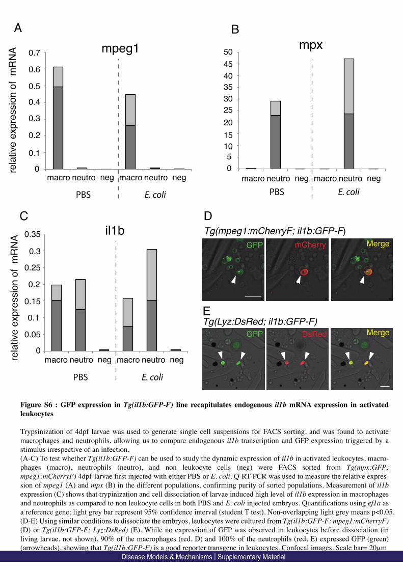
mpxmpeg1
0
0.1
0.2
0.3
0.4
0.5
0.6
0.7
0
5
10
15
20
25
30
35
GFP mCherry Merge
GFP DsRed Merge
Tg(mpeg1:mCherryF; il1b:GFP-F)
Tg(Lyz:DsRed; il1b:GFP-F)
B
C D
E
0
0.05
0.1
0.15
0.2
0.25
macro neutro neg
rela
tive
exp
ressio
n o
f m
RN
A
il1b
A
0.3
0.35
macro neutro neg
PBS E. coli
40
45
50
macro neutro neg macro neutro neg
rela
tive
exp
ressio
n o
f m
RN
A
PBS E. coli
macro neutro neg macro neutro neg
PBS E. coli
Figure S6 : GFP expression in Tg(il1b:GFP-F) line recapitulates endogenous il1b mRNA expression in activated
leukocytes
Trypsinization of 4dpf larvae was used to generate single cell suspensions for FACS sorting, and was found to activate
macrophages and neutrophils, allowing us to compare endogenous il1b transcription and GFP expression triggered by a
stimulus irrespective of an infection,
(A-C) To test whether Tg(il1b:GFP-F) can be used to study the dynamic expression of il1b in activated leukocytes, macro-
phages (macro), neutrophils (neutro), and non leukocyte cells (neg) were FACS sorted from Tg(mpx:GFP;
mpeg1:mCherryF) 4dpf-larvae first injected with either PBS or E. coli. Q-RT-PCR was used to measure the relative expres-
sion of mpeg1 (A) and mpx (B) in the different populations, confirming purity of sorted populations. Measurement of il1b
expression (C) shows that trypinization and cell dissociation of larvae induced high level of il1b expression in macrophages
and neutrophils as compared to non leukocyte cells in both PBS and E. coli injected embryos. Quantifications using ef1a as
a reference gene; light grey bar represent 95% confidence interval (student T test). Non-overlapping light grey means p<0.05.
(D-E) Using similar conditions to dissociate the embryos, leukocytes were cultured from Tg(il1b:GFP-F; mpeg1:mCherryF)
(D) or Tg(il1b:GFP-F; Lyz:DsRed) (E). While no expression of GFP was observed in leukocytes before dissociation (in
living larvae, not shown), 90% of the macrophages (red, D) and 100% of the neutrophils (red, E) expressed GFP (green)
(arrowheads), showing that Tg(il1b:GFP-F) is a good reporter transgene in leukocytes. Confocal images, Scale bar= 20µm
Disease Models & Mechanisms | Supplementary Material

scram MO il1b MO
0
50
100
150
200
250
300
350
400
num
ber
of re
cru
ited m
acro
phages
scram MO il1b MO
num
ber
of re
cru
ited m
acro
phages
0
5
10
15
20
25
Figure S7: Macrophage recruitment is not affected by il1b loss-of-function upon notochord infection.
To evaluate the effect of il1b loss-of-function on macrophage recruitment, Tg(mpeg1:mCherryF) larvae were
injected with either il1b (il1b MO) or scramble (scram MO) morpholinos. Then the larvae were infected with
E. coli-GFP in the notochord and recruited macrophages counted. Plots represent the number of recruited mac-
rophages to the notochord at 4 hpi (A) and at 24 hpi (B). The number of recruited macrophages in il1b mor-
phans was similar to that of controls. Graphs are mean value ± s.e.m. (A) p=0.80 and (B) p=0.69
4 hpi 24 hpi
Disease Models & Mechanisms | Supplementary Material

accession number genes Fold s.e.m
NM_212859.2 TNFa1 55.68 0.48
NM_001024447.1 TNFa2 3.26 1.62 increased
NM_001020785 Il10 3.44 0.78
NM_001082955.1 Il20 1.18 0.07
NM_001020799 Il26 1.76 0.09
NM_001111083.1 IFNφ3 1.57 0.52
NM_001020792 Il22 1.03 0.05
XM_002662056.3 CXC11L 0.63 0.30
unchanged
NM_131834 CXCR4b 0.75 0.06
decreased XM_005165411.1 CXC10L 0.40 0.19
Table S1: Induction of cytokines upon notochord infection with E. coli
The relative expression levels of cytokines and chemokines associated with E. coli
infection in the notochord. Expressions were measured by real-time PCR (using ef1a as a
reference gene) on cDNA made from RNA isolated from larvae injected with PBS or
with E. coli in the notochord at 48 hpi. Results are represented as fold change in E. coli
infection relative to PBS injection +/- standard error of the mean. n=3 independent
samples of 10 pooled larvae. We detected no significant expression of interferon φ1, φ2,
γ1 and γ2 in the different conditions.!
Disease Models & Mechanisms | Supplementary Material

MOVIE LEGENDS
Movie 1: General behaviour of neutrophils following notochord infection
Tg(mpx:GFP) larvae were infected with crimson expressing E. coli (magenta) in the
notochord at 48hpf. General behaviour of neutrophil (green) was imaged using 4-
dimensional confocal microscopy at 2 hpi during 3 hours. Scale bar: 20µm.
Movie 2: General behaviour of neutrophils following notochord infection
Tg(mpx:GFP) larvae were infected with DsRed expressing E. coli (red) in the notochord
at 48hpf. Scale bar: 50µm. Neutrophil behaviour (green) was imaged at 1 hpi during 2
hours and 50 min.
Movie 3: Typical behaviour of one neutrophil following notochord infection
High magnification of a region of movie S2 shows the representative behaviour of one
neutrophil that fail to engulf bacteria. Scale bar: 20µm.
Movie 4: General behaviour of macrophages following notochord infection
Tg(mpeg1:mCherryF) larvae were infected with GFP expressing E. coli (green) at 48hpf
in the notochord. The behaviour of macrophages (red) was imaged using 4-dimensional
confocal microscopy at 1 hpi during 3 hours. Scale bar: 50μm.
Movie 5: Typical behaviour of few macrophages following notochord infection
The representative behaviour of few macrophages was image in a similar experiment that
describe in movie S5 from 1hpi during 3 hours 20 min. Scale bar: 20µm.
Disease Models & Mechanisms | Supplementary Material

Movie 6: General behaviour of macrophages following systemic infection
Tg(mpeg1:mCherryF) larvae were infected with GFP expressing E. coli (green) at 48hpf
in the posterior caudal vein. The behaviour of macrophages (red) was imaged using 4-
dimensional confocal microscopy at 1 hpi during 1 hour. Scale bar: 50μm.
Disease Models & Mechanisms | Supplementary Material
Page 52
PART II
Neutrophils control E. coli infection at a distance
with superoxide
Authors:
PHAN Quang Tien, GONZALEZ Catherine, LEVRAUD Jean-Pierre,
LUTFALLA Georges, and NGUYEN-CHI Mai
Submitted for publication
2016

Page 53
Neutrophils control E. coli infection at a distance
with superoxide
PHAN Quang Tien12, GONZALEZ Catherine12, LEVRAUD Jean-Pierre4,
LUTFALLA Georges12 and NGUYEN-CHI Mai13
1Dynamique des Interactions Membranaires Normales et Pathologiques,
Université Montpellier 2, UMR 5235, case 107, Place Eugène Bataillon, 34095
Montpellier Cedex 05, France.
2CNRS; UMR 5235, Place Eugène Bataillon, 34095 Montpellier Cedex 05,
France.
3INSERM; Institute for regenerative medicine and Biotherapies, CHRU
Montpellier, France.
4Macrophages et Développement de l’Immunité, Institut Pasteur, Paris F-
75015, France.
*Authors for correspondence ([email protected]; lutfalla@univ-
montp2.fr)
Key points:
Neutrophils, but not macrophages, are instrumental in killing bacteria
when they cannot be reached for phagocytosis.
Superoxide produced by neutrophil oxidative burst is essential to clear
the infecting bacteria at a distance.
Abstract
Understanding the roles of neutrophils and macrophages in fighting bacterial
infections is a critical issue in human pathologies. Although phagocytic killing
has been extensively studied, little is known about how bacteria are eliminated
extracellularly in live vertebrates. We have recently developed an infection
model in the zebrafish embryo in which leucocytes cannot reach the injected
bacteria. We have described that when E. coli bacteria are injected within the

Page 54
notochord, both neutrophils and macrophages are massively recruited during
several days, but do not infiltrate the infected tissue presumably because of its
tough collagen sheath. Interestingly, the bacteria are cleared during the first
24hours, and we report here that macrophages are not involved in the control
of the infection. On the contrary, using transgenic lines and chemical
approaches, we show that the bactericidal action is achieved by production of
superoxide by neutrophils and that the outcome of infection depends on the
presence of a sufficient arsenal of neutrophils compared to bacterial load. We
thus reveal a novel host effector mechanism mediated by neutrophils that
eliminates bacteria that cannot be reached by phagocytes and that is
independent of macrophages, NO synthase, Myeloperoxidase or NETs.
Introduction
The innate immune system, first line of defence of the host, includes large
phagocytes (such as macrophages and granulocytes) equipped with a battery
of weapons to destroy the invader within minutes or hours. Since the seminal
work of Elie Metchnikoff1, the defence mechanisms of leukocytes remain a
challenging subject. When microbes penetrate the epithelial barrier,
macrophages and neutrophils are rapidly recruited and upon contact, engulf
the bacteria into a vacuole called a phagosome that fuses with intracellular
granules or lysosomes to form the phagolysosome in which bacteria may be
killed by a wide variety of mechanisms involving chemicals and enzymes2,3.
Non-oxidative effectors include antimicrobial proteins, while the oxygen-
dependent mechanism, also known as the respiratory burst, involves the
generation of reactive oxygen species (ROS) 4-6. ROS production inside the
phagocytic vacuole involves NADPH oxidase and the major ROS, superoxide
(O2-) and hydrogen peroxide (H2O2), can directly or indirectly promote the
death of the microbe, according to the nature of the pathogens7. Nitric oxide
(NO), produced by NO synthase, can contribute to microbicidal activity and is
essential for the defence against intracellular organisms such as S. enterica
and mycobacteria8,9.
Many microbes manage to survive within phagocytes after phagocytosis.
While some cope with the phagolysosomal conditions (Salmonella

Page 55
typhimurium10), others like Listeria, Shigella and some mycobacteria11-13 are
able to block the maturation of the phagosome or even to escape from these
compartments. Host cells, however, have developed counter strategies to fight
cytosolic bacteria including directing them to autophagosomes14.
While microbe killing inside the phagosome has been extensively studied, it is
less well understood how phagocytes are capable of killing microbes
extracellularly in whole organisms. Neutrophils can fight bacterial pathogens
without phagocytosis either by release of toxic granule contents
(degranulation) 15 or by expelling neutrophil extracellular traps (NETs), which
are networks of extracellular fibres built upon expulsion of chromatin16.
However events such as these are very hard to disentangle from
phagocytosis-mediated killing in the full context of tissue infection.
Thanks to its transparency and genetic amenability, the zebrafish embryo is a
useful model for the study of host/pathogen interactions in vivo. The zebrafish
model has been used to evaluate the respective roles of neutrophils and
macrophages in eliminating invading bacteria9,17,18; this relies not only on the
nature of the invading microbe, but also on the anatomic site of infection and
on the route of infection. One striking observation was that macrophages are
very efficient at engulfing microbes from body fluids (“flypaper” strategy) while
neutrophils may be very efficient at clearing surface associated microbes in a
“vacuum-cleaner”-like behaviour19.
We have recently developed an infection model in the zebrafish embryo in
which the bacteria are trapped in a tissue in which macrophages and
neutrophils cannot enter. When non-pathogenic Escherichia coli (E. coli)
bacteria are injected in the notochord, the swollen rod that provides axial
stiffness to the developing embryo, they slip between notochord cells and the
thick cylindrical collagen sheath that encases the cord. Although unable to
thread their way through this envelope, neutrophils and macrophages are
massively recruited all along the infected notochord where they stay in a highly
activated state for days. Interestingly, these inaccessible bacteria are cleared
within the first 24 hours20. While zebrafish embryo notochord represents a
physical barrier for leukocytes, there are differences in host responses to

Page 56
notochord infection by different invaders as E. coli is immediately detected by
leukocytes and induces heavy inflammation, whereas mycobacteria remain
undetected and their proliferation results in notochord destruction20-22.
Here we address the mechanisms of E. coli clearance in the notochord
infection model where professional phagocytes cannot directly encounter the
injected bacteria. We first investigate whether macrophages or neutrophils are
involved in this clearance and then investigate the nature of the molecules
instrumental for bacterial killing.
Materials and Methods
Ethics statement
Animal experimentation procedures were carried out according to the
European Union guidelines for handling of laboratory animals
(http://ec.europa.eu/environment/chemicals/lab_animals/home_en.htm) and
were approved by the Direction Sanitaire et Vétérinaire de l'Hérault and
Comité d'Ethique pour l'Expérimentation Animale under reference CEEA-LR-
13007. Fish husbandry and experiments were performed at the University of
Montpellier.
Fish husbandry
Fish maintenance, staging and husbandry were performed as described20 with
golden strain and transgenic lines. Tg(mpeg1:mCherry-F)umpTg2, referred as
tg(mpeg1:mCherry-F) 23 was used to visualize macrophages.
Tg(mpx:GFP)i114 and tg(lyz:DsRed)nz50 used to label neutrophils and the
mpx−/−NL144 ‘spotless’ mutant, are referred here as tg(mpx:GFP) 24,
tg(lyz:DsRed) 25 and mpx-/-27. tg(mpx:Gal4/USA:nfsB-mCherry) was used to
ablate neutrophils26. Embryos were obtained from pairs of adult fishes by
natural spawning and raised at 28.5°C in tank water. Embryos and larvae were
staged according to28.
E. coli and Salmonella injections and CFU count
E. coli K12 or Salmonella enterica serovar Typhimurium (here called
Salmonella) carrying plasmids encoding GFP or DsRed fluorescent proteins
Page 57
were injected in the notochord of 2dpf embryos and counted as described20.
Four different doses of E. coli were used: very low (1000 CFU), low (<3000
CFU), high (3000<n<6000 CFU) and very high (>7000 CFU). 3000 CFU of
Salmonella were injected in the hindbrain or in the notochord. Enteroinvasive
E. coli AIEC bacteria strain LF8229 and its mutants, LF82-Δ chiA30 and LF82-
ΔlpfA31 were injected at a low dose (CFU <3000) in the notochord.
Macrophage and neutrophil ablation and overproduction
To induce depletion of macrophages, 10nl of Lipo-Clodronate or Lipo-PBS
(clodronateliposomes.com) was i.v. injected in tg(mpeg1:mCherry-F)
transgenic larvae at 1dpf. Macrophage-depleted larvae were selected based
on the reduction of red labelled macrophages 24h after the treatment23. For
depletion neutrophils, 3nl of antisense translational morpholino csf3r 0.7mM
(5’GAAGCACAAGCGAGACGGATGCCAT3’, Gene-Tools) was microinjected
in the one-cell stage tg(mpx:GFP) embryos. Neutrophils were alternatively
depleted using metronidazole treatment of tg(mpx:gal4/UAS:nfsb-mCherry)
larvae (see below). Microinjection of gcsf3a or gcsf3b over-expressing
plasmids32 at 1-cell stage was used to increase neutrophil supply in
tg(mpx:GFP) embryos.
Drug treatments of zebrafish larvae
Tg(mpx:Gal4/USA:nfsB-mCherry) 2 dpf embryos expressing a Nitroreductase-
mCherry fusion protein specifically in neutrophils, were placed in fish water
containing 10mM Metronidazole and 0.1% DMSO (freshly prepared) for 24h.
After 3 washes with fish water, neutrophil-depleted larvae were selected based
on the number of red fluorescent cells. VAS2870 (Sigma-Aldrich SML0273)
stock was prepared in DMSO at 15mM. Two dpf tg(lyz:DsRed) embryos were
injected in the yolk with 5nl of 20µM VAS2870 diluted in miliQ water or with 5
nl of water-diluted DMSO. Apocynin (Santa Cruz, CAS498-02-2) was
dissolved at 100mM in DMSO. E. coli-infected larvae were placed in fish water
containing 250 µM Apocynin for 1 day. Decrease of superoxide production was
checked at 1dpi using DHE (Dihydroethidium, Santa Cruz CAS104821-25-2)
staining (see below). Nitric Oxide inhibition was performed with the pan-NOS
inhibitor NG-Nitro-L-Arginine Methyl Ester (L-NAME) (sigma, CAS 51298-62-
Page 58
5). After Notochord injection, embryos were placed in 1mM L-NAME fish water
for the whole time course of the experiments.
Staining and immuno-labelling in whole embryo
Mpx activity and neutrophils were detected in tg(mpx:GFP)/mpx+/- and
tg(mpx:GFP)/mpx-/- larvae at 1dpi using Sudan black staining20. DHE was
added to the fish medium at 3 µM at 1dpi for one hour and larvae washed 2
times before imaging33. Nitric Oxide was stained with 4-Amino-5-methylamino-
2’,7’-difluorofluorescein diacetate, Diaminofluorescein-FM diacetate (DAF-FM-
DA) (Sigma, CAS 254109-22-3) 34. Infected tg(lyz:DsRed) embryos were
treated with DAF-FM-DA 5µM by adding it directly to the fish medium for 2
hours at 6, 10hpi and 1dpi (for E. coli infection) or 2hpi (for Salmonella
infection). Larvae were then washed 3 times with fish water before imaging.
H2O2 was stained with Pentafluorobenzen sulfonyl fluorescein (Santa Cruz,
CAS 728912-45-6). Notochord infected tg(lyz:DsRed) embryos were soaked in
50µM of the fluorescent probe at 1dpi for 2 hours. The larvae were then
washed in fish water 3 times before imaging. Apoptotic cells were detected
using Acridine Orange staining; embryos were treated at 40hpi with 5 μg/mL of
Acridine Orange in fish water for 30 min, at 28.5 °C. Larvae were then rinsed
three times in fish water before imaging. DHE: excitation/emission 518/605
nm, DAF-FM-DA: 495/515 nm, Acridine Orange 500⁄526 nm.
Statistics analysis
Prism 4.0 (GraphPad Software) was used to construct plots and analyze data
in all figures except figures 4D and 6F, which were performed in Excel 2010
(Microsoft). A Mann-Whitney, two-tailed, unpaired t-test was used to calculate
the statistical significance. Data are shown as means with SEM. The survival
rate of treated embryos was compared with that of the DMSO/infected
embryos using the log-rank (Mantel-Cox) test.
Imaging of live zebrafish larvae
Larvae were anesthetized and mounted as previously described 20. Epi-
fluorescence microscopy was performed using a MVX10 Olympus microscope
(MVPLAPO 1X objective; XC50 camera), and a confocal Leica SPE upright

Page 59
microscope (40x HCX APO L 0.80 W and 20x CHX APO L 0.5 W objectives).
Image stacks for time lapse movies were acquired at 23–26°C every 4 min,
typically spanning 50 μm at 2 μm intervals, at 1024x512 or 512x512 pixel
resolution. The 4D files generated from time-lapse acquisitions were
processed using Image J, compressed into maximum intensity projections and
cropped. Brightness, contrast, and colour levels were adjusted for maximal
visibility.
Results
Macrophages are not required for the control of E. coli infection in the
notochord
We first compared enteric adherent invasive E. coli strains35 and laboratory
K12 strains in our notochord infection model and observed that they behaved
similarly (data not shown). We therefore performed our experiments with the
laboratory K12 strain. To investigate the role of macrophages in the observed
bacterial clearance, we injected liposome-encapsulated clodronate (Lipo-
clodronate) that kills phagocytic macrophages36,37. At one day post fertilization
(1dpf), macrophage/neutrophil dual reporter embryos, tg(mpeg1:mCherry-
F)/tg(mpx:GFP), were injected with 10nl of Lipo-Clodronate in the posterior
caudal vein (intravenous, i.v.). As previously described36 24h after Lipo-
Clodronate injection, macrophages were efficiently eliminated without affecting
the neutrophil population, nor inducing unspecific toxicity (Fig. 1A, B).
Macrophage depleted larvae were selected and injected in the notochord with
DsRed-expressing E. coli. Bacteria were cleared within the first 24 hours post
infection (hpi) in both, macrophage-depleted larvae (Fig. 1C) and Lipo-PBS
injected control larvae; the red signal observed along the notochord at 2dpi
was due solely to macrophages. Importantly, upon notochord infection,
neutrophils were normally recruited around the infected notochord regardless
of the presence or absence of macrophages (Fig. 1C). Thus, macrophages are
not required for bacterial clearance in this model.

Page 60
Neutrophils are essential for the control of notochord infection by E. coli
To investigate the role of neutrophils in bacterial clearance, we ablated
neutrophils by two independent approaches. First, we specifically inhibited
neutrophil development by knocking down the G-CSF/GCSF3R pathway using
a morpholino oligo (MO) specifically blocking csf3r translation (MO csf3r) 38.
Injection of MO csf3r in the neutrophil reporter embryos, tg(mpx:GFP), led to
approximately 70% reduction in the total number of neutrophils as compared
to larvae injected with a control morpholino (MO CTRL) at 3dpf (Fig. 2A-D).
We infected these morphants with 2500 CFUs fluorescent E. coli. Bacteria
were cleared in the control larvae (Fig. 2B, 2E) while they proliferated in
neutrophil-depleted embryos (Fig. 2B, 2F). The bacterial proliferation
correlates with a further dramatic reduction in neutrophil number at 1 and 2dpi
suggesting neutrophil death (Fig. 2D). Subsequently, infected csf3r morphants
died at 2-3dpi with overwhelming bacterial proliferation and neutropenia.
We also ablate neutrophils, using tg(mpx:Gal4/UAS:nfsB-mCherry) embryos in
which the myeloperoxidase promoter (mpx) indirectly drives the expression of
E. coli nitroreductase enzyme (nfsB) in neutrophils. Treatment of
tg(mpx:Gal4/USA:nfsB-mCherry) embryos with the pro-drug metronidazole
(MTZ) at 2dpf specifically depleted neutrophils (Fig. S1A, B) 39. Similarly to
csf3r morphants, bacteria were cleared in control larvae, while bacteria
proliferated in embryos with low neutrophil density (Fig. S1A). These two
experiments demonstrate that neutrophils are essential for the control of
notochord infection by E. coli.
Neutrophil/bacterial balance dictates infection outcome
We investigated the relationship between neutrophil supply and bacterial
clearance in the notochord. Embryo with a higher number of neutrophils
(overexpression of csf3a) can cope with higher amounts of injected bacteria
(Fig 3D). Similar results were observed by overexpressing csf3b (not shown).
Our data reveal that the balance of neutrophils versus bacteria is instrumental
for the outcome of the infection. Normal neutrophil levels are necessary to
eliminate small amounts of bacteria (Fig 3A), but embryos with depressed

Page 61
neutrophil populations are killed even by low bacterial loads (Fig 3C) and
embryo injected with higher amount of bacteria need a larger neutrophil
population to eliminate the bacteria (Fig 3B & D). This shows that neutrophil
populations are limiting in fighting the infection. Acridine Orange staining of
embryos experiencing neutropenia suggests that when the neutrophil versus
bacteria balance is not correct, neutrophils die by apoptosis (fig S2).
The neutrophil Myeloperoxidase is not required to control notochord
infection
Our previous study revealed that approximate 35% of recruited neutrophils
undergo degranulation of their primary granules around infected notochords 20.
We therefore investigated the role of the neutrophil-specific myeoloperoxidase
(Mpx) that is present in the azurophilic granules, in bacterial clearance. We
introduced the mpx:GFP transgene in the mpx-null mutant ‘spotless’27 to
generate tg(mpx:GFP)/mpx-/- offspring in which neutrophils express the eGFP
but lack Mpx activity23. Sudan Black staining confirmed that neutrophils did not
contain active Mpx in tg(mpx:GFP)/mpx-/- while in tg(mpx:GFP)/mpx+/-
siblings, neutrophils contained active Mpx in their granules (Fig. 4A). A low
dose of fluorescent E. coli was injected in the notochord of 2dpf
tg(mpx:GFP)/mpx-/- embryos; neutrophils were normally recruited along the
notochord, and the injected E. coli cleared at 1dpi as in the wild type (n>60
larvae, Fig. 4B). Mpx is therefore not required for the clearance of E. coli in the
notochord.
Larvae without NADPH Oxidase activity are highly susceptible to
notochord infection
Neutrophils use a wide variety of diffusible molecules to fight infections,
including NO and ROS. We investigated NO production by neutrophils during
the course of notochord infections using the NO reporter fluorescent probe
DAF-FM-DA. We used Salmonella infected embryos as controls to detect NO
production in neutrophils within the Aorta-Gonad-Mesonephros (AGM) (Fig.
S3A)40. As described34, the notochord itself was labelled by DAF-FM-DA in
uninfected embryos, but we could not observe any evidence of NO production
by neutrophils in our notochord infection model (Fig. S3B). Next we specifically

Page 62
inhibited NO synthases using L-NAME and injected E. coli into the thus treated
embryos. We did not observe any difference in the outcome of the infection in
L-NAME-treated larvae compared to controls (DMSO) (not shown).
To examine the role of ROS in the bacterial clearance, we used VAS2870
(VAS), a NADPH oxidase (NOX) inhibitor41. Injection of VAS in the yolk of 2dpf
tg(lyz:DsRed) embryos did not induce any toxicity. One hour after VAS
treatment, embryos were infected with low-dose GFP-expressing E. coli in the
notochord (Fig. 5A). At 1dpi, VAS treatment had no effect on the total
neutrophil population as seen in PBS control-injected embryos (Fig. 5B). While
bacteria were cleared in DMSO-treated embryos, E. coli proliferated in the
notochord of VAS treated larvae despite comparable neutrophil recruitment
followed by neutropenia and death in 74% of infected larvae between 2 and
3dpi (Fig. 5B-D), showing the essential role of NOX in the control of bacterial
infection. The effect was specific to the clearance of bacteria in this notochord
infection model since VAS treatment did not interfere with the clearance of
bacteria injected in muscle of the embryo where phagocytosis occurs (data not
shown).
Superoxide is produced in neutrophils of bacterial infected embryos
To check H2O2 production by neutrophils, we stained infected tg(lyz:DsRed)
embryos with the fluorescent probe Pentafluorobenzenesulfonyl fluorescein
that specifically detects H2O2. We did not detect fluorescence in recruited
neutrophils along the infected notochord during the first 24 h (data not shown).
As a control, we checked that, as reported41, H2O2 was produced by cells at
the amputated fin (data not shown). This suggests that H2O2 is not important
for bacterial clearance.
Another major ROS produced by neutrophils, superoxide, is also involved in
bacterial killing 42. To detect superoxide production in tg(mpx:GFP) embryos
infected with E. coli, we used Dyhroethidium (DHE), a fluorescent probe that
turns red after reacting with superoxide33. First, we imaged the injection site,
where some bacteria initially leak from the pierced notochord and get engulfed
by neutrophils and observed that these phagocytosing leukocytes, abundantly
produced superoxide in intracellular compartments harboring bacteria, which

Page 63
are most probably phagosomes (Fig. 6B). Green fluorescent E. coli were
rapidly lysed within 20 minutes in the putative phagosome (Fig. 6B). We then
image the upstream region, where bacteria are separated from the recruited
neutrophils by the notochord collagen sheath. Interestingly, these recruited
neutrophils also produced large amounts of superoxide, even though they had
not phagocytosed bacteria (Fig. 6C).
Apocynin decreases superoxide production and prevents bacterial
killing
We decided to investigate whether this superoxide production could be
involved in the control of bacterial proliferation by neutrophils. Apocynin, like
VAS, is a NOX inhibitor, but it targets phagocytes more specifically43,44. Upon
notochord infection, Apocynin-treated embryos had reduced number of
superoxide producing cells, including recruited neutrophils at the site of
inflammation, as compared to DMSO-treated larvae (Fig. 7A, B, C). We
infected tg(lyz:DsRed) embryos with a very low dose of E. coli (103 ± 20 CFUs)
in the notochord. Even with the very low dose infection, 80% of Apocynin-
treated embryos failed to clear bacteria, while all bacteria were efficiently killed
in DMSO-control embryo. Apocynin treated embryo displayed unrestricted
bacterial growth in the notochord at 1dpi, neutropenia and eventually death at
2-3dpi (Fig. 7 A, D, E, F). These data show that inhibition of superoxide
production in neutrophils increases susceptibility to notochord infection.
Discussion
Many studies have used the zebrafish embryo model to address the respective
roles of neutrophils and macrophages in eliminating invading bacteria, but in
all instances, at least one of these two cellular populations had direct access
to the bacteria. In our model neither neutrophils nor macrophages could reach
the bacteria. We first observed an active recruitment of both macrophages and
neutrophils around the infected notochord that is correlated with the clearing of
the bacteria within the notochord in less than 24 hours. By specific depletion of
individual myeloid populations, we have investigated their contribution in the
clearance of E. coli at a distance and describe how neutrophil may eliminate
inaccessible bacteria.

Page 64
Using chemical ablation of macrophages, we revealed that despite being
massively recruited to the notochord, macrophages are not required for the
bacterial killing. Whichever the strategy to lower the amount of neutrophils
within the developing zebrafish, the embryo becomes unable to cope with low-
dose infection, leading to bacterial proliferation and death of the embryo.
Importantly, we highlight the importance of the numerical balance between
neutrophils and bacteria in the control of notochord infection suggesting that
the molecules produced by the neutrophils to fight the bacteria are produced in
limiting amount. During Salmonella infections, the correct population of
neutrophils is maintained through a mechanism of demand-driven
granulopoiesis in the main site of hematopoietic stem cells emergence, i.e.,
the AGM40. Similarly, we show here, that when a low amount of bacteria is
injected, the host may increase the neutrophil pool to control notochord
infection. We also report that either increasing bacterial load or decreasing the
number of neutrophils results in bacterial proliferation, onset of neutropenia,
and death within 2 to 3 dpi. Conversely, we show that embryos with an
increased number of neutrophils can cope with a very high dose of bacteria.
To capture and kill microbes they cannot phagocytize, neutrophils have been
described to expel their chromatin to form Neutrophil Extracellular Traps
(NETs), but this may lead to neutrophil death (Netosis) 45,46. NET formation
relies on complex intracellular processes involving the activity, among others,
of myeloperoxidase47. Myeloperoxidase activity is not necessary to fight the
infection in our experimental system and we found no indication of NET
expulsion (data not shown). Since it is clear that the main cause of death of
the neutrophils is not NETosis, it will be very interesting to investigate the
signals triggering neutrophil death in our model.
Interestingly, neutrophils do not employ their traditional defence mechanisms,
phagocytosis and NETosis, to cope with notochord infection by E. coli; we
demonstrate that NOX activity and the production of superoxide by neutrophils
are essential. Indeed, treatment of embryos with a specific inhibitor of NOX
assembly, VAS2870, leads to bacterial proliferation. Using fluorescent probes,
we showed that neutrophils do not produce significant amount of H2O2 but
produce large amounts of superoxide. Apocynin blocks superoxide production
Page 65
specifically in phagocytes expressing Mpx44. Indeed, infected embryos treated
with Apocynin have a significantly reduced superoxide production and
increased susceptibility to notochord infection consistent with an essential role
of superoxide in the clearance of E. coli without direct phagocytosis (Fig. 7F).
The present work raises different questions related to the death of the different
actors, the bacteria, the neutrophils, and the embryo. Foremost is the question
as to how bacteria are killed at a distance by neutrophils. Neutrophil massively
degranulate around the infected notochord and we show here that an oxidative
burst is necessary for bacterial elimination. As hypothesised by JM Slaugh, we
propose that bacterial death is the result of cooperation between superoxide
and antimicrobial peptides48. A massive release of toxic molecules is
consistent with the tissue-induced damage and subsequent spine
malformations that we previously observed as a consequence of this transient
infection20. Why do neutrophils die when the bacteria/neutrophil ratio is too
much in favor of the invaders? If bacteria are not rapidly killed and start
proliferating within the infected notochord, then neutrophil massively die and
the embryo becomes neutropenic. This could be due to a factor released by
the densely packed bacteria within the notochord, possibly induced through a
quorum sensing mechanism. However, there is no reason why this virulence
factor would specifically kill neutrophils while sparing the highly endocytic
macrophages that are also massively recruited to the notochord but not
affected by bacterial proliferation. For this reason, we propose that death of
neutrophils could rather be a consequence of the excessive number of
bacteria triggering hyper activation of the neutrophils and leading to their
death49. This death would not be purely due to an excessive release of ROS
since it is not prevented by our NOX inhibitors; there is, however, a wide
choice of toxic molecules released by activated neutrophils besides ROS. This
hyper activation, akin to a cytokine burst is probably also responsible for the
death of the embryo in cases where E. coli proliferates within the notochord.
Importantly, we have no indication that the bacteria used in this study could kill
the embryo by themselves; injection of up to 5.104 CFU of E. coli within the
blood circulation does not lead to embryo death and phagocytes can easily
eliminate these huge amounts of bacteria (JPL, unpublished observations).

Page 66
We consider that in cases where the embryos die, it is the consequence of
their heavy inflammatory status mimicking a cytokine storm. This hypothesis is
consistent with our experiments with mycobacteria. We have demonstrated
that mycobacteria can replicate within the notochord without triggering the
heavy inflammation described here with E. coli. Mycobacteria remain almost
undetected within the notochord and proliferate, ultimately leading to
notochord break down. The subsequent fate of the embryo depends on the
virulence of the mycobacteria. The non-virulent Mycobacterium smegmatis is
eliminated by phagocytosis, leading to the host survival while M. marinum
resists destruction by phagocytosis and keeps proliferating until the host dies
(Alibaud et al 2011). Conversely, E. coli only effectively kills infected embryos
when injected in excessive amounts in the notochord where this triggers a
heavy inflammation that kills the neutrophils and ultimately the embryo.
To overcome killing by neutrophils, some pathogenic bacteria developed a
strategy to prevent engulfment. Indeed Yersinia pestis (bubonic and
pneumonic plaque), Yersinia pseudotuberculosis (gastroenteritis) and Yersinia
enterocolitica (gastroenteritis and mesenteric adenitis) are able to inhibit the
actin cytoskeleton required for engulfment, by the secretion of effector proteins
into the cytoplasm of the immune cell, which results in the decrease
phagocytosis by neutrophils and increased virulence3. Oxidative burst at a
distance is an alternative mechanism employed by neutrophils to control such
infections. Further investigations should determine whether host targeted
therapeutic strategies may be beneficial against medically relevant infections
where bacteria cannot be engulfed by phagocytes.
Acknowledgements
We have special thanks to Annette Vergunst, Paul Guglielmi, Etienne Lelièvre
and the members of DIMNP for helpful discussions. We thank David Stachura
for the gift of the Csf3 over-expressing plasmids, Nicolas Darnish for LF82 E.
coli strain and Steve Renshaw for transgenic lines. This work was supported
by a grant from the European Community’s Seventh Framework Program
(FP7- PEOPLE-2011-ITN) under the Marie-Curie Initial Training Network
FishForPharma [grant agreement no. PITN-GA-2011-289209]. QTP is

Page 67
currently supported by Fondation de la Recherche Médicale
(FDT20150532259). We thank Montpellier Rio Imaging for access to the
microscopes.
Author Contributions
Q.T.P., J-P. L., G.L. and M.N.-C. designed the project and the experiments,
Q.T.P., C.G. G.L. and M.N.-C. performed the experiments and analysed the
results. Q.T.P., G.L. and M.N.-C. wrote the manuscript with the input of J-P.L.
Conflict of Interest Disclosures
The authors declare that there are no conflicts of interest.

Page 68
FIGURES AND LEGENDS
Figure 1: Macrophages are not involved in the clearance of bacteria injected
within the notochord
(A) Experimental scheme. One dpf tg(mpeg1:mCherry-F/mpx: GFP) embryos were
i.v. injected with Lipo-Clodronate or Lipo-PBS. Correctly depleted larvae were
selected based on the loss of red fluorescence macrophages, and DsRed expressing
E. coli were injected within their notochord at 2dpf. The infection outcome was
analyzed at 2dpi using fluorescence microscopy. (B) Lipo-Clodronate efficiently
depletes macrophages without affecting neutrophil population. Experiments were
performed as described in (A). GFP (Neutrophils) and mCherry (macrophages)
expression were analysed by fluorescence microscopy at 2dpf. (C) E. coli infections in
the notochord are cleared in the macrophage-depleted embryos. Experiments were
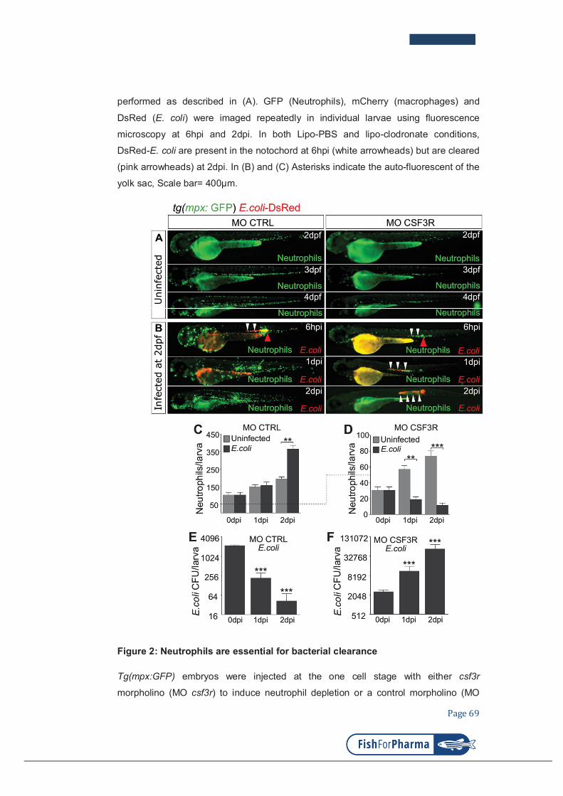
Page 69
performed as described in (A). GFP (Neutrophils), mCherry (macrophages) and
DsRed (E. coli) were imaged repeatedly in individual larvae using fluorescence
microscopy at 6hpi and 2dpi. In both Lipo-PBS and lipo-clodronate conditions,
DsRed-E. coli are present in the notochord at 6hpi (white arrowheads) but are cleared
(pink arrowheads) at 2dpi. In (B) and (C) Asterisks indicate the auto-fluorescent of the
yolk sac, Scale bar= 400μm.
Figure 2: Neutrophils are essential for bacterial clearance
Tg(mpx:GFP) embryos were injected at the one cell stage with either csf3r
morpholino (MO csf3r) to induce neutrophil depletion or a control morpholino (MO

Page 70
CTRL). (A) The steady-state of the neutrophil population was imaged repeatedly in
individual morphants using GFP fluorescence in both MO csf3r and control conditions
between 2 and 4dpf. (B) Fluorescent E. coli-DsRed were injected in the notochord of
csf3r and CTRL morphants (Red arrowheads: bacterial injection sites). GFP
(Neutrophils) and DsRed (E. coli) fluorescence was imaged at indicated time points.
E. coli-DsRed (red) disappeared from 1 dpi in the control embryos (left panel), while it
increased in csf3r morphants at 1 and 2dpi (white arrowheads) with a concomitant
decrease in neutrophil number (green, neutropenia). ( C,D) Quantification of
neutrophils in CTRL (C) and csf3r (D) morphants at the indicated time points following
PBS (light grey columns) or E. coli (dark grey columns) injection. (E, F) E. coli log
counts (CFU) in CTRL (E) and csf3r morphants (F). Neutrophils and CFU counts were
taken in whole embryos and expressed as mean number of cells/CFU per larva ±
SEM,**p<0.005, ***p<0.001, 5<n<11.

Page 71
Figure 3: Embryo needs correct neutrophil densities to fight different levels of
infection
(A, B) Two dpf tg(mpx:GFP) embryos were infected in the notochord with low dose (<
3000 CFUs, A) or high dose (> 4000 CFUs, B) DsRed fluorescent E. coli. Red-bar
charts represent the CFU counts at 0, 1 and 2dpi. Green-bar charts show the
quantification of total neutrophils at 0, 1 and 2dpi. Larvae images are representative

Page 72
overlays of fluorescence (green neutrophils and red E. coli) and transmitted light
images at 2dpi. (C): To decrease neutrophil density, tg(mpx:GFP) embryos were
injected at the one cell stage with the csf3r morpholino and then infected in the
notochord at 2dpf with a low dose (<3000 CFUs) of red fluorescent E. coli-DsRed. (D)
To increase neutrophil density, tg(mpx:GFP) embryos were injected at the one cell
stage with the gcsfa over-expressing plasmid and then infected in the notochord at
2dpf with a very high dose (>7000 CFUs) of red fluorescent E. coli-DsRed. Charts
correspond to CFU (red) and neutrophil (green) number counts at 0, 1 and 2dpi.
*p<0.05, ** p<0.01 and ***p<0.001, with respect to 0hpi.

Page 73
Figure 4: MPO is not required for bacterial clearance
Tg(mpx:GFP)/mpx+/- (A1, A2) and tg(mpx:GFP)/mpx-/- (A3, A4) embryos were
infected with E. coli in the notochord. Sudan Black staining and immuno-detection of
neutrophils (anti-GFP) were performed in whole embryos at 1dpi. The top right panel
shows the regions imaged by confocal microscopy in the larvae in A1 and A3 (green
box) and in A2 and A4 (red box). Representative transmitted light images, overlaid
with a maximal projection of confocal fluorescence images show the presence of
black granules in the neutrophils (white arrows) of tg(mpx:GFP)/mpx+/ embryos (A1

Page 74
and A2), revealing MPX activity. MPX granules are absent in neutrophils
(arrowheads) of tg(mpx:GFP)/mpx-/- embryos (A3 and A4). Scale bars: 10µm and
white dotted lines outline neutrophils in (A2, A3 and A4). Leica SPE confocal
microscope (488nm laser and bright field)
(B) Tg(mpx:GFP)/mpx-/ embryos were infected with E. coli-DsRed in the notochord.
Neutrophils (GFP) and E. coli (DsRed) were imaged repeatedly in individual larvae
using fluorescent microscopy at 6hpi and 1dpi. While E. coli locates in the notochord
at 6hpi (arrowheads), it disappears at 1dpi. (N= 60 mpx mutant embryos displaying
bacterial clearance similarly to the control embryos from three independent
experiments).
Figure 5: Larvae are more susceptible to notochord infection in the presence of
NADPH oxidase inhibitors

Page 75
Figure 5: Larvae are more susceptible to notochord infection in the presence of
NADPH oxidase inhibitors
(A) Experimental scheme. VAS2870 was injected in the yolk of tg(lyz:DsRed)
embryos at 2dpf. One hour later, E. coli-GFP bacteria were injected in the notochord.
Injected embryos were scored from 1dpi. (B, C, D) The consequences of VAS2870
treatment on neutrophil recruitment and E. coli infection. Trunk images are
representative overlays of DsRed, GFP and transmitted light images at 1dpi. Scale
bars: 100 µm. White arrowheads: E. coli in the notochord, blue arrowheads:
neutrophils around the notochord. (C) Larva survival was scored for uninfected and E.
coli-infected embryos, previously treated with either DMSO or VAS2870 (n>24 each).
These results are representative of three independent experiments; mean number of
larvae ±SEM, error bar p<0.0001. (D) Zebrafish larvae were treated as described in
(A). At 1 dpi larvae phenotype and bacterial outcome were scored. Larvae with no
defects and complete bacterial clearance were scored as having no bacterial
proliferation (grey). Results showed the mean percentage ±SEM, numbers on bars
are percentages, 42<n<69.

Page 76
Figure 6: Superoxide is produced in neutrophils of infected embryos
Two dpf tg(mpx:GFP) embryos were either injected with PBS (A) or infected with E.
coli-GFP in the notochord (B,C). At 6hpi, Superoxide was detected using
Dihydroethidium (DHE, red) and neutrophils were visualized using GFP fluorescence
(green). (Top panel) Black boxes in the larva image show the regions imaged by high
resolution confocal microscopy. (A) Representative transmitted light images, overlaid
with a maximal projection of confocal fluorescence images show that superoxide is
lightly produced in the recruited neutrophil due to injury at the injection site. (B)
Representative time-lapse maximum projections starting 6hpi during 16min, show

Page 77
superoxide presence in phagosomes (white arrows) bearing bacteria (yellow stars: E.
coli-GFP, Green) in recruited neutrophils at the injection site. Time is in minutes. (C):
Representative transmitted light images, overlaid with a maximal projection of
confocal fluorescence images show that, in the upstream region of the injection site,
superoxide is detected intracellularly (white arrowheads) over the E. coli (yellow stars)
infected notochord. Scale bars: 15µm, dotted line encases the notochord (NC).
Figure 7: Larvae with Phagocyte-specific superoxide inhibition are more
susceptible to notochord infection

Page 78
Figure 7: Larvae with Phagocyte-specific superoxide inhibition are more
susceptible to notochord infection
(A,B,C) GFP fluorescent E. coli were injected in 2dpf tg(mpx:GFP) embryos
immediately followed by either DMSO or 250µM Apocynin treatment. (A) At 1dpi,
superoxide production was visualised using dihydroethidium (DHE, red) and,
neutrophils and E. coli were detected using GFP by confocal microscopy.
Representative transmitted light images, overlaid with a maximal projection of
confocal fluorescence images show that Apocynin-treated larvae have less DHE-
positive neutrophils than DMSO-treated larvae (pink arrowheads). White arrows:
neutrophils DHE-, white arrowheads: E. coli, NC*: mid of notochord, scale bars:
30µm. (B) Quantification of DHE-positive cells in DMSO- and Apocynin-treated
larvae. Results were expressed as the mean of DHE-positive cells per larva ±SEM,
n>5 larvae. (C) Corresponding percentage of DHE-positive neutrophils that were
recruited to the notochord; mean percentage ±SEM, n>5 larvae.
(D, E, F) Two dpf tg(lyz:DsRed) embryos were infected in the notochord with E.coli-
GFP (green arrows: injection sites) and immediately treated with 250µM Apocynin.
(D) Neutrophils (DsRed) and E. coli (GFP) were imaged repeatedly in individual
larvae using fluorescent microscopy at 6hpi and 1dpi. Bacteria (white arrowheads)
were present at 6hpi in both DMSO- and Apocynin-treated embryos. At 1dpi, bacteria
disappeared in DMSO-treated embryos (arrows) while their number increased in
Apocynin-treated embryos (white arrowheads). Bacterial proliferation was
accompanied by a decreased number of neutrophils (neutropenia). (E) Larvae
survival was scored for uninfected and E. coli infected embryos, treated with either
DMSO or Apocynine. Results represent the mean of three independent experiments,
error bar: +-SEM, p<0.0001, 20<n<27. (F) Graphical abstract of host response during
notochord infection. 1/ Low dose of E. coli infection in the notochord leads to the rapid
recruitment of neutrophils to the notochord. During the first phase of infection,
neutrophils cannot penetrate the collagen sheath and engulf bacteria. NOX activity in
recruited neutrophils leads to the production of the ROS superoxide. Superoxide
production participates in bacterial clearance without neutrophil-microbe physical
contact, as early as 24hpi and results in host survival. 2/ Reducing the ROS
superoxide using a drug that inhibits NADPH Oxidase assembly (VAS2870) or a drug
that specifically blocks NADPH Oxidase in the leukocytes results in bacteria growth in
the notochord and host neutropenia and death. Increasing the ratio (E. coli
CFU)/(neutrophil number) either by genetic manipulation or higher dose of bacterial
injections leads to a similar outcome.

Page 79
Figure S1: Neutrophils are essential for the control of notochord infection
(A) Two dpf tg(mpx:Gal4/UAS:nfsB-mCherry) embryos were treated with 10mM
Metronidazole (Mtz) in fish water for 24 hours. At 3dpf, larvae with neutrophil
depletion were selected and injected either with PBS or E. coli-GFP in the notochord.
The outcome of the infection was analysed by fluorescent microscopy. Larvae images
are representative overlays of fluorescence (DsRed and GFP) and transmitted light
images at 2dpi. In the absence of Mtz, neutrophils are massively recruited to the
notochord and E. coli is cleared (white arrowheads). In Mtz-treated larvae, E. coli
(green, white arrowheads) grew heavily. This correlated with a dramatic decrease in
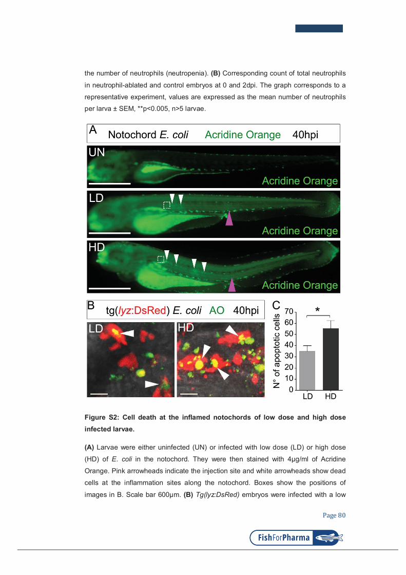
Page 80
the number of neutrophils (neutropenia). (B) Corresponding count of total neutrophils
in neutrophil-ablated and control embryos at 0 and 2dpi. The graph corresponds to a
representative experiment, values are expressed as the mean number of neutrophils
per larva ± SEM, **p<0.005, n>5 larvae.
Figure S2: Cell death at the inflamed notochords of low dose and high dose
infected larvae.
(A) Larvae were either uninfected (UN) or infected with low dose (LD) or high dose
(HD) of E. coli in the notochord. They were then stained with 4µg/ml of Acridine
Orange. Pink arrowheads indicate the injection site and white arrowheads show dead
cells at the inflammation sites along the notochord. Boxes show the positions of
images in B. Scale bar 600µm. (B) Tg(lyz:DsRed) embryos were infected with a low

Page 81
dose (LD) or a high dose (HD) of E. coli in the notochord. Representative transmitted
light images, overlaid with a maximal projection of confocal fluorescence images
show neutrophils stained with Acridine Orange (AO) (Arrowheads) at the inflammation
sites. (C) The number of Acridine Orange positive cells around the infected notochord
in indicated conditions. Scale bar 10µm. Error bar: mean ± SEM, *P<0.05 (0.035),
***p<0.001.
Figure S3: Recruited neutrophils do not produce detectable Nitric Oxide
(A) Nitric oxide is produced by neutrophils in the AGM following Salmonella
Typhimurium infection. Two dpf tg(lyz:DsRed) embryos were infected in the hindbrain
or in the notochord with Salmonella Typhimurium. At 2dpi Nitric oxide was detected
with DAF-FM-DA (green) using confocal microscopy. Representative overlay of

Page 82
maximum projections of multi scan acquisitions (DsRed and DAF-FM-DA) with
transmitted light images shows that Nitric oxide is produced by neutrophils in the
AGM (A left panel) and in the notochord (A right panel), but not in the recruited
neutrophils (A right panel).
(B) Two dpf tg(lyz:DsRed) embryos were infected with E. coli-GFP in the notochord.
(B1, B2) Representative overlay of maximum projections of multi-scan acquisitions
(DsRed and DAF-FM-DA) with transmitted light images shows that Nitric oxide
(green) is produced constitutively in the notochord (white arrowheads) but not in
recruited neutrophils at 6hpi (pink arrowheads). (B3-B6) Trunk images are
representative DAF-FM-DA fluorescence (B3-B4) and DsRed fluorescence images
(B5-B6) from PBS- or E. coli-injected embryos at 1dpi. AGM: Aorta-gonad-
mesonephros, NC: notochord, scale bars: 30µm
References
1. Metchnikoff É. Leçons Sur La Pathologie Comparée de L ’ Inflammation : Faites à L ’ Institut Pasteur En Avril et Mai 1891 / Par Élie Metchnokoff. Edited by G Masson. 1892;1 vol.(XI-. Paris: Librairie de l’académie de médecine, 120, Boulevard Saint-Germain, Paris.
2. Flannagan RS, Cosio G, Grinstein S. Antimicrobial mechanisms of phagocytes and bacterial evasion strategies. Nat Rev Microbiol. 2009;7:355-366.
3. Urban CF, Lourido S, Zychlinsky A. How do microbes evade neutrophil killing? Cell Microbiol. 2006;8:1687-1696.
4. Dupre-Crochet S, Erard M, Nubetae O. ROS production in phagocytes: why, when, and where? J Leukoc Biol. 2013;94:657-670.
5. Paiva CN, Bozza MT. Are reactive oxygen species always detrimental to pathogens? Antioxid Redox Signal. 2014;20:1000-1037.
6. Segal AW. How neutrophils kill microbes. Annu Rev Immunol. 2005;23:197-223.
7. Staudinger BJ, Oberdoerster MA, Lewis PJ, Rosen H. mRNA expression profiles for Escherichia coli ingested by normal and phagocyte oxidase-deficient human neutrophils. J Clin Invest. 2002;110:1151-1163.
8. Chakravortty D, Hensel M. Inducible nitric oxide synthase and control of intracellular bacterial pathogens. Microbes Infect. 2003;5:621-627.
9. Yang CT, Cambier CJ, Davis JM, Hall CJ, Crosier PS, Ramakrishnan L. Neutrophils exert protection in the early tuberculous granuloma by oxidative killing of mycobacteria phagocytosed from infected macrophages. Cell Host Microbe. 2012;12:301-312.
10. Eswarappa SM, Negi VD, Chakraborty S, Chandrasekhar Sagar BK, Chakravortty D. Division of the Salmonella-containing vacuole and depletion of acidic lysosomes in Salmonella-infected host cells are novel strategies of Salmonella enterica to avoid lysosomes. Infect Immun. 2010;78:68-79.
11. Pieters J. Mycobacterium tuberculosis and the macrophage: maintaining a balance. Cell Host Microbe. 2008;3:399-407.
12. Ray K, Marteyn B, Sansonetti PJ, Tang CM. Life on the inside: the intracellular lifestyle of cytosolic bacteria. Nat Rev Microbiol. 2009;7:333-340.

Page 83
13. Tilney LG, Portnoy DA. Actin filaments and the growth, movement, and spread of the intracellular bacterial parasite, Listeria monocytogenes. J Cell Biol. 1989;109:1597-1608.
14. Mostowy S. Autophagy and bacterial clearance: a not so clear picture. Cell Microbiol. 2013;15:395-402.
15. Nathan C. Neutrophils and immunity: challenges and opportunities. Nat Rev Immunol. 2006;6:173-182.
16. Brinkmann V, Reichard U, Goosmann C, et al. Neutrophil extracellular traps kill bacteria. Science. 2004;303:1532-1535.
17. Pagan AJ, Yang CT, Cameron J, et al. Myeloid Growth Factors Promote Resistance to Mycobacterial Infection by Curtailing Granuloma Necrosis through Macrophage Replenishment. Cell Host Microbe. 2015;18:15-26.
18. Torraca V, Masud S, Spaink HP, Meijer AH. Macrophage-pathogen interactions in infectious diseases: new therapeutic insights from the zebrafish host model. Dis Model Mech. 2014;7:785-797.
19. Colucci-Guyon E, Tinevez JY, Renshaw SA, Herbomel P. Strategies of professional phagocytes in vivo: unlike macrophages, neutrophils engulf only surface-associated microbes. J Cell Sci. 2011;124:3053-3059.
20. Nguyen-Chi M, Phan QT, Gonzalez C, Dubremetz JF, Levraud JP, Lutfalla G. Transient infection of the zebrafish notochord with E. coli induces chronic inflammation. Dis Model Mech. 2014;7:871-882.
21. Alibaud L, Rombouts Y, Trivelli X, et al. A Mycobacterium marinum TesA mutant defective for major cell wall-associated lipids is highly attenuated in Dictyostelium discoideum and zebrafish embryos. Mol Microbiol. 2011;80:919-934.
22. Benard EL, van der Sar AM, Ellett F, Lieschke GJ, Spaink HP, Meijer AH. Infection of zebrafish embryos with intracellular bacterial pathogens. J Vis Exp. 2012.
23. Bernut A, Herrmann JL, Kissa K, et al. Mycobacterium abscessus cording prevents phagocytosis and promotes abscess formation. Proc Natl Acad Sci U S A. 2014;111:E943-952.
24. Renshaw SA, Loynes CA, Trushell DM, Elworthy S, Ingham PW, Whyte MK. A transgenic zebrafish model of neutrophilic inflammation. Blood. 2006;108:3976-3978.
25. Hall C, Flores MV, Storm T, Crosier K, Crosier P. The zebrafish lysozyme C promoter drives myeloid-specific expression in transgenic fish. BMC Dev Biol. 2007;7:42.
26. Robertson AL, Holmes GR, Bojarczuk AN, et al. A zebrafish compound screen reveals modulation of neutrophil reverse migration as an anti-inflammatory mechanism. Sci Transl Med. 2014;6:225ra229.
27. Elks PM, van der Vaart M, van Hensbergen V, et al. Mycobacteria counteract a TLR-mediated nitrosative defense mechanism in a zebrafish infection model. PLoS One. 2014;9:e100928.
28. Kimmel CB, Ballard WW, Kimmel SR, Ullmann B, Schilling TF. Stages of embryonic development of the zebrafish. Dev Dyn. 1995;203:253-310.
29. Boudeau J, Glasser AL, Masseret E, Joly B, Darfeuille-Michaud A. Invasive ability of an Escherichia coli strain isolated from the ileal mucosa of a patient with Crohn's disease. Infect Immun. 1999;67:4499-4509.
30. Low D, Tran HT, Lee IA, et al. Chitin-binding domains of Escherichia coli ChiA mediate interactions with intestinal epithelial cells in mice with colitis. Gastroenterology. 2013;145:602-612 e609.

Page 84
31. Chassaing B, Rolhion N, de Vallee A, et al. Crohn disease--associated adherent-invasive E. coli bacteria target mouse and human Peyer's patches via long polar fimbriae. J Clin Invest. 2011;121:966-975.
32. Stachura DL, Svoboda O, Campbell CA, et al. The zebrafish granulocyte colony-stimulating factors (Gcsfs): 2 paralogous cytokines and their roles in hematopoietic development and maintenance. Blood. 2013;122:3918-3928.
33. Morash MG, Douglas SE, Robotham A, Ridley CM, Gallant JW, Soanes KH. The zebrafish embryo as a tool for screening and characterizing pleurocidin host-defense peptides as anti-cancer agents. Dis Model Mech. 2011;4:622-633.
34. Lepiller S, Laurens V, Bouchot A, Herbomel P, Solary E, Chluba J. Imaging of nitric oxide in a living vertebrate using a diamino-fluorescein probe. Free Radic Biol Med. 2007;43:619-627.
35. Dreux N, Denizot J, Martinez-Medina M, et al. Point mutations in FimH adhesin of Crohn's disease-associated adherent-invasive Escherichia coli enhance intestinal inflammatory response. PLoS Pathog. 2013;9:e1003141.
36. Travnickova J, Tran Chau V, Julien E, et al. Primitive macrophages control HSPC mobilization and definitive haematopoiesis. Nat Commun. 2015;6:6227.
37. van Rooijen N, Sanders A, van den Berg TK. Apoptosis of macrophages induced by liposome-mediated intracellular delivery of clodronate and propamidine. J Immunol Methods. 1996;193:93-99.
38. Ellett F, Pase L, Hayman JW, Andrianopoulos A, Lieschke GJ. mpeg1 promoter transgenes direct macrophage-lineage expression in zebrafish. Blood. 2011;117:e49-56.
39. Palha N, Guivel-Benhassine F, Briolat V, et al. Real-time whole-body visualization of Chikungunya Virus infection and host interferon response in zebrafish. PLoS Pathog. 2013;9:e1003619.
40. Hall CJ, Flores MV, Oehlers SH, et al. Infection-responsive expansion of the hematopoietic stem and progenitor cell compartment in zebrafish is dependent upon inducible nitric oxide. Cell Stem Cell. 2012;10:198-209.
41. Niethammer P, Grabher C, Look AT, Mitchison TJ. A tissue-scale gradient of hydrogen peroxide mediates rapid wound detection in zebrafish. Nature. 2009;459:996-999.
42. Babior BM, Curnutte JT, Kipnes BS. Pyridine nucleotide-dependent superoxide production by a cell-free system from human granulocytes. J Clin Invest. 1975;56:1035-1042.
43. Altenhofer S, Radermacher KA, Kleikers PW, Wingler K, Schmidt HH. Evolution of NADPH Oxidase Inhibitors: Selectivity and Mechanisms for Target Engagement. Antioxid Redox Signal. 2015;23:406-427.
44. Vejrazka M, Micek R, Stipek S. Apocynin inhibits NADPH oxidase in phagocytes but stimulates ROS production in non-phagocytic cells. Biochim Biophys Acta. 2005;1722:143-147.
45. Urban CF, Reichard U, Brinkmann V, Zychlinsky A. Neutrophil extracellular traps capture and kill Candida albicans yeast and hyphal forms. Cell Microbiol. 2006;8:668-676.
46. Yousefi S, Mihalache C, Kozlowski E, Schmid I, Simon HU. Viable neutrophils release mitochondrial DNA to form neutrophil extracellular traps. Cell Death Differ. 2009;16:1438-1444.
47. Metzler KD, Fuchs TA, Nauseef WM, et al. Myeloperoxidase is required for neutrophil extracellular trap formation: implications for innate immunity. Blood. 2011;117:953-959.
48. Slauch JM. How does the oxidative burst of macrophages kill bacteria? Still an open question. Mol Microbiol. 2011;80:580-583.
49. Geering B, Simon HU. Peculiarities of cell death mechanisms in neutrophils. Cell Death Differ. 2011;18:1457-1469.

Page 85
PART III
Identification of polarized macrophage subsets
in zebrafish
Authors:
NGUYEN-CHI Mai, LAPLACE-BUILHE Béryl, TRAVNICKOVA Jana, LUZ-CRAWFORD Patricia, TEJEDOR Gautier, PHAN Quang Tien, DUROUX-
RICHARD Isabelle, LEVRAUD Jean-Pierre, KISSA Karima, LUTFALLA Georges, JORGENSEN Christian, and DJOUAD Farida
eLIFE
2015

elifesciences.org
RESEARCH ARTICLE
Identification of polarized macrophagesubsets in zebrafishMai Nguyen-Chi1,2†, Beryl Laplace-Builhe1,2†, Jana Travnickova2,3,Patricia Luz-Crawford1,2, Gautier Tejedor1,2, Quang Tien Phan3,Isabelle Duroux-Richard1,2, Jean-Pierre Levraud4,5, Karima Kissa2,3,Georges Lutfalla2,3, Christian Jorgensen1,2,6‡, Farida Djouad1,2*‡
1Institut de Medecine Regenerative et Biotherapies, Institut national de la sante et dela recherche medicale, Montpellier, France; 2Universite de Montpellier, Montpellier,France; 3Dynamique des Interactions Membranaires Normales et Pathologiques,Centre national de la recherche scientifique, Montpellier, France; 4Macrophages etDeveloppement de l’Immunite, Institut Pasteur, Paris, France; 5Departement deBiologie du Developpement et Cellules Souches, Institut Pasteur, Paris, France;6Clinical unit for osteoarticular diseases and Department for Biotherapy, CentreHospitalier Universitaire, Montpellier, France
Abstract While the mammalian macrophage phenotypes have been intensively studied in vitro,
the dynamic of their phenotypic polarization has never been investigated in live vertebrates. We
used the zebrafish as a live model to identify and trail macrophage subtypes. We generated
a transgenic line whose macrophages expressing tumour necrosis factor alpha (tnfa), a key feature of
classically activated (M1) macrophages, express fluorescent proteins Tg(mpeg1:mCherryF/tnfa:
eGFP-F). Using 4D-confocal microscopy, we showed that both aseptic wounding and Escherichia coli
inoculation triggered macrophage recruitment, some of which started to express tnfa. RT-qPCR on
Fluorescence Activated Cell Sorting (FACS)-sorted tnfa+ and tnfa− macrophages showed that they,
respectively, expressed M1 and alternatively activated (M2) mammalian markers. Fate tracing of
tnfa+ macrophages during the time-course of inflammation demonstrated that pro-inflammatory
macrophages converted into M2-like phenotype during the resolution step. Our results reveal the
diversity and plasticity of zebrafish macrophage subsets and underline the similarities with
mammalian macrophages proposing a new system to study macrophage functional dynamic.
DOI: 10.7554/eLife.07288.001
IntroductionBehind the generic name ‘macrophage’ hides various cell types with distinct phenotypes and
functions. Currently, it is well established that macrophages are not just important immune effector
cells but also cells with critical homeostatic roles, exerting a myriad of functions in development,
homeostasis, and tissue repair and playing a pivotal role in disease progression (Wynn et al., 2013).
Therefore, there is a high interest in a better characterization of these cells to establish an early and
accurate diagnosis. The wide variety of macrophage functions might be explained by the outstanding
plasticity and versatility of macrophages that efficiently respond to environmental challenges and
changes in tissue physiology by modifying their phenotype (Mosser and Edwards, 2008). Although
there is a consensus that macrophages are a diversified set of cells, macrophage subtypes are still
poorly characterized. Indeed, although these cell populations have been extensively investigated in
mouse and human, these studies were mostly performed in vitro using monocyte-derived
macrophages induced under specific stimuli. A comprehensive characterization of macrophage
*For correspondence: farida.
†These authors contributed
equally to this work
‡These authors also contributed
equally to this work
Competing interests: The
authors declare that no
competing interests exist.
Funding: See page 13
Received: 03 March 2015
Accepted: 07 July 2015
Published: 08 July 2015
Reviewing editor: Utpal
Banerjee, University of California,
Los Angeles, United States
Copyright Nguyen Chi et al.
This article is distributed under
the terms of the Creative
Commons Attribution License,
which permits unrestricted use
and redistribution provided that
the original author and source are
credited.
Nguyen Chi et al. eLife 2015;4:e07288. DOI: 10.7554/eLife.07288 1 of 14

subsets that takes into account their specific behaviour, phenotypic diversity, functions, and
modulation shall rely on a real-time tracking in the whole organism in response to environmental
challenges.
Mouse and human macrophages have been classified according to their polarization state. In this
classification, M1 macrophages, also referred as classically activated macrophages, are pro-
inflammatory cells associated with the first phases of inflammation, while M2 macrophages, also
known as alternatively activated macrophages, are involved in the resolution of inflammation and
tissue remodelling (Gordon, 2003; Biswas and Mantovani, 2010; Sica and Mantovani, 2012).
Differential cytokine and chemokine production and receptor expression define the polarization state
of macrophages. However, it is worthwhile to note that such binary naming does not fully reflect the
diversity of macrophage phenotypes in complex in vivo environments in which several cytokines and
growth factors are released and adjust the final differentiated state (Chazaud, 2013; Thomas and
Mattila, 2014). Macrophages might adopt intermediate activation phenotypes classified by the
relative levels of macrophage subset-specific markers. Therefore, macrophage plasticity results in
a full spectrum of macrophage subsets with a myriad of functions (Mosser and Edwards, 2008; Xue
et al., 2014). Although the possible phenotype conversion of macrophages from M1 to M2 has been
suggested in in vitro studies, a recent study argues for the sequential homing of M1 and M2
macrophages to the site of injury (Stout et al., 2005; Sica and Mantovani, 2012; Shechter et al.,
2013). Such controversies highlight the lack of accurate real-time tracing of macrophage subtypes in
vivo in the entire animal.
Inflammation is a model of choice to study the wide range of macrophage subsets involved from its
initiation to its resolution. Therefore, in the present study, we propose to decipher in vivo in real time
the kinetic of macrophage subset recruitment, their behaviour and their phenotypic plasticity at the
molecular level during a multiple-step inflammatory process. We used the zebrafish larvae model for
its easy genetic manipulation, transparency, and availability of fluorescent reporter lines to track
macrophages (Ellett et al., 2011). While the existence of macrophage subtypes in zebrafish embryos
eLife digest Inflammation plays an important role in helping the body to heal wounds and fight
off certain diseases. Immune cells called macrophages—which are perhaps best known for their
ability to engulf and digest microbes and cell debris—help to control inflammation. In mammals,
different types of macrophage exist; the most functionally extreme of which are the M1
macrophages that stimulate inflammation and M2 macrophages that reduce the inflammatory
response.
Macrophages acquire different abilities through a process called polarization, which is controlled
by signals produced by a macrophage’s environment. Polarization has been well investigated in
human and mouse cells grown in the laboratory, but less is understood about how this process
occurs in live animals.
Nguyen Chi, Laplace-Builhe et al. investigated whether zebrafish larvae (which are naturally
transparent) could form an experimental model in which to investigate macrophage polarization in
living animals. Zebrafish were first genetically engineered to produce two fluorescent proteins: one
that marks macrophages and one that marks M1 macrophages. These fluorescent proteins allow the
movement and polarization of macrophages to be tracked in real time in living larvae using
a technique called confocal microscopy. Nguyen Chi, Laplace-Builhe et al. also isolated macrophage
cells from these zebrafish at different times during the inflammatory process to identify which
macrophage subtypes form and when.
The results show that unpolarized macrophages move to the sites of inflammation (caused by
wounds or bacterial infection), where they become polarized into M1 cells. Over time, these M1
macrophages progressively convert into an M2-like macrophage subtype, presumably to help clear
up the inflammation.
Furthermore, Nguyen Chi, Laplace-Builhe et al. show that the M1 and M2 macrophage subtypes in
zebrafish are similar to those found in mammals. Therefore, genetically engineered zebrafish larvae
are likely to prove useful for studying macrophage activity and polarization in living animals.
DOI: 10.7554/eLife.07288.002
Nguyen Chi et al. eLife 2015;4:e07288. DOI: 10.7554/eLife.07288 2 of 14
Research article Developmental biology and stem cells | Immunology

Figure 1. The (tnfa:eGFP-F) reporter line recapitulates transcriptional activation of tnfa upon wound-induced
inflammation and Escherichia coli infection. (A–E) Tumour necrosis factor alpha (tnfa) mRNA expression (blue,
arrowhead) was detected by in situ hybridization using tnfa anti-sense probe: at 6 hpA in (A) intact (control) and
(B) amputated fins from 3 dpf WT larvae, (C) in uninfected larvae (54 hpf, hours post-fertilization) and (D, E) E. coli
infected larvae (24 hpi, 54 hpf). Arrows show melanocytes (black). (E) Imaging of tnfamRNA expression in the muscle
at higher magnification, asterisks show muscle fibres, scale bar in (B) = 100 μm and in (E) = 50 μm. (F, G) eGFP
fluorescence (green) was analyzed by fluorescent microscopy in (F) intact (control) and (G) amputated Tg(tnfa:eGFP-F)
fins at 6 hpA, dotted lines outline the caudal fin, scale bar = 100 μm and at 16 hpi in Tg(tnfa:eGFP-F) larvae injected
with (H) PBS or (I, J) E. coli (red) in the muscle. Arrows show auto-fluorescent xanthophores. (J) Multi-scan confocal
analysis of GFP expression in E. coli-infected Tg(tnfa:eGFP-F) larvae, scale bar = 20 μm. (K) tnfa mRNA and eGFP-F
expressions were analyzed using microscopy at 6 hpA in amputated fins from 3 dpf Tg(tnfa:eGFP-F) larvae.
Dotted lines delimit the caudal fin, arrowheads show overlapping signals, and arrows show the pigments. Scale
bar = 100 μm. (L) Graphed data of representative fluorescence-activated flow cytometry analysis of eGFP+ cells in
upon amputation. Tg(tnfa:eGFP-F) larvae were either kept intact (control) or amputated at 3 dpf, and cells were
collected at 6 hr post-treatment. Green gates represent eGFP+ population and mean percentage of eGFP+
Figure 1. continued on next page
Nguyen Chi et al. eLife 2015;4:e07288. DOI: 10.7554/eLife.07288 3 of 14
Research article Developmental biology and stem cells | Immunology

has been suggested, they have not been fully characterized (Herbomel et al., 1999; Ellett et al.,
2011; Cambier et al., 2013; Petrie et al., 2014). Here, we report a new reporter transgene for TNFa,
a central inflammatory cytokine and well-established marker of M1 macrophages, instrumental to
discriminate macrophage subsets during intravital imaging.
Results and discussion
In vivo visualization of macrophage activation and polarizationFin wounding-induced inflammation and Escherichia coli inoculation in zebrafish larvae of 3 dpf are
two well-established models triggering macrophage recruitment. Using in situ hybridization, we
observed that the expression of the tumour necrosis factor alpha (tnfa), a consensus marker of M1
macrophages, was induced in cells accumulated in the caudal fin and the muscle following amputation
(nlarvae = 29/33) and E. coli inoculation (nlarvae = 12/12), respectively (Figure 1A–E). To study the cells
that express the tnfa transcripts, we established the Tg(tnfa:eGFP-F) transgenic zebrafish line
expressing a farnesylated (membrane-bound) eGFP (eGFP-F) under the control of the tnfa promoter.
While eGFP-F was undetectable in intact fins of Tg(tnfa:eGFP-F) larvae (nlarvae = 10/10), it was
expressed in cells recruited to the wound at 6 hr post-amputation (hpA) (nlarvae = 16/16, Figure 1F,G).
Similarly, eGFP-F expression was upregulated in cells accumulated in the muscle of Tg(tnfa:eGFP-F)
larvae at 16 hr post-inoculation (hpi) of DsRed-expressing E. coli (nlarvae = 8/8), compared to
Phosphate-buffered saline (PBS) injection (nlarvae = 3/3, Figure 1H,I). Confocal analysis confirmed the
presence of a membrane-bound eGFP in cells displaying a typical myeloid morphology (Figure 1J). To
demonstrate that the Tg(tnfa:eGFP-F) line recapitulates transcriptional activation of tnfa, we
performed a simultaneous detection of tnfa mRNA by in situ hybridization and GFP-F protein by
immunofluorescence in amputated larvae 6 hpA. We observed a consistent overlap between tnfa and
GFP-F signal in the fin (nlarvae = 11/11), showing the direct correlation of eGFP-F and tnfa transcriptional
activation in the fin of the reporter line (Figure 1K). In addition, we FACS-sorted GFP+ cells from
wounded Tg(TNFa:eGFP-F) larvae 6 hpA and performed RT-qPCR to analyze tnfa expression. We
observed a significant increase of tnfa mRNA level in eGFP+ cells as compared to eGFP− cells
(Figure 1L,M). All together these results indicate that the Tg(TNFa:eGFP-F) reporter line recapitulates
transcriptional activation of tnfa. Then, with the ability to specifically track tnfa-expressing cells, we used
Tg(tnfa:eGFP-F) fish to study macrophage activation by mating them with Tg(mpeg1:mCherryF) fish in
which macrophages express farnesylated mCherry (mCherryF) under the control of the macrophage-
specific mpeg1 promoter (Ellett et al., 2011; Nguyen-Chi et al., 2014). In intact Tg(tnfa:eGFP-F/
mpeg1:mCherryF) larvae, no eGFP-F was observed in macrophages (Figure 2A). We imaged double
transgenic larvae Tg(tnfa:eGFP-F/mpeg1:mCherryF) using 4D confocal microscopy from 45 min post-
amputation and found that macrophages were recruited to the wound from 1 hpA, some starting to
express eGFP from 3 hpA (Figure 2B,C and Video 1). From 5 hpA already activated macrophages, that
is, expressing tnfa arrived at the wound (Video 1 and Figure 2C). Similarly, infection with a crimson-
expressing E. coli in the muscle induced the expression of tnfa in phagocytes few hours following the
infection (Figure 2—figure supplement 1, Video 2). Imaging of the double transgenic larvae Tg(tnfa:
eGFP-F/mpeg1:mCherryF) showed that tnfa-expressing phagocytes were mainly macrophages
(Figure 2—figure supplement 1, Video 2). These results show the dynamic macrophage activation
in real-time in vivo including recruitment and rapid phenotypic change. During the revision of this paper,
a similar result has been published (Sanderson et al., 2015).
Morphology and behaviour of macrophage phenotypesTo test whether tnfa+ and tnfa− macrophages harboured different cellular characteristics, we first
analyzed their morphology in fin-wounded Tg(tnfa:eGFP-F/mpeg1:mCherryF) larvae. tnfa+mpeg1+
cells displayed flattened and lobulated morphology (Figure 2D), while tnfa−mpeg1+ were elongated
Figure 1. Continued
population ±s.e.m is indicated. (M) Relative expression of tnfa in eGFP- and GFP+ cells in amputated larvae. Real-
time RT-PCR on separated cells using EF1a as a reference gene. Graph represents the mean value of three
independent experiments ±s.e.m. *p < 0.05.
DOI: 10.7554/eLife.07288.003
Nguyen Chi et al. eLife 2015;4:e07288. DOI: 10.7554/eLife.07288 4 of 14
Research article Developmental biology and stem cells | Immunology

Figure 2. Activation, morphology, and behaviour of TNF-α+ macrophages in (tnfa:eGFP-F/mpeg1:mCherry-F)
transgenic larvae upon wound-induced inflammation. (A) eGFP-F (green) and mCherryF (red) fluorescence was
analyzed by fluorescent microscopy in intact (control) and amputated Tg(mpeg1:mCherryF/tnfa:eGFP-F) fins at 6
hpA of 3 dpf larvae. Arrowheads show recruited macrophages that express tnfa, arrows show tnfa+ cells that are not
macrophages, and asterisks show auto-fluorescent pigments. Dotted lines outline the caudal fin, scale bar = 100 μm.
(B) Bright-field image of the wounded fin of a 3 dpf Tg(mpeg1:mCherryF/tnfa:eGFP-F) larva. Dotted red box shows
the region imaged in C. (C) Representative time-lapse maximum projections show the activation of macrophages
arriving at the wound in 3 dpf amputated Tg(mpeg1:mCherryF/tnfa:eGFP-F). The time pA is shown on top right
Figure 2. continued on next page
Nguyen Chi et al. eLife 2015;4:e07288. DOI: 10.7554/eLife.07288 5 of 14
Research article Developmental biology and stem cells | Immunology

and dendritic (Figure 2E). As we observed that tnfa+mpeg1+ cells were predominant at the wound at
18 hpA and tnfa−mpeg1+ cells at 24 hpA (data not shown), we imaged the behaviour of these
macrophage populations in wounded fins from Tg(tnfa:eGFP-F/mpeg1:mCherryF) larvae at these time
points. tnfa+mpeg1+ cells presented a lower velocity (0.32 μm/min) than tnfa−mpeg1+ macrophages
(1.09 μm/min, Figure 2F) but a higher cell–cell contact frequency (0.036 VS 0.016 contacts/min)
with other macrophages (Figure 2G,I,J and Videos 3, 4). Measurements of the duration of
macrophage–macrophages contacts showed that these contacts lasted longer (48.6 min/contact) than
that of tnfa−mpeg1+ macrophages (15.9 min/contact, Figure 2H–J and Videos 3, 4). All together
these data highlight different morphology and behaviour of macrophage phenotypes in live zebrafish
suggesting the existence of macrophage subsets
exhibiting different functions.
Macrophages phenotypes areactivated in a time-dependantmannerTo quantify the respective frequency of tnfa+
macrophages (mCherry+eGFP+ referred as dbl+)
and tnfa− macrophages (mCherry+eGFP− re-
ferred as mCh+), we performed flow cytometry
analysis on cells isolated from Tg(tnfa:eGFP-F/
mpeg1:mCherryF) larvae at different time points
following caudal fin amputation or E. coli. in-
oculation (Figure 3A,B). While only 5.6% ± 0.9
(s.e.m.) dbl+ cells were detected in the mpeg1+
population of the intact larvae, a steady increase
of the dbl+ population from 6 to 20 hpA (up to
27.33 ± 0.2%) was observed. This percentage
decreased dramatically at 26 hpA to 8.75 ± 1%.
In E. coli inoculation experiments, the frequency of
dbl+ cells increased as soon as 3 hpi (55.60 ± 0.6%)
and remained stable until 26 hpi. These results
demonstrate that wound-induced macrophage
activation is transient compared to infection-
induced macrophage activation.
Figure 2. Continued
corner and indicated in hours and minutes, white lines outline the caudal fin. The transcriptional activation of tnfa
(green) in recruited macrophage (red, arrowhead) was first observed from 3 hpA. Scale bar = 30 μm. White lines
outline the caudal fin. (D, E) Maximum projections of confocal analysis of eGFP-F (green) and mCherryF (red)
expressions in recruited macrophages at (D) 18 hpA and (E) 24 hpA in Tg(mpeg1:mCherryF/tnfa:eGFP-F). tnfa+
mpeg1+ macrophages exhibit a round and protrusive morphology, while tnfa−mpeg1+ macrophages exhibit
a dendritic morphology. (F) Velocity of tnfa+mpeg1+ and tnfa−mpeg1+ macrophages (N = 18). (G) Frequency of
macrophage–macrophage contacts and (H) time length of the contacts of tnfa−mpeg1+ and tnfa+mpeg1+ cells.
Measurements were extracted from three independent videos of amputated Tg(mpeg1:mCherryF/tnfa:eGFP-F), for
contact frequency, N = 15 and for duration of the interaction, N = 11 macrophages. ****p < 0.0001.
(I) Representative time-lapse maximum projections show the behaviour of tnfa+mpeg1+ macrophages, starting
19h20 pA during 42 min. Two macrophages (green + red) interact by cell–cell contact. These macrophages (eGFP in
grey) remain attached up to 40 min. Scale bar 20 = μm. (J) Representative time-lapse maximum projections show the
behaviour of tnfa−mpeg1+ macrophages, starting 25h12 pA during 52 min. Macrophages (red) barely establish
cell–cell contact. Scale bar = 30 μm.
DOI: 10.7554/eLife.07288.004
The following figure supplement is available for figure 2:
Figure supplement 1. Activation of tnfa+ macrophages in (tnfa:eGFP-F/mpeg1:mCherry-F) transgenic larvae upon
E. coli infection.
DOI: 10.7554/eLife.07288.005
Video 1. Transcriptional activation of tnfa in macro-
phages of (tnfa:eGFP-F/mpeg1:mCherry-F) transgenic
larvae upon amputation. Representative time-lapse
maximum projections show the transcriptional activation
of tumour necrosis factor alpha (tnfa) in macrophages
arriving at the wound in 3 dpf amputated Tg(mpeg1:
mCherryF/tnfa:eGFP-F). The time pA is shown on top
right corner, white line outline the caudal fin. Scale bar =
30 μm. Image stacks were acquired every 3 min 30 s
from 45 min pA to 7 hr 48 min pA at 2-μm intervals,
1024 × 512 pixel resolution using a confocal microscope
TCSSP5 SP5 inverted equipped with a HCXPL APO 40×/
1.25–0.75 oil objective (Leica). Excitation wavelengths
used were 488 nm for EGFP-F and 570 nm for mCherryF.
DOI: 10.7554/eLife.07288.006
Nguyen Chi et al. eLife 2015;4:e07288. DOI: 10.7554/eLife.07288 6 of 14
Research article Developmental biology and stem cells | Immunology

Molecular signature of tnfa+ andtnfa− macrophage populations
To characterize at the molecular level tnfa− and tnfa+ macrophage populations during early and late
phases of inflammation, we FACS-sorted dbl+ and mCh+ cells from Tg(tnfa:eGFP-F/mpeg1:mCherryF)
tail-amputated larvae (Figure 3C) and analyzed them by qRT-PCR. In mammals, M1 and M2
macrophages are reported to be involved, respectively, in the initial phase of inflammation and in the
resolution phase. Cell sorting was thus performed at 6 hpA and 26 hpA following caudal fin
amputation since the kinetic analysis of macrophage subset activation (Figure 3B) suggested that
these two time points correspond to initiation and resolution of inflammation, respectively. As
expected, high levels of mpeg1 expression was observed in the mCh+ and dbl+ sorted cells at 6 and
26 hpA (Figure 3D), and high levels of tnfa expression was detected in double-positive populations at
6 hpA (Figure 3E). These observations demonstrated that fluorescence of these transgenes can be
efficiently used to track and separate macrophage sub-populations. At 6 hpA, dbl+ macrophages
expressed high levels of tnfb, il1b, and il6 compared to mCh+ macrophages (Figure 3F,G) that are
well-known markers of M1 macrophages in mammals (Mantovani et al., 2002;Martinez et al., 2006).
By contrast, mCh+ macrophages expressed low levels of these pro-inflammatory cytokines at both 6
and 26 hpA (Figure 3F,G), but expressed high levels of tgfb1, ccr2, and cxcr4b (Figure 3H), that are
specifically expressed in mammalian M2 macrophages (Mantovani et al., 2002; Martinez et al.,
2006; Hao et al., 2012; Beider et al., 2014; Machado et al., 2014). Of note, neither Arginase 1
(Arg1), which is largely used as a M2 marker in mouse but not in human (Chinetti-Gbaguidi and
Staels, 2011; Pourcet and Pineda-Torra, 2013), nor il10 (data not shown), a known M2 marker in
mammals (Mantovani et al., 2002), was detected in zebrafish macrophages. Importantly, based on
Video 2. Transcriptional activation of tnfa of (tnfa:
eGFP-F/mpeg1:mCherry-F) transgenic larvae upon
E. coli infection. Tg(mpeg1:mCherryF/tnfa:eGFP-F)
larvae were infected with crimson-expressing E. coli
(blue) at 3 dpf in the muscle and imaged from 30 min pi
to 10 hr 30 min pi. Representative time-lapse maximum
projections show the expression of tnfa (green) induced
in myeloid-like cells at the infection site from 3 hpA. The
time pA is shown on top right corner, scale bar = 25 μm.
Image stacks were acquired every acquired 3 min 30 s at
2-μm intervals, 512 × 512 pixel resolution with a X2 zoom
using a confocal microscope TCSSP5 SP5 inverted
equipped with a APO 20× objective (Leica). Excitation
wavelengths used were 488 nm for EGFP-F and
580 nm for Crimson. To distinguish Crimson from
mCherry, emission filter was adjusted from 630 to
750 nm.
DOI: 10.7554/eLife.07288.007
Video 3. tnfa+ macrophage behaviour following
amputation. Representative time-lapse maximum pro-
jections show the behaviour of tnfa+mpeg1+ macro-
phages in Tg(mpeg1:mCherryF/tnfa:eGFP-F) larvae
following amputation, starting 19h20 pA during 42 min.
Two macrophages tnfa+mpeg1+ interact by cell–cell
contact. These macrophages (GFP in grey) remain
attached up to 40 min. Scale bar = 20 μm. Image stacks
were acquired every 3 min 30 s at 2 μm-intervals, 1024 ×
512 pixel resolution using a confocal microscope
TCSSP5 SP5 inverted equipped with a HCXPL APO
40×/1.25–0.75 oil objective (Leica). Excitation wave-
lengths used were 488 nm for EGFP-F and 570 nm for
mCherryF.
DOI: 10.7554/eLife.07288.008
Nguyen Chi et al. eLife 2015;4:e07288. DOI: 10.7554/eLife.07288 7 of 14
Research article Developmental biology and stem cells | Immunology

the stability of the eGFP in Tg(tnfa:eGFP-F/
mpeg1:mCherryF) larvae allowing us to specifi-
cally track the behaviour and fate of pro-
inflammatory macrophages, we found that the
dbl+ pro-inflammatory macrophages changed
their phenotype at 26 hpA. Indeed, dbl+ macro-
phages negative for M2 markers at 6 hpA,
displayed at 26 hpA, in parallel to a significant
decrease of tnfa, il1b, and il6 expression level,
a significant increased expression level of ccr2
and cxcr4b (Figure 3E,H). Of note, a tendency
toward differential expression level was observed
for tnfb and tgfb1 between 6 and 26 hpA. To go
further and demonstrate that the same macro-
phages are present at the wound site during
inflammation and its resolution, we generated
the Tg(mpeg1:GAL4/UAS:Kaede) larvae to track
macrophages exploiting the conversion of the
native green fluorescence of Kaede into red
fluorescence under UV light. Recruited macro-
phages were photoconverted 6 hpA and imaged
at 26 hpA revealing that early recruited macro-
phages were still present at the wound area 20 hr
later (Figure 4—figure supplement 1). Then,
GFP+ macrophages were specifically tracked
using time-lapse imaging of wounded Tg
(mpeg1:mCherryF/TNFa:GFP-F) fins from 6 to
26 hpA. We show that initially recruited eGFP+
macrophages remain at the injury site and still express the GFP (Figure 4A–C and Video 5). The
analysis of macrophage behaviour over time shows that among eGFP+ macrophages displaying an
amyboid phenotype at the wound edge 6 hpA, 50% change toward a fibroblastic phenotype from 11
hpA when they moved distally (Video 5). All together these data show that pro-inflammatory
macrophages underwent a phenotypic conversion toward an intermediate phenotype in which both
M1 and M2 markers are expressed. In addition, this molecular characterization of macrophages in
zebrafish reveals the conservation of macrophage subtypes between zebrafish and human.
In conclusion, we identified macrophage subsets in zebrafish and described their behaviour
and fate during a process of inflammation (Figure 4D). Live imaging of transparent transgenic
zebrafish larvae allowed the first real-time visualization of macrophage activation and
polarization. In parallel, a molecular analysis of macrophage sub-populations highlights the
evolutionary conservation of macrophages from fish to mammals. We propose that in response to
wounding zebrafish, unpolarized macrophages are recruited to the inflammation site and adopt
a M1-like phenotype. Subsequently, they progressively convert their functional phenotype from
M1-like to M2-like in response to progressive inflammatory microenvironment changes within the
tissue (Figure 4D). Live imaging of the new transgenic line we generated opens new avenues to
study in real time in live vertebrates the full spectrum of macrophage activation, polarization, and
functions.
Materials and methods
Ethics statementAll animal experiments described in the present study were conducted at the University of Montpellier
according to European Union guidelines for handling of laboratory animals (http://ec.europa.eu/
environment/chemicals/lab_animals/home_en.htm) and were approved by the Direction Sanitaire
et Veterinaire de l’Herault and Comite d’Ethique pour l’Experimentation Animale under reference
CEEA-LR-13007.
Video 4. tnfa− macrophage behaviour following
amputation. Tg(mpeg1:mCherryF/tnfa:eGFP-F) caudal
fins were amputated at 3 dpf. Representative time-lapse
maximum projections show the behaviour of tnfa−
mpeg1+ macrophages, starting 25h12 pA during 42 min.
Macrophages (red) scarcely establish cell–cell contact.
Scale bar = 30 μm. Image stacks were acquired every
4 min at 2 μm-intervals at 1024 × 512 pixel resolution
using a confocal microscope TCSSP5 SP5 inverted
equipped with a HCXPL APO 40×/1.25–0.75 oil objec-
tive (Leica). Excitation wavelengths used were
488 nm for EGFP-F and 570 nm for mCherryF.
DOI: 10.7554/eLife.07288.009
Nguyen Chi et al. eLife 2015;4:e07288. DOI: 10.7554/eLife.07288 8 of 14
Research article Developmental biology and stem cells | Immunology

Figure 3. Isolation and molecular characterization of macrophage phenotypes. (A) Graphed data of representative fluorescence-activated flow cytometry
analysis of tnfa+ and tnfa− macrophages upon inflammatory stimulations. Tg(mpeg1:mCherryF/tnfa:eGFP-F) larvae were either kept intact (control), or
amputated, or injected with PBS or with E. coli at 3 dpf, and cells were collected at 6 hr post-treatment. Red, green, and yellow gates represent mCherry+,
eGFP+, and mCherry+eGFP+ populations, respectively. (B) Graph represents the kinetic of the frequency of mpeg1+tnfa+ macrophages in macrophage
population (mpeg1+) in three independent experiments following stimulation: amputation and E. coli infection (E. coli) at indicated time points. *p < 0.05
Figure 3. continued on next page
Nguyen Chi et al. eLife 2015;4:e07288. DOI: 10.7554/eLife.07288 9 of 14
Research article Developmental biology and stem cells | Immunology

Zebrafish line and maintenanceFish and embryo maintenance, staging, and husbandry were as previously described (Nguyen-Chi
et al., 2014). Experiments were performed using the AB zebrafish strain (ZIRC) and the transgenic line
Tg(mpeg1:mCherryF) to visualize macrophages. For the photoconversion experiments, a cross of Tg
(mpeg1:Gal4)gl25 and Tg(UAS:kaede)rk8 lines was used, using breeders selected for progeny with
negligible silencing of the UAS transgene.
Transgenic line constructionThe TNFa promoter (Gene ID: 405785) was amplified from zebrafish genomic DNA using primers zTNFaP4
(CCCGCATGCTCCACGTCTCC) and zTNFaE11N (TTATAGCGGCCGCCCGACTCTCAAGCTTCA). The
resulting fragment was phosphorylated using T4PNK, digested by NotI and cloned in a farnesylated
eGFP (eGFP-F) derivative of pBSKI2 (Thermes et al., 2002). The resulting plasmid (pI2promTNFa:
eGFP-F) harbours a 3.8-kb fragment of the zebrafish tnfa promoter, including part of the first coding
exon. It uses the endogenous ATG codon of tnfa to drive the translation of eGFP-F. The expressed
eGFP-F harbours the first 7 amino acids of zebrafish TNFa at its N-terminus (MKLESRA).
The expression cassette is flanked by two I-SceI sites. pI2promTNFa:eGFP-F was co-injected in
fertilized eggs with the enzyme I-SceI (New England Biolabs, France). Developing embryos were
injected with non-pathogenic E. coli at 3 dpf (days post-fertilization), and those that developed
a specific green fluorescence were raised as putative founders. The offspring of putative founders was
tested the same way in order to establish the stable transgenic line. Low expression of eGFP in the
pharynx at 3 dpf was used to check larvae for the presence of the transgene.
Larva manipulation for inflammation assays and imagingCaudal fin amputation was performed on 3 dpf larvae as described in Pase et al. (2012). The caudal
fin was transected with a sterile scalpel, posterior to muscle and notochord under anaesthesia with
0.016% Tricaine (ethyl 3-aminobenzoate, Sigma Aldrich, France) in zebrafish water. Larvae were
inoculated at 3 dpf by 2.103 CFU E. coli K12 bacteria harbouring either DsRed (van der Sar et al.,
2003) or Crimson (Clontech, France) expression plasmid or no plasmid. Imaging was performed as
previously described (Nguyen-Chi et al., 2014) using a confocal TCS SP5 inverted microscope with
a HCXPL APO 40×/1.25–0.75 oil objective (Leica, France). Image stacks for time-lapse videos were
acquired every 3–5 min, typically spanning 30–60 μm at 2-μm intervals, 1024 × 512 or 512 × 512 pixel
resolution. The 4D files generated from time-lapse acquisitions were processed using Image J. They
were compressed into maximum intensity projections and cropped. Brightness, contrast, and colour
levels were adjusted for maximal visibility. Velocity of macrophages was measured using Manual
Tracking Image J plugin. Frequency of macrophage–macrophage interaction and duration of
interactions were measured manually on stack images. For tracking of macrophages, eGFP-F+
mCherryF+ cells from Tg(mpeg1:mCherryF/tnfa:eGFP-F) wounded fins were tracked using time-
lapse image series from 6 hpA (hours post-amputation) to 26 hpA.
FACS analysis, isolation of mRNA from macrophages and RT-qPCR300 Tg(tnfa:eGFP-F/mpeg1:mCherryF) larvae were either amputated or infected as described above,
then crushed on a 70-μm cell strainer (Falcon, France). Isolated cells were washed in PBS/2 mM
ethylenediaminetetraacetic acid (EDTA)/2% Foetal Calf Serum (FCS), filtered through a 40-μm cell
strainer, and counted. Counting of mCherry+eGFP− and mCherry+eGFP+ cells was performed on
LSRFortessa (BD Bioscience, France), and data analyzed using the Flowjo software (Tree start,
Ashland, Or, USA). Sorting was done using FACS ARIA (BD Bioscience, France) and collected in 50%
FCS/50% Leibovitz L-15 medium (21083-027, Gibco, France) on ice. To isolate total RNA, cells were
Figure 3. Continued
vs 3 hpA, mean value of three experiments ±s.e.m. (C) Gating strategy to isolate control cells (mCherry− eGFP−, neg), tnfa− macrophages (mCherry+
eGFP−, mCh+), tnfa+ macrophages (mCherry+eGFP+, dbl+). (D–H) Relative expression of (D)mpeg1, (E) tnfa, (F) tnfb, il1b, (G) il6, (H) tgfb1, ccr2, and cxcr4b
in cells neg, mCh+, and dbl+. Tg(mpeg1:mCherryF/tnfa:eGFP-F) were amputated at 3 dpf and cells were collected and separated at 6 hpA and 26 hpA.
Real-time RT-PCR on separated cells using EF1a as a reference gene. Graph represents the mean value of five independent experiments ±s.e.m.
Statistical significance between bars are indicated *p < 0.05, **p < 0.01.
DOI: 10.7554/eLife.07288.010
Nguyen Chi et al. eLife 2015;4:e07288. DOI: 10.7554/eLife.07288 10 of 14
Research article Developmental biology and stem cells | Immunology

Figure 4. M1-like macrophages convert their phenotype toward M2-like phenotype in the wounded fin. (A) Diagram
showing the site where caudal fin was transected (dotted red line) in 3 dpf Tg(mpeg1:mCherryF/tnfa:eGFP-F) larvae.
The black dotted box represents the region imaged by confocal microscopy. (B) Representative time-lapse
maximum projections of 3 dpf Tg(mpeg1:mCherryF/tnfa:eGFP-F) amputated fins showing the fate of tnfa+
macrophages (magenta + green) at the indicated times pA (hours:minutes) from 6 hpA to 26 hpA. White lines
delimit the caudal fin. Scale bar = 30 μm. (C) Tracking of tnfa+ macrophages from 6 to 26 hpA. The distinct colours of
the lines correspond to the distinct macrophages that were indicated with an arrowhead in B. (D) Diagram
representing macrophage activation and polarization in zebrafish. Unpolarized macrophages (mpeg1+) are
mobilized and recruited to the wound following fin amputation. They are activated and polarized toward a M1-like
phenotype (pro-inflammatory) few hours following fin amputation. After 24 hpA, in response to changes in
environmental cues, the same macrophages progressively change their phenotype toward intermediate phenotypes
and maybe fully polarized M2-like phenotype (non-inflammatory). Main markers of macrophage subtypes are
indicated and resemble those found in human (tnfa/b indicates tumour necrosis factor alpha; il1b, interleukin 1-beta;
il6, interleukin 6; tgfb1, tumour growth factor beta 1; ccr2, c–c chemokine receptor type 2; cxcr4b, chemokine (C-X-C
motif) receptor 4b).
DOI: 10.7554/eLife.07288.011
The following figure supplement is available for figure 4:
Figure supplement 1. Recruited macrophages remain in the region of tissue injury at 26 hpA.
DOI: 10.7554/eLife.07288.012
Nguyen Chi et al. eLife 2015;4:e07288. DOI: 10.7554/eLife.07288 11 of 14
Research article Developmental biology and stem cells | Immunology

lysed in QIAzol Lysis Reagent (Qiagen, France)
and RNA extracted using miRNeasy mini kit
(Qiagen-21704, France). 20 ng of total RNA was
reverse transcribed using High Capacity RNA
Reverse Transcription kit (Applied Biosystems,
France). QPCR were performed on a LightCycler
480 system (Roche, France), following manufac-
turer’s instructions (SYBR Green format, Roche
Applied Science, Meylan, France) and using primers
in Supplementary file 1: denaturation 15 s at 95˚C,
annealing 10 s at 64˚C, and elongation 20 s at 72˚C.
Expression levels were determined with the
LightCycler analysis software (version-3.5) from
5 independent experiments. The relative amount
of a given mRNA was calculated by using the
formulae 2−∆Ct with ef1a as reference.
Statistical analysisSignificance testing for Figures 1M, 3D–H was
done using Mann–Whitney unpaired t-test,
one-tail and Figure 2F–H using Mann–Whitney
unpaired t-test, two-tails using GraphPad Prism 6
Software. *p < 0.05, **p < 0.01, ****p < 0.0001.
In situ hybridizationA tnfa probe was amplified from total cDNA by
PCR using tnfa.55 and tnfa.58 primers
(Supplementary file 1) and cloned in plasmid
pCRII-TOPO. Digoxigenin (DIG)-labelled (Roche,
France) sense and anti-sense RNA probes were in
vitro transcribed (Biolabs, France). In situ hybridizations on whole-mount embryos were as previously
described (Nguyen-Chi et al., 2012). For simultaneous detection of eGFP-F proteins and tnfa mRNA
by immuno-detection and in situ hybridization, fixed and rehydrated Tg(tnfa:eGFP-F) larvae were
permeabilised in ice in 100% ethanol for 5 min, then in a mixture of 50% Xylene-50% ethanol for 1 hr
and in 80% acetone for 10 min at −20˚C as described in Nagaso et al. (2001). After washes in PBS-
0.1% Tween, larvae were post-fixed in 4% paraformaldehyde (PFA) for 20 min. Subsequent steps of
hybridization, washes, and staining with NBT-BCIP (Roche, France) were as previously described in
Nguyen-Chi et al. (2012). Next, unspecific-binding sites were saturated in PBS-1% bovin serum
albumin (BSA)-1% lamb serum-10% Goat serum and larvae incubated 3 days with an anti-GFP
antibody (MBL, 1/500). After extensive washes, larvae were incubated with a goat anti-rabbit
antibody. Stained embryos were imaged using a MVX10 Olympus microscope with MVPLAPO 1×
objective and XC50 camera and using a Zeiss Axioimager with a Zeiss 40× Plan-Apo 1.3 oil objective.
Photoconversion of macrophage-specific Kaede proteinTg(mpeg1:GAL4/UAS:Kaede) embryos were raised to 3 dpf in the dark, and caudal fin was transected
as described above. At 6 hpA, larvae were mounted in 1% low-melting point agarose. A 405-nm Laser
Cube 405-50C on a confocal TCS SP5 inverted microscope with a HCXPL APO 40×/1.25–0.75 oil
objective (Leica) was used to photoconvert the Kaede-labelled cells using 6% laser power scanning for
60 s (optimized before the experiments; data not shown). Fins were imaged before and after the
photoconversion (at 6 and 26 hpA) in the green and red channels.
AcknowledgementsThis work was supported by Inserm and grants from the Medical Research Foundation (projet FRM
2011 ‘Comite Languedoc-Roussillon-Rouergue (LRR)’), from ‘La region Languedoc-Roussillon projet
Chercheurs d’avenir 2011’ (chercheur avenir 2012-Q-173), from the French National Research Agency
for ‘Zebraflam’ program n˚ ANR-10-MIDI-009. We thank Myriam Boyer-Clavel from the Montpellier
Video 5. Recruited GFP+ macrophages persist in the
region of tissue injury at 26 hpA. Tg(mpeg1:mCherryF/
tnfa:eGFP-F) caudal fins were amputated at 3 dpf.
Representative time-lapse maximum projections show
the movements of tnfa+mpeg1+ macrophages (green +
magenta), starting 6 hpA to 26 hpA. Coloured lines
correspond to eGFP+ macrophage tracking that stay in
the wounded fin and still express eGFP at 26 hpA. Scale
bar = 30 μm. Image stacks were acquired every 4 min 40
s at 3.5-μm intervals at 512 × 512 pixel resolution using
a confocal microscope TCSSP5 SP5 inverted equipped
with a HCXPL APO 40×/1.25–0.75 oil objective (Leica).
Excitation wavelengths used were 488 nm for EGFP-F
and 570 nm for mCherryF.
DOI: 10.7554/eLife.07288.013
Nguyen Chi et al. eLife 2015;4:e07288. DOI: 10.7554/eLife.07288 12 of 14
Research article Developmental biology and stem cells | Immunology

RIO Imaging platform (MRI) for their help with cytometry and Pr Christine Dambly-Chaudiere, U5235/
CNRS, France for their help with in situ hybridization.
Additional information
Funding
Funder Grant reference Author
Conseil RegionalLanguedoc-Roussillon
chercheur avenir 2012-Q-173
Farida Djouad
Agence Nationale de laRecherche
ANR-10-MIDI-009 Jean-Pierre Levraud,Georges Lutfalla
Conseil Regional Languedoc-Roussillon: supported Farida Djouad’s work by coveringthe expenses for reagent, imaging and animal facilities as well as the salary for anengineer. Agence Nationale de la Recherche: supported Jean-Pierre Levraud’s work bycovering animal facility expenses and expenses for reagent and Georges Lutfalla’s workby covering animal facility expenses and some expenses for reagents. The funders hadno role in study design, data collection and interpretation, or the decision to submit thework for publication.
Author contributions
MN-C, BL-B, FD, Conception and design, Acquisition of data, Analysis and interpretation of data,
Drafting or revising the article; JT, PL-C, GT, QTP, ID-R, J-PL, GL, Acquisition of data, Analysis and
interpretation of data; KK, CJ, Conception and design, Drafting or revising the article
Ethics
Animal experimentation: All animal experiments described in the present study were conducted at
the University Montpellier 2 according to European Union guidelines for handling of laboratory
animals (http://ec.europa.eu/environment/chemicals/lab_animals/home_en.htm) and were approved
by the Direction Sanitaire et Veterinaire de l’Herault and Comite d’Ethique pour l’Experimentation
Animale under reference CEEA-LR-13007.
Additional files
Supplementary file
· Supplementary file 1. Genes, accession numbers, and sequences of the primers.DOI: 10.7554/eLife.07288.014
ReferencesBeider K, Bitner H, Leiba M, Gutwein O, Koren-Michowitz M, Ostrovsky O, Abraham M, Wald H, Galun E, Peled A,Nagler A. 2014. Multiple myeloma cells recruit tumor-supportive macrophages through the CXCR4/CXCL12 axisand promote their polarization toward the M2 phenotype. Oncotarget 5:11283–11296.
Biswas SK, Mantovani A. 2010. Macrophage plasticity and interaction with lymphocyte subsets: cancer asa paradigm. Nature Immunology 11:889–896. doi: 10.1038/ni.1937.
Cambier CJ, Takaki KK, Larson RP, Hernandez RE, Tobin DM, Urdahl KB, Cosma CL, Ramakrishnan L. 2013.Mycobacteria manipulate macrophage recruitment through coordinated use of membrane lipids. Nature 505:218–222. doi: 10.1038/nature12799.
Chazaud B. 2013. Macrophages: supportive cells for tissue repair and regeneration. Immunobiology 219:172–178.doi: 10.1016/j.imbio.2013.09.001.
Chinetti-Gbaguidi G, Staels B. 2011. Macrophage polarization in metabolic disorders: functions and regulation.Current Opinion in Lipidology 22:365–372. doi: 10.1097/MOL.0b013e32834a77b4.
Ellett F, Pase L, Hayman JW, Andrianopoulos A, Lieschke GJ. 2011. mpeg1 promoter transgenes directmacrophage-lineage expression in zebrafish. Blood 117:e49–e56. doi: 10.1182/blood-2010-10-314120.
Gordon S. 2003. Alternative activation of macrophages. Nature Reviews. Immunology 3:23–35. doi: 10.1038/nri978.
Hao NB, Lu MH, Fan YH, Cao YL, Zhang ZR, Yang SM. 2012. Macrophages in tumor microenvironments and theprogression of tumors. Clinical & Developmental Immunology 2012:948098. doi: 10.1155/2012/948098.
Herbomel P, Thisse B, Thisse C. 1999. Ontogeny and behaviour of early macrophages in the zebrafish embryo.Development 126:3735–3745.
Nguyen Chi et al. eLife 2015;4:e07288. DOI: 10.7554/eLife.07288 13 of 14
Research article Developmental biology and stem cells | Immunology

Machado CM, Andrade LN, Teixeira VR, Costa FF, Melo CM, dos Santos SN, Nonogaki S, Liu FT, Bernardes ES,Camargo AA, Chammas R. 2014. Galectin-3 disruption impaired tumoral angiogenesis by reducing VEGFsecretion from TGFbeta1-induced macrophages. Cancer Medicine 3:201–214. doi: 10.1002/cam4.173.
Mantovani A, Sozzani S, Locati M, Allavena P, Sica A. 2002. Macrophage polarization: tumor-associatedmacrophages as a paradigm for polarized M2 mononuclear phagocytes. Trends in Immunology 23:549–555.doi: 10.1016/S1471-4906(02)02302-5.
Martinez FO, Gordon S, Locati M, Mantovani A. 2006. Transcriptional profiling of the human monocyte-to-macrophage differentiation and polarization: new molecules and patterns of gene expression. The Journal ofImmunology 177:7303–7311. doi: 10.4049/jimmunol.177.10.7303.
Mosser DM, Edwards JP. 2008. Exploring the full spectrum of macrophage activation. Nature Reviews.Immunology 8:958–969. doi: 10.1038/nri2448.
Nagaso H, Murata T, Day N, Yokoyama KK. 2001. Simultaneous detection of RNA and protein by in situhybridization and immunological staining. The Journal of Histochemistry and Cytochemistry 49:1177–1182.doi: 10.1177/002215540104900911.
Nguyen-Chi ME, Bryson-Richardson R, Sonntag C, Hall TE, Gibson A, Sztal T, Chua W, Schilling TF, Currie PD.2012. Morphogenesis and cell fate determination within the adaxial cell equivalence group of the zebrafishmyotome. PLOS Genetics 8:e1003014. doi: 10.1371/journal.pgen.1003014.
Nguyen-Chi M, Phan QT, Gonzalez C, Dubremetz JF, Levraud JP, Lutfalla G. 2014. Transient infection of thezebrafish notochord with E. coli induces chronic inflammation. Disease Models & Mechanisms 7:871–882. doi: 10.1242/dmm.014498.
Pase L, Nowell CJ, Lieschke GJ. 2012. In vivo real-time visualization of leukocytes and intracellular hydrogenperoxide levels during a zebrafish acute inflammation assay. Methods Enzymol 506:135–156. doi: 10.1016/B978-0-12-391856-7.00032-9.
Petrie TA, Strand NS, Tsung-Yang C, Rabinowitz JS, Moon RT. 2014. Macrophages modulate adult zebrafish tail finregeneration. Development 141:2581–2591. doi: 10.1242/dev.098459.
Pourcet B, Pineda-Torra I. 2013. Transcriptional regulation of macrophage arginase 1 expression and its role inatherosclerosis. Trends in Cardiovascular Medicine 23:143–152. doi: 10.1016/j.tcm.2012.10.003.
Sanderson LE, Chien AT, Astin JW, Crosier KE, Crosier PS, Hall CJ. 2015. An inductible transgene reportsactivation of macrophages in live zebrafish larvae. Developmental and Comparative Immunology 53:63–69.doi: 10.1016/j.dci.2015.06.013.
Shechter R, Miller O, Yovel G, Rosenzweig N, London A, Ruckh J, Kim KW, Klein E, Kalchenko V, Bendel P, Lira SA,Jung S, Schwartz M. 2013. Recruitment of beneficial M2 macrophages to injured spinal cord is orchestrated byremote brain choroid plexus. Immunity 38:555–569. doi: 10.1016/j.immuni.2013.02.012.
Sica A, Mantovani A. 2012. Macrophage plasticity and polarization: in vivo veritas. The Journal of ClinicalInvestigation 122:787–795. doi: 10.1172/JCI59643.
Stout RD, Jiang C, Matta B, Tietzel I, Watkins SK, Suttles J. 2005. Macrophages sequentially change theirfunctional phenotype in response to changes in microenvironmental influences. The Journal of Immunology 175:342–349. doi: 10.4049/jimmunol.175.1.342.
Thermes V, Grabher C, Ristoratore F, Bourrat F, Choulika A, Wittbrodt J, Joly JS. 2002. I-SceI meganucleasemediates highly efficient transgenesis in fish. Mechanisms of Development 118:91–98. doi: 10.1016/S0925-4773(02)00218-6.
Thomas AC, Mattila JT. 2014. ‘Of mice and men’: arginine metabolism in macrophages. Front Immunol 5:479.doi: 10.3389/fimmu.2014.00479.
van der Sar AM, Musters RJ, van Eeden FJ, Appelmelk BJ, Vandenbroucke-Grauls CM, Bitter W. 2003. Zebrafishembryos as a model host for the real time analysis of Salmonella typhimurium infections. Cellular Microbiology 5:601–611. doi: 10.1046/j.1462-5822.2003.00303.x.
Wynn TA, Chawla A, Pollard JW. 2013. Macrophage biology in development, homeostasis and disease. Nature496:445–455. doi: 10.1038/nature12034.
Xue J, Schmidt SV, Sander J, Draffehn A, Krebs W, Quester I, De Nardo D, Gohel TD, Emde M, Schmidleithner L,Ganesan H, Nino-Castro A, Mallmann MR, Labzin L, Theis H, Kraut M, Beyer M, Latz E, Freeman TC, Ulas T,Schultze JL. 2014. Transcriptome-based network analysis reveals a spectrum model of human macrophageactivation. Immunity 40:274–288. doi: 10.1016/j.immuni.2014.01.006.
Nguyen Chi et al. eLife 2015;4:e07288. DOI: 10.7554/eLife.07288 14 of 14
Research article Developmental biology and stem cells | Immunology

Page 86
PART IV
Rearing zebrafish larvae with Tetrahymenas
Authors:
PHAN Quang Tien, NGUYEN-CHI Mai, GONZALEZ Catherine, LEVRAUD
Jean-Pierre, and LUTFALLA Georges
Manuscript in preparation
2016

Page 87
Rearing zebrafish larvae with Tetrahymenas
PHAN Quang Tien12, NGUYEN-CHI Mai13, GONZALEZ Catherine12,
LEVRAUD Jean-Pierre4, and LUTFALLA Georges12
1Dynamique des Interactions Membranaires Normales et Pathologiques,
Université Montpellier 2, UMR 5235, case 107, Place Eugène Bataillon,
34095 Montpellier Cedex 05, France.
2CNRS; UMR 5235, Place Eugène Bataillon, 34095 Montpellier Cedex
05, France.
3INSERM; Institute for regenerative medicine and Biotherapies, CHRU
Montpellier, France.
4Macrophages et Développement de l’Immunité, Institut Pasteur, Paris
F-75015, France.
Abstract
Diet for the first feeding stages of zebrafish larvae is the critical issue for
efficient rearing. We describe here a new rearing technique for zebrafish
larvae using polycultures in static water with Tetrahymenas for the first month
of development. We observe that the presence of Tetrahymenas helps
maintaining high quality water during the first feeding stages. Using this
technique, we addressed the question of the necessity to start feeding as soon
as the mouth opens. We could see no differences between larvae for which
feedings started from the fifth to the ninth day post fertilization. Starting the
feeding the tenth day post fertilization, leads to a significant decrease in
survival for the first month or rearing. However, the animals which reached that
stage are then undistinguishable from those fed early. Setting up Tetrahymena
cultures is so easy that it can be rapidly implemented in any laboratory with
existing material in a couple of days.
Keywords: Zebrafish, nourishment, Tetrahymena

Page 88
Introduction
The zebrafish is now a well-established animal model in many different fields
of life sciences: development, host pathogen interactions, nutrition, behavior…
While many very sophisticated techniques have been specifically developed to
use embryo, larva and adult zebrafish for experimental purposes, fish
husbandry remains a critical issue. Methods and tools for fish husbandry are
still very different from one laboratory to another and still need to be improved.
While researchers often consider getting reproducible batches of eggs as the
critical point, larviculture is clearly the limiting point in most zebrafish facilities.
Many authors have made significant contributions to increase the percentage
of embryos that can be raised to adulthood. Two parameters are most often
used to measure the efficiency of larviculture, the survival and the time needed
to reach sexual maturity. The accepted thought is that larvae shall be fed as
soon as the swimming bladder is inflated and the mouth open, that is around
the fifth day of development. It is also generally accepted that living diet is the
best for the raising zebrafish. The considered exquisite live diet for zebrafish is
Artemia nauplii, but due to their relatively large size (over 400µm) as
compared to the mouth opening of 5 to 9 dpf larvae, these nauplii are usually
not used to feed the first feeding stages. Historically, Paramecium has been
used as live diet for the first feeding stages of zebrafish larviculture.
Paramecium has to be fed with bacteria and Paramecium cultures are not that
easy to maintain but are still used in most laboratories for larvicultures.
However, some authors consider that Paramecium does not possess
adequate levels of key nutriments and therefore strongly recommend feeding
zebrafish larvae with Rotifers. These rotifers are fed with microalgae-based
feeds, harvested and added to the larvae tanks (Lawrence et al., 2012). In
order for the rotifers to bring the maximum feed to the larvae, they are heavily
fed prior to harvest; this is called “gut loading”. Furthermore, as the larvae
cannot immediately eat all the added rotifers, microalgae are added to the
larvae tanks so that un-eaten rotifers could go on feeding and still be the
optimal feed for the larvae. Reported survival indexes for larvae fed with
rotifers are around 90%. Due to the relatively low density of rotifer cultures (in
the order of million per liter) setting up rotifer cultures is not an easy task.

Page 89
One of the important issues when rearing larvae is to find the correct
equilibrium between the maintenance of suitable water quality and provision of
ample food supply for the developing larvae. This is critical when rearing
individual larvae for a follow up experiment involving many individuals. Based
upon the numerous reported experiments of zebrafish larvae feeding, we
decided to investigate the possibility of feeding zebrafish larvae with
Tetrahymena thermophila. These rapidly growing protists have the size of 50
µm long similar to that of Paramecium. Tetrahymena can be grown simply in
axenic conditions and handling them is much easier than handling
Paramecium. Tetrahymena has two separate nutrient uptake systems; the first
is phagocytosis that involves intake of particulate matter via its oral apparatus
and nutrient digestion in food vacuoles, and the second is the surface uptake
system which imports solute nutrients into the cell. This dual system is very
convenient both to cultivate the Terahymena and to use them to clean the
water of the larviculture tanks.
Larval feeding is a major constraint in experimental settings where animals
have to be maintained individually. Tetrahymena are much less polluted than
standard food sources and have actually been successfully used to extend the
lifespan of germ-free larvae maintained individually, routinely up to 16 days at
28°C, without having to clean the wells (Rendueles et al., 2012).
Using Tetrahymena as an early feed for zebrafish larvae, we have decided to
investigate the survival and time to reach sexual maturity of larvae first fed at
different time points after fertilization. We also tested feeding conditions to rear
individual larvae on a large scale in multi-well plates with Tetrahymena feeding
and no well cleaning.
Materials and Methods
Fishes and embryos
Experiments were done using golden zebrafish mutants (Lamason et al.,
2005) purchased from Singapore through a local company (Antinea) and
housed in a Techniplast Zebtec aquaculture system at 28°C, 400 µSiemens
(Instant Ocean salt), pH 7.5 (Bicarbonate). Photoperiod is set to 13 hours of

Page 90
light, 11 hours of darkness. Embryos were obtained from pairs of adult fish by
natural spawning rinsed in tap water and bleached by incubating in 0,005%
bleach for 5 minutes at room temperature and thorough rinse with tap water.
They are then transferred in 9 cm diameter plastic bacterial grade Petri dishes
with 30 mL of embryo water. Embryo water is obtained by adding 60mg of
Instant Ocean Salt and 40 µL of 10 N NaOH to 1 L of 15 MΩ/cm Reverse
Osmosis water. Next, embryos are kept at 28.5°C in these Petri dishes from
day 0 to the first day of feeding and ages are expressed as hours post
fertilization (hpf) or days post fertilization (dpf). We never house more than 100
embryos in these Petri dishes. The dishes are inspected daily to remove
unfertilized eggs, empty chorions or abnormal embryos until first feeding.
Rearing larvae
Before first feeding, the larvae from each plastic Petri dish are transferred to a
20 cm diameter, 4 cm deep glass Petri dish with 200 mL of embryo water. As
long as the larvae are kept in these glass dishes, each day, 3 million
Tetrahymenas are added to the water. Starting from the fifth day, three times a
day, 0.4 mg of NovoTom (JBL GmbH) powder diet is added to the surface of
the water both to feed the Tetrahymenas and as surface feed for the larvae.
From the sixth day of feeding, NovoTom is changed to ZM100 (ZM fish Food),
and Artemia nauplii are added one to three times per day depending on the
supply. One snail (Planorbella sp.) is added to each glass Petri dish. Dishes
are cleaned one to three times a day by aspirating wastes, feces and possible
dead embryo at the bottom allowing 20% renewal of water each day. The
fiftieth day of feeding, ZM100, is changed to ZM200. One month after
fertilization, juveniles are transferred to 20 L autonomous tanks and fed three
times a day with Artemia nauplii and either ZM200, ZM300 or ZM400 or flakes
according to their size. When they reach three months, the adults are
transferred to a Techniplast Zebtec aquaculture system and fed two times per
day either with flakes or Artemias or bloodworms.
Tetrahymena thermophila
Stock cultures of 15 ml are maintained at room temperature in 25 cm2 Tissue
Culture Flasks in PPYE medium: 0,25% Proteose-peptone (BD ref 211693)

Page 91
and 0,25% Yeast extract. They are diluted 1/1000 once a week. Cultures are
checked using an inverted microscope with 10x objective. In case of a
bacterial contamination, an antibiotic brand is added to the PPYE cultures
(PSN GIBCO 15640). Before being fed to larvae, Tetrahymenas are “gut
loaded” at 33°C in the enriched MYE medium: 1% powder skim milk, 1% Yeast
extract. 75 or 150 cm2 Tissue Cultures Flasks are used for MYE cultures with
1/4 dilution each day. These media are autoclaved and dilutions are performed
in a laminar flow cabinet (tissue culture hood). Cultures at 106cells/mL are
washed by centrifugation three times in conical centrifuge tubes (1500rpm, 5
mins, room temperature) and re-suspended in the same volume of embryo
water. Tetrahymenas are counted using Roche CASY counter system (Roche
CASY counter and analyzer).
Results
Cultivating Tetrahymenas to feed zebrafish larvae
Small volumes (15 ml) of Tetrahymenas stock cultures are maintained in
minimal medium with weekly dilutions. These cultures can be kept few weeks
at room temperature and are ready for inoculating the larger volume (50 or 100
ml Tissue Culture Flasks) cultures in rich medium MYE. The flask is held
horizontally to offer a large surface/volume ratio which provides optimal
oxygenation for the medium. The skim milk in MYE medium appears as
aggregated particles that can be seen when the cultures are observed under
the microscope with 10x objective. These MYE cultures are ready for larvae
feeding when all the particles have been phagocytosed. At that time point,
Tetrahymenas are full of the phagocytosed food and have perfect size for the
opening mouth of young zebrafish larvae. These Tetrahymena cultures are
rinsed in embryo water before being fed to larvae by adding directly into the
glass disk with 100µL per larva per day.
Due to their high mobility, counting the Tetrahymenas is not an easy task. The
only way to immobilize them is to fix them in PFA 2%, but we found that the
easiest way to count them is to use the Roche CASY Counter.

Page 92
Larvicultures
Our preliminary result has showed that zebrafish larvae can feed on
Tetrahymenas, we decided to test the influence of the day of first feeding on
the survival as measured three months after fertilization. A single batch of
eggs from one pair of golden fishes was collected, bleached and the
unfertilized eggs were removed at 6 hpf. Empty chorions were removed from
the Petri dishes as embryos hatch. From the fifth day post fertilization to the
tenth day, each day, thirty embryos were transferred to 20 cm glass Petri
dishes and fed with Tetrahymenas. As has been stressed by many authors, it
is important that early feeding of larvae be as continuous as possible. For this
reason, Tetrahymenas are added to the dishes together with powder diet that
is both used by larvae as surface feed and by the Tetrahymenas for their
continuous growths. We have observed that the presence of high
concentrations of Tetrahymenas helps maintain good quality of water in the
dishes. They both phagocytose many particles including the excess powder
feed and bacteria that often proliferate in early larvicultures.
The larvae are maintained in glass Petri dishes for the first month with daily
supply of Tetrahymenas and three times a day addition of either Artemia
nauplii or dry food and thereon as described in Material and Methods. Larvae
are counted one month after fertilization as they are transferred in 20L tanks
and two months later as they are transferred in the ZebTEC system. As shown
Figure 1: Tetrahymena thermophila cultivated either in minimal PPYE medium
(A) or in rich medium MYE (B).

Page 93
in Table 1, first feeding of the larvae with Tetrahymenas is very efficient for
rearing zebrafish larvae and it appears that it is absolutely not necessary to
first feed the larvae before the ninth day of development.
Day of first
feeding 6th 7th 8th 9th 10th
One
month 30/30/28 30/30/29 30/29/28 30/30/28 27/22/20
Three
month 30/29/28 30/30/29 29/29/28 30/30/28 26/22/20
Table 1: Efficiency of rearing zebrafish larvae as a function of the first day of
feeding. The experiment was performed three successive weeks. Embryos
from one spawn were divided in five different batches of 30 embryos at 5dpf.
Feeding started at the indicated day. Larvae were fed as mentioned in the
result section and counted at 30 dpf as they were transferred to 20 L tanks and
adults counted at 90 dpf as they reached adulthood and were transferred to
the ZebTEC system. Counted numbers of living animals is reported in the
table.
Rearing individual larvae in multiwall plates
Based on our previous experience (Rendueles, 2012), we tested more
thoroughly this approach in conventional (i.e. not germ-free) settings, trying to
improve survival and growth. Our goal was to keep the technique simple and
practical, so we stuck to three feedings per week, on Monday, Wednesday and
Friday morning, and no cleaning. We found that the addition of ~104 freshly
washed, plump Tetrahymena per milliliter of water to individual larvae in 24
well plates, 3 times a week, was sufficient to extend their lifespan to an
average of 25,5 days at 28°C.
We also tested variety of parameters in this setting. Increasing the volume of
water from 1 to 2 ml in 24-well plates did not improve survival of larvae;
provided evaporation was controlled (i.e. plates were maintained within
transparent plastic boxes, and thus not directly exposed to the airflow of the

Page 94
incubator; not shown). Keeping the larvae in small groups (6 larvae in 6 ml of
water in the 6-well plates) did not change their survival either. Because some
experiments require larvae to be maintained in the dark (as when larvae are
injected with or harbor a transgene encoding for a photoconvertible dye), we
also tested our feeding regimen to see whether it affects the survivals of larvae
when boxes were covered in tin foil immediately after each feeding. Compared
with the control larvae that were lit 14 hours a day, these larvae survived just
as well (Figure 2), either kept individually or in small groups. This suggests
that the density of Tetrahymena was sufficient for the larvae to eat enough
without having to visually chase their prey.
To ensure that our protocol provided suitable nutrition to the larvae, we also
evaluated their growth. In this case, larvae were maintained in groups of 6
larvae in 6-well plates, as size measurements require anesthetizing and
pipetting the larvae multiple times, and it is easier to avoid damaging them
when they are taken from a larger well. Measurements of standard length, as
defined by (Parichy et al., 2009), revealed that larvae did grow under a strict
diet of Tetrahymena (Figure 2). Although larvae were not followed individually,
it was obvious that they grew at fairly different rates. This is a routine
observation in zebrafish larviculture, often attributed to food competition;
however, this could not be the main cause here, since Tetrahymenas were
always abundant in the wells. Growth slowed down after two weeks of feeding,
suggesting that some factor became limiting at this stage, consistent with the
death of the juveniles about one week later. This limiting factor could be
nutrition provided by Tetrahymena or quality of water, since no water changing
has been performed during three weeks. In addition to length increase, typical
features of larvae growing into juveniles were observed, such as upwards
bending of the caudal spine, apparition of caudal fin rays, thickening of anal
and dorsal fin primordia, and even in some cases formation of the secondary
lobe of the swim bladder (Parichy et al., 2009).

Page 95
These features appeared in the same order and at the same sizes described
for larvae fed with standard diet (not shown; (Parichy et al., 2009)). Thus, at
least for the two first weeks following the inflation of the swim bladder in free-
swimming larvae, a sufficiently abundant Tetrehymena-only diet is able to
provide appropriate nutrition to larvae.
Discussion
When rearing zebrafish larvae, the important issue is to find the correct
equilibrium between abundant and high quality food as well as correct water
quality. Powder and processed diets often lead to poor quality water due to
uneaten food that leads to bacterial or fungi proliferation. On the other end,
Paramecium and Rotifers, the classical live diets for zebrafish feeding do not
grow to high densities and they themselves need live diet supply. We therefore
Figure 2: Survival and growth of zebrafish larvae feed with Tetrahymenas as
an only diet
A) Survival of larvae reared with Tetrahymena only diet in 24 wells plates B) Six
larvae in six mL of embryo water per wells in six-well plates. Three times a week
addition of 6.104 Tetrahymenas.

Page 96
decided to use to another protist as live diet. Tetrahymenas can be grown
easily on defined axenic media and can be simply fattened up with skim milk.
These plump Tetrahymenas are like bags of good food supply that can be
used to feed the first feeding stages of the zebrafish larvae. They can be
added directly to the larvae water at high densities, offering an abundant feed
to the growing larvae and concomitantly constantly purifying the water. In order
to get efficient long term rearing, daily partial renewal of the water is necessary
for lessening nitrogen wastes that compromise larval survival. Addition of a
snail in each vessel is an excellent cleaning solution specifically because
Tetrahymenas cannot cope with fungi proliferation. In our experience
Tetrahymenas are critical as a larval feed for the first week of feeding before
larvae can eat Artemia nauplii. As soon as all the larvae in a given batch do
take up nauplii, Tetrahymenas are mainly useful to help maintain a good
quality of water as long as the fishes are not transferred in tanks with water
recirculation and purification systems. However, despite the obvious benefit of
the Artemia nauplii, a powder diet supplemented with Tetrahymenas is
sufficient to raise batches zebrafish larvae for the first month without severely
compromising their growth up to adulthood.
We have tested the possibility of feeding larvae in multi-well plates with
individual larvae in the wells. In this case, without any water change, addition
of Tetrahymenas thrice a week allows correct larval growth up to one month.
However, with such regimen, larvae could not be raised longer, probably due
to the quality of water and food supply. We therefore are currently testing the
addition of 10e5 Tetrahymenas per well as a sole diet but with water change in
order to raise the larvae up to one month without compromising their further
growth to adulthood.
We have completely shifted our zebrafish nursery to the use of Tetrahymena
polycultures for more than two years with a systematic high rate of growth and
survival with many different lines including the AB reference line and casper
mutants.

Page 97
Acknowledgements
We would like to thank Karima Kissa, the members of DIMNP and Kris
Rogowski for helpful discussions, Etienne Scwob and Simonetta Piaty for
access to the Roche Casy counter and Nicolas Cubedo for help with fish
husbandry. This study was funded by the french National Research Agency
(ANR-10-MIDI-009; ZebraFlam) to MNC, JPL and GL. The research leading to
these results has received funding from the European Community’s Seventh
Framework Programme [FP7-PEOPLE-2011-ITN] under grant agreement no.
PITN-GA-2011-289209 for the Marie-Curie Initial Training Network
FishForPharma. QTP is a Marie-Curie fellow. We wish to thank the
InfectioPôle Sud for funding part of the fish facility and Vicky Diakou and the
whole Montpellier RIO Imaging facility.
References
Lamason, R. L., Mohideen, M.-A. P. K., Mest, J. R., Wong, A. C., Norton, H. L., Aros, M. C., Jurynec, M. J., Mao, X., Humphreville, V. R., Humbert, J. E., et al. (2005). SLC24A5, a putative cation exchanger, affects pigmentation in zebrafish and humans. Science 310, 1782–6.
Lawrence, C., Sanders, E. and Henry, E. (2012). Methods for culturing saltwater rotifers (Brachionus plicatilis) for rearing larval zebrafish. Zebrafish 9, 140–6.
Parichy, D. M., Elizondo, M. R., Mills, M. G., Gordon, T. N. and Engeszer, R. E. (2009). Normal table of postembryonic zebrafish development: staging by externally visible anatomy of the living fish. Dev. Dyn. 238, 2975–3015.
Rendueles, O., Ferrières, L., Frétaud, M., Bégaud, E., Herbomel, P., Levraud, J.-P. and Ghigo, J.-M. (2012). A new zebrafish model of oro-intestinal pathogen colonization reveals a key role for adhesion in protection by probiotic bacteria. PLoS Pathog. 8, e1002815.

Page 98
CHAPTER III
DISCUSSION AND PERSPECTIVES
High dose E. coli notochord infection in transgenic embryos tg(lyz:DsRed)
(A1,A2) and tg(mpeg1:mCherry-F) (B1,B2) at 2dpf. A1: Neutropenia caused by
severe infection; A2: Suppression of bacterial growth rescues granulopoiesis.
B1,B2: Macrophage population remains abundant in neutropenic larvae.

Page 99
CHAPTER III: DISCUSSION AND PERSPECTIVES
1. Innate immune response to tissue-specific infection
During evolution bacteria have accumulated diverse mechanisms to invade
hosts and transmit their infections. In most cases either they produce cytotoxic
molecules to suppress the immune response or they avoid the host killing by
hiding themselves in specific cellular or structural compartments.
Neutrophils and macrophages are dynamic players of the innate immune
system known as controller of different types of infections. However, their
activities are limited by the nature of the infecting agents or by the specificity of
tissues where infections take place. By studying the E. coli infection in
zebrafish embryo Colucci has reported that although neutrophils are recruited
early to the infection site, they are more efficiently in engulfing bacteria on the
surface-based structure than the one located in fluid-filled cavity. It is contrary
for macrophages which are very effective phagocytes in both environments
(Colucci-Guyon et al., 2011). Through notochord infection model new
observation regarding contributions of neutrophils and macrophages upon
bacterial infections has also been described. First observation was made on
the infection of Tes:A mutant of Mycobacteria marinum in which both
neutrophils and macrophages were not actively sensing the bacteria encased
within the thick collagen layer of the notochord. Only few leukocytes piling up
the notochord were not able to control the injected mycobacteria. These
bacteria then silently proliferated and eventually burst the notochord and
caused death to the embryos (Alibaud et al., 2011; data not shown). The
situation turned another direction by infection with low dose of non-pathogenic
E. coli. Injection of E. coli in the notochord noisily triggered neutrophil and
macrophage recruitments. However, both leukocytes could not come in
contact to phagocytose the bacteria. By depletion of single myeloid population
at a time the result clearly indicated that although present massively around
the notochord macrophages did not participate to the bacterial killing. They
rather functioned as cleaner of death cells and might play role in tissue
homeostasis. Interestingly, while being inaccessible to the E .coli hidden
behind the thick collagen sheath, neutrophils were responsible for the bacterial
clearance at a distance. Further investigation revealed that this bacterial

Page 100
elimination did not involve conventional effectors such as phagocytosis, NETs,
or Myeloperoxidase degranulation. Neutrophils rather employed superoxide
production through the activity of NADPH Oxidase complexes. Inhibition of the
activity of these complexes decreased superoxide concentration which in turn
made neutrophils unable to put out the notochord infection, even with very low
dose of E. coli. Observations were also made on the E. coli infections in
muscle and blood circulation. Very quickly after injections the bacteria were
efficiently killed by both neutrophils and macrophages by phagocytosis, even
with very high dose injection of up to 8000 CFUs of E. coli, while this high dose
would not be controlled when injected in the notochord.
All these observations showed the very dynamic behaviors of phagocytes,
especially neutrophils in response to wide variety of bacterial infections.
2. Roles of cytokines in chronic inflammation and bone diseases
The pro-inflammatory cytokines IL-1β and TNFα are critical mediators of the
host response to microbial infections.
2.1. Interleukin 1β
The IL-1β acts on different cell types and has many functions – including
growth and proliferation, the induction of adhesion molecules in epithelial cells,
the activation of phagocytosis and release of proteases in macrophages and
neutrophils, and the activation of B and T cells (Dinarello, 2009). Using
transgenic zebrafish larvae that expressed farnesylated GFP under the control
of the il1b promoter, it is shown here, for the first time, the in toto dynamic
transcriptional activity of il1b. Its expression during zebrafish development,
especially in the keratinocytes of the caudal and pectoral fin bud, correlates
with the regions of the body where active keratinocyte proliferation occurs.
This is in agreement with findings in mammals where keratinocytes express IL-
1β (Kupper et al., 1986). However, resting keratinocytes express IL-1β in its
unprocessed biologically inactive form – pro-IL-1β. Indeed, to be secreted as
an active cytokine, pro-IL-1β must be cleaved by caspases (caspase-1,
caspase-11 and others), which are themselves activated through
inflammasome assembly (Kayagaki et al., 2011). Although il1b transcription is
low in most cell types and strongly inducible following the detection of

Page 101
microbial molecules – notably in myeloid cells – constitutive expression of pro-
IL-1β by some tissues should allow for a faster response to external injuries.
This is logical for cells that are directly exposed to the environment such as
keratinocytes, olfactory epithelial cells, enterocytes or neuromast cells – all cell
types that were seen to spontaneously express endogenous il1b mRNA and
GFP in our transgenic line tg(il1b:GFP-F).
In addition to its basal expression during development, GFP is also induced
following E. coli infection. This induction is strictly correlated in intensity and
time with the recruitment of leukocytes. Indeed, infection in muscles induces a
low level of expression that quickly lasts, whereas infection of the notochord
promotes strong and persistent expression. The GFP expression level
decreases one day before the resolution of inflammation. In most mammalian
systems, IL-1β has been reported to be mainly produced by circulating
monocytes, tissue macrophages and dendritic cells (Dinarello, 2009). Here, we
Figure 16: Modulations of il1b and tnfa during chronic inflammation
(1) Notochord infection triggers recruitment of neutrophils and macrophages along the notochord; (2) the recruited macrophages express strongly tnfa and low level of il1b; (3) il1b is highly transcribed in neutrophils and is crucial for the persistence of the inflammation; the chronic inflammation induces abnormal appearance of notochordal cells and reconstruction of the extra cellular matrix.
Page 102
describe that, in our zebrafish notochord infection model, il1b is first expressed
in macrophages during the early phase of inflammation and is mainly
expressed by neutrophils during the late phases (depicted in figure 16). This is
coincide with the recent work on mouse model (Karmakar et al., 2012), which
showed that neutrophils were the main source of IL-1β in response to
Pseudomonas aeruginosa.
One interesting question arising in this work is which signal stimulates the
recruitment and maintenance of leukocytes to the infected notochord? Is it a
primary signal that is actively emitted by the notochordal cells, does it come
from the injected bacteria or does it correspond to bacterial leftovers after
destruction within the notochord? Our data demonstrate that IL-1β is partially
required for the initial recruitment of neutrophils and is also required for the
persistence of neutrophils at the inflammation site. By injecting different
components of bacteria such as LPS, DNA, heat-killed E. coli, PFA-fixed E.
coli and the live bacteria we confirmed that only the live E. coli injection
induced strong and prolonged inflammation (Annex1).
2.2. TNFα
TNFα and IL-1β are key processors in bone inflammatory diseases (Wright
and Nair, 2010). TNFα suppression has long been efficient treatment to relief
inflammation in rheumatoid arthritis. IL-1β antagonists have also been
intensively tested for these classes of diseases (Dinarello et al., 2012; Geiler
et al., 2011). During the chronic inflammation of the notochord, while IL-1β is
dynamically expressed by macrophages and neutrophils, TNFα is strongly
expressed by activated pro-inflammatory macrophages as seen in our
transgenic line tg(tnfα:GFP-F). Function of tnfα has not been deeply studied in
the notochord infection and inflammation model; however, it is found to be
dynamically expressed by recruited macrophages during the acute
inflammation of tail fin amputation. This cytokines also contributes to the
regeneration of the injured tail fin. Interestingly, in our notochord model,
knocking down il1b suppressed the long lasting inflammation. This observation
confirmed the significance of applying il1b targeting strategy as treatment of
chronic inflammatory symptoms.

Page 103
Interestingly, chronic inflammation in the absence of infection and bone loss
during the inflammatory episode seen in this model (Figure 19) are mimicking
the symptoms found in human bone and joints diseases. Altogether, we
strongly consider the notochord infection to be a perfect model for study of
infectious and inflammatory diseases of bone and cartilage.
3. Combat of neutrophils and E. coli, victory for the crowd
The outcome of an infection is determined by the nature of microbe, the state
of host immunity and the site where infection takes place. The highly
pathogenic genomes are able to induce severity to hosts with very low number
of bacterial cells (Bernut et al., 2014; Vergunst et al., 2010). On the other
hand, non-pathogenic bacteria, in normal conditions, present no threat even at
high density as exemplified the microbial flora of the gastrointestinal tracts.
Still, these bacteria reserve the infectivity when host immunity become
compromised (Davis, 1996).
In this study, based on the context of notochord infection, we highlight the
importance of the numerical balance between neutrophils and bacteria at the
onset of notochord infection, simplified in figure 18. The normal neutrophil
population is necessary for embryo to eliminate low amount of bacteria.
However, to cope with high dose of infection it needs larger neutrophil
population. This suggests that neutrophils are limited in producing
antimicrobial molecules that act extracellularly. However, it is important to note
that the infections of very high dose of E. coli in the muscle or blood could be
controlled by normal density of neutrophils within the embryo. This could be
explained by the fact that phagocytosis is the most efficient mechanism of
clearance in the case of avirulent bacterial strains. Also, both macrophages
and neutrophils participate in the control of infections in these tissues, while
only neutrophils fight infection in the notochord.
In low dose notochord infection, after clearing the bacteria, embryo programs
itself to produce more neutrophils known as demand-driven granulopoiesis at
2dpi. In the high dose infection, the normal population of neutrophils is
gradually eliminated by apoptosis, but lack of granulopoiesis, making the
embryo neutropenic and leading to larval death with overwhelming of bacterial

Page 104
proliferation. This observation leads to two questions of 1) why do neutrophils
die so rapidly upon recruitment, 2) why granulopoiesis is not correctly
operational, and 3) why does the embryo die? One of the answers could be
that the high density of bacteria in the notochord could get themselves armed
via quorum sensing effect, which in turn releases cytotoxic compounds to
induce deaths of recruited neutrophils, which in turn inhibiting the
GCSF/GCSFR pathway where neutrophils are the source of GCSFR. While
being injected in muscle or blood the bacteria would be separated by
phagocytes so that they could not carry out the quorum sensing activity.
During the inflammation triggered by high dose infection we recorded in our
transgenic embryos a strong systemic induction of interleukin-1b. It, perhaps
together with other pro-inflammatory cytokines might become out of control
and subsequently cause devastating effect to neutrophils and dysfunctions of
other organs as described in septic cytokines storm reports.
Figure 17: Embryo needs proper neutrophil density to fight different
levels of infection
Notochord infection with different doses of E. coli and the outcomes of
infections. The normal, but not the disrupted neutrophil population of zebrafish
embryo is able to cope with low dose infection (<3000cfus). While this normal
neutrophil population could not control high dose infection (>3000cfus), the
larvae with ectopic neutrophilia can efficiently clear very high dose of infection
(>7000cfus). Upon sensing the bacterial signals neutrophils come and kill
bacteria in the notochord through superoxide production. However, when the
bacterial density overwhelms the neutrophil population capacity, they
proliferate and the recruited neutrophils die without adequate replenishment.

P
ag
e 1
05
Figure 17: Embryo needs proper neutrophil density to fight different levels of infection

Page 106
General conclusion
E. coli infection in the notochord of zebrafish embryo results in chronic
inflammation, leukocyte infiltration, extracellular matrix reconstruction and
vertebral column malformation. These characteristics resemble those seen in
bone infection and in some chondropathies in human. With the availability of
transgenic reporter lines of leukocytes and cytokines, which are the crucial
players of the chronic inflammation, we strongly propose the notochord as a
new model to study in vivo and in toto the cellular and molecular mechanisms
in cartilage and bone infections.
Due to its very good efficiency, GCSF administration has long been used in
neutropenic patients to help resist infections. This fact is interestingly
conserved in zebrafish where GCSFa and/or GCSFb overexpression
stimulates massive production of neutrophils, making the larvae highly immune
to the severe infection. According to our preliminary data the GCSF injection
might be, in this case, more efficient than antibiotic treatment, since addition of
antibiotics to the severe E. coli infected larvae, even with very high dose, could
not efficiently killed the bacteria once they have set their proliferating stages.
In this work we disclose the ability of neutrophils to control infection at a
distance through superoxide production. However, the exact acting
mechanism of this ROS remains to be further investigated.

Page 107
ANNEXES
1. Only live bacteria could induce chronic inflammation
Figure 18: Only live E. coli bacteria induce chronic inflammation

Page 108
Very quickly following the LPS injection neutrophils and macrophages are recruited around the notochord and mount the inflammation in a dose dependent manner. However, even at a very high quantity (200ng/larva) LPS could not maintain the inflammation for more than two days. The low concentration LPS is supposed to diffuse easily across the collagen sheath of the notochord and is sensed quickly by phagocytes. At high concentration LPS molecules tend to aggregate in clusters which need some time to disaggregate before diffusing out of the notochord. This may explain the delay in triggering phagocyte recruitments when injected with 200ng of LPS.
Injections of RNA derived from E. coli or heat-killed-, or PFA-fixed E. coli did not induce strong and prolonged inflammation as seen in the injection with live bacteria. Interestingly, after fixing with PFA the bacterial cells and the DsRed fluorescent protein become stable. After being injected in the notochord the fixed bacteria were found localized constantly in between the notochordal cell and the collagen sheath without decaying or triggering immune response. These bacteria remain in the notochord for up to more than 2 weeks.
We conclude here that only live E. coli, when injected in the notochord, trigger an episode of prolonged inflammation. The debris from dead bacteria could not maintain the inflammation suggesting that before being killed by neutrophils, the live bacteria release certain factor which activates the chronic program of the inflammation.
Figure 18: Only live E. coli bacteria induce chronic inflammation
(A) Double transgenic embryos tg(mpx:GFP/mpeg1:mCherry) at 2dpf were
injected in the notochord with indicated amount of LPS or RNA from E. coli.
The low dose infections of E. coli-DsRed were performed with live bacteria or
dead bacteria killed by heating or fixing with PFA 4%, PBS injection was used
as control. White arrowheads show recruitment of neutrophils and
macrophages, pink arrowheads indicate the presence of E. coli-DsRed.
(B) Quantification of recruited neutrophils along the notochord upon different
types of injections mentioned in (A). Error bars are means of neutrophils +-
SEM.

Page 109
2. Chronic inflammation affects vertebral column ossification
Figure 19: Chronic inflammation affects vertebral column ossification
Tg(mpx:GFP) embryos were injected with E. coli or PBS in the notochord at 2dpf.
At 13-, 15-, 20-, 21-, 57 dpi fishes were anesthetized with overdose of tricaine and
fixed in PFA 4% for 1 day. These fishes were then washed with PBS and later
stained with 0.001% Alizarin Red in 0.5% KOH for 24 hours. Images in (A) and
(C) were taken using red channel of fluorescent microscope MVX10 Olympus. (A)
Inflammation delays vertebral column calcification. Due to the developmental
variation larvae were categorized according their lengths. White arrowheads in (A)
indicate normal ossification of the vertebrae, pink arrowheads show loss of bone
ossification. (B) E. coli infected larvae experience slower development and have
shorter bodies than that of the control larvae at 21dpi. Error bars show means +-
SEM, P< 0.05, n >20. (C) Transient infection induced chronic inflammation which
subsequently led to malformation of the bone. While infection is cleared 1 day
after infection, the inflammation is prolonged until juvenile stage. Dashed boxes
and arrowheads show the sites of inflammation. C2, severe bone loss at the
inflammation site; C3 and C4, region in box in C1.

Page 110
In zebrafish the calcification of vertebral column occurs on the anterior-
posterior direction with contribution of intact notochordal cells (Bensimon-Brito
et al., 2012). Infection of E. coli in the notochord results in chronic
inflammation along the notochord, which in turn causes severe damages and
abnormal appearance of cells in the notochord. To understand whether defect
of the notochord affects vertebrate ossification we infected the notochord with
E. coli and follow the infected fishes up to their juvenile stages. We observed
that fishes with notochord defects experienced delay in their vertebrate
calcification. Surprisingly, in some fishes the inflammations were not resolved
for very long duration. These chronic inflammations devastatingly prevent the
correct formation of the vertebral columns.
LIST OF TABLES
Table 1: Receptors involved in the phagocytosis………………………………21
Table 2: NOX enzymes expression in different tissues ……………………….25
Table 3: Cytokines production and function ……………………………………29
Table 4: Active cytokines of the immune responses…………………………..39
ABBREVIATIONS
RAC: Ras-related C3 botulinum toxin substrate
RAS: Rat sarcoma
NETs: Neutrophil Extracellular Traps
NOX: nicotinamide adenine dinucleotide phosphate-oxidase
DOUX: Dual Oxidase
MPX: Myeloperoxidase
MPEG1: Macrophage expressed gene 1
GCSF: Granulocyte colony stimulating factor
GCSFR: Granulocyte colony stimulating factor receptor

Page 111
LISTS OF FIGURES
Figure 1: Neutrophils deliver multiple anti-microbial molecules………….……..9
Figure 2: Killing mechanisms of neutrophil………………………………….…...12
Figure 3: Killing of bacteria in neutrophil phagosome……………….......……..14
Figure 4: Embryonic macrophage development…………………………...……16
Figure 5: Monocyte-derived macrophage development………………......……17
Figure 6: Phagocytic function of macrophages…………………………...……..19
Figure 7: Process of the phagocytosis……………………………………………22
Figure 8: NADPH oxidase and DUOX isoforms……………………………...….24
Figure 9: Distribution of ROS and their interactions with NETs formation…..27
Figure 10: Some cytokines and their receptors………………………………….30
Figure 11: Causes, physiopathology and outcomes of inflammation…………35
Figure 12: Zebrafish. Most organs are functionally formed at larval stage…...37
Figure 13: Scheme of primitive and definitive hematopoiesis during zebrafish
embryogenesis ……………………………………………………………...……...38
Figure 14: Zebrafish notochord ……………………….………………….………39
Figure 15: Structural aspects of the notochord………...………………………..41
Figure 16: Modulations of IL1b and TNFa during chronic inflammation….…102
Figure 17: Embryo needs proper neutrophil density to fight different severity of
infections……………………………………………………………….……….…106
Figure 18: Only live E. coli bacteria induce chronic inflammation………...…107
Figure 19: Chronic inflammation affects vertebrate column ossification…...109

Page 112
REFERENCES
Abdel-Latif, D., Steward, M. and Lacy, P. (2005). Neutrophil primary granule release and maximal superoxide generation depend on Rac2 in a common signalling pathway. Can. J. Physiol. Pharmacol. 83, 69–75.
Ablain, J. and Zon, L. I. (2013). Of fish and men: using zebrafish to fight human diseases. Trends Cell Biol. 23, 584–6.
Aggarwal, N. R., King, L. S. and D’Alessio, F. R. (2014). Diverse macrophage populations mediate acute lung inflammation and resolution. Am. J. Physiol. Lung Cell. Mol. Physiol. 306, L709–25.
Albiger, B., Dahlberg, S., Henriques-Normark, B. and Normark, S. (2007). Role of the innate immune system in host defence against bacterial infections: focus on the Toll-like receptors. J. Intern. Med. 261, 511–28.
Alibaud, L., Rombouts, Y., Trivelli, X., Burguière, A., Cirillo, S. L. G., Cirillo, J. D., Dubremetz, J.-F., Guérardel, Y., Lutfalla, G. and Kremer, L. (2011). A Mycobacterium marinum TesA mutant defective for major cell wall-associated lipids is highly attenuated in Dictyostelium discoideum and zebrafish embryos. Mol. Microbiol. 80, 919–34.
Allen, J. P. and Neely, M. N. (2010). Trolling for the ideal model host: zebrafish take the bait. Future Microbiol. 5, 563–9.
Amatruda, J. F. and Zon, L. I. (1999). Dissecting hematopoiesis and disease using the zebrafish. Dev. Biol. 216, 1–15.
Arango Duque, G. and Descoteaux, A. (2014). Macrophage cytokines: involvement in immunity and infectious diseases. Front. Immunol. 5, 491.
ATHENS, J. W., HAAB, O. P., RAAB, S. O., MAUER, A. M., ASHENBRUCKER, H., CARTWRIGHT, G. E. and WINTROBE, M. M. (1961). Leukokinetic studies. IV. The total blood, circulating and marginal granulocyte pools and the granulocyte turnover rate in normal subjects. J. Clin. Invest. 40, 989–95.
Balla, K. M., Lugo-Villarino, G., Spitsbergen, J. M., Stachura, D. L., Hu, Y., Bañuelos, K., Romo-Fewell, O., Aroian, R. V and Traver, D. (2010). Eosinophils in the zebrafish: prospective isolation, characterization, and eosinophilia induction by helminth determinants. Blood 116, 3944–54.
Barnes, P. J. (2008). The cytokine network in asthma and chronic obstructive pulmonary disease. J. Clin. Invest. 118, 3546–56.
Barton, G. M. (2008). A calculated response: control of inflammation by the innate immune system. J. Clin. Invest. 118, 413–20.
Baum, R. and Gravallese, E. M. (2014). Impact of inflammation on the osteoblast in rheumatic diseases. Curr. Osteoporos. Rep. 12, 9–16.
Becken, U., Jeschke, A., Veltman, K. and Haas, A. (2010). Cell-free fusion of bacteria-containing phagosomes with endocytic compartments. Proc. Natl. Acad. Sci. 107, 20726–20731.
Bedard, K. and Krause, K.-H. (2007). The NOX family of ROS-generating NADPH oxidases: physiology and pathophysiology. Physiol. Rev. 87, 245–313.

Page 113
Belon, C., Soscia, C., Bernut, A., Laubier, A., Bleves, S. and Blanc-Potard, A.-B. (2015). A Macrophage Subversion Factor Is Shared by Intracellular and Extracellular Pathogens. PLoS Pathog. 11, e1004969.
Benoit, M., Desnues, B. and Mege, J.-L. (2008). Macrophage Polarization in Bacterial Infections. J. Immunol. 181, 3733–3739.
Bensimon-Brito, A., Cardeira, J., Cancela, M., Huysseune, A. and Witten, P. (2012). Distinct patterns of notochord mineralization in zebrafish coincide with the localization of Osteocalcin isoform 1 during early vertebral centra formation. BMC Dev. Biol. 12, 28.
Bernut, A., Herrmann, J.-L., Kissa, K., Dubremetz, J.-F., Gaillard, J.-L., Lutfalla, G. and Kremer, L. (2014). Mycobacterium abscessus cording prevents phagocytosis and promotes abscess formation. Proc. Natl. Acad. Sci. U. S. A. 111, E943–52.
Bird, S. and Tafalla, C. (2015). Teleost Chemokines and Their Receptors. Biology (Basel). 4, 756–84.
Björnsdottir, H., Welin, A., Michaëlsson, E., Osla, V., Berg, S., Christenson, K., Sundqvist, M., Dahlgren, C., Karlsson, A. and Bylund, J. (2015). Neutrophil NET formation is regulated from the inside by myeloperoxidase-processed reactive oxygen species. Free Radic. Biol. Med. 89, 1024–1035.
Bordon, J., Aliberti, S., Fernandez-Botran, R., Uriarte, S. M., Rane, M. J., Duvvuri, P., Peyrani, P., Morlacchi, L. C., Blasi, F. and Ramirez, J. A. (2013). Understanding the roles of cytokines and neutrophil activity and neutrophil apoptosis in the protective versus deleterious inflammatory response in pneumonia. Int. J. Infect. Dis. 17, e76–83.
Bozza, F. a, Salluh, J. I., Japiassu, A. M., Soares, M., Assis, E. F., Gomes, R. N., Bozza, M. T., Castro-Faria-Neto, H. C. and Bozza, P. T. (2007). Cytokine profiles as markers of disease severity in sepsis: a multiplex analysis. Crit. Care 11, R49.
Brandt, D. T., Marion, S., Griffiths, G., Watanabe, T., Kaibuchi, K. and Grosse, R. (2007). Dia1 and IQGAP1 interact in cell migration and phagocytic cup formation. J. Cell Biol. 178, 193–200.
Branzk, N., Lubojemska, A., Hardison, S. E., Wang, Q., Gutierrez, M. G., Brown, G. D. and Papayannopoulos, V. (2014). Neutrophils sense microbe size and selectively release neutrophil extracellular traps in response to large pathogens. Nat. Immunol. 15, 1017–1025.
Brinkmann, V., Reichard, U., Goosmann, C., Fauler, B., Uhlemann, Y., Weiss, D. S., Weinrauch, Y. and Zychlinsky, A. (2004). Neutrophil extracellular traps kill bacteria. Science 303, 1532–5.
Candel, S., de Oliveira, S., López-Muñoz, A., García-Moreno, D., Espín-Palazón, R., Tyrkalska, S. D., Cayuela, M. L., Renshaw, S. A., Corbalán-Vélez, R., Vidal-Abarca, I., et al. (2014). Tnfa signaling through tnfr2 protects skin against oxidative stress-induced inflammation. PLoS Biol. 12, e1001855.
Caron, E. and Hall, A. (1998). Identification of two distinct mechanisms of phagocytosis controlled by different Rho GTPases. Science 282, 1717–21.
Charles A Janeway, J., Travers, P., Walport, M. and Shlomchik, M. J. (2001). The front line of host defense.

Page 114
Chatani, M., Takano, Y. and Kudo, A. (2011). Osteoclasts in bone modeling, as revealed by in vivo imaging, are essential for organogenesis in fish. Dev. Biol. 360, 96–109.
Chimen, M., McGettrick, H. M., Apta, B., Kuravi, S. J., Yates, C. M., Kennedy, A., Odedra, A., Alassiri, M., Harrison, M., Martin, A., et al. (2015). Homeostatic regulation of T cell trafficking by a B cell-derived peptide is impaired in autoimmune and chronic inflammatory disease. Nat. Med. 21, 467–75.
Clemens, D. L., Lee, B.-Y. and Horwitz, M. A. (2004). Virulent and avirulent strains of Francisella tularensis prevent acidification and maturation of their phagosomes and escape into the cytoplasm in human macrophages. Infect. Immun. 72, 3204–17.
Colucci-Guyon, E., Tinevez, J.-Y., Renshaw, S. A. and Herbomel, P. (2011). Strategies of professional phagocytes in vivo: unlike macrophages, neutrophils engulf only surface-associated microbes. J. Cell Sci. 124, 3053–9.
Corleis, B., Korbel, D., Wilson, R., Bylund, J., Chee, R. and Schaible, U. E. (2012). Escape of Mycobacterium tuberculosis from oxidative killing by neutrophils. Cell. Microbiol. 14, 1109–1121.
Courtney, D. M., Aldeen, A. Z., Gorman, S. M., Handler, J. A., Trifilio, S. M., Parada, J. P., Yarnold, P. R. and Bennett, C. L. (2007). Cancer-associated neutropenic fever: clinical outcome and economic costs of emergency department care. Oncologist 12, 1019–26.
Covarrubias, L., Hernández-García, D., Schnabel, D., Salas-Vidal, E. and Castro-Obregón, S. (2008). Function of reactive oxygen species during animal development: Passive or active? Dev. Biol. 320, 1–11.
Dancey, J. T., Deubelbeiss, K. A., Harker, L. A. and Finch, C. A. (1976). Neutrophil kinetics in man. J. Clin. Invest. 58, 705–15.
Davidson, A. J. and Zon, L. I. (2004). The “definitive” (and “primitive”) guide to zebrafish hematopoiesis. Oncogene 23, 7233–46.
Davis, C. P. (1996). Normal Flora.
De Deken, X., Wang, D., Many, M.-C., Costagliola, S., Libert, F., Vassart, G., Dumont, J. E. and Miot, F. (2000). Cloning of Two Human Thyroid cDNAs Encoding New Members of the NADPH Oxidase Family. J. Biol. Chem. 275, 23227–23233.
de Haas, C. J. C., Veldkamp, K. E., Peschel, A., Weerkamp, F., Van Wamel, W. J. B., Heezius, E. C. J. M., Poppelier, M. J. J. G., Van Kessel, K. P. M. and van Strijp, J. A. G. (2004). Chemotaxis inhibitory protein of Staphylococcus aureus, a bacterial antiinflammatory agent. J. Exp. Med. 199, 687–95.
DeVries, M. E., Kelvin, A. A., Xu, L., Ran, L., Robinson, J. and Kelvin, D. J. (2005). Defining the Origins and Evolution of the Chemokine/Chemokine Receptor System. J. Immunol. 176, 401–415.
Dinarello, C. A. (2009). Immunological and inflammatory functions of the interleukin-1 family. Annu. Rev. Immunol. 27, 519–50.
Dinarello, C. A., Simon, A. and van der Meer, J. W. M. (2012). Treating inflammation by blocking interleukin-1 in a broad spectrum of diseases. Nat. Rev. Drug Discov. 11, 633–52.

Page 115
Dinh, Q. N., Drummond, G. R., Sobey, C. G. and Chrissobolis, S. (2014). Roles of inflammation, oxidative stress, and vascular dysfunction in hypertension. Biomed Res. Int. 2014, 406960.
Donadieu, J., Beaupain, B., Rety-Jacob, F. and Nove-Josserand, R. (2009). Respiratory distress and sudden death of a patient with GSDIb chronic neutropenia: possible role of pegfilgrastim. Haematologica 94, 1175–7.
Dooley, K. (2000). Zebrafish: a model system for the study of human disease. Curr. Opin. Genet. Dev. 10, 252–256.
Dupré-Crochet, S., Erard, M. and Nüβe, O. (2013). ROS production in phagocytes: why, when, and where? J. Leukoc. Biol. 94, 657–70.
Dupuy, C., Ohayon, R., Valent, A., Noël-Hudson, M. S., Dème, D. and Virion, A. (1999). Purification of a novel flavoprotein involved in the thyroid NADPH oxidase. Cloning of the porcine and human cdnas. J. Biol. Chem. 274, 37265–9.
Elenkov, I. J., Iezzoni, D. G., Daly, A., Harris, A. G. and Chrousos, G. P. (2005). Cytokine dysregulation, inflammation and well-being. Neuroimmunomodulation 12, 255–69.
Elks, P. M., van der Vaart, M., van Hensbergen, V., Schutz, E., Redd, M. J., Murayama, E., Spaink, H. P. and Meijer, A. H. (2014). Mycobacteria counteract a TLR-mediated nitrosative defense mechanism in a zebrafish infection model. PLoS One 9, e100928.
Ellett, F., Pase, L., Hayman, J. W., Andrianopoulos, A. and Lieschke, G. J. (2011). mpeg1 promoter transgenes direct macrophage-lineage expression in zebrafish. Blood 117, e49–56.
Ellett, F., Elks, P. M., Robertson, A. L., Ogryzko, N. V and Renshaw, S. A. (2015). Defining the phenotype of neutrophils following reverse migration in zebrafish. J. Leukoc. Biol. 98, 975–81.
Ellis, K., Bagwell, J. and Bagnat, M. (2013). Notochord vacuoles are lysosome-related organelles that function in axis and spine morphogenesis. J. Cell Biol. 200, 667–79.
Emery, P. (2012). Optimizing outcomes in patients with rheumatoid arthritis and an inadequate response to anti-TNF treatment. Rheumatology (Oxford). 51 Suppl 5, v22–30.
Ernst, R. K., Guina, T. and Miller, S. I. Salmonella typhimurium outer membrane remodeling: role in resistance to host innate immunity. Microbes Infect. 3, 1327–34.
Fang, F. C. (2011). Antimicrobial actions of reactive oxygen species. MBio 2, e00141–11–.
Feng, Y., Santoriello, C., Mione, M., Hurlstone, A. and Martin, P. (2010). Live imaging of innate immune cell sensing of transformed cells in zebrafish larvae: parallels between tumor initiation and wound inflammation. PLoS Biol. 8, e1000562.
Fleming, A., Jankowski, J. and Goldsmith, P. (2010). In vivo analysis of gut function and disease changes in a zebrafish larvae model of inflammatory bowel disease: a feasibility study. Inflamm. Bowel Dis. 16, 1162–72.
Forteza, R., Salathe, M., Miot, F., Forteza, R. and Conner, G. E. (2005). Regulated hydrogen peroxide production by Duox in human airway epithelial cells. Am. J.

Page 116
Respir. Cell Mol. Biol. 32, 462–9.
Foster, J. R. (2001). The functions of cytokines and their uses in toxicology. Int. J. Exp. Pathol. 82, 171–92.
Fradin, C., De Groot, P., MacCallum, D., Schaller, M., Klis, F., Odds, F. C. and Hube, B. (2005). Granulocytes govern the transcriptional response, morphology and proliferation of Candida albicans in human blood. Mol. Microbiol. 56, 397–415.
Fridovich, I. (1997). Superoxide anion radical (O2-.), superoxide dismutases, and related matters. J. Biol. Chem. 272, 18515–7.
Fuchs, T. A., Abed, U., Goosmann, C., Hurwitz, R., Schulze, I., Wahn, V., Weinrauch, Y., Brinkmann, V. and Zychlinsky, A. (2007). Novel cell death program leads to neutrophil extracellular traps. J. Cell Biol. 176, 231–41.
Garcia-Carbonero, R., Mayordomo, J. I., Tornamira, M. V., Lopez-Brea, M., Rueda, A., Guillem, V., Arcediano, A., Yubero, A., Ribera, F., Gomez, C., et al. (2001). Granulocyte Colony-Stimulating Factor in the Treatment of High-Risk Febrile Neutropenia: a Multicenter Randomized Trial. JNCI J. Natl. Cancer Inst. 93, 31–38.
Gauron, C., Rampon, C., Bouzaffour, M., Ipendey, E., Teillon, J., Volovitch, M. and Vriz, S. (2013). Sustained production of ROS triggers compensatory proliferation and is required for regeneration to proceed. Sci. Rep. 3, 2084.
Gautier, E. L., Shay, T., Miller, J., Greter, M., Jakubzick, C., Ivanov, S., Helft, J., Chow, A., Elpek, K. G., Gordonov, S., et al. (2012). Gene-expression profiles and transcriptional regulatory pathways that underlie the identity and diversity of mouse tissue macrophages. Nat. Immunol. 13, 1118–28.
Geiler, J., Buch, M. and McDermott, M. F. (2011). Anti-TNF treatment in rheumatoid arthritis. Curr. Pharm. Des. 17, 3141–54.
Geiszt, M., Kapus, A. and Ligeti, E. (2001). Chronic granulomatous disease: more than the lack of superoxide? J. Leukoc. Biol. 69, 191–196.
Geiszt, M., Witta, J., Baffi, J., Lekstrom, K. and Leto, T. L. (2003). Dual oxidases represent novel hydrogen peroxide sources supporting mucosal surface host defense. FASEB J. 17, 1502–4.
Georgopoulos, K., Winandy, S. and Avitahl, N. (1997). The role of the Ikaros gene in lymphocyte development and homeostasis. Annu. Rev. Immunol. 15, 155–76.
Ghysen, A. and Dambly-Chaudière, C. (2007). The lateral line microcosmos. Genes Dev. 21, 2118–30.
Ginhoux, F. and Jung, S. (2014). Monocytes and macrophages: developmental pathways and tissue homeostasis. Nat. Rev. Immunol. 14, 392–404.
Glickman, N. S. (2003). Shaping the zebrafish notochord. Development 130, 873–887.
Goodman, R. B., Pugin, J., Lee, J. S. and Matthay, M. A. (2003). Cytokine-mediated inflammation in acute lung injury. Cytokine Growth Factor Rev. 14, 523–35.
Gresser, I., Bourali, C., Levy, J. P., Fontaine-Brouty-Boye, D. and Thomas, M. T. (1969). INCREASED SURVIVAL IN MICE INOCULATED WITH TUMOR CELLS AND TREATED WITH INTERFERON PREPARATIONS. Proc. Natl. Acad. Sci. 63, 51–57.

Page 117
Guimarães-Costa, A. B., Nascimento, M. T. C., Froment, G. S., Soares, R. P. P., Morgado, F. N., Conceição-Silva, F. and Saraiva, E. M. (2009). Leishmania amazonensis promastigotes induce and are killed by neutrophil extracellular traps. Proc. Natl. Acad. Sci. U. S. A. 106, 6748–53.
H. Meijer, A. and P. Spaink, H. (2011). Host-Pathogen Interactions Made Transparent with the Zebrafish Model. Curr. Drug Targets 12, 1000–1017.
Hagedorn, M., McCarthy, M., Carter, V. L. and Meyers, S. A. (2012). Oxidative stress in zebrafish (Danio rerio) sperm. PLoS One 7, e39397.
Hall, C., Flores, M. V., Storm, T., Crosier, K. and Crosier, P. (2007). The zebrafish lysozyme C promoter drives myeloid-specific expression in transgenic fish. BMC Dev. Biol. 7, 42.
Halverson, T. W. R., Wilton, M., Poon, K. K. H., Petri, B. and Lewenza, S. (2015). DNA is an antimicrobial component of neutrophil extracellular traps. PLoS Pathog. 11, e1004593.
Hamann, A. (2000). T-cell trafficking into sites of inflammation. Rheumatology 39, 696–699.
Hanada, T. and Yoshimura, A. Regulation of cytokine signaling and inflammation. Cytokine Growth Factor Rev. 13, 413–21.
Harrison, M. R. M., Bussmann, J., Huang, Y., Zhao, L., Osorio, A., Burns, C. G., Burns, C. E., Sucov, H. M., Siekmann, A. F. and Lien, C.-L. (2015a). Chemokine-guided angiogenesis directs coronary vasculature formation in zebrafish. Dev. Cell 33, 442–54.
Harrison, M. R. M., Bussmann, J., Huang, Y., Zhao, L., Osorio, A., Burns, C. G., Burns, C. E., Sucov, H. M., Siekmann, A. F. and Lien, C.-L. (2015b). Chemokine-guided angiogenesis directs coronary vasculature formation in zebrafish. Dev. Cell 33, 442–54.
Harvie, E. A. and Huttenlocher, A. (2015). Neutrophils in host defense: new insights from zebrafish. J. Leukoc. Biol. 98, 523–37.
Harvie, E. A., Green, J. M., Neely, M. N. and Huttenlocher, A. (2013). Innate immune response to Streptococcus iniae infection in zebrafish larvae. Infect. Immun. 81, 110–21.
Hennigan, S. and Kavanaugh, A. (2008). Interleukin-6 inhibitors in the treatment of rheumatoid arthritis. Ther. Clin. Risk Manag. 4, 767–75.
Henrotin, Y. ., Bruckner, P. and Pujol, J.-P. . (2003). The role of reactive oxygen species in homeostasis and degradation of cartilage. Osteoarthr. Cartil. 11, 747–755.
Henry, K. M., Loynes, C. A., Whyte, M. K. B. and Renshaw, S. A. (2013). Zebrafish as a model for the study of neutrophil biology. J. Leukoc. Biol. 94, 633–42.
Herbomel, P., Thisse, B. and Thisse, C. (1999). Ontogeny and behaviour of early macrophages in the zebrafish embryo. Development 126, 3735–45.
Hu, Y.-L., Pan, X.-M., Xiang, L.-X. and Shao, J.-Z. (2010). Characterization of C1q in teleosts: insight into the molecular and functional evolution of C1q family and classical pathway. J. Biol. Chem. 285, 28777–86.

Page 118
Hume, D. a (2008). Macrophages as APC and the dendritic cell myth. J. Immunol. 181, 5829–5835.
Italiani, P. and Boraschi, D. (2014). From Monocytes to M1/M2 Macrophages: Phenotypical vs. Functional Differentiation. Front. Immunol. 5,.
Jault, C., Pichon, L. and Chluba, J. (2004). Toll-like receptor gene family and TIR-domain adapters in Danio rerio. Mol. Immunol. 40, 759–71.
Jones, R. E. and Moreland, L. W. (1999). Tumor necrosis factor inhibitors for rheumatoid arthritis. Bull. Rheum. Dis. 48, 1–4.
Kapetanovic, R., Nahori, M.-A., Balloy, V., Fitting, C., Philpott, D. J., Cavaillon, J.-M. and Adib-Conquy, M. (2007). Contribution of phagocytosis and intracellular sensing for cytokine production by Staphylococcus aureus-activated macrophages. Infect. Immun. 75, 830–7.
Kar, B. (2013). Zebrafish: An in Vivo Model for the Study of Human Diseases. Int. J. Genet. Genomics 1, 6.
Karin, M., Lawrence, T. and Nizet, V. (2006). Innate immunity gone awry: linking microbial infections to chronic inflammation and cancer. Cell 124, 823–35.
Karmakar, S., Kay, J. and Gravallese, E. M. (2010). Bone damage in rheumatoid arthritis: mechanistic insights and approaches to prevention. Rheum. Dis. Clin. North Am. 36, 385–404.
Karmakar, M., Sun, Y., Hise, A. G., Rietsch, A. and Pearlman, E. (2012). Cutting edge: IL-1β processing during Pseudomonas aeruginosa infection is mediated by neutrophil serine proteases and is independent of NLRC4 and caspase-1. J. Immunol. 189, 4231–5.
Kawahara, T., Quinn, M. T. and Lambeth, J. D. (2007). Molecular evolution of the reactive oxygen-generating NADPH oxidase (Nox/Duox) family of enzymes. BMC Evol. Biol. 7, 109.
Kissa, K., Murayama, E., Zapata, A., Cortés, A., Perret, E., Machu, C. and Herbomel, P. (2008). Live imaging of emerging hematopoietic stem cells and early thymus colonization. Blood 111, 1147–56.
Klebanoff, S. J. (2005). Myeloperoxidase: friend and foe. J. Leukoc. Biol. 77, 598–625.
Klebanoff, S. J., Kettle, A. J., Rosen, H., Winterbourn, C. C. and Nauseef, W. M. (2013). Myeloperoxidase: a front-line defender against phagocytosed microorganisms. J. Leukoc. Biol. 93, 185–98.
Kobayashi, S. D. and DeLeo, F. R. Role of neutrophils in innate immunity: a systems biology-level approach. Wiley Interdiscip. Rev. Syst. Biol. Med. 1, 309–33.
Kolaczkowska, E. and Kubes, P. (2013a). Neutrophil recruitment and function in health and inflammation. Nat. Rev. Immunol. 13, 159–75.
Kolaczkowska, E. and Kubes, P. (2013b). Neutrophil recruitment and function in health and inflammation. Nat. Rev. Immunol. 13, 159–75.
Koppensteiner, H., Brack-Werner, R. and Schindler, M. (2012). Macrophages and their relevance in Human Immunodeficiency Virus Type I infection. Retrovirology 9, 82.

Page 119
Krishnan, B. R. (1999). Interleukin-1 receptor antagonist gene therapy for arthritis. Curr. Opin. Mol. Ther. 1, 454–7.
Kulkeaw, K. and Sugiyama, D. (2012). Zebrafish erythropoiesis and the utility of fish as models of anemia. Stem Cell Res. Ther. 3, 55.
Lalor, P. F., Shields, P., Grant, A. and Adams, D. H. (2002). Recruitment of lymphocytes to the human liver. Immunol. Cell Biol. 80, 52–64.
Lamason, R. L., Mohideen, M.-A. P. K., Mest, J. R., Wong, A. C., Norton, H. L., Aros, M. C., Jurynec, M. J., Mao, X., Humphreville, V. R., Humbert, J. E., et al. (2005). SLC24A5, a putative cation exchanger, affects pigmentation in zebrafish and humans. Science 310, 1782–6.
Langenau, D. M., Ferrando, A. A., Traver, D., Kutok, J. L., Hezel, J.-P. D., Kanki, J. P., Zon, L. I., Look, A. T. and Trede, N. S. (2004). In vivo tracking of T cell development, ablation, and engraftment in transgenic zebrafish. Proc. Natl. Acad. Sci. U. S. A. 101, 7369–74.
Laplace-Builhe, B., Nguyen-Chi, M., Travnickova, J., Luz-Crawford, P., Tejedor, G., Kissa, K., Lutfalla, G., Jorgensen, C. and Djouad, F. (2015). A2.2 Pro-inflammatory macrophages mediated TNF-alpha signalling is required for caudal fin regenerationin zebrafish larvae. Ann. Rheum. Dis. 74, A16–A16.
Lawrence, T., Willoughby, D. A. and Gilroy, D. W. (2002). Anti-inflammatory lipid mediators and insights into the resolution of inflammation. Nat. Rev. Immunol. 2, 787–95.
Lawrence, C., Sanders, E. and Henry, E. (2012). Methods for culturing saltwater rotifers (Brachionus plicatilis) for rearing larval zebrafish. Zebrafish 9, 140–6.
Le Guyader, D., Redd, M. J., Colucci-Guyon, E., Murayama, E., Kissa, K., Briolat, V., Mordelet, E., Zapata, A., Shinomiya, H. and Herbomel, P. (2008). Origins and unconventional behavior of neutrophils in developing zebrafish. Blood 111, 132–41.
Lee, N. K., Choi, Y. G., Baik, J. Y., Han, S. Y., Jeong, D.-W., Bae, Y. S., Kim, N. and Lee, S. Y. (2005). A crucial role for reactive oxygen species in RANKL-induced osteoclast differentiation. Blood 106, 852–9.
Levraud, J.-P., Boudinot, P., Colin, I., Benmansour, A., Peyrieras, N., Herbomel, P. and Lutfalla, G. (2007). Identification of the Zebrafish IFN Receptor: Implications for the Origin of the Vertebrate IFN System. J. Immunol. 178, 4385–4394.
Li, K., Sacks, S. H. and Zhou, W. (2007). The relative importance of local and systemic complement production in ischaemia, transplantation and other pathologies. Mol. Immunol. 44, 3866–3874.
Li, L., Yan, B., Shi, Y.-Q., Zhang, W.-Q. and Wen, Z.-L. (2012). Live imaging reveals differing roles of macrophages and neutrophils during zebrafish tail fin regeneration. J. Biol. Chem. 287, 25353–60.
Lieschke, G. J. (2001). Morphologic and functional characterization of granulocytes and macrophages in embryonic and adult zebrafish. Blood 98, 3087–3096.
Lieschke, G. J. and Currie, P. D. (2007). Animal models of human disease: zebrafish swim into view. Nat. Rev. Genet. 8, 353–67.
Lieschke, G. J., Oates, A. C., Crowhurst, M. O., Ward, A. C. and Layton, J. E. (2001).

Page 120
Morphologic and functional characterization of granulocytes and macrophages in embryonic and adult zebrafish. Blood 98, 3087–96.
Litvack, M. L. and Palaniyar, N. (2010). Review: Soluble innate immune pattern-recognition proteins for clearing dying cells and cellular components: implications on exacerbating or resolving inflammation. Innate Immun. 16, 191–200.
Lucey, D. R., Clerici, M. and Shearer, G. M. (1996). Type 1 and type 2 cytokine dysregulation in human infectious, neoplastic, and inflammatory diseases. Clin. Microbiol. Rev. 9, 532–62.
Lugo-Villarino, G., Balla, K. M., Stachura, D. L., Bañuelos, K., Werneck, M. B. F. and Traver, D. (2010). Identification of dendritic antigen-presenting cells in the zebrafish. Proc. Natl. Acad. Sci. U. S. A. 107, 15850–5.
Lushchak, V. I. (2014). Free radicals, reactive oxygen species, oxidative stress and its classification. Chem. Biol. Interact. 224, 164–175.
Ma, D., Zhang, J., Lin, H., Italiano, J. and Handin, R. I. (2011). The identification and characterization of zebrafish hematopoietic stem cells. Blood 118, 289–97.
Manz, M. G. and Boettcher, S. (2014). Emergency granulopoiesis. Nat. Rev. Immunol. 14, 302–14.
Martins, A., Han, J. and Kim, S. O. (2010). The multifaceted effects of granulocyte colony-stimulating factor in immunomodulation and potential roles in intestinal immune homeostasis. IUBMB Life 62, 611–7.
Maskrey, B. H., Megson, I. L., Whitfield, P. D. and Rossi, A. G. (2011). Mechanisms of resolution of inflammation: a focus on cardiovascular disease. Arterioscler. Thromb. Vasc. Biol. 31, 1001–6.
Mastroeni, P., Grant, A., Restif, O. and Maskell, D. (2009). A dynamic view of the spread and intracellular distribution of Salmonella enterica. Nat. Rev. Microbiol. 7, 73–80.
Matsumoto, K. and Kanmatsuse, K. (2000). Interleukin-18 and interleukin-12 synergize to stimulate the production of vascular permeability factor by T lymphocytes in normal subjects and in patients with minimal-change nephrotic syndrome. Nephron 85, 127–33.
McInnes, I. B. and Schett, G. (2007). Cytokines in the pathogenesis of rheumatoid arthritis. Nat. Rev. Immunol. 7, 429–442.
Medzhitov, R. (2008). Origin and physiological roles of inflammation. Nature 454, 428–35.
Metzler, K. D., Fuchs, T. A., Nauseef, W. M., Reumaux, D., Roesler, J., Schulze, I., Wahn, V., Papayannopoulos, V. and Zychlinsky, A. (2011). Myeloperoxidase is required for neutrophil extracellular trap formation: implications for innate immunity. Blood 117, 953–9.
Mikkelsen, R. B. and Wardman, P. (2003). Biological chemistry of reactive oxygen and nitrogen and radiation-induced signal transduction mechanisms. Oncogene 22, 5734–54.
Mills, C. D. and Ley, K. (2014). M1 and M2 macrophages: the chicken and the egg of immunity. J. Innate Immun. 6, 716–26.

Page 121
Morsch, M., Radford, R., Lee, A., Don, E. K., Badrock, A. P., Hall, T. E., Cole, N. J. and Chung, R. (2015). In vivo characterization of microglial engulfment of dying neurons in the zebrafish spinal cord. Front. Cell. Neurosci. 9, 321.
Mostowy, S. and Shenoy, A. R. (2015). The cytoskeleton in cell-autonomous immunity: structural determinants of host defence. Nat. Rev. Immunol. 15, 559–73.
Müller-Redetzky, H., Lienau, J., Suttorp, N. and Witzenrath, M. (2015). Therapeutic strategies in pneumonia: going beyond antibiotics. Eur. Respir. Rev. 24, 516–24.
Murayama, E., Kissa, K., Zapata, A., Mordelet, E., Briolat, V., Lin, H.-F., Handin, R. I. and Herbomel, P. (2006). Tracing hematopoietic precursor migration to successive hematopoietic organs during zebrafish development. Immunity 25, 963–75.
Nathan, C. (2006). Neutrophils and immunity: challenges and opportunities. Nat. Rev. Immunol. 6, 173–182.
Neurath, M. F. (2014). Cytokines in inflammatory bowel disease. Nat. Rev. Immunol. 14, 329–42.
Newburger, P. E. (2006). Disorders of neutrophil number and function. Hematology Am. Soc. Hematol. Educ. Program 2006, 104–10.
Nguyen-Chi, M., Phan, Q. T., Gonzalez, C., Dubremetz, J.-F., Levraud, J.-P. and Lutfalla, G. (2014). Transient infection of the zebrafish notochord with E. coli induces chronic inflammation. Dis. Model. Mech. 7, 871–82.
Nguyen-Chi, M., Laplace-Builhe, B., Travnickova, J., Luz-Crawford, P., Tejedor, G., Richard, I., Kissa, K., Lutfalla, G., Jorgensen, C. and Djouad, F. (2015). A1.19 Identification of macrophage subsets in zebrafish larvae. Ann. Rheum. Dis. 74, A8–A8.
Niethammer, P., Grabher, C., Look, A. T. and Mitchison, T. J. (2009a). A tissue-scale gradient of hydrogen peroxide mediates rapid wound detection in zebrafish. Nature 459, 996–9.
Niethammer, P., Grabher, C., Look, A. T. and Mitchison, T. J. (2009b). A tissue-scale gradient of hydrogen peroxide mediates rapid wound detection in zebrafish. Nature 459, 996–9.
O’Shea, J. J., Ma, A. and Lipsky, P. (2002). Cytokines and autoimmunity. Nat. Rev. Immunol. 2, 37–45.
Ogryzko, N. V, Hoggett, E. E., Solaymani-Kohal, S., Tazzyman, S., Chico, T. J. A., Renshaw, S. A. and Wilson, H. L. (2014). Zebrafish tissue injury causes upregulation of interleukin-1 and caspase-dependent amplification of the inflammatory response. Dis. Model. Mech. 7, 259–64.
Olavarría, V. H., Sepulcre, M. P., Figueroa, J. E. and Mulero, V. (2010). Prolactin-induced production of reactive oxygen species and IL-1β in leukocytes from the bony fish gilthead seabream involves Jak/Stat and NF-κB signaling pathways. J. Immunol. 185, 3873–83.
Orman, M. A., Nguyen, T. T., Ierapetritou, M. G., Berthiaume, F. and Androulakis, I. P. (2011). Comparison of the cytokine and chemokine dynamics of the early inflammatory response in models of burn injury and infection. Cytokine 55, 362–71.
Paik, E. J. and Zon, L. I. (2010). Hematopoietic development in the zebrafish. Int. J. Dev.

Page 122
Biol. 54, 1127–37.
Paiva, C. N. and Bozza, M. T. (2014). Are reactive oxygen species always detrimental to pathogens? Antioxid. Redox Signal. 20, 1000–37.
Palić, D., Andreasen, C. B., Ostojić, J., Tell, R. M. and Roth, J. A. (2007). Zebrafish (Danio rerio) whole kidney assays to measure neutrophil extracellular trap release and degranulation of primary granules. J. Immunol. Methods 319, 87–97.
Palti, Y. (2011). Toll-like receptors in bony fish: from genomics to function. Dev. Comp. Immunol. 35, 1263–72.
Panopoulos, A. D. and Watowich, S. S. (2008). Granulocyte colony-stimulating factor: Molecular mechanisms of action during steady state and “emergency” hematopoiesis. Cytokine 42, 277–288.
Papayannopoulos, V. and Zychlinsky, A. (2009). NETs: a new strategy for using old weapons. Trends Immunol. 30, 513–21.
Papayannopoulos, V., Metzler, K. D., Hakkim, A. and Zychlinsky, A. (2010). Neutrophil elastase and myeloperoxidase regulate the formation of neutrophil extracellular traps. J. Cell Biol. 191, 677–91.
Parichy, D. M., Elizondo, M. R., Mills, M. G., Gordon, T. N. and Engeszer, R. E. (2009). Normal table of postembryonic zebrafish development: staging by externally visible anatomy of the living fish. Dev. Dyn. 238, 2975–3015.
Pase, L., Layton, J. E., Wittmann, C., Ellett, F., Nowell, C. J., Reyes-Aldasoro, C. C., Varma, S., Rogers, K. L., Hall, C. J., Keightley, M. C., et al. (2012). Neutrophil-delivered myeloperoxidase dampens the hydrogen peroxide burst after tissue wounding in zebrafish. Curr. Biol. 22, 1818–24.
Patton, E. E., Dhillon, P., Amatruda, J. F. and Ramakrishnan, L. (2014). Spotlight on zebrafish: translational impact. Dis. Model. Mech. 7, 731–3.
Pène, J., Chevalier, S., Preisser, L., Vénéreau, E., Guilleux, M.-H., Ghannam, S., Molès, J.-P., Danger, Y., Ravon, E., Lesaux, S., et al. (2008). Chronically inflamed human tissues are infiltrated by highly differentiated Th17 lymphocytes. J. Immunol. 180, 7423–30.
Petrie, T. A., Strand, N. S., Yang, C.-T., Tsung-Yang, C., Rabinowitz, J. S. and Moon, R. T. (2014). Macrophages modulate adult zebrafish tail fin regeneration. Development 141, 2581–91.
Pfeffer, K., Matsuyama, T., Kündig, T. M., Wakeham, a, Kishihara, K., Shahinian, a, Wiegmann, K., Ohashi, P. S., Krönke, M. and Mak, T. W. (1993). Mice deficient for the 55 kd tumor necrosis factor receptor are resistant to endotoxic shock, yet succumb to L. monocytogenes infection. Cell 73, 457–467.
Pozzoli, O., Vella, P., Iaffaldano, G., Parente, V., Devanna, P., Lacovich, M., Lamia, C. L., Fascio, U., Longoni, D., Cotelli, F., et al. (2011). Endothelial fate and angiogenic properties of human CD34+ progenitor cells in zebrafish. Arterioscler. Thromb. Vasc. Biol. 31, 1589–97.
Pressley, M. E., Phelan, P. E., Witten, P. E., Mellon, M. T. and Kim, C. H. (2005). Pathogenesis and inflammatory response to Edwardsiella tarda infection in the zebrafish. Dev. Comp. Immunol. 29, 501–13.
Page 123
Quiniou, S. M. A., Boudinot, P. and Bengtén, E. (2013). Comprehensive survey and genomic characterization of Toll-like receptors (TLRs) in channel catfish, Ictalurus punctatus: identification of novel fish TLRs. Immunogenetics 65, 511–30.
Rada, B. and Leto, T. L. (2008). Oxidative innate immune defenses by Nox/Duox family NADPH oxidases. Contrib. Microbiol. 15, 164–87.
Reeves, E. P., Lu, H., Jacobs, H. L., Messina, C. G. M., Bolsover, S., Gabella, G., Potma, E. O., Warley, A., Roes, J. and Segal, A. W. (2002). Killing activity of neutrophils is mediated through activation of proteases by K+ flux. Nature 416, 291–7.
Remijsen, Q., Vanden Berghe, T., Wirawan, E., Asselbergh, B., Parthoens, E., De Rycke, R., Noppen, S., Delforge, M., Willems, J. and Vandenabeele, P. (2011a). Neutrophil extracellular trap cell death requires both autophagy and superoxide generation. Cell Res. 21, 290–304.
Remijsen, Q., Vanden Berghe, T., Wirawan, E., Asselbergh, B., Parthoens, E., De Rycke, R., Noppen, S., Delforge, M., Willems, J. and Vandenabeele, P. (2011b). Neutrophil extracellular trap cell death requires both autophagy and superoxide generation. Cell Res. 21, 290–304.
Remijsen, Q., Kuijpers, T. W., Wirawan, E., Lippens, S., Vandenabeele, P. and Vanden Berghe, T. (2011c). Dying for a cause: NETosis, mechanisms behind an antimicrobial cell death modality. Cell Death Differ. 18, 581–8.
Rendueles, O., Ferrières, L., Frétaud, M., Bégaud, E., Herbomel, P., Levraud, J.-P. and Ghigo, J.-M. (2012). A new zebrafish model of oro-intestinal pathogen colonization reveals a key role for adhesion in protection by probiotic bacteria. PLoS Pathog. 8, e1002815.
Renshaw, S. A., Loynes, C. A., Trushell, D. M. I., Elworthy, S., Ingham, P. W. and Whyte, M. K. B. (2006). A transgenic zebrafish model of neutrophilic inflammation. Blood 108, 3976–8.
Robertson, A. L., Holmes, G. R., Bojarczuk, A. N., Burgon, J., Loynes, C. A., Chimen, M., Sawtell, A. K., Hamza, B., Willson, J., Walmsley, S. R., et al. (2014). A zebrafish compound screen reveals modulation of neutrophil reverse migration as an anti-inflammatory mechanism. Sci. Transl. Med. 6, 225ra29.
Robinson, J. M. (2008). Reactive oxygen species in phagocytic leukocytes. Histochem. Cell Biol. 130, 281–97.
Rock, K. L. and Kono, H. (2008). The inflammatory response to cell death. Annu. Rev. Pathol. 3, 99–126.
Rodrigues Viana, C. T., Castro, P. R., Marques, S. M., Paz Lopes, M. T., Gonçalves, R., Campos, P. P. and Andrade, S. P. (2015). Differential Contribution of Acute and Chronic Inflammation to the Development of Murine Mammary 4T1 Tumors. PLoS One 10, e0130809.
Rosenbauer, F. and Tenen, D. G. (2007). Transcription factors in myeloid development: balancing differentiation with transformation. Nat. Rev. Immunol. 7, 105–17.
Rubbert-Roth, A. and Finckh, A. (2009). Treatment options in patients with rheumatoid arthritis failing initial TNF inhibitor therapy: a critical review. Arthritis Res. Ther. 11 Suppl 1, S1.

Page 124
Rubin-Bejerano, I., Fraser, I., Grisafi, P. and Fink, G. R. (2003). Phagocytosis by neutrophils induces an amino acid deprivation response in Saccharomyces cerevisiae and Candida albicans. Proc. Natl. Acad. Sci. U. S. A. 100, 11007–12.
Russell, D. G., Vanderven, B. C., Glennie, S., Mwandumba, H. and Heyderman, R. S. (2009). The macrophage marches on its phagosome: dynamic assays of phagosome function. Nat. Rev. Immunol. 9, 594–600.
Santoriello, C. and Zon, L. I. (2012). Hooked! Modeling human disease in zebrafish. J. Clin. Invest. 122, 2337–43.
Scheller, J., Chalaris, A., Schmidt-Arras, D. and Rose-John, S. (2011). The pro- and anti-inflammatory properties of the cytokine interleukin-6. Biochim. Biophys. Acta 1813, 878–88.
Schett, G. (2011). Effects of inflammatory and anti-inflammatory cytokines on the bone. Eur. J. Clin. Invest. 41, 1361–6.
Schorpp, M., Bialecki, M., Diekhoff, D., Walderich, B., Odenthal, J., Maischein, H.-M., Zapata, A. G. and Boehm, T. (2006). Conserved Functions of Ikaros in Vertebrate Lymphocyte Development: Genetic Evidence for Distinct Larval and Adult Phases of T Cell Development and Two Lineages of B Cells in Zebrafish. J. Immunol. 177, 2463–2476.
Schuerwegh, A. J., Dombrecht, E. J., Stevens, W. J., Van Offel, J. F., Bridts, C. H. and De Clerck, L. S. (2003). Influence of pro-inflammatory (IL-1 alpha, IL-6, TNF-alpha, IFN-gamma) and anti-inflammatory (IL-4) cytokines on chondrocyte function. Osteoarthritis Cartilage 11, 681–7.
Schwarzer, C., Machen, T. E., Illek, B. and Fischer, H. (2004). NADPH oxidase-dependent acid production in airway epithelial cells. J. Biol. Chem. 279, 36454–61.
Segal, A. W. (2006). How superoxide production by neutrophil leukocytes kills microbes. Novartis Found. Symp. 279, 92–8; discussion 98–100, 216–9.
Serhan, C. N., Brain, S. D., Buckley, C. D., Gilroy, D. W., Haslett, C., O’Neill, L. A. J., Perretti, M., Rossi, A. G. and Wallace, J. L. (2007). Resolution of inflammation: state of the art, definitions and terms. FASEB J. 21, 325–32.
Seruga, B., Zhang, H., Bernstein, L. J. and Tannock, I. F. (2008). Cytokines and their relationship to the symptoms and outcome of cancer. Nat. Rev. Cancer 8, 887–99.
Sharif, F., de Bakker, M. A. G. and Richardson, M. K. (2014). Osteoclast-like Cells in Early Zebrafish Embryos. Cell J. 16, 211–24.
Shenoy, G. N., Chatterjee, P., Kaw, S., Mukherjee, S., Rathore, D. K., Bal, V., Rath, S. and George, A. (2012). Recruitment of memory B cells to lymph nodes remote from the site of immunization requires an inflammatory stimulus. J. Immunol. 189, 521–8.
Slauch, J. M. (2011). How does the oxidative burst of macrophages kill bacteria? Still an open question. Mol. Microbiol. 80, 580–3.
Smith, M. W., Schmidt, J. E., Rehg, J. E., Orihuela, C. J. and McCullers, J. A. (2007). Induction of pro- and anti-inflammatory molecules in a mouse model of pneumococcal pneumonia after influenza. Comp. Med. 57, 82–9.
Solau-Gervais, E., Laxenaire, N., Cortet, B., Dubucquoi, S., Duquesnoy, B. and Flipo, R.-M. (2006). Lack of efficacy of a third tumour necrosis factor alpha

Page 125
antagonist after failure of a soluble receptor and a monoclonal antibody. Rheumatology (Oxford). 45, 1121–4.
Song, H.-D., Sun, X.-J., Deng, M., Zhang, G.-W., Zhou, Y., Wu, X.-Y., Sheng, Y., Chen, Y., Ruan, Z., Jiang, C.-L., et al. (2004). Hematopoietic gene expression profile in zebrafish kidney marrow. Proc. Natl. Acad. Sci. U. S. A. 101, 16240–5.
Stasia, M. J. and Li, X. J. (2008). Genetics and immunopathology of chronic granulomatous disease. Semin. Immunopathol. 30, 209–35.
Stemple, D. L. (2005). Structure and function of the notochord: an essential organ for chordate development. Development 132, 2503–12.
Stöger, J. L., Gijbels, M. J. J., van der Velden, S., Manca, M., van der Loos, C. M., Biessen, E. A. L., Daemen, M. J. A. P., Lutgens, E. and de Winther, M. P. J. (2012). Distribution of macrophage polarization markers in human atherosclerosis. Atherosclerosis 225, 461–8.
Stoiber, W., Obermayer, A., Steinbacher, P. and Krautgartner, W.-D. (2015). The Role of Reactive Oxygen Species (ROS) in the Formation of Extracellular Traps (ETs) in Humans. Biomolecules 5, 702–23.
Sumimoto, H. (2008). Structure, regulation and evolution of Nox-family NADPH oxidases that produce reactive oxygen species. FEBS J. 275, 3249–77.
Suzuki, T. and Sasakawa, C. (2001). Molecular basis of the intracellular spreading of Shigella. Infect. Immun. 69, 5959–66.
Svahn, A. J., Graeber, M. B., Ellett, F., Lieschke, G. J., Rinkwitz, S., Bennett, M. R. and Becker, T. S. (2013). Development of ramified microglia from early macrophages in the zebrafish optic tectum. Dev. Neurobiol. 73, 60–71.
Tanaka, T. and Kishimoto, T. (2012). Targeting interleukin-6: All the way to treat autoimmune and inflammatory diseases. Int. J. Biol. Sci. 8, 1227–1236.
Torraca, V., Masud, S., Spaink, H. P. and Meijer, A. H. (2014). Macrophage-pathogen interactions in infectious diseases: new therapeutic insights from the zebrafish host model. Dis. Model. Mech. 7, 785–97.
Trede, N. S., Langenau, D. M., Traver, D., Look, A. T. and Zon, L. I. (2004). The Use of Zebrafish to Understand Immunity. Immunity 20, 367–379.
Turner, M. D., Nedjai, B., Hurst, T. and Pennington, D. J. (2014). Cytokines and chemokines: At the crossroads of cell signalling and inflammatory disease. Biochim. Biophys. Acta 1843, 2563–2582.
Underhill, D. M. and Goodridge, H. S. (2012). Information processing during phagocytosis. Nat. Rev. Immunol. 12, 492–502.
Urban, C. F., Lourido, S. and Zychlinsky, A. (2006). How do microbes evade neutrophil killing? Cell. Microbiol. 8, 1687–96.
van der Vaart, M., Spaink, H. P. and Meijer, A. H. (2012). Pathogen recognition and activation of the innate immune response in zebrafish. Adv. Hematol. 2012, 159807.
van Ham, T. J., Kokel, D. and Peterson, R. T. (2012). Apoptotic cells are cleared by directional migration and elmo1- dependent macrophage engulfment. Curr. Biol. 22, 830–6.

Page 126
Van Kampen, C., Gauldie, J. and Collins, S. M. (2005). Proinflammatory properties of IL-4 in the intestinal microenvironment. Am. J. Physiol. Gastrointest. Liver Physiol. 288, G111–7.
Varela, M., Dios, S., Novoa, B. and Figueras, A. (2012). Characterisation, expression and ontogeny of interleukin-6 and its receptors in zebrafish (Danio rerio). Dev. Comp. Immunol. 37, 97–106.
Vergunst, A. C., Meijer, A. H., Renshaw, S. A. and O’Callaghan, D. (2010). Burkholderia cenocepacia creates an intramacrophage replication niche in zebrafish embryos, followed by bacterial dissemination and establishment of systemic infection. Infect. Immun. 78, 1495–508.
Vilcek, J. and Feldmann, M. (2004). Historical review: Cytokines as therapeutics and targets of therapeutics. Trends Pharmacol. Sci. 25, 201–9.
Warga, R. M., Kane, D. A. and Ho, R. K. (2009). Fate mapping embryonic blood in zebrafish: multi- and unipotential lineages are segregated at gastrulation. Dev. Cell 16, 744–55.
Weiss, G. and Schaible, U. E. (2015). Macrophage defense mechanisms against intracellular bacteria. Immunol. Rev. 264, 182–203.
Winkelstein, J. A., Marino, M. C., Johnston, R. B., Boyle, J., Curnutte, J., Gallin, J. I., Malech, H. L., Holland, S. M., Ochs, H., Quie, P., et al. (2000). Chronic granulomatous disease. Report on a national registry of 368 patients. Medicine (Baltimore). 79, 155–69.
Wright, T. W., Notter, R. H., Wang, Z., Harmsen, A. G. and Gigliotti, F. (2001). Pulmonary inflammation disrupts surfactant function during Pneumocystis carinii pneumonia. Infect. Immun. 69, 758–64.
Wynn, T. A., Chawla, A. and Pollard, J. W. (2013). Macrophage biology in development, homeostasis and disease. Nature 496, 445–55.
Yang, S., Madyastha, P., Bingel, S., Ries, W. and Key, L. (2001). A new superoxide-generating oxidase in murine osteoclasts. J. Biol. Chem. 276, 5452–8.
Yeh, D.-W., Liu, Y.-L., Lo, Y.-C., Yuh, C.-H., Yu, G.-Y., Lo, J.-F., Luo, Y., Xiang, R. and Chuang, T.-H. (2013). Toll-like receptor 9 and 21 have different ligand recognition profiles and cooperatively mediate activity of CpG-oligodeoxynucleotides in zebrafish. Proc. Natl. Acad. Sci. U. S. A. 110, 20711–6.
Zimmerli, S., Majeed, M., Gustavsson, M., Stendahl, O., Sanan, D. A. and Ernst, J. D. (1996). Phagosome-lysosome fusion is a calcium-independent event in macrophages. J. Cell Biol. 132, 49–61.
Zwerina, J., Hayer, S., Tohidast-Akrad, M., Bergmeister, H., Redlich, K., Feige, U., Dunstan, C., Kollias, G., Steiner, G., Smolen, J., et al. (2004). Single and combined inhibition of tumor necrosis factor, interleukin-1, and RANKL pathways in tumor necrosis factor-induced arthritis: effects on synovial inflammation, bone erosion, and cartilage destruction. Arthritis Rheum. 50, 277–90.

Page 127
SUMMARY
In bacterial infections, according to the infected tissue and the nature of
pathogens, the body responds by mobilizing various actors. I decided to use
zebrafish or Danio rerio model to study the innate immune response to
bacterial infection in the situations that professional phagocytes cannot come
in direct contact with the bacteria. For this, I developed a model of infection in
the notochord of zebrafish embryo. Upon injection of bacteria in this
compartment, the microbes find themselves protected by the thick collagen
sheath where the phagocytes cannot penetrate. While mycobacteria are not
detected by phagocytes; E. coli bacteria are sensed and a significant local
inflammation around the notochord is mounted. The E. coli, although
inaccessible to phagocytosis are eliminated within the first 24 hours after
injection, the inflammation lasts several days.
I studied the mechanisms that lead to this persistent inflammation and its long
term consequences on the development of the fish. I showed the central role
of the cytokine IL1B in this process, and I developed a transgenic line that
allows studying in vivo the induction of this cytokine in fish.
I then studied the roles of the two main populations of phagocytes in the
elimination of E. coli. I revealed that macrophages are not involved in the
removal of bacteria but neutrophils, although unable to penetrate inside the
collagen casing, are necessary for the bacterial elimination.
I also confirmed that myeloperoxidase and nitrogen monoxide are not involved
in the removal of bacteria, rather the reactive oxygen species produced by
neutrophils are needed to eradicate the infection.