Manipulation of the nuclear factor-κB pathway and the innate immune response by viruses
Upload
khangminh22Category
view
3download
0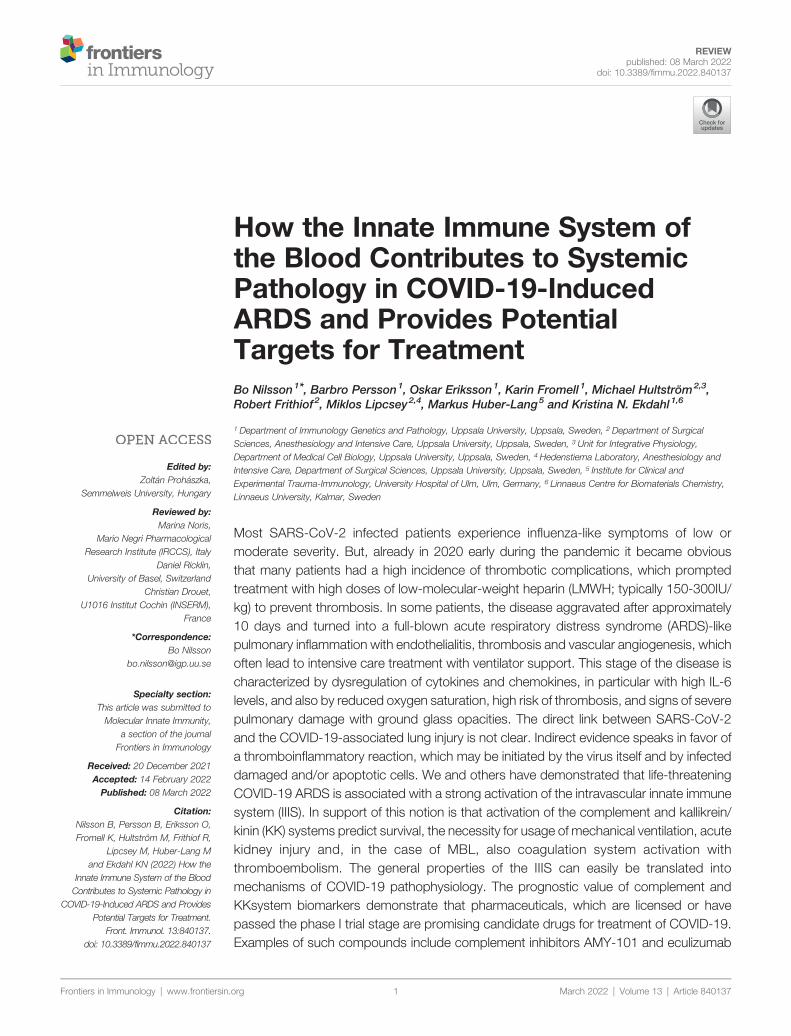
Frontiers in Immunology | www.frontiersin.
Edited by:Zoltan Prohaszka,
Semmelweis University, Hungary
Reviewed by:Marina Noris,
Mario Negri PharmacologicalResearch Institute (IRCCS), Italy
Daniel Ricklin,University of Basel, Switzerland
Christian Drouet,U1016 Institut Cochin (INSERM),
France
*Correspondence:Bo Nilsson
Specialty section:This article was submitted toMolecular Innate Immunity,
a section of the journalFrontiers in Immunology
Received: 20 December 2021Accepted: 14 February 2022Published: 08 March 2022
Citation:Nilsson B, Persson B, Eriksson O,Fromell K, Hultström M, Frithiof R,
Lipcsey M, Huber-Lang Mand Ekdahl KN (2022) How the
Innate Immune System of the BloodContributes to Systemic Pathology in
COVID-19-Induced ARDS and ProvidesPotential Targets for Treatment.
Front. Immunol. 13:840137.doi: 10.3389/fimmu.2022.840137
REVIEWpublished: 08 March 2022
doi: 10.3389/fimmu.2022.840137
How the Innate Immune System ofthe Blood Contributes to SystemicPathology in COVID-19-InducedARDS and Provides PotentialTargets for TreatmentBo Nilsson1*, Barbro Persson1, Oskar Eriksson1, Karin Fromell 1, Michael Hultström2,3,Robert Frithiof2, Miklos Lipcsey2,4, Markus Huber-Lang5 and Kristina N. Ekdahl1,6
1 Department of Immunology Genetics and Pathology, Uppsala University, Uppsala, Sweden, 2 Department of SurgicalSciences, Anesthesiology and Intensive Care, Uppsala University, Uppsala, Sweden, 3 Unit for Integrative Physiology,Department of Medical Cell Biology, Uppsala University, Uppsala, Sweden, 4 Hedenstierna Laboratory, Anesthesiology andIntensive Care, Department of Surgical Sciences, Uppsala University, Uppsala, Sweden, 5 Institute for Clinical andExperimental Trauma-Immunology, University Hospital of Ulm, Ulm, Germany, 6 Linnaeus Centre for Biomaterials Chemistry,Linnaeus University, Kalmar, Sweden
Most SARS-CoV-2 infected patients experience influenza-like symptoms of low ormoderate severity. But, already in 2020 early during the pandemic it became obviousthat many patients had a high incidence of thrombotic complications, which promptedtreatment with high doses of low-molecular-weight heparin (LMWH; typically 150-300IU/kg) to prevent thrombosis. In some patients, the disease aggravated after approximately10 days and turned into a full-blown acute respiratory distress syndrome (ARDS)-likepulmonary inflammation with endothelialitis, thrombosis and vascular angiogenesis, whichoften lead to intensive care treatment with ventilator support. This stage of the disease ischaracterized by dysregulation of cytokines and chemokines, in particular with high IL-6levels, and also by reduced oxygen saturation, high risk of thrombosis, and signs of severepulmonary damage with ground glass opacities. The direct link between SARS-CoV-2and the COVID-19-associated lung injury is not clear. Indirect evidence speaks in favor ofa thromboinflammatory reaction, which may be initiated by the virus itself and by infecteddamaged and/or apoptotic cells. We and others have demonstrated that life-threateningCOVID-19 ARDS is associated with a strong activation of the intravascular innate immunesystem (IIIS). In support of this notion is that activation of the complement and kallikrein/kinin (KK) systems predict survival, the necessity for usage of mechanical ventilation, acutekidney injury and, in the case of MBL, also coagulation system activation withthromboembolism. The general properties of the IIIS can easily be translated intomechanisms of COVID-19 pathophysiology. The prognostic value of complement andKKsystem biomarkers demonstrate that pharmaceuticals, which are licensed or havepassed the phase I trial stage are promising candidate drugs for treatment of COVID-19.Examples of such compounds include complement inhibitors AMY-101 and eculizumab
org March 2022 | Volume 13 | Article 8401371

Abbreviations: ACE, angiotensin convertiuremic syndrome; AP, alternative pathrespiratory distress syndrome; BK, bradyk2; C1-INH, C1 inhibitor; CAC, COVID-19pathway of complement; CT, computed tommolecular patterns; DIC, disseminated intracting oral anticoagulants; DVT, deep veiintensive care unit; HMWK, high moleculainnate immune system; KK, kallikrein/kiweight heparin; LP, lectin pathway ofrespiratory syndrome; NETs, neutrophil eassociated molecular patterns; PMNs, pparoxysmal noctural hemoglobinuria; RAsevere acute respiratory ryndrome-1; TF,activator; vWF, von Willebrand factor.
Nilsson et al. IIIS and COVID-19
Frontiers in Immunology | www.frontiersin.
(targeting C3 and C5, respectively) as well as kallikrein inhibitors ecallantide andlanadelumab and the bradykinin receptor (BKR) 2 antagonist icatibant. In thisconceptual review we discuss the activation, crosstalk and the therapeutic options thatare available for regulation of the IIIS.
Keywords: cascade system, leukocytes, platelets, plasma proteins, COVID-19
INTRODUCTION
COVID-19 has been shown to have a multifaceted effect on theimmune system. In a recently published article, we reported thatthe innate immune system of the blood, here designated theintravascular innate immune system (IIIS), is strongly activatedin severe COVID-19 with ARDS (1), which is the majorexplanation for the serious course of the disease. In this articlewe review the IIIS and how its physiological function contributesto tissue injury and to the proinflammatory state during severeCOVID-19. Finally, we review potential treatment targets withinthe IIIS with existing drugs that could be expected to modulatethe disease course in COVID-19.
THE INTRAVASCULAR INNATE IMMUNESYSTEM (IIIS)
The blood contains a large number of plasma proteins and cellsthat constitute our innate barrier both in terms of recognitionand elimination of microorganisms. Here, we define the IIIS andfocus on the network of proteins and cells that forms the innateimmune system in blood leading to thromboinflammation (2).The IIIS consists of the cascade system of the blood: thecomplement system, the coagulation system, the kallikrein/kinin (KK; or contact system), and the fibrinolytic system.Also, individual proteins related to the cascade systems such ascollectins, pentraxins, etc. belong to the IIIS, as do blood cells,e.g., granulocytes, monocytes, platelets and endothelial cells.These proteins are also available in the mucous membranes ofthe body, especially during inflammation either by passivediffusion or by active synthesis in the lining epithelial cells,while cells, such as granulocytes and monocytes, are recruited
ng enzyme; aHUS, atypical hemolyticway of complement; ARDS, acuteinin; BKR1/2, bradykinin receptor 1/-associated coagulopathy; CP, classicalography; DAMPs, damage-associatedavascular coagulation; DOAC, direct-n thrombosis; FXII, Factor XII; ICU,r weight kininogen; IIIS, intravascularnin system; LMWH, low-molecular-complement; MERS, middle Eastxtracellular traps; PAMPs, pathogen-olymorphonuclear leukocytes; PNH,S, renin-angiotensin system; SARS-1,tissue factor; tPA, tissue plasminogen
org 2
by chemotaxis mediated by the anaphylatoxins (C3a, C5a), bybradykinin (BK) and by chemokines.
Over the years, IIIS research has been very separated, not onlyin the laboratory but also in the collaboration betweenresearchers and in the literature. For obvious reasons, humanblood samples have been used for the studies in the variousdisciplines of the IIIS, and since the blood needs to beanticoagulated to be separated into a fluid phase and bloodcells, different anticoagulants have been used. Divalent cationsare necessary for IIIS function: the coagulation cascade requiresCa2+ and the complement cascade both Ca2+ and Mg2+ ions,which affects the usage of anticoagulant for the different cascadesystem. As a consequence, EDTA (chelating both Ca2+ andMg2+) isused for assessment of complement activation fragments/productsin plasma, while the corresponding anticoagulant for thecoagulation, contact and fibrinolytic systems is citrate (chelatingCa2+ ions, but not Mg2+). Studies of the complement systemfunction is performed using serum (i.e., the remaining fluid-phaseafter blood is coagulated), whereas for the coagulation, KK, andfibrinolysis systems, citrate reconstituted with Ca2+ ions is theplasma preparation of choice. These pre-analytic procedures havetotally separated the research disciplines. Further aggravating theproblem, cell studies have inmany cases been performed on heparinblood, where activation of the coagulation and complement systemsare considerably dampened by the high concentrations of heparin.Consequently, there is no generally used holistic assay formatavailable for simultaneous assessment of all IIIS functions.However, for specialized in vitro studies heparin-coated tubes ortubing can be used, leaving the blood fresh and unaffected (3). Inthis conceptual review the intension is to consider thesecomponents of the blood as an intact network that gives rise toan integrated innate immune response in late-stage COVID-19. Thefindings by us and others support this approach.
ORGANIZATION OF THE IIIS
The schematic structure of the IIIS is described in Figure 1,which focuses on the interaction of the cascade systems of theblood, i.e., the complement, coagulation, KK and fibrinolyticsystems. The reason for highlighting the cascade systems isthat they contain most of the recognition molecules thatspecifically target DAMPs and PAMPs and trigger activationof the entire IIIS. In the figure, they are marked as componentsof the activation pathways that they initiate. The complementsystem has three defined activation pathways: the classical(CP), the lectin pathways (LP) and the alternative (AP) thatare triggered by different stimuli (5).
March 2022 | Volume 13 | Article 840137

ent system (A), the contact or kallikrein/kininstimuli such as microorganisms or necrotic,transplantation, or treatment with biomaterials e.g.
which are triggered by different recognitionentraxins e.g. CRP and pentraxin 3. The lectine alternative pathway (AP) is be activated/regulatedcomplexes C4bC2a and C3bBb, which cleave C3ore potent anaphylatoxin C5a. (B) The primaryct/KK system is factor XII (FXII), which is activated,tes an amplification loop, which involves prekallikreinr of coagulation, tissue factor (TF), is exposed in ae, which leads to formation of high amounts ofbrin network to soluble fibrin fragments. Please see
Nilsson
etal.
IIISand
COVID
-19
Frontiersin
Immunology
|www.frontiersin.org
March
2022|Volum
e13
|Article
8401373
FIGURE 1 | Physiological and pathological conditions and treatments involving IIIS activation. The IIIS consists of the cascade systems of the blood: the complemsystem (KKS) (B), the tissue factor (TF) pathway of the coagulation system (C), and the fibrinolytic system (D). Activation of IIIS occur in response to physiologicalapoptotic or virus infected cells (top in figure), which leads to thromboinflammation. But also during pathological or therapeutic conditions such as ischemia duringstents, intravascular devices, and extracorporeal treatment (bottom in figure) a similar reaction occur. (A) The complement system has three activation pathways,molecules. The classical pathway (CP) is initiated by C1q, which binds to antigen bound IgG and IgM, but also to negatively charged surfaces and target-bound ppathway (LP) is activated by a number of carbohydrate binding proteins (lectins) such as mannan binding lectin (MBL), Ficolin-1, -2, -3 and Collectin 10/11 (4). Thby surface-specific binding of factor H, C3 or properdin to a target, and has its main role as an amplifier. Complement activation leads to assembly of two enzymeinto the anaphylatoxin C3a and surface bound C3b (opsonization) and cleaves C5, which initiates the formation of the membrane attack complex (MAC) and the mfunction of the kallikrein/kinin and coagulation systems is in hemostasis but both systems are also engaged in inflammation. The recognition molecule in the contae.g., by negatively charged molecules such as LPS, glycosaminoglycans or extracellular matrix molecules exposed to the blood stream. The KK system also initiathat cleaves high molecular-weight kininogen (HMWK) leading to the generation of the proinflammatory mediator bradykinin (BK). (C) The main physiological triggefunctionally active form only after damage to vessels and activation of blood cells including platelets. It thereby initiates the extrinsic part of the coagulation cascadthrombin. (D) The fibrinolytic system is initiated when urokinase or tissue plasminogen activator (tPA) activate plasminogen to plasmin, which degrades a formed fi
the text for information on the roles of monocytes, PMNs and platelets.

Nilsson et al. IIIS and COVID-19
• The CP is initiated by C1q, which binds to negatively chargedsurfaces, to IgG and IgM in immune complexes, and to target-bound pentraxins such as CRP and pentraxin 3.
• The LP is activated by a number of lectins (i.e., carbohydrate-binding proteins) such as MBL, Ficolin-1, -2, and -3, and bythe Collectins 10/11 (4).
• The AP functions primarily as an amplification loop but canbe specifically regulated by properdin in concert with C3 andby factor H as an important recognition molecule controllingthe AP convertase.
In vivo, the coagulation system is mainly activated by theextrinsic pathway elicited by tissue factor (TF), which is exposedin the vessel wall after endothelial cell damage. TF is alsoexpressed by multiple cells in response to inflammatory signals(6) and when cell-bound TF is exposed to blood it leads to astrong coagulation activation. Active TF can also be expressed byactivated monocytes in response to, e.g., C5a and on theendothelium during inflammation. The KK system is analternative route for coagulation activation, initiated on contactbetween blood and negatively charged foreign surfaces.Activation of the contact system is the reason why bloodcollected without an anticoagulant, coagulates in a test tube.KK is also regulating the vascular permeability on endothelialcells (7).
Factor XII (FXII) has a dual role in that it is the starting pointof both the intrinsic pathway activation of the coagulation systemand KK system. FXII probably has a limited role in physiologicalhemostasis, which is illustrated by the fact that FXII deficiency(Hageman disease) does not lead to an increased tendency forbleeding (8). In contrast, inhibition of FXII is considered as anew possible strategy as an antithrombotic drug with minor riskfor bleeding (9). The KK system consists of FXII, prekallikreinand high molecular weight kininogen (HMWK) (8). FXIIacleaves prekallikrein generating kallikrein. Kallikrein can cleaveFXII and prekallikrein thereby providing a positive feedbackloop. The KK system elicits inflammation via kallikrein, whichcleaves HMWK generating BK.
The fibrinolytic system is initiated by urokinase, tissueplasminogen activator (tPA) and FXIIa by activating plasminogento plasmin (10). Activated plasmin in turn breaks down the fibrinnetwork formed in the final stages of the coagulation reaction, andthus acts as a physiological restriction mechanism that limits thepropagation of the clot (10).
THE FUNCTION OF THE IIIS
Significant cross-activation can take place directly orindirectly via leukocytes and platelets, which means thatactivation of one of the cascade systems can spread to theentire IIIS. The physiological end result of IIIS activation isthromboinflammation, which stops bleeding by sealing bloodvessel leakage through fibrin formation and plateletaggregation, and supports the clearance of damaged cells byattracting leukocytes that remove the damaged tissue. The IIISis the start of wound healing after an injury, where the “waste
Frontiers in Immunology | www.frontiersin.org 4
management function”, i.e., removal of foreign substances,particles and apoptotic or necrotic cells is an important task.This process is combined with the release of growth factorsfrom, e.g., activated platelets, which ultimately leads to tissuehealing and scar formation (11).
In a similar way, the IIIS reacts to infections caused bydifferent types of microorganisms. In these reactions, the IIIShelps to kill and remove the microorganisms or the infected cells,after which the tissue is cleansed and healed as a result of IIISfunctions. Sometimes, however, the reaction can shoot over thetarget and become too strong, which leads to severeinflammation and tissue damage. The end result is an excessiveand pathological thromboinflammation with activation of all IIIScomponents such as in sepsis (Figure 1).
COVID-19 AND IIIS ACTIVATION LEADINGTO THROMBINFLAMMATION
The COVID-19 pandemic, which is caused by the corona virusSARS-CoV-2 was initially described in Wuhan, China at the endof 2019 and has since had an immense effect on human societyworldwide. Most SARS-CoV-2 infected patients experienceinfluenza-like symptoms of low or moderate severity that arecharacterized by sore throat, fever, a dry cough, intestinalproblems, and a loss of taste and smell. Early in the pandemic,it was reported that most patients suffered from an acute-phasereaction, with high levels of certain plasma proteins, e.g.,fibrinogen, C3, and ferritin that were sometimes multiple timeshigher than normal (12). It also became clear that there was anincreased risk of severe thromboembolism (13, 14). In somepatients, the disease aggravated after approximately 9 to10 days and turned into a full-blown ARDS-like pulmonaryinflammation with endothelialitis, thrombosis and vascularangiogenesis, which often required intensive care treatmentwith ventilator support (15). The lung injury in COVID-19resembles the pulmonary complications that have beenobserved in earlier SARS-1 and MERS epidemics (16). In thebeginning of the COVID pandemic, up to 30% of the intensivecare unit (ICU) patients died due to pulmonary and other organdysfunctions. Those patients who reached the late-stagesyndrome developed a severe pulmonary inflammation withgeneral cyto-/chemokine expression (17), reduced oxygensaturation, increased risk of pulmonary thrombosis, and signsof severe pulmonary damage revealed by CT, particularly in theperiphery of the lungs (18, 19).
In 2020, we studied the first 65 patients with intensive care-requiring COVID-19 admitted to the ICU of the universityhospital in Uppsala, Sweden (1). These patients were admittedto the ICU before the therapeutic procedures now used, such ascorticosteroids, were introduced. These patients werecomprehensively investigated and were found to have apronounced activation of the IIIS, with activation of all cascadesystems and especially the KK system with prekallikrein, FXIIand HMWK consumption (i.e. activated) and kallikrein/C1-INHcomplex formation (1).
March 2022 | Volume 13 | Article 840137

Nilsson et al. IIIS and COVID-19
THROMBOINFLAMMATORY CHANGESIN VARIOUS TISSUES OFCOVID-19 PATIENTS
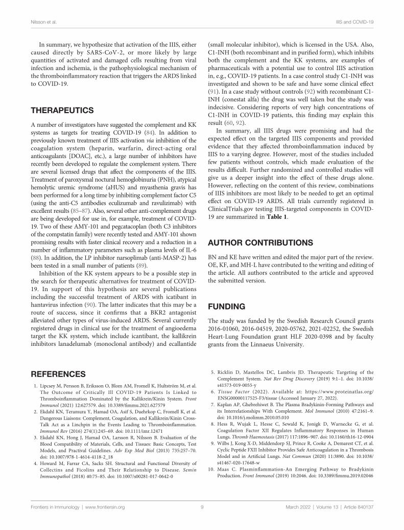
In COVID-19 patients, all parts of the IIIS are strongly activated,especially in those who are admitted to the ICU with ARDS-likesymptoms. Most morphological studies have been performed onlung tissue from autopsies of patients that had succumbed at theICU, e.g. (20, 21). Macroscopic observations show that largesections of the pulmonary lobes were clogged with cells and fluid,explaining the poor oxygenation of the blood in severely ill patients.From these cases of seriously ill COVD-19 patients, it was observedthat the pulmonary tissue was infiltrated with immune cells (20).Initially, lymphocytes and macrophages were reported to be thedominating cell types (22), but later studies also showed that PMNswere important players (23). This discrepancy was probably due tothat the latter observation were obtained in patients with earlierdisease. Another important finding is that the alveolar space is filledwith plasma proteins, e.g., fibrinogen giving rise to fibrin clots andthat the interalveolar capillaries are activated and obstructed withplatelet-containing thrombi (Figure 2). A few cases withimmunohistochemical studies of complement components havebeen performed (24, 25). They show deposition of C4d, C3d, factorB, sC5b-9 on the endothelium in various tissues, preferentially in thelung, which can be interpreted as if complement activation via theCP or/and the LP occurs with amplification via the AP. Takentogether these findings are consistent with a widespreadthromboinflammation in the lungs.
Late-stage severe COVID-19 primarily affects the lungs butextends to other organs causing, in the more serious cases,multiorgan impact including cardiac, central nervous systemand kidney injury (26–28). Myocardial dysfunction,
Frontiers in Immunology | www.frontiersin.org 5
arrhythmia and acute coronary syndrome have been reportedin association with COVID-19, but in a recently performed studyon 30 patients with severe COVID-19 disease late cardiac pathologyof biopsies showed no relevant cardiac histopathological alterations(29, 30). At present the contribution of SARS-CoV2 on cardiaclesions remains to be established (31). Activation of the complementsystemmight be involved in the worsening of renal symptoms sincedeposition of C3 was significant in renal arteries and in glomerularcapillaries of COVID-19 biopsies (32). This is supported by findingsin our own study, which shows a correlation between C3ageneration and glomerular filtration rate (1). Neurologicaldisorders associated with COVID-19, such as outright encephalitisis rare but has been reported with direct SARS-CoV2 detection inbrain tissue (33). However, severe COVID-19 with long ICU stayhas been associated with critical illness involving neuro/myopathy(34) and post intensive care syndrome (35) wherethromboinflammation may play a role.
THROMBOSIS IN COVID-19
Already early in the COVID-19 pandemic, it appeared thatmoderately to seriously ill patients showed a very stronginvolvement of the coagulation system, and a high incidence ofthromboembolic complications was reported on both the arterialand the venous side. In order to prevent these complications,treatment of the patients with high doses (typically 150-300 IU/kg) of LMWH was used (13, 14). Initially it was thought that thecause of the thromboembolism was an effect directly targetingthe coagulation system but there was no real evidence of asystemic activation. Morphological data from the lungs showedthat defined sections of the tissue were rich in microcapillary
FIGURE 2 | Proposed mechanism for IIIS involvement in SARS-CoV-2-induced ARDS. (A) Under normal circumstances gas is exchanged over a narrow gapbetween alveolar epithelial cells (blue) and capillary endothelial cells (red). SARS-CoV-2 infects the alveolar/bronchial epithelial cells via ACE-2. Bronchial epithelial cellsexpress ACE-2 and endothelial cells bradykinin receptor 2 (BKR2). Infection of alveolar epithelial cells activates the complement and KK systems and generates C5aand bradykinin (BK). (B) C5a and BK activate endothelial cells and elicit increased vascular permeability, which widens the gap between the cell linings. Activatedendothelial cells also trigger complement and KK systems activation that upregulates BKR1, further increasing vascular permeability, damaging cells and inducingnecrosis and apoptosis. BK and C5a elicit chemotaxis of PMNs that release neutrophil extracellular traps (NETs, depicted as a blue mesh). (C) Activated endothelialcells (TF) and NETs (TF and FXIIa) trigger coagulation activation and thrombus formation and further amplifie KK and complement activation. Plasma proteins leakinto the alveolae causing fibrin precipitation. Invasion of PMNs and monocytes further increases the gap between the cell linings, ultimately leading to a collapsedexchange of gases over the epithelial and endothelial border.
March 2022 | Volume 13 | Article 840137

Nilsson et al. IIIS and COVID-19
thrombi suggesting rather that the clotting process was takingplace locally close to the activated endothelium (18). Thecoagulation abnormalities that occur during COVID -19disease were given a special name, COVID -19-associatedcoagulopathy (CAC), to emphasize what has emerged overtime, that it is a condition with certain characteristics of itsown. It is clear that CAC differs from the picture seen in sepsisand the so-called DIC (disseminated intravascular coagulation)where low platelet counts are a typical sign and extensiveconsumption of platelets and coagulation factors leads tothrombosis and bleeding at the same time (12, 36). Severely illCOVID -19 patients instead have normal or high platelet levelsand are characterized by greatly elevated plasma levels of thefibrin degradation product D-dimer, which is a sensitive markerof fibrinolysis and coagulation turnover and probably reflectswidespread thrombosis in e.g. small vessels in the lungs. It hasbeen possible to reproduce the CAC phenotype in a primatemodel infected with SARS-CoV-2, and where an extensive fibrindeposition in the lung tissue was observed, in line with findingsfrom autopsy materials of deceased COVID -19 patients (15, 37).
A number of hypotheses regarding the mechanisms behindCAC have been proposed. We have studied the relationshipbetween complement and CAC, and shown that thrombosis inintensive care COVID -19 patients is linked to high activity in theLP and high plasma levels of the LP protein MBL (38). Futurestudies may show whether there is an underlying causalrelationship between the LP and thrombosis, something that issupported by preclinical results where LP components MASP-1and MASP-2 have a coagulation factor-like activity and candirectly activate fibrinogen and prothrombin in in vitroexperiments (39). There are also reports that neutrophilextracellular traps (NETs) that are released in response to C5amay be an initiator of coagulation since NETs expose TF andbind FXII (40). Similarly, activated endothelial cells express TF,which may contribute to the thromboinflammation.
ACTIVATION INDIVIDUAL COMPONENTSOF IIIS IN COVID-19
In COVID-19 an acute phase reaction increases theconcentration of a number of plasma proteins multi-fold andthereby facilitates IIIS activation (41). Multiple proinflammatorycytokines and chemokines such as TNF, IL-1b, IL-6, and MCP-1are produced in response to C3a and C5a generation that furtheramplifies the production of acute-phase proteins (41, 42). TheIIIS activation is multifactorial: The virus itself can activate theIIIS directly via the CP and LP of the complement system and viathe KK system (43–45). Another likely activation mechanism istissue damage in the lungs with apoptotic and necrotic cells thatinitiates the overactivation of IIIS in its function to eliminate cellsor cell debris (46).
The Complement SystemThe complement system was early suspected to be involved inthe pathophysiology of severe COVID-19 disease (26, 47–51)
Frontiers in Immunology | www.frontiersin.org 6
and has thereafter been thoroughly investigated and shown to beinvolved in the disease and correlated with the severity of thedisease. The first indication was reported early in the pandemicwhere increased levels of sC5b-9 were detected and a bit later amore complete report was published; both demonstrating thatincreased levels of sC5b-9 were associated with severe disease(52, 53). Thereafter, several comprehensive studies have beenpublished, which assess a number of different complementparameters. These studies show that individual complementcomponents increase and decrease in concentration during thecourse of the disease without any clear patterns (1, 54). Forinstance, C3 and C4 and other components such as C1q andMBL, and complement function (CP and AP) are elevated insome patients and decreased in others, indicating that bothoverexpression and complement activation with consumptionoccur. Overexpressed and elevated levels of factor B tend to beassociated with severe COVID-19. In addition to sC5b-9generation, formation of other activation markers occurs: C4d,Bb, C3a, and C3d,g are generated and seem to be activated earlyin the disease. The early trigger of complement activation is stillunclear; most likely several activators elicit this response.Activation has been reported to be mediated by the intact virusvia intracellular complement activation (55) but also by virusproteins expressed on the surface of infected cells. The virus itselfhas been shown to bind MBL, Ficolin-2 and Collectin-11, via itsS- and N-proteins, with subsequent LP-mediated C3b and C4bdeposition (44, 45). Structural parts of the virus have also beenshown to bind C1q and trigger complement activation (43).COVID-19 is strongly associated with ischemic cells, apoptosis,and cell death (15, 20, 40), initially occurring in the lung tissue.These conditions lead to binding of MBL, MASP-2 and C1q tothe cells (see below), which may be the combined initiators of theaccelerating complement activation that occurs particularly inseverely ill patients. In our own study, the C1q concentration islow (consumed) in several patients at admission to the ICUapproximately at day 10, possibly caused by damaged cells in thelungs (1).
The combined data indicate that the maximum activationoccurs at approximately day 10 coinciding with when patientsare most likely to be admitted to the ICU. C4 and C3consumption combined with C4d, C3a and C3d,g generationsupports complement activation via CP/LP activation. Dailymonitoring of C3d,g has been reported to predict outcome inpatients hospitalized with COVID-19 in combination withSARS-CoV-2 nucleocapsid antigen, RNA in blood, IL-6, andCRP (56).
The KK SystemIn several early reviews and in vitro studies, the KK system wassuggested to participate in the thromboinflammation of COVID-19 (43, 57–59), without presenting any evidence of KK systemactivation in vivo. No direct evidence of such activation waspublished until reports from us, and Busch et al. showed strongKK system activation in severely ill ICU patients (1, 60). The KKsystem activation was assessed by consumption of prekallikrein,HMWK and FXII, and by increased levels of kallikrein/C1-INHcomplexes (1). The activation of prekallikrein and HMWK on
March 2022 | Volume 13 | Article 840137

Nilsson et al. IIIS and COVID-19
the admission to the ICU was shown to have prognosticsignificance for survival and the need for mechanicalventilation, but also for other systemic and specific organparameters, such as, poor kidney function.
The Fibrinolytic SystemA consequence of the ongoing coagulation activation isgeneration of fibrin from fibrinogen. In the clot that is formed,plasmin is generated, which cleaves fibrin into fragmentsincluding D-dimer. D-dimer is an important clinical biomarkerfor fibrinolysis and indirectly for clot formation. Due to itsgeneral availability, this marker was one of the first biomarkersof IIIS reported to increase in COVID-19 and the levels werefound to be associated with disease severity (61).
Granulocytes (PMN)The neutrophilic granulocytes, which are found in the lungs earlyin the course of the disease (23),have a profound effect on theoutcome of COVID-19 (23). Several drivers of cell infiltrationexist and the anaphylatoxins of the IIIS (C3a and C5a) as well asBK are likely to be important contributors. Chemotactic effectsare mediated by the specific receptors C3aR (C3a), C5aR1 and 2(C5a) and by BKR1 and 2 (BK). These receptors also triggeractivation of the PMNs, which release proteases and othergranular proteins, e.g., elastase, myeloperoxidase and protein-arginine deiminase type 4. One specific effect related to PMNs isthe generation of NETs, which are large, extracellular, web-likestructures consisting of cytosolic and granule proteins with ascaffold of decondensed chromatin in which the majority of theDNA originates from the nucleus (62). NETs protect againstinfection but have also been associated with a number of otherimmunoregulatory events such as binding antimicrobial peptidesand priming immune cells (62). In addition to this, NETspromote thrombotic events, since they bind both TF (40) andFXIIa (63) thereby being able to trigger both the extrinsicpathway of coagulation as well as the contact system andconsequently the intrinsic pathway of coagulation. NETformation has been suggested to be an important promotor ofthrombosis in the lungs of COVID-19 patients where C5a hasbeen proposed to elicit the release of these morphologicalstructures (60, 62).
Platelets and Endothelial CellsIncreased platelet activation and platelet-monocyte aggregates wereobserved in COVID-19 patients but not in patients presenting amild syndrome (64). Platelet counts tend to increase in COVID-19patients and not be consumed and decrease in number as in patientswith thromboembolism. This observed difference, which speaksagainst that the thrombi found in the lungs in COVID-19 patientsare the result of embolism with a major thrombus formed elsewheresuch as in deep vein thrombosis (DVT). Instead, it has beenobserved that COVID-19 patients exhibit reduced procoagulantplatelet responses (65), but platelets are found in the thrombiformed in the lung capillaries either as a result of activatedendothelial cells or NETs formation (40). Endothelial cells arepoorly infected by Cov-Sars-2 since these cells express a lownumber of ACE-2 molecules (66), but generation of both C5a
Frontiers in Immunology | www.frontiersin.org 7
and BK by the nearby pneumocytes can initiate expression of TF onboth endothelial cells (67)and neutrophils (40, 68) therebypromoting local thrombus formation.
ISCHEMIC INJURY
Hypoxia is anticipated to occur, particularly in the lungs duringCOVID-19, where major parts of the small airways may betotally clogged with fibrin, platelet-rich thrombi, and cells[Figure 2; (46)]. Ischemia is expected to be a mechanism thatis involved in the injury mediated by the IIIS in COVID-19,considering the low oxygen saturation in these patients. Ischemiais a major stress to the cell, which can lead to changes in thecomposition and protein expression of the cell membrane (62).Consequently, IIIS in contact with a cell subjected to hypoxia,can recognize the cell surface as foreign, causing athromboinflammatory reaction and, by extension, ischemia/reperfusion injury-like damage and cell death. The ischemiccell has a distinctive phenotype compared to the native one,which leads to that IIIS recognition molecules of the complementand the KK systems target the cell as foreign (non-self). Inischemia/reperfusion injury, MBL (69) and MASP-2 (70) of theLP and innate IgM antibodies (71, 72) of the CP have beensuspected to be involved in the IIIS activation that occurs.
THE RENIN-ANGIOTENSIN SYSTEM (RAS)
Hypertension is linked to the renin-angiotensin system (RAS)and an increased risk for severe COVID-19 infection. Thedocking protein for SARS-CoV-2 on human cells is angiotensinconverting enzyme (ACE)-2 of the angiotensinogen cascade system,which may destroy the function of this protein. ACE-1 inhibitors(common hypertensive drugs) block the cleavage of angiotensin I toangiotensin II. ACE-1 is a regulator of BK, making it feasible thatinhibition of ACE-1 could aggravate the ARDS condition inCOVID-19 patients by increasing the levels of active BK.Although in a meta study focusing on the effects of renin-angiotensin system (RAS) inhibitors on the RAS and the outcomeof COVID-19, no support for this concept was found. However,since this study is based on several clinical trials that treat RASinhibitors as a common group further studies are needed toelucidate this issue (58, 73).
CONCEPTUAL MECHANISMS INDUCINGCOVID-19-TRIGGERED ARDS
SARS-CoV-2-infected and ischemic, damaged epithelial andendothelial cells can activate all the IIIS cascade systems in a jointthromboinflammatory reaction in the lungs (Figure 2). During thedevelopment of ARDS, SARS CoV-2 and damaged cells (SARSCoV-2-infected, apoptotic, necrotic cells) are potential targets forthe recognition molecules of the blood cascade systems. C1q, MBL,and FXII are known to recognize apoptotic and necrotic cells that
March 2022 | Volume 13 | Article 840137

Nilsson et al. IIIS and COVID-19
can trigger the CP and LP of complement ultimately leading tocleavage of C3/C5 into C3a/C5a and C3b/C5b. BK generated by theKK system activation can cause dry cough (74) and pulmonaryinflammation with edema (75) by binding to BKR2. C3a and C5abind to the anaphylatoxin receptors: C3aR, C5aR1, and C5aR2 andC5a is also able to activate endothelial cells in COVID-19 patientsleading to vonWillebrand Factor (vWF) and p-selectin exposure onthe endothelial lining thereby contributing to the thromboticphenotype (76). Supporting the importance of the C5a/C5aR1axis is that COVID-19 patients generate C5a as detected inpulmonary lavage in proportion to the severity of the disease andhigh expression of C5aR1 was found in blood and on pulmonarymyeloid cells (77). C5a also induces expression of BKR1, (whichcontrary to BKR2 is not constitutively expressed) and BK can thenact via both BKR2 (expressed on both the pulmonary epitheliumand endothelium) and after cleavage to desArg9-BK via BKR1 (78–80), which also elicits increased vascular permeability, PMNchemotaxis, and nerve end stimulation, amplifying all of theabove-mentioned reactions. Thus, C3a, C5a, and BK combinedhave the potential to cause a local edema and pulmonary leukocyteinfiltration/inflammation that increases the distance betweenepithelial and endothelial cells in the alveolae, thereby hinderingoxygenation of the blood (Figure 2). The poor oxygenation can leadto further ischemia (as described above) that in turn leads to evenmore IIIS activation thereby creating a vicious circle. Localthrombosis in the vasculature of the lung induced by activatedendothelial cells that bind MBL, and expose selectins, TF and vWF
Frontiers in Immunology | www.frontiersin.org 8
on their surfaces, further aggravates the hypoxia. Chemotaxis andactivation of neutrophlic granulocytes also cause NET formationand FXII activation (40, 81). Taken together these phenomena arelikely drivers of the endothelialitis, the thrombus formation and theincreased vascular angiogenesis linked to COVID-19 patients (15).
Activation of the IIIS in COVID-19-induced ARDS have manysimilarities to ARDS of other etiologies, e.g., as in sepsis but there arealso large differences both regarding activation mechanisms and thefocus of the inflammation. We have previously reported that theactivation of the IIIS in COVID-19mainly occurs via the KK systemand the LP and CP of the complement system (1). By contrast, inseptic shock, it has been reported by others that the key steps ofcomplement activation consist of first the AP followed by the CP(36). One explanation of these discrepancies may be the specificpathophysiologic process in SARS-CoV-2 infection as comparedwith the diverse and heterogeneous microbiology in sepsis ofdifferent origin. Another important dissimilarity between COVID-19 and sepsis-induced ARDS is the focus and origin ofinflammation. In COVID-19, ARDS originates from the lung,which is in contrast to sepsis-induced ARDS that is caused by asecondary systemic inflammatory response in a distant organ,unless the infection focus already is in the lung (82, 83). In thelatter case, usually termed pulmonary ARDS, the lung is the driverof IIIS activation with the highest IIIS activation locally, while in theformer, usually termed extrapulmonary ARDS, the lung is onlysecondarily affected and IIIS activation measured as activationproducts in the plasma is less dependent on the severity of ARDS.
TABLE 1 | All trials currently (2022-01-28) registered in ClinicalTrials.gov testing targeting IIIS components in COVID-19.
Drug (target) Identifier Participants Study design Last update
Kallikreinlanadelumab (kallikrein) NCT04422509 43 Randomized vs SOC Nov 16, 2021lanadelumab (kallikrein) NCT04460105 0 Randomized vs placebo Oct 20, 2020ISIS 721744 (kallikrein antisense) NCT04549922 111 Randomized vs placebo April 19, 2021IcatibantC1-INH ± icatibant NCT05010876 44 Randomized vs SOC Aug 18, 2021Iactibant NCT04978051 120 Randomized vs SOC July 27, 2021C1-INHConestat alfa (recomb C1-INH) NCT04414631 80 Randomized vs SOC Nov 9, 2021Ruconest (recomb C1-INH) NCT04705831 40 Randomized vs SOC, crossover Jan 12, 2021Ruconest (recomb C1-INH) NCT04530136 120 Randomized vs SOC Dec 10, 2020C5 cleavage inhibitorsEculizumab NCT04346797 120 Randomized vs SOC April 20, 2020Eculizumab NCT04288713 no info no info found March 20, 2020Ravulizumab NCT04390464 1167 (3 arms) Randomized vs SOC May 18, 2020Ravulizumab NCT04570397 32 Randomized vs SOC Jan 14, 2021Ravulizumab NCT04369469 120 Randomized vs SOC Sept 22, 2021Zilucoplan (C5 cleavage inhibiting peptide) NCT04382755 81 Randomized vs SOC + antibiotics July 2, 2021Zilucoplan (C5 cleavage inhibiting peptide) NCT04590586 516 (7 arms) Randomized vs SOC + placebo Nov 23, 2021C3 cleavage inhibitorsAMY-101 NCT04395456 144 Randomized vs placebo Feb 20, 2021APL-9 NCT04402060 65 Randomized vs placebo Sept 1, 2021Lectin pathway inhibitorNarsoplimab (anti MASP-2) NCT04488081 1500 (8 arms) Randomized July 21, 2021+ Remdesivir (anti CD14)C5aR antagonistsavdoralimab (anti C5aR mAb) NCT04371367 208 Randomized vs placebo May 27, 2021avdoralimab (anti C5aR mAb) NCT04333914 219 Randomized vs SOC Aug 5, 2021vilobelimab (anti C5a mAb) NCT04333420 390 Randomized vs SOC + placebo Dec 31, 2021
March 2022 | Volume 13
SOC, standard of care.
| Article 840137
Nilsson et al. IIIS and COVID-19
In summary, we hypothesize that activation of the IIIS, eithercaused directly by SARS-CoV-2, or more likely by largequantities of activated and damaged cells resulting from viralinfection and ischemia, is the pathophysiological mechanism ofthe thromboinflammatory reaction that triggers the ARDS linkedto COVID-19.
THERAPEUTICS
A number of investigators have suggested the complement and KKsystems as targets for treating COVID-19 (84). In addition topreviously known treatment of IIIS activation via inhibition of thecoagulation system (heparin, warfarin, direct-acting oralanticoagulants [DOAC], etc.), a large number of inhibitors haverecently been developed to regulate the complement system. Thereare several licensed drugs that affect the components of the IIIS.Treatment of paroxysmal noctural hemoglobinuria (PNH), atypicalhemolytic uremic syndrome (aHUS) and myasthenia gravis hasbeen performed for a long time by inhibiting complement factor C5(using the anti-C5 antibodies eculizumab and ravulizimab) withexcellent results (85–87). Also, several other anti-complement drugsare being developed for use in, for example, treatment of COVID-19. Two of these AMY-101 and pegcatacoplan (both C3 inhibitorsof the compstatin family) were recently tested and AMY-101 shownpromising results with faster clinical recovery and a reduction in anumber of inflammatory parameters such as plasma levels of IL-6(88). In addition, the LP inhibitor narsoplimab (anti-MASP-2) hasbeen tested in a small number of patients (89).
Inhibition of the KK system appears to be a possible step inthe search for therapeutic alternatives for treatment of COVID-19. In support of this hypothesis are several publicationsincluding the successful treatment of ARDS with icatibant inhantavirus infection (90). The latter indicates that this may be aroute of success, since it confirms that a BKR2 antagonistalleviated other types of virus-induced ARDS. Several currentlyregistered drugs in clinical use for the treatment of angioedematarget the KK system, which include icantibant, the kallikreininhibitors lanadelumab (monoclonal antibody) and ecallantide
Frontiers in Immunology | www.frontiersin.org 9
(small molecular inhibitor), which is licensed in the USA. Also,C1-INH (both recombinant and in purified form), which inhibitsboth the complement and the KK systems, are examples ofpharmaceuticals with a potential use to control IIIS activationin, e.g., COVID-19 patients. In a case control study C1-INH wasinvestigated and shown to be safe and have some clinical effect(91). In a case study without controls (92) with recombinant C1-INH (conestat alfa) the drug was well taken but the study wasindecisive. Considering reports of very high concentrations ofC1-INH in COVID-19 patients, this finding may explain thisresult (60, 92).
In summary, all IIIS drugs were promising and had theexpected effect on the targeted IIIS components and providedevidence that they affected thromboinflammation induced byIIIS to a varying degree. However, most of the studies includedfew patients without controls, which made evaluation of theresults difficult. Further randomized and controlled studies willgive us a deeper insight into the effect of these drugs alone.However, reflecting on the content of this review, combinationsof IIIS inhibitors are most likely to be needed to get an optimaleffect on COVID-19 ARDS. All trials currently registered inClinicalTrials.gov testing IIIS-targeted components in COVID-19 are summarized in Table 1.
AUTHOR CONTRIBUTIONS
BN and KE have written and edited the major part of the review.OE, KF, andMH-L have contributed to the writing and editing ofthe article. All authors contributed to the article and approvedthe submitted version.
FUNDING
The study was funded by the Swedish Research Council grants2016-01060, 2016-04519, 2020-05762, 2021-02252, the SwedishHeart-Lung Foundation grant HLF 2020-0398 and by facultygrants from the Linnaeus University.
REFERENCES1. Lipcsey M, Persson B, Eriksson O, Blom AM, Fromell K, Hultström M, et al.
The Outcome of Critically Ill COVID-19 Patients Is Linked toThromboinflammation Dominated by the Kallikrein/Kinin System. FrontImmunol (2021) 12:627579. doi: 10.3389/fimmu.2021.627579
2. Ekdahl KN, Teramura Y, Hamad OA, Asif S, Duehrkop C, Fromell K, et al.Dangerous Liaisons: Complement, Coagulation, and Kallikrein/Kinin Cross-Talk Act as a Linchpin in the Events Leading to Thromboinflammation.Immunol Rev (2016) 274(1):245–69. doi: 10.1111/imr.12471
3. Ekdahl KN, Hong J, Hamad OA, Larsson R, Nilsson B. Evaluation of theBlood Compatibility of Materials, Cells, and Tissues: Basic Concepts, TestModels, and Practival Guidelines. Adv Exp Med Biol (2013) 735:257–70.doi: 10.1007/978-1-4614-4118-2_18
4. Howard M, Farrar CA, Sacks SH. Structural and Functional Diversity ofCollectins and Ficolins and Their Relationship to Disease. SeminImmunopathol (2018) 40:75–85. doi: 10.1007/s00281-017-0642-0
5. Ricklin D, Mastellos DC, Lambris JD. Therapeutic Targeting of theComplement System. Nat Rev Drug Discovery (2019) 9:1–1. doi: 10.1038/s41573-019-0055-y
6. Tissue Factor (2022). Available at: https://www.proteinatlas.org/ENSG00000117525-F3/tissue (Accessed January 27, 2022).
7. Kaplan AP, Ghebrehiwet B. The Plasma Bradykinin-Forming Pathways andits Interrelationships With Complement. Mol Immunol (2010) 47:2161–9.doi: 10.1016/j.molimm.2010.05.010
8. Hess R, Wujak L, Hesse C, Sewald K, Jonigk D, Warnecke G, et al.Coagulation Factor XII Regulates Inflammatory Responses in HumanLungs. Thromb Haemostasis (2017) 117:1896–907. doi: 10.1160/th16-12-0904
9. Wilbs J, Kong X-D, Middendorp SJ, Prince R, Cooke A, Demarest CT, et al.Cyclic Peptide FXII Inhibitor Provides Safe Anticoagulation in a ThrombosisModel and in Artificial Lungs. Nat Commun (2020) 11:3890. doi: 10.1038/s41467-020-17648-w
10. Maas C. Plasminflammation-An Emerging Pathway to BradykininProduction. Front Immunol (2019) 10:2046. doi: 10.3389/fimmu.2019.02046
March 2022 | Volume 13 | Article 840137

Nilsson et al. IIIS and COVID-19
11. Broughton G, Janis JE, Attinger CE. The Basic Science of Wound Healing. PlastReconstr Surg (2006) 117:12S–34S. doi: 10.1097/01.prs.0000225430.42531.c2
12. van Dam LF, Kroft LJM, van der Wal LI, Cannegieter SC, Eikenboom J, deJonge E, et al. Clinical and Computed Tomography Characteristics ofCOVID-19 Associated Acute Pulmonary Embolism: A Different Phenotypeof Thrombotic Disease? Thromb Res (2020) 193:86–9. doi: 10.1016/j.thromres.2020.06.010
13. Marietta M, Coluccio V, Luppi M. COVID-19, Coagulopathy and VenousThromboembolism: More Questions Than Answers. Intern Emerg Med(2020) 15(8):1375–87. doi: 10.1007/s11739-020-02432-x
14. Stattin K, Lipcsey M, Andersson H, Ponten E, Anderberg SB, Gradin A, et al.Inadequate Prophylactic Effect of Low-Molecular Weight Heparin inCritically Ill COVID-19 Patients. J Crit Care (2020) 60:249–52.doi: 10.1016/j.jcrc.2020.08.026
15. Ackermann M, Verleden SE, Kuehnel M, Haverich A, Welte T, Laenger F,et al. Pulmonary Vascular Endothelialitis, Thrombosis, and Angiogenesis inCovid-19. New Engl J Med (2020) 383:120–8. doi: 10.1056/nejmoa2015432
16. Zeidler A, Karpinski TM. SARS-CoV, MERS-CoV, SARS-CoV-2 Comparisonof Three Emerging Coronaviruses. Jundishapur J Microb (2020) 13(6):e103744. doi: 10.5812/jjm.103744
17. Skevaki C, Fragkou PC, Cheng C, Xie M, Renz H. Laboratory Characteristicsof Patients Infected With the Novel SARS-CoV-2 Virus. J Infection (2020)81:205–12. doi: 10.1016/j.jinf.2020.06.039
18. Cavagna E, Muratore F, Ferrari F. Pulmonary Thromboembolism in COVID-19: Venous Thromboembolism or Arterial Thrombosis? Radiol CardiothoracImaging (2020) 2:e200289. doi: 10.1148/ryct.2020200289
19. Mackman N, Antoniak S, Wolberg AS, Kasthuri R, Key NS. CoagulationAbnormalities and Thrombosis in Patients Infected With SARS-CoV-2 andOther Pandemic Viruses. Arterioscler Thromb Vasc Biol (2020) 40(9):2033–44. doi: 10.1161/atvbaha.120.314514
20. Szekely L, Bozoky B, Bendek M, Ostad M, Lavignasse P, Haag L, et al. PulmonaryStromal Expansion and Intra-Alveolar Coagulation are Primary Causes ofCOVID-19 Death. Heliyon (2021) 7:e07134. doi: 10.1016/j.heliyon.2021.e07134
21. Azkur AK, Akdis M, Azkur D, Sokolowska M, Veen W, Brüggen M, et al.Immune Response to SARS-CoV-2 and Mechanisms of ImmunopathologicalChanges in COVID-19. Allergy (2020) 75:1564–81. doi: 10.1111/all.14364
22. Stassi C, Mondello C, Baldino G, Cardia L, Asmundo A, Spagnolo EV. AnInsight Into the Role of Postmortem Immunohistochemistry in theComprehension of the Inflammatory Pathophysiology of COVID-19Disease and Vaccine-Related Thrombotic Adverse Events: A NarrativeReview. Int J Mol Sci (2021) 22:12024. doi: 10.3390/ijms222112024
23. Reusch N, Domenico ED, Bonaguro L, Schulte-Schrepping J, Baßler K,Schultze JL, et al. Neutrophils in COVID-19. Front Immunol (2021)12:652470. doi: 10.3389/fimmu.2021.652470
24. Macor P, Durigutto P, Mangogna A, Bussani R, D’Errico S, Zanon M, et al.Multi-Organ Complement Deposition in COVID-19 Patients. Biomedicines(2021) 9:1003. doi: 10.3390/biomedicines9081003
25. Magro C, Mulvey JJ, Berlin D, Nuovo G, Salvatore S, Harp J, et al.Complement Associated Microvascular Injury and Thrombosis in thePathogenesis of Severe COVID-19 Infection: A Report of Five Cases. TranslRes J Lab Clin Med (2020) 220:1–13. doi: 10.1016/j.trsl.2020.04.007
26. Noris M, Benigni A, Remuzzi G. The Case of Complement Activation inCOVID-19 Multiorgan Impact. Kidney Int (2020) 98:314–22. doi: 10.1016/j.kint.2020.05.013
27. Luther T, Bülow-Anderberg S, Larsson A, Rubertsson S, Lipcsey M, Frithiof R,et al. COVID-19 Patients in Intensive Care Develop Predominantly Oliguric AcuteKidney Injury. Acta Anaesth Scand (2021) 65:364–72. doi: 10.1111/aas.13746
28. Virhammar J, Nääs A, Fällmar D, Cunningham JL, Klang A, Ashton NJ, et al.Biomarkers for Central Nervous System Injury in Cerebrospinal Fluid areElevated in COVID-19 and Associated With Neurological Symptoms andDisease Severity. Eur J Neurol (2021) 28:3324–31. doi: 10.1111/ene.14703
29. Gupta A, Madhavan MV, Sehgal K, Nair N, Mahajan S, Sehrawat TS, et al.Extrapulmonary Manifestations of COVID-19. Nat Med (2020) 26:1017–32.doi: 10.1038/s41591-020-0968-3
30. Rosen J, Noreland M, Stattin K, Lipcsey M, Frithiof R, Malinovschi A, et al.Group UICC-19 R. ECG Pathology and its Association With Death inCritically Ill COVID-19 Patients, a Cohort Study. PloS One (2021) 16:e0261315. doi: 10.1371/journal.pone.0261315
Frontiers in Immunology | www.frontiersin.org 10
31. Ferrer-Gomez A, Pian-Arias H, Carretero-Barrio I, Navarro-Cantero A,Pestaña D, de Pablo R, et al. Late Cardiac Pathology in Severe Covid-19. APostmortem Series of 30 Patients. Front Cardiovasc Med (2021) 8:748396.doi: 10.3389/fcvm.2021.748396
32. Pfister F, Vonbrunn E, Ries T, Jäck H-M, Überla K, Lochnit G, et al.Complement Activation in Kidneys of Patients With COVID-19. FrontImmunol (2021) 11:594849. doi: 10.3389/fimmu.2020.594849
33. Virhammar J, Kumlien E, FällmarD, Frithiof R, Jackmann S, SköldMK, et al. AcuteNecrotizing EncephalopathyWith SARS-CoV-2RNAConfirmed inCerebrospinalFluid. Neurology (2020) 95:445–9. doi: 10.1212/wnl.0000000000010250
34. Frithiof R, Rostami E, Kumlien E, Virhammar J, Fällmar D, Hultström M,et al. Critical Illness Polyneuropathy, Myopathy and Neuronal Biomarkers inCOVID-19 Patients: A Prospective Study. Clin Neurophysiol Off J Int Fed ClinNeurophysiol (2021) 132:1733–40. doi: 10.1016/j.clinph.2021.03.016
35. Wallin E, HultströmM, Lipcsey M, Frithiof R, Rubertsson S, Larsson I-M. IntensiveCare-Treated COVID-19 Patients’ Perception of Their Illness and RemainingSymptoms. Acta Anaesth Scand (2022) 66:240–7. doi: 10.1111/aas.13992
36. Charchaflieh J, Wei J, Labaze G, Hou YJ, Babarsh B, Stutz H, et al. The Role ofComplement System in Septic Shock. Clin Dev Immunol (2012) 2012:407324.doi: 10.1155/2012/407324
37. Aid M, Busman-Sahay K, Vidal SJ, Maliga Z, Bondoc S, Starke C, et al.Vascular Disease and Thrombosis in SARS-CoV-2-Infected RhesusMacaques. Cell (2020) 183:1354–1366.e13. doi: 10.1016/j.cell.2020.10.005
38. Eriksson O, Persson B, Lipcsey M, Ekdahl K, Nilsson B. Mannose-BindingLectin is Associated With Thrombosis and Coagulopathy in Critically IllCOVID-19 Patients. Thromb Haemostasis (2020) 120(12):1720–4.doi: 10.1055/s-0040-1715835
39. Ip WKE, Chan KH, Law HKW, Tso GHW, Kong EKP, Wong WHS, et al.Mannose-Binding Lectin in Severe Acute Respiratory Syndrome CoronavirusInfection. J Infect Dis (2005) 191:1697–704. doi: 10.1086/429631
40. Skendros P, Mitsios A, Chrysanthopoulou A, Mastellos DC, Metallidis S,Rafailidis P, et al. Complement and Tissue Factor-Enriched NeutrophilExtracellular Traps are Key Drivers in COVID-19 Immunothrombosis.J Clin Invest (2020) 130(11):6151–7. doi: 10.1172/jci141374
41. Epstein FH, Gabay C, Kushner I. Acute-Phase Proteins and Other SystemicResponses to Inflammation. New Engl J Med (1999) 340:448–54. doi: 10.1056/nejm199902113400607
42. Wang R, Xiao H, Guo R, Li Y, Shen B. The Role of C5a in Acute Lung InjuryInduced by Highly Pathogenic Viral Infections. Emerg Microbes Infect (2019)4:1–7. doi: 10.1038/emi.2015.28
43. Savitt AG, Manimala S, White T, Fandaros M, Yin W, Duan H, et al. SARS-CoV-2 Exacerbates COVID-19 Pathology Through Activation of theComplement and Kinin Systems. Front Immunol (2021) 12:767347.doi: 10.3389/fimmu.2021.767347
44. Ali YM, Ferrari M, Lynch NJ, Yaseen S, Dudler T, Gragerov S, et al. LectinPathway Mediates Complement Activation by SARS-CoV-2 Proteins. FrontImmunol (2021) 12:714511. doi: 10.3389/fimmu.2021.714511
45. Stravalaci M, Pagani I, Paraboschi E, Pedotti M, Doni A, Scavello F, et al.Recognition and Inhibition of SARS-CoV-2 by Humoral Innate ImmunityPattern Recognition Molecules. Nat Immunol (2022) 23(2):275–86. doi:10.1038/s41590-021-01114-w.
46. Brosnahan SB, Jonkman AH, Kugler MC, Munger JS, Kaufman DA. COVID-19 and Respiratory System Disorders: Current Knowledge, Future Clinicaland Translational Research Questions. Arterioscler Thromb Vasc Biol (2020)40:2586–97. doi: 10.1161/atvbaha.120.314515
47. Cugno M, Meroni PL, Gualtierotti R, Griffini S, Grovetti E, Torri A, et al.Complement Activation and Endothelial Perturbation Parallel COVID-19Severity and Activity. J Autoimmun (2021) 116:102560. doi: 10.1016/j.jaut.2020.102560
48. Java A, Apicelli AJ, Liszewski MK, Coler-Reilly A, Atkinson JP, Kim AHJ,et al. The Complement System in COVID-19: Friend and Foe? JCI Insight(2020) 5(15):e140711. doi: 10.1172/jci.insight.140711
49. Lo MW, Kemper C, Woodruff TM. COVID-19: Complement, Coagulation,and Collateral Damage. J Immunol (2022) 205(6):1488–90. doi: 10.4049/jimmunol.2000644
50. Risitano AM, Mastellos DC, Huber-Lang M, Yancopoulou D, Garlanda C,Ciceri F, et al. Complement as a Target in COVID-19? Nat Rev Immunol(2020) 20:343–4. doi: 10.1038/s41577-020-0320-7
March 2022 | Volume 13 | Article 840137

Nilsson et al. IIIS and COVID-19
51. Tomo S, Kumar KP, Roy D, Sankanagoudar S, Purohit P, Yadav D, et al.Complement Activation and Coagulopathy - an Ominous Duo in COVID19.Expert Rev Hematol (2021) 14:1–19. doi: 10.1080/17474086.2021.1875813
52. Holter JC, Pischke SE, de BE, Lind A, Jenum S, Holten AR, et al. SystemicComplement Activation is Associated With Respiratory Failure in COVID-19Hospitalized Patients. Proc Natl Acad Sci (2020) 117:25018–25. doi: 10.1073/pnas.2010540117
53. Cugno M, Meroni PL, Gualtierotti R, Griffini S, Grovetti E, Torri A, et al.COMPLEMENT ACTIVATION IN PATIENTS WITH COVID-19: ANOVEL THERAPEUTIC TARGET. J Allergy Clin Immunol (2020)146:215–7. doi: 10.1016/j.jaci.2020.05.006
54. Ma L, Sahu SK, Cano M, Kuppuswamy V, Bajwa J, McPhatter J, et al. IncreasedComplement Activation is a Distinctive Feature of Severe SARS-CoV-2Infection. Sci Immunol (2021) 6:eabh2259. doi: 10.1126/sciimmunol.abh2259
55. Yan B, Freiwald T, Chauss D, Wang L, West E, Mirabelli C, et al. SARS-CoV-2Drives JAK1/2-Dependent Local Complement Hyperactivation. Sci Immunol(2021) 6:eabg0833. doi: 10.1126/sciimmunol.abg0833
56. Brasen CL, Christensen H, Olsen DA, Kahns S, Andersen RF, Madsen JB, et al.Daily Monitoring of Viral Load Measured as SARS-CoV-2 Antigen and RNAin Blood, IL-6, CRP and Complement C3d Predicts Outcome in PatientsHospitalized With COVID-19. Clin Chem Lab Med Cclm (2021)0:000010151520210694. doi: 10.1515/cclm-2021-0694
57. Colarusso C, Terlizzi M, Pinto A, Sorrentino R. A Lesson From a Saboteur:High Molecular Weight Kininogen (HMWK) Impact in COVID-19. Brit JPharmacol (2020) 177(21):4866–72. doi: 10.1111/bph.15154
58. Garvin MR, Alvarez C, Miller JI, Prates ET, Walker AM, Amos BK, et al. AMechanistic Model and Therapeutic Interventions for COVID-19 Involving aRAS-Mediated Bradykinin Storm. Elife (2020) 9:e59177. doi: 10.7554/elife.59177
59. Maglakelidze N, Manto KM, Craig TJ. A Review: Does Complement or theContact System Have a Role in Protection or Pathogenesis of COVID-19?Pulm Ther (2020) 6(2):169–76. doi: 10.1007/s41030-020-00118-5
60. Busch MH, Timmermans SAMEG, Nagy M, Visser M, Huckriede J,Aendekerk JP, et al. Neutrophils and Contact Activation of Coagulation asPotential Drivers of COVID-19. Circulation (2020) 142:1787–90.doi: 10.1161/circulationaha.120.050656
61. Zhou F, Yu T, Du R, Fan G, Liu Y, Liu Z, et al. Clinical Course and RiskFactors for Mortality of Adult Inpatients With COVID-19 in Wuhan, China:A Retrospective Cohort Study. Lancet (2020) 395:1054–62. doi: 10.1016/s0140-6736(20)30566-3
62. Papayannopoulos V. Neutrophil Extracellular Traps in Immunity andDisease. Nat Rev Immunol (2018) 18:134–47. doi: 10.1038/nri.2017.105
63. Oehmcke S, Mörgelin M, Herwald H. Activation of the Human ContactSystem on Neutrophil Extracellular Traps. J Innate Immun (2009) 1:225–30.doi: 10.1159/000203700
64. Hottz ED, Azevedo-Quintanilha IG, Palhinha L, Teixeira L, Barreto EA, PãoCRR, et al. Platelet Activation and Platelet-Monocyte Aggregate FormationTrigger Tissue Factor Expression in Patients With Severe COVID-19. Blood(2020) 136:1330–41. doi: 10.1182/blood.2020007252
65. Denorme F, Manne BK, Portier I, Petrey AC, Middleton E, Kile BT, et al.COVID-19 Patients Exhibit Reduced Procoagulant Platelet Responses.J Thromb Haemost (2020) 18(11):3067–73. doi: 10.1111/jth.15107
66. Liu F, Han K, Blair R, Kenst K, Qin Z, Upcin B, et al. SARS-CoV-2 InfectsEndothelial Cells In Vivo and In Vitro. Front Cell Infect Mi (2021) 11:701278.doi: 10.3389/fcimb.2021.701278
67. Ikeda K, Nagasawa K, Horiuchi T, Tsuru T, Nishizaka H, Niho Y. C5a InducesTissue Factor Activity on Endothelial Cells. Thromb Haemostasis (1997)77:394 398. doi: 10.1055/s-0038-1655974
68. Ritis K, Doumas M, Mastellos D, Micheli A, Giaglis S, Magotti P, et al.Lambris JD. A Novel C5a Receptor-Tissue Factor Cross-Talk in NeutrophilsLinks Innate Immunity to Coagulation Pathways. J Immunol (2006)177:4794–802. doi: 10.4049/jimmunol.177.7.4794
69. van der Pol P, Schlagwein N, van Gijlswijk DJ, Berger SP, Roos A, Bajema IM, et al.Mannan-Binding Lectin Mediates Renal Ischemia/Reperfusion InjuryIndependent of Complement Activation: MBL Is Cytotoxic to Tubular EpithelialCells. Am J Transplant (2012) 12:877–87. doi: 10.1111/j.1600-6143.2011.03887.x
70. Zhang M, Hou YJ, Cavusoglu E, Lee DC, Steffensen R, Yang L, et al. MASP-2Activation is Involved in Ischemia-Related Necrotic Myocardial Injury inHumans. Int J Cardiol (2013) 166:499 504. doi: 10.1016/j.ijcard.2011.11.032
Frontiers in Immunology | www.frontiersin.org 11
71. Zhang M, Alicot EM, Carroll MC. Human Natural IgM can Induce Ischemia/Reperfusion Injury in a Murine Intestinal Model. Mol Immunol (2008)45:4036–9. doi: 10.1016/j.molimm.2008.06.013
72. van der Pol P, RoosA, Berger SP,DahaMR, vanKootenC.Natural IgMAntibodiesare Involved in the Activation of Complement by Hypoxic Human Tubular Cells.Am J Physiol-renal (2011) 300:F932 40. doi: 10.1152/ajprenal.00509.2010
73. Chung MK, Karnik S, Saef J, Bergmann C, Barnard J, Lederman MM, et al. SARS-CoV-2 and ACE2: The Biology and Clinical Data Settling the ARB and ACEIControversy. Ebiomedicine (2020) 58:102907. doi: 10.1016/j.ebiom.2020.102907
74. Hewitt MM, Adams G, Mazzone SB, Mori N, Yu L, Canning BJ.Pharmacology of Bradykinin-Evoked Coughing in Guinea Pigs. JPharmacol Exp Ther (2016) 357:620–8. doi: 10.1124/jpet.115.230383
75. Ostenfeld S, BygumA,RasmussenER. Life-ThreateningACE Inhibitor-InducedAngio-Oedema Successfully Treated With Icatibant: A Bradykinin Receptor Antagonist. BMJCase Rep (2015) 2015):bcr2015212891. doi: 10.1136/bcr-2015-212891
76. Aiello S, Gastoldi S, Galbusera M, Ruggenenti PL, Portalupi V, Rota S, et al.C5a and C5aR1 are Key Drivers of Microvascular Platelet Aggregation inClinical Entities Spanning From aHUS to COVID-19. Blood Adv (2022) 6(3):866–81. doi: 10.1182/bloodadvances.2021005246
77. Carvelli J, Demaria O, Vely F, Batista L, Benmansour NC, Fares J, et al.Association of COVID-19 Inflammation With Activation of the C5a-C5aR1Axis. Nature (2020) 588(7836):146–50. doi: 10.1038/s41586-020-2600-6
78. Kaplan AP, Joseph K. Pathogenic Mechanisms of Bradykinin MediatedDiseases: Dysregulation of an Innate Inflammatory Pathway. Adv Immunol(2014) 121:41 89. doi: 10.1016/b978-0-12-800100-4.00002-7
79. Mugisho OO, Robilliard LD, Nicholson LFB, Graham ES, O’Carroll SJ.Bradykinin Receptor-1 Activation Induces Inflammation and Increases thePermeability of Human Brain Microvascular Endothelial Cells. Cell Biol Int(2020) 44:343–51. doi: 10.1002/cbin.11232
80. Terzuoli E, Corti F, Nannelli G, Giachetti A, Donnini S, Ziche M.Bradykinin B2 Receptor Contributes to Inflammatory Responses inHuman Endothelial Cells by the Transactivation of the FibroblastGrowth Factor Receptor FGFR-1. Int J Mol Sci (2018) 19:2638.doi: 10.3390/ijms19092638
81. Renne T, Stavrou EX. Roles of Factor XII in Innate Immunity. Front Immunol(2019) 10:2011. doi: 10.3389/fimmu.2019.02011
82. Pelosi P, D’Onofrio D, Chiumello D, Paolo S, Chiara G, Capelozzi VL, et al.Pulmonary and Extrapulmonary Acute Respiratory Distress Syndrome areDifferent. Eur Respir J (2003) 22:48s–56s. doi: 10.1183/09031936.03.00420803
83. Gibson PG, Qin L, Puah SH. COVID-19 Acute Respiratory Distress Syndrome(ARDS): Clinical Features and Differences From Typical Pre-COVID-19ARDS. Med J Aust (2020) 213:54. doi: 10.5694/mja2.50674
84. Campbell CM, Kahwash R. Will Complement Inhibition Be the New Target inTreating COVID-19-Related Systemic Thrombosis? Circulation (2020)141:1739–41. doi: 10.1161/circulationaha.120.047419
85. Diurno F, Numis FG, Porta G, Cirillo F, Maddaluno S, Ragozzino A, et al.Eculizumab Treatment in Patients With COVID-19: Preliminary ResultsFrom Real Life ASL Napoli 2 Nord Experience. Eur Rev Med Pharmaco(2020) 24:4040–7. doi: 10.26355/eurrev_202004_20875
86. Giudice V, Pagliano P, Vatrella A, Masullo A, Poto S, Polverino BM, et al.Combination of Ruxolitinib and Eculizumab for Treatment of Severe SARS-CoV-2-Related Acute Respiratory Distress Syndrome: A Controlled Study.Front Pharmacol (2020) 11:857. doi: 10.3389/fphar.2020.00857
87. Laurence J, Mulvey JJ, Seshadri M, Racanelli A, Harp J, Schenck EJ, et al. Anti-Complement C5 Therapy With Eculizumab in Three Cases of CriticalCOVID-19. Clin Immunol Orlando Fla (2020) 219:108555. doi: 10.1016/j.clim.2020.108555
88. Mastaglio S, Ruggeri A, Risitano AM, Angelillo P, Yancopoulou D, MastellosDC, et al. The First Case of COVID-19 Treated With the Complement C3Inhibitor AMY-101. Clin Immunol (2020) 215:108450. doi: 10.1016/j.clim.2020.108450
89. Rambaldi A, Gritti G, Micò MC, Frigeni M, Borleri G, Salvi A, et al.Endothelial Injury and Thrombotic Microangiopathy in COVID-19:Treatment With the Lectin-Pathway Inhibitor Narsoplimab. Immunobiology(2020) 225:152001. doi: 10.1016/j.imbio.2020.152001
90. Laine O, Leppänen I, Koskela S, Antonen J, Mäkelä S, Sinisalo M, et al. SeverePuumala Virus Infection in a Patient With a Lymphoproliferative Disease TreatedWith Icatibant. Infect Dis (2014) 47:107–11. doi: 10.3109/00365548.2014.969304
March 2022 | Volume 13 | Article 840137

Nilsson et al. IIIS and COVID-19
91. Mansour E, Palma AC, Ulaf RG, Ribeiro LC, Bernardes AF, Nunes TA, et al.Safety and Outcomes Associated With the Pharmacological Inhibition of theKinin–Kallikrein System in Severe COVID-19. Viruses (2021) 13:309.doi: 10.3390/v13020309
92. Urwyler P, Moser S, Charitos P, Heijnen IAFM, Rudin M, Sommer G, et al.Treatment of COVID-19With Conestat Alfa, a Regulator of the Complement,Contact Activation and Kallikrein-Kinin System. Front Immunol (2020)11:2072. doi: 10.3389/fimmu.2020.02072
Conflict of Interest: The authors declare that the research was conducted in theabsence of any commercial or financial relationships that could be construed as apotential conflict of interest.
Frontiers in Immunology | www.frontiersin.org 12
Publisher’s Note: All claims expressed in this article are solely those of the authorsand do not necessarily represent those of their affiliated organizations, or those ofthe publisher, the editors and the reviewers. Any product that may be evaluated inthis article, or claim that may be made by its manufacturer, is not guaranteed orendorsed by the publisher.
Copyright © 2022 Nilsson, Persson, Eriksson, Fromell, Hultström, Frithiof, Lipcsey,Huber-Lang and Ekdahl. This is an open-access article distributed under the terms ofthe Creative Commons Attribution License (CC BY). The use, distribution orreproduction in other forums is permitted, provided the original author(s) and thecopyright owner(s) are credited and that the original publication in this journal iscited, in accordance with accepted academic practice. No use, distribution orreproduction is permitted which does not comply with these terms.
March 2022 | Volume 13 | Article 840137
Copyright © 2022 FDOKUMEN