Rabies virus expressing dendritic cell-activating molecules enhances the innate and adaptive immune...
11
JOURNAL OF VIROLOGY, Feb. 2011, p. 1634–1644 Vol. 85, No. 4 0022-538X/11/$12.00 doi:10.1128/JVI.01552-10 Copyright © 2011, American Society for Microbiology. All Rights Reserved. Rabies Virus Expressing Dendritic Cell-Activating Molecules Enhances the Innate and Adaptive Immune Response to Vaccination Yongjun Wen, 1,2 † Hualei Wang, 1 † Hua Wu, 2 Fuhe Yang, 2 Ralph A. Tripp, 3 Robert J. Hogan, 3,4 and Zhen F. Fu 1,3,5 * Departments of Pathology, 1 Infectious Diseases, 3 and Anatomy and Radiology, 4 University of Georgia, Athens, Georgia 30602; Institute of Special Economic Animal and Plant Sciences, Chinese Academy of Agricultural Sciences, Zuojia, Jilin 132109, China 2 ; and State Key Laboratory of Agricultural Microbiology, Huazhong Agricultural University, Wuhan 430070, China 5 Received 23 July 2010/Accepted 11 November 2010 Our previous studies indicated that recruitment and/or activation of dendritic cells (DCs) is important in enhancing the protective immune responses against rabies virus (RABV) (L. Zhao, H. Toriumi, H. Wang, Y. Kuang, X. Guo, K. Morimoto, and Z. F. Fu, J. Virol. 84:9642–9648). To address the importance of DC activation for RABV vaccine efficacy, the genes for several DC recruitment and/or activation molecules, e.g., granulocyte- macrophage colony-stimulating factor (GM-CSF), macrophage-derived chemokine (MDC), and macrophage inflammatory protein 1 (MIP-1), were individually cloned into RABV. The ability of these recombinant viruses to activate DCs was determined in vitro and in vivo. Infection of mouse bone marrow-derived DCs with each of the recombinant viruses resulted in DC activation, as shown by increased surface expression of CD11c and CD86 as well as an increased level of alpha interferon (IFN-) production compared to levels observed after infection with the parent virus. Intramuscular infection of mice with each of the viruses recruited and/or activated more DCs and B cells in the periphery than infection with the parent virus, leading to the production of higher levels of virus-neutralizing antibodies. Furthermore, a single immunization with recombinant RABV expressing GM-CSF or MDC protected significantly more mice against intracerebral challenge with virulent RABV than did immuni- zation with the parental virus. Yet, these viruses did not show more virulence than the parent virus, since direct intracerebral inoculation with each virus at up to 1 10 7 fluorescent focus units each did not induce any overt clinic symptom, such as abnormal behavior, or any neurological signs. Together, these data indicate that recombinant RABVs expressing these molecules activate/recruit DCs and enhance protective immune responses. Rabies virus (RABV) is a single-strand, negative-sense RNA virus in the family Rhabdoviridae and is the causative agent for rabies in many species of mammals (33). Its genome encodes five structural proteins in the following order: nucleoprotein (N), phosphoprotein (P), matrix protein (M), glycoprotein (G), and RNA-dependent RNA polymerase (L) (55). Despite the fact that rabies is one of the oldest human infections, it con- tinues to present a public health threat worldwide. Each year, more than 55,000 humans die from rabies around the globe and millions more undergo postexposure prophylaxis (PEP) (38). Most of the human cases occur in the developing nations of Asia and Africa, where canine rabies remains the main source for human exposure (22). In developed countries, hu- man rabies has dramatically declined during the past 50 years as a direct consequence of routine vaccination of pet animals. However, rabies in wildlife has emerged as a major threat (46). Therefore, controlling rabies and protecting humans from ra- bies requires multilayered control strategies, particularly vac- cination of humans before or after exposure and routine vac- cination of pet and wildlife animals. Current human rabies vaccines are produced in cultured cells, and virions are then inactivated with -propiolactone (21). Although these vaccines are safe and efficacious, multiple doses (at least four) must be administered over an extended period of time (14 days) to people who have been exposed to rabid animals or animals suspected of being rabid (45). In addition, the high cost (more than $600 for four doses) (40) associated with these inactivated RABV vaccines prevents their effective use in developing countries, where the vaccines are needed most (50). Routine vaccination of pet animals (dogs and cats) is carried out by using inactivated vaccines (12). Although these vaccines provide adequate protection, they in- duce local reactions, and multiple immunizations are required to maintain sufficient immunity throughout the life of the an- imal. Live attenuated RABV vaccines or recombinant live vac- cines, particularly for wild animals, have been licensed. A re- combinant vaccinia virus expressing the RABV G protein (VRG) has been used for large-scale elimination of fox rabies in Europe (6, 8) as well as coyote and raccoon rabies in North America (27). A live avirulent RABV, SAG-2, has also been used for immunization of wildlife against rabies in many parts of Europe (20). These vaccines are effective; however, they have problems. Human exposure to VRG has been associated with intensive skin inflammation and systemic vaccinia infec- tion (9, 44). A low virus-neutralizing antibody (VNA) response has been reported after oral immunization with live attenuated SAG-2 (28). Therefore, more efficacious and affordable RABV vaccines are needed, particularly in developing nations. Recently, attempts were made to develop avirulent live * Corresponding author. Mailing address: Department of Pathology, College of Veterinary Medicine, University of Georgia, 501 D. W. Brooks Drive, Athens, GA 30602. Phone: (706) 542-7021. Fax: (706) 542-5828. E-mail: [email protected]. † Y. Wen and H. Wang contributed equally to this paper. Published ahead of print on 24 November 2010. 1634
Transcript of Rabies virus expressing dendritic cell-activating molecules enhances the innate and adaptive immune...

JOURNAL OF VIROLOGY, Feb. 2011, p. 1634–1644 Vol. 85, No. 40022-538X/11/$12.00 doi:10.1128/JVI.01552-10Copyright © 2011, American Society for Microbiology. All Rights Reserved.
Rabies Virus Expressing Dendritic Cell-Activating Molecules Enhancesthe Innate and Adaptive Immune Response to Vaccination�
Yongjun Wen,1,2† Hualei Wang,1† Hua Wu,2 Fuhe Yang,2 Ralph A. Tripp,3
Robert J. Hogan,3,4 and Zhen F. Fu1,3,5*Departments of Pathology,1 Infectious Diseases,3 and Anatomy and Radiology,4 University of Georgia, Athens, Georgia 30602;
Institute of Special Economic Animal and Plant Sciences, Chinese Academy of Agricultural Sciences, Zuojia,Jilin 132109, China2; and State Key Laboratory of Agricultural Microbiology,
Huazhong Agricultural University, Wuhan 430070, China5
Received 23 July 2010/Accepted 11 November 2010
Our previous studies indicated that recruitment and/or activation of dendritic cells (DCs) is important inenhancing the protective immune responses against rabies virus (RABV) (L. Zhao, H. Toriumi, H. Wang, Y.Kuang, X. Guo, K. Morimoto, and Z. F. Fu, J. Virol. 84:9642–9648). To address the importance of DC activationfor RABV vaccine efficacy, the genes for several DC recruitment and/or activation molecules, e.g., granulocyte-macrophage colony-stimulating factor (GM-CSF), macrophage-derived chemokine (MDC), and macrophageinflammatory protein 1� (MIP-1�), were individually cloned into RABV. The ability of these recombinantviruses to activate DCs was determined in vitro and in vivo. Infection of mouse bone marrow-derived DCs witheach of the recombinant viruses resulted in DC activation, as shown by increased surface expression of CD11cand CD86 as well as an increased level of alpha interferon (IFN-�) production compared to levels observed afterinfection with the parent virus. Intramuscular infection of mice with each of the viruses recruited and/or activatedmore DCs and B cells in the periphery than infection with the parent virus, leading to the production of higher levelsof virus-neutralizing antibodies. Furthermore, a single immunization with recombinant RABV expressing GM-CSFor MDC protected significantly more mice against intracerebral challenge with virulent RABV than did immuni-zation with the parental virus. Yet, these viruses did not show more virulence than the parent virus, since directintracerebral inoculation with each virus at up to 1 � 107 fluorescent focus units each did not induce any overt clinicsymptom, such as abnormal behavior, or any neurological signs. Together, these data indicate that recombinantRABVs expressing these molecules activate/recruit DCs and enhance protective immune responses.
Rabies virus (RABV) is a single-strand, negative-sense RNAvirus in the family Rhabdoviridae and is the causative agent forrabies in many species of mammals (33). Its genome encodesfive structural proteins in the following order: nucleoprotein(N), phosphoprotein (P), matrix protein (M), glycoprotein (G),and RNA-dependent RNA polymerase (L) (55). Despite thefact that rabies is one of the oldest human infections, it con-tinues to present a public health threat worldwide. Each year,more than 55,000 humans die from rabies around the globeand millions more undergo postexposure prophylaxis (PEP)(38). Most of the human cases occur in the developing nationsof Asia and Africa, where canine rabies remains the mainsource for human exposure (22). In developed countries, hu-man rabies has dramatically declined during the past 50 yearsas a direct consequence of routine vaccination of pet animals.However, rabies in wildlife has emerged as a major threat (46).Therefore, controlling rabies and protecting humans from ra-bies requires multilayered control strategies, particularly vac-cination of humans before or after exposure and routine vac-cination of pet and wildlife animals.
Current human rabies vaccines are produced in cultured
cells, and virions are then inactivated with �-propiolactone(21). Although these vaccines are safe and efficacious, multipledoses (at least four) must be administered over an extendedperiod of time (14 days) to people who have been exposed torabid animals or animals suspected of being rabid (45). Inaddition, the high cost (more than $600 for four doses) (40)associated with these inactivated RABV vaccines preventstheir effective use in developing countries, where the vaccinesare needed most (50). Routine vaccination of pet animals(dogs and cats) is carried out by using inactivated vaccines (12).Although these vaccines provide adequate protection, they in-duce local reactions, and multiple immunizations are requiredto maintain sufficient immunity throughout the life of the an-imal. Live attenuated RABV vaccines or recombinant live vac-cines, particularly for wild animals, have been licensed. A re-combinant vaccinia virus expressing the RABV G protein(VRG) has been used for large-scale elimination of fox rabiesin Europe (6, 8) as well as coyote and raccoon rabies in NorthAmerica (27). A live avirulent RABV, SAG-2, has also beenused for immunization of wildlife against rabies in many partsof Europe (20). These vaccines are effective; however, theyhave problems. Human exposure to VRG has been associatedwith intensive skin inflammation and systemic vaccinia infec-tion (9, 44). A low virus-neutralizing antibody (VNA) responsehas been reported after oral immunization with live attenuatedSAG-2 (28). Therefore, more efficacious and affordable RABVvaccines are needed, particularly in developing nations.
Recently, attempts were made to develop avirulent live
* Corresponding author. Mailing address: Department of Pathology,College of Veterinary Medicine, University of Georgia, 501 D. W.Brooks Drive, Athens, GA 30602. Phone: (706) 542-7021. Fax: (706)542-5828. E-mail: [email protected].
† Y. Wen and H. Wang contributed equally to this paper.� Published ahead of print on 24 November 2010.
1634

RABV vaccines by expressing multiple copies of the glycopro-tein (G) (19) or other innate immune response-specific mole-cules (17, 58, 59). It has been found that recruitment/activationof dendritic cells (DCs) is important in inducing protectiveimmunity (59). DCs are the most efficient antigen-presentingcells (APCs) and a key element of both innate and adaptiveimmune responses to viral infections (3). DCs are present insmall quantities in tissues, and once activated, they migrate tothe lymphoid organs, where they interact with T and B cells toinitiate and shape the adaptive immune response (7). One ofthe cytokines, granulocyte-macrophage colony-stimulating fac-tor (GM-CSF), plays an important role in the differentiation ofmonocytes into immature DCs as well as in the maturationand/or activation of DCs (14, 31). Activated DCs augmentantigen-induced humoral and cellular immune responses (49).Thus, GM-CSF has been extensively used as an effective ge-netic and protein adjuvant to enhance the immunogenicity oftumor and pathogen antigens (15, 25, 42, 53).
In the present study, the genes for GM-CSF and otherDC-stimulating molecules (macrophage-derived chemokine[MDC] or CCL22 and macrophage inflammatory protein 1�[MIP-1�] or CCL3) were individually cloned into the RABVSAD L16 strain. It was found that overexpression of DC-stimulating molecules further increases the immunogenicityof RABV.
MATERIALS AND METHODS
Cells, viruses, antibodies, and animals. Mouse neuroblastoma (NA) cells weremaintained in RPMI 1640 medium (Mediatech, Herndon, VA) supplementedwith 10% fetal bovine serum (FBS) (Gibco, Grand Island, NY). BSR cells, acloned cell line derived from BHK-21 cells, were maintained in Dulbecco’smodified Eagle’s medium (DMEM) (Mediatech) containing 10% FBS. Recom-binant RABV (rRABV) strains were propagated in BSR cells. Challenge virusstandard 11 (CVS-11) was propagated in NA cells. CVS-24 was propagated insuckling mouse brains. Fluorescein isothiocyanate (FITC)-conjugated antibodyagainst the RABV N protein was purchased from Fujirebio Diagnostics, Inc.(Malvern, PA). Antibodies used for flow cytometric analysis, such as CD4(GK1.5), CD8 (53-6.7), CD11b (M1/70), CD11c (HL3), CD19 (1D3), CD40(3/23), CD45 (30-F11), CD80 (16-10A1), and CD86 (GL1), were purchased fromBD Pharmingen (San Jose, CA). Female BALB/c and ICR mice were purchasedfrom Harlan and housed in the animal facility of the College of VeterinaryMedicine, University of Georgia. All animal experiments were carried out underInstitutional Animal Care and Use Committee-approved protocols (animal wel-fare assurance no. A3085-01).
Construction of rRABV cDNA clones. The rRABV vector pLBNSE, flanked byhammerhead ribozyme and hepatitis virus delta ribozyme sequences, was gen-erated from an SAD L16 cDNA clone in pcDNA3.1(�) (Invitrogen, Carlsbad,CA) as described previously (48). A transcription unit with the BwsiI and NheIrestriction sites was created between the G- and L-coding sequences by deleting
the pseudogene. Site-directed mutagenesis was carried out to mutate the glyco-protein at amino acid positions 194 and 333 (18) by overlap PCR with thefollowing primers: position 194 mutation primers 5�-TCTTGTGACATTTTTACCTCCAGTAGAGGGAAGAGAGCAT-3� (forward) and 5�-ATGCTCTCTTCCCTCTACTGGAGGTAAAAATGTCACAAGA-3� (reverse) and position333 mutation primers 5�-TGCTCACTACAAGTCAGTCGAAACTTGGAATGAGATCCTC-3� (forward) and 5�-GGAGGATCTCATTCCAAGTTTCGACTGACTTGTAGTGAGC-3� (reverse) (boldface italics indicates positions 194 and333, respectively). The RABV N, P, G, and L genes were individually cloned intopcDNA as helper plasmids. Primers used for the construction of these infectiousclones and helper plasmids were designed by using Primer5.0, as listed in Tables1 and 2, respectively. Murine GM-CSF (mGM-CSF) and murine MDC (mMDC)sequences were amplified from plasmids pORF9-mGMCSF and pORF5-mMDC, respectively (InvivoGen, San Diego, CA). The murine MIP-1� gene wasamplified from mouse spleen by reverse transcription-PCR (RT-PCR) as de-scribed previously (58). Each of the genes was cloned into the transcript unitbetween the G- and L-coding sequences in the pLBNSE vector. All of theinserted genes were amplified using the following specific primers: (i) the GM-CSF upper primer (5�-GGTAGCGTACGAACATGTGGCTGCAGAATTTAC-3�) and lower primer (5�-TCGAGCTAGCTGGGCTTCCTCATTTTTGGC-3�), (ii) the MDC upper primer (5�-CAGAGCGTACGATGGCTACCCTGCGTGTCC-3�) and lower primer (5�-ATGTCGAGCTAGCATGGTCATCAGGTCCTC-3�), and (iii) the MIP-1� upper primer (5�-TGCTCGTACGCATGAAGGTCTCCACCACTGC-3�) and lower primer (5�-GCCTGCTAGCCTCTCAGGCATTCAGTTCCAG-3�) (a BsiWI site is underlined in the upper primers, andan NheI site is underlined in the lower primers) to introduce BsiWI and NheIrecognition sites before and after the insert. The resulting plasmids were desig-nated pLBNSE-GM-CSF, pLBNSE-MDC, and pLBNSE-MIP-1�, respectively(Fig. 1A). Inserts were verified by restriction analysis and DNA sequencing.
Rescue of recombinant RABV. Recombinant RABVs were rescued as de-scribed previously (32, 52). Briefly, BSR cells were transfected with 2.0 �g of thefull-length infectious clone and 0.5 �g of N-, 0.25 �g of P-, 0.1 �g of L-, and 0.15�g of G-expressing plasmids using the SuperFect transfection reagent (Qiagen,Valencia, CA) according to the manufacturer’s protocol. After incubation for 4days, the culture medium was removed and fresh medium added to the cells.After incubation for another 3 days, the culture medium was harvested and the
TABLE 1. Primers used for construction of recombinant RABV infectious clones
Primer Nucleotide sequence (5�–3�) Sense Enzyme
RP1 TCCTCCGATCGTTGTCAGAAGTAAG � PvuIRP2 GATCTGGTTGTTAAGCGT �RP3 ACGCTTAACAACCAGATC �RP4 CTTTCCCTAGGGTTATACAGG � BlnIRP5 GTATAACCCTAGGAAAGGCTCCCGATTTAA � BlnIRP6 AACGTACGGGAGGGGTGTTAGTTTTTTTCATGGACTTG � BsiWI
GATCGTTGAAAGGACG � NheIRP7 TTTTGCTAGCTTATAAAGTGCTGGGTCATCTAAGC � KpnIRP8 AGCCGGGTACCCGCCCTCCCTTAGCCATCCGAGT
Bold letters (nucleotides) in sequences denote the restriction enzyme sites, and the underlined letters in the RP6 sequence indicate RABV transcription stop/startsites.
TABLE 2. Primers used for construction of helper plasmids
Primer Nucleotide sequence (5�–3�) Sense
Relativeposition in
thegenome
RPNf GTAGCTAGCCTACAATGGATGCCGA � 62RPNr TTAGGTACCTTCTTATGAGTCACTC � 1415RPPf GAAGCTAGCCAAACATGAGCAAG � 1505RPPr GAGAGGTACCGTTAGCAAGATG � 2401RPGf GACGCTAGCAAAGATGGTTCCTCAG � 3309RPGr AAAGGTACCCCAGTCCTTACAGTCT � 4887RPLf GAGGCTAGCTTCAAGATGCTCGATC � 5404RPLr CACAGGTACCTTAGCATGGGCAGGC � 11819
Upper primers have the BsiWI enzyme site, and lower primers have the NheIsite (boldface).
VOL. 85, 2011 RECOMBINANT RABV EXPRESSES DC-ACTIVATING MOLECULES 1635

FIG. 1. Construction and in vitro characterization of recombinant RABVs. (A) Construction of the RABV glycoprotein (G) with theAsn1943Ser194 and Arg3333Glu333 mutations (G-SE). (B) Schematic diagram for the construction of LBNSE–GM-CSF, LBNSE-MDC, andLBNSE–MIP-1� recombinant RABVs. The pLBNSE vector was derived from L16 by removing the pseudogene and introducing BsiWI and NheIsites between the G and L genes. The GM-CSF, MDC, or MIP-1� gene was individually cloned into the BsiWI and NheI sites. N, P, M, G-SE,and L indicate the nucleoprotein gene, phosphoprotein gene, matrix protein gene, G gene, and polymerase gene, respectively. Growth curves ofrecombinant RABV were assessed in BSR (C) or NA (D) cells. Cells were infected with LBNSE, LBNSE-MDC, LBNSE–GM-CSF, andLBNSE–MIP-1� at a multiplicity of infection (MOI) of 0.01 FFU per cell and incubated at 37°C. Viruses were harvested at 1, 2, 3, 4, and 5 dpiand viral titers determined as described in Materials and Methods. All titrations were carried out in quadruplicate, and titers are expressed as meanvalues � standard errors of the means (SEM). (E) Production of chemokines or cytokines in NA cells by recombinant RABV. NA cells wereinfected with different viruses at MOIs of 0.001, 0.01, 0.1, and 1 FFU/cell. After incubation at 37°C for 24 h, the culture supernatants were collectedand the concentrations of the indicated chemokines/cytokines were determined with a commercial ELISA kit.
1636

cells were examined for the presence of rescued viruses by using FITC-conju-gated antibody against the RABV N protein.
Virus titration. Virus titration was carried out by using the direct fluorescent-antibody assay with NA cells. NA cells in a 96-well plate were inoculated withserial 10-fold dilutions of virus and incubated at 34°C for 2 days. The culturesupernatant was removed, and the cells were fixed with ice-cold 80% acetone for30 min. The cells were then stained with FITC-conjugated anti-RABV N anti-bodies. Antigen-positive foci were counted under a fluorescence microscope(Zeiss, Germany), and viral titers were calculated as numbers of fluorescentfocus units (FFU) per milliliter. All titrations were carried out in quadruplicate.
RFFIT. Blood was collected from each mouse for measurement of VNA usingthe rapid fluorescent focus inhibition test (RFFIT) as described previously (51).Briefly, 50 �l of serial 5-fold dilutions of serum were prepared in Lab-Tekchamber slides (Nalge Nunc International, Rochester, NY). Fifty 50% fluoresc-ing-focus doses (FFD50) of CVS-11 was added to each chamber and incubated at37°C for 90 min. NA cells (5 � 105 cells/ml) were added into each chamber, andthe slides were incubated at 37°C for 20 h. Then the slides were fixed withice-cold 80% acetone and stained with FITC-conjugated anti-RABV N antibod-ies. Twenty fields in each chamber were observed under a fluorescence micro-scope, and the 50% endpoint titers were calculated according to the Reed-Muench formula (43). The values were compared with those obtained with thereference serum (National Institute for Biological Standards and Control, HertsEN6 3QH, United Kingdom) and normalized to international units (IU)/ml.
ELISA. Commercial mGM-CSF, mMDC, and mMIP-1� enzyme-linked im-munosorbent assay (ELISA) kits (Quantikine M; R&D Systems, Minneapolis,MN) were used to quantify the amounts of GM-CSF, MDC, and MIP-1� in cellculture supernatants. A mouse alpha interferon (IFN-�) ELISA kit was pur-chased from Biomedical Laboratories (Piscataway, NJ). All assays were per-formed according to the manufacturer’s instructions.
Real-time PCR. A real-time (RT) SYBR green PCR assay was carried out inan Mx3000P apparatus (Stratagene, La Jolla, CA) to quantify the rate of viralreplication and the expression of chemokines and cytokines. Muscle tissues at thesite of immunization were removed from infected mice and flash frozen on dryice before being stored at �80°C. RNA was extracted from the tissue with Trizoland used for quantitative RT PCR (qRT-PCR) as described previously (35). Thereverse transcriptase and DNA polymerase were utilized from a one-step Bril-liant II SYBR green qRT-PCR master mix kit (Stratagene). The primers of theinserted genes are listed in Table 3. Amplification was carried out at 50°C for 2min and 95°C for 10 min, followed by 40 cycles in two steps: 95°C for 15 s and60°C for 1 min. For absolute quantification of viral genomic RNA, a standardcurve was generated by using a serially diluted RNA in vitro transcribed from aplasmid expressing RABV N, and the copy numbers of viral genomic RNA werenormalized to 1 �g of total RNA. For the expression of chemokines/cytokinesand markers of immune cells, mRNA copy numbers of a particular gene werenormalized to those of the housekeeping gene glyceraldehyde-3-phosphate de-hydrogenase (GAPDH). Levels of gene expression in a test sample are presentedas the fold increase over that detected in uninfected controls.
Cultivation of bone marrow-derived DCs. Bone marrow-derived DCs wereisolated as described previously (23, 37). Briefly, bone marrow was harvested andprocessed from euthanized BALB/c mice by cutting between the femur and hipjoint. Bone marrow was transferred to a 6-well plate using a 10-ml syringe loadedwith phosphate-buffered saline (PBS) and a needle and dissociated into single-cell suspensions. The DC precursors were counted on a hemocytometer andcultured at a density of 2 � 105 DC precursors per ml in DC medium (RPMImedium containing 0.1% 2-mercaptoethanol, 1� nonessential amino acids, and1� sodium pyruvate) supplemented with 40 ng/ml recombinant mGM-CSF (Pep-rotech Inc., Princeton, NJ).
Flow cytometry. Flow cytometry was carried out to quantify immune cells inthe lymph nodes and in the peripheral blood as well as in in vitro-cultured DCs.Briefly, mouse lymph nodes were collected, pressed through a 40-�m nylon filter,and washed with 1� PBS. Red blood cells were lysed with ACK lysis buffer(BioSource International, Inc., Camarillo, CA) for 1 min at room temperature.Single-cell suspensions (at 106 cells) were prepared in Hanks balanced saltsolution (HBSS) (Invitrogen) and stained for CD4, CD8, CD11b, CD11c, CD19,CD40, CD45, CD80, or CD86 with antibodies (BD PharMingen). After incuba-tion on ice for 30 min, cells were washed twice in PBS containing 2% FBS and0.02% NaN3. Then the cells were fixed with 1% paraformaldehyde. Data collec-tion and analysis were performed with a BD LSR-II flow cytometer, BD FACS-Diva software (BD Pharmingen), FlowJo software (TreeStar, San Carlos, CA),Prism software, and Microsoft Excel (Seattle, WA).
Statistical analyses. The statistical significance of the differences betweengroup values was determined using one-way analysis of variance (ANOVA) orFisher’s exact test (2; GraphPad).
RESULTS
Construction and selection of recombinant RABV (rRABV)expressing GM-CSF, MDC, or MIP-1�. Our previous studiesindicated that overexpression of the chemokine MIP-1� fur-ther attenuated RABV virulence yet increased its immunoge-nicity (58). One of the mechanisms for the increased immuno-genicity is the recruitment of DC and B cells in the periphery,including the site of immunization (muscle tissue), draininglymph nodes, and blood (59). To further investigate the role ofDCs in enhancing RABV immunogenicity, murine GM-CSF,MDC, and MIP-1� genes were individually cloned into theRABV SAD genome as described previously (11, 48) (Fig. 1B).The RABV L16 strain was selected over Flurry strain HEP (58)because L16 can grow to higher titers than HEP can in BSR cells.Insertion of the mouse GM-CSF, MDC, and MIP-1� genes wasconfirmed by sequencing these fragments within the infectiousclones. The rRABVs were rescued using the procedures de-scribed by Inoue et al. (32) and designated LBNSE (the parentvirus), LBNSE–GM-CSF, LBNSE-MDC, and LBNSE–MIP-1�,respectively. Since previous studies indicated that mutations atpositions 194 and 333 in the glycoprotein attenuate and stabilizethe recombinant RABV (18), overlap PCR was performed tointroduce these mutations (Fig. 1A).
In vitro characterization of rRABVs. To characterize therRABVs in vitro, viral growth kinetics were examined in BSRand NA cells. As shown in Fig. 1C and D, no significantdifference in values was observed between recombinant virusesand the parental virus, indicating that viral growth was notaffected by the insertion of the GM-CSF, MDC, or MIP-1�gene. The ability of the rRABVs to produce GM-CSF, MDC,and MIP-1� was determined by measuring GM-CSF, MDC,
TABLE 3. Primers used for amplifying chemokines, cytokines, and markers of immune cells
Gene Upper primer (5�–3�) Lower primer (5�–3�)
GAPDH GGAGAAGCTGCCAATGGATA TTACGCTTGCACTTCTGGTGGM-CSF CAGTTGGAAGGCAGTATA CAGTTGGAAGGCAGTATAMDC ATGGCTACCCTGCGTGTCC ATGGTCATCAGGTCCTCMIP-1� CATGAAGGTCTCCACCACTGC TCTCAGGCATTCAGTTCCAGCD11b ATTCTTCTGTTGAGGAGTA ATTCTTCTGTTGAGGAGTACD11c GGAAGTGAGAATAATGTA GGAAGTGAGAATAATGTACD19 TAGCCTGGACTTCGTTAG TAGCCTGGACTTCGTTAGIL-4 TAGGTAGAGAACAAGATG TAGGTAGAGAACAAGATG
VOL. 85, 2011 RECOMBINANT RABV EXPRESSES DC-ACTIVATING MOLECULES 1637
and MIP-1� in virus-infected cells with ELISA kits. As shownin Fig. 1E, GM-CSF, MDC, and MIP-1� was produced byrespective rRABVs in a dose-dependent manner. No GM-CSF, MDC, or MIP-1� product was detected in the superna-tant of NA cells infected with the parent LBNSE virus.
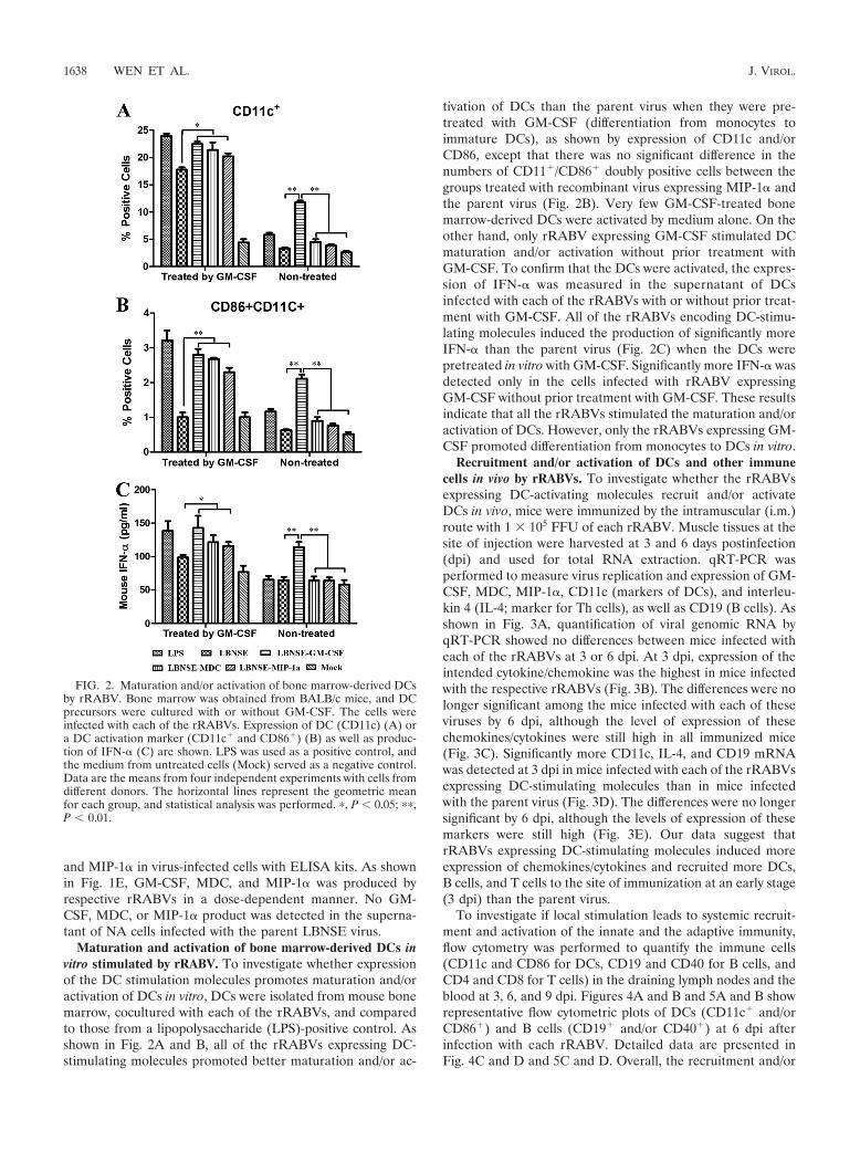
Maturation and activation of bone marrow-derived DCs invitro stimulated by rRABV. To investigate whether expressionof the DC stimulation molecules promotes maturation and/oractivation of DCs in vitro, DCs were isolated from mouse bonemarrow, cocultured with each of the rRABVs, and comparedto those from a lipopolysaccharide (LPS)-positive control. Asshown in Fig. 2A and B, all of the rRABVs expressing DC-stimulating molecules promoted better maturation and/or ac-
tivation of DCs than the parent virus when they were pre-treated with GM-CSF (differentiation from monocytes toimmature DCs), as shown by expression of CD11c and/orCD86, except that there was no significant difference in thenumbers of CD11�/CD86� doubly positive cells between thegroups treated with recombinant virus expressing MIP-1� andthe parent virus (Fig. 2B). Very few GM-CSF-treated bonemarrow-derived DCs were activated by medium alone. On theother hand, only rRABV expressing GM-CSF stimulated DCmaturation and/or activation without prior treatment withGM-CSF. To confirm that the DCs were activated, the expres-sion of IFN-� was measured in the supernatant of DCsinfected with each of the rRABVs with or without prior treat-ment with GM-CSF. All of the rRABVs encoding DC-stimu-lating molecules induced the production of significantly moreIFN-� than the parent virus (Fig. 2C) when the DCs werepretreated in vitro with GM-CSF. Significantly more IFN-� wasdetected only in the cells infected with rRABV expressingGM-CSF without prior treatment with GM-CSF. These resultsindicate that all the rRABVs stimulated the maturation and/oractivation of DCs. However, only the rRABVs expressing GM-CSF promoted differentiation from monocytes to DCs in vitro.
Recruitment and/or activation of DCs and other immunecells in vivo by rRABVs. To investigate whether the rRABVsexpressing DC-activating molecules recruit and/or activateDCs in vivo, mice were immunized by the intramuscular (i.m.)route with 1 � 105 FFU of each rRABV. Muscle tissues at thesite of injection were harvested at 3 and 6 days postinfection(dpi) and used for total RNA extraction. qRT-PCR wasperformed to measure virus replication and expression of GM-CSF, MDC, MIP-1�, CD11c (markers of DCs), and interleu-kin 4 (IL-4; marker for Th cells), as well as CD19 (B cells). Asshown in Fig. 3A, quantification of viral genomic RNA byqRT-PCR showed no differences between mice infected witheach of the rRABVs at 3 or 6 dpi. At 3 dpi, expression of theintended cytokine/chemokine was the highest in mice infectedwith the respective rRABVs (Fig. 3B). The differences were nolonger significant among the mice infected with each of theseviruses by 6 dpi, although the level of expression of thesechemokines/cytokines were still high in all immunized mice(Fig. 3C). Significantly more CD11c, IL-4, and CD19 mRNAwas detected at 3 dpi in mice infected with each of the rRABVsexpressing DC-stimulating molecules than in mice infectedwith the parent virus (Fig. 3D). The differences were no longersignificant by 6 dpi, although the levels of expression of thesemarkers were still high (Fig. 3E). Our data suggest thatrRABVs expressing DC-stimulating molecules induced moreexpression of chemokines/cytokines and recruited more DCs,B cells, and T cells to the site of immunization at an early stage(3 dpi) than the parent virus.
To investigate if local stimulation leads to systemic recruit-ment and activation of the innate and the adaptive immunity,flow cytometry was performed to quantify the immune cells(CD11c and CD86 for DCs, CD19 and CD40 for B cells, andCD4 and CD8 for T cells) in the draining lymph nodes and theblood at 3, 6, and 9 dpi. Figures 4A and B and 5A and B showrepresentative flow cytometric plots of DCs (CD11c� and/orCD86�) and B cells (CD19� and/or CD40�) at 6 dpi afterinfection with each rRABV. Detailed data are presented inFig. 4C and D and 5C and D. Overall, the recruitment and/or
FIG. 2. Maturation and/or activation of bone marrow-derived DCsby rRABV. Bone marrow was obtained from BALB/c mice, and DCprecursors were cultured with or without GM-CSF. The cells wereinfected with each of the rRABVs. Expression of DC (CD11c) (A) ora DC activation marker (CD11c� and CD86�) (B) as well as produc-tion of IFN-� (C) are shown. LPS was used as a positive control, andthe medium from untreated cells (Mock) served as a negative control.Data are the means from four independent experiments with cells fromdifferent donors. The horizontal lines represent the geometric meanfor each group, and statistical analysis was performed. �, P 0.05; ��,P 0.01.
1638 WEN ET AL. J. VIROL.

activation of immune cells was detected first in the lymphnodes and then in the peripheral blood. In addition, the dura-tion of recruitment and activation of immune cells is shorter inthe lymph nodes (3 and 6 dpi) than in the blood (3 to 9 dpi).Significantly more immune cells (DCs as detected by CD11cand CD86 and B cells as detected by CD19 and CD40) weredetected in mice infected with rRABVs expressing GM-CSF orMDC than in mice infected with the parent virus. rRABVexpressing MIP-1� also recruited and/or activated more im-mune cells in the lymph nodes and the blood than the parentvirus did, but no statistical significance was detected except inthe blood at 3 dpi, when a significant difference was detectedbetween the group of mice immunized with rRABV expressing
MIP-1� and the group immunized with the parent virus. Asimilar trend was also observed for T cells, as indicated byincreased numbers of CD4� and CD8� cells in the lymphnodes and the peripheral blood (data not shown). Thus, ourstudies suggest that rRABV expressing DC-stimulating mole-cules recruited and activated more DCs as well as other im-mune cells in the lymph nodes and the peripheral blood thanthe parent virus.
Immunogenicity of rRABV. To determine if recruitmentand/or activation of DCs and other immune cells in the pe-riphery increases the immunogenicity of the rRABV, mice (10in each group) were immunized once by the i.m. route withdifferent doses of rRABV (1 � 103, 1 � 104, 1 � 105, or 1 �
FIG. 3. Quantification of viral genomic RNA as well as mRNA for chemokines/cytokines and markers of immune cells by qRT-PCR at the siteof immunization. BALB/c mice were infected i.m. with rRABVs at 1 � 105 FFU per mouse, and muscle tissues were harvested from the site ofimmunization at 3 and 6 dpi. Total RNA was prepared and used in a qRT-PCR to determine levels of viral genomic RNA (A) or mRNA levelsfor chemokines/cytokines (B and C) as well as markers of immune cells (D and E). All data are from experiments with 4 mice per group. Asterisksindicate significant differences between the indicated experimental groups (*, P 0.05; **, P 0.01).
VOL. 85, 2011 RECOMBINANT RABV EXPRESSES DC-ACTIVATING MOLECULES 1639

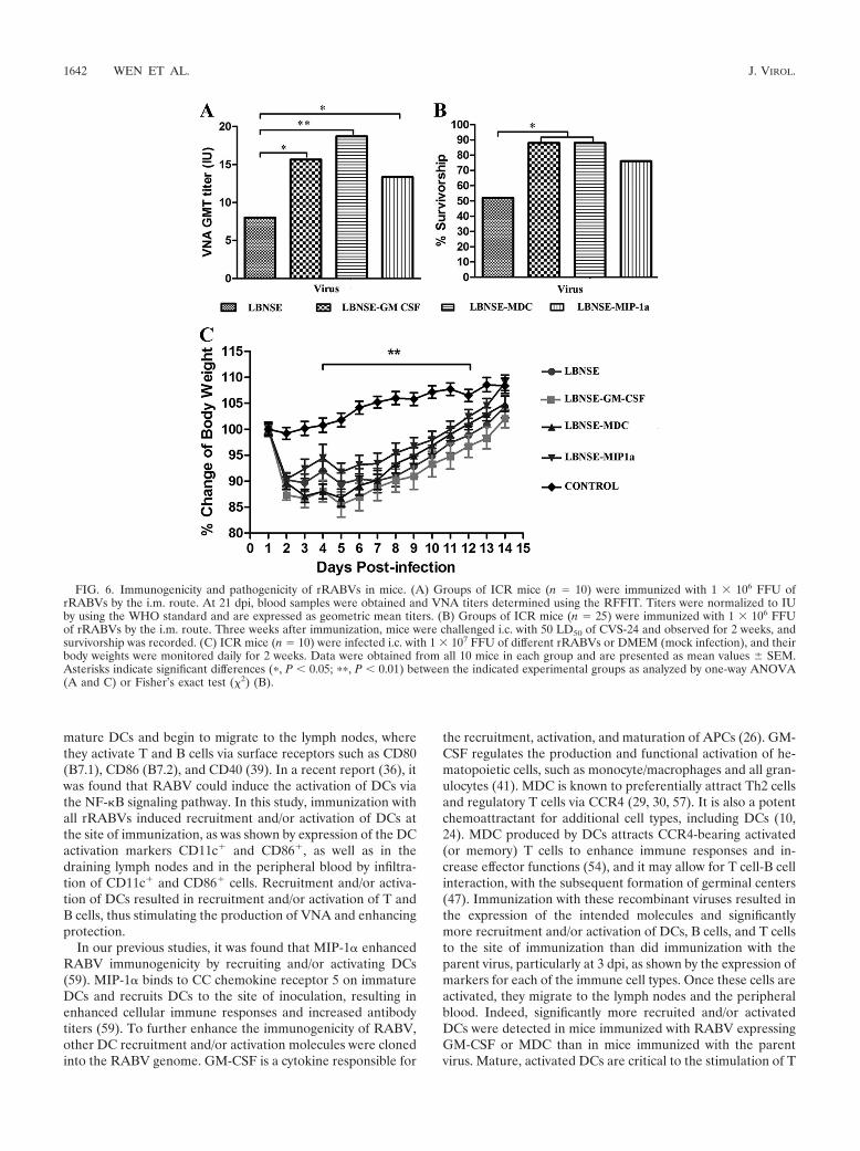
106 FFU per mouse). Blood samples were collected 21 dpi, andsera were used for determination of VNA by the RFFIT (51).Overall, the level of VNA is dose dependent for all viruses(data not shown). Figure 6A shows the level of VNA in miceimmunized with 1 � 106 FFU of RABV. Significantly higherVNA titers were detected in mice immunized with the LBNSE-MDC (18.72 IU) (P 0.01), LBNSE–GM-CSF (15.65 IU) (P 0.05), and LBNSE–MIP-1� (13.36 IU) (P 0.05) viruses thanwere induced by immunization with the parent virus, LBNSE(8.01 IU). To investigate if the higher VNA titers correlate withbetter protection, mice (25 in each group) immunized with 106
FFU of each virus were then challenged with 50 LD50 of virulentCVS-24 on day 21 after vaccination and observed for develop-ment of disease and death for 2 weeks. As depicted in Fig. 6B,significantly more survivors were observed among mice immu-nized with LBNSE–GM-CSF or LBNSE-MDC than among thoseimmunized with the parent LBNSE virus. More survivors werealso observed among mice immunized with LBNSE–MIP-1� thanamong mice immunized with the parent virus, but no significantdifference was detected. Together, these data indicate thatrRABV expressing GM-CSF or MDC provides better protectiveimmunity than the parent virus.
Pathogenicity of rRABVs in mice. To determine whetheroverexpression of GM-CSF, MDC, or MIP-1� has adverseeffect in animals, three groups of 10 4- to 6-week-old ICR micewere infected with 1 � 105, 1 � 106, or 1 � 107 FFU ofrecombinant viruses by the intracerebral (i.c.) route. Infectedmice were monitored daily for 2 weeks for weight loss as well asfor development of disease and death. When infected at the lowdoses (1 � 105 and 1 � 106 FFU), infected mice lost 5 to 10% oftheir body weight (data not shown) but 10 to 15% of their bodyweight when they were infected with the high dose (1 � 107
FFU), which is significant compared to the value for sham-in-fected mice (Fig. 6C). Most of the mice regained their prechal-lenge body weight by 15 dpi. However, no overt clinic symptoms,such as abnormal behaviors, or any neurological signs were ob-served in these mice. These data indicate that expression of DC-stimulating molecules did not increase RABV virulence.
DISCUSSION
Previous studies with recombinant RABV expressing thechemokine MIP-1� revealed that it further attenuated RABVvirulence by inducing a transient innate immune response in
FIG. 4. Recruitment and/or activation of DCs in the lymph nodes and blood after infection with rRABVs. BALB/c mice were infected i.m. with1 � 105 FFU of different rRABVs, and draining (inguinal) lymph nodes and blood were harvested after extensive perfusion at 3, 6, and 9 dpi.Single-cell suspensions were prepared, stained with antibodies against DCs and the DC activation markers CD11c and CD86, and analyzed by flowcytometry. Representative flow cytometric plots of DCs are shown from the lymph nodes (A) and the blood (B). The results of a detailed analysisfor activated DCs (CD11c� and CD86�) at 3, 6, and 9 dpi are presented for the lymph nodes (C) and blood (D). Asterisks indicate significantdifferences between the indicated experimental groups (*, P 0.05; **, P 0.01).
1640 WEN ET AL. J. VIROL.

the central nervous system (CNS) (58). In addition, viral ex-pression of MIP-1� enhanced RABV immunogenicity by in-ducing higher levels of VNA (59). This was achieved via re-cruitment and/or activation of DCs at the site of immunization,in the draining lymph nodes, and in the peripheral blood (59).To further confirm the role of DCs in enhancing RABV im-munogenicity, more genes for DC recruitment/activation mol-ecules (GM-CSF and MDC) in addition to MIP-1� were indi-vidually cloned into RABV and the immunogenicity andpathogenicity of these recombinant viruses were determined ina mouse model. Each of the viruses stimulated more matura-tion and activation of murine bone marrow-derived DCs invitro and more recruitment and/or activation of DCs and ma-ture B cells, as well as T cells, in the periphery than the parentvirus, which led to higher levels of VNA and better protection.Most importantly, a single immunization with recombinantRABV expressing GM-CSF or MDC protected significantlymore mice against intracerebral challenge with virulent RABVthan did the parental virus. Yet, these viruses did not showmore virulence than the parent virus, since direct intracerebralinoculation with each virus (up to 1 � 107 FFU) did not induceany overt clinic symptom, such as abnormal behavior, or anyneurological signs. Thus, our data suggest that recruitment/
activation of DCs is important in enhancing RABV immuno-genicity and protection.
DCs probably arise from monocytes and white blood cells.These cells circulate in the body and, depending on the appro-priate signal, can turn into either DCs or macrophages (16).Treatment of in vitro-derived monocytes with GM-CSF leadsto differentiation of immature DCs in about a week (56). Priorstudies showed that RABV can activate DCs in vitro (36). Ourdata indicate that rRABV expressing GM-CSF, MDC, orMIP-1� stimulated more maturation and/or activation of DCsin vitro than the parent virus. All RABVs stimulated matura-tion and/or activation of DCs when the cells were pretreated invitro with GM-CSF. Activation of DCs is shown not only byexpression of the DC markers CD86 and CD80 (data notshown) but also by production of IFN-�. However, only therRABV expressing GM-CSF promoted differentiation frommonocytes to DCs as well as the maturation and activation ofDCs in vitro.
DCs are the most efficient APCs and thus play a key role inboth innate and adaptive immune responses in vivo (3). Im-mature DCs constantly sample the surrounding environmentfor pathogens such as viruses and bacteria. Once they havecome into contact with antigens, DCs become activated into
FIG. 5. Recruitment and/or activation of B cells in the lymph nodes and blood after infection with rRABVs. BALB/c mice were infected i.m.with 1 � 105 FFU of different rRABVs, and draining (inguinal) lymph nodes and blood were harvested after extensive perfusion at 3, 6, and 9 dpi.Single-cell suspensions were prepared, stained with antibodies against B cells and the B cell activation markers CD19 and CD40, and analyzed byflow cytometry. Representative flow cytometric plots of B cells are shown from the lymph nodes (A) and the blood (B). The detailed analyses foractivated B cells (CD19� and CD40�) at 3, 6, and 9 dpi are presented for lymph nodes (C) and blood (D). Asterisks denote significant differencesbetween the indicated experimental groups (*, P 0.05; **, P 0.01).
VOL. 85, 2011 RECOMBINANT RABV EXPRESSES DC-ACTIVATING MOLECULES 1641
mature DCs and begin to migrate to the lymph nodes, wherethey activate T and B cells via surface receptors such as CD80(B7.1), CD86 (B7.2), and CD40 (39). In a recent report (36), itwas found that RABV could induce the activation of DCs viathe NF-�B signaling pathway. In this study, immunization withall rRABVs induced recruitment and/or activation of DCs atthe site of immunization, as was shown by expression of the DCactivation markers CD11c� and CD86�, as well as in thedraining lymph nodes and in the peripheral blood by infiltra-tion of CD11c� and CD86� cells. Recruitment and/or activa-tion of DCs resulted in recruitment and/or activation of T andB cells, thus stimulating the production of VNA and enhancingprotection.
In our previous studies, it was found that MIP-1� enhancedRABV immunogenicity by recruiting and/or activating DCs(59). MIP-1� binds to CC chemokine receptor 5 on immatureDCs and recruits DCs to the site of inoculation, resulting inenhanced cellular immune responses and increased antibodytiters (59). To further enhance the immunogenicity of RABV,other DC recruitment and/or activation molecules were clonedinto the RABV genome. GM-CSF is a cytokine responsible for
the recruitment, activation, and maturation of APCs (26). GM-CSF regulates the production and functional activation of he-matopoietic cells, such as monocyte/macrophages and all gran-ulocytes (41). MDC is known to preferentially attract Th2 cellsand regulatory T cells via CCR4 (29, 30, 57). It is also a potentchemoattractant for additional cell types, including DCs (10,24). MDC produced by DCs attracts CCR4-bearing activated(or memory) T cells to enhance immune responses and in-crease effector functions (54), and it may allow for T cell-B cellinteraction, with the subsequent formation of germinal centers(47). Immunization with these recombinant viruses resulted inthe expression of the intended molecules and significantlymore recruitment and/or activation of DCs, B cells, and T cellsto the site of immunization than did immunization with theparent virus, particularly at 3 dpi, as shown by the expression ofmarkers for each of the immune cell types. Once these cells areactivated, they migrate to the lymph nodes and the peripheralblood. Indeed, significantly more recruited and/or activatedDCs were detected in mice immunized with RABV expressingGM-CSF or MDC than in mice immunized with the parentvirus. Mature, activated DCs are critical to the stimulation of T
FIG. 6. Immunogenicity and pathogenicity of rRABVs in mice. (A) Groups of ICR mice (n � 10) were immunized with 1 � 106 FFU ofrRABVs by the i.m. route. At 21 dpi, blood samples were obtained and VNA titers determined using the RFFIT. Titers were normalized to IUby using the WHO standard and are expressed as geometric mean titers. (B) Groups of ICR mice (n � 25) were immunized with 1 � 106 FFUof rRABVs by the i.m. route. Three weeks after immunization, mice were challenged i.c. with 50 LD50 of CVS-24 and observed for 2 weeks, andsurvivorship was recorded. (C) ICR mice (n � 10) were infected i.c. with 1 � 107 FFU of different rRABVs or DMEM (mock infection), and theirbody weights were monitored daily for 2 weeks. Data were obtained from all 10 mice in each group and are presented as mean values � SEM.Asterisks indicate significant differences (�, P 0.05; ��, P 0.01) between the indicated experimental groups as analyzed by one-way ANOVA(A and C) or Fisher’s exact test (2) (B).
1642 WEN ET AL. J. VIROL.

cells and the generation of the virus-specific adaptive immuneresponses (1, 2). The epitopes presented by APCs can berecognized by T cells, which can provide help for B cells toproduce large quantities of antibodies (34). Our data demon-strate that recruitment and/or activation of DCs by recombi-nant RABVs resulted in recruitment and activation of B and Tcells at the site of immunization and in the draining lymphnodes, as well as in the peripheral blood. Furthermore, wehave shown that infection of DCs with these recombinantRABVs promoted the production of IFN-�, which can in turnpromote the differentiation of B cells into plasma cells viaincreased expression of TLR7 in naïve B cells (4, 13). Takentogether, these data indicate that recruitment and/or activationof more DCs leads to activation of T and B cells and results inthe production of higher levels of VNA in mice immunizedwith rRABVs expressing DC-activating molecules than in miceimmunized with the parent virus. Expression of DC-activatingmolecules, particularly GM-CSF, has been reported to en-hance the immunogenicity of many other antigens, includingviral and tumor antigens (15, 25, 42, 53). MDC has also beenreported to enhance systemic and mucosal immune responsesfor HIV gp120 (5).
Our previous studies showed that recombinant RABVexpressing MIP-1� activated significantly more DCs andstimulated more production of VNA than did the parentvirus (59). In the previous study, RABV strain HEP wasused, while in the present study, RABV strain L16 was used.The reason for the switch of vectors was that HEP can growto titers up to 107 FFU (58), while L16 can grow to titers upto 108 FFU. High virus titers are needed for assessment ofpathogenicity and immunogenicity. Despite the use of dif-ferent vectors (HEP versus the L16 strain of RABV), similarfindings were found for mice immunized with each of theviruses, as reported earlier (59) and in this study. Bothviruses recruited/activated more immune cells in the periph-ery and induced higher VNA titers in mice than the parentvirus. In fact, the VNA titers (about 10 IU) were similar inmice immunized with both viruses at high doses (5 � 105 forHEP–MIP-1� and 1 � 106 for LBNSE–MIP-1�). Mice im-munized with the highest dose for each virus protectedabout 80% of the mice against challenge.
In summary, recombinant RABVs expressing DC activationmolecules enhanced the immunogenicity of RABV via recruit-ment, maturation, and/or activation of DCs. Yet, the virusesdid not increase RABV virulence, as direct infection by the i.c.route with up to 1 � 107 FFU did not induce any overt clinicalsymptoms in mice infected with the recombinant or the paren-tal RABV. The weight loss in mice infected with recombinantRABVs by the intracerebral route could be related to RABVreplication in the brain, expression of chemokines in the brainindependent of viral replication, inflammatory responses in-duced by the increased expression of chemokines/cytokines, ora combination of all these factors. Indeed, previous experi-ments with recombinant RABV expressing RANTES or IP-10induced extensive inflammation in the CNS (58). However, ifthese recombinant RABVs are to be used for vaccine devel-opment, further studies will be required to address the issue ofthe residual virulence of these viruses.
ACKNOWLEDGMENTS
This work is supported partially by Public Health Service grantAI-051560 from the National Institute of Allergy and Infectious Dis-eases.
We thank Guoqing Zhang for technical help during the study.
REFERENCES
1. Banchereau, J., et al. 2000. Immunobiology of dendritic cells. Annu. Rev.Immunol. 18:767–811.
2. Banchereau, J., and R. M. Steinman. 1998. Dendritic cells and the control ofimmunity. Nature 392:245–252.
3. Becker, Y. 2003. Immunological and regulatory functions of uninfected andvirus infected immature and mature subtypes of dendritic cells—a review.Virus Genes 26:119–130.
4. Bekeredjian-Ding, I. B., et al. 2005. Plasmacytoid dendritic cells controlTLR7 sensitivity of naive B cells via type I IFN. J. Immunol. 174:4043–4050.
5. Biragyn, A., et al. 2002. DNA vaccines encoding human immunodeficiencyvirus-1 glycoprotein 120 fusions with proinflammatory chemoattractants in-duce systemic and mucosal immune responses. Blood 100:1153–1159.
6. Blancou, J., et al. 1989. Safety and efficacy of an antirabies vaccine consistingof recombinant vaccinia-rabies virus administered orally to the fox, dog andcat. Ann. Rech. Vet. 20:195–204. (In French.)
7. Brilot, F., T. Strowig, and C. Munz. 2008. NK cell interactions with dendriticcells shape innate and adaptive immunity. Front. Biosci. 13:6443–6454.
8. Brochier, B., et al. 1991. Large-scale eradication of rabies using recombinantvaccinia-rabies vaccine. Nature 354:520–522.
9. Centers for Disease Control and Prevention. 2009. Human vaccinia infectionafter contact with a raccoon rabies vaccine bait—Pennsylvania. MMWRMorb. Mortal. Wkly. Rep. 58:3.
10. Chantry, D., et al. 1999. Macrophage-derived chemokine is localized tothymic medullary epithelial cells and is a chemoattractant for CD3(�),CD4(�), CD8(low) thymocytes. Blood 94:1890–1898.
11. Conzelmann, K. K., J. H. Cox, L. G. Schneider, and H. J. Thiel. 1990.Molecular cloning and complete nucleotide sequence of the attenuatedrabies virus SAD B19. Virology 175:485–499.
12. Crick, J. 1973. The vaccination of man and other animals against rabies.Postgrad. Med. J. 49:551–564.
13. Diebold, S. S., T. Kaisho, H. Hemmi, S. Akira, and C. Reis e Sousa. 2004.Innate antiviral responses by means of TLR7-mediated recognition of single-stranded RNA. Science 303:1529–1531.
14. Dieu, M. C., et al. 1998. Selective recruitment of immature and maturedendritic cells by distinct chemokines expressed in different anatomic sites. J.Exp. Med. 188:373–386.
15. Disis, M. L., et al. 1996. Granulocyte-macrophage colony-stimulating factor:an effective adjuvant for protein and peptide-based vaccines. Blood 88:202–210.
16. Dominguez, P. M., and C. Ardavin. 2010. Differentiation and function ofmouse monocyte-derived dendritic cells in steady state and inflammation.Immunol. Rev. 234:90–104.
17. Faber, M., et al. 2005. Overexpression of tumor necrosis factor alpha by arecombinant rabies virus attenuates replication in neurons and preventslethal infection in mice. J. Virol. 79:15405–15416.
18. Faber, M., et al. 2007. Dominance of a nonpathogenic glycoprotein geneover a pathogenic glycoprotein gene in rabies virus. J. Virol. 81:7041–7047.
19. Faber, M., et al. 2009. Effective preexposure and postexposure prophylaxis ofrabies with a highly attenuated recombinant rabies virus. Proc. Natl. Acad.Sci. U. S. A. 106:11300–11305.
20. Flamand, A., P. Coulon, F. Lafay, and C. Tuffereau. 1993. Avirulent mutantsof rabies virus and their use as live vaccine. Trends Microbiol. 1:317–320.
21. Frazatti-Gallina, N. M., et al. 2004. Vero-cell rabies vaccine produced usingserum-free medium. Vaccine 23:511–517.
22. Fu, Z. F. 1997. Rabies and rabies research: past, present and future. Vaccine15(Suppl.):S20–S24.
23. Gilboa, E. 2007. DC-based cancer vaccines. J. Clin. Invest. 117:1195–1203.24. Godiska, R., et al. 1997. Human macrophage-derived chemokine (MDC), a
novel chemoattractant for monocytes, monocyte-derived dendritic cells, andnatural killer cells. J. Exp. Med. 185:1595–1604.
25. Haddad, D., et al. 2000. Plasmid vaccine expressing granulocyte-macrophagecolony-stimulating factor attracts infiltrates including immature dendriticcells into injected muscles. J. Immunol. 165:3772–3781.
26. Hamilton, J. A., and G. P. Anderson. 2004. GM-CSF biology. Growth Fac-tors 22:225–231.
27. Hanlon, C. A., et al. 1998. First North American field release of a vaccinia-rabies glycoprotein recombinant virus. J. Wildl. Dis. 34:228–239.
28. Hanlon, C. A., M. Niezgoda, P. Morrill, and C. E. Rupprecht. 2002. Oralefficacy of an attenuated rabies virus vaccine in skunks and raccoons. J.Wildl. Dis. 38:420–427.
29. Iellem, A., et al. 2001. Unique chemotactic response profile and specificexpression of chemokine receptors CCR4 and CCR8 by CD4(�)CD25(�)regulatory T cells. J. Exp. Med. 194:847–853.
30. Imai, T., et al. 1999. Selective recruitment of CCR4-bearing Th2 cells toward
VOL. 85, 2011 RECOMBINANT RABV EXPRESSES DC-ACTIVATING MOLECULES 1643

antigen-presenting cells by the CC chemokines thymus and activation-regu-lated chemokine and macrophage-derived chemokine. Int. Immunol. 11:81–88.
31. Inaba, K., et al. 1992. Generation of large numbers of dendritic cells frommouse bone marrow cultures supplemented with granulocyte/macrophagecolony-stimulating factor. J. Exp. Med. 176:1693–1702.
32. Inoue, K., et al. 2003. An improved method for recovering rabies virus fromcloned cDNA. J. Virol. Methods 107:229–236.
33. Jackson, A. C. 2002. Rabies pathogenesis. J. Neurovirol. 8:267–269.34. Jelley-Gibbs, D. M., T. M. Strutt, K. K. McKinstry, and S. L. Swain. 2008.
Influencing the fates of CD4 T cells on the path to memory: lessons frominfluenza. Immunol. Cell Biol. 86:343–352.
35. Kuang, Y., S. N. Lackay, L. Zhao, and Z. F. Fu. 2009. Role of chemokines inthe enhancement of BBB permeability and inflammatory infiltration afterrabies virus infection. Virus Res. 144:18–26.
36. Li, J., J. P. McGettigan, M. Faber, M. J. Schnell, and B. Dietzschold.2008. Infection of monocytes or immature dendritic cells (DCs) with anattenuated rabies virus results in DC maturation and a strong activationof the NFkappaB signaling pathway. Vaccine 26:419–426.
37. Lutz, M. B., et al. 1999. An advanced culture method for generating largequantities of highly pure dendritic cells from mouse bone marrow. J. Immu-nol. Methods 223:77–92.
38. Martinez, L. 2000. Global infectious disease surveillance. Int. J. Infect. Dis.4:222–228.
39. Martin-Fontecha, A., A. Lanzavecchia, and F. Sallusto. 2009. Dendritic cellmigration to peripheral lymph nodes. Handb. Exp. Pharmacol. 2009:31–49.
40. Meltzer, M. I. 1996. Assessing the costs and benefits of an oral vaccine forraccoon rabies: a possible model. Emerg. Infect. Dis. 2:343–349.
41. Metcalf, D. 2008. Hematopoietic cytokines. Blood 111:485–491.42. Morrissey, P. J., L. Bressler, L. S. Park, A. Alpert, and S. Gillis. 1987.
Granulocyte-macrophage colony-stimulating factor augments the primaryantibody response by enhancing the function of antigen-presenting cells.J. Immunol. 139:1113–1119.
43. Reed, L. J., and H. Muench. 1938. A simple method of estimating fiftypercent endpoints. Am. J. Epidemiol. 27:493–497.
44. Rupprecht, C. E., et al. 2001. Human infection due to recombinant vaccinia-rabies glycoprotein virus. N. Engl. J. Med. 345:582–586.
45. Rupprecht, C. E., et al. 2010. Use of a reduced (4-dose) vaccine schedule forpostexposure prophylaxis to prevent human rabies: recommendations of theadvisory committee on immunization practices. MMWR Recomm. Rep.59:1–9.
46. Rupprecht, C. E., J. S. Smith, M. Fekadu, and J. E. Childs. 1995. Theascension of wildlife rabies: a cause for public health concern or interven-tion? Emerg. Infect. Dis. 1:107–114.
47. Schaniel, C., et al. 1998. Activated murine B lymphocytes and dendritic cellsproduce a novel CC chemokine which acts selectively on activated T cells. J.Exp. Med. 188:451–463.
48. Schnell, M. J., T. Mebatsion, and K. K. Conzelmann. 1994. Infectious rabiesviruses from cloned cDNA. EMBO J. 13:4195–4203.
49. Shi, Y., et al. 2006. Granulocyte-macrophage colony-stimulating factor (GM-CSF) and T-cell responses: what we do and don’t know. Cell Res. 16:126–133.
50. Shim, E., K. Hampson, S. Cleaveland, and A. P. Galvani. 2009. Evaluatingthe cost-effectiveness of rabies post-exposure prophylaxis: a case study inTanzania. Vaccine 27:7167–7172.
51. Smith, J. S., P. A. Yager, and G. M. Baer. 1996. A rapid fluorescence focusinhibition test (RFFIT) for determining rabies virus-neutralising antibody, p.181–192. In F.-X. Meslin, M. M. Kaplan, and H. Koprowski (ed.), Labora-tory techniques in rabies, 4th ed. World Health Organization, Geneva, Swit-zerland.
52. Takayama-Ito, M., et al. 2006. A highly attenuated rabies virus HEP-Flurystrain reverts to virulent by single amino acid substitution to arginine atposition 333 in glycoprotein. Virus Res. 119:208–215.
53. Tarr, P. E. 1996. Granulocyte-macrophage colony-stimulating factor and theimmune system. Med. Oncol. 13:133–140.
54. Wu, M., H. Fang, and S. T. Hwang. 2001. Cutting edge: CCR4 mediatesantigen-primed T cell binding to activated dendritic cells. J. Immunol. 167:4791–4795.
55. Wunner, W. 1991. The chemical composition and molecular structure ofrabies viruses, p. 31–67. In G. M. Baer (ed.), Natural history of rabies, 2nded. CRC Press, Inc., Boca Raton, FL.
56. Yi, H., L. Zhang, Y. Zhen, X. He, and Y. Zhao. 2007. Dendritic cells inducedin the presence of GM-CSF and IL-5. Cytokine 37:35–43.
57. Yoshie, O., T. Imai, and H. Nomiyama. 2001. Chemokines in immunity. Adv.Immunol. 78:57–110.
58. Zhao, L., H. Toriumi, Y. Kuang, H. Chen, and Z. F. Fu. 2009. The roles ofchemokines in rabies virus infection: overexpression may not always bebeneficial. J. Virol. 83:11808–11818.
59. Zhao, L., et al. 2010. Expression of MIP-1� (CCL3) by a recombinant rabiesvirus enhances its immunogenicity by inducing innate immunity and recruit-ing dendritic cells and B cells. J. Virol. 84:9642–9648.
1644 WEN ET AL. J. VIROL.