The affirmative response of the innate immune system to apoptotic cells
12
The affirmative response of the innate immune system to apoptotic cells VIMAL A. PATEL 1 , ANGELIKA LONGACRE-ANTONI 2 , MARIJA CVETANOVIC 3 , DANIEL J. LEE 1 , LANFEI FENG 1 , HANLI FAN 1 , JOYCE RAUCH 4 , DAVID S. UCKER 5 , and JERROLD S. LEVINE 1,5 1 Division of Nephrology, Department of Medicine, University of Illinois at Chicago, Chicago, IL 60612, USA 2 Division of Biology, Kutztown University of Pennsylvania, Kutztown, PA 19530, USA 3 Department of Neurology, Northwestern University School of Medicine, Chicago, IL 60611, USA 4 Division of Rheumatology, Department of Medicine, Research Institute of the McGill University Health Centre, Montreal, Que., Canada H3G 1A4 and 5 Department of Microbiology and Immunology, University of Illinois at Chicago, Chicago, IL 60612, USA Abstract Growing evidence exists for a new role for apoptotic cell recognition and clearance in immune homeostasis. Apoptotic cells at all stages, irrespective of membrane integrity, elicit a signature set of signaling events in responding phagocytes, both professional and non-professional. These signaling events are initiated by receptor-mediated recognition of apoptotic determinants, independently of species, cell type, or apoptotic stimulus. We propose that the ability of phagocytes to respond to apoptotic targets with a characteristic set of signaling events comprises a second distinct dimension of innate immunity, as opposed to the traditional innate discrimination of self vs. non-self. We further propose that a loss or abnormality of the signaling events elicited by apoptotic cells, as distinct from the actual clearance of those cells, may predispose to autoimmunity. Keywords Innate immunity; apoptosis; autoimmunity; inflammation; phagocytosis; signal transduction Introduction and overview Cells are continually dying throughout the body. Such death occurs in one of two predominant forms: physiologic (apoptosis) or pathologic (necrosis). The very large number of dying cells, even in health, requires that cell corpses, whether apoptotic or necrotic, be recognized and cleared in a rapid, efficient manner. Professional phagocytes, such as macrophages and dendritic cells, are primarily charged with the duty of clearance, but all cells, including fibroblasts and epithelial cells, are capable of dead cell clearance. Importantly, the consequences of these two forms of cell death for surrounding tissues are drastically different. Apoptotic cell death is a conserved, cell-autonomous process that ensures specifically targeted removal of dying cells in an orderly and non-inflammatory fashion. In contrast, necrotic cell death, which also triggers phagocytosis, is marked by rapid © 2007 Informa UK Ltd. Correspondence: J. S. Levine, Division of Nephrology, Department of Medicine, University of Illinois at Chicago College of Medicine, MC793, 820 S. Wood Street, Chicago, IL 60612, USA. Tel: 1 312 413 1178. Fax: 1 312 996 7378. [email protected]. PubMed Central CANADA Author Manuscript / Manuscrit d'auteur Autoimmunity. Author manuscript; available in PMC 2012 September 11. Published in final edited form as: Autoimmunity. 2007 June ; 40(4): 274–280. doi:10.1080/08916930701357463. PMC Canada Author Manuscript PMC Canada Author Manuscript PMC Canada Author Manuscript
-
Upload
independent -
Category
Documents
-
view
1 -
download
0
Transcript of The affirmative response of the innate immune system to apoptotic cells

The affirmative response of the innate immune system toapoptotic cells
VIMAL A. PATEL1, ANGELIKA LONGACRE-ANTONI2, MARIJA CVETANOVIC3, DANIEL J.LEE1, LANFEI FENG1, HANLI FAN1, JOYCE RAUCH4, DAVID S. UCKER5, and JERROLD S.LEVINE1,5
1Division of Nephrology, Department of Medicine, University of Illinois at Chicago, Chicago, IL60612, USA 2Division of Biology, Kutztown University of Pennsylvania, Kutztown, PA 19530, USA3Department of Neurology, Northwestern University School of Medicine, Chicago, IL 60611, USA4Division of Rheumatology, Department of Medicine, Research Institute of the McGill UniversityHealth Centre, Montreal, Que., Canada H3G 1A4 and 5Department of Microbiology andImmunology, University of Illinois at Chicago, Chicago, IL 60612, USA
AbstractGrowing evidence exists for a new role for apoptotic cell recognition and clearance in immunehomeostasis. Apoptotic cells at all stages, irrespective of membrane integrity, elicit a signature setof signaling events in responding phagocytes, both professional and non-professional. Thesesignaling events are initiated by receptor-mediated recognition of apoptotic determinants,independently of species, cell type, or apoptotic stimulus. We propose that the ability ofphagocytes to respond to apoptotic targets with a characteristic set of signaling events comprises asecond distinct dimension of innate immunity, as opposed to the traditional innate discriminationof self vs. non-self. We further propose that a loss or abnormality of the signaling events elicitedby apoptotic cells, as distinct from the actual clearance of those cells, may predispose toautoimmunity.
KeywordsInnate immunity; apoptosis; autoimmunity; inflammation; phagocytosis; signal transduction
Introduction and overviewCells are continually dying throughout the body. Such death occurs in one of twopredominant forms: physiologic (apoptosis) or pathologic (necrosis). The very large numberof dying cells, even in health, requires that cell corpses, whether apoptotic or necrotic, berecognized and cleared in a rapid, efficient manner. Professional phagocytes, such asmacrophages and dendritic cells, are primarily charged with the duty of clearance, but allcells, including fibroblasts and epithelial cells, are capable of dead cell clearance.Importantly, the consequences of these two forms of cell death for surrounding tissues aredrastically different. Apoptotic cell death is a conserved, cell-autonomous process thatensures specifically targeted removal of dying cells in an orderly and non-inflammatoryfashion. In contrast, necrotic cell death, which also triggers phagocytosis, is marked by rapid
© 2007 Informa UK Ltd.
Correspondence: J. S. Levine, Division of Nephrology, Department of Medicine, University of Illinois at Chicago College ofMedicine, MC793, 820 S. Wood Street, Chicago, IL 60612, USA. Tel: 1 312 413 1178. Fax: 1 312 996 7378. [email protected].
PubMed Central CANADAAuthor Manuscript / Manuscrit d'auteurAutoimmunity. Author manuscript; available in PMC 2012 September 11.
Published in final edited form as:Autoimmunity. 2007 June ; 40(4): 274–280. doi:10.1080/08916930701357463.
PMC
Canada Author M
anuscriptPM
C C
anada Author Manuscript
PMC
Canada Author M
anuscript
and disorganized cellular swelling and rupture, and is typically seen in association withinflammation and tissue injury.
In this review, we will focus primarily on the signaling events induced in healthysurrounding cells by their interaction with apoptotic vs. necrotic targets. As far as possible,we will distinguish between signaling events initiated by the specific recognition ofapoptotic vs. necrotic targets and signaling events initiated by the common machinery ofphagocytosis. Our data support the view that dead cells are not merely cleared byphagocytes, but instead elicit a potent affirmative response that critically modulates thesurvival, proliferation, and inflammatory activity of interacting cells. We propose that thisresponse to dead cells comprises an important and under-appreciated dimension of innateimmunity. This added dimension depends upon the discrimination of apoptotic and necroticcells from viable cells, as opposed to the discrimination of “self” from “non-self”, typicallyconsidered the crucial focus of immunity. Appreciation of these two independentdimensions of innate immune recognition has important implications for the pathogenesis ofautoimmunity, in particular the popular “delayed clearance model”. As we will discuss, theimpaired clearance of apoptotic targets has two important consequences, whose roles in thedevelopment of autoimmunity have not been properly distinguished: persistence of apoptoticcorpses, and loss of apoptotic target-initiated affirmative signaling.
Discrimination of apoptotic from necrotic corpsesThe differential response by surrounding tissues to apoptotic vs. necrotic targets led to thehypothesis that properties unique to the dying cell determine the mode and outcome ofclearance [1]. Moreover, the fact that clearance of apoptotic targets occurs before rupture ofthe cell membrane focused attention on the presumptive expression of apoptotic cell surfacedeterminants for phagocytosis, or so-called “eat me” signals [2]. Identification of thesesurface determinants, which may include alterations in lipid and carbohydrate compositionas well as the appearance of oxidation products or other modified molecules, remains anarea of ongoing investigation [1]. Complementary studies have implicated severalphagocyte-specific molecules as putative receptors for apoptotic corpses [1].
It is critical to note that the analysis of these putative receptors has, in many cases, beenincomplete. Most functional tests have evaluated apoptotic target “uptake”, withoutdiscriminating between actual recognition and engulfment. Indeed, in those cases for whichthe functional roles of particular gene products were assessed, the actions of those molecules(for example, the Class A scavenger receptor, the ABC-type transporter ABC1, and the Mertyrosine kinase) were found to be limited to the phagocytic process, and to play no role inspecific recognition [3–5]. Genetic studies of cell death in Caenorhabditis elegans furtherilluminate this issue. Engulfment in C. elegans involves the products of at least seven genes,which act within redundant converging pathways for the clearance of dead cells [6,7]. Themammalian orthologues of these gene products (such as Rac1, DOCK180 and ELMO) havealso been implicated in the clearance of dead cells [6–8]. Importantly, recent work hasshown that the engulfment pathways encoded by these genes in C. elegans are not specificfor cells undergoing apoptotic cell death [9]. Rather, these genes are required for theclearance of both apoptotic and necrotic corpses.
To date, then, the phagocyte receptors that mediate the recognition of apoptotic vs. necroticcells remain elusive. Since apoptotic and necrotic targets share a common pathway forphagocytosis, it would seem that the differential response to these two targets must lie at thelevel of target recognition. In keeping with this notion, we have found that discriminationbetween apoptotic and necrotic corpses occurs at the level of target recognition, or binding,and is independent of engulfment [10]. As shown by quantitative and objective fluorescence
PATEL et al. Page 2
Autoimmunity. Author manuscript; available in PMC 2012 September 11.
PMC
Canada Author M
anuscriptPM
C C
anada Author Manuscript
PMC
Canada Author M
anuscript
assays, the recognition by macrophages of each type of dead cell occurs via a saturable,receptor-mediated process [10]. Notably, these two receptor-mediated processes are distinctand non-competing [10].
Recognition-dependent anti-inflammatory responses by apoptotic cellsThe uniquely defining property of an apoptotic corpse is its ability to be removed withoutinflammation. Our experiments have shown that the distinct modes of recognition forapoptotic vs. necrotic targets by engulfing macrophages are linked to their opposing effectson the inflammatory response [10]. The anti-inflammatory activity of apoptotic cells hasbeen documented by several groups, and is readily observed at the level of inhibition ofsecretion of inflammatory cytokines from engulfing macrophages [11,12]. In contrast,necrotic cells, which are recognized by a distinct mechanism, tend to enhance pro-inflammatory macrophage responses (although alone they are usually not sufficient totrigger those responses) [10]. Typically, necrosis is induced by brief heating; but othermethods, including freeze-thaw lysis, yield similar results. Strikingly, the inhibitory effect ofapoptotic targets is dominant to the stimulatory effect of necrotic targets [10,13],demonstrating unequivocally that the acquisition of a cell-associated anti-inflammatoryactivity represents a gain-of-function by the apoptotic cell. Moreover, this inhibitory effectpersists at all stages of apoptotic cell death, irrespective of cell membrane integrity[10,13,14].
In a series of studies, we have made the following observations on the anti-inflammatoryresponse to apoptotic targets by macrophages [10,14]. First, recognition of apoptotic targetsinhibited transcription of several cytokine genes, including interleukin-6 (IL-6) and tumornecrosis factor-α (TNF-α) [14]. Apoptotic inhibition occurred independently of new proteinsynthesis, and, therefore, represented an immediate-early response [14]. Second,transcriptional reporters, such as a luciferase construct driven by the authentic IL-8 promoteror a simplified NF-κB-dependent promoter, reliably recapitulated the inhibitory effect ofapoptotic targets [14]. Third, the inhibitory effect of apoptotic targets, as assessed by NF-κB-dependent transcriptional activity 5 h after exposure to apoptotic targets, was a directconsequence of recognition [14]. An autocrine/paracrine contribution by secretedtransforming growth factor-β (TGF-β) was excluded both by blocking antibodies and bypharmacological inhibition of TGF-β type I receptor kinase activity [14]. These findings donot exclude a secondary role for TGF-β, or other secreted factors, which may act at latertimes to amplify and disseminate the modulatory response [11,12]. Finally, since NF-κB-dependent transcriptional activity was unaffected by the cytoskeletal inhibitor cytochalasinD, which effectively blocked engulfment, modulation was dependent on apoptotic cellrecognition and independent of engulfment [10,14].
Proximal signaling events elicited by recognition of apoptotic vs. necroticcorpses
These observations prompted us to examine the proximal signaling events that are inducedin primary macrophage cultures (elicited peritoneal and bone marrow-derived) by therecognition and/or phagocytosis of apoptotic vs. necrotic targets [13,15]. We hypothesizedthat signaling events for which apoptotic and necrotic targets elicit opposite responses mustbe triggered by distinct receptor-mediated recognition, while signaling events for whichapoptotic and necrotic targets elicit similar responses are triggered by the shared machineryof phagocytosis. We also examined the response to cell-sized latex beads, as a non-receptor-mediated trigger of phagocytosis. We focused on early signaling events, within 15 min ofexposure, that could impact the survival or proliferative potential of respondingmacrophages, particularly within the phosphatidylinositol 3-kinase (PI3K)/Akt axis and the
PATEL et al. Page 3
Autoimmunity. Author manuscript; available in PMC 2012 September 11.
PMC
Canada Author M
anuscriptPM
C C
anada Author Manuscript
PMC
Canada Author M
anuscript
family of mitogen-activated protein kinase (MAPK) modules. Our results are summarized inFigure 1.
With respect to the PI3K/Akt axis, a major regulator of cell survival, we hypothesized thatthe phagocytosis of dead cells (apoptotic or necrotic) might confer a survival advantage onprofessional phagocytic cells such as macrophages [15]. For example, in situations in whicha relative deficiency of survival factors results in apoptotic death, it would be crucial thatphagocytic cells remain viable to facilitate apoptotic cell clearance. In accord with thishypothesis, apoptotic and necrotic targets both promoted the survival of macrophagesinduced to undergo apoptosis by withdrawal of serum and survival factors [15]. Apoptoticand necrotic targets activated Akt in a dose-dependent manner. The effect of dead cells onsurvival seemed to depend on phagocytosis, as prior exposure to colchicine, a microtubularinhibitor, blocked the activation of Akt by apoptotic and necrotic targets [15]. In support ofthe notion that activation of Akt is dependent on phagocytosis, rather than specificrecognition of apoptotic and necrotic cells, exposure to latex beads, a neutral phagocyticstimulus, also activated Akt [15].
In marked contrast, the effects of apoptotic vs. necrotic targets on MAPK signaling appearedto depend on recognition, as these two types of targets induced opposite signaling events[13,15]. Exposure to apoptotic cells strongly inhibited both basal and macrophage-colonystimulated factor (M-CSF)-induced phosphorylation of ERK1/2 [13,15]. Inhibition ofERK1/2 by apoptotic targets occurred to an equivalent extent whether apoptotic targets wereadded before or after M-CSF stimulation [15]. Notably, while contact with macrophageswas required for apoptotic targets to exert their inhibitory effect, they were potentlyinhibitory even when present at far fewer that one target per macrophage, suggesting thatphagocytosis was not required [13]. This is consistent with our observations of brief, serialbinding of targets to macrophages. In accord with their inhibition of ERK1/2, apoptotictargets inhibited M-CSF-stimulated proliferation by up to 100% [15]. Opposite to the effectsof apoptotic cells, exposure to necrotic cells stimulated proliferation and activated ERK1/2in a dose-dependent manner [15]. Latex beads, a neutral phagocytic stimulus, had no effecton proliferation or ERK1/2 phosphorylation [13,15].
We also examined the effects of apoptotic and necrotic cells on two additional MAPKmodules, Jun N-terminal kinases 1 and 2 (JNK1/2) and p38 [13]. In contrast to theirinhibitory effect on ERK1/2 activity, apoptotic cells induced dose-dependent phosphoryl-ation of both JNK1/2 and p38. The concentration dependency of the JNK1/2 and p38responses to apoptotic cells was similar to that of the ERK1/2 response. Although the JNKand p38 modules are activated in response to a wide variety of stresses and inflammatorystimuli, exposure to necrotic cells had no detectable effect on these two pathways, eitheralone or administered prior to M-CSF stimulation. Latex beads, like necrotic cells, had noeffect on the levels of active JNK1/2 and p38. The divergent responses triggered in the threemajor MAPK modules by apoptotic and necrotic cells emphasizes the distinct processes ofrecognition and proximal signaling that are engaged by these different types of targets[10,13,15].
Ubiquity of the responses to apoptotic recognitionAs the ability to ingest apoptotic cells is not restricted to professional phagocytes, we askedwhether non-professional phagocytic cells can also discriminate apoptotic and necrotictargets and respond to apoptotic modulation in an anti-inflammatory manner similar to thatby macrophages. For both mouse fibroblasts and human epithelial cells, as for macrophages,the specific recognition of apoptotic targets inhibited pro-inflammatory transcription[16,17]. We have observed similar specific apoptotic modulation in a wide variety of other
PATEL et al. Page 4
Autoimmunity. Author manuscript; available in PMC 2012 September 11.
PMC
Canada Author M
anuscriptPM
C C
anada Author Manuscript
PMC
Canada Author M
anuscript

cell types [17]. Indeed, we have observed no exceptions to date to the ubiquity of thiscellular response to apoptotic recognition. Even non-phagocytic B- and T-lymphocytesexhibit this response to the recognition of apoptotic cells [17].
We have examined the full repertoire of responses to apoptotic vs. necrotic recognition inseveral of these cell types. To date, we have found that the differential response of apoptoticvs. necrotic targets on MAPK modules is conserved in mouse fibroblasts and mouseepithelial cells [16, 17]. Taken together, these results reinforce the view that the process ofapoptotic recognition, linked to the modulation of such vital cell processes as inflammationand proliferation, is widely conserved.
Two-dimensionality of innate immune discriminationOur observations prompt a fundamental reconsideration of the classical view of immunediscrimination. The innate immune system is commonly viewed simply as a preprogrammedhost response with predetermined specificity to non-self (especially microbial) invaders. Incontrast to this essentially one-dimensional view of innate responsiveness, along a self vs.other (non-self) axis, our data suggest that the recognition of apoptotic determinantsprovides a second, distinct dimension of innate immune discrimination. These twoindependent criteria of innate recognition are essentially antagonistic with regard tofunctional inflammatory outcome. Importantly, the specific modulatory activity of apoptoticcells is neither species-specific nor influenced by self/non-self discrimination. The responseof professional and non-professional phagocytes to apoptotic targets appears to beconserved, regardless of whether the targets derive from the same or different species orrepresent a homotypic or heterotypic interaction.
Figure 2 is a graphical representation of immune responsiveness as a function of these twoindependent criteria of innate immune recognition. As depicted, immune responsiveness isan integrated function of (at least) these two inputs. Within a population of responding cells,integration of these two independent criteria of responsiveness results in a continuum ofpotential responses within a two-dimensional field. Indeed, our cell culture experimentsdemonstrate the graded nature of responsiveness [10,13,14,17]. It remains to be determinedwhether individual cells can express such a continuum of responses or instead exist inalternative and discrete states. We hypothesize that the integration of responsiveness alongthese two axes corresponds to specific functional states.
Dual consequences of impaired clearance of apoptotic cellsA prevalent and popular model for the pathogenesis of autoimmunity is the delayedclearance model [18–20]. According to this model, autoimmunity can ensue if delayed orreduced clearance of apoptotic cells leads to their persistence in tissues past the point of cellmembrane rupture. The model presumes that such late apoptotic cells will behave likenecrotic cells, because of their release of potentially inflammatory intracellular contents, andthat subsequent uptake and presentation of apoptotic antigens in the context of inflammatorysignals will result in autoimmunity. Several studies in mice, in which genetic deficiencies ofgene products involved in the engulfment of apoptotic targets (for example, C1q and theMER receptor tyrosine kinase) gave rise both to delayed clearance of apoptotic cells and tothe development of systemic autoimmunity [19,20], lend support to this model.
Our data showing that recognition of apoptotic targets elicits an affirmative set of signalingevents in responding phagocytes [10,13–17] suggest an alternative interpretation for theassociation between impaired clearance of apoptotic cells and the development of systemicautoimmunity (Figure 3). Indeed, most discussions of this association have neglected thefact that impaired clearance of apoptotic targets has two important consequences, whose
PATEL et al. Page 5
Autoimmunity. Author manuscript; available in PMC 2012 September 11.
PMC
Canada Author M
anuscriptPM
C C
anada Author Manuscript
PMC
Canada Author M
anuscript

roles in the development of autoimmunity have not been properly distinguished: (i) thepersistence of the apoptotic corpses, and (ii) the loss of apoptotic target-initiated affirmativesignaling [13]. We hypothesize that it is the second consequence—namely, the diminutionor absence of apoptotic cell-dependent proximal signaling events in professional phagocytes—that plays the larger role in the development of autoimmunity [13].
Importantly, there are at least three strong predictions of these two models that allow theirdiscrimination. In comparing the models, we took advantage of the fact that necrotic andapoptotic targets have opposing effects on both pro-inflammatory transcription and theactivity of the ERK, JNK, and p38 MAPK modules [10,13,15]. First, the delayed clearancemodel predicts that late apoptotic cells, which have lost membrane integrity, should behavelike necrotic cells, whereas we predicted that signaling events triggered by late apoptoticcells should resemble those of early apoptotic cells. In fact, as discussed above, lateapoptotic cells were indistinguishable from early apoptotic cells in all of their effects onmacrophages. Late apoptotic cells inhibited basal and M-CSF-induced ERK1/2 activity, theyactivated JNK1/2 and p38, and they inhibited pro-inflammatory transcription [10,13,14].Second, the delayed clearance model predicts that the signaling events induced by necroticcells should be dominant over those induced by apoptotic cells. This prediction arises fromthe following considerations. Since apoptotic cells are generated continuously in vivo,delayed clearance of apoptotic cells will lead to the co-existence of both early and lateapoptotic cells. Immunity will result only if the inflammatory signals in response to thepresumed inflammatory intracellular contents of late apoptotic cells can override the anti-inflammatory effects of early apoptotic cells. Since the intracellular contents of necroticcells should be at least as inflammatory as those of late apoptotic cells, we pitted apoptoticcells against necrotic cells. Again, in sharp contrast to the prediction of the delayedclearance model, the signaling events induced by apoptotic cells were strongly dominantover those induced by necrotic cells [10,13]. Of particular note, late apoptotic cells were justas dominant as early apoptotic cells in terms of inhibiting ERK1/2 activity and stimulatingJNK1/2 and p38 [13]. Finally, in direct opposition to the delayed clearance model, whichpredicts that the leaked cellular contents of late apoptotic cells should be pro-inflammatory,soluble material recovered from these cells had no inflammatory effect [10,13]. Thus, instriking contrast to the predictions of the delayed clearance model, apoptotic cells appear tobe functionally equivalent throughout their existence, irrespective of membrane integrity.
Abnormalities of apoptotic cell-dependent signaling in phagocytes frommultiple murine models of autoimmunity
Taken together, our results argue strongly against the delayed clearance model, as currentlyformulated. Indeed, independent data demonstrate that the persistence of apoptotic corpses isnot in and of itself sufficient to produce autoimmunity. Targeted deletion of CD14 or themannose binding lectin led to the in vivo accumulation of apoptotic cells in the absence ofautoimmunity [21,22].
The questions then are, how do apoptotic cells contribute to adaptive immuneresponsiveness normally, and how do aberrations associated with their clearance lead toautoimmunity? We hypothesize that, just as apoptotic cells affirmatively modulate innateimmune responsiveness in cells that recognize them, so too do they specifically suppress theability of antigen-presenting cells to stimulate antigen-specific adaptive immune responses.
According to the model we propose, loss of self-tolerance with resultant autoimmunity mayoccur through acquired or genetic abnormalities that interfere with the affirmative set ofsignaling events induced in phagocytes by the recognition and/or engulfment of apoptotictargets. As shown in Figure 3, abnormalities predisposing to autoimmunity can occur at
PATEL et al. Page 6
Autoimmunity. Author manuscript; available in PMC 2012 September 11.
PMC
Canada Author M
anuscriptPM
C C
anada Author Manuscript
PMC
Canada Author M
anuscript
multiple steps proximal to the generation of necessary tolerogenic signals, affecting not onlythe recognition and/or engulfment of apoptotic corpses (as in the targeted deletion of C1qand MER [19, 20]), but also any of the proximal signaling events or cascades elicited byspecific recognition of apoptotic corpses [10,13–17].
Consistent with our hypothesis, macrophages from mice of all the major inbred murinemodels of spontaneous autoimmunity, including multiple strains that develop systemic lupuserythematosus (SLE), as well as the autoimmune diabetes-prone NOD strain, have anidentical apoptotic cell-dependent abnormality in the expression of multiple cytokines[23,24]. Affected SLE-prone strains include MRL/+, MRL/lpr, NZB, NZW, NZB/W F1,BXSB, and LG/J. No similar defect in cytokine expression can be found in macrophagesfrom 16 nonautoimmune strains, including three strains that develop type II (non-autoimmune) diabetes mellitus [23,24]. Importantly, in the absence of apoptotic cells,cytokine expression by these autoimmune-prone strains is completely comparable to that bynonautoimmune mice [23,24]. Furthermore, abberant cytokine expression is not the soleapoptotic cell-dependent abnormality observed in macrophages from auto-immune-pronemice. Macrophages from NOD and the same SLE strains also have a reversible defect in theactivity of the cytoplasmic G-protein Rho, a key regulator of the cytoskeleton, resulting inprofound abnormalities of adhesion and cytoskeletal organization [25,26]. Once again, nosimilar abnormalities were observed in macrophages from multiple non-autoimmune controlstrains. Also, in the absence of apoptotic cells, Rho activity, adhesion, and cytoskeletalorganization were comparable between auto-immune and nonautoimmune mice [25,26].These results provide strong support for the notion that abnormalities in the affirmativesignaling events induced by apoptotic cells may be causally related to the development ofautoimmunity.
ConclusionsOur studies have revealed that apoptotic cells affirmatively elicit specific signalingresponses and functional outcomes in all cells with which they interact. Most significantly,apoptotic cells potently modulate the proliferation, survival, and inflammatory activity ofresponding cells. Our work leads us to suggest that apoptotic cells play an important role innormal immune homeostasis, and that aberrations in the recognition and signaling responsesto apoptotic cells (as opposed to apoptotic cell engulfment) contribute to immune pathology.Recent work demonstrating the opposing effects of apoptotic and necrotic cells on immunehomeostasis [27] also supports the view that the disparate effects of these two types of deadcells on immune function arise through signaling events initiated upon their receptor-dependent discrimination [10]. We suggest that acquired and/or genetic abnormalities thataffect the signaling pathways elicited by dead cell discrimination play a major role in thedevelopment of immune pathology and, in particular, autoimmunity [13,23–26].
References1. Savill J, Dransfield I, Gregory C, Haslett C. A blast from the past: Clearance of apoptotic cells
regulates immune responses. Nat Rev Immunol. 2002; 2:965–975. [PubMed: 12461569]
2. Savill JS, Wyllie AH, Henson JE, Walport MJ, Henson PM, Haslett C. Macrophage phagocytosis ofaging neutrophils in inflammation: Programmed cell death in the neutrophil leads to its recognitionby macrophages. J Clin Invest. 1989; 83:865–875. [PubMed: 2921324]
3. Platt N, Suzuki H, Kurihara Y, Kodama T, Gordon S. Role for the class A macrophage scavengerreceptor in the phagocytosis of apoptotic thymocytes in vitro. Proc Natl Acad Sci USA. 1996;93:12456–12460. [PubMed: 8901603]
PATEL et al. Page 7
Autoimmunity. Author manuscript; available in PMC 2012 September 11.
PMC
Canada Author M
anuscriptPM
C C
anada Author Manuscript
PMC
Canada Author M
anuscript
4. Marguet D, Luciani MF, Moynault A, Williamson P, Chimini G. Engulfment of apoptotic cellsinvolves the redistribution of membrane phosphatidylserine on phagocyte and prey. Nat Cell Biol.1999; 1:454–456. [PubMed: 10559991]
5. Scott RS, McMahon EJ, Pop SM, Reap EA, Caricchio R, Cohen PL, Earp HS, Matsushima GK.Phagocytosis and clearance of apoptotic cells is mediated by MER. Nature. 2001; 411:207–211.[PubMed: 11346799]
6. Kinchen JM, Cabello J, Klingele D, Wong K, Feichtinger R, Schnabel H, Schnabel R, HengartnerMO. Two pathways converge at CED-10 to mediate actin rearrangement and corpse removal inC.elegans. Nature. 2005; 434:93–99. [PubMed: 15744306]
7. Reddien PW, Horvitz HR. CED-2/CrkII and CED-10/Rac control phagocytosis and cell migration inCaenorhabditis elegans. Nat Cell Biol. 2000; 2:131–136. [PubMed: 10707082]
8. Brugnera E, Haney L, Grimsley C, Lu M, Walk SF, Tosello-Trampont AC, Macara IG, Madhani H,Fink GR, Ravichandran KS. Unconventional Rac-GEF activity is mediated through the Dock180–ELMO complex. Nat Cell Biol. 2002; 4:574–582. [PubMed: 12134158]
9. Chung S, Gumienny TL, Hengartner MO, Driscoll M. A common set of engulfment genes mediatesremoval of both apoptotic and necrotic cell corpses in C. elegans. Nat Cell Biol. 2000; 2:931–937.[PubMed: 11146658]
10. Cocco RE, Ucker DS. Distinct modes of macrophage recognition for apoptotic and necrotic cellsare not specified exclusively by phosphatidylserine exposure. Mol Biol Cell. 2001; 12:919–930.[PubMed: 11294896]
11. Fadok VA, Bratton DL, Konowal A, Freed PW, Westcott JY, Henson PM. Macrophages that haveingested apoptotic cells in vitro inhibit proinflammatory cytokine production through autocrine/paracrine mechanisms involving TGF-β, PGE2, and PAF. J Clin Invest. 1998; 101:890–898.[PubMed: 9466984]
12. Voll RE, Herrmann M, Roth EA, Stach C, Kalden JR, Girkontaite I. Immunosuppressive effects ofapoptotic cells. Nature. 1997; 390:350–351. [PubMed: 9389474]
13. Patel VA, Longacre A, Hsiao K, Fan H, Meng F, Mitchell JE, Rauch J, Ucker DS, Levine JS.Apoptotic cells, at all stages of the death process, trigger characteristic signaling events that aredivergent from and dominant over those triggered by necrotic cells: Implications for the delayedclearance model of autoimmunity. J Biol Chem. 2006; 281:4663–4670. [PubMed: 16377620]
14. Cvetanovic M, Ucker DS. Innate immune discrimination of apoptotic cells: Repression of pro-inflammatory macrophage transcription is coupled directly to specific recognition. J Immunol.2004; 172:880–889. [PubMed: 14707059]
15. Reddy SM, Hsiao KH, Abernethy VE, Fan H, Longacre A, Lieberthal W, Rauch J, Koh JS, LevineJS. Phagocytosis of apoptotic cells by macrophages induces novel signaling events leading tocytokine-independent survival and inhibition of proliferation: Activation of Akt and inhibition ofextracellular signal-regulated kinases 1 and 2. J Immunol. 2002; 169:702–713. [PubMed:12097372]
16. Mitchell JE, Cvetanovic M, Tibrewal N, Patel V, Colamonici OR, Li MO, Flavell RA, Levine JS,Birge RB, Ucker DS. The presumptive phosphatidylserine receptor is dispensable for innate anti-inflammatory recognition and clearance of apoptotic cells. J Biol Chem. 2006; 281:5718–5725.[PubMed: 16317002]
17. Cvetanovic M, Mitchell JE, Patel V, Avner BS, Su Y, van der Saag PT, Witte PL, Fiore S, LevineJS, Ucker DS. Specific recognition of apoptotic cells reveals a ubiquitous and unconventionalinnate immunity. J Biol Chem. 2006; 281:20055–20067. [PubMed: 16707494]
18. Gaipl US, Voll RE, Sheriff A, Franz S, Kalden JR, Herrmann M. Impaired clearance of dying cellsin systemic lupus erythematosus. Autoimmun Rev. 2005; 4:189–194. [PubMed: 15893710]
19. Mitchell DA, Pickering MC, Warren J, Fossati-Jimack L, Cortes-Hernandez J, Cook HT, Botto M,Walport MJ. C1q deficiency and autoimmunity: The effects of genetic background on diseaseexpression. J Immunol. 2002; 168:2538–2543. [PubMed: 11859149]
20. Cohen PL, Caricchio R, Abraham V, Camenisch TD, Jennette JC, Roubey RA, Earp HS,Matsushima G, Reap EA. Delayed apoptotic cell clearance and lupus-like autoimmunity in micelacking the c-mer membrane tyrosine kinase. J Exp Med. 2002; 196:135–140. [PubMed:12093878]
PATEL et al. Page 8
Autoimmunity. Author manuscript; available in PMC 2012 September 11.
PMC
Canada Author M
anuscriptPM
C C
anada Author Manuscript
PMC
Canada Author M
anuscript

21. Devitt A, Parker KG, Ogden CA, Oldreive C, Clay MF, Melville LA, Bellamy CO, Lacy-HulbertA, Gangloff SC, Goyert SM, Gregory CD. Persistence of apoptotic cells without autoimmunedisease or inflammation in CD14−/− mice (2004). J Cell Biol. 2004; 167:1161–1170. [PubMed:15611337]
22. Stuart LM, Takahashi K, Shi L, Savill J, Ezekowitz RA. Mannose-binding lectin-deficient micedisplay defective apoptotic cell clearance but no autoimmune phenotype. J Immunol. 2005;174:3220–3226. [PubMed: 15749852]
23. Koh JS, Wang Z, Levine JS. Cytokine dysregulation induced by apoptotic cells is a sharedcharacteristic of murine lupus. J Immunol. 2000; 165:4190–4201. [PubMed: 11035051]
24. Fan H, Longacre A, Meng F, Patel V, Hsiao K, Koh JS, Levine JS. Cytokine dysregulation inducedby apoptotic cells is a shared characteristic of macrophages from NOD and SLE-prone mice. JImmunol. 2004; 172:4834–4843. [PubMed: 15067061]
25. Longacre A, Koh JS, Hsiao K-H, Gilligan H, Fan H, Patel VA, Levine JS. Macrophages fromlupus-prone MRL mice are characterized by abnormalities in Rho activity, cytoskeletalorganization, and adhesiveness to extracellular matrix proteins. J Leukoc Biol. 2004; 76:971–984.[PubMed: 15316033]
26. Fan H, Patel VA, Longacre A, Levine JS. Abnormal regulation of the cytoskeletal regulator Rhotypifies macrophages of the major murine models of spontaneous autoimmunity. J Leuko Biol.2006; 79:155–165. [PubMed: 16244106]
27. Albert ML. Death-defying immunity: Do apoptotic cells influence antigen processing andpresentation? Nat Rev Immunol. 2004; 4:223–231. [PubMed: 15039759]
PATEL et al. Page 9
Autoimmunity. Author manuscript; available in PMC 2012 September 11.
PMC
Canada Author M
anuscriptPM
C C
anada Author Manuscript
PMC
Canada Author M
anuscript

Figure 1.Discrete signaling events elicited within phagocytes by the recognition vs. engulfment ofdead cells. The clearance of dead cells by phagocytes, both professional and non-professional, is associated with a number of early signaling events. These signaling eventscan be broken up into: (A) those mediated by the recognition of apoptotic vs. necroticcorpses, and (B) those mediated by their engulfment via a common phagocytic machinery.(A) Receptor-mediated discrimination of apoptotic vs. necrotic corpses elicits directionallyopposite responses in pro-inflammatory transcription (denoted here as NFκB-dependenttranscriptional activity) and the activity of the signaling kinases ERK1/2, JNK1/2 and p38.Apoptotic cells, at all stages, irrespective of the integrity of the plasma membrane, initiateidentical signaling events. (B) Signaling events linked to the engulfment of apoptotic andnecrotic cells are directionally similar and lead to the activation of Akt and its downstreamtargets, GSK3β and BAD. Latex particles, which are taken up in a receptor-independentmanner, elicit no recognition-dependent signaling events, but, like dead cells, activate Aktvia their engulfment.
PATEL et al. Page 10
Autoimmunity. Author manuscript; available in PMC 2012 September 11.
PMC
Canada Author M
anuscriptPM
C C
anada Author Manuscript
PMC
Canada Author M
anuscript
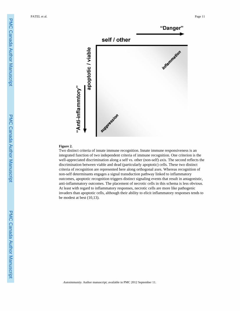
Figure 2.Two distinct criteria of innate immune recognition. Innate immune responsiveness is anintegrated function of two independent criteria of immune recognition. One criterion is thewell-appreciated discrimination along a self vs. other (non-self) axis. The second reflects thediscrimination between viable and dead (particularly apoptotic) cells. These two distinctcriteria of recognition are represented here along orthogonal axes. Whereas recognition ofnon-self determinants engages a signal transduction pathway linked to inflammatoryoutcomes, apoptotic recognition triggers distinct signaling events that result in antagonistic,anti-inflammatory outcomes. The placement of necrotic cells in this schema is less obvious.At least with regard to inflammatory responses, necrotic cells are more like pathogenicinvaders than apoptotic cells, although their ability to elicit inflammatory responses tends tobe modest at best (10,13).
PATEL et al. Page 11
Autoimmunity. Author manuscript; available in PMC 2012 September 11.
PMC
Canada Author M
anuscriptPM
C C
anada Author Manuscript
PMC
Canada Author M
anuscript
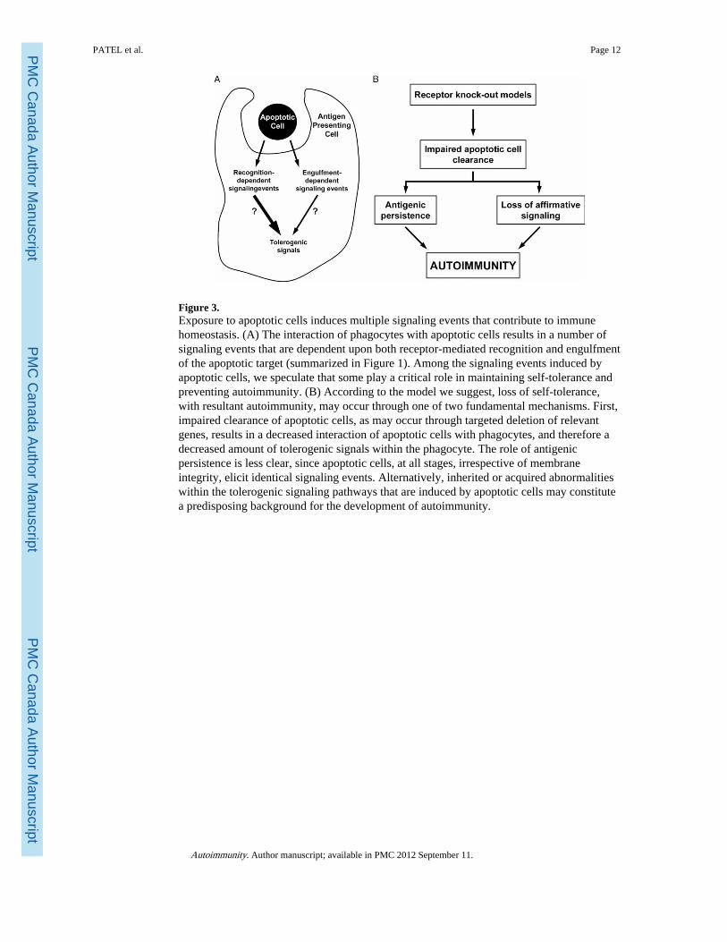
Figure 3.Exposure to apoptotic cells induces multiple signaling events that contribute to immunehomeostasis. (A) The interaction of phagocytes with apoptotic cells results in a number ofsignaling events that are dependent upon both receptor-mediated recognition and engulfmentof the apoptotic target (summarized in Figure 1). Among the signaling events induced byapoptotic cells, we speculate that some play a critical role in maintaining self-tolerance andpreventing autoimmunity. (B) According to the model we suggest, loss of self-tolerance,with resultant autoimmunity, may occur through one of two fundamental mechanisms. First,impaired clearance of apoptotic cells, as may occur through targeted deletion of relevantgenes, results in a decreased interaction of apoptotic cells with phagocytes, and therefore adecreased amount of tolerogenic signals within the phagocyte. The role of antigenicpersistence is less clear, since apoptotic cells, at all stages, irrespective of membraneintegrity, elicit identical signaling events. Alternatively, inherited or acquired abnormalitieswithin the tolerogenic signaling pathways that are induced by apoptotic cells may constitutea predisposing background for the development of autoimmunity.
PATEL et al. Page 12
Autoimmunity. Author manuscript; available in PMC 2012 September 11.
PMC
Canada Author M
anuscriptPM
C C
anada Author Manuscript
PMC
Canada Author M
anuscript