Cotinine-induced convergence of the cholinergic and PI3 kinase-dependent anti-inflammatory pathways...
8
Cotinine-induced convergence of the cholinergic and PI3 kinase-dependent anti-inflammatory pathways in innate immune cells Kunal Rehani, David A. Scott ⁎ , Diane Renaud, Hashir Hamza, Lisa R. Williams, Huizhi Wang, Michael Martin Oral Health and Systemic Disease Research Group, University of Louisville School of Dentistry, 501 S Preston Street, Louisville, KY, 40292, USA Received 25 June 2007; received in revised form 3 December 2007; accepted 3 December 2007 Available online 15 December 2007 Abstract Nicotine [(S)-3-(1-methyl-2-pyrrolidinyl)pyridine] is a major component of tobacco and a highly efficient acetylcholine receptor (nAChR) agonist that triggers the cholinergic anti-inflammatory pathway. We demonstrate that pre-treatment of monocytes with the stable nicotine catabolite, cotinine [(S)-1-methyl-5-(3-pyridinyl)-2-pyrrolidinone], dramatically alters the nature of the inflammatory response to Gram negative bacteria by abrogating the production of cytokines that are under the transcriptional control of the NF-κB system (TNF-α, IL-1β, IL-6, IL-12/IL-23 p40) and shifting the response towards an IL-10-dominated anti-inflammatory profile. This anti-inflammatory phenomenon is initiated specifically by engagement of the monocytic α7 nAChR; and is PI3K/GSK-3β-dependent; but NF-κB- independent. These mechanistic insights suggest an ability to exploit convergent, endogenous anti-inflammatory pathway(s) to either up- regulate or down-regulate the production of specific cytokine groups (pro- or anti-inflammatory cytokines) depending on the clinical necessity. © 2007 Elsevier B.V. All rights reserved. Keywords: Cholinergic anti-inflammatory pathway; Cotinine; Cytokine; Inflammation; Monocyte; PI3K-dependent anti-inflammatory pathway; Tobacco smoking 1. Introduction Components of Gram negative bacteria, and particularly lipopolysaccharides (LPS), are potent inducers of the inflam- matory response. LPS recruits, activates, and promotes degra- nulation events in the most numerous inflammatory leukocyte, the neutrophil. LPS and neutrophil degranulation products each recruit monocytes and macrophages to the locus of infection. While neutrophils are, in relative terms, short-lived and trans- criptionally quiescent, monocytes/macrophages are longer-lived cells that when stimulated by LPS, and other inflammatory mediators, produce large amounts of pro-inflammatory cyto- kines, including TNF, IL-1β, IL-6, IL-12/IL-23 p40, IL-18, and HMGB-1 de novo. These macrophage-derived mediators amplify and direct the inflammatory response and link the innate and adaptive immune responses. The ability to regulate against prolonged or excessive in- flammation is critical in preventing the onset of septic shock and the host-mediated damage associated with multiple chronic inflammatory diseases. However, the mechanisms that dictate the establishment of pro- versus anti-inflammatory cytokine- dominated environments are poorly understood. In recent years it has become clear that the nicotinic acetylcholine receptor α7 subunit is a critical regulator of inflammation [1,2]. Acetyl- choline, produced by the vagus nerve network, is an endoge- nous α7 nAChR agonist. Acetylcholine and the exogenous α7 nAChR agonist, nicotine, both suppress the release of TNF-α from activated cells of monocytic lineage and are protec- tive against LPS-mediated toxicity [2–5]. Thus, nAChRs and downstream components of the cholinergic anti-inflamma- tory pathway present novel therapeutic targets for controlling inflammatory diseases. However, we are only beginning to understand the mechanisms of action of acetylcholine and nicotine in innate immune sup- pression. Furthermore, nicotine is rapidly converted into multiple Available online at www.sciencedirect.com Biochimica et Biophysica Acta 1783 (2008) 375 – 382 www.elsevier.com/locate/bbamcr ⁎ Corresponding author. Tel.: +1 502 852 8905; fax: +1 502 852 4052. E-mail address: [email protected] (D.A. Scott). 0167-4889/$ - see front matter © 2007 Elsevier B.V. All rights reserved. doi:10.1016/j.bbamcr.2007.12.003
-
Upload
independent -
Category
Documents
-
view
0 -
download
0
Transcript of Cotinine-induced convergence of the cholinergic and PI3 kinase-dependent anti-inflammatory pathways...

Available online at www.sciencedirect.com
a 1783 (2008) 375–382www.elsevier.com/locate/bbamcr
Biochimica et Biophysica Act
Cotinine-induced convergence of the cholinergic and PI3 kinase-dependentanti-inflammatory pathways in innate immune cells
Kunal Rehani, David A. Scott ⁎, Diane Renaud, Hashir Hamza, Lisa R. Williams,Huizhi Wang, Michael Martin
Oral Health and Systemic Disease Research Group, University of Louisville School of Dentistry, 501 S Preston Street, Louisville, KY, 40292, USA
Received 25 June 2007; received in revised form 3 December 2007; accepted 3 December 2007Available online 15 December 2007
Abstract
Nicotine [(S)-3-(1-methyl-2-pyrrolidinyl)pyridine] is a major component of tobacco and a highly efficient acetylcholine receptor(nAChR) agonist that triggers the cholinergic anti-inflammatory pathway. We demonstrate that pre-treatment of monocytes with the stablenicotine catabolite, cotinine [(S)-1-methyl-5-(3-pyridinyl)-2-pyrrolidinone], dramatically alters the nature of the inflammatory response toGram negative bacteria by abrogating the production of cytokines that are under the transcriptional control of the NF-κB system (TNF-α,IL-1β, IL-6, IL-12/IL-23 p40) and shifting the response towards an IL-10-dominated anti-inflammatory profile. This anti-inflammatoryphenomenon is initiated specifically by engagement of the monocytic α7 nAChR; and is PI3K/GSK-3β-dependent; but NF-κB-independent. These mechanistic insights suggest an ability to exploit convergent, endogenous anti-inflammatory pathway(s) to either up-regulate or down-regulate the production of specific cytokine groups (pro- or anti-inflammatory cytokines) depending on the clinicalnecessity.© 2007 Elsevier B.V. All rights reserved.
Keywords: Cholinergic anti-inflammatory pathway; Cotinine; Cytokine; Inflammation; Monocyte; PI3K-dependent anti-inflammatory pathway; Tobacco smoking
1. Introduction
Components of Gram negative bacteria, and particularlylipopolysaccharides (LPS), are potent inducers of the inflam-matory response. LPS recruits, activates, and promotes degra-nulation events in the most numerous inflammatory leukocyte,the neutrophil. LPS and neutrophil degranulation products eachrecruit monocytes and macrophages to the locus of infection.While neutrophils are, in relative terms, short-lived and trans-criptionally quiescent, monocytes/macrophages are longer-livedcells that when stimulated by LPS, and other inflammatorymediators, produce large amounts of pro-inflammatory cyto-kines, including TNF, IL-1β, IL-6, IL-12/IL-23 p40, IL-18,and HMGB-1 de novo. These macrophage-derived mediatorsamplify and direct the inflammatory response and link the innateand adaptive immune responses.
⁎ Corresponding author. Tel.: +1 502 852 8905; fax: +1 502 852 4052.E-mail address: [email protected] (D.A. Scott).
0167-4889/$ - see front matter © 2007 Elsevier B.V. All rights reserved.doi:10.1016/j.bbamcr.2007.12.003
The ability to regulate against prolonged or excessive in-flammation is critical in preventing the onset of septic shock andthe host-mediated damage associated with multiple chronicinflammatory diseases. However, the mechanisms that dictatethe establishment of pro- versus anti-inflammatory cytokine-dominated environments are poorly understood. In recent yearsit has become clear that the nicotinic acetylcholine receptor α7subunit is a critical regulator of inflammation [1,2]. Acetyl-choline, produced by the vagus nerve network, is an endoge-nous α7 nAChR agonist. Acetylcholine and the exogenous α7nAChR agonist, nicotine, both suppress the release of TNF-αfrom activated cells of monocytic lineage and are protec-tive against LPS-mediated toxicity [2–5]. Thus, nAChRsand downstream components of the cholinergic anti-inflamma-tory pathway present novel therapeutic targets for controllinginflammatory diseases.
However, we are only beginning to understand the mechanismsof action of acetylcholine and nicotine in innate immune sup-pression. Furthermore, nicotine is rapidly converted into multiple
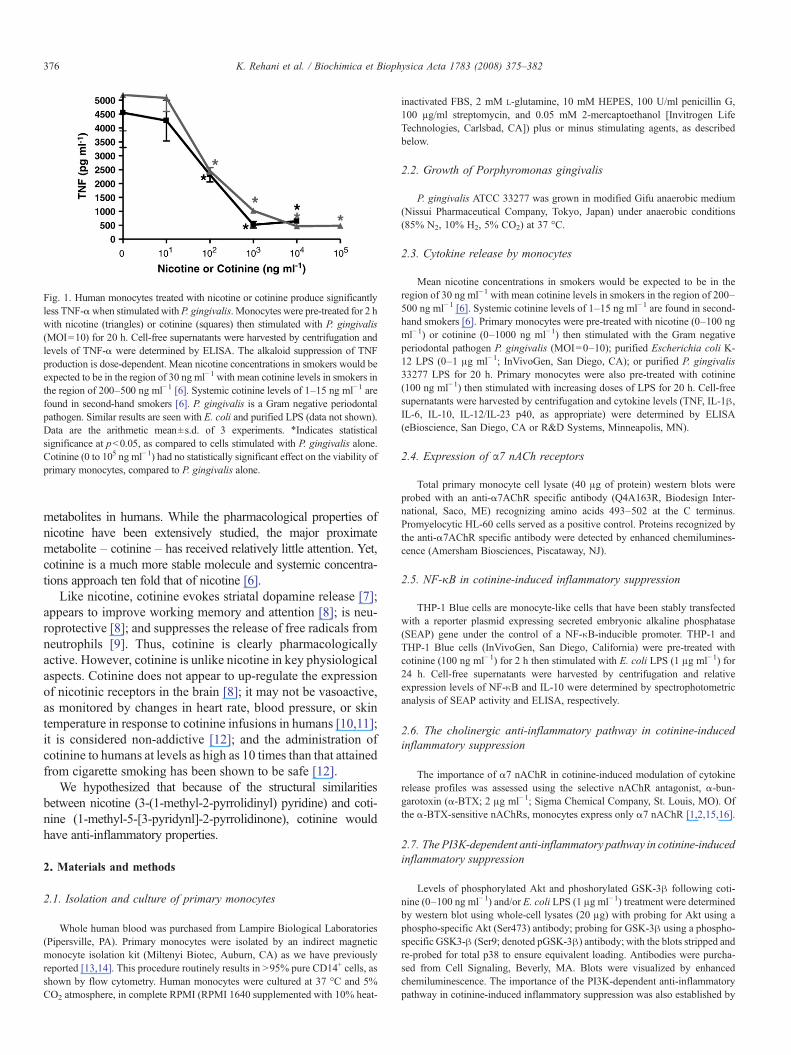
Fig. 1. Human monocytes treated with nicotine or cotinine produce significantlyless TNF-αwhen stimulatedwithP. gingivalis. Monocytes were pre-treated for 2 hwith nicotine (triangles) or cotinine (squares) then stimulated with P. gingivalis(MOI=10) for 20 h. Cell-free supernatants were harvested by centrifugation andlevels of TNF-α were determined by ELISA. The alkaloid suppression of TNFproduction is dose-dependent. Mean nicotine concentrations in smokers would beexpected to be in the region of 30 ng ml−1 with mean cotinine levels in smokers inthe region of 200–500 ng ml−1 [6]. Systemic cotinine levels of 1–15 ng ml−1 arefound in second-hand smokers [6]. P. gingivalis is a Gram negative periodontalpathogen. Similar results are seen with E. coli and purified LPS (data not shown).Data are the arithmetic mean±s.d. of 3 experiments. ⁎Indicates statisticalsignificance at pb0.05, as compared to cells stimulated with P. gingivalis alone.Cotinine (0 to 105 ng ml−1) had no statistically significant effect on the viability ofprimary monocytes, compared to P. gingivalis alone.
376 K. Rehani et al. / Biochimica et Biophysica Acta 1783 (2008) 375–382
metabolites in humans. While the pharmacological properties ofnicotine have been extensively studied, the major proximatemetabolite – cotinine – has received relatively little attention. Yet,cotinine is a much more stable molecule and systemic concentra-tions approach ten fold that of nicotine [6].
Like nicotine, cotinine evokes striatal dopamine release [7];appears to improve working memory and attention [8]; is neu-roprotective [8]; and suppresses the release of free radicals fromneutrophils [9]. Thus, cotinine is clearly pharmacologicallyactive. However, cotinine is unlike nicotine in key physiologicalaspects. Cotinine does not appear to up-regulate the expressionof nicotinic receptors in the brain [8]; it may not be vasoactive,as monitored by changes in heart rate, blood pressure, or skintemperature in response to cotinine infusions in humans [10,11];it is considered non-addictive [12]; and the administration ofcotinine to humans at levels as high as 10 times than that attainedfrom cigarette smoking has been shown to be safe [12].
We hypothesized that because of the structural similaritiesbetween nicotine (3-(1-methyl-2-pyrrolidinyl) pyridine) and coti-nine (1-methyl-5-[3-pyridynl]-2-pyrrolidinone), cotinine wouldhave anti-inflammatory properties.
2. Materials and methods
2.1. Isolation and culture of primary monocytes
Whole human blood was purchased from Lampire Biological Laboratories(Pipersville, PA). Primary monocytes were isolated by an indirect magneticmonocyte isolation kit (Miltenyi Biotec, Auburn, CA) as we have previouslyreported [13,14]. This procedure routinely results in N95% pure CD14+ cells, asshown by flow cytometry. Human monocytes were cultured at 37 °C and 5%CO2 atmosphere, in complete RPMI (RPMI 1640 supplemented with 10% heat-
inactivated FBS, 2 mM L-glutamine, 10 mM HEPES, 100 U/ml penicillin G,100 µg/ml streptomycin, and 0.05 mM 2-mercaptoethanol [Invitrogen LifeTechnologies, Carlsbad, CA]) plus or minus stimulating agents, as describedbelow.
2.2. Growth of Porphyromonas gingivalis
P. gingivalis ATCC 33277 was grown in modified Gifu anaerobic medium(Nissui Pharmaceutical Company, Tokyo, Japan) under anaerobic conditions(85% N2, 10% H2, 5% CO2) at 37 °C.
2.3. Cytokine release by monocytes
Mean nicotine concentrations in smokers would be expected to be in theregion of 30 ng ml−1 with mean cotinine levels in smokers in the region of 200–500 ng ml−1 [6]. Systemic cotinine levels of 1–15 ng ml−1 are found in second-hand smokers [6]. Primary monocytes were pre-treated with nicotine (0–100 ngml−1) or cotinine (0–1000 ng ml−1) then stimulated with the Gram negativeperiodontal pathogen P. gingivalis (MOI=0–10); purified Escherichia coli K-12 LPS (0–1 μg ml−1; InVivoGen, San Diego, CA); or purified P. gingivalis33277 LPS for 20 h. Primary monocytes were also pre-treated with cotinine(100 ng ml−1) then stimulated with increasing doses of LPS for 20 h. Cell-freesupernatants were harvested by centrifugation and cytokine levels (TNF, IL-1β,IL-6, IL-10, IL-12/IL-23 p40, as appropriate) were determined by ELISA(eBioscience, San Diego, CA or R&D Systems, Minneapolis, MN).
2.4. Expression of α7 nACh receptors
Total primary monocyte cell lysate (40 μg of protein) western blots wereprobed with an anti-α7AChR specific antibody (Q4A163R, Biodesign Inter-national, Saco, ME) recognizing amino acids 493–502 at the C terminus.Promyelocytic HL-60 cells served as a positive control. Proteins recognized bythe anti-α7AChR specific antibody were detected by enhanced chemilumines-cence (Amersham Biosciences, Piscataway, NJ).
2.5. NF-κB in cotinine-induced inflammatory suppression
THP-1 Blue cells are monocyte-like cells that have been stably transfectedwith a reporter plasmid expressing secreted embryonic alkaline phosphatase(SEAP) gene under the control of a NF-κB-inducible promoter. THP-1 andTHP-1 Blue cells (InVivoGen, San Diego, California) were pre-treated withcotinine (100 ng ml−1) for 2 h then stimulated with E. coli LPS (1 μg ml−1) for24 h. Cell-free supernatants were harvested by centrifugation and relativeexpression levels of NF-κB and IL-10 were determined by spectrophotometricanalysis of SEAP activity and ELISA, respectively.
2.6. The cholinergic anti-inflammatory pathway in cotinine-inducedinflammatory suppression
The importance of α7 nAChR in cotinine-induced modulation of cytokinerelease profiles was assessed using the selective nAChR antagonist, α-bun-garotoxin (α-BTX; 2 μg ml−1; Sigma Chemical Company, St. Louis, MO). Ofthe α-BTX-sensitive nAChRs, monocytes express only α7 nAChR [1,2,15,16].
2.7. The PI3K-dependent anti-inflammatory pathway in cotinine-inducedinflammatory suppression
Levels of phosphorylated Akt and phoshorylated GSK-3β following coti-nine (0–100 ng ml−1) and/or E. coli LPS (1 μg ml−1) treatment were determinedby western blot using whole-cell lysates (20 μg) with probing for Akt using aphospho-specific Akt (Ser473) antibody; probing for GSK-3β using a phospho-specific GSK3-β (Ser9; denoted pGSK-3β) antibody; with the blots stripped andre-probed for total p38 to ensure equivalent loading. Antibodies were purcha-sed from Cell Signaling, Beverly, MA. Blots were visualized by enhancedchemiluminescence. The importance of the PI3K-dependent anti-inflammatorypathway in cotinine-induced inflammatory suppression was also established by
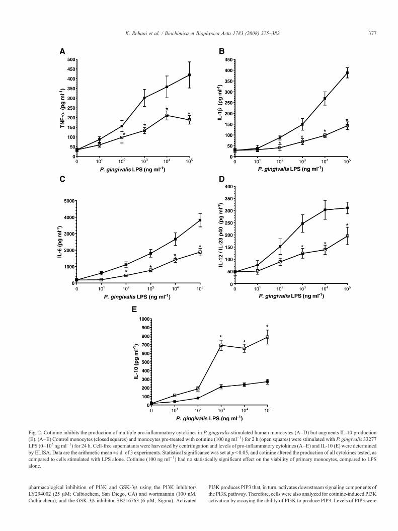
Fig. 2. Cotinine inhibits the production of multiple pro-inflammatory cytokines in P. gingivalis-stimulated human monocytes (A–D) but augments IL-10 production(E). (A–E) Control monocytes (closed squares) and monocytes pre-treated with cotinine (100 ng ml−1) for 2 h (open squares) were stimulated with P. gingivalis 33277LPS (0–105 ng ml−1) for 24 h. Cell-free supernatants were harvested by centrifugation and levels of pro-inflammatory cytokines (A–E) and IL-10 (E) were determinedby ELISA. Data are the arithmetic mean±s.d. of 3 experiments. Statistical significance was set at pb0.05, and cotinine altered the production of all cytokines tested, ascompared to cells stimulated with LPS alone. Cotinine (100 ng ml−1) had no statistically significant effect on the viability of primary monocytes, compared to LPSalone.
377K. Rehani et al. / Biochimica et Biophysica Acta 1783 (2008) 375–382
pharmacological inhibition of PI3K and GSK-3β using the PI3K inhibitorsLY294002 (25 μM; Calbiochem, San Diego, CA) and wortmannin (100 nM,Calbiochem); and the GSK-3β inhibitor SB216763 (6 μM; Sigma). Activated
PI3K produces PIP3 that, in turn, activates downstream signaling components ofthe PI3K pathway. Therefore, cells were also analyzed for cotinine-induced PI3Kactivation by assaying the ability of PI3K to produce PIP3. Levels of PIP3 were

Fig. 3. α-Bungarotoxin treatment of α7AChR-expressing human monocytesabrogates the anti-inflammatory effects of cotinine. (A) Total cell lysate (40μg ofprotein) blots of primary monocytes (Lanes 2–4) and a positive control(promyelocytic HL-60 cells; Lane 1) were probed with an anti-α7AChR specificantibody (Q4A163R, Biodesign Intl.) recognizing amino acids 493–502 at theC terminus. Monocytes expressed α7 nACh receptors with relative molecularmasses of 55 kDa. (B) Control monocytes (open squares) and monocytes pre-treated for 30 min with the α7 nACHR antagonist α-BTX (2μg ml−1, closedsquares) were incubated for 2 h with various concentrations of cotinine. Cellswere then stimulated with P. gingivalis (MOI=10) for 20 h. Cell-freesupernatants were harvested by centrifugation and levels of TNF weredetermined by ELISA. Data are the arithmetic mean±s.d. of 3 experiments.⁎Indicates statistical significance at pb0.05, as compared to control cells.
Fig. 5. Cotinine (A) enhances Akt signaling and (B) augments levels of phos-phorylated (Ser9) GSK3-β in P. gingivalis-stimulated monocytes. (A) Mono-cytes were pre-treated for 2 h with cotinine then stimulated for 60 min with LPS(1 mg ml−1). Western blot was performed using whole-cell lysates (20 μg)with probing for Akt using a phospho-specific Akt (Ser473) antibody. Blotwas stripped and re-probed for total p38 to ensure equivalent loading. Lane 1:Non-stimulated; Lane 2: LPS only; Lane 3: LPS+cotinine (100 ngml−1), Lane 4:LPS+cotinine+wortmannin (a PI3K inhibitor, 100 nM). Data are representativeof three experiments. Similar results were seen with 60 min stimulations withP. gingivalis (MOI=10; data not shown). (B) Monocytes were pre-treated for 2 hwith cotinine then stimulated for 60 min with LPS (1 mgml−1). Western blot wasperformed using whole-cell lysates (20 μg) with probing for GSK-3β using aphospho-specific GSK3-β (Ser9; denoted pGSK3) antibody. Blot was strippedand re-probed for total p38 to ensure equivalent loading. Lane 1: Non-stimulated;Lane 2: LPS only; Lane 3: LPS+cotinine (10 ng ml−1), Lane 4: LPS+cotinine(100 ng ml−1). Data are representative of three experiments. Similar results wereseen with 60 min stimulations with P. gingivalis (MOI=10; data not shown).
378 K. Rehani et al. / Biochimica et Biophysica Acta 1783 (2008) 375–382
determined by ELISA using a competitive inhibition assay (Echelon Bioscience,Salt Lake City, UT). The specific PI3K inhibitor LY294002 was used at 25 μMand served as a control to block PI3K activity.
Fig. 4. The anti-inflammatory potential of cotinine is a GSK-3β-dependent butNF-κB-independent phenomenon. THP-1 Blue cells are stably transfected with areporter plasmid expressing secreted embryonic alkaline phosphatase (SEAP)gene under the control of a NF-κB-inducible promoter. THP-1 and THP-1 Bluecells were pre-treated with cotinine (100 ng ml−1) for 2 h then stimulated withLPS (1 μgml−1) for 24 h. Cell-free supernatants were harvested by centrifugationand relative expression levels of SEAP (reflecting NF-κB; THP-1 Blue; circles)and IL-10 (THP-1; bars) were determined by spectrophotometric analysis ofSEAP activity and ELISA, respectively. Here we also show that the GSK-3βinhibitor – SB216763 – enhances IL-10 production in response to LPS, but it isless potent than even low doses of cotinine (10 ng ml−1; pb0.05). Induction ofNF-κB on stimulation with LPS is not influenced by cotinine treatment. All dataare the arithmetic mean±s.d. of 3 experiments. ⁎pb0.05; compared tounstimulated cells. Cotinine (100 ng ml−1) had no statistically significant effecton the viability of THP-1 or THP-1 Blue cells compared to LPS alone.
2.8. Convergence of the cholinergic and PI3 kinase-dependentanti-inflammatory pathways during cotinine-induced inflammatorysuppression
Interactions between the cholinergic and PI3 kinase-dependent anti-in-flammatory pathways were examined by assessing the efficacy of α-BTX (2 μgml−1) to block activation of the PI3K pathway. PIP3 levels were monitored byELISA following 60 min with LPS (1 μg ml−1) in the presence and absence ofcotinine (0–100 ng ml−1).
2.9. Statistical approaches
Statistical significance between groups was evaluated by ANOVA and theTukey multiple-comparison test using the InStat program (GraphPad Software,San Diego, CA). Differences between groups were considered significant at thelevel of pb0.05.
3. Results
3.1. Cotinine inhibits TNF release
Cotinine is equally as effective as nicotine at inhibiting TNFrelease (Fig. 1). Indeed cotinine blocks more than 80% of bacteria-induced TNF release from monocytes at a dose of 100 ng ml−1
(Fig. 1), a level commonly observed in cigarette smokers [6].
3.2. Cotinine promotes an anti-inflammatory phenotype
Cotinine also dramatically suppresses the release of multiplepro-inflammatory cytokines (TNF-α, IL-1β, IL-6, and the common

Fig. 6. Innate immune suppression by cotinine requires the α7 nAChR-dependent enhancement of PI3K activation. (A) Inhibition of PI3K usingLY294002 abrogates the anti-inflammatory effects of cotinine. Cells were pre-treated with (closed triangles) and without (closed squares) 25 μM of the PI3Kinhibitor LY294002 for 30 min followed by 2 h with various concentrations ofcotinine. Following 24 h P. gingivalis-stimulation (MOI=10), cell-free super-natants were harvested by centrifugation and levels of TNF-α were determinedby ELISA. ⁎Indicates statistical significance at pb0.05, as compared to cellsstimulated with P. gingivalis alone (no LY294002). Data are the arithmeticmean±s.d. of 3 experiments. All media used in these experiments contained0.01% DMSO to act as the organic solvent control used to solubilize LY294002.Similar results are also seen with the alternative PI3K inhibitor, wortmannin(100 nM; data not shown). (B) Cotinine (or nicotine) treated monocytes exhibitincreased PIP3 levels when stimulated with LPS in an α7 nAChR-dependentmanner. Cells were stimulated for 60 min with 1 μg ml−1 of LPS. Cells wereanalyzed for PI3K activation by the ability of PI3K to produce PIP3. ActivatedPI3K produces PIP3 that, in turn, activates downstream signaling components ofthe PI3K pathway. Levels of PIP3 were determined by ELISA using acompetitive inhibition assay (Echelon Bioscience). The specific PI3K inhibitorLY294002 was used at 25 μM (cells were pre-treated for 1 h) and served as acontrol to block PI3K activity. α7 nAchR-dependence was established throughthe use of the α7 nAChR antagonist, α-bungarotoxin. ⁎Indicates statisticalsignificance at pb0.05, as compared to unstimulated control cells. ¶ Indicatesstatistical significance at pb0.05, as compared to cells stimulated with LPSalone. § Indicates statistical significance at pb0.05 for cells stimulated withLPS, nicotine and LY294002 (or α-BTX) compared to LPS and nicotine only. †Indicates statistical significance at pb0.05 for cells stimulated with LPS,cotinine and LY294002 (or α-BTX) compared to LPS and cotinine only. Dataare the arithmetic mean±s.d. of 3 experiments.
Fig. 7. Enhancement of IL-10 release by cotinine requires the α7 nAChR-dependent enhancement of PI3K activation. Inhibition of PI3K using LY294002abrogates the anti-inflammatory effects of cotinine and nicotine, as assessed byIL-10 release. Cells were pre-treated with and without the PI3K inhibitorLY294002 (25 μM); and with or without α-bungarotoxin (2 μg ml−1) for30 min, followed by 2 h with nicotine (100 ng ml−1) or cotinine (100 ng ml−1).Following 24 h LPS stimulation (2 μg ml−1), cell-free supernatants wereharvested by centrifugation and levels of IL-10 were determined by ELISA.⁎Indicates statistical significance at pb0.05, as compared to unstimulatedcontrol cells. ¶ Indicates statistical significance at pb0.05, as compared to cellsstimulated with LPS alone. § Indicates statistical significance at pb0.05 for cellsstimulated with LPS, nicotine and LY294002 (or α-BTX) compared to LPS andnicotine only. † Indicates statistical significance at pb0.05 for cells stimulatedwith LPS, cotinine and LY294002 (or α-BTX) compared to LPS and cotinineonly. Data are the arithmetic mean±s.d. of 3 experiments.
379K. Rehani et al. / Biochimica et Biophysica Acta 1783 (2008) 375–382
IL-12/IL-23 p40 subunit) from monocytes (Fig. 2A–D) whilesimultaneously enhancing production of the key anti-inflammatorycytokine, IL-10 (Fig. 2E).
3.3. Cotinine-induced inflammatory suppression likely occursvia α7 nAChR
Of the α-bungarotoxin sensitive human nAChRs (α1, α7,and α9), monocytes and macrophages express only functionalα7 receptors [1,2,15,16]. The α7 receptor expressed by primarymacrophages has a relative molecular mass of 55 kDa bywestern blot (Fig. 3A), identical to that of the neuronal recep-tor [17,18]. Cotinine-induced suppression of TNF release wasabrogated by pre-treatment with α-bungarotoxin (Fig. 3B),suggesting that the anti-inflammatory activity of cotinine maybe mediated via the nicotinic acetylcholine receptor α7 subunit.The importance of α7 nAChR in cotinine-induced immunomo-dulation is also suggested by the ability of α-bungarotoxinto block cotinine enhancement of IL-10 release (Fig. 7) and toabrogate cotinine-induced alterations to PI3K activity (mea-sured as PIP3 release; Fig. 6).
3.4. The anti-inflammatory potential of cotinine is a PI3K/GSK-3β-dependent but NF-κB-independent phenomenon
Physiologically relevant doses of cotinine augment IL-10production in LPS-stimulated monocytes, yet LPS-induced NF-κB is not affected by cotinine (Fig. 4). Therefore, cotinineappears to act as an anti-inflammatory agent in an NF-κB-independent manner. We have previously shown that pharma-cological inhibition of GSK-3β enhances IL-10 production inresponse to LPS [13], and confirm this finding herein (Fig. 4).

380 K. Rehani et al. / Biochimica et Biophysica Acta 1783 (2008) 375–382
However, SB216763 inhibition of GSK-3β is a significantlyless potent inducer of IL-10 than cotinine (Fig. 4). To explorethis further, we examined the effects of cotinine on the ac-tivation state of Akt (Ser473) and GSK3-β (Ser9). As shown inFig. 5, cotinine enhances levels of phosphorylated (active) Aktin a PI3K-dependendent manner and augments levels ofphosphorylated (inactive) GSK3-β in P. gingivalis-stimulatedmonocytes.
3.5. The cholinergic and PI3K anti-inflammatory pathways areconvergent on cotinine stimulation
Cotinine potently augments the activation of PI3K and theproduction of the anti-inflammatory cytokine IL-10. In contrast,blockade of PI3K abrogates the ability of cotinine to suppressthe inflammatory response (TNF-α) in P. gingivalis-stimulatedcells (Figs. 6A and 7). Moreover, inhibition of the α7 nAChR-signaling pathway abrogated the ability of cotinine to enhancePI3K activity (Fig. 6B).
4. Discussion
Borovikova et al have demonstrated that electrical stimula-tion of the vagus nerve protected against death by LPS toxicity inmice, and showed that acetylcholine significantly attenuated therelease of TNF, IL-1β, and IL-6 from macrophages [3]. It isimportant to note, however, that such vagal protection isabrogated by splenectomy [19]. Specifically it is the α7 nAChreceptor subtype that appears key to acetylcholine-inducedinflammatory suppression, as we and others have recentlyreviewed [20–23]. α7 nAChR-deficient mice are not onlyhypersensitive to LPS, producing high amounts of TNF-α, theyalso exhibit an exaggerated production of the pro-inflammatorycytokines IL-1β and IL-6 [4,24]. Furthermore, nicotine-dependent suppression of TNF release from primary macro-phages is abrogated by α7 AChR-specific antisense oligonu-cleotides [4], while selective α7 AChR agonists protect againstsevere sepsis [25]. It should be noted that a limitation of thepresent study is that we have not confirmed our data, obtainedfrom primary human cells, in vivo.
Functional AChRs are pentameric and are composed ofmultiple combinations of a possible 16 monomer subtypes (α1–7; α9–10; β1–4; δ; ɛ; and γ) that exhibit divergent phar-macological behaviors [4,16,26]. nAChR subtype expressionby monocytes and macrophages is highly restricted. The α7receptor expressed by primary macrophages has a relativemolecular mass of 55 kDa by western blot, identical to that ofthe neuronal receptor [17,18]. Of the known AChRs, α7nAChR exhibits a number of unusual features [27]. First of all,it can assemble and function as a homopentamer [27,28]; the ionchannel exhibits high permeability for calcium ions in pre-ference to Na+ [29]; and it is widely expressed in the central andperipheral nervous system [26] as well as on leukocytes [4].αBTX is not specific for α7 nAChR. However, of the α-bungarotoxin sensitive human nAChRs (α1, α7, and α9),monocytes and macrophages express only functional α7 re-ceptors [1,2,15,16]. We have been able to use α-bungarotoxin to
strongly suggest that cotinine-induced inflammatory suppres-sion occurs through the nicotinic acetylcholine receptor α7subunit. Definitive confirmation of the role of α7 nAChR willrequire further experimentation, such as utilizing antisense orsilencing RNA techniques.
We extend existing knowledge by demonstrating that co-tinine has similar anti-inflammatory properties to its addictiveand toxic catabolic precursor, nicotine, showing that cotinineblocks the production of multiple pro-inflammatory cytokinesthat are known to be under the transcriptional control of NF-κB,including the innate/adaptive immunity bridging cytokine, IL-12/IL-23 p40 [30,31]; while simultaneously augmenting therelease of the anti-inflammatory molecule, IL-10; and establish-ing that these actions of cotinine are likely to be mediatedthrough the α7 nAChR. The suppression of the p40 subunitshared by IL-12 and IL23 is particularly interesting as it iscritical for the development of IFN-γ-producing Th1 cells andinfluences cell-mediated immunity and IgG2a antibody produc-tion from B cells [32–35]. Recently, Chen et al have reportedthat alveolar macrophages from smokers exhibit a reduced pro-inflammatory response (TNF-α, IL-1β, IL-6, IL-8) to TLR-2and TLR-4 agonists (Pam3Cys and LPS, respectively) com-pared to cells from non-smokers [36].
We next set out to address the mechanisms by which coti-nine exerts anti-inflammatory effects on innate cells. The NF-κBpathway is critical in the activation of mononuclear phagocytesand in the production of multiple pro-inflammatory cytokines bythese cells [30,31]. Limited evidence suggests that nicotinemaintains cytoplasmic concentrations of IκB and thus preventsLPS-induced NF-κB activation in monocytes and macrophages,in a dose-dependent manner [3,4,37]. One recent report suggeststhat smoking inhibits the TLR-2/-4 inducible phosphorylation ofIRAK-1 and p38 and also inhibits IκB-α degradation [36].However, our data shows that cotinine appears to act as an anti-inflammatory agent in an NF-κB-independent manner, as phy-siologically relevant doses of cotinine augment IL-10 produc-tion in LPS-stimulated monocytes, yet LPS-induced NF-κB isnot affected by cotinine. Indeed, we demonstrate that cotinineregulates the phosphorylation of GSK3β (Serine 9) via thePI3K-Akt pathway. These data are consistent with previousstudies by our laboratory that have shown that GSK3β does notaffect the absolute or phosphorylated (activated) levels of NF-κB (p65) but affects the levels of CREB (Ser133) that displacesNF-κB (p65) from the transcriptional complex (CBP) [13]. Wehave also previously demonstrated that the levels of CREB(Ser133) are important for IL-10 production in TLR-stimulatedcells but that the loss of NF-κB (p65) does not affect IL-10 [13].
Once phosphatidylinositol 3 kinase (PI3K) becomes acti-vated, it catalyzes the production of phosphatidylinositol-3,4,5-trisphosphate [PIP3]. The generation of PIP3 subsequentlyallows for the recruitment and co-localization of phosphoinosi-tide-dependent kinase 1 (PDK1) and the serine/threonine-dependent kinase Akt via their pleckstrin homology domains[38,39]. This co-localization to the plasma membrane allowsfor the activation of Akt via the phosphorylation of threonine308 by PDK1 and phosphorylation at serine 473 by a stillunidentified kinase called PDK2 [38–40]. Upon dual

381K. Rehani et al. / Biochimica et Biophysica Acta 1783 (2008) 375–382
phosphorylation, Akt becomes activated and phosphorylates amultitude of downstream targets including the serine phosphor-ylation that results in the inhibition of glycogen synthase kinase3 (Ser21, GSK-3α; and Ser9 GSK-3β) [41]. Previous studiesfrom our laboratories have identified that the phosphatidylino-sitol-3 kinase (PI3K) pathway plays a fundamental role inregulating the host inflammatory response to P. gingivalis bynegatively regulating IL-12 p40/p70 production while concur-rently augmenting IL-10 levels in a toll like receptor (TLR)-dependent manner. Sequential downstreammapping of the PI3Kpathway identified that inactivation (by phosphorylation) of theconstitutively active serine/threonine kinase, glycogen synthasekinase 3, via Akt, is central to the ability of this pathway tosuppress the levels of pro-inflammatory cytokines whileaugmenting anti-inflammatory cytokine production (IL-10)[13]. Thus, the PI3K pathway can act as a central regulator inmodulating the nature (pro- versus anti-inflammatory) andmagnitude (absolute levels) of the host inflammatory response.However, at this stage it is important to note that this PI3K anti-inflammatory pathway is normally minimally engaged.
The ability to selectively augment anti-inflammatory cyto-kine production while concurrently suppressing pro-inflamma-tory cytokines provides a strong focal point for the elucidation,characterization, and application of potential therapeutic targetsthat could attenuate or modulate the inflammatory diseaseprocess. Thus, we confirmed our previous data that pharmaco-logical inhibition of GSK-3β enhances IL-10 production inresponse to LPS. However, SB216763 inhibition of GSK-3β is asignificantly less potent inducer of IL-10 than even low doses(10 ng ml− 1) of cotinine, suggesting a convergence of thecholinergic andGSK-3β-mediated anti-inflammatory pathways.Indeed, cotinine enhances levels of phosphorylated (active) Aktin a PI3K-dependent manner and augments levels of phos-phorylated (inactive) GSK3-β in P. gingivalis-stimulatedmonocytes. Inhibition of the α7 nAChR-signaling pathwayabrogated the ability of cotinine to enhance PI3K activity. Thus,cotinine augments the activity of PI3K via the α7 nAChR.
In summary, we have shown that, in primary human mo-nocytes, cotinine is a potent anti-inflammatory agent that mostlikely acts through the α7 nAChR resulting in a convergence ofthe cholinergic anti-inflammatory pathway and an endogenousPI3K-dependent anti-inflammatory pathway in monocytes. ThePI3K-dependent anti-inflammatory pathway in monocytes isnormally minimally engaged, but signaling through this PI3K-Akt-GSK-3β route is amplified by cotinine—probably onengagement of α7 nAChR. As our knowledge of these signalinginteractions increases, we are likely to identify further attractiveanti-inflammatory targets.
Acknowledgments
Kunal Rehani, Hashir Hamza, Diane Renaud, Huizi Wangand Lisa Williams performed the experimental work includedin the manuscript and contributed to the data analyses andinterpretation. David Scott and Michael Martin designed theresearch, analyzed data, wrote the paper, and contributedequally to the manuscript. The research was funded, in part, by
the Centers for Disease Control (H75/CCH424215-01, DAS)and National Institutes of Health (DE017680-01, MM). None ofthe authors have any relevant conflict of interest. However, M.Martin and D.A. Scott hold a provisional US patent on the anti-inflammatory properties of cotinine (Therapeutic CotinineCompositions. U.S. Patent Application 60/890,954; 2007).
References
[1] L. Ulloa, The vagus nerve and the nicotinic anti-inflammatory pathway,Nat. Rev. Drug Discov. 4 (2005) 673–684.
[2] H. Wang, M. Yu, M. Ochani, C.A. Amella, M. Tanovic, S. Susarla, J.H. Li,H. Wang, H. Yang, L. Ulloa, Y. Al-Abed, C.J. Czura, K.J. Tracey, Nicotinicacetylcholine receptor alpha7 subunit is an essential regulator ofinflammation, Nature 421 (2003) 384–388.
[3] L.V. Borovikova, S. Ivanova, M. Zhang, H. Yang, G.I. Botchkina, L.R.Watkins, H. Wang, N. Abumrad, J.W. Eaton, K.J. Tracey, Vagus nervestimulation attenuates the systemic inflammatory response to endotoxin,Nature 405 (2000) 458–462.
[4] H. Wang, H. Liao, M. Ochani, M. Justiniani, X. Lin, L. Yang, Y. Al-Abed,H. Wang, C. Metz, E.J. Miller, K.J. Tracey, L. Ulloa, Cholinergic agonistsinhibit HMGB1 release and improve survival in experimental sepsis, Nat.Med. 10 (2004) 1216–1221.
[5] K. Matsunaga, T.W. Klein, H. Friedman, Y. Yamamoto, Involvement ofnicotinic acetylcholine receptors in suppression of antimicrobial activityand cytokine responses of alveolar macrophages to Legionella pneumo-phila infection by nicotine, J. Immunol. 167 (2001) 6518–6524.
[6] D.A. Scott, R.M. Palmer, J.A. Stapleton, Validation of smoking status inclinical research into inflammatory periodontal disease, J. Clin. Period-ontol. 28 (2001) 715–722.
[7] L.P. Dwoskin, L. Teng, S.T. Buxton, P.A. Crooks, (S)-(-)-Cotinine, themajor brain metabolite of nicotine, stimulates nicotinic receptors to evoke[3H]dopamine release from rat striatal slices in a calcium-dependentmanner, J. Pharmacol. Exp. Ther. 288 (1999) 905–911.
[8] A.V. Terry Jr., C.M. Hernandez, E.J. Hohnadel, K.P. Bouchard, J.J.Buccafusco, Cotinine, a neuroactive metabolite of nicotine: potential fortreating disorders of impaired cognition, CNS Drug Rev. 11 (2005)229–252.
[9] E.D. Srivastava, M.B. Hallett, J. Rhodes, Effect of nicotine and cotinine onthe production of oxygen free radicals by neutrophils in smokers and non-smokers, Hum. Toxicol. 8 (1989) 461–463.
[10] N.L. Benowitz, F. Kuyt, P. Jacob III, R.T. Jones, A.L. Osman, Cotininedisposition and effects, Clin. Pharmacol. Ther. 34 (1983) 604–611.
[11] S. Zevin, P. Jacob, P. Geppetti, N.L. Benowitz, Clinical pharmacology oforal cotinine, Drug Alcohol Depend. 60 (2000) 13–18.
[12] D.K. Hatsukami, M. Grillo, P.R. Pentel, C. Oncken, R. Bliss, Safety ofcotinine in humans: physiologic, subjective, and cognitive effects,Pharmacol. Biochem. Behav. 57 (1997) 643–650.
[13] M. Martin, K. Rehani, R.S. Jope, S.M. Michalek, Toll-like receptor-mediated cytokine production is differentially regulated by glycogensynthase kinase 3, Nat. Immunol. 6 (2005) 777–784.
[14] M. Martin, R.E. Schifferle, N. Cuesta, S.N. Vogel, J. Katz, S.M. Michalek,Role of the phosphatidylinositol 3 kinase-Akt pathway in the regulation ofIL-10 and IL-12 by Porphyromonas gingivalis lipopolysaccharide,J. Immunol. 171 (2003) 717–725.
[15] R.C. Hogg, M. Raggenbass, D. Bertrand, Nicotinic acetylcholinereceptors: from structure to brain function, Rev. Physiol. Biochem.Pharmacol. 147 (2003) 1–46.
[16] C. Gotti, F. Clementi, Neuronal nicotinic receptors: from structure topathology, Prog. Neurobiol. 74 (2004) 363–396.
[17] X. Peng, M. Katz, V. Gerzanich, R. Anand, J. Lindstrom, Human alpha7acetylcholine receptor: cloning of the alpha7 subunit from the SH-SY5Ycell line and determination of pharmacological properties of nativereceptors and functional alpha7 homomers expressed in Xenopus oocytes,Mol. Pharmacol. 45 (1994) 546–554.
[18] R.C. Drisdel, W.N. Green, Neuronal alpha-bungarotoxin receptors arealpha7 subunit homomers, J. Neurosci. 20 (2000) 133–139.

382 K. Rehani et al. / Biochimica et Biophysica Acta 1783 (2008) 375–382
[19] J.M. Huston, M. Ochani, M. Rosas-Ballina, H. Liao, K. Ochani, V.A.Pavlov, M. Gallowitsch-Puerta, M. Ashok, C.J. Czura, B. Foxwell, K.J.Tracey, L. Ulloa, Splenectomy inactivates the cholinergic anti-inflamma-tory pathway during lethal endotoxemia and polymicrobial sepsis, J. Exp.Med. 203 (2006) 1623–1628.
[20] W.J. de Jonge, L. Ulloa, The alpha7 nicotinic acetylcholine receptor as apharmacological target for inflammation, Br. J. Pharmacol. (2007).
[21] D.A. Scott, M. Martin, Exploitation of the nicotinic anti-inflammatorypathway for the treatment of epithelial inflammatory diseases, WorldJ. Gastroenterol. 12 (2006) 7451–7459.
[22] V.A. Pavlov, K.J. Tracey, Controlling inflammation: the cholinergic anti-inflammatory pathway, Biochem. Soc. Trans. 34 (2006) 1037–1040.
[23] M. Gallowitsch-Puerta, V.A. Pavlov, Neuro-immune interactions viathe cholinergic anti–inflammatory pathway, Life Sci. 80 (2007)2325–2329.
[24] C. Libert, Inflammation: a nervous connection, Nature 421 (2003)328–329.
[25] V.A. Pavlov, M. Ochani, L.H. Yang, M. Gallowitsch-Puerta, K. Ochani,X. Lin, J. Levi, W.R. Parrish, M. Rosas-Ballina, C.J. Czura, G.J. Larosa,E.J. Miller, K.J. Tracey, Y. Al-Abed, Selective alpha7-nicotinic acetylcho-line receptor agonist GTS-21 improves survival in murine endotoxemiaand severe sepsis, Crit. Care Med. 35 (2007) 1139–1144.
[26] J. Lindstrom, Nicotinic acetylcholine receptors in health and disease, Mol.Neurobiol. 15 (1997) 193–222.
[27] Y. Villiger, I. Szanto, S. Jaconi, C. Blanchet, B. Buisson, K.H. Krause, D.Bertrand, J.A. Romand, Expression of an alpha7 duplicate nicotinicacetylcholine receptor-related protein in human leukocytes, J. Neuroim-munol. 126 (2002) 86–98.
[28] D. Chen, J.W. Patrick, The alpha-bungarotoxin-binding nicotinic acet-ylcholine receptor from rat brain contains only the alpha7 subunit, J. Biol.Chem. 272 (1997) 24024–24029.
[29] D. Bertrand, J.L. Galzi, A. Devillers-Thiery, S. Bertrand, J.P. Changeux,Mutations at two distinct sites within the channel domain M2 alter calcium
permeability of neuronal alpha 7 nicotinic receptor, Proc. Natl. Acad. Sci.U. S. A. 90 (1993) 6971–6975.
[30] Q. Li, I.M. Verma, NF-kappaB regulation in the immune system, Nat. Rev.Immunol. 2 (2002) 725–734.
[31] Q. Li, S.Withoff, I.M. Verma, Inflammation-associated cancer: NF-kappaBis the lynchpin, Trends Immunol. 26 (2005) 318–325.
[32] C.R. Raetz, C. Whitfield, Lipopolysaccharide endotoxins, Annu. Rev.Biochem. 71 (2002) 635–700.
[33] R. Medzhitov, C. Janeway Jr., Innate immunity, N. Engl. J. Med. 343(2000) 338–344.
[34] B. Beutler, A. Cerami, Tumor necrosis, cachexia, shock, and inflammation:a common mediator, Annu. Rev. Biochem. 57 (1988) 505–518.
[35] C.A. Dinarello, Interleukin-1 and interleukin-1 antagonism, Blood 77(1991) 1627–1652.
[36] C. Chen, J.C. Cowan, J.D. Hasday, S.V. Vogel, A.E. Medvedev, Smokinginhibits expression of proinflammatory cytokines and activation of Irak-1,p38 and NF-kB in alveolar macrophages stimulated with TLR2 and TLR4agonists (Abstract), J. Immunol. 178 (2007) 40–48.
[37] N. Sugano, K. Shimada, K. Ito, S. Murai, Nicotine inhibits the production ofinflammatory mediators in U937 cells through modulation of nuclear factor-kappaB activation, Biochem. Biophys. Res. Commun. 252 (1998) 25–28.
[38] T.F. Franke, D.R. Kaplan, L.C. Cantley, A. Toker, Direct regulation of theAkt proto-oncogene product by phosphatidylinositol-3,4-bisphosphate,Science 275 (1997) 665–668.
[39] D.L.R. Stokoe, L.R. Stephens, T. Copeland, R. Piers, J. Gaffney, C.B. Reese,G.F. Painter, A.B. Holmes, F. McCormick, P.T. Hawkins, Dual role ofphosphatidylinositol-3,4,5-trisphosphate in the activation of protein kinase B,Science 277 (1997) 567–570.
[40] M.A. Lawlor, D.R. Alessi, PKB/Akt: a key mediator of cell proliferation,survival and insulin responses? J. Cell. Sci. 114 (2001) 2903–2910.
[41] D.A. Cross, D.R. Alessi, P. Cohen, M. Andjelkovich, B.A. Hemmings,Inhibition of glycogen synthase kinase-3 by insulin mediated by proteinkinase B, Nature 378 (1995) 785–789.