Cholinergic receptor pathways involved in apoptosis, cell proliferation and neuronal differentiation
20
BioMed Central Open Access Page 1 of 20 (page number not for citation purposes) Cell Communication and Signaling Review Cholinergic receptor pathways involved in apoptosis, cell proliferation and neuronal differentiation Rodrigo R Resende* 1,2 and Avishek Adhikari 3 Address: 1 Department of Physics, Institute of Exact Sciences, Federal University of Minas Gerais, Belo Horizonte, MG, 31270-901, Brazil, 2 Institute of Learning and Research Santa Casa of BH (IEPSC – BH), Belo Horizonte, Brazil and 3 Department of Biological Sciences, Columbia University, New York, NY, 10027, USA Email: Rodrigo R Resende* - [email protected]; Avishek Adhikari - [email protected] * Corresponding author Abstract Acetylcholine (ACh) has been shown to modulate neuronal differentiation during early development. Both muscarinic and nicotinic acetylcholine receptors (AChRs) regulate a wide variety of physiological responses, including apoptosis, cellular proliferation and neuronal differentiation. However, the intracellular mechanisms underlying these effects of AChR signaling are not fully understood. It is known that activation of AChRs increase cellular proliferation and neurogenesis and that regulation of intracellular calcium through AChRs may underlie the many functions of ACh. Intriguingly, activation of diverse signaling molecules such as Ras-mitogen- activated protein kinase, phosphatidylinositol 3-kinase-Akt, protein kinase C and c-Src is modulated by AChRs. Here we discuss the roles of ACh in neuronal differentiation, cell proliferation and apoptosis. We also discuss the pathways involved in these processes, as well as the effects of novel endogenous AChRs agonists and strategies to enhance neuronal-differentiation of stem and neural progenitor cells. Further understanding of the intracellular mechanisms underlying AChR signaling may provide insights for novel therapeutic strategies, as abnormal AChR activity is present in many diseases. Introduction Acetylcholine (ACh) is an ancient signaling molecule, [1] and is present in both prokaryotes and eukaryotes [2-4]. Although ACh has been extensively studied for its role as a neurotransmitter, it also has autocrine functions [5] in diverse cell types. ACh has been shown to promote cytoskeleton organization, cellular proliferation, differen- tiation and apoptosis [2-4,6-8] throughout development [2,3,9]. Intriguingly nAChR signaling pathways have been preserved throughout evolution [10], suggesting that they have critical functions. We shall attempt to discuss the physiology of ACh as well as ACh's relevant downstream pathways in apoptosis, cell proliferation and neuronal differentiation of embryonic stem cells. Interestingly, nicotinic receptors are expressed in undiffer- entiated and differentiating cells, [8,11-13] suggesting that ACh-mediated signaling between neuronal and non- neuronal cells may influence cell fate [8,11,12,14]. Sup- porting this idea, ACh has been shown to modulate neu- ronal cell differentiation during development [15,16]. Moreover, transfecting a non-neuronal cell line such as a neuroblastoma with choline acetyltransferase induces expression of neuronal markers, muscarinic receptors and Published: 27 August 2009 Cell Communication and Signaling 2009, 7:20 doi:10.1186/1478-811X-7-20 Received: 8 June 2009 Accepted: 27 August 2009 This article is available from: http://www.biosignaling.com/content/7/1/20 © 2009 Resende and Adhikari; licensee BioMed Central Ltd. This is an Open Access article distributed under the terms of the Creative Commons Attribution License (http://creativecommons.org/licenses/by/2.0 ), which permits unrestricted use, distribution, and reproduction in any medium, provided the original work is properly cited.
-
Upload
independent -
Category
Documents
-
view
0 -
download
0
Transcript of Cholinergic receptor pathways involved in apoptosis, cell proliferation and neuronal differentiation

BioMed Central
ss
Cell Communication and Signaling
Open AcceReviewCholinergic receptor pathways involved in apoptosis, cell proliferation and neuronal differentiationRodrigo R Resende*1,2 and Avishek Adhikari3
Address: 1Department of Physics, Institute of Exact Sciences, Federal University of Minas Gerais, Belo Horizonte, MG, 31270-901, Brazil, 2Institute of Learning and Research Santa Casa of BH (IEPSC – BH), Belo Horizonte, Brazil and 3Department of Biological Sciences, Columbia University, New York, NY, 10027, USA
Email: Rodrigo R Resende* - [email protected]; Avishek Adhikari - [email protected]
* Corresponding author
AbstractAcetylcholine (ACh) has been shown to modulate neuronal differentiation during earlydevelopment. Both muscarinic and nicotinic acetylcholine receptors (AChRs) regulate a widevariety of physiological responses, including apoptosis, cellular proliferation and neuronaldifferentiation. However, the intracellular mechanisms underlying these effects of AChR signalingare not fully understood. It is known that activation of AChRs increase cellular proliferation andneurogenesis and that regulation of intracellular calcium through AChRs may underlie the manyfunctions of ACh. Intriguingly, activation of diverse signaling molecules such as Ras-mitogen-activated protein kinase, phosphatidylinositol 3-kinase-Akt, protein kinase C and c-Src is modulatedby AChRs. Here we discuss the roles of ACh in neuronal differentiation, cell proliferation andapoptosis. We also discuss the pathways involved in these processes, as well as the effects of novelendogenous AChRs agonists and strategies to enhance neuronal-differentiation of stem and neuralprogenitor cells. Further understanding of the intracellular mechanisms underlying AChR signalingmay provide insights for novel therapeutic strategies, as abnormal AChR activity is present in manydiseases.
IntroductionAcetylcholine (ACh) is an ancient signaling molecule, [1]and is present in both prokaryotes and eukaryotes [2-4].Although ACh has been extensively studied for its role asa neurotransmitter, it also has autocrine functions [5] indiverse cell types. ACh has been shown to promotecytoskeleton organization, cellular proliferation, differen-tiation and apoptosis [2-4,6-8] throughout development[2,3,9]. Intriguingly nAChR signaling pathways have beenpreserved throughout evolution [10], suggesting that theyhave critical functions. We shall attempt to discuss thephysiology of ACh as well as ACh's relevant downstream
pathways in apoptosis, cell proliferation and neuronaldifferentiation of embryonic stem cells.
Interestingly, nicotinic receptors are expressed in undiffer-entiated and differentiating cells, [8,11-13] suggestingthat ACh-mediated signaling between neuronal and non-neuronal cells may influence cell fate [8,11,12,14]. Sup-porting this idea, ACh has been shown to modulate neu-ronal cell differentiation during development [15,16].Moreover, transfecting a non-neuronal cell line such as aneuroblastoma with choline acetyltransferase inducesexpression of neuronal markers, muscarinic receptors and
Published: 27 August 2009
Cell Communication and Signaling 2009, 7:20 doi:10.1186/1478-811X-7-20
Received: 8 June 2009Accepted: 27 August 2009
This article is available from: http://www.biosignaling.com/content/7/1/20
© 2009 Resende and Adhikari; licensee BioMed Central Ltd. This is an Open Access article distributed under the terms of the Creative Commons Attribution License (http://creativecommons.org/licenses/by/2.0), which permits unrestricted use, distribution, and reproduction in any medium, provided the original work is properly cited.
Page 1 of 20(page number not for citation purposes)

Cell Communication and Signaling 2009, 7:20 http://www.biosignaling.com/content/7/1/20
production of ACh [14]. Lastly, ACh also regulates cellproliferation [17] and apoptosis [18]. These and otherfindings marked the beginning of a new field: the role ofnAChRs in the development and progression of cancerand in stem cell physiology.
Nicotinic ACh receptorsACh receptors can be nicotinic (nAChRs), which are ionchannels, or G protein-coupled (GPCR) muscarinic recep-tors (mAChRs). In the central nervous system, nAChRshave been shown to regulate diverse processes such asneurotransmitter release and cellular excitability. Nico-tinic receptors also influence physiologic processes suchas arousal, sleep, fatigue, anxiety, pain processing, hungerand various higher cognitive functions. [19-22].
nAChRs structure and functionnAChRs are multisubunit proteins of neuromuscular andneuronal origins. These receptors form ligand-gated ionchannels that mediate synaptic transmission both in theneuromuscular junction and between neurons. Since var-ious neuronal nAChR subunits exist, nAChRs can beformed by different combinations of subunits. [23]. Nico-tinic receptors of different compositions exhibit differentspecificities for various ligands and are thereby pharmaco-logically distinguishable. For example, the elapid alpha-neurotoxins that block activation of nAChRs at the neu-romuscular junction do not block activation of other neu-ronal nAChR subtypes [24].
A functional nAChR consists of five subunits which maybe different (certain combinations of α1–9 and β1–4, γ, δ,ε subunits) or identical (α7–9) i.e. subunits [25]. All sub-units have a similar structure with one extended extracel-lular domain (N-terminal), four transmembrane domains(M1–M4), one intracellular domain of variable lengthwhich joins M3 and M4 domains and one small extracel-lular C-terminal domain [26]. The binding site for AChand other agonists is located on the N-terminal extracellu-lar domain at the boundary between α and non-α subu-nits. In heteromeric neuronal receptors the α and βsubunits contribute to the binding site The amino acidsequence analysis of various subunits shows that nicotinicreceptors can be divided into three sub-classes. The firstfamily includes α-bungarotoxin-sensitive muscle-typeheteromeric receptors, typically found in skeletal muscleand fish electrical organs, with (α1)2β1γδ and (α1)2β1γεpentameric structures in fetal and adult form, respectively.The second family includes nAChRs consisting of α-bun-garotoxin-insensitive, heteromeric subunits. These recep-tors have various combinations of α2, α3, α4 and α6 withβ2, β4, α5 and β3 subunits. The third family includes α-bungarotoxin-binding nicotinic neuronal receptors con-sisting of five identical subunits (α7, α8 or α9) [19].
Neuronal nAChRs are expressed in the autonomic nerv-ous system ganglia, and in the CNS, in post- pre and extrasynaptic locations. The α7 nAChR subtype is highlyexpressed in regions of the brain involved in learning andmemory, such as the hippocampus and the neocortex[27]. This subtype has a particularly high permeability forcalcium ions, increases glutamatergic neurotransmission,and modulates neuronal plasticity by influencing thegrowth of axons [28].
Studies on the structure, functions and pharmacology ofnAChRs neuronal receptors are necessary because thesereceptors are involved in a large number of nervous sys-tem diseases (for review see Clementi and Adlkofer Spe-cial Issue on "nicotinic neuronal receptors" 2000).
Muscarinic ACh receptorsmAChRs structure and functionMuscarinic receptors are members of the G Protein-cou-pled receptors (GPCRs), and are composed of a family offive receptor subtypes (M1, M2, M3, M4 and M5). Thesereceptors are widely distributed on multiple organs andtissues and are critical to the maintenance of central andperipheral cholinergic neurotransmission. The distribu-tion of these receptor subtypes in the brain and otherorgans has been extensively studied. M1 is the predomi-nant subtype found in the cerebral cortex and is involvedin the control of cognitive functions. M2 is the main sub-type in the heart and is believed to play a role in the con-trol of heart rate. M3 is involved in gastrointestinal andurinary tract functioning as well as sweating. M4 is presentin the brain and may have a role in locomotion. Lastly,M5, also present in the brain, modulates certain functionsof the central nervous system associated with thedopaminergic system, such as dopamine release in thenucleus accumbens following mesopontine stimulationin mice [29]. The M5 subtype is also important for brain-stimulation reward [30], opiate reward [31], latent inhibi-tion learning and amphetamine-induced locomotion [32-34].
As mAChRs are involved in such a wide array of processes,it is not surprising that muscarinic signaling has beenshown to be abnormal in many diseases, such as overac-tive bladder [35,36], chronic obstructive pulmonary dis-ease [36,37], neurodegenerative disease as Alzheimer'sdisease [38], dementia [39], Sjogren's disease [40], vascu-lar dementia [41] and others. Consequently, there is con-siderable interest in finding pharmacological agents toselectively modulate each receptor subtype. Agonists suchas muscarine and pilocarpine and antagonists such asatropine have been known for over a century; however,these drugs do not target specific subtypes. Unfortunately,little progress has been made in the discovery of subtype-
Page 2 of 20(page number not for citation purposes)

Cell Communication and Signaling 2009, 7:20 http://www.biosignaling.com/content/7/1/20
selective compounds making it challenging to study spe-cific functions of individual receptor subtypes. The clini-cal utility of classic muscarinic antagonists such asatropine is limited due to the high incidence of bothperipheral and central adverse effects such as tachycardia,blurred vision, dryness of mouth, constipation, dementia,etc. Atropine derivatives such as ipratropium bromide arebetter tolerated but most of them lack of selectivity forspecific muscarinic receptor subtypes. [42-44]. Thus, thesearch for subtype selective muscarinic drugs remains anactive area of research.
Muscarinic subtypes are coupled to different GPCRs, lead-ing to activation of distinct downstream pathways. M1,M3 e M5 are G-protein coupled receptors (subunit α ofthe Gq/11 family), while M2 and M4 subtypes are coupledto the α subunit of Gi and Go. Consequently different sec-ond messenger dependent-pathways being activated byeach muscarinic subtype. For example, phospholipase Cβ(PLC) is activated by M1, M3 and M5, while adenilatecyclase is activated by M2 and M4 subtypes [45]. Processesmodulated by pathways involving these G-proteinsinclude smooth muscle contraction (through M2 and M3receptors), stimulation of glandular secretion (M3 recep-tors) and inhibition of cardiac voltage-dependent calciumchannels (M2 subtypes) [45].
Expression and function of n- and mAChRs in embryonic cellsACh [46] is present in the brain prior to axonogenesis andsynaptogenesis, suggesting that it may mediate non-classi-cal signaling. Furthermore, muscarinic receptors arewidely expressed in embryonic cells [8,13,47-54], andhave been shown to regulate neuronal cell proliferationand differentiation [47,50,51]. In neuronal progenitorcells, muscarinic receptor expression also occurs prior tothe onset of synaptogenesis and neurotransmission[9,55], indicating that ACh may act via a local autocrineloop in the embryo. In early development, M2 receptorsare expressed in the dorsal root ganglia neurons, as well asin non-neural cells such as Schwann cells, where they con-trol sensory neuronal differentiation and axonal growth[56]. Intriguingly, muscarinic receptors are also expressedin primary and metastatic tumor cells in which ACh alsoacts in an autocrine fashion [57]. As discussed above,expression of muscarinic receptors is an embryonic trait.The expression of these receptors in tumor cells [8,13,58-63] likely arises from reactivation of embryonic genes dur-ing malignant growth [52]. Proliferation due to mAChRactivation has been reported in many tumor cells. Forexample, activation of muscarinic M1, M3 or M5 mus-carinic receptors (but not M2 or M4 receptors) inducesfoci of transformation in 3T3 cells [64,65]. Furthermore,activation of the Gqα11-coupled muscarinic receptors invarious cell lines [59,66,67] or the α7 nicotinic subtype in
P19 embryonic carcinoma cells [13,68,69] inducesgrowth and proliferation. These effects of muscarinicreceptor activation depend on the cellular phenotype[70,71]. Activation of M3 induces proliferation in humancolon cancer cell lines and prostate carcinoma cells [72].In contrast, activation of endogenous M3 inhibits DNAsynthesis in several small cell lung carcinoma cell lines[72]. These contradictory responses also occur in cellstransfected with mAChRs, as transfected M3 mAChRs hasbeen reported to both inhibit and stimulate proliferation[73-75]. In cells deprived of trophic factors, muscarinicM3 receptor activation elicits anti-proliferative signals viaactivation of the small GTP-binding protein, Rac1[72,76,77]. However, muscarinic agonists can also inhibitapoptosis. Pretreatment of tumor cell lines with mus-carinic agonists inhibits apoptosis induced by DNA dam-age [78-81]. Moreover, agonists for M1, M3 and M5subtypes showed a protective response against apoptosisin Chinese hamster ovary cells transfected with these mus-carinic receptors [82]. This effect occurs via a mechanismindependent of Ca2+/PLC signaling that may involveupregulation of the anti-apoptopic protein Bcl-2 [6,83]. Itis unclear whether muscarinic receptor activation isrelated to tumor growth and stem cell proliferation [8,84-87]. In addition to morphogenesis, non-neuronal mus-carinic receptors also control cell migration, as M3, M4and M5 subtypes were shown to control cell migration byfacilitating fibronectin-induced movement [58,88-90].Contraction and aggregation of cells in embryonic tissueis also induced by muscarinic receptor activation [91]. Inthis way, mAChRs could modulate cellular movementduring morphogenesis [92,93].
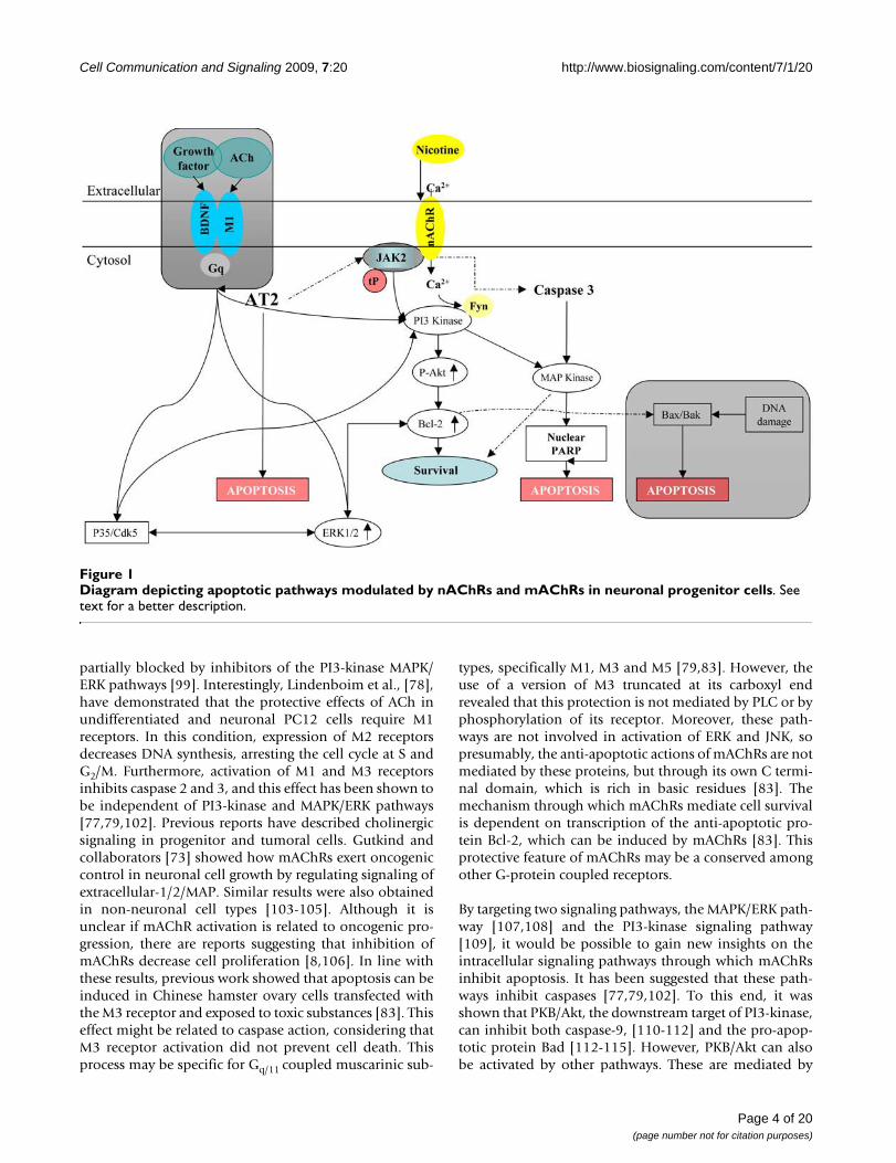
Apoptotic signaling pathwaysApoptotic pathways associated with mAChRsMany G-protein-coupled receptors protect cells fromapoptosis induced by growth factors, DNA damage or cel-lular stress. Among those receptors, mAChRs have beenshown to be protective in many cell lines and primary cellcultures [8,78-80,94,95]. These reports raise the possibil-ity that damage to cholinergic pathways might contributeto the development of neurodegenerative disorders, suchas Alzheimer and Huntington. In some neurodegenerativedisorders losses of neuronal survival stimuli occur, lead-ing to cell death. It is known that mAChRs inhibit apop-tosis through activation of PI3-kinase(phosphatidylinositol-3-OH kinase) and its downstreamtargets, protein kinase B (PKB)/Akt and MAPK/ERK (Fig-ure 1) [96]. These kinases activate pro-survival pathwaysin diverse cell types [96], including neurons [97,98]. Pre-vious work [77,80,99-101] has demonstrated that Akt canbe activated effectively through Gq coupled to M1 and M3,and Gi coupled to M2. Activation of Akt occurs through βγcomplexes and the α, Gαq and Gαi subunits of Gs pro-teins. The cell survival effects mediated by mAChRs are
Page 3 of 20(page number not for citation purposes)
Cell Communication and Signaling 2009, 7:20 http://www.biosignaling.com/content/7/1/20
partially blocked by inhibitors of the PI3-kinase MAPK/ERK pathways [99]. Interestingly, Lindenboim et al., [78],have demonstrated that the protective effects of ACh inundifferentiated and neuronal PC12 cells require M1receptors. In this condition, expression of M2 receptorsdecreases DNA synthesis, arresting the cell cycle at S andG2/M. Furthermore, activation of M1 and M3 receptorsinhibits caspase 2 and 3, and this effect has been shown tobe independent of PI3-kinase and MAPK/ERK pathways[77,79,102]. Previous reports have described cholinergicsignaling in progenitor and tumoral cells. Gutkind andcollaborators [73] showed how mAChRs exert oncogeniccontrol in neuronal cell growth by regulating signaling ofextracellular-1/2/MAP. Similar results were also obtainedin non-neuronal cell types [103-105]. Although it isunclear if mAChR activation is related to oncogenic pro-gression, there are reports suggesting that inhibition ofmAChRs decrease cell proliferation [8,106]. In line withthese results, previous work showed that apoptosis can beinduced in Chinese hamster ovary cells transfected withthe M3 receptor and exposed to toxic substances [83]. Thiseffect might be related to caspase action, considering thatM3 receptor activation did not prevent cell death. Thisprocess may be specific for Gq/11 coupled muscarinic sub-
types, specifically M1, M3 and M5 [79,83]. However, theuse of a version of M3 truncated at its carboxyl endrevealed that this protection is not mediated by PLC or byphosphorylation of its receptor. Moreover, these path-ways are not involved in activation of ERK and JNK, sopresumably, the anti-apoptotic actions of mAChRs are notmediated by these proteins, but through its own C termi-nal domain, which is rich in basic residues [83]. Themechanism through which mAChRs mediate cell survivalis dependent on transcription of the anti-apoptotic pro-tein Bcl-2, which can be induced by mAChRs [83]. Thisprotective feature of mAChRs may be a conserved amongother G-protein coupled receptors.
By targeting two signaling pathways, the MAPK/ERK path-way [107,108] and the PI3-kinase signaling pathway[109], it would be possible to gain new insights on theintracellular signaling pathways through which mAChRsinhibit apoptosis. It has been suggested that these path-ways inhibit caspases [77,79,102]. To this end, it wasshown that PKB/Akt, the downstream target of PI3-kinase,can inhibit both caspase-9, [110-112] and the pro-apop-totic protein Bad [112-115]. However, PKB/Akt can alsobe activated by other pathways. These are mediated by
Diagram depicting apoptotic pathways modulated by nAChRs and mAChRs in neuronal progenitor cellsFigure 1Diagram depicting apoptotic pathways modulated by nAChRs and mAChRs in neuronal progenitor cells. See text for a better description.
Page 4 of 20(page number not for citation purposes)

Cell Communication and Signaling 2009, 7:20 http://www.biosignaling.com/content/7/1/20
protein kinase A (PKA), calcium/calmodulin-dependentkinase and Bad [114,116-119]. Interestingly, inhibitors ofthe PI3-kinase or MAPK/ERK pathways could not sup-press the muscarinic effect on caspases, although all wereable to inhibit the muscarinic-dependent phosphoryla-tion of PKB/Akt and MAPK/ERK. Furthermore, previousstudies in PC12 cells have shown that these inhibitorsblock survival effects and ERK and PI3-kinase signaling[120,121]. It has been suggested that the MAPK/ERK andPI3-kinase pathways are not essential for mediating themuscarinic effect on caspase activity in PC12 cells [79],but rather that this effect is mediated by an unidentifiedpathway. This finding is in line with recent studies show-ing that in some cases the survival effects of growth factorsand cytokine receptors are mediated by pathways inde-pendent of both MAPK/ERK and PI3-kinase. For example,neither the survival effect of nerve growth factor (NGF) onsympathetic neurons [122] or rat-1/MycER cells trans-fected with TrkA [123], nor that of granulocyte/macro-phage colony-stimulating factor on MC cells, is mediatedby the PI3-kinase pathway [124]. Moreover, the MAPK/ERK and the PI3-kinase pathways are not essential for thesurvival effect of NGF on apoptosis induced by ceramidein PC12 cells [121]. However, the PI3-K/Akt pathway hasan established role in NGF-promoted cell survival [125].Activation of PI3-K by NGF initiates a cascade involvingAkt, which leads to the phosphorylation and inhibition ofthe pro-apoptotic protein Bad and activation of the pro-survival inhibitor κB kinase α (IKKα) [116,126]. Blockadeof the PI3-K/Akt pathway by a dominant negative Aktmutant or treatment of cells with PI3-K inhibitors revealthat activation of this signaling pathway is required forNGF-promoted cell survival [127]. However, it was shownthat pertussis toxin did not inhibit Akt phosphorylation.This finding is consistent with the previous observation ofpertussis toxin having a more profound inhibitory effectduring the early phase of NGF-induced Erk1/2 activation[128]. Upon activation, the TrkA receptor activates thePI3-K pathway, leading to activation of Akt in sympatheticneurons [125]. Previous studies have shown that TrkAforms complexes with GRK2 and GAIP/GIPC (GAIP-inter-acting protein, C terminus) [128,129], thereby providinga bridge to link TrkA and G protein signaling pathways.Furthermore, there is prior evidence pointing to a func-tional association between pertussis toxin-sensitive G pro-teins and growth factor receptors such as the insulinreceptor tyrosine kinase [130]. In addition, some pertussistoxin-sensitive growth factor-induced responses havebeen reported. For example, insulin-like growth factor 1has been shown to activate Gi and release Gβγ subunits[131,132], while TrkA is able to utilize Gi/o to stimulateErk1/2 [128,133]. Increasing evidence shows that GPCRsoften cooperate with RTKs (receptor tyrosine kinases) inthe regulation of numerous signal transduction pathways[134-136]. More recently, a report has demonstrated that
NGF and lysophosphatidate receptor signaling systemscan interact to promote G protein-mediated activation ofthe Erk pathway [133]. These observations are consistentwith the notion that TrkA can utilize pertussis toxin-sensi-tive Gi/o proteins to activate Akt, thereby inhibiting Badand stimulating the NFκB regulator, IKK, via Gαγ activa-tion [137,138] to allow cell survival through the M3 sub-type [83,139,140].
One candidate for mediating the muscarinic effect on cas-pases is sphingosine-1-phosphate, which was shown toplay an important role in the survival effect of NGF onPC12 cells [141]. Interestingly, sphingosine-1-phosphatecan be induced by the M2 muscarinic receptor [142].Intriguingly, inhibition of the PI3-kinase pathway par-tially attenuates the muscarinic survival effect on the via-bility of the cells but not on caspase inhibition. Onepossible explanation is that serum-deprived cells can dievia both caspase-dependent and -independent pathways,as shown in some apoptotic paradigms such as Bax-induced cell death in the presence of caspase inhibitors[143-145]. Despite the fact that the caspase-dependentpathway seems to play a major role in the death of serum-deprived PC12 cells, it is possible that once this pathwayis inhibited, the caspase-independent pathway has amajor role. However, one cannot exclude the possibilitythat there are unknown caspases which are activated andinvolved in apoptosis induced by trophic-factor-depriva-tion and that these caspases are inhibited by the mus-carinic receptor in a different mechanism than that usedto inhibit the DEVDase caspases and caspase-2. It wasshown that NGF withdrawal from differentiated PC12cells induces expression of FasL, which in turn may con-tribute to the apoptotic process via activation of the CD95receptor [146,147]. In this case, it is still possible thatmuscarinic receptors will inhibit the activation of caspase-8, the caspase which is directly activated when CD95 isactivated [80,115,148] by a PI3-kinase-dependent mecha-nism as was shown for CD3 activation in Fas-treated Th2-type cells [80,115]. In some systems, one signaling path-way appears to be sufficient for mediating survivalinduced by trophic agents such as NGF (PI3-kinase) andN-acetylcysteine (ERK) [109,120]. However, in other sys-tems, the survival effect may require the combined actionof several signaling pathways. For example, insulin-likegrowth factor-1 inhibited apoptosis in differentiatedPC12 cells requires both PI3-kinase and MAPK/ERK sign-aling pathways [127,149,150]. It has been suggested thatthe muscarinic survival effect could be mediated by thecombined effect of at least two different pathways. Onepathway may lead to caspase inhibition and be independ-ent of PI3-kinase and ERK signaling. This pathway couldbe mediated by the Gi/o-coupled receptors. The otherpathway would act through Gq and may involve the PI3-kinase pathway, and could promote survival by a mecha-
Page 5 of 20(page number not for citation purposes)

Cell Communication and Signaling 2009, 7:20 http://www.biosignaling.com/content/7/1/20
nism that does not affect caspases and that could be medi-ated by the Gq-coupled receptors.
Previous reports showed that muscarinic receptor stimula-tion leads to activation of the Rho family of small G-pro-teins [151,152]. This pathway can lead to activation ofRho kinase [153] and is critical for the protective capacityof muscarinic receptors. It has been demonstrated thatexcitatory receptor agonist-induced Rho activation isCa2+-dependent [154]. The ability of mAChRs to activateSRF-mediated gene transcription and the involvement ofG protein subunits in SRF activation were investigated inJurkat T cells. It has been shown that Gαq-coupled M1, butnot Gαi/o-coupled M2 receptors can activate SRF througha RhoA-mediated pathway [73,155]. In contrast, M3mAChR failed to activate SRF even though M1 and M3 arethought to induce similar signaling pathways that involveGαq/11. Yet, Gαq coupling of both M1 and M3 remainedintact, as revealed by a robust calcium response in JurkatT cells. Moreover, use of the chimeric Gα protein con-struct Gαiq5, which allowed the M2 receptor to signalalong Gαq/11-mediated pathways, restored the Ca2+
response for M2, but not SRF activation. This suggestseither that the activation of SRF through M1 involves aGαq/11-independent pathway or that Gαq/11 is insufficientin Jurkat T cells. The inhibition of M1-SRF signaling by co-transfection with the Gαq/11 suppressors, RGS2 and RGS4,indicated that Gαq/11 did play a role in M1-SRF activation,but it appeared to be insufficient per se. However, anincrease of M1-SRF signaling inhibition was observedwhen intracellular calcium was decreased.
Apoptotic pathways associated with nAChRsNicotinic receptors are expressed in neural and non-neu-ronal tissues; however, in the latter their function is notclear. Although nAChRs are primarily known for theiraction as ligand-gated ion channels transducing actionpotentials across synapses, they may have other actions aswell, such as cell-to-cell communications in various non-neuronal tissues controling important cell functions suchas proliferation, adhesion, migration, secretion, survivaland apoptosis in an autocrinal, justacrinal and paracrinalmanner [22]. Interestingly, nicotinic receptors in neuronsprotect against cell death in some settings [8,12,68,81]. Inneurons, the α7 nicotinic receptor activates PI3-kinasethrough a src-family kinase, activating the anti-apoptotickinase AKT [156]. One pathway involved in AKT signalinginvolves phosphorylation of the forkhead transcriptionfactor FKHRL1, causing its retention in the cytoplasmassociated with 14-3-3. This in turn blocks expression ofthe apoptotic protein fas [157]. The PI3-kinase/AKT path-way protects a broad range of neurons against apoptoticcell death and may block apoptosis triggered by beta-amy-loid fragments, which contribute to progression of Alzhe-imer's disease. If so, nicotinic agents may prove useful in
the treatment of this and other neurodegenerative condi-tions. Interestingly, removal of extracellular Ca2+ sup-pressed Akt phosphorylation induced by nicotine. It wasshown that an inhibitor of Src tyrosine kinase alsoreduced Akt phosphorylation. In addition, PI3-K and Fynare physically associated with α7 nicotinic receptors.Therefore, nicotinic receptor stimulation might lead tophosphorylation of Akt through Fyn [157-159]. The α7subtype can mobilize Ca2+ from ryanodine-sensitive intra-cellular stores and promote cell survival [8,13]. This intra-cellular Ca2+ mobilization can lead to BDNF-inducedCdk5-mediated neuroprotection through increased Bcl-2expression. (Figures 1 and 2). Nicotine has also beenshown to regulate the Bcl-2 family of proteins. For exam-ple, nicotine induces phosphorylation of Bcl-2 leading toprotection of human small cell lung carcinoma cellsagainst cisplatin-induced apoptosis [160,161]. Moreover,nAChRs heterodimers containing α3 and α4 mediatetheir apoptotic activity in normal human bronchial epi-thelial cells through Akt [162]. Lastly nicotine also phos-phorylates downstream targets of Akt, such as mTOR,FKHR, elf-4, GSK3b, tuberin and S6K [162].
Nicotine can also promote anti-apoptotic effects throughactivation of PKC, PKA and NF-κB, and downregulationof the tumor suppressor p53 [160,161]. Nicotine-inducedNF-κB phosphorylation promotes the phosphorylation ofthe apoptotic protein Bad (Bcl-2 antagonist of cell death),which becomes inactivated and prevents cell death. Otherpathways underlying the anti-apoptotic effects of nicotineinclude the MEK and PI3K pathways [163]. It was previ-ously shown that ERKs, AKT, and PKA could function as aBad Ser112, Ser136, or Ser155 kinase, respectively [164-167].One interesting study demonstrated that nicotine-induced Bad phosphorylation is mediated by β-adrenergicreceptors [163] (for a review see [168]), and that nicotinecan also induce phosphorylation of Bax (another Bcl-2antagonist of cell death) through PKCζ, thereby inactivat-ing Bax and suppressing cell death [169,170].
The protective effects of nicotine have been studied inNSCLC and PC12 cell lines, as well as in other experimen-tal systems [171-176]. Nicotine can protect A549 NSCLCcells against apoptosis induced by anticancer drugsthrough the upregulation of XIAP and survivin in a α3-nAChR-dependent manner [171]. Furthermore, adminis-tration of nicotine in the CNS can stimulate release ofneurotransmitters [177,178] and neurotrophic factors,such as basic fibroblast growth factor (bFGF or FGF-2)and brain-derived neurotrophic factor (BDNF)[179]. Inaddition, nicotine exposure can lead to elevated cellularcAMP levels [180]. It was demonstrated that nicotineattenuates both arachidonic acid-induced caspase activa-tion and apoptosis of spinal cord neurons [181]. How-ever, controversy exists on the specific nAChR subunit
Page 6 of 20(page number not for citation purposes)

Cell Communication and Signaling 2009, 7:20 http://www.biosignaling.com/content/7/1/20
responsible for the anti-apoptotic effects of nicotine. Theα7 and the α4β2 subtypes may play the most significantrole in the central nervous system [180,182,183]. Datafrom some laboratories indicate that phosphorylation ofERK1/2 in nicotine-treated neurons can be specificallyprevented by pre-exposure to the α7 blocker α-bugaro-toxin, but not dihydro-β-erythroidine (an antagonist of
the β2 subunit) [172,178,181], indicating an importantrole for the α7 receptor in nicotine-mediated neuropro-tection. On the other hand, studies in tumor and tumoralstem cells have implicated the dihydro-β-erythroidine-sensitive α3 and α4 receptors. This finding implies thatthe proliferative effects (mediated by α7 nAChR) and pro-survival effects (mediated by α3 or α4 nAChR) of nicotine
Diagram depicting proliferative and survival signaling pathways in cellsFigure 2Diagram depicting proliferative and survival signaling pathways in cells. In vitro, the homomeric and heteromeric nAChRs jointly stimulate the indicated signaling cascades. Yellow arrows indicate proliferative pathways triggered by nAChRs. This activation triggers the MAP kinase pathway, leading to DNA synthesis. Sustained mitogenic signaling induces to S-phase entry. Black arrows indicate nAChR survival and proliferation pathways triggered by intracellular calcium increases involving indirect activation of β-adrenergic receptor signaling, which in turn, induces activation of epidermal growth factor (EGF) recep-tor leading to the cascade indicated by blue arrows. α7 nAChR and heteromeric α-βnAChRs are activated by their agonists. Influx of Ca2+ and other cations through the nAChRs and voltage-gated Ca2+ channels trigger the release of adrenaline and noradrenalin. Adenylyl cyclase activation downstream of β-adrenergic receptors induce the cyclic AMP-protein kinase A (PKA)-CREB (cAMP response element-binding protein) pathway, transactivates epidermal growth factor receptor (EGFR) and induces the release of EGF, and perhaps another growth factors. The responsiveness of this pathway is enhanced by α7 nAChR-mediated activation of Ras through β-arrestin-dependent SRC signaling. In turn, the EGFR activates the Akt pathway and its downstream effectors, X-linked inhibitor of apoptosis protein (XIAP)-survivin and nuclear factor-κB (NF-κB).
Page 7 of 20(page number not for citation purposes)

Cell Communication and Signaling 2009, 7:20 http://www.biosignaling.com/content/7/1/20
are mediated by two distinct classes of receptors[8,13,162,171]. These data demonstrate that during dif-ferentiation there are changes in nAChR activity and func-tion. This could be due to β-adrenergic receptor activationthrough α7 nAChR, which has been found to mediate theanti-apoptotic effects of nicotine in some cell lines [184-186] (for a more detailed discussion see [168]). Such dis-crepancies can be partially explained by the pleiotropicnature of nAChR subunit inhibitors. It is also possible thatthe anti-apoptotic effects of nicotine are mediated by dif-ferent nAChR subunits in a tissue-specific manner. Thesepossibilities underscore the need for further studies toidentify the nAChR subunits responsible for the anti-apoptotic effects of nicotine.
It is known that ERK1/2 signaling can participate in theneuroprotective effects of nicotine through a variety of dif-ferent mechanisms. Previous reports showed that ERK2can increase expression of bcl-2 and inhibit apoptosis[187]. In addition, the neuroprotective effects of ERK1and ERK2 may be related to activation of a variety of tran-scription factors, which in turn can regulate transcriptionof neurotrophic factors, leading to overexpression of "sur-vival" genes and enhanced neuronal viability. Among thetranscription factors that are involved in the ERK-medi-ated cellular survival are Elk1, nuclear factor-κB (NF-κB),and cAMP response element (CRE)-binding factor (CREB)[188-190]. A role for NF-κB in neuronal survival has beensuggested [191]. In addition, recent evidence indicatesthat CREB has a crucial role in regulation of cell viability[192], as it is required to induce transcription of BDNF[193]. Elk1 functions as a nuclear transcriptional activatorthrough the interaction with the serum response element(SRE), which is present in the promoter of many immedi-ate early genes [194]. Among others, Elk1 is involved inregulation of expression of FGF-2 [195,196].
Signaling pathways of mAChRs involved in proliferation and neuronal differentiationPrevious studies have demonstrated that proliferation anddifferentiation of neuronal precursor cells can be modu-lated by mAChR signaling [8,13,197,198]. The mecha-nism involved initially amplifies mAChR and nAChRsignals, inducing calcium influx, which in turn activatesMAPK-dependent pathways [8,13,199]. Transient calciumincreases induced by ACh independently of MAPk activa-tion has been shown to be necessary for differentiationand proliferation, as muscarinic antagonists and calciumchelating agents block these effects (Figures 3 and 4)[8,13].
Activation of M2 and M3 receptors has been shown toincrease proliferation of tumoral cells in a dose-depend-ent manner. Cellular proliferation induced by the M3 sub-type is mediated by production of inositol triphosphate,[8,66] and nitric oxide [for a review see [200]], while the
effects of the M2 subtype were dependent on concomitantactivation of M1, promoting the release of E2 prostaglan-din and arginase catabolism. These events are related totumoral cell growth [66], and inhibition of caspases[79,83].
In murine mammary adenocarcionoma cells, the M3 sub-type is the most highly expressed muscarinic receptor.Stimulation of M3 receptors activates adenilate cyclase,phospholipase A2 (PLA2), IP3 and diacylglicerol (DAG)through PLC [201]. Each of these molecules in turn acti-vates different pathways. DAG activates protein kinase C(PKC), while IP3 induces release of calcium from intracel-lular stores. It is known that both pathways regulateMAPK and ERK signaling. Free intracellular Ca2+ can mod-ulate MAPK/ERK either through Ca2+-dependent proteintyrosine kinase (PYK2) [202] or by Ca2+/calmodulinkinase (Ca2+/CaM) [203]. PKC isoforms are also known toregulate MAPK/ERK through Raf-1 [204] or through trans-activation of the epidermal growth factor receptor (EGFR)mediated by the Src/PYK2 complex (Figures 2 and 3)[205].
Recent evidence suggests that activation of MAPK/ERKthrough GPCRs occurs through PKC-dependent and -independent mechanisms, depending on the receptoractivated and on the cell type [206,207]. It is known thatactivation of MAPK/ERK GPCR agonists mediate cell pro-liferation [55,208], and that pathways involving choliner-gic receptors seem to depend on the cell growth [8,74].
Stimulation of mAChRs promotes an increase in [Ca2+]iand induces phosphorylation of MAPK/ERK in MCF-7human breast cancer cells [105]. Activation of this path-way increases protein synthesis and cell proliferationthrough MAPK kinase, besides inducing DNA synthesis inneuronal progenitor cells during early neurogenesis [209].Inhibition of PLC or incubation of cells in a calcium freemedium did not alter MAPK/ERK phosphorylation; how-ever, this phosphorylation can also be induced throughtreatment with phorbol 12-myristate acetate (PMA), aPKC activator. Activation of MAPK/ERK was not affectedby PKC modulation or by its inhibition. Interestingly,phosphorylation of MAPK/ERK by mAChRs could beblocked by a PKC-ζ (a miroystoilated pseudo substrate ofPKC) inhibitor and by high doses of staurosporine (a rel-atively non-selective protein kinase inhibitor). This path-way involves PI3-K and tyrosine kinases, such as Src, andErk 1/2 [105,209]. Cells in the neuroepithelial ventricularzone of the embryonic rat cortex also express the M2receptor. The presence of M2 induces cell proliferationand accelerates neuronal differentiation.
Adrenergic receptors can transform fibroblasts whenactively mutated [210]. Interestingly, transformation bymAChRs was ligand-dependent [211]. Furthermore, some
Page 8 of 20(page number not for citation purposes)

Cell Communication and Signaling 2009, 7:20 http://www.biosignaling.com/content/7/1/20
Page 9 of 20(page number not for citation purposes)
Muscarinic and nicotinic receptor-coupled signal transduction pathways mediating MAPK activity and proliferationFigure 3Muscarinic and nicotinic receptor-coupled signal transduction pathways mediating MAPK activity and prolifer-ation. Both Erk1/2 activity and cell proliferation are activated by cholinergic (ACh) stimulation of mAChRs. ACh binds to M3, leading to a Gq-protein-mediated activation of PLC, which hydrolyses PIP2 to IP3 and DAG, subsequently mobilizing Ca2+ from organellar stores, leading to activation of PKC. Both ACh-induced MAPK activity and proliferation are reduced by the PKC inhibitor H7, indicating that PKC activity appears to be one of the upstream events critical to MAPK activation. mAChR stimu-lation induces increases in [Ca2+]i via both Ca2+ influx and mobilization from intracellular stores. Muscarinic stimulation of MAPK activity and proliferation is prevented both by BAPTA-AM and EGTA, demonstrating that elevation of [Ca2+]i is essen-tial, and may stimulate Pyk2 phosphorylation and activate the MAPK. Muscarinic stimulation of MAPK activity is effectively elim-inated by the MAPK kinase (MEK) inhibitor PD98059. M2 and M4 may provide parallel pathways to MAPK activation via pertussis toxin-sensitive Gi-proteins and βγ subunits. ERKI/II (MAPK) can serve as a convergence site for multiple extracellular signals known to induce plasticity in mature neurons. The best documented activation of the MAPK cascade occurs via ligand binding to RTK. Activation of RTK recruits the Shc-Grb2-SOS1 complex, which in turn activates Ras. Ras induces MAPK acti-vation via an evolutionarily conserved pathway, which includes Raf, MEK (MAPKK), and ERKI/II (MAPK) (MAPK cascade). ERKI/II is known to have both cytoplasmic and nuclear targets and can translocate to the nucleus to modulate transcription in neu-rons. The block of proliferation induction by the MEK inhibitor PD98059 suggests that MAPK plays a role in the induction phase of proliferation. MAPK can also be activated in neural progenitor cells via nAChRs which increase [Ca2+]i, and may mod-ulate the MAPK cascade via activation of a Ca2+-dependent tyrosine kinase (PYK2) or calmodulin (CaM).

Cell Communication and Signaling 2009, 7:20 http://www.biosignaling.com/content/7/1/20
viruses encode constitutively active GPCRs linked to cellproliferation (for a review see [212,213]), suggesting thatsignals initiated by GPCRs can be mitogenic.
MAPKs target numerous cellular proteins and transcrip-tion factors involved in cell growth and differentiation[214-216]. It is known that GPCRs activate MAPK throughthe small GTP-binding protein, p21Ras [217]. Howp21Ras is activated is still controversial; however, it islikely that transactivation of EGFR upon stimulation ofGPCRs participates in p21Ras activation [218,219].Indeed, several types of GPCRs including thrombin,endothelin, and angiotensin II receptors have been shownto transactivate EGFR, leading to MAPK activation[220,221].
The vast majority of the currently described pathwaysleading to ERK stimulation have been considered as lin-ear. While GPCRs coupled with Gi-protein activate thep21Ras-ERK pathway through the βγ subunit and PI-3kinase, GPCRs with Gq-protein activate it in a PKC-dependent manner [222,223]. However, Blaukat et al.[223] recently showed that GPCRs mediate ERK activationthrough cooperation of Gi and Gq, suggesting that multi-ple G-proteins could act in concert to attain full activationof p21Ras-ERK pathway. Muscarinic receptors in manycells have been shown to activate ERK by carbachol, and
this is not altered by treatment with pertussis toxin, indi-cating that Gq-, but not Gi-protein, may be involved inERK activation [61,224-229]. Muscarinic receptor activa-tion by carbachol rapidly and transiently stimulatesERK1/2 phosphorylation in many cells in a time- anddose-dependent manner [61,226-229], as observed in var-ious cell lines [230,231]. It was shown that the inhibitionof PLC led to a total blockade of Ca2+ mobilizationinduced by AChRs agonists [13,232]. A role of Ca2+ in thispathway is also supported by the finding that an increasein intracellular Ca2+ caused by thapsigargin[8,13,105,233] is sufficient to induce ERK phosphoryla-tion up to levels similar to those induced by AChRs ago-nists. The mechanisms by which intracellular Ca2+
stimulates the phosphorylation of ERK1/2 are complexand appear to be dependent on the nature of mAChR sub-type coupling to heterotrimeric G proteins. IntracellularCa2+ can modulate the MAPK cascade, via activation of themonomeric G-protein p21ras [234-236], through two con-vergent mechanisms; one through the calcium-dependenttyrosine kinase (PYK2) and the other mediated by cal-modulin [228,231,237]. In T84 colon epithelial cells,which express endogenous M3 mAChR subtypes,increases in [Ca2+]i in response to carbachol activate sign-aling mechanisms involving calmodulin-, PYK2-, andp60src-mediated transactivation of the EGF receptor [238].Besides Ca2+, the other downstream pathway induced
Calcium signaling pathways in stem cells and neural progenitor cellsFigure 4Calcium signaling pathways in stem cells and neural progenitor cells. Left panel represents a embryonic or adult stem cells and right panel the neuronal progenitor cells. Ca2+ signaling depends on the increase of the intracellular Ca2+ levels [Ca2+]i, derived from extracellular calcium (Ca2+)o sources or intracellular stores of the endoplasmic reticulum (ER Ca2+). It can enter through calcium channels operated by voltage (voltage-operated Ca2+ channels, VOCCs) in excitable cells such as neurons and muscular cells, or through calcium channels operated by receptors (receptor-operated Ca2+ channels, ROCs) in response to neurotransmitters. SOC's (store-operated Ca2+ channels, SOCs), open when internal Ca2+ stores are empty, and are generally present in non-excitable cells. Calcium from the ER is released by two types of channels, Inositol 1,4,5-trisphos-phate (IP3) channels and ryanodine channels. The first is present in both neural progenitor and stem cells, while the latter is expressed only in nural progenitor cells. IP3 is generated by the action of the enzyme PLC in phosphatidylinositol 4,5-bisphos-phate (PIP2). IP3 acts on receptors in the endoplasmic reticulum, promoting the release of Ca2+ from ER stores. IP3PIP2.
Page 10 of 20(page number not for citation purposes)
Cell Communication and Signaling 2009, 7:20 http://www.biosignaling.com/content/7/1/20
after PLC activation is the PKC transduction cascade. Itwas demonstrated that the direct activation of PKC, by thephorbol ester PMA, was sufficient to increase the phos-phorylation of ERK1/2 reaching levels similar to thoseinduced by carbachol in FRT cells [105]. However, the car-bachol-induced ERK1/2 phosphorylation was not medi-ated by PKC. These data indicate that the mAChR-inducedERK phosphorylation is mediated by a Ca2+-dependentbut PKC-independent mechanism.
Although some data suggest that intracellular Ca2+ partlymediates the activation of ERK1 and ERK2, other intracel-lular signaling pathways may be involved in the MAPK/ERK activation in undifferentiated cells. Activation ofMAPK by GPCRs, including mAChRs, involves phosphor-ylation of one or more proteins, such as p125FAK, p130cas,or paxillin [239]. Moreover, the Src family of protein tyro-sine kinases has been implicated in mAChR-induced ERKactivation in different cell lines [240-242]. Further studiesare needed to determine the connection between activa-tion of these protein tyrosine kinases and the downstreameffects of mAChR after G protein activation.
It has been suggested that carbachol's effects on ERK1 andERK2 phosphorylation were probably mediated throughthe activation of protein tyrosine kinases. Furthermore, ithas been demonstrated that carbachol-induced ERK acti-vation is dependent on the activity of cytoplasmatic Src-like tyrosine kinase family, since pharmacological inhibi-tion of the Src family of tyrosine kinases with specific PP2blocks the carbachol-induced MAPK/ERK activation [55].The Src family of tyrosine kinases has been implicated inthe ERK activation by various GPCRs agonists. Recent datasuggests that activation of Src tyrosine kinases may lead tothe phosphorylation of the adaptor protein Shc and therecruitment of Grb/Sos complex to the plasma mem-brane, resulting in the activation of the ERK pathway[223,243].
Signaling pathways of nAChRs involved in proliferation and neuronal differentiationPrevious reports have shown that nAChRs are expressed innon-neuronal cells within the nervous system [244,245],embryonic stem cells [8,13], neural stem cells [197], andembryonic tissues [246].
Microglia express α7 nAChRs [247], and stimulation ofα7 nAChRs promotes anti-inflammatory pathways andblunts the response of migroglia to lipopolysaccaride[247], suggesting that nAChRs may have a role in control-ling localized brain inflammation. Nicotinic receptors arealso expressed on O2A oligodendrocyte precursors, butare not detectable after induction of differentiation, indi-cating that nAChR expression is developmentally control-led in these cells [248]. Although the physiological
functions of nAChRs in O2A oligodendrocyte precursorsare not understood, data suggest that activation ofnAChRs might control migration, survival and differenti-ation in these cells [248].
It has been found that in non-neuronal tissues nicotineinduces the secretion of growth factors like bFGF, TGF-α,VEGF, and PDGF [249] Nicotine also upregulates expres-sion of the calpain family of proteins [250] as well asCOX-2 and VEGFR-2 [251], activating the Raf/MAPKkinase/ERK pathway [252]. Since nAChRs do not haveintrinsic tyrosine kinase activity [22], the molecular mech-anisms underlying its effects on proliferation remainunclear. It was demonstrated that nicotine-mediatedinduction of cell proliferation involves recruitment of β-arrestin, which facilitates the activation of Src. This in turnleads to binding of Raf-1 kinase to Rb, leading to cell cycleentry [253]. Dasgupta and coworkers demonstrated thathuman non-small cell lung cancer (NSCLC) tumor tissueshad high levels of Rb-Raf-1 complexes in tumors relativeto adjacent normal lung tissue, suggesting that perhapsthe Rb-Raf-1 pathway contributes to the genesis of thesetumors. Furthermore, chromatin IP (ChIP) analysis ofhuman NSCLC tumor samples demonstrated increasedrecruitment of E2F1 and Raf-1 to proliferative promoterslike cdc6 and cdc25A. These results suggest that binding ofβ-arrestin to nAChRs is an early and critical event in theinitiation of nicotine-induced mitogenesis. The subse-quent steps resemble growth factor-induced cell prolifera-tion, as they include activation of Src, association of Rb toRaf-1, inactivation of Rb, and enhanced recruitment ofE2F1 and Raf-1 to promoters of genes that induce prolif-eration [253]. These events are likely to contribute to thegrowth and progression of tumoral cells.
Therapeutic uses of cholinergic receptor modulators in stem cell specificationIn vivo proliferation, differentiation, and genetic modification of neural stem cell progenyAs previously demonstrated [8,13], AChRs have differentroles on proliferation in embryonic and neural stem cells.In embryonic cells, nAChRs decrease proliferation. Con-versely, neural stem cells and their progeny can beinduced to proliferate in vivo by administering α7 agonists[13]. To initiate neuronal differentiation, agonists for theGαi-coupled mAChRs, such as the M2 subtype, could beused for treatment. These pharmacological agents includeany substance that acts through AChR activation orthrough pathways activated by them. The examplesdescribed here to modulate proliferation, differentiation,and genetic modification of neural stem cells in vitro canbe adapted to in vivo techniques. Such in vivo manipula-tion and modification of these cells allows cells lost dueto injury or disease to be endogenously replaced. Thiswould abolish the need for transplanting foreign cells into
Page 11 of 20(page number not for citation purposes)

Cell Communication and Signaling 2009, 7:20 http://www.biosignaling.com/content/7/1/20
a patient. Additionally, cells can be modified or geneti-cally engineered in vivo so that they express various bio-logical agents useful in the treatment of neurologicaldisorders. However, fine control of muscarinic signalingrequires compounds that selectively modulate specificmuscarinic receptor subtypes. Unfortunately, such drugshave not been discovered yet. M1 muscarinic agonistssuch as arecoline have also been found to be weak ago-nists of M2 and M3 subtypes, and are not very effective intreating cognitive impairment, most likely because ofdose-limiting side effects [254,255]. Selective muscarinicagonists for M5 and M2 subtypes could be used both aspharmacological tools and as therapeutic agents [8], asM5 and M2 mediate most of the muscarinic [Ca2+]i-response and seem to control proliferation and differenti-ation induction, respectively.
Treatment with nicotinic receptor agonists also has thera-peutic potential, similarly to muscarinic agonists. How-ever, nAChR agonists which bind the same site as ACh arenot a viable solution, for ACh not only activates, but alsoblocks receptor activity through desensitization [256] anduncompetitive blockade [for review see [257]]. Further-more, prolonged activation appears to induce a long-last-ing inactivation. Therefore, agonists of ACh may reduce orenhance receptor activation. In nAChRs, desensitizationgenerally limits the duration of current during agonistapplication [258]. However, positive allosteric modula-tors can enhance the efficacy of agonists at nicotinic recep-tors. It is believed that such compounds would be usefulfor treatment of conditions associated with decreased nic-otinic transmission. In a therapeutic setting, these com-pounds could restore normal interneuronalcommunication without affecting the temporal profile ofactivation. In addition, they would not produce long-terminactivation, contrary to prolonged application of an ago-nist.
A naturally existing allosteric modulator of nicotinictransmission is the CGRP (Calcitonin Gene Related Pep-tide) neuropeptide [259,260]. Previous reports show thatCGRP blocks nAChRs competitively. It is noteworthy topoint out that this effect is not mediated by conventionalG-protein-coupling [261], as it was demonstrated that thispeptide's activity is contained within the 1–7 N-terminalfragment. Similarly to the native CGRP, CGRP 1–7 showsa rapidly developing, competitive antagonism which isreadily reversible after washout [261]. Intriguingly, someCGRP fragments quickly and reversibly enhanceresponses mediated by the activation of native neuronalnAChRs [262]. Mutant versions of the CGRP peptide frag-ment can be used as neuronal nAChRs enhancers, as inthe absence of nAChR activation these peptides were inac-tive [262].
The CGRP 1–6 peptide did not modify the muscle-typenicotinic receptor responses, indicating its selectivity forneuronal receptors. This finding suggests that certain pep-tide derivatives shorter than CGRP 1–7 exert an unusualaction, involving an apparently competitive modulationof the agonist-binding site. CGRP 1–6 and its derivativesmay be used for the treatment of symptoms of neurologi-cal diseases associated with functional deficits of nAChRsand may be used as stem cell neuronal differentiationenhancers.
Interestingly, nicotine has been shown to protect cellsfrom apoptosis induced by anticancer drugs. The acquisi-tion of drug resistance is a considerable challenge in can-cer therapy, and nAChR antagonists could be potentiallyused in combination with established chemotherapeuticdrugs to enhance the therapeutic response to chemother-apy. The bioactivity of nAChR antagonists, however, hasyet to be tested in animal models. Carefully designed ani-mal studies are essential to investigate the potential sideeffects of nAChR antagonists on the brain, central nervoussystem, immune cells and muscle cells, all of whichexpress high levels of nicotinic receptors.
The study of the roles of nAChRs in development and pro-gression of cancer and stem cells differentiation providesnovel opportunities for the prevention and therapy ofcancer and degenerative disorders. However, it is impor-tant to consider that vital cell and organ functions are reg-ulated by these receptors. Antagonists for α7 nAChR maysuccessfully block cancer cells and promote proliferationof stem cells without cytotoxicity to normal control cellsin vitro, but in vivo it would induce adverse effects on thecontrol of inflammatory reactions and the regulation ofrespiratory and cardiovascular functions, and may alsolead to psychiatric symptoms [22,168,263].
Blockers of Ca2+ channels are known for their anti-prolif-erative properties and might be an alternative becausethey can desensitize the hyperactive α7 nAChR (Figure 1).However, experimental findings appear to be divergentand depend on the cell type and mode of administrationused. Among the calcium channel inhibitors, L- and T-type calcium blockers, were reported to inhibit neuronaldifferentiation [12,264-266], but have limited effect onother cell types. Mibefradil, a selective blocker of T-typechannels, has significant anti-proliferative action in vari-ous cell types in vitro as well as in vivo [267,268]. The non-selective calcium blocker amlodipine is also a very effec-tive protector of neuronal cells [269-271]. Calcium chan-nel blockers induce a rapid decrease in intracellular Ca2+,even in cells lacking depolarization-induced calcium flux[272,273]. These observations suggest that amlodipineinhibits cell proliferation through intracellular signaling
Page 12 of 20(page number not for citation purposes)

Cell Communication and Signaling 2009, 7:20 http://www.biosignaling.com/content/7/1/20
pathways rather than through inhibition of Ca2+ entrychannels.
Another possible pharmacological treatment is the use ofSluRP1, a drug that reduces the responsiveness ofα7nAChR to agonists. SluRP1 may have an effect similarto that of voltage-gated Ca2+ channels blockers [274].However, any attempt to prevent or treat cancer by target-ing nAChRs must be based on the identification of molec-ular markers. The goal of this strategy is the restoration ofbalance between stimulatory and inhibitory signaling andnot complete blockade of a given pathway.
Nicotinic signaling in non-neuronal cells has huge impli-cations for cell fate and survival. Research in nAChR sign-aling networks will be especially relevant to stem cellproduction in a large scale and cancer treatment. Futurestudies will need to define both the function of differentnAChR subtypes in non-neuronal cells and the down-stream signaling pathways that underlie the proliferativeand anti-apoptotic activities of nicotine.
Similarly to nicotinic signaling, the mechanisms thatunderlie the pro-mitogenic effects of muscarinic receptorstimulation have not yet been studied in detail. However,several intracellular signaling pathways that regulate thesynergistic mitogenic interaction of other GPCR agonistswith growth factors in neural progenitor cells have beenidentified (Figures 1 and 3). These pathways are not thesame for every GPCR agonist The GPCRs M1 and M3increase EGF-induced proliferation through a pathwayinvolving Gβγ, phosphatidylinositol-3-kinase, Akt andPKC [105,238]. Muscarinic stimulation of MAPK activityand proliferation is prevented both by the intracellularCa2+ chelator BAPTA-AM and by a reduction in extracellu-lar Ca2+ with EGTA. This suggests that increases in intrac-ellular calcium are essential, and may stimulate Pyk2phosphorylation and then activate the MAPK signalingpathway [238]. PKC also regulates p42/p44 MAP kinaseactivation by muscarinic receptor agonists in neuronalprogenitor cells [55].
Concluding remarksThere is a growing body of evidence indicating that nico-tinic and muscarinic receptors play important roles instem cell differentiation and physiology. The disruptionof developmental patterns and of normal function is oftencorrelated with pathological conditions. Therefore, thecontrolled manipulation of ACh function may lead tonovel therapies. Strikingly, studies have revealed thatdrugs currently used to treat disorders such as Alzheimer'sdisease and depression, increase adult neurogenesis,which may be the mechanism mediating the activity ofthese drugs. However, some of these studies are controver-sial, and remain to be confirmed. Hence, the role of neu-
rogenesis in treating central nervous system disorders, aswell as the effects of drugs on embryonic and adult stemcells' neuronal differentiation remain areas of activeresearch. Discriminating specific contributions of nAChRand mAChR signaling for the control of phenotypic fea-tures as specialized structural and functional behaviors isa great challenge and has undeniable potential regardingfuture applications.
Competing interestsThe authors declare that they have no competing interests.
Authors' contributionsRRR and AA wrote the manuscript. The authors read andapproved the final manuscript.
AcknowledgementsThis work was supported by Instituto do Milenio/CNPq-MCT and Instituto Nacional de Ciência e Tecnologia de Nanomateriais de Carbono, CNPq (Conselho Nacional de Desenvolvimento Científico e Tecnológico), Brazil. R.R.R is grateful for grants from CNPq, and FAPEMIG (Fundação de Amparo à Pesquisa do Estado de Minas Gerais), Brazil. English editing has been performed throughout the text by native English speaker Dra. Eliza-beth Speed.
References1. Horiuchi Y, Kimura R, Kato N, Fujii T, Seki M, Endo T, Kato T,
Kawashima K: Evolutional study on acetylcholine expression.Life Sci 2003, 72:1745-1756.
2. Grando SA: Biological functions of keratinocyte cholinergicreceptors. J Investig Dermatol Symp Proc 1997, 2:41-48.
3. Wessler I, Kirkpatrick CJ, Racke K: The cholinergic 'pitfall': ace-tylcholine, a universal cell molecule in biological systems,including humans. Clin Exp Pharmacol Physiol 1999, 26:198-205.
4. Kawashima K, Fujii T: Extraneuronal cholinergic system in lym-phocytes. Pharmacol Ther 2000, 86:29-48.
5. Wessler I, Reinheimer T, Kilbinger H, Bittinger F, Kirkpatrick CJ,Saloga J, Knop J: Increased acetylcholine levels in skin biopsiesof patients with atopic dermatitis. Life Sci 2003, 72:2169-2172.
6. Tobin AB, Budd DC: The anti-apoptotic response of the Gq/11-coupled muscarinic receptor family. Biochem Soc Trans 2003,31:1182-1185.
7. Kawashima K, Fujii T: Expression of non-neuronal acetylcholinein lymphocytes and its contribution to the regulation ofimmune function. Front Biosci 2004, 9:2063-2085.
8. Resende RR, Alves AS, Britto LR, Ulrich H: Role of acetylcholinereceptors in proliferation and differentiation of P19 embryo-nal carcinoma cells. Exp Cell Res 2008, 314:1429-1443.
9. Williams BP, Milligan CJ, Street M, Hornby FM, Deuchars J, BuckleyNJ: Transcription of the M1 muscarinic receptor gene in neu-rons and neuronal progenitors of the embryonic rat fore-brain. J Neurochem 2004, 88:70-77.
10. Wessler I, Kirkpatrick CJ: Acetylcholine beyond neurons: thenon-neuronal cholinergic system in humans. Br J Pharmacol2008, 154:1558-1571.
11. Martins AH, Resende RR, Majumder P, Faria M, Casarini DE, TarnokA, Colli W, Pesquero JB, Ulrich H: Neuronal differentiation ofP19 embryonal carcinoma cells modulates kinin B2 receptorgene expression and function. J Biol Chem 2005,280:19576-19586.
12. Resende RR, Majumder P, Gomes KN, Britto LR, Ulrich H: P19embryonal carcinoma cells as in vitro model for studyingpurinergic receptor expression and modulation of N-methyl-D-aspartate-glutamate and acetylcholine receptors duringneuronal differentiation. Neuroscience 2007, 146:1169-1181.
13. Resende RR, Gomes KN, Adhikari A, Britto LR, Ulrich H: Mecha-nism of acetylcholine-induced calcium signaling during neu-
Page 13 of 20(page number not for citation purposes)
http://www.ncbi.nlm.nih.gov/entrez/query.fcgi?cmd=Retrieve&db=PubMed&dopt=Abstract&list_uids=9487015
Cell Communication and Signaling 2009, 7:20 http://www.biosignaling.com/content/7/1/20
ronal differentiation of P19 embryonal carcinoma cells invitro. Cell Calcium 2008, 43:107-121.
14. Buznikov GA, Shmukler YB, Lauder JM: From oocyte to neuron:do neurotransmitters function in the same way throughoutdevelopment? Cell Mol Neurobiol 1996, 16:537-559.
15. Levitt P, Harvey JA, Friedman E, Simansky K, Murphy EH: New evi-dence for neurotransmitter influences on brain develop-ment. Trends Neurosci 1997, 20:269-274.
16. Bignami F, Bevilacqua P, Biagioni S, De Jaco A, Casamenti F, Felsani A,Augusti-Tocco G: Cellular acetylcholine content and neuronaldifferentiation. J Neurochem 1997, 69:1374-1381.
17. Schuller HM: Cell type specific, receptor-mediated modula-tion of growth kinetics in human lung cancer cell lines by nic-otine and tobacco-related nitrosamines. Biochem Pharmacol1989, 38:3439-3442.
18. Maneckjee R, Minna JD: Opioid and nicotine receptors affectgrowth regulation of human lung cancer cell lines. Proc NatlAcad Sci USA 1990, 87:3294-3298.
19. Lindstrom JM: Acetylcholine receptors and myasthenia. MuscleNerve 2000, 23:453-477.
20. Changeux J, Edelstein SJ: Allosteric mechanisms in normal andpathological nicotinic acetylcholine receptors. Curr Opin Neu-robiol 2001, 11:369-377.
21. Hogg RC, Raggenbass M, Bertrand D: Nicotinic acetylcholinereceptors: from structure to brain function. Rev Physiol BiochemPharmacol 2003, 147:1-46.
22. Gotti C, Clementi F: Neuronal nicotinic receptors: from struc-ture to pathology. Prog Neurobiol 2004, 74:363-396.
23. McGehee DS, Role LW: Presynaptic ionotropic receptors. CurrOpin Neurobiol 1996, 6:342-349.
24. Koh DC, Armugam A, Jeyaseelan K: Snake venom componentsand their applications in biomedicine. Cell Mol Life Sci 2006,63:3030-3041.
25. Portugal GS, Gould TJ: Genetic variability in nicotinic acetyl-choline receptors and nicotine addiction: converging evi-dence from human and animal research. Behav Brain Res 2008,193:1-16.
26. Changeux JP, Edelstein SJ: Allosteric receptors after 30 years.Neuron 1998, 21:959-980.
27. Seguela P, Wadiche J, Dineley-Miller K, Dani JA, Patrick JW: Molec-ular cloning, functional properties, and distribution of ratbrain alpha 7: a nicotinic cation channel highly permeable tocalcium. J Neurosci 1993, 13:596-604.
28. Broide RS, Leslie FM: The alpha7 nicotinic acetylcholine recep-tor in neuronal plasticity. Mol Neurobiol 1999, 20:1-16.
29. Forster GL, Yeomans JS, Takeuchi J, Blaha CD: M5 muscarinicreceptors are required for prolonged accumbal dopaminerelease after electrical stimulation of the pons in mice. J Neu-rosci 2002, 22:RC190.
30. Yeomans J, Forster G, Blaha C: M5 muscarinic receptors areneeded for slow activation of dopamine neurons and forrewarding brain stimulation. Life Sci 2001, 68:2449-2456.
31. Basile AS, Fedorova I, Zapata A, Liu X, Shippenberg T, Duttaroy A,Yamada M, Wess J: Deletion of the M5 muscarinic acetylcho-line receptor attenuates morphine reinforcement and with-drawal but not morphine analgesia. Proc Natl Acad Sci USA 2002,99:11452-11457.
32. Wang H, Ng K, Hayes D, Gao X, Forster G, Blaha C, Yeomans J:Decreased amphetamine-induced locomotion and improvedlatent inhibition in mice mutant for the M5 muscarinicreceptor gene found in the human 15q schizophrenia region.Neuropsychopharmacology 2004, 29:2126-2139.
33. Tzavara ET, Bymaster FP, Davis RJ, Wade MR, Perry KW, Wess J,McKinzie DL, Felder C, Nomikos GG: M4 muscarinic receptorsregulate the dynamics of cholinergic and dopaminergic neu-rotransmission: relevance to the pathophysiology and treat-ment of related CNS pathologies. Faseb J 2004, 18:1410-1412.
34. Di Cara B, Panayi F, Gobert A, Dekeyne A, Sicard D, De Groote L,Millan MJ: Activation of dopamine D1 receptors enhancescholinergic transmission and social cognition: a parallel dial-ysis and behavioural study in rats. Int J Neuropsychopharmacol2007, 10:383-399.
35. Hegde SS, Mammen M, Jasper JR: Antimuscarinics for the treat-ment of overactive bladder: current options and emergingtherapies. Curr Opin Investig Drugs 2004, 5:40-49.
36. Racke K, Matthiesen S: The airway cholinergic system: physiol-ogy and pharmacology. Pulm Pharmacol Ther 2004, 17:181-198.
37. Keam SJ, Keating GM: Tiotropium bromide. Treat Respir Med2004, 3:247-268.
38. Terry AV Jr, Buccafusco JJ: The cholinergic hypothesis of age andAlzheimer's disease-related cognitive deficits: recent chal-lenges and their implications for novel drug development. JPharmacol Exp Ther 2003, 306:821-827.
39. Bartus RT: On neurodegenerative diseases, models, and treat-ment strategies: lessons learned and lessons forgotten a gen-eration following the cholinergic hypothesis. Exp Neurol 2000,163:495-529.
40. Fox RI: Sjogren's syndrome: evolving therapies. Expert OpinInvestig Drugs 2003, 12:247-254.
41. Roman GC: Cholinergic dysfunction in vascular dementia.Curr Psychiatry Rep 2005, 7:18-26.
42. Felder CC, Bymaster FP, Ward J, DeLapp N: Therapeutic oppor-tunities for muscarinic receptors in the central nervous sys-tem. J Med Chem 2000, 43:4333-4353.
43. Birdsall NJ, Lazareno S, Popham A, Saldanha J: Multiple allostericsites on muscarinic receptors. Life Sci 2001, 68:2517-2524.
44. Eglen RM, Choppin A, Watson N: Therapeutic opportunitiesfrom muscarinic receptor research. Trends Pharmacol Sci 2001,22:409-414.
45. Matsui M, Yamada S, Oki T, Manabe T, Taketo MM, Ehlert FJ: Func-tional analysis of muscarinic acetylcholine receptors usingknockout mice. Life Sci 2004, 75:2971-2981.
46. Slotkin TA, Cousins MM, Seidler FJ: Administration of nicotine toadolescent rats evokes regionally selective upregulation ofCNS alpha 7 nicotinic acetylcholine receptors. Brain Res 2004,1030:159-163.
47. Zoli M, Le Novere N, Hill JA Jr, Changeux JP: Developmental reg-ulation of nicotinic ACh receptor subunit mRNAs in the ratcentral and peripheral nervous systems. J Neurosci 1995,15:1912-1939.
48. Aubert I, Cecyre D, Gauthier S, Quirion R: Comparativeontogenic profile of cholinergic markers, including nicotinicand muscarinic receptors, in the rat brain. J Comp Neurol 1996,369:31-55.
49. Court JA, Lloyd S, Johnson M, Griffiths M, Birdsall NJ, Piggott MA,Oakley AE, Ince PG, Perry EK, Perry RH: Nicotinic and muscariniccholinergic receptor binding in the human hippocampal for-mation during development and aging. Brain Res Dev Brain Res1997, 101:93-105.
50. Torrao AS, Carmona FM, Lindstrom J, Britto LR: Expression ofcholinergic system molecules during development of thechick nervous system. Brain Res Dev Brain Res 2000, 124:81-92.
51. Atluri P, Fleck MW, Shen Q, Mah SJ, Stadfelt D, Barnes W, GoderieSK, Temple S, Schneider AS: Functional nicotinic acetylcholinereceptor expression in stem and progenitor cells of the earlyembryonic mouse cerebral cortex. Dev Biol 2001, 240:143-156.
52. Oppitz M, Mobus V, Brock S, Drews U: Muscarinic receptors incell lines from ovarian carcinoma: negative correlation withsurvival of patients. Gynecol Oncol 2002, 85:159-164.
53. Hoogduijn MJ, Cheng A, Genever PG: Functional Nicotinic andMuscarinic Receptors on Mesenchymal Stem Cells. Stem CellsDev 2008 in press.
54. Greco SJ, Zhou C, Ye JH, Rameshwar P: An interdisciplinaryapproach and characterization of neuronal cells transdiffer-entiated from human mesenchymal stem cells. Stem Cells Dev2007, 16:811-826.
55. Zhao WQ, Alkon DL, Ma W: c-Src protein tyrosine kinase activ-ity is required for muscarinic receptor-mediated DNA syn-thesis and neurogenesis via ERK1/2 and c-AMP-responsiveelement-binding protein signaling in neural precursor cells. JNeurosci Res 2003, 72:334-342.
56. Bernardini N, Levey AI, Augusti-Tocco G: Rat dorsal root gangliaexpress m1–m4 muscarinic receptor proteins. J Peripher NervSyst 1999, 4:222-232.
57. Williams CL, Hayes VY, Hummel AM, Tarara JE, Halsey TJ: Regula-tion of E-cadherin-mediated adhesion by muscarinic acetyl-choline receptors in small cell lung carcinoma. J Cell Biol 1993,121:643-654.
58. Boss A, Oppitz M, Drews U: Muscarinic cholinergic receptors inthe human melanoma cell line SK-Mel 28: modulation ofchemotaxis. Clin Exp Dermatol 2005, 30:557-564.
Page 14 of 20(page number not for citation purposes)
http://www.ncbi.nlm.nih.gov/entrez/query.fcgi?cmd=Retrieve&db=PubMed&dopt=Abstract&list_uids=8956008
http://www.ncbi.nlm.nih.gov/entrez/query.fcgi?cmd=Retrieve&db=PubMed&dopt=Abstract&list_uids=8956008
http://www.ncbi.nlm.nih.gov/entrez/query.fcgi?cmd=Retrieve&db=PubMed&dopt=Abstract&list_uids=8956008
http://www.ncbi.nlm.nih.gov/entrez/query.fcgi?cmd=Retrieve&db=PubMed&dopt=Abstract&list_uids=9185309
http://www.ncbi.nlm.nih.gov/entrez/query.fcgi?cmd=Retrieve&db=PubMed&dopt=Abstract&list_uids=9185309
http://www.ncbi.nlm.nih.gov/entrez/query.fcgi?cmd=Retrieve&db=PubMed&dopt=Abstract&list_uids=9185309
http://www.ncbi.nlm.nih.gov/entrez/query.fcgi?cmd=Retrieve&db=PubMed&dopt=Abstract&list_uids=9326265
http://www.ncbi.nlm.nih.gov/entrez/query.fcgi?cmd=Retrieve&db=PubMed&dopt=Abstract&list_uids=9326265
http://www.ncbi.nlm.nih.gov/entrez/query.fcgi?cmd=Retrieve&db=PubMed&dopt=Abstract&list_uids=2573356
http://www.ncbi.nlm.nih.gov/entrez/query.fcgi?cmd=Retrieve&db=PubMed&dopt=Abstract&list_uids=2573356
http://www.ncbi.nlm.nih.gov/entrez/query.fcgi?cmd=Retrieve&db=PubMed&dopt=Abstract&list_uids=2573356
http://www.ncbi.nlm.nih.gov/entrez/query.fcgi?cmd=Retrieve&db=PubMed&dopt=Abstract&list_uids=2159143
http://www.ncbi.nlm.nih.gov/entrez/query.fcgi?cmd=Retrieve&db=PubMed&dopt=Abstract&list_uids=2159143
http://www.ncbi.nlm.nih.gov/entrez/query.fcgi?cmd=Retrieve&db=PubMed&dopt=Abstract&list_uids=8794090
http://www.ncbi.nlm.nih.gov/entrez/query.fcgi?cmd=Retrieve&db=PubMed&dopt=Abstract&list_uids=9856454
http://www.ncbi.nlm.nih.gov/entrez/query.fcgi?cmd=Retrieve&db=PubMed&dopt=Abstract&list_uids=7678857
http://www.ncbi.nlm.nih.gov/entrez/query.fcgi?cmd=Retrieve&db=PubMed&dopt=Abstract&list_uids=7678857
http://www.ncbi.nlm.nih.gov/entrez/query.fcgi?cmd=Retrieve&db=PubMed&dopt=Abstract&list_uids=7678857
http://www.ncbi.nlm.nih.gov/entrez/query.fcgi?cmd=Retrieve&db=PubMed&dopt=Abstract&list_uids=7891142
http://www.ncbi.nlm.nih.gov/entrez/query.fcgi?cmd=Retrieve&db=PubMed&dopt=Abstract&list_uids=7891142
http://www.ncbi.nlm.nih.gov/entrez/query.fcgi?cmd=Retrieve&db=PubMed&dopt=Abstract&list_uids=7891142
http://www.ncbi.nlm.nih.gov/entrez/query.fcgi?cmd=Retrieve&db=PubMed&dopt=Abstract&list_uids=8723701
http://www.ncbi.nlm.nih.gov/entrez/query.fcgi?cmd=Retrieve&db=PubMed&dopt=Abstract&list_uids=8723701
http://www.ncbi.nlm.nih.gov/entrez/query.fcgi?cmd=Retrieve&db=PubMed&dopt=Abstract&list_uids=8723701
http://www.ncbi.nlm.nih.gov/entrez/query.fcgi?cmd=Retrieve&db=PubMed&dopt=Abstract&list_uids=9263584
http://www.ncbi.nlm.nih.gov/entrez/query.fcgi?cmd=Retrieve&db=PubMed&dopt=Abstract&list_uids=9263584
http://www.ncbi.nlm.nih.gov/entrez/query.fcgi?cmd=Retrieve&db=PubMed&dopt=Abstract&list_uids=9263584
http://www.ncbi.nlm.nih.gov/entrez/query.fcgi?cmd=Retrieve&db=PubMed&dopt=Abstract&list_uids=8387530
http://www.ncbi.nlm.nih.gov/entrez/query.fcgi?cmd=Retrieve&db=PubMed&dopt=Abstract&list_uids=8387530

Cell Communication and Signaling 2009, 7:20 http://www.biosignaling.com/content/7/1/20
59. Rimmaudo LE, de la Torre E, Sacerdote de Lustig E, Sales ME: Mus-carinic receptors are involved in LMM3 tumor cells prolifer-ation and angiogenesis. Biochem Biophys Res Commun 2005,334:1359-1364.
60. Newman MB, Misiuta I, Willing AE, Zigova T, Karl RC, Borlongan CV,Sanberg PR: Tumorigenicity issues of embryonic carcinoma-derived stem cells: relevance to surgical trials using NT2 andhNT neural cells. Stem Cells Dev 2005, 14:29-43.
61. Wu EH, Wong YH: Activation of muscarinic M4 receptor aug-ments NGF-induced pro-survival Akt signaling in PC12 cells.Cell Signal 2006, 18:285-293.
62. Pollett JB, Corsi KA, Weiss KR, Cooper GM, Barry DA, Gharaibeh B,Huard J: Malignant transformation of multipotent muscle-derived cells by concurrent differentiation signals. Stem Cells2007, 25:2302-2311.
63. Song P, Sekhon HS, Lu A, Arredondo J, Sauer D, Gravett C, Mark GP,Grando SA, Spindel ER: M3 muscarinic receptor antagonistsinhibit small cell lung carcinoma growth and mitogen-acti-vated protein kinase phosphorylation induced by acetylcho-line secretion. Cancer Res 2007, 67:3936-3944.
64. Janulis M, Silberman S, Ambegaokar A, Gutkind JS, Schultz RM: Roleof mitogen-activated protein kinases and c-Jun/AP-1 trans-activating activity in the regulation of protease mRNAs andthe malignant phenotype in NIH 3T3 fibroblasts. J Biol Chem1999, 274:801-813.
65. Blanco M, Robinson MJ: Muscarinic M3 acetylcholine receptorimmunostaining in paraffin-embedded normal and neoplas-tic prostatic gland tissue. Ann Diagn Pathol 2004, 8:333-336.
66. Espanol AJ, Sales ME: Different muscarinc receptors areinvolved in the proliferation of murine mammary adenocar-cinoma cell lines. Int J Mol Med 2004, 13:311-317.
67. Metzger M, Just L, Boss A, Drews U: Identification and functionalcharacterization of the muscarinic receptor M3 in thehuman keratinocyte cell line HaCaT. Cells Tissues Organs 2005,180:96-105.
68. Shin S, Dalton S, Stice SL: Human motor neuron differentiationfrom human embryonic stem cells. Stem Cells Dev 2005,14:266-269.
69. Hoogduijn MJCA, Genever PG: Functional Nicotinic and Mus-carinic Receptors on Mesenchymal Stem Cells. Stem Cells andDevelopment 2008.
70. Inoue M, Yoshii M: Modulation of ion channels by somatostatinand acetylcholine. Prog Neurobiol 1992, 38:203-230.
71. van Koppen CJ, Kaiser B: Regulation of muscarinic acetylcholinereceptor signaling. Pharmacol Ther 2003, 98:197-220.
72. Williams CL: Muscarinic signaling in carcinoma cells. Life Sci2003, 72:2173-2182.
73. Gutkind JS, Crespo P, Xu N, Teramoto H, Coso OA: The pathwayconnecting m2 receptors to the nucleus involves small GTP-binding proteins acting on divergent MAP kinase cascades.Life Sci 1997, 60:999-1006.
74. Nicke B, Detjen K, Logsdon CD: Muscarinic cholinergic recep-tors activate both inhibitory and stimulatory growth mecha-nisms in NIH3T3 cells. J Biol Chem 1999, 274:21701-21706.
75. Burdon D, Patel R, Challiss RA, Blank JL: Growth inhibition by themuscarinic M(3) acetylcholine receptor: evidence forp21(Cip1/Waf1) involvement in G(1) arrest. Biochem J 2002,367:549-559.
76. Brown JH, Sah V, Moskowitz S, Ramirez T, Collins L, Post G, Gold-stein D: Pathways and roadblocks in muscarinic receptor-mediated growth regulation. Life Sci 1997, 60:1077-1084.
77. Shafer SH, Williams CL: Elevated Rac1 activity changes the M3muscarinic acetylcholine receptor-mediated inhibition ofproliferation to induction of cell death. Mol Pharmacol 2004,65:1080-1091.
78. Lindenboim L, Pinkas-Kramarski R, Sokolovsky M, Stein R: Activa-tion of muscarinic receptors inhibits apoptosis in PC12M1cells. J Neurochem 1995, 64:2491-2499.
79. Leloup C, Michaelson DM, Fisher A, Hartmann T, Beyreuther K, SteinR: M1 muscarinic receptors block caspase activation by phos-phoinositide 3-kinase- and MAPK/ERK-independent path-ways. Cell Death Differ 2000, 7:825-833.
80. De Sarno P, Shestopal SA, King TD, Zmijewska A, Song L, Jope RS:Muscarinic receptor activation protects cells from apoptoticeffects of DNA damage, oxidative stress, and mitochondrialinhibition. J Biol Chem 2003, 278:11086-11093.
81. Serobyan N, Orlovskaya I, Kozlov V, Khaldoyanidi SK: Exposure tonicotine during gestation interferes with the colonization offetal bone marrow by hematopoietic stem/progenitor cells.Stem Cells Dev 2005, 14:81-91.
82. Budd DC, Spragg EJ, Ridd K, Tobin AB: Signalling of the M3-mus-carinic receptor to the anti-apoptotic pathway. Biochem J2004, 381:43-49.
83. Budd DC, McDonald J, Emsley N, Cain K, Tobin AB: The C-termi-nal tail of the M3-muscarinic receptor possesses anti-apop-totic properties. J Biol Chem 2003, 278:19565-19573.
84. Tipton DA, Dabbous MK: Effects of nicotine on proliferationand extracellular matrix production of human gingivalfibroblasts in vitro. J Periodontol 1995, 66:1056-1064.
85. Peng X, Gerzanich V, Anand R, Wang F, Lindstrom J: Chronic nico-tine treatment up-regulates alpha3 and alpha7 acetylcholinereceptor subtypes expressed by the human neuroblastomacell line SH-SY5Y. Mol Pharmacol 1997, 51:776-784.
86. Wang F, Nelson ME, Kuryatov A, Olale F, Cooper J, Keyser K, Lind-strom J: Chronic nicotine treatment up-regulates humanalpha3 beta2 but not alpha3 beta4 acetylcholine receptorsstably transfected in human embryonic kidney cells. J BiolChem 1998, 273:28721-28732.
87. Giannopoulou C, Roehrich N, Mombelli A: Effect of nicotine-treated epithelial cells on the proliferation and collagen pro-duction of gingival fibroblasts. J Clin Periodontol 2001, 28:769-775.
88. Varker KA, Williams CL: Involvement of the muscarinic acetyl-choline receptor in inhibition of cell migration. Biochem Phar-macol 2002, 63:597-605.
89. Chernyavsky AI, Arredondo J, Wess J, Karlsson E, Grando SA: Novelsignaling pathways mediating reciprocal control of keratino-cyte migration and wound epithelialization through M3 andM4 muscarinic receptors. J Cell Biol 2004, 166:261-272.
90. Yoon SY, Kim HW, Roh DH, Kwon YB, Han HJ, Beitz AJ, Lee JH:Intrathecal clonidine suppresses zymosan-induced periph-eral leukocyte migration in a mouse air pouch model viaactivation of spinal muscarinic type 2 receptors and sym-pathoadrenal medullary activity. Neuropharmacology 2006,51:829-837.
91. Oppitz M, Mack A, Drews U: Ca2+-mobilization and cell con-traction after muscarinic cholinergic stimulation of the chickembryo lens. Invest Ophthalmol Vis Sci 2003, 44:4813-4819.
92. Drews U, Mengis W: Contraction wave in the chick blastoderminduced by muscarinic stimulation. Anat Embryol (Berl) 1990,182:447-454.
93. Sailer M, Oppitz M, Drews U: Induction of cellular contractionsin the human melanoma cell line SK-mel 28 after muscariniccholinergic stimulation. Anat Embryol (Berl) 2000, 201:27-37.
94. Koh JY, Palmer E, Cotman CW: Activation of the metabotropicglutamate receptor attenuates N-methyl-D-aspartate neu-rotoxicity in cortical cultures. Proc Natl Acad Sci USA 1991,88:9431-9435.
95. Yan GM, Lin SZ, Irwin RP, Paul SM: Activation of muscariniccholinergic receptors blocks apoptosis of cultured cerebellargranule neurons. Mol Pharmacol 1995, 47:248-257.
96. Marte BM, Downward J: PKB/Akt: connecting phosphoinositide3-kinase to cell survival and beyond. Trends Biochem Sci 1997,22:355-358.
97. Dudek H, Datta SR, Franke TF, Birnbaum MJ, Yao R, Cooper GM,Segal RA, Kaplan DR, Greenberg ME: Regulation of neuronal sur-vival by the serine-threonine protein kinase Akt. Science 1997,275:661-665.
98. Kulik G, Klippel A, Weber MJ: Antiapoptotic signalling by theinsulin-like growth factor I receptor, phosphatidylinositol 3-kinase, and Akt. Mol Cell Biol 1997, 17:1595-1606.
99. Murga C, Laguinge L, Wetzker R, Cuadrado A, Gutkind JS: Activa-tion of Akt/protein kinase B by G protein-coupled receptors.J Biol Chem 1998, 273:19080-19085.
100. Desdouits-Magnen J, Desdouits F, Takeda S, Syu LJ, Saltiel AR, Bux-baum JD, Czernik AJ, Nairn AC, Greengard P: Regulation of secre-tion of Alzheimer amyloid precursor protein by themitogen-activated protein kinase cascade. J Neurochem 1998,70:524-530.
101. Ueda H, Morishita R, Narumiya S, Kato K, Asano T: Galphaq/11 sig-naling induces apoptosis through two pathways involvingreduction of Akt phosphorylation and activation of RhoA inHeLa cells. Exp Cell Res 2004, 298:207-217.
Page 15 of 20(page number not for citation purposes)
http://www.ncbi.nlm.nih.gov/entrez/query.fcgi?cmd=Retrieve&db=PubMed&dopt=Abstract&list_uids=9873019
http://www.ncbi.nlm.nih.gov/entrez/query.fcgi?cmd=Retrieve&db=PubMed&dopt=Abstract&list_uids=9873019
http://www.ncbi.nlm.nih.gov/entrez/query.fcgi?cmd=Retrieve&db=PubMed&dopt=Abstract&list_uids=9873019
http://www.ncbi.nlm.nih.gov/entrez/query.fcgi?cmd=Retrieve&db=PubMed&dopt=Abstract&list_uids=1372125
http://www.ncbi.nlm.nih.gov/entrez/query.fcgi?cmd=Retrieve&db=PubMed&dopt=Abstract&list_uids=1372125
http://www.ncbi.nlm.nih.gov/entrez/query.fcgi?cmd=Retrieve&db=PubMed&dopt=Abstract&list_uids=9121367
http://www.ncbi.nlm.nih.gov/entrez/query.fcgi?cmd=Retrieve&db=PubMed&dopt=Abstract&list_uids=9121367
http://www.ncbi.nlm.nih.gov/entrez/query.fcgi?cmd=Retrieve&db=PubMed&dopt=Abstract&list_uids=9121350
http://www.ncbi.nlm.nih.gov/entrez/query.fcgi?cmd=Retrieve&db=PubMed&dopt=Abstract&list_uids=9121350
http://www.ncbi.nlm.nih.gov/entrez/query.fcgi?cmd=Retrieve&db=PubMed&dopt=Abstract&list_uids=7760028
http://www.ncbi.nlm.nih.gov/entrez/query.fcgi?cmd=Retrieve&db=PubMed&dopt=Abstract&list_uids=7760028
http://www.ncbi.nlm.nih.gov/entrez/query.fcgi?cmd=Retrieve&db=PubMed&dopt=Abstract&list_uids=7760028
http://www.ncbi.nlm.nih.gov/entrez/query.fcgi?cmd=Retrieve&db=PubMed&dopt=Abstract&list_uids=8683418
http://www.ncbi.nlm.nih.gov/entrez/query.fcgi?cmd=Retrieve&db=PubMed&dopt=Abstract&list_uids=8683418
http://www.ncbi.nlm.nih.gov/entrez/query.fcgi?cmd=Retrieve&db=PubMed&dopt=Abstract&list_uids=8683418
http://www.ncbi.nlm.nih.gov/entrez/query.fcgi?cmd=Retrieve&db=PubMed&dopt=Abstract&list_uids=9145915
http://www.ncbi.nlm.nih.gov/entrez/query.fcgi?cmd=Retrieve&db=PubMed&dopt=Abstract&list_uids=9145915
http://www.ncbi.nlm.nih.gov/entrez/query.fcgi?cmd=Retrieve&db=PubMed&dopt=Abstract&list_uids=9145915
http://www.ncbi.nlm.nih.gov/entrez/query.fcgi?cmd=Retrieve&db=PubMed&dopt=Abstract&list_uids=9786868
http://www.ncbi.nlm.nih.gov/entrez/query.fcgi?cmd=Retrieve&db=PubMed&dopt=Abstract&list_uids=9786868
http://www.ncbi.nlm.nih.gov/entrez/query.fcgi?cmd=Retrieve&db=PubMed&dopt=Abstract&list_uids=9786868
http://www.ncbi.nlm.nih.gov/entrez/query.fcgi?cmd=Retrieve&db=PubMed&dopt=Abstract&list_uids=2291489
http://www.ncbi.nlm.nih.gov/entrez/query.fcgi?cmd=Retrieve&db=PubMed&dopt=Abstract&list_uids=2291489
http://www.ncbi.nlm.nih.gov/entrez/query.fcgi?cmd=Retrieve&db=PubMed&dopt=Abstract&list_uids=1658782
http://www.ncbi.nlm.nih.gov/entrez/query.fcgi?cmd=Retrieve&db=PubMed&dopt=Abstract&list_uids=1658782
http://www.ncbi.nlm.nih.gov/entrez/query.fcgi?cmd=Retrieve&db=PubMed&dopt=Abstract&list_uids=1658782
http://www.ncbi.nlm.nih.gov/entrez/query.fcgi?cmd=Retrieve&db=PubMed&dopt=Abstract&list_uids=7870032
http://www.ncbi.nlm.nih.gov/entrez/query.fcgi?cmd=Retrieve&db=PubMed&dopt=Abstract&list_uids=7870032
http://www.ncbi.nlm.nih.gov/entrez/query.fcgi?cmd=Retrieve&db=PubMed&dopt=Abstract&list_uids=7870032
http://www.ncbi.nlm.nih.gov/entrez/query.fcgi?cmd=Retrieve&db=PubMed&dopt=Abstract&list_uids=9301337
http://www.ncbi.nlm.nih.gov/entrez/query.fcgi?cmd=Retrieve&db=PubMed&dopt=Abstract&list_uids=9301337
http://www.ncbi.nlm.nih.gov/entrez/query.fcgi?cmd=Retrieve&db=PubMed&dopt=Abstract&list_uids=9005851
http://www.ncbi.nlm.nih.gov/entrez/query.fcgi?cmd=Retrieve&db=PubMed&dopt=Abstract&list_uids=9005851
http://www.ncbi.nlm.nih.gov/entrez/query.fcgi?cmd=Retrieve&db=PubMed&dopt=Abstract&list_uids=9032287
http://www.ncbi.nlm.nih.gov/entrez/query.fcgi?cmd=Retrieve&db=PubMed&dopt=Abstract&list_uids=9032287
http://www.ncbi.nlm.nih.gov/entrez/query.fcgi?cmd=Retrieve&db=PubMed&dopt=Abstract&list_uids=9032287
http://www.ncbi.nlm.nih.gov/entrez/query.fcgi?cmd=Retrieve&db=PubMed&dopt=Abstract&list_uids=9668091
http://www.ncbi.nlm.nih.gov/entrez/query.fcgi?cmd=Retrieve&db=PubMed&dopt=Abstract&list_uids=9668091
http://www.ncbi.nlm.nih.gov/entrez/query.fcgi?cmd=Retrieve&db=PubMed&dopt=Abstract&list_uids=9453546
http://www.ncbi.nlm.nih.gov/entrez/query.fcgi?cmd=Retrieve&db=PubMed&dopt=Abstract&list_uids=9453546

Cell Communication and Signaling 2009, 7:20 http://www.biosignaling.com/content/7/1/20
102. Novakova J, Mikasova L, Machova E, Lisa V, Dolezal V: Chronictreatment with amyloid beta(1–42) inhibits non-cholinergichigh-affinity choline transport in NG108-15 cells throughprotein kinase C signaling. Brain Res 2005, 1062:101-110.
103. Kodaira M, Kajimura M, Takeuchi K, Lin S, Hanai H, Kaneko E: Func-tional muscarinic m3 receptor expressed in gastric cancercells stimulates tyrosine phosphorylation and MAP kinase. JGastroenterol 1999, 34:163-171.
104. Ukegawa JI, Takeuchi Y, Kusayanagi S, Mitamura K: Growth-pro-moting effect of muscarinic acetylcholine receptors in coloncancer cells. J Cancer Res Clin Oncol 2003, 129:272-278.
105. Jimenez E, Montiel M: Activation of MAP kinase by muscariniccholinergic receptors induces cell proliferation and proteinsynthesis in human breast cancer cells. J Cell Physiol 2005,204:678-686.
106. Song P, Sekhon HS, Jia Y, Keller JA, Blusztajn JK, Mark GP, Spindel ER:Acetylcholine is synthesized by and acts as an autocrinegrowth factor for small cell lung carcinoma. Cancer Res 2003,63:214-221.
107. Xia Z, Dickens M, Raingeaud J, Davis RJ, Greenberg ME: Opposingeffects of ERK and JNK-p38 MAP kinases on apoptosis. Sci-ence 1995, 270:1326-1331.
108. Anderson CN, Tolkovsky AM: A role for MAPK/ERK in sympa-thetic neuron survival: protection against a p53-dependent,JNK-independent induction of apoptosis by cytosine arabino-side. J Neurosci 1999, 19:664-673.
109. Yao R, Cooper GM: Requirement for phosphatidylinositol-3kinase in the prevention of apoptosis by nerve growth factor.Science 1995, 267:2003-2006.
110. Cardone MH, Roy N, Stennicke HR, Salvesen GS, Franke TF, Stan-bridge E, Frisch S, Reed JC: Regulation of cell death proteasecaspase-9 by phosphorylation. Science 1998, 282:1318-1321.
111. Page C, Lin HJ, Jin Y, Castle VP, Nunez G, Huang M, Lin J: Overex-pression of Akt/AKT can modulate chemotherapy-inducedapoptosis. Anticancer Res 2000, 20:407-416.
112. Henshall DC, Araki T, Schindler CK, Lan JQ, Tiekoter KL, Taki W,Simon RP: Activation of Bcl-2-associated death protein andcounter-response of Akt within cell populations during sei-zure-induced neuronal death. J Neurosci 2002, 22:8458-8465.
113. del Peso L, Gonzalez-Garcia M, Page C, Herrera R, Nunez G: Inter-leukin-3-induced phosphorylation of BAD through the pro-tein kinase Akt. Science 1997, 278:687-689.
114. Kennedy SG, Wagner AJ, Conzen SD, Jordan J, Bellacosa A, TsichlisPN, Hay N: The PI 3-kinase/Akt signaling pathway delivers ananti-apoptotic signal. Genes Dev 1997, 11:701-713.
115. Cui QL, Fogle E, Almazan G: Muscarinic acetylcholine receptorsmediate oligodendrocyte progenitor survival through Src-like tyrosine kinases and PI3K/Akt pathways. Neurochem Int2006, 48:383-393.
116. Datta SR, Brunet A, Greenberg ME: Cellular survival: a play inthree Akts. Genes Dev 1999, 13:2905-2927.
117. Janssens V, Goris J: Protein phosphatase 2A: a highly regulatedfamily of serine/threonine phosphatases implicated in cellgrowth and signalling. Biochem J 2001, 353:417-439.
118. Testa JR, Bellacosa A: AKT plays a central role in tumorigene-sis. Proc Natl Acad Sci USA 2001, 98:10983-10985.
119. Song G, Ouyang G, Bao S: The activation of Akt/PKB signalingpathway and cell survival. J Cell Mol Med 2005, 9:59-71.
120. Yan CY, Greene LA: Prevention of PC12 cell death by N-ace-tylcysteine requires activation of the Ras pathway. J Neurosci1998, 18:4042-4049.
121. Hartfield PJ, Bilney AJ, Murray AW: Neurotrophic factors preventceramide-induced apoptosis downstream of c-Jun N-termi-nal kinase activation in PC12 cells. J Neurochem 1998,71:161-169.
122. Pettigrew DB, Li YQ, Kuntz Ct, Crutcher KA: Global expression ofNGF promotes sympathetic axonal growth in CNS whitematter but does not alter its parallel orientation. Exp Neurol2007, 203:95-109.
123. Ulrich E, Duwel A, Kauffmann-Zeh A, Gilbert C, Lyon D, Rudkin B,Evan G, Martin-Zanca D: Specific TrkA survival signals interferewith different apoptotic pathways. Oncogene 1998, 16:825-832.
124. Scheid MP, Duronio V: Dissociation of cytokine-induced phos-phorylation of Bad and activation of PKB/akt: involvement ofMEK upstream of Bad phosphorylation. Proc Natl Acad Sci USA1998, 95:7439-7444.
125. Crowder RJ, Freeman RS: Phosphatidylinositol 3-kinase and Aktprotein kinase are necessary and sufficient for the survival ofnerve growth factor-dependent sympathetic neurons. J Neu-rosci 1998, 18:2933-2943.
126. Bui NT, Konig HG, Culmsee C, Bauerbach E, Poppe M, Krieglstein J,Prehn JH: p75 neurotrophin receptor is required for constitu-tive and NGF-induced survival signalling in PC12 cells andrat hippocampal neurones. J Neurochem 2002, 81:594-605.
127. Zheng WH, Kar S, Quirion R: FKHRL1 and its homologs are newtargets of nerve growth factor Trk receptor signaling. J Neu-rochem 2002, 80:1049-1061.
128. Rakhit S, Pyne S, Pyne NJ: Nerve growth factor stimulation ofp42/p44 mitogen-activated protein kinase in PC12 cells: roleof G(i/o), G protein-coupled receptor kinase 2, beta-arrestinI, and endocytic processing. Mol Pharmacol 2001, 60:63-70.
129. Lou X, Yano H, Lee F, Chao MV, Farquhar MG: GIPC and GAIPform a complex with TrkA: a putative link between G pro-tein and receptor tyrosine kinase pathways. Mol Biol Cell 2001,12:615-627.
130. Kanoh Y, Ishizuka T, Morita H, Ishizawa M, Miura A, Kajita K, KimuraM, Suzuki T, Sakuma H, Yasuda K: Effect of pertussis toxin oninsulin-induced signal transduction in rat adipocytes andsoleus muscles. Cell Signal 2000, 12:223-232.
131. Balteskard L, Unneberg K, Mjaaland M, Jenssen TG, Revhaug A:Growth hormone and insulinlike growth factor 1 promoteintestinal uptake and hepatic release of glutamine in sepsis.Ann Surg 1998, 228:131-139.
132. Kim S, Garcia A, Jackson SP, Kunapuli SP: Insulin-like growth fac-tor-1 regulates platelet activation through PI3-Kalpha iso-form. Blood 2007, 110:4206-4213.
133. Moughal NA, Waters C, Sambi B, Pyne S, Pyne NJ: Nerve growthfactor signaling involves interaction between the Trk Areceptor and lysophosphatidate receptor 1 systems: nucleartranslocation of the lysophosphatidate receptor 1 and Trk Areceptors in pheochromocytoma 12 cells. Cell Signal 2004,16:127-136.
134. Piiper A, Zeuzem S: Receptor tyrosine kinases are signalingintermediates of G protein-coupled receptors. Curr Pharm Des2004, 10:3539-3545.
135. Natarajan K, Berk BC: Crosstalk coregulation mechanisms of Gprotein-coupled receptors and receptor tyrosine kinases.Methods Mol Biol 2006, 332:51-77.
136. Zhao Y, He D, Stern R, Usatyuk PV, Spannhake EW, Salgia R, Natara-jan V: Lysophosphatidic acid modulates c-Met redistributionand hepatocyte growth factor/c-Met signaling in humanbronchial epithelial cells through PKC delta and E-cadherin.Cell Signal 2007, 19:2329-2338.
137. Xie P, Browning DD, Hay N, Mackman N, Ye RD: Activation of NF-kappa B by bradykinin through a Galpha(q)- and Gbetagamma-dependent pathway that involves phosphoinositide3-kinase and Akt. J Biol Chem 2000, 275:24907-24914.
138. Clerk A, Aggeli IK, Stathopoulou K, Sugden PH: Peptide growthfactors signal differentially through protein kinase C toextracellular signal-regulated kinases in neonatal cardiomy-ocytes. Cell Signal 2006, 18:225-235.
139. Chong ZZ, Lin SH, Li F, Maiese K: The sirtuin inhibitor nicotina-mide enhances neuronal cell survival during acute anoxicinjury through AKT, BAD, PARP, and mitochondrial associ-ated "anti-apoptotic" pathways. Curr Neurovasc Res 2005,2:271-285.
140. Chong ZZ, Li F, Maiese K: Erythropoietin requires NF-kappaBand its nuclear translocation to prevent early and late apop-totic neuronal injury during beta-amyloid toxicity. Curr Neu-rovasc Res 2005, 2:387-399.
141. Laychock SG, Sessanna SM, Lin MH, Mastrandrea LD: Sphingosine1-phosphate affects cytokine-induced apoptosis in rat pan-creatic islet beta-cells. Endocrinology 2006, 147:4705-4712.
142. Meyer zu Heringdorf D, Lass H, Kuchar I, Alemany R, Guo Y, SchmidtM, Jakobs KH: Role of sphingosine kinase in Ca(2+) signallingby epidermal growth factor receptor. FEBS Lett 1999,461:217-222.
143. Xiang J, Chao DT, Korsmeyer SJ: BAX-induced cell death maynot require interleukin 1 beta-converting enzyme-like pro-teases. Proc Natl Acad Sci USA 1996, 93:14559-14563.
144. Wang NS, Unkila MT, Reineks EZ, Distelhorst CW: Transientexpression of wild-type or mitochondrially targeted Bcl-2
Page 16 of 20(page number not for citation purposes)
http://www.ncbi.nlm.nih.gov/entrez/query.fcgi?cmd=Retrieve&db=PubMed&dopt=Abstract&list_uids=7481820
http://www.ncbi.nlm.nih.gov/entrez/query.fcgi?cmd=Retrieve&db=PubMed&dopt=Abstract&list_uids=7481820
http://www.ncbi.nlm.nih.gov/entrez/query.fcgi?cmd=Retrieve&db=PubMed&dopt=Abstract&list_uids=9880587
http://www.ncbi.nlm.nih.gov/entrez/query.fcgi?cmd=Retrieve&db=PubMed&dopt=Abstract&list_uids=9880587
http://www.ncbi.nlm.nih.gov/entrez/query.fcgi?cmd=Retrieve&db=PubMed&dopt=Abstract&list_uids=9880587
http://www.ncbi.nlm.nih.gov/entrez/query.fcgi?cmd=Retrieve&db=PubMed&dopt=Abstract&list_uids=7701324
http://www.ncbi.nlm.nih.gov/entrez/query.fcgi?cmd=Retrieve&db=PubMed&dopt=Abstract&list_uids=7701324
http://www.ncbi.nlm.nih.gov/entrez/query.fcgi?cmd=Retrieve&db=PubMed&dopt=Abstract&list_uids=9812896
http://www.ncbi.nlm.nih.gov/entrez/query.fcgi?cmd=Retrieve&db=PubMed&dopt=Abstract&list_uids=9812896
http://www.ncbi.nlm.nih.gov/entrez/query.fcgi?cmd=Retrieve&db=PubMed&dopt=Abstract&list_uids=9381178
http://www.ncbi.nlm.nih.gov/entrez/query.fcgi?cmd=Retrieve&db=PubMed&dopt=Abstract&list_uids=9381178
http://www.ncbi.nlm.nih.gov/entrez/query.fcgi?cmd=Retrieve&db=PubMed&dopt=Abstract&list_uids=9381178
http://www.ncbi.nlm.nih.gov/entrez/query.fcgi?cmd=Retrieve&db=PubMed&dopt=Abstract&list_uids=9087425
http://www.ncbi.nlm.nih.gov/entrez/query.fcgi?cmd=Retrieve&db=PubMed&dopt=Abstract&list_uids=9087425
http://www.ncbi.nlm.nih.gov/entrez/query.fcgi?cmd=Retrieve&db=PubMed&dopt=Abstract&list_uids=9592085
http://www.ncbi.nlm.nih.gov/entrez/query.fcgi?cmd=Retrieve&db=PubMed&dopt=Abstract&list_uids=9592085
http://www.ncbi.nlm.nih.gov/entrez/query.fcgi?cmd=Retrieve&db=PubMed&dopt=Abstract&list_uids=9648862
http://www.ncbi.nlm.nih.gov/entrez/query.fcgi?cmd=Retrieve&db=PubMed&dopt=Abstract&list_uids=9648862
http://www.ncbi.nlm.nih.gov/entrez/query.fcgi?cmd=Retrieve&db=PubMed&dopt=Abstract&list_uids=9648862
http://www.ncbi.nlm.nih.gov/entrez/query.fcgi?cmd=Retrieve&db=PubMed&dopt=Abstract&list_uids=9484773
http://www.ncbi.nlm.nih.gov/entrez/query.fcgi?cmd=Retrieve&db=PubMed&dopt=Abstract&list_uids=9484773
http://www.ncbi.nlm.nih.gov/entrez/query.fcgi?cmd=Retrieve&db=PubMed&dopt=Abstract&list_uids=9636168
http://www.ncbi.nlm.nih.gov/entrez/query.fcgi?cmd=Retrieve&db=PubMed&dopt=Abstract&list_uids=9636168
http://www.ncbi.nlm.nih.gov/entrez/query.fcgi?cmd=Retrieve&db=PubMed&dopt=Abstract&list_uids=9636168
http://www.ncbi.nlm.nih.gov/entrez/query.fcgi?cmd=Retrieve&db=PubMed&dopt=Abstract&list_uids=9526010
http://www.ncbi.nlm.nih.gov/entrez/query.fcgi?cmd=Retrieve&db=PubMed&dopt=Abstract&list_uids=9526010
http://www.ncbi.nlm.nih.gov/entrez/query.fcgi?cmd=Retrieve&db=PubMed&dopt=Abstract&list_uids=9526010
http://www.ncbi.nlm.nih.gov/entrez/query.fcgi?cmd=Retrieve&db=PubMed&dopt=Abstract&list_uids=9671077
http://www.ncbi.nlm.nih.gov/entrez/query.fcgi?cmd=Retrieve&db=PubMed&dopt=Abstract&list_uids=9671077
http://www.ncbi.nlm.nih.gov/entrez/query.fcgi?cmd=Retrieve&db=PubMed&dopt=Abstract&list_uids=8962091
http://www.ncbi.nlm.nih.gov/entrez/query.fcgi?cmd=Retrieve&db=PubMed&dopt=Abstract&list_uids=8962091

Cell Communication and Signaling 2009, 7:20 http://www.biosignaling.com/content/7/1/20
induces apoptosis, whereas transient expression of endoplas-mic reticulum-targeted Bcl-2 is protective against Bax-induced cell death. J Biol Chem 2001, 276:44117-44128.
145. Li A, Harris DA: Mammalian prion protein suppresses Bax-induced cell death in yeast. J Biol Chem 2005, 280:17430-17434.
146. Facchinetti F, Furegato S, Terrazzino S, Leon A: H(2)O(2) inducesupregulation of Fas and Fas ligand expression in NGF-differ-entiated PC12 cells: modulation by cAMP. J Neurosci Res 2002,69:178-188.
147. Ulloth JE, Casiano CA, De Leon M: Palmitic and stearic fattyacids induce caspase-dependent and -independent cell deathin nerve growth factor differentiated PC12 cells. J Neurochem2003, 84:655-668.
148. Liu XB, Masago R, Kong L, Zhang BX, Masago S, Vela-Roch N, KatzMS, Yeh CK, Zhang GH, Talal N, Dang H: G-protein signalingabnormalities mediated by CD95 in salivary epithelial cells.Cell Death Differ 2000, 7:1119-1126.
149. Pugazhenthi S, Boras T, O'Connor D, Meintzer MK, Heidenreich KA,Reusch JE: Insulin-like growth factor I-mediated activation ofthe transcription factor cAMP response element-bindingprotein in PC12 cells. J Biol Chem 1999, 274:2829-2837.
150. Zheng WH, Kar S, Quirion R: Insulin-like growth factor-1-induced phosphorylation of the forkhead family transcrip-tion factor FKHRL1 is mediated by Akt kinase in PC12 cells.J Biol Chem 2000, 275:39152-39158.
151. Kozma R, Sarner S, Ahmed S, Lim L: Rho family GTPases and neu-ronal growth cone remodelling: relationship betweenincreased complexity induced by Cdc42Hs, Rac1, and acetyl-choline and collapse induced by RhoA and lysophosphatidicacid. Mol Cell Biol 1997, 17:1201-1211.
152. Linseman DA, Heidenreich KA, Fisher SK: Stimulation of M3 mus-carinic receptors induces phosphorylation of the Cdc42effector activated Cdc42Hs-associated kinase-1 via a Fyntyrosine kinase signaling pathway. J Biol Chem 2001,276:5622-5628.
153. Kimura K, Ito M, Amano M, Chihara K, Fukata Y, Nakafuku M,Yamamori B, Feng J, Nakano T, Okawa K, et al.: Regulation ofmyosin phosphatase by Rho and Rho-associated kinase (Rho-kinase). Science 1996, 273:245-248.
154. Wang Y, Yoshioka K, Azam MA, Takuwa N, Sakurada S, Kayaba Y,Sugimoto N, Inoki I, Kimura T, Kuwaki T, Takuwa Y: Class II phos-phoinositide 3-kinase alpha-isoform regulates Rho, myosinphosphatase and contraction in vascular smooth muscle. Bio-chem J 2006, 394:581-592.
155. Fromm C, Coso OA, Montaner S, Xu N, Gutkind JS: The smallGTP-binding protein Rho links G protein-coupled receptorsand Galpha12 to the serum response element and to cellulartransformation. Proc Natl Acad Sci USA 1997, 94:10098-10103.
156. Shaw S, Bencherif M, Marrero MB: Janus kinase 2, an early targetof alpha 7 nicotinic acetylcholine receptor-mediated neuro-protection against Abeta-(1–42) amyloid. J Biol Chem 2002,277:44920-44924.
157. Brunet A, Bonni A, Zigmond MJ, Lin MZ, Juo P, Hu LS, Anderson MJ,Arden KC, Blenis J, Greenberg ME: Akt promotes cell survival byphosphorylating and inhibiting a Forkhead transcription fac-tor. Cell 1999, 96:857-868.
158. Cahill CM, Tzivion G, Nasrin N, Ogg S, Dore J, Ruvkun G, Alexander-Bridges M: Phosphatidylinositol 3-kinase signaling inhibitsDAF-16 DNA binding and function via 14-3-3-dependent and14-3-3-independent pathways. J Biol Chem 2001,276:13402-13410.
159. Rena G, Prescott AR, Guo S, Cohen P, Unterman TG: Roles of theforkhead in rhabdomyosarcoma (FKHR) phosphorylationsites in regulating 14-3-3 binding, transactivation and nucleartargetting. Biochem J 2001, 354:605-612.
160. Zeidler R, Albermann K, Lang S: Nicotine and apoptosis. Apoptosis2007, 12:1927-1943.
161. Mai H, May WS, Gao F, Jin Z, Deng X: A functional role for nico-tine in Bcl2 phosphorylation and suppression of apoptosis. JBiol Chem 2003, 278:1886-1891.
162. West KA, Brognard J, Clark AS, Linnoila IR, Yang X, Swain SM, HarrisC, Belinsky S, Dennis PA: Rapid Akt activation by nicotine and atobacco carcinogen modulates the phenotype of normalhuman airway epithelial cells. J Clin Invest 2003, 111:81-90.
163. Jin Z, Gao F, Flagg T, Deng X: Nicotine induces multi-site phos-phorylation of Bad in association with suppression of apopto-sis. J Biol Chem 2004, 279:23837-23844.
164. Scheid MP, Schubert KM, Duronio V: Regulation of bad phospho-rylation and association with Bcl-x(L) by the MAPK/Erkkinase. J Biol Chem 1999, 274:31108-31113.
165. Datta SR, Dudek H, Tao X, Masters S, Fu H, Gotoh Y, Greenberg ME:Akt phosphorylation of BAD couples survival signals to thecell-intrinsic death machinery. Cell 1997, 91:231-241.
166. Zhou XM, Liu Y, Payne G, Lutz RJ, Chittenden T: Growth factorsinactivate the cell death promoter BAD by phosphorylationof its BH3 domain on Ser155. J Biol Chem 2000,275:25046-25051.
167. Fang X, Yu S, Eder A, Mao M, Bast RC Jr, Boyd D, Mills GB: Regula-tion of BAD phosphorylation at serine 112 by the Ras-mitogen-activated protein kinase pathway. Oncogene 1999,18:6635-6640.
168. Schuller HM: Is cancer triggered by altered signalling of nico-tinic acetylcholine receptors? Nat Rev Cancer 2009, 9:195-205.
169. Xin M, Deng X: Nicotine inactivation of the proapoptotic func-tion of Bax through phosphorylation. J Biol Chem 2005,280:10781-10789.
170. Xin M, Gao F, May WS, Flagg T, Deng X: Protein kinase Czetaabrogates the proapoptotic function of Bax through phos-phorylation. J Biol Chem 2007, 282:21268-21277.
171. Dasgupta P, Kinkade R, Joshi B, Decook C, Haura E, Chellappan S:Nicotine inhibits apoptosis induced by chemotherapeuticdrugs by up-regulating XIAP and survivin. Proc Natl Acad SciUSA 2006, 103:6332-6337.
172. Kihara T, Shimohama S, Sawada H, Honda K, Nakamizo T, ShibasakiH, Kume T, Akaike A: alpha 7 nicotinic receptor transduces sig-nals to phosphatidylinositol 3-kinase to block A beta-amy-loid-induced neurotoxicity. J Biol Chem 2001, 276:13541-13546.
173. Berger F, Gage FH, Vijayaraghavan S: Nicotinic receptor-inducedapoptotic cell death of hippocampal progenitor cells. J Neuro-sci 1998, 18:6871-6881.
174. Guan ZZ, Yu WF, Nordberg A: Dual effects of nicotine on oxida-tive stress and neuroprotection in PC12 cells. Neurochem Int2003, 43:243-249.
175. Hejmadi MV, Dajas-Bailador F, Barns SM, Jones B, Wonnacott S:Neuroprotection by nicotine against hypoxia-induced apop-tosis in cortical cultures involves activation of multiple nico-tinic acetylcholine receptor subtypes. Mol Cell Neurosci 2003,24:779-786.
176. Jonnala RR, Graham JH 3rd, Terry AV Jr, Beach JW, Young JA, Buc-cafusco JJ: Relative levels of cytoprotection produced by ana-logs of choline and the role of alpha7-nicotinic acetylcholinereceptors. Synapse 2003, 47:262-269.
177. Okuda S, Saito H, Katsuki H: Arachidonic acid: toxic and trophiceffects on cultured hippocampal neurons. Neuroscience 1994,63:691-699.
178. Serova L, Sabban EL: Involvement of alpha 7 nicotinic acetyl-choline receptors in gene expression of dopamine biosyn-thetic enzymes in rat brain. J Pharmacol Exp Ther 2002,303:896-903.
179. Belluardo N, Mudo G, Blum M, Itoh N, Agnati L, Fuxe K: Nicotine-induced FGF-2 mRNA in rat brain is preserved during aging.Neurobiol Aging 2004, 25:1333-1342.
180. Hsu YN, Edwards SC, Wecker L: Nicotine enhances the cyclicAMP-dependent protein kinase-mediated phosphorylationof alpha4 subunits of neuronal nicotinic receptors. J Neuro-chem 1997, 69:2427-2431.
181. Garrido R, Mattson MP, Hennig B, Toborek M: Nicotine protectsagainst arachidonic-acid-induced caspase activation, cyto-chrome c release and apoptosis of cultured spinal cord neu-rons. J Neurochem 2001, 76:1395-1403.
182. Nakayama H, Shioda S, Okuda H, Nakashima T, Nakai Y: Immuno-cytochemical localization of nicotinic acetylcholine receptorin rat cerebral cortex. Brain Res Mol Brain Res 1995, 32:321-328.
183. Berg DK, Conroy WG: Nicotinic alpha 7 receptors: synapticoptions and downstream signaling in neurons. J Neurobiol2002, 53:512-523.
184. Trombino S, Cesario A, Margaritora S, Granone P, Motta G, Falugi C,Russo P: Alpha7-nicotinic acetylcholine receptors affectgrowth regulation of human mesothelioma cells: role of
Page 17 of 20(page number not for citation purposes)
http://www.ncbi.nlm.nih.gov/entrez/query.fcgi?cmd=Retrieve&db=PubMed&dopt=Abstract&list_uids=9915817
http://www.ncbi.nlm.nih.gov/entrez/query.fcgi?cmd=Retrieve&db=PubMed&dopt=Abstract&list_uids=9915817
http://www.ncbi.nlm.nih.gov/entrez/query.fcgi?cmd=Retrieve&db=PubMed&dopt=Abstract&list_uids=9915817
http://www.ncbi.nlm.nih.gov/entrez/query.fcgi?cmd=Retrieve&db=PubMed&dopt=Abstract&list_uids=9032247
http://www.ncbi.nlm.nih.gov/entrez/query.fcgi?cmd=Retrieve&db=PubMed&dopt=Abstract&list_uids=9032247
http://www.ncbi.nlm.nih.gov/entrez/query.fcgi?cmd=Retrieve&db=PubMed&dopt=Abstract&list_uids=9032247
http://www.ncbi.nlm.nih.gov/entrez/query.fcgi?cmd=Retrieve&db=PubMed&dopt=Abstract&list_uids=8662509
http://www.ncbi.nlm.nih.gov/entrez/query.fcgi?cmd=Retrieve&db=PubMed&dopt=Abstract&list_uids=8662509
http://www.ncbi.nlm.nih.gov/entrez/query.fcgi?cmd=Retrieve&db=PubMed&dopt=Abstract&list_uids=8662509
http://www.ncbi.nlm.nih.gov/entrez/query.fcgi?cmd=Retrieve&db=PubMed&dopt=Abstract&list_uids=9294169
http://www.ncbi.nlm.nih.gov/entrez/query.fcgi?cmd=Retrieve&db=PubMed&dopt=Abstract&list_uids=9294169
http://www.ncbi.nlm.nih.gov/entrez/query.fcgi?cmd=Retrieve&db=PubMed&dopt=Abstract&list_uids=9294169
http://www.ncbi.nlm.nih.gov/entrez/query.fcgi?cmd=Retrieve&db=PubMed&dopt=Abstract&list_uids=9346240
http://www.ncbi.nlm.nih.gov/entrez/query.fcgi?cmd=Retrieve&db=PubMed&dopt=Abstract&list_uids=9346240
http://www.ncbi.nlm.nih.gov/entrez/query.fcgi?cmd=Retrieve&db=PubMed&dopt=Abstract&list_uids=9346240
http://www.ncbi.nlm.nih.gov/entrez/query.fcgi?cmd=Retrieve&db=PubMed&dopt=Abstract&list_uids=9712657
http://www.ncbi.nlm.nih.gov/entrez/query.fcgi?cmd=Retrieve&db=PubMed&dopt=Abstract&list_uids=9712657
http://www.ncbi.nlm.nih.gov/entrez/query.fcgi?cmd=Retrieve&db=PubMed&dopt=Abstract&list_uids=7898670
http://www.ncbi.nlm.nih.gov/entrez/query.fcgi?cmd=Retrieve&db=PubMed&dopt=Abstract&list_uids=7898670
http://www.ncbi.nlm.nih.gov/entrez/query.fcgi?cmd=Retrieve&db=PubMed&dopt=Abstract&list_uids=9375675
http://www.ncbi.nlm.nih.gov/entrez/query.fcgi?cmd=Retrieve&db=PubMed&dopt=Abstract&list_uids=9375675
http://www.ncbi.nlm.nih.gov/entrez/query.fcgi?cmd=Retrieve&db=PubMed&dopt=Abstract&list_uids=9375675
http://www.ncbi.nlm.nih.gov/entrez/query.fcgi?cmd=Retrieve&db=PubMed&dopt=Abstract&list_uids=7500844
http://www.ncbi.nlm.nih.gov/entrez/query.fcgi?cmd=Retrieve&db=PubMed&dopt=Abstract&list_uids=7500844
Cell Communication and Signaling 2009, 7:20 http://www.biosignaling.com/content/7/1/20
mitogen-activated protein kinase pathway. Cancer Res 2004,64:135-145.
185. Schuller HM, Tithof PK, Williams M, Plummer H 3rd: The tobacco-specific carcinogen 4-(methylnitrosamino)-1-(3-pyridyl)-1-butanone is a beta-adrenergic agonist and stimulates DNAsynthesis in lung adenocarcinoma via beta-adrenergic recep-tor-mediated release of arachidonic acid. Cancer Res 1999,59:4510-4515.
186. Gimonet D, Grailhe R, Coninx P, Antonicelli F, Haye B, Liautaud-Roger F: Functional role of nicotinic acetylcholine receptorsin apoptosis in HL-60 cell line. Eur J Pharmacol 2003, 482:25-29.
187. Heusch WL, Maneckjee R: Signalling pathways involved in nico-tine regulation of apoptosis of human lung cancer cells. Car-cinogenesis 1998, 19:551-556.
188. Finkbeiner S: CREB couples neurotrophin signals to survivalmessages. Neuron 2000, 25:11-14.
189. Mazzucchelli C, Brambilla R: Ras-related and MAPK signalling inneuronal plasticity and memory formation. Cell Mol Life Sci2000, 57:604-611.
190. Zhu Y, Culmsee C, Klumpp S, Krieglstein J: Neuroprotection bytransforming growth factor-beta1 involves activation ofnuclear factor-kappaB through phosphatidylinositol-3-OHkinase/Akt and mitogen-activated protein kinase-extracellu-lar-signal regulated kinase1,2 signaling pathways. Neuroscience2004, 123:897-906.
191. Mattson MP, Culmsee C, Yu Z, Camandola S: Roles of nuclear fac-tor kappaB in neuronal survival and plasticity. J Neurochem2000, 74:443-456.
192. Mayr B, Montminy M: Transcriptional regulation by the phos-phorylation-dependent factor CREB. Nat Rev Mol Cell Biol 2001,2:599-609.
193. Zha XM, Bishop JF, Hansen MR, Victoria L, Abbas PJ, Mouradian MM,Green SH: BDNF synthesis in spiral ganglion neurons is con-stitutive and CREB-dependent. Hear Res 2001, 156:53-68.
194. Wasylyk B, Hagman J, Gutierrez-Hartmann A: Ets transcriptionfactors: nuclear effectors of the Ras-MAP-kinase signalingpathway. Trends Biochem Sci 1998, 23:213-216.
195. Shibata F, Baird A, Florkiewicz RZ: Functional characterization ofthe human basic fibroblast growth factor gene promoter.Growth Factors 1991, 4:277-287.
196. Schweppe RE, Frazer-Abel AA, Gutierrez-Hartmann A, Bradford AP:Functional components of fibroblast growth factor (FGF)signal transduction in pituitary cells. J Biol Chem 1997,272:30852-30859.
197. Zhou C, Wen ZX, Shi DM, Xie ZP: Muscarinic acetylcholinereceptors involved in the regulation of neural stem cell pro-liferation and differentiation in vitro. Cell Biol Int 2004, 28:63-67.
198. Jang YY, Sharkis SJ: Stem cell plasticity: a rare cell, not a rareevent. Stem Cell Rev 2005, 1:45-51.
199. Chang L, Karin M: Mammalian MAP kinase signalling cascades.Nature 2001, 410:37-40.
200. Resende RR, Faria M, Ulrich H: The contribution of nitric oxideand carbon monoxide to neuronal function and develop-ment. Central Nervous System Agents in Medicinal Chemistry 2007,7:85-96.
201. Felder CC: Muscarinic acetylcholine receptors: signal trans-duction through multiple effectors. Faseb J 1995, 9:619-625.
202. Iwasaki H, Shichiri M, Marumo F, Hirata Y: Adrenomedullin stim-ulates proline-rich tyrosine kinase 2 in vascular smooth mus-cle cells. Endocrinology 2001, 142:564-572.
203. Della Rocca GJ, Mukhin YV, Garnovskaya MN, Daaka Y, Clark GJ, Lut-trell LM, Lefkowitz RJ, Raymond JR: Serotonin 5-HT1A receptor-mediated Erk activation requires calcium/calmodulin-dependent receptor endocytosis. J Biol Chem 1999,274:4749-4753.
204. Kolch W, Heidecker G, Kochs G, Hummel R, Vahidi H, Mischak H,Finkenzeller G, Marme D, Rapp UR: Protein kinase C alpha acti-vates RAF-1 by direct phosphorylation. Nature 1993,364:249-252.
205. Shah BH, Catt KJ: Calcium-independent activation of extracel-lularly regulated kinases 1 and 2 by angiotensin II in hepaticC9 cells: roles of protein kinase Cdelta, Src/proline-rich tyro-sine kinase 2, and epidermal growth receptor trans-activa-tion. Mol Pharmacol 2002, 61:343-351.
206. Liebmann C, Bohmer FD: Signal transduction pathways of Gprotein-coupled receptors and their cross-talk with receptor
tyrosine kinases: lessons from bradykinin signaling. Curr MedChem 2000, 7:911-943.
207. Pearson G, Robinson F, Beers Gibson T, Xu BE, Karandikar M, Ber-man K, Cobb MH: Mitogen-activated protein (MAP) kinasepathways: regulation and physiological functions. Endocr Rev2001, 22:153-183.
208. Wilkinson MG, Millar JB: Control of the eukaryotic cell cycle byMAP kinase signaling pathways. Faseb J 2000, 14:2147-2157.
209. Li BS, Ma W, Zhang L, Barker JL, Stenger DA, Pant HC: Activationof phosphatidylinositol-3 kinase (PI-3K) and extracellularregulated kinases (Erk1/2) is involved in muscarinic recep-tor-mediated DNA synthesis in neural progenitor cells. JNeurosci 2001, 21:1569-1579.
210. Allen LF, Lefkowitz RJ, Caron MG, Cotecchia S: G-protein-coupledreceptor genes as protooncogenes: constitutively activatingmutation of the alpha 1B-adrenergic receptor enhancesmitogenesis and tumorigenicity. Proc Natl Acad Sci USA 1991,88:11354-11358.
211. Gutkind JS, Novotny EA, Brann MR, Robbins KC: Muscarinic ace-tylcholine receptor subtypes as agonist-dependent onco-genes. Proc Natl Acad Sci USA 1991, 88:4703-4707.
212. Couty JP, Gershengorn MC: Insights into the viral G protein-coupled receptor encoded by human herpesvirus type 8(HHV-8). Biol Cell 2004, 96:349-354.
213. Vischer HF, Leurs R, Smit MJ: HCMV-encoded G-protein-cou-pled receptors as constitutively active modulators of cellularsignaling networks. Trends Pharmacol Sci 2006, 27:56-63.
214. Ballif BA, Blenis J: Molecular mechanisms mediating mamma-lian mitogen-activated protein kinase (MAPK) kinase (MEK)-MAPK cell survival signals. Cell Growth Differ 2001, 12:397-408.
215. Mercer SE, Friedman E: Mirk/Dyrk1B: a multifunctional dual-specificity kinase involved in growth arrest, differentiation,and cell survival. Cell Biochem Biophys 2006, 45:303-315.
216. Dhanasekaran DN, Kashef K, Lee CM, Xu H, Reddy EP: Scaffoldproteins of MAP-kinase modules. Oncogene 2007,26:3185-3202.
217. Pronk GJ, Bos JL: The role of p21ras in receptor tyrosine kinasesignalling. Biochim Biophys Acta 1994, 1198:131-147.
218. van Corven EJ, Hordijk PL, Medema RH, Bos JL, Moolenaar WH: Per-tussis toxin-sensitive activation of p21ras by G protein-cou-pled receptor agonists in fibroblasts. Proc Natl Acad Sci USA1993, 90:1257-1261.
219. Koch WJ, Hawes BE, Allen LF, Lefkowitz RJ: Direct evidence thatGi-coupled receptor stimulation of mitogen-activated pro-tein kinase is mediated by G beta gamma activation ofp21ras. Proc Natl Acad Sci USA 1994, 91:12706-12710.
220. Daub H, Weiss FU, Wallasch C, Ullrich A: Role of transactivationof the EGF receptor in signalling by G-protein-coupledreceptors. Nature 1996, 379:557-560.
221. Eguchi S, Numaguchi K, Iwasaki H, Matsumoto T, Yamakawa T, Utsu-nomiya H, Motley ED, Kawakatsu H, Owada KM, Hirata Y, et al.: Cal-cium-dependent epidermal growth factor receptortransactivation mediates the angiotensin II-inducedmitogen-activated protein kinase activation in vascularsmooth muscle cells. J Biol Chem 1998, 273:8890-8896.
222. Hawes BE, van Biesen T, Koch WJ, Luttrell LM, Lefkowitz RJ: Dis-tinct pathways of Gi- and Gq-mediated mitogen-activatedprotein kinase activation. J Biol Chem 1995, 270:17148-17153.
223. Blaukat A, Barac A, Cross MJ, Offermanns S, Dikic I: G protein-cou-pled receptor-mediated mitogen-activated protein kinaseactivation through cooperation of Galpha(q) and Galpha(i)signals. Mol Cell Biol 2000, 20:6837-6848.
224. Winitz S, Russell M, Qian NX, Gardner A, Dwyer L, Johnson GL:Involvement of Ras and Raf in the Gi-coupled acetylcholinemuscarinic m2 receptor activation of mitogen-activatedprotein (MAP) kinase kinase and MAP kinase. J Biol Chem 1993,268:19196-19199.
225. Russell M, Winitz S, Johnson GL: Acetylcholine muscarinic m1receptor regulation of cyclic AMP synthesis controls growthfactor stimulation of Raf activity. Mol Cell Biol 1994,14:2343-2351.
226. Guizzetti M, Costa LG: Activation of phosphatidylinositol 3kinase by muscarinic receptors in astrocytoma cells. Neurore-port 2001, 12:1639-1642.
227. Yeo A, Samways DS, Fowler CE, Gunn-Moore F, Henderson G:Coincident signalling between the Gi/Go-coupled delta-opi-
Page 18 of 20(page number not for citation purposes)
http://www.ncbi.nlm.nih.gov/entrez/query.fcgi?cmd=Retrieve&db=PubMed&dopt=Abstract&list_uids=9600337
http://www.ncbi.nlm.nih.gov/entrez/query.fcgi?cmd=Retrieve&db=PubMed&dopt=Abstract&list_uids=9600337
http://www.ncbi.nlm.nih.gov/entrez/query.fcgi?cmd=Retrieve&db=PubMed&dopt=Abstract&list_uids=9644975
http://www.ncbi.nlm.nih.gov/entrez/query.fcgi?cmd=Retrieve&db=PubMed&dopt=Abstract&list_uids=9644975
http://www.ncbi.nlm.nih.gov/entrez/query.fcgi?cmd=Retrieve&db=PubMed&dopt=Abstract&list_uids=9644975
http://www.ncbi.nlm.nih.gov/entrez/query.fcgi?cmd=Retrieve&db=PubMed&dopt=Abstract&list_uids=1764264
http://www.ncbi.nlm.nih.gov/entrez/query.fcgi?cmd=Retrieve&db=PubMed&dopt=Abstract&list_uids=1764264
http://www.ncbi.nlm.nih.gov/entrez/query.fcgi?cmd=Retrieve&db=PubMed&dopt=Abstract&list_uids=9388230
http://www.ncbi.nlm.nih.gov/entrez/query.fcgi?cmd=Retrieve&db=PubMed&dopt=Abstract&list_uids=9388230
http://www.ncbi.nlm.nih.gov/entrez/query.fcgi?cmd=Retrieve&db=PubMed&dopt=Abstract&list_uids=9388230
http://www.ncbi.nlm.nih.gov/entrez/query.fcgi?cmd=Retrieve&db=PubMed&dopt=Abstract&list_uids=7768353
http://www.ncbi.nlm.nih.gov/entrez/query.fcgi?cmd=Retrieve&db=PubMed&dopt=Abstract&list_uids=7768353
http://www.ncbi.nlm.nih.gov/entrez/query.fcgi?cmd=Retrieve&db=PubMed&dopt=Abstract&list_uids=9988712
http://www.ncbi.nlm.nih.gov/entrez/query.fcgi?cmd=Retrieve&db=PubMed&dopt=Abstract&list_uids=9988712
http://www.ncbi.nlm.nih.gov/entrez/query.fcgi?cmd=Retrieve&db=PubMed&dopt=Abstract&list_uids=9988712
http://www.ncbi.nlm.nih.gov/entrez/query.fcgi?cmd=Retrieve&db=PubMed&dopt=Abstract&list_uids=8321321
http://www.ncbi.nlm.nih.gov/entrez/query.fcgi?cmd=Retrieve&db=PubMed&dopt=Abstract&list_uids=8321321
http://www.ncbi.nlm.nih.gov/entrez/query.fcgi?cmd=Retrieve&db=PubMed&dopt=Abstract&list_uids=1662393
http://www.ncbi.nlm.nih.gov/entrez/query.fcgi?cmd=Retrieve&db=PubMed&dopt=Abstract&list_uids=1662393
http://www.ncbi.nlm.nih.gov/entrez/query.fcgi?cmd=Retrieve&db=PubMed&dopt=Abstract&list_uids=1662393
http://www.ncbi.nlm.nih.gov/entrez/query.fcgi?cmd=Retrieve&db=PubMed&dopt=Abstract&list_uids=1905013
http://www.ncbi.nlm.nih.gov/entrez/query.fcgi?cmd=Retrieve&db=PubMed&dopt=Abstract&list_uids=1905013
http://www.ncbi.nlm.nih.gov/entrez/query.fcgi?cmd=Retrieve&db=PubMed&dopt=Abstract&list_uids=1905013
http://www.ncbi.nlm.nih.gov/entrez/query.fcgi?cmd=Retrieve&db=PubMed&dopt=Abstract&list_uids=7819271
http://www.ncbi.nlm.nih.gov/entrez/query.fcgi?cmd=Retrieve&db=PubMed&dopt=Abstract&list_uids=7819271
http://www.ncbi.nlm.nih.gov/entrez/query.fcgi?cmd=Retrieve&db=PubMed&dopt=Abstract&list_uids=7679495
http://www.ncbi.nlm.nih.gov/entrez/query.fcgi?cmd=Retrieve&db=PubMed&dopt=Abstract&list_uids=7679495
http://www.ncbi.nlm.nih.gov/entrez/query.fcgi?cmd=Retrieve&db=PubMed&dopt=Abstract&list_uids=7679495
http://www.ncbi.nlm.nih.gov/entrez/query.fcgi?cmd=Retrieve&db=PubMed&dopt=Abstract&list_uids=7809106
http://www.ncbi.nlm.nih.gov/entrez/query.fcgi?cmd=Retrieve&db=PubMed&dopt=Abstract&list_uids=7809106
http://www.ncbi.nlm.nih.gov/entrez/query.fcgi?cmd=Retrieve&db=PubMed&dopt=Abstract&list_uids=7809106
http://www.ncbi.nlm.nih.gov/entrez/query.fcgi?cmd=Retrieve&db=PubMed&dopt=Abstract&list_uids=8596637
http://www.ncbi.nlm.nih.gov/entrez/query.fcgi?cmd=Retrieve&db=PubMed&dopt=Abstract&list_uids=8596637
http://www.ncbi.nlm.nih.gov/entrez/query.fcgi?cmd=Retrieve&db=PubMed&dopt=Abstract&list_uids=8596637
http://www.ncbi.nlm.nih.gov/entrez/query.fcgi?cmd=Retrieve&db=PubMed&dopt=Abstract&list_uids=9535870
http://www.ncbi.nlm.nih.gov/entrez/query.fcgi?cmd=Retrieve&db=PubMed&dopt=Abstract&list_uids=9535870
http://www.ncbi.nlm.nih.gov/entrez/query.fcgi?cmd=Retrieve&db=PubMed&dopt=Abstract&list_uids=9535870
http://www.ncbi.nlm.nih.gov/entrez/query.fcgi?cmd=Retrieve&db=PubMed&dopt=Abstract&list_uids=7615510
http://www.ncbi.nlm.nih.gov/entrez/query.fcgi?cmd=Retrieve&db=PubMed&dopt=Abstract&list_uids=7615510
http://www.ncbi.nlm.nih.gov/entrez/query.fcgi?cmd=Retrieve&db=PubMed&dopt=Abstract&list_uids=7615510
http://www.ncbi.nlm.nih.gov/entrez/query.fcgi?cmd=Retrieve&db=PubMed&dopt=Abstract&list_uids=8396128
http://www.ncbi.nlm.nih.gov/entrez/query.fcgi?cmd=Retrieve&db=PubMed&dopt=Abstract&list_uids=8396128
http://www.ncbi.nlm.nih.gov/entrez/query.fcgi?cmd=Retrieve&db=PubMed&dopt=Abstract&list_uids=8396128
http://www.ncbi.nlm.nih.gov/entrez/query.fcgi?cmd=Retrieve&db=PubMed&dopt=Abstract&list_uids=8139539
http://www.ncbi.nlm.nih.gov/entrez/query.fcgi?cmd=Retrieve&db=PubMed&dopt=Abstract&list_uids=8139539
Cell Communication and Signaling 2009, 7:20 http://www.biosignaling.com/content/7/1/20
oid receptor and the Gq-coupled m3 muscarinic receptor atthe level of intracellular free calcium in SH-SY5Y cells. J Neu-rochem 2001, 76:1688-1700.
228. Berkeley JL, Levey AI: Cell-specific extracellular signal-regu-lated kinase activation by multiple G protein-coupled recep-tor families in hippocampus. Mol Pharmacol 2003, 63:128-135.
229. Chan AS, Yeung WW, Wong YH: Integration of G protein sig-nals by extracellular signal-regulated protein kinases in SK-N-MC neuroepithelioma cells. J Neurochem 2005, 94:1457-1470.
230. Slack BE: The m3 muscarinic acetylcholine receptor is cou-pled to mitogen-activated protein kinase via protein kinaseC and epidermal growth factor receptor kinase. Biochem J2000, 348(Pt 2):381-387.
231. Rosenblum K, Futter M, Jones M, Hulme EC, Bliss TV: ERKI/II reg-ulation by the muscarinic acetylcholine receptors in neu-rons. J Neurosci 2000, 20:977-985.
232. Montiel M, Pavia J, Marsigliante S, Jimenez E: Activation of mus-carinic acetylcholine receptors induces Ca(2+) mobilizationin FRT cells. Cell Signal 2001, 13:207-212.
233. Montiel M, Quesada J, Jimenez E: Activation of second messen-ger-dependent protein kinases induces muscarinic acetyl-choline receptor desensitization in rat thyroid epithelialcells. Mol Cell Endocrinol 2004, 223:35-41.
234. Fan WT, Koch CA, de Hoog CL, Fam NP, Moran MF: The exchangefactor Ras-GRF2 activates Ras-dependent and Rac-depend-ent mitogen-activated protein kinase pathways. Curr Biol1998, 8:935-938.
235. Ebinu JO, Stang SL, Teixeira C, Bottorff DA, Hooton J, Blumberg PM,Barry M, Bleakley RC, Ostergaard HL, Stone JC: RasGRP links T-cell receptor signaling to Ras. Blood 2000, 95:3199-3203.
236. Sanjuan MA, Pradet-Balade B, Jones DR, Martinez AC, Stone JC, Gar-cia-Sanz JA, Merida I: T cell activation in vivo targets diacylglyc-erol kinase alpha to the membrane: a novel mechanism forRas attenuation. J Immunol 2003, 170:2877-2883.
237. Hamilton SE, Nathanson NM: The M1 receptor is required formuscarinic activation of mitogen-activated protein (MAP)kinase in murine cerebral cortical neurons. J Biol Chem 2001,276:15850-15853.
238. Keely SJ, Calandrella SO, Barrett KE: Carbachol-stimulated trans-activation of epidermal growth factor receptor and mitogen-activated protein kinase in T(84) cells is mediated by intrac-ellular ca(2+), PYK-2, and p60(src). J Biol Chem 2000,275:12619-12625.
239. Derkinderen P, Siciliano J, Toutant M, Girault JA: Differential regu-lation of FAK+ and PYK2/Cakbeta, two related tyrosinekinases, in rat hippocampal slices: effects of LPA, carbachol,depolarization and hyperosmolarity. Eur J Neurosci 1998,10:1667-1675.
240. Li X, Lee JW, Graves LM, Earp HS: Angiotensin II stimulates ERKvia two pathways in epithelial cells: protein kinase C sup-presses a G-protein coupled receptor-EGF receptor transac-tivation pathway. Embo J 1998, 17:2574-2583.
241. Jope RS, Song L, Grimes CA, Zhang L: Oxidative stress oppositelymodulates protein tyrosine phosphorylation stimulated bymuscarinic G protein-coupled and epidermal growth factorreceptors. J Neurosci Res 1999, 55:329-340.
242. Shah BH, Olivares-Reyes JA, Catt KJ: The protein kinase C inhib-itor Go6976 [12-(2-cyanoethyl)-6,7,12,13-tetrahydro-13-methyl-5-oxo-5H-indolo(2,3-a)py rrolo(3,4-c)-carbazole]potentiates agonist-induced mitogen-activated proteinkinase activation through tyrosine phosphorylation of theepidermal growth factor receptor. Mol Pharmacol 2005,67:184-194.
243. Crespo P, Xu N, Daniotti JL, Troppmair J, Rapp UR, Gutkind JS: Sig-naling through transforming G protein-coupled receptors inNIH 3T3 cells involves c-Raf activation. J Biol Chem 1994,269:21103-21109.
244. Hawkins BT, Egleton RD, Davis TP: Modulation of cerebralmicrovascular permeability by endothelial nicotinic acetyl-choline receptors. Am J Physiol Heart Circ Physiol 2005,289:H212-219.
245. Abbruscato TJ, Lopez SP, Roder K, Paulson JR: Regulation ofblood-brain barrier Na,K,2Cl-cotransporter through phos-phorylation during in vitro stroke conditions and nicotineexposure. J Pharmacol Exp Ther 2004, 310:459-468.
246. Howard MJ, Gershon MD, Margiotta JF: Expression of nicotinicacetylcholine receptors and subunit mRNA transcripts incultures of neural crest cells. Dev Biol 1995, 170:479-495.
247. De Simone R, Ajmone-Cat MA, Carnevale D, Minghetti L: Activa-tion of alpha7 nicotinic acetylcholine receptor by nicotineselectively up-regulates cyclooxygenase-2 and prostaglandinE2 in rat microglial cultures. J Neuroinflammation 2005, 2:4.
248. Sharma G, Vijayaraghavan S: Nicotinic receptor signaling in non-excitable cells. J Neurobiol 2002, 53:524-534.
249. Conklin BS, Zhao W, Zhong DS, Chen C: Nicotine and cotinineup-regulate vascular endothelial growth factor expression inendothelial cells. Am J Pathol 2002, 160:413-418.
250. Xu L, Deng X: Protein kinase Ciota promotes nicotine-induced migration and invasion of cancer cells via phosphor-ylation of micro- and m-calpains. J Biol Chem 2006,281:4457-4466.
251. Shin VY, Wu WK, Chu KM, Wong HP, Lam EK, Tai EK, Koo MW,Cho CH: Nicotine induces cyclooxygenase-2 and vascularendothelial growth factor receptor-2 in association withtumor-associated invasion and angiogenesis in gastric can-cer. Mol Cancer Res 2005, 3:607-615.
252. Chu M, Guo J, Chen CY: Long-term exposure to nicotine, viaras pathway, induces cyclin D1 to stimulate G1 cell cycletransition. J Biol Chem 2005, 280:6369-6379.
253. Dasgupta P, Rastogi S, Pillai S, Ordonez-Ercan D, Morris M, Haura E,Chellappan S: Nicotine induces cell proliferation by beta-arres-tin-mediated activation of Src and Rb-Raf-1 pathways. J ClinInvest 2006, 116:2208-2217.
254. Leanza G, Muir J, Nilsson OG, Wiley RG, Dunnett SB, Bjorklund A:Selective immunolesioning of the basal forebrain cholinergicsystem disrupts short-term memory in rats. Eur J Neurosci1996, 8:1535-1544.
255. Xie DP, Chen LB, Liu CY, Zhang CL, Liu KJ, Wang PS: Arecolineexcites the colonic smooth muscle motility via M3 receptorin rabbits. Chin J Physiol 2004, 47:89-94.
256. Margiotta JF, Berg DK, Dionne VE: The properties and regulationof functional acetylcholine receptors on chick ciliary gan-glion neurons. J Neurosci 1987, 7:3612-3622.
257. Arias HR: Agonist self-inhibitory binding site of the nicotinicacetylcholine receptor. J Neurosci Res 1996, 44:97-105.
258. Sokolova E, Matteoni C, Nistri A: Desensitization of neuronalnicotinic receptors of human neuroblastoma SH-SY5Y cellsduring short or long exposure to nicotine. Br J Pharmacol 2005,146:1087-1095.
259. Mulle C, Benoit P, Pinset C, Roa M, Changeux JP: Calcitonin gene-related peptide enhances the rate of desensitization of thenicotinic acetylcholine receptor in cultured mouse musclecells. Proc Natl Acad Sci USA 1988, 85:5728-5732.
260. Miles K, Greengard P, Huganir RL: Calcitonin gene-related pep-tide regulates phosphorylation of the nicotinic acetylcholinereceptor in rat myotubes. Neuron 1989, 2:1517-1524.
261. Giniatullin R, Di Angelantonio S, Marchetti C, Sokolova E, Khiroug L,Nistri A: Calcitonin gene-related peptide rapidly downregu-lates nicotinic receptor function and slowly raises intracellu-lar Ca2+ in rat chromaffin cells in vitro. J Neurosci 1999,19:2945-2953.
262. Di Angelantonio S, Costa V, Carloni P, Messori L, Nistri A: A novelclass of peptides with facilitating action on neuronal nicotinicreceptors of rat chromaffin cells in vitro: functional andmolecular dynamics studies. Mol Pharmacol 2002, 61:43-54.
263. Leonard S, Gault J, Hopkins J, Logel J, Vianzon R, Short M, Drebing C,Berger R, Venn D, Sirota P, et al.: Association of promoter vari-ants in the alpha7 nicotinic acetylcholine receptor subunitgene with an inhibitory deficit found in schizophrenia. ArchGen Psychiatry 2002, 59:1085-1096.
264. D'Ascenzo M, Piacentini R, Casalbore P, Budoni M, Pallini R, AzzenaGB, Grassi C: Role of L-type Ca2+ channels in neural stem/progenitor cell differentiation. Eur J Neurosci 2006, 23:935-944.
265. Resende RR, da Costa JL, Kihara AH, Adhikari A, Lorencon E: Intra-cellular Ca(2+) Regulation During Neuronal Differentiationof Murine Embryonal Carcinoma and Mesenchymal StemCells. Stem Cells Dev 2008 in press.
266. Nagasawa K, Tarui T, Yoshida S, Sekiguchi F, Matsunami M, Ohi A,Fukami K, Ichida S, Nishikawa H, Kawabata A: Hydrogen sulfideevokes neurite outgrowth and expression of high-voltage-
Page 19 of 20(page number not for citation purposes)
http://www.ncbi.nlm.nih.gov/entrez/query.fcgi?cmd=Retrieve&db=PubMed&dopt=Abstract&list_uids=9707409
http://www.ncbi.nlm.nih.gov/entrez/query.fcgi?cmd=Retrieve&db=PubMed&dopt=Abstract&list_uids=9707409
http://www.ncbi.nlm.nih.gov/entrez/query.fcgi?cmd=Retrieve&db=PubMed&dopt=Abstract&list_uids=9707409
http://www.ncbi.nlm.nih.gov/entrez/query.fcgi?cmd=Retrieve&db=PubMed&dopt=Abstract&list_uids=9751139
http://www.ncbi.nlm.nih.gov/entrez/query.fcgi?cmd=Retrieve&db=PubMed&dopt=Abstract&list_uids=9751139
http://www.ncbi.nlm.nih.gov/entrez/query.fcgi?cmd=Retrieve&db=PubMed&dopt=Abstract&list_uids=9751139
http://www.ncbi.nlm.nih.gov/entrez/query.fcgi?cmd=Retrieve&db=PubMed&dopt=Abstract&list_uids=9564040
http://www.ncbi.nlm.nih.gov/entrez/query.fcgi?cmd=Retrieve&db=PubMed&dopt=Abstract&list_uids=9564040
http://www.ncbi.nlm.nih.gov/entrez/query.fcgi?cmd=Retrieve&db=PubMed&dopt=Abstract&list_uids=9564040
http://www.ncbi.nlm.nih.gov/entrez/query.fcgi?cmd=Retrieve&db=PubMed&dopt=Abstract&list_uids=8063729
http://www.ncbi.nlm.nih.gov/entrez/query.fcgi?cmd=Retrieve&db=PubMed&dopt=Abstract&list_uids=8063729
http://www.ncbi.nlm.nih.gov/entrez/query.fcgi?cmd=Retrieve&db=PubMed&dopt=Abstract&list_uids=8063729
http://www.ncbi.nlm.nih.gov/entrez/query.fcgi?cmd=Retrieve&db=PubMed&dopt=Abstract&list_uids=7649378
http://www.ncbi.nlm.nih.gov/entrez/query.fcgi?cmd=Retrieve&db=PubMed&dopt=Abstract&list_uids=7649378
http://www.ncbi.nlm.nih.gov/entrez/query.fcgi?cmd=Retrieve&db=PubMed&dopt=Abstract&list_uids=7649378
http://www.ncbi.nlm.nih.gov/entrez/query.fcgi?cmd=Retrieve&db=PubMed&dopt=Abstract&list_uids=8758961
http://www.ncbi.nlm.nih.gov/entrez/query.fcgi?cmd=Retrieve&db=PubMed&dopt=Abstract&list_uids=8758961
http://www.ncbi.nlm.nih.gov/entrez/query.fcgi?cmd=Retrieve&db=PubMed&dopt=Abstract&list_uids=8758961
http://www.ncbi.nlm.nih.gov/entrez/query.fcgi?cmd=Retrieve&db=PubMed&dopt=Abstract&list_uids=2445940
http://www.ncbi.nlm.nih.gov/entrez/query.fcgi?cmd=Retrieve&db=PubMed&dopt=Abstract&list_uids=2445940
http://www.ncbi.nlm.nih.gov/entrez/query.fcgi?cmd=Retrieve&db=PubMed&dopt=Abstract&list_uids=2445940
http://www.ncbi.nlm.nih.gov/entrez/query.fcgi?cmd=Retrieve&db=PubMed&dopt=Abstract&list_uids=8723217
http://www.ncbi.nlm.nih.gov/entrez/query.fcgi?cmd=Retrieve&db=PubMed&dopt=Abstract&list_uids=8723217
http://www.ncbi.nlm.nih.gov/entrez/query.fcgi?cmd=Retrieve&db=PubMed&dopt=Abstract&list_uids=2456580
http://www.ncbi.nlm.nih.gov/entrez/query.fcgi?cmd=Retrieve&db=PubMed&dopt=Abstract&list_uids=2456580
http://www.ncbi.nlm.nih.gov/entrez/query.fcgi?cmd=Retrieve&db=PubMed&dopt=Abstract&list_uids=2456580
http://www.ncbi.nlm.nih.gov/entrez/query.fcgi?cmd=Retrieve&db=PubMed&dopt=Abstract&list_uids=2560647
http://www.ncbi.nlm.nih.gov/entrez/query.fcgi?cmd=Retrieve&db=PubMed&dopt=Abstract&list_uids=2560647

Cell Communication and Signaling 2009, 7:20 http://www.biosignaling.com/content/7/1/20
Publish with BioMed Central and every scientist can read your work free of charge
"BioMed Central will be the most significant development for disseminating the results of biomedical research in our lifetime."
Sir Paul Nurse, Cancer Research UK
Your research papers will be:
available free of charge to the entire biomedical community
peer reviewed and published immediately upon acceptance
cited in PubMed and archived on PubMed Central
yours — you keep the copyright
Submit your manuscript here:http://www.biomedcentral.com/info/publishing_adv.asp
BioMedcentral
activated Ca2+ currents in NG108-15 cells: involvement ofT-type Ca2+ channels. J Neurochem 2009, 108:676-684.
267. Hirooka K, Bertolesi GE, Kelly ME, Denovan-Wright EM, Sun X,Hamid J, Zamponi GW, Juhasz AE, Haynes LW, Barnes S: T-Typecalcium channel alpha1G and alpha1H subunits in humanretinoblastoma cells and their loss after differentiation. JNeurophysiol 2002, 88:196-205.
268. Bertolesi GE, Shi C, Elbaum L, Jollimore C, Rozenberg G, Barnes S,Kelly ME: The Ca(2+) channel antagonists mibefradil andpimozide inhibit cell growth via different cytotoxic mecha-nisms. Mol Pharmacol 2002, 62:210-219.
269. Mason RP, Leeds PR, Jacob RF, Hough CJ, Zhang KG, Mason PE,Chuang DM: Inhibition of excessive neuronal apoptosis by thecalcium antagonist amlodipine and antioxidants in cerebel-lar granule cells. J Neurochem 1999, 72:1448-1456.
270. Yamagata K, Ichinose S, Tagami M: Amlodipine and carvedilolprevent cytotoxicity in cortical neurons isolated fromstroke-prone spontaneously hypertensive rats. Hypertens Res2004, 27:271-282.
271. Brinton RD, Wang JM: Preclinical analyses of the therapeuticpotential of allopregnanolone to promote neurogenesis invitro and in vivo in transgenic mouse model of Alzheimer'sdisease. Curr Alzheimer Res 2006, 3:11-17.
272. Liu JW, Tian SJ, de Barry J, Luu B: Panaxadiol glycosides thatinduce neuronal differentiation in neurosphere stem cells. JNat Prod 2007, 70:1329-1334.
273. Yoshida J, Ishibashi T, Nishio M: Antiproliferative effect of Ca2+channel blockers on human epidermoid carcinoma A431cells. Eur J Pharmacol 2003, 472:23-31.
274. Arredondo J, Chernyavsky AI, Grando SA: SLURP-1 and -2 in nor-mal, immortalized and malignant oral keratinocytes. Life Sci2007, 80:2243-2247.
Page 20 of 20(page number not for citation purposes)