Innate immune defence mechanisms of tench, Tinca tinca (L.), naturally infected with the tapeworm...
9
Innate immune defence mechanisms of tench, Tinca tinca (L.), naturally infected with the tapeworm Monobothrium wageneri B. S. DEZFULI, 1 A. LUI, 1 L. GIARI, 1 G. CASTALDELLI, 1 A. P. SHINN 2 & M. LORENZONI 3 1 Department of Biology & Evolution, University of Ferrara, St. Borsari, Ferrara, Italy, 2 Institute of Aquaculture, University of Stirling, Stirling, Scotland, UK, 3 Department of Cellular and Environmental Biology, Universityof Perugia, St. Elce di Sotto, Perugia, Italy SUMMARY A histochemical and ultrastructural investigation of the cel- lular inflammatory response within the intestines of tench Tinca tinca L. naturally infected with the caryophyllidean cestode Monobothrium wageneri was conducted and the data obtained compared to those in uninfected counterparts. Cestode infections within the intestines were evident through the appearance of raised inflammatory swellings induced by the deep penetration of their scolices into the intestinal wall. Cestodes typically attached in tight clusters, inducing a mas- sive hyperplastic granulocyte response of mast cells and neu- trophils, which were significantly more numerous (P < 001) in the intestines of infected (n = 14) than of uninfected (n = 9) tench. Neutrophils were more abundant than mast cells (P < 0 01) in host tissues in close proximity to the parasite tegument. In transmission electron microscopy sec- tions, mast cells and neutrophils were frequently observed in contact with or inside capillaries, and in close proximity to the cestode. Degranulation of both cell types was seen in the submucosa and lamina muscularis , notably in the immedi- ate tissues surrounding the scolex of M. wageneri. No tegu- mental secretions were seen at the host–parasite interface. Occasional rodlet cells were encountered in the submucosa of infected fish. Keywords inflammation, mast cells, Monobothrium wage- neri, neutrophils, rodlet cells, tapeworms INTRODUCTION Although several caryophyllidean cestodes are recorded from tench, Tinca tinca (L.), only Monobothrium wage- neri Nybelin, 1922 is known to be specific to this host within Europe (1). Yet despite its wide geographical range and its popularity as a species with coarse anglers, the pathological effects of their cestodes have received very little or no attention. Although M. wageneri has been reported as being nonpathogenic (2), caryophyllaeid cestodes affect their hosts in three ways: by blocking the intestinal tract, through the production of lesions induc- ing a marked inflammatory response at their site of attachment and by disrupting the physiological balance of the host (3,4). The alimentary canal represents one of a few major entry points for pathogens and parasitic infection (5), and that of teleosts, as in other vertebrates, possesses an effec- tive local immune system (6), with well-developed physical and chemical barriers used in combination with an effec- tive mucosal immune system (6). Most protozoan and helminths exert their effects on intestinal tissue either through their adhesion to it or their penetration through it (7). Parasitic infections can induce several alterations to the host immune response, frequently provoking an inflammatory response resulting in variable numbers and types of leucocytes subsequently being observed in the epi- thelium and lamina propria of host tissue (5,8–10). Inflam- mation is a very important mediator of resistance because of its rapid and broad efficacy in clearing infection, and the majority of immune responses begin with the induc- tion and propagation of inflammation by a series of posi- tive-feedback loops (11). Under normal conditions, fish maintain a healthy state by defending themselves against pathogens, using a com- plex system of innate defence mechanisms (12). In fish, these innate defences in response to helminth infection are associated with inflammatory reactions (5) that are most frequently elicited by the migrating stages of the parasite (13). Innate immunity is the first line of defence against infection, directing the type of response that the adaptive immune system makes (14,15). The innate immune system Correspondence: Bahram S. Dezfuli, Department of Biology and Evolution, University of Ferrara, Borsari St. 46, 44121 Ferrara, Italy (e-mail: [email protected]). Disclosures: None. Received: 8 March 2012 Accepted for publication: 8 June 2012 Parasite Immunology, 2012, 34, 511–519 DOI: 10.1111/j.1365-3024.2012.01373.x ȑ 2012 Blackwell Publishing Ltd 511
-
Upload
independent -
Category
Documents
-
view
1 -
download
0
Transcript of Innate immune defence mechanisms of tench, Tinca tinca (L.), naturally infected with the tapeworm...
Innate immune defence mechanisms of tench, Tinca tinca (L.),
naturally infected with the tapeworm Monobothrium wageneri
B. S. DEZFULI, 1 A. LUI, 1 L. GIARI, 1 G. CASTALDELLI, 1 A. P. SHINN2 & M. LORENZONI3
1Department of Biology & Evolution, University of Ferrara, St. Borsari, Ferrara, Italy, 2Institute of Aquaculture, University of Stirling,Stirling, Scotland, UK, 3Department of Cellular and Environmental Biology, University of Perugia, St. Elce di Sotto, Perugia, Italy
SUMMARY
A histochemical and ultrastructural investigation of the cel-lular inflammatory response within the intestines of tenchTinca tinca L. naturally infected with the caryophyllideancestode Monobothrium wageneri was conducted and thedata obtained compared to those in uninfected counterparts.Cestode infections within the intestines were evident throughthe appearance of raised inflammatory swellings induced bythe deep penetration of their scolices into the intestinal wall.Cestodes typically attached in tight clusters, inducing a mas-sive hyperplastic granulocyte response of mast cells and neu-trophils, which were significantly more numerous (P < 0Æ01)in the intestines of infected (n = 14) than of uninfected(n = 9) tench. Neutrophils were more abundant than mastcells (P < 0Æ01) in host tissues in close proximity to theparasite tegument. In transmission electron microscopy sec-tions, mast cells and neutrophils were frequently observed incontact with or inside capillaries, and in close proximity tothe cestode. Degranulation of both cell types was seen in thesubmucosa and lamina muscularis, notably in the immedi-ate tissues surrounding the scolex of M. wageneri. No tegu-mental secretions were seen at the host–parasite interface.Occasional rodlet cells were encountered in the submucosaof infected fish.
Keywords inflammation, mast cells, Monobothrium wage-neri, neutrophils, rodlet cells, tapeworms
INTRODUCTION
Although several caryophyllidean cestodes are recordedfrom tench, Tinca tinca (L.), only Monobothrium wage-
neri Nybelin, 1922 is known to be specific to this hostwithin Europe (1). Yet despite its wide geographicalrange and its popularity as a species with coarse anglers,the pathological effects of their cestodes have receivedvery little or no attention. Although M. wageneri hasbeen reported as being nonpathogenic (2), caryophyllaeidcestodes affect their hosts in three ways: by blocking theintestinal tract, through the production of lesions induc-ing a marked inflammatory response at their site ofattachment and by disrupting the physiological balanceof the host (3,4).
The alimentary canal represents one of a few majorentry points for pathogens and parasitic infection (5), andthat of teleosts, as in other vertebrates, possesses an effec-tive local immune system (6), with well-developed physicaland chemical barriers used in combination with an effec-tive mucosal immune system (6). Most protozoan andhelminths exert their effects on intestinal tissue eitherthrough their adhesion to it or their penetration throughit (7). Parasitic infections can induce several alterations tothe host immune response, frequently provoking aninflammatory response resulting in variable numbers andtypes of leucocytes subsequently being observed in the epi-thelium and lamina propria of host tissue (5,8–10). Inflam-mation is a very important mediator of resistance becauseof its rapid and broad efficacy in clearing infection, andthe majority of immune responses begin with the induc-tion and propagation of inflammation by a series of posi-tive-feedback loops (11).
Under normal conditions, fish maintain a healthy stateby defending themselves against pathogens, using a com-plex system of innate defence mechanisms (12). In fish,these innate defences in response to helminth infection areassociated with inflammatory reactions (5) that are mostfrequently elicited by the migrating stages of the parasite(13). Innate immunity is the first line of defence againstinfection, directing the type of response that the adaptiveimmune system makes (14,15). The innate immune system
Correspondence: Bahram S. Dezfuli, Department of Biology andEvolution, University of Ferrara, Borsari St. 46, 44121 Ferrara,Italy (e-mail: [email protected]).Disclosures: None.Received: 8 March 2012Accepted for publication: 8 June 2012
Parasite Immunology, 2012, 34, 511–519 DOI: 10.1111/j.1365-3024.2012.01373.x
� 2012 Blackwell Publishing Ltd 511
of fish comprises the following: (i) cytotoxic (i.e. naturalkiller) or phagocytic (i.e. macrophages and granulocytes)cells, (ii) proteins that mediate the responses (e.g. comple-ment) to helminth infection that subsequently initiates theinflammatory response or the release of cytokines to con-trol specific cellular components and (iii) the use of physi-cal and chemical barriers to minimize the likelihood ofparasitic infection (e.g. epithelial barriers and antimicro-bial peptides) (14). Evidence for the involvement of granu-locytes, that is, mast cells (MCs) (16–18) and neutrophils(15,19,20), in the immune system of fish is growing wherethey have been reported to play a critical role in thedefence against pathogens (21,22).
MCs, or eosinophilic granule cells (23), which have beenreported from all vertebrate groups, commonly occur in theconnective tissues of the alimentary canal and the respira-tory, urinary, tegumentary and reproductive systems ofmost fish species (23,24). Given the similarities in the loca-tion, structural, functional and cytochemical properties offish MCs, it has been proposed that they are analogous tothose of mammalian MCs (23,25). Given the body of evi-dence now available, it is now widely accepted that MCshave a role in the immune response of fish (16,18,26,27).MCs are motile and their distribution and abundancechange in response to the pathogen that is attempting toinfect the host (8,17,23,28). At the site of parasitic infection,these cells release their contents that include various trypta-ses, lysosyme, piscidin and antimicrobial peptides (6,25);their degranulation in response to the presence of parasiteshaving been reported in several recent studies (29,30).
It has been suggested that the secretions produced byMCs may have a role in attracting other types of granulo-cytes such as neutrophils, which are among the first celltypes to arrive at the sites of inflammation and are a criti-cal component of the teleost innate immune defence sys-tem (31). Neutrophils are involved in the inflammatoryprocess, especially during the period of initial pathogenchallenge (22,32), migrating to and accumulating at thesite of parasitic infection or injury (5), their numberincreasing in response to the parasitic infection (33,34).Fish neutrophils have been shown to phagocytize smallforeign particles (8) and to degranulate in close proximityto parasites, releasing the contents (11,34, current study).
Rodlet cells (RCs) are a type of an inflammatory cellthat are closely linked to other piscine inflammatory cells,such as MCs (23), mesothelial and epithelioid cells (23).RCs are commonly associated with epithelia, for exampleintestine, and the general consensus among researchers isthat they have an important role in host defence (23,35).Interestingly, in infected tench, RCs have been frequentlyobserved distributed among MCs and neutrophils withinthe submucosal layer of the intestine (4).
Cestodes possess a diverse range of glands within theirscolices, the secretions of which have an array of differentfunctions and effects on their hosts (36,37). Many of thesesecretions are histolytic in nature (38), protecting the tape-worm from the host’s immune response (37). The notedincrease in the number of host neutrophils and MCs atthe site of M. wageneri infection in T. tinca (4) and theintense degranulation of both cell types in close proximityto the cestode’s tegument prompted a further study andcomparative survey of un- and infected hosts. Findingsfrom this study provide evidence for the role of theimmune system of T. tinca in the modulation of theinflammatory response to a M. wageneri infection.
MATERIALS AND METHODS
Twenty-three tench from Lake Piediluco (Province ofTerni, Central Italy 42� 31¢ 01¢¢ N; 12� 45¢ 00¢¢ E) werecaught by professional fishermen belonging to the Piedi-luco Fish Consortium using a gill net that was deployedon two occasions (April and July 2011). The tench weretransferred alive to the Consortium’s facility where theywere subsequently euthanized using 125 mg ⁄ L MS222 (tri-caine methanesulfonate, Sandoz, Basel, Switzerland) andtheir spinal cords severed before being lengthed,47Æ2 € 3Æ9 cm (mean total length € SD), and weighed,1745Æ7 € 435Æ3 g (mean weight € SD). The tench were dis-sected and sexed before the digestive tract from each wasremoved and opened longitudinally in search of helminths.For tapeworms found still attached to the intestine, theirposition was registered before a 15 · 15 mm piece of tis-sue that surrounded the site of attachment was excisedand then fixed in either chilled (4�C) bouins or in 10%neutral buffered formalin for 24 h. The bouin fixed mate-rial was subsequently rinsed in several changes of 4�C70% ethanol before being stored in the same medium untilprocessed for histology. After fixation, the tissues weredehydrated through an alcohol series and then paraffinwax embedded using a Shandon Citadel 2000 Tissue Pro-cessor (Shandon Citadel 2000, London, UK). After block-ing out, 5-lm-thick sections were cut and then stainedwith haematoxylin and eosin and ⁄ or alcian blue 8 GX pH2Æ5 and periodic acid Schiff’s reagent (AB ⁄ PAS). Multiplehistological sections were taken from each tissue block,examined and photographed using a Nikon MicroscopeECLIPSE 80i (Nikon, Tokyo, Japan).
For transmission electron microscopy (TEM), 7 · 7 mmpieces of infected intestinal tissue were fixed in chilled2Æ5% glutaraldehyde in 0Æ1 M sodium cacodylate buffer for3 h. The fixed tissues were then post-fixed in 1% osmiumtetroxide for 2 h and then rinsed and stored in 0Æ1 M
sodium cacodylate buffer containing 6% sucrose for 12 h.
B. S. Dezfuli et al. Parasite Immunology
512 � 2012 Blackwell Publishing Ltd, Parasite Immunology, 34, 511–519
Thereafter, the pieces of tissue were dehydrated through agraded acetone series and embedded in epoxy resin (Dur-cupan ACM, Fluka). Semi-thin sections (1Æ5 lm) were cuton a Reichert Om U 2 ultra microtome and stained withtoluidine blue. Ultra-thin sections (90 nm) were stainedwith 4% uranyl acetate solution in 50% ethanol andReynold’s lead citrate and then examined using an HitachiH-800 transmission electron microscope (Hitachi H-800,Tokyo, Japan). For each method, corresponding pieces ofuninfected intestine were also processed, so that a directcomparison with the infected material could be made.
For comparative purposes, the number of granulocytesin an area measuring 30 000 lm2 was determined using aNikon Microscope ECLIPSE 80i and computerized imageanalysis software (Nis Elements AR 3.0) in 10 separatezones on each section of infected fish (i.e. in the submucosalayer close to the site of cestode attachment) and in 10 sepa-rate areas on each section of uninfected fish material.
Granulocyte subsets (i.e. neutrophils and mast cells) wereidentified on subcellular features observed using transmis-sion electron microscopy. Neutrophilic granulocytes containgranules with an electron-dense rod-like structure, whilemast cells typically contain spherical granules of variousdiameters, with contents of differing electron densities (seethe results section for a further description of each cell type).
Using TEM, the number of neutrophils and MCs werecounted on two intestinal grids for each infected fish. Thenumber of each type of granulocyte was determined in anarea measuring 1800 lm2 in close proximity to the pointof cestode attachment (i.e. the interface region) and in asecond area measuring 1800 lm2 at a distance of approxi-mately 200 lm from the site of cestode attachment.
Prior to analysis, the Gaussian distributions (i.e. normal-ity) and the homogeneity of variances of the data wereassessed; the data were subsequently square root trans-formed to meet these assumptions. Using the software pack-age Statistica 7, ANOVAs (Statistica 7, Praha, Cech Republic)were performed to detect significant differences in the num-ber of granulocytes determined from the uninfected andinfected tench and in the abundance of neutrophils andMCs at the point of cestode attachment and then at a dis-tance of 200 lm away. Bonferroni post hoc tests and aP < 0Æ01 level of significance were used throughout.
RESULTS
Light microscopy
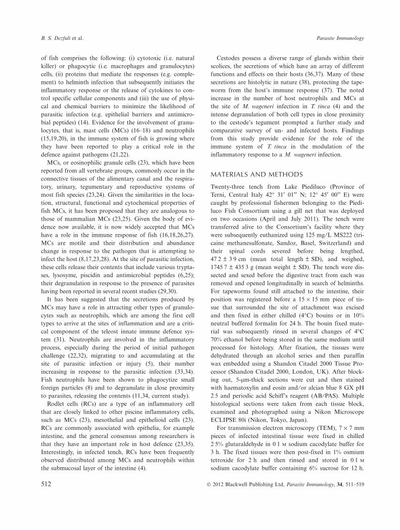
Fourteen (60Æ9%) of the 23 tench were parasitized withM. wageneri; identity of the cestodes was confirmed usingmorphology and standard taxonomic keys. The intensityof infection ranged from 3 to 130 worms per host
(39Æ5 € 47Æ7, mean € SD). The anterior part of the intes-tine bore the heaviest infections with the vast majority oftapeworms still attached with their scolices embeddedwithin the intestinal wall (Figure 1a). Upon dissectionin situ, M. wageneri were noticed in groups of variablenumbers and in some portion of the host intestine the pre-sence of more than one foci was frequent (Figure 1a). Intench gut wall, at the site of M. wageneri attachment, araised plaque-like formation or round nodule encircled thefirmly attached scolex (Figure 1b).
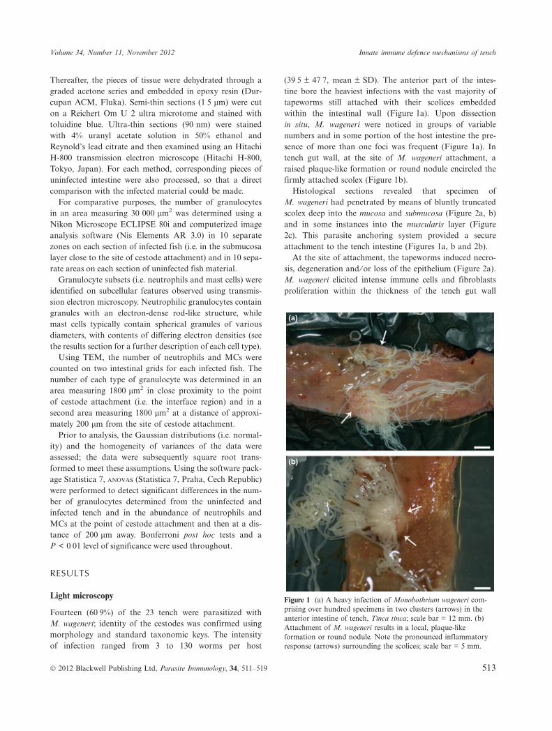
Histological sections revealed that specimen ofM. wageneri had penetrated by means of bluntly truncatedscolex deep into the mucosa and submucosa (Figure 2a, b)and in some instances into the muscularis layer (Figure2c). This parasite anchoring system provided a secureattachment to the tench intestine (Figures 1a, b and 2b).
At the site of attachment, the tapeworms induced necro-sis, degeneration and ⁄ or loss of the epithelium (Figure 2a).M. wageneri elicited intense immune cells and fibroblastsproliferation within the thickness of the tench gut wall
(a)
(b)
Figure 1 (a) A heavy infection of Monobothrium wageneri com-prising over hundred specimens in two clusters (arrows) in theanterior intestine of tench, Tinca tinca; scale bar = 12 mm. (b)Attachment of M. wageneri results in a local, plaque-likeformation or round nodule. Note the pronounced inflammatoryresponse (arrows) surrounding the scolices; scale bar = 5 mm.
Volume 34, Number 11, November 2012 Innate immune defence mechanisms of tench
� 2012 Blackwell Publishing Ltd, Parasite Immunology, 34, 511–519 513

(Figure 2b, c). Diffuse hyperplastic inflammation wasnoticed in tench with few M. wageneri as well as in thoseharbouring numerous tapeworms (Figure 2a–c). Withinthe submucosa layer, beneath the point of M. wageneri sco-lex insertion, numerous granulocytes (e.g. neutrophils,MCs) (Figure 2d), rodlet cells (Figure 2e) and collagenousfibres were observed. Degranulation of the granulocytes,which was visible by light microscopy (Figure 2d), was
common in the submucosa. Parasitized intestines weredetermined to have a significantly higher number of granu-locytes than those that were uninfected (Table 1; ANOVA,P < 0Æ01).
In situ, cestode-infected areas of the intestine were cov-ered ⁄ surrounded by a yellowish catarrh. In histologicalsections, the occurrence of numerous alcian blue–positivemucous cells was observed among the intestinal epithelial
(a) (b)
(c) (d)
(e)
Figure 2 (a) Transverse section through the intestine of a tench, Tinca tinca, infected with Monobothrium wageneri. There is a marked lackof epithelia at the site of parasite attachment and an intense inflammatory response (curved arrows) surrounding the scolices. Note thepresence of intact epithelia (arrows) in close proximity to the parasite induced nodule; scale bar = 200 lm. (b) Anterior intestine of a tenchinfected with several M. wageneri. Deep penetration of the scolices (asterisks) and necks of the tapeworms can be seen. M = muscularis;scale bar = 200 lm. (c) Focal attachment of M. wageneri. An intense host cellular response (curved arrows) penetrating the intestine as faras the muscularis and surrounding the cestode scolices (asterisks) can be seen. M = muscularis; scale bar = 200 lm. (d) Scolex of M. wage-neri (asterisk) is encircled by several granulocytes (curved arrows); scale bar = 10 lm. (e) Within the submucosa, the scolex is surroundedby numerous granulocytes (arrows). In addition, several rodlet cells (arrow heads) can be seen inside the capillaries or scattered among thegranulocytes and collagenous fibres; scale bar = 20 lm.
B. S. Dezfuli et al. Parasite Immunology
514 � 2012 Blackwell Publishing Ltd, Parasite Immunology, 34, 511–519
cells of infected fish notably within the epithelia in closeproximity to the nodule (Figure 2a).
Transmission electron microscopy
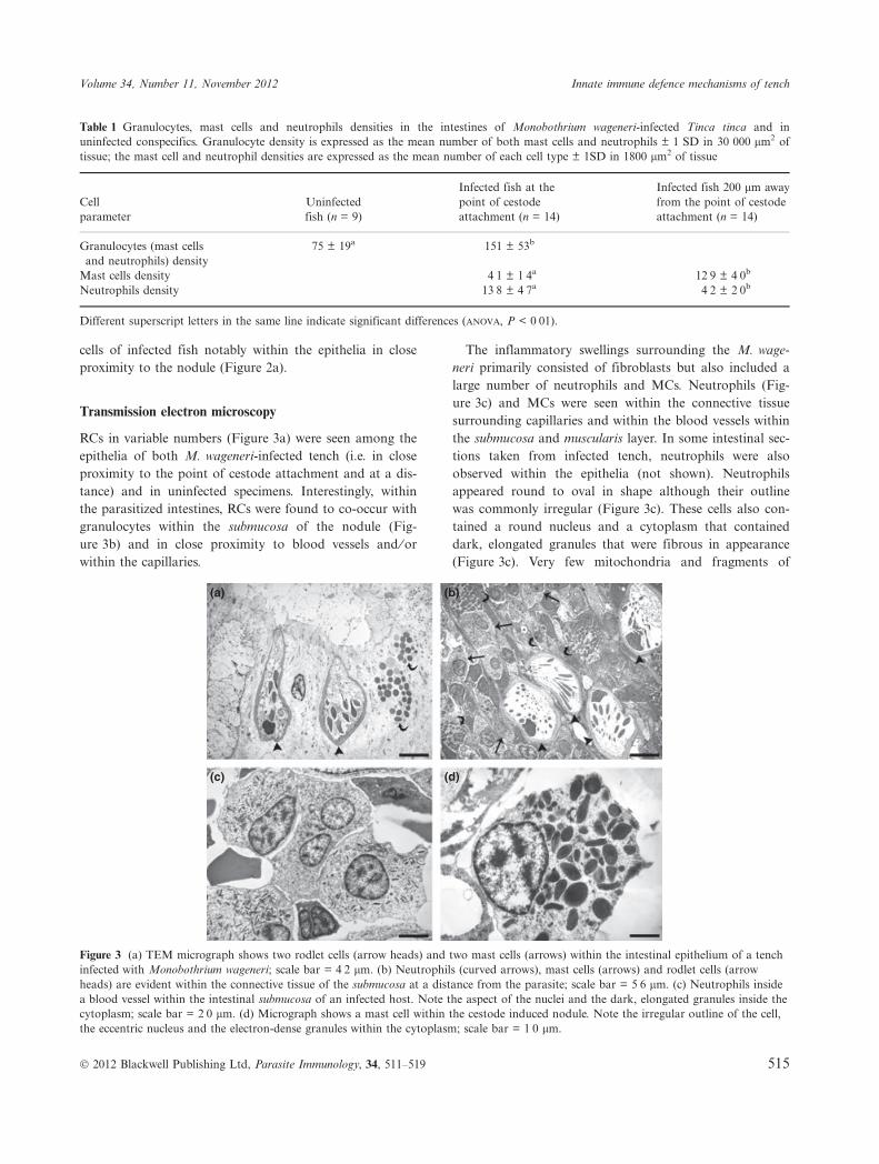
RCs in variable numbers (Figure 3a) were seen among theepithelia of both M. wageneri-infected tench (i.e. in closeproximity to the point of cestode attachment and at a dis-tance) and in uninfected specimens. Interestingly, withinthe parasitized intestines, RCs were found to co-occur withgranulocytes within the submucosa of the nodule (Fig-ure 3b) and in close proximity to blood vessels and ⁄ orwithin the capillaries.
The inflammatory swellings surrounding the M. wage-neri primarily consisted of fibroblasts but also included alarge number of neutrophils and MCs. Neutrophils (Fig-ure 3c) and MCs were seen within the connective tissuesurrounding capillaries and within the blood vessels withinthe submucosa and muscularis layer. In some intestinal sec-tions taken from infected tench, neutrophils were alsoobserved within the epithelia (not shown). Neutrophilsappeared round to oval in shape although their outlinewas commonly irregular (Figure 3c). These cells also con-tained a round nucleus and a cytoplasm that containeddark, elongated granules that were fibrous in appearance(Figure 3c). Very few mitochondria and fragments of
Table 1 Granulocytes, mast cells and neutrophils densities in the intestines of Monobothrium wageneri-infected Tinca tinca and inuninfected conspecifics. Granulocyte density is expressed as the mean number of both mast cells and neutrophils € 1 SD in 30 000 lm2 oftissue; the mast cell and neutrophil densities are expressed as the mean number of each cell type € 1SD in 1800 lm2 of tissue
Cellparameter
Uninfectedfish (n = 9)
Infected fish at thepoint of cestodeattachment (n = 14)
Infected fish 200 lm awayfrom the point of cestodeattachment (n = 14)
Granulocytes (mast cellsand neutrophils) density
75 € 19a 151 € 53b
Mast cells density 4Æ1 € 1Æ4a 12Æ9 € 4Æ0b
Neutrophils density 13Æ8 € 4Æ7a 4Æ2 € 2Æ0b
Different superscript letters in the same line indicate significant differences (anova, P < 0Æ01).
(a) (b)
(c) (d)
Figure 3 (a) TEM micrograph shows two rodlet cells (arrow heads) and two mast cells (arrows) within the intestinal epithelium of a tenchinfected with Monobothrium wageneri; scale bar = 4Æ2 lm. (b) Neutrophils (curved arrows), mast cells (arrows) and rodlet cells (arrowheads) are evident within the connective tissue of the submucosa at a distance from the parasite; scale bar = 5Æ6 lm. (c) Neutrophils insidea blood vessel within the intestinal submucosa of an infected host. Note the aspect of the nuclei and the dark, elongated granules inside thecytoplasm; scale bar = 2Æ0 lm. (d) Micrograph shows a mast cell within the cestode induced nodule. Note the irregular outline of the cell,the eccentric nucleus and the electron-dense granules within the cytoplasm; scale bar = 1Æ0 lm.
Volume 34, Number 11, November 2012 Innate immune defence mechanisms of tench
� 2012 Blackwell Publishing Ltd, Parasite Immunology, 34, 511–519 515

rough endoplasmic reticulum were observed in the cyto-plasm of the neutrophils.
The MCs, which were frequently observed within theepithelia of infected hosts (Figure 3a), were irregular inshape with an eccentric, polar nucleus, and a cytoplasmcharacterized by numerous large, electron-dense, mem-brane-bounded granules (Figure 3d). The cytoplasm typi-cally contained two to three mitochondria and aninconspicuous Golgi apparatus. Accurate counts of MCsand neutrophils were obtained from two intestinal gridsfrom each infected fish. Neutrophils were found to benumerous within the nodule, in close proximity to the teg-ument of the cestode, but their number was seen todecrease towards the periphery of the nodule. Neutrophilswere significantly more abundant than MCs (Table 1; ANO-
VA, P < 0Æ01) in host tissue close to the point of cestodeattachment. At a distance of 200 lm from the site of para-site attachment, however, the number of neutrophils wassignificantly lower than the MCs (Table 1; ANOVA,P < 0Æ01). There were significant differences in the numberof neutrophils in close proximity to and at a distance of200 lm from the point of cestode attachment (Table 1;ANOVA, P < 0Æ01). Likewise, there were significant differ-ences in the number of MCs at the site of infection and200 lm away (Table 1; ANOVA, P < 0Æ01).
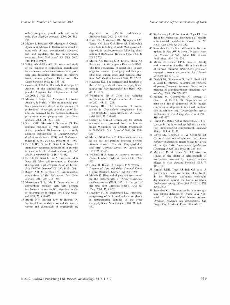
Commonly, the neutrophils and MCs adjacent to theM. wageneri scolex tegument had a cytoplasm thatappeared vacuolized (Figure 4a) and contained very feworganelles. These were quite unlike the same cell typesobserved in zones further away from the body of the cestode(e.g. Figure 3b). The degranulation of MCs and neutrophilswas characterized by free granules that were frequently seenclose to the capilliform filitriches (Figure 4b) or adjacent toand ⁄ or between the coniform spinitriches of the scolex (Fig-ure 4b) (see 39 for cestode microtriche terminology). Insome grids, because of the plane of the section, the freegranules from neutrophils and MCs were found in contactwith the scolex tegument (respectively, Figure 4c,d).
Several glandular cytons within the syncytial tegumentalong the anterior and lateral parts of the M. wageneriscolex were observed (not shown). No discharge fromthese glands or the presence of an adhesive layer in theinterface region between the tench intestine and the tape-worm was evident.
DISCUSSION
Cyprinids are the main group of freshwater fish that havea global importance as a source of food in many countries.The study of disease in cyprinids held in captivity and in
(a) (b)
(c) (d)
Figure 4 (a) Neutrophils (arrow heads) and mast cells (arrows) adjacent to the scolex tegument of Monobothrium wageneri (asterisk). Thecytoplasm of both cell types appears vacuolized and contains very few organelles; scale bar = 4Æ9 lm. (b) A neutrophil (curved arrow)adhering to the scolex microtriches with the presence of free mast cell granules (arrows) evident among the microtriches. The asteriskdenotes the scolex tegument; scale bar = 2Æ0 lm. (c) Micrograph showing the free granules of a neutrophil adhering to or in close vicinityto the tegument of the scolex (asterisk); scale bar = 0Æ6 lm. (d) Degranulation of a mast cell is characterized by free electron-dense granules(arrows) in close contact with the tegument of the scolex (asterisk); scale bar = 0Æ7 lm.
B. S. Dezfuli et al. Parasite Immunology
516 � 2012 Blackwell Publishing Ltd, Parasite Immunology, 34, 511–519

semi-wild stocks is essential for Public Health Authority.The pathological alterations to the intestine of cyprinidsdue to cestodes have been detailed in several papers(3,4,40). Among gross effects of tapeworms on fish hosts,intestinal occlusion and rupture are infrequent andextreme consequences of cestode infection (41). Such phe-nomena are among the most serious impacts induced byintestinal tapeworms, which have been associated withdebilitation, nutritional disturbance and even the death ofheavily parasitized fish (42).
Generally, infection of the gastrointestinal tract by para-sites has detrimental effects on digestion function (5,7).Most intestinal pathology associated with tapeworm infec-tions results from the deep penetration of the scolex intothe gut wall (43). The organs used by intestinal helminthsduring the process of attachment to their host’s gut fre-quently induces inflammation of the alimentary canal(5,10). This is the case in M. wageneri that induces markedpathological changes, penetrating the muscularis layer (41,current study), causing a significant inflammatoryresponse in all layers of the intestine in both light andheavy infections.
M. wageneri is a caryophyllidean cestode and it wasreported that the tegumentary glands of this group oftapeworms release neutral glycoproteins which protect theparasite against host cellular responses (44). This interpre-tation, however, does not appear plausible given that nodischarge from these glands nor the presence of anadhesive layer between the tench intestine and M. wageneriwas evident in the material studied here. The presence ofabundant immune cells at the site of M. wageneri attach-ment and presence of free granules discharged from MCsand neutrophils in close contact with the scolex micro-triches rule out earlier interpretations (44).
Rodlet cells (23) and two type of granulocytes, MCs(23,24,30,45) and neutrophils (20,31), have been repeatedlyshown to play an essential role in the immune system offish. There is therefore a growing interest regarding therole of these inflammatory cells in the innate immune sys-tem of fish (21). Granulocytes are generally consideredeffector cells of the innate immune response (46). Theimportance of each of these cell types (i.e. RCs, MCs andneutrophils) therefore is worth considering in the contextof the current study.
Recent studies on both wild and farmed fish suggestthat RCs represent an immune cell type closely linked toother piscine inflammatory cells (45,47). RCs are foundexclusively in fish in a wide range of tissues and are com-monly associated with epithelia (23). As M. wageneridestroys the epithelia at the site of attachment, it was notpossible to compare the number of RCs in uninfected andparasitized tench. The presence of RCs in the intestinal
submucosa of infected tench and those in direct contactwith the blood vessels is interesting and suggests that RCsalso use the circulatory system to migrate to the site ofinfection. Similar findings have been reported for fish thatwere infected with acanthocephalans (10,48).
Fish MCs, also known as eosinophilic granule cells,have cytochemical features, functional properties and tis-sue locations that have led to the suggestion that they areanalogous to mammalian MCs (22,23,25). Several pub-lished reports on the intratissue migratory nature of MCssuggest that fish may have two populations of MCs, onecirculating and one resident, and that the presence of par-asites induces the recruitment of MCs to the site of infec-tion (25,28). The significantly higher number of MCsfound at the site of parasite attachment, when comparedto uninfected tench, in the current study supports similarresults reported for other fish–helminth systems (48).
In teleosts, considerable descriptive data exist showinghow MCs degranulate in response to a variety of knowndegranulating agents (49) and pathogens (23,25,30). Inparasitized tench, an intense degranulation of MCs wasseen at the site of tapeworm infection, notably in theimmediate zone surrounding the scolex. It is likely that thesecretions produced by the MCs may have a role inattracting other cell types (i.e. neutrophils) involved in theinflammatory process, particularly during the period ofinitial pathogen challenge (24,32). One study reported thatintra-epithelial MCs are present in low numbers in healthyepithelium but then dramatically increase in number withcertain parasitic infections (50). In the current study, MCs,in the intestines of parasitized tench, were frequentlyobserved among epithelial cells.
Neutrophils are among the first cell types to arrive at thesites of inflammation and play a critical role in the teleostinnate immune defence system (31). In infected tench,numerous neutrophils were observed to co-occur with MCsin the submucosa at the sites of M. wageneri attachment. Asimilar observation was found in the livers of minnows,Phoxinus phoxinus (L.), infected with the nematode larvaeof Raphidascaris acus (Bloch, 1779) (17). The findings fromthe current study suggest that the neutrophils appear tohave closer contact with the tegument of the cestode thando the MCs. Neutrophils commonly co-occur with macro-phages that readily engulf small extracellular pathogens,such as viruses and bacteria (12), or parasites of a smallersize, such as the migrating diplostomules of Diplostomumspathaceum (Rudolphi, 1819), that can be killed by hostmacrophages (51). No macrophages were encountered atthe sites of M. wageneri attachment in the current studyand as yet the reasons for their absence are unknownand are open to conjecture. One possible interpretationis that the size of M. wageneri, which can measure sev-
Volume 34, Number 11, November 2012 Innate immune defence mechanisms of tench
� 2012 Blackwell Publishing Ltd, Parasite Immunology, 34, 511–519 517

eral centimetres in length, is too large to be effectivelyengulfed by host macrophages. Based on the currentstudy, it appears that an infection of M. wageneri intench preferentially induces the recruitment of neutroph-ils and MCs and, to a lesser degree, RCs.
There are several records of mammals infected by helm-inths where the host cells (e.g. macrophages) were able tokill trematode larvae (52) and ⁄ or eosinophils and neu-trophils were able to kill adult and nematode larvae(33,34,53). The mechanism by which these cells mediatedprotection against helminth infection is that they arerecruited at the site of infection, where they surround theworm and then adhere to the parasite’s body. The eosin-ophils and neutrophils then degranulate on the cuticle ofnematodes (33,34,53), while the macrophages penetratethe tegument of the trematode (52) inflicting damage thatultimately results in the death of the parasite. The tightclustering of M. wageneri and the deep penetration oftheir scolices inflict severe mechanical damage to theirhost’s intestine. The presence of this tapeworm in tenchinduces an intense inflammatory response that results inthe migration and recruitment of RCs, neutrophils andMCs to the site of infection and the subsequent degranu-
lation of cells, which release their contents into the zoneimmediately next to the scolex tegument. No dead tape-worms were encountered during dissection; nevertheless,the roles of MCs and neutrophils as effectors of innateimmunity against histozoic parasites require further inves-tigation (54). The findings from the current study agreeclosely with the statement of Feist and Longshaw (9),who said ‘In most instances, an evolutionary balance hasbeen achieved between the host and the parasite andeven when histopathology is evident, this is frequentlylocalised and does not unduly impair performance of theaffected organ. Examples include chronic inflammation,granuloma formation and focal fibrosis’.
ACKNOWLEDGEMENTS
We are grateful to S. Squerzanti, A. Margutti and P. Bold-rini from the University of Ferrara for technical assistancewith aspects of this study. Thanks are due to F. Bisonnifrom the Fisheries Cooperation of the Lake Piediluco forhis assistance in collecting fish. This work was supportedby grants from the Italian Ministry of the University andScientific Research and Technology.
REFERENCES
1 Gibson DI. Monobothrium wageneri: anotherimported tapeworm established in wild Brit-ish freshwater fishes? J Fish Biol 1993; 43:281–285.
2 Kennedy CR. The ecology of introductions.In Pike AW & Lewis JW (eds): Parasitic Dis-eases of Fish. Dyfed, UK: Samara Publish-ing, 1994: 189–208.
3 Mackiewicz JS, Cosgrove GE & Gude WD.Relationship of pathology to scolex mor-phology among caryophyllid cestodes.Z Parasitenk 1972; 39: 233–246.
4 Dezfuli BS, Giari L, Squerzanti S, et al. His-tological damage and inflammatory responseelicited by Monobothrium wageneri (Cestoda)in the intestine of Tinca tinca (Cyprinidae).Parasit Vector 2011; 4: 225.
5 Secombes CJ & Chappell LH. Fish immuneresponses to experimental and natural infec-tion with helminth parasites. Annu Rev FishDis 1996; 6: 167–177.
6 Rombout JHWM, Abelli L, Picchietti S,Scapigliati G & Kiron V. Teleost intestinalimmunology. Fish Shellfish Immunol 2011;31: 616–626.
7 Hoste H. Adaptive physiological process inthe host during gastrointestinal parasitism.Int J Parasitol 2001; 31: 231–244.
8 Alvarez-Pellitero P. Fish immunity and para-site infections: from innate immunity toimmunoprophylactic prospects. Vet ImmunolImmunopathol 2008; 126: 171–198.
9 Feist SW & Longshaw M. Histopathologyof fish parasite infections-importance forpopulations. J Fish Biol 2008; 73: 2143–2160.
10 Dezfuli BS, Castaldelli G, Bo T, LorenzoniM & Giari L. Intestinal immune response ofSilurus glanis and Barbus barbus naturallyinfected with Pomphorhynchus laevis (Acan-thocephala). Parasite Immunol 2011; 33: 116–123.
11 Sears BF, Rohr JR, Allen JE & MartinLB. The economy of inflammation: when isless more? Trends Parasitol 2011; 27: 382–387.
12 Ellis AE. Innate host defence mechanisms offish against viruses and bacteria. Dev CompImmunol 2001; 25: 827–839.
13 Paperna I & Dzikowski R. Digenea (PhylumPlatyhelminthes). In Woo PTK (ed.): FishDiseases and Disorders, Vol. 1. Protozoan andMetazoan Infections. Wallingford, Oxon,UK: CAB International, 2006: 345–390.
14 Dixon B & Stet RJ. The relationship betweenmajor histocompatibility receptors andinnate immunity in teleost fish. Dev CompImmunol 2001; 25: 683–699.
15 Magnad�ttir B. Innate immunity of fish(overview). Fish Shellfish Immunol 2006; 20:137–151.
16 Silphaduang U & Noga E. Peptide antibiot-ics in mast cells of fish. Nature 2001; 414:268–269.
17 Dezfuli BS, Manera M & Giari L. Immuneresponse to nematode larvae in the liver andpancreas of minnow, Phoxinus phoxinus (L.).J Fish Dis 2009; 32: 383–390.
18 Andrews M, Battaglene S, Cobcroft J,Adams M, Noga E & Nowak B. Hostresponse to the chondracathid copepodChondracanthus goldsmidi, a gill parasite ofthe striped trumpeter, Latris lineata (For-ster), in Tasmania. J Fish Dis 2010; 33: 211–220.
19 Hamdani SH, McMillan DN, Pettersen EF,et al. Isolation of rainbow trout neutrophilswith an anti-granulocyte monoclonal anti-body. Vet Immunol Immunopathol 1998; 63:369–380.
20 Forlenza M, Scharsack JP, KachamakovaNM, Taverne-Thiele AJ, Rombout JHWM &Wiegertjes GF. Differential contribution ofneutrophilic granulocytes and macrophagesto nitrosative stress in a host–parasite animalmodel. Mol Immunol 2008; 45: 3178–3189.
21 Jones SRM. The occurrence and mechanismsof innate immunity against parasites in fish.Dev Comp Immunol 2001; 25: 841–852.
22 Ur�n PA, Schrama JW, Rombout JHWM,et al. Time-related changes of the intestinalmorphology of Atlantic salmon, Salmo salarL., at two different soybean meal inclusionlevels. J Fish Dis 2009; 32: 733–744.
23 Reite OB & Evensen Ø. Inflammatory cellsof teleostean fish: a review focusing on mast
B. S. Dezfuli et al. Parasite Immunology
518 � 2012 Blackwell Publishing Ltd, Parasite Immunology, 34, 511–519
cells ⁄ eosinophilic granule cells and rodletcells. Fish Shellfish Immunol 2006; 20: 192–208.
24 Mulero I, Sepulcre MP, Meseguer J, Garcia-Ayala A & Mulero V. Histamine is stored inmast cells of most evolutionarily advancedfish and regulates the fish inflammatoryresponse. Proc Natl Acad Sci USA 2007;104: 19434–19439.
25 Vallejo AN & Ellis AE. Ultrastructural studyof the response of eosinophilic granule cellsto Aeromonas salmonicida extracellular prod-ucts and histamine liberators in rainbowtrout, Salmo gairdneri Richardson. DevComp Immunol 1989; 13: 133–148.
26 Colorni A, Ullal A, Heinisch G & Noga EJ.Activity of the antimicrobial polypeptidepiscidin 2 against fish ectoparasites. J FishDis 2008; 31: 423–432.
27 Mulero I, Noga EJ, Meseguer J, Garc�a-Ayala A & Mulero V. The antimicrobial pep-tides piscidins are stored in the granules ofprofessional phagocytic granulocytes of fishand are delivered to the bacteria-containingphagosome upon phagocytosis. Dev CompImmunol 2008; 32: 1531–1538.
28 Sharp GJE, Pike AW & Secombes CJ. Theimmune response of wild rainbow troutSalmo gairdneri Richardson to naturallyacquired plerocercoid of Diphyllobothriumdendriticun (Nitzsch 1824) and D. ditremun(Creplin 1825). J Fish Biol 1989; 35: 781–794.
29 Dezfuli BS, Pironi F, Giari L & Noga EJ.Immunocytochemical localization of piscidinin mast cells of infected seabass gill. FishShellfish Immunol 2010; 28: 476–482.
30 Dezfuli BS, Giari L, Lui A, Lorenzoni M &Noga EJ. Mast cell responses to Ergasilus(Copepoda), a gill ectoparasite of sea bream.Fish Shellfish Immunol 2011; 30: 1087–1094.
31 Rieger AM & Barreda DR. Antimicrobialmechanisms of fish leukocytes. Dev CompImmunol 2011; 35: 1238–1245.
32 Matsuyama T & Iida T. Degranulation ofeosinophilic granular cells with possibleinvolvement in neutrophil migration to siteof inflammation in tilapia. Dev Comp Immu-nol 1999; 23: 451–457.
33 Brattig NW, B�ttner DW & Hoerauf A.Neutrophil accumulation around Onchocercaworms and chemotaxis of neutrophils are
dependent on Wolbachia endobacteria.Microbes Infect 2001; 3: 439–446.
34 Nfon CK, Makepeace BL, Njongmeta LM,Tanya VN, Bain O & Trees AJ. Eosinophilscontribute to killing of adult Onchocerca och-engi within onchocercomata following elimi-nation of Wolbachia. Microbes Infect 2006; 8:2698–2705.
35 Mazon AF, Huising MO, Taverne-Thiele AJ,Bastiaans J & Verburg-van Kemenade BML.The first appearance of rodlet cells in carp(Cyprinus carpio L.) ontogeny and their pos-sible roles during stress and parasite infec-tion. Fish Shellfish Immunol 2007; 22: 27–37.
36 Hayunga EG. The structure and function ofthe scolex glands of three caryophyllideantapeworms. Proc Helminthol Soc Wash 1979;46: 171–179.
37 Whittington ID & Cribb BW. Adhesivesecretions in the Platyhelminthes. Adv Parasi-tol 2001; 48: 101–224.
38 Farooqi HU. The occurrence of frontalglands in Tentacularia coryphaenae Bosc1802 (Cestoda: Trypanorhyncha). Z Parasit-enkd 1986; 72: 653–659.
39 Chervy L. Unified terminology for cestodemicrotriches: a proposal from the Interna-tional Workshops on Cestode Systematicsin 2002-2008. Folia Parasitol 2009; 56: 199–230.
40 Morley NJ & Hoole D. Ultrastructural stud-ies on the host-parasite interface betweenKhawia sinensis (Cestoda: Caryophyllidea)and carp Cyprinus carpio. Dis Aquat Org1995; 23: 93–99.
41 Williams H & Jones A. Parasitic Worms ofFishes. London: Taylor & Francis Ltd, 1994:593.
42 Hoole D, Bucke D, Burgess P & Wellby I.Diseases of Carp and Other Cyprinid Fishes.Oxford: Blackwell Science Ltd, 2001: 280.
43 Moln�r K. Histopathological changes causedby the metacestodes of Neogryporhynchuscheilancristrotus (Wedl, 1855) in the gut ofthe gibel carp Carassius gibelio. Acta VetHung 2005; 53: 45–52.
44 Davydov VG & Poddubnaya LG. Functionalmorphology of the frontal and uterine glandsin representative cestodes of the orderCaryophyllidea. Parazitologiia 1988; 22: 449–457.
45 Silphaduang U, Colorni A & Noga EJ. Evi-dence for widespread distribution of piscidinantimicrobial peptides in teleost fish. DisAquat Org 2006; 72: 241–252.
46 Secombes CJ. Cellular defences in fish: anupdate. In Pike AW & Lewis JW (eds): Para-sitic Diseases of Fish. Dyfed, UK: SamaraPublishing, 1994: 209–224.
47 Matisz CE, Goater CP & Bray D. Densityand maturation of rodlet cells in brain tissueof fathead minnows (Pimephales promelas)exposed to trematode cercariae. Int J Parasi-tol 2010; 40: 307–312.
48 Dezfuli BS, Giovinazzo G, Lui A, Boldrini P& Giari L. Intestinal inflammatory responseof powan Coregonus lavaretus (Pisces) to thepresence of acanthocephalan infections. Par-asitology 2009; 136: 929–937.
49 Manera M, Giammarino A, Borreca C,Giari L & Dezfuli BS. Degranulation ofmast cells due to compound 48 ⁄ 80 inducesconcentration-dependent intestinal contrac-tion in rainbow trout (Oncorhynchus mykissWalbaum) ex vivo. J Exp Zool Part A 2011;315: 447–457.
50 Ernest PB, Befus AD & Bienenstock J. Leu-kocytes in the intestinal epithelium: an unu-sual immunological compartment. ImmunolToday 1985; 6: 50–55.
51 Whyte SK, Chappell LH & Secombes CJ.Cytotoxic reactions of rainbow trout, Salmogairdneri Richardson, macrophages for larvaeof the eye fluke Diplostomum spathaceum(Digenea). J Fish Biol 1989; 35: 333–345.
52 McLaren DJ & James SL. Ultrastructurestudies of the killing of schistosomula ofSchistosoma mansoni by activated macro-phages in vitro. Parasite Immunol 1985; 7:315–331.
53 Hansen RDE, Trees AJ, Bah GS, et al. Aworm’s best friend: recruitment of neutroph-ils by Wolbachia confounds eosinophildegranulation against the filarial nematodeOnchocerca ochengi. Proc Biol Sci 2011; 278:2293–2302.
54 Secombes CJ. The nonspecific immune sys-tem: cellular defences. In Iwama G & Nak-anishi T (eds): The Fish Immune System:Organism Pathogen and Environment. SanDiego, CA: Academic Press, 1996: 63–105.
Volume 34, Number 11, November 2012 Innate immune defence mechanisms of tench
� 2012 Blackwell Publishing Ltd, Parasite Immunology, 34, 511–519 519