Follistatin-related protein/follistatin-like 1 evokes an innate immune response via CD14 and...
6
Follistatin-related protein/follistatin-like 1 evokes an innate immune response via CD14 and toll-like receptor 4 Kosaku Murakami a , Masao Tanaka b,⇑ , Takashi Usui a , Daisuke Kawabata a , Aoi Shiomi a , Mikiko Iguchi-Hashimoto a , Masakazu Shimizu a , Naoichiro Yukawa a , Hajime Yoshifuji a , Takaki Nojima a , Koichiro Ohmura a , Takao Fujii a , Hisanori Umehara b , Tsuneyo Mimori a a Department of Rheumatology and Clinical Immunology, Kyoto University Graduate School of Medicine, Kyoto 606-8507, Japan b Division of Hematology and Immunology, Department of Internal Medicine, Kanazawa Medical University, Ishikawa 920-0293, Japan article info Article history: Received 2 November 2011 Revised 2 January 2012 Accepted 4 January 2012 Available online 17 January 2012 Edited by Masayuki Miyasaka Keywords: FRP FSTL1 CD14 TLR4 Innate immunity Rheumatoid arthritis abstract Follistatin-related protein (FRP)/follistatin-like 1 (FSTL1) has multi-specific binding nature espe- cially with TGF-b superfamily proteins, and thereby modulates organ development. However, its function in immune systems remains unclear. Previously, we reported FRP interacts with CD14, which is known to mediate toll-like receptor 4 (TLR4) signaling. Here, we investigated whether FRP activates TLR4 signaling. Recombinant FRP induced interleukin 6 or interleukin 8 production from target cells in a CD14- and TLR4-dependent manner. Moreover, similar to lipopolysaccharide (LPS), FRP induced tolerance to the second LPS stimulation. FRP has the function of evoking innate immune responses as one of the endogenous TLR4 agonists. Structured summary of protein interactions: FRP physically interacts with CD14 by fluorescence-activated cell sorting (View interaction) Ó 2012 Federation of European Biochemical Societies. Published by Elsevier B.V. All rights reserved. 1. Introduction Follistatin-related protein (FRP)/follistatin-like 1 (FSTL1) is a secreted glycoprotein and a member of the follistatin protein family. FRP binds transforming growth factor b (TGF-b) superfamily proteins: activin, TGF-b, bone morphogenetic protein 2/4, their receptors and follistatin [1]. Previously, we and Ouchi et al. reported that DIP2 disco-interacting protein homolog A from Drosophila (DIP2A) is one of the receptors for FRP [1,2]. Through the interaction with these molecules, FRP appears to play a role in the regulation of tissue development or formation. Findings on the immunological activities of FRP began to be reported after our discovery of FRP as one of the target antigens of autoantibodies in rheumatoid arthritis (RA) [3]. Subsequently, we reported that serum autoantibodies to FRP were detected in a substantial proportion of patients with RA, and their appearance correlated with disease activity. These autoantibodies neutralized FRP activity and abrogated its suppressive effect on the production of matrix metalloproteinases (MMPs/Mmps) and prostaglandins from synovial cells [4]. Administration of recombinant human FRP to mice with anti-type II collagen antibody-induced arthritis (CAIA) ameliorated their arthritis and down-regulated Fos, Ets-2, interleukin 6 (IL-6), Mmp3 and Mmp9 gene expression in their joint tissue [5]. Le Luduec et al. reported that adenovirus-mediated systemic administration of FRP to rats with transplanted hearts improved their allograft survival, and down-regulated IL-6, inter- leukin 17A and interferon c gene expression in their allografts [6]. On the contrary, Miyame et al. reported that overexpression of FRP using an adenoviral vector in mice with collagen-induced arthritis (CIA) exacerbated their arthritis and promoted the pro- duction of IL-6, tumor necrosis factor a and interleukin 1b [7]. This group also reported that administration of FRP-neutralizing antibodies to CIA mice reduced their arthritis severity [8]. These inconsistent results prevented us from understanding the definite role of FRP in the immune system. 0014-5793/$36.00 Ó 2012 Federation of European Biochemical Societies. Published by Elsevier B.V. All rights reserved. doi:10.1016/j.febslet.2012.01.010 Abbreviations: FRP, follistatin-related protein; FSTL1, follistatin-like 1; TLR4, toll-like receptor 4; TGF-b, transforming growth factor b; DIP2A, disco-interacting protein 2 homolog A from Drosophila; RA, rheumatoid arthritis; MMPs/Mmps, matrix metalloproteinases; CAIA, anti-type II collagen antibody-induced arthritis; IL-6, interleukin 6; CIA, collagen-induced arthritis; LPS, lipopolysaccharide; PMB, Polymixin B; RS-LPS, Rhodobacter sphaeroides LPS; IL-8, interleukin 8; PAMPs, pathogen-associated molecular patterns; DAMPs, damage-associated molecular patterns; MRP-8/14, myeloid-related protein-8/14; HMGB1, high-mobility group box1 ⇑ Corresponding author. Address: Division of Hematology and Immunology, Department of Internal Medicine, Kanazawa Medical University, 1-1 Daigaku, Uchinada, Kahoku-gun, Ishikawa 920-0293, Japan. Fax: +81 76 286 9290. E-mail address: [email protected] (M. Tanaka). FEBS Letters 586 (2012) 319–324 journal homepage: www.FEBSLetters.org
-
Upload
independent -
Category
Documents
-
view
2 -
download
0
Transcript of Follistatin-related protein/follistatin-like 1 evokes an innate immune response via CD14 and...
FEBS Letters 586 (2012) 319–324
journal homepage: www.FEBSLetters .org
Follistatin-related protein/follistatin-like 1 evokes an innate immune responsevia CD14 and toll-like receptor 4
Kosaku Murakami a, Masao Tanaka b,⇑, Takashi Usui a, Daisuke Kawabata a, Aoi Shiomi a,Mikiko Iguchi-Hashimoto a, Masakazu Shimizu a, Naoichiro Yukawa a, Hajime Yoshifuji a,Takaki Nojima a, Koichiro Ohmura a, Takao Fujii a, Hisanori Umehara b, Tsuneyo Mimori a
a Department of Rheumatology and Clinical Immunology, Kyoto University Graduate School of Medicine, Kyoto 606-8507, Japanb Division of Hematology and Immunology, Department of Internal Medicine, Kanazawa Medical University, Ishikawa 920-0293, Japan
a r t i c l e i n f o a b s t r a c t
Article history:Received 2 November 2011Revised 2 January 2012Accepted 4 January 2012Available online 17 January 2012
Edited by Masayuki Miyasaka
Keywords:FRPFSTL1CD14TLR4Innate immunityRheumatoid arthritis
0014-5793/$36.00 � 2012 Federation of European Biodoi:10.1016/j.febslet.2012.01.010
Abbreviations: FRP, follistatin-related protein; FStoll-like receptor 4; TGF-b, transforming growth factoprotein 2 homolog A from Drosophila; RA, rheumamatrix metalloproteinases; CAIA, anti-type II collageIL-6, interleukin 6; CIA, collagen-induced arthritis; LPolymixin B; RS-LPS, Rhodobacter sphaeroides LPS;pathogen-associated molecular patterns; DAMPs, dpatterns; MRP-8/14, myeloid-related protein-8/14; Hbox1⇑ Corresponding author. Address: Division of He
Department of Internal Medicine, Kanazawa MediUchinada, Kahoku-gun, Ishikawa 920-0293, Japan. Fa
E-mail address: [email protected] (M
Follistatin-related protein (FRP)/follistatin-like 1 (FSTL1) has multi-specific binding nature espe-cially with TGF-b superfamily proteins, and thereby modulates organ development. However, itsfunction in immune systems remains unclear. Previously, we reported FRP interacts with CD14,which is known to mediate toll-like receptor 4 (TLR4) signaling. Here, we investigated whetherFRP activates TLR4 signaling. Recombinant FRP induced interleukin 6 or interleukin 8 productionfrom target cells in a CD14- and TLR4-dependent manner. Moreover, similar to lipopolysaccharide(LPS), FRP induced tolerance to the second LPS stimulation. FRP has the function of evoking innateimmune responses as one of the endogenous TLR4 agonists.
Structured summary of protein interactions:FRP physically interacts with CD14 by fluorescence-activated cell sorting (View interaction)
� 2012 Federation of European Biochemical Societies. Published by Elsevier B.V. All rights reserved.
1. Introduction Findings on the immunological activities of FRP began to be
Follistatin-related protein (FRP)/follistatin-like 1 (FSTL1) is asecreted glycoprotein and a member of the follistatin proteinfamily. FRP binds transforming growth factor b (TGF-b) superfamilyproteins: activin, TGF-b, bone morphogenetic protein 2/4, theirreceptors and follistatin [1]. Previously, we and Ouchi et al. reportedthat DIP2 disco-interacting protein homolog A from Drosophila(DIP2A) is one of the receptors for FRP [1,2]. Through the interactionwith these molecules, FRP appears to play a role in the regulation oftissue development or formation.
chemical Societies. Published by E
TL1, follistatin-like 1; TLR4,r b; DIP2A, disco-interactingtoid arthritis; MMPs/Mmps,
n antibody-induced arthritis;PS, lipopolysaccharide; PMB,IL-8, interleukin 8; PAMPs,amage-associated molecularMGB1, high-mobility group
matology and Immunology,cal University, 1-1 Daigaku,x: +81 76 286 9290.
. Tanaka).
reported after our discovery of FRP as one of the target antigensof autoantibodies in rheumatoid arthritis (RA) [3]. Subsequently,we reported that serum autoantibodies to FRP were detected in asubstantial proportion of patients with RA, and their appearancecorrelated with disease activity. These autoantibodies neutralizedFRP activity and abrogated its suppressive effect on the productionof matrix metalloproteinases (MMPs/Mmps) and prostaglandinsfrom synovial cells [4]. Administration of recombinant humanFRP to mice with anti-type II collagen antibody-induced arthritis(CAIA) ameliorated their arthritis and down-regulated Fos, Ets-2,interleukin 6 (IL-6), Mmp3 and Mmp9 gene expression in theirjoint tissue [5]. Le Luduec et al. reported that adenovirus-mediatedsystemic administration of FRP to rats with transplanted heartsimproved their allograft survival, and down-regulated IL-6, inter-leukin 17A and interferon c gene expression in their allografts[6]. On the contrary, Miyame et al. reported that overexpressionof FRP using an adenoviral vector in mice with collagen-inducedarthritis (CIA) exacerbated their arthritis and promoted the pro-duction of IL-6, tumor necrosis factor a and interleukin 1b [7]. Thisgroup also reported that administration of FRP-neutralizingantibodies to CIA mice reduced their arthritis severity [8]. Theseinconsistent results prevented us from understanding the definiterole of FRP in the immune system.
lsevier B.V. All rights reserved.
320 K. Murakami et al. / FEBS Letters 586 (2012) 319–324
In the previous paper, we reported CD14, which is known tomediate toll-like receptor 4 (TLR4) signaling, is one of theFRP-interacting molecules [1]. Therefore, aiming to find the immu-nological function of FRP, we investigated whether FRP couldactivate TLR4 signaling.
2. Materials and methods
2.1. Flow cytometry
Cells were incubated with recombinant human FRP with a FLAGtag (OriGene) or FLAG-tagged bacterial alkaline phosphatase con-trol protein (Sigma) for 30 min, then stained with APC-conjugatedantibody to CD14 (Biolegend) and FITC-conjugated antibody toFLAG (Sigma). Stained cells were analyzed on a FACS Calibur (BD).
2.2. Cells and culture reagents
HEK293(293), HT-1080 and NIH-3T3 cells were purchased fromATCC. 293-hTLR4A-MD2-CD14 cells were purchased from Invivo-gen. 293-CD14 cells were generated by transfecting 293 with theplasmid pSPORT2-hCD14 (Thermo Fisher Scientific). 293-hTLR4-MD2 cells were generated by transfecting 293 with the plasmidpDUO-hMD2/TLR4A (Invivogen). Synovial tissue samples from RApatients were kindly provided by Dr. T. Nakamura (KyotoUniversity). This study was approved by the ethics committee ofthe Kyoto University Graduate School and Faculty of Medicine.Reagents used in these in vitro experiments were lipopolysaccha-rides (LPS; Sigma), Polymixin B (PMB; Nacalai Tesque), Rhodobactersphaeroides LPS (RS-LPS; Invivogen), and recombinant human FRP(OriGene) which has a FLAG tag. Recombinant mouse FRP was pre-pared by purifying the supernatant of pBMN-mFRP-transfectedHT-1080 cells with anti-FLAG tag antibody beads (WAKO). Con-taminated endotoxin was measured by Chromogenic LAL Endo-toxin Assay Kit (GenScript).
2.3. Western blotting
Samples were subjected to SDS–PAGE and transferred toImmobilon-NC transfer membranes (Millipore). The blots were re-acted with antibodies to human or mouse FRP (R&D systems) anddetected by HRP-conjugated anti-goat IgG antibody (Invitrogen).
2.4. Cytokine production analyses
3 � 104 cells were inoculated into the wells of 48-well platesfilled with 300 ll of Dulbecco’s modified Eagle’s medium including10% fetal calf serum and indicated reagents. After 24 h, the culturemedium was collected and assayed by mouse or human IL-6 ELISA(eBioscience) or human interleukin 8 (IL-8) ELISA (Biolegend).
2.5. Generation of FRP-overexpressing cells
A retrovirus vector pBMN-IRES-EGFP was kindly provided byDr. G. Nolan (Stanford University). FLAG-tagged mouse and humanFRP cDNAs were inserted into the multi-cloning site of pBMN-IRES-EGFP to make pBMN-mFRP and pBMN-hFRP. Production ofretroviruses and their infection to target cells was performed aspreviously described [9].
2.6. Mice
C57BL/6 and DBA/1J mice were purchased from Charles RiverLaboratories. Mice deficient in TLR4 (TLR4�/�) were purchasedfrom Oriental Bio Service. All animal procedures were approvedby the ethics committee on Animal Experimentation of Kyoto
University. Methods for ectopically FRP-overexpressing T cellstransfer experiments using CIA mice are described in Supplemen-tary material.
2.7. RNA interference (RNAi) analysis of FRP
The plasmid vector expressing microRNA (miR) against FRP,pcDNA6.2-GW/EmGFP-miR-FRP, was prepared using a BLOCK-iTPolII miR RNAi expression vector kit and primers (K4936-00 andHmi427737; Invitrogen). FRP knockdown and control cell lines,SF1/miR-FRP and SF1/miR-lacZ, were established by transfectingpcDNA6.2-GW/EmGFP-miR-FRP and pcDNA6.2-GW/EmGFP-miR-lacZ (V49350-00; Invitrogen) into SF1, and selecting them withBlasticidin (10 lg/ml). The expression levels of FRP gene are eval-uated by RT-PCR using SYBR Green I (Applied Biosystems). Theprimer pairs are described in Supplementary material.
2.8. Induction of LPS tolerance
Two milliliters of ice-cold Hanks’ balanced salt solutions wereinjected into abdominal cavities of C57BL/6 mice. After 2 h, thesewash solutions were collected and cells were isolated from them.These mononuclear cells were stimulated with mouse FRP, humanFRP or LPS. After 24 h, the medium was completely replaced withfresh medium, and cells were stimulated with LPS for the next24 h. The cell supernatants were collected and their IL-6 concen-tration was measured by ELISA.
2.9. Statistics
Each experiment is performed in triplicate or more, and resultsare expressed as mean + S.E. A one factor ANOVA, Mann–WhitneyU test or Student’s unpaired t test was performed using Statview5.0 (SAS Institute Inc.). In all analyses, probability values <0.05were considered significant.
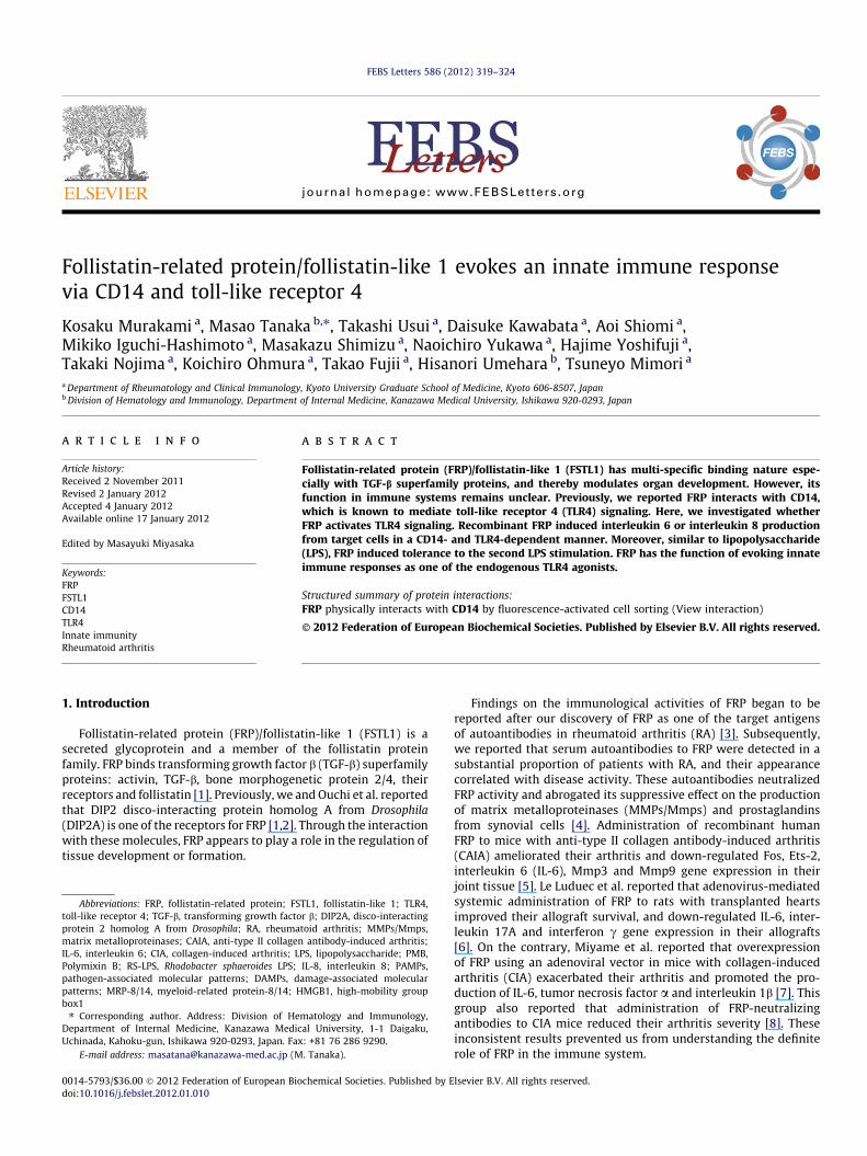
3. Results
Interaction of FRP with CD14 was observed also in a cell-basedsystem using CD14-expressing HEK293 cells (Fig. 1). Then weexamined proinflammatory activity of FRP. Human FRP signifi-cantly induced IL-6 from cultured synovial cells from RA patients(Fig. 2A). There, maximum concentration of 50 ng/ml was consid-ered to be about 10-fold average concentration in healthy humansera [10]. Notably, it was not human but mouse FRP that signifi-cantly induced IL-6 from NIH-3T3 cells of mouse origin (Fig. 2B).Thus, both human and mouse FRP substantially induced IL-6 fromtested cells of the same species as the FRP. In these and followingexperiments, we used recombinant human and mouse FRP whoseundiluted solutions had little or no endotoxin activity, which wasat most 0.032 EU/ml, equivalent to 0.02 ng/ml LPS.
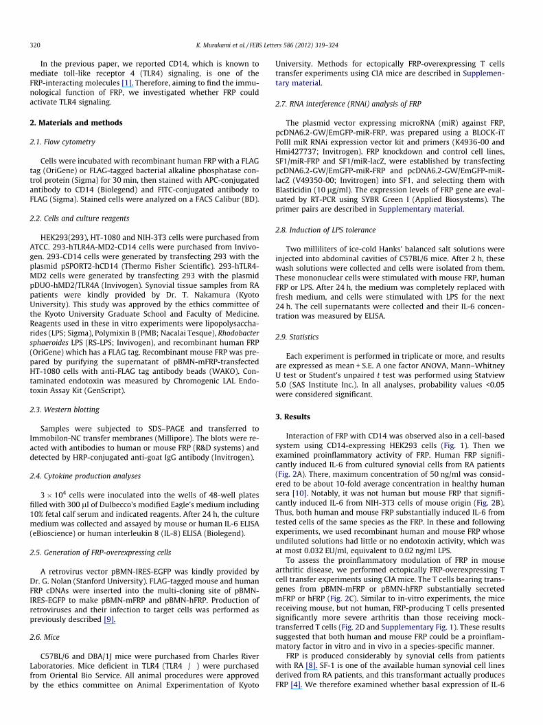
To assess the proinflammatory modulation of FRP in mousearthritic disease, we performed ectopically FRP-overexpressing Tcell transfer experiments using CIA mice. The T cells bearing trans-genes from pBMN-mFRP or pBMN-hFRP substantially secretedmFRP or hFRP (Fig. 2C). Similar to in-vitro experiments, the micereceiving mouse, but not human, FRP-producing T cells presentedsignificantly more severe arthritis than those receiving mock-transferred T cells (Fig. 2D and Supplementary Fig. 1). These resultssuggested that both human and mouse FRP could be a proinflam-matory factor in vitro and in vivo in a species-specific manner.
FRP is produced considerably by synovial cells from patientswith RA [8]. SF-1 is one of the available human synovial cell linesderived from RA patients, and this transformant actually producesFRP [4]. We therefore examined whether basal expression of IL-6

3.7 %
7.3 %
15.8 %
26.5 % 7.3 %
293 293-CD14
1001000
1001000
BAP (ng/m
l)
No ligands
0.1 %
0.0 %
0.1 %
FLAG-tagged protein
CD
14
hFRP (ng/m
l)
103
102
101
100
103
102
101
100
103
102
101
100
103102101100103102101100103102101100
Fig. 1. FRP interacts with cell surface CD14. 293, HEK293 cells. 293-CD14, HEK293 cells expressing human CD14. hFRP, FLAG-tagged recombinant human FRP. BAP, FLAG-tagged bacterial alkaline phosphatase control protein.
K. Murakami et al. / FEBS Letters 586 (2012) 319–324 321
depended on autocrined FRP in SF-1, using its FRP-knockdown cellline SF-1/miR-FRP. FRP mRNA expression levels in SF-1/miR-FRPdecreased to about 20% of that in its control SF-1/miR-lacZ(Fig. 2E). As expected, IL-6 production from SF-1/miR-FRP was sig-nificantly lower than that from SF-1/miR-lacZ. Furthermore, notSF-1/miR-lacZ but SF-1/miR-FRP responded to additional FRP(Fig. 2F).
We checked the TLR4 dependency of the proinflammatoryactivity in FRP using splenocytes from wild-type and TLR4�/�mice. FRP induced IL-6 from splenocytes from C57BL/6 mice, whilethose from TLR4�/� mice hardly responded to FRP (Fig. 3A). PMB,a LPS-neutralizing reagent, did not abrogate IL-6 induction by FRP,which excluded the possibility of LPS contamination in recombi-nant FRP. PMB slightly increased FRP activity for some reason,however similar phenomena were reported on other TLR4-stimu-lating molecules [11].
Next, we examined CD14 dependency of the proinflammatoryactivity in FRP using HEK293 and their transformants. Since IL-8is a better NF-jB activation indicator than IL-6 in HEK293, we mea-sured IL-8 production from tested cells. FRP and LPS induced IL-8significantly only from 293-hTLR4A-MD2-CD14 (Fig. 3B andSupplementary Fig. 2). We made sure that FRP actually activatedNF-jB using its reporter system (Supplementary Fig. 3). LPS con-tamination was excluded by the fact that PMB hardly affectedthe FRP activity (Fig. 3C). Interestingly, LPS-RS, a specific LPS antag-onist, neutralized the activity of FRP. Although PMB antagonizesLPS by preventing LPS from binding to CD14, LPS-RS is thoughtto compete with LPS for the same binding site on MD2 and to forminactive complexes with TLR4/MD2 [12]. As shown in Fig. 1, FRPbound to CD14 on a cell surface. It is probable that after interactingwith CD14 FRP may get translocated to a TLR4/MD2 complex viabinding to MD2.
LPS tolerance is a transient state that is unresponsive to furtherchallenges of LPS and is considered a protective mechanism against
over inflammation. Therefore, we investigated whether FRP in-duced LPS tolerance. In mouse peritoneal macrophages, 24-hourexposure to LPS is known to induce complete LPS tolerance [13].As shown in Fig. 4, both human and mouse FRP brought about a re-duced response to the second LPS stimulation in inverse proportionto the first response. While cell surface TLR4 expression transientlydecreased roughly according to a degree of tolerance, this effect ap-peared quite partial to abrogate TLR4 signaling (SupplementaryFig. 4). This suggests another concomitant mechanism such as im-paired association of TLR4 with MyD88 [14].
4. Discussion
To date, we have discovered that FRP has two cell signalingpathways. One is through DIP2A [1,2] and the other is throughCD14 and TLR4, as presented here. In addition to Akt1 phosphory-lation, the DIP2A pathway mediates FOS down-regulation leadingto the suppression of MMPs and prostaglandins [2,4]. The CD14-TLR4 pathway mediates the proinflammatory reaction. Therefore,in the physiological reaction to inflammation, FRP seems to havetwo contradicting activities; one is to prevent tissue destructionand the other is to promote inflammation. Moreover, we found thatFRP has species-specific activity to TLR4 signaling in target cells be-tween human and mouse, even though there is at least 91% aminoacid sequence identity between human and mouse FRP. BecauseTLR4 signaling needs CD14 and MD2 besides TLR4, species mis-match for FRP can originate from pairing with all of thosemolecules.
At least within the limits of innate immunity, our new resultsshown here can define FRP as a proinflammatory factor. They canalso resolve the contradiction raised by our previous CAIA mouseexperiment. In a standard protocol for CAIA induction, LPS wasinjected 2-day after the administration of anti-type II collagenantibody in order to develop arthritis. As we performed daily

*****
*hFRP
MockmFRP
15 20 25 30 35 40 45 50 551.4
1.6
1.8
2.0
2.2
2.4
Ankl
e (m
m)
Days
A
B
D
0 50
50
300
100
400
200
0 50
50
300
100
400
200
0 50
50
300
100
400
200
Pt. #2 Pt. #3
3.0
1.0
4.0
2.0
00 5 50
Pts. #1-#35.0
*
E F SF1/miR-FRP
**
0 5 50
hIL-
6 (p
g/m
l)
1400
200
0
400
1000
1200
800
600
SF1/miR-lacZ
0
20
40
60
80
100
120
140
*
FRP
gene
exp
ress
ion
(% c
ontro
l)
SF1/miR-lacZ SF1/miR-FRP
hFRP (ng/ml)
Fold
cha
nge
hIL-
6 (p
g/m
l)m
IL-6
(pg/
ml)
* *
LPS (ng/ml)
10
20
30
05 50 5
mFRP (ng/ml)
Pt. #1
hFRP (ng/ml)0 5 50
C
0 1
hFRP (ng/ml)
150102
76
52
3831
24(kDa)
Sup SupPP PP
mFRP hFRP
mFR
P
mFR
P
hFRP
hFRP
Mock
Mock
*
50
Fig. 2. FRP induces IL-6 and exacerbates CIA. (A) Synovial tissue from RA patients was stimulated by human FRP (hFRP). Left, individual data from each synovial tissue. Right,fold change in response to hFRP. ⁄P < 0.05. (B) NIH-3T3 cells were stimulated by recombinant mouse FRP (mFRP) or hFRP. ⁄P < 0.05, values were compared with control (LPS0 ng/ml). (C) Immunoblotting of recombinant mFRP and hFRP (arrowhead indicates the protein bands). Samples were aliquots of culture supernatant of mFRP, hFRP or mockcDNA-transferred CD4+ T cells (Sup) and purified proteins (PP). PP for hFRP showed smaller molecular weight, probably due to lack of a FLAG tag and producing cell typedifference. (D) Adoptive transfer of mFRP- or hFRP-overexpressing CD4+ T cells to CIA mice (n = 10 for each group). ⁄ and ⁄⁄ indicates P < 0.05 compared between mFRP andhFRP groups, and hFRP and mock groups, respectively. (E) FRP mRNA expression in SF1/miR-lacZ and SF1/miR-FRP. ⁄P < 0.05. (F) SF1/miR-lacZ and SF1/miR-FRP werestimulated by hFRP. ⁄P < 0.05. ⁄⁄P < 0.05.
322 K. Murakami et al. / FEBS Letters 586 (2012) 319–324
administration of high-dose human FRP (1000 ng/mg of a body),starting at the same time of antibody administration, FRP was chal-lenged twice prior to LPS injection. Therefore, we speculate thathuman FRP could induce LPS tolerance. On the other hand, the
DIP2A pathway was not likely to have been abrogated and couldexert protective functions. As for the induction of allograft toler-ance by FRP, it is unclear which signaling pathway through DIP2A,TLR4 or as-yet-unidentified receptors is involved.

A
C
B
0
10
20
30
40
50
mIL
-6 (p
g/m
l)
***
***
0 0.5 5 50
LPS (ng/ml) mFRP (ng/ml)
100
10
20
30
40
50
20
40
50
60
hIL-
8 (p
g/m
l)
0 5 500
10
20
30
40
50 *
10
30
0 5 500
10
20
30
40
50
0 5 500
10
20
30
40
50
0 5 500
10
20
30
40
50
hFRP (ng/ml)
hIL-
8 (p
g/m
l)
00 5 50 0 5 50 0 5 50
hFRP (ng/ml)
**
293 293-CD14 293-hTLR4A- MD2
293-hTLR4A- MD2-CD14
hFRP (ng/ml) hFRP (ng/ml)+ PMB + LPS-RS
293-hTLR4A-MD2-CD14
C57BL/6
PMB −PMB +
PMB −PMB +
TLR4−/−
0 0.5 5 50
LPS (ng/ml) mFRP (ng/ml)
10
Fig. 3. FRP evokes proinflammatory responses in a CD14- and TLR4- dependent manner. (A) Splenocytes from C57BL/6 mice and those from TLR4�/� mice were stimulatedby LPS or mFRP with or without 1 lg/ml of PMB, a LPS-neutralizing reagent. ⁄, ⁄⁄P < 0.05, values were compared with the sample treated by 0 ng/ml LPS ⁄⁄with or ⁄withoutPMB. (B) 293, 293-CD14, 293-hTLR4A-MD2 and 293-hTLR4A-MD2-CD14 cells were stimulated by hFRP. ⁄P < 0.05. (C) 293-hTLR4A-MD2-CD14 cells were stimulated by hFRPin the presence or absence of PMB (1 lg/ml) or LPS-RS (500 ng/ml). ⁄P < 0.05.
0 18001600200 400
200
400
600
800
0
200
400
600
800
00 800600200 400
200
400
600
800
00 800600200 400
0 ng/ml100 ng/ml
1000 ng/ml
0 ng/ml
100 ng/ml1000 ng/ml
0 ng/ml
100 ng/ml10 ng/ml
LPS mFRP hFRP
Induced mIL-6 (pg/ml) 1st stimulation by LPS, mFRP or hFRP
2nd
stim
ulat
ion
by 1
0-ng
/ml L
PS
Indu
ced
mIL
-6 (p
g/m
l)
* ***
Fig. 4. FRP induces LPS tolerance. Peritoneal mononuclear cells from C57BL/6 were subjected sequentially to two 24-hour stimulations; first by mFRP, hFRP or LPS, andsecondly by 10 ng/ml LPS. Induced IL-6 concentrations after the first and the second stimulation are indicated by X and Y axes. The broken line stands for Y = X. ⁄P < 0.01 and⁄⁄P < 0.05 for the second stimulation by LPS.
K. Murakami et al. / FEBS Letters 586 (2012) 319–324 323

324 K. Murakami et al. / FEBS Letters 586 (2012) 319–324
TLR4 is known to recognize not only various exogenous proteinsfrom microbes, called pathogen-associated molecular patterns(PAMPs), but also many endogenous proteins, called damage-asso-ciated molecular patterns (DAMPs) or ‘‘alarmins’’, such as heatshock proteins, myeloid-related protein-8/14 (MRP-8/14), surfac-tant protein A, high-mobility group box 1 (HMGB1), b-defensinand hyaluronan, which may be expressed or released in responseto tissue damage [15]. In a sense, FRP can be considered as oneof the DAMPs. Notably, HMGB1 is also known to be one of the auto-antigens in particular autoimmune diseases, such as RA [16].
Besides direct stimulation of TLR4 molecules, DAMPs arethought to have a sensing function of circulating LPS/PAMPs indamaged tissues. For example, HMGB1 was shown to associatewith LPS and transfer its monomers to TLR4 molecules on targetcells, thereby enhancing their responsiveness to LPS [17]. Althoughit remains to be clarified whether FRP can bind LPS or any PAMPs,dysregulated FRP production from synovium may play a part inautoimmune arthritic diseases. It is also possible that impaired tol-erance to LPS/PAMPs and DAMPs including FRP may be involved.
In conclusion, we revealed that one of the immunological func-tions of FRP is to evoke innate immune responses through CD14and TLR4. As FRP has broad physiological activity like TGF-b, vari-ous approaches will be required to comprehensively elucidate itsunknown functions.
Acknowledgements
This work was supported by a Grant-in-Aid for scientificresearch (No. 22390201) from the Japan Society for the Promotionof Science and by a Grant for Promoted Research from KanazawaMedical University (S2011-7).
Appendix A. Supplementary data
Supplementary data associated with this article can be found, inthe online version, at doi:10.1016/j.febslet.2012.01.010.
References
[1] Tanaka, M., Murakami, K., Ozaki, S., Imura, Y., Tong, X.P., Watanabe, T., Sawaki,T., Kawanami, T., Kawabata, D., Fujii, T., Usui, T., Masaki, Y., Fukushima, T., Jin,Z.X., Umehara, H. and Mimori, T. (2010) DIP2 disco-interacting protein 2homolog A (Drosophila) is a candidate receptor for follistatin-related protein/follistatin-like 1–analysis of their binding with TGF-beta superfamily proteins.FEBS J. 277, 4278–4289.
[2] Ouchi, N., Asaumi, Y., Ohashi, K., Higuchi, A., Sono-Romanelli, S., Oshima, Y.and Walsh, K. (2010) DIP2A functions as a FSTL1 receptor. J. Biol. Chem. 285,7127–7134.
[3] Tanaka, M., Ozaki, S., Osakada, F., Mori, K., Okubo, M. and Nakao, K. (1998)Cloning of follistatin-related protein as a novel autoantigen in systemicrheumatic diseases. Int. Immunol. 10, 1305–1314.
[4] Tanaka, M., Ozaki, S., Kawabata, D., Kishimura, M., Osakada, F., Okubo, M.,Murakami, M., Nakao, K. and Mimori, T. (2003) Potential preventive effects offollistatin-related protein/TSC-36 on joint destruction and antagonisticmodulation of its autoantibodies in rheumatoid arthritis. Int. Immunol. 15,71–77.
[5] Kawabata, D., Tanaka, M., Fujii, T., Umehara, H., Fujita, Y., Yoshifuji, H., Mimori,T. and Ozaki, S. (2004) Ameliorative effects of follistatin-related protein/TSC-36/FSTL1 on joint inflammation in a mouse model of arthritis. Arthritis Rheum.50, 660–668.
[6] Le Luduec, J.B., Condamine, T., Louvet, C., Thebault, P., Heslan, J.M., Heslan, M.,Chiffoleau, E. and Cuturi, M.C. (2008) An immunomodulatory role for follistatin-like 1 in heart allograft transplantation. Am. J. Transplant 8, 2297–2306.
[7] Miyamae, T., Marinov, A.D., Sowders, D., Wilson, D.C., Devlin, J., Boudreau, R.,Robbins, P. and Hirsch, R. (2006) Follistatin-like protein-1 is a novelproinflammatory molecule. J. Immunol. 177, 4758–4762.
[8] Clutter, S.D., Wilson, D.C., Marinov, A.D. and Hirsch, R. (2009) Follistatin-likeprotein 1 promotes arthritis by up-regulating IFN-gamma. J. Immunol. 182,234–239.
[9] Usui, T., Nishikomori, R., Kitani, A. and Strober, W. (2003) GATA-3 suppressesTh1 development by downregulation of Stat4 and not through effects on IL-12Rbeta2 chain or T-bet. Immunity 18, 415–428.
[10] Li, D., Wang, Y., Xu, N., Wei, Q., Wu, M., Li, X., Zheng, P., Sun, S., Jin, Y., Zhang, G.,Liao, R. and Zhang, P. (2011) Follistatin-like protein 1 is elevated in systemicautoimmune diseases and correlated with disease activity in patients withrheumatoid arthritis. Arthritis Res. Ther. 13, R17.
[11] Uehara, A., Sugawara, S. and Takada, H. (2002) Priming of human oralepithelial cells by interferon-gamma to secrete cytokines in response tolipopolysaccharides, lipoteichoic acids and peptidoglycans. J. Med. Microbiol.51, 626–634.
[12] Coats, S.R., Pham, T.T., Bainbridge, B.W., Reife, R.A. and Darveau, R.P. (2005)MD-2 mediates the ability of tetra-acylated and penta-acylatedlipopolysaccharides to antagonize Escherichia coli lipopolysaccharide at theTLR4 signaling complex. J. Immunol. 175, 4490–4498.
[13] Nomura, F., Akashi, S., Sakao, Y., Sato, S., Kawai, T., Matsumoto, M., Nakanishi, K.,Kimoto, M., Miyake, K., Takeda, K. and Akira, S. (2000) Cutting edge: endotoxintolerance in mouse peritoneal macrophages correlates with down-regulation ofsurface toll-like receptor 4 expression. J. Immunol. 164, 3476–3479.
[14] Medvedev, A.E., Lentschat, A., Wahl, L.M., Golenbock, D.T. and Vogel, S.N.(2002) Dysregulation of LPS-induced Toll-like receptor 4-MyD88 complexformation and IL-1 receptor-associated kinase 1 activation in endotoxin-tolerant cells. J. Immunol. 169, 5209–5216.
[15] Erridge, C. (2010) Endogenous ligands of TLR2 and TLR4: agonists orassistants? J. Leukoc. Biol. 87, 989–999.
[16] Uesugi, H., Ozaki, S., Sobajima, J., Osakada, F., Shirakawa, H., Yoshida, M. andNakao, K. (1998) Prevalence and characterization of novel pANCA, antibodiesto the high mobility group non-histone chromosomal proteins HMG1 andHMG2, in systemic rheumatic diseases. J. Rheumatol. 25, 703–709.
[17] Youn, J.H., Oh, Y.J., Kim, E.S., Choi, J.E. and Shin, J.S. (2008) High mobility groupbox 1 protein binding to lipopolysaccharide facilitates transfer oflipopolysaccharide to CD14 and enhances lipopolysaccharide-mediated TNF-alpha production in human monocytes. J. Immunol. 180, 5067–5074.