Murine norovirus infection does not cause major disruptions in ...
Upload
tracuuxaydungCategory
view
2download
0
Infection, Genetics and Evolution 18 (2013) 335–343
Contents lists available at SciVerse ScienceDirect
Infection, Genetics and Evolution
journal homepage: www.elsevier .com/locate /meegid
The dynamics of GII.4 Norovirus in Ho Chi Minh City, Vietnam q
1567-1348/$ - see front matter � 2013 The Authors. Published by Elsevier B.V. All rights reserved.http://dx.doi.org/10.1016/j.meegid.2013.04.014
q This is an open-access article distributed under the terms of the CreativeCommons Attribution License, which permits unrestricted use, distribution, andreproduction in any medium, provided the original author and source are credited.⇑ Corresponding author at: Enteric Infections Group, Wellcome Trust Major
Overseas Programme, Oxford University Clinical Research Unit, 764 Vo Van Kiet,District 5, Ho Chi Minh City, Viet Nam. Tel.: +84 8 39239210; fax: +84 8 39238904.
E-mail address: [email protected] (S. Baker).
Phan Vu Tra My a,b, Ha Minh Lam a, Corinne N. Thompson a,b, Hoang Le Phuc c, Pham Thi Ngoc Tuyet d,Ha Vinh e, Nguyen Van Minh Hoang a, PhamVan Minh a, Nguyen Thanh Vinh a, Cao Thu Thuy a,Tran Thi Thu Nga a, Nguyen Thi Thu Hau d, Nguyen Tran Chinh e, Tang Chi Thuong c, Ha Manh Tuan d,James I. Campbell a,b, Archie C.A. Clements f, Jeremy Farrar a,b, Maciej F. Boni a,b, Stephen Baker a,b,g,⇑a Wellcome Trust Major Overseas Programme, Oxford University Clinical Research Unit, 764 Vo Van Kiet, District 5, Ho Chi Minh City, Viet Namb Centre for Tropical Medicine, Nuffield Department of Clinical Medicine, University of Oxford, Wellington Square, Oxford OX1 2JD, United Kingdomc Children’s Hospital 1, 341 Su Van Hanh, District 5, Ho Chi Minh City, Viet Namd Children’s Hospital 2, 14 Ly Tu Trong, District 1, Ho Chi Minh City, Viet Name Hospital for Tropical Diseases, 764 Vo Van Kiet, District 5, Ho Chi Minh City, Viet Namf University of Queensland, School of Population Health, Brisbane St Lucia, QLD 4072, Australiag The London School of Hygiene and Tropical Medicine, Keppel Street, London WC1E 7HT, United Kingdom
a r t i c l e i n f o
Article history:Received 18 September 2012Received in revised form 5 April 2013Accepted 6 April 2013Available online 21 April 2013
Keywords:NorovirusGII.4PhylogeneticSpatialTemporalCluster
a b s t r a c t
Norovirus (NoV) is a major cause of epidemic gastroenteritis in industrialized countries, yet the epidemi-ological significance of NoV in industrializing countries remains poorly understood. The spatiotemporaldistribution of NoV genotypes identified in 2054 enrolled children was investigated between May 2009and December 2010, in Ho Chi Minh City (HCMC), Vietnam. A total of 315 NoV extracted from stool sam-ples were genotyped and GPS mapped to their source. Genogroup II NoV, particularly GII.4, were predom-inant, and the GII.4 strains could be subgrouped into GII.4-2006b (Minerva) and GII.4-2010 (NewOrleans) variants. There was no spatiotemporal structure among the endemic GII strains; yet a significantspatiotemporal signal corresponding with the novel introduction of GII.4-2010 variant was detected.These data show that NoV GII.4 variants are highly endemic in HCMC and describe a scenario of rapidNoV strain replacement occurring in HCMC in early 2010.
� 2013 The Authors. Published by Elsevier B.V. All rights reserved.
1. Introduction 2010); as a result, NoV frequently causes explosive gastroenteritis
Norovirus (NoV) is a non-enveloped positive-sense single-stranded RNA virus belonging to the taxonomic family Caliciviridae(Green et al., 2000; Jiang et al., 1990). NoV accounts for a signifi-cant proportion of the global burden of viral gastroenteritis (Glasset al., 2000, 2009; Patel et al., 2009, 2008), and up to 50% of all-cause outbreaks of diarrhea (Patel et al., 2009). The diseasetypically presents as acute watery diarrhea with vomiting and alow-grade fever (Patel et al., 2009), and is usually self-limiting,lasting between one and three days, but can be aggressive, severeand protracted in young children, the elderly and the immuno-compromised (Estes et al., 2006). NoV has an exceptionally lowinfectious dose (10–100 viral particles (Patel et al., 2009)) andcan survive on surfaces for prolonged time periods (Weber et al.,
epidemics (Estes et al., 2006; Patel et al., 2009, 2008).The 7.5 kb genome of NoV has three open reading frames
(ORFs), encoding the RNase-dependent RNA polymerase (RdRp,ORF1), the major capsid protein (VP1, ORF2) and the minor capsidprotein (VP2, ORF3) (Bertolotti-Ciarlet et al., 2003; Jiang et al.,1993). The current classification divides NoV into five genogroups(GI – GV) on the basis of sequence identity within the major capsidprotein (VP1). Genogroups GI, GII and GIV are associated withinfections in humans (Zheng et al., 2006). Molecular characteriza-tion of coding sequences within RdRp (region A and B) (Andoet al., 1995, 2000; Fankhauser et al., 2002; Jiang et al., 1999; Vinjeand Koopmans, 1996) and ORF2 (region C, D and E) (Kageyamaet al., 2004; Kojima et al., 2002; Noel et al., 1997; Vinje et al.,2004) is targeted for NoV detection, genogrouping and genotyping.Genogroups I and II are the most common cause of human infec-tions (Donaldson et al., 2010), and can be differentiated into 8 GIand 23 GII capsid genotypes, and 14 GI and 29 GII polymerasegenotypes (Kroneman et al., 2011; Zheng et al., 2006).
The epidemiology of NoV is complex and is influenced by amultitude of factors, including population immunity, the environ-ment, and seasonality (Donaldson et al., 2010; Marshall and

336 P.V. Tra My et al. / Infection, Genetics and Evolution 18 (2013) 335–343
Bruggink, 2011), making molecular epidemiology challenging. Theacknowledged interpretation of global NoV epidemiology, particu-larly GII.4 genotype, is that strain replacement occurs every two tothree years (Bull et al., 2010; Bull and White, 2011; CDC, 2010;Donaldson et al., 2008; Siebenga et al., 2009). Over the last twodecades, these replacements were typically caused by strains of asingle lineage of a GII.4 genotype, which have been responsiblefor the majority of NoV outbreaks worldwide since being first iden-tified in the USA in the mid 1990s (Bull and White, 2011; Noelet al., 1999). At least four major global NoV replacements havebeen described since 1995, each due to a novel GII.4 variant (Bullet al., 2006; Noel et al., 1999; Siebenga et al., 2010; Tu et al.,2008), believed to have escaped immunity in the populationthrough antigenic variation (Lindesmith et al., 2012b, 2012c, 2011).
The majority of NoV studies are performed in industrializedcountries and disease outbreaks are continually monitoredthrough several disease surveillance networks (Vega et al., 2011;Verhoef et al., 2009). However, little is known about the transmis-sion, molecular diversity or spatiotemporal dynamics of NoV infec-tions in areas with differing public health infrastructure anddemographics. Vietnam is an industrializing country with denselypopulated urban centers and a changing spectrum of infectiousdiseases as a presumed consequence of rapid economic develop-ment and urbanization (Vinh et al., 2009). NoV was first reportedin Ho Chi Minh City (HCMC) in 1999 (Hansman et al., 2004), andour recent work has demonstrated that NoV is endemic throughoutthe year, in contrast to the winter outbreaks observed in temperatelocations (Lopman et al., 2009; Mounts et al., 2000). To understandthe epidemiology and NoV strain diversity in HCMC, we investi-gated the molecular and spatiotemporal distribution of NoV geno-types in young hospitalized children between May 2009 andDecember 2010 in HCMC, Vietnam.
2. Material and methods
2.1. Study setting and design
This study was conducted according to the principles expressedin the Declaration of Helsinki and was approved by the ethical re-view boards of Children’s Hospital 1 (HCMC), Children’s Hospital 2(HCMC), the Hospital for Tropical Diseases (HCMC), and the OxfordTropical Research Ethics Committee (OxTREC Approval No. 0109)(Oxford). The parents or legal guardians of the enrolled childrenwere required to provide written informed consent for sample col-lection and residential mapping.
A stool specimen was collected within 24 h of enrollment fromeach recruited individual (1443 diarrheal patients and 611 asymp-tomatic controls). These participants (N = 2054) were children of0–60 months of age and residents of HCMC, Vietnam, over thestudy period from May 2009 to December 2010. Diarrheal patientswere children with acute diarrheal disease (P3 loose stools or atleast one bloody loose stool within 24 h period (WHO, 2005))who were admitted to the three study sites and had not receivedtreatment with antimicrobials in the three days prior to hospitaladmission. Asymptomatic controls were diarrhea-free childrenattending Children’s Hospital 1 or Children’s Hospital 2 for nutri-tional health checks or for other gastrointestinal issues unrelatedto diarrhea or gastroenteritis without any history of diarrhea,respiratory illness or treatment with antimicrobials within 7 daysof study enrollment.
2.2. Norovirus detection
Total viral RNA was extracted and reverse transcribed intocDNA as previously described (Tra My et al., 2011). Norovirusgenogroup I (GI) and II (GII) were detected in separate reactions
by conventional Reverse Transcriptase Polymerase Chain Reaction(RT PCR) using consensus primers, G1SKF/G1SKR (Kojima et al.,2002) and COG2F/G2SKR (Kageyama et al., 2003; Kojima et al.,2002) for GI and GII, respectively. These PCR primers amplify a re-gion between position 5342 and 5671 (330 bp) in the genome ofNoV GI (Norwalk/68, GenBank accession No. M87661) containingan overlap of 17 bp of 30 end ORF1 and 313 bp of 50 end ORF2,and between position 5003 and 5389 (387 bp) in the genome ofNoV GII (Lordsdale/93, GenBank accession No. X86557) containingan overlap of 83 bp of 30 end ORF1 and 304 bp of 50 end ORF2.
2.3. Norovirus genotyping
NoV positive PCR amplicons were purified using the QIAquickPCR purification kit (QIAGEN, Hilden, Germany), and subjected todirect sequencing using the amplification primers. DNA concentra-tions were determined using a NanoDrop ND-1000 spectropho-tometer (Thermo Fisher Scientific, United Kingdom) and directsequencing was performed using a BigDye Terminator CycleSequencing kit (Applied Biosystems, USA) and generated with anABI Prism 3130xl Genetic Analyzer (Applied Biosystems, USA).DNA sequences were assembled using DNA Baser Sequence Assem-bler v3.0.17 (Heracle Biosoft, Pitesti, Romania). NoV genotypeswere assigned based on ORF2 sequences using the online Norovi-rus Automated Genotyping Tool, as directed (Kroneman et al.,2011).
2.4. Construction of NoV phylogenies
DNA sequences were uploaded into GenBank (HE716437 toHE716751) and used for local phylogenetic construction. Manualalignment of all sequences was performed in Se-AL (http://tree.-bio.ed.ac.uk/software/figtree/) prior to phylogenetic reconstruc-tion. Maximum likelihood (ML) trees were inferred using RAxML(Stamatakis et al., 2008), employing the general-time reversiblemodel of nucleotide substitution with a gamma distribution ofamong-site rate variation (GTR + C) and 1000 bootstrap replicates.Resulting trees were visualized in FigTree v1.3.1 (http://tree.bio.e-d.ac.uk/software/figtree/) and mean pairwise genetic distanceswere estimated in MEGA 5 (Tamura et al., 2011).
Two hundred and sixty-nine global GII.4 strains encompassingthe global diversity of GII.4 variants were retrieved from GenBank(http://www.ncbi.nlm.nih.gov/); 43 of these strains originatedfrom previous studies conducted in Vietnam (Hansman et al.,2004; Nguyen et al., 2008, 2007a, 2007b; Tamura et al., 2010;Trang et al., 2012). In addition, available archived samples thatwere Enzyme-Immuno Assay (EIA) positive for NoV (GI and GII)from our previous work in 2008 from southern Vietnam (Tra Myet al., 2011) were also selected for analysis and genotyped usingthe same methodology.
The time of isolation for each of the NoV strains was retrievedfrom GenBank or the publication associated with the sequence,and the year of isolation was used to calculate evolutionary rate.All sequences were aligned in Se-AL and trimmed to 378 bp to cor-respond with the sequences identified in this study to maximizesequence homology for phylogenetic reconstruction. Phylogeneticreconstructions of relationships among the GII.4 variants identifiedin this study and global GII.4 sequences were inferred using theBayesian Markov chain Monte Carlo (MCMC) method as imple-mented in BEAST (Drummond and Rambaut, 2007). A GTR substi-tution model with gamma-distributed rate variation and arelaxed uncorrelated lognormal clock model with a constant pop-ulation size were employed. The MCMC analysis was run for 50million generations (with a burn-in of 5 million) and analyzedusing Tracer (http://tree.bio.ed.ac.uk/software/tracer/) to ensurethat all parameters had converged. Maximum clade credibility
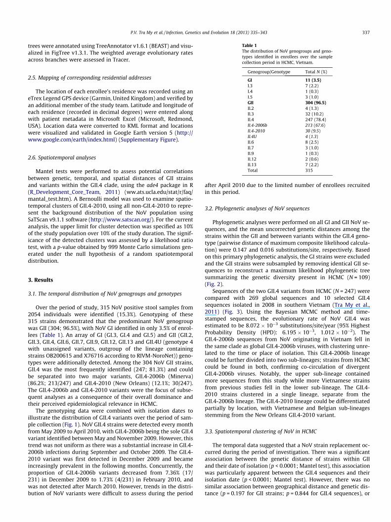
Table 1The distribution of NoV genogroups and geno-types identified in enrollees over the samplecollection period in HCMC, Vietnam.
P.V. Tra My et al. / Infection, Genetics and Evolution 18 (2013) 335–343 337
trees were annotated using TreeAnnotator v1.6.1 (BEAST) and visu-alized in FigTree v1.3.1. The weighted average evolutionary ratesacross branches were assessed in Tracer.
Genogroup/Genotype Total N (%)
GI 11 (3.5)I.3 7 (2.2)I.4 1 (0.3)I.5 3 (1.0)GII 304 (96.5)II.2 4 (1.3)II.3 32 (10.2)II.4 247 (78.4)II.4-2006b 213 (67.6)II.4-2010 30 (9.5)II.4U 4 (1.3)II.6 8 (2.5)
2.5. Mapping of corresponding residential addresses
The location of each enrollee’s residence was recorded using aneTrex Legend GPS device (Garmin, United Kingdom) and verified byan additional member of the study team. Latitude and longitude ofeach residence (recorded in decimal degrees) were entered alongwith patient metadata in Microsoft Excel (Microsoft, Redmond,USA). Location data were converted to KML format and locationswere visualized and validated in Google Earth version 5 (http://www.google.com/earth/index.html) (Supplementary Figure).
II.7 3 (1.0)II.9 1 (0.3)II.12 2 (0.6)II.13 7 (2.2)Total 315
2.6. Spatiotemporal analyses
Mantel tests were performed to assess potential correlationsbetween genetic, temporal, and spatial distances of GII strainsand variants within the GII.4 clade, using the ade4 package in R(R_Development_Core_Team, 2011) (ww.ats.ucla.edu/stat/r/faq/mantal_test.htm). A Bernoulli model was used to examine spatio-temporal clusters of GII.4-2010, using all non-GII.4-2010 to repre-sent the background distribution of the NoV population usingSaTScan v9.1.1 software (http://www.satscan.org/). For the currentanalysis, the upper limit for cluster detection was specified as 10%of the study population over 10% of the study duration. The signif-icance of the detected clusters was assessed by a likelihood ratiotest, with a p-value obtained by 999 Monte Carlo simulations gen-erated under the null hypothesis of a random spatiotemporaldistribution.
3. Results
3.1. The temporal distribution of NoV genogroups and genotypes
Over the period of study, 315 NoV positive stool samples from2054 individuals were identified (15.3%). Genotyping of these315 strains demonstrated that the predominant NoV genogroupwas GII (304; 96.5%), with NoV GI identified in only 3.5% of enrol-lees (Table 1). An array of GI (GI.3, GI.4 and GI.5) and GII (GII.2,GII.3, GII.4, GII.6, GII.7, GII.9, GII.12, GII.13 and GII.4U (genotype 4with unassigned variants, outgroup of the lineage containingstrains OB200615 and X76716 according to RIVM-NoroNet)) geno-types were additionally detected. Among the 304 NoV GII strains,GII.4 was the most frequently identified (247; 81.3%) and couldbe separated into two major variants, GII.4-2006b (Minerva)(86.2%; 213/247) and GII.4-2010 (New Orleans) (12.1%; 30/247).The GII.4-2006b and GII.4-2010 variants were the focus of subse-quent analyses as a consequence of their overall dominance andtheir perceived epidemiological relevance in HCMC.
The genotyping data were combined with isolation dates toillustrate the distribution of GII.4 variants over the period of sam-ple collection (Fig. 1). NoV GII.4 strains were detected every monthfrom May 2009 to April 2010, with GII.4-2006b being the sole GII.4variant identified between May and November 2009. However, thistrend was not uniform as there was a substantial increase in GII.4-2006b infections during September and October 2009. The GII.4-2010 variant was first detected in December 2009 and becameincreasingly prevalent in the following months. Concurrently, theproportion of GII.4-2006b variants decreased from 7.36% (17/231) in December 2009 to 1.73% (4/231) in February 2010, andwas not detected after March 2010. However, trends in the distri-bution of NoV variants were difficult to assess during the period
after April 2010 due to the limited number of enrollees recruitedin this period.
3.2. Phylogenetic analyses of NoV sequences
Phylogenetic analyses were performed on all GI and GII NoV se-quences, and the mean uncorrected genetic distances among thestrains within the GII and between variants within the GII.4 geno-type (pairwise distance of maximum composite likelihood calcula-tion) were 0.147 and 0.016 substitutions/site, respectively. Basedon this primary phylogenetic analysis, the GI strains were excludedand the GII strains were subsampled by removing identical GII se-quences to reconstruct a maximum likelihood phylogenetic treesummarizing the genetic diversity present in HCMC (N = 109)(Fig. 2).
Sequences of the two GII.4 variants from HCMC (N = 247) werecompared with 269 global sequences and 10 selected GII.4sequences isolated in 2008 in southern Vietnam (Tra My et al.,2011) (Fig. 3). Using the Bayesian MCMC method and time-stamped sequences, the evolutionary rate of NoV GII.4 wasestimated to be 8.072 � 10�3 substitutions/site/year (95% HighestProbability Density (HPD): 6.195 � 10�3, 1.012 � 10�2). TheGII.4-2006b sequences from NoV originating in Vietnam fell inthe same clade as global GII.4-2006b viruses, with clustering unre-lated to the time or place of isolation. This GII.4-2006b lineagecould be further divided into two sub-lineages; strains from HCMCcould be found in both, confirming co-circulation of divergentGII.4-2006b viruses. Notably, the upper sub-lineage containedmore sequences from this study while more Vietnamese strainsfrom previous studies fell in the lower sub-lineage. The GII.4-2010 strains clustered in a single lineage, separate from theGII.4-2006b lineage. The GII.4-2010 lineage could be differentiatedpartially by location, with Vietnamese and Belgian sub-lineagesstemming from the New Orleans GII.4-2010 variant.
3.3. Spatiotemporal clustering of NoV in HCMC
The temporal data suggested that a NoV strain replacement oc-curred during the period of investigation. There was a significantassociation between the genetic distance of strains within GIIand their date of isolation (p < 0.0001; Mantel test), this associationwas particularly apparent between the GII.4 sequences and theirisolation date (p < 0.0001; Mantel test). However, there was nosimilar association between geographical distance and genetic dis-tance (p = 0.197 for GII strains; p = 0.844 for GII.4 sequences), or
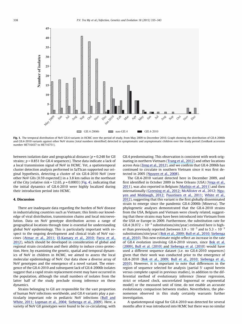
Fig. 1. The temporal distribution of NoV GII.4 variants in HCMC over the period of study, from May 2009 to December 2010. Graph showing the distribution of GII.4-2006band GII.4-2010 variants against other NoV strains (total numbers identified) detected in symptomatic and asymptomatic children over the study period (GenBank accessionnumber HE716437 to HE716751).
338 P.V. Tra My et al. / Infection, Genetics and Evolution 18 (2013) 335–343
between isolation date and geographical distance (p = 0.248 for GIIstrains; p = 0.851 for GII.4 sequences). These data indicate a lack ofa local transmission signal of NoV in HCMC. Yet, a spatiotemporalcluster detection analysis performed in SaTScan supported our ori-ginal hypothesis, detecting a cluster of six GII.4-2010 NoV (overother NoV GIIs (0.59 expected)) in a 3.8 km radius in the northeastof the City (relative risk = 12.65, p = 0.0003) (Fig. 4), indicating thatthe initial dynamics of GII.4-2010 were highly localized duringtheir introduction period into HCMC.
4. Discussion
There are inadequate data regarding the burden of NoV diseasein industrializing countries such as Vietnam; this limits our knowl-edge of viral distribution, transmission chains and local microevo-lution. Data on NoV genotype distribution across a range ofgeographical locations through time is essential for understandingglobal NoV epidemiology. This is particularly important with re-spect to the ongoing development and clinical trials of NoV vac-cines (Atmar et al., 2011; El-Kamary et al., 2010; Parra et al.,2012), which should be developed in consideration of global andregional strain circulation and their ability to induce cross-protec-tion. Here, by examining the genetic, spatial and temporal dynam-ics of NoV in children in HCMC, we aimed to assess the localmolecular epidemiology of NoV. Our data show a diverse array ofNoV genotypes and the emergence of a novel variant. The emer-gence of the GII.4-2010 and subsequent lack of GII.4-2006b isolatessuggest that a rapid strain replacement event may have occurred inthe population, although the small numbers of isolates from thelatter half of the study preclude strong inference on thesedynamics.
Strains belonging to GII are responsible for the vast proportionof human NoV infections worldwide, and GII.4 variants play a par-ticularly important role in pediatric NoV infections (Bull andWhite, 2011; Lopman et al., 2004; Siebenga et al., 2009). Here, avariety of NoV GII genotypes were found to be co-circulating, with
GII.4 predominating. This observation is consistent with work orig-inating in northern Vietnam (Trang et al., 2012) and other locationsacross Asia (Zeng et al., 2012), and we confirm that GII.4-2006b hascontinued to circulate in southern Vietnam since it was first de-tected in 2005 (Nguyen et al., 2008).
The GII.4-2010 variant detected here in December 2009, andfirst identified in October 2009 in New Orleans (USA) (Vega et al.,2011), was also reported in Belgium (Mathijs et al., 2011) and theninternationally (Greening et al., 2012; McAllister et al., 2012; Ngu-yen and Middaugh, 2012; Puustinen et al., 2011; White et al.,2012), suggesting that this variant is the first globally disseminatedstrain to emerge since the pandemic GII.4-2006b (Minerva). Thephylogenetic analyses demonstrated that the GII.4-2010 strainsfrom the USA, Belgium and Vietnam were closely related, suggest-ing that these strains may have been introduced into Vietnam fromthe USA or Europe in 2009. Furthermore, the substitution rate forGII.4 (8.072 � 10�3 substitutions/site/year) estimated here is high-er than previously reported (between 3.9 � 10�3 and to 5.3 � 10�3
substitutions/site/year) (Bok et al., 2009; Bull et al., 2010; Siebengaet al., 2010). This new estimate might reflect an increase in the rateof GII.4 evolution involving GII.4-2010 viruses, since Bok et al.(2009), Bull et al. (2010) and Siebenga et al. (2010) would haveused a different sequence dataset (i.e. no GII.4-2010 sequences)given that their work was conducted prior to the emergence ofGII.4-2010 (Bok et al., 2009; Bull et al., 2010; Siebenga et al.,2010). However, it is important to note that differences in theregion of sequence selected for analysis (partial 50 capsid hereinversus complete capsid in previous studies), in addition to the dif-ferential method of evolutionary inference (linear regression,strict or relaxed clock, uncorrelated lognormal or exponentialmodel) or the measured unit of time, do not enable an accurateevolutionary comparison between studies. Nevertheless, the phe-nomenon observed in this study certainly warrants furtherinvestigation.
A spatiotemporal signal for GII.4-2010 was detected for severalmonths after it was introduced into HCMC but there was no similar

Fig. 2. Phylogenetic tree of 109 NoV GII strains from HCMC. Tree constructed from 109 NoV GII strains collected during this study and based on the GII amplification fragmenttrimmed to 378 bp. All horizontal branch lengths are drawn to the scale of a nucleotide substitution per site. Tree is mid-point rooted with branches according to viralgenotypes/variants. Strains from asymptomatic individuals are labeled with a circle. Only bootstrap values of >75 are shown.
P.V. Tra My et al. / Infection, Genetics and Evolution 18 (2013) 335–343 339
spatiotemporal association for GII.4-2006b and non-GII.4. A poten-tial explanation for the absence (GII.4-2006b and non-GII.4) andpresence (GII.4-2010) of spatial signals in the NoV sequences isthat GII.4-2006b and non-GII.4 genotypes were in a state of equi-librium when GII.4-2010 was introduced, and the GII.4-2010exhibited an outbreak dynamic and exponential growth uponintroduction. Similar replacement of GII.4-2006b viruses followingthe introduction of GII.4-2010 has been observed in Belgium(Mathijs et al., 2011), and NoV strain replacement has been ob-served in various NoV pandemics. These replacements include,
GII.4-1997 (USA 95/96 variant), -2002 (Farmington Hills variant),-2004 (Hunter variant), -2006 comprising of GII.4-2006a (Laurensvariant) and the -2006b (Minerva variant) (Bull and White, 2011;Donaldson et al., 2008; Lindesmith et al., 2011). Novel pandemicNoV GII.4 strains emerge every two to three years and it appearsthat novel GII.4 genotypes are capable of replacing existing GII.4variants but not other endemic strains (such as GII.3, GII.6, GII.2)(Bull et al., 2010; Bull and White, 2011; CDC, 2010; Donaldsonet al., 2008; Lindesmith et al., 2011; Siebenga et al., 2009). How-ever, there is no current consensus on the underlying mechanism

Fig. 3. Phylogenetic tree of GII.4 NoV strains from HCMC and globally representative sequences. Maximum likelihood phylogenetic tree of 526 global and HCMC GII.4 NoVstrains constructed from the amplification region trimmed to 378 bp. All horizontal branch lengths are drawn to the scale of a nucleotide substitution per site per year. Branchtips are colored according to the viral genotype and color-coded by their continent of isolation. Vietnamese strains included are from this study (N = 247), other studies(‘‘other’’; N = 43) and from a 2008 study in southern Vietnam (N = 10) (Tra My et al., 2011) denoted by (⁄). The GII.4-2010 clade is magnified to highlight the strainsoriginating from Vietnam, Belgium and America.
340 P.V. Tra My et al. / Infection, Genetics and Evolution 18 (2013) 335–343
of GII.4 strain emergence and replacement, but may be induced byantigenic drift and herd immunity escape (Debbink et al., 2012;Donaldson et al., 2010; Lindesmith et al., 2012a, 2012c). Recentstudies on mapping blockade epitopes in GII.4 VLPs (virus-likeparticles) have provided evidence that variation in the majorneutralizing epitopes facilitate evasion of herd immunity againstGII.4-2006b (Minerva), thus facilitating the emergence of GII.4-2010 (New Orleans) (Lindesmith et al., 2012b, 2012c, 2011).
Our study has some limitations; including not tracking thesource and/or route of transmission or examining the genotypedistribution after the period of investigation. Therefore, it is diffi-cult to determine if the shift in the distribution of the GII.4 variantsover time is due to an emergent virus becoming fixed in the pop-ulation, or a local outbreak in the northeast of the city. The shorttemporal investigation also limits the determination of the magni-tude to which the local NoV dynamics observed in HCMC reflect or

Fig. 4. The spatiotemporal clustering of NoV GII.4-2010 strains. When compared to all other GII strains, GII.4-2010 strains were found to cluster in the northeast of Ho ChiMinh City during March–April 2010. This significant cluster with radius 3.8 km, shown by the black circle, contained 6 GII.4-2010 NoV (0.59 expected, relative risk = 12.65,p = 0.0003). Orange dots represent NoV GII.4-2010 strains located within the cluster, blue dots represent the non-GII.4-2010 NoV within the radius of the cluster, and thegreen dots represent all other NoV strains (GII.4-2010 and non-GII.4-2010) found over the study period that were not found to cluster. The yellow square indicates thelocation of the Hospital for Tropical Diseases, the star indicates Children’s Hospital 1 and the triangle indicates Children’s Hospital 2. The thin black lines represent districtboundaries within Ho Chi Minh City.
P.V. Tra My et al. / Infection, Genetics and Evolution 18 (2013) 335–343 341
follow the global evolutionary trend, such that we are unable todetermine whether GII.4-2010 viruses continued circulating with-in this setting after the study period or were capable of diffusingacross the country in the presence or absence of GII.4-2006bviruses. The status of population-level immunity to NoV in thepopulation of HCMC is unknown, so we are unsure if exposure toGII.4-2006b NoV is protective against the -2010 variants. Thesefindings highlight a broader scientific issue concerning outstandingquestions on immune cross-protection in the space of NoV variants(Lindesmith et al., 2012c, 2011). Furthermore, the analysis was notperformed on whole genome sequences and focused on a fragmentof the genome, which may restrict the phylogenetic interpretation.Whole genome sequencing would greatly improve the utility ofNoV epidemiological datasets, specifically to study the evolutionof novel GII.4-2010 variants, aiding the detection of genomic sitesthat may induce potential antigenic variation. Finally, our hospital-based study design may be influenced by healthcare-seekingbehavior, and may not be representative of the NoV in the localcommunity.
5. Conclusions
This study expands the knowledge of NoV in industrializingcountries, outlining a range of endemic NoV genotypes over aone-year period in HCMC. The analysis describes the co-circulationof heterogeneous NoV strains, and reports the identification of
GII.4-2010 (New Orleans) in Asia, resulting in the replacement ofa GII.4-2006b variant by the emergent GII.4-2010 strain. In conclu-sion, during the period of study, NoV GII-4 infections in HCMCdemonstrated a spatiotemporal phylogenetic relationship, drivenby the emergence of the GII.4-2010 (New Orleans) variant.
Financial support
This work was supported through funding from The WellcomeTrust Major Overseas Programme core funding and Vizions initia-tive [089276/B/09/Z], the Li Ka Shing Foundation – Univeristy ofOxford Global Health Programme [LG05], and in part by the Inter-national Society for Infectious Diseases (USA). PVTM is funded by aPhD fellowship from The Wellcome Trust Major Overseas Pro-gramme (UK) and the International Society for Infectious Diseases(USA). ACAC is funded by an Australian National Health and Med-ical Research Council Career Development Award (Grant No.631619). SB is a Sir Henry Dale Fellow, supported by the RoyalSociety and the Wellcome Trust, UK [SHDF34523].
Conflict of interest
The authors do not have a commercial or other association thatmight pose a conflict of interest. The funding bodies have no role inthe study design, data collection and analysis, and the decision topublish. The authors wish to declare no competing interests.

342 P.V. Tra My et al. / Infection, Genetics and Evolution 18 (2013) 335–343
Acknowledgements
We are grateful to Dr Marcel Wolbers (Centre for Tropical Dis-eases, University of Oxford, United Kingdom) and Dr Ben Cooper(Mahidol-Oxford Tropical Medicine Research Unit, Mahidol Uni-versity, Thailand) for their advice in using R software. We wouldalso like to thank Dr Ricardo Soares Magalhaes (School of Popula-tion Health, University of Queensland, Australia) for his technicalassistance in using the SaTScan program. Appreciation also goesto Dr Matthew Cotton (Wellcome Trust Sanger Institute, Cam-bridge, UK) and Dr Maia A. Rabaa (Centre for Tropical Diseases,University of Oxford, United Kingdom) for their critical review ofthe manuscript.
Appendix A. Supplementary data
Supplementary data associated with this article can be found, inthe online version, at http://dx.doi.org/10.1016/j.meegid.2013.04.014. These data include Google maps of the most important areasdescribed in this article.
References
Ando, T., Monroe, S.S., Gentsch, J.R., Jin, Q., Lewis, D.C., Glass, R.I., 1995. Detectionand differentiation of antigenically distinct small round-structured viruses(Norwalk-like viruses) by reverse transcription-PCR and southernhybridization. J. Clin. Microbiol. 33 (1), 64–71.
Ando, T., Noel, J.S., Fankhauser, R.L., 2000. Genetic classification of ‘‘Norwalk-likeviruses’’. J. Infect. Dis. 181 (Suppl. 2), S336-48.
Atmar, R.L., Bernstein, D.I., Harro, C.D., Al-Ibrahim, M.S., Chen, W.H., Ferreira, J.,Estes, M.K., Graham, D.Y., Opekun, A.R., Richardson, C., Mendelman, P.M., 2011.Norovirus vaccine against experimental human Norwalk Virus illness. N. Engl. J.Med. 365 (23), 2178–2187.
Bertolotti-Ciarlet, A., Crawford, S.E., Hutson, A.M., Estes, M.K., 2003. The 30 end ofNorwalk virus mRNA contains determinants that regulate the expression andstability of the viral capsid protein VP1: a novel function for the VP2 protein. J.Virol. 77 (21), 11603–11615.
Bok, K., Abente, E.J., Realpe-Quintero, M., Mitra, T., Sosnovtsev, S.V., Kapikian, A.Z.,Green, K.Y., 2009. Evolutionary dynamics of GII.4 noroviruses over a 34-yearperiod. J. Virol. 83 (22), 11890–11901.
Bull, R.A., White, P.A., 2011. Mechanisms of GII.4 norovirus evolution. TrendsMicrobiol. 19 (5), 233–240.
Bull, R.A., Tu, E.T., McIver, C.J., Rawlinson, W.D., White, P.A., 2006. Emergence of anew norovirus genotype II.4 variant associated with global outbreaks ofgastroenteritis. J. Clin. Microbiol. 44 (2), 327–333.
Bull, R.A., Eden, J.S., Rawlinson, W.D., White, P.A., 2010. Rapid evolution of pandemicnoroviruses of the GII.4 lineage. PLoS Pathog. 6 (3), e1000831.
CDC, 2010. Surveillance for foodborne disease outbreaks – United States, 2007,MMWR. Morb. Mortal. Wkly. Rep. 2011 (November 20), pp. 973–979.
Debbink, K., Donaldson, E.F., Lindesmith, L.C., Baric, R.S., 2012. Genetic mapping of ahighly variable norovirus GII.4 blockade epitope: potential role in escape fromhuman herd immunity. J. Virol. 86 (2), 1214–1226.
Donaldson, E.F., Lindesmith, L.C., Lobue, A.D., Baric, R.S., 2008. Noroviruspathogenesis: mechanisms of persistence and immune evasion in humanpopulations. Immunol. Rev. 225, 190–211.
Donaldson, E.F., Lindesmith, L.C., Lobue, A.D., Baric, R.S., 2010. Viral shape-shifting:norovirus evasion of the human immune system. Nat. Rev. Microbiol. 8 (3),231–241.
Drummond, A.J., Rambaut, A., 2007. BEAST: Bayesian evolutionary analysis bysampling trees. BMC Evol. Biol. 7, 214.
El-Kamary, S.S., Pasetti, M.F., Mendelman, P.M., Frey, S.E., Bernstein, D.I., Treanor, J.J.,Ferreira, J., Chen, W.H., Sublett, R., Richardson, C., Bargatze, R.F., Sztein, M.B.,Tacket, C.O., 2010. Adjuvanted intranasal Norwalk virus-like particle vaccineelicits antibodies and antibody-secreting cells that express homing receptorsfor mucosal and peripheral lymphoid tissues. J. Infect. Dis. 202 (11), 1649–1658.
Estes, M.K., Prasad, B.V., Atmar, R.L., 2006. Noroviruses everywhere: has somethingchanged? Curr. Opin. Infect. Dis. 19 (5), 467–474.
Fankhauser, R.L., Monroe, S.S., Noel, J.S., Humphrey, C.D., Bresee, J.S., Parashar, U.D.,Ando, T., Glass, R.I., 2002. Epidemiologic and molecular trends of ‘‘Norwalk-likeviruses’’ associated with outbreaks of gastroenteritis in the United States. J.Infect. Dis. 186 (1), 1–7.
Glass, R.I., Noel, J., Ando, T., Fankhauser, R., Belliot, G., Mounts, A., Parashar, U.D.,Bresee, J.S., Monroe, S.S., 2000. The epidemiology of enteric caliciviruses fromhumans: a reassessment using new diagnostics. J. Infect. Dis. 181 (Suppl. 2),S254-61.
Glass, R.I., Parashar, U.D., Estes, M.K., 2009. Norovirus gastroenteritis. N. Engl. J.Med. 361 (18), 1776–1785.
Green, K.Y., Ando, T., Balayan, M.S., Berke, T., Clarke, I.N., Estes, M.K., Matson, D.O.,Nakata, S., Neill, J.D., Studdert, M.J., Thiel, H.J., 2000. Taxonomy of thecaliciviruses. J. Infect. Dis. 181 (Suppl. 2), S322-30.
Greening, G.E., Hewitt, J., Rivera-Aban, M., Croucher, D., 2012. Molecularepidemiology of norovirus gastroenteritis outbreaks in New Zealand from2002–2009. J. Med. Virol. 84 (9), 1449–1458.
Hansman, G.S., Doan, L.T., Kguyen, T.A., Okitsu, S., Katayama, K., Ogawa, S., Natori, K.,Takeda, N., Kato, Y., Nishio, O., Noda, M., Ushijima, H., 2004. Detection ofnorovirus and sapovirus infection among children with gastroenteritis in Ho ChiMinh City, Vietnam. Arch. Virol. 149 (9), 1673–1688.
Jiang, X.N., Graham, D.Y., Wang, K.N., Estes, M.K., 1990. Norwalk virus genomecloning and characterization. Science 250 (4987), 1580–1583.
Jiang, X., Wang, M., Wang, K., Estes, M.K., 1993. Sequence and genomic organizationof Norwalk virus. Virology 195 (1), 51–61.
Jiang, X., Espul, C., Zhong, W.M., Cuello, H., Matson, D.O., 1999. Characterization of anovel human calicivirus that may be a naturally occurring recombinant. Arch.Virol. 144 (12), 2377–2387.
Kageyama, T., Kojima, S., Shinohara, M., Uchida, K., Fukushi, S., Hoshino, F.B., Takeda,N., Katayama, K., 2003. Broadly reactive and highly sensitive assay for Norwalk-like viruses based on real-time quantitative reverse transcription-PCR. J. Clin.Microbiol. 41 (4), 1548–1557.
Kageyama, T., Shinohara, M., Uchida, K., Fukushi, S., Hoshino, F.B., Kojima, S., Takai,R., Oka, T., Takeda, N., Katayama, K., 2004. Coexistence of multiple genotypes,including newly identified genotypes, in outbreaks of gastroenteritis due toNorovirus in Japan. J. Clin. Microbiol. 42 (7), 2988–2995.
Kojima, S., Kageyama, T., Fukushi, S., Hoshino, F.B., Shinohara, M., Uchida, K., Natori,K., Takeda, N., Katayama, K., 2002. Genogroup-specific PCR primers fordetection of Norwalk-like viruses. J. Virol. Methods 100 (1–2), 107–114.
Kroneman, A., Vennema, H., Deforche, K., v d Avoort, H., Penaranda, S., Oberste, M.S.,Vinje, J., Koopmans, M., 2011. An automated genotyping tool for enterovirusesand noroviruses. J. Clin. Virol. 51 (2), 121–125.
Lindesmith, L.C., Donaldson, E.F., Baric, R.S., 2011. Norovirus GII.4 strain antigenicvariation. J. Virol. 85 (1), 231–242.
Lindesmith, L.C., Beltramello, M., Donaldson, E.F., Corti, D., Swanstrom, J., Debbink,K., Lanzavecchia, A., Baric, R.S., 2012a. Immunogenetic mechanisms drivingnorovirus GII.4 antigenic variation. PLoS Pathog. 8 (5), e1002705.
Lindesmith, L.C., Costantini, V., Swanstrom, J., Debbink, K., Donaldson, E.F., Vinje, J.,Baric, R.S., 2012b. Norovirus GII.4 strain emergence correlates with changes inevolving blockade epitopes. J. Virol. 87 (5), 2803–2813.
Lindesmith, L.C., Debbink, K., Swanstrom, J., Vinje, J., Costantini, V., Baric, R.S.,Donaldson, E.F., 2012c. Monoclonal antibody-based antigenic mapping ofnorovirus GII.4-2002. J. Virol. 86 (2), 873–883.
Lopman, B., Vennema, H., Kohli, E., Pothier, P., Sanchez, A., Negredo, A., Buesa, J.,Schreier, E., Reacher, M., Brown, D., Gray, J., Iturriza, M., Gallimore, C., Bottiger,B., Hedlund, K.O., Torven, M., von Bonsdorff, C.H., Maunula, L., Poljsak-Prijatelj,M., Zimsek, J., Reuter, G., Szucs, G., Melegh, B., Svennson, L., van Duijnhoven, Y.,Koopmans, M., 2004. Increase in viral gastroenteritis outbreaks in Europe andepidemic spread of new norovirus variant. Lancet 363 (9410),682–688.
Lopman, B., Armstrong, B., Atchison, C., Gray, J.J., 2009. Host, weather and virologicalfactors drive norovirus epidemiology: time-series analysis of laboratorysurveillance data in England and Wales. PLoS One 4 (8), e6671.
Marshall, J.A., Bruggink, L.D., 2011. The dynamics of norovirus outbreak epidemics:recent insights. Int. J. Environ. Res. Public Health 8 (4), 1141–1149.
Mathijs, E., Denayer, S., Palmeira, L., Botteldoorn, N., Scipioni, A., Vanderplasschen,A., Thiry, E., Dierick, K., 2011. Novel norovirus recombinants and of GII.4 sub-lineages associated with outbreaks between 2006 and 2010 in Belgium. Virol. J.8 (10), 310.
McAllister, G., Holmes, A., Garcia, L., Cameron, F., Cloy, K., Danial, J., Cepeda, J.A.,Simmonds, P., Templeton, K.E., 2012. Molecular epidemiology of norovirus inEdinburgh healthcare facilities, Scotland 2007–2011. Epidemiol. Infect. 140(12), 2273–2281.
Mounts, A.W., Ando, T., Koopmans, M., Bresee, J.S., Noel, J., Glass, R.I., 2000. Coldweather seasonality of gastroenteritis associated with Norwalk-like viruses. J.Infect. Dis. 181 (Suppl. 2), S284-7.
Nguyen, L.M., Middaugh, J.P., 2012. Suspected transmission of norovirus in eightlong-term care facilities attributed to staff working at multiple institutions.Epidemiol. Infect. 140 (9), 1702–1709.
Nguyen, T.A., Khamrin, P., Takanashi, S., Le Hoang, P., Pham le, D., Hoang, K.T., Satou,K., Masuoka, Y., Okitsu, S., Ushijima, H., 2007a. Evaluation ofimmunochromatography tests for detection of rotavirus and norovirus amongVietnamese children with acute gastroenteritis and the emergence of a novelnorovirus GII.4 variant. J. Trop. Pediatr. 53 (4), 264–269.
Nguyen, T.A., Yagyu, F., Okame, M., Phan, T.G., Trinh, Q.D., Yan, H., Hoang, K.T., Cao,A.T., Le Hoang, P., Okitsu, S., Ushijima, H., 2007b. Diversity of viruses associatedwith acute gastroenteritis in children hospitalized with diarrhea in Ho Chi MinhCity, Vietnam. J. Med. Virol. 79 (5), 582–590.
Nguyen, T.A., Hoang, L., Pham le, D., Hoang, K.T., Okitsu, S., Mizuguchi, M., Ushijima,H., 2008. Norovirus and sapovirus infections among children with acutegastroenteritis in Ho Chi Minh City during 2005–2006. J. Trop. Pediatr. 54 (2),102–113.
Noel, J.S., Ando, T., Leite, J.P., Green, K.Y., Dingle, K.E., Estes, M.K., Seto, Y., Monroe,S.S., Glass, R.I., 1997. Correlation of patient immune responses with geneticallycharacterized small round-structured viruses involved in outbreaks ofnonbacterial acute gastroenteritis in the United States, 1990–1995. J. Med.Virol. 53 (4), 372–383.

P.V. Tra My et al. / Infection, Genetics and Evolution 18 (2013) 335–343 343
Noel, J.S., Fankhauser, R.L., Ando, T., Monroe, S.S., Glass, R.I., 1999. Identification of adistinct common strain of ‘‘Norwalk-like viruses’’ having a global distribution. J.Infect. Dis. 179 (6), 1334–1344.
Parra, G.I., Bok, K., Taylor, R., Haynes, J.R., Sosnovtsev, S.V., Richardson, C., Green,K.Y., 2012. Immunogenicity and specificity of norovirus consensus GII.4 virus-like particles in monovalent and bivalent vaccine formulations. Vaccine 30 (24),3580–3586.
Patel, M.M., Widdowson, M.A., Glass, R.I., Akazawa, K., Vinje, J., Parashar, U.D., 2008.Systematic literature review of role of noroviruses in sporadic gastroenteritis.Emerg. Infect. Dis. 14 (8), 1224–1231.
Patel, M.M., Hall, A.J., Vinje, J., Parashar, U.D., 2009. Noroviruses: a comprehensivereview. J. Clin. Virol. 44 (1), 1–8.
Puustinen, L., Blazevic, V., Salminen, M., Hamalainen, M., Rasanen, S., Vesikari, T.,2011. Noroviruses as a major cause of acute gastroenteritis in children inFinland, 2009–2010. Scand. J. Infect. Dis. 43 (10), 804–808.
R: A language and environment for statistical computing [database on the Internet].2011. Available from: <http://www.R-project.org/>.
Siebenga, J.J., Vennema, H., Zheng, D.P., Vinje, J., Lee, B.E., Pang, X.L., Ho, E.C., Lim, W.,Choudekar, A., Broor, S., Halperin, T., Rasool, N.B., Hewitt, J., Greening, G.E., Jin,M., Duan, Z.J., Lucero, Y., O’Ryan, M., Hoehne, M., Schreier, E., Ratcliff, R.M.,White, P.A., Iritani, N., Reuter, G., Koopmans, M., 2009. Norovirus illness is aglobal problem: emergence and spread of norovirus GII.4 variants, 2001–2007.J. Infect. Dis. 200 (5), 802–812.
Siebenga, J.J., Lemey, P., Kosakovsky Pond, S.L., Rambaut, A., Vennema, H.,Koopmans, M., 2010. Phylodynamic reconstruction reveals norovirus GII.4epidemic expansions and their molecular determinants. PLoS Pathog. 6 (5),e1000884.
Stamatakis, A., Hoover, P., Rougemont, J., 2008. A rapid bootstrap algorithm for theRAxML Web servers. Syst. Biol. 57 (5), 758–771.
Tamura, T., Nishikawa, M., Anh, D.D., Suzuki, H., 2010. Molecular epidemiologicalstudy of rotavirus and norovirus infections among children with acutegastroenteritis in Nha Trang, Vietnam, December 2005–June 2006. Jpn. J.Infect. Dis. 63 (6), 405–411.
Tamura, K., Peterson, D., Peterson, N., Stecher, G., Nei, M., Kumar, S., 2011. MEGA5:molecular evolutionary genetics analysis using maximum likelihood,evolutionary distance, and maximum parsimony methods. Mol. Biol. Evol. 28(10), 2731–2739.
Tra My, P.V., Rabaa, M.A., Vinh, H., Holmes, E.C., Hoang, N.V., Vinh, N.T., Phuong le,T., Tham, N.T., Bay, P.V., Campbell, J.I., Farrar, J., Baker, S., 2011. The emergenceof rotavirus G12 and the prevalence of enteric viruses in hospitalized pediatricdiarrheal patients in southern Vietnam. Am. J. Trop. Med. Hyg. 85 (4), 768–775.
Trang, N.V., Luan le, T., Kim-Anh le, T., Hau, V.T., Nhung le, T.H., Phasuk, P., Setrabutr,O., Shirley, H., Vinje, J., Anh, D.D., Mason, C.J., 2012. Detection and molecularcharacterization of noroviruses and sapoviruses in children admitted to hospitalwith acute gastroenteritis in Vietnam. J. Med. Virol. 84 (2), 290–297.
Tu, E.T., Bull, R.A., Greening, G.E., Hewitt, J., Lyon, M.J., Marshall, J.A., McIver, C.J.,Rawlinson, W.D., White, P.A., 2008. Epidemics of gastroenteritis during 2006were associated with the spread of norovirus GII.4 variants 2006a and 2006b.Clin. Infect. Dis. 46 (3), 413–420.
Vega, E., Barclay, L., Gregoricus, N., Williams, K., Lee, D., Vinje, J., 2011. Novelsurveillance network for norovirus gastroenteritis outbreaks, United States.Emerg. Infect. Dis. 17 (8), 1389–1395.
Verhoef, L.P., Kroneman, A., van Duynhoven, Y., Boshuizen, H., van Pelt, W.,Koopmans, M.Foodborne Viruses in Europe, Network, 2009. Selection tool forfoodborne norovirus outbreaks. Emerg. Infect. Dis. 15 (1), 31–38.
Vinh, H., Nhu, N.T., Nga, T.V., Duy, P.T., Campbell, J.I., Hoang, N.V., Boni, M.F., My,P.V., Parry, C., Nga, T.T., Van Minh, P., Thuy, C.T., Diep, T.S., Phuong le, T., Chinh,M.T., Loan, H.T., Tham, N.T., Lanh, M.N., Mong, B.L., Anh, V.T., Bay, P.V., Chau,N.V., Farrar, J., Baker, S., 2009. A changing picture of shigellosis in southernVietnam: shifting species dominance, antimicrobial susceptibility and clinicalpresentation. BMC Infect. Dis. 9, 204.
Vinje, J., Koopmans, M.P., 1996. Molecular detection and epidemiology of smallround-structured viruses in outbreaks of gastroenteritis in the Netherlands. J.Infect. Dis. 174 (3), 610–615.
Vinje, J., Hamidjaja, R.A., Sobsey, M.D., 2004. Development and application of acapsid VP1 (region D) based reverse transcription PCR assay for genotyping ofgenogroup I and II noroviruses. J. Virol. Methods 116 (2), 109–117.
Weber, D.J., Rutala, W.A., Miller, M.B., Huslage, K., Sickbert-Bennett, E., 2010. Role ofhospital surfaces in the transmission of emerging health care-associatedpathogens: norovirus, Clostridium difficile and Acinetobacter species. Am. J.Infect. Control 38 (5, Suppl. 1), S25-33.
White, P.A., Eden, J.S., Hansman, G.S., 2012. Molecular epidemiology of norovirusesand sapoviruses and their role in Australian outbreaks of acute gastroenteritis.Microbiology Australia 33 (2), 70–73.
WHO, 2005. The Treatment of diarrhoea: a manual for physicians and other seniorhealth workers. World Health Organization.
Zeng, M., Xu, X., Zhu, C., Chen, J., Zhu, Q., Lin, S., Jie, Y., Shu, X., 2012. Clinical andmolecular epidemiology of norovirus infection in childhood diarrhea in China. J.Med. Virol. 84 (1), 145–151.
Zheng, D.P., Ando, T., Fankhauser, R.L., Beard, R.S., Glass, R.I., Monroe, S.S., 2006.Norovirus classification and proposed strain nomenclature. Virology 346 (2),312–323.
Copyright © 2022 FDOKUMEN