HCV entry receptors as potential targets for siRNA-based inhibition of HCV
Upload
independentCategory
view
0download
0
6) 357–367www.elsevier.com/locate/yviro
Virology 352 (200
Study of a novel hypervariable region in hepatitis C virus (HCV)E2 envelope glycoprotein
Myriam Troesch a,d, Isabelle Meunier a,d,1, Pascal Lapierre b,e, Normand Lapointe c,f,Fernando Alvarez b,f, Marc Boucher c,g, Hugo Soudeyns a,d,f,⁎
a Unité d'immunopathologie virale, Centre de recherche du CHU mère-enfant Sainte-Justine, Montreal, Quebec, Canada H3T 1C5b Service de gastroentérologie, hépatologie et nutrition, Centre de recherche du CHU mère-enfant Sainte-Justine, Montreal, Quebec, Canada H3T 1C5
c Centre maternel et infantile sur le SIDA, Centre de recherche du CHU mère-enfant Sainte-Justine, Montreal, Quebec, Canada H3T 1C5d Department of Microbiology and Immunology, Université de Montréal, Montreal, Quebec, Canada H3C 3J7
e Program of Biomedical Sciences, Université de Montréal, Montreal, Quebec, Canada H3C 3J7f Department of Pediatrics, Université de Montréal, Montreal, Quebec, Canada H3C 3J7
g Department of Gynecology and Obstetrics, Université de Montréal, Montreal, Quebec, Canada H3C 3J7
Received 3 February 2006; returned to author for revision 30 April 2006; accepted 10 May 2006Available online 16 June 2006
Abstract
A large share of hepatitis C virus amino acid sequence variation is concentrated within two hypervariable regions located at the N-terminus ofthe E2 envelope glycoprotein (HVR1 and HVR2). Interhost and intrahost comparison of 391 E2 sequences derived from 17 subjects infected withHCV using amino acid entropy revealed clustering of amino acid variability at a third site (residues 431–466), which was termed HVR3. Geneticdistance analysis supported the division of HVR3 into three subdomains (HVR3a, HVR3b, and HVR3c). Study of synonymous andnonsynonymous nucleic acid substitutions confirmed that HVR3a was subjected to strong intrahost-selective pressure. Physicochemical andantigenicity predictions, conservation of key residues, and molecular modeling were concordant with one another and further validated theproposed organization of HVR3. Taken together, these results are suggestive of a role for HVR3 in cell surface receptor binding and viral entryakin to that proposed for HVR1 and HVR2.© 2006 Elsevier Inc. All rights reserved.
Keywords: Hepatitis C virus; Quasispecies; Envelope; Positive selection; Molecular modeling
Introduction
Hepatitis C virus (HCV) is an enveloped RNA virus be-longing to the Flaviviridae family (Choo et al., 1989, 1991).Despite the fact that some infected individuals are able to clearthe virus, more than 80% become chronically infected and maydevelop liver cirrhosis and hepatocellular carcinoma (Saito et al.,1990; Alter et al., 1992; Bach et al., 1992). Hepatitis C is aleading cause of liver transplantation and represents a continuing
⁎ Corresponding author. Unité d'immunopathologie virale, Centre derecherche du CHU mère-enfant Sainte-Justine, 3175 Côte Sainte-Catherine,room 6735, Montreal, Quebec, Canada H3T 1C5. Fax: +1 514 345 4794.
E-mail address: [email protected] (H. Soudeyns).1 Present address: INRS-Institut Armand-Frappier, Laval, Quebec, Canada
H7V 1B7.
0042-6822/$ - see front matter © 2006 Elsevier Inc. All rights reserved.doi:10.1016/j.virol.2006.05.015
public health concern. There is currently no vaccine to preventHCV infection.
As a result of common modes of transmission, the globalprevalence of HCV infection among human immunodeficiencyvirus type 1 (HIV-1)-infected subjects ranges between 30%and 50%, with rates of coinfection as high as 90% in injectiondrug users and almost 100% in patients with hemophilia(Dieterich, 1999; Dodig and Tavill, 2001). Several studieshave shown that coinfection with HCV and HIV-1 adverselyaffects liver fibrosis, HCV viral load, and progression of HCVdisease (Soriano et al., 1999; Bonacini and Puoti, 2000;Benhamou et al., 2001).
The high genetic variability of HCV contributes to the chro-nicity of hepatitis C. Based on nucleotide sequence analysis,HCV was classified into 6 genotypes and a series of subtypes(Simmonds et al., 1993, 2005). Moreover, in individual subjects,
358 M. Troesch et al. / Virology 352 (2006) 357–367
HCV is present as a dynamic population of distinct but closelyrelated variants designated quasispecies (Martell et al., 1992;Bukh et al., 1995). HCV quasispecies evolution is influencedmainly by (a) the high production rate of viral particles; (b) thelack of error correction mechanisms by viral RNA-dependentRNA polymerase; and (c) selective pressure exerted by hostimmune responses. The HCVE2 viral envelope glycoprotein is apreferred target for humoral (Weiner et al., 1992; Kato et al.,1993) and cell-mediated immune responses (Shirai et al., 1999).Not surprisingly, a large share of HCV sequence variation isconcentrated within the hypervariable regions of E2, includinghypervariable region 1 (HVR1), a sequence of 27 amino acidslocated at the N-terminus of the protein (Weiner et al., 1991;Kato et al., 1992). The conservation of overall conformation andpositively charged amino acid residues at specific positions ofHVR1 are consistent with a role in target cell recognition andvirus attachment (Penin et al., 2001). HVR1 exhibits the highestdegree of intersequence variability in the HCV genome andconstitute a practical model for the study of host-selectivepressure and quasispeciation; that is, the process of viral geneticdiversification leading to quasispecies formation following clo-nal infection (Sala and Wain-Hobson, 2000). Quasispecies dy-namics based on HVR1 are indicative of outcomes such asspontaneous viral clearance (Farci et al., 2000; Chen and Wang,2005), response to interferon treatment (Farci et al., 2002; Gaudyet al., 2003; Abbate et al., 2004), and HCV-associated liverhistopathology (Honda et al., 1994; Gonzalez-Peralta et al.,1996). The second hypervariable region of E2, HVR2, consistsof 9 amino acids located downstream of HVR1 (Kato et al.,1992). In this case, structure predictions are consistent with apotential involvement in cell surface receptor binding (Yagniket al., 2000). Here, we report on the identification of anadditional highly variable region in HCV glycoprotein E2,which we termed HVR3. Quasispecies diversity was studied in
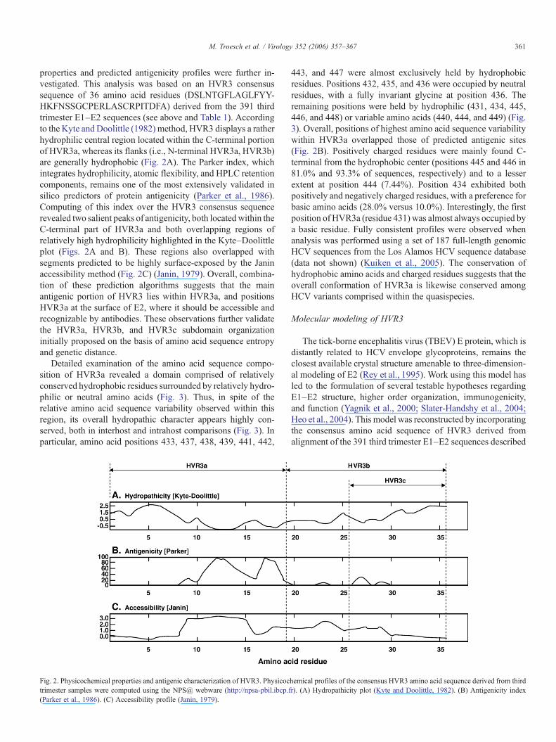
Fig. 1. Amino acid sequence diversity in HCV E1–E2 envelope glycoproteins. VariabE1–E2 sequences derived from third trimester samples from all study subjects and thmethods. Amino acid positions correspond to the M62321 reference sequence (Choowith previous reports (Weiner et al., 1991; Kato et al., 1992).
order to determine whether HVR3 was potentially subjected toselective pressure exerted by host HCV specific immunity, andthe physicochemical and structural characteristics of this regionwere evaluated to gain insights into its possible functional roles.
Results
Identification and characterization of HVR3
HCV E1 and E2 envelope gene sequences were amplifiedstarting with serial serum samples (pregestational; 1st, 2nd, 3rdtrimesters; postpartum) obtained from 17 pregnant women in-fectedwithHCV,including13whowerecoinfectedwithHCVandHIV-1. Four women were studied throughout two conscutivepregnancies. Nine patients were infected with HCV genotype 1a,5withHCVgenotype 1b, and 3withHCVgenotype 3a.A total of1337 nucleic acid sequences (570–576 nucleotides) wereobtained, including 1101 that were nonredundant, representinga mean of 78.7 sequences per patient (range = 52–147).Nucleotide sequences were edited and translated, spanningamino acid positions 319–508 of theHCV-1a reference sequence(GenBankaccessionno.M62321),andcorrespondingto the65C-terminal amino acid residues of E1 and the 125N-terminal aminoacid residues of E2 (E1/128–E2/125).
First, for the purposes of estimating amino acid sequencediversity, a data set comprised of all 391 E1–E2 sequencesderived from third trimester samples from all patients wasassembled, of which 331 (84.7%) were nonredundant at thenucleic acid level, representing a mean of 23 sequences perpatient (range = 17–38). Hence, this data set simultaneouslysampled roughly equivalent contributions from both intrahostand interhost E1–E2 amino acid sequence diversity. Amino acidsequences were aligned, and the entropy or variability at eachposition of the alignment was determined (Fig. 1). The entropy
ility at each amino acid position was computed using a data set comprised of 391e Entropy-ONE Web tool (Korber et al., 1994), as described under Materials andet al., 1989). Boundaries of E1, E2, HVR1, and HVR2 were set in accordance
359M. Troesch et al. / Virology 352 (2006) 357–367
profile revealed that amino acid variability was not uniformlydistributed across the amplified E1–E2 segments, with visuallydiscernible clusters of amino acid sequence diversity. The firstcluster (amino acid positions 384–410) was well defined andcorresponded to the 27 amino acids of HVR1 (Weiner et al.,1991), whereas a second cluster, further downstream (aminoacid positions 474–482), corresponded to HVR2 (Kato et al.,1992). Interestingly, distribution of amino acid entropy in theregion between HVR1 and HVR2 (amino acid positions 411–473) was clearly discontinuous, and a third cluster of amino acidvariability was readily delineated, spanning amino acid positions431–466, approximately halfway between HVR1 and HVR2(Fig. 1). For the purpose of the present study, we termed thisregion HVR3. HVR3 was empirically subdivided into two re-gions of roughly equal size, which were termed HVR3a (aminoacid positions 431–449) and HVR3b (amino acid positions450–466). Additional sequence variation was observed at the Nterminus and C terminus of the amplified segment (Fig. 1).Analysis was focused on HVR3 because (a) the first 65 N-terminal amino acids are in fact localized within the C-terminalportion of the E1 envelope protein; and (b) amino acids at the Cterminus are localized at the extreme end of our sequence reads,where base miscalls are more prevalent. The overall entropyprofile was not affected by removal from the alignment of allHCV-3a sequences (n = 58; subjects TV453, TVC45, andTVC73) and/or all sequences from patient TV531 (n = 17), whorespectively exhibited a 1 and 2 amino acid insertion betweenpositions 476 and 477 (data not shown). Likewise, comparableamino acid entropy profiles were obtained when analysis wasperformed with (a) additional subsamples from the complete1337 sequence data set (Troesch et al., unpublished); or (b) a setof 187 full-length genomic HCV sequences comprising all 6major HCV subtypes (http://hcv.lanl.gov/content/hcv-db/; Kui-ken et al., 2005) (Supplemental Fig. 1; data not shown).
To test relative amino acid sequence variability, geneticdistances were estimated within each of these domains andsubdomains (Table 1). As expected, highest variability wasobserved at the positions corresponding to HVR1 and HVR2.Mean genetic distance was lower in HVR3 (431–466) than in
Table 1Amino acid sequence variability in selected subregions of HCV E1 and E2envelope glycoproteins
Sequenceidentification
Amino acidposition a
Mean geneticdistance b
Standarderror c
E1/128–E2/125 319–508 0.253 0.020HVR1 384–410 0.509 0.049HVR2 474–482 0.541 0.044HVR3 431–466 0.315 0.042HVR3a 431–449 0.305 0.056HVR3b 450–466 0.326 0.061HVR3c 456–466 0.430 0.064
Nucleic acid sequences were obtained, edited, translated, and analyzed as des-cribed under Materials and methods.a Amino acid positions correspond to the M62321 reference sequence (Choo
et al., 1989).b Mean genetic distance was calculated as mean of pairwise p distances.c Standard error of the mean was computed by bootstrapping (500 replicates)
(Kumar et al., 2001).
HVR1 or HVR2 but higher than that obtained when the com-plete E1/128–E2/125 segment was analyzed (Table 1). WithinHVR3, the HVR3b subdomain was comparatively more va-riable than HVR3a or than the whole of HVR3. Additionaltruncation of the HVR3b subdomain into HVR3c (amino acidpositions 456–466) circumscribed a region of high sequencediversity, with a mean genetic distance of 0.430 ± 0.064 (Fig. 1).These values represented 84.5% and 79.5% of the mean geneticdistances measured within HVR1 and HVR2, respectively, and170% of the mean variability observed in the complete E1/128–E2/125 region (Table 1). Again, none of these estimates wasadversely affected by exclusion of all HCV-3a sequences and/orsequences from patient TV531 (see above), and all were com-parable to those obtained using a set of 187 full-length genomicHCV sequences retrieved from the Los Alamos database(Kuiken et al., 2005) (Supplemental Table 1; data not shown).
Intrahost selective pressure
Next, the presence and extent of intrahost selective pressureexerted on HCV E2 sequences were examined within genesegments encoding the HVR1, HVR2, HVR3, HVR3a, HVR3b,and HVR3c subdomains, which showed the highest amino acidvariability when initial interhost comparisons were performed(Table 1). To do this, we assembled data sets comprised ofnucleic acid sequences derived from longitudinal samples ob-tained from each patient throughout pregnancy (pregestational;1st, 2nd, 3rd trimesters; postpartum). The ratio of the meannumber of nonsynonymous substitutions per nonsynonymoussite over the mean number of synonymous substitutions persynonymous site (dN/dS) was then computed within the above-mentioned E1–E2 subdomains, with a dN/dS ratio >1 taken as anindex of positive selection for specific amino acid residues (Neiand Gojobori, 1986). As HVR3 and HVR3c represented broadlyoverlapping and/or nested subregions, comparisons of dN/dSratios were not drawn that involved either of these regions.Finally, it should be noted that the Los Alamos data set essen-tially samples interhost not intrahost variability (i.e., all of thesesequences are presumably unrelated). The extent of intrahost-selective pressure can therefore not be computed using such adata set.
Overall, statistically significant differences in median dN/dSratio were observed between subregions in multiple group ana-lysis (P = 0.0002, Kruskal–Wallis test). As expected, mediandN/dS ratio for HVR1 (2.391; interquartile range (IQR) = 0.816–3.092) was significantly higher than that observed in HVR2(0.227; IQR = 0.089–0.858), HVR3b (0.204; IQR = 0.129–0.330), but not HVR3a (0.719; IQR = 0.280–1.120) (P < 0.01,P < 0.001, and P > 0.05, respectively, Dunn's multiple com-parison test) (Table 2). Thirteen of 17 HCV-infected subjectstested (76.5%) had a dN/dS ratio >1 in HVR1, as compared with6 of 17 (35.3%) in HVR3a, 2 of 17 (11.8%) in HVR2, and 1 of 17(5.88%) in HVR3b (Table 2). In addition, 10 of 17 HCV-infectedsubjects (58.8%) exhibited statistically significant positiveselection in HVR1 in analysis of variance (P < 0.10, Z test), ascompared with 2 of 17 (11.8%) in HVR3a, 1 of 17 (5.88%) inHVR2, and 0 of 17 (0%) in HVR3b (Table 2). These results are
Table 2Estimate of selective pressure exerted on various subregions of the HCV E2 envelope glycoprotein
Subject HIVcoinfection
HCVgenotype
dN/dS
HVR1 a
(384–410)HVR2(474–482)
HVR3(431–466)
HVR3a(431–449)
HVR3b(450–466)
HVR3c(456–466)
TVC57 − 1a 2.614 b, c 0.090 0.812 1.177 b 0.424 2.812 b, c
TVC67 − 1a 1.482 b 0.163 0.607 0.634 0.498 0.246TVC75 − 1b 1.181 b 0.227 0.282 0.395 0.163 0.151TVC79 − 1a 3.826 b, c 0.000 0.513 1.542 b 0.174 0.119TV075 d + 1a 0.081 0.024 0.110 0.041 0.340 0.766TV179 d + 1b 0.136 7.317 b, c 0.202 0.164 0.204 0.163TV233 d + 1b 2.743 b, c 0.175 0.685 3.825 b, c 0.096 0.180TV289 + 1b 2.391 b, c nc 0.461 0.431 1.549 b 1.283 b
TV453 + 3a 0.428 0.166 0.037 0.023 0.052 0.037TV519 + 1b 4.263 b, c nc 0.552 1.062 b 0.044 0.204TV531 + 1a 1.860 b, c 0.253 0.391 0.955 0.233 0.521TVC17 + 1a 3.975 b, c 2.191 b 1.182 b 4.674 b, c 0.320 1.592 b
TVC33 + 1a 3.054 b, c 0.515 0.558 1.036 b 0.289 0.210TVC45 + 3a 1.040 b 0.858 0.626 0.904 0.227 0.345TVC55d + 1a 2.724 b, c 0.965 0.363 0.693 0.173 0.283TVC59 + 1a 0.592 0.248 0.165 0.139 0.185 0.220TVC73 + 3a 3.131 b, c 0.065 0.383 0.719 0.088 0.314
Nucleic acid sequencing, HCV genotyping, and calculation of the dN/dS ratio were performed as described under Materials and methods.a Amino acid positions correspond to the M62321 reference sequence (Choo et al., 1989).b Denotes dN > dS.c Denotes significant positive selection as determined by analysis of variance (P < 0.10, Z test) (Kumar et al., 2001). nc: not computed due to absence of
synonymous substitutions in that particular sequence subset.d Analysis performed on two consecutive pregnancies.
360 M. Troesch et al. / Virology 352 (2006) 357–367
consistent with the established role of HVR1 as one of theprincipal foci of intrahost immune-selective pressure (Weineret al., 1992; Kato et al., 1993; Farci et al., 2000, 2002; Canobioet al., 2004). These results also indicate that HVR3a (residues431–449) was the second most highly selected subdomain afterHVR1 within the E2 region examined, and HVR3b was the leastselected. Overall, this observed hierarchy in positive subdomainselection provides experimental support for the amino acidentropy and genetic distance-based clustering approach initiallyused to define HVR3, HVR3a, HVR3b, and HVR3c (Fig. 1;Table 1; Supplemental Fig. 1; Supplemental Table 1). Interest-ingly, linear regression analysis revealed a statistically signifi-cant relationship between the dN/dS ratios computed in HVR1and HVR3a (r2 = 0.360, P = 0.0105). However, there were nosuch significant relationships observed that involved eitherHVR2 or HVR3b. These results suggest that the selective forcesthat shape the amino acid sequences of HVR1 and HVR3a act ina similar and/or synchronous manner on both subregions,whereas genetic diversification in HVR2 and HVR3b wouldappear to be driven by distinct selective forces.
Influence of HCV subtype and coinfection with HIV-1
Three subjects in our study group (TV453, TVC45, andTCV73) were infected with HCV-3a, a subtype that not onlydisplays marked amino acid sequence dissimilarity as comparedwith HCV-1a and HCV-1b, but also exhibits variant biologicalproperties in terms of pathogenesis and resistance to interferontreatment (Pawlotsky, 2000; Rubbia-Brandt et al., 2001). Forthese reasons, intrahost-selective pressure was examined sepa-rately in these subjects.Median dN/dS ratio was lower in patients
infected with HCV-3a than HCV-1a or HCV-1b for allsubdomains analyzed (Table 2). Within HVR1, 2 of 3 subjects(66.6%) with HCV-3a infection displayed a dN/dS ratio >1, and1 of 3 (33.3%) exhibited significant positive selection asdetermined by analysis of variance (Table 2). These proportionswere similar to those observed in subjects infected with HCV-1aor HCV-1b with respect to HVR1 and all other E2 subdomains,but the statistical significance of these observations was nottested due to the limited number of HCV-3a-infected subjectsenrolled in our study group.
In addition, a number of studies have reported that infectionwith human immunodeficiency virus type 1 (HIV-1) influencedHCV quasispecies distribution and reduced host-selective pres-sure exerted on HVR1 in coinfected subjects (Sherman et al.,1996; Mao et al., 2001; Canobio et al., 2004). Because of this,intrahost-selective pressure on HVR1, HVR2, HVR3a, andHVR3b was compared between HCV/HIV-1 coinfected patients(n = 14) and patients infected solely with HCV (n = 4). Overall,the median dN/dS ratio was not significantly higher in subjectsinfected only with HCV than in coinfected patients (P > 0.117,Mann–Whitney U test), and there was no significant differencein the proportion of coinfected subjects exhibiting either a dN/dSratio >1 or significant positive selection for any of the E2subdomains analyzed (P > 0.235, Fisher's exact test).
Physicochemical properties and predicted antigenicity ofHVR3
Because HVR3, and in particular HVR3a, stood out as HCVE2 envelope glycoprotein subregions subjected to relatively highintrahost selective pressure, details of their physicochemical
361M. Troesch et al. / Virology 352 (2006) 357–367
properties and predicted antigenicity profiles were further in-vestigated. This analysis was based on an HVR3 consensussequence of 36 amino acid residues (DSLNTGFLAGLFYY-HKFNSSGCPERLASCRPITDFA) derived from the 391 thirdtrimester E1–E2 sequences (see above and Table 1). Accordingto the Kyte and Doolittle (1982) method, HVR3 displays a ratherhydrophilic central region located within the C-terminal portionof HVR3a, whereas its flanks (i.e., N-terminal HVR3a, HVR3b)are generally hydrophobic (Fig. 2A). The Parker index, whichintegrates hydrophilicity, atomic flexibility, and HPLC retentioncomponents, remains one of the most extensively validated insilico predictors of protein antigenicity (Parker et al., 1986).Computing of this index over the HVR3 consensus sequencerevealed two salient peaks of antigenicity, both locatedwithin theC-terminal part of HVR3a and both overlapping regions ofrelatively high hydrophilicity highlighted in the Kyte–Doolittleplot (Figs. 2A and B). These regions also overlapped withsegments predicted to be highly surface-exposed by the Janinaccessibility method (Fig. 2C) (Janin, 1979). Overall, combina-tion of these prediction algorithms suggests that the mainantigenic portion of HVR3 lies within HVR3a, and positionsHVR3a at the surface of E2, where it should be accessible andrecognizable by antibodies. These observations further validatethe HVR3a, HVR3b, and HVR3c subdomain organizationinitially proposed on the basis of amino acid sequence entropyand genetic distance.
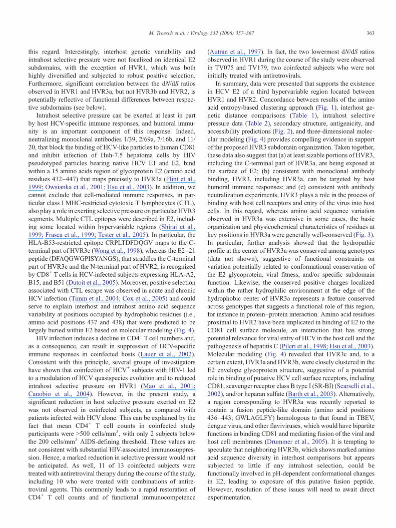
Detailed examination of the amino acid sequence compo-sition of HVR3a revealed a domain comprised of relativelyconserved hydrophobic residues surrounded by relatively hydro-philic or neutral amino acids (Fig. 3). Thus, in spite of therelative amino acid sequence variability observed within thisregion, its overall hydropathic character appears highly con-served, both in interhost and intrahost comparisons (Fig. 3). Inparticular, amino acid positions 433, 437, 438, 439, 441, 442,
Fig. 2. Physicochemical properties and antigenic characterization of HVR3. Physicoctrimester samples were computed using the NPS@ webware (http://npsa-pbil.ibcp.f(Parker et al., 1986). (C) Accessibility profile (Janin, 1979).
443, and 447 were almost exclusively held by hydrophobicresidues. Positions 432, 435, and 436 were occupied by neutralresidues, with a fully invariant glycine at position 436. Theremaining positions were held by hydrophilic (431, 434, 445,446, and 448) or variable amino acids (440, 444, and 449) (Fig.3). Overall, positions of highest amino acid sequence variabilitywithin HVR3a overlapped those of predicted antigenic sites(Fig. 2B). Positively charged residues were mainly found C-terminal from the hydrophobic center (positions 445 and 446 in81.0% and 93.3% of sequences, respectively) and to a lesserextent at position 444 (7.44%). Position 434 exhibited bothpositively and negatively charged residues, with a preference forbasic amino acids (28.0% versus 10.0%). Interestingly, the firstposition of HVR3a (residue 431) was almost always occupied bya basic residue. Fully consistent profiles were observed whenanalysis was performed using a set of 187 full-length genomicHCV sequences from the Los Alamos HCV sequence database(data not shown) (Kuiken et al., 2005). The conservation ofhydrophobic amino acids and charged residues suggests that theoverall conformation of HVR3a is likewise conserved amongHCV variants comprised within the quasispecies.
Molecular modeling of HVR3
The tick-borne encephalitis virus (TBEV) E protein, which isdistantly related to HCV envelope glycoproteins, remains theclosest available crystal structure amenable to three-dimension-al modeling of E2 (Rey et al., 1995). Work using this model hasled to the formulation of several testable hypotheses regardingE1–E2 structure, higher order organization, immunogenicity,and function (Yagnik et al., 2000; Slater-Handshy et al., 2004;Heo et al., 2004). This model was reconstructed by incorporatingthe consensus amino acid sequence of HVR3 derived fromalignment of the 391 third trimester E1–E2 sequences described
hemical profiles of the consensus HVR3 amino acid sequence derived from thirdr). (A) Hydropathicity plot (Kyte and Doolittle, 1982). (B) Antigenicity index
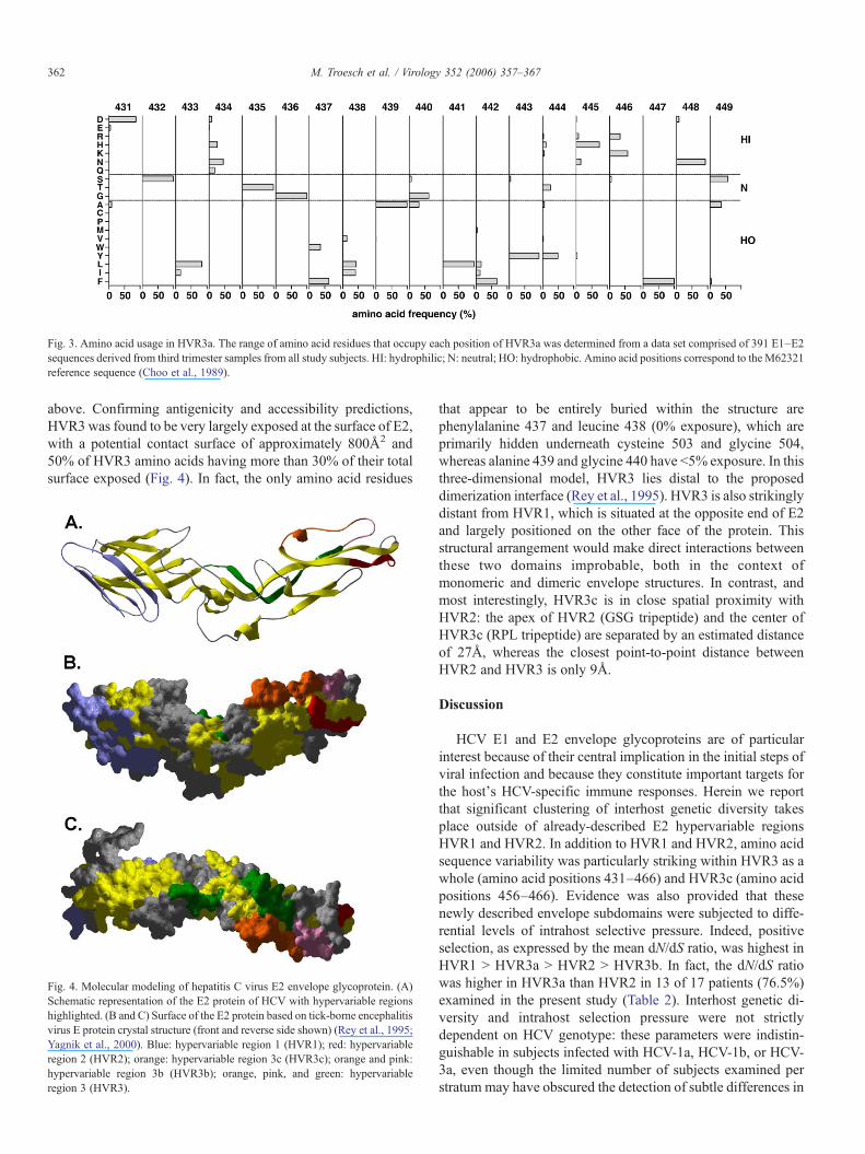
Fig. 3. Amino acid usage in HVR3a. The range of amino acid residues that occupy each position of HVR3a was determined from a data set comprised of 391 E1–E2sequences derived from third trimester samples from all study subjects. HI: hydrophilic; N: neutral; HO: hydrophobic. Amino acid positions correspond to the M62321reference sequence (Choo et al., 1989).
362 M. Troesch et al. / Virology 352 (2006) 357–367
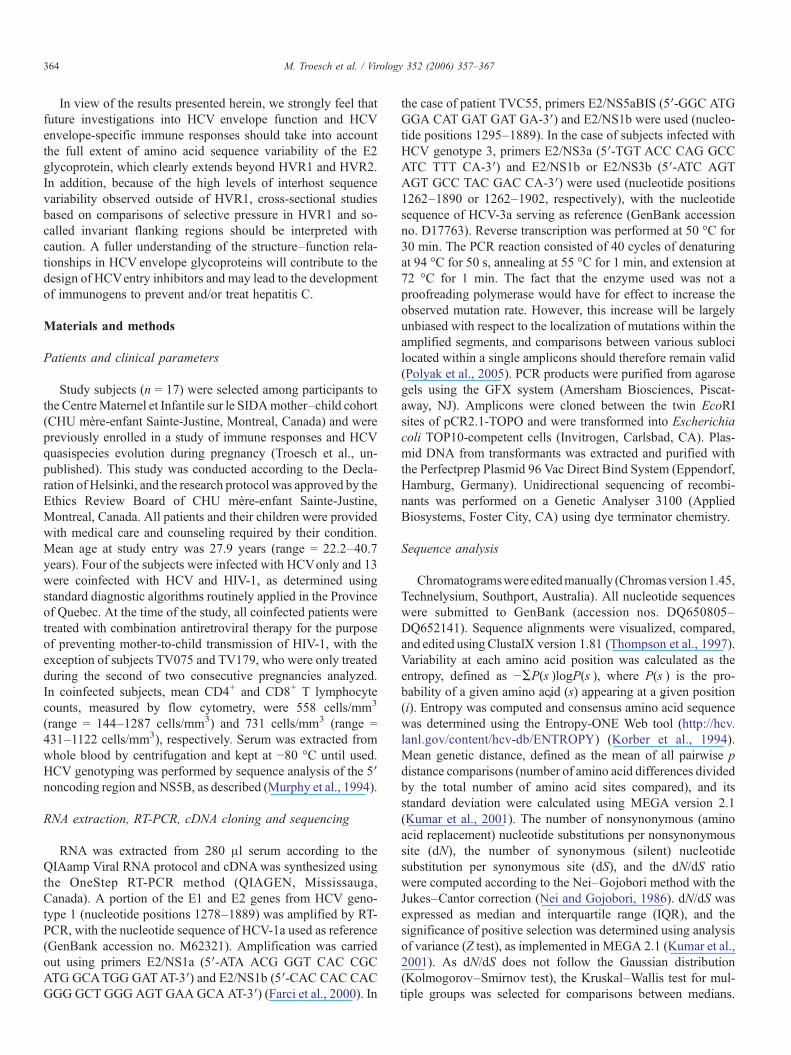
above. Confirming antigenicity and accessibility predictions,HVR3 was found to be very largely exposed at the surface of E2,with a potential contact surface of approximately 800Å2 and50% of HVR3 amino acids having more than 30% of their totalsurface exposed (Fig. 4). In fact, the only amino acid residues
Fig. 4. Molecular modeling of hepatitis C virus E2 envelope glycoprotein. (A)Schematic representation of the E2 protein of HCV with hypervariable regionshighlighted. (B and C) Surface of the E2 protein based on tick-borne encephalitisvirus E protein crystal structure (front and reverse side shown) (Rey et al., 1995;Yagnik et al., 2000). Blue: hypervariable region 1 (HVR1); red: hypervariableregion 2 (HVR2); orange: hypervariable region 3c (HVR3c); orange and pink:hypervariable region 3b (HVR3b); orange, pink, and green: hypervariableregion 3 (HVR3).
that appear to be entirely buried within the structure arephenylalanine 437 and leucine 438 (0% exposure), which areprimarily hidden underneath cysteine 503 and glycine 504,whereas alanine 439 and glycine 440 have <5% exposure. In thisthree-dimensional model, HVR3 lies distal to the proposeddimerization interface (Rey et al., 1995). HVR3 is also strikinglydistant from HVR1, which is situated at the opposite end of E2and largely positioned on the other face of the protein. Thisstructural arrangement would make direct interactions betweenthese two domains improbable, both in the context ofmonomeric and dimeric envelope structures. In contrast, andmost interestingly, HVR3c is in close spatial proximity withHVR2: the apex of HVR2 (GSG tripeptide) and the center ofHVR3c (RPL tripeptide) are separated by an estimated distanceof 27Å, whereas the closest point-to-point distance betweenHVR2 and HVR3 is only 9Å.
Discussion
HCV E1 and E2 envelope glycoproteins are of particularinterest because of their central implication in the initial steps ofviral infection and because they constitute important targets forthe host's HCV-specific immune responses. Herein we reportthat significant clustering of interhost genetic diversity takesplace outside of already-described E2 hypervariable regionsHVR1 and HVR2. In addition to HVR1 and HVR2, amino acidsequence variability was particularly striking within HVR3 as awhole (amino acid positions 431–466) and HVR3c (amino acidpositions 456–466). Evidence was also provided that thesenewly described envelope subdomains were subjected to diffe-rential levels of intrahost selective pressure. Indeed, positiveselection, as expressed by the mean dN/dS ratio, was highest inHVR1 > HVR3a > HVR2 > HVR3b. In fact, the dN/dS ratiowas higher in HVR3a than HVR2 in 13 of 17 patients (76.5%)examined in the present study (Table 2). Interhost genetic di-versity and intrahost selection pressure were not strictlydependent on HCV genotype: these parameters were indistin-guishable in subjects infected with HCV-1a, HCV-1b, or HCV-3a, even though the limited number of subjects examined perstratum may have obscured the detection of subtle differences in
363M. Troesch et al. / Virology 352 (2006) 357–367
this regard. Interestingly, interhost genetic variability andintrahost selective pressure were not focalized on identical E2subdomains, with the exception of HVR1, which was bothhighly diversified and subjected to robust positive selection.Furthermore, significant correlation between the dN/dS ratiosobserved in HVR1 and HVR3a, but not HVR3b and HVR2, ispotentially reflective of functional differences between respec-tive subdomains (see below).
Intrahost selective pressure can be exerted at least in partby host HCV-specific immune responses, and humoral immu-nity is an important component of this response. Indeed,neutralizing monoclonal antibodies 1/39, 2/69a, 7/16b, and 11/20, that block the binding of HCV-like particles to human CD81and inhibit infection of Huh-7.5 hepatoma cells by HIVpseudotyped particles bearing native HCV E1 and E2, bindwithin a 15 amino acids region of glycoprotein E2 (amino acidresidues 432–447) that maps precisely to HVR3a (Flint et al.,1999; Owsianka et al., 2001; Hsu et al., 2003). In addition, wecannot exclude that cell-mediated immune responses, in par-ticular class I MHC-restricted cytotoxic T lymphocytes (CTL),also play a role in exerting selective pressure on particular HVR3segments. Multiple CTL epitopes were described in E2, includ-ing some located within hypervariable regions (Shirai et al.,1999; Frasca et al., 1999; Tester et al., 2005). In particular, theHLA-B53-restricted epitope CRPLTDFDQGV maps to the C-terminal part of HVR3c (Wong et al., 1998), whereas the E2–21peptide (DFAQGWGPISYANGS), that straddles the C-terminalpart of HVR3c and the N-terminal part of HVR2, is recognizedby CD8+ T cells in HCV-infected subjects expressing HLA-A2,B15, and B51 (Dutoit et al., 2005). Moreover, positive selectionassociated with CTL escape was observed in acute and chronicHCV infection (Timm et al., 2004; Cox et al., 2005) and couldserve to explain interhost and intrahost amino acid sequencevariability at positions occupied by hydrophobic residues (i.e.,amino acid positions 437 and 438) that were predicted to belargely buried within E2 based on molecular modeling (Fig. 4).
HIV infection induces a decline in CD4+ T cell numbers and,as a consequence, can result in suppression of HCV-specificimmune responses in coinfected hosts (Lauer et al., 2002).Consistent with this principle, several groups of investigatorshave shown that coinfection of HCV+ subjects with HIV-1 ledto a modulation of HCV quasispecies evolution and to reducedintrahost selective pressure on HVR1 (Mao et al., 2001;Canobio et al., 2004). However, in the present study, asignificant reduction in host selective pressure exerted on E2was not observed in coinfected subjects, as compared withpatients infected with HCValone. This can be explained by thefact that mean CD4+ T cell counts in coinfected studyparticipants were >500 cells/mm3, with only 2 subjects belowthe 200 cells/mm3 AIDS-defining threshold. These values arenot consistent with substantial HIV-associated immunosuppres-sion. Hence, a marked reduction in selective pressure would notbe anticipated. As well, 11 of 13 coinfected subjects weretreated with antiretroviral therapy during the course of the study,including 10 who were treated with combinations of antire-troviral agents. This commonly leads to a rapid restoration ofCD4+ T cell counts and of functional immunocompetence
(Autran et al., 1997). In fact, the two lowermost dN/dS ratiosobserved in HVR1 during the course of the study were observedin TV075 and TV179, two coinfected subjects who were notinitially treated with antiretrovirals.
In summary, data were presented that supports the existencein HCV E2 of a third hypervariable region located betweenHVR1 and HVR2. Concordance between results of the aminoacid entropy-based clustering approach (Fig. 1), interhost ge-netic distance comparisons (Table 1), intrahost selectivepressure data (Table 2), secondary structure, antigenicity, andaccessibility predictions (Fig. 2), and three-dimensional molec-ular modeling (Fig. 4) provides compelling evidence in supportof the proposed HVR3 subdomain organization. Taken together,these data also suggest that (a) at least sizable portions of HVR3,including the C-terminal part of HVR3a, are being exposed atthe surface of E2; (b) consistent with monoclonal antibodybinding, HVR3, including HVR3a, can be targeted by hosthumoral immune responses; and (c) consistent with antibodyneutralization experiments, HVR3 plays a role in the process ofbinding with host cell receptors and entry of the virus into hostcells. In this regard, whereas amino acid sequence variationobserved in HVR3a was extensive in some cases, the basicorganization and physicochemical characteristics of residues atkey positions in HVR3a were generally well-conserved (Fig. 3).In particular, further analysis showed that the hydropathicprofile at the center of HVR3a was conserved among genotypes(data not shown), suggestive of functional constraints onvariation potentially related to conformational conservation ofthe E2 glycoprotein, viral fitness, and/or specific subdomainfunction. Likewise, the conserved positive charges localizedwithin the rather hydrophilic environment at the edge of thehydrophobic center of HVR3a represents a feature conservedacross genotypes that suggests a functional role of this region,for instance in protein–protein interaction. Amino acid residuesproximal to HVR2 have been implicated in binding of E2 to theCD81 cell surface molecule, an interaction that has strongpotential relevance for viral entry of HCV in the host cell and thepathogenesis of hepatitis C (Pileri et al., 1998; Hsu et al., 2003).Molecular modeling (Fig. 4) revealed that HVR3c and, to acertain extent, HVR3a and HVR3b, were closely clustered in theE2 envelope glycoprotein structure, suggestive of a potentialrole in binding of putative HCV cell surface receptors, includingCD81, scavenger receptor class B type I (SR-BI) (Scarselli et al.,2002), and/or heparan sulfate (Barth et al., 2003). Alternatively,a region corresponding to HVR3a was recently reported tocontain a fusion peptide-like domain (amino acid positions436–443; GWLAGLFY) homologous to that found in TBEV,dengue virus, and other flaviviruses, which would have bipartitefunctions in binding CD81 and mediating fusion of the viral andhost cell membranes (Drummer et al., 2005). It is tempting tospeculate that neighboring HVR3b, which shows marked aminoacid sequence diversity in interhost comparisons but appearssubjected to little if any intrahost selection, could befunctionally involved in pH-dependent conformational changesin E2, leading to exposure of this putative fusion peptide.However, resolution of these issues will need to await directexperimentation.
364 M. Troesch et al. / Virology 352 (2006) 357–367
In view of the results presented herein, we strongly feel thatfuture investigations into HCV envelope function and HCVenvelope-specific immune responses should take into accountthe full extent of amino acid sequence variability of the E2glycoprotein, which clearly extends beyond HVR1 and HVR2.In addition, because of the high levels of interhost sequencevariability observed outside of HVR1, cross-sectional studiesbased on comparisons of selective pressure in HVR1 and so-called invariant flanking regions should be interpreted withcaution. A fuller understanding of the structure–function rela-tionships in HCV envelope glycoproteins will contribute to thedesign of HCVentry inhibitors and may lead to the developmentof immunogens to prevent and/or treat hepatitis C.
Materials and methods
Patients and clinical parameters
Study subjects (n = 17) were selected among participants tothe CentreMaternel et Infantile sur le SIDAmother–child cohort(CHU mère-enfant Sainte-Justine, Montreal, Canada) and werepreviously enrolled in a study of immune responses and HCVquasispecies evolution during pregnancy (Troesch et al., un-published). This study was conducted according to the Decla-ration of Helsinki, and the research protocol was approved by theEthics Review Board of CHU mère-enfant Sainte-Justine,Montreal, Canada. All patients and their children were providedwith medical care and counseling required by their condition.Mean age at study entry was 27.9 years (range = 22.2–40.7years). Four of the subjects were infected with HCVonly and 13were coinfected with HCV and HIV-1, as determined usingstandard diagnostic algorithms routinely applied in the Provinceof Quebec. At the time of the study, all coinfected patients weretreated with combination antiretroviral therapy for the purposeof preventing mother-to-child transmission of HIV-1, with theexception of subjects TV075 and TV179, who were only treatedduring the second of two consecutive pregnancies analyzed.In coinfected subjects, mean CD4+ and CD8+ T lymphocytecounts, measured by flow cytometry, were 558 cells/mm3
(range = 144–1287 cells/mm3) and 731 cells/mm3 (range =431–1122 cells/mm3), respectively. Serum was extracted fromwhole blood by centrifugation and kept at −80 °C until used.HCV genotyping was performed by sequence analysis of the 5′noncoding region and NS5B, as described (Murphy et al., 1994).
RNA extraction, RT-PCR, cDNA cloning and sequencing
RNA was extracted from 280 μl serum according to theQIAamp Viral RNA protocol and cDNAwas synthesized usingthe OneStep RT-PCR method (QIAGEN, Mississauga,Canada). A portion of the E1 and E2 genes from HCV geno-type 1 (nucleotide positions 1278–1889) was amplified by RT-PCR, with the nucleotide sequence of HCV-1a used as reference(GenBank accession no. M62321). Amplification was carriedout using primers E2/NS1a (5′-ATA ACG GGT CAC CGCATG GCATGG GATAT-3′) and E2/NS1b (5′-CAC CAC CACGGGGCT GGGAGT GAAGCA AT-3′) (Farci et al., 2000). In
the case of patient TVC55, primers E2/NS5aBIS (5′-GGC ATGGGA CAT GAT GAT GA-3′) and E2/NS1b were used (nucleo-tide positions 1295–1889). In the case of subjects infected withHCV genotype 3, primers E2/NS3a (5′-TGT ACC CAG GCCATC TTT CA-3′) and E2/NS1b or E2/NS3b (5′-ATC AGTAGT GCC TAC GAC CA-3′) were used (nucleotide positions1262–1890 or 1262–1902, respectively), with the nucleotidesequence of HCV-3a serving as reference (GenBank accessionno. D17763). Reverse transcription was performed at 50 °C for30 min. The PCR reaction consisted of 40 cycles of denaturingat 94 °C for 50 s, annealing at 55 °C for 1 min, and extension at72 °C for 1 min. The fact that the enzyme used was not aproofreading polymerase would have for effect to increase theobserved mutation rate. However, this increase will be largelyunbiased with respect to the localization of mutations within theamplified segments, and comparisons between various sublocilocated within a single amplicons should therefore remain valid(Polyak et al., 2005). PCR products were purified from agarosegels using the GFX system (Amersham Biosciences, Piscat-away, NJ). Amplicons were cloned between the twin EcoRIsites of pCR2.1-TOPO and were transformed into Escherichiacoli TOP10-competent cells (Invitrogen, Carlsbad, CA). Plas-mid DNA from transformants was extracted and purified withthe Perfectprep Plasmid 96 Vac Direct Bind System (Eppendorf,Hamburg, Germany). Unidirectional sequencing of recombi-nants was performed on a Genetic Analyser 3100 (AppliedBiosystems, Foster City, CA) using dye terminator chemistry.
Sequence analysis
Chromatogramswereeditedmanually (Chromasversion1.45,Technelysium, Southport, Australia). All nucleotide sequenceswere submitted to GenBank (accession nos. DQ650805–DQ652141). Sequence alignments were visualized, compared,and edited using ClustalX version 1.81 (Thompson et al., 1997).Variability at each amino acid position was calculated as theentropy, defined as −ΣP(s
i
)logP(si
), where P(si
) is the pro-bability of a given amino acid (s) appearing at a given position(i). Entropy was computed and consensus amino acid sequencewas determined using the Entropy-ONE Web tool (http://hcv.lanl.gov/content/hcv-db/ENTROPY) (Korber et al., 1994).Mean genetic distance, defined as the mean of all pairwise pdistance comparisons (number of amino acid differences dividedby the total number of amino acid sites compared), and itsstandard deviation were calculated using MEGA version 2.1(Kumar et al., 2001). The number of nonsynonymous (aminoacid replacement) nucleotide substitutions per nonsynonymoussite (dN), the number of synonymous (silent) nucleotidesubstitution per synonymous site (dS), and the dN/dS ratiowere computed according to the Nei–Gojobori method with theJukes–Cantor correction (Nei and Gojobori, 1986). dN/dS wasexpressed as median and interquartile range (IQR), and thesignificance of positive selection was determined using analysisof variance (Z test), as implemented in MEGA 2.1 (Kumar et al.,2001). As dN/dS does not follow the Gaussian distribution(Kolmogorov–Smirnov test), the Kruskal–Wallis test for mul-tiple groups was selected for comparisons between medians.
365M. Troesch et al. / Virology 352 (2006) 357–367
Comparisons between regions were examined using Dunn'spost test, which corrects for multiple comparisons in nonpara-metric tests.
Molecular modeling of HCV E2 glycoprotein
The model of E2 was generated essentially as described byYagnik and coworkers (2000). Briefly, secondary structurepredictions were used to align HCV E2 with the sequence of theE envelope glycoprotein of TBEV. The alignment generated wasused to model the E2 protein using the structure of the TBEV Eprotein (PDB code 1SVB; Rey et al., 1995) and theMODELLERprogram (Sali and Blundell, 1993). Energy minimization of theresulting model was then performed using 100 steps of thesteepest descents algorithm with the consistent valence forcefield (CVFF) (Dauber-Osguthorpe et al., 1988). The proteinstructure was analyzed with the Swiss PDB Viewer 3.7 program(Guex and Peitsch, 1997), and the final images were renderedusing POVRAYversion 3.5.
Acknowledgments
The authors wish to thank Martine Caty, Silvie Valois, andAmpha Khammy for expert technical assistance, and Marc-André Rodrigue and Julie Lacaille for automated DNAsequencing. Supported by grants from the CIHR-Health CanadaResearch Initiative on Hepatitis C (grant no. EOP-41537),CANFAR-Canadian Foundation for AIDS Research (grant no.013515), and The Elizabeth Glaser Pediatric AIDS Foundation(grant no. 28-PG-51355). M.T. was the recipient of a scho-larship from Fondation de l'Hôpital Sainte-Justine. H.S. is aJunior-II Scientist of le Fonds de la recherche en santé duQuébec (FRSQ).
Appendix A. Supplementary data
Supplementary data associated with this article can be found,in the online version, at doi:10.1016/j.virol.2006.05.015.
References
Abbate, I., Lo Iacono, O., Di Stefano, R., Cappiello, G., Girardi, E., Longo, R.,Ferraro, D., Antonucci, G., Di Marco, V., Solmone, M., Craxi, A., Ippolito,G., Capobianchi, M.R., 2004. HVR-1 quasispecies modifications occurearly and are correlated to initial but not sustained response in HCV-infected patients treated with pegylated- or standard-interferon and riba-virin. J. Hepatol. 40, 831–836.
Alter, M.J., Margolis, H.S., Krawczynski, K., Judson, F.N., Mares, A.,Alexander, W.J., Hu, P.Y., Gerber, M.A., Sampliner, R.E., Meeks, E.L.,Beach, M.J., 1992. The natural history of community-acquired hepatitis C inthe United States. The Sentinel Counties Chronic non-A, non-B HepatitisStudy Team. N. Engl. J. Med. 327, 1899–1905.
Autran, B., Carcelain, G., Li, T.S., Blanc, C., Mathez, D., Tubiana, R., Katlama,C., Debre, P., Leibowitch, J., 1997. Positive effects of combinedantiretroviral therapy on CD4+ T cell homeostasis and function in advancedHIV disease. Science 277, 112–116.
Bach, N., Thung, S.N., Schaffner, F., 1992. The histological features of chronichepatitis C and autoimmune chronic hepatitis: a comparative analysis.Hepatology 15, 572–577.
Barth, H., Schafer, C., Adah, M.I., Zhang, F., Linhardt, R.J., Toyoda, H.,Kinoshita-Toyoda, A., Toida, T., Van Kuppevelt, T.H., Depla, E., VonWeizsacker, F., Blum, H.E., Baumert, T.F., 2003. Cellular binding ofhepatitis C virus envelope glycoprotein E2 requires cell surface heparansulfate. J. Biol. Chem. 278, 41003–41012.
Benhamou, Y., Di Martino, V., Bochet, M., Colombet, G., Thibault, V., Liou, A.,Katlama, C., Poynard, T., 2001. Factors affecting liver fibrosis in humanimmunodeficiency virus- and hepatitis C virus-coinfected patients: impact ofprotease inhibitor therapy. Hepatology 34, 283–287.
Bonacini, M., Puoti, M., 2000. Hepatitis C in patients with humanimmunodeficiency virus infection: diagnosis, natural history, meta-analysisof sexual and vertical transmission, and therapeutic issues. Arch. Intern.Med. 160, 3365–3373.
Bukh, J., Miller, R.H., Purcell, R.H., 1995. Genetic heterogeneity of hepatitis Cvirus: quasispecies and genotypes. Semin. Liver Dis. 15, 41–63.
Canobio, S., Guilbert, C., Troesch, M., Samson, J., Lemay, M., Pelletier, V.A.,Bernard-Bonnin, A.C., Kozielski, R., Lapointe, N., Martin, S.R., Soudeyns,H., 2004. Differing patterns of liver disease progression and hepatitis C virus(HCV) quasispecies evolution in children vertically coinfectedwithHCVandhuman immunodeficiency virus type 1. J. Clin. Microbiol. 42, 4365–4369.
Chen, S., Wang, Y.M., 2005. Multigene tracking of quasispecies in viralpersistence and clearance of hepatitis C virus. World J. Gastroenterol. 11,2874–2884.
Choo, Q.L., Kuo, G., Weiner, A.J., Overby, L.R., Bradley, D.W., Houghton, M.,1989. Isolation of a cDNA clone derived from a blood-borne non-A, non-Bviral hepatitis genome. Science 244, 359–362.
Choo, Q.L., Richman, K.H., Han, J.H., Berger, K., Lee, C., Dong, C., Gallegos,C., Coit, D., Medina-Selby, R., Barr, P.J., Weiner, A.J., Bradley, D.W., Kuo,G., Houghton, M., 1991. Genetic organization and diversity of the hepatitisC virus. Proc. Natl. Acad. Sci. U.S.A. 88, 2451–2455.
Cox, A.L., Mosbruger, T., Mao, Q., Liu, Z., Wang, X.H., Yang, H.C., Sidney, J.,Sette, A., Pardoll, D., Thomas, D.L., Ray, S.C., 2005. Cellular immuneselection with hepatitis C virus persistence in humans. J. Exp. Med. 201,1741–1752.
Dauber-Osguthorpe, P., Roberts, V.A., Osguthorpe, D.J., Wolff, J., Genest, M.,Hagler, A.T., 1988. Structure and energetics of ligand binding to proteins:Escherichia coli dihydrofolate reductase-trimethoprim, a drug-receptorsystem. Proteins 4, 31–47.
Dieterich, D.T., 1999. Hepatitis C virus and human immunodeficiency virus:clinical issues in coinfection. Am. J. Med. 107, 79S–84S.
Dodig, M., Tavill, A.S., 2001. Hepatitis C and human immunodeficiency viruscoinfections. J. Clin. Gastroenterol. 33, 367–374.
Drummer, H., Maerz, A., Poumbourios, P., 2005. Structural and functional roleof a fusion-peptide like sequence in hepatitis C virus glycoprotein E2.Abstracts of the 12th International Meeting on HCV and Related Viruses,Montreal, Canada, p. 26.
Dutoit, V., Ciuffreda, D., Comte, D., Gonvers, J.J., Pantaleo, G., 2005.Differences in HCV-specific T cell responses between chronic HCVinfection and HIV/HCV co-infection. Eur. J. Immunol. 35, 3493–3504.
Farci, P., Shimoda, A., Coiana, A., Diaz, G., Peddis, G., Melpolder, J.C.,Strazzera, A., Chien, D.Y., Munoz, S.J., Balestrieri, A., Purcell, R.H., Alter,H.J., 2000. The outcome of acute hepatitis C predicted by the evolution ofthe viral quasispecies. Science 288, 339–344.
Farci, P., Strazzera, R., Alter, H.J., Farci, S., Degioannis, D., Coiana, A., Peddis,G., Usai, F., Serra, G., Chessa, L., Diaz, G., Balestrieri, A., Purcell, R.H.,2002. Early changes in hepatitis C viral quasispecies during interferontherapy predict the therapeutic outcome. Proc. Natl. Acad. Sci. U.S.A. 99,3081–3086.
Flint, M., Maidens, C., Loomis-Price, L.D., Shotton, C., Dubuisson, J., Monk,P., Higginbottom, A., Levy, S., McKeating, J.A., 1999. Characterization ofhepatitis C virus E2 glycoprotein interaction with a putative cellularreceptor, CD81. J. Virol. 73, 6235–6244.
Frasca, L., Del Porto, P., Tuosto, L., Marinari, B., Scotta, C., Carbonari, M.,Nicosia, A., Piccoletta, E., 1999. Hypervariable region 1 variants act as TCRantagonists for hepatitis C virus-specific CD4+ T cells. J. Immunol. 163,650–658.
Gaudy, C., Moreau, A., Veillon, P., Temoin, S., Lunel, F., Goudeau, A., 2003.Significance of pretreatment analysis of hepatitis C virus genotype 1b
366 M. Troesch et al. / Virology 352 (2006) 357–367
hypervariable region 1 sequences to predict antiviral outcome. J. Clin.Microbiol. 41, 3615–3622.
Gonzalez-Peralta, R.P., Qian, K., She, J.Y., Davis, G.L., Ohno, T., Mizokami,M., Lau, J.Y., 1996. Clinical implications of viral quasispecies heterogeneityin chronic hepatitis C. J. Med. Virol. 49, 242–247.
Guex, N., Peitsch, M.C., 1997. SWISS-MODEL and the Swiss-PdbViewer: anenvironment for comparative protein modeling. Electrophoresis 18,2714–2723.
Heo, T.H., Chang, J.H., Lee, J.W., Foung, S.K.H., Dubuisson, J., Kang, C.Y.,2004. Incomplete humoral immunity against hepatitis C virus is linked withdistinct recognition of putative multiple receptors by E2 envelopeglycoproteins. J. Immunol. 173, 446–455.
Honda, M., Kaneko, S., Sakai, A., Unoura, M., Murakami, S., Kobayashi, K.,1994. Degree of diversity of hepatitis C virus quasispecies and progressionof liver disease. Hepatology 20, 1144–1151.
Hsu, M., Zhang, J., Flint, M., Logvinoff, C., Cheng-Mayer, C., Rice, C.M.,McKeating, J.A., 2003. Hepatitis C virus glycoproteins mediate pH-dependent cell entry of pseudotyped viral particles. Proc. Natl. Acad. Sci.U.S.A. 100, 7271–7276.
Janin, J., 1979. Surface and inside volumes in globular proteins. Nature 277,491–492.
Kato, N., Ootsuyama, Y., Tanaka, T., Nakagawa, M., Nakazawa, T., Muraiso,K., Ohkoshi, S., Hijikata, M., Shimotohno, K., 1992. Marked sequencediversity in the putative envelope proteins of hepatitis C viruses. Virus Res.22, 107–123.
Kato, N., Sekiya, H., Ootsuyama, Y., Nakazawa, T., Hijikata, M., Ohkoshi, S.,Shimotohno, K., 1993. Humoral immune response to hypervariable region 1of the putative envelope glycoprotein (gp70) of hepatitis C virus. J. Virol.67, 3923–3930.
Korber, B.T.M., Kunstman, K.J., Patterson, B.K., Furtado, M., McEvilly,M.M., Levy, R., Wolinsky, S.M., 1994. Genetic differences betweenblood- and brain-derived viral sequences from human immunodeficiencyvirus type 1-infected patients: evidence of conserved elements in the V3region of the envelope protein of brain-derived sequences. J. Virol. 68,7467–7481.
Kuiken, C., Yusim, K., Boykin, L., Richardson, R., 2005. The Los Alamos HCVsequence database. Bioinformatics 21, 379–384.
Kumar, S., Tamura, K., Jakobsen, I.B., Nei, M., 2001. MEGA2: molecularevolutionary genetics analysis software. Bioinformatics 17, 1244–1245.
Kyte, J., Doolittle, R.F., 1982. A simple method for displaying the hydropathiccharacter of a protein. J. Mol. Biol. 157, 105–132.
Lauer, G.M., Nguyen, T.N., Day, C.L., Robbins, G.K., Flynn, T., McGowan, K.,Rosenberg, E.S., Lucas, M., Klenerman, P., Chung, R.T., Walker, B.D.,2002. Human immunodeficiency virus type 1-hepatitis C virus coinfection:intraindividual comparison of cellular immune responses against twopersistent viruses. J. Virol. 76, 2817–2826.
Mao, Q., Ray, S.C., Laeyendecker, O., Ticehurst, J.R., Strathdee, S.A., Vlahov,D., Thomas, D.L., 2001. Human immunodeficiency virus seroconversionand evolution of the hepatitis C virus quasispecies. J. Virol. 75, 3259–3267.
Martell, M., Esteban, J.I., Quer, J., Genesca, J., Weiner, A., Esteban, R.,Guardia, J., Gomez, J., 1992. Hepatitis C virus (HCV) circulates as apopulation of different but closely related genomes: quasispecies nature ofHCV genome distribution. J. Virol. 66, 3225–3229.
Murphy, D., Willems, B., Delage, G., 1994. Use of the 5′ noncoding region forgenotyping hepatitis C virus. J. Infect. Dis. 169, 473–475.
Nei, M., Gojobori, T., 1986. Simple methods for estimating the numbers ofsynonymous and nonsynonymous nucleotide substitutions. Mol. Biol. Evol.3, 418–426.
Owsianka, A., Clayton, R.F., Loomis-Price, L.D., McKeating, J.A., Patel, A.H.,2001. Functional analysis of hepatitis C virus E2 glycoproteins and virus-like particles reveals structural dissimilarities between different forms of E2.J. Virol. 82, 1877–1883.
Parker, J.M., Guo, D., Hodges, R.S., 1986. New hydrophilicity scale derivedfrom high-performance liquid chromatography peptide retention data:correlation of predicted surface residues with antigenicity and X-ray-derivedaccessible sites. Biochemistry 25, 5425–5432.
Pawlotsky, J.M., 2000. Hepatitis C virus resistance to antiviral therapy.Hepatology 32, 889–896.
Penin, F., Combet, C., Germanidis, G., Frainais, P.O., Deleage, G., Pawlotsky,J.M., 2001. Conservation of the conformation and positive charges ofhepatitis C virus E2 envelope glycoprotein hypervariable region 1 points to arole in cell attachment. J. Virol. 75, 5703–5710.
Pileri, P., Uematsu, Y., Campagnoli, S., Galli, G., Falugi, F., Petracca, R.,Weiner, A.J., Houghton, M., Rosa, D., Grandi, G., Abrignani, S., 1998.Binding of hepatitis C virus to CD81. Science 282, 938–941.
Polyak, S.J., Sullivan, D.G., Austin, M.A., Dai, J.Y., Shunhart, M.C., Lindsay,K.L., Bonkovsky, H.L., Di Bisceglie, A.M., Lee, W.M., Morishima, C.,Gretch, D.R., 2005. Comparison of amplification enzymes for hepatitis Cvirus quasispecies analysis. Virol J 2, 41.
Rey, F.A., Heinz, F.X., Mandl, C., Kunz, C., Harrison, S.C., 1995. The envelopeglycoprotein from tick-borne encephalitis virus at 2 A resolution. Nature375, 291–298.
Rubbia-Brandt, L., Leandro, G., Spahr, L., Giostra, E., Quadri, R., Male, P.J.,Negro, F., 2001. Liver steatosis in chronic hepatitis C: a morphologicalsign suggesting infection with HCV genotype 3. Histopathology 39,119–124.
Saito, I., Miyamura, T., Ohbayashi, A., Harada, H., Katayama, T., Kikuchi,S., Watanabe, Y., Koi, S., Onji, M., Ohta, Y., Choo, Q.L., Houghton, M.,Kuo, G., 1990. Hepatitis C virus infection is associated with thedevelopment of hepatocellular carcinoma. Proc. Natl. Acad. Sci. U.S.A.87, 6547–6549.
Sala, M., Wain-Hobson, S., 2000. Are RNA viruses adapting or merelychanging? J. Mol. Evol. 51, 12–20.
Sali, A., Blundell, T.L., 1993. Comparative protein modelling by satisfaction ofspatial restraints. J. Mol. Biol. 234, 779–815.
Scarselli, E., Ansuini, H., Cerino, R., Roccasecca, R.M., Acali, S., Filocamo, G.,Traboni, C., Nicosia, A., Cortese, R., Vitelli, A., 2002. The human scavengerreceptor class B type I is a novel candidate receptor for the hepatitis C virus.EMBO J. 21, 5017–5025.
Sherman, K.E., Andreatta, C., O'Brien, J., Gutierrez, A., Harris, R., 1996.Hepatitis C in human immunodeficiency virus-coinfected patients: increasedvariability in the hypervariable envelope coding domain. Hepatology 23,688–694.
Shirai, M., Arichi, T., Chen, M., Masaki, T., Nishioka, M., Ikeda, K., Takahashi,H., Enomoto, N., Saito, T., Major, M.E., Nakazawa, T., Akatsuka, T.,Feinstone, S.M., Berzofsky, J.A., 1999. T cell recognition of hypervariableregion-1 from hepatitis C virus envelope protein with multiple class II MHCmolecules in mice and humans: preferential help for induction of antibodiesto the hypervariable region. J. Immunol. 162, 568–576.
Simmonds, P., Holmes, E.C., Cha, T.A., Chan, S.W., McOmish, F., Irvine,B., Beall, E., Yap, P.L., Kolberg, J., Urdea, M.S., 1993. Classificationof hepatitis C virus into six major genotypes and a series of subtypesby phylogenetic analysis of the NS-5 region. J. Gen. Virol. 74,2391–2399.
Simmonds, P., Bukh, J., Combet, C., Deleage, G., Enomoto, N., Feistone, S.,Halfon, P., Inchauspe, G., Kuiken, C., Maertens, G., Mizokami, M., Murphy,D.G., Okamoto, H., Pawlotsky, J.M., Penin, F., Sablon, E., Shin-I, T.,Stuyver, L.J., Thiel, H.J., Viazov, S., Weiner, A.J., Widell, A., 2005.Consensus proposals for a unified system of nomenclature of hepatitis Cvirus genotypes. Hepatology 42, 962–973.
Slater-Handshy, T., Droll, D.A., Di Bisceglie, A.M., Chambers, T.J., 2004. HCVE2 glycoprotein: mutagenesis of N-linked glycosylation sites and its effectson E2 expression and processing. Virology 319, 36–48.
Soriano, V., Rodriguez-Rosado, R., Garcia-Samaniego, J., 1999. Managementof chronic hepatitis C in HIV-infected patients. AIDS 13, 539–546.
Tester, I., Smyk-Pearson, S., Wang, P., Wertheimer, A., Yao, E., Lewinsohn,D.M., Tavis, J.E., Rosen, H.R., 2005. Immune evasion versus recoveryafter acute hepatitis C virus infection from a shared source. J. Exp. Med.201, 1725–1731.
Thompson, J.D., Gibson, T.J., Plewniak, F., Jeanmougin, F., Higgins, D.G.,1997. The ClustalX windows interface: flexible strategies for multiplesequence alignment aided by quality analysis tools. Nucleic Acids Res. 25,4876–4882.
Timm, J., Lauer, G.M., Kavanagh, D.G., Sheridan, I., Kim, A.Y., Lucas, M.,Pillay, T., Ouchi, K., Reyor, L.L., Zur Wiesch, J.S., Gandhi, R.T., Chung,R.T., Bhardwaj, N., Klenerman, P., Walker, B.D., Allen, T.M., 2004. CD8

367M. Troesch et al. / Virology 352 (2006) 357–367
epitope escape and reversion in acute HCV infection. J. Exp. Med. 200,1593–1604.
Weiner, A.J., Brauer, M.J., Rosenblatt, J., Richman, K.H., Tung, J., Crawford,K., Bonino, F., Saracco, G., Choo, Q.L., Houghton, M., 1991. Variable andhypervariable domains are found in the regions of HCV corresponding tothe flavivirus envelope and NS1 proteins and the pestivirus envelopeglycoproteins. Virology 180, 842–848.
Weiner, A.J., Geysen, H.M., Christopherson, C., Hall, J.E., Mason, T.J.,Saracco, G., Bonino, F., Crawford, K., Marion, C.D., Crawford, K.A.,Brunetto, M., Barr, P.J., Miyamura, T., McHutchinson, J., Houghton, M.,
1992. Evidence for immune selection of hepatitis C virus (HCV) putativeenvelope glycoprotein variants: potential role in chronic HCV infections.Proc. Natl. Acad. Sci. U.S.A. 89, 3468–3472.
Wong, D.K., Dudley, D.D., Afdhal, N.H., Dienstag, J., Rice, C.M., Wang, L.,Houghton, M., Walter, B.D., Koziel, M.J., 1998. Liver-derived CTL inhepatitis C virus infection: breadth and specificity of responses in a cohort ofpersons with chronic infection. J. Immunol. 160, 1479–1488.
Yagnik, A.T., Lahm, A., Meola, A., Roccasecca, R.M., Ercole, B.B., Nicosia,A., Tramontano, A., 2000. A model for the hepatitis C virus envelopeglycoprotein E2. Proteins 40, 355–366.
Copyright © 2022 FDOKUMEN