Sequence variability of HCV Core region: Important predictors of HCV induced pathogenesis and viral...
14
Review Sequence variability of HCV Core region: Important predictors of HCV induced pathogenesis and viral production Saba Khaliq *, Shah Jahan, Asim Pervaiz Functional and Applied Genomics Laboratory, National Center of Excellence in Molecular Biology, University of the Punjab, Lahore 53700, Pakistan Contents 1. Introduction ..................................................................................................... 543 2. HCV genetic diversity ............................................................................................. 544 3. HCV Core protein ................................................................................................. 544 4. Pathogenic effect of HCV Core....................................................................................... 545 5. Importance of Core sequence in HCV life cycle ......................................................................... 546 5.1. Role of Core gene sequence in HCV processing/production/assembly .................................................. 546 5.2. Sequence importance of Core interaction with lipid droplets and viral assembly......................................... 547 6. Effect of sequence variation in Core region on HCV pathogenesis .......................................................... 548 6.1. HCV Core mutations and steatosis .............................................................................. 548 6.2. HCV Core and insulin resistance ............................................................................... 549 6.3. HCV Core interference in cell cycle resulting in HCC ............................................................... 550 6.4. HCV Core and interferon response .............................................................................. 550 7. Conclusion ...................................................................................................... 552 References ...................................................................................................... 552 1. Introduction Hepatitis C virus (HCV) has chronically infected more than 3% world population especially in developing countries (Giannini and Brechot, 2003). HCV infection accounts for approximately 15–20% cases of acute hepatitis and 50–80% cases of chronic hepatitis with viral persistence being at risk to develop liver inflammation, fibrosis, cirrhosis and HCC (Timm and Roggendorf, 2007). In 40– 60% of HCV infected individuals persistent infection is mainly associated with liver cirrhosis and steatosis leading to HCC with mortality rate of 2–5% per year (Alberti et al., 1999; Hoofnagle, 2002). In most of the HCV infected patients this virus evades the Infection, Genetics and Evolution 11 (2011) 543–556 ARTICLE INFO Article history: Received 21 November 2010 Received in revised form 17 January 2011 Accepted 21 January 2011 Available online 1 February 2011 Keywords: HCV Core Amino acid substitutions Interferon therapy Genetic diversity ABSTRACT Hepatitis C virus (HCV), a RNA virus belonging to the family Flaviviridae, has been considered to be a significant risk factor in HCV induced liver diseases and development of hepatocellular carcinoma (HCC). Current combination treatment of pegylated interferon-a (PEG-IFN-a) and ribavirin has shown limited efficiency, poor tolerability and significant expense mainly depending upon the HCV genotype. HCV has been divided into six genotypes and 52 subtypes present all over the world. The genetic diversity is more than 30% in different genotypes and 20% in subtypes. It has been suggested that different genotypes do vary in their infectivity and pathogenicity due to the variations in amino acid sequence, thereby influencing the rate of disease progression, severity to cirrhosis and the risk of HCC. HCV Core protein has multifunctional activities in regulation of cells growth and host genes expression essential for infectivity including apoptosis, HCV associated steatosis, immune cell functions, cell transformation, signal transduction and transcriptional regulation. Recent studies have shown variable responses for IFN– ribavirin combination therapy, steatosis, insulin resistance and HCC due to amino acid substitutions in HCV Core region of different genotypes. In the present review, we emphasize on the pathogenicity cause by HCV Core and effect of amino acid sequence variation in disease progression and HCV life cycle. ß 2011 Elsevier B.V. All rights reserved. * Corresponding author. Tel.: +92 300 4531036. E-mail address: [email protected] (S. Khaliq). Contents lists available at ScienceDirect Infection, Genetics and Evolution journal homepage: www.elsevier.com/locate/meegid 1567-1348/$ – see front matter ß 2011 Elsevier B.V. All rights reserved. doi:10.1016/j.meegid.2011.01.017
-
Upload
independent -
Category
Documents
-
view
0 -
download
0
Transcript of Sequence variability of HCV Core region: Important predictors of HCV induced pathogenesis and viral...

Infection, Genetics and Evolution 11 (2011) 543–556
Review
Sequence variability of HCV Core region: Important predictors of HCV inducedpathogenesis and viral production
Saba Khaliq *, Shah Jahan, Asim Pervaiz
Functional and Applied Genomics Laboratory, National Center of Excellence in Molecular Biology, University of the Punjab, Lahore 53700, Pakistan
Contents
1. Introduction . . . . . . . . . . . . . . . . . . . . . . . . . . . . . . . . . . . . . . . . . . . . . . . . . . . . . . . . . . . . . . . . . . . . . . . . . . . . . . . . . . . . . . . . . . . . . . . . . . . . . 543
2. HCV genetic diversity . . . . . . . . . . . . . . . . . . . . . . . . . . . . . . . . . . . . . . . . . . . . . . . . . . . . . . . . . . . . . . . . . . . . . . . . . . . . . . . . . . . . . . . . . . . . . 544
3. HCV Core protein . . . . . . . . . . . . . . . . . . . . . . . . . . . . . . . . . . . . . . . . . . . . . . . . . . . . . . . . . . . . . . . . . . . . . . . . . . . . . . . . . . . . . . . . . . . . . . . . . 544
4. Pathogenic effect of HCV Core. . . . . . . . . . . . . . . . . . . . . . . . . . . . . . . . . . . . . . . . . . . . . . . . . . . . . . . . . . . . . . . . . . . . . . . . . . . . . . . . . . . . . . . 545
5. Importance of Core sequence in HCV life cycle . . . . . . . . . . . . . . . . . . . . . . . . . . . . . . . . . . . . . . . . . . . . . . . . . . . . . . . . . . . . . . . . . . . . . . . . . 546
5.1. Role of Core gene sequence in HCV processing/production/assembly . . . . . . . . . . . . . . . . . . . . . . . . . . . . . . . . . . . . . . . . . . . . . . . . . . 546
5.2. Sequence importance of Core interaction with lipid droplets and viral assembly. . . . . . . . . . . . . . . . . . . . . . . . . . . . . . . . . . . . . . . . . 547
6. Effect of sequence variation in Core region on HCV pathogenesis . . . . . . . . . . . . . . . . . . . . . . . . . . . . . . . . . . . . . . . . . . . . . . . . . . . . . . . . . . 548
6.1. HCV Core mutations and steatosis . . . . . . . . . . . . . . . . . . . . . . . . . . . . . . . . . . . . . . . . . . . . . . . . . . . . . . . . . . . . . . . . . . . . . . . . . . . . . . 548
6.2. HCV Core and insulin resistance . . . . . . . . . . . . . . . . . . . . . . . . . . . . . . . . . . . . . . . . . . . . . . . . . . . . . . . . . . . . . . . . . . . . . . . . . . . . . . . 549
6.3. HCV Core interference in cell cycle resulting in HCC . . . . . . . . . . . . . . . . . . . . . . . . . . . . . . . . . . . . . . . . . . . . . . . . . . . . . . . . . . . . . . . 550
6.4. HCV Core and interferon response . . . . . . . . . . . . . . . . . . . . . . . . . . . . . . . . . . . . . . . . . . . . . . . . . . . . . . . . . . . . . . . . . . . . . . . . . . . . . . 550
7. Conclusion . . . . . . . . . . . . . . . . . . . . . . . . . . . . . . . . . . . . . . . . . . . . . . . . . . . . . . . . . . . . . . . . . . . . . . . . . . . . . . . . . . . . . . . . . . . . . . . . . . . . . . 552
References . . . . . . . . . . . . . . . . . . . . . . . . . . . . . . . . . . . . . . . . . . . . . . . . . . . . . . . . . . . . . . . . . . . . . . . . . . . . . . . . . . . . . . . . . . . . . . . . . . . . . . 552
A R T I C L E I N F O
Article history:
Received 21 November 2010
Received in revised form 17 January 2011
Accepted 21 January 2011
Available online 1 February 2011
Keywords:
HCV Core
Amino acid substitutions
Interferon therapy
Genetic diversity
A B S T R A C T
Hepatitis C virus (HCV), a RNA virus belonging to the family Flaviviridae, has been considered to be a
significant risk factor in HCV induced liver diseases and development of hepatocellular carcinoma (HCC).
Current combination treatment of pegylated interferon-a (PEG-IFN-a) and ribavirin has shown limited
efficiency, poor tolerability and significant expense mainly depending upon the HCV genotype. HCV has
been divided into six genotypes and 52 subtypes present all over the world. The genetic diversity is more
than 30% in different genotypes and 20% in subtypes. It has been suggested that different genotypes do
vary in their infectivity and pathogenicity due to the variations in amino acid sequence, thereby
influencing the rate of disease progression, severity to cirrhosis and the risk of HCC. HCV Core protein has
multifunctional activities in regulation of cells growth and host genes expression essential for infectivity
including apoptosis, HCV associated steatosis, immune cell functions, cell transformation, signal
transduction and transcriptional regulation. Recent studies have shown variable responses for IFN–
ribavirin combination therapy, steatosis, insulin resistance and HCC due to amino acid substitutions in
HCV Core region of different genotypes. In the present review, we emphasize on the pathogenicity cause
by HCV Core and effect of amino acid sequence variation in disease progression and HCV life cycle.
� 2011 Elsevier B.V. All rights reserved.
Contents lists available at ScienceDirect
Infection, Genetics and Evolution
journal homepage: www.elsev ier .com/ locate /meegid
1. Introduction
Hepatitis C virus (HCV) has chronically infected more than 3%world population especially in developing countries (Giannini and
* Corresponding author. Tel.: +92 300 4531036.
E-mail address: [email protected] (S. Khaliq).
1567-1348/$ – see front matter � 2011 Elsevier B.V. All rights reserved.
doi:10.1016/j.meegid.2011.01.017
Brechot, 2003). HCV infection accounts for approximately 15–20%cases of acute hepatitis and 50–80% cases of chronic hepatitis withviral persistence being at risk to develop liver inflammation,fibrosis, cirrhosis and HCC (Timm and Roggendorf, 2007). In 40–60% of HCV infected individuals persistent infection is mainlyassociated with liver cirrhosis and steatosis leading to HCC withmortality rate of 2–5% per year (Alberti et al., 1999; Hoofnagle,2002). In most of the HCV infected patients this virus evades the
S. Khaliq et al. / Infection, Genetics and Evolution 11 (2011) 543–556544
immune system while the standard treatment for HCV, acombination therapy of pegylated interferon-a (PEG-IFN-a) andguanosine analog ribavirin, has limited efficiency and assure longterm eradication of virus in less than half proportion of the treatedpatients largely dependent on the significant variation amongdifferent HCV genotypes (McHutchison and Fried, 2003). HCV ismainly divided into six genotypes and 52 subtypes present indifferent regions of the world (genotypes 1–6 and subtypes a, b,c. . .). The genetic diversity is more than 30% in different genotypesand 20% in subtypes (Bukh et al., 1995; Simmonds, 2004).
Hepatitis C was first discovered in post-transfusion hepatitispatients in 1975 as a 30–60 nm particle with a lipid envelope(Feinstone et al., 1975). In 1989, HCV was cloned using a molecularbiological approach where many other techniques had failed (Chooet al., 1989). Recent filtration and electron microscopic studies ofthe purified virions from HCV infected patients have confirmedHCV as an enveloped virus of approximately 40–70 nm in diameter(Shimizu et al., 1996; Wakita et al., 2005). The nucleocapsid of viralparticle exhibits an icosahedral structure surrounded by a lipidenvelope with surface spike-like projections of envelope glyco-proteins (Takahashi et al., 1992). HCV is the sole member of genusHepacivirus belonging to a group of genetically diverse RNAviruses of family Flaviviridae. HCV is a positive single-strandedRNA (ssRNA) virus approximately 9.6 kb in length encoding largeviral polyprotein of about 3010 amino acids (aa) with 50 and 30
untranslated regions (UTRs) at both termini (Choo et al., 1989;Reed and Rice, 2000). Viral translation is mediated through aninternal ribosome entry site (IRES) found within the 50 UTR(Kolykhalov et al., 2000; Tsukiyama-Kohara et al., 1992; Wanget al., 1993). The polyprotein precursor is co- and post-translationally processed by cellular and viral proteases atendoplasmic reticulum (ER) to yield 10 mature proteins (Bartens-chlager et al., 1994; Choo et al., 1991; Hijikata et al., 1991). Theone-third N-terminal of the polyprotein results in structuralproteins in the following order: Core a highly basic, non-glycosylated nucleocapsid protein and two envelope E1 and E2transmembrane glycoproteins followed by p7 which form the ionchannel (Hijikata et al., 1991; Pavlovic et al., 2003). The non-structural proteins include NS2-3 a protease and RNA helicase,NS4a cofactor of NS3, NS4b, NS5a an interferon sensitivity-determining region (ISDR) and NS5b RNA dependent RNApolymerase (RdRp) which play important roles in HCV replication,assembly and pathogenesis (Bartenschlager et al., 1993, 1994;Choo et al., 1991; De and Steinkuhler, 2000; Grakoui et al., 1993;Hijikata et al., 1991; Pavlovic et al., 2003; Reed and Rice, 2000).
HCV virion comprises a complex between genomic RNA andviral envelope glycoproteins (E1 and E2) which are anchoredwithin host derived double-layered lipid membrane surroundingthe nucleocapsid composed of several copies of Core protein (Lavieet al., 2007). Core is a multifunctional protein, besides nucleocap-sid formation it has regulatory function in cellular transcription,virus-induced transformation, steatosis and HCC (Hope et al.,2002; Jin et al., 2000; Lerat et al., 2002; Moriya et al., 1998; Peninet al., 2004; Tellinghuisen and Rice, 2002). Core is thought to be themost conserved protein in the virus genome; results of nucleotideand deduced amino acid sequence analysis across diverse strains ofHCV reveal 81–88% nucleotide and 96% amino acid sequencehomology (Davis, 1999; Simmonds et al., 1994). Recent studieshave shown variable responses to IFN–ribavirin therapy, oxidativestress/steatosis and insulin resistance due to amino acid substitu-tions in Core region of different HCV genotypes (Akuta et al., 2005,2009c; Tachi et al., 2010).
Regardless of the HCV Core induced pathogenesis andinfectivity in HCV infection, understanding the relationshipbetween HCV Core sequence and HCV pathogenesis may help toget information about the disease condition. In this review, we try
to emphasize on the pathogenic effect of HCV Core in relation tothe sequence variation of different genotypes in differentpathogenic conditions.
2. HCV genetic diversity
The genetic diversity of HCV, a hallmark of RNA viruses, isassociated primarily to the error-prone nature of NS5B encodedRdRp with high HCV replication rate in vivo (Lohmann et al., 2000;Neumann et al., 1998). Due to the lack of proof reading mechanisma closely related but diverse population of viral variants known asquasispecies is produced at a rate of approximately one mutationper replication cycle within infected individuals (Gomez et al.,1999). Although HCV specific immunity develops after primaryinfection but due to constant mutation in HCV genome HCV escapehost immunologic detection and elimination, and maintainpersistent infection (Gretch et al., 1996; Sheridan et al., 2004;Simmonds, 2004). Based on nucleotide sequence comparisonvariants from different individuals and geographical regions, HCVgenome is grouped into at least six genotypes, or clades, andseveral subtypes. Infections caused by genotype 1a and 1b are mostfrequent in Europe and United States, followed by genotype 2 and 3viruses. The other genotypes are rare and found in distinctgeographical regions, such as Egypt (genotype 4), South Africa(genotype 5), and Southeast Asia (genotype 6). In Pakistan themajor HCV genotype is 3a followed by 3b and 1a (Idrees andRiazuddin, 2008). There is 30–50% variation among viral genotypesand 15–30% among different subtypes while there is 1–5%variation in nucleotide sequence from a single HCV infectedpatient (Bukh et al., 1995; Simmonds, 2004). Sequence variation isequally distributed throughout the viral genome, apart from thehighly conserved 50 UTR and Core, and highly variable i.e.,hypervariable region 1 (HVR1) in E2 with persistent viremia(Sakamoto et al., 1994; Simmonds, 2004; Walewski et al., 2002). Allcurrently recognized HCV genotypes are hepatotropic andpathogenic, however it has been suggested that differentgenotypes do vary in their infectivity and pathogenicity, therebyinfluencing the rate of progression to cirrhosis and the risk of HCC.
3. HCV Core protein
Core gene is processed by host signal peptidase into a matureprotein that promotes the transport of Core from ER membrane tothe surface of lipid droplets (LDs), the major site of HCV particleassembly influencing the infectivity of mature virions (Hope andMcLauchlan, 2000; McLauchlan et al., 2002; Miyanari et al., 2007).Core protein is important for the production of virus-like particlesand infectious virion progeny (it-Goughoulte et al., 2006; Targett-Adams et al., 2008a). The 191-aa polypeptide, also known as p23, isan immature form of the Core which is processed by anintramembrane protease the signal peptide peptidase (SPP) thatcleaves within the hydrophobic sequence present at the C-terminalsignal peptide and releases N-terminal 173–179aa Core protein;SPP also act as the signal sequence necessary for the translocationof E1 protein into the ER (Hijikata et al., 1991; Liu et al., 1997;McLauchlan et al., 2002). After cleavage the mature capsid protein,p21 is then available for association with LDs or morphogenesis ofthe viral particle (Hussy et al., 1996; it-Goughoulte et al., 2006;Lemberg and Martoglio, 2002; McLauchlan et al., 2002). The p21 ispredominantly found in the secreted viral particle as shown byanalysis of serum samples of HCV infected patients (Yasui et al.,1998).
Based on the hydrophobicity and predicted structural andfunctional characteristics, the Core protein consists of threedomains i.e., a basic hydrophilic region covering two-thirds ofthe N-terminal (D1; 1–118aa); a hydrophobic domain of the
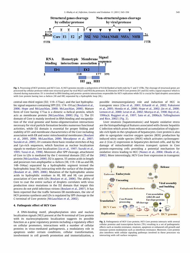
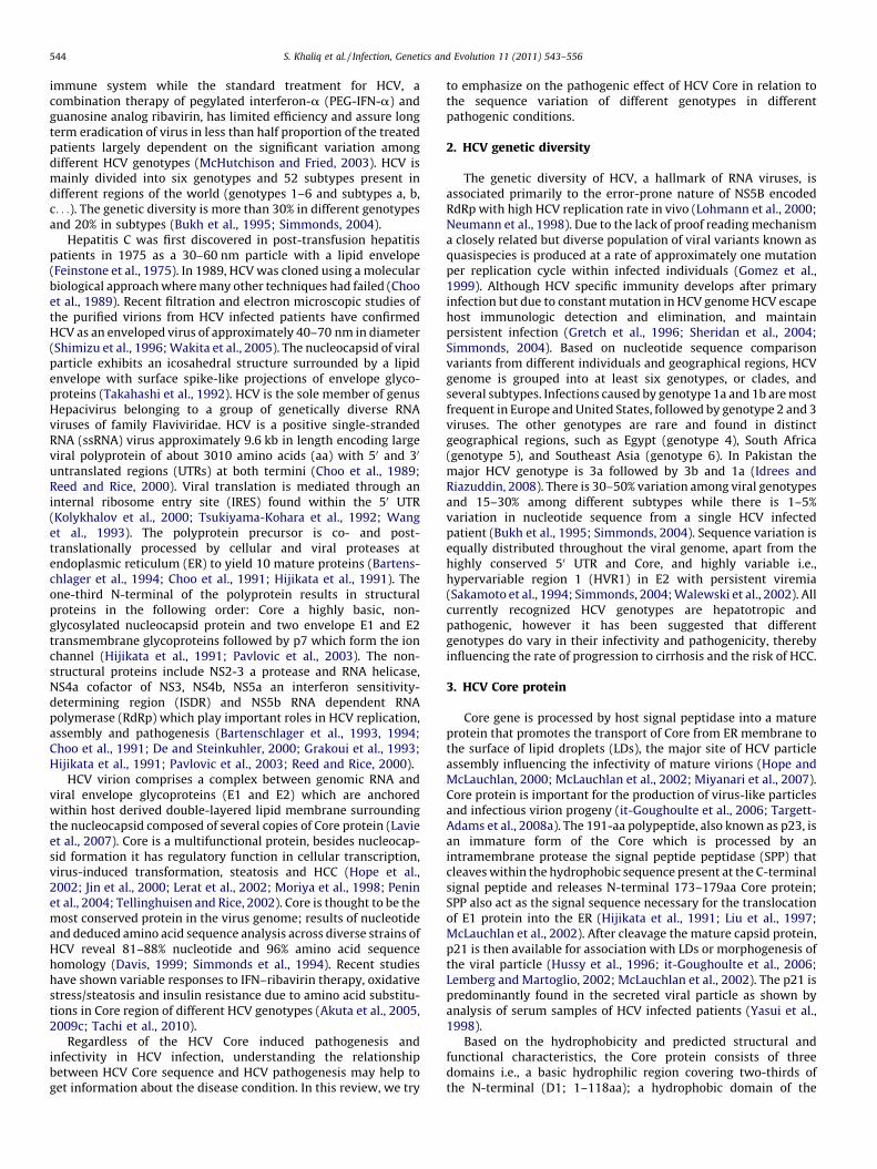
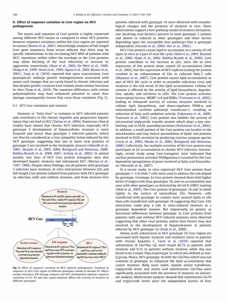
Fig. 1. Processing of HCV proteins and HCV Core. A) HCV genome encodes a polyprotein of 9.6 kb flanked at both ends by 50 and 30 UTRs. The cleavage of structural genes are
processed by cellular protease while non-structural genes by viral NS2/3 and NS3/4a proteases. B) Domains of HCV Core protein (D1 and D2) with a signal sequence which is
cleaved during maturation. D1 is involved in RNA binding and protein–protein interactions responsible for HCV replication while D2 is crucial for lipid droplets association
with Core protein having two a-helices (HI and HII) separated by a hydrophilic loop (HL).
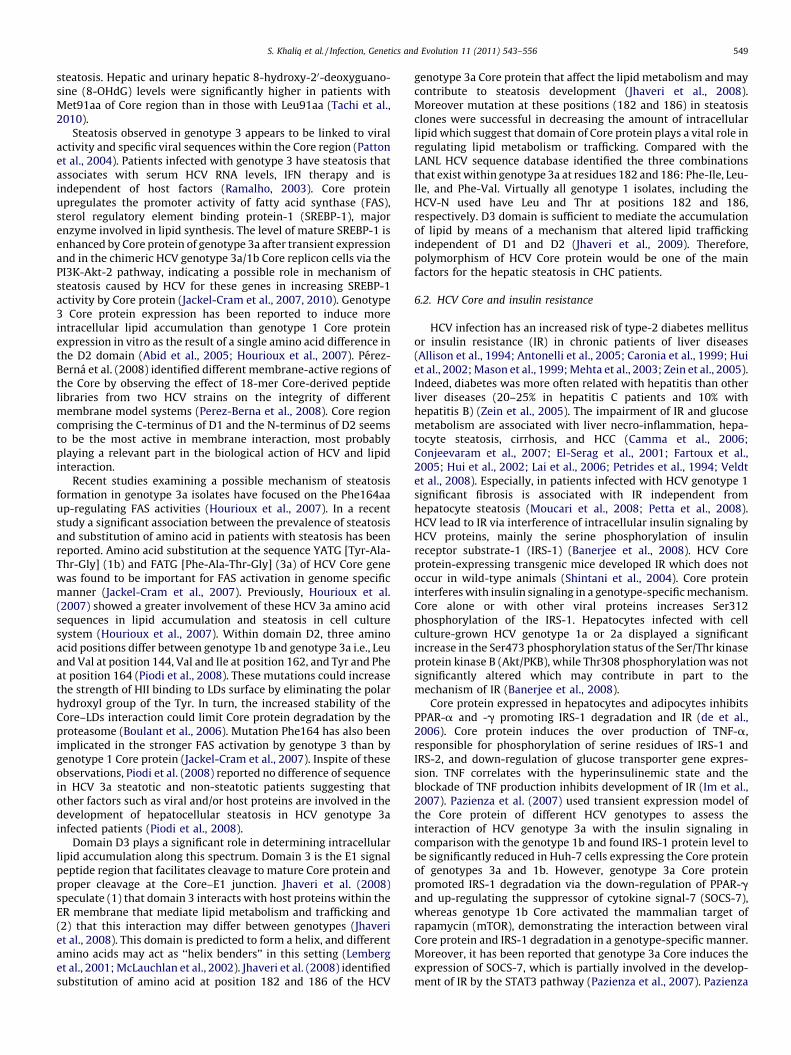
Fig. 2. Pathogenesis of HCV Core protein. HCV Core protein interacts with several
cellular proteins and transcription factors (TFs) resulting in a set of pathogenetic
effects such as insulin resistance, steatosis, apoptosis or enhanced cell growth and
immune system modulation such as interferon resistance. Moreover, Core protein
also interferes with cellular signaling pathways involved in these processes via
interacting with cell surface receptors.
S. Khaliq et al. / Infection, Genetics and Evolution 11 (2011) 543–556 545
central one-third region (D2; 119–173aa); and the last hydropho-bic signal sequence containing SPP (D3; 174–191aa) (Boulant et al.,2006; Hope and McLauchlan, 2000; McLauchlan, 2000). Matureform of Core having 177aa is a dimeric a-helical protein, whichacts as membrane protein (McLauchlan, 2000) (Fig. 1). The D1domain of Core is mainly involved in RNA binding and encapsida-tion of the viral genome and homo-oligomerization interactionsnecessary for viral particle formation besides numerous functionalactivities, while D2 domain is essential for proper folding andstability of D1 and membrane characteristics of the Core includingassociation with ER and outer mitochondrial membranes (Boulantet al., 2005, 2006; McLauchlan, 2000; Moradpour et al., 2007;Suzuki et al., 2005). D1 contains three basic residue stretches of Argand Lys-rich sequences, which function as nuclear localizationsignals to mediate Core localization (Liu et al., 1997; Suzuki et al.,1995; Yasui et al., 1998). Moreover after SPP cleavage, attachmentof Core to LDs is mediated by the C-terminal domain (D2) of theprotein (McLauchlan, 2000). D2 is approx. 55 amino acids in lengthand possesses two amphipathic a-helices (HI, 119–136 aa and HII,148–164aa) separated by a hydrophobic segment termed thehydrophobic loop (HL) interacting with the surface of the droplets(Boulant et al., 2005, 2006). Mutation of the hydrophobic aminoacids to hydrophilic residues in HI, HII and HL can preventassociation of Core with LDs (Boulant et al., 2006). The ability ofCore to coat the entire surface of droplets correlates with virusproduction since mutations in the D2 domain that impair thisprocess do not yield infectious virions (Boulant et al., 2007). It hasbeen reported that the traffic between ER membranes, the site ofHCV proteins synthesis and LDs is regulated by SPP cleavage in theC-terminal of Core protein (McLauchlan et al., 2002).
4. Pathogenic effect of HCV Core
A DNA-binding motif, phosphorylation sites and nuclearlocalization signals (NLS) present at the N-terminal of Core proteinwith its nucleocytoplasmic localization suggests its possiblefunction as a gene regulatory protein including a regulatory effecton cellular promoters, interactions with a number of cellularproteins in virus-mediated pathogenesis, a modulatory role inapoptosis under certain conditions, cellular transformation,involvement in cell growth promotion and immortalization, a
possible immunoregulatory role and induction of HCC intransgenic mice (Cho et al., 2001; Erhardt et al., 2002; Fukutomiet al., 2005; Honda et al., 2000; Hope et al., 2002; Jin et al., 2000;Lemon et al., 2000; Lerat et al., 2002; Moriya et al., 1998; Ray et al.,1996a,b; Ruggieri et al., 1997; Sato et al., 2006a,b; Tellinghuisenand Rice, 2002) (Fig. 2).
Liver steatosis (hapatosteatosis) and hepatic oxidative stressare the histopathological features associated with chronic hepatitisC infection which arises from enhanced accumulation of triglycer-ide-rich lipids in the cytoplasm of hepatocytes. Core protein is alsoable to upregulate reactive oxygen species (ROS) production byinduced nitric oxide species (iNOS) which activates cyclooxygen-ase-2 (Cox-2) expression in hepatocytes derived cells, and by thedamage of mitochondrial electron transport system in Coreprotein-expressing cells providing a potential mechanism foroxidative stress leading to HCC (Nunez et al., 2004; Okuda et al.,2002). More interestingly, HCV Core liver expression in transgenic
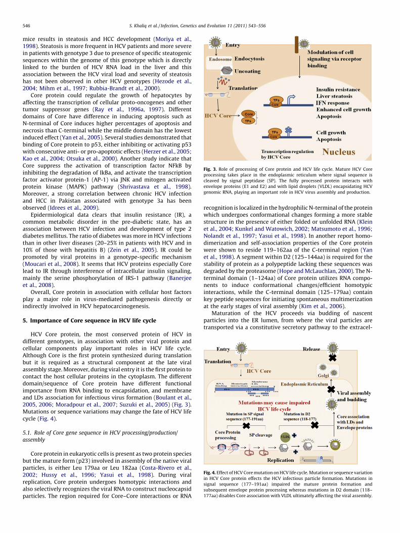
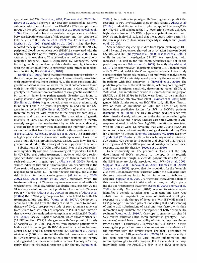
Fig. 3. Role of processing of Core protein and HCV life cycle. Mature HCV Core
processing takes place in the endoplasmic reticulum where signal sequence is
cleaved by signal peptidase (SP). The fully processed protein interacts with
envelope proteins (E1 and E2) and with lipid droplets (VLDL) encapsidating HCV
genomic RNA, playing an important role in HCV virus assembly and production.
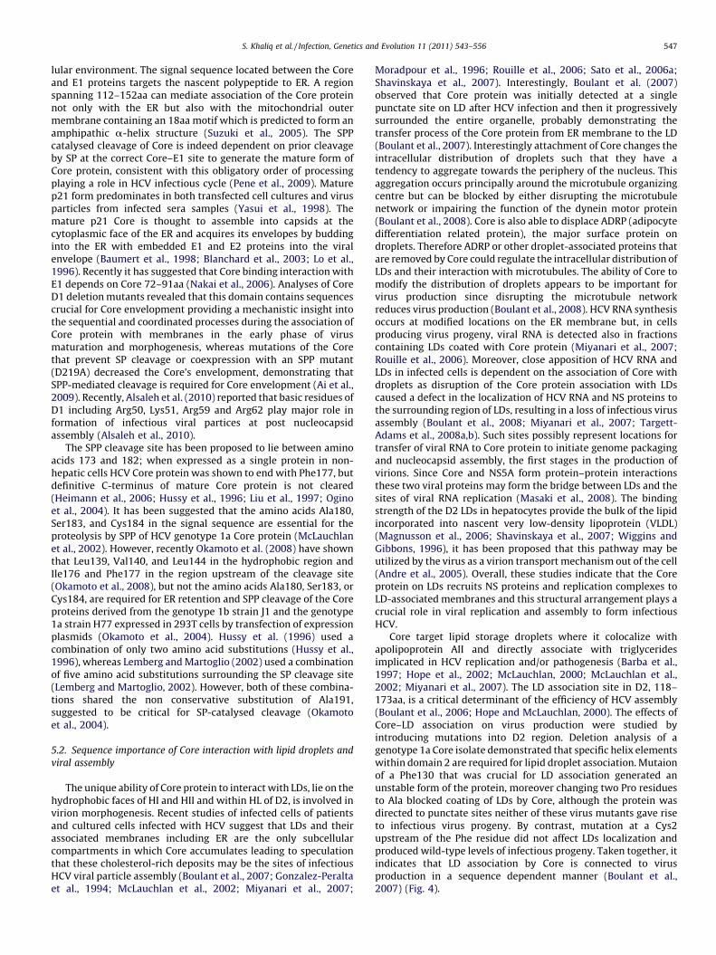
Fig. 4. Effect of HCV Core mutation on HCV life cycle. Mutation or sequence variation
in HCV Core protein effects the HCV infectious particle formation. Mutations in
signal sequence (177–191aa) impaired the mature protein formation and
subsequent envelope protein processing whereas mutations in D2 domain (118–
177aa) disables Core association with VLDL ultimately affecting the viral assembly.
S. Khaliq et al. / Infection, Genetics and Evolution 11 (2011) 543–556546
mice results in steatosis and HCC development (Moriya et al.,1998). Steatosis is more frequent in HCV patients and more severein patients with genotype 3 due to presence of specific steatogenicsequences within the genome of this genotype which is directlylinked to the burden of HCV RNA load in the liver and thisassociation between the HCV viral load and severity of steatosishas not been observed in other HCV genotypes (Hezode et al.,2004; Mihm et al., 1997; Rubbia-Brandt et al., 2000).
Core protein could regulate the growth of hepatocytes byaffecting the transcription of cellular proto-oncogenes and othertumor suppressor genes (Ray et al., 1996a, 1997). Differentdomains of Core have difference in inducing apoptosis such asN-terminal of Core induces higher percentages of apoptosis andnecrosis than C-terminal while the middle domain has the lowestinduced effect (Yan et al., 2005). Several studies demonstrated thatbinding of Core protein to p53, either inhibiting or activating p53with consecutive anti- or pro-apoptotic effects (Herzer et al., 2005;Kao et al., 2004; Otsuka et al., 2000). Another study indicate thatCore suppress the activation of transcription factor NFkB byinhibiting the degradation of IkBa, and activate the transcriptionfactor activator protein-1 (AP-1) via JNK and mitogen activatedprotein kinase (MAPK) pathway (Shrivastava et al., 1998).Moreover, a strong correlation between chronic HCV infectionand HCC in Pakistan associated with genotype 3a has beenobserved (Idrees et al., 2009).
Epidermiological data clears that insulin resistance (IR), acommon metabolic disorder in the pre-diabetic state, has anassociation between HCV infection and development of type 2diabetes mellitus. The ratio of diabetes was more in HCV infectionsthan in other liver diseases (20–25% in patients with HCV and in10% of those with hepatitis B) (Zein et al., 2005). IR could bepromoted by viral proteins in a genotype-specific mechanism(Moucari et al., 2008). It seems that HCV proteins especially Corelead to IR through interference of intracellular insulin signaling,mainly the serine phosphorylation of IRS-1 pathway (Banerjeeet al., 2008).
Overall, Core protein in association with cellular host factorsplay a major role in virus-mediated pathogenesis directly orindirectly involved in HCV hepatocarcinogenesis.
5. Importance of Core sequence in HCV life cycle
HCV Core protein, the most conserved protein of HCV indifferent genotypes, in association with other viral protein andcellular components play important roles in HCV life cycle.Although Core is the first protein synthesized during translationbut it is required as a structural component at the late viralassembly stage. Moreover, during viral entry it is the first protein tocontact the host cellular proteins in the cytoplasm. The differentdomain/sequence of Core protein have different functionalimportance from RNA binding to encapsidation, and membraneand LDs association for infectious virus formation (Boulant et al.,2005, 2006; Moradpour et al., 2007; Suzuki et al., 2005) (Fig. 3).Mutations or sequence variations may change the fate of HCV lifecycle (Fig. 4).
5.1. Role of Core gene sequence in HCV processing/production/
assembly
Core protein in eukaryotic cells is present as two protein speciesbut the mature form (p23) involved in assembly of the native viralparticles, is either Leu 179aa or Leu 182aa (Costa-Rivero et al.,2002; Hussy et al., 1996; Yasui et al., 1998). During viralreplication, Core protein undergoes homotypic interactions andalso selectively recognizes the viral RNA to construct nucleocapsidparticles. The region required for Core–Core interactions or RNA
recognition is localized in the hydrophilic N-terminal of the proteinwhich undergoes conformational changes forming a more stablestructure in the presence of either folded or unfolded RNA (Kleinet al., 2004; Kunkel and Watowich, 2002; Matsumoto et al., 1996;Nolandt et al., 1997; Yasui et al., 1998). In another report homo-dimerization and self-association properties of the Core proteinwere shown to reside 119–162aa of the C-terminal region (Yanet al., 1998). A segment within D2 (125–144aa) is required for thestability of protein as a polypeptide lacking these sequences wasdegraded by the proteasome (Hope and McLauchlan, 2000). The N-terminal domain (1–124aa) of Core protein utilizes RNA compo-nents to induce conformational changes/efficient homotypicinteractions, while the C-terminal domain (125–179aa) containkey peptide sequences for initiating spontaneous multimerizationat the early stages of viral assembly (Kim et al., 2006).
Maturation of the HCV proceeds via budding of nascentparticles into the ER lumen, from where the viral particles aretransported via a constitutive secretory pathway to the extracel-
S. Khaliq et al. / Infection, Genetics and Evolution 11 (2011) 543–556 547
lular environment. The signal sequence located between the Coreand E1 proteins targets the nascent polypeptide to ER. A regionspanning 112–152aa can mediate association of the Core proteinnot only with the ER but also with the mitochondrial outermembrane containing an 18aa motif which is predicted to form anamphipathic a-helix structure (Suzuki et al., 2005). The SPPcatalysed cleavage of Core is indeed dependent on prior cleavageby SP at the correct Core–E1 site to generate the mature form ofCore protein, consistent with this obligatory order of processingplaying a role in HCV infectious cycle (Pene et al., 2009). Maturep21 form predominates in both transfected cell cultures and virusparticles from infected sera samples (Yasui et al., 1998). Themature p21 Core is thought to assemble into capsids at thecytoplasmic face of the ER and acquires its envelopes by buddinginto the ER with embedded E1 and E2 proteins into the viralenvelope (Baumert et al., 1998; Blanchard et al., 2003; Lo et al.,1996). Recently it has suggested that Core binding interaction withE1 depends on Core 72–91aa (Nakai et al., 2006). Analyses of CoreD1 deletion mutants revealed that this domain contains sequencescrucial for Core envelopment providing a mechanistic insight intothe sequential and coordinated processes during the association ofCore protein with membranes in the early phase of virusmaturation and morphogenesis, whereas mutations of the Corethat prevent SP cleavage or coexpression with an SPP mutant(D219A) decreased the Core’s envelopment, demonstrating thatSPP-mediated cleavage is required for Core envelopment (Ai et al.,2009). Recently, Alsaleh et al. (2010) reported that basic residues ofD1 including Arg50, Lys51, Arg59 and Arg62 play major role information of infectious viral partices at post nucleocapsidassembly (Alsaleh et al., 2010).
The SPP cleavage site has been proposed to lie between aminoacids 173 and 182; when expressed as a single protein in non-hepatic cells HCV Core protein was shown to end with Phe177, butdefinitive C-terminus of mature Core protein is not cleared(Heimann et al., 2006; Hussy et al., 1996; Liu et al., 1997; Oginoet al., 2004). It has been suggested that the amino acids Ala180,Ser183, and Cys184 in the signal sequence are essential for theproteolysis by SPP of HCV genotype 1a Core protein (McLauchlanet al., 2002). However, recently Okamoto et al. (2008) have shownthat Leu139, Val140, and Leu144 in the hydrophobic region andIle176 and Phe177 in the region upstream of the cleavage site(Okamoto et al., 2008), but not the amino acids Ala180, Ser183, orCys184, are required for ER retention and SPP cleavage of the Coreproteins derived from the genotype 1b strain J1 and the genotype1a strain H77 expressed in 293T cells by transfection of expressionplasmids (Okamoto et al., 2004). Hussy et al. (1996) used acombination of only two amino acid substitutions (Hussy et al.,1996), whereas Lemberg and Martoglio (2002) used a combinationof five amino acid substitutions surrounding the SP cleavage site(Lemberg and Martoglio, 2002). However, both of these combina-tions shared the non conservative substitution of Ala191,suggested to be critical for SP-catalysed cleavage (Okamotoet al., 2004).
5.2. Sequence importance of Core interaction with lipid droplets and
viral assembly
The unique ability of Core protein to interact with LDs, lie on thehydrophobic faces of HI and HII and within HL of D2, is involved invirion morphogenesis. Recent studies of infected cells of patientsand cultured cells infected with HCV suggest that LDs and theirassociated membranes including ER are the only subcellularcompartments in which Core accumulates leading to speculationthat these cholesterol-rich deposits may be the sites of infectiousHCV viral particle assembly (Boulant et al., 2007; Gonzalez-Peraltaet al., 1994; McLauchlan et al., 2002; Miyanari et al., 2007;
Moradpour et al., 1996; Rouille et al., 2006; Sato et al., 2006a;Shavinskaya et al., 2007). Interestingly, Boulant et al. (2007)observed that Core protein was initially detected at a singlepunctate site on LD after HCV infection and then it progressivelysurrounded the entire organelle, probably demonstrating thetransfer process of the Core protein from ER membrane to the LD(Boulant et al., 2007). Interestingly attachment of Core changes theintracellular distribution of droplets such that they have atendency to aggregate towards the periphery of the nucleus. Thisaggregation occurs principally around the microtubule organizingcentre but can be blocked by either disrupting the microtubulenetwork or impairing the function of the dynein motor protein(Boulant et al., 2008). Core is also able to displace ADRP (adipocytedifferentiation related protein), the major surface protein ondroplets. Therefore ADRP or other droplet-associated proteins thatare removed by Core could regulate the intracellular distribution ofLDs and their interaction with microtubules. The ability of Core tomodify the distribution of droplets appears to be important forvirus production since disrupting the microtubule networkreduces virus production (Boulant et al., 2008). HCV RNA synthesisoccurs at modified locations on the ER membrane but, in cellsproducing virus progeny, viral RNA is detected also in fractionscontaining LDs coated with Core protein (Miyanari et al., 2007;Rouille et al., 2006). Moreover, close apposition of HCV RNA andLDs in infected cells is dependent on the association of Core withdroplets as disruption of the Core protein association with LDscaused a defect in the localization of HCV RNA and NS proteins tothe surrounding region of LDs, resulting in a loss of infectious virusassembly (Boulant et al., 2008; Miyanari et al., 2007; Targett-Adams et al., 2008a,b). Such sites possibly represent locations fortransfer of viral RNA to Core protein to initiate genome packagingand nucleocapsid assembly, the first stages in the production ofvirions. Since Core and NS5A form protein–protein interactionsthese two viral proteins may form the bridge between LDs and thesites of viral RNA replication (Masaki et al., 2008). The bindingstrength of the D2 LDs in hepatocytes provide the bulk of the lipidincorporated into nascent very low-density lipoprotein (VLDL)(Magnusson et al., 2006; Shavinskaya et al., 2007; Wiggins andGibbons, 1996), it has been proposed that this pathway may beutilized by the virus as a virion transport mechanism out of the cell(Andre et al., 2005). Overall, these studies indicate that the Coreprotein on LDs recruits NS proteins and replication complexes toLD-associated membranes and this structural arrangement plays acrucial role in viral replication and assembly to form infectiousHCV.
Core target lipid storage droplets where it colocalize withapolipoprotein AII and directly associate with triglyceridesimplicated in HCV replication and/or pathogenesis (Barba et al.,1997; Hope et al., 2002; McLauchlan, 2000; McLauchlan et al.,2002; Miyanari et al., 2007). The LD association site in D2, 118–173aa, is a critical determinant of the efficiency of HCV assembly(Boulant et al., 2006; Hope and McLauchlan, 2000). The effects ofCore–LD association on virus production were studied byintroducing mutations into D2 region. Deletion analysis of agenotype 1a Core isolate demonstrated that specific helix elementswithin domain 2 are required for lipid droplet association. Mutaionof a Phe130 that was crucial for LD association generated anunstable form of the protein, moreover changing two Pro residuesto Ala blocked coating of LDs by Core, although the protein wasdirected to punctate sites neither of these virus mutants gave riseto infectious virus progeny. By contrast, mutation at a Cys2upstream of the Phe residue did not affect LDs localization andproduced wild-type levels of infectious progeny. Taken together, itindicates that LD association by Core is connected to virusproduction in a sequence dependent manner (Boulant et al.,2007) (Fig. 4).
S. Khaliq et al. / Infection, Genetics and Evolution 11 (2011) 543–556548
6. Effect of sequence variation in Core region on HCVpathogenesis
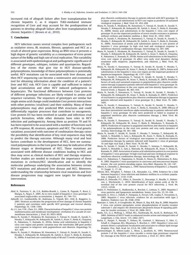
The amino acid sequence of Core protein is highly conservedamong different HCV strains as compared to other HCV proteinshowever sequence variations within the Core region are reportedin tumors (Ruster et al., 2001). Interestingly analyses of full-lengthCore gene sequences from serum indicate that there may bespecific substitutions in the circulating HCV RNA of patients withHCC; mutations within either the envelope or the Core proteinsmay allow blocking of the viral infectivity or increase itsaggression, respectively (Alam et al., 2002; De Mitri et al., 1998;Hong et al., 1999; Horie et al., 1996; Ogata et al., 2002; Ruster et al.,2001). Tang et al. (2010) reported that upon transmission, Corequasispecies undergo genetic homogenization associated withamino acid changes that are rarely found in chronic infection andthat despite genetic variation Core isolates retain similar functionsin vitro (Tang et al., 2010). The sequence differences with certainpolymorphisms may have enhanced potential to cause liverdamage consequently viruses that carry these variations (Fig. 5).
6.1. HCV Core mutations and steatosis
Steatosis or ‘‘fatty liver’’ is common in HCV infected patientsand contributes to the chronic hepatitis and progressive hepaticinjury that can lead to HCC (Patton et al., 2004). Numerous clinicalstudies have shown that chronic HCV infection, especially HCVgenotype 3 development of hepatocellular steatosis is morefrequent and severe than genotype 1 infected patients, whichcan thus be considered as a true cytopathic lesion induced by thisHCV genotype, suggesting that one or more viral proteins ofgenotype 3 are involved in the steatogenic process (Adinolfi et al.,2001; Hezode et al., 2003, 2004; Roingeard and Hourioux, 2008;Rubbia-Brandt et al., 2000, 2001; Serfaty et al., 2002). In animalmodels, two lines of HCV Core protein transgenic mice alsodeveloped hepatic steatosis and subsequent HCC (Moriya et al.,1997, 1998). Despite these findings, not all patients with genotype3 infection have steatosis. In vitro interactions between LDs andfull-length Core protein isolated from patients with HCV genotype3a infection, with and without steatosis, and from steatosis-free
Fig. 5. Effect of sequence variation on HCV induced pathogenesis. Variation in
sequence in HCV Core region of different genotypes mainly in domain D1 effects
insulin resistance, IFN therapy response and HCC development whereas sequence
variations in D1, D2 and also signal sequence affects the severity of steatosis in
different genotypes.
patients infected with genotype 1b were observed with morpho-logical changes and the presence of steatosis in vivo. Theseobservations support a two-pathway model of steatosis formation:one involving viral factor(s) present in most genotype 3 isolatesand absent or reduced in other genotypes and other factorsdepending upon the susceptible host pathways that is genotypeindependent (Hezode et al., 2004; Hui et al., 2002).
HCV Core protein causes lipid to accumulate in a variety of celltypes in vitro as a part of viral life cycle (Abid et al., 2005; Boulantet al., 2006; Hope et al., 2002; Rubbia-Brandt et al., 2000). Coreprotein contribute to the increase in LDs, since the in vitroexpression of this protein alone caused LD accumulation (Abidet al., 2005), but the expression of HCV replicon lacking the proteinresulted in no enhancement of LDs in cultured Huh-7 cells(Miyanari et al., 2007). Core protein causes lipid accumulation aspart of HCV life cycle in order to assemble infectious virus andsteatosis is the end result of this lipid accumulation. Cellular LDcontent is affected by the activity of lipid biosynthesis, degrada-tion, uptake, and secretion in cells. The Core protein activatestranscription factors, SREBP-1c4 and RXRa (Tsutsumi et al., 2002),leading to enhanced activity of various enzymes involved incellular lipid biosynthesis, and down-regulates PPAR-a andmitochondrial carnitine palmitoyl transferase-1, resulting inreduction of fatty acid oxidation activity (Fukasawa et al., 2006;Tsutsumi et al., 2002). Core protein also inhibits the activity ofmicrosomal triglyceride transfer protein which plays a key rate-limiting role in VLDL assembly/secretion (Perlemuter et al., 2002).In addition, a small portion of the Core protein can localize in themitochondria and may induce peroxidation of lipids and proteinsinvolved in VLDL secretion by producing reactive oxygene species(Lerat et al., 2002; Okuda et al., 2002; Roingeard and Hourioux,2008). Collectively, the multiple activities of the Core protein mayparticipate in LD accumulation in chronic HCV infection. Interest-ingly, recent study using Core-transgenic mice indicated thatnuclear proteasome activator PA28gamma is essential for the Coredependent upregulation of genes involved in fatty acid biosynthe-sis (Moriishi et al., 2007).
In a recent study, in vitro expressed HCV Core protein fromgenotypes 1–5 in Huh-7 cells were used to address the role playedby genotype. Genotype 3a Core protein showed three-fold higherlevels of triglyceride than genotypes 1b, and no accumulation wasseen with other genotypes, as detected by oil red O (ORO) staining(Abid et al., 2005). The Core protein of genotypes 1b and 3a bindstightly to the surface of intracellular LDs. However, cellstransfected with genotype 3a contain more neutral lipids in LDsthan cells transfected with genotype 1b suggesting that Core–LDsinteraction could play a role in virus-induced steatosis in agenotype dependent manner. But importantly no genetic orfunctional differences between genotype 3a Core proteins frompatients with and without HCV induced steatosis were observedsuggesting that other viral proteins and/or host factors may alsoinvolved in the development of hepatosteatosis in patientsinfected by HCV genotype 3a (Piodi et al., 2008).
Amino acids substitution in HCV genotype 1b Core region areassociated with hepatic steatosis and oxidative stress in patientswith chronic hepatitis C. Tachi et al. (2010) reported thatsubstitution of Glu70aa (Q) were found 46.7% in patients withsteatosis and 9.1% in patients without steatosis while Arg wasfound to be a major 70aa of genotype 3a which also defined as non-Q group. Hence, HCV genotype 1b with the Glu70aa which was notcommon in genotype 3a enhanced the lipid accumulation thatcauses steatosis. Body mass index, alanine amino transferase,triglyceride levels and amino acid substitutions Glu70aa weresignificantly associated with the presence of steatosis on univari-ate analysis. Multivariate analysis showed that substitution Glu70and triglyceride levels were the independent factors of liver
S. Khaliq et al. / Infection, Genetics and Evolution 11 (2011) 543–556 549
steatosis. Hepatic and urinary hepatic 8-hydroxy-20-deoxyguano-sine (8-OHdG) levels were significantly higher in patients withMet91aa of Core region than in those with Leu91aa (Tachi et al.,2010).
Steatosis observed in genotype 3 appears to be linked to viralactivity and specific viral sequences within the Core region (Pattonet al., 2004). Patients infected with genotype 3 have steatosis thatassociates with serum HCV RNA levels, IFN therapy and isindependent of host factors (Ramalho, 2003). Core proteinupregulates the promoter activity of fatty acid synthase (FAS),sterol regulatory element binding protein-1 (SREBP-1), majorenzyme involved in lipid synthesis. The level of mature SREBP-1 isenhanced by Core protein of genotype 3a after transient expressionand in the chimeric HCV genotype 3a/1b Core replicon cells via thePI3K-Akt-2 pathway, indicating a possible role in mechanism ofsteatosis caused by HCV for these genes in increasing SREBP-1activity by Core protein (Jackel-Cram et al., 2007, 2010). Genotype3 Core protein expression has been reported to induce moreintracellular lipid accumulation than genotype 1 Core proteinexpression in vitro as the result of a single amino acid difference inthe D2 domain (Abid et al., 2005; Hourioux et al., 2007). Perez-Berna et al. (2008) identified different membrane-active regions ofthe Core by observing the effect of 18-mer Core-derived peptidelibraries from two HCV strains on the integrity of differentmembrane model systems (Perez-Berna et al., 2008). Core regioncomprising the C-terminus of D1 and the N-terminus of D2 seemsto be the most active in membrane interaction, most probablyplaying a relevant part in the biological action of HCV and lipidinteraction.
Recent studies examining a possible mechanism of steatosisformation in genotype 3a isolates have focused on the Phe164aaup-regulating FAS activities (Hourioux et al., 2007). In a recentstudy a significant association between the prevalence of steatosisand substitution of amino acid in patients with steatosis has beenreported. Amino acid substitution at the sequence YATG [Tyr-Ala-Thr-Gly] (1b) and FATG [Phe-Ala-Thr-Gly] (3a) of HCV Core genewas found to be important for FAS activation in genome specificmanner (Jackel-Cram et al., 2007). Previously, Hourioux et al.(2007) showed a greater involvement of these HCV 3a amino acidsequences in lipid accumulation and steatosis in cell culturesystem (Hourioux et al., 2007). Within domain D2, three aminoacid positions differ between genotype 1b and genotype 3a i.e., Leuand Val at position 144, Val and Ile at position 162, and Tyr and Pheat position 164 (Piodi et al., 2008). These mutations could increasethe strength of HII binding to LDs surface by eliminating the polarhydroxyl group of the Tyr. In turn, the increased stability of theCore–LDs interaction could limit Core protein degradation by theproteasome (Boulant et al., 2006). Mutation Phe164 has also beenimplicated in the stronger FAS activation by genotype 3 than bygenotype 1 Core protein (Jackel-Cram et al., 2007). Inspite of theseobservations, Piodi et al. (2008) reported no difference of sequencein HCV 3a steatotic and non-steatotic patients suggesting thatother factors such as viral and/or host proteins are involved in thedevelopment of hepatocellular steatosis in HCV genotype 3ainfected patients (Piodi et al., 2008).
Domain D3 plays a significant role in determining intracellularlipid accumulation along this spectrum. Domain 3 is the E1 signalpeptide region that facilitates cleavage to mature Core protein andproper cleavage at the Core–E1 junction. Jhaveri et al. (2008)speculate (1) that domain 3 interacts with host proteins within theER membrane that mediate lipid metabolism and trafficking and(2) that this interaction may differ between genotypes (Jhaveriet al., 2008). This domain is predicted to form a helix, and differentamino acids may act as ‘‘helix benders’’ in this setting (Lemberget al., 2001; McLauchlan et al., 2002). Jhaveri et al. (2008) identifiedsubstitution of amino acid at position 182 and 186 of the HCV
genotype 3a Core protein that affect the lipid metabolism and maycontribute to steatosis development (Jhaveri et al., 2008).Moreover mutation at these positions (182 and 186) in steatosisclones were successful in decreasing the amount of intracellularlipid which suggest that domain of Core protein plays a vital role inregulating lipid metabolism or trafficking. Compared with theLANL HCV sequence database identified the three combinationsthat exist within genotype 3a at residues 182 and 186: Phe-Ile, Leu-Ile, and Phe-Val. Virtually all genotype 1 isolates, including theHCV-N used have Leu and Thr at positions 182 and 186,respectively. D3 domain is sufficient to mediate the accumulationof lipid by means of a mechanism that altered lipid traffickingindependent of D1 and D2 (Jhaveri et al., 2009). Therefore,polymorphism of HCV Core protein would be one of the mainfactors for the hepatic steatosis in CHC patients.
6.2. HCV Core and insulin resistance
HCV infection has an increased risk of type-2 diabetes mellitusor insulin resistance (IR) in chronic patients of liver diseases(Allison et al., 1994; Antonelli et al., 2005; Caronia et al., 1999; Huiet al., 2002; Mason et al., 1999; Mehta et al., 2003; Zein et al., 2005).Indeed, diabetes was more often related with hepatitis than otherliver diseases (20–25% in hepatitis C patients and 10% withhepatitis B) (Zein et al., 2005). The impairment of IR and glucosemetabolism are associated with liver necro-inflammation, hepa-tocyte steatosis, cirrhosis, and HCC (Camma et al., 2006;Conjeevaram et al., 2007; El-Serag et al., 2001; Fartoux et al.,2005; Hui et al., 2002; Lai et al., 2006; Petrides et al., 1994; Veldtet al., 2008). Especially, in patients infected with HCV genotype 1significant fibrosis is associated with IR independent fromhepatocyte steatosis (Moucari et al., 2008; Petta et al., 2008).HCV lead to IR via interference of intracellular insulin signaling byHCV proteins, mainly the serine phosphorylation of insulinreceptor substrate-1 (IRS-1) (Banerjee et al., 2008). HCV Coreprotein-expressing transgenic mice developed IR which does notoccur in wild-type animals (Shintani et al., 2004). Core proteininterferes with insulin signaling in a genotype-specific mechanism.Core alone or with other viral proteins increases Ser312phosphorylation of the IRS-1. Hepatocytes infected with cellculture-grown HCV genotype 1a or 2a displayed a significantincrease in the Ser473 phosphorylation status of the Ser/Thr kinaseprotein kinase B (Akt/PKB), while Thr308 phosphorylation was notsignificantly altered which may contribute in part to themechanism of IR (Banerjee et al., 2008).
Core protein expressed in hepatocytes and adipocytes inhibitsPPAR-a and -g promoting IRS-1 degradation and IR (de et al.,2006). Core protein induces the over production of TNF-a,responsible for phosphorylation of serine residues of IRS-1 andIRS-2, and down-regulation of glucose transporter gene expres-sion. TNF correlates with the hyperinsulinemic state and theblockade of TNF production inhibits development of IR (Im et al.,2007). Pazienza et al. (2007) used transient expression model ofthe Core protein of different HCV genotypes to assess theinteraction of HCV genotype 3a with the insulin signaling incomparison with the genotype 1b and found IRS-1 protein level tobe significantly reduced in Huh-7 cells expressing the Core proteinof genotypes 3a and 1b. However, genotype 3a Core proteinpromoted IRS-1 degradation via the down-regulation of PPAR-gand up-regulating the suppressor of cytokine signal-7 (SOCS-7),whereas genotype 1b Core activated the mammalian target ofrapamycin (mTOR), demonstrating the interaction between viralCore protein and IRS-1 degradation in a genotype-specific manner.Moreover, it has been reported that genotype 3a Core induces theexpression of SOCS-7, which is partially involved in the develop-ment of IR by the STAT3 pathway (Pazienza et al., 2007). Pazienza

S. Khaliq et al. / Infection, Genetics and Evolution 11 (2011) 543–556550
et al. (2010) found that INF-a treatment induced STAT3 activationand consequently SOCS-1 and SOCS-3 upregulation in HCVgenotype 3a Core expressing Huh-7 cells (Pazienza et al., 2010);SOCS-7 mRNA expression is independent of STAT3 and seems to bemodulated by PPAR-g activity. After HCV infection enhanced SOCSproduction also seems to play a critical role in inducing IFNresistance by inhibiting IFN-a intracellular signaling (Banks et al.,2005).
The impact of aa substitutions of HCV genotype 1b Core proteinare the important predictor of severe IR in Japanese patientswithout cirrhosis and diabetes mellitus. Multivariate analysisidentified steatosis (> or =5%), age (> or =55 years) and aasubstitutions of the Core (Glu70 (His70) and/or Met91) assignificant determinants of severe IR. Especially, significantlylower proportions of patients with Glu70 (His70) and/or Met91were noted among those without severe IR (59.6%) than those withsevere IR (82.4%), suggesting that substitutions of HCV-1b Corewere the important predictor of severe IR in patients withoutcirrhosis and diabetes mellitus. The only limitations of the studywere that it did not investigate the effect of other genotypes exceptfor HCV-1b and other races apart from Japan i.e., the geographicdiversities of HCV-1b Core region (distribution of Arg70 or Glu70(His70), and Leu91 or Met91) (Akuta et al., 2009b).
6.3. HCV Core interference in cell cycle resulting in HCC
HCV Core gene has been concerned in the development of HCCin chronic infections. The p53 gene has property to suppressoncogenes and promote DNA recovery in impaired cells bysuppressing G1/S transition phase of the cell cycle, on the otherhand may promote apoptosis by switching on apoptosis. Coreprotein, especially 80–122aa, has the ability to suppress p53promoter activity which led to the suppression of apoptosismechanism due to the interference with anti-carcinoma activity ofp53 gene (Ray et al., 1996a,b, 1997). Core has been reported toaffect the Cdk-inhibitory activity of p21/Waf1 and regulated cellcycle may be as a result of the increased degradation of p21/Waf1,or because of Core binding to the C-terminal of p21/Waf1 (Mutohet al., 1999; Wang et al., 2000). On the other hand, Core maypromote cell proliferation through the over-expression of cyclin Ein the cells, which contributes to the development of many types ofhuman cancers (Cho et al., 2001; Nielsen et al., 1998; Spruck et al.,1999). Nuclear factor kB (NFkB) is known to be involved in a varietyof physiological events such as cells proliferation, differentiationand survival (Tai et al., 2000). Under inactive conditions, NFkBactivity is negatively regulated by the formation of a complex withinhibitory kB (IkB) in the cytoplasm. Activation signals for NFkBinduce phosphorylation and subsequent degradation of IkBthrough the kination cascade resulting in dissociation of NFkBfrom IkB, subsequent release of NFkB induces the expression ofnumerous genes (Karin, 1999; Kato et al., 2000). In addition, Coremodulate a certain step of this cytoplasmic NFkB signalingpathway which may promote cell proliferation and suppress cellapoptosis by activation of NFkB (Watashi et al., 2001; Yoshida et al.,2001).
Amino acid mutations in the Core protein of genotype 1 areassociated with IFN therapy response and HCC development. InHCV genotype 1 patients’ clinical study it was proposed that usingpretreatment sera HCC risk could be predicted by studyingmutations in Core region (Ala70 and Leu91), response to IFN inrelation to the host factors like age, gender and fibrosis stage(Nakamoto et al., 2010). Multivariate analysis performed in elderlypatients with HCV genotype 1b related HCC identified male sex, aasubstitutions of Core (Met91) and therapy as factors thatinfluenced mortality after first treatment for HCC (Ogura et al.,2009). Recently, Kobayashi et al. (2010) reported that the
substitution of Arg70 for Glu70 in HCV genotype 1b patientsincreases with age, and severe liver disease accompanied byelevated AST and gamma-GTP levels as well as HCC development(Kobayashi et al., 2010). Moreover, Fishman et al. (2009) analyzedfull-length HCV-1b sequences from HCC patients and controls andidentified seven polymorphisms (nucleotide) significantly associ-ated with increased risk of HCC (36G/C, 209A, 271U/C, 309A/C,435A/C, 481A, and 546A/C) (Fishman et al., 2009). Interestingly,209A has been associated with IFN resistance and HCC suggestingthat Core gene sequence data might provide useful informationabout HCC risk. The G209A substitution replaces the basic aminoacid, Arg, with the neutral amino acid Glu. The 209A mutationremained significantly associated with HCC in multivariateanalysis of the full-length Core sequences. The increased riskassociated with this polymorphism was also observed in the set ofpartial sequences with a strong association between the G209Asubstitution and the presence of HCC (Fishman et al., 2009). Huet al. (2009) identified eight mutations in the HCV genotype 1bCore gene related to the increased risk of HCC i.e., A028C, G209A,C219U/A, U264C, A271C/U, C378U/A, G435A/C, and G481Awhereas U303C/A was found to be associated with decreasedHCC risk. Overall these mutations bring about four amino acidsubstitutions: Lys10Glu, Arg70Glu, Met91Leu, and Gly161Ser (Huet al., 2009).
In a recent study, a significant association of Core substitutionswith multistep hepatocarcinogenesis has recently been reported.Sequence analysis of the Core protein was performed and theassociation between the substitution rates in Core and the degreeof steatosis or fibrosis during the development of HCC and tumordifferentiation was analyzed. The substitution rates of amino acid70, 75, 91 and 147 exceeded 25% (aa 70, 51%; 75, 45%; 91, 36%; 147,30%). All substitution rates were comparable among cancerous andnon-cancerous regions of patients with HCC and non-cancerousregion without HCC and concluded that the substitutions in theCore region are possibly not associated with HCV-related multistephepatocarcinogenesis (Fukuhara et al., 2010).
6.4. HCV Core and interferon response
The most effective therapy available against HCV is thecombination of PEG-IFN and ribavirin, with achieving serumHCV RNA levels negativity by week 12 of treatment (early virologicresponse: EVR) as an important predictor of a sustained virologicresponse (SVR) in chronic patients with a high viral load (Daviset al., 2003; Ferenci et al., 2005). HCV genotypes differ in theirresponse to the current therapy (Fried et al., 2002; Manns et al.,2001). A clinical study of HCV patients examined factors associatedwith non-response to PEG-INF and ribavirin treatment in HCVgenotype 1a or 1b patients and found that the poor response totreatment in patients was not explained by clinical factors such asgender, age, body weight, severity of underlying hepatitis,pretreatment viral levels, or amount of drug taken (Conjeevaramet al., 2006).
Previous studies have shown that the HCV Core protein mightbe associated with IFN therapy resistance involving the Jak-STATsignaling cascade (Blindenbacher et al., 2003; Bode et al., 2003;Ciccaglione et al., 2007; Lin et al., 2006; Melen et al., 2004). WhenIFN initiates its intracellular signal transduction pathway, Jak1 andSTAT2 kinases are activated which in turn phosphorylate STAT1.Association of Jak1 with STAT1 initiates via phosphorylation ofSTAT1, after which phosphorylated STAT1 forms a heterotrimericcomplex together with phosphorylated STAT2 and IFN regulatoryfactor 9 forming a complex known as IFN-stimulated gene (ISG)factor 3 (ISGF3) and it undergoes translocation to the nucleuswhere it binds to IFN-stimulated response element (ISRE) andupregulates a large number of ISGs including 20,50-oligoadenylate
S. Khaliq et al. / Infection, Genetics and Evolution 11 (2011) 543–556 551
synthetase (2–5AS) (Chen et al., 2003; Kisseleva et al., 2002; TenHoeve et al., 2002). The type I IFN receptor consists of at least twosubunits, which are referred to as IFNAR-1 (IFN-a receptor) andIFNAR-2 (IFN-a/b receptor) (Domanski et al., 1995; Novick et al.,1994). Recent studies have demonstrated a significant correlationbetween hepatic expression of this receptor and the response ofCHC patients to IFN (Mathai et al., 1999; Mizukoshi et al., 1998;Morita et al., 1999; Yatsuhashi et al., 1999). It has also beenreported that expression of messenger RNA (mRNA) for IFNAR-2 byperipheral blood mononuclear cells (PBMCs) is correlated with thehepatic expression of this mRNA (Yamaguchi et al., 2002). Flowcytometry showed that aa 70 substitution in the Core region down-regulated baseline IFNAR-2 expression by Monocytes. Afterinitiating combination therapy, this substitution might interferewith the induction of IFNAR-2 expression with intracellular signaltransduction by IFN (Ishii et al., 2010).
Donlin et al. (2010) found that pretreatment genetic variation inthe two major subtypes of genotype 1 were robustly associatedwith outcome of treatment against HCV. The most consistent viralgenetic variations associations with treatment outcome were foundwith in the NS5A region of genotype 1a and in Core and NS2 ofgenotype 1b. Moreover on examination of viral genetic variation in94 patients, higher inter-patient genetic diversity was found toclosely associate with response to therapy at day 28 of treatment(Donlin et al., 2010). Higher genetic diversity was predominatelyfound in NS3 and NS5A genes in genotype 1a, and Core and NS3gene in genotype 1b (Donlin et al., 2007). Higher diversity wastherefore associated with response to the drugs for both day 28response and treatment outcome. The association of geneticdiversity in Core, NS3/4A and NS5A with response to therapystrongly suggests the mechanisms by which genetic diversityaffects treatment efficacy through altering the interferon-suppres-sive activities that have been identified for these proteins in vitro(Foy et al., 2003; Gale et al., 1998; Yao et al., 2004). The distributionof higher genetic diversity associated with suppression of viral titersacross multiple viral proteins suggests various ways in which HCVgenome could reduce the efficacy of these suppressive functions.
Substitutions of Arg70Glu, and/or Leu91Met in the Core regionwere significantly common in non-virological responders. The fallsin HCV RNA level during treatment in patients with Core regionspecific substitutions were significantly less than in those withoutsuch substitutions in genotype 1b (Akuta et al., 2005). Previousstudies indicated that substitutions at position 70 and/or 91 in theCore region of genotype 1b were predictors of poor virologicalresponse to 48-week PEG-IFN and ribavirin therapy, and also therisk factors for hepatocarcinogenesis (Akuta et al., 2006,2007a,b,c,d, 2008; Donlin et al., 2007). Moreover, when thetreatment efficacy of 72-week regimen was compared with 48-week patients, it was cleared that aa substitution at position 70 and91 is also a useful pretreatment predictor of response to 72-weekPEG-IFN/ribavirin (Akuta et al., 2009a). In studies of Akuta et al.(2007a) the G209A polymorphism has been linked to both IFNtreatment failure and HCC (Akuta et al., 2007a). Genotype 1bsequences obtained from the study of viral resistance to antiviraltherapy of CHC, a prospective study of pretreatment sequencesfrom serum to assess rates of response to PEG-IFN and ribavirintherapy, were also analyzed polymorphism at position 209 (Donlinet al., 2007). Base 271 is part of codon 91, which encodes either Leu(271U/C) or Met (271A) in the genotype 1b Core protein. Analysisof position 271 in >300 sequences from Japanese patients withhigh viral load genotype 1b HCV showed associations betweenMet91 (271A) and IFN resistance and HCC (Akuta et al., 2007a).Akuta et al. (2009) also studied the effect of these aa substitutionsin HCV genotype 2a Japanese patients after combination therapyand suggested that the aa substitution pattern of genotype 2a maypartly affect the virological response to IFN therapy (Akuta et al.,
2009c). Substitution in genotype 1b Core region can predict theresponse to PEG-IFN/ribavirin therapy, but recently Akuta et al.(2010b) studied the impact on triple therapy of telaprevir/PEG-IFN/ribavirin and concluded that 12-week triple therapy achievedhigh rates of loss of HCV RNA in Japanese patients infected withHCV-1b and high viral load, and that the aa substitution pattern inthe Core region seems to influence very early viral dynamics (Akutaet al., 2010b).
Smaller direct sequencing studies from Japan involving 28 HCCand 15 control sequences showed an association between Leu91(271U/C) and HCC (Nagayama et al., 2000; Takahashi et al., 2001),while in another investigation 271U/C was associated withincreased HCC risk in the full-length sequences but not in thepartial sequences (Fishman et al., 2009). Recently Hayashi et al.(2010) also reported a SVR in patients without Glu70 than in thosewith Glu70 and Leu91 in the Core region based on a clinical studysuggesting that factors related to SVR on multivariate analysis werenon-Q70 and ISDR mutant-type and predicting the response to IFNin patients with HCV genotype 1b (Hayashi et al., 2010). Thepredictive potential of the viral mutations, including Core region (70and 91aa), interferon sensitivity-determining region (ISDR, aa2209–2248) and interferon/ribavirin resistance-determining region(IRRDR, aa 2334–2379) in NS5A, were found to be significantpredictors for SVR after liver transplant (Fukuhara et al., 2010). Malegender, high platelet count, low HCV RNA load, mild liver fibrosis,two or more aa mutations of ISDR and Core (70aa) wereindependent predictive factors for SVR in HCV-1b patients(Okanoue et al., 2009). The HCV-1b amino acid sequence wasdetermined and analyzed according to the viral response during thetreatment. Mutations in NS5A-ISDR are associated with rapid viralresponse at week 4 while Core Arg70Glu mutation is associatedwith no EVR at week 12, revealing that Core 70 and NS5A areimportant factors determining the virological kinetics during PEG-IFN and ribavirin therapy (Enomoto and Maekawa, 2010). Recently,Toyoda et al. (2010) studied the factors including aa substitutions inSVR against HCV genotype 1b patients and observed that Arg70 inCore region and NS5A-ISDR region could possibly predict a clinicalresponse against IFN therapy (Toyoda et al., 2010).
Genetic diversity in HCV genome is clearly not the onlydeterminant of HCV’s response to therapy. It is recentlydemonstrated that single nucleotide polymorphisms (SNPs) inthe IL28B gene are closely associated with SVR (Ge et al., 2009;Suppiah et al., 2009; Tanaka et al., 2009; Thomas et al., 2009).Suppiah et al. (2009) reported that the population for the favorableallele was 32%, indicating that variation within the IL28 locus is notthe sole determining factor but an important contributor toresponse (Suppiah et al., 2009). Furthermore, the favorable allele atthis locus is less frequent in African–Americans, partially explain-ing the poor response to treatment (Ge et al., 2009; Thomas et al.,2009). Recently, Akuta et al. (2010) in a multivariate analysisidentified a genetic variation near IL28B gene and Core 70aasubstitution as important predictor of sustained virologicalresponse to a triple therapy of Teleprevir with INF + Ribavirin inHCV genotype 1b infected patients indicating that understandingof amino acid substitutions of viral and host factors with theirinteraction may facilitate the development of better therapeuticregimen (Akuta et al., 2010a). Genotype 1a genome carrying 31SVR related variations (the mean number in genotype 1 SVRsequences) would have a probability of therapy response almosttwice as high (31 variations � 3%/variation = 93%) than a 1a viruscarrying the population consensus sequence used as a reference inthe analysis; with the similar effect size that is reported forvariation in the IL28B gene (Ge et al., 2009). Moreover recently ithas been reported that as Core induces inflammation andimmunity through a toll-like receptor (TLR) 2-dependent pathway,individuals with the Arg753Glu SNP in the TLR2 gene have
S. Khaliq et al. / Infection, Genetics and Evolution 11 (2011) 543–556552
increased risk of allograft failure after liver transplantation forchronic hepatitis C, as it impairs TLR2-mediated immunerecognition of Core and may account for the predisposition ofpatients to develop allograft failure after liver transplantation forchronic hepatitis C (Brown et al., 2010).
7. Conclusion
HCV infection causes several complex liver pathogenesis suchas oxidative stress, IR, steatosis, fibrosis, apoptosis and HCC as aresult of altered gene expression. Being an RNA virus it presents ahigh degree of genetic variability as a consequence of error-pronenature of the RdRp and high rate of viral replication. This variabilityis associated with epidemiological and pathogenetic significance ofdifferent genotypes, subtypes, isolates and quasispecies. Regard-less of the events that give rise to HCV disease-specificpolymorphisms, knowledge of these polymorphisms is potentiallyuseful. HCV mutations can be associated with liver disease, andthen HCV sequencing can become a noninvasive and economicaltool for obtaining information about the condition of a patient’sliver and HCC risk. HCV Core is capable and sufficient to inducelipid accumulation and other HCV induced pathogenesis inhepatocytes. The functional differences between Core proteinsof different genotype infections are likely related to amino acidsequence variations. The sequences of genotypes are similar butsingle amino acid change could modulate Core protein interactionswith other proteins (viral/host) and their stability. Many of thesepolymorphisms may alter predicted RNA structures and coulddifferentially regulate processes that contribute to oncogenesis.Core protein D3 has been involved in satable and infectious viralparticle formation, while other domains have roles in HCVinfection and pathogenicity. Among them aa position 70 and 91has been reported to be associated with a number of functions suchas IFN response, IR, steatosis, apoptosis and HCC. The viral geneticvariations associated with outcome of combination therapy raisesthe possibility that identification of key viral sequences may helpto predict the therapy outcome. Although many viral and hostfactors contribute to the development of HCC, here we summa-rized polymorphisms in the Core gene that may be indicative of thedisease stages or development of HCC. These mutations areassociated with different disease conditions leading to HCC andthus may serve as clinical markers of HCC and therapy response.Further studies are needed to evaluate the importance of thesemutations in cirrhosis/HCC identification and to identify themolecular pathways underlying the association between certainHCV mutations and advanced liver disease and HCC. Moreover,understanding the relationship between viral mutations and liverdisease progression may suggest new targets for therapeuticintervention.
References
Abid, K., Pazienza, V., de, G.A., Rubbia-Brandt, L., Conne, B., Pugnale, P., Rossi, C.,Mangia, A., Negro, F., 2005. An in vitro model of hepatitis C virus genotype 3a-associated triglycerides accumulation. J. Hepatol. 42, 744–751.
Adinolfi, L.E., Gambardella, M., Andreana, A., Tripodi, M.F., Utili, R., Ruggiero, G.,2001. Steatosis accelerates the progression of liver damage of chronic hepatitisC patients and correlates with specific HCV genotype and visceral obesity.Hepatology 33, 1358–1364.
Ai, L.S., Lee, Y.W., Chen, S.S., 2009. Characterization of hepatitis C virus core proteinmultimerization and membrane envelopment: revelation of a cascade of core-membrane interactions. J. Virol. 83, 9923–9939.
Akuta, N., Suzuki, F., Hirakawa, M., Kawamura, Y., Yatsuji, H., Sezaki, H., Suzuki, Y.,Hosaka, T., Kobayashi, M., Kobayashi, M., Saitoh, S., Arase, Y., Ikeda, K., Chayama,K., Nakamura, Y., Kumada, H., 2010a. Amino acid substitution in hepatitis Cvirus core region and genetic variation near the interleukin 28B gene predictviral response to telaprevir with peginterferon and ribavirin. Hepatology 52,421–429.
Akuta, N., Suzuki, F., Hirakawa, M., Kawamura, Y., Yatsuji, H., Sezaki, H., Suzuki, Y.,Hosaka, T., Kobayashi, M., Kobayashi, M., Saitoh, S., Arase, Y., Ikeda, K., Kumada,H., 2009a. A matched case-controlled study of 48 and 72 weeks of peginterferon
plus ribavirin combination therapy in patients infected with HCV genotype 1bin Japan: amino acid substitutions in HCV core region as predictor of sustainedvirological response. J. Med. Virol. 81, 452–458.
Akuta, N., Suzuki, F., Hirakawa, M., Kawamura, Y., Yatsuji, H., Sezaki, H., Suzuki, Y.,Hosaka, T., Kobayashi, M., Kobayashi, M., Saitoh, S., Arase, Y., Ikeda, K., Kumada,H., 2009b. Amino acid substitutions in the hepatitis C virus core region ofgenotype 1b are the important predictor of severe insulin resistance in patientswithout cirrhosis and diabetes mellitus. J. Med. Virol. 81, 1032–1039.
Akuta, N., Suzuki, F., Hirakawa, M., Kawamura, Y., Yatsuji, H., Sezaki, H., Suzuki, Y.,Hosaka, T., Kobayashi, M., Kobayashi, M., Saitoh, S., Arase, Y., Ikeda, K., Kumada,H., 2009c. Association of amino acid substitution pattern in core protein ofhepatitis C virus genotype 2a high viral load and virological response tointerferon-ribavirin combination therapy. Intervirology 52, 301–309.
Akuta, N., Suzuki, F., Hirakawa, M., Kawamura, Y., Yatsuji, H., Sezaki, H., Suzuki, Y.,Hosaka, T., Kobayashi, M., Kobayashi, M., Saitoh, S., Arase, Y., Ikeda, K., Chayama,K., Nakamura, Y., Kumada, H., 2010b. Amino acid substitutions in the hepatitis Cvirus core region of genotype 1b affect very early viral dynamics duringtreatment with telaprevir, peginterferon, and ribavirin. J. Med. Virol. 82,575–582.
Akuta, N., Suzuki, F., Kawamura, Y., Yatsuji, H., Sezaki, H., Suzuki, Y., Hosaka, T.,Kobayashi, M., Kobayashi, M., Arase, Y., Ikeda, K., Kumada, H., 2007a. Amino acidsubstitutions in the hepatitis C virus core region are the important predictor ofhepatocarcinogenesis. Hepatology 46, 1357–1364.
Akuta, N., Suzuki, F., Kawamura, Y., Yatsuji, H., Sezaki, H., Suzuki, Y., Hosaka, T.,Kobayashi, M., Kobayashi, M., Arase, Y., Ikeda, K., Kumada, H., 2007b. Predictivefactors of early and sustained responses to peginterferon plus ribavirin combi-nation therapy in Japanese patients infected with hepatitis C virus genotype 1b:amino acid substitutions in the core region and low-density lipoprotein cho-lesterol levels. J. Hepatol. 46, 403–410.
Akuta, N., Suzuki, F., Kawamura, Y., Yatsuji, H., Sezaki, H., Suzuki, Y., Hosaka, T.,Kobayashi, M., Kobayashi, M., Arase, Y., Ikeda, K., Kumada, H., 2007c. Predictorsof viral kinetics to peginterferon plus ribavirin combination therapy in Japanesepatients infected with hepatitis C virus genotype 1b. J. Med. Virol. 79, 1686–1695.
Akuta, N., Suzuki, F., Kawamura, Y., Yatsuji, H., Sezaki, H., Suzuki, Y., Hosaka, T.,Kobayashi, M., Kobayashi, M., Arase, Y., Ikeda, K., Kumada, H., 2008. Efficacy oflow-dose intermittent interferon-alpha monotherapy in patients infected withhepatitis C virus genotype 1b who were predicted or failed to respond topegylated interferon plus ribavirin combination therapy. J. Med. Virol. 80,1363–1369.
Akuta, N., Suzuki, F., Kawamura, Y., Yatsuji, H., Sezaki, H., Suzuki, Y., Hosaka, T.,Kobayashi, M., Kobayashi, M., Arase, Y., Ikeda, K., Miyakawa, Y., Kumada, H.,2007d. Prediction of response to pegylated interferon and ribavirin in hepatitisC by polymorphisms in the viral core protein and very early dynamics ofviremia. Intervirology 50, 361–368.
Akuta, N., Suzuki, F., Sezaki, H., Suzuki, Y., Hosaka, T., Someya, T., Kobayashi, M.,Saitoh, S., Watahiki, S., Sato, J., Kobayashi, M., Arase, Y., Ikeda, K., Kumada, H.,2006. Predictive factors of virological non-response to interferon-ribavirincombination therapy for patients infected with hepatitis C virus of genotype1b and high viral load. J. Med. Virol. 78, 83–90.
Akuta, N., Suzuki, F., Sezaki, H., Suzuki, Y., Hosaka, T., Someya, T., Kobayashi, M.,Saitoh, S., Watahiki, S., Sato, J., Matsuda, M., Kobayashi, M., Arase, Y., Ikeda, K.,Kumada, H., 2005. Association of amino acid substitution pattern in core proteinof hepatitis C virus genotype 1b high viral load and non-virological response tointerferon-ribavirin combination therapy. Intervirology 48, 372–380.
Alam, S.S., Nakamura, T., Naganuma, A., Nozaki, A., Nouso, K., Shimomura, H., Kato,N., 2002. Hepatitis C virus quasispecies in cancerous and noncancerous hepaticlesions: the core protein-encoding region. Acta Med. Okayama 56, 141–147.
Alberti, A., Chemello, L., Benvegnu, L., 1999. Natural history of hepatitis C. J. Hepatol.31 (Suppl. 1), 17–24.
Allison, M.E., Wreghitt, T., Palmer, C.R., Alexander, G.J., 1994. Evidence for a linkbetween hepatitis C virus infection and diabetes mellitus in a cirrhotic popula-tion. J. Hepatol. 21, 1135–1139.
Alsaleh, K., Delavalle, P.Y., Pillez, A., Duverlie, G., Descamps, V., Rouille, Y., Dubuis-son, J., Wychowski, C., 2010. Identification of basic amino acids at the N-terminal end of the core protein crucial for HCV infectivity. J. Virol. 84,12515–12528.
Andre, P., Perlemuter, G., Budkowska, A., Brechot, C., Lotteau, V., 2005. Hepatitis Cvirus particles and lipoprotein metabolism. Semin. Liver Dis. 25, 93–104.
Antonelli, A., Ferri, C., Fallahi, P., Pampana, A., Ferrari, S.M., Goglia, F., Ferrannini, E.,2005. Hepatitis C virus infection: evidence for an association with type 2diabetes. Diabetes Care 28, 2548–2550.
Banerjee, S., Saito, K., it-Goughoulte, M., Meyer, K., Ray, R.B., Ray, R., 2008. HepatitisC virus core protein upregulates serine phosphorylation of insulin receptorsubstrate-1 and impairs the downstream akt/protein kinase B signaling path-way for insulin resistance. J. Virol. 82, 2606–2612.
Banks, A.S., Li, J., McKeag, L., Hribal, M.L., Kashiwada, M., Accili, D., Rothman, P.B.,2005. Deletion of SOCS7 leads to enhanced insulin action and enlarged islets ofLangerhans. J. Clin. Invest. 115, 2462–2471.
Barba, G., Harper, F., Harada, T., Kohara, M., Goulinet, S., Matsuura, Y., Eder, G.,Schaff, Z., Chapman, M.J., Miyamura, T., Brechot, C., 1997. Hepatitis C virus coreprotein shows a cytoplasmic localization and associates to cellular lipid storagedroplets. Proc. Natl. Acad. Sci. U.S.A. 94, 1200–1205.
Bartenschlager, R., hlborn-Laake, L., Mous, J., Jacobsen, H., 1993. Nonstructuralprotein 3 of the hepatitis C virus encodes a serine-type proteinase requiredfor cleavage at the NS3/4 and NS4/5 junctions. J. Virol. 67, 3835–3844.
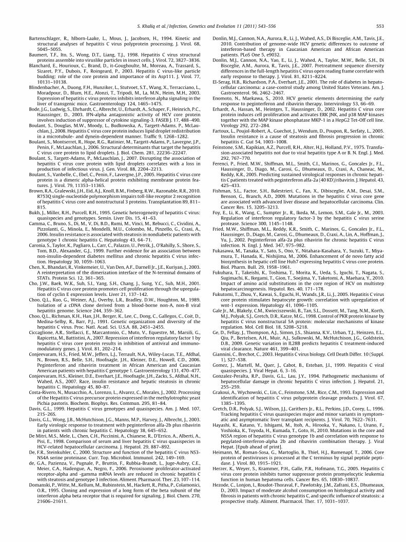
S. Khaliq et al. / Infection, Genetics and Evolution 11 (2011) 543–556 553
Bartenschlager, R., hlborn-Laake, L., Mous, J., Jacobsen, H., 1994. Kinetic andstructural analyses of hepatitis C virus polyprotein processing. J. Virol. 68,5045–5055.
Baumert, T.F., Ito, S., Wong, D.T., Liang, T.J., 1998. Hepatitis C virus structuralproteins assemble into viruslike particles in insect cells. J. Virol. 72, 3827–3836.
Blanchard, E., Hourioux, C., Brand, D., it-Goughoulte, M., Moreau, A., Trassard, S.,Sizaret, P.Y., Dubois, F., Roingeard, P., 2003. Hepatitis C virus-like particlebudding: role of the core protein and importance of its Asp111. J. Virol. 77,10131–10138.
Blindenbacher, A., Duong, F.H., Hunziker, L., Stutvoet, S.T., Wang, X., Terracciano, L.,Moradpour, D., Blum, H.E., Alonzi, T., Tripodi, M., La, M.N., Heim, M.H., 2003.Expression of hepatitis c virus proteins inhibits interferon alpha signaling in theliver of transgenic mice. Gastroenterology 124, 1465–1475.
Bode, J.G., Ludwig, S., Ehrhardt, C., Albrecht, U., Erhardt, A., Schaper, F., Heinrich, P.C.,Haussinger, D., 2003. IFN-alpha antagonistic activity of HCV core proteininvolves induction of suppressor of cytokine signaling-3. FASEB J. 17, 488–490.
Boulant, S., Douglas, M.W., Moody, L., Budkowska, A., Targett-Adams, P., McLau-chlan, J., 2008. Hepatitis C virus core protein induces lipid droplet redistributionin a microtubule- and dynein-dependent manner. Traffic 9, 1268–1282.
Boulant, S., Montserret, R., Hope, R.G., Ratinier, M., Targett-Adams, P., Lavergne, J.P.,Penin, F., McLauchlan, J., 2006. Structural determinants that target the hepatitisC virus core protein to lipid droplets. J. Biol. Chem. 281, 22236–22247.
Boulant, S., Targett-Adams, P., McLauchlan, J., 2007. Disrupting the association ofhepatitis C virus core protein with lipid droplets correlates with a loss inproduction of infectious virus. J. Gen. Virol. 88, 2204–2213.
Boulant, S., Vanbelle, C., Ebel, C., Penin, F., Lavergne, J.P., 2005. Hepatitis C virus coreprotein is a dimeric alpha-helical protein exhibiting membrane protein fea-tures. J. Virol. 79, 11353–11365.
Brown, R.A., Gralewski, J.H., Eid, A.J., Knoll, B.M., Finberg, R.W., Razonable, R.R., 2010.R753Q single-nucleotide polymorphism impairs toll-like receptor 2 recognitionof hepatitis C virus core and nonstructural 3 proteins. Transplantation 89, 811–815.
Bukh, J., Miller, R.H., Purcell, R.H., 1995. Genetic heterogeneity of hepatitis C virus:quasispecies and genotypes. Semin. Liver Dis. 15, 41–63.
Camma, C., Bruno, S., Di, M., V, Di, B.D., Rumi, M., Vinci, M., Rebucci, C., Cividini, A.,Pizzolanti, G., Minola, E., Mondelli, M.U., Colombo, M., Pinzello, G., Craxi, A.,2006. Insulin resistance is associated with steatosis in nondiabetic patients withgenotype 1 chronic hepatitis C. Hepatology 43, 64–71.
Caronia, S., Taylor, K., Pagliaro, L., Carr, C., Palazzo, U., Petrik, J., O’Rahilly, S., Shore, S.,Tom, B.D., Alexander, G.J., 1999. Further evidence for an association betweennon-insulin-dependent diabetes mellitus and chronic hepatitis C virus infec-tion. Hepatology 30, 1059–1063.
Chen, X., Bhandari, R., Vinkemeier, U., Van Den, A.F., Darnell Jr., J.E., Kuriyan, J., 2003.A reinterpretation of the dimerization interface of the N-terminal domains ofSTATs. Protein Sci. 12, 361–365.
Cho, J.W., Baek, W.K., Suh, S.I., Yang, S.H., Chang, J., Sung, Y.C., Suh, M.H., 2001.Hepatitis C virus core protein promotes cell proliferation through the upregula-tion of cyclin E expression levels. Liver 21, 137–142.
Choo, Q.L., Kuo, G., Weiner, A.J., Overby, L.R., Bradley, D.W., Houghton, M., 1989.Isolation of a cDNA clone derived from a blood-borne non-A, non-B viralhepatitis genome. Science 244, 359–362.
Choo, Q.L., Richman, K.H., Han, J.H., Berger, K., Lee, C., Dong, C., Gallegos, C., Coit, D.,Medina-Selby, R., Barr, P.J., 1991. Genetic organization and diversity of thehepatitis C virus. Proc. Natl. Acad. Sci. U.S.A. 88, 2451–2455.
Ciccaglione, A.R., Stellacci, E., Marcantonio, C., Muto, V., Equestre, M., Marsili, G.,Rapicetta, M., Battistini, A., 2007. Repression of interferon regulatory factor 1 byhepatitis C virus core protein results in inhibition of antiviral and immuno-modulatory genes. J. Virol. 81, 202–214.
Conjeevaram, H.S., Fried, M.W., Jeffers, L.J., Terrault, N.A., Wiley-Lucas, T.E., Afdhal,N., Brown, R.S., Belle, S.H., Hoofnagle, J.H., Kleiner, D.E., Howell, C.D., 2006.Peginterferon and ribavirin treatment in African American and CaucasianAmerican patients with hepatitis C genotype 1. Gastroenterology 131, 470–477.
Conjeevaram, H.S., Kleiner, D.E., Everhart, J.E., Hoofnagle, J.H., Zacks, S., Afdhal, N.H.,Wahed, A.S., 2007. Race, insulin resistance and hepatic steatosis in chronichepatitis C. Hepatology 45, 80–87.
Costa-Rivero, N., Musacchio, A., Lorenzo, L., Alvarez, C., Morales, J., 2002. Processingof the Hepatitis C virus precursor protein expressed in the methylotrophic yeastPichia pastoris. Biochem. Biophys. Res. Commun. 295, 81–84.
Davis, G.L., 1999. Hepatitis C virus genotypes and quasispecies. Am. J. Med. 107,21S–26S.
Davis, G.L., Wong, J.B., McHutchison, J.G., Manns, M.P., Harvey, J., Albrecht, J., 2003.Early virologic response to treatment with peginterferon alfa-2b plus ribavirinin patients with chronic hepatitis C. Hepatology 38, 645–652.
De Mitri, M.S., Mele, L., Chen, C.H., Piccinini, A., Chianese, R., D’Errico, A., Alberti, A.,Pisi, E., 1998. Comparison of serum and liver hepatitis C virus quasispecies inHCV-related hepatocellular carcinoma. J. Hepatol. 29, 887–892.
De, F.R., Steinkuhler, C., 2000. Structure and function of the hepatitis C virus NS3-NS4A serine proteinase. Curr. Top. Microbiol. Immunol. 242, 149–169.
de, G.A., Pazienza, V., Pugnale, P., Bruttin, F., Rubbia-Brandt, L., Juge-Aubry, C.E.,Meier, C.A., Hadengue, A., Negro, F., 2006. Peroxisome proliferator-activatedreceptor-alpha and -gamma mRNA levels are reduced in chronic hepatitis Cwith steatosis and genotype 3 infection. Aliment. Pharmacol. Ther. 23, 107–114.
Domanski, P., Witte, M., Kellum, M., Rubinstein, M., Hackett, R., Pitha, P., Colamonici,O.R., 1995. Cloning and expression of a long form of the beta subunit of theinterferon alpha beta receptor that is required for signaling. J. Biol. Chem. 270,21606–21611.
Donlin, M.J., Cannon, N.A., Aurora, R., Li, J., Wahed, A.S., Di Bisceglie, A.M., Tavis, J.E.,2010. Contribution of genome-wide HCV genetic differences to outcome ofinterferon-based therapy in Caucasian American and African Americanpatients. PLoS One 5, e9032.
Donlin, M.J., Cannon, N.A., Yao, E., Li, J., Wahed, A., Taylor, M.W., Belle, S.H., DiBisceglie, A.M., Aurora, R., Tavis, J.E., 2007. Pretreatment sequence diversitydifferences in the full-length hepatitis C virus open reading frame correlate withearly response to therapy. J. Virol. 81, 8211–8224.
El-Serag, H.B., Richardson, P.A., Everhart, J.E., 2001. The role of diabetes in hepato-cellular carcinoma: a case-control study among United States Veterans. Am. J.Gastroenterol. 96, 2462–2467.
Enomoto, N., Maekawa, S., 2010. HCV genetic elements determining the earlyresponse to peginterferon and ribavirin therapy. Intervirology 53, 66–69.
Erhardt, A., Hassan, M., Heintges, T., Haussinger, D., 2002. Hepatitis C virus coreprotein induces cell proliferation and activates ERK JNK, and p38 MAP kinasestogether with the MAP kinase phosphatase MKP-1 in a HepG2 Tet-Off cell line.Virology 292, 272–284.
Fartoux, L., Poujol-Robert, A., Guechot, J., Wendum, D., Poupon, R., Serfaty, L., 2005.Insulin resistance is a cause of steatosis and fibrosis progression in chronichepatitis C. Gut 54, 1003–1008.
Feinstone, S.M., Kapikian, A.Z., Purcell, R.H., Alter, H.J., Holland, P.V., 1975. Transfu-sion-associated hepatitis not due to viral hepatitis type A or B. N. Engl. J. Med.292, 767–770.
Ferenci, P., Fried, M.W., Shiffman, M.L., Smith, C.I., Marinos, G., Goncales Jr., F.L.,Haussinger, D., Diago, M., Carosi, G., Dhumeaux, D., Craxi, A., Chaneac, M.,Reddy, K.R., 2005. Predicting sustained virological responses in chronic hepati-tis C patients treated with peginterferon alfa-2a (40 KD)/ribavirin. J. Hepatol. 43,425–433.
Fishman, S.L., Factor, S.H., Balestrieri, C., Fan, X., Dibisceglie, A.M., Desai, S.M.,Benson, G., Branch, A.D., 2009. Mutations in the hepatitis C virus core geneare associated with advanced liver disease and hepatocellular carcinoma. Clin.Cancer Res. 15, 3205–3213.
Foy, E., Li, K., Wang, C., Sumpter Jr., R., Ikeda, M., Lemon, S.M., Gale Jr., M., 2003.Regulation of interferon regulatory factor-3 by the hepatitis C virus serineprotease. Science 300, 1145–1148.
Fried, M.W., Shiffman, M.L., Reddy, K.R., Smith, C., Marinos, G., Goncales Jr., F.L.,Haussinger, D., Diago, M., Carosi, G., Dhumeaux, D., Craxi, A., Lin, A., Hoffman, J.,Yu, J., 2002. Peginterferon alfa-2a plus ribavirin for chronic hepatitis C virusinfection. N. Engl. J. Med. 347, 975–982.
Fukasawa, M., Tanaka, Y., Sato, S., Ono, Y., Nitahara-Kasahara, Y., Suzuki, T., Miya-mura, T., Hanada, K., Nishijima, M., 2006. Enhancement of de novo fatty acidbiosynthesis in hepatic cell line Huh7 expressing hepatitis C virus core protein.Biol. Pharm. Bull. 29, 1958–1961.
Fukuhara, T., Takeishi, K., Toshima, T., Morita, K., Ueda, S., Iguchi, T., Nagata, S.,Sugimachi, K., Ikegami, T., Gion, T., Soejima, Y., Taketomi, A., Maehara, Y., 2010.Impact of amino acid substitutions in the core region of HCV on multistephepatocarcinogenesis. Hepatol. Res. 40, 171–178.
Fukutomi, T., Zhou, Y., Kawai, S., Eguchi, H., Wands, J.R., Li, J., 2005. Hepatitis C viruscore protein stimulates hepatocyte growth: correlation with upregulation ofwnt-1 expression. Hepatology 41, 1096–1105.
Gale Jr., M., Blakely, C.M., Kwieciszewski, B., Tan, S.L., Dossett, M., Tang, N.M., Korth,M.J., Polyak, S.J., Gretch, D.R., Katze, M.G., 1998. Control of PKR protein kinase byhepatitis C virus nonstructural 5A protein: molecular mechanisms of kinaseregulation. Mol. Cell Biol. 18, 5208–5218.
Ge, D., Fellay, J., Thompson, A.J., Simon, J.S., Shianna, K.V., Urban, T.J., Heinzen, E.L.,Qiu, P., Bertelsen, A.H., Muir, A.J., Sulkowski, M., McHutchison, J.G., Goldstein,D.B., 2009. Genetic variation in IL28B predicts hepatitis C treatment-inducedviral clearance. Nature 461, 399–401.
Giannini, C., Brechot, C., 2003. Hepatitis C virus biology. Cell Death Differ. 10 (Suppl.1), S27–S38.
Gomez, J., Martell, M., Quer, J., Cabot, B., Esteban, J.I., 1999. Hepatitis C viralquasispecies. J. Viral Hepat. 6, 3–16.
Gonzalez-Peralta, R.P., Davis, G.L., Lau, J.Y., 1994. Pathogenetic mechanisms ofhepatocellular damage in chronic hepatitis C virus infection. J. Hepatol. 21,255–259.
Grakoui, A., Wychowski, C., Lin, C., Feinstone, S.M., Rice, C.M., 1993. Expression andidentification of hepatitis C virus polyprotein cleavage products. J. Virol. 67,1385–1395.
Gretch, D.R., Polyak, S.J., Wilson, J.J., Carithers Jr., R.L., Perkins, J.D., Corey, L., 1996.Tracking hepatitis C virus quasispecies major and minor variants in symptom-atic and asymptomatic liver transplant recipients. J. Virol. 70, 7622–7631.
Hayashi, K., Katano, Y., Ishigami, M., Itoh, A., Hirooka, Y., Nakano, I., Urano, F.,Yoshioka, K., Toyoda, H., Kumada, T., Goto, H., 2010. Mutations in the core andNS5A region of hepatitis C virus genotype 1b and correlation with response topegylated-interferon-alpha 2b and ribavirin combination therapy. J. ViralHepat. [Epub ahead of print].
Heimann, M., Roman-Sosa, G., Martoglio, B., Thiel, H.J., Rumenapf, T., 2006. Coreprotein of pestiviruses is processed at the C terminus by signal peptide pepti-dase. J. Virol. 80, 1915–1921.
Herzer, K., Weyer, S., Krammer, P.H., Galle, P.R., Hofmann, T.G., 2005. Hepatitis Cvirus core protein inhibits tumor suppressor protein promyelocytic leukemiafunction in human hepatoma cells. Cancer Res. 65, 10830–10837.
Hezode, C., Lonjon, I., Roudot-Thoraval, F., Pawlotsky, J.M., Zafrani, E.S., Dhumeaux,D., 2003. Impact of moderate alcohol consumption on histological activity andfibrosis in patients with chronic hepatitis C, and specific influence of steatosis: aprospective study. Aliment. Pharmacol. Ther. 17, 1031–1037.
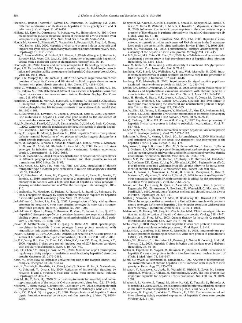
S. Khaliq et al. / Infection, Genetics and Evolution 11 (2011) 543–556554
Hezode, C., Roudot-Thoraval, F., Zafrani, E.S., Dhumeaux, D., Pawlotsky, J.M., 2004.Different mechanisms of steatosis in hepatitis C virus genotypes 1 and 3infections. J. Viral Hepat. 11, 455–458.
Hijikata, M., Kato, N., Ootsuyama, Y., Nakagawa, M., Shimotohno, K., 1991. Genemapping of the putative structural region of the hepatitis C virus genome by invitro processing analysis. Proc. Natl. Acad. Sci. U.S.A. 88, 5547–5551.
Honda, M., Kaneko, S., Shimazaki, T., Matsushita, E., Kobayashi, K., Ping, L.H., Zhang,H.C., Lemon, S.M., 2000. Hepatitis C virus core protein induces apoptosis andimpairs cell-cycle regulation in stably transformed Chinese hamster ovary cells.Hepatology 31, 1351–1359.
Hong, Z., Beaudet-Miller, M., Lanford, R.E., Guerra, B., Wright-Minogue, J., Skelton, A.,Baroudy, B.M., Reyes, G.R., Lau, J.Y., 1999. Generation of transmissible hepatitis Cvirions from a molecular clone in chimpanzees. Virology 256, 36–44.
Hoofnagle, J.H., 2002. Course and outcome of hepatitis C. Hepatology 36, S21–S29.Hope, R.G., McLauchlan, J., 2000. Sequence motifs required for lipid droplet associ-
ation and protein stability are unique to the hepatitis C virus core protein. J. Gen.Virol. 81, 1913–1925.
Hope, R.G., Murphy, D.J., McLauchlan, J., 2002. The domains required to direct coreproteins of hepatitis C virus and GB virus-B to lipid droplets share commonfeatures with plant oleosin proteins. J. Biol. Chem. 277, 4261–4270.
Horie, C., Iwahana, H., Horie, T., Shimizu, I., Yoshimoto, K., Yogita, S., Tashiro, S., Ito,S., Itakura, M., 1996. Detection of different quasispecies of hepatitis C virus coreregion in cancerous and noncancerous lesions. Biochem. Biophys. Res. Com-mun. 218, 674–681.
Hourioux, C., Patient, R., Morin, A., Blanchard, E., Moreau, A., Trassard, S., Giraudeau,B., Roingeard, P., 2007. The genotype 3-specific hepatitis C virus core proteinresidue phenylalanine 164 increases steatosis in an in vitro cellular model. Gut56, 1302–1308.
Hu, Z., Muroyama, R., Kowatari, N., Chang, J., Omata, M., Kato, N., 2009. Character-istic mutations in hepatitis C virus core gene related to the occurrence ofhepatocellular carcinoma. Cancer Sci. 100, 2465–2468.
Hui, J.M., Kench, J., Farrell, G.C., Lin, R., Samarasinghe, D., Liddle, C., Byth, K., George,J., 2002. Genotype-specific mechanisms for hepatic steatosis in chronic hepati-tis C infection. J. Gastroenterol. Hepatol. 17, 873–881.
Hussy, P., Langen, H., Mous, J., Jacobsen, H., 1996. Hepatitis C virus core protein:carboxy-terminal boundaries of two processed species suggest cleavage by asignal peptide peptidase. Virology 224, 93–104.
Idrees, M., Rafique, S., Rehman, I., Akbar, H., Yousaf, M.Z., Butt, S., Awan, Z., Manzoor,S., Akram, M., Aftab, M., Khubaib, B., Riazuddin, S., 2009. Hepatitis C virusgenotype 3a infection and hepatocellular carcinoma: Pakistan experience.World J. Gastroenterol. 15, 5080–5085.
Idrees, M., Riazuddin, S., 2008. Frequency distribution of hepatitis C virus genotypesin different geographical regions of Pakistan and their possible routes oftransmission. BMC Infect. Dis. 8, 69.
Im, S.S., Kwon, S.K., Kim, T.H., Kim, H.I., Ahn, Y.H., 2007. Regulation of glucosetransporter type 4 isoform gene expression in muscle and adipocytes. IUBMBLife 59, 134–145.
Ishii, K., Shinohara, M., Sawa, M., Kogame, M., Higami, K., Sano, M., Morita, T.,Sumino, Y., 2010. Interferon alpha receptor 2 expression by peripheral bloodmonocytes in patients with a high viral load of hepatitis C virus genotype 1showing substitution of amino acid 70 in the core region. Intervirology 53, 105–110.
it-Goughoulte, M., Hourioux, C., Patient, R., Trassard, S., Brand, D., Roingeard, P.,2006. Core protein cleavage by signal peptide peptidase is required for hepatitisC virus-like particle assembly. J. Gen. Virol. 87, 855–860.
Jackel-Cram, C., Babiuk, L.A., Liu, Q., 2007. Up-regulation of fatty acid synthasepromoter by hepatitis C virus core protein: genotype-3a core has a strongereffect than genotype-1b core. J. Hepatol. 46, 999–1008.
Jackel-Cram, C., Qiao, L., Xiang, Z., Brownlie, R., Zhou, Y., Babiuk, L., Liu, Q., 2010.Hepatitis C virus genotype-3a core protein enhances sterol regulatory element-binding protein-1 activity through the phosphoinositide 3-kinase-Akt-2 path-way. J. Gen. Virol. 91, 1388–1395.
Jhaveri, R., McHutchison, J., Patel, K., Qiang, G., Diehl, A.M., 2008. Specific poly-morphisms in hepatitis C virus genotype 3 core protein associated withintracellular lipid accumulation. J. Infect. Dis. 197, 283–291.
Jhaveri, R., Qiang, G., Diehl, A.M., 2009. Domain 3 of hepatitis C virus core protein issufficient for intracellular lipid accumulation. J. Infect. Dis. 200, 1781–1788.
Jin, D.Y., Wang, H.L., Zhou, Y., Chun, A.C., Kibler, K.V., Hou, Y.D., Kung, H., Jeang, K.T.,2000. Hepatitis C virus core protein-induced loss of LZIP function correlateswith cellular transformation. EMBO J. 19, 729–740.
Kao, C.F., Chen, S.Y., Chen, J.Y., Wu Lee, Y.H., 2004. Modulation of p53 transcriptionregulatory activity and post-translational modification by hepatitis C virus coreprotein. Oncogene 23, 2472–2483.
Karin, M., 1999. How NF-kappaB is activated: the role of the IkappaB kinase (IKK)complex. Oncogene 18, 6867–6874.
Kato, N., Yoshida, H., Ono-Nita, S.K., Kato, J., Goto, T., Otsuka, M., Lan, K., Matsushima,K., Shiratori, Y., Omata, M., 2000. Activation of intracellular signaling byhepatitis B and C viruses: C-viral core is the most potent signal inducer.Hepatology 32, 405–412.
Kim, M., Ha, Y., Park, H.J., 2006. Structural requirements for assembly and homo-typic interactions of the hepatitis C virus core protein. Virus Res. 122, 137–143.
Kisseleva, T., Bhattacharya, S., Braunstein, J., Schindler, C.W., 2002. Signaling throughthe JAK/STAT pathway, recent advances and future challenges. Gene 285, 1–24.
Klein, K.C., Polyak, S.J., Lingappa, J.R., 2004. Unique features of hepatitis C viruscapsid formation revealed by de novo cell-free assembly. J. Virol. 78, 9257–9269.
Kobayashi, M., Akuta, N., Suzuki, F., Hosaka, T., Sezaki, H., Kobayashi, M., Suzuki, Y.,Arase, Y., Ikeda, K., Watahiki, S., Mineta, R., Iwasaki, S., Miyakawa, Y., Kumada,H., 2010. Influence of amino-acid polymorphism in the core protein on pro-gression of liver disease in patients infected with hepatitis C virus genotype 1b.J. Med. Virol. 82, 41–48.
Kolykhalov, A.A., Mihalik, K., Feinstone, S.M., Rice, C.M., 2000. Hepatitis C virus-encoded enzymatic activities and conserved RNA elements in the 30 nontrans-lated region are essential for virus replication in vivo. J. Virol. 74, 2046–2051.
Kunkel, M., Watowich, S.J., 2002. Conformational changes accompanying self-assembly of the hepatitis C virus core protein. Virology 294, 239–245.
Lai, M.S., Hsieh, M.S., Chiu, Y.H., Chen, T.H., 2006. Type 2 diabetes and hepatocellularcarcinoma: a cohort study in high prevalence area of hepatitis virus infection.Hepatology 43, 1295–1302.
Lavie, M., Goffard, A., Dubuisson, J., 2007. Assembly of a functional HCV glycoproteinheterodimer. Curr. Issues Mol. Biol. 9, 71–86.
Lemberg, M.K., Bland, F.A., Weihofen, A., Braud, V.M., Martoglio, B., 2001. Intra-membrane proteolysis of signal peptides: an essential step in the generation ofHLA-E epitopes. J. Immunol. 167, 6441–6446.
Lemberg, M.K., Martoglio, B., 2002. Requirements for signal peptide peptidase-catalyzed intramembrane proteolysis. Mol. Cell 10, 735–744.
Lemon, S.M., Lerat, H., Weinman, S.A., Honda, M., 2000. A transgenic mouse model ofsteatosis and hepatocellular carcinoma associated with chronic hepatitis Cvirus infection in humans. Trans. Am. Clin. Climatol. Assoc. 111, 146–156.
Lerat, H., Honda, M., Beard, M.R., Loesch, K., Sun, J., Yang, Y., Okuda, M., Gosert, R.,Xiao, S.Y., Weinman, S.A., Lemon, S.M., 2002. Steatosis and liver cancer intransgenic mice expressing the structural and nonstructural proteins of hepa-titis C virus. Gastroenterology 122, 352–365.
Lin, W., Kim, S.S., Yeung, E., Kamegaya, Y., Blackard, J.T., Kim, K.A., Holtzman, M.J.,Chung, R.T., 2006. Hepatitis C virus core protein blocks interferon signaling byinteraction with the STAT1 SH2 domain. J. Virol. 80, 9226–9235.
Liu, Q., Tackney, C., Bhat, R.A., Prince, A.M., Zhang, P., 1997. Regulated processing ofhepatitis C virus core protein is linked to subcellular localization. J. Virol. 71,657–662.
Lo, S.Y., Selby, M.J., Ou, J.H., 1996. Interaction between hepatitis C virus core proteinand E1 envelope protein. J. Virol. 70, 5177–5182.
Lohmann, V., Roos, A., Korner, F., Koch, J.O., Bartenschlager, R., 2000. Biochemicaland structural analysis of the NS5B RNA-dependent RNA polymerase of thehepatitis C virus. J. Viral Hepat. 7, 167–174.
Magnusson, B., Asp, L., Bostrom, P., Ruiz, M., Stillemark-Billton, P., Linden, D., Boren,J., Olofsson, S.O., 2006. Adipocyte differentiation-related protein promotes fattyacid storage in cytosolic triglycerides and inhibits secretion of very low-densitylipoproteins. Arterioscler. Thromb. Vasc. Biol. 26, 1566–1571.
Manns, M.P., McHutchison, J.G., Gordon, S.C., Rustgi, V.K., Shiffman, M., Reindollar,R., Goodman, Z.D., Koury, K., Ling, M., Albrecht, J.K., 2001. Peginterferon alfa-2bplus ribavirin compared with interferon alfa-2b plus ribavirin for initial treat-ment of chronic hepatitis C: a randomised trial. Lancet 358, 958–965.
Masaki, T., Suzuki, R., Murakami, K., Aizaki, H., Ishii, K., Murayama, A., Date, T.,Matsuura, Y., Miyamura, T., Wakita, T., Suzuki, T., 2008. Interaction of hepatitis Cvirus nonstructural protein 5A with core protein is critical for the production ofinfectious virus particles. J. Virol. 82, 7964–7976.
Mason, A.L., Lau, J.Y., Hoang, N., Qian, K., Alexander, G.J., Xu, L., Guo, L., Jacob, S.,Regenstein, F.G., Zimmerman, R., Everhart, J.E., Wasserfall, C., Maclaren, N.K.,Perrillo, R.P., 1999. Association of diabetes mellitus and chronic hepatitis C virusinfection. Hepatology 29, 328–333.
Mathai, J., Shimoda, K., Banner, B.F., Mori, M., Bonkovsky, H.L., Barnard, G.F., 1999.IFN-alpha receptor mRNA expression in a United States sample with predomi-nantly genotype 1a/I chronic hepatitis C liver biopsies correlates with responseto IFN therapy. J. Interferon Cytokine Res. 19, 1011–1018.
Matsumoto, M., Hwang, S.B., Jeng, K.S., Zhu, N., Lai, M.M., 1996. Homotypic interac-tion and multimerization of hepatitis C virus core protein. Virology 218, 43–51.
McHutchison, J.G., Fried, M.W., 2003. Current therapy for hepatitis C: pegylatedinterferon and ribavirin. Clin. Liver Dis. 7, 149–161.
McLauchlan, J., 2000. Properties of the hepatitis C virus core protein: a structuralprotein that modulates cellular processes. J. Viral Hepat. 7, 2–14.
McLauchlan, J., Lemberg, M.K., Hope, G., Martoglio, B., 2002. Intramembrane pro-teolysis promotes trafficking of hepatitis C virus core protein to lipid droplets.EMBO J. 21, 3980–3988.
Mehta, S.H., Brancati, F.L., Strathdee, S.A., Pankow, J.S., Netski, D., Coresh, J., Szklo, M.,Thomas, D.L., 2003. Hepatitis C virus infection and incident type 2 diabetes.Hepatology 38, 50–56.
Melen, K., Fagerlund, R., Nyqvist, M., Keskinen, P., Julkunen, I., 2004. Expression ofhepatitis C virus core protein inhibits interferon-induced nuclear import ofSTATs. J. Med. Virol. 73, 536–547.
Mihm, S., Fayyazi, A., Hartmann, H., Ramadori, G., 1997. Analysis of histopatholog-ical manifestations of chronic hepatitis C virus infection with respect to virusgenotype. Hepatology 25, 735–739.
Miyanari, Y., Atsuzawa, K., Usuda, N., Watashi, K., Hishiki, T., Zayas, M., Bartens-chlager, R., Wakita, T., Hijikata, M., Shimotohno, K., 2007. The lipid droplet is animportant organelle for hepatitis C virus production. Nat. Cell Biol. 9, 1089–1097.
Mizukoshi, E., Kaneko, S., Yanagi, M., Ohno, H., Kaji, K., Terasaki, S., Shimoda, A.,Matsushita, E., Kobayashi, K., 1998. Expression of interferon alpha/beta receptorin the liver of chronic hepatitis C patients. J. Med. Virol. 56, 217–223.
Moradpour, D., Englert, C., Wakita, T., Wands, J.R., 1996. Characterization of celllines allowing tightly regulated expression of hepatitis C virus core protein.Virology 222, 51–63.
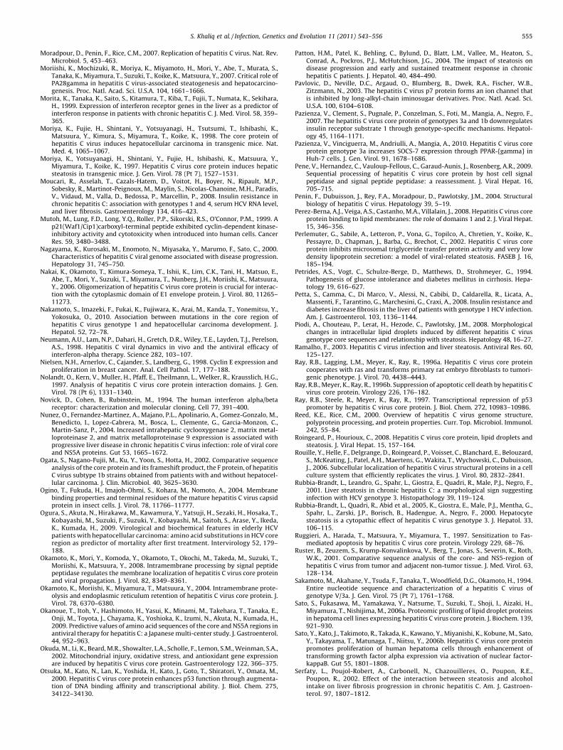
S. Khaliq et al. / Infection, Genetics and Evolution 11 (2011) 543–556 555
Moradpour, D., Penin, F., Rice, C.M., 2007. Replication of hepatitis C virus. Nat. Rev.Microbiol. 5, 453–463.
Moriishi, K., Mochizuki, R., Moriya, K., Miyamoto, H., Mori, Y., Abe, T., Murata, S.,Tanaka, K., Miyamura, T., Suzuki, T., Koike, K., Matsuura, Y., 2007. Critical role ofPA28gamma in hepatitis C virus-associated steatogenesis and hepatocarcino-genesis. Proc. Natl. Acad. Sci. U.S.A. 104, 1661–1666.
Morita, K., Tanaka, K., Saito, S., Kitamura, T., Kiba, T., Fujii, T., Numata, K., Sekihara,H., 1999. Expression of interferon receptor genes in the liver as a predictor ofinterferon response in patients with chronic hepatitis C. J. Med. Virol. 58, 359–365.
Moriya, K., Fujie, H., Shintani, Y., Yotsuyanagi, H., Tsutsumi, T., Ishibashi, K.,Matsuura, Y., Kimura, S., Miyamura, T., Koike, K., 1998. The core protein ofhepatitis C virus induces hepatocellular carcinoma in transgenic mice. Nat.Med. 4, 1065–1067.
Moriya, K., Yotsuyanagi, H., Shintani, Y., Fujie, H., Ishibashi, K., Matsuura, Y.,Miyamura, T., Koike, K., 1997. Hepatitis C virus core protein induces hepaticsteatosis in transgenic mice. J. Gen. Virol. 78 (Pt 7), 1527–1531.
Moucari, R., Asselah, T., Cazals-Hatem, D., Voitot, H., Boyer, N., Ripault, M.P.,Sobesky, R., Martinot-Peignoux, M., Maylin, S., Nicolas-Chanoine, M.H., Paradis,V., Vidaud, M., Valla, D., Bedossa, P., Marcellin, P., 2008. Insulin resistance inchronic hepatitis C: association with genotypes 1 and 4, serum HCV RNA level,and liver fibrosis. Gastroenterology 134, 416–423.
Mutoh, M., Lung, F.D., Long, Y.Q., Roller, P.P., Sikorski, R.S., O’Connor, P.M., 1999. Ap21(Waf1/Cip1)carboxyl-terminal peptide exhibited cyclin-dependent kinase-inhibitory activity and cytotoxicity when introduced into human cells. CancerRes. 59, 3480–3488.
Nagayama, K., Kurosaki, M., Enomoto, N., Miyasaka, Y., Marumo, F., Sato, C., 2000.Characteristics of hepatitis C viral genome associated with disease progression.Hepatology 31, 745–750.
Nakai, K., Okamoto, T., Kimura-Someya, T., Ishii, K., Lim, C.K., Tani, H., Matsuo, E.,Abe, T., Mori, Y., Suzuki, T., Miyamura, T., Nunberg, J.H., Moriishi, K., Matsuura,Y., 2006. Oligomerization of hepatitis C virus core protein is crucial for interac-tion with the cytoplasmic domain of E1 envelope protein. J. Virol. 80, 11265–11273.
Nakamoto, S., Imazeki, F., Fukai, K., Fujiwara, K., Arai, M., Kanda, T., Yonemitsu, Y.,Yokosuka, O., 2010. Association between mutations in the core region ofhepatitis C virus genotype 1 and hepatocellular carcinoma development. J.Hepatol. 52, 72–78.
Neumann, A.U., Lam, N.P., Dahari, H., Gretch, D.R., Wiley, T.E., Layden, T.J., Perelson,A.S., 1998. Hepatitis C viral dynamics in vivo and the antiviral efficacy ofinterferon-alpha therapy. Science 282, 103–107.
Nielsen, N.H., Arnerlov, C., Cajander, S., Landberg, G., 1998. Cyclin E expression andproliferation in breast cancer. Anal. Cell Pathol. 17, 177–188.
Nolandt, O., Kern, V., Muller, H., Pfaff, E., Theilmann, L., Welker, R., Krausslich, H.G.,1997. Analysis of hepatitis C virus core protein interaction domains. J. Gen.Virol. 78 (Pt 6), 1331–1340.
Novick, D., Cohen, B., Rubinstein, M., 1994. The human interferon alpha/betareceptor: characterization and molecular cloning. Cell 77, 391–400.
Nunez, O., Fernandez-Martinez, A., Majano, P.L., Apolinario, A., Gomez-Gonzalo, M.,Benedicto, I., Lopez-Cabrera, M., Bosca, L., Clemente, G., Garcia-Monzon, C.,Martin-Sanz, P., 2004. Increased intrahepatic cyclooxygenase 2, matrix metal-loproteinase 2, and matrix metalloproteinase 9 expression is associated withprogressive liver disease in chronic hepatitis C virus infection: role of viral coreand NS5A proteins. Gut 53, 1665–1672.
Ogata, S., Nagano-Fujii, M., Ku, Y., Yoon, S., Hotta, H., 2002. Comparative sequenceanalysis of the core protein and its frameshift product, the F protein, of hepatitisC virus subtype 1b strains obtained from patients with and without hepatocel-lular carcinoma. J. Clin. Microbiol. 40, 3625–3630.
Ogino, T., Fukuda, H., Imajoh-Ohmi, S., Kohara, M., Nomoto, A., 2004. Membranebinding properties and terminal residues of the mature hepatitis C virus capsidprotein in insect cells. J. Virol. 78, 11766–11777.
Ogura, S., Akuta, N., Hirakawa, M., Kawamura, Y., Yatsuji, H., Sezaki, H., Hosaka, T.,Kobayashi, M., Suzuki, F., Suzuki, Y., Kobayashi, M., Saitoh, S., Arase, Y., Ikeda,K., Kumada, H., 2009. Virological and biochemical features in elderly HCVpatients with hepatocellular carcinoma: amino acid substitutions in HCV coreregion as predictor of mortality after first treatment. Intervirology 52, 179–188.
Okamoto, K., Mori, Y., Komoda, Y., Okamoto, T., Okochi, M., Takeda, M., Suzuki, T.,Moriishi, K., Matsuura, Y., 2008. Intramembrane processing by signal peptidepeptidase regulates the membrane localization of hepatitis C virus core proteinand viral propagation. J. Virol. 82, 8349–8361.
Okamoto, K., Moriishi, K., Miyamura, T., Matsuura, Y., 2004. Intramembrane prote-olysis and endoplasmic reticulum retention of hepatitis C virus core protein. J.Virol. 78, 6370–6380.
Okanoue, T., Itoh, Y., Hashimoto, H., Yasui, K., Minami, M., Takehara, T., Tanaka, E.,Onji, M., Toyota, J., Chayama, K., Yoshioka, K., Izumi, N., Akuta, N., Kumada, H.,2009. Predictive values of amino acid sequences of the core and NS5A regions inantiviral therapy for hepatitis C: a Japanese multi-center study. J. Gastroenterol.44, 952–963.
Okuda, M., Li, K., Beard, M.R., Showalter, L.A., Scholle, F., Lemon, S.M., Weinman, S.A.,2002. Mitochondrial injury, oxidative stress, and antioxidant gene expressionare induced by hepatitis C virus core protein. Gastroenterology 122, 366–375.
Otsuka, M., Kato, N., Lan, K., Yoshida, H., Kato, J., Goto, T., Shiratori, Y., Omata, M.,2000. Hepatitis C virus core protein enhances p53 function through augmenta-tion of DNA binding affinity and transcriptional ability. J. Biol. Chem. 275,34122–34130.
Patton, H.M., Patel, K., Behling, C., Bylund, D., Blatt, L.M., Vallee, M., Heaton, S.,Conrad, A., Pockros, P.J., McHutchison, J.G., 2004. The impact of steatosis ondisease progression and early and sustained treatment response in chronichepatitis C patients. J. Hepatol. 40, 484–490.
Pavlovic, D., Neville, D.C., Argaud, O., Blumberg, B., Dwek, R.A., Fischer, W.B.,Zitzmann, N., 2003. The hepatitis C virus p7 protein forms an ion channel thatis inhibited by long-alkyl-chain iminosugar derivatives. Proc. Natl. Acad. Sci.U.S.A. 100, 6104–6108.
Pazienza, V., Clement, S., Pugnale, P., Conzelman, S., Foti, M., Mangia, A., Negro, F.,2007. The hepatitis C virus core protein of genotypes 3a and 1b downregulatesinsulin receptor substrate 1 through genotype-specific mechanisms. Hepatol-ogy 45, 1164–1171.
Pazienza, V., Vinciguerra, M., Andriulli, A., Mangia, A., 2010. Hepatitis C virus coreprotein genotype 3a increases SOCS-7 expression through PPAR-{gamma} inHuh-7 cells. J. Gen. Virol. 91, 1678–1686.
Pene, V., Hernandez, C., Vauloup-Fellous, C., Garaud-Aunis, J., Rosenberg, A.R., 2009.Sequential processing of hepatitis C virus core protein by host cell signalpeptidase and signal peptide peptidase: a reassessment. J. Viral Hepat. 16,705–715.
Penin, F., Dubuisson, J., Rey, F.A., Moradpour, D., Pawlotsky, J.M., 2004. Structuralbiology of hepatitis C virus. Hepatology 39, 5–19.
Perez-Berna, A.J., Veiga, A.S., Castanho, M.A., Villalain, J., 2008. Hepatitis C virus coreprotein binding to lipid membranes: the role of domains 1 and 2. J. Viral Hepat.15, 346–356.
Perlemuter, G., Sabile, A., Letteron, P., Vona, G., Topilco, A., Chretien, Y., Koike, K.,Pessayre, D., Chapman, J., Barba, G., Brechot, C., 2002. Hepatitis C virus coreprotein inhibits microsomal triglyceride transfer protein activity and very lowdensity lipoprotein secretion: a model of viral-related steatosis. FASEB J. 16,185–194.
Petrides, A.S., Vogt, C., Schulze-Berge, D., Matthews, D., Strohmeyer, G., 1994.Pathogenesis of glucose intolerance and diabetes mellitus in cirrhosis. Hepa-tology 19, 616–627.
Petta, S., Camma, C., Di Marco, V., Alessi, N., Cabibi, D., Caldarella, R., Licata, A.,Massenti, F., Tarantino, G., Marchesini, G., Craxi, A., 2008. Insulin resistance anddiabetes increase fibrosis in the liver of patients with genotype 1 HCV infection.Am. J. Gastroenterol. 103, 1136–1144.
Piodi, A., Chouteau, P., Lerat, H., Hezode, C., Pawlotsky, J.M., 2008. Morphologicalchanges in intracellular lipid droplets induced by different hepatitis C virusgenotype core sequences and relationship with steatosis. Hepatology 48, 16–27.
Ramalho, F., 2003. Hepatitis C virus infection and liver steatosis. Antiviral Res. 60,125–127.
Ray, R.B., Lagging, L.M., Meyer, K., Ray, R., 1996a. Hepatitis C virus core proteincooperates with ras and transforms primary rat embryo fibroblasts to tumori-genic phenotype. J. Virol. 70, 4438–4443.
Ray, R.B., Meyer, K., Ray, R., 1996b. Suppression of apoptotic cell death by hepatitis Cvirus core protein. Virology 226, 176–182.
Ray, R.B., Steele, R., Meyer, K., Ray, R., 1997. Transcriptional repression of p53promoter by hepatitis C virus core protein. J. Biol. Chem. 272, 10983–10986.
Reed, K.E., Rice, C.M., 2000. Overview of hepatitis C virus genome structure,polyprotein processing, and protein properties. Curr. Top. Microbiol. Immunol.242, 55–84.
Roingeard, P., Hourioux, C., 2008. Hepatitis C virus core protein, lipid droplets andsteatosis. J. Viral Hepat. 15, 157–164.
Rouille, Y., Helle, F., Delgrange, D., Roingeard, P., Voisset, C., Blanchard, E., Belouzard,S., McKeating, J., Patel, A.H., Maertens, G., Wakita, T., Wychowski, C., Dubuisson,J., 2006. Subcellular localization of hepatitis C virus structural proteins in a cellculture system that efficiently replicates the virus. J. Virol. 80, 2832–2841.
Rubbia-Brandt, L., Leandro, G., Spahr, L., Giostra, E., Quadri, R., Male, P.J., Negro, F.,2001. Liver steatosis in chronic hepatitis C: a morphological sign suggestinginfection with HCV genotype 3. Histopathology 39, 119–124.
Rubbia-Brandt, L., Quadri, R., Abid et al., 2005, K., Giostra, E., Male, P.J., Mentha, G.,Spahr, L., Zarski, J.P., Borisch, B., Hadengue, A., Negro, F., 2000. Hepatocytesteatosis is a cytopathic effect of hepatitis C virus genotype 3. J. Hepatol. 33,106–115.
Ruggieri, A., Harada, T., Matsuura, Y., Miyamura, T., 1997. Sensitization to Fas-mediated apoptosis by hepatitis C virus core protein. Virology 229, 68–76.
Ruster, B., Zeuzem, S., Krump-Konvalinkova, V., Berg, T., Jonas, S., Severin, K., Roth,W.K., 2001. Comparative sequence analysis of the core- and NS5-region ofhepatitis C virus from tumor and adjacent non-tumor tissue. J. Med. Virol. 63,128–134.
Sakamoto, M., Akahane, Y., Tsuda, F., Tanaka, T., Woodfield, D.G., Okamoto, H., 1994.Entire nucleotide sequence and characterization of a hepatitis C virus ofgenotype V/3a. J. Gen. Virol. 75 (Pt 7), 1761–1768.
Sato, S., Fukasawa, M., Yamakawa, Y., Natsume, T., Suzuki, T., Shoji, I., Aizaki, H.,Miyamura, T., Nishijima, M., 2006a. Proteomic profiling of lipid droplet proteinsin hepatoma cell lines expressing hepatitis C virus core protein. J. Biochem. 139,921–930.
Sato, Y., Kato, J., Takimoto, R., Takada, K., Kawano, Y., Miyanishi, K., Kobune, M., Sato,Y., Takayama, T., Matunaga, T., Niitsu, Y., 2006b. Hepatitis C virus core proteinpromotes proliferation of human hepatoma cells through enhancement oftransforming growth factor alpha expression via activation of nuclear factor-kappaB. Gut 55, 1801–1808.
Serfaty, L., Poujol-Robert, A., Carbonell, N., Chazouilleres, O., Poupon, R.E.,Poupon, R., 2002. Effect of the interaction between steatosis and alcoholintake on liver fibrosis progression in chronic hepatitis C. Am. J. Gastroen-terol. 97, 1807–1812.
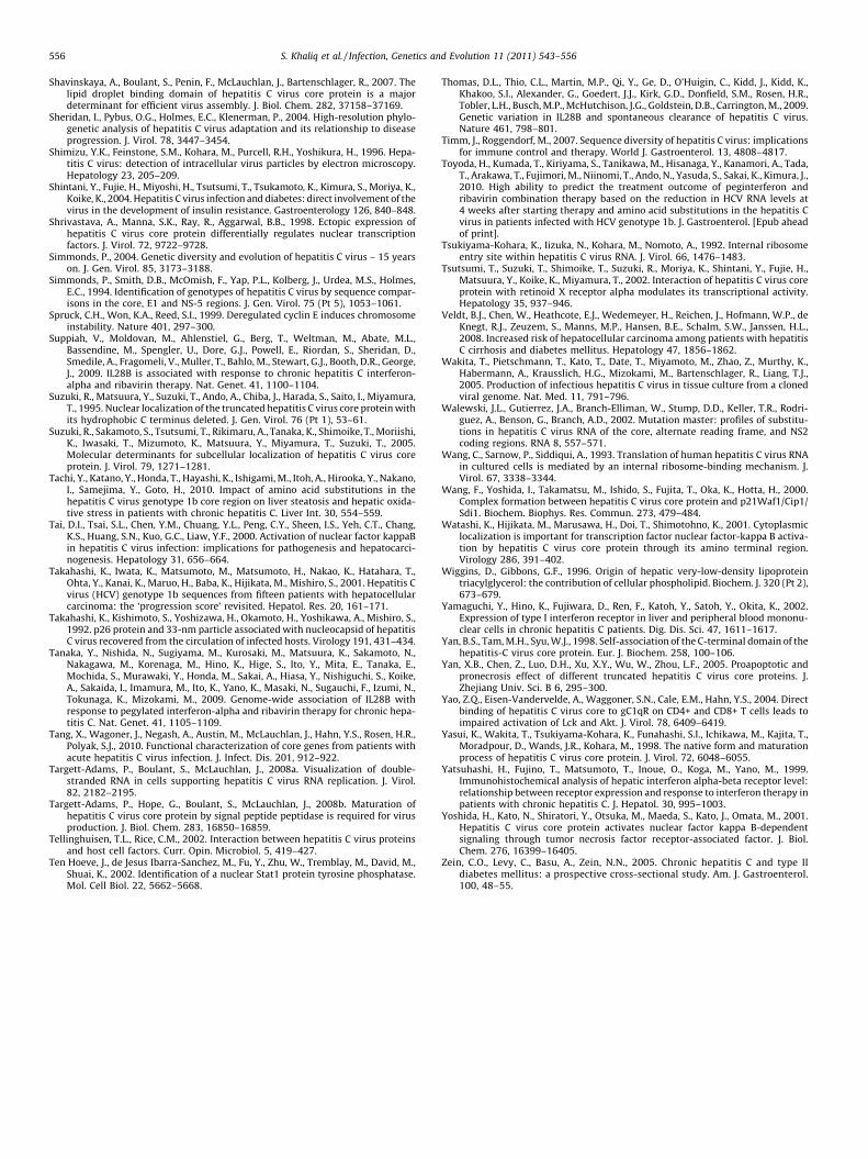
S. Khaliq et al. / Infection, Genetics and Evolution 11 (2011) 543–556556
Shavinskaya, A., Boulant, S., Penin, F., McLauchlan, J., Bartenschlager, R., 2007. Thelipid droplet binding domain of hepatitis C virus core protein is a majordeterminant for efficient virus assembly. J. Biol. Chem. 282, 37158–37169.
Sheridan, I., Pybus, O.G., Holmes, E.C., Klenerman, P., 2004. High-resolution phylo-genetic analysis of hepatitis C virus adaptation and its relationship to diseaseprogression. J. Virol. 78, 3447–3454.
Shimizu, Y.K., Feinstone, S.M., Kohara, M., Purcell, R.H., Yoshikura, H., 1996. Hepa-titis C virus: detection of intracellular virus particles by electron microscopy.Hepatology 23, 205–209.
Shintani, Y., Fujie, H., Miyoshi, H., Tsutsumi, T., Tsukamoto, K., Kimura, S., Moriya, K.,Koike, K., 2004. Hepatitis C virus infection and diabetes: direct involvement of thevirus in the development of insulin resistance. Gastroenterology 126, 840–848.
Shrivastava, A., Manna, S.K., Ray, R., Aggarwal, B.B., 1998. Ectopic expression ofhepatitis C virus core protein differentially regulates nuclear transcriptionfactors. J. Virol. 72, 9722–9728.
Simmonds, P., 2004. Genetic diversity and evolution of hepatitis C virus – 15 yearson. J. Gen. Virol. 85, 3173–3188.
Simmonds, P., Smith, D.B., McOmish, F., Yap, P.L., Kolberg, J., Urdea, M.S., Holmes,E.C., 1994. Identification of genotypes of hepatitis C virus by sequence compar-isons in the core, E1 and NS-5 regions. J. Gen. Virol. 75 (Pt 5), 1053–1061.
Spruck, C.H., Won, K.A., Reed, S.I., 1999. Deregulated cyclin E induces chromosomeinstability. Nature 401, 297–300.
Suppiah, V., Moldovan, M., Ahlenstiel, G., Berg, T., Weltman, M., Abate, M.L.,Bassendine, M., Spengler, U., Dore, G.J., Powell, E., Riordan, S., Sheridan, D.,Smedile, A., Fragomeli, V., Muller, T., Bahlo, M., Stewart, G.J., Booth, D.R., George,J., 2009. IL28B is associated with response to chronic hepatitis C interferon-alpha and ribavirin therapy. Nat. Genet. 41, 1100–1104.
Suzuki, R., Matsuura, Y., Suzuki, T., Ando, A., Chiba, J., Harada, S., Saito, I., Miyamura,T., 1995. Nuclear localization of the truncated hepatitis C virus core protein withits hydrophobic C terminus deleted. J. Gen. Virol. 76 (Pt 1), 53–61.
Suzuki, R., Sakamoto, S., Tsutsumi, T., Rikimaru, A., Tanaka, K., Shimoike, T., Moriishi,K., Iwasaki, T., Mizumoto, K., Matsuura, Y., Miyamura, T., Suzuki, T., 2005.Molecular determinants for subcellular localization of hepatitis C virus coreprotein. J. Virol. 79, 1271–1281.
Tachi, Y., Katano, Y., Honda, T., Hayashi, K., Ishigami, M., Itoh, A., Hirooka, Y., Nakano,I., Samejima, Y., Goto, H., 2010. Impact of amino acid substitutions in thehepatitis C virus genotype 1b core region on liver steatosis and hepatic oxida-tive stress in patients with chronic hepatitis C. Liver Int. 30, 554–559.
Tai, D.I., Tsai, S.L., Chen, Y.M., Chuang, Y.L., Peng, C.Y., Sheen, I.S., Yeh, C.T., Chang,K.S., Huang, S.N., Kuo, G.C., Liaw, Y.F., 2000. Activation of nuclear factor kappaBin hepatitis C virus infection: implications for pathogenesis and hepatocarci-nogenesis. Hepatology 31, 656–664.
Takahashi, K., Iwata, K., Matsumoto, M., Matsumoto, H., Nakao, K., Hatahara, T.,Ohta, Y., Kanai, K., Maruo, H., Baba, K., Hijikata, M., Mishiro, S., 2001. Hepatitis Cvirus (HCV) genotype 1b sequences from fifteen patients with hepatocellularcarcinoma: the ‘progression score’ revisited. Hepatol. Res. 20, 161–171.
Takahashi, K., Kishimoto, S., Yoshizawa, H., Okamoto, H., Yoshikawa, A., Mishiro, S.,1992. p26 protein and 33-nm particle associated with nucleocapsid of hepatitisC virus recovered from the circulation of infected hosts. Virology 191, 431–434.
Tanaka, Y., Nishida, N., Sugiyama, M., Kurosaki, M., Matsuura, K., Sakamoto, N.,Nakagawa, M., Korenaga, M., Hino, K., Hige, S., Ito, Y., Mita, E., Tanaka, E.,Mochida, S., Murawaki, Y., Honda, M., Sakai, A., Hiasa, Y., Nishiguchi, S., Koike,A., Sakaida, I., Imamura, M., Ito, K., Yano, K., Masaki, N., Sugauchi, F., Izumi, N.,Tokunaga, K., Mizokami, M., 2009. Genome-wide association of IL28B withresponse to pegylated interferon-alpha and ribavirin therapy for chronic hepa-titis C. Nat. Genet. 41, 1105–1109.
Tang, X., Wagoner, J., Negash, A., Austin, M., McLauchlan, J., Hahn, Y.S., Rosen, H.R.,Polyak, S.J., 2010. Functional characterization of core genes from patients withacute hepatitis C virus infection. J. Infect. Dis. 201, 912–922.
Targett-Adams, P., Boulant, S., McLauchlan, J., 2008a. Visualization of double-stranded RNA in cells supporting hepatitis C virus RNA replication. J. Virol.82, 2182–2195.
Targett-Adams, P., Hope, G., Boulant, S., McLauchlan, J., 2008b. Maturation ofhepatitis C virus core protein by signal peptide peptidase is required for virusproduction. J. Biol. Chem. 283, 16850–16859.
Tellinghuisen, T.L., Rice, C.M., 2002. Interaction between hepatitis C virus proteinsand host cell factors. Curr. Opin. Microbiol. 5, 419–427.
Ten Hoeve, J., de Jesus Ibarra-Sanchez, M., Fu, Y., Zhu, W., Tremblay, M., David, M.,Shuai, K., 2002. Identification of a nuclear Stat1 protein tyrosine phosphatase.Mol. Cell Biol. 22, 5662–5668.
Thomas, D.L., Thio, C.L., Martin, M.P., Qi, Y., Ge, D., O’Huigin, C., Kidd, J., Kidd, K.,Khakoo, S.I., Alexander, G., Goedert, J.J., Kirk, G.D., Donfield, S.M., Rosen, H.R.,Tobler, L.H., Busch, M.P., McHutchison, J.G., Goldstein, D.B., Carrington, M., 2009.Genetic variation in IL28B and spontaneous clearance of hepatitis C virus.Nature 461, 798–801.
Timm, J., Roggendorf, M., 2007. Sequence diversity of hepatitis C virus: implicationsfor immune control and therapy. World J. Gastroenterol. 13, 4808–4817.
Toyoda, H., Kumada, T., Kiriyama, S., Tanikawa, M., Hisanaga, Y., Kanamori, A., Tada,T., Arakawa, T., Fujimori, M., Niinomi, T., Ando, N., Yasuda, S., Sakai, K., Kimura, J.,2010. High ability to predict the treatment outcome of peginterferon andribavirin combination therapy based on the reduction in HCV RNA levels at4 weeks after starting therapy and amino acid substitutions in the hepatitis Cvirus in patients infected with HCV genotype 1b. J. Gastroenterol. [Epub aheadof print].
Tsukiyama-Kohara, K., Iizuka, N., Kohara, M., Nomoto, A., 1992. Internal ribosomeentry site within hepatitis C virus RNA. J. Virol. 66, 1476–1483.
Tsutsumi, T., Suzuki, T., Shimoike, T., Suzuki, R., Moriya, K., Shintani, Y., Fujie, H.,Matsuura, Y., Koike, K., Miyamura, T., 2002. Interaction of hepatitis C virus coreprotein with retinoid X receptor alpha modulates its transcriptional activity.Hepatology 35, 937–946.
Veldt, B.J., Chen, W., Heathcote, E.J., Wedemeyer, H., Reichen, J., Hofmann, W.P., deKnegt, R.J., Zeuzem, S., Manns, M.P., Hansen, B.E., Schalm, S.W., Janssen, H.L.,2008. Increased risk of hepatocellular carcinoma among patients with hepatitisC cirrhosis and diabetes mellitus. Hepatology 47, 1856–1862.
Wakita, T., Pietschmann, T., Kato, T., Date, T., Miyamoto, M., Zhao, Z., Murthy, K.,Habermann, A., Krausslich, H.G., Mizokami, M., Bartenschlager, R., Liang, T.J.,2005. Production of infectious hepatitis C virus in tissue culture from a clonedviral genome. Nat. Med. 11, 791–796.
Walewski, J.L., Gutierrez, J.A., Branch-Elliman, W., Stump, D.D., Keller, T.R., Rodri-guez, A., Benson, G., Branch, A.D., 2002. Mutation master: profiles of substitu-tions in hepatitis C virus RNA of the core, alternate reading frame, and NS2coding regions. RNA 8, 557–571.
Wang, C., Sarnow, P., Siddiqui, A., 1993. Translation of human hepatitis C virus RNAin cultured cells is mediated by an internal ribosome-binding mechanism. J.Virol. 67, 3338–3344.
Wang, F., Yoshida, I., Takamatsu, M., Ishido, S., Fujita, T., Oka, K., Hotta, H., 2000.Complex formation between hepatitis C virus core protein and p21Waf1/Cip1/Sdi1. Biochem. Biophys. Res. Commun. 273, 479–484.
Watashi, K., Hijikata, M., Marusawa, H., Doi, T., Shimotohno, K., 2001. Cytoplasmiclocalization is important for transcription factor nuclear factor-kappa B activa-tion by hepatitis C virus core protein through its amino terminal region.Virology 286, 391–402.
Wiggins, D., Gibbons, G.F., 1996. Origin of hepatic very-low-density lipoproteintriacylglycerol: the contribution of cellular phospholipid. Biochem. J. 320 (Pt 2),673–679.
Yamaguchi, Y., Hino, K., Fujiwara, D., Ren, F., Katoh, Y., Satoh, Y., Okita, K., 2002.Expression of type I interferon receptor in liver and peripheral blood mononu-clear cells in chronic hepatitis C patients. Dig. Dis. Sci. 47, 1611–1617.
Yan, B.S., Tam, M.H., Syu, W.J., 1998. Self-association of the C-terminal domain of thehepatitis-C virus core protein. Eur. J. Biochem. 258, 100–106.
Yan, X.B., Chen, Z., Luo, D.H., Xu, X.Y., Wu, W., Zhou, L.F., 2005. Proapoptotic andpronecrosis effect of different truncated hepatitis C virus core proteins. J.Zhejiang Univ. Sci. B 6, 295–300.
Yao, Z.Q., Eisen-Vandervelde, A., Waggoner, S.N., Cale, E.M., Hahn, Y.S., 2004. Directbinding of hepatitis C virus core to gC1qR on CD4+ and CD8+ T cells leads toimpaired activation of Lck and Akt. J. Virol. 78, 6409–6419.
Yasui, K., Wakita, T., Tsukiyama-Kohara, K., Funahashi, S.I., Ichikawa, M., Kajita, T.,Moradpour, D., Wands, J.R., Kohara, M., 1998. The native form and maturationprocess of hepatitis C virus core protein. J. Virol. 72, 6048–6055.
Yatsuhashi, H., Fujino, T., Matsumoto, T., Inoue, O., Koga, M., Yano, M., 1999.Immunohistochemical analysis of hepatic interferon alpha-beta receptor level:relationship between receptor expression and response to interferon therapy inpatients with chronic hepatitis C. J. Hepatol. 30, 995–1003.
Yoshida, H., Kato, N., Shiratori, Y., Otsuka, M., Maeda, S., Kato, J., Omata, M., 2001.Hepatitis C virus core protein activates nuclear factor kappa B-dependentsignaling through tumor necrosis factor receptor-associated factor. J. Biol.Chem. 276, 16399–16405.
Zein, C.O., Levy, C., Basu, A., Zein, N.N., 2005. Chronic hepatitis C and type IIdiabetes mellitus: a prospective cross-sectional study. Am. J. Gastroenterol.100, 48–55.