Survivin is required for stable checkpoint activation in taxol-treated HeLa cells
12
Introduction Since its discovery by Altieri and co-workers (Ambrosini et al., 1997), considerable effort has been directed at understanding the function of Survivin. This small protein (16.5 kDa) is only expressed in embryonic or proliferating adult tissues and is highly overexpressed in many forms of cancer (Altieri, 2001). Although Survivin has a prominent BIR domain, consistent with a potential role in apoptotic regulation, the principle defects manifested upon interference with Survivin function are in mitosis (for reviews, see Altieri et al., 1999; Silke and Vaux, 2001; Reed and Bischoff, 2000). Recently Survivin has been shown to be a chromosomal passenger protein (Skoufias et al., 2000; Uren et al., 2000; Jiang et al., 2001; Wheatley et al., 2001a). These highly conserved proteins are present at centromeres during prometaphase, then transfer to the central spindle and presumptive cleavage furrow at the cell cortex during early anaphase (see Earnshaw and Bernat, 1990; Earnshaw and Cooke, 1991; Adams et al., 2001a). Chromosomal passenger proteins cloned to date include INCENP/Sli-15, Aurora-B/Ipl- 1/AIR-2 and Survivin/BIR1 (for reviews, see Adams et al., 2001a; Terada, 2001), but at least one other protein with similar behaviour also exists, TD60 (telophase-disc 60) (Andreassen et al., 1991). INCENP is a multidomain protein that interacts with heterochromatin protein-1 (Ainsztein et al., 1998) and microtubules (Wheatley et al., 2001b), and it is thought to be a targeting or scaffolding subunit for Aurora-B kinase (Adams et al., 2000; Bolton et al., 2002). In contrast, relatively little is known about the mitotic role of Survivin other than that it can stimulate Aurora-B kinase activity in vitro (Bolton et al., 2002; Bishop and Schumacher, 2002). It is likely that the chromosomal passengers act in concert to execute their role(s) during mitosis, and they are stockpiled as a complex in Xenopus eggs and in yeast (Adams et al., 2000; Bolton et al., 2002; Cheeseman et al., 2002). Interactions between the three proteins have also been detected by two-hybrid and in vitro binding experiments (Kaitna et al., 2000; Wheatley et al., 2001a). INCENP and Aurora-B are required for phosphorylation of histone H3 on serine 10 (Hsu et al., 2000; Adams et al., 2001b; Murnion et al., 2001) and, in Drosophila and S. pombe [though not Xenopus (MacCallum et al., 2002)], for targeting of condensin to chromosomes (Giet and Glover, 2001; Morishita et al., 2001). The passengers are essential for chromosomes to achieve a stable metaphase biorientation (Adams et al., 2001b; Kaitna et al., 2002), possibly by regulating the interaction of kinetochores with microtubules (Yoon and Carbon, 1999; 2987 Survivin is an essential chromosomal passenger protein whose function remains unclear. Here, we have used RNA interference to specifically repress Survivin in cultured HeLa cells. Immunoblot analysis showed that Survivin was no longer detectable in cultures 60 hours after transfection with Survivin-specific siRNA. Live cell analysis showed that many Survivin-depleted cells were delayed in mitosis, and immunofluorescence analysis of fixed specimens revealed that Survivin-depleted cells accumulated in prometaphase with misaligned chromosomes. The chromosomal passenger proteins, INCENP and Aurora-B, which can interact directly with Survivin, were absent from the centromeres of Survivin-depleted cells. These data contribute to the emerging picture that Survivin operates together with INCENP and Aurora-B to perform its mitotic duties. Some Survivin-depleted cells eventually exited mitosis without completing cytokinesis. This resulted in a gradual increase in the percentage of multinucleated cells in the culture. Time-lapse imaging of synchronized cultures revealed that control and Survivin-depleted cells arrested in mitosis in the presence of nocodazole; however, the latter failed to arrest in mitosis when treated with taxol. Immunofluorescence studies revealed that Survivin- depleted cells were unable to stably maintain BubR1 at the kinetochores in the presence of either taxol or nocodazole. Our data reveal that Survivin is not required for the spindle assembly checkpoint when it is activated by the loss of microtubules. However, Survivin is required for the maintenance of the checkpoint when it is activated by taxol, which is generally thought to cause a loss of spindle tension. Key words: Mitosis, Chromosomal passenger proteins, Survivin, Aurora-B, INCENP, RNA interference, BubR1 Summary Survivin is required for stable checkpoint activation in taxol-treated HeLa cells Ana Carvalho 1,2,3 , Mar Carmena 1 , Clara Sambade 2,3 , William C. Earnshaw 1,4, * and Sally P. Wheatley 1 1 Chromosome Structure Group, Wellcome Trust Centre for Cell Biology, Institute of Cell and Molecular Biology, University of Edinburgh, King’s Buildings, Mayfield Road, Edinburgh EH9 3JR, Scotland, UK 2 University of Porto, IPATIMUP Institute of Molecular Pathology and Immunology of the University of Porto, Porto, Portugal 3 Faculty of Medicine, University of Porto, Porto, Portugal *Author for correspondence (e-mail: [email protected]) Accepted 11 April 2003 Journal of Cell Science 116, 2987-2998 © 2003 The Company of Biologists Ltd doi:10.1242/jcs.00612 Research Article JCS ePress online publication date 3 June 2003
-
Upload
independent -
Category
Documents
-
view
1 -
download
0
Transcript of Survivin is required for stable checkpoint activation in taxol-treated HeLa cells

IntroductionSince its discovery by Altieri and co-workers (Ambrosini et al.,1997), considerable effort has been directed at understandingthe function of Survivin. This small protein (16.5 kDa) is onlyexpressed in embryonic or proliferating adult tissues and ishighly overexpressed in many forms of cancer (Altieri, 2001).Although Survivin has a prominent BIR domain, consistentwith a potential role in apoptotic regulation, the principledefects manifested upon interference with Survivin functionare in mitosis (for reviews, see Altieri et al., 1999; Silke andVaux, 2001; Reed and Bischoff, 2000).
Recently Survivin has been shown to be a chromosomalpassenger protein (Skoufias et al., 2000; Uren et al., 2000;Jiang et al., 2001; Wheatley et al., 2001a). These highlyconserved proteins are present at centromeres duringprometaphase, then transfer to the central spindle andpresumptive cleavage furrow at the cell cortex during earlyanaphase (see Earnshaw and Bernat, 1990; Earnshaw andCooke, 1991; Adams et al., 2001a). Chromosomal passengerproteins cloned to date include INCENP/Sli-15, Aurora-B/Ipl-1/AIR-2 and Survivin/BIR1 (for reviews, see Adams et al.,2001a; Terada, 2001), but at least one other protein with similarbehaviour also exists, TD60 (telophase-disc 60) (Andreassenet al., 1991).
INCENP is a multidomain protein that interacts withheterochromatin protein-1 (Ainsztein et al., 1998) andmicrotubules (Wheatley et al., 2001b), and it is thought to bea targeting or scaffolding subunit for Aurora-B kinase (Adamset al., 2000; Bolton et al., 2002). In contrast, relatively little isknown about the mitotic role of Survivin other than that itcan stimulate Aurora-B kinase activity in vitro (Bolton et al.,2002; Bishop and Schumacher, 2002). It is likely that thechromosomal passengers act in concert to execute their role(s)during mitosis, and they are stockpiled as a complex inXenopuseggs and in yeast (Adams et al., 2000; Bolton et al.,2002; Cheeseman et al., 2002). Interactions between the threeproteins have also been detected by two-hybrid and in vitrobinding experiments (Kaitna et al., 2000; Wheatley et al.,2001a).
INCENP and Aurora-B are required for phosphorylation ofhistone H3 on serine 10 (Hsu et al., 2000; Adams et al., 2001b;Murnion et al., 2001) and, in Drosophila and S. pombe[thoughnot Xenopus (MacCallum et al., 2002)], for targeting ofcondensin to chromosomes (Giet and Glover, 2001; Morishitaet al., 2001). The passengers are essential for chromosomes toachieve a stable metaphase biorientation (Adams et al., 2001b;Kaitna et al., 2002), possibly by regulating the interaction ofkinetochores with microtubules (Yoon and Carbon, 1999;
2987
Survivin is an essential chromosomal passenger proteinwhose function remains unclear. Here, we have used RNAinterference to specifically repress Survivin in culturedHeLa cells. Immunoblot analysis showed that Survivin wasno longer detectable in cultures 60 hours after transfectionwith Survivin-specific siRNA. Live cell analysis showed thatmany Survivin-depleted cells were delayed in mitosis, andimmunofluorescence analysis of fixed specimens revealedthat Survivin-depleted cells accumulated in prometaphasewith misaligned chromosomes. The chromosomalpassenger proteins, INCENP and Aurora-B, which caninteract directly with Survivin, were absent from thecentromeres of Survivin-depleted cells. These datacontribute to the emerging picture that Survivin operatestogether with INCENP and Aurora-B to perform its mitoticduties. Some Survivin-depleted cells eventually exitedmitosis without completing cytokinesis. This resulted in a
gradual increase in the percentage of multinucleated cellsin the culture. Time-lapse imaging of synchronized culturesrevealed that control and Survivin-depleted cells arrestedin mitosis in the presence of nocodazole; however, thelatter failed to arrest in mitosis when treated with taxol.Immunofluorescence studies revealed that Survivin-depleted cells were unable to stably maintain BubR1 at thekinetochores in the presence of either taxol or nocodazole.Our data reveal that Survivin is not required for the spindleassembly checkpoint when it is activated by the loss ofmicrotubules. However, Survivin is required for themaintenance of the checkpoint when it is activated by taxol,which is generally thought to cause a loss of spindle tension.
Key words: Mitosis, Chromosomal passenger proteins, Survivin,Aurora-B, INCENP, RNA interference, BubR1
Summary
Survivin is required for stable checkpoint activation intaxol-treated HeLa cellsAna Carvalho 1,2,3, Mar Carmena 1, Clara Sambade 2,3, William C. Earnshaw 1,4,* and Sally P. Wheatley 1
1Chromosome Structure Group, Wellcome Trust Centre for Cell Biology, Institute of Cell and Molecular Biology, University of Edinburgh, King’sBuildings, Mayfield Road, Edinburgh EH9 3JR, Scotland, UK2University of Porto, IPATIMUP Institute of Molecular Pathology and Immunology of the University of Porto, Porto, Portugal3Faculty of Medicine, University of Porto, Porto, Portugal*Author for correspondence (e-mail: [email protected])
Accepted 11 April 2003Journal of Cell Science 116, 2987-2998 © 2003 The Company of Biologists Ltddoi:10.1242/jcs.00612
Research Article
JCS ePress online publication date 3 June 2003
2988
Biggins et al., 1999; Biggins and Murray, 2001; Kang et al.,2001; Tanaka et al., 2002; Kaitna et al., 2002). Ipl-1p (thebudding yeast Aurora kinase) is essential for the tension-sensitive arm of the spindle assembly checkpoint (Biggins andMurray, 2001), and studies using dominant-negative mutantsand antibody injection have implicated mammalian Survivinand Aurora-B in the spindle assembly checkpoint (Murata-Hori et al., 2002; Kallio et al., 2001; Kallio et al., 2002).Aurora-B has been implicated in anaphase spindle midzoneorganization in many species (Speliotes et al., 2000; Giet andGlover, 2001; Murata-Hori et al., 2002; Murata-Hori andWang, 2002; Giodini et al., 2002) and spindle elongation inyeast (Rajagopalan and Balasubramanian, 2002). Survivin mayalso contribute to the regulation of microtubule dynamics (Liet al., 1998; Giodini et al., 2002) and has been reported to beinvolved in spindle assembly (Giodini et al., 2002). In C.elegans, Aurora-B/AIR-2 is also required for separation ofhomologous chromosomes in meiosis I (Rogers et al., 2002;Kaitna et al., 2002). Lastly, the passengers are required for theexecution of cytokinesis (Mackay et al., 1998; Schumacher etal., 1998; Terada et al., 1998; Cutts et al., 1999; Fraser et al.,1999; Li et al., 1999; Speliotes et al., 2000; Uren et al., 2000;Oegema et al., 2001; Leverson et al., 2002).
In the present study we have used siRNA (Elbashir et al.,2001) to silence Survivin expression in human cells. Ourresults indicate that Survivin is required for metaphasechromosome alignment and maintenance of the spindleassembly checkpoint arrest in the presence of taxol andcytokinesis. Furthermore, we show that a monoclonal antibody(Fortugno et al., 2002) used in many studies of Survivinlocalization apparently recognizes another spindle-associatedepitope(s) in addition to Survivin.
Materials and MethodsOligonucleotidesA 21-mer oligonucleotide (ggaccaccgcaucucuacadtdt) covering bases45-65 downstream of the translational start codon of human SurvivincDNA (Acc. No. U75285) was selected as the targeting sequenceaccording to the criteria described previously (Elbashir et al., 2001).This sequence is unique to human Survivin and is present in themRNA from all known Survivin splice variants (Mahotka et al., 1999).A 21-mer oligonucleotide (cguacgcggaauacuucgadtdt) labelled withor without rhodamine that had no significant homology to any knownhuman mRNA in the databases was used as control (Elbashir et al.,2001). Double-stranded RNA (dsRNA) molecules were supplied asannealed duplexes (www.xeragon.com).
Cell culture, RNAi application and drug treatmentsHeLa cells in exponential growth were seeded onto poly-lysine-coatedglass coverslips in 24-well plates at 3×10⁴ cells per well and grownovernight in RPMI/10% FBS (Gibco-BRL) and maintained in 5%CO2 at 37°C. HeLa cells stably expressing Survivin-GFP weredescribed previously (Wheatley et al., 2001a). RNAi was performedaccording to Elbashir et al., (Elbashir et al., 2001) (see alsowww.mpibpc.gwdg.de/abteilungen/100/105/). Briefly, a singlepulse of 60 pmoles of siRNA was administered to the cellsat 50% confluency by transfection with OligoFectamine(www.invitrogen.com) in complete medium without antibiotics. Cellswere maintained in this medium for the duration of the experimentand assayed for Survivin silencing by indirect immunofluorescenceand immunoblotting. For synchronisation in S-phase, cells weretreated with 2 mM thymidine for 20 hours at 37°C then released into
fresh medium. To depolymerize microtubules, cells were treated witheither 0.1 µg/ml colcemid (Sigma) for 2-24 hours or 0.1 µg/mlnocodazole (Sigma) for 12-18 hours. To stabilise microtubules, cellswere incubated with 33 nM taxol (Sigma) for 12-18 hours.
ImmunoblottingTotal extracts of 5×10⁵ cells treated with control or Survivin-specificoligonucleotides for 60 hours or 84 hours were boiled for 5 minutesin Laemmli buffer containing β-mercaptoethanol, run in a 15% SDS-PAGE gel and blotted onto a nitrocellulose membrane (Amersham;www.apbiotech.com). The membrane was blocked with 5% skimmedmilk in PBS/0.1% Tween 20 and incubated with an anti-Survivinantibody (diluted 1/500; NB500-201 www.novus-biologicals.com) in3% milk/PBS/0.1% Tween 20 for 3 hours at room temperature. Afterwashing in 1.5% milk/PBS/0.1% Tween 20, the membrane wasincubated for 1 hour with anti-rabbit horseradish-peroxidase-linkedsecondary antibody (diluted 1/10,000, www.apbiotech.com) in 3%milk/PBS/0.1% Tween 20. After washing, the membrane waslabelled using the enhanced chemiluminescence protocol(www.apbiotech.com).
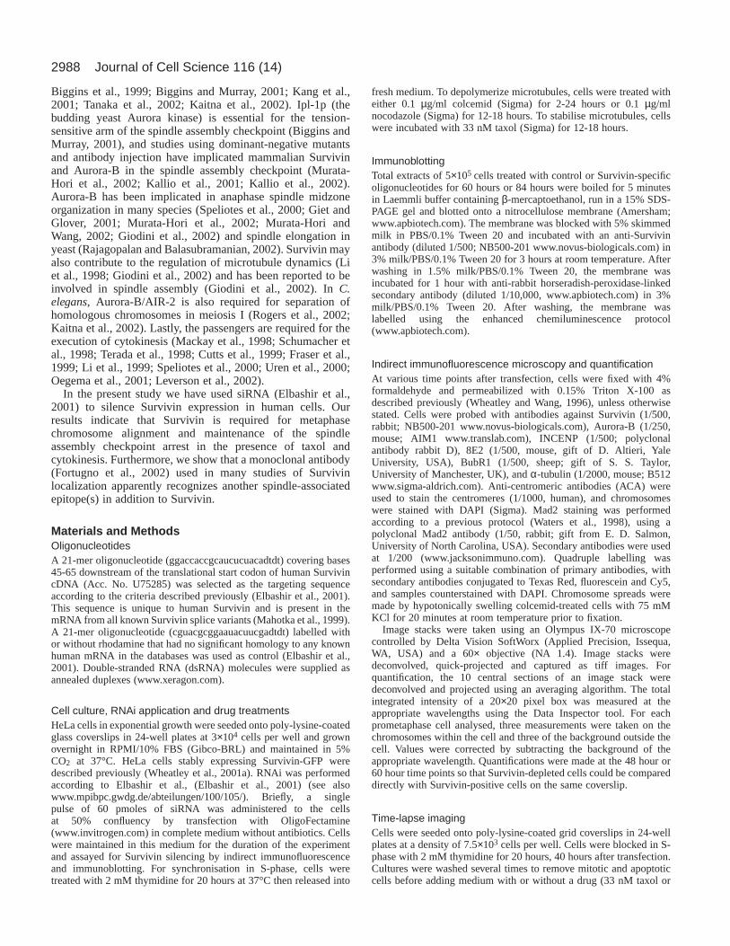
Indirect immunofluorescence microscopy and quantification At various time points after transfection, cells were fixed with 4%formaldehyde and permeabilized with 0.15% Triton X-100 asdescribed previously (Wheatley and Wang, 1996), unless otherwisestated. Cells were probed with antibodies against Survivin (1/500,rabbit; NB500-201 www.novus-biologicals.com), Aurora-B (1/250,mouse; AIM1 www.translab.com), INCENP (1/500; polyclonalantibody rabbit D), 8E2 (1/500, mouse, gift of D. Altieri, YaleUniversity, USA), BubR1 (1/500, sheep; gift of S. S. Taylor,University of Manchester, UK), and α-tubulin (1/2000, mouse; B512www.sigma-aldrich.com). Anti-centromeric antibodies (ACA) wereused to stain the centromeres (1/1000, human), and chromosomeswere stained with DAPI (Sigma). Mad2 staining was performedaccording to a previous protocol (Waters et al., 1998), using apolyclonal Mad2 antibody (1/50, rabbit; gift from E. D. Salmon,University of North Carolina, USA). Secondary antibodies were usedat 1/200 (www.jacksonimmuno.com). Quadruple labelling wasperformed using a suitable combination of primary antibodies, withsecondary antibodies conjugated to Texas Red, fluorescein and Cy5,and samples counterstained with DAPI. Chromosome spreads weremade by hypotonically swelling colcemid-treated cells with 75 mMKCl for 20 minutes at room temperature prior to fixation.
Image stacks were taken using an Olympus IX-70 microscopecontrolled by Delta Vision SoftWorx (Applied Precision, Issequa,WA, USA) and a 60× objective (NA 1.4). Image stacks weredeconvolved, quick-projected and captured as tiff images. Forquantification, the 10 central sections of an image stack weredeconvolved and projected using an averaging algorithm. The totalintegrated intensity of a 20×20 pixel box was measured at theappropriate wavelengths using the Data Inspector tool. For eachprometaphase cell analysed, three measurements were taken on thechromosomes within the cell and three of the background outside thecell. Values were corrected by subtracting the background of theappropriate wavelength. Quantifications were made at the 48 hour or60 hour time points so that Survivin-depleted cells could be compareddirectly with Survivin-positive cells on the same coverslip.
Time-lapse imagingCells were seeded onto poly-lysine-coated grid coverslips in 24-wellplates at a density of 7.5×10³ cells per well. Cells were blocked in S-phase with 2 mM thymidine for 20 hours, 40 hours after transfection.Cultures were washed several times to remove mitotic and apoptoticcells before adding medium with or without a drug (33 nM taxol or
Journal of Cell Science 116 (14)

2989Survivin RNAi in HeLa cells
0.1 µg/ml nocodazole) for 12-18 hours. When cells entered mitosis(~8-10 hours after release from the thymidine block), medium wassupplemented with 10 mM HEPES (pH 7.5), and phase-contrastimages were taken every hour using an inverted Nikon Diaphotmicroscope heated at 37°C and a 20× objective. At least 50 mitoticcells were followed for 6 hours in each experiment.
Cell population dynamics and cell cycle/apoptosisquantificationGrowth curves were plotted based on the number of viable cells, asdetermined by Trypan blue exclusion (Sigma). Mitotic stages werescored separately for cells staining negatively [Survivin(–)] orpositively [Survivin(+)] for Survivin. Both mitotic and apoptoticcells were scored based on DAPI morphology and α-tubulinimmunostaining. As the precise timing of depletion varied from oneexperiment to another (typically 36-72 hours), representative graphsare shown to avoid blending of the data. All experiments wereperformed at least three times and produced similar results.
FACS analysisThe apoptotic status of cells was assessed using TUNEL label andAnnexin V-FLUOS staining kits according to the manufacturer’sguidelines (www.biochem.roche.com).
Before TUNEL labeling, the total population (including non-adherent cells) was washed in PBS, fixed in 4% formaldehyde for 30minutes at room temperature and permeabilized in 0.1% sodiumcitrate/Triton X-100 for 2 minutes on ice.
To assess ploidy, cells were washed in PBS and fixed for 1 hour in70% ethanol at 4°C. After treatment with 50 µg/ml RNAse (Sigma)for 20 minutes at room temperature, cells were washed with PBS andincubated with 40 µg/ml propidium iodide (Sigma) for 30 minutes.
All samples were analysed using a fluorescence activated cell sorter(FACSCalibur, Becton Dickinson, Mountain View, CA) and CellQuest software.
ResultsSuccessful repression of Survivin in HeLa cells usingsiRNATo investigate the role of Survivin in mitosis, we have reducedthe levels of this protein in exponentially growing adherentHeLa cells using RNA interference (RNAi). The targetsequence was directed against nucleotides 45-65 of Survivinthat code for a region common to the three known isoforms ofthe protein: Survivin, Survivin 2B and Survivin Delta Ex3(Mahotka et al., 1999). This sequence was not detected in anyother human gene by BLAST analysis. After transfection withSurvivin-specific or control siRNA, cultures were harvested at12 hours intervals from 24 hours, and the cell number wasassessed (Fig. 1A). The growth rate of cells exposed toSurvivin siRNA began to slow at 48 hours, and proliferationceased after 60 hours. Similar data were obtained whencolonies of cells were microinjected with the sameoligonucleotides (data not shown). Immunoblot analysis usinga polyclonal antibody previously shown to recognise all knownforms of Survivin (Fortugno et al., 2002) revealed that Survivinwas substantially repressed by 60 hours and 84 hours posttransfection (Fig. 1B). We will refer to cells in which Survivinwas not detected by indirect immunofluorescence as ‘Survivin-depleted’, but note that low levels of the protein may remainin such cells.
Following transfection with rhodamine-labelled control
siRNA Survivin was detected at the centromeres ofprometaphase/metaphase cells (Fig. 1C) and showed a typicalchromosome passenger pattern of localization at other stagesof mitosis (data not shown). In 50-75% (n=100) ofprometaphase cells exposed to specific siRNA, Survivin wasnot detectable at centromeres by 48-60 hours post transfection(Fig. 1E). By contrast, untransfected cells from this populationexhibited a normal Survivin distribution (Fig. 1D). We alsotransfected cells that stably express Survivin-GFP with controlor Survivin siRNA. As shown in Fig. 2A, in cells transfectedwith control siRNA, Survivin-GFP was localized at thecentromeres and was also diffuse in the cytoplasm as describedpreviously (Wheatley et al., 2001a). By contrast, in cells
Fig. 1.Survivin can be depleted from HeLa cells using RNAi.(A) Growth curve of viable HeLa cells transfected with controlsiRNA (blue triangles) or Survivin siRNA (red circles). Cell numberin Survivin RNAi population does not increase after 48 hours.(B) Immunoblot analysis of control (C) and Survivin-siRNA-transfected (S) cultures at 0 hours, 60 hours or 84 hours posttransfection as indicated. Tubulin was monitored as a loadingcontrol. (C-E) Indirect immunofluorescence images showingSurvivin in red, DNA in blue and tubulin in green. (C) Control celltransfected with rhodamine-labelled control siRNA (shown in green).Survivin (red) localises normally to the centromeres. (D) Survivin-positive cell from same culture as E, showing abundant Survivin atthe centromeres. (E) Survivin-depleted cell from a Survivin siRNA-transfected population, fixed and stained at 60 hours. No Survivin(red) is detectable at the centromeres. Bars, 5 µm.

2990
exposed to Survivin siRNA, no GFP signal remained (Fig. 2B).This result was confirmed by immunoblot analysis using anti-GFP antibody (data not shown). We conclude that Survivin isessential for the growth of HeLa cells and that the protein canbe depleted from cells by RNAi.
Staining of cells with a monoclonal antibody that detectsmicrotubule associated Survivin is unaffected bySurvivin-specific RNAiAn ongoing vigorous debate in the field is whether humanSurvivin is a microtubule associated protein, as originallydescribed (Li et al., 1998; Li et al., 1999), a chromosomepassenger protein, as subsequently claimed (Skoufias et al.,2000; Wheatley et al., 2001a; Uren et al., 2000) or whether twoSurvivin pools of different magnitude exist within the cell
(Fortugno et al., 2002). An important reagent used in thisdebate has been the monoclonal antibody 8E2, which helpedto define Survivin originally (Li et al., 1998; Li et al., 1999)and which recognizes a full-length Survivin fusion protein invitro (Wheatley et al., 2001a).
A surprising result was obtained when we probed SurvivinsiRNA-treated cells with the 8E2 monoclonal antibody or witha commercial polyclonal antibody (NB500-201; www.novus-biologicals.com) reported to recognise all forms of Survivin(Fortugno et al., 2002). In cells transfected with control siRNA,the polyclonal antibody stained centromeric Survivin (Fig. 2Cand ACA staining in Fig. 2C’, red), whereas 8E2 demarcatedthe mitotic spindle (Fig. 2C,C’, green). However, in cellsnegative for Survivin, as detected with the commercialpolyclonal antibody (Fig. 2D, red), the pattern of 8E2localization remained unaltered (Fig. 2D,D’, green). Since wehave shown that 8E2 can recognise Survivin in vitro (Wheatleyet al., 2001a), this result strongly suggests that in vivo,8E2 recognizes an epitope present on at least one othermicrotubule-associated protein in addition to survivin.
Survivin depletion causes mislocalisation of thechromosomal passenger proteins, Aurora-B andINCENPIt is now widely believed that INCENP and Aurora-B may acttogether with Survivin throughout mitosis in a chromosomalpassenger complex (Wheatley et al., 2001a; Bolton et al., 2002;Leverson et al., 2002). As predicted by this, at 60 hours aftertransfection with specific siRNA, cells without detectableSurvivin also lacked centromeric Aurora-B (Fig. 3B). A diffusepool of Aurora-B and occasionally some residual staining ofthe chromosome arms was observed (compare Fig. 3A’’ andFig. 3B’’). The availability of a mixed population of Survivin-positive and Survivin-depleted cells following transfectionenabled us to use Survivin-positive cells as internal stainingcontrols for quantitative analysis, which revealed a linearcorrelation between levels of Survivin and Aurora-B at thecentromeres (Fig. 3E). A similar correlation was obtainedbetween the levels of Aurora-B and INCENP at centromeresfollowing transfection with Survivin siRNA (Fig. 3C,D,F).These data demonstrate that both Aurora-B and INCENPrequire Survivin to accumulate at centromeres during the earlystages of mitosis.
Survivin depletion causes an accumulation ofprometaphase cells with defects in chromosomecongression To determine the effect of depletion of Survivin on the passageof cells through mitosis, populations transfected with SurvivinsiRNA were stained for Survivin, and the percentage of cellsin each phase of mitosis was scored. Survivin-positive cells(n=100 mitotics) maintained a wild-type distribution of mitoticstages for the duration of the experiment (Fig. 4A). In contrast,we observed a decline in the percentage of Survivin-depletedmitotic cells in metaphase, anaphase, telophase and cytokinesisfrom 36 hours post-transfection (Fig. 4B; n=100 mitotics).Instead, we observed many prometaphase cells with bipolarspindles but many maloriented chromosomes (Fig. 4D,arrows). Although cells with multipolar spindles were
Journal of Cell Science 116 (14)
Fig. 2.Stably expressed Survivin-GFP, but not the epitope(s)recognized by the monoclonal antibody 8E2, can also be repressedby Survivin RNAi. (A,B) HeLa cells stably expressing Survivin-GFP(green) were subjected to Survivin RNAi and immunostained fortubulin (red) and counterstained with DAPI (DNA; blue). (A)Control cell with Survivin-GFP concentrated at the centromeres(arrows). (B) The Survivin-siRNA-transfected cell is depleted ofSurvivin-GFP signal. (C,C’-D,D’) Control or Survivin-siRNA-transfected cells stained for Survivin using the polyclonal antibody(C,D in red) and centromeres (C’,D’ in red); the epitope(s)recognized by the monoclonal antibody 8E2 (green) and DNA (blue).Survivin-specific siRNA does not abolish spindle staining by 8E2.Bars, 5 µm.

2991Survivin RNAi in HeLa cells
occasionally seen, they were also present within the Survivin-positive population (6% in Survivin-positive versus 4% inSurvivin-depleted cells).
Live-cell imaging by time-lapse phase contrast microscopyconfirmed that cells were delayed in mitosis followingtransfection with Survivin siRNA. Cells transfected withcontrol or Survivin siRNA, were blocked in S-phase using 2mM thymidine for 20 hours, then released into fresh mediumand followed individually by time-lapse imaging (Fig. 4F). Inthe population transfected with control siRNA, 92% of mitoticcells (n=73) completed mitosis within 1 hour (Fig. 4G). Incontrast, in the population transfected with specific siRNA,53% of cells (n=93) remained in mitosis for longer than 1 hour
(for an average of 1.7 hours in mitosis). Thus, repression ofSurvivin results in a delay in mitosis, and this correlates withdifficulties in chromosome alignment at the metaphase plate.
Survivin depletion inhibits cytokinesis and causes aslight increase in apoptosis Previous studies in which Survivin function was affected bythe expression of dominant-negative mutants or treatment withantisense oligonucleotides have shown an increase in apoptosisand multinucleation (Li et al., 1999), albeit only a moderateincrease in some cases (Kallio et al., 2001). In the presentstudy, time-lapse imaging revealed that cells exposed toSurvivin siRNA, after being delayed in mitosis for longer thancontrol cells (see above), either flattened in interphase afterfailing cytokinesis or blebbed and took on an abnormalappearance. In contrast, all cells in the control populationexited mitosis normally (Fig. 4F,G). We occasionally observeda very low number of Survivin-depleted cells in anaphase andtelophase (Fig. 4B) but failed to detect any Survivin-depletedcells in cytokinesis (data not shown). Consistent with theseobservations, cultures treated with Survivin siRNA showed agradual increase in multinucleation, culminating in a four-foldincrease by 84 hours (Fig. 4E). In addition, when DNA contentwas measured by FACS analysis of propidium-iodide-stainedcells at 84 hours, 43% of the Survivin siRNA-transfectedpopulation had a DNA content of 4N or greater compared with22% in the control population (data not shown). Together, theseobservations reveal that Survivin-depleted cells eventually exitmitosis but generally fail to complete cytokinesis.
To assess the apoptotic status of cells transfected withSurvivin siRNA, we performed FACS analysis using TUNELand Annexin V labelling and confirmed the results byexamining the DNA morphology microscopically (data notshown). A similar trend was observed using both methods: amoderate increase in apoptosis was detected at 72 hours aftertransfection with Survivin siRNA (16% higher than controlsusing the TUNEL assay to detect DNA fragmentation; 7%higher than controls using the Annexin V assay to detect lossof membrane asymmetry).
Together, these observations confirm that Survivin isrequired for the efficient completion of cytokinesis, but itsrequirement for the prevention of apoptosis is minor.
Fig. 3.Survivin depletion causes loss of Aurora-B and INCENP fromthe centromeres. (A-D) Survivin-siRNA-transfected cells were fixedat 60 hours and stained for Survivin, Aurora-B, INCENP andcentromeres (using ACA). (A,B) Survivin versus Aurora-B.(A) Survivin-positive cell shows Survivin (A’) and Aurora-B (A’’) atthe centromeres. (B) The Survivin-depleted cell (B’) shows Aurora-Bdiffusely localised with some residual staining on the chromosomearms (B”). Merged images do not show Survivin staining.(C,D) INCENP versus Aurora-B. (C) Survivin-positive cell, as judgedby the presence of Aurora-B staining at the centromeres (green, C’)shows centromeric INCENP staining (C’’). (D) The Survivin-depletedcell shows no Aurora-B (green, D’) or INCENP (D”) at thecentromeres. (E) A plot of the intensity of Survivin versus Aurora-Bat centromeres revealed a linear correlation between the abundance ofthe two proteins. (F) A plot of the intensity of Aurora-B (to indicateSurvivin depletion) versus INCENP at centromeres revealed a linearcorrelation between the abundance of the two proteins. (E,F) Unitsindicate fluorescence intensity (×103). Bars, 5 µm.

2992
Association of the spindle checkpoint protein BubR1with kinetochores is unstable in Survivin-depleted cellsThe increase in multinucleation at later time points followingtransfection with Survivin siRNA (Fig. 4E) suggested that cellswere eventually overriding the spindle checkpoint and exiting
mitosis despite the presence of maloriented chromosomes. Wetherefore examined the status of the spindle checkpoint in cellsdepleted of Survivin using antibodies against BubR1 and (insome experiments) Mad2. Normally, both proteins are presenton kinetochores as cells enter prometaphase and are part of the
Journal of Cell Science 116 (14)
Fig. 4.Survivin RNAi causesaccumulation in prometaphase,failure in cytokinesis andmultinucleation. (A,B) Timecourse of mitotic staging ofcells transfected with SurvivinsiRNA. Survivin-positive cellswere scored in A and Survivin-depleted cells were scored in B.From 36 hours posttransfection, the percentage ofSurvivin-depleted cells inprometaphase increases and thepercentage of cells in latermitotic stages decreases.(C,D) Examples of Survivin-positive and Survivin-depletedcells in mitosis. Centromeres(red); tubulin (green); DNA(blue). Inset panels show theSurvivin staining in the regionindicated by the box.(C) Survivin-positive cell withall chromosomes aligned.(D) Survivin-depleted cellshave misaligned chromosomes(arrows). (C’,D’) Chromosomesin C and D in greyscale. Bars, 5µm. (E) Representative graphshowing the percentage ofmultinucleated cells in culturesfollowing transfection withcontrol (red) versus Survivin(blue) siRNA. Multinucleationincreases gradually in culturestransfected with SurvivinsiRNA. (F) Phase-contrastimages of cells from culturestransfected with control orSurvivin siRNAs, blocked in S-phase, released in fresh mediumand followed by time-lapsesince entering mitosis (whichbegins approximately 10 hourspost-release from S-phase).Numbers in images indicatetime (in hours) since enteringmitosis. The two SurvivinsiRNA transfected cells shownspent >1 hour in mitosis andfailed cytokinesis. (G)Histogram showing the percentages of cells from cultures transfected with control (n=73) or Survivin siRNAs (n=101) that spent up to 1 houror more than 1 hour in mitosis before completing (yellow) or failing (green) cytokinesis.

2993Survivin RNAi in HeLa cells
constitutive cell cycle checkpoint that delays anaphase onsetuntil all chromosomes are properly aligned on the spindle(Musacchio and Hardwick, 2002). Mad2 is lost fromkinetochores as microtubules bind (Waters et al., 1998), andkinetochore-associated BubR1 levels fall significantly as thespindle develops tension on the kinetochore (Taylor et al.,2001; Skoufias et al., 2001).
When cells transfected with control siRNA were stained forBubR1, the kinetochores of maloriented chromosomes stainedbrightly, whereas the kinetochores of chromosomes aligned atthe metaphase plate stained only very faintly (Fig. 5A). Incontrast, in Survivin-depleted cells the BubR1 staining atkinetochores of lagging chromosomes (quantified as describedin Materials and Methods) varied widely within individual
cells (n=10), from negative to control levels (Fig. 5B). Thepresence of kinetochores with intense BubR1 staining suggeststhat the checkpoint pathway that monitors spindle tension canbe active in Survivin-depleted cells. Therefore, Survivin is notessential for localization of BubR1 to kinetochores. However,the simultaneous presence of BubR1-negative and BubR1-positive kinetochores within the same cell implies that either(1) certain kinetochores may be defective and unable to bindBubR1 or (2) BubR1 association with kinetochores may beunstable in the absence of Survivin – either it binds once, butthen fails to re-bind if spindle tension is lost, or it normallycycles on and off the kinetochore (Howell et al., 2001), and inthe absence of Survivin tends to spend much more time in theoff state.
Fig. 5. Survivin depletion decreases BubR1 abundance at kinetochores of maloriented chromosomes. (A,B) Immunofluorescence images ofcontrol (A) and Survivin-depleted cells 60 hours post-transfection (B). Cells are stained for BubR1 (red), Survivin (green) and DNA (blue).Panels A’-B’ show the BubR1 status of the inset regions indicated in a thermal scale where blue indicates a low signal intensity, greenintermediate intensity and yellow-red high. Panels A’’-B’’ show enlargements of the lagging chromosomes shown in the insets stained withDAPI and ACA. Unattached chromosomes of control cells have a high BubR1 signal (A’), while both high (B’, upper chromosome) and lowsignal intensities (B’’, lower chromosome) are found on unattached chromosomes of a Survivin-depleted cell. (C) Chromosome spreads ofHeLa cells following transfection with control or Survivin siRNA. Cells were treated for 2 hours with 0.1 µg/ml colcemid to depolymerizemicrotubules. In all panels Survivin is shown in green and DNA in blue. Top panels show BubR1 in red and lower panels show centromeres inred. All kinetochores are potentially capable of expressing high levels of BubR1 following Survivin depression. (D) Control and Survivin-depleted cells were treated with 0.1 µg/ml colcemid for 12 hours from 60 hours post transfection. A Survivin-positive and a Survivin-depletedcell are shown. BubR1 (red, D’’), Survivin (greyscale, D’), centromeres (green) and DNA (blue). Survivin-depleted cell shows no BubR1staining at the kinetochores. Bars, 5 µm.

2994
To distinguish between these two possibilities, we treatedcultures transfected with Survivin-siRNA with colcemid todepolymerize spindle microtubules. As shown in Fig. 5C, after2 hours exposure to colcemid, both Survivin-positive andSurvivin-depleted cells had BubR1 staining at all kinetochores.Therefore, kinetochores of chromosomes lacking detectableSurvivin are capable of binding BubR1. This eliminateshypothesis one.
To test the stability of BubR1 localization to thekinetochores, cultures transfected with Survivin siRNA for 60hours were exposed to colcemid for 3 hours, 6 hours or 12hours, then fixed and stained for Survivin and BubR1. After 3hours of colcemid treatment, BubR1 levels at kinetochoreswere similar for both Survivin-positive and Survivin-depletedcells (data not shown). However, by 12 hours in colcemid,BubR1 staining was absent from the majority of kinetochoresin Survivin-depleted cells, although it remained readilydetectable at kinetochores of Survivin-positive cells (Fig. 5D).Quantification of the fluorescence data revealed a linearcorrelation between the levels of Survivin at centromeres andBubR1 at kinetochores following 6 or 12 hours in colcemid(data not shown), indicating that Survivin is apparentlyrequired for the stable maintenance of BubR1 at kinetochoresunder conditions of protracted checkpoint activation.
Survivin is required for proper functioning of the spindlecheckpoint in the presence of taxolThe spindle assembly checkpoint has been proposed to bebipartite, with one arm monitoring kinetochore occupancy withmicrotubules (Waters et al., 1998) and the other monitoringspindle tension exerted on the kinetochores (Taylor et al., 2001;Skoufias et al., 2001). The abnormalities in BubR1 stainingobserved in Survivin-depleted cells led us to test whether thesecells were defective for one or both arms of this checkpointpathway.
Control or Survivin siRNA-transfected cells were blocked inS-phase and released into fresh medium supplemented with 33nM taxol for 18 hours. Taxol dampens microtubule dynamicsand effectively abolishes tension within the spindle but doesnot necessarily prevent kinetochore-microtubule interactions(Waters et al., 1998). Cells entered mitosis ~10 hours afterrelease from the S-phase block and were followed by time-lapse phase-contrast imaging. In the population transfectedwith control siRNA, 99% of the cells that entered mitosis(n=101) arrested there until the end of the experiment (Fig.6A,C). However, in the population exposed to Survivin siRNA,44% of the mitotics (n=103) exited mitosis as judged by cellre-spreading and formation of multiple micronuclei (Fig.6A,C). That these cells had exited mitosis was subsequentlyconfirmed by staining with DAPI (data not shown). Bycontrast, when cells were released from the S-phase blockinto medium supplemented with 0.1 µg/ml nocodazole, amicrotubule depolymerizing agent, both control and Survivin-siRNA-transfected populations arrested efficiently in mitosis(Fig. 6B,C). These results show that repression of Survivincompromises checkpoint function following exposure to taxolbut not nocodazole.
To further characterise the checkpoint response afterSurvivin repression, live cells were followed in the presenceof nocodazole or taxol and then subsequently fixed and
immunostained for BubR1 and Mad2. Mitotic cells in culturestransfected with control siRNA showed a normal response toboth drugs. Cells in taxol (kinetochores occupied withmicrotubules, but spindle tension defective) stained brightly forBubR1 at kinetochores, but only a few stained for Mad2 (mostof the kinetochores are attached to microtubules) (Fig. 6D).Cells in nocodazole (kinetochores unoccupied and no tension)stained brightly for both BubR1 and Mad2 at kinetochores(Fig. 6F). In contrast, kinetochores of Survivin-depleted cellsin taxol either completely lacked or showed a very decreasedstaining for both BubR1 and Mad2 (Fig. 6E). Furthermore, inthe presence of nocodazole, Survivin-depleted cells alsoshowed no or very low staining for BubR1, but Mad2 remainedbright at the kinetochores (Fig. 6G).
In summary, these data, which are summarized in Table 1,indicate that Survivin is required for the function of the spindlecheckpoint in response to taxol treatment but not in responseto nocodazole.
DiscussionTransfection of HeLa cells with an siRNA designed toeliminate all known forms of human Survivin (Mahotka et al.,1999; Rodriguez et al., 2002) led to the loss of detectableSurvivin as assayed by immunoblotting and indirectimmunofluorescence microscopy using an antibody recentlyshown to recognise all known forms of the protein (Fortugnoet al., 2002). This correlated with a cessation of populationgrowth 48-60 hours post-transfection and was accompanied byan accumulation of prometaphase cells with bipolar spindlesand misaligned chromosomes.
Recently, microinjection of polyclonal antibodies thatrecognise all known forms of Survivin into HeLa cells wasreported to result in similar chromosome congression defects(Giodini et al., 2002). However, earlier studies had suggestedthat interference with Survivin function compromised theability of cells to form a normal bipolar spindle. In HeLa cells(the cell type used here), a significant increase in multipolarspindles was reported after exposure of cells to a dominant-negative Survivin construct (C84A) or antisenseoligonucleotides (Li et al., 1999) and, shown more recently,after microinjection of Survivin antibodies (Fortugno et al.,2002). Multipolar spindles were also observed after interferencewith Survivin function in other systems, for example, earlyembryonic cells derived from Survivin-knockout mice (Uren etal., 2000). It is worth noting that in the present study, althoughwe detected an approximately 10% increase in multinucleatedcells over control levels, no significant increase in thepercentage of multipolar mitosis was observed.
Journal of Cell Science 116 (14)
Table 1. Summary of immunofluorescence resultsSurvivin Mitotic
Drug functional* BubR1 Mad2 arrest?
Nocodazole + + + +Nocodazole – – + +Taxol + + – +Taxol – – – –
*Cells were stained for Aurora-B/AIM-1. As shown in Fig. 3, levels ofSurvivin correlate directly with levels of Aurora-B; therefore centromeresnegative for Aurora-B were judged to be negative for Survivin function.

2995Survivin RNAi in HeLa cells
Time-lapse imaging revealed that transfection with SurvivinsiRNA caused a mitotic delay and failure of cytokinesis. Thenet result was a significant increase in the number of polyploidand multinucleated cells in the population. Furthermore, wefound that levels of apoptosis rose only slightly, confirming
that Survivin functions predominantly in mitotic progressionas opposed to inhibition of cell death.
These observations confirm that Survivin is required forchromosome biorientation and completion of cytokinesis inHeLa cells. The reason for the defects in cytokinesis induced
Fig. 6.Survivin is required for checkpoint arrest in response to taxol. (A,B) Phase-contrast images of cells from cultures transfected with control orSurvivin siRNAs that were blocked in S-phase, released in medium supplemented with 33 nM taxol (A) or 0.1 µg/ml nocodazole (B) and followedevery hour after entering mitosis for 8 hours. Arrows, arrowheads or asterisks indicate individual cells of interest. (C) Histogram showing thepercentages of cells from cultures transfected with control (C) or Survivin siRNAs (S) that either arrested in mitosis (yellow) or exited into interphase(green) in the above experiment. (D-G) Immunofluorescence staining of cells treated as in Fig. 6A,B, stained for Aurora-B (green), DNA (blue),BubR1 (greyscale in D’,E’,F’,G’, red in D’’,E’’,F’’,G’’) and Mad2 (green in D’’,E’’,F’’,G’’). Survivin-depleted cells (as judged by Aurora-B staining)show no little or no staining for BubR1 or Mad2 at the kinetochores in the presence of taxol. In the presence of nocodazole, Mad2 staining is presentat the kinetochores of both control and Survivin-depleted cells, whereas BubR1 is either greatly depleted or absent. Bar, 5 µm.

2996
by repression of Survivin is not known, but one possibility isthat Survivin-depleted cells exit mitosis with malorientedchromosomes and that these are trapped in the intercellularbridge, where they block the completion of abscission.Consistent with this view, we have observed that Survivin-depleted cells have defects in the spindle assembly checkpoint(discussed further below).
Survivin is not a mitotic spindle associated protein priorto anaphaseSurvivin was originally described as a mitotic-spindle-associated protein (Li et al., 1998; Li et al., 1999). Howeverwe and others have subsequently found that Survivin behaveslike a chromosomal passenger protein in human (Skoufias etal., 2000; Wheatley et al., 2001a; Jiang et al., 2001) and murinecells (Uren et al., 2000). Recent papers raised the possibilitythat there are two populations of Survivin (Fortugno et al.,2002) or that different cell types had different localizations ofthe protein (Jiang et al., 2001). At the centre of this controversyis a monoclonal antibody, 8E2, which strongly stains the entirespindle in metaphase cells (Fig. 2C). In this study we foundthat the pattern of 8E2 staining remained unaltered whenSurvivin was depleted from HeLa cells (Fig. 2D). Althoughthis monoclonal antibody does recognise human Survivin invitro (Wheatley et al., 2001a), the persistence of spindlestaining after depletion of all known Survivin isoformsdemonstrates that 8E2 is likely to also recognise an additionalunknown microtubule-associated protein in mitotic HeLa cells.
Mutual dependence of Survivin, Aurora-B and INCENPfor localization at centromeresAurora-B and INCENP fail to concentrate at centromeres inprometaphase cells depleted of Survivin. Because a dominant-negative form of INCENP causes the mislocalization ofAurora-B and Survivin in HeLa cells (Adams et al., 2000;Wheatley et al., 2001a), it appears that these three proteinsare mutually dependent on each other for localization tocentromeres. In C. elegans, the homologues of Survivin [BIR1(Fraser et al., 1999)], Aurora-B (AIR-2) and INCENP (ICP1)also appear to interact with one another, and the localizationof AIR-2 is dependent upon ICP-1 (Kaitna et al., 2000) andBIR1 (Speliotes et al., 2000). In addition, corroboratingevidence regarding the relationship(s) between these threechromosome passengers is also emerging in flies (Adams et al.,2001c), frog eggs (Bolton et al., 2002) and yeast (Yoon andCarbon, 1999; Rajagopalan and Balasubramanian, 2002;Cheeseman et al., 2002). One possible explanation for thesedata is that a stable complex between Survivin, Aurora-B andINCENP is required to target this group of proteins to thecentromeres. However, the persistence of such a complexthroughout mitosis has yet to be demonstrated directly.
Survivin is required for the efficient localization of BubR1to kinetochores and a robust spindle checkpointresponse in the presence of taxolAs cells enter mitosis, kinetochores constitutively expresssignals that delay the activation of the APC/C (anaphase-promoting complex/cyclosome) and onset of anaphase (Pines
and Rieder, 2001). These signals are gradually extinguished assister kinetochores capture microtubules from opposite spindlepoles and tension develops throughout the spindle. Thissignalling pathway, termed the spindle assembly checkpoint,has been proposed to involve two separate components. TheBubR1 arm of the pathway has been suggested to monitorspindle tension (Taylor et al., 2001; Skoufias et al., 2001),whereas the Mad2 pathway monitors microtubule attachmentat the kinetochore (Waters et al., 1998). However, a recentstudy has questioned whether these represent two completelyindependent, parallel pathways or a single pathway thatdepends on both BubR1 and Mad2 (Shannon et al., 2002).
The present study has shown conclusively that Survivin-depleted cells exhibit important differences from control cells inthe function of their spindle assembly checkpoint when exposedto agents that perturb microtubule behaviour. We found that inSurvivin-depleted cells, some kinetochores of malorientedchromosomes expressed high levels of BubR1 protein, whereaskinetochores of other maloriented chromosomes did not.Importantly, kinetochores of Survivin-depleted chromosomesaccumulated BubR1 normally during a short colcemid (2 hours)treatment, indicating that Survivin is not required for BubR1binding to the kinetochore upon activation of the spindleassembly checkpoint. Instead, we found that Survivin is requiredfor the maintenance of BubR1 at kinetochores during persistentactivation of the checkpoint. This is consistent with the resultsof a recent study (Kallio et al., 2001) in which it was reportedthat the phospho-epitope, 3F3/2, which identifies kinetochoresthat are not under tension during prometaphase (Nicklas et al.,1995), was precociously lost from centromeres when cells weremicroinjected with Survivin-specific antibodies.
Normally, in the presence of taxol, kinetochores have elevatedlevels of BubR1 and are negative for Mad2. In the presence ofnocodazole both proteins are normally present at elevated levels.Although recent experiments have revealed that it is dangerousto draw conclusions about the activity of the spindle assemblycheckpoint based solely on the level of kinetochore staining withanti-Mad2 (Martin-Lluesma et al., 2002), in the case of SurvivinsiRNA treatment, the observed pattern of kinetochoreassociation of BubR1 and Mad2 is able to explain the responseof the cells to persistent checkpoint activation.
We have found that following Survivin RNAi, BubR1accumulation at kinetochores is unstable in the presence ofeither taxol or nocodazole. Therefore, in the presence of taxol,kinetochores lack elevated levels of both BubR1 and Mad2. Asa result, Survivin RNAi abrogates the ability of cells to sustaina prolonged mitotic arrest in the presence of taxol. In contrast,Survivin RNAi has no effect on the ability of kinetochores toretain high levels of Mad2 protein. Thus, in the presence ofnocodazole or colcemid, kinetochores are BubR1 negative butMad2 positive, and cells are able to sustain a prolonged mitoticarrest.
Kinetochores that express elevated levels of either BubR1 orMad2 alone appear to be able to generate a ‘wait anaphase’signal and activate the spindle assembly checkpoint. Theformer are seen normally in the presence of taxol, and the latterare described here for the first time. It thus appears that eitherBubR1 or Mad2 and their downstream effectors are sufficientto activate the checkpoint.
We note that the major conclusions described above, andparticularly the requirement of Survivin for a robust checkpoint
Journal of Cell Science 116 (14)

2997Survivin RNAi in HeLa cells
response to taxol but not nocodazole, are consistent with theresults of an independent study in which Survivin silencing wasachieved using a plasmid vector in several different cell lines(Lens et al., 2003).
Importantly, our data should not be interpreted as indicatingthat Survivin is directly required for the detection of spindletension. Rather, they indicate that Survivin is required for stablemaintenance of elevated levels of BubR1 at kinetochores. Thepreferential sensitivity of the taxol-induced checkpoint to lossof Survivin reflects more the behaviour of Mad2 than anyspecific alteration in the behaviour of BubR1. The result mayappear to be selective for one arm of the checkpoint pathway,but the actual effect on BubR1 is not selective. Thus, althoughthe data presented here appear to support the emerging view ofa dual checkpoint pathway, where one arm detects kinetochoreoccupancy, and the other detects spindle tension (Taylor et al.,2001; Skoufias et al., 2001), whether or not there is a dual inputto the spindle assembly checkpoint pathway remains animportant question for future investigations.
Concluding remarksAs Survivin presumably functions as a targeting or stimulatoryfactor for the Aurora-B protein kinase, these results in effectconfirm the remarkable conservation of the checkpointpathway. Studies in budding yeast have shown that the Ipl-1kinase (yeast homologue of Aurora-B) is required for thespindle checkpoint in response to a loss of spindle tensionbut not in response to the loss of microtubule binding tokinetochores (Biggins and Murray, 2001). Thus, not only thecore checkpoint machinery but also the modulation of thismachinery by the Aurora-B kinase is conserved from yeast toman. Aurora-B regulates kinetochore-microtubule interactionsin budding yeast (Tanaka et al., 2002), and it phosphorylatescomponents of the Dam1 complex (Cheeseman et al., 2002),whose human homologues have yet to be identified. It nowbecomes a matter of some urgency to identify the kinetochoreprotein(s) whose modification by Aurora-B is required forthe stable association of BubR1 with kinetochores underconditions of persistent checkpoint activation.
It is likely that one common thread underlying the multiplephenotypes seen in the present study is the action of theAurora-B kinase. The search for relevant substrates of thiskinase is ongoing and likely to yield many candidates.However, in addition to targeting and activating Aurora-B,which is becoming ever more recognised as an essentialfacilitator of mitotic events (Adams et al., 2001a), it is alsopossible that Survivin and INCENP have other roles in mitoticprogression. Thus, this remains an area of growing excitementand important discovery.
We thank Stephen Taylor for BubR1 antibody, Dario Altieri for themonoclonal antibody 8E2 and Ted Salmon for the Mad2 antibody. Wealso thank Reto Gassmann for comments on the manuscript. A.C. isfunded by Praxis XXI, BD21811/99, Portugal. Work in the W.C.E.lab is supported by The Wellcome Trust, of which W.C.E. is aPrincipal Research Fellow.
ReferencesAdams, R. R., Wheatley, S. P., Gouldsworthy, A. M., Kandels-Lewis, S. E.,
Carmena, M., Smythe, C., Gerloff, D. L. and Earnshaw, W. C.(2000).INCENP binds the Aurora-related kinase AIRK2 and is required to target it
to chromosomes, the central spindle and the cleavage furrow. Curr. Biol. 10,1075-1078.
Adams, R. R., Carmena, M. and Earnshaw, W. C.(2001a). Chromosomalpassengers and the (Aurora) ABCs of mitosis. Trends Cell Biol.11, 49-54.
Adams, R. R., Maiato, H., Earnshaw, W. C. and Carmena, M.(2001b).Essential roles of Drosophila inner centromere protein (INCENP) andAurora-B in histone H3 phosphorylation, metaphase chromosomealignment, kinetochore disjunction, and chromosome segregation. J. CellBiol. 153, 865-880.
Adams, R. R., Maiato, H., Earnshaw, W. C. and Carmena, M.(2001c).Essential roles of Drosophila inner centromere protein (INCENP) andAurora-B in histone H3 phosphorylation, metaphase chromosomealignment, kinetochore disjunction, and chromosome segregation. J. CellBiol. 153, 865-880.
Ainsztein, A. M., Kandels-Lewis, S. E., Mackay, A. M. and Earnshaw, W.C. (1998). INCENP centromere and spindle targeting: Identification ofessential conserved motifs and involvement of heterochromatin protein HP1.J. Cell Biol.143, 1763-1774.
Altieri, D. C. (2001). The molecular basis and potential role of survivin incancer diagnosis and therapy. Trends Mol. Med.7, 542-547.
Altieri, D. C., Marchisio, P. C. and Marchisio, C.(1999). Survivin apoptosis:an interloper between cell death and cell proliferation in cancer. Lab Invest.79, 1327-1333.
Ambrosini, G., Adida, C. and Altieri, D. C. (1997). A novel anti-apoptosisgene, survivin, expressed in cancer and lymphoma. Nat. Med.3, 917-921.
Andreassen, P. R., Palmer, D. K., Wener, M. H. and Margolis, R. L.(1991).Telophase disk: a new mammalian mitotic organelle that bisects telophasecells with a possible function in cytokinesis. J. Cell Sci.99, 523-534.
Biggins, S. and Murray, A. W. (2001). The budding yeast protein kinaseIpl1/Aurora allows the absence of tension to activate the spindle checkpoint.Genes Dev. 15, 3118-3129.
Biggins, S., Severin, F. F., Bhalla, N., Sassoon, I., Hyman, A. A. and Murray,A. W. (1999). The conserved protein kinase Ipl1 regulates microtubulebinding to kinetochores in budding yeast. Genes Dev.13, 532-544.
Bishop, J. D. and Schumacher, J. M.(2002). Phosphorylation of thecarboxyl terminus of inner Centromere Protein (INCENP) by the Aurora Bkinase stimulates Aurora B kinase activity. J. Biol. Chem.277, 27577-27580.
Bolton, M. A., Lan, W., Powers, S. E., McCleland, M. L., Kuang, J. andStukenberg, P. T.(2002). Aurora B kinase exists in a complex with survivinand INCENP and its kinase activity is stimulated by survivin binding andphosphorylation. Mol. Biol. Cell (in press).
Cheeseman, I. M., Anderson, S., Jwa, M., Green, E. M., Kang, J.-S., Yates,J. R., Chan, C. S. M., Drubin, D. G. and Barnes, G.(2002). Phospho-regulation of kinetochore-microtubule attachments by the Aurora kinaseIpl1p. Cell 111, 163-172.
Cutts, S. M., Fowler, K. J., Kile, B. T., Hii, L. L., O’Dowd, R. A., Hudson,D. F., Saffery, R., Kalitsis, P., Earle, E. and Choo, K. H.(1999). Defectivechromosome segregation, microtubule bundling and nuclear bridging ininner centromere protein gene (Incenp)-disrupted mice. Hum. Mol. Genet.8, 1145-1155.
Earnshaw, W. C. and Bernat, R. L. (1990). Chromosomal passengers:Towards an integrated view of mitosis. Chromosoma (Berl.). 100, 139-146.
Earnshaw, W. C. and Cooke, C. A.(1991). Analysis of the distribution ofthe INCENPs throughout mitosis reveals the existence of three distinctsubstages of metaphase and early events in cleavage furrow formation. J.Cell Sci.98, 443-461.
Elbashir, S. M., Harborth, J., Lendeckel, W., Yalcin, A., Weber, K. andTuschl, T. (2001). Duplexes of 21-nucleotide RNAs mediate RNAinterference in cultured mammalian cells. Nature411, 494-498.
Fortugno, P., Wall, N. R., Giodini, A., O’Connor, D. S., Plescia, J., Padgett,K. M., Tognin, S., Marchisio, P. C. and Altieri, D. C.(2002). Survivinexists in immunochemically distinct subcellular pools and is involved inspindle microtubule function. J. Cell Sci.115, 575-585.
Fraser, A. G., James, C., Evan, G. I. and Hengartner, M. O.(1999).Caenorhabditis elegansinhibitor of apoptosis protein (IAP) homologueBIR-1 plays a conserved role in cytokinesis. Curr. Biol. 9, 292-301.
Giet, R. and Glover, D. M. (2001). DrosophilaAurora B kinase is requiredfor histone H3 phosphorylation and condensin recruitment duringchromosome condensation and to organize the central spindle duringcytokinesis. J. Cell Biol.152, 669-681.
Giodini, A., Kallio, M. J., Wall, N. R., Gorbsky, G. J., Tognin, S.,Marchisio, P. C., Symons, M. and Altieri, D. C.(2002). Regulation ofmicrotubule stability and mitotic progression by survivin. Cancer Res.62,2462-2467.

2998
Howell, B. J., McEwen, B. F., Canman, J. C., Hoffman, D. B., Farrar, E.M., Rieder, C. L. and Salmon, E. D.(2001). Cytoplasmic dynein/dynactindrives kinetochore protein transport to the spindle poles and has a role inmitotic spindle checkpoint inactivation. J. Cell Biol.155, 1159-1172.
Hsu, J. Y., Sun, Z. W., Li, X., Reuben, M., Tatchell, K., Bishop, D. K.,Grushcow, J. M., Brame, C. J., Caldwell, J. A., Hunt, D. F. et al. (2000).Mitotic phosphorylation of histone H3 is governed by Ipl1/aurora kinase andGlc7/PP1 phosphatase in budding yeast and nematodes. Cell 102, 279-291.
Jiang, X., Wilford, C., Duensing, S., Munger, K., Jones, G. and Jones, D.(2001). Participation of Survivin in mitotic and apoptotic activities of normaland tumor-derived cells. J. Cell. Biochem.83, 342-354.
Kaitna, S., Mendoza, M., Jantsch-Plunger, V. and Glotzer, M.(2000).Incenp and an Aurora-like kinase form a complex that is essential forchromosome segregation and for efficient completion of cytokinesis. Curr.Biol. 10, 1172-1181.
Kaitna, S., Pasierbek, P., Jantsch, M., Loidl, J. and Glotzer, M.(2002). TheAurora B kinase AIR-2 regulates kinetochores during mitosis and is requiredfor separation of homologous chromosomes during meiosis. Curr. Biol. 12,798-812.
Kallio, M. J., Nieminen, M. and Eriksson, J. E.(2001). Human inhibitor ofapoptosis protein (IAP) survivin participates in regulation of chromosomesegregation and mitotic exit. Faseb J.15, 2721-2723.
Kallio, M. J., McCleland, M. L., Stukenberg, P. T. and Gorbsky, G. J.(2002). Inhibition of aurora B kinase blocks chromosome segregation,overrides the spindle checkpoint, and perturbs microtubule dynamics inmitosis. Curr. Biol. 12, 900-905.
Kang, J., Cheeseman, I. M., Kallstrom, G., Velmurugan, S., Barnes, G.and Chan, C. S.(2001). Functional cooperation of Dam1, Ipl1, and theinner centromere protein (INCENP)-related protein Sli15 duringchromosome segregation. J. Cell Biol.155, 763-774.
Lens, S. M. A., Wolthuis, R. M. F., Klompmaker, R., Kauw, J., Agami, R.,Brummelkamp, T., Kops, G. and Medema, R. H.(2003). Survivin isrequired for a sustained spindle checkpoint arrest in response to lack oftension.EMBO J.(in press).
Leverson, J. D., Huang, H. K., Forsburg, S. L. and Hunter, T.(2002). TheSchizosaccharomyces pombeAurora-related kinase Ark1 interacts with theinner centromere protein Pic1 and mediates chromosome segregation andcytokinesis. Mol. Biol. Cell 13, 1132-1143.
Li, F., Ambrosini, G., Chu, E. Y., Plescia, J., Tognin, S., Marchisio, P. C.and Altieri, D. C. (1998). Control of apoptosis and mitotic spindlecheckpoint by survivin. Nature396, 580-584.
Li, F., Ackermann, E. J., Bennett, C. F., Rothermel, A. L., Plescia, J.,Tognin, S., Villa, A., Marchisio, P. C. and Altieri, D. C. (1999).Pleiotropic cell-division defects and apoptosis induced by interference withsurvivin function. Nat. Cell Biol. 1, 461-466.
MacCallum, D. E., Losada, A., Kobayashi, R. and Hirano, T.(2002). ISWIremodeling complexes in Xenopusegg extracts: identification as majorchromosomal components that are regulated by INCENP-aurora B. Mol.Biol. Cell 13, 25-39.
Mackay, A. M., Ainsztein, A. M., Eckley, D. M. and Earnshaw, W. C.(1998). A dominant mutant of inner centromere protein (INCENP), achromosomal protein, disrupts prometaphase congression and cytokinesis.J. Cell Biol. 140, 991-1002.
Mahotka, C., Wenzel, M., Springer, E., Gabbert, H. E. and Gerharz, C.D. (1999). Survivin-deltaEx3 and survivin-2B: two novel splice variants ofthe apoptosis inhibitor survivin with different antiapoptotic properties.Cancer Res.59, 6097-6102.
Martin-Lluesma, S., Stucke, V. M. and Nigg, E. A.(2002). Role of Hec1 inspindle checkpoint signaling and kinetochore recruitment of Mad1/Mad2.Science297, 2267-2270.
Morishita, J., Matsusaka, T., Goshima, G., Nakamura, T., Tatebe, H. andYanagida, M. (2001). Bir1/Cut17 moving from chromosome to spindleupon the loss of cohesion is required for condensation, spindle elongationand repair. Genes Cells6, 743-763.
Murata-Hori, M. and Wang, Y. (2002). The Kinase activity of Aurora B isrequired for kinetochore-microtubule interactions during mitosis. Curr. Biol.12, 894-899.
Murata-Hori, M., Tatsuka, M. and Wang, Y. L. (2002). Probing thedynamics and functions of Aurora B kinase in living cells during mitosisand cytokinesis. Mol. Biol. Cell 13, 1099-1108.
Murnion, M. E., Adams, R. A., Callister, D. M., Allis, C. D., Earnshaw, W.C. and Swedlow, J. R.(2001). Chromatin-associated protein phosphatase1 regulates aurora-B and histone H3 phosphorylation. J. Biol. Chem.276,26656-26665.
Musacchio, A. and Hardwick, K. G.(2002). The spindle checkpoint: structuralinsights into dynamic signalling. Nat. Rev. Mol. Cell. Biol.3, 731-741.
Nicklas, R. B., Wards, S. C. and Gorbsky, G. J.(1995). Kinetochorechemistry is sensitive to tension and may link mitotic forces for a cell cyclecheckpoint. J. Cell Biol.130, 929-939.
Oegema, K., Desai, A., Rybina, S., Kirkham, M. and Hyman, A. A.(2001).Functional analysis of kinetochore assembly in Caenorhabditis elegans. J.Cell Biol. 153, 1209-1226.
Pines, J. and Rieder, C. L.(2001). Re-staging mitosis: a contemporary viewof mitotic progression. Nat. Cell Biol. 3, E3-E6.
Rajagopalan, S. and Balasubramanian, M. K.(2002). SchizosaccharomycespombeBir1p, a nuclear protein that localizes to kinetochores and the spindlemidzone, is essential for chromosome condensation and spindle elongationduring mitosis. Genetics160, 445-456.
Reed, J. C. and Bischoff, J. R.(2000). BIRinging chromosomes through celldivision – and survivin’ the experience. Cell 102, 545-548.
Rodriguez, J. A., Span, S. W., Ferreira, C. G., Kruyt, F. A. and Giaccone,G. (2002). CRM1-mediated nuclear export determines the cytoplasmiclocalization of the antiapoptotic protein Survivin. Exp. Cell Res.275, 44-53.
Rogers, E., Bishop, J. D., Waddle, J. A., Schumacher, J. M. and Lin, R.(2002). The aurora kinase AIR-2 functions in the release of chromosomecohesion in Caenorhabditis elegansmeiosis. J. Cell Biol.157, 219-229.
Schumacher, J. M., Golden, A. and Donovan, P. J.(1998). AIR-2: AnAurora/Ipl1-related protein kinase associated with chromosomes andmidbody microtubules is required for polar body extrusion and cytokinesisin Caenorhabditis elegansembryos. J. Cell Biol.143, 1635-1646.
Shannon, K. B., Canman, J. C. and Salmon, E. D.(2002). Mad2 and BubR1function in a single checkpoint pathway that responds to a loss of tension.Mol. Biol. Cell13, 3706-3719.
Silke, J. and Vaux, D. L. (2001). Two kinds of BIR-containing protein –inhibitors of apoptosis, or required for mitosis. J. Cell Sci.114, 1821-1827.
Skoufias, D. A., Mollinari, C., Lacroix, F. B. and Margolis, R. L.(2000).Human Survivin is a kinetochore-associated passenger protein. J. Cell Biol.151, 1575-1582.
Skoufias, D. A., Andreassen, P. R., Lacroix, F. B., Wilson, L. and Margolis,R. L. (2001). Mammalian mad2 and bub1/bubR1 recognize distinct spindle-attachment and kinetochore-tension checkpoints. Proc. Natl. Acad. Sci. USA98, 4492-4497.
Speliotes, E. K., Uren, A., Vaux, D. and Horvitz, H. R.(2000). The survivin-like C. elegansBIR-1 protein acts with the Aurora-like kinase AIR-2 toaffect chromosomes and the spindle midzone. Mol. Cell. 6, 211-223.
Tanaka, T. U., Rachidi, N., Janke, C., Pereira, G., Galova, M., Schiebel,E., Stark, M. J. and Nasmyth, K. (2002). Evidence that the Ipl1-Sli15(Aurora kinase-INCENP) complex promotes chromosome bi-orientation byaltering kinetochore-spindle pole connections. Cell 108, 317-329.
Taylor, S. S., Hussein, D., Wang, Y., Elderkin, S. and Morrow, C. J.(2001).Kinetochore localisation and phosphorylation of the mitotic checkpointcomponents Bub1 and BubR1 are differentially regulated by spindle eventsin human cells. J. Cell Sci.114, 4385-4395.
Terada, Y. (2001). Role of chromosomal passenger complex in chromosomesegregation and cytokinesis. Cell Struct. Funct.26, 653-657.
Terada, Y., Tatsuka, M., Suzuki, F., Yasuda, Y., Fujita, S. and Otsu, M.(1998). AIM-1: a mammalian midbody-associated protein required forcytokinesis. EMBO J.17, 667-676.
Uren, A. G., Wong, L., Pakusch, M., Fowler, K. J., Burrows, F. J., Vaux,D. L. and Choo, K. H. A.(2000). Survivin and the inner centromere proteinINCENP show similar cell-cycle localization and gene knockout phenotype.Curr. Biol. 10, 1319-1328.
Waters, J. C., Chen, R. H., Murray, A. W. and Salmon, E. D.(1998).Localization of Mad2 to kinetochores depends on microtubule attachment,not tension. J. Cell Biol.141, 1181-1191.
Wheatley, S. P. and Wang, Y.(1996). Midzone microtubule bundles arecontinuously required for cytokinesis in cultured epithelial cells. J. Cell Biol.135, 981-989.
Wheatley, S. P., Carvalho, A., Vagnarelli, P. and Earnshaw, W. C.(2001a).INCENP is required for proper targeting of survivin to the centromeres andthe anaphase spindle during mitosis. Curr. Biol. 11, 886-890.
Wheatley, S. P., Kandels-Lewis, S. E., Adams, R. R., Ainsztein, A. M. andEarnshaw, W. C. (2001b). INCENP binds directly to tubulin and requiresdynamic microtubules to target to the cleavage furrow. Exp. Cell Res. 262,122-127.
Yoon, H. J. and Carbon, J.(1999). Participation of Bir1p, a member of theinhibitor of apoptosis family, in yeast chromosome segregation events. Proc.Natl. Acad. Sci. USA96, 13208-13213.
Journal of Cell Science 116 (14)