![[sp1] oocyte collection from superstimulated disease-free ...](https://static.fdokumen.com/doc/165x107/631dd3361aedb9cd850f788f/sp1-oocyte-collection-from-superstimulated-disease-free-.jpg)
[sp1] oocyte collection from superstimulated disease-free ...

PRECLINICAL STUDIES
Tetra-O-methyl nordihydroguaiaretic acid, an inhibitorof Sp1-mediated survivin transcription, induces apoptosisand acts synergistically with chemo-radiotherapyin glioblastoma cells
Angel Mauricio Castro-Gamero & Kleiton Silva Borges & Daniel Antunes Moreno &
Veridiana Kill Suazo & Mayara Missono Fujinami & Rosane de Paula Gomes Queiroz &
Harley Francisco de Oliveira & Carlos Gilberto Carlotti Jr. & Carlos Alberto Scrideli &Luiz Gonzaga Tone
Received: 30 October 2012 /Accepted: 14 December 2012# Springer Science+Business Media New York 2013
Summary Glioblastoma (GBM), one of the most malig-nant human neoplasias, responds poorly to current treatmentmodalities, with temozolomide (TMZ) being the drug mostfrequently used for its treatment. Tetra-O-methylNordihydroguaiaretic Acid (M4N) is a global transcriptionalrepressor of genes dependent on the Sp1 transcription factor,such as Survivin and Cdk1. In the present study we evalu-ated the gene expression of Survivin, its spliced variants andCdk1 in GBM samples and cell lines. Moreover, we inves-tigated the effects of M4N combined or not with TMZ and/
or radiation on GBM primary cultures and cell lines. qRT-PCR assays were performed to determine the Survivin-spliced variants and Cdk1 gene mRNA expression inGBM tumor samples and cell lines. Cell proliferation wasmeasured by XTT assay and cell cycle and apoptosis weredetermined by flow cytometry. Drug combination analysesusing different schedules of administration (simultaneousand sequential) were performed on GBM cell lines andprimary cultures based on the Chou-Talalay method. Forclonogenic survival, doses of 2, 4, and 6 Gy of gammaradiation. were used. All Survivin-spliced variants and theCdk1 gene were expressed in GBM samples (n=16) and celllines (n=6), except the Survivin-2B variant that was onlyexpressed in GBM cell lines. M4N treatment down regulat-ed the expression of Cdk1, Survivin and the Survivin-ΔEx3variant, while the Survivin-2B variant was up-regulated.M4N decreased the cell proliferation separately and syner-gistically with TMZ, and enhanced the effects of radiation,mainly when associated with TMZ. M4N also inducedapoptotic cell death, decreased the mitotic index andarrested the cell cycle mainly in the G2/M phase. Our resultssuggest a potential clinical application of M4N in combina-tion with TMZ and radiation for GB treatment.
Keywords Cdk1 gene . Drug combination . Glioblastoma .
M4N . Temozolomide . Survivin gene
Introduction
Glioblastoma (GBM) is the most common primary malig-nant brain tumor [1]. Despite advancements in surgery,radiation (RT), and chemotherapy [2], the median survival is
Electronic supplementary material The online version of this article(doi:10.1007/s10637-012-9917-4) contains supplementary material,which is available to authorized users.
A. M. Castro-Gamero :K. S. Borges :D. A. MorenoDepartment of Genetics, Faculty of Medicine of Ribeirão Preto,University of São Paulo (USP), Ribeirão Preto, Brazil
V. K. Suazo :M. M. Fujinami : R. de Paula Gomes Queiroz :C. A. Scrideli : L. G. ToneDepartment of Pediatrics, Faculty of Medicine of Ribeirão Preto,University of São Paulo (USP), Ribeirão Preto, Brazil
H. F. de OliveiraDepartment of Internal Medicine, Faculty of Medicine of RibeirãoPreto, University of São Paulo (USP), Ribeirão Preto, Brazil
C. G. Carlotti Jr.Department of Anatomy and Surgery, Faculty of Medicine ofRibeirão Preto, University of São Paulo (USP),Ribeirão Preto, Brazil
A. M. Castro-Gamero (*)Laboratório de Pediatria, Faculdade de Medicina de RibeirãoPreto, Hospital das Clínicas, Universidade de São Paulo, Bloco G,Av. Bandeirantes 3900,14048-900, Ribeirão Preto, SP, Brazile-mail: [email protected]
Invest New DrugsDOI 10.1007/s10637-012-9917-4

12–15 months [3]. Currently, temozolomide (TMZ) is the maindrug used for GBM therapy, acting through alkylation at the 0–6 position of guanine. Both TMZ and RT therapy can induceDNA damage; however their efficacy is hindered in part byalterations in the apoptotic and cell cycle mechanisms of cancercells [4, 5], which are becoming promising targets for thedevelopment of novel and more efficient therapeutic agents.
Survivin protein has been considered an ideal target forcancer treatment due to its high and unique expression in mosthuman tumors and its critical role in the control of cell divisionand inhibition of apoptosis [6–10]. Its high expression inmalignant brain tumors, particularly GBM [11], has beenstrongly associated with a poor prognosis and resistance tochemo- and radiotherapy [12, 13], suggesting that survivinrepresent a promising target for GBM therapy. Moreover,several Survivin splice variants such as Survivin-ΔEx3 andSurvivin-2B have also been identified in GBM thus far andlittle is known about their function in these cells.
A critical necessity for survivin to perform its role efficientlyduring mitotic processes is phosphorylation by cyclin-dependent kinase 1 (Cdk1) [14]. Cdk1 is a master modulatorin the initiation and transition process through mitosis duringthe cell cycle [15]. The high expression and activity of Cdk1protein is often observed in several human cancers [16, 17] andis associatedwith tumor progression [18], includingGBM [19],suggesting Cdk1 as a promising target for GBM treatment.
Tetra-o-methyl nordihydroguaiaretic acid (also known asM4N, terameprocol, and EM1421) blocks cell cycle progres-sion by inhibiting the expression of the Cdk1 gene [20] andsimultaneously promotes apoptosis by inhibiting the expres-sion of the Survivin gene [21]. Recently, a Phase I clinical trialin patients with recurrent high-grade gliomas showed adequatepharmacokinetic tolerance and a promising anti-tumor activity.However, this previous study recommended the conduction ofpreclinical studies of combined therapy with TMZ and RTprior to initiating human studies of toxicity and efficacy [22].
The aim of the present study was to evaluate the geneexpression of Survivin variants and Cdk1 gene in GBMtumor samples and cell lines. Moreover, we demonstratedthe potent anti-GBM activity of M4N in both GBM celllines and primary cultures, as well as its synergistic effectswhen combined with TMZ and RT. Additionally, our studyshows that M4N deregulated the cell cycle, caused apopto-sis, and altered gene expression in GBM cell lines.
Material and methods
Patients
Twenty six fresh-frozen microdissected tumor samples wereobtained from macrodissected glioblastomas according tothe WHO classification [23], from subjects admitted for
diagnosis and treatment to the University Hospital of theSchool of Medicine of Ribeirão Preto, SP, Brazil. Six sam-ples of microdissected non-neoplastic brain tissue (whitematter) were obtained from patients who had undergonesurgery for the treatment of epilepsy. The average age ofthe patients was 44.3 years and three subjects were pediatricpatients. Ten subjects were men and 6 were women. Thestudy was approved by the Institutional Research EthicsCommittee (protocol number 8608/2009).
Cell culture and primary culture
Human adult GBM cell lines (U251, U343, T98G, U87,LN319) and a pediatric GBM cell line (SF188) were obtainedand grown as previously described [24]. The GBM primarycultures (GBPCs) were derived from biopsy specimens ofGBM patients under a protocol approved by the institutionalreview board of the University Hospital, School of Medicineof Ribeirão Preto, SP, Brazil. Cultures were obtained accord-ing to Brassesco et al. [25] and maintained at low passagenumbers (p2–p6) under standard culture conditions.
Reagents
M4Nwas purchased fromCayman Chemical Co. (AnnArbor,MI, USA, #70302). M4N was dissolved in dimethylsulfoxide(DMSO; Mallinckrodt Chemical Works, St Louis, MO, USA)at 50 mM. A stock solution was then warmed to 70 °C for 3 has recommended by Chang et al. [21], before the addition tothe cell culture medium at a final DMSO concentration of 1%.TMZ (Sigma-Aldrich, São Paulo, Brazil) was also diluted inDMSO at a stock concentration of 200 mM. Stock solutionswere kept at − 20 °C and dissolved in culture medium imme-diately before the experiments.
RNA isolation and reverse transcription-PCR
mRNA was extracted from cell lines and tumor samplesusing the Trizol® reagent (Gibco BRL, Life technologies®,Carlsbad, CA, USA). Complementary DNA (cDNA)was obtained with the High Capacity® kit (AppliedBiosystems®, Foster City, CA, USA) according to the man-ufacturers’ instructions. cDNA levels of the Survivin,Survivin-2B, Survivin-ΔEx3, and Cdk1 genes were mea-sured using an ABI 7500 Real Time PCR System (PEApplied Biosystems). Amplifications were obtained usingTaqMan® probes: Survivin (Hs00977612_mH), Survivin-2B(Hs03043575_m1), Survivin-deltaEx3 (Hs03043576_m1),Cdk1 (Hs00938777_m1), TBP (4326322E0811008), andHPRT (4310809E0502006) (Applied Biosystems). The rel-ative expression was calculated using the 2-ΔΔCT method[26] with two internal controls, TBP and HPRT, as recom-mended by Valente et al. [27]. To determine the effects of
Invest New Drugs

M4N on survivin gene expression, their spliced variants andCdk1 gene, the cell lines SF188, T98G, U251, and U343were treated with 40 μM of M4N for 6, 12, and 24 h. Inthese experiments, control cells (DMSO) collected at thesame treatment times were used as calibrators.
Growth inhibition assays
Growth inhibition was determined by two proliferationassays: XTT-assay and/or Giemsa-staining of attached cellsas previously published [24, 28]. SF188, T98G, U251, andU343 cells were seeded at 3×10³ cells/well in 96-wellplates. After attachment, cells were treated with serial dosesof M4N or TMZ for 24, 48, and 72 h. Similarly, elevenGBPCs were treated for 48 h with M4N, and four of themwere additionally treated with M4N or TMZ for 96 h. TheIC50 (the drug concentration that inhibits cell proliferationby 50 % compared with controls) was calculated usingCalcuSyn software (Biosoft, Ferguson, Missouri, USA).
Combination drug analysis
The interaction between M4N and TMZ was analyzed usingCalcusyn. For the analyses of combinations, increasingdoses of each drug were used according to their equipotentmolar ratio for each cell line through three different expo-sure schedules: simultaneous, sequential with TMZ afterM4N, and sequential with M4N after TMZ. For GBPCs,similar exposures were performed using the fixed doses of20 μM or 40 μM M4N, and 500 μM TMZ. Treatments of48 h or 96 h were used for simultaneous exposure of celllines and GBPCs, respectively. For sequential exposure, thetreatments with each drug were for 24 h and 48 h for the celllines and GBPCs, respectively. Combination indexes (CIs)were generated for each set of combinations and synergistic(CI<1), additive (CI=1), or antagonistic effects (CI>1)were calculated by the Chou and Talalay equation [29] usingCalcusyn software. The dose reduction index (DRI) repre-sents the measure of how much the dose of each drug in asynergistic combination may be reduced at a given effectlevel compared with the dose of each drug alone [29].
Clonogenic assay
The effects of M4N and TMZ on clonogenic capacity wereevaluated according to Franken’s protocol [30]. Single cellsuspensions of 300 cells/well were seeded in six-well platesand, once attached, were treated with serial concentrationsof M4N or TMZ for 48 h. After treatment, the culturemedium was exchanged with a drug-free medium for incu-bation for 7–10 days. Next, surviving colonies were fixedwith methanol and stained with Giemsa. The colonies withmore than 50 cells were scored. These data were used to
calculate the relative survival fraction and clonogenic IC50
values (concentration for the reduction of 50 % survivalfraction) calculated using the Calcusyn software. The IC50
values for each drug were used for combination experi-ments. Cells were treated with M4N, TMZ, or M4N+TMZfor 48 h and then irradiated, cultured, and further analyzedas described by Borges et al. [24]. In all cases, three inde-pendent experiments were performed.
Apoptosis assessment, cell cycle analysis and mitotic indexdetermination
Cells were treated with M4N for 48 h and apoptotic celldeath was determined as previously reported [28]. For cellcycle analysis, sub-confluent cultures were exposed to 40μM M4N (SF188 and T98G cells) or 20 μM M4N (U251and U343 cells) for 24 h. Cells were trypsinized, washedwith 1X PBS, and stained with propidium iodide (PI) usingthe CycleTESTTM PLUS DNA reagent kit (BectonDickinson Immunocytometry Systems, San Jose, CA). Thecell DNA content was immediately analyzed using aFACSCalibur apparatus (Becton Dickinson, Lincoln Park,NJ, USA). The percentages of cells in each cell cycle phasewere calculated using ModFit LT software (Verity SoftwareHouse, Topsham, ME, USA). For apoptosis and cell cycleassays at least 10,000 cells were measured per experiment.For mitotic index determination, exponentially growing cul-tures were treated with 0–80 μM M4N for 48 h and har-vested as previously described [28]. In all cases, threeindependent experiments were performed.
Statistical analysis
Statistical analysis for gene expression was performed usingthe Mann–Whitney non-parametric test. For the functionalstudies, one-way or two-way ANOVA was used, followedby the nonparametric Bonferroni test, as appropriate. Theeffect of M4N and its combinations on colony formationwas tested by logistic regression. Statistical significance wasconsidered when P<0.05. Data analysis was carried outusing the SPSS17.0 statistical software package (SPSS,Chicago, IL, USA). Results are reported as means±SD.
Results
Expression of survivin-spliced variants and CDK1 in GBMsamples and cell lines
We found that Survivin, Survivin-ΔEx3 and Cdk1 weresignificantly expressed in tumor samples, while survivin-2B expression did not differ significantly from normal sam-ples (Fig. 1a–d). We also found that all survivin splice
Invest New Drugs

variants were expressed in all cell lines, with the Survivinvariant being the one most expressed in cell lines, mainly inSF188 and U251 cells. The Cdk1 gene expression wasfound in all cell lines, mainly in SF188 and U343 cells(Fig. 1e).
M4N inhibited cell proliferation of GBM cell lines
The SF188, T98G, U251 and U343 cells were sensitive toM4N, showing inhibited growth in a dose- and time-dependent manner (p<0.05) except for SF188 cells that onlydisplayed a dose-dependent inhibition (p=0.009) (Fig. 2a–d). The 48 h-IC50 values corroborated the remarkable sen-sitivity of T98G, U251 and U343 cells, and a relative
resistance of SF188 cells (Table 1). To expand the relevanceof our findings, we assessed the response of GBPCs toM4N. A total of eleven GBPCs were included in the anal-ysis exhibiting a variety of IC50 values at 48 h (18.4 –282.2 μM). We found that most GBPCs showed a dose-dependent response to M4N (Fig. 2e, Table 2). The M4N-IC50 values were drastically diminished in four GBPCs afterM4N-treatment for 96 h (GB27, GB28, GB45, and GB49)(Table 2). Our results demonstrate that M4N was efficient ininhibiting the proliferation of GBM cells.
Additionally, the chemosensitivity of cell lines andGBPCs to TMZ was determined by drug combinationassays (Supplementary Data Fig. S1-S2). The IC50 valuesobtained showed high sensitivity to TMZ of U251 and U343
Fig. 1 Survivin, Survivin-2B,Survivin-ΔEx3 and Cdk1mRNA expression in thesamples of glioblastoma tumorand cell lines. Relativeexpression levels of Survivin(a), Survivin-2B (b), Survivin-ΔEx3 (c) and Cdk1 (d) areshown in glioblastoma samples[GBM samples] (n=16) andnon-neoplastic brain tissue[NT] (n=6). The relative ex-pression levels of the samegenes in six cell lines are shown(e). Expression levels are dis-played as a ratio between thetarget genes and two referencegenes (TBP and HPRT) to cor-rect for variation in the amountsof RNA. The line correspondsto the median value (a–d). Thep values are shown to indicatestatistical differences
Invest New Drugs

cells and a relative resistance of SF188 and T98G cells(Table 1). Taken together, these data showed molar ratiosproportional to IC50 values as 1:25 (M4N:TMZ) for SF188and T98G, and 1:9.375 (M4N:TMZ) for U251 and U343.
Fig. 2 M4N inhibited the cellproliferation of glioblastomacells. SF188 (a), T98G (b),U251 (c), and U343 (d)glioblastoma cell lines andeleven primary cultures ofglioblastoma tumors (e) werecultured in the presence ofserial concentrations of M4N(0–80 μM) for 24, 48, and 72 hfor cell lines and 48 h for theprimary cultures. Proliferationwas measured with the XTT kit.*P<0.05; **P<0.01;***P<0.001
Table 1 Doses required to induce 50 % inhibition of cell growth(IC50) in GBM cell lines
Cell line IC50 (μM) Ratio IC50
(M4N): IC50
(TMZ)
Ratio used incombinations
M4N 48 h TMZ 48 h
SF188 171.39±8.3 4464.2±92.4 1 : 26.05 1 : 25
T98G 87.5±2.2 2062.0±26.1 1 : 23.65 1 : 25
U343 81.41±4.6 664.56±11.8 1 : 8.16 1 : 9.375
U251 64.27±6.1 396.74±9.4 1 : 6.17 1 : 9.375
Table 2 IC50 values for M4N in GBM primary cultures
Tumor samples IC50 48 h M4N (μM) IC50 96 h M4N (μM)
GB12 250.390 —
GM15 175.870 —
GM16 85.714 —
GM17 182.140 —
GM20 282.200 —
GM22 169.000 —
GM27 68.714 25.130
GM28 n.pa 20.860
GM41 75.756 —
GM45 21.883 9.000
GM49 18.431 7.113
a It was not possible to calculate IC50
Invest New Drugs

M4N acts synergistically with TMZ in GBM cells
We determined the combined effects of M4N and TMZ oncell lines, as represented by the DRI, the CI and the dose-effect levels of cell growth inhibition (ED50–ED95) (sum-marized in Supplementary Data Table S1).
Figure 3 illustrates the schedule-dependent synergismbetween M4N and TMZ. Simultaneous exposure to bothdrugs (M4N+TMZ) for 48 h resulted in synergism inall cell lines and in all the ranges of affected fractions(CI<1, Fig. 3a–d), except for the T98G cell line thatshowed a synergistic effect only up to 65 % of the
Fig. 3 Sequence-dependent interactions between M4N and TMZ inSF188, T98G, U343 and U251 cells in vitro. Plots of the combinationindex (CI) from glioblastoma cell lines exposed to M4N and TMZusing different schedules of administration: simultaneous 48-h exposures (a–d); sequential M4N→TMZ exposures, 24-h each (e-h); and sequential TMZ→M4N exposures, 24-h each (i–l) at different
molar ratios of the two agents (M4N:TMZ). The line across the CIvalue of 1 indicates additivity; CIs above and below indicate antago-nism and synergism, respectively. Crosses represent the CIs of theactual data points and continuous lines represent computer-derivedCIs at effect levels ranging from 10 to 100 % inhibition of clonalgrowth according with Calcusyn software
Invest New Drugs

affected fraction (Fig. 3b). Similarly, the TMZ→M4Nsequence mainly showed synergistic effects in all affect-ed fractions in all cell lines (CI<1; Fig. 3i–l). Thesequential M4N→TMZ exposure produced antagonisticeffects in T98G and U251 cells in all the ranges of theaffected fraction (CI>1, Fig. 3f, h), while in SF188 itproduced antagonistic effects only in up to 35 % of theaffected fraction (Fig. 3e), and in U343 cells it pro-duced synergistic effect in up to 75 % of the affectedfraction (Fig. 3g).
We also performed drug combination assays on fourGBPCs using 20 μM (GB45 and GB49) or 40 μM(GB27 and GB28) M4N and 500 μM TMZ with thesame three different schedules described above (Fig. 4).As found in cell lines, we detected significant differ-ences between drug treatment with each drug separatelyand the combination of both in simultaneous exposureand sequential TMZ→M4N exposure (GB27 andGB49), whereas the inverse M4N→TMZ sequence didnot produce statistical differences in these cells (Fig. 4a,d). For GB28 cells, all the three drug combinationmodes resulted in greater cytotoxicity than obtainedwith TMZ and M4N alone (Fig. 4b). Conversely, inGB45 cells we did not detect statistical differencesbetween separate treatment with each drug and treat-ment with drug combinations (Fig. 4c).
Taken together, these data suggest that the combina-tion of M4N and TMZ caused a synergistic effect inGBM cells preferentially when simultaneous combina-tion or sequential TMZ→M4N exposure was used.
M4N enhanced the response to radiation in glioblastomacell lines, especially when combined with TMZ
After the determination of clonogenic IC50 values for eachdrug (Supplementary Data Fig. S3), combination assayswith both drugs (IC50) and radiation was performed. M4Nalone significantly enhanced the radiosensitivity of onlyU343 cells (p<0.001) (Supplementary Data Fig. S4). Onthe other hand, the combination of M4N with TMZ signif-icantly enhanced the effects of radiation in all cell lineswhen compared to each drug alone (Fig. 5). The strongesteffect was achieved with the highest irradiation dose (6 Gy)in all cell lines, as indicated by dose enhancement ratios of6.33, 1.59, 5.29, and 5.01 for SF188, T98G, U251, andU343 cell, respectively (Table 3). Thus, M4N treatment isable to sensitize GBM cell lines to radiation and may in-crease the radiosensitization caused by TMZ.
M4N caused apoptotic cell death, altered the cell cycle anddecreased the mitotic index in GBM cell lines
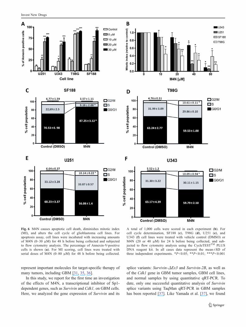
The number of apoptotic cells increased in M4N-treatedcells (annexin-V-positive cells) in a dose-dependent manner.The strongest effect was obtained with 30 μMM4N, mainlyin SF188 and T98G cells, with 92 % and 90 % of apoptoticcells, respectively (Fig. 6a). Moreover, M4N significantlydecreased the mitotic index in GBM cell lines (Fig. 6b).Although the strongest effect was obtained with 80 μMM4N in U343, U251 and T98G cells (90 %, 94 % and72 %, respectively), only a 62 % decrease was achieved in
Fig. 4 Effects of M4N andTMZ combination on primaryglioblastoma cultures. Theprimary cultures GB27 (a),GB28 (b), GB45 (c) and GB49(d) were treated with M4N (40 μM or 20 μM) and TMZ(500 μM) using three differentschedules of administration: si-multaneous 96-h exposures(M4N+TMZ); sequential expo-sures M4N→TMZ (M4N/TMZ), 48-h each, and sequen-tial TMZ→M4N exposures(TMZ/M4N), 48-h each. Sig-nificant differences betweencombined treatments and iso-lated treatments are shown.*P<0.05
Invest New Drugs

the SF188 cell line with this concentration (Fig. 6b).Additionally, as shown in Fig. 6d–f, M4N induced a small,but significant, increase in the number of cells in the G2/Mphase in the T98G, U251 and U343 cell lines (a 6 %, 3.5 %and 5 % increase, respectively). In contrast, treatment with40 μMM4N induced a significant arrest in the G0/G1 phasein the SF188 cell line (Fig. 6c) with a 17 % increase of theG0/G1 population, accompanied by a 13 % reduction of theS-phase population.
Effect of M4N on gene expression in GBM cell lines
Furthermore, gene expression in SF188, T98G, U251 andU343 cells was investigated after M4N treatment at 6, 12and 24 h by qRT-PCR procedures (Fig. 7). M4N (40 μM)down-regulated Survivin mRNA levels in a time-dependentmanner in T98G, U251 and U343 cells, with the strongesteffect at 24 h (17 %, 30 % and 46 % of control, respective-ly). Although down-regulation of this splice variant was notstatistically different in SF188 cells, we observed a tendencytoward significance. Moreover, M4N also decreased mRNAlevels of the Survivin-ΔEx3 gene variant and Cdk1 gene inU251 and U343 cells at 24 h. Interestingly, M4N caused asignificant increase of Survivin-2B expression in T98G cells(p<0.001), a tendency toward it in SF188 cells, and adecreased expression of this gene variant in U343 cells(Fig. 7).
Discussion
GBM is an aggressive tumor for which efficient treatmentoptions are limited and new therapies with novel mecha-nisms of action are urgently required [1, 31, 32]. Currently,molecules that are differentially expressed in cancer andintersect multiple pathways required for tumor maintenanceare considered better choices for targeted cancer therapy[33, 34]. The anti-apoptotic Survivin and the main regulatorof the cell cycle Cdk1 embody these properties and
Fig. 5 M4N enhanced theradio-sensitizing effects ofTMZ in glioblastoma cell lines.SF188 (a), T98G (b), U251 (c),and U343 (d) glioblastoma celllines were treated with clono-genic IC50 values of TMZ(white square) or M4N+TMZ(black square) for 48 h and thenexposed to radiation (0–6 Gy).After 7–10 days, colonies werestained and scored. Valuesshown are the means±SD ofthree separate experiments. TheP values for clonogenic surviv-al obtained by linear regressionare shown
Table 3 Effects of treatment on the dose enhancement ratio (DER) ofglioblastoma cell lines SF188, T98G, U251, and U343
Cell line Treatment 2 Gy 4 Gy 6 Gy
SF188 M4N 1.06 1.10 1.12
TMZ 1.43 1.83 2.77
M4N+TMZ 2.24 2.91 6.33
T98G M4N 1.06 1.13 1.01
TMZ 1.08 1.52 1.19
M4N+TMZ 1.19 1.51 1.59
U251 M4N 1.05 1.11 1.17
TMZ 1.21 1.67 2.76
M4N+TMZ 1.80 2.35 5.29
U343 M4N 1.11 1.39 1.58
TMZ 1.52 1.94 1.92
M4N+TMZ 1.89 3.25 5.01
Highlighted in bold DER values statistically different obtained bylinear regression as described previously in Material and Methods
Invest New Drugs
represent important molecules for target-specific therapy ofmany tumors, including GBM [31, 35, 36].
In this study, we report for the first time an investigationof the effects of M4N, a transcriptional inhibitor of Sp1-dependent genes, such as Survivin and Cdk1, on GBM cells.Here, we analyzed the gene expression of Survivin and its
splice variants: Survivin-ΔEx3 and Survivin-2B, as well asof the Cdk1 gene in GBM tumor samples, GBM cell lines,and normal samples by using quantitative qRT-PCR. Todate, only one successful quantitative analysis of Survivinsplice variants using TaqMan qRT-PCR in GBM sampleshas been reported [37]. Like Yamada et al. [37], we found
Fig. 6 M4N causes apoptotic cell death, diminishes mitotic index(MI), and alters the cell cycle of glioblastoma cell lines. Forapoptosis assay, cell lines were incubated with increasing amountsof M4N (0–30 μM) for 48 h before being collected and subjectedto flow cytometry analysis. The percentage of Annexin-V-positivecells is shown (a). For MI scoring, cell lines were treated withserial doses of M4N (0–80 μM) for 48 h before being collected.
A total of 1,000 cells were scored in each experiment (b). Forcell cycle determination, SF188 (c), T98G (d), U251 (e), andU343 (f) cell lines were treated with vehicle control (DMSO) orM4N (20 or 40 μM) for 24 h before being collected, and sub-jected to flow cytometry analysis using the CycleTESTTM PLUSDNA reagent kit. In all cases data represent the mean±SD ofthree independent experiments. *P<0.05; **P<0.01; ***P<0.001
Invest New Drugs

that Survivin and Survivin-ΔEx3 mRNA expression wassignificant in GBM samples and cell lines, while that geneexpression of Survivin-2B in GBM tumor samples did notdiffer from normal samples. Moreover, we also showedhigher Cdk1 mRNA expression in all GBM tumor samplesand cell lines than in normal samples, similarly to Chen, etal. [19]. All of these data provide enough evidence toevaluate both these targets as potential alternatives forGBM treatment.
We also found that M4N acted efficiently by inhibitingcell growth in cell lines and GBPCs. Since primary tumorcultures are believed to reflect more accurately the in vivotumors than established cancer cell lines [24, 38], we con-sidered the anti-growth activity of M4N on these cells tohave been highlighted. The growth inhibitory effects ofM4N on cancer cells lines and various kinds of xenografttumors have been reported elsewhere [39–45]. Our studyindicates that GBM is an additional promising target forM4N therapy.
We performed drug combination assays with M4N andTMZ using three different strategies of administrationschedule in cell lines and four GBPCs. We observed thatboth simultaneous combination (M4N+TMZ) and sequen-tial treatment (TMZ→M4N) exhibited synergistic effectswhile the reverse sequence (M4N→TMZ) yielded a prefer-ential antagonistic effect. The antagonism resulting fromM4N→TMZ could be explained by the recovery of growthcaused by removal of M4N [21], together with activation ofmechanisms of TMZ-mediated cell resistance such as up-regulation of Survivin [46].
Interestingly, the combination results obtained in thisstudy permitted us to demonstrate not only a potentchemo-sensitizing effect of M4N when simultaneously com-bined with TMZ, but also to recognize the critical impor-tance of the sequence of chemotherapeutic drugadministration in terms of antitumor efficacy [47], as dem-onstrated by the synergistic TMZ→M4N combination.
Our investigation also evaluated the radio-sensitizingpotential of M4N using clonogenic assays. We found thatM4N treatment alone significantly enhanced the sensitivityto radiation of the U343 cell line (the only p53 proficientone). It is known that radiation-mediated DNA damagestabilizes p53 protein and consequently activates its role asa transcriptional repressor of Survivin [48, 49]. Thus, p53-
�Fig. 7 M4N deregulates gene expression of glioblastoma cell lines.The cell lines were treated for 6, 12, and 24 h with 40 μM M4N. Thelevel of expression of the different mRNAs was assessed by qRT-PCRas reported in Materials and methods and displayed as a ratio betweenthe target genes and two reference genes (TBP and HPRT) to correctfor variation in the amounts of RNA. Cells treated with vehicle alone(Control) collected at the same treatment times were used as calibra-tors. Data are the mean±SE of three independent experiments.*P<0.05; **P<0.01; ***P<0.001
Invest New Drugs

proficient cells that are able to block survivin transcriptioncould be more susceptible to the M4N→radiation combina-tion, whereas p53-deficient cells such as SF188, T98G, andU251 cells [50] are M4N-resistant, as demonstrated in thepresent study. However, a different drug administrationschedule could overcome this resistance mechanism, asdone by Sun, et al. [51] who administered radiation tonon-small cell lung carcinoma followed by M4N treat-ment (radiation→M4N). Thus, we believe that the radio-sensitizing effect of M4N could be schedule- and p53status-dependent.
Interestingly, the combination of M4N and TMZ signif-icantly enhanced the sensitivity of all GBM cell lines to RTwhen compared to drug treatment alone. It has been previ-ously demonstrated that survivin inhibition by other strate-gies enhanced the radiosensitivity of cancer cells [52–55].However, this is the first description of the radiosensitiza-tion caused by the combination of M4N and a chemothera-peutic agent.
M4N treatment of several human tumor cells in vitro andxenograft models results in induction of apoptosis by sup-pressing the Survivin gene and in the arrest of the cell cycleassociated with diminished expression of Cdk1 [21, 41, 51].Thus, although resistance to apoptosis is intrinsically char-acteristic of GBM [56, 57], M4N was able to induce apo-ptosis in a dose-dependent manner in all GBM cell lines,mainly in SF188 cells. In addition, we evaluated the cellcycle changes and found a mild, but significant, increase ofthe cell population in the G2/M phase in T98G, U251, andU343 cells after 24-h M4N treatment. Interestingly, theSF188 cell line displayed a particular response with anincreased cell population in the G0/G1 phase and alower reduction of the mitotic index when comparedto other cell lines.
The SF188 cell line showed the highest levels of bothSurvivin and Cdk1 gene expression, as well as an elevatedproliferative resistance to M4N when compared to theT98G, U343 and U251 cell lines, probably due to lessprofound down-regulation of Survivin and Cdk1 transcrip-tion in SF188 cells. Nevertheless, SF188 showed highM4N-mediated apoptotic sensitivity and the strongest syn-ergistic effect when combined with TMZ compared to othercell lines. All of these interesting differences between thisSF188 pediatric GBM cell line and adult cell lines shownhere are consistent with several lines of clinical and molec-ular evidence suggesting that pediatric GBMs are indeeddifferent from adult GBMs [58–60].
We also report that M4N decreased mRNA levels ofSurvivin-ΔEx3 in U251 and U343 cells and significantlyincreased Survivin-2BmRNA in T98G cells. Although theseinteresting data could contribute to the view that Survivin-ΔEx3 plays a positive role in inhibiting apoptosis whileSurvivin-2B acts as a pro-apoptotic factor [8, 61–65], the
down regulation of Survivin-2B observed in M4N-treatedU343 cells (p53 proficient) suggests that these concepts arestill controversial [66, 67].
In conclusion, this study showed that M4N inducedgrowth arrest and apoptosis of GBM cells. Furthermore,drug combination analysis supported the potential of M4Nas a chemo- and radio-sensitizing agent in GBM, suggestingrational administration schedules that may be of clinical usein GBM therapy. Moreover, our data also indicate biologicaldifferences between pediatric and adult GBM cells related totheir response levels. On this basis, we propose that M4Nmay be a promising tool for adjuvant therapy and for en-hancing the efficacy of conventional treatment modalitiesfor GBM.
Acknowledgments We would like to thank Augusto Faria Andrade,Department of Genetics, School of Medicine of Ribeirão Preto, Uni-versity of São Paulo (USP), Ribeirão Preto, Brazil for assistance withmanuscript editing. We also thank Patrícia Vianna Bonini Palma,Camila Cristina de Oliveira Menezes Bonaldo and Daiane Fernandados Santos, Hemocentro-FMRP-USP, Ribeirão Preto, Brazil, for assis-tance with flow cytometry. Research supported by Fundação deAmparo a Pesquisa do Estado de São Paulo (FAPESP, process number2009/50118-2), Coordenação de Aperfeiçoamento de Pessoal de NívelSuperior and Fundação de Apoio ao Ensino, Pesquisa e Assistência,Hospital das Clínicas, Faculdade de Medicina de Ribeirão Preto, Uni-versidade de São Paulo.
Competing interests The authors declare that they have no compet-ing interests.
References
1. Khasraw M, Lassman AB (2010) Advances in the treatment ofmalignant gliomas. Curr Oncol Rep 12:26–33. doi:10.1007/s11912-009-0077-4
2. Benítez JA, Domínguez-Monzón G, Segovia J (2008)Conventional and gene therapy strategies for the treatment of braintumors. Curr Med Chem 15:729–742
3. Henson JW (2006) Treatment of glioblastoma multiforme: a newstandard. Arch Neurol 63:337–341
4. Verheij M, Bartelink H (2000) Radiation-induced apoptosis. CellTissue Res 301:133–142. doi:10.1007/s004410000188
5. Shah MA, Schwartz GK (2001) Cell cycle-mediated drug resis-tance: an emerging concept in cancer therapy. Clin Cancer Res 7(8):2168–2181
6. Sah NK, Khan Z, Khan GJ, Bisen PS (2006) Structural, functionaland therapeutic biology of survivin. Cancer Lett 244:164–171.doi:10.1016/j.canlet.2006.03.007
7. Ambrosini G, Adida C, Sirugo G, Altieri DC (1998) Induction ofapoptosis and inhibition of cell proliferation by survivin gene target-ing. J Biol Chem 273:11177–11182. doi:10.1074/jbc.273.18.11177
8. Altieri DC (2003) Survivin, versatile modulation of cell divisionand apoptosis in cancer. Oncogene 22:8581–8589. doi:10.1038/sj.onc.1207113
9. Ryan BM, O’Donovan N, Duffy MJ (2009) Survivin: a newtarget for anti-cancer therapy. Cancer Treat Rev 35:553–562.doi:10.1016/j.ctrv.2009.05.003
10. Blanc-Brude OP, Mesri M, Wall NR, Plescia J, Dohi T, Altieri DC(2003) Therapeutic targeting of the survivin pathway in cancer:
Invest New Drugs

initiation of mitochondrial apoptosis and suppression of tumor-associated angiogenesis. Clin Cancer Res 9:2683–2692
11. Chakravarti A, Noll E, Black PM, Finkelstein DF, Finkelstein DM,Dyson NJ, Loeffler JS (2002) Quantitatively determined survivinexpression levels are of prognostic value in human gliomas. J ClinOncol 20(4):1063–1068
12. Chakravarti A, Zhai GG, Zhang M, Malhotra R, Latham DE,Delaney MA, Robe P, Nestler U, Song Q, Loeffler J (2004)Survivin enhances radiation resistance in primary human glioblas-toma cells via caspase-independent mechanisms. Oncogene23:7494–7506. doi:10.1038/sj.onc.1208049
13. Reichert S, Rödel C, Mirsch J, Harter PN, Tomicic MT,Mittelbronn M, Kaina B, Rödel F (2011) Survivin inhibition andDNA double-strand break repair: a molecular mechanism to over-come radioresistance in glioblastoma. Radiother Oncol 101(1):51–58. doi:10.1016/j.radonc.2011.06.037
14. O’Connor DS, GrossmanD, Plescia J, Li F, ZhangH,Villa A, TogninS, Marchisio PC, Altieri DC (2000) Regulation of apoptosis at celldivision by p34cdc2 phosphorylation of survivin. Proc Natl Acad SciUSA 97(24):13103–13107. doi:10.1073ypnas.240390697
15. Wang Q, Su L, Liu N, Zhang L, Xu W, Fang H (2011) Cyclindependent kinase 1 inhibitors: a review of recent progress. CurrMed Chem 18(13):2025–2043
16. Malumbres M, Barbacid M (2001) To cycle or not to cycle: acritical decision in cancer. Nat Rev Cancer 1:222–231
17. Malumbres M, Barbacid M (2009) Cell cycle, CDKs andcancer: a changing paradigm. Nat Rev Cancer 9(3):153–166.doi:10.1038/nrc2602
18. Bodey B, Siegel SE, Kaiser HE (2002) Expression of proline-directed protein kinase, (p34cdc2/p58cyclin A), a novel cell pro-liferation marker in childhood brain tumors. In Vivo 16:589–594
19. Chen H, Huang Q, Dong J, Zhai DZ, Wang AD, Lan Q (2008)Overexpression of CDC2/CyclinB1 in gliomas, and CDC2 deple-tion inhibits proliferation of human glioma cells in vitro and invivo. BMC Cancer 8:29. doi:10.1186/1471-2407-8-29
20. Heller JD, Kuo J, Wu TC, Kast WM, Huang RC (2001) Tetra-O-methyl nordihydroguaiaretic acid induces G2 arrest in mammaliancells and exhibits tumoricidal activity in vivo. Cancer Res61:5499–5504
21. Chang CC, Heller JD, Kuo J, Huang RC (2004) Tetra-O-methylnordihydroguaiaretic acid induces growth arrest and cellular apo-ptosis by inhibiting Cdc2 and survivin expression. Proc Natl AcadSci U S A 101(36):13239–13244. doi:10.1073/pnas.0405407101
22. Grossman SA, Ye X, Peereboom D, Rosenfeld MR, Mikkelsen T,Supko JG, Desideri S, Adult Brain Tumor Consortium (2012) Phase Istudy of terameprocol in patients with recurrent high-grade gliomas.Neuro-Oncology 14(4):511–517. doi:10.1093/neuonc/nor230
23. Louis DN, Ohgaki H, Wiestler OD (2007) The 2007 WHO classi-fication of tumours of the central nervous system. ActaNeuropathol 114:97–109. doi:10.1007/s00401-007-0243-4
24. Borges KS, Castro-Gamero AM, Moreno DA, da Silva Silveira V,Brassesco MS, de Paula Queiroz RG, de Oliveira HF, Carlotti CGJr, Scrideli CA, Tone LG (2012) Inhibition of Aurora kinasesenhances chemosensitivity to temozolomide and causes radiosen-sitization in glioblastoma cells. J Cancer Res Clin Oncol 138(3):405–414. doi:10.1007/s00432-011-1111-0
25. Brassesco MS, Valera ET, Neder L, Castro-Gamero AM, Arruda D,Machado HR, Sakamoto-Hojo ET, Tone LG (2009) Polyploidy inatypical choroid plexus papilloma of the posterior fossa.Neuropathology 29:293–298. doi:10.1111/j.1440-1789.2008.00949.x
26. Livak KJ, Schmittgen TD (2001) Analysis of relative gene expres-sion data using real-time quantitative PCR and the 2(−delta delta C(T)) method. Methods 25(4):402–408. doi:10.1006/meth.2001.1262
27. Valente V, Teixeira SA, Neder L, Okamoto OK, Oba-Shinjo SM,Marie SK, Scrideli CA, Paçó-Larson ML, Carlotti CG Jr (2009)Selection of suitable housekeeping genes for expression analysis in
glioblastoma using quantitative RT-PCR. BMC Mol Biol 10:17.doi:10.1186/1471-2199-10-17
28. Castro-Gamero AM, Borges KS, da Silva Silveira V, Lira RC, dePaula Gomes Queiroz R, Valera FC, Scrideli CA, Umezawa K,Tone LG (2012) Inhibition of nuclear factor-κB by dehydroxyme-thylepoxyquinomicin induces schedule-dependent chemosensitiv-ity to anticancer drugs and enhances chemoinduced apoptosis inosteosarcoma cells. Anticancer Drugs 23(6):638–650.doi:10.1097/CAD.0b013e328350e835
29. Chou TC, Talalay P (1984) Quantitative analysis of dose-effectrelationships: the combined effects of multiple drugs or enzymeinhibitors. Adv Enzym Regul 22:27–55
30. Franken NA, Rodermond HM, Stap J, Haveman J, van Bree C(2006) Clonogenic assay of cells in vitro. Nat Protoc 1:2315–2319.doi:10.1038/nprot.2006.339
31. Ohgaki H, Kleihues P (2007) Genetic pathways to primary andsecondary glioblastoma. Am J Pathol 170:1445–1453.doi:10.2353/ajpath.2007.070011
32. Shirahata M, Oba S, Iwao-Koizumi K, Saito S, Ueno N, OdaM, Hashimoto N, Ishii S, Takahashi JA, Kato K (2009) Usinggene expression profiling to identify a prognostic molecularspectrum in gliomas. Cancer Sci 100:165–172. doi:10.1111/j.1349-7006.2008.01002.x
33. Perry J, Okamoto M, Guiou M, Shirai K, Errett A, Chakravarti A(2012) Novel therapies in glioblastoma. Neurol Res Int 2012:428–565. doi:10.1155/2012/428565
34. Lackner MR, Wilson TR, Settleman J (2012) Mechanisms ofacquired resistance to targeted cancer therapies. Future Oncol 8(8):999–1014. doi:10.2217/fon.12.86
35. Altieri DC (2008) New wirings in the survivin networks.Oncogene 27:6276–6284. doi:10.1038/onc.2008.303
36. Shapiro GI (2006) Cyclin-dependent kinase pathways as tar-gets for cancer treatment. J Clin Oncol 24(11):1770–1783.doi:10.1200/JCO.2005.03.7689
37. Yamada Y, Kuroiwa T, Nakagawa T, Kajimoto Y, Dohi T, AzumaH, Tsuji M, Kami K, Miyatake S (2003) Transcriptional expressionof survivin and its splice variants in brain tumors in humans. JNeurosurg 99(4):738–745
38. Qian X, LaRochelle WJ, Ara G, Wu F, Petersen KD, Thougaard A,Sehested M, Lichenstein HS, Jeffers M (2006) Activity ofPXD101, a histone deacetylase inhibitor, in preclinical ovariancancer studies. Mol Cancer Ther 5:2086–2095
39. Lambert JD, Meyers RO, Timmermann BN, Dorr RT (2001) Tetra-O-methylnordihydroguaiaretic acid inhibits melanoma in vivo.Cancer Lett 171:47–56. doi:10.1016/S0304-3835(01)00560-2
40. Meyers RO, Lambert JD, Hajicek N, Pourpak A, Kalaitzis JA,Dorr RT (2009) Synthesis, characterization, and anti-melanoma activity of tetra-O-substituted analogs of nordihy-droguaiaretic acid. Bioorg Med Chem Lett 19:4752–4755.doi:10.1016/j.bmcl.2009.06.063
41. Park R, Chang CC, Liang YC, Chung Y, Henry RA, Lin E,Mold DE, Huang RC (2005) Systemic treatment with tetra-O-methyl nordihydroguaiaretic acid suppresses the growth ofhuman xenograft tumors. Clin Cancer Res 11:4601–4609.doi:10.1158/1078-0432.CCR-04-2188
42. Mak DH, Schober WD, Chen W, Heller J, Andreeff M, Carter BZ(2007) Tetra-O-methyl nordihydroguaiaretic acid inhibits growth andinduces death of leukemia cells independent of Cdc2 and survivin.Leuk Lymphoma 48:774–785. doi:10.1080/10428190601186143
43. Zhu X, Ma Y, Liu D (2010) Novel agents and regimens for acutemyeloid leukemia: 2009 ASH annual meeting highlights. JHematol Oncol 3:17. doi:10.1186/1756-8722-3-17
44. Hansel DE, Dhara S, Huang RC et al (2005) CDC2/CDK1 expres-sion in esophageal adenocarcinoma and precursor lesions serves asa diagnostic and cancer progression marker and potential noveldrug target. Am J Surg Pathol 29:390–399
Invest New Drugs

45. Lopez RA, Goodman AB, Rhodes M, Blomberg JA, Heller J(2007) The anticancer activity of the transcription inhibitor tera-meprocol (meso-tetra-O-methyl nordihydroguaiaretic acid) formu-lated for systemic administration. Anticancer Drugs 18:933–939.doi:10.1097/CAD.0b013e32813148e0
46. Liu G, Yuan X, Zeng Z, Tunici P, Ng H, Abdulkadir IR, Lu L, IrvinD, Black KL, Yu JS (2006) Analysis of gene expression andchemoresistance of CD133+ cancer stem cells in glioblastoma.Mol Cancer 5:67. doi:10.1186/1476-4598-5-67
47. Shah MA, Schwartz GK (2000) The relevance of drug sequence incombination chemotherapy. Drug Resist Updat 3(6):335–356.doi:10.1054/drup.2000.0165
48. Hoffman WH, Biade S, Zilfou JT, Chen J, Murphy M (2002)Transcriptional repression of the antiapoptotic survivin gene bywild type p53. J Biol Chem 277(5):3247–3257. doi:10.1074/jbc.M106643200
49. Yamamoto H, Ngan CY, Monden M (2008) Cancer cells survivewith survivin. Cancer Sci 99(9):1709–1714. doi:10.1111/j.1349-7006.2008.00870.x
50. Ishii N, Maier D, Merlo A, Tada M, Sawamura Y, Diserens AC,Van Meir EG (1999) Frequent co-alterations of TP53, p16/CDKN2A, p14ARF, PTEN tumor suppressor genes in humanglioma cell lines. Brain Pathol 9(3):469–479
51. Sun Y, Giacalone NJ, Lu B (2011) Terameprocol (tetra-O-methylnordihydroguaiaretic acid), an inhibitor of Sp1-mediated survivin tran-scription, induces radiosensitization in non-small cell lung carcinoma.J Thorac Oncol 6(1):8–14. doi:10.1097/JTO.0b013e3181fa646a
52. Iwasa T, Okamoto I, Suzuki M et al (2008) Radiosensitizing effectof YM155, a novel small-molecule survivin suppressant, in non-small cell lung cancer cell lines. Clin Cancer Res 14:6496–6504.doi:10.1158/1078-0432.CCR-08-0468
53. Cao C, Mu Y, Hallahan DE, Lu B (2004) XIAP and survivin astherapeutic targets for radiation sensitization in preclinical modelsof lung cancer. Oncogene 23:7047–7052. doi:10.1038/sj.onc.1207929
54. Rödel F, Frey B, Leitmann W, Capalbo G, Weiss C, Rödel C(2008) Survivin antisense oligonucleotides effectively radiosensi-tize colorectal cancer cells in both tissue culture and murinexenograft models. Int J Radiat Oncol Biol Phys 71:247–255.doi:10.1016/j.ijrobp.2008.02.011
55. Kami K, Doi R, Koizumi M, Toyoda E, Mori T, Ito D, KawaguchiY, Fujimoto K, Wada M, Miyatake S, Imamura M (2005)Downregulation of survivin by siRNA diminishes radioresistanceof pancreatic cancer cells. Surgery 138:299–305. doi:10.1016/j.surg.2005.05.009
56. Eisele G, Weller M (2011) Targeting apoptosis pathways in glio-blastoma. Cancer Lett. doi:10.1016/j.canlet.2010.12.012 [Epubahead of print]
57. Sgorbissa A, Tomasella A, Potu H, Manini I, Brancolini C (2011)Type I IFNs signaling and apoptosis resistance in glioblastomacells. Apoptosis 16(12):1229–1244. doi:10.1007/s10495-011-0639-4
58. Gaspar N, Sharp SY, Eccles SA, Gowan S, Popov S, Jones C,Pearson A, Vassal G, Workman P (2010) Mechanistic evaluation ofthe novel HSP90 inhibitor NVP-AUY922 in adult and pediatricglioblastoma. Mol Cancer Ther 9(5):1219–1233. doi:10.1158/1535-7163.MCT-09-0683
59. Valera ET, de Freitas Cortez MA, de Paula Queiroz RG, deOliveira FM, Brassesco MS, Jabado N, Faury D, Bobola MS,Machado HR, Scrideli CA, Tone LG (2009) Pediatric glioblastomacell line shows different patterns of expression of transmembraneABC transporters after in vitro exposure to vinblastine. ChildsNerv Syst 25(1):39–45. doi:10.1007/s00381-008-0740-3
60. Bax DA, Little SE, Gaspar N et al (2009) Molecular and pheno-typic characterisation of paediatric gliomas cell lines as models forpreclinical drug development. PLoS One 4(4):e5209. doi:10.1371/journal.pone.0005209
61. Mahotka C, Wenzel M, Springer E, Gabbert HE, Gerharz CD(1999) Survivin-deltaEx3 and survivin-2B: two novel splice var-iants of the apoptosis inhibitor survivin with different antiapoptoticproperties. Cancer Res 59(24):6097–6102
62. Sampath J, Pelus LM (2007) Alternative splice variants ofsurvivin as potential targets in cancer. Curr Drug DiscovTechnol 43:174–191
63. Wang HW, Sharp TV, Koumi A, Koentges G, Boshoff C (2002)Characterization of an anti-apoptotic glycoprotein encoded byKaposi’s sarcoma-associated herpes virus which resembles aspliced variant of human survivin. EMBO J 21(11):2602–2615.doi:10.1093/emboj/21.11.2602
64. Zhu N, Gu L, Findley HW, Li F, Zhou M (2004) An alternativelyspliced survivin variant is positively regulated by p53 and sensi-tizes leukemia cells to chemotherapy. Oncogene 2345:7545–7551.doi:10.1038/sj.onc.1208038
65. Ling X, Cheng Q, Black JD, Li F (2007) Forced expression ofsurvivin-2B abrogates mitotic cells and induces mitochondria-dependent apoptosis by blockade of tubulin polymerization andmodulation of Bcl-2, Bax, and survivin. J Biol Chem 282(37):27204–27214. doi:10.1074/jbc.M705161200
66. Zheng WY, Kang YY, Li LF, Xu YX, Ma XY (2011) Levels ofeffectiveness of gene therapies targeting survivin and its splicevariants in human breast cancer cells. Drug Discov Ther 5(6):293–298. doi:10.5582/ddt.2011.v5.6.293
67. Jacob NK, Cooley JV, Shirai K, Chakravarti A (2012) Survivinsplice variants are not essential for mitotic progression or inhibi-tion of apoptosis induced by doxorubicin and radiation. OncoTargets Ther 5:7–20. doi:10.2147/OTT.S28147
Invest New Drugs
Copyright © 2022 FDOKUMEN