Physiological characterization of taxol-induced large-fiber sensory neuropathy in the rat
Upload
independentCategory
view
0download
0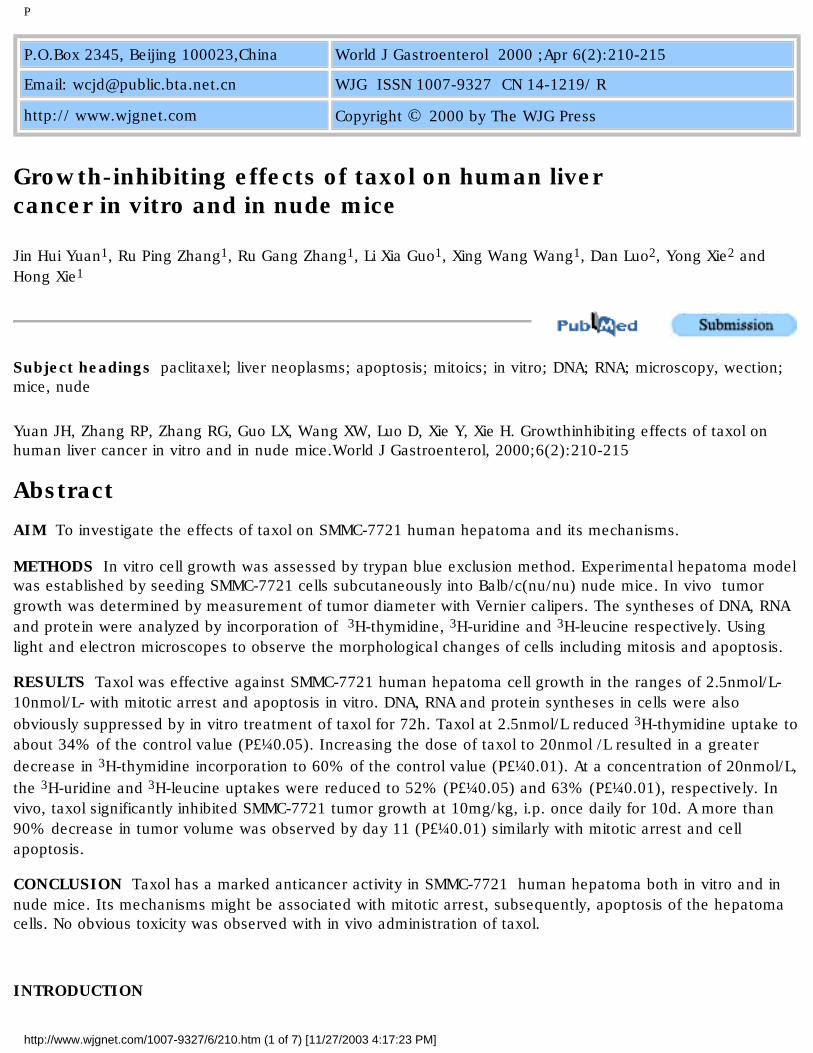
P
P.O.Box 2345, Beijing 100023,China World J Gastroenterol 2000 ;Apr 6(2):210-215
Email: [email protected] WJG ISSN 1007-9327 CN 14-1219/ R
http:// www.wjgnet.com Copyright © 2000 by The WJG Press
Growth-inhibiting effects of taxol on human liver cancer in vitro and in nude mice
Jin Hui Yuan1, Ru Ping Zhang1, Ru Gang Zhang1, Li Xia Guo1, Xing Wang Wang1, Dan Luo2, Yong Xie2 and Hong Xie1
Subject headings paclitaxel; liver neoplasms; apoptosis; mitoics; in vitro; DNA; RNA; microscopy, wection; mice, nude
Yuan JH, Zhang RP, Zhang RG, Guo LX, Wang XW, Luo D, Xie Y, Xie H. Growthinhibiting effects of taxol on human liver cancer in vitro and in nude mice.World J Gastroenterol, 2000;6(2):210-215
AbstractAIM To investigate the effects of taxol on SMMC-7721 human hepatoma and its mechanisms.
METHODS In vitro cell growth was assessed by trypan blue exclusion method. Experimental hepatoma model was established by seeding SMMC-7721 cells subcutaneously into Balb/c(nu/nu) nude mice. In vivo tumor growth was determined by measurement of tumor diameter with Vernier calipers. The syntheses of DNA, RNA and protein were analyzed by incorporation of 3H-thymidine, 3H-uridine and 3H-leucine respectively. Using light and electron microscopes to observe the morphological changes of cells including mitosis and apoptosis.
RESULTS Taxol was effective against SMMC-7721 human hepatoma cell growth in the ranges of 2.5nmol/L-10nmol/L- with mitotic arrest and apoptosis in vitro. DNA, RNA and protein syntheses in cells were also obviously suppressed by in vitro treatment of taxol for 72h. Taxol at 2.5nmol/L reduced 3H-thymidine uptake to about 34% of the control value (P£¼0.05). Increasing the dose of taxol to 20nmol /L resulted in a greater decrease in 3H-thymidine incorporation to 60% of the control value (P£¼0.01). At a concentration of 20nmol/L, the 3H-uridine and 3H-leucine uptakes were reduced to 52% (P£¼0.05) and 63% (P£¼0.01), respectively. In vivo, taxol significantly inhibited SMMC-7721 tumor growth at 10mg/kg, i.p. once daily for 10d. A more than 90% decrease in tumor volume was observed by day 11 (P£¼0.01) similarly with mitotic arrest and cell apoptosis.
CONCLUSION Taxol has a marked anticancer activity in SMMC-7721 human hepatoma both in vitro and in nude mice. Its mechanisms might be associated with mitotic arrest, subsequently, apoptosis of the hepatoma cells. No obvious toxicity was observed with in vivo administration of taxol.
INTRODUCTION
http://www.wjgnet.com/1007-9327/6/210.htm (1 of 7) [11/27/2003 4:17:23 PM]

P
Taxol, a stimulator of tubulin polymerization and microtubule assembly, was init ially considered promising because of its cytotoxic activity against several tum or cell lines in vitro£Û1,2£Ý. Subsequent in vivo studies demonstr ated that taxol was effective against a variety of murine tumors and human xenog rafts£Û3,4£Ý, especially advanced human carcinomas refractory to conventi onal chemotherapy£Û5,6£Ý. Now, taxol has emerged as one of the most active anticancer agents in clinic for the therapy of ovarian, breast, and non-small-cell lung cancer£Û7,8£Ý. Hepatocellular carcinoma (HCC) is among the most common malignancies in the world. The incidence rate is as high as 34 persons per 100000 per year in some high-inci dence areas in Asia such as China. However, few studies show that taxol is effective against HCC, especially in vivo£Û9-13£Ý. Furthermore, HCC is usually insensitive to chemotherapeutic drugs used previously in clinic and there is thus an urgent ne ed for the evaluation of new active drugs against HCC. Therefore, we systematica lly investigated the activities of taxol against HCC in vitro and in nude mi ce. Apoptosis is an active cellular death process induced by physiological or pathological factors£Û14£Ý. Most anticancer agents cause cell death by apopto sis£Û15£Ý. Incubation of cells with taxol has been found to result in cel l cycle arrest at G2/M phase and cell apoptosis in many tumor cell lines £Û16,17£Ý. Our previous in vitro studies also show that SMMC-7721 human hepatoma cells treated with taxol exhib it significant G2/M phase arrest and subsequently occurred apoptosis£Û18£Ý. However, Milross et al£Û11£Ý reported that taxol caused mit otic arrest but could not induce apoptosis in hepatoma cells. The present studie s indicate that taxol can induce mitotic arrest and apoptosis in SMMC-7721 cell s not only in vitro but also in vivo.
MATERIALS AND METHODS
DrugsTaxol was obtained from Shanghai Hualian Pharmaceutical Factory, Shanghai, China. For in vitro experiments, it was dissolved in dimethyl sulfoxide (DMSO) to make stock solution, which was then diluted to desired concentrations with the cult ure media. The DMSO concentration was kept under 0.001% in all the experiment s that did not exert any detectable effect on cell growth or apoptosis. For in vivo experiments, taxol was dissolved in a Cremophor EL vehicle (Cremophor¡Ãalcohol=50¡Ã50,v/v) at a concentration of -6¦Ìg/L. This st ock solution was further diluted with normal saline (1¡Ã5.2, v/v) immedia tely before injection. 5-Fluorouracil was from Shanghai Puguang Pharmaceutical Factory, Shanghai, China and diluted with normal saline to desired concentration before injectio n.
Cell culture and cell viability assaySMMC-7721 human liver carcinoma cell line was obtained from Chinese Type Cultur e Collection (Shanghai Institute of Cell Biology, Chinese Academy of Sciences, S hanghai, China). The cells were grown on monolayer cultures in 37¡æ, 5% CO2 incubator in RPMI 1640 media (Gibco, Grand Island, NY) supplemented with 15% heat-inactivated new-born calf serum with or without compounds as described above. At assay time, cells were collected, mixed with an equal volume of phosphate buffer salt solution containing ª©4g/L trypan blue dye, and manually counted. Actual cell numbers were calculated by multiplying diluted times compar ed with initial cell numbers.
AnimalFemale Balb/c (nu/nu) nude mice (18g-20g; 6wk-8wk of age) were purchased from Shanghai Institute of Materia Medica, Chinese Academ y of Sciences and maintained in our own specific pathogen-free mouse colony. All experiments involving animals were performed in accordance with the guidelin es of the Institutional Animal Care and Use Committee.
Measures of biomacromolecular synthesisAccording to the previous literature£Û19£Ý, the syntheses of DNA, RNA, an d protein of cells were measured by quantitating 3H-thymidine, 3H- uricine, and 3H-leucine incorporation, respectively, in the presence and absence
http://www.wjgnet.com/1007-9327/6/210.htm (2 of 7) [11/27/2003 4:17:23 PM]

P
of taxol. Briefly, cells were seeded into each well of 96-well Costar flat-bottom culture plates (Costar, Charlotte, NC) at 5¡Á103 cells/well in 100¦ÌL of culture medium. After 24h, taxol was adde d into each well according to the desired concentrations. Control samples contaned culture medium and DMSO. After 48h, the cells were incubated with 3H-thymidine, 3H-uridine, or 3H-leucine (1¦ÌCi/well) for 24h at 37¡æ respectively. At the end of the incubation, the plates were immediately placed on ice and frozen at -80¡æ. After thawing, 3H-labeled DNA,RNA,or protein were precip itated onto glass fiber discs (Whatman Ltd, Maidstone, UK) using a micro-harvester.For harvesting, the wells were extensively rinsed with distilled water, the n, air-dried. The discs were then added to scintillation vials along with 1mL of scintillant. Radioactivity was measured on a Beckman LS6000 TA¦Â counter (Beckman Instruments, Irvine, CA).
Human hepatoma implantation into nude miceSMMC-7721 human solitary hepatoma was implanted into the right flank of female nude mice by subcutaneous injection of 2.5¡Á106 viable cells. Tumor size (mm3) was evaluated by measurement of two perpendicular diameters with Vernier calipers and using the formula 1/2LW2, where L is the longest diameter and W is the diameter perpendicular to L£Û20£Ý. Palpable tumors were usua lly detected after 1wk and reached 5mm to 10mm in diameter by day 10. When tumor grew to 5mm-10mm in diameter, the mice were treated with taxol or vehicle by i.p. injection in a volume of 0.2mL per mouse, once daily for 10 days. Body and internal organ weight of animals were also observed to assess the toxicity of taxol.
Microscopic observationIn vitro, cells were fixed with 25g/L glutaraldehyde, and postfixed with 20g/L osmium tetroxide. After dehydration, the samples were embed ded in Epon 812 epoxydic resin, and then sectioned. The sections were routinely stained. In vivo, after the mice were killed by cervical dislocation, tumor tissue was immediately excised and placed in neutral-buffered formalin. The tis sue was then imbedded in paraffin blocks from which 4¦Ìm sections were cut and stained with hematoxylin and eosin. The morphologies of cells including mitosis and apoptosis were examined under electron and light microscopes.
Data analysisThe data of in vitro cell growth and the incorporation of 3H-thymidin e, 3H-uridine and 3H-leucine into cells were analyzed by Student£§ s t test. Statistical comparisons of tumor volumes were performed by analys is of variance at different time points after initiating treatment.
RESULTS
In vitro studiesSMMC-7721 cells were treated with different concentrations of taxol for differe nt times and the effect of taxol on the cell growth was examined by trypan blue exclusion method. As shown in Figure 1, the growth of SMMC-7721 cells was m arkedly inhibited by taxol in the ranges of 2.5nmol/L-10nmol/L. Moreover, taxol-induced cytotoxicity was found to be time-dependent when the cells were exposed to the agent for 48h-120h. Taxol was significantly effective at inhibiting 3H-thymidine incorporatio n with dose-dependent relationship when SMMC-7721 cells were continuously expo sed to taxol for 72h. Taxol at 2.5nmol/L reduced 3H-thym idine uptake to about 34% of the control value(P£¼0.05). Increasing the dose of taxol to 20nmol/L resulted in a greater decrease in 3H-thymidine incorporation to 60% of the control value (P£¼0.01)(Figure 2). Taxol also displayed similar dose-dependent efficacy at sup pressing 3H-uridine and 3H-leucine incorporation when the cells were constantly exposed to the drug for 72h. At a concentration of 20n mol/L, the 3H-uridine and 3H-leucine uptake was reduced to 52% (P£¼0.05) and 63% (P£¼0.01), respectively (Figure 2). These results show that taxol has significant inhibiting effects on DNA, RNA and protein syntheses of SMMC-7721 cells.The results from light microscope observation indicated that taxol at 10nmol/L caused obvious morphologic changes of SMMC-7721 cells when compared wit h untreated cells (Figure 3A). After 24h
http://www.wjgnet.com/1007-9327/6/210.htm (3 of 7) [11/27/2003 4:17:23 PM]

P
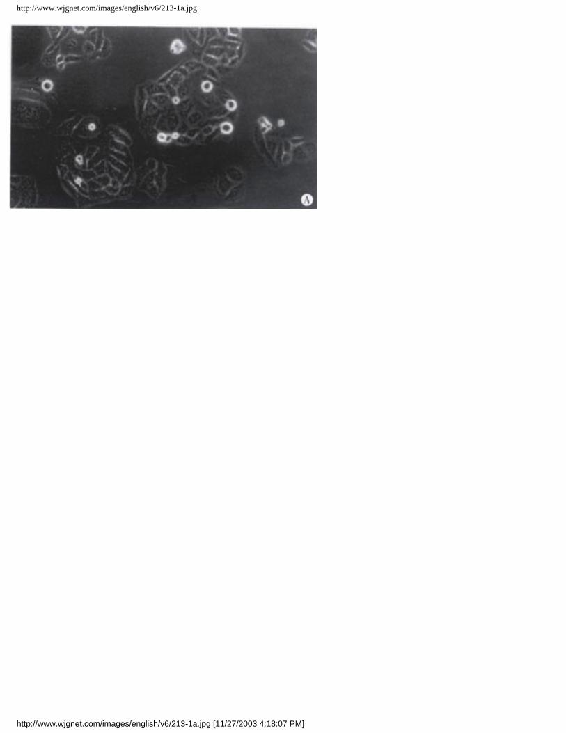
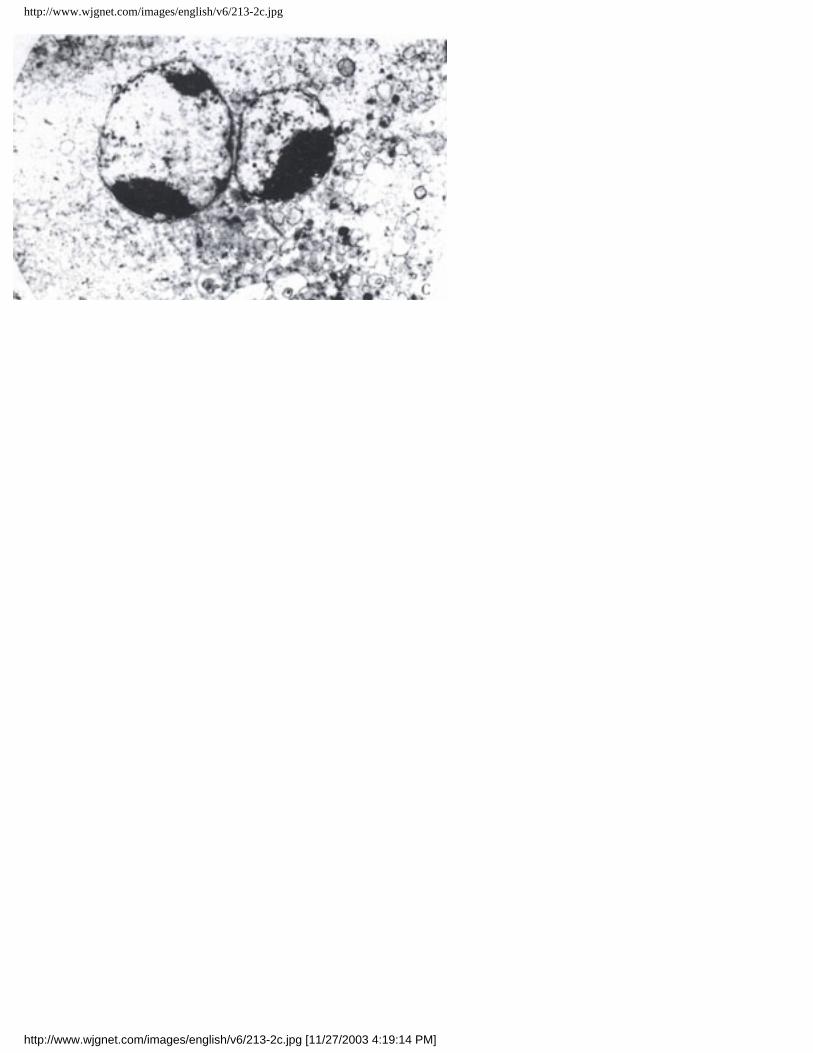
treatment (Figure 3B), the cells got round, diopter-enhanced. As time went on for 48h, cells det ached and blebbed (Figure 3C). By analysis with electron microscope, it was found that SMMC-7721 cells exposed to taxol 10nmol/L for 24h presented mitotic arrest (Figure 4B), and some cells appeared in pre-apopto tic sign as compared with untreated cells (Figure 4A). After cells were trea ted by taxol for 48 h, the nuclei were shrank, and the chromatin was condensed to form high density clumps, which were located along the nuclear envelope (Figure 4C), or formed regularly shaped crescents at the nuclear edges. It is suggested that taxol could induce typical characteristic morphologi cal changes of mitotic arrest and apoptosis of cells.
Figure 1 Effect of paclitaxel on SMMC-7721 cell growth.Figure 2 Effects of taxol on 3H-thymidine, 3H-uridine and 3H-leucine incorporation.
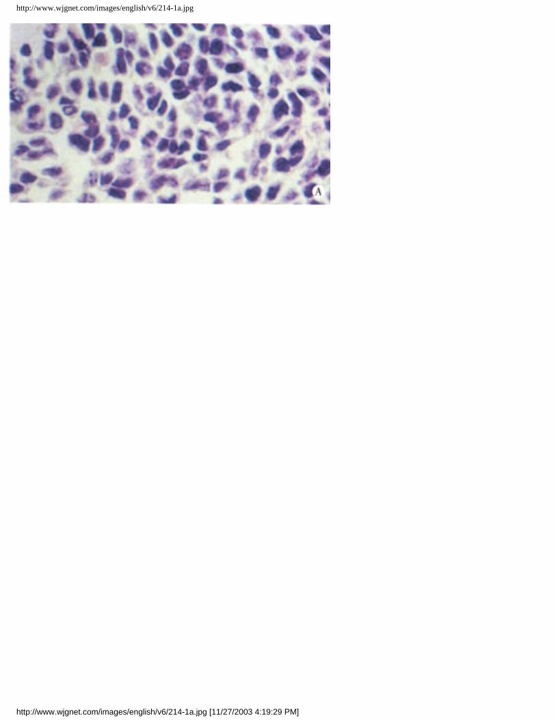
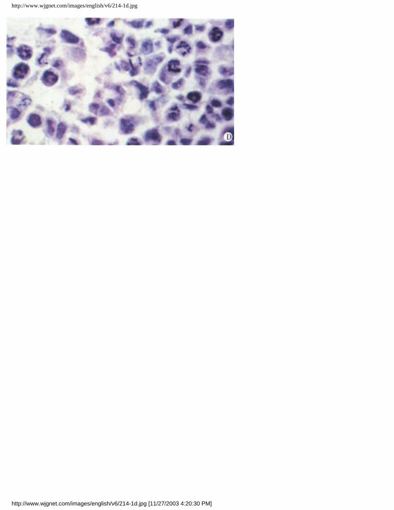
In vivo studiesNude mice bearing SMMC-7721 tumors were treated with different doses of taxol. The taxol response of SMMC-7721 human hepatoma depicted as tumor growth curves in Figure 5 showed that tumors were significantly responsive to taxol when t reated at 10mg/kg, i.p. once daily for 10d, although treatment with 2mg/kg proved less effective. Tumor regression was evident by day 3 after taxol treatment (P£¼0.05), and a greater than 90% decrease in tumor volume was observed by day 11(P£¼0.01).In contrast, the tumors in animals that only received corresponding solvent cont inued exponential growth. Mice treated with low or high doses of taxol did not display a body weight loss by day 11(P£¾0.05). No difference of internal organ weight was found between treated and control groups. No obvious other toxicity was observed in mice receiving either 2mg/kg or 10mg/kg taxol for 10d. Animals were sacrificed at various times among 24h and 120h after administrating taxol. As seen in Figure 6, taxol treatment caused marked mor phological changes in SMMC-7721 tumor. Compared with control (Figure 6A), slight mitotic arrest and apoptosis of hepatoma cells were observed in mice with 2mg/kg taxol treatment for 10 days (Figure 6B). However, most cel ls exhibited the characteristic appearance of agent-induced apoptosis after tre ated with 10mg/kg taxol for 10 days (Figure 6C). These results suggested that taxol-induced mitotic arrest and apoptosis of human hepatoma cel ls might be dose-dependent. Time-effect analysis indicated that taxol at 10mg/kg, i.p. for 24h caused mitotic arrest of most tumor cells with slight apoptosis (Figure 6D). By 48h, apoptosis beca me the dominant morphological features (Figure 6E). This phenomenon was more significant after 120h treatment than ever before (Figure 6F). Apoptotic cells were characterized by overall shrinkage and homogeneous dark basophili a. Frequently, several small apoptotic fragments were encountered in close proxi mity. On the basis of size and clustering, such fragments were considered to rep resent the remains of a single cell or apoptotic body.
Figure 3 Morphological changes of SMMC-7721 cells observed by light microscope after treatment with taxol for different times. (A) Untreated cells; (B) SMMC-7721 cells got round, diopter-enhanced after exposure to 10nmol/L taxol for 24h; (C) SMMC-7721 cells detached and blebbed after exposure to 10nmol/L taxol for 48h. ¡Á400.Figure 4 Ultrastructures of SMMC-7721 cells with o r without treatment by taxol. Following 24h treatment of taxol, cells presented more mitotic Figure (A 5760¡Á)(B 7680¡Á). For 48h, the cells appeared with regularly shaped crescents (C 7680¡Á). The nuclei of untreated cells were very irregular in shape, with many gulfs and protrusions .Figure 5 Effect of taxol on SMMC-7721 human hepatom a in nude mice.Figure 6 Histological changes of SMMC-7721 tumors after taxol treatment. (A) untreated control. (B) 2mg/kg taxol, i.p. once daily for 10d. Only slight morphological changes were presented. (C) 10mg/kg taxol, i.p. once daily for 10d. Most cells showed apoptotic changes and many apoptotic bodies
http://www.wjgnet.com/1007-9327/6/210.htm (4 of 7) [11/27/2003 4:17:23 PM]

P
were presented. Apoptosis were widespread. Apoptotic bodies were the small ovoid structures. (D) 24h after i.p. injection of 10mg/kg taxol. Mitotic arrest was marked. (E) 72h after i.p. injection of 10mg/kg taxol. Mitotic arrest could still be presented with more apoptotic cells. (F) 120h after i.p. injection of 10mg/kg taxol. Apoptosis was dominant phenomenon than ever before. Hematoxylin and eosin. ¡Á400
DISCUSSIONThis study has demonstrated that taxol is significantly effective against SMMC-7721 human hepatoma growth both in vitro and in vivo. In most reports th e arrest of cells in G2/M phases and cell apoptosis caused by taxol are responsible for its anticancer activities£Û21,22£Ý. We have also found th at taxol causes SMMC-7721 cell mitotic arrest and apoptosis in vitro and in vivo. It suggests that the anti-hepatoma effects of taxol are related to its mitotic arrest and cell apoptosis. Emphasis has been placed on the anti-hepatoma effects of taxol in vivo. Previous studies showed that taxol had no significant anticancer effect in HCC patients£Û13£Ý. Others reports also suggested no statistically significant hepatoma growth inhibition in mice treat ed with taxol at 40mg/kg, i.v.£Û11£Ý. The results presented herein indicate that taxol at 10mg/kg, i.p. once daily for 10 days has significant inhibiting activity on HCC with more than 90% suppressive perce ntage and without any obvious toxicity. It is inferred that taxol may cause hepa toma growth inhibition at low dose and continuous administration. Our present st udies also clearly demonstrate that taxol causes profound mitotic arrest and apoptosis. A time-course microscopic analysis of tumor cell mitosis and apoptosis in nude mice treated with taxol revealed that mitotic arrest occurred previously to apoptosis. Development of apoptosis lagged several hours behind mitotic arrest. Moreover, either the mitotic arrest or apoptosis of hepatoma cells induced b y taxol was clearly dose and time- dependent. These data have important therap eutic implications in HCC. In conclusion, this is the first report of the anti-hepatoma effect of taxol, its mechanisms, and the relationship between the mitotic arrest and apoptosis both in vitro and in vivo.
REFERENCES1 Wani MC, Taylor HL, Wall ME, Coggon P, Mcphail AT. Plant antitumor agents. VI. The isolation and structure of taxol, a novel antileukemic and antitumor agent from Taxus brevifolia. J Am Chem Soc,1971;93:2325-23272 Douros J, Suffness M. New natural products of interest under development at the National Cancer Institute.Cancer Chemother Pharmacol,1978;1:91-1003 Riondel J, Jacrot M, Picot F, Beriel H, Mouriquand C, Potier P. Therapeutic response to taxol of six human tumors xenografted into nude mice.Cancer Chemother Pharmacol,1986;17:137-1424 Riondel J, Jacrot M, Nissou MF, Picot F, Beriel H, Mouriquand C, Potier P. Antineoplastic activity of two taxol derivatives on an ovarian tumor xenografted into nude mice.Anticancer Res,1988;8:387-3905 Rowinsky EK, Donehower RC. Taxol: Twenty years later, the story unfolds. J Natl Cancer Inst,1991;83:1778-17816 Rowinsky EK, Donehower RC. Paclitaxel.N Engl J Med,1995;332:1004-10147 Sandercock J, Parmar MK, Torri V. Firstª²line chemotherapy for advanced ovarian cancer: paclitaxel, cisplatin and the evidence. Br J Cancer,1998;78:1471-14788 Ferrante K, Winograd B, Canetta R. Promising new developments in cancer chemotherapy. Cancer Chemother Pharmacol,1999;43(Suppl):S61-689 Lui WY, Chang YF, Li LL, Ho LK, Su TL, Chen JY, Liu TY, P£§Eng FK, Chi CW. Differential paclitaxel-induced cytotoxicity in rodent and human hepatoma cell lines.Anticancer Res,1998;18:3339-3345
http://www.wjgnet.com/1007-9327/6/210.htm (5 of 7) [11/27/2003 4:17:23 PM]
P
10 Gagandeep S, Novikoff PM, Ott M, Gupta S. Paclitaxel shows cytotoxic activity in human hepatocellular carcinoma cell lines.Cancer Lett,1999;136:109-11811 Milross CG, Mason KA, Hunter NR, Chung WK, Peters LJ, Milas L. Relationship of mitotic arrest and apoptosis to antitumor effect of paclitaxel. J Natl Cancer Inst,1996;88:1308-131412 Strumberg D, Erhard J, Harstrick A, Klaassen U, Muller C, Eberhardt W, Wilke H, Seeber S. Phase I study of a weekly 1h infusion of paclitaxel in patients with unresectable hepatocellular carcinoma.Eur J Cancer,1998;34:1290-129213 Chao Y, Chan WK, Birkhofer MJ, Hu OY, Wang SS, Huang YS, Liu M, Whang Peng J, Chi KH, Lui WY, Lee SD. Phase II and pharmacokinetic study of paclitaxel therapy for unresectable hepatocellular carcinoma patients.Br J Cancer,1998;78:34-3914 Kroemer G, Petit P, Zamzami N, Vayssiere JL, Mignotte B. The biochemistry of programmed cell death. FASEB J,1995;9:1277-128715 Solary E, Bertrand R, Pommier Y. Apoptosis induced by DNA topoisomerase ¢ñ and ¢ò inhibitors in human leukemic HL-60 cells.Leuk Lymphoma,1994;15:21-3216 Torres K, Horwitz SB. Mechanisms of Taxol-induced cell death are concentration dependent. Cancer Res,1998;58:3620-362617 Yu D, Jing T, Liu B, Yao J, Tan M, McDonnell TJ, Hung MC. Overexpression of ErbB2 blocks Taxol induced apoptosis by upregulation of p21cip1, which inhibits p34cdc2 kinase. Mol Cell,1998;2:581-59118 Yuan JH, Wang XW, Luo D, Xie Y, Xie H. Antiª²hepatoma activity of Taxol: an in vitro study.Zhongguo Yaoli Xuebao, 2000;21:in press19 Richardson DR, Milnes K. The potential of iron chelators of the pyridoxal isonicotinoyl hydrazone class as effective antiproliferative agents ¢ò: the mechanism of action of ligands derived from salicylaldehyde benzoyl hydrazone and 2- hydroxy-1-naphthylaldehyde benzoyl hydrazone.Blood,1997;89:3025-303820 Bullard DE, Schold SC Jr, Bigner SH, Bigner DD. Growth and chemotherapeutic response in athymic mice of tumors arising from human glioma-derived cell lines.J Neuropathol Exp Neurol,1981;40:410-42721 Roth W, Wagenknecht B, Grimmel C, Dichgans J, Weller M. Taxol-mediated augmentation of CD95 ligand-induced apoptosis of human malignant glioma cells: association with bcl-2 phosphorylation but neither activation of p53 nor G2/M cell cycle arrest.Br J Cancer,1998;77:404-41122 Ling YH, Consoli U, Tornos C, Andreeff M, Perez Soler R. Accumulation of cyclin B 1, activation of cyclin B1 dependent kinase and induction of programmed cell death in human epidermoid carcinoma KB cells treated with taxol. Int J Cancer,1998;75:925-932
1Shanghai Institute of Cell Biology, Chinese Academy of Sciences, Shanghai 200031, China2Hong Kong University of Science and Technology, Hong Kong, China
http://www.wjgnet.com/1007-9327/6/210.htm (6 of 7) [11/27/2003 4:17:23 PM]

P
Jin Hui Yuan, graduated from Henan Luoyang Medical School in 1990, got Master of Medicine at Henan Medical University in 1997. Now Ph.D. candidate under Professor XIE Hong as the supervisor at Shanghai Institute of Cell Biology, Chinese Academy of Sciences.Presented at the 4th National Liver Cancer Meeting of the Chinese Cancer Foundation in Chengdu, Sichuan, November 29 - December 1, 1999.Correspondence to: Prof. Hong Xie, Shanghai Institute of Cell Biology, Chinese Academy of Sciences, 320 Yue-Yang Road, Shanghai 200031, ChinaTel. 0086-21-64735609, Fax. 0086-21-64331090Received 1999-12-27 Accepted 2000-01-02
http://www.wjgnet.com/1007-9327/6/210.htm (7 of 7) [11/27/2003 4:17:23 PM]
http://www.wjgnet.com/images/english/v6/213-1a.jpg
http://www.wjgnet.com/images/english/v6/213-1a.jpg [11/27/2003 4:18:07 PM]

http://www.wjgnet.com/images/english/v6/213-1b.jpg
http://www.wjgnet.com/images/english/v6/213-1b.jpg [11/27/2003 4:18:25 PM]

http://www.wjgnet.com/images/english/v6/213-1c.jpg
http://www.wjgnet.com/images/english/v6/213-1c.jpg [11/27/2003 4:18:36 PM]

http://www.wjgnet.com/images/english/v6/213-2a.jpg
http://www.wjgnet.com/images/english/v6/213-2a.jpg [11/27/2003 4:18:46 PM]

http://www.wjgnet.com/images/english/v6/213-2b.jpg
http://www.wjgnet.com/images/english/v6/213-2b.jpg [11/27/2003 4:19:00 PM]
http://www.wjgnet.com/images/english/v6/213-2c.jpg
http://www.wjgnet.com/images/english/v6/213-2c.jpg [11/27/2003 4:19:14 PM]
http://www.wjgnet.com/images/english/v6/214-1a.jpg
http://www.wjgnet.com/images/english/v6/214-1a.jpg [11/27/2003 4:19:29 PM]

http://www.wjgnet.com/images/english/v6/214-1b.jpg
http://www.wjgnet.com/images/english/v6/214-1b.jpg [11/27/2003 4:19:43 PM]

http://www.wjgnet.com/images/english/v6/214-1c.jpg
http://www.wjgnet.com/images/english/v6/214-1c.jpg [11/27/2003 4:20:03 PM]
http://www.wjgnet.com/images/english/v6/214-1d.jpg
http://www.wjgnet.com/images/english/v6/214-1d.jpg [11/27/2003 4:20:30 PM]

http://www.wjgnet.com/images/english/v6/214-1e.jpg
http://www.wjgnet.com/images/english/v6/214-1e.jpg [11/27/2003 4:20:48 PM]

http://www.wjgnet.com/images/english/v6/214-1f.jpg
http://www.wjgnet.com/images/english/v6/214-1f.jpg [11/27/2003 4:21:04 PM]
Copyright © 2022 FDOKUMEN