An exploratory study of factors inhibiting the advancement of ...
Upload
independentCategory
view
1download
0
ARTICLE IN PRESS
Ultramicroscopy ] (]]]]) ]]]–]]]
Contents lists available at ScienceDirect
Ultramicroscopy
0304-39
doi:10.1
� Corr
E-m1 Th
Pleas
journal homepage: www.elsevier.com/locate/ultramic
Organic compounds inhibiting S. epidermidis adhesion and biofilm formation
Zhiqiang Qin a,c,d,1, Jingdong Zhang b,1, Yifan Hu b, Qijin Chi b, Ninell P. Mortensen a,e, Di Qu c,Søren Molin a, Jens Ulstrup b,�
a Department of Systems Biology, Technical University of Denmark, Dk-2800 Kgs. Lyngby, Denmarkb Department of Chemistry, Building 207, NanoDTU, Technical University of Denmark, DK-2800 Kgs. Lyngby, Denmarkc Key Laboratory of Medical Molecular Virology of Ministry of Education and Public Health, Institute of Medical Microbiology and Institutes of Biomedical Science, Shanghai Medical
School of Fudan University, Yi Xue Yuan Road 138, Shanghai 200032, PR Chinad Division of Infectious Diseases, Department of Medicine, Hollings Cancer Center, Medical University of South Carolina, 86 Jonathan Lucas Street, Charleston, SC 29425, USAe Department of Biochemistry and Cellular and Molecular Biology, University of Tennessee, Knoxville, TN 37932, USA
a r t i c l e i n f o
PACS:
01.03.Bb
61.66.Hq
68.37.Ps
87.18Fx
Keywords:
Biofilm
Atomic force microscopy
Antimicrobial effect of organic compounds
Staphylococcus epidermidis
91/$ - see front matter & 2009 Elsevier B.V. A
016/j.ultramic.2009.03.040
esponding author. Tel.: +45 4525 2359; fax: +
ail address: [email protected] (J. Ulstrup).
ese authors have equal contributions.
e cite this article as: Z. Qin, et al., U
a b s t r a c t
The formation of biofilms on surfaces of indwelling medical devices is a serious medical problem.
Staphylococcus epidermidis is a common pathogen found to colonize implanted devices and as a biofilm
is more resistant to the host immune system as well as to antibiotic treatments. Combating S.
epidermidis infections by preventing or eradicating biofilm formation of the bacterium is therefore a
medically important challenge. We report here a study of biofilm formation of S. epidermidis on solid
surfaces using a combination of confocal laser scanning (CLSM) and atomic force microscopy (AFM) in
both air and aqueous environments. We have investigated the inhibitory effects of surfaces treated with
four organic compounds, two benzoate derivatives denoted as compound 59 and 75 and two
carboxamide derivatives denoted as compound 47 and 73, on S. epidermidis adhesion and biofilm
formation. All four compounds evoke significant inhibitory effects on the formation of S. epidermidis
biofilms with compounds 47 and 73 being most effective. None of the compounds were found to inhibit
growth of S. epidermidis in liquid cultures. Bacteria attached to the substrate when exposed to the
compounds were not affected indicating that these compounds inhibit initial adhesion. These results
suggest a pretreatment for medically implanted surfaces that can prevent the biofilm formation and
reduce infection.
& 2009 Elsevier B.V. All rights reserved.
1. Introduction
Assembly of bacteria on solid surfaces to form biofilms is afundamental property of microbial cell cultures. This is especiallyproblematic where medical implants and devices such ascatheters and shunts are inserted into the body. Both substratesurface chemistry and topography have been shown to beimportant factors affecting the formation of biofilms [1]. Theinitial phases of biofilm formation are particularly susceptible tothe solid surface properties that serve as controlling factors forfurther biofilm growth to macroscopic levels [2–5]. Previously alibrary with 76 organic molecules to combat Staphylococcus
epidermidis infections was established [6]. These compoundswere designed according to structure-based virtual screening(SBVS) to identify active compounds (binders or hits) targeting aS. epidermidis histidine kinase YycG, which regulates cell-wall
ll rights reserved.
45 4588 3136.
ltramicroscopy (2009), doi:
synthesis [6]. In addition to the six compounds identified as YycGinhibitors [6], several compounds that did not inhibit histidinekinase but prevented biofilm formation were identified. In thepresent work we have studied the effects of four of thesecompounds on initiation of biofilm formation in S. epidermidis.
Coagulase-negative S. epidermidis has been known for a decadeas a leading cause of infections related to indwelling medicaldevices such as artificial heart valves, prosthetic joints andvascular catheters [2–5]. The major pathogenicity of S. epidermidis
is related to extensive biofilm formation on solid surfaces, whichis extremely difficult to treat with antibiotics, thereby necessitat-ing invasive procedures that involve removal of the infected tissueor device [7,8]. Decreased susceptibility of S. epidermidis cells toconventional antibiotics has further complicated treatmentoptions [9].
S. epidermidis biofilm formation can be divided into two steps:(1) initial attachment of single cells to the surface; and, (2)creation of stabilizing links among the attached cells, i.e. cell–cellaggregation, multilayer formation and finally growth into micro-colonies on the surfaces [10,11]. Elimination of attachment and/orcell–cell linking factors could inhibit biofilm development. Several
10.1016/j.ultramic.2009.03.040

ARTICLE IN PRESS
Z. Qin et al. / Ultramicroscopy ] (]]]]) ]]]–]]]2
strategies towards these ends have been applied. Chaw et al. [12]discovered that silicon surfaces coated with silver ions couldreduce initial S. epidermidis attachment and subsequent biofilmformation. Kaplan and associates [13] showed that an enzymeproduced by the Gram-negative periodontal pathogen Actinoba-
cillus actinomycetemcomitans rapidly and efficiently removedS. epidermidis biofilms from plastic surfaces. Curtin and Donlan[14] employed bacteriophages to treat silicone catheters andfound that S. epidermidis biofilm formation was efficientlyreduced. Addition of organic compounds offers another approachto biofilm inhibition [6,15] the merits of which is that a widerange of compounds can be designed and synthesized. Geske andassociates [15] developed a solid-phase synthetic route to preparesystematically small organic molecules such as N-acyl L-homo-serine lactone derivatives and investigated their effects onPseudomonas aeruginosa biofilm disruption. Covalently bindingof the hydrophilic PEG to various surface has also shown efficientin reducing bacterial adhesion to the substrate [16,17].
In the present report we have addressed S. epidermidis biofilmdevelopment and the inhibitory effects of four organic compounds(47, 59, 73, 75) selected from the 76 compound library for theirability to impede biofilm formation. We focused on the criticalinitial phases of biofilm growth so as to reduce subsequentmacroscopic biofilm growth. As a strategy, we used a combinationof confocal laser scanning (CLSM) and atomic force microscopy(AFM) both in air and aqueous environment in this study. The useof AFM allows addressing the early stage of biofilm developmentat the nanoscale. This offers new information in biofilm researchcomplementary to macroscopic approaches. The present effortexploits two complementary approaches to the initial stages ofbiofilm growth and the inhibiting effects of the four compoundsused in this study. We specifically investigated the attachment ofS. epidermidis to three different surfaces i.e. polystyrene, glass andmica by AFM and compared dry samples with rehydratedbiofilms. The four compounds studied showed significant inhibi-tion of bacterial adhesion to substrates pre-treated with thecompounds. The biomass of the biofilm was also found to be
Fig. 1. Chemical structures of the four selected organic compounds inhibiting S. epiderm
units while compound 75 holds a thiophene sulfur atom. Hydroxide, amide and arom
compounds. Functional groups that would adhere to the mica and glass surfaces are th
Please cite this article as: Z. Qin, et al., Ultramicroscopy (2009), doi:
drastically reduced when exposed to the compounds. Such anapproach for inhibitor screening coupled with analysis of biofilmgrowth has significant potential for development of medical andclinical chemistry applications.
2. Materials and methods
2.1. Bacterial strains, growth media and reagents
S. epidermidis RP62A and ATCC12228 strains were purchasedfrom the American Type Culture Collection (ATCC, MA, USA). S.
epidermidis strains SE408, SE203, SE847 and SE886 are all clinicalisolates from infected patients at Zhongshan Hospital in Shanghai,China, and verified by biochemical characterization using Gram’sstain and the API-20-Staph system (bioMerieux, France). TrypticSoy Broth (TSB) (Oxford, England) medium containing 0.25%glucose was used for biofilm formation in a 96-well plate andstatic chamber system. Biofilms and batch cultures were grown at37 1C unless otherwise specified. SYTO 9 staining (MolecularProbes, Eugene, OR, USA) that emits the green signal detected byCLSM was used at a concentration of 1mM. All other chemicalswere Reagent grade (Sigma, Germany).
The following compounds 47, 59, 73 and 75 were used in thestudy and are referred to in the text by the number thatthey occupy in the library of 76 compounds: The followingcompounds (see Fig. 1) were from SPECS Company (Amsterdam,Netherlands):
Compound 47: 2-(6-{5-[(3-hydroxyanilino)carbonyl]-1,3-di-oxo-1,3-dihydro-2H-isoindol-2-yl}pyridin-2-yl)-N-(3-hydroxy-phenyl)-1,3-dioxoisoindoline-5-carboxamide,Compound 59: 3-{[{2-[1-(2-anilino-2-oxoethyl)-2-oxo-1,2-di-hydro-3H-indol-3-ylidene]hydrazino}(oxo)acetyl]amino ben-zoic acid,Compound 73: 2-{[2-(2,4-dimethylphenyl)-4-quinolinyl]car-bonyl}-N-[3-({[(2-{[2-(2,4-dimethylphenyl)-4-quinolinyl]car-
idis biofilm formation. Compounds 47 and 59 include carbonyl groups as functional
atic nitrogen groups are other functional groups common to several or all of the
e polar hydroxide and carbonyl groups and the amide and aromatic N-based units.
10.1016/j.ultramic.2009.03.040

ARTICLE IN PRESS
Z. Qin et al. / Ultramicroscopy ] (]]]]) ]]]–]]] 3
bonyl}hydrazino)carbonyl]amino}methyl)benzyl]hydrazine-carboxamide, andCompound 75: methyl-4-{[(2-{[(4-methoxyphenyl)acetyl]ami-no}-4,5,6,7-tetrahydro-1-benzothien-3-yl)carbonyl]amino}-benzoate.
Their purities were 497% according to liquid chromatographymass spectrometry (LC–MS). All compounds were dissolved in 1%dimethyl sulfoxide (DMSO, Sigma, Germany) due to their limitedsolubility in water. DMSO itself at this concentration did not affectbacterial biofilm growth (see Section 2.2).
2.2. Cultivation of biofilms with and without test compound
Three solid surfaces, polystyrene, glass, and mica were used forgrowing biofilms. Biofilm cultivation on polystyrene microtiter96-well plates (Nunc, Roskilde, Denmark) was carried outas previously described [18]. Briefly, overnight cultures ofS. epidermidis strains grown in TSB+0.25% glucose medium werediluted 1:200 into fresh media and divided into 3 equal aliquots.To observe biofilm inhibition, one of the four compounds (47, 59,73, 75) at a concentration of 200mM in DMSO was added to one ofthe diluted bacterial suspension, 1% DMSO was added to a seconddiluted bacterial suspension while the third suspension wasuntreated. All three samples were incubated in the microtiterplates (200ml per well) for �20 h at 37 1C. Biofilms were thenwashed with 0.9% NaCl, rinsed in Milli-Q water, dried, stainedwith 2% crystal violet for 5 min, and the absorbance OD570
determined. Each assay was repeated at least three times.Biofilms were also grown in the static chamber system using
cover glass cell culture chambers (Nunc, Roskilde, Denmark) aspreviously described [19]. Briefly, overnight cultures ofS. epidermidis strains grown in TSB+0.25% glucose were dilutedto OD600 ¼ 0.1, inoculated in the wells (1.5 ml per well) andincubated at 37 1C for a given time. Similarly, 200mM of the testcompounds dissolved in DMSO were added into bacterial cultureprior to incubation, for observing their inhibitory effects. Ascontrols untreated cultures and cultures treated with 1% DMSOadded to the growth medium were employed. All of the chamberswere washed gently four times with 1 ml sterile PBS and stainedby SYTO 9 for 15 min.
Mica (Doll Artist’s Workshop, Gardnerville, USA) sheets with2.0�1.0 cm2 was freshly cut and cleaved by tape on both sides. Priorto use, they were sterilized by immersion in sodium hypochloritesolution (0.5%) for 2 h. The mica samples were then washed threetimes with sterilized Millipore water, immersed into the well of 6-well plates (Nunc, Roskilde, Denmark) containing 2 ml of TSB+0.25%glucose. The inoculum of bacteria, growth conditions, incubationwith the test compound, and the DMSO control were as describedfor the cover glass cultures. The staining procedure using SYTO 9 wasemployed as previously described [19].
2.3. Initial bacterial attachment and inhibition assays
Initial cell attachment was tested according to [6]. Briefly, a cellsuspension from mid-exponential growth phase was diluted toOD600 ¼ 0.1 in PBS. One of the test compounds (47, 59, 73, 75) at aconcentration of 200mM was then added to the suspension andincubated in the cover glass cell culture chambers (Nunc, Roskilde,Denmark) wells (1 ml per well) for 40 min at 37 1C. Untreated andDMSO (1%) treated cultures were used as controls. The chamberswere washed gently four times with 1 ml sterile PBS and stainedby SYTO 9 for 15 min.
To investigate the effects of test compounds (47, 59, 73, 75) onalready attached bacterial cells before biofilm development, the
Please cite this article as: Z. Qin, et al., Ultramicroscopy (2009), doi:
bacterial suspension was incubated in cover glass cell culturechambers for 40 min, and the unattached cells in the supernatantdiscarded. The wells were gently washed three times with sterilePBS prior to adding fresh PBS (1 ml per well) containing thedifferent compounds (200mM) for another 40 min treatment. Theappropriate controls of untreated and cultures treated with 1%DMSO were employed. The chambers was washed and stained asdescribed.
To investigate the effect of mica surface pretreatment with thefour compounds, 50ml DMSO containing 200mM test compoundwas deposited on the surface of sterilized mica and left at 37 1C for30 min, then washed two times in sterile PBS twice. 50ml ofbacterial culture (OD600 ¼ 0.1) was then deposited onto thecompound-covered surface and incubated for 40 min at 37 1C.The mica sheets were washed gently four times with 1 ml sterilePBS, then stained by SYTO 9 for 15 min, and the number ofattached bacteria was counted by CSLM. An untreated surface andan equal volume of DMSO (50ml) without compounds was used asthe controls.
2.4. Confocal laser scanning microscopy (CLSM) and image
acquisition
A Zeiss LSM 510 CLSM (Carl Zeiss, Jena, Germany) instrumentequipped with detectors and filter sets for monitoring SYTO 9fluorescence was used for CLSM imaging. Images were obtainedusing a 63� /1.4 or a 40� /1.3i objective. Simulated three-dimensional images and sections were generated using theIMARIS software package (Bitplane AG, Zurich, Switzerland).
2.5. Atomic force microscopy (AFM), sample preparation and image
acquisition
A DualScopeTM microscope equipped with a C-21 controller(DME A/S, Copenhagen, Denmark) was used for AFM measure-ments. The X, Y and Z directions of the scanner were calibrated vs.a standard Si grating sample (TGZ01). Tapping mode was used torecord all images. Tips (Arrow-NCR-50) were from Nano World(Neuchatel, Switzerland). All AFM images shown are representa-tive of a large number of images obtained. Liquid (aqueous)samples were examined using a PicoScan5500 from Agilentinstrument (Agilent Technologies, Chandler, AZ, USA) and a100mm scanner using model: MLCT-AUHW cantilevers fromVeeco (Camarillo, CA, USA). Biofilms were washed and examinedin buffer M63 [20], or dehydrated followed by rehydration inbuffer M63 and imaged by AFM in the same buffer.
3. Results and discussion
3.1. Time-dependent biofilm development monitored by AFM of dry
samples
Fig. 2 shows AFM images of time-dependent S. epidermidis
biofilm development at the scale of the single cell after sampledrying. In the early stages the mica surface (Fig. 2) is covered withmaterial (data not shown), likely to be biological material fromlysed bacteria and extracellular polymers (EPS) or proteins fromthe growth media, as previously observed [21,22]. This material,considered to be the matrix material for the formation of biofilms,could also come from the release of material from S. epidermidis
cells while the biofilms are formed [23].Biofilm formation begins with single bacteria forming colonies
that grow and eventually associate with other colonies. Fig. 2A–D,shows that S. epidermidis biofilm expands two-dimensionally over
10.1016/j.ultramic.2009.03.040

ARTICLE IN PRESS
20.0A B C
FED
20.00
0
[µm
]
[µm]
20.00 [µm] 3.000 [µm] 3.000 [µm]
20.00 [µm] 20.00 [µm]
20.0
0
[µm
]
3.00
00
0.2
0.4
0.6
0.8
1.0
1.2
1.4
[µm
]
20.0
00 0
2.0 0.20.4
0.6
0.8
1.0
1.2
0.4
0.6
0.8
1.0
1.21.4
[µm
]
[µm] [µm]
[µm]
0
0
200
400
600
[µm][µm]
0
200
100
300
400
500
0.2
0.4
0.6
0.8
1.0
1.4
1.2
[µm]20.0
0
[µm
]
3.00
0
[µm
]
[1.33 µm] 1.68 µm
[1.23 µm] 1.66 µm
[1.13 µm] 1.48 µm [1.37 µm] 1.67 µm
[855 nm] 919 nm[405 nm] 665 nm
Fig. 2. AFM images of S. epidermidis biofilm development on mica surface. Images in air in nonconductive mode, applied force: 0.2 nN with DualScopeTM Microscope
equipped with a C-21 controller (DME A/S, Copenhagen, Denmark). Scan areas in (A–D) are 20�20mm2 and 3�3mm2 for (E) and (F). Biofilm growth: (A) 6 h, (B) 8 h, (C)
16 h, (D) 24 h, (E) 1 h and (F) 6 h. Fiber-like bumps are observed on the surface in (E) and (F) especially around areas adjacent to neighboring cells, as indicated by black
arrows. The white arrows in (F) show biological material surrounding the bacteria.
Z. Qin et al. / Ultramicroscopy ] (]]]]) ]]]–]]]4
6, 8, 16 and 24 h, and that the entire surface gradually becomescovered by a bacterial monolayer. Some protrusions begin to stickout from the colony (6 h, Fig. 2A) and form network-like sectionsby interaction or fusion with each other (8 h, Fig. 2B). The micro-colonies on the substrate eventually grow and fuse together, but inthese early stages of biofilm formation the observed protrusions,on dried samples, are more likely a result of the dehydration thatmoves individual or loosely attached bacterial cells in closecontact with the more firmly attached micro-colonies. Similarobservations have been reported previously [24]. The biofilmcoverage reaches a maximum after 8 h. At this stage the biofilmbegins to develop into three-dimensional multiple layers Fig. 2Cand D. Fig. 2E and F shows high-resolution images of the S.
epidermidis packing in the biofilm that looks to be hexagonal dueto the spherical morphology of the bacteria. Although the cell sizeis uniform, the cell surface is not completely smooth, and somefiber-like bumps are observed on the surface especially aroundareas adjacent to neighboring cells, as indicated by black arrows inFig. 2E. S. epidermidis is known to have a surrounding layer of EPS,and the bumps may serve as linker material in the initial growthstage to enhance S. epidermidis cell assembly by ‘‘gluing’’ them tothe surface and to each other (black arrow, Fig. 2F). Dehydration ofthe biofilm could, however, also form bridges to surroundingbiological material when the cells are closely packed. Santos et al.[25] observed similar bumps and filaments in Enterococcus faecalis
biofilm and also concluded that these structures are dryingartifacts. Fig. 2F shows an area with only a few S. epidermidis cellsaligned in a chain connecting two colonies. The white arrows inthe image point to some extracellular material that may be EPS orother biomaterial involved with attachment of the biofilm to thesubstrate. Observations by CLSM showed that biofilm develop-ment of S. epidermidis proceeds similarly on mica, polystyrene andglass surfaces (data not shown).
Please cite this article as: Z. Qin, et al., Ultramicroscopy (2009), doi:
3.2. AFM studies of S. epidermidis biofilms in liquid
The images shown in Fig. 2 were recorded from S. epidermidis
biofilm samples grown on mica and dried after being isolatedfrom the growth chamber at various stages of development. Withour current apparatus for growing biofilms the growth processcould not be followed directly by in situ AFM in biological aqueousenvironment. However, AFM images of rehydrated samples in anaqueous environment could be recorded with high resolution withthe PicoScan instrument.
Freshly prepared biofilms rehydrated in buffer and imaged byAFM showed a height increase in the biofilm reflecting an unevensurface. Clear images could not be obtained probably due to theeffects of the buffer on the sample and the soft and gelatinousnature of the biofilm surface that caused the surface to respond tothe lateral force of the AFM tip. If the biofilm was dehydrated andthen rehydrated in water or buffer, clearer images of the biofilmstructure could be observed although the surface appeared to beuneven due to the increase in height caused by rehydration of thebiofilm (Fig. 3). The structures of the biofilm in dehydrated andrehydrated biofilms are virtually identical, also at the sub-cellularlevel. In dehydrated biofilms EPS is clearly seen surrounding thebiofilm occupying the space between the individual cells. EPSencasing the cells in biofilms has been observed before by AFM[25,26]. We also found EPS to be present at the same locations inrehydrated biofilms.
3.3. Inhibition of S. epidermidis biofilm formation
In a previous study of large-scale screening of potentialinhibitors of YycG kinase, some compounds were found to inhibitS. epidermidis biofilm formation [6]. Four of these (47, 59, 73 and
10.1016/j.ultramic.2009.03.040

ARTICLE IN PRESS
Fig. 3. AFM images of rehydrated S. epidermidis 24 h biofilm sample imaged in buffer M63 with PicoScan5500 (Agilent Technologies, AZ). Scan areas in (A) and (B) are
20�20mm2 and 5�5mm2 for (C) and (D). Image (A) and (C) are topography and image (B) and (D) are deflection mode. Notice that z-scale in image (A) and (B) is of the
same scan size as in Fig. 2(A–D), but the z-range is twice as large for the rehydrated samples as for the dehydrated samples.
Z. Qin et al. / Ultramicroscopy ] (]]]]) ]]]–]]] 5
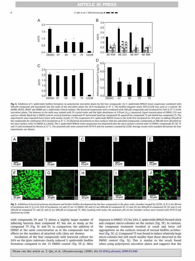
75) were chosen here for further investigation of their inhibitoryeffects on S. epidermidis biofilm formation (Fig. 1). Compounds 47and 73 are derivatives of carboxamide while compounds 59 and75 are derivatives of benzoate. A linear array of aromatic orcondensed aromatic units linked to each other either directly orvia peptide bonds is a common feature of the molecularstructures. Compounds 47 and 59 each include carbonyl groupsas functional units while compound 75 has a thiophene sulfuratom. Hydroxide, and amide or aromatic nitrogen groups are otherfunctional groups common to the compounds. Functional groupsthat would adhere to the mica and glass surfaces are the polarhydroxide and carbonyl groups and the amide and aromaticN-based units see, however, Section 4. Addition of 200mM of eachof the four compounds to cultures of S. epidermidis biofilm-positive reference strain RP62A at zero time, followed byincubation in microtiter plates, for 24 h reduced the biomassdensity by 450% relative to a DMSO control (Fig. 4A). Compound47 displayed the most efficient inhibitory effect (�70%). Thebiofilm-negative strain ATCC12228 in this assay was used as acontrol. Further experiments showed that the four compoundscould also effectively hinder biofilm formation of biofilm-positiveS. epidermidis clinical strains (SE408, SE203, SE847 and SE886)(Fig. 4B). Compound 47 was still found to be the most effective.This implies that these organic compounds are generallyinhibitory towards the formation of S. epidermidis biofilms.
The four compounds lost their inhibitory effect, when added tothe media after the biofilm had been allowed to develop for 10 h
Please cite this article as: Z. Qin, et al., Ultramicroscopy (2009), doi:
(Fig. 4C). This suggests that these organic compounds are effectiveduring the initial process of biofilm formation, perhaps by (1)inhibiting cell division or (2) by reacting with the cells in theculture to prevent attachment to the substrate or (3) bypreventing the initial attachment of single cells to surfaces pre-treated with a compound. Option (1) was most likely not theproblem as there was no difference in growth curves, determinedby optical density readings at 600 nm, of liquid cultures with orwithout 200mM of the compounds added to the media (data notshown).
Options (2) and (3) were addressed by growth experimentsusing a glass substrate in a static chamber system established totest the effects of the four compounds incubated with bacterialcultures for primary bacterial attachment and subsequent biofilmdevelopment. After incubation at 37 1C for 40 min and stainingwith SYTO 9, the untreated and 1% DMSO-treated bacterialcultures showed up in large numbers of single cells distributedevenly on the glass surface (Fig. 5A). In contrast, addition of200mM of compounds 47, 59, 73 or 75 to the bacterial culture andincubation for 40 min at 37 1C, resulted in only very few cellsscattered over the surfaces (Fig. 5B–E). Among the fourcompounds, addition of compound 47 (Fig. 5B) resulted in thelowest density of S. epidermidis colonies and the best inhibitoryeffect on glass surfaces, consistent with the ability of inhibitingbiofilm development shown in Fig. 4A for the polystyrene surface.In addition, compound 75 treatment induced the attached cells toform small aggregates on the surface (Fig. 5E). Surfaces incubated
10.1016/j.ultramic.2009.03.040
ARTICLE IN PRESS
Fig. 5. Inhibition of bacterial primary attachment and further biofilm development by the four compounds in the glass static chamber imaged by CLCM. (A–E) is for 40 min
of incubation and (F–J) is for 24 h of incubation. (A) and (F) are 1% DMSO, (B) and (G) are 200mM of compound 47, (C) and (H) are 200mM of compound 59, (D) and (I) are
200mM of compound 73, and (E) and (J) are 200mM of compound 75. After washing with water, the attached cells on the chamber surface were stained with SYTO 9 and
observed by CLSM.
2.0
1.5
1.0
0.5
0.047 59 73 75 12228
A57
0
2.0
3.0
2.5
1.5
1.0
0.5
0.0
A57
0
2.0
2.5
1.5
1.0
0.5
0.0
A57
0
DMSO
47 59 73 75DMSO47 59 73 754.5
5.0
5.5
6.0
6.5
12228DMSO
SE 408 SE 203 SE 847 SE 886
Adh
esiv
e ce
lls/c
m2
(Log
10)
Fig. 4. Inhibition of S. epidermidis biofilm formation on polystyrene microtiter plates by the four compounds. (A) S. epidermidis RP62A strain suspension combined with
200mM compound and inoculated into the wells of the microtiter plates for 24-h incubation at 37 1C. The biofilm-negative strain ATCC12228 was used as a control. (B)
SE408, SE203, SE847 and SE886 are S. epidermidis clinical isolates. The bacterial suspensions were combined with 200mM compounds and incubated for 24 h at 37 1C in the
microtiter plates. The biomass in the wells was stained with 2% crystal violet and the light absorbance at 570 nm (A570) measured. Equal concentration of DMSO (1%) was
used as a blank. Black bar is DMSO control, vertical lined bar compound 47, horizontal lined bar compound 59, squared bar compound 73 and dotted bar compound 75. The
experiments were repeated three times with similar results. (C) The suspension of S. epidermidis RP62A strain in the wells first incubated for 10 h prior to adding 200mM of
the compounds for continuous 20-h incubation at 37 1C. (D) Bacterial attachment on mica surfaces with pre-adsorbed compounds. Compounds at 200mM were absorbed on
the mica surface, with 1% DMSO as a blank. The S. epidermidis RP62A strain suspension was deposited onto the mica surfaces covered with 1% DMSO, compounds 47, 59, 73
and 75 and incubated for 40 min at 37 1C. The attached cells were stained with SYTO 9 and the numbers counted by CLSM. Average results with standard deviations for four
experiments are shown.
Z. Qin et al. / Ultramicroscopy ] (]]]]) ]]]–]]]6
with compounds 59 and 73 shows a slightly larger number ofadhering bacteria than compound 47, but not as many as forcompound 75 (Fig. 5C and D). In comparison, the addition ofDMSO at the same concentration as in the compounds had noeffects on the numbers of attached cells (data not shown).
Incubation of the four compounds with bacterial culture for24 h on the glass substrate clearly reduced S. epidermidis biofilmformation compared to the 1% DMSO control (Fig. 5F–J). After
Please cite this article as: Z. Qin, et al., Ultramicroscopy (2009), doi:
exposure to DMSO (1%) for 24 h, S. epidermidis RP62A formed thickand compact micro-colonies on the surface (Fig. 5F). In contrast,the compound treatment resulted in small and loose cellaggregations on the surfaces instead of normal biofilm architec-ture (Fig. 5G–J). Compound 75 was found to induce relatively largemicro-colonies but still much smaller than those observed in theDMSO control (Fig. 5J). This is similar to the result foundwhen using polystyrene microtiter plates and suggests that the
10.1016/j.ultramic.2009.03.040

ARTICLE IN PRESS
20.0A
E F
B C
G H
D
00 20.0
[µm
]
20.0
0
[µm
]
[µm]
0 20.0[µm]0 20.0[µm]
00
0
5.00
0
0.5
1.0
1.5
0
0.5
1.0
1.5
5.00
5.00
[µm]
0 5.00[µm]
00
20.0
20.0[µm]
[µm
][µ
m]
[µm]
[µm]
0
0.2
0.2
0.4
0.4
0.6
0.8
0.6
0.8
[µm]
0
0.5
1.0
1.5
0
0.5
1.0
1.5
0
0.5
1.0
1.5
2.5
2.0
2.0
[µm]
0
0.2
0.4
0.6
0.8
1.0
1.2
[µm]
[µm
]
0
20.0
[µm
]
00
5.00
5.00
5.00
[µm
][µ
m]
[µm]
00
5.00[µm]
[µm][µm]
0
[µm]
[1.46 µm] 2.30 µm [822 µm] 1.02 µm [1.91 µm] 2.59 µm [1.26 µm] 1.47 µm
[1.66 µm] 2.06 µm[2.54 µm] 3.18 µm[759 nm] 1.12 µm[1.69 µm] 2.33 µm
Fig. 6. AFM of S. epidermidis biofilm inhibition by the compounds 47, 59, 73 and 75. All S. epidermidis RP62A biofilms developed over 24 h at 37 1C on mica surfaces with
200mM compounds in the culture. Nonconductive mode, applied force: 0.28 nN. (A) and (B) compound 47, (C) and (D) compound 59, (E) and (F) compound 73 and (G) and
(H) compound 75. Scan area: 20�20mm2 for (A), (C), (E) and (G), 5�5mm2 for (B), (D), (F) and (H). The arrows in (B) and (F) shows the nodules, with a height of ca 340
(740) nm (B) and 230 (770) nm (F). In (D) and (H) the arrows indicate the longish structures with a size range of 4.8 (70.4)mm length and 0.68 (70.04)mm width, and of
2.8 (70.2)mm length and 0.8 (70.1)mm width, respectively. The two controls with untreated bacteria and 1% DMSO-treated bacteria are not shown.
Z. Qin et al. / Ultramicroscopy ] (]]]]) ]]]–]]] 7
inhibition mechanisms of these compounds on S. epidermidis
biofilm formation is similar for different solid surfaces assubstrates for biofilm growth.
We next addressed the inhibitory effects of the four com-pounds on the S. epidermidis primary attachment and subsequentbiofilm formation if pre-adsorbed to the solid substrate surface.Mica with atomically flat surfaces was selected as a substratematerial for further investigations by AFM. Mica pre-treated withthe four compounds was found to reduce significantly thebacterial primary attachment as documented by the CLSM images(Fig. 4D). The attached cell populations of S. epidermidis wereabout 1.78�106 cells cm�2 after 40-min incubation in the 1%DMSO-absorbed mica surface (the control group), while thepopulation was reduced to about 1.7�105–4.0�105 cells cm�2 inthe presence of either of the four compounds. Among the fourcompounds, compounds 47 and 59 were again found to have thestrongest inhibitory effect.
AFM was employed for further investigation of the initialstages of biofilm development complementary to the CLSM data.Fig. 6 shows a series of images of S. epidermidis biofilms on micasurfaces in the presence and absence of the compounds in theculture. Again 1% DMSO was used as a control since all fourcompounds are dissolved in this solvent. S. epidermidis biofilms onthe mica surface in the presence of only 1% DMSO showed denselypacked multi-layers of bacteria as well as individual roundS. epidermidis cells, consistent with observations in Fig. 2 whereno DMSO was added to the incubation culture. This indicates thatDMSO has little inhibitory influence on S. epidermidis biofilmformation itself (data not shown).
Fig. 6A–H shows surface morphologies of S. epidermidis
biofilms in the presence of the four compounds in the bacterialculture, with the following observations: (1) The biofilm coverageis significantly reduced and the bacterial cells much less denselypacked. Some areas are not covered at all by biofilms, such as forcompound 47 (Fig. 6A) and compound 73, Fig. 6E. (2) Some smallfeatures such as nodules (indicated by black arrows in Fig. 6B andF) and longish structures (shown by the arrows in Fig. 6D and H)are found mixed together with S. epidermidis cells in the biofilms
Please cite this article as: Z. Qin, et al., Ultramicroscopy (2009), doi:
after treatment with each of the compounds 47 and 73, 59 and 75.The heights of the nodules are ca 340 (740) nm (Fig. 6B) and 230(770) nm (Fig. 6F). The longish structures resemble micro-crystallites, most likely of the inhibitor molecules. These couldbe crystallized directly, or crystallization could be triggered byattachment to the bacterial surface. It is, however, clear from thedata that the tested compounds 47 and 73 have a notablerepressing effect on S. epidermidis biofilm formation.
Compounds 59 and 75 added to the bacterial culture causedformation mostly of longish structures with a size range of 4.8(70.4)mm length and 0.68 (70.04)mm width, Fig. 6D and of 2.8(70.2)mm length and 0.8 (70.1)mm width, Fig. 6H. The origin ofsuch features is presently unclear (perhaps micro-crystallites), butthe longish structures certainly result in a loose packing ofS. epidermidis cells and thus might hamper the formation ofbiofilms, cf. above. From AFM all the four compounds showsignificant impact on the impediment of S. epidermidis biofilmformation on mica surfaces. Compound 47 is the most efficientinhibitor that agrees well with the conclusions from Figs. 4 and 5on polystyrene and glass surfaces.
4. Conclusions and perspectives
We have presented a study of S. epidermidis bacterialimmobilization and biofilm growth on polystyrene, glass andmica surfaces using a combination of CLSM and AFM and of theeffects of four organic inhibitor molecules from the 76-compoundlibrary. The compounds were found to display strong inhibitoryeffects both when added directly to the bacterial growth mediaprior to bacterial surface attachment and when the surfaces wereseparately incubated by compound followed by bacterial attach-ment. In contrast, the inhibitory effects were quite insignificantwhen the compounds were added to an already establishedbiofilm. These observations offer strategies for combating biofilmsin medical environments. Molecular selectivity of the inhibitorsand surface coating rather than bulk effects seems to beimportant. The apparently highly efficient solid and bacterial
10.1016/j.ultramic.2009.03.040

ARTICLE IN PRESS
Z. Qin et al. / Ultramicroscopy ] (]]]]) ]]]–]]]8
surface activity on solid surfaces of rather different chemicalnature is suggestive of multifarious binding patterns. Hydrophilicor hydrogen bond interactions were suggested above but thiswould not apply to the highly hydrophobic polystyrene surface.Notable parts of the molecular compound structures are in facthydrophobic. The combination of molecular hydrophobicity withspecific hydrophilic functional groups could offer a first clue to thecompound inhibitory efficiency such as also seen for proteinimmobilization on electrochemical surfaces [27]. This view is,however, presently of a somewhat conjectural character.
The study, however, shows that other issues should beaddressed in order to consolidate and widen the medicalperspectives. AFM can achieve surface information broadly withhigh resolution as well as mapping of solid-liquid interfaces. Withthe reservation that samples had to be immersed and dried atgiven times, the present investigation shows that the micro-structure and some sub-cellular features of the biofilm can bevisualized and followed in real time. However, a crucial issue thatmust be pursued is eliminating the problem of having to imagedried samples instead of conducting the imaging process directlyin the relevant aqueous media. In some ways immobilization ofnatural systems to surfaces parallels strategies for functionalprotein immobilization on solid–liquid interfaces [28–31]. AFMforce spectroscopy would also be a powerful tool to measuredirectly adhesion forces between bacterial cells and solid surfacesduring the formation of biofilms or adhesion forces betweenbacterial cells within biofilms. Systematic investigations of themechanisms of interaction between S. epidermidis cells andcompounds that interfere with the formation of biofilms are animportant issue. Other substrate materials and particularlyindwelling medical device surfaces should be integrated intosuch studies.
Acknowledgements
Financial support from The Danish Strategic Research CouncilProgram NABIIT, Contract no. 2306-06-0007, The Danish ResearchCouncil for Technology and Production Sciences, Contract no. 274-07-0272, the Scientific Technology Development Foundation ofShanghai (055407069) and the State Key Program of BasicResearch of China (973) (2002CB512803) is acknowledged. NPMwould like to thank Lundbeckfonden for financial support.
Please cite this article as: Z. Qin, et al., Ultramicroscopy (2009), doi:
References
[1] B. Kasemo, Surf. Sci. 500 (2002) 656–677.[2] I.I. Raad, G.P. Bodey, Clin. Infect. Dis. 15 (1992) 197–210.[3] C. Vuong, M. Otto, Microbes Infect. 4 (2002) 481–489.[4] M.E. Rupp, G.L. Archer, Clin. Infect. Dis. 19 (1994) 231–243 (quiz 244–235).[5] L. Hall-Stoodley, J.W. Costerton, P. Stoodley, Nat. Rev. Microbiol. 2 (2004)
95–108.[6] Z.Q. Qin, J. Zhang, B. Xu, L.L. Chen, Y. Wu, X.M. Yang, X. Shen, S. Molin, A.
Danchin, H.L. Jiang, D. Qu, BMC Microbiol. 6 (2006) Article no. 96.[7] R.M. Donlan, Clin. Infect. Dis. 33 (2001) 1387–1392.[8] L.K. Archibald, R.P. Gaynes, Infect. Dis. Clin. North Am. 11 (1997) 245–255.[9] A.R. Costa, M. Henriques, R. Oliveira, J. Azeredo, Eur. J. Clin. Microbiol. Infect.
Dis. (2009).[10] F. Gotz, Mol. Microbiol. 43 (2002) 1367–1378.[11] A. Reisner, N. Hoiby, T. Tolker-Nielsen, S. Molin, Contrib. Microbiol. 12 (2005)
114–131.[12] K.C. Chaw, M. Manimaran, F.E. Tay, Antimicrob. Agents Chemother. 49 (2005)
4853–4859.[13] J.B. Kaplan, C. Ragunath, K. Velliyagounder, D.H. Fine, N. Ramasubbu,
Antimicrob. Agents Chemother. 48 (2004) 2633–2636.[14] J.J. Curtin, R.M. Donlan, Antimicrob. Agents Chemother. 50 (2006) 1268–1275.[15] G.D. Geske, R.J. Wezeman, A.P. Siegel, H.E. Blackwell, J. Am. Chem. Soc. 127
(2005) 12762–12763.[16] E. Ostuni, R.G. Chapman, M.N. Liang, G. Meluleni, G. Pier, D.E. Ingber, G.M.
Whitesides, Langmuir 17 (2001) 6336–6343.[17] P. Kingshott, J. Wei, D. Bagge-Ravn, N. Gadegaard, L. Gram, Langmuir 19
(2003) 6912–6921.[18] G.D. Christensen, W.A. Simpson, J.J. Younger, L.M. Baddour, F.F. Barrett, D.M.
Melton, E.H. Beachey, J. Clin. Microbiol. 22 (1985) 996–1006.[19] Z. Qin, X. Yang, L. Yang, J. Jiang, Y. Ou, S. Molin, D. Qu, J. Med. Microbiol. 56
(2007) 83–93.[20] J.H. Miller, Experiments in Molecular Genetics, 1972. p. 466.[21] T. Schmid, J. Burkhard, B.S. Yeo, W. Zhang, R. Zenobi, Anal. Bioanal. Chem. 391
(2008) 1899–1905.[22] Y.J. Oh, W. Jo, Y. Yang, S. Park, Ultramicroscopy 107 (2007) 869–874.[23] C.B. Whitchurch, T. Tolker-Nielsen, P.C. Ragas, J.S. Mattick, Science 295 (2002)
1487.[24] K. Jonas, H. Tomenius, A. Kader, S. Normark, U. Roemling, L.M. Belova, O.
Melefors, BMC Microbiol. 7 (2007) (Article no.: 70).[25] R.P. Santos, T.T. Arruda, C.B. Carvalho, V.A. Carneiro, L.Q. Braga, E.H. Teixeira,
F.V. Arruda, B.S. Cavada, A. Havt, T.M. de Oliveira, G.A. Bezerra, V.N. Freire,Microsc. Microanal. 14 (2008) 150–158.
[26] I.B. Beech, J.R. Smith, A.A. Steele, I. Penegar, S.A. Campbell, Colloids Surf BBiointerfaces 23 (2002) 231–247.
[27] A.C. Welinder, J. Zhang, A.G. Hansen, K. Moth-Poulsen, H.E.M. Christensen,A.M. Kuznetsov, T. Bjornholm, J. Ulstrup, Z. Phys. Chem.—Int. J. Res. Phys.Chem. Chem. Phys. 221 (2007) 1343–1378.
[28] J. Zhang, Q. Chi, J.E.T. Andersen, J. Ulstrup, J. Phys. Chem. B 105 (2001)4669–4679.
[29] Q. Chi, O. Farver, J. Ulstrup, Proc. Natl. Acad. Sci. USA 102 (2005)16203–16208.
[30] J.D. Zhang, A.C. Welinder, A.G. Hansen, H.E.M. Christensen, J. Ulstrup, J. Phys.Chem. B 107 (2003) 12480–12484.
[31] J.D. Zhang, H.E.M. Christensen, B.L. Ooi, J. Ulstrup, Langmuir 20 (2004)10200–10207.
10.1016/j.ultramic.2009.03.040
Copyright © 2022 FDOKUMEN