Structural Behaviour of Cold Formed Stainless Steel Bolted ...
Upload
michiganstateCategory
view
1download
0
Characterization of biofilm formed by human-derivednanoparticles
Maria K Schwartz1, Larry W Hunter2, Marianne Huebner2,3, John C Lieske4,5, and VirginiaM Miller1,2,†1 Department of Physiology & Biomedical Engineering, College of Medicine, Mayo Clinic,Rochester, MN 55905, USA, Tel.: +1 507 284 2290; Fax: +1 507 266 2233;[email protected] Department of Surgery, College of Medicine, Mayo Clinic, MN 55905, USA3 Department of Biostatistics College of Medicine, Mayo Clinic, MN 55905, USA4 Internal Medicine, Division of Nephrology & Hypertension, College of Medicine, Mayo Clinic, MN55905, USA5 Laboratory Medicine & Pathology, College of Medicine, Mayo Clinic, MN 55905, USA
AbstractAim—Microbial biofilm matrix contains polysaccharides and proteins and can requireextracellular nucleic acids for initial formation. Experiments were designed to identify infectiouspathogens in human aneurysms and to characterize biofilm formed by calcified human arterial-derived nanoparticles.
Materials & method—A total of 26 different microbial pathogens were isolated from 48inflammatory aneurysms. Consistent amounts (0.49 McFarland units) of nanoparticles derivedfrom similar tissue were seeded into 24-well plates and cultured for 21 days in the absence(control) or presence of RNase, tetracycline or gentamicin.
Results—Control biofilm developed within 14 days, as detected by concanavalin A andBacLight™ Green staining. The formation of biofilm in wells treated with RNase was notdifferent from the control; however, gentamicin partially inhibited and tetracycline completelyinhibited biofilm formation. Therefore, nanoparticle biofilm retains some characteristics ofconventional bacterial biofilm and requires protein–calcium interactions, although extracellularRNA is not required.
Conclusion—This model system may also allow study of nanosized vesicles derived from donortissue, including any microbes present, and could provide a useful tool for in vitro investigation ofnanoparticle biofilm formation.
†Author for correspondence.For reprint orders, please contact: [email protected] conduct of researchThe authors state that they have obtained appropriate institutional review board approval or have followed the principles outlined inthe Declaration of Helsinki for all human or animal experimental investigations. In addition, for investigations involving humansubjects, informed consent has been obtained from the participants involved.Financial & competing interests disclosureThe authors acknowledge support from the NIH (grant number HL889888), Fetzer Foundation and Mayo Foundation. The authorshave no other relevant affiliations or financial involvement with any organization or entity with a financial interest in or financialconflict with the subject matter or materials discussed in the manuscript apart from those disclosed.No writing assistance was utilized in the production of this manuscript.
NIH Public AccessAuthor ManuscriptNanomedicine (Lond). Author manuscript; available in PMC 2011 January 20.
Published in final edited form as:Nanomedicine (Lond). 2009 December ; 4(8): 931–941. doi:10.2217/nnm.09.72.
NIH
-PA Author Manuscript
NIH
-PA Author Manuscript
NIH
-PA Author Manuscript
Keywordsbiofilm; gentamicin; matrix; tetracycline
In nature, biofilms are surface-associated structures of communal microorganisms encasedwithin a secreted matrix of exopolysaccharides. Biofilms may contribute to human diseaseand reduce performance of implantable medical devices [1–4]. It is critical, therefore, tobetter understand the composition and factors leading to their formation.
Atherosclerosis results from inflammatory processes and infectious agents, which includebacteria (i.e., Treponema pallidum, Chlamydia pneumoniae, Helicobacter pylori andPorphyromonas gingivalis) and viruses (i.e., coxsackie B4, herpes simplex virus andcytomegalovirus) [5–11]. Antibiotics, including tetracycline (TCN), are effective inreducing some vascular diseases, including syphilitic aortitis, expansion of aortic aneurysmsand a number of adverse events in patients with coronary artery disease [12–18].Discrepancies among studies regarding the efficacy of antibiotic treatment forcardiovascular conditions may reflect concurrent pathophysiological parameters, such asheterogeneous patient populations, and confounding risk factors, such as metabolicsyndrome, which may not necessarily cause atherosclerosis but could predispose selectindividuals to the risk of chronic infection. Therefore, associations among infection,antibiotics and vascular disease require further investigation.
Nanosized particles (nanoparticles [NPs]) have been identified in diseased human tissuesincluding arterial plaque, kidney stones, gall stones, prostate glands, placenta and ovariantumors. Those NPs isolated from arterial tissue and kidney stones form calcific films understandard tissue-culture conditions [19–25]. Whether or not these films can rightfully becalled biofilms is debatable.
Nanoparticle films contain both organic (proteins, nucleic acids and lipids) and inorganicmineral components [26–31]. A DNA sequence identified as Nanobacterium sanguineumwas reported from NPs isolated from human and cow sera [32]. However, only partial DNAsequences of common environmental bacteria were amplified by PCR from film derivedfrom NPs isolated from human saliva. Thus, characterization of a NP film may depend uponthe tissue of origin and could be derived from contaminating flora [33] rather than from aunique nanosized bacterium [32,34].
Extracellular nucleic acids participate in the formation of some bacterial biofilm [35]. Inaddition, some enzymes participating in RNA translation were found in cultures of NPs[27,36]. Other studies provide evidence that the formation of a NP film may result fromphysical–chemical interactions of proteins in serum with hydroxyapatite or calciumcarbonate [29–31,37]. However, formation of a NP film is reduced or limited by inhibitorsof aerobic metabolism and antimicrobial agents, suggesting the involvement of enzymaticprocesses in addition to physical–chemical interactions of proteins with inorganic salts [26–28,38,39].
The extracellular matrix of microbial bio-films contains polysaccharides and enzymes thataffect propagation and stability of the biofilm [3]. Depending upon environmentalconditions, some types of bacteria precipitate intracellular hydroxyapatite or form cell wallvesicles that could potentially interact with mammalian cells, initiating inflammatoryresponses [40–42]. Indeed, cultured, human-derived NPs reduce platelet aggregation [43]and modulate vascular remodeling when injected intravenously into animals [44,45],suggesting a pathological potential. Therefore, studies were designed to identify infectiouspathogens in diseased arterial tissue and to characterize the film formed by cultured NPs
Schwartz et al. Page 2
Nanomedicine (Lond). Author manuscript; available in PMC 2011 January 20.
NIH
-PA Author Manuscript
NIH
-PA Author Manuscript
NIH
-PA Author Manuscript
derived from similar arteries. Experiments tested the hypothesis that formation of a NP filmis dependent upon extracellular nucleic acids, enzymatic and physical–chemical calcificationprocesses. In this article, the nomenclature biofilm, initially proposed by Cisar et al. todescribe a NP-derived film in culture is used, even though culturable NPs may not be abiofilm in the traditional sense [33].
MethodsIdentification of microbes in vascular tissue & isolation of NPs
Segments of aneurysms explanted as part of surgical vascular repair were analyzed.Explanted segments were placed in sterile saline, homogenized and cultured for aerobic oranaerobic organisms in the Clinical Microbiology Laboratory at the Mayo Clinic (MN,USA) using standard techniques. Some segments of explanted aneurysms werehomogenized and forced through a 0.2 μm filter. The filtrate was inoculated into 10 mlvented tissue-culture flasks containing standard culture medium [28] comprising Dulbecco’smodified Eagle’s medium (DMEM 10–013, Mediatech, Inc., VA, USA), 10% γ-irradiatedfetal bovine serum (Atlanta Biologicals, Inc., GA, USA) and 50-μM β-mercaptoethanol,which had been filtered (0.2 μm) prior to use. Flasks then were placed in a humidifiedincubator at 37°C, 90% O2/10% CO2. Every 4–6 weeks, flasks were scraped with a rubberspatula and divided 1:10 into fresh DMEM and 10% fetal bovine serum. NPs exist as bothadherent biofilm and planktonic (floating) forms suspended in the medium. In this study,planktonic forms of NPs were used exclusively. Random NP cultures were screened forMycoplasma using a rapid PCR test in the Mayo Clinic Microbiology Laboratory; all NPcultures tested negative. The collection of human tissue and review of medical records wereperformed in compliance with regulations of the State of Minnesota and the USA(Institutional Review Board-approved protocols 291-99, 2126-01 and 1030-03).
Analysis of NP biofilmThe outer wells of 24-well culture plates (Costar®, Corning, Inc., NY, USA) were filled withsterile-filtered water to reduce evaporation from the seeded wells during the course of theexperiment. A sterile glass cover slip was placed in the bottom of the remaining inner eightwells of each plate. These wells were filled with 3 ml of standard culture medium, eitheralone, or with DNase (40 units/ml, three different lots, TURBO™ DNase; Ambion, Inc.,TX, USA), RNase (50 units/ml; RNase I; Ambion, Inc., TX, USA), TCN HCl (12μg/ml;Gallipot, Inc., MN, USA) [27] or gentamicin sulfate (GM; 12 μg/ml; Spectrum ChemicalMFG CORP, CA, USA) [46] in duplicate (two wells per condition). DNase and RNase wereused to determine whether extracellular nucleic acids were required for the formation of theNP biofilm. TCN and GM are antibiotics of the same class but differ in their capacity tochelate calcium and were chosen in order to differentiate between the potential mechanismsof action: mineral–protein interactions versus enzymatic activity [47,48].
A total of 12 sets of plates were prepared for each harvest day: six contained medium withand without treatments (blanks), and six contained identical treatments but were seeded withNPs (density of 0.49 McFarland units). This initial seeding density was chosen based onpreliminary experiments that demonstrated measureable changes in turbidity within the 3-week time frame. Plates were incubated (37°C, 90% O2/10% CO2) for up to 21 days.Treatments (diluted in medium) or equal volumes of medium (control) were added to eachwell every day. At days 1, 7, 14 and 21, a 2-ml aliquot of medium was collected from eachwell. Care was taken not to agitate the well, thus leaving the biofilm undisturbed. Mediumwas analyzed for turbidity (Hach 2100N Turbidimeter, CO, USA) in McFarland units,reflecting the density and size of suspended NPs.
Schwartz et al. Page 3
Nanomedicine (Lond). Author manuscript; available in PMC 2011 January 20.
NIH
-PA Author Manuscript
NIH
-PA Author Manuscript
NIH
-PA Author Manuscript
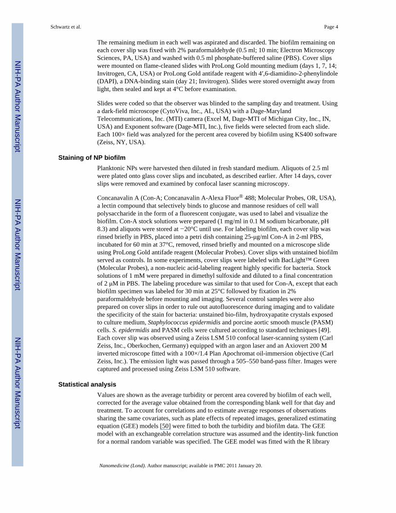
The remaining medium in each well was aspirated and discarded. The biofilm remaining oneach cover slip was fixed with 2% paraformaldehyde (0.5 ml; 10 min; Electron MicroscopySciences, PA, USA) and washed with 0.5 ml phosphate-buffered saline (PBS). Cover slipswere mounted on flame-cleaned slides with ProLong Gold mounting medium (days 1, 7, 14;Invitrogen, CA, USA) or ProLong Gold antifade reagent with 4′,6-diamidino-2-phenylindole(DAPI), a DNA-binding stain (day 21; Invitrogen). Slides were stored overnight away fromlight, then sealed and kept at 4°C before examination.
Slides were coded so that the observer was blinded to the sampling day and treatment. Usinga dark-field microscope (CytoViva, Inc., AL, USA) with a Dage-MarylandTelecommunications, Inc. (MTI) camera (Excel M, Dage-MTI of Michigan City, Inc., IN,USA) and Exponent software (Dage-MTI, Inc.), five fields were selected from each slide.Each 100× field was analyzed for the percent area covered by biofilm using KS400 software(Zeiss, NY, USA).
Staining of NP biofilmPlanktonic NPs were harvested then diluted in fresh standard medium. Aliquots of 2.5 mlwere plated onto glass cover slips and incubated, as described earlier. After 14 days, coverslips were removed and examined by confocal laser scanning microscopy.
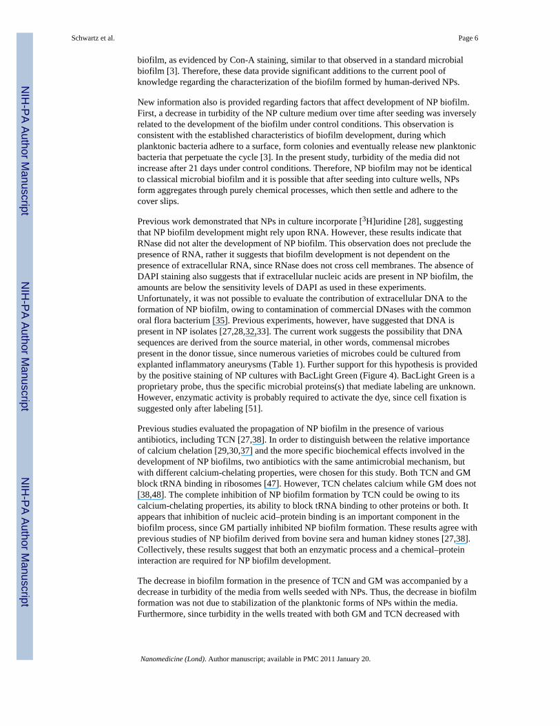
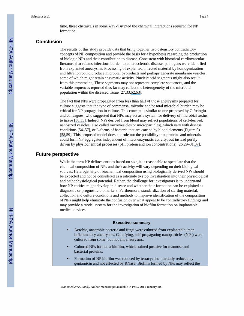
Concanavalin A (Con-A; Concanavalin A-Alexa Fluor® 488; Molecular Probes, OR, USA),a lectin compound that selectively binds to glucose and mannose residues of cell wallpolysaccharide in the form of a fluorescent conjugate, was used to label and visualize thebiofilm. Con-A stock solutions were prepared (1 mg/ml in 0.1 M sodium bicarbonate, pH8.3) and aliquots were stored at −20°C until use. For labeling biofilm, each cover slip wasrinsed briefly in PBS, placed into a petri dish containing 25-μg/ml Con-A in 2-ml PBS,incubated for 60 min at 37°C, removed, rinsed briefly and mounted on a microscope slideusing ProLong Gold antifade reagent (Molecular Probes). Cover slips with unstained biofilmserved as controls. In some experiments, cover slips were labeled with BacLight™ Green(Molecular Probes), a non-nucleic acid-labeling reagent highly specific for bacteria. Stocksolutions of 1 mM were prepared in dimethyl sulfoxide and diluted to a final concentrationof 2 μM in PBS. The labeling procedure was similar to that used for Con-A, except that eachbiofilm specimen was labeled for 30 min at 25°C followed by fixation in 2%paraformaldehyde before mounting and imaging. Several control samples were alsoprepared on cover slips in order to rule out autofluorescence during imaging and to validatethe specificity of the stain for bacteria: unstained bio-film, hydroxyapatite crystals exposedto culture medium, Staphylococcus epidermidis and porcine aortic smooth muscle (PASM)cells. S. epidermidis and PASM cells were cultured according to standard techniques [49].Each cover slip was observed using a Zeiss LSM 510 confocal laser-scanning system (CarlZeiss, Inc., Oberkochen, Germany) equipped with an argon laser and an Axiovert 200 Minverted microscope fitted with a 100×/1.4 Plan Apochromat oil-immersion objective (CarlZeiss, Inc.). The emission light was passed through a 505–550 band-pass filter. Images werecaptured and processed using Zeiss LSM 510 software.
Statistical analysisValues are shown as the average turbidity or percent area covered by biofilm of each well,corrected for the average value obtained from the corresponding blank well for that day andtreatment. To account for correlations and to estimate average responses of observationssharing the same covariates, such as plate effects of repeated images, generalized estimatingequation (GEE) models [50] were fitted to both the turbidity and biofilm data. The GEEmodel with an exchangeable correlation structure was assumed and the identity-link functionfor a normal random variable was specified. The GEE model was fitted with the R library
Schwartz et al. Page 4
Nanomedicine (Lond). Author manuscript; available in PMC 2011 January 20.
NIH
-PA Author Manuscript
NIH
-PA Author Manuscript
NIH
-PA Author Manuscript
geepack (R programming language version 2.8.0, open source), which yielded estimates forthe treatment effect and time effect. Differences were estimated between each treatment andcontrol group at each time point, as well as for differences within treatments compared withday 1. Statistical significance was accepted at p < 0.05.
ResultsIdentification of microbes in vascular tissue
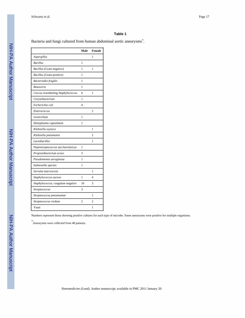
Explanted segments of abdominal aortic aneurysms, classified by the attending surgeon asinflammatory without adhesions or anastomoses to the intestines, were obtained from 48patients. A total of 26 different organisms were cultured from these aneurysms using routinemicrobiologic techniques (Table 1). Multiple bacteria were cultured from some samples; onesample produced six different organisms. Additional aneurysms (n = 9) not sent for routinemicrobial culture were processed using conditions to propagate NPs as described in themethods section. Calcific NP biofilm developed in four (44%) of these nine preparations.Subcultures of one of these isolates were used for experiments below.
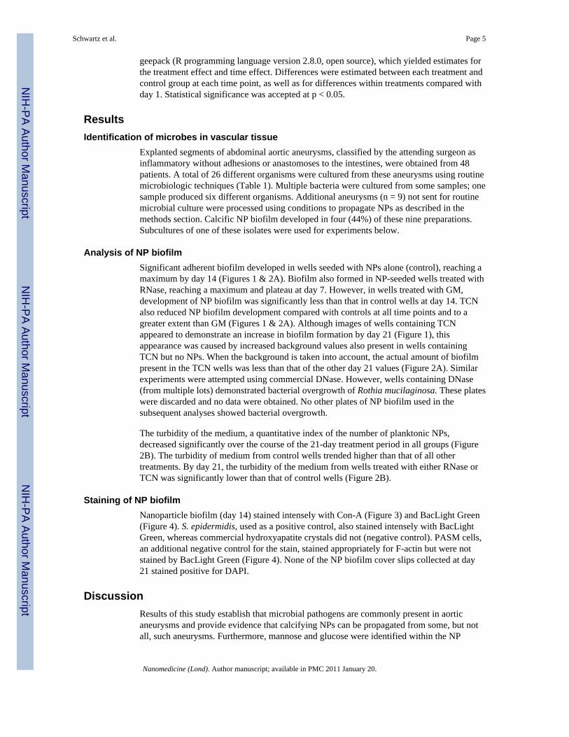
Analysis of NP biofilmSignificant adherent biofilm developed in wells seeded with NPs alone (control), reaching amaximum by day 14 (Figures 1 & 2A). Biofilm also formed in NP-seeded wells treated withRNase, reaching a maximum and plateau at day 7. However, in wells treated with GM,development of NP biofilm was significantly less than that in control wells at day 14. TCNalso reduced NP biofilm development compared with controls at all time points and to agreater extent than GM (Figures 1 & 2A). Although images of wells containing TCNappeared to demonstrate an increase in biofilm formation by day 21 (Figure 1), thisappearance was caused by increased background values also present in wells containingTCN but no NPs. When the background is taken into account, the actual amount of biofilmpresent in the TCN wells was less than that of the other day 21 values (Figure 2A). Similarexperiments were attempted using commercial DNase. However, wells containing DNase(from multiple lots) demonstrated bacterial overgrowth of Rothia mucilaginosa. These plateswere discarded and no data were obtained. No other plates of NP biofilm used in thesubsequent analyses showed bacterial overgrowth.
The turbidity of the medium, a quantitative index of the number of planktonic NPs,decreased significantly over the course of the 21-day treatment period in all groups (Figure2B). The turbidity of medium from control wells trended higher than that of all othertreatments. By day 21, the turbidity of the medium from wells treated with either RNase orTCN was significantly lower than that of control wells (Figure 2B).
Staining of NP biofilmNanoparticle biofilm (day 14) stained intensely with Con-A (Figure 3) and BacLight Green(Figure 4). S. epidermidis, used as a positive control, also stained intensely with BacLightGreen, whereas commercial hydroxyapatite crystals did not (negative control). PASM cells,an additional negative control for the stain, stained appropriately for F-actin but were notstained by BacLight Green (Figure 4). None of the NP biofilm cover slips collected at day21 stained positive for DAPI.
DiscussionResults of this study establish that microbial pathogens are commonly present in aorticaneurysms and provide evidence that calcifying NPs can be propagated from some, but notall, such aneurysms. Furthermore, mannose and glucose were identified within the NP
Schwartz et al. Page 5
Nanomedicine (Lond). Author manuscript; available in PMC 2011 January 20.
NIH
-PA Author Manuscript
NIH
-PA Author Manuscript
NIH
-PA Author Manuscript
biofilm, as evidenced by Con-A staining, similar to that observed in a standard microbialbiofilm [3]. Therefore, these data provide significant additions to the current pool ofknowledge regarding the characterization of the biofilm formed by human-derived NPs.
New information also is provided regarding factors that affect development of NP biofilm.First, a decrease in turbidity of the NP culture medium over time after seeding was inverselyrelated to the development of the biofilm under control conditions. This observation isconsistent with the established characteristics of biofilm development, during whichplanktonic bacteria adhere to a surface, form colonies and eventually release new planktonicbacteria that perpetuate the cycle [3]. In the present study, turbidity of the media did notincrease after 21 days under control conditions. Therefore, NP biofilm may not be identicalto classical microbial biofilm and it is possible that after seeding into culture wells, NPsform aggregates through purely chemical processes, which then settle and adhere to thecover slips.
Previous work demonstrated that NPs in culture incorporate [3H]uridine [28], suggestingthat NP biofilm development might rely upon RNA. However, these results indicate thatRNase did not alter the development of NP biofilm. This observation does not preclude thepresence of RNA, rather it suggests that biofilm development is not dependent on thepresence of extracellular RNA, since RNase does not cross cell membranes. The absence ofDAPI staining also suggests that if extracellular nucleic acids are present in NP biofilm, theamounts are below the sensitivity levels of DAPI as used in these experiments.Unfortunately, it was not possible to evaluate the contribution of extracellular DNA to theformation of NP biofilm, owing to contamination of commercial DNases with the commonoral flora bacterium [35]. Previous experiments, however, have suggested that DNA ispresent in NP isolates [27,28,32,33]. The current work suggests the possibility that DNAsequences are derived from the source material, in other words, commensal microbespresent in the donor tissue, since numerous varieties of microbes could be cultured fromexplanted inflammatory aneurysms (Table 1). Further support for this hypothesis is providedby the positive staining of NP cultures with BacLight Green (Figure 4). BacLight Green is aproprietary probe, thus the specific microbial proteins(s) that mediate labeling are unknown.However, enzymatic activity is probably required to activate the dye, since cell fixation issuggested only after labeling [51].
Previous studies evaluated the propagation of NP biofilm in the presence of variousantibiotics, including TCN [27,38]. In order to distinguish between the relative importanceof calcium chelation [29,30,37] and the more specific biochemical effects involved in thedevelopment of NP biofilms, two antibiotics with the same antimicrobial mechanism, butwith different calcium-chelating properties, were chosen for this study. Both TCN and GMblock tRNA binding in ribosomes [47]. However, TCN chelates calcium while GM does not[38,48]. The complete inhibition of NP biofilm formation by TCN could be owing to itscalcium-chelating properties, its ability to block tRNA binding to other proteins or both. Itappears that inhibition of nucleic acid–protein binding is an important component in thebiofilm process, since GM partially inhibited NP biofilm formation. These results agree withprevious studies of NP biofilm derived from bovine sera and human kidney stones [27,38].Collectively, these results suggest that both an enzymatic process and a chemical–proteininteraction are required for NP biofilm development.
The decrease in biofilm formation in the presence of TCN and GM was accompanied by adecrease in turbidity of the media from wells seeded with NPs. Thus, the decrease in biofilmformation was not due to stabilization of the planktonic forms of NPs within the media.Furthermore, since turbidity in the wells treated with both GM and TCN decreased with
Schwartz et al. Page 6
Nanomedicine (Lond). Author manuscript; available in PMC 2011 January 20.
NIH
-PA Author Manuscript
NIH
-PA Author Manuscript
NIH
-PA Author Manuscript
time, these chemicals in some way disrupted the chemical interactions required for NPformation.
ConclusionThe results of this study provide data that bring together two ostensibly contradictoryconcepts of NP composition and provide the basis for a hypothesis regarding the productionof biologic NPs and their contribution to disease. Consistent with historical cardiovascularliterature that relates infectious burden to atherosclerotic disease, pathogens were identifiedfrom explanted aneurysms. Processing of explanted, infected material by homogenizationand filtration could produce microbial byproducts and perhaps generate membrane vesicles,some of which might retain enzymatic activity. Nucleic acid segments might also resultfrom this processing. These segments may not represent complete sequences, and thevariable sequences reported thus far may reflect the heterogeneity of the microbialpopulation within the diseased tissue [27,33,52,53].
The fact that NPs were propagated from less than half of those aneurysms prepared forculture suggests that the type of commensal microbe and/or total microbial burden may becritical for NP propagation in culture. This concept is similar to one proposed by Ciftciogluand colleagues, who suggested that NPs may act as a system for delivery of microbial toxinsto tissue [38,53]. Indeed, NPs derived from blood may reflect populations of cell-derived,nanosized vesicles (also called microvesicles or microparticles), which vary with diseaseconditions [54–57], or L-forms of bacteria that are carried by blood elements (Figure 5)[58,59]. This proposed model does not rule out the possibility that proteins and mineralscould form NP aggregates independent of intact enzymatic activity, but instead purelydriven by physiochemical processes (pH, protein and ion concentrations) [26,29–31,37].
Future perspectiveWhile the term NP defines entities based on size, it is reasonable to speculate that thechemical composition of NPs and their activity will vary depending on their biologicalsources. Heterogeneity of biochemical composition using biologically derived NPs shouldbe expected and not be considered as a rationale to stop investigation into their physiologicaland pathophysiological potential. Rather, the challenge for investigators is to understandhow NP entities might develop in disease and whether their formation can be exploited asdiagnostic or prognostic biomarkers. Furthermore, standardization of starting material,collection and culture conditions and methods to improve identification of the compositionof NPs might help eliminate the confusion over what appear to be contradictory findings andmay provide a model system for the investigation of biofilm formation on implantablemedical devices.
Executive summary
• Aerobic, anaerobic bacteria and fungi were cultured from explanted humaninflammatory aneurysms. Calcifying, self-propagating nanoparticles (NPs) werecultured from some, but not all, aneurysms.
• Cultured NPs formed a biofilm, which stained positive for mannose andbacterial proteins.
• Formation of NP biofilm was reduced by tetracycline, partially reduced bygentamicin and not affected by RNase. Biofilm formed by NPs may reflect the
Schwartz et al. Page 7
Nanomedicine (Lond). Author manuscript; available in PMC 2011 January 20.
NIH
-PA Author Manuscript
NIH
-PA Author Manuscript
NIH
-PA Author Manuscript
tissue of origin and, perhaps, retain proteins and enzymatic activitycharacteristic of that tissue, including those of resident pathogens.
BibliographyPapers of special note have been highlighted as:
■ of interest
1. Deva A, Chang L. Bacterial biofilms: a cause for accelerated capsular contracture? Aesthet Surg J1999;16:130–133.
2. Braxton EE Jr, Ehrlich GD, Hall-Stoodley L, et al. Role of biofilms in neurosurgical device-relatedinfections. Neurosurg Rev 2005;28(4):249–255. [PubMed: 15991051]
3■. Costerton JW, Stewart PS, Greenberg EP. Bacterial biofilms: a common cause of persistentinfections. Science 1999;284(5418):1318–1322. One of the original descriptions of biofilmformation. [PubMed: 10334980]
4. Singh PK, Schaefer AL, Parsek MR, Moninger TO, Welsh MJ, Greenberg EP. Quorum-sensingsignals indicate that cystic fibrosis lungs are infected with bacterial biofilms. Nature2000;407(6805):762–764. [PubMed: 11048725]
5. Frishman WH, Ismail A. Role of infection in atherosclerosis and coronary artery disease: a newtherapeutic target? Cardiol Rev 2002;10:199–210. [PubMed: 12144730]
6. Kiechl S, Egger G, Mayr M, et al. Chronic infections and the risk of carotid atherosclerosis.Prospective results from a large population study. Circulation 2001;103:1064–1070. [PubMed:11222467]
7. Zhu J, Shearer GM, Norman JE, et al. Host response to cytomegalovirus infection as a determinantof susceptibility to coronary artery disease. Sex-based differences in inflammation and type ofimmune response. Circulation 2000;102:2491–2496. [PubMed: 11076822]
8■. Epstein SE, Zhu J, Burnett MS, Zhou YF, Vercellotti GM, Hajjar D. Infection and atherosclerosis:potential roles of pathogen burden and molecular mimicry. Arterioscler Thromb Vasc Biol2000;20:1417–1420. Important discussion linking chronic infection to cardiovascular disease.[PubMed: 10845851]
9. Prasad A, Zhu J, Halcox JPJ, Waclawiw MAA, Epstein SE, Quyyumi AA. Predisposition toatherosclerosis by infections. Role of endothelial dysfunction. Circulation 2002;106:184–190.[PubMed: 12105156]
10. Zhu J, Nieto FJ, Horne BD, Anderson JL, Muhlestein JB, Epstein SE. Prospective study ofpathogen burden and risk of myocardial infarction or death. Circulation 2001;103:45–51.[PubMed: 11136684]
11. Espinola-Klein C, Rupprecht HJ, Blankenberg S, et al. Impact of infectious burden on progressionof carotid atherosclerosis. Stroke 2002;33(11):2581–2586. [PubMed: 12411646]
12. Frank MW, Mehlman DJ, Tsai F, Lomasney JW, Joob AW. Syphilitic aortitis. Circulation1999;100:1582–1583. [PubMed: 10510064]
13. Vammen S, Lindholt JS, Ostergaard L, Fasting H, Henneberg EW. Randomized double-blindcontrolled trial of roxithromycin for prevention of abdominal aortic aneurysm expansion. Br JSurg 2001;88:1066–1072. [PubMed: 11488791]
14. Meier CR, Derby LR, Jick SS, Vasilakis C, Jick H. Antibiotics and risk of subsequent first-timeacute myocardial infarction. JAMA 1999;281:427–431. [PubMed: 9952202]
15. Stone AFM, Mendall MA, Kaski JC, et al. Effect of treatment for Chlamydia pneumoniae andHelicobacter pylori on markers of inflammation and cardiac events in patients with acute coronarysyndromes. South Thames trial of antibiotics in myocardial infarction and unstable angina(stamina). Circulation 2002;106:1219–1223. [PubMed: 12208796]
16. Pilote L, Green L, Joseph L, Richard H, Eisenberg MJ. Antibiotics against Chlamydia pneumoniaeand prognosis after acute myocardial infarction. Am Heart J 2002;143:294–300. [PubMed:11835034]
Schwartz et al. Page 8
Nanomedicine (Lond). Author manuscript; available in PMC 2011 January 20.
NIH
-PA Author Manuscript
NIH
-PA Author Manuscript
NIH
-PA Author Manuscript
17. Arnestad G, Scheel O, Hungnes O. Chronic infections and coronary heart disease(correspondence). Lancet 1997;350:1028.
18. Luchsinger JA, Pablos-Mendez A, Knirsch C, Rabinowitz D, Shea S. Relation of antibiotic use torisk of myocardial infarction in the general population. Am J Cardiol 2002;89:18–21. [PubMed:11779516]
19. Sedivy R, Battistutti WB. Nanobacteria promote crystallization of psammoma bodies in ovariancancer. APMIS 2003;111:951–954. [PubMed: 14616547]
20■. Ciftcioglu N, Mckay DS, Kajander EO. Association between nanobacteria and periodontaldiseases. Circulation 2003;108:e58–e59. Links infection to formation of nanoparticles. [PubMed:12939248]
21. Khullar M, Sharma SK, Singh SK, et al. Morphological and immunological characteristics ofnanobacteria from human renal stones of a North Indian population. Urol Res 2004;32(3):190–195. [PubMed: 15205851]
22. Wang L, Shen W, Wen J, An X, Cao L, Wang B. An animal model of black pigment gallstonescaused by nanobacteria. Dig Dis Sci 2006;51:1126–1132. [PubMed: 16865581]
23. Bratos-Perez MA, Sanchez PL, Garcia De Cruz S, et al. Association between self-replicatingcalcifying nanoparticles and aortic stenosis: a possible link to valve calcification. Eur Heart J2008;29:371–376. [PubMed: 18192703]
24. Zhou Z, Hong L, Shen X, et al. Detection of nanobacteria infection in type III prostatitis. Urology2008;71(6):1091–1095. [PubMed: 18538692]
25. Agababov RM, Abashina TN, Suzina NE, Vainshtein MB, Schwartsburd PM. Link between theearly calcium deposition in placenta and nanobacterial-like infection. J Biosci 2007;32(6):1163–1168. [PubMed: 17954977]
26■. Vali H, Mckee MD, Cifticioglu N, et al. Nanoforms: a new type of protein-associatedmineralization. Geochim Cosmochim Acta 2001;65:63–74. Good discussion of the interfacebetween organic and inorganic mineralization.
27. Kumar V, Farell G, Yu S, et al. Cell biology of pathologic renal calcification: contribution ofcrystal transcytosis, cell-mediated calcification, and nanoparticles. J Investig Med 2006;54(7):412–424.
28. Miller VM, Rodgers G, Charlesworth JA, et al. Evidence of nanobacterial-like structures in humancalcified arteries and cardiac valves. Am J Physiol Heart Circ Physiol 2004;287:H1115–H1124.[PubMed: 15142839]
29■. Young JD, Martel J, Young L, Wu CY, Young A, Young D. Putative nanobacteria representphysiological remnants and culture by-products of normal calcium homeostasis. PLoS ONE2009;4(2):e4417. Describes link between cell-derived enzymatic processes and formation ofnanoparticles. [PubMed: 19198665]
30. Young JD, Martel J, Young D, et al. Characterization of granulations of calcium and apatite inserum as pleomorphic mineralo–protein complexes and as precursors of putative nanobacteria.PLoS ONE 2009;4(5):e5421. [PubMed: 19412552]
31. Raoult D, Drancourt M, Azza S, et al. Nanobacteria are mineralo fetuin complexes. PLoS Pathog2008;4(2):e41. [PubMed: 18282102]
32. Kajander EO, Ciftcioglu N. Nanobacteria: an alternative mechanism for pathogenic intra- andextracellular calcification and stone formation. Proc Natl Acad Sci USA 1998;95:8274–8279.[PubMed: 9653177]
33■. Cisar JO, Xu D-Q, Thompson J, Swaim W, Hu L, Kopecko DJ. An alternative interpretation ofnanobacteria-induced biomineralization. Proc Natl Acad Sci USA 2000;97:11511–11515.Expanded concept of bacterial remnants and formation of nanoparticle biofilm. [PubMed:11027350]
34. Akerman KK, Juronen I, Kajander EO. Scanning electron microscopy of nanobacteria – novelbiofilm producing organisms in blood. Scan Electron Microsc 1993;15(sIII):90–91.
35. Whitchurch CB, Tolker-Nielsen T, Ragas PC, Mattick JS. Extracellular DNA required for bacterialbiofilm formation. Science 2002;295(5559):1487. [PubMed: 11859186]
36. Shiekh FA, Miller VM, Lieske JC. Do calcifying nanoparticles promote nephrolithiasis? ClinNephrol 2009;71:1–8. [PubMed: 19203544]
Schwartz et al. Page 9
Nanomedicine (Lond). Author manuscript; available in PMC 2011 January 20.
NIH
-PA Author Manuscript
NIH
-PA Author Manuscript
NIH
-PA Author Manuscript
37. Martel J, Ding E, Young J. Purported nanobacteria in human blood as calcium carbonatenanoparticles. Proc Natl Acad Sci USA 2008;105(14):5549–5554. [PubMed: 18385376]
38. Ciftcioglu N, Miller-Hjelle MA, Hjelle JT, Kajander EO. Inhibition of nanobacteria byantimicrobial drugs as measured by a modified microdilution method. Antimicrob AgentsChemother 2002;46:2077–2086. [PubMed: 12069958]
39. Mathew G, McKay DS, Ciftcioglu N. Do blood-borne calcifying nanoparticles self-propagate? IntJ Nanomedicine 2008;3(2):265–275. [PubMed: 18686786]
40. Spiewak R, Dutkiewicz J. In vitro study of pro-inflammatory and anti-tumour properties ofmicrovesicles from bacterial cell wall of pantoea agglomerans. Ann Agric Environ Med2008;15(1):153–161. [PubMed: 18581995]
41. Howell RE, Boyan-Salyers B. Comparison of calcification between bacterionema matruchotii andactinomyces naeslundii. J Dent Res 1980;59(11):1999–2005. [PubMed: 6933191]
42. Macdiarmid JA, Mugridge NB, Weiss JC, et al. Bacterially derived 400 nm particles forencapsulation and cancer cell targeting of chemotherapeutics. Cancer Cell 2007;11(5):431–445.[PubMed: 17482133]
43. Miller VM, Hunter LW, Chu K, et al. Biologic nanoparticles and platelet reactivity. Nanomedicine2009;4(7):725–733. [PubMed: 19839809]
44. Schwartz MA-K, Lieske JC, Kumar V, Farell-Baril G, Miller VM. Human-derived nanoparticlesand vascular responses to injury in rabbit carotid arteries: proof of principle. Int J Nanomed2008;3:243–248.
45■. Schwartz MK, Lieske JC, Hunter LW, Miller VM. Systemic injection of planktonic forms ofmammalian-derived nanoparticles alters arterial response to injury in rabbits. Am J Physiol HeartCirc Physiol 2009;296:1434–1441. Demonstration of systemic pathogenic potential of biologicnanoparticles.
46. Bjorklund M, Ciftcioglu N, Kajander EO. Extraordinary survival of nanobacteria under extremeconditions. Proc Soc Photo Opt Instrum Eng 1998;3441:123–129.
47. Yonath A. Antibiotics targeting ribosomes: resistance, selectivity, synergism, and cellularregulation. Annu Rev Biochem 2005;74:649–679. [PubMed: 16180279]
48. Kohlhepp SJ, Plant SB, Mccarron DA, Gilbert DN. Gentamicin does not chelate calcium.Antimicrob Agents Chemother 1982;21:668–669. [PubMed: 7081982]
49. Bracamonte MP, Rud KS, Owen WG, Miller VM. Ovariectomy increases mitogens and platelet-induced proliferation of arterial smooth muscle. Am J Physiol Heart Circ Physiol 2002;283:H853–H860. [PubMed: 12181111]
50. Liang K, Zeger S. Longitudinal data analysis using generalized linear models. Biometrika1986;73:13–22.
51. BacLight™ Green, package insert. Molecular Probes; OR, USA:52. Kajander EO, Kuronen I, Akerman KK, Pelttari A, Cifticioglu N. Nanobacteria from blood, the
smallest culturable autonomously replicating agent on earth. Proc Soc Photo Opt Instrum Eng1997;3111:420–428.
53. Hjelle JT, Miller-Hjelle MA, Nowak DM, Dombrink-Kurtzman MA, Peterson SW. Polycystickidney disease, fungi, and bacterial endotoxin: shifting paradigms involving infection and diet.Rev Med Microbiol 2000;11:23–35.
54■. Morel O, Toti F, Hugel B, Freyssinet JM. Cellular microparticles: a disseminated storage pool ofbioactive vascular effectors. Curr Opin Hematol 2004;11(3):156–164. Developed concept of cell-derived microvesicles as carriers of biological information. [PubMed: 15257014]
55. Piccin A, Murphy WG, Smith OP. Circulating microparticles: pathophysiology and clinicalimplications. Blood Rev 2007;21:157–171. [PubMed: 17118501]
56. Jayachandran M, Litwiller RD, Owen WG, et al. Characterization of blood borne microparticles asmarkers of premature coronary calcification in recently menopausal women. Am J Physiol HeartCirc Physiol 2008;295:931–938.
57. Amabile N, Guerin AP, Leroyer A, et al. Circulating endothelial microparticles are associated withvascular dysfunction in patients with end-stage renal failure. J Am Soc Nephrol 2005;16(11):3381–3388. [PubMed: 16192427]
Schwartz et al. Page 10
Nanomedicine (Lond). Author manuscript; available in PMC 2011 January 20.
NIH
-PA Author Manuscript
NIH
-PA Author Manuscript
NIH
-PA Author Manuscript
58. Domingue GJ Sr, Woody HB. Bacterial persistence and expression of disease. Clin Microbiol Rev1997;10:320–344. [PubMed: 9105757]
59■. Domingue GJ, Schlegel JU. Novel bacterial structures in human blood: cultural isolation. InfectImmun 1977;15:621–627. Classic description of forms of microbes that contribute to chronicsubclinical infection. [PubMed: 844907]
Schwartz et al. Page 11
Nanomedicine (Lond). Author manuscript; available in PMC 2011 January 20.
NIH
-PA Author Manuscript
NIH
-PA Author Manuscript
NIH
-PA Author Manuscript
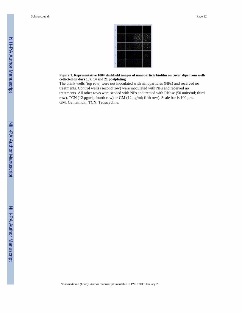
Figure 1. Representative 100× darkfield images of nanoparticle biofilm on cover slips from wellscollected on days 1, 7, 14 and 21 postplatingThe blank wells (top row) were not inoculated with nanoparticles (NPs) and received notreatments. Control wells (second row) were inoculated with NPs and received notreatments. All other rows were seeded with NPs and treated with RNase (50 units/ml; thirdrow), TCN (12 μg/ml; fourth row) or GM (12 μg/ml; fifth row). Scale bar is 100 μm.GM: Gentamicin; TCN: Tetracycline.
Schwartz et al. Page 12
Nanomedicine (Lond). Author manuscript; available in PMC 2011 January 20.
NIH
-PA Author Manuscript
NIH
-PA Author Manuscript
NIH
-PA Author Manuscript
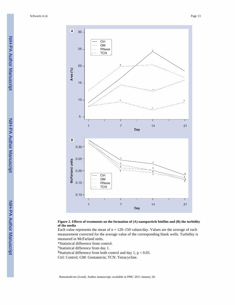
Figure 2. Effects of treatments on the formation of (A) nanoparticle biofilm and (B) the turbidityof the mediaEach value represents the mean of n = 120–150 values/day. Values are the average of eachmeasurement corrected for the average value of the corresponding blank wells. Turbidity ismeasured in McFarland units.*Statistical difference from control.‡Statistical difference from day 1.§Statistical difference from both control and day 1, p < 0.05.Ctrl: Control; GM: Gentamicin; TCN: Tetracycline.
Schwartz et al. Page 13
Nanomedicine (Lond). Author manuscript; available in PMC 2011 January 20.
NIH
-PA Author Manuscript
NIH
-PA Author Manuscript
NIH
-PA Author Manuscript
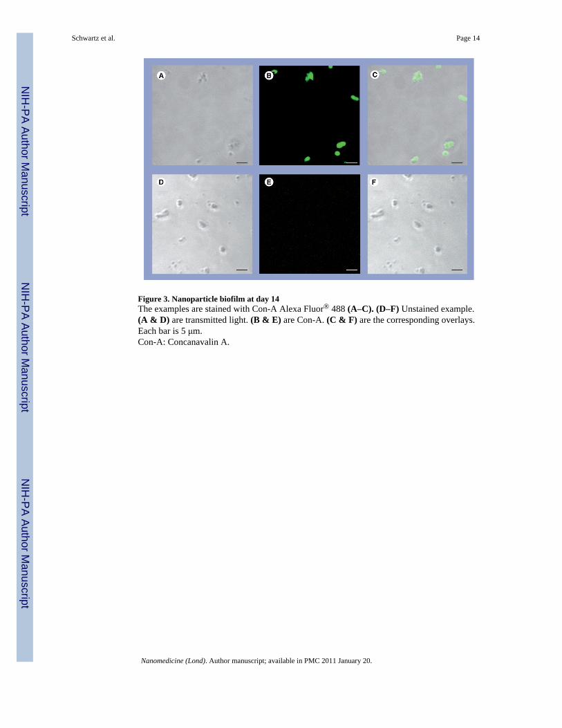
Figure 3. Nanoparticle biofilm at day 14The examples are stained with Con-A Alexa Fluor® 488 (A–C). (D–F) Unstained example.(A & D) are transmitted light. (B & E) are Con-A. (C & F) are the corresponding overlays.Each bar is 5 μm.Con-A: Concanavalin A.
Schwartz et al. Page 14
Nanomedicine (Lond). Author manuscript; available in PMC 2011 January 20.
NIH
-PA Author Manuscript
NIH
-PA Author Manuscript
NIH
-PA Author Manuscript
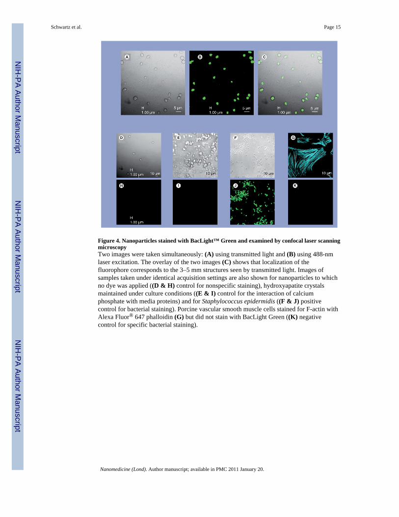
Figure 4. Nanoparticles stained with BacLight™ Green and examined by confocal laser scanningmicroscopyTwo images were taken simultaneously: (A) using transmitted light and (B) using 488-nmlaser excitation. The overlay of the two images (C) shows that localization of thefluorophore corresponds to the 3–5 mm structures seen by transmitted light. Images ofsamples taken under identical acquisition settings are also shown for nanoparticles to whichno dye was applied ((D & H) control for nonspecific staining), hydroxyapatite crystalsmaintained under culture conditions ((E & I) control for the interaction of calciumphosphate with media proteins) and for Staphylococcus epidermidis ((F & J) positivecontrol for bacterial staining). Porcine vascular smooth muscle cells stained for F-actin withAlexa Fluor® 647 phalloidin (G) but did not stain with BacLight Green ((K) negativecontrol for specific bacterial staining).
Schwartz et al. Page 15
Nanomedicine (Lond). Author manuscript; available in PMC 2011 January 20.
NIH
-PA Author Manuscript
NIH
-PA Author Manuscript
NIH
-PA Author Manuscript
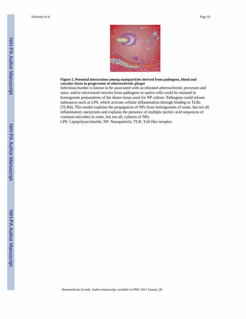
Figure 5. Potential interactions among nanoparticles derived from pathogens, blood andvascular tissue in progression of atherosclerotic plaqueInfectious burden is known to be associated with accelerated atherosclerotic processes andnano- and/or microsized vesicles from pathogens or native cells could be retained inhomogenate preparations of the donor tissue used for NP culture. Pathogens could releasesubstances such as LPS, which activate cellular inflammation through binding to TLRs(TLR4). This model explains the propagation of NPs from homogenates of some, but not all,inflammatory aneurysms and explains the presence of multiple nucleic acid sequences ofcommon microbes in some, but not all, cultures of NPs.LPS: Lipopolysaccharide; NP: Nanoparticle; TLR: Toll-like receptor.
Schwartz et al. Page 16
Nanomedicine (Lond). Author manuscript; available in PMC 2011 January 20.
NIH
-PA Author Manuscript
NIH
-PA Author Manuscript
NIH
-PA Author Manuscript
NIH
-PA Author Manuscript
NIH
-PA Author Manuscript
NIH
-PA Author Manuscript
Schwartz et al. Page 17
Table 1
Bacteria and fungi cultured from human abdominal aortic aneurysms*.
Male Female
Aspergillus 1
Bacillus 1
Bacillus (Gram negative) 1 1
Bacillus (Gram positive) 1
Bacteroides fragilis 1
Beauveria 1
Coccus resembeling Staphylococcus 6 1
Corynebacterium 1
Escherichia coli 4
Enterococcus 2
Geotrichum 1
Histoplasma capsulatum 1
Klebsiella oxytoca 1
Klebsiella pneumonia 2
Lactobacillus 1
Peptostreptococcus saccharolyticus 1
Propionibacterium acnes 3
Pseudomonas aeruginosa 1
Salmonella species 1
Serratia marcescens 1
Staphylococcus aureus 1 4
Staphylococcus, coagulase negative 16 3
Streptococcus 3
Streptococcus pneumoniae 1
Streptococcus virdans 2 2
Yeast 1
Numbers represent those showing positive cultures for each type of microbe. Some aneurysms were positive for multiple organisms.
*Aneurysms were collected from 48 patients.
Nanomedicine (Lond). Author manuscript; available in PMC 2011 January 20.
Copyright © 2022 FDOKUMEN