Environmental and biofilm-dependent changes in a Bacillus ...
15
HAL Id: hal-00641810 https://hal.archives-ouvertes.fr/hal-00641810 Submitted on 29 May 2020 HAL is a multi-disciplinary open access archive for the deposit and dissemination of sci- entific research documents, whether they are pub- lished or not. The documents may come from teaching and research institutions in France or abroad, or from public or private research centers. L’archive ouverte pluridisciplinaire HAL, est destinée au dépôt et à la diffusion de documents scientifiques de niveau recherche, publiés ou non, émanant des établissements d’enseignement et de recherche français ou étrangers, des laboratoires publics ou privés. Copyright Environmental and biofilm-dependent changes in a Bacillus cereus secondary cell wall polysaccharide T. Candela, Emmanuel Maes, Estelle Garénaux, yoann Rombouts, Frédéric Krzewinski, Michel Gohar, yann Guerardel To cite this version: T. Candela, Emmanuel Maes, Estelle Garénaux, yoann Rombouts, Frédéric Krzewinski, et al.. En- vironmental and biofilm-dependent changes in a Bacillus cereus secondary cell wall polysaccharide. Journal of Biological Chemistry, American Society for Biochemistry and Molecular Biology, 2011, 286 (36), pp.31250-31262. 10.1074/jbc.M111.249821. hal-00641810
-
Upload
khangminh22 -
Category
Documents
-
view
1 -
download
0
Transcript of Environmental and biofilm-dependent changes in a Bacillus ...

HAL Id: hal-00641810https://hal.archives-ouvertes.fr/hal-00641810
Submitted on 29 May 2020
HAL is a multi-disciplinary open accessarchive for the deposit and dissemination of sci-entific research documents, whether they are pub-lished or not. The documents may come fromteaching and research institutions in France orabroad, or from public or private research centers.
L’archive ouverte pluridisciplinaire HAL, estdestinée au dépôt et à la diffusion de documentsscientifiques de niveau recherche, publiés ou non,émanant des établissements d’enseignement et derecherche français ou étrangers, des laboratoirespublics ou privés.
Copyright
Environmental and biofilm-dependent changes in aBacillus cereus secondary cell wall polysaccharide
T. Candela, Emmanuel Maes, Estelle Garénaux, yoann Rombouts, FrédéricKrzewinski, Michel Gohar, yann Guerardel
To cite this version:T. Candela, Emmanuel Maes, Estelle Garénaux, yoann Rombouts, Frédéric Krzewinski, et al.. En-vironmental and biofilm-dependent changes in a Bacillus cereus secondary cell wall polysaccharide.Journal of Biological Chemistry, American Society for Biochemistry and Molecular Biology, 2011, 286(36), pp.31250-31262. �10.1074/jbc.M111.249821�. �hal-00641810�

Environmental and Biofilm-dependent Changes in a Bacilluscereus Secondary Cell Wall Polysaccharide*□S
Received for publication, April 12, 2011, and in revised form, July 21, 2011 Published, JBC Papers in Press, July 22, 2011, DOI 10.1074/jbc.M111.249821
Thomas Candela‡§1,2, Emmanuel Maes¶�1, Estelle Garenaux¶�, Yoann Rombouts¶�, Frederic Krzewinski¶�,Michel Gohar‡, and Yann Guerardel¶�3
From the ¶Universite de Lille1, Unite de Glycobiologie Structurale et Fonctionnelle, F-59650 Villeneuve d’Ascq, France, �CNRS, UMR8576, F-59650 Villeneuve d’Ascq, France, ‡Micalis, INRA (UMR1319), Biofilms and Regulation Genetique chez les Bacillacees,Domaine de Vilvert, 78352 Jouy-en-Josas, France, and §Micalis, INRA (UMR1319), Genetique Microbienne and Environnement,Domaine de Vilvert, 78352 Jouy-en-Josas, France
Bacterial species from the Bacillus genus, including Bacilluscereus and Bacillus anthracis, synthesize secondary cell wallpolymers (SCWP) covalently associated to the peptidoglycanthrough a phospho-diester linkage. Although such componentswere observed in a wide panel of B. cereus and B. anthracisstrains, the effect of culture conditions or of bacterial growthstate on their synthesis has never been addressed. Herein weshow that B. cereus ATCC 14579 can synthesize not only one,as previously reported, but two structurally unrelated sec-ondary cell wall polymers (SCWP) polysaccharides. The firstof these SCWP, 34)[GlcNAc(�1–3)]GlcNAc(�1–6)[Glc(�1-3)][ManNAc(�1–4)]GalNAc(�1–4)ManNAc(�13, althoughpresenting an original sequence, fits to the already described thecanonical sequence motif of SCWP. In contrast, the secondpolysaccharidewasmade up by a totally original sequence,36)-Gal(�1–2)(2-R-hydroxyglutar-5-ylamido)Fuc2NAc4N(�1-6)GlcNAc(�13, which no equivalent has ever been identified inthe Bacillus genus. In addition, we established that the synthe-ses of these two polysaccharides were differently regulated. Thefirst one is constantly expressed at the surface of the bacteria,whereas the expression of the second is tightly regulated by cul-ture conditions and growth states, planktonic, or biofilm.
The cell wall surrounding cytoplasmicmembrane is of primeimportance in bacteria survival and adaptation to their environ-ment. In Gram-positive bacteria, the cell wall includes a thickmultilayer peptidoglycan to which most often are associatedother cell-surface structures called secondary cell wall poly-mers (SCWPs)4 (1). These secondary cell wall polymers includelipoteichoic acids anchored in the outer leaflet of plasmicmem-
brane as well as teichoic acids, teichuronic acids, or neutralpolysaccharides (SCWP polysaccharides), which are covalentlybound, through a phosphodiester link, to theN-acetylmuramicacid moieties of the peptidoglycan. SCWPs are essential inBacillus subtilis and were shown to be involved in the bacterialvirulence in Staphylococcus aureus (2). In addition, SCWPsbind SLH (S-layer homology) domains containing proteins,thus anchoring them non-covalently to the cell wall (3). Theseproteins are involved in various functions, including pepti-doglycan maturation (4, 5), binding to host tissues (6), or pro-tein degradation (7). Teichoic acids and the secondary cell wallpolysaccharide were also reported to be involved in biofilm for-mation (8–10).Biofilms are multicellular structures attached to a solid or a
liquid surface and embedded in a self-produced matrix. Thismatrix is made of polymers, mostly polysaccharides, DNA,and/or proteins, provides cohesion to the bacterial community(11–13), and acts as a shield, protecting bacteria within thebiofilm. Biofilms are, therefore, persistent structures, resistingdesiccation, cleaning procedures, and antimicrobial substances(14), whichmakes thema challenge in humanhealth and indus-trial processes. Several species from the Bacillus genus, includ-ing B. subtilis, Bacillus cereus and Bacillus anthracis, have beenidentified to form biofilms in a wide range of different environ-ments (15–17). In B. cereus, the formation of biofilms isenhanced by low nutrient supplies and requires the presence ofbiosurfactants (18). B. cereus is a Gram-positive, spore-formingbacterium, genetically close to Bacillus thuringiensis, a patho-gen of insects used in crop protection, and to B. anthracis, alethal pathogen of mammals previously used as a weapon inbioterrorism acts. This bacterium is an opportunistic pathogenfrequently diagnosed in gastroenteritis cases (19) but alsoinvolved in other human diseases, including endophthalmitisand meningitis (20). It produces a high number of virulencefactors,most of themcontrolled by amaster virulence regulator(21). In addition, spores and biofilms produced by B. cereus arepersistent contaminants of the food industry equipment (22).B. cereus cell surface displays teichoic acids and secondary
cell wall polysaccharides (23–26). These secondary cell wallpolysaccharides were suggested to represent in this bacteriumand its close relatives a virulence-associated carbohydrate anti-
* This work was supported by the Conseil Regional Nord-Pas de Calais ARCir(to Y. G.), the Ministere de l’Enseignement Superieur (to Y. R.), and theRegion Ile de France (to T. C.). Research on the 800 and 900 MHz spectrom-eters was supported by the TGE RMN THC Fr3050.
□S The on-line version of this article (available at http://www.jbc.org) containssupplemental Figs. S1–S7.
1 Both authors equally contributed to this study.2 Present address: Faculte de Pharmacie ParisXI sud, EA 4043-Ecosysteme
Microbien Digestif and Sante, Chatenay Malabry 92290, France.3 To whom correspondence should be addressed. E-mail: yann.guerardel@
univ-lille1.fr.4 The abbreviations used are: SCWP, secondary cell wall polymer; PS, polysac-
charide; Ch HF-PS, for charged HF-PS; Ne HF-PS, neutral HF-PS; TOCSY, totalcorrelation spectroscopy; HMBC, heteronuclear multiple bond coherence;DATDH, diaminotrideoxyhexose; HR-MAS, high resolution magic angle
spinning; HCT, hydrolysate of casein Tryptone; BHI, brain heart infusion;HSQC, heteronuclear single quantum correlation.
THE JOURNAL OF BIOLOGICAL CHEMISTRY VOL. 286, NO. 36, pp. 31250 –31262, September 9, 2011© 2011 by The American Society for Biochemistry and Molecular Biology, Inc. Printed in the U.S.A.
31250 JOURNAL OF BIOLOGICAL CHEMISTRY VOLUME 286 • NUMBER 36 • SEPTEMBER 9, 2011
at INR
A Institut N
ational de la Recherche A
gronomique on M
ay 13, 2019http://w
ww
.jbc.org/D
ownloaded from

genmotif (23). The exact structures of the secondary cell wallpolysaccharides from two B. cereus strains were recentlycompared with those of B. anthracis Ames after their releasefrom the cell wall using aqueous hydrogen fluoride (HF). Thedetailed analysis of the peptidoglycan HF-labile fractionsestablished the occurrence of the consensus sequence (�-HexNAc-�-ManNAc-�-GlcNAc) in all three strains (24, 27). Inparticular, the backbone of the secondary cell wall polysaccha-rides isolated from B. anthracisAmes and B. cereus 10987 wereshown to be composed of structurally related trisacchariderepeating units differentially substituted by a strain-specificpattern of �- and �-D-galactosidase residues. In comparison,B. cereus 14579 strain appears to synthesize similar but morecomplex polysaccharides containing the consensus trisaccha-ride sequence, the structure of which is, however, still elusive(24).Although much information is currently available on the
structure of cell wall-associated components, including teichoicacids and secondary cell wall polysaccharides, nothing is cur-rently known about the evolution of these components inB. cereus and relatives species along the bacterial growth and inparticular during the transition between planktonic and biofilmmodes of life. Previous reports established, however, thatgrowth conditions may influence their cell wall compositions.Particularly, in B. subtilis, teichoic acids are replaced by teichu-ronic acids during phosphate starvation (28). Such structuralmodifications of the bacterial cell wallmay have an influence onthe overall physicochemical properties of the cell surface thatregulate bacterial interactions with substratum. Here we inves-tigated the changes undergone by the secondary cell wall poly-saccharide in the B. cereusATCC 14579 strain during bacterialexponential and stationary growth and between bacteria inplanktonic or in biofilm states. We first established that thepolysaccharide fraction released by HF from B. cereus strainATCC 14579 grown in planktonic conditions was composed oftwo distinct polysaccharides presenting very different struc-tural features. Whereas one presented strong similarities withHF-released polysaccharide (HF-PS) isolated from B. cereus10987 (24), the other one exhibited totally original structuralfeatures. Then, we demonstrated that these two polysaccha-rides were differentially regulated along the growth of bacteriain the biofilm phase but not in the planktonic phase.
EXPERIMENTAL PROCEDURES
Bacterial Strains and Culture Conditions—Biofilms wereproduced as floating pellicles with the B. cereus ATCC 14579strain in HCT medium (29) at 37 °C in glass tubes (30) or in50-ml beakers. Biofilms in 50-ml beakers were obtained in thesame way as in glass tubes. Planktonic cultures were performedwith the ATCC14579 strain in HCT or in BHI culture media at37 °C and 175 rpm. InHCT, planktonic cultures were harvestedat 5, 10, 15, and 20 h post-inoculation, and biofilms were har-vested at 28, 48, 72, and 96 h post-inoculation. These timescalesspanned the whole life of ATCC14579 planktonic cultures orbiofilms in a poor medium. Planktonic cultures in BHI (a richmedium) were harvested at an optical density of 4.0 (6 hpost-inoculation).
Preparation of Polysaccharide Fractions—Secondary cell wallpolymers were extracted similarly to the peptidoglycan (31),with an additional hydrolysis step using hydrofluoric acid. Thebacterial cells, harvested fromplanktonic cells or frombiofilms,were boiled for 10min in 40ml of (50mMTris-HCl, pH 7.4, 150mMNaCl, 1% SDS) and spun down at 6500 � g for 10min. Thisstep was repeated once to obtain only the peptidoglycantogetherwith its covalently linked secondary cell wall polymers.The resulting pellet was washed in 40ml of 50 mMTris, pH 7.4,sonicated, and centrifuged at 50,000 � g for 20 min. Theremaining contaminants were discarded by treating the result-ing pellet, solubilized in 20 mM MgSO4, successively by DNase(1 mg/ml) and RNase (5 mg/ml), then by proteinase K (20�g/ml at room temperature for 12 h) in the presence of 10 mM
CaCl2, and finally by SDS (1% at 100 °C for 10 min.). The insol-uble material, recovered after centrifugation at 50,000 � g for20min, was rinsed in purewater and treated successively with 8M LiCl2 and with 100 mM EDTA. The resulting insoluble mate-rial, spun down at 50,000 � g for 20 min, was rinsed twice inpure water, and digested with 46% hydrofluoric acid for 48 h atroom temperature. After centrifugation at 20,000 � g for 10min, the supernatant containing the secondary cell wall poly-mers was precipitated with 3 volumes of ethanol. The precipi-tate was then dried and stored at �20 °C until use.Mild Hydrolysis of Polysaccharide Fractions—Total polysac-
charide fraction was hydrolyzed repetitively under mild condi-tions (0.1 M trifluoroacetic acid at 80 °C for 1 h). After eachhydrolysis step, four volumes of ethanol were added, and theliberated oligosaccharides were collected in the ethanol frac-tion. Release of oligosaccharides from total polysaccharide wasassessed by TLC analysis on silica gel using butanol/acetic acid/H2O (40/30/20) as solvent. Completeness of the reaction wasassessed by dosing carbohydrate by phenol-sulfuric stainingand weighing of the ethanol soluble fraction. Typically, fourhydrolysis steps were sufficient to liberate all hydrolysablematerial. Oligosaccharides were further purified by gel filtra-tion on a Bio-Gel P-2 column.Anion Exchange Chromatography of Polysaccharide—Total
polysaccharide was fractionated into a neutral and an anionicfraction on a HPLC apparatus fitted with a strong anionexchange columnMonoQ (Amersham Biosciences). The sam-ple was loaded and eluted with a flow rate of 0.5ml/minwith 20mM Tris-HCl (pH 8.0) followed by a NaCl gradient (0–10 min,0 M; 10–60 min, 0–0.6 M; 60–65 min, 0.80 M) in 20 mM Tris-HCl (pH 8.0).Glycosyl LinkageAnalysis—Linkage analyses ofmonosaccha-
rides were achieved by permethylation followed by hydrolysis,reduction, and derivatization with acetyl groups. Briefly, sam-ples were permethylated according toCiucanu (32), hydrolyzedin 4 M trifluoroacetic acid for 4 h at 100 °C, and then reducedwith sodium borohydride in 0.05 M NH4OH for 4 h. Reductionwas stopped by dropwise addition of acetic acid until pH 6 wasreached, and borate salts were co-distilled by repetitive evapo-ration in drymethanol. Partiallymethylated, reducedmonosac-charides were acetylated in 500 �l of acetic anhydride at 100 °Cfor 2 h. Solution was dried and partially methylated, and acety-lated reduced monosaccharides were extracted three times inchloroform.
B. cereus Synthesizes Two SCWP Polysaccharides
SEPTEMBER 9, 2011 • VOLUME 286 • NUMBER 36 JOURNAL OF BIOLOGICAL CHEMISTRY 31251
at INR
A Institut N
ational de la Recherche A
gronomique on M
ay 13, 2019http://w
ww
.jbc.org/D
ownloaded from

NMR Analysis—After two exchanges with 2H2O (Eurisotop-Saclay France), sample was dissolved in pureD2O. Experimentswere recorded at various temperatures (280, 300, 343 K), butonly spectra recorded at 300 K are shown. The pD was keptneutral. Experiments were recorded on Bruker spectrometersat three different fields. 9.4, 18.8, and 21.6 tesla were 1H-reso-nated at 400.33, 800.12, and 900.11 MHz but 13C-resonated at100.2, 200.3, and 220.0 MHz, respectively. 31P spectrum wasrecorded at 9.4 tesla. Pulse programs used were extracted fromBruker pulse program library where pulses and delays wereoptimized for each experiment. For TOCSY experiments, 40,60, 80, and 100 ms were used for mixing time in spectrarecording.HR-MAS NMR experiments were achieved on an 18.8 T
Avance II Bruker spectrometer. The experiments wereacquired with a 13C,1H/31P/2H probe with uniaxial gradients.Before analysis, cell-pellets were washed twice with deuteriumoxide (Euriso-top, Gif-sur-Yvette, France). The 4-mm ZrO2rotors (CortecNet, Paris, France) were filled with 50 �l of cellpellets including 0.5 �l of acetone as the internal standard andfinally centrifuged at 3000 rpm. All spectra were recorded at293 K, and the rotor spinning rate was 8 kHz. All experimentscame from the Bruker library pulse program, and delays andpowerswere optimized for each. For 13C,1HHSQC, the spectralwidths were 12.820Hz (1H) with 1.024 points for free inductiondecay resolution and 29.994 Hz (13C) for 400 scans, giving 12.5and 75.0 Hz/point respectively.Mass Spectroscopy—MALDI-MS and MS/MS analyses of
polysaccharides were performed on 4800 Proteomics Analyzer(Applied Biosystems, Framingham, MA) mass spectrometer,operated in the reflectron mode. For MS acquisition, 5 �l ofdiluted samples in H2Oweremixed with 5 �l of 2,5-dihydroxy-benzoic acid matrix solution (10 mg/ml dissolved in H2O/CH3OH (1:1, v/v)). The mixtures (2 �l) were then spotted onthe target plate and air-dried. Peaks observed in theMS spectrawere selected for further MS/MS. Collision-induced dissocia-tion MS/MS data comprise a total of 100 subspectra of 3000laser shots. Two or more spectra can be combined post-acqui-sition with mass tolerance set at 0.1 Da to improve the signal-
to-noise ratio. The potential difference between the sourceacceleration voltage and the collision cell was set to 1 kV, andargon was used as the collision gas.
RESULTS
Comparison of Polysaccharides—In a first approach, polysac-charides were released by aqueous hydrogen fluoride from dif-ferent growth times between 5 and 20 h or between 28 and 96 hpost-inoculation for planktonic and biofilm cultures, respec-tively, then analyzed by 1HNMR. As observed on Fig. 1, growthtime did not have any visible influence on polysaccharide struc-ture in planktonic phase, whereas it did in the biofilm phase. Inparticular, polysaccharides extracted from early (28 and 48 h)and late (72 and 96 h) biofilms exhibited differentNMR spectra.Surprisingly, spectra from late biofilms were identical to allplanktonic polysaccharides, whereas early biofilms showedidentical simpler spectra. These were characterized by the dis-appearance of many NMR signals, in particular the easilyobserved signals at 4.91, 2.52, and 1.13 ppm, all other signalsbeing identical. Thus, to appreciate the origin and the extent ofthe structural modifications that take place in the surface poly-saccharide during biofilm growth, we first focused our study onthe structural elucidation of the complex polysaccharideexpressed in planktonic and late biofilm phases.Analysis of the Planktonic Phase Polysaccharide—A 13C,1H
HSQCNMR experiments of total polysaccharide isolated from5–20 h planktonic phase permitted identification of nine ano-meric signals (I to IX) out of which four were tentativelyassigned to� anomers (1JH,C � 170Hz, I, II, IV,V) and five to�anomers (1JH,C � 160 Hz, III, VI, VII, VIII, IX) (Fig. 2A). Fur-thermore, eight 13C,1H signals were assigned toN-acetylated orN-acylated carbons according to their high field deshieldedNMR parameters (Fig. 2D). These were later assigned as C2 ofresidues I, III, IV,V,VI,VII, andVIII as well as C4 of I residuesby two-dimensional 1H,1H NMR experiments. Altogether,these experiments established the presence of nine monosac-charides, out of which seven were assigned to N-acetylatedhexosamines and two to neutral hexoses.However, total assign-ment of individual spin systems was delicate at this stage of the
FIGURE 1. Structural mapping of HF-PS isolated from planktonic and biofilm phases of B. cereus ATCC 14579. 1H NMR spectra of HF-PS from planktonic(A) and biofilm (B) phases at four different growth times are shown. Spectra from all planktonic time points were identical, whereas spectra from early (28 and48 h) differed from late biofilms (72 and 96 h). Three easily identified signals at 4.91, 2.52, and 1.13 ppm that show variations along growth times are highlightedin gray.
B. cereus Synthesizes Two SCWP Polysaccharides
31252 JOURNAL OF BIOLOGICAL CHEMISTRY VOLUME 286 • NUMBER 36 • SEPTEMBER 9, 2011
at INR
A Institut N
ational de la Recherche A
gronomique on M
ay 13, 2019http://w
ww
.jbc.org/D
ownloaded from

study because of numerous overlaps of anomeric signals both inthe proton (II and V; I and VI; VII and VIII) and in the carbon(I, IV, and V; II and VI; III and VIII) dimensions (Fig. 2A).Thus, to simplify the NMR spectra and gain access to its finestructure, we separated the total polysaccharide in two frac-tions using two different strategies (Fig. 3A).Fractionation of Polysaccharide—The first strategy consisted
of separating the native polysaccharides by strong anionexchange chromatography assuming that the total polysaccha-ridewas constituted by two distinctmacromolecules. As shownon Fig. 3B, the elution profile of intact polysaccharide on astrong anion exchange chromatography column by a gradientof sodium chloride generated two fractions; that is, one non-retained fraction and one retained fraction that were furtherindividually analyzed. As will be explained in the followingchapters, the retained polysaccharide, called Ch HF-PS forcharged HF-PS, included one free carboxyl group that con-ferred a negative charge, whereas the non-retained polysaccha-ride was devoid of acidic group and was thus called Ne HF-PSfor neutral HF-PS.The second strategy consisted of repetitively hydrolyzing the
polysaccharide in mild acid conditions to generate two frac-tions, one acid stable and one acid labile. Liberated oligosaccha-rides were extracted by ethanol after each hydrolysis step,whereas the non-labile fraction was submitted to another
hydrolysis step. These conditions, which were devised to max-imize liberation of acidic labile domain as repeating units bypreserving the acidic resistant domain, have been previouslysuccessfully used to generate fucosylated oligosaccharides fromsponge polysaccharides (33). Mild acid hydrolysis of intactpolysaccharide (HF-PS) permitted the generation of two frac-tions presenting different solubility in 70% alcohol solution(Fig. 3A). The stepwise monitoring of hydrolysis processshowed that five hydrolysis steps were enough to cleave andliberate as an ethanol-soluble fraction all the acid labile fractionfrom the total polysaccharide, which represented about 70% ofthe initial polysaccharide (Fig. 3C). Structural analysis of acidstable and acid labile fractions (see after) showed that these twofractions presented identical structural features to Ne HF-PSand Ch HF-PS separated by anion exchange chromatography,albeit the depolymerization of Ch HF-PS after acid hydrolysis.Thus both strategies lead to the separation of HF-PS in twodistinct fractions, Ne HF-PS and Ch HF-PS.As shown in Fig. 2, B and E, Ch HF-PS and Ne HF-PS exhib-
ited totally different 13C,1H HSQC NMR spectra, establishingthat they were devoid of any trans-contaminating material.Comparison of all fractions (Fig. 2) showed that the 13C,1HHSQCNMR spectrum of total HF-PS from planktonic phase isthe result of the combination of Ch HF-PS and Ne HF-PS sub-spectra. Individual signal assignments established that Ch
FIGURE 2. 13C,1H HSQC NMR spectra of HF-PS from planktonic and biofilm phases of B. cereus ATCC 14579. Shown are the anomer regions (A–C) andnitrogen-bearing carbon regions (E–F) of 13C,1H HSQC spectra of intact HF-PS from planktonic phase (A and D), Ne HS-PS and Ch HF-PS fractions generated bymild hydrolysis from planktonic phase (B and E), and 24-h biofilm phase (C and F). In B and E, blue signals correspond to Ne HS-PS ,and red signals to Ch HF-PSfrom planktonic phase.
B. cereus Synthesizes Two SCWP Polysaccharides
SEPTEMBER 9, 2011 • VOLUME 286 • NUMBER 36 JOURNAL OF BIOLOGICAL CHEMISTRY 31253
at INR
A Institut N
ational de la Recherche A
gronomique on M
ay 13, 2019http://w
ww
.jbc.org/D
ownloaded from

HF-PS consisted of threemonosaccharide units (signals I to III)and NeHF-PS of six units (IV to IX). Separation of polysaccha-ride in two fractions resolved most of the overlapping NMRsignals and further enabled an exact interpretation of spectra ofindividual polysaccharides. Unexpectedly, we observed thatII-H1 and III-H1chemical shifts slightly changed when ChHF-PS was analyzed alone (II-H1 �� � 0.024; III-H1 ���0.028) or in amixturewithNeHF-PS.Although this phenom-enon was never reported to our knowledge, we suggest thatthese variations arose from sugar-sugar interaction phenomenain a similar fashion that is observed during protein-proteininteractions (34).Altogether, these data strongly suggested that the total
SCWP polysaccharide was constituted by two independentpolysaccharides; one charged and acid labile polysaccharide(ChHF-PS) and another neutral and acid stable polysaccharide(Ne HF-PS). Structural analysis of these two polysaccharideswill permit (1) the establishment of the nature of the struc-tural differences between polysaccharides and (2) determinewhether the structural differences between phases arise fromthe modification of a common polysaccharide core or from thespecific induction of the two different polysaccharides.Analysis of Neutral Polysaccharide Ne HF-PS—Six anomer
signals (IV to IX) were identified on the 13C,1H HSQC spec-trum of Ne HF-PS, establishing the presence of six differentmonosaccharide units, out of which five appeared to be hexo-samines, according to the observation of five N-acetylated C2signals between 49 and 57 ppm (IV- toVIII-C2). The nature ofindividual monosaccharides was established by a combination
of NMR analysis of polysaccharide and gas chromatographyanalysis of partially methylated and acetylated reduced mono-saccharides. In particular, spin systems of all units wereassigned by NMR analysis using two-dimensional COSY,TOCSY, nuclear Overhauser effect spectroscopy (NOESY),HSQC (decoupled or not), and HMBC experiments, andsequencing information was obtained by 1H,1H NOESY and13C,1H HMBC experiments.Large 1JH,C coupling constant (178 Hz) and the deshielded
IV-H1 value at 5.588 ppm established that IV unit exhibited an� configuration. Then, the total spin system and the 1H,1H vic-inal coupling constant pattern of IV in which 3JH1,H2, 3JH2,H3,3JH3,H4, and 3JH4,H5 were small (S), large (L), S and S (S/L/S/S)showed that this residue had an �-galacto configuration (35).Finally, the upfield chemical shifts of IV-H2/C2 at � 4.551/49.72and the presence of an N-acetamido group at 1.999/23.62 ppmdemonstrated that IV was an �-GalNAc residue (Table 1, Fig.4). Deshielding of IV-C3, -C4, and -C6 chemical shift values at�77.1, 75.2, and 68.6 compared with unsubstituted �-GalNAcresidue (C3, �� � 8.5; C4, �� �5.5, and C6, �� �6.2) estab-lished that IV residuewas trisubstituted in C3, C4, andC6 posi-tions, in accordance with the splitting of H6 and H6� signals at3.907 and 3.636 ppm (36). The presence of this unusual trisub-stituted GalNAc residue was confirmed by linkage analysis ingas chromatography coupled to mass spectrometry (supple-mental Fig. S1). Similarly, despite the strong coupling constantthat prevented the attribution of vicinal 3JH1,H2 and 3JH2,H3 cou-pling constants, the unit VI could be assigned to a �-D-Man-NAc unit based on the attribution of the spin system and of the
FIGURE 3. Purification schemes of HF-PS components. Two distinct components were separated using two different procedures. A, Ch HF-PS and Ne HF-PSwere purified from total HF-PS by either mild acid hydrolysis or anion exchange chromatography. B, anion exchange chromatography separated a chargedretained fraction (Ch HF-PS) from a neutral non-retained fraction (Ne HF-PS). C, mild acid hydrolysis followed by ethanol precipitation generated an acid labilefraction from an acid stable fraction, which subsequent structural analysis identified as Ch HF-PS and Ne HF-PS, respectively. The efficiency of acid hydrolysiswas monitored along the hydrolysis steps (C). All purified fractions, as well as total HF-PS and intact B. cereus cells were analyzed by different combinations ofanalytical methods. *, contaminant non-carbohydrate signal.
B. cereus Synthesizes Two SCWP Polysaccharides
31254 JOURNAL OF BIOLOGICAL CHEMISTRY VOLUME 286 • NUMBER 36 • SEPTEMBER 9, 2011
at INR
A Institut N
ational de la Recherche A
gronomique on M
ay 13, 2019http://w
ww
.jbc.org/D
ownloaded from

remaining 1H,1H and 13C,1H coupling constant pattern. As forIV, observation ofVI-C2 at 49.6 ppm and ofN-acetyl group at �2.092/23.62 backed up its attribution to a hexosamine residue.Strong coupling constants also occurred between VIII-H2,-H3, and -H4 that resulted in identical chemical shifts for thesethree signals at 3.721 ppm irrespective of the experimental con-ditions. However, 13C,1H anomeric signal at � 4.471/96.6 with
1JH1,C1 of 163 Hz as well as the VIII-C2 signal at � 56.4 typifiedthat that unit was a �-N-acetylhexosamine unit. Such a strongcoupling constants pattern is often observed for �-GlcNAc res-idues linked to �-HexNAc in C6 position. On this basis, weproposed that the VIII unit was a �-GlcNAc residue. Then,deshielded VIII-C3 and VIII-C4 signals at 80.3 and 73.6 ppmestablished that it was substituted in both positions in accor-dance with the observation of a 6-methyl-3,4-diacetyl-2-N-methyl-acetyl hexosaminitol residue in GC/MS (data notshown).Strong inter-residue effects 1JH1,C1 observed in rotating from
Overhauser enhancement spectroscopy experiments demon-strated that VIII was linked to IV, IV, to VI and VI to VIII,which established the presence of a linear polysaccharide corecomposed of the repeating sequence VIII-IV-VI (supplementalFig. S2). HMBC experiments permitted establishing the sequenceIV(1–4)VI(1–4)VIII because of 3JH,C IV-H1/VI-C4 and VI-H1/VIII-H4 correlations (data not shown). Finally, a strong nuclearOverhauser effect between VIII-H1 and IV-H6 and H6� showedthat the VIII unit substituted IV in C6 position, establishing thesequence of the core polysaccharide from Ne HF-PS as34)GlcNAc(�1–6)GalNAc(�1–4)ManNAc(�13.
As established by the respective chemical shifts of VIII-C3,IV-C3, and IV-C4 at 80.3, 77.08, and 75.20 ppm, this trisaccha-ride unit is further substituted in three positions by the remain-ing yet unidentified residues V, VII, and IX. The configurationof unitVwas determined using undecoupled 13C,1HHSQC, 1Dselective 1H TOCSY with various mixing times, 1H,1H two-dimensional COSY and TOCSY NMR experiments. 1JH1,C1coupling constant of 171Hz and chemical shift ofV-C2 at 55.20ppm indicated that V was a 2-N-acetylated monosaccharidewith an �-configuration. Then residue V was assigned to an�-ManNAc residue based on its 3JH,H S/S/L/L coupling con-stants pattern and attribution of total spin system. All carbonsresonated like unsubstituted carbons (Table 1), establishingthat V was a terminal non-reducing �-ManNAc residue. TheunitVIIwas characterized as a �-GlcNAc residue based on theobservation of total vicinal coupling constants on serial 1H,1HCOSY and TOCSY spectra and of the direct coupling constant1JH,C at 160 Hz. 13C,1H values established that VII is an unsub-stituted �-GlcNAc residue present in terminal non-reducingend, unlike VIII, which is disubstituted in C3 and C4 positions(36). Finally, IXwas easily identified as a terminal non-reducing
TABLE 1Proton and carbon chemical shifts (ppm) of Ne HF-PSValues in parentheses correspond to direct coupling constant (1JH,C) between 1H and 13C of anomer position. Bold values correspond to carbon-bearing substitution.
Chemical shifts
ppmResidues 1 2 3 4 5 6/6� NAc�-GalpNAc (IV) 1H 5.588 4.551 3.956 4.258 4.086 3.907/3.636 1.999
13C 98.5 ( �178 Hz) 49.72 77.08 75.20 71.3 68.60 23.62�-ManpNAc (V) 1H 5.021 3.915 3.911 3.610 4.323 �3.9/3.7 2.092
13C 98.04 (�172Hz) 55.20 71.64 70.72 72.62 �62 23.62�-ManpNAc (VI) 1H 4.897 4.740 4.538 3.855 3.570 3.99/3.66 2,092
13C 100.93 (�164Hz) 49.63 75.8 70.1 76.3 62.4 23.62�-GlcpNAc (VII) 1H 4.521 3.592 3.480 3.298 3.473 3.99/3.66 2.064
13C 96.6 (�160Hz) 57.0 75.8 72.1 77.4 62.4 23.62�-GlcpNAc (VIII) 1H 4.471 3.721 3.721 3.721 3.55 3.99/3.66 2.125
13C 102.5 (�163Hz) 56.4 80.3 73.6 75.4 62.4 23.62�-Glcp (IX) 1H 4.428 3.139 3.473 3.350 3.55 3.99/3.66 no
13C 105.9 (�160Hz) 74.3 76.0 71.4 75.4 62.4 no
FIGURE 4. 13C,1H HSQC NMR analysis of Ne HF-PS from planktonic phase.Shown is 13C,1H HSQC NMR spectra of the bulk region (A) and anomer region(B) of Ne HF-PS.
B. cereus Synthesizes Two SCWP Polysaccharides
SEPTEMBER 9, 2011 • VOLUME 286 • NUMBER 36 JOURNAL OF BIOLOGICAL CHEMISTRY 31255
at INR
A Institut N
ational de la Recherche A
gronomique on M
ay 13, 2019http://w
ww
.jbc.org/D
ownloaded from

�-Glc residue because of its vicinal coupling constants patternL/L/L/L and its 13C,1H spin system (Table 1). The substitutionpattern of the VIII-IV-VI core repeating unit by residues V,VII, and IXwas established by a combination of 1H,1H rotatingframe Overhauser enhancement spectroscopy and 13C,1HHMBC NMR experiments (supplemental Fig. S2 and data notshown). A dense network of correlations including V-H1 3IV-H4, V-H13 IV-C4, IX-H13 IV-H3, IX-H13 IV-C3, andVII-H13VII-H3 permitted deduction of the final structure ofthe hexasaccharide repeating unit of the neutral polysac-charide as [GlcNAc(�1–3)]GlcNAc(�1– 6)[Glc(�1–3)][ManNAc(�1–4)]GalNAc(�1–4)ManNAc(�1–4).The nature of this repeating unit was confirmed by mass
spectrometry analysis. MALDI-TOF spectrum of Ne HF-PSshowed a very complex profile characterized by a large numberof signals between 794 and 8288 presentingm/z values intervalsof either 203 or 162 atomic mass units, attributed to HexNAc
and Hex, respectively (Fig. 5). All signals could be assigned topolymers containing from 6 (m/z 1200) to 42 (m/z 8288)mono-saccharide (HexNAc and Hex) units. However, two sets of sig-nals were identified; one corresponded to [M�Na]� adducts ofHexNAc- and Hex-containing polymers ranging from m/z1218 to 8288, the other to [M�Na]� oxonium-type adductsranging from m/z 1200 to 4735 (Fig. 5). Oxonium types arebelieved to be generated by in-source fragmentations of thepolysaccharide during high energy desorption from theMALDImatrix. Based onm/z calculations, a series of [M�Na]� signalsat m/z 1218, 2396, 3574, 4753, 5931, 7110, and 8288 wasassigned as polysaccharides containing from one to seven(HexNAc5Hex1) repeating units (Fig. 5). Similar oxonium-de-rived signals atm/z 1200, 2379, 3556, and 4735were assigned topolysaccharide fragments containing up to four repeating units.In addition, many intermediate values signals with HexNAc orHex increments were assigned. MS/MS fragmentation of the
FIGURE 5. Determination of the size distribution of Ne HF-PS from planktonic phase by MALDI-TOF-MS analysis. MS analysis in positive mode permittedidentification of polymers with [M�Na]� apparent molecular weights ranging from m/z 1218 to 8288. *, correspond to [M�Na-18]� oxonium type fragmentsgenerated by in source fragmentations of the polysaccharide core.
B. cereus Synthesizes Two SCWP Polysaccharides
31256 JOURNAL OF BIOLOGICAL CHEMISTRY VOLUME 286 • NUMBER 36 • SEPTEMBER 9, 2011
at INR
A Institut N
ational de la Recherche A
gronomique on M
ay 13, 2019http://w
ww
.jbc.org/D
ownloaded from
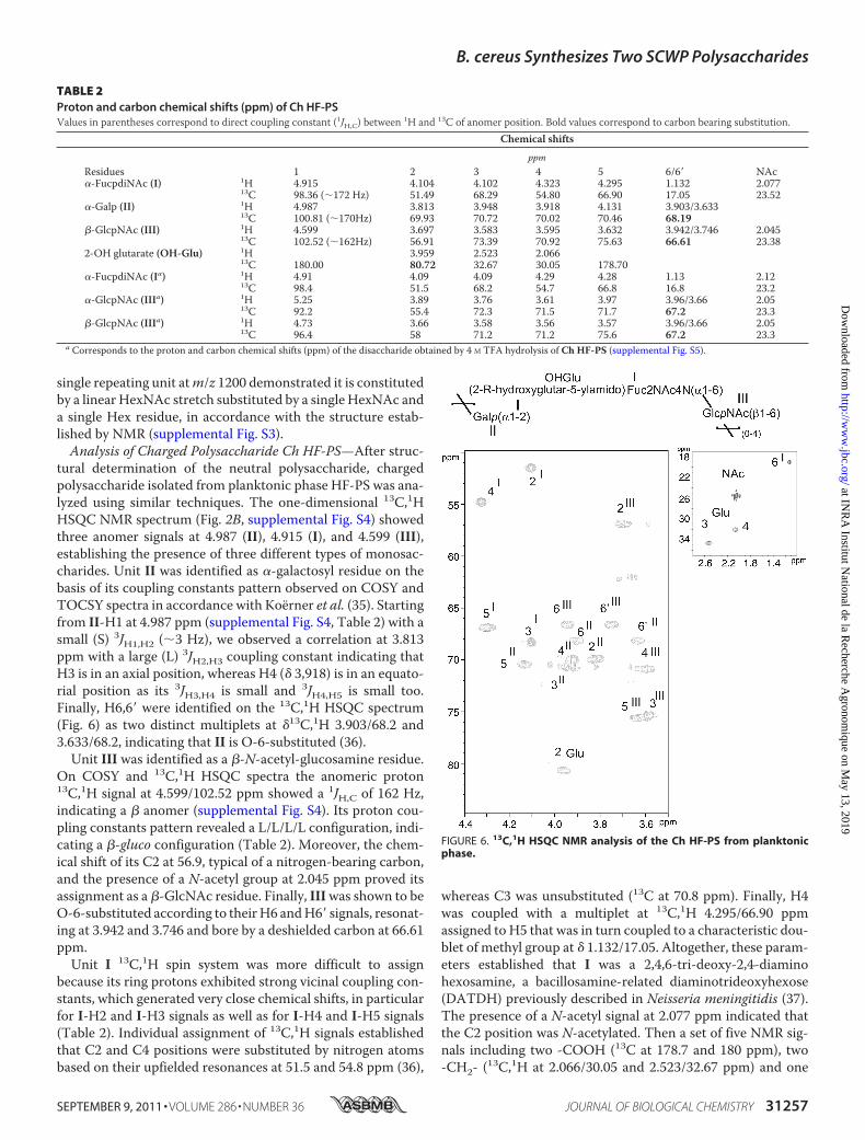
single repeating unit atm/z 1200 demonstrated it is constitutedby a linear HexNAc stretch substituted by a single HexNAc anda single Hex residue, in accordance with the structure estab-lished by NMR (supplemental Fig. S3).Analysis of Charged Polysaccharide Ch HF-PS—After struc-
tural determination of the neutral polysaccharide, chargedpolysaccharide isolated from planktonic phase HF-PS was ana-lyzed using similar techniques. The one-dimensional 13C,1HHSQC NMR spectrum (Fig. 2B, supplemental Fig. S4) showedthree anomer signals at 4.987 (II), 4.915 (I), and 4.599 (III),establishing the presence of three different types of monosac-charides. Unit II was identified as �-galactosyl residue on thebasis of its coupling constants pattern observed on COSY andTOCSY spectra in accordance with Koerner et al. (35). Startingfrom II-H1 at 4.987 ppm (supplemental Fig. S4, Table 2) with asmall (S) 3JH1,H2 (�3 Hz), we observed a correlation at 3.813ppm with a large (L) 3JH2,H3 coupling constant indicating thatH3 is in an axial position, whereas H4 (� 3,918) is in an equato-rial position as its 3JH3,H4 is small and 3JH4,H5 is small too.Finally, H6,6� were identified on the 13C,1H HSQC spectrum(Fig. 6) as two distinct multiplets at �13C,1H 3.903/68.2 and3.633/68.2, indicating that II is O-6-substituted (36).Unit III was identified as a �-N-acetyl-glucosamine residue.
On COSY and 13C,1H HSQC spectra the anomeric proton13C,1H signal at 4.599/102.52 ppm showed a 1JH,C of 162 Hz,indicating a � anomer (supplemental Fig. S4). Its proton cou-pling constants pattern revealed a L/L/L/L configuration, indi-cating a �-gluco configuration (Table 2). Moreover, the chem-ical shift of its C2 at 56.9, typical of a nitrogen-bearing carbon,and the presence of a N-acetyl group at 2.045 ppm proved itsassignment as a �-GlcNAc residue. Finally, IIIwas shown to beO-6-substituted according to theirH6 andH6� signals, resonat-ing at 3.942 and 3.746 and bore by a deshielded carbon at 66.61ppm.Unit I 13C,1H spin system was more difficult to assign
because its ring protons exhibited strong vicinal coupling con-stants, which generated very close chemical shifts, in particularfor I-H2 and I-H3 signals as well as for I-H4 and I-H5 signals(Table 2). Individual assignment of 13C,1H signals establishedthat C2 and C4 positions were substituted by nitrogen atomsbased on their upfielded resonances at 51.5 and 54.8 ppm (36),
whereas C3 was unsubstituted (13C at 70.8 ppm). Finally, H4was coupled with a multiplet at 13C,1H 4.295/66.90 ppmassigned to H5 that was in turn coupled to a characteristic dou-blet of methyl group at � 1.132/17.05. Altogether, these param-eters established that I was a 2,4,6-tri-deoxy-2,4-diaminohexosamine, a bacillosamine-related diaminotrideoxyhexose(DATDH) previously described in Neisseria meningitidis (37).The presence of a N-acetyl signal at 2.077 ppm indicated thatthe C2 position was N-acetylated. Then a set of five NMR sig-nals including two -COOH (13C at 178.7 and 180 ppm), two-CH2- (13C,1H at 2.066/30.05 and 2.523/32.67 ppm) and one
TABLE 2Proton and carbon chemical shifts (ppm) of Ch HF-PSValues in parentheses correspond to direct coupling constant (1JH,C) between 1H and 13C of anomer position. Bold values correspond to carbon bearing substitution.
Chemical shifts
ppmResidues 1 2 3 4 5 6/6� NAc�-FucpdiNAc (I) 1H 4.915 4.104 4.102 4.323 4.295 1.132 2.077
13C 98.36 (�172 Hz) 51.49 68.29 54.80 66.90 17.05 23.52�-Galp (II) 1H 4.987 3.813 3.948 3.918 4.131 3.903/3.633
13C 100.81 (�170Hz) 69.93 70.72 70.02 70.46 68.19�-GlcpNAc (III) 1H 4.599 3.697 3.583 3.595 3.632 3.942/3.746 2.045
13C 102.52 (�162Hz) 56.91 73.39 70.92 75.63 66.61 23.382-OH glutarate (OH-Glu) 1H 3.959 2.523 2.066
13C 180.00 80.72 32.67 30.05 178.70�-FucpdiNAc (Ia) 1H 4.91 4.09 4.09 4.29 4.28 1.13 2.12
13C 98.4 51.5 68.2 54.7 66.8 16.8 23.2�-GlcpNAc (IIIa) 1H 5.25 3.89 3.76 3.61 3.97 3.96/3.66 2.05
13C 92.2 55.4 72.3 71.5 71.7 67.2 23.3�-GlcpNAc (IIIa) 1H 4.73 3.66 3.58 3.56 3.57 3.96/3.66 2.05
13C 96.4 58 71.2 71.2 75.6 67.2 23.3a Corresponds to the proton and carbon chemical shifts (ppm) of the disaccharide obtained by 4 M TFA hydrolysis of Ch HF-PS (supplemental Fig. S5).
FIGURE 6. 13C,1H HSQC NMR analysis of the Ch HF-PS from planktonicphase.
B. cereus Synthesizes Two SCWP Polysaccharides
SEPTEMBER 9, 2011 • VOLUME 286 • NUMBER 36 JOURNAL OF BIOLOGICAL CHEMISTRY 31257
at INR
A Institut N
ational de la Recherche A
gronomique on M
ay 13, 2019http://w
ww
.jbc.org/D
ownloaded from

-CHOR- (13C,1H at 3.959/80.72 ppm) groups was associated toa glutaric acid group substituting theC4 position ofDATDH, inaccordance to previously published parameters (38) (Table 2).This glutarate (Glu) was shown to be attached on the DATDHthrough anN-acyl linkage on the four-amino group, due to thevicinal heteronuclear coupling constant (3JH,C) between I-H4and Glu-C5 observed at � 4.323/178.70 on the 13C,1H HMBCNMR spectrum (data not shown). The configuration of thismonosaccharide could not be directly obtained from NMRspectra because of the strong 1H-1H coupling constants, prob-ably generated by an unusual spatial conformation. To solvethis problem, we hydrolyzed the supernatant in strong acidicconditions, hypothesizing that the cleavage of the polysaccha-ride to mono- or disaccharides would relax the molecular con-straints and allow ameasurement of 1H-1H coupling constants.After N-re-acetylation and HPLC separation, we identified amajor disaccharide that contained a GlcNAc �/� residue interminal reducing position and DATDH as observed in theintact Ch HF-PS (supplemental Fig. S5). As expected, weobserved the 1H-1H coupling constant pattern of DATDH afterhydrolysis of the polysaccharide as S,L,S,S (3JH1,H2 � 3Hz;3JH2,H3 � 8Hz; 3JH3,H4 � 3Hz; 3JH4,H5 � 3Hz), which typified an�-galacto configuration. Furthermore, the presence ofN-acetylgroups in C2 (�H2�/C2�3.87/55.7, �H2bC2 3.68/58.5) and C4(�H4/C4 4.30/54.8) as well as the methyl group in C6 (�H1/C11.13/16.9) permitted identification of this monosaccharide asan �-2,4,6 tri-deoxy-2,4-di-N-acetylated-galactosamine, com-monly referred as �-FucdiNAc. The FucdiNAc in terminalnonreducing position was linked to the C6 position of theGlcNAc residue, as proved by the deshielded C6 carbon at 67.2ppm and the H6,6� resonances at 3.96 and 3.66 accordingly toresults obtained from intact Ch HF-PS.The sequence of the Ch HF-PS repeat unit was established
essentially by the 13C,1H HMBC heteronuclear NMR experi-ment due to the observation of numerous extra-residual 3JH,Ccorrelations (data not shown). Indeed, in accordance with theobservation of the deshielded value of III-C6 at 66.61 ppmcompared with 61 ppm for the free �-GlcpNAc residue, thestrong I-H1/III-C6 correlation signal at 4.915/66.61 ppm onthe HMBC spectrum established the sequence �FucdiNAc(1–6)GlcpNAc�. Similarly, 3JH,C correlation between III-H1 andII-C6 at 4.599/68.19 ppm as well as the deshielding of II-C6 to68.09 ppm established that the �-GlcpNAc III residue waslinked to �-Galp II in C6 position. Finally, the Glu-C2 chemicalshift at 80.72 ppm and the II-H1/Glu-2 correlation signal dem-onstrated that the �-Galp residue was linked to C2 position ofglutarate group. This asymmetric carbon presented an (R)absolute configuration, as demonstrated by gas chromatogra-phy analysis compared with the 2-(R)-hydroxyglutarate stan-dard (data not shown). In conclusion, the NMR analyses per-mitted definition of the structure of the repeating trisaccharideunit from charged polysaccharide Ch HF-PS as Galp(�1-2)[2-R-hydroxyglutar-5ylamido(4)]-4,6-dideoxy-GalpNAc(�1-6)GlcpNAc(�1–6).MALDI-MS analysis of the Ch HF-PS in negative mode
showed four sets of major signals at m/z 1381/1403, 2063/2085, 2745/2767, and 3428/3449 attributed to [M-H]�/[M-2H�Na]� adducts of intact oligosaccharides containing two,
three, four, and five repeating units, respectively (supplementalFig. S6). Indeed, the difference of 682 mass units matches withthe calculated composition of the trisaccharide, whose struc-ture has been established by NMR, confirming the previousattribution. Furthermore, several sets of signals correspondingto partially truncated repeating units were observed alongintact ones (supplemental Fig. S6).Analysis of the Early BiofilmPhase Polysaccharide—After the
total structural analysis of the two polysaccharides isolatedfrom the planktonic phase, the structure of the early biofilm cellwall polysaccharide was established using identical methods.As shown in Fig. 2, C and F, the 13C,1H HSQC NMR spectrumof early biofilm polysaccharide was identical to the neutralpolysaccharide Ne HF-PS isolated from the planktonic phasebacteria. This demonstrated that the cell wall polysaccharideisolated from bacteria in early phase of biofilm forma-tion is constituted by a single polysaccharide with a[GlcNAc(�1–3)]GlcNAc(�1–6)[Glc(�1–3)][ManNAc(�1-4)]GalNAc(�1–4)ManNAc(�1–4) repeating unit.Thus, the comparison of cell-wall polysaccharides from dif-
ferent phases established that B. cereus synthesizes differentsets of polysaccharides depending on the phase of growth. Acommon polysaccharide constituted by the hexasaccharide[GlcNAc(�1–3)]GlcNAc(�1–6)[Glc(�1–3)][ManNAc(�1-4)]GalNAc(�1–4)ManNAc(�1–4) repeating unit (Ne HF-PS)was observed in both planktonic and biofilm phases at all timepoints observed. Conversely, another very unusual polysaccha-ride presenting a Galp(�1–2)[2-R-hydroxyglutar-5ylamido(4)]-4,6-dideoxy-GalpNAc(�1–6)GlcpNAc(�1–6) repeatingunit (Ch HF-PS) appeared to be phase-specific. Indeed, thiscompound was observed in bacterial cell walls at every timepoint of planktonic cultures from 5 to 20 h as well as in biofilmfrom 72 to 96 h but totally disappeared in bacteria from earlybiofilms.In Vivo Analysis of the Surface Polysaccharides by HR-MAS
NMR—To rule out the possibility that the disappearance of ChHF-PS may be linked to variations in the extraction procedure,we performedHR-MASNMR analysis on total bacterial cells ineach growth phase. This technique was effectively used to ana-lyze the structure of glycosylated cell wall components, includ-ing mycobacterial and yeast polysaccharides, from intact livingbacterial cells without proceeding to any purification proce-dure (39, 40). As previously shown, parameters of NMR signalsoriginating fromHR-MASNMR experiments of total cell wallsand from liquid NMR experiment of purified polysaccharidesare strictly identical when experiments are run in similar exper-imental conditions, which permits direct identification of sig-nals fromHR-MAS based on liquid NMR analyses. Thus, basedon data collected on purified polysaccharides, 13C,1H HSQCHR-MASNMRspectrumof intact bacteria from the planktonicphase showed all the signals associated with both polysaccha-rides (Fig. 7 and supplemental Fig. S7). Interestingly, HSQCsignals associated with Ch HF-PS (I, II and III) exhibited amuch higher intensity than those associated with Ne HF-PSwhen compared with the liquid NMR spectrum of total poly-saccharide isolated from the same growth phase. The relativelower responsiveness of Ne HF-PS to HR-MASNMRwas mostprobably the result of the specific feature of this technique that
B. cereus Synthesizes Two SCWP Polysaccharides
31258 JOURNAL OF BIOLOGICAL CHEMISTRY VOLUME 286 • NUMBER 36 • SEPTEMBER 9, 2011
at INR
A Institut N
ational de la Recherche A
gronomique on M
ay 13, 2019http://w
ww
.jbc.org/D
ownloaded from

suppresses the signal from constrainedmolecules that exhibit ashort transversal relaxation time (T2). This provides a clearindication that the linear Ch HF-PS is more mobile and acces-sible at the bacterial cell surface than the branched Ne HF-PS.Converselywith the planktonic phase, anHR-MAS spectrumofearly biofilm intact bacteria only showed signals associated toCh HF-PS (data not shown). This set of data definitely estab-lished that the phenotypic differences that we observedbetween phases are not artifacts generated by multiple purifi-cation and chemical degradation steps of polysaccharides. Fur-thermore, it confirmed that Ch HF-PS was physically associ-ated to bacteria cell surface alongNeHF-PS and not released inthe culture medium.Comparison of HF-PS from Bacteria Grown in Different Cul-
ture Media—Altogether, our experimental data unambigu-ously established that B. cereus ATCC 14579 was able to syn-thesize two secondary cell wall polysaccharides presentingtotally different structures, Ne HF-PS and ChHF-PS (Fig. 8). Intheir previous work, Leoff et al. (23, 24) did report the presenceof Ne HF-PS polysaccharide in the B. cereusATCC14579 with-out providing its full structure but conclusively established thefine structures of its equivalent in a wide panel of B. cereusstrains (41). In contrast, these authors never mentioned thepresence of an equivalent of Ch HF-PS in any B. cereus strain,including Bc 14579, or in B. anthracis (23–25, 27, 41). Torationalize such an important discrepancy between our respec-tive data, we hypothesized that the production of Ch HF-PSmight be dependent on the culture medium, as Leoff et al. (23,24) used a richmedium (BHI), whereas we used a poormedium(HCT). As recently reported, B. cereus ATCC 14579 is capableof forming biofilm in poormedium culture conditions, whereasrich medium cannot sustain biofilm formation at any temper-ature (22). Thus, herein the choice of a poor medium for study-ing the qualitativemodifications of surface polysaccharides wasan absolute requirement. Assessment of the extraction andprocessing procedures of the HF-PS revealed some otherpotential sources of differences such as the heat inactivation ofbacteria (121 °C for 1 h) before extraction and sonication stepsthat may lead to a loss in the acid labile Ch HF-PS (27). To find
out the origin of the discrepancies between our respectiveobservations and understand the precise conditions in whichthe synthesis of Ch HF-PS is promoted, we compared the poly-saccharide composition of HF-PS extracted from bacteriagrown in planktonic phases in different culture media, BHI orHCT. As observed on the comparative analysis of 1H NMRspectra, total HF-PS isolated from bacteria grown in richmedium exclusively contained Ne HF-PS, whereas bacteriagrown in poor medium synthesized both Ch HF-PS and NeHF-PS (Fig. 9). Heat inactivation and sonication of bacteriagrown in rich medium did not induce more structural differ-ences (data not shown). This experiment unambiguouslydemonstrated that the synthesis of Ch HF-PS is strictly reg-ulated by the culture conditions of bacteria. Altogether, ourstudy showed that B. cereus strain ATCC 14579 displays twosecondary cell wall polysaccharides in which syntheses aretightly regulated by 1) the culture conditions and 2) the cul-ture phase.
DISCUSSION
Various polysaccharides covalently bound to the peptidogly-can have been described in Gram-positive bacteria (1). How-ever, this is the first report of two different peptidoglycan-asso-ciated coexisting polysaccharides. We independently isolatedfrom the B. cereus ATCC 14579 strain these polysaccharidesusing two totally different strategies. One strategy was based onthe partial acid depolymerization and solubilization in a hydro-alcoholic solution, which permitted differentiation of the twocomponents according to their susceptibility tomild acid treat-ment. Five steps ofmild hydrolysis enabled a total solubilizationof the acid labile polysaccharide. Despite its total solubilization,the overall structure of this acid-labile polysaccharide was keptintact without gross modification, albeit a partial depolymer-ization (data not shown). Conversely, the acid-resistant fractionwas totally insoluble after five rounds of mild acid hydrolysis,
FIGURE 7. Detail of the anomer region of 13C,1H HSQC HR-MAS NMR anal-ysis of bacterial cell surface of B. cereus from planktonic phase ofgrowth. When looked at a low sensitivity, only signals from Ch HF-PS (I to III)can be seen (A), whereas increasing sensitivity permits to recover signals fromNe HF-PS (IV to IX) (B).
FIGURE 8. Structure of HF-PS repetition units isolated from B. cereusstrains ATCC and 14579. Two unrelated polysaccharides, Ne HF-PS and ChHF-PS, were identified from B. cereus 14579. Ne HF-PS is synthesized irrespec-tive of the growing conditions, whereas Ch HF-PS synthesis is regulated alongthe biofilm formation and according to culture medium, HCT (poor medium)or BHI (rich medium). a, and d, planktonic phase; b, early biofilm formation(28 – 48 h); c, late biofilm formation (72–96 h).
B. cereus Synthesizes Two SCWP Polysaccharides
SEPTEMBER 9, 2011 • VOLUME 286 • NUMBER 36 JOURNAL OF BIOLOGICAL CHEMISTRY 31259
at INR
A Institut N
ational de la Recherche A
gronomique on M
ay 13, 2019http://w
ww
.jbc.org/D
ownloaded from

which permitted the obtaining of two fractions devoid of crosscontaminations. The two polysaccharides could also be sepa-ratedwithout prior acid hydrolysis according to their charge ona strong anion exchange column. One neutral fraction (NeHF-PS) corresponding to the acid-resistant polysaccharide andone charged fraction (Ch HF-PS) corresponding to the acid-labile fraction were purified that way.The detailed structural analysis of individual polysaccharides
provided in the present report established that these two poly-saccharides were structurally unrelated. Their structures aresummarized in Fig. 8. Neutral polysaccharide was composed ofthe major hexasaccharide repeat unit 34)[GlcNAc(�1-3)]GlcNAc(�1–6)[Glc(�1–3)][ManNAc(�1–4)]GalNAc(�1-4)ManNAc(�13. Although the major repeating unit is fullysubstituted by�GlcNAc,�Glc, and�ManNAc residues to formthe hexasaccharide unit, the observation of minor NMR signals(data not shown) strongly suggested the presence of a slightheterogeneity in the branching pattern of the trisaccharidebackbone 34)GlcNAc(�1–6)GalNAc(�1–4)ManNAc(�13due to non-stoichiometric substitutions of the backbone. Thestructure of the polysaccharide backbone that we have estab-lished fits well with the consensus sequence that was nicelydefined by Leoff and co-workers by comparing the structures ofHF-PS isolated from B. anthracis and B. cereus ATCC 10987(24, 27). Indeed, the authors reported the exact structures of thepolysaccharide backbones of HF-PS isolated from B. anthracisas34)GlcNAc(�1–6)GlcNAc(�1–4)ManNAc(�13 and fromB. cereus ATCC 10987 as 34)GlcNAc(�1–6)GalNAc(�1-4)ManNAc(�13. Based on these structural features anddespite species-specific branching patterns by �-galactosidaseand �-galactosidase residues, high similarity permittedthe proposition of the potential consensus sequence�GlcNAc-�HexNAc-�ManNAc for HF-PS in Bacillus species.As established by the present report, the trisaccharide back-bones of B. cereus ATCC 14579 and B. cereus ATCC 10987 aresimilar, but the branching pattern of Ne HF-PS from ATCC14579 is more complex than ATCC 10987 due to an abundantsubstitution by �GlcNAc, �Glc, and �ManNAc residues (Fig.
8). However, in contrast to ATCC 10987, no data supported thepresence of O-acetylation in the polysaccharide of B. cereus14579.Along these small scale species-specific differences between
ATCC 14579 and ATCC 10987, the most striking feature ofB. cereus ATCC 14579 was the observation of another, so farunidentified, stage-specific charged polysaccharide, ChHF-PS. It was specifically released from bacteria by aqueoushydrogen fluoride treatment along Ne HF-PS and repre-sented about 70% of the total HF-PS content of bacteriagrown in planktonic phase. This polysaccharide exhibited avery unusual repeating unit 36)Gal(�1–2)(2-R-hydroxy-glutar-5-ylamido)Fuc2NAc4N(�1–6)GlcNAc(�13 so farnever identified in the Bacillus genus. HR-MAS NMR analysisof the intact bacteria demonstrated that this component wasassociated to its cell surface. Furthermore, comparison of theNMR data originating from liquid and HR-MAS experi-ments strongly suggested that this component was part ofthe outmost layer surrounding the bacteria. Even more sur-prising and in contrast to Ne HF-PS, the expression profile ofCh HF-PS dramatically changed along the bacterial growthand according to the culture medium in which bacteria weregrown. The fact that it is exclusively expressed at the surfaceof bacteria grown in poor culture medium explains theapparent discrepancy of our results compared with previousstudies (24). One may easily speculate that the expression ofCh HF-PS in poor medium is somehow correlated to thebiofilm formation capacity of B. cereus ATCC 14579 (22).Correlation between biofilm formation and SCWP polysac-charides is further substantiated by the fact that Ch HF-PSsurface expression is also regulated along the biofilm forma-tion. Indeed, whereas Ch HF-PS was observed at every pointof the bacterial growth in planktonic phase, it could only beobserved in later cultures (from 72 h) of biofilm phase, theearlier cultures being totally devoid of Ch HF-PS. It is note-worthy that peptidoglycan is not modified during the biofilmphase, suggesting that the structural bacterial cell wall is only
FIGURE 9. Comparison of the HF-PS isolated from bacteria grown in different media. 1H-NMR spectra of HF-PS isolated from the planktonic phases ofbacteria grown in rich medium (BHI) (A) and poor medium (HCT) (B).
B. cereus Synthesizes Two SCWP Polysaccharides
31260 JOURNAL OF BIOLOGICAL CHEMISTRY VOLUME 286 • NUMBER 36 • SEPTEMBER 9, 2011
at INR
A Institut N
ational de la Recherche A
gronomique on M
ay 13, 2019http://w
ww
.jbc.org/D
ownloaded from

changed by the covalently associated polysaccharides (datanot shown). And, as SLH proteins are anchored to the HF-PS(3), any change in the HF-PS composition may result inchanges in cell surface proteins.As Ch HF-PS is not required for biofilm initiation and early
development, it is unlikely to play a role in cell-substrate inter-action or in cell-cell interaction within the biofilm. However,high quantities of extracellularDNAwere found in theB. cereusbiofilm matrix (42). The negatively charged backbone of thisextracellular DNAmight interactmore easily with bacterial cellsurfaces devoid of negatively charged polysaccharide, causingan increase of the biofilm cohesion. The interaction betweenextracellular DNA and surface polysaccharides was supportedby the observation that deletion of genes likely to be involved inextracellular DNA production impaired biofilm formation inB. cereus, but this hypothesis still awaits definitive experimentalproof (42). In planktonic cultures, Ch HF-PS could be involvedin the bacterium protection against environmental stresses, forinstance by sequestering positively charged antimicrobialagents. This putative role of ChHF-PSwould not be required inbiofilms, as the matrix was shown to act as an efficient shieldagainst various environmental stresses (43). The appearance ofChHF-PS in aging biofilms could be attributed to biofilms cellsswitching back to a planktonic state of growthwhile still locatedwithin the biofilm. Aged biofilms are prone to dispersal andinclude a leaving bacterial subpopulation showing importantphenotypic changes (44). In addition, the co-existence of bac-teria in various growth states was shown to occur withinB. sub-tilis biofilms (45).In conclusion, the identification of a new tightly regulated
surface component in B. cereus 14579 offers new insights intothe comprehension of the mechanisms that control adhesionand biofilm formation. Although the exact nature of the rela-tionships between SCWP polysaccharide and biofilm forma-tion are still unclear, we believe that our study will providesolid structural bases for the functional analysis of this newpolysaccharide. Furthermore, the induction of this newSCWP polysaccharide in specific culture conditions pointsto the possibility that other, still unknown SCWP polysac-charides could be induced in other environmental condi-tions, such as the host internal milieu. Changes in theB. cereus cell wall polysaccharides components could beinvolved in the high adaptability of this bacterium to envi-ronmental changes.
Acknowledgments—We thank I. G. Boneca from the Institut Pas-teur for the analysis of peptidoglycans by HPLC, B. Coddeville fromUnite de Glycobiologie Structurale et Fonctionnelle for helping inthe MS/MS analysis of polysaccharides, and Prof. A. Dell for pro-viding access to mass spectrometry facility (Division of MolecularBiosciences, Faculty of Natural Sciences, Imperial College Lon-don). The 800- and 900-MHz spectrometers were funded by RegionNord-Pas de Calais, European Union (FEDER), Ministere Fran-cais de la Recherche, Universite Lille1-Sciences and Technologies,and CNRS. The 400 MHz facility was funded by the Centre Com-mun de Mesure RMN de l’Universite de Lille1.
REFERENCES1. Vollmer, W. (2008) FEMS Microbiol. Rev 32, 287–3062. Weidenmaier, C., Peschel, A., Xiong, Y. Q., Kristian, S. A., Dietz, K., Yea-
man, M. R., and Bayer, A. S. (2005) J. Infect. Dis. 191, 1771–17773. Mesnage, S., Fontaine, T., Mignot, T., Delepierre, M., Mock, M., and
Fouet, A. (2000) EMBO J. 19, 4473–44844. Chitlaru, T., Ariel, N., Zvi, A., Lion, M., Velan, B., Shafferman, A., and
Elhanany, E. (2004) Proteomics 4, 677–6915. Gohar,M., Gilois, N., Graveline, R., Garreau, C., Sanchis, V., and Lereclus,
D. (2005) Proteomics 5, 3696–37116. Antikainen, J., Anton, L., Sillanpaa, J., and Korhonen, T. K. (2002) Mol.
Microbiol. 46, 381–3947. Sara, M., and Sleytr, U. B. (2000) J. Bacteriol. 182, 859–8688. Moscoso, M., García, E., and Lopez, R. (2006) J. Bacteriol. 188,
7785–77959. Gotz, F. (2002)Mol. Microbiol. 43, 1367–137810. Lebeer, S., Verhoeven, T. L., Perea Velez, M., Vanderleyden, J., and De
Keersmaecker, S. C. (2007) Appl. Environ. Microbiol. 73, 6768–677511. Kolter, R. (2005) Trends Microbiol. 13, 1–212. Davey, M. E., and O’toole, G. A. (2000) Microbiol. Mol. Biol. Rev 64,
847–86713. Whitchurch, C. B., Tolker-Nielsen, T., Ragas, P. C., and Mattick, J. S.
(2002) Science 295, 148714. Peng, J. S., Tsai, W. C., and Chou, C. C. (2002) Int. J. Food Microbiol. 77,
11–1815. Auger, S., Ramarao, N., Faille, C., Fouet, A., Aymerich, S., and Gohar, M.
(2009) Appl. Environ. Microbiol. 75, 6616–661816. Ren, D., Bedzyk, L. A., Setlow, P., Thomas, S. M., Ye, R. W., and Wood,
T. K. (2004) Biotechnol. Bioeng. 86, 344–36417. Lee, K., Costerton, J. W., Ravel, J., Auerbach, R. K., Wagner, D. M., Keim,
P., and Leid, J. G. (2007)Microbiology 153, 1693–170118. Hsueh, Y. H., Somers, E. B., Lereclus, D., andWong, A. C. L. (2006) Appl.
Environ. Microbiol. 72, 5089–509219. Kotiranta, A., Lounatmaa, K., andHaapasalo,M. (2000)Microbes Infect. 2,
189–19820. Bottone, E. J. (2010) Clin. Microbiol. Rev. 23, 382–39821. Gohar,M., Faegri, K., Perchat, S., Ravnum, S., Økstad, O. A., Gominet,M.,
Kolstø, A. B., and Lereclus, D. (2008) PLoS ONE 3, e279322. Wijman, J. G., de Leeuw, P. P.,Moezelaar, R., Zwietering,M.H., andAbee,
T. (2007) Appl. Environ. Microbiol. 73, 1481–148823. Leoff, C., Saile, E., Rauvolfova, J., Quinn, C. P., Hoffmaster, A. R., Zhong,
W.,Mehta, A. S., Boons, G. J., Carlson, R.W., andKannenberg, E. L. (2009)Glycobiology 19, 665–673
24. Leoff, C., Choudhury, B., Saile, E., Quinn, C. P., Carlson, R. W., and Kan-nenberg, E. L. (2008) J. Biol. Chem. 283, 29812–29821
25. Leoff, C., Saile, E., Sue, D., Wilkins, P., Quinn, C. P., Carlson, R. W., andKannenberg, E. L. (2008) J. Bacteriol. 190, 112–121
26. Archibald, A. R., Hancock, I. C., and Harwood, C. R. (1993) in BacillusSubtilis and Other Gram-Positive Bacteria (Sonenshein, A. L., Hoch, J. A.,and Losick, R., eds) pp. 381–410, American Society for MicrobiologyPress, Washington, D. C.
27. Choudhury, B., Leoff, C., Saile, E., Wilkins, P., Quinn, C. P., Kannenberg,E. L., and Carlson, R. W. (2006) J. Biol. Chem. 281, 27932–27941
28. Lang, W. K., Glassey, K., and Archibald, A. R. (1982) J. Bacteriol. 151,367–375
29. Lecadet,M.M., Blondel,M.O., and Ribier, J. (1980) J. Gen.Microbiol. 121,203–212
30. Houry, A., Briandet, R., Aymerich, S., and Gohar, M. (2010)Microbiology156, 1009–1018
31. Candela, T., and Fouet, A. (2005)Mol. Microbiol. 57, 717–72632. Ciucanu, I., and Kerek, F. (1984) Carbohydr. Res. 131, 209–21733. Guerardel, Y., Czeszak, X., Sumanovski, L. T., Karamanos, Y., Popescu, O.,
Strecker, G., and Misevic, G. N. (2004) J. Biol. Chem. 279, 15591–1560334. Zuiderweg, E. R. (2002) Biochemistry 41, 1–735. Koerner, T. A., Prestegard, J. H., and Yu, R. K. (1987) Methods Enzymol.
138, 38–5936. Bradbury, J. H., and Jenkins, G. A. (1984) Carbohydr. Res. 126, 125–156
B. cereus Synthesizes Two SCWP Polysaccharides
SEPTEMBER 9, 2011 • VOLUME 286 • NUMBER 36 JOURNAL OF BIOLOGICAL CHEMISTRY 31261
at INR
A Institut N
ational de la Recherche A
gronomique on M
ay 13, 2019http://w
ww
.jbc.org/D
ownloaded from

37. Stimson, E., Virji, M., Makepeace, K., Dell, A., Morris, H. R., Payne, G.,Saunders, J. R., Jennings, M. P., Barker, S., and Panico, M. (1995) Mol.Microbiol. 17, 1201–1214
38. Vinogradov, E., MacLean, L. L., Crump, E. M., Perry, M. B., and Kay,W. W. (2003) Eur. J. Biochem. 270, 1810–1815
39. Lee, R. E., Li, W., Chatterjee, D., and Lee, R. E. (2005) Glycobiology 15,139–151
40. Maes, E., Mille, C., Trivelli, X., Janbon, G., Poulain, D., and Guerardel, Y.(2009) J. Biochem. 145, 413–419
41. Forsberg, L. S., Choudhury, B., Leoff, C.,Marston, C. K., Hoffmaster, A. R.,
Saile, E., Quinn, C. P., Kannenberg, E. L., and Carlson, R. W. (2011) Gly-cobiology 21, 934–948
42. Vilain, S., Pretorius, J. M., Theron, J., and Brozel, V. S. (2009) Appl. Envi-ron. Microbiol. 75, 2861–2868
43. Flemming, H. C., and Wingender, J. (2010) Nat. Rev. Microbiol. 8,623–633
44. Karatan, E., and Watnick, P. (2009) Microbiol. Mol. Biol. Rev. 73,310–347
45. Lopez, D., Vlamakis, H., and Kolter, R. (2009) FEMS Microbiol. Rev. 33,152–163
B. cereus Synthesizes Two SCWP Polysaccharides
31262 JOURNAL OF BIOLOGICAL CHEMISTRY VOLUME 286 • NUMBER 36 • SEPTEMBER 9, 2011
at INR
A Institut N
ational de la Recherche A
gronomique on M
ay 13, 2019http://w
ww
.jbc.org/D
ownloaded from

Krzewinski, Michel Gohar and Yann GuérardelThomas Candela, Emmanuel Maes, Estelle Garénaux, Yoann Rombouts, Frédéric
Cell Wall Polysaccharide SecondaryBacillus cereusEnvironmental and Biofilm-dependent Changes in a
doi: 10.1074/jbc.M111.249821 originally published online July 22, 20112011, 286:31250-31262.J. Biol. Chem.
10.1074/jbc.M111.249821Access the most updated version of this article at doi:
Alerts:
When a correction for this article is posted•
When this article is cited•
to choose from all of JBC's e-mail alertsClick here
Supplemental material:
http://www.jbc.org/content/suppl/2011/07/22/M111.249821.DC1
http://www.jbc.org/content/286/36/31250.full.html#ref-list-1
This article cites 44 references, 17 of which can be accessed free at
at INR
A Institut N
ational de la Recherche A
gronomique on M
ay 13, 2019http://w
ww
.jbc.org/D
ownloaded from