Mutation of luxS Affects Biofilm Formation in Streptococcus mutans
9
10.1128/IAI.71.4.1972-1979.2003. 2003, 71(4):1972. DOI: Infect. Immun. Anderson and Wenyuan Shi Justin Merritt, Fengxia Qi, Steven D. Goodman, Maxwell H. Streptococcus mutans in Affects Biofilm Formation luxS Mutation of http://iai.asm.org/content/71/4/1972 Updated information and services can be found at: These include: REFERENCES http://iai.asm.org/content/71/4/1972#ref-list-1 at: This article cites 42 articles, 26 of which can be accessed free CONTENT ALERTS more» articles cite this article), Receive: RSS Feeds, eTOCs, free email alerts (when new http://journals.asm.org/site/misc/reprints.xhtml Information about commercial reprint orders: http://journals.asm.org/site/subscriptions/ To subscribe to to another ASM Journal go to: on September 27, 2014 by guest http://iai.asm.org/ Downloaded from on September 27, 2014 by guest http://iai.asm.org/ Downloaded from
-
Upload
independent -
Category
Documents
-
view
1 -
download
0
Transcript of Mutation of luxS Affects Biofilm Formation in Streptococcus mutans
10.1128/IAI.71.4.1972-1979.2003.
2003, 71(4):1972. DOI:Infect. Immun. Anderson and Wenyuan ShiJustin Merritt, Fengxia Qi, Steven D. Goodman, Maxwell H.
Streptococcus mutansin Affects Biofilm FormationluxSMutation of
http://iai.asm.org/content/71/4/1972Updated information and services can be found at:
These include:
REFERENCEShttp://iai.asm.org/content/71/4/1972#ref-list-1at:
This article cites 42 articles, 26 of which can be accessed free
CONTENT ALERTS more»articles cite this article),
Receive: RSS Feeds, eTOCs, free email alerts (when new
http://journals.asm.org/site/misc/reprints.xhtmlInformation about commercial reprint orders: http://journals.asm.org/site/subscriptions/To subscribe to to another ASM Journal go to:
on Septem
ber 27, 2014 by guesthttp://iai.asm
.org/D
ownloaded from
on S
eptember 27, 2014 by guest
http://iai.asm.org/
Dow
nloaded from

INFECTION AND IMMUNITY, Apr. 2003, p. 1972–1979 Vol. 71, No. 40019-9567/03/$08.00�0 DOI: 10.1128/IAI.71.4.1972–1979.2003Copyright © 2003, American Society for Microbiology. All Rights Reserved.
Mutation of luxS Affects Biofilm Formation in Streptococcus mutansJustin Merritt,1 Fengxia Qi,1 Steven D. Goodman,2 Maxwell H. Anderson,3 and Wenyuan Shi1*
UCLA Molecular Biology Institute and School of Dentistry, Los Angeles, California 900951; Department of DiagnosticSciences, University of Southern California, Los Angeles, California 900892; and Washington Dental Service,
Seattle, Washington 981253
Received 20 August 2002/Returned for modification 30 October 2002/Accepted 18 December 2002
Quorum sensing is a bacterial mechanism for regulating gene expression in response to changes in popu-lation density. Many bacteria are capable of acyl-homoserine lactone-based or peptide-based intraspeciesquorum sensing and luxS-dependent interspecies quorum sensing. While there is good evidence about theinvolvement of intraspecies quorum sensing in bacterial biofilm, little is known about the role of luxS in biofilmformation. In this study, we report for the first time that luxS-dependent quorum sensing is involved in biofilmformation of Streptococcus mutans. S. mutans is a major cariogenic bacterium in the multispecies bacterialbiofilm commonly known as dental plaque. An ortholog of luxS for S. mutans was identified using the dataavailable in the S. mutans genome project (http://www.genome.ou.edu/smutans.html). Using an assay developedfor the detection of the LuxS-associated quorum sensing signal autoinducer 2 (AI-2), it was demonstrated thatthis ortholog was able to complement the luxS negative phenotype of Escherichia coli DH5�. It was also shownthat AI-2 is indeed produced by S. mutans. AI-2 production is maximal during mid- to late-log growth in batchculture. Mutant strains devoid of the luxS gene were constructed and found to be defective in producing theAI-2 signal. There are also marked phenotypic differences between the wild type and the luxS mutants.Microscopic analysis of in vitro-grown biofilm structure revealed that the luxS mutant biofilms adopted a muchmore granular appearance, rather than the relatively smooth, confluent layer normally seen in the wild type.These results suggest that LuxS-dependent signal may play an important role in biofilm formation of S. mutans.
A bacterial biofilm is a community of bacteria (either singleor multiple bacterial species) that adhere to a solid surface (8).In recent years, biofilms have received much attention due totheir impact on industry and medicine. Biofilms are responsi-ble for a plethora of problems ranging from biofouling ofpipelines to facilitation of tissue damage in cystic fibrosis pa-tients (1, 37, 42).
Studies have clearly shown that a bacterial biofilm is not aresult of random accretions of bacterial cells; rather, it is thenet result of a community of bacteria cooperating to formwell-differentiated structures (7). The production of biofilm isdependent on the progression through several steps, from ini-tial attachment to full maturation into a stable community (29).Given the tremendous metabolic and physiological changesthat are required for the switch from planktonic to biofilmgrowth, it would seem reasonable that there exist some generegulators responsible for facilitating this process. Indeed, var-ious gene regulation systems, including quorum sensing sys-tems, have been found to be involved in bacterial biofilm for-mation (9).
Quorum sensing is a mechanism for bacteria to change geneexpression at very specific cell densities. To date, there are twotypes of recognized quorum sensing systems in bacteria. Thefirst, known as intraspecies quorum systems, are species spe-cific. In gram-negative bacteria, intraspecies quorum signalsare composed of an acyl-homoserine lactone backbone withspecies-specific substitutions, while gram-positive bacteria usevarious peptides as their signals (11, 15, 28). Recently, a second
quorum sensing system was characterized for Vibrio harveyi.This system has been referred to as the interspecies quorumsystem and may operate as a universal quorum system formany bacteria possessing the characteristic luxS gene (2, 34,39). The luxS gene is highly conserved among many species ofgram-negative and gram-positive bacteria and is thought to beresponsible for synthesizing a universally recognized cell signalreferred to as autoinducer-2 (AI-2) (39). The chemical struc-ture of the actual signal is still under investigation; however,crystallographic studies of the AI-2 receptor in V. harveyi seemto suggest that AI-2 is a furanosyl borate diester formed fromthe metabolite 4,5-dihydroxy-2,3-pentadione (5, 33, 34). Theecological role of luxS in bacteria is still poorly characterized,but one logical possibility is that it functions to allow bacteriato optimize gene expression in response to the density of allluxS-containing species occupying the same niche.
One feature regarding quorum sensing that has been exten-sively studied is the link between quorum sensing and biofilm-related gene expression. There are several well-characterizedexamples for the involvement of intraspecies quorum sensingand biofilm formation. For example, lasI of Pseudomonasaeruginosa directs the synthesis of an acyl-homoserine lactonesignal molecule used for P. aeruginosa intraspecies quorumsignaling (9). Mutants in this gene were unable to producebiofilms that progressed beyond the very early stages of biofilmdevelopment (9). However, exogenous addition of the appro-priate signal complemented the defect (9). A similar result wasalso obtained due to inactivation of the cep intraspecies quo-rum sensing system of Burkholderia cepacia (20). Furthermore,a transposon mutagenesis study of the oral pathogen Strepto-coccus gordonii had detected a severe biofilm deficiency due todisruption of the two-component system required for its in-
* Corresponding author. Mailing address: UCLA School of Den-tistry, 10833 Le Conte Ave., Los Angeles, CA 90095-1668. Phone:(310) 825-8356. Fax: (310) 794-7109. E-mail: [email protected].
1972
on Septem
ber 27, 2014 by guesthttp://iai.asm
.org/D
ownloaded from
traspecies quorum sensing system (25). In Staphylococcus au-reus, intraspecies quorum signaling has been implicated as anegative regulator of biofilm formation (41). LuxS-dependentAI-2 signals have also been detected in a variety of bacterialspecies and have been found to be involved in various cellularprocesses in a cell density-dependent manner (10, 13, 14, 21,26, 36). However, to date, no connection between LuxS-de-pendent AI-2 signals and biofilm formation has been reported.Here we report the first case of mutations of luxS affectingbiofilm formation of the oral pathogen Streptococcus mutans.
S. mutans is a major cariogenic bacterium that normallyinhabits a complex, multispecies biofilm on the tooth surface(dental plaque) (40). The bacteria produce large amounts ofexopolysaccharides, especially in the presence of sucrose, thatenable them to efficiently adhere to the tooth. The bacteriaalso have the ability to produce large amounts of acids fromfermentable sugars in the diet. Acid accumulation can eventu-ally dissolve the hard, crystalline structure of the tooth, result-ing in a carious lesion (32). Previous studies have establishedsome sophisticated interactions among the oral streptococci aswell as with other oral bacteria within the same dental plaque(22–24). For this reason, S. mutans and dental plaque comprisean ideal model system for studying the role of interspeciessignaling and biofilm formation. In this study, we report that S.mutans indeed possesses a functional luxS gene that is capableof signaling to V. harveyi. Additionally, we show by gene dele-tion that luxS of S. mutans is involved in biofilm formation.
MATERIALS AND METHODS
Bacterial strains and culture conditions. All bacterial strains used in this studyand their characteristics are listed in Table 1. All S. mutans strains were grownin brain heart infusion (BHI) medium (Difco) or on BHI agar plates. luxSdeletion mutants were grown using the same medium supplemented with 15 �gof erythromycin/ml. All S. mutans strains were grown anaerobically (80% N2,10% CO2, 10% H2) at 37°C. Escherichia coli cells were grown in Luria-Bertanimedium with aeration at 37°C. E. coli cells carrying plasmids were grown inLuria-Bertani medium containing 100 �g of erythromycin/ml. V. harveyi BB170(sensor 1�, sensor 2�) was kindly provided by B. Bassler (Princeton University)and grown in AB medium (37) overnight at 30°C.
Cloning and analyses of the S. mutans luxS gene. A 990-bp DNA fragmentcontaining the luxS gene from S. mutans strain 25175 was PCR amplified fromgenomic DNA using the primers WTlux5 (5�-GATGCTGCACGCTCTGTC-3�)and WTlux3 (5�-GCAGTTAGGGTATCCATCC-3�). Primer sequences weredesigned using sequence data obtained from the S. mutans Genome SequencingProject, University of Oklahoma (B. A. Roe, R. Y. Tian, H. G. Jia, Y. D. Qian,S. P. Lin, S. Li, S. Kenton, H. Lai, J. D. White, R. E. McLaughlin, M. McShan,D. Ajdic, and J. Ferretti [http://www.genome.ou.edu/smutans.html]). The result-ing fragment was cloned into the TOPO TA cloning vector (Invitrogen) andsequenced.
RNA isolation and RT-PCR. S. mutans total RNA was isolated as follows.Twenty-five-milliliter cultures were grown overnight as previously described.Cells were centrifuged and resuspended in 1.5 ml of Tris-EDTA buffer and thenlysed using a Mini-Bead Beater 8 according to the manufacturer’s instructions.One hundred fifty microliters of lysate was then passed through a Qiashredder
column (Qiagen), and 100 �l of the resulting lysate was used for total RNAisolation with the RNeasy mini kit (Qiagen). cDNA buffer was added to RNAsamples to obtain a 1� solution, and 3 �l of RQ1 RNase-free DNase was addedto the sample and incubated overnight at 37°C. Reverse transcription (RT) wasperformed according to the manufacturer’s protocol using random hexamers.
AI-2 assay. The AI-2 luminescence reporter assay was performed essentially asdescribed previously (38), with the following modifications. To obtain cell-freeconditioned medium, S. mutans was grown overnight as described. Stationary-phase cells were resuspended in AB medium to an optical density at 600 nm(OD600) of 0.4. Cells were then incubated at 37°C with aeration for 3 h. After theincubation period, cells were pelleted by centrifugation, and the resulting super-natant was filtered through a 0.22-mm-pore-size filter (Millipore). The cell-freeconditioned medium was either used immediately or stored at �20°C. To deter-mine luminescence, an overnight culture of V. harveyi BB170 was diluted 1:1,000into fresh AB medium, and 180 �l of cells was added to each 1.5-ml microfugetube. Conditioned medium (20 �l) was then added to the cells at a 10% (vol/vol)final concentration. Positive control samples were obtained by adding cell-freeconditioned medium from an overnight culture of V. harveyi BB170 to a finalconcentration of 10%. In some cases, conditioned medium from a wild-typeculture of S. mutans served as an additional positive control. To determine levelsof background luminescence, a sample containing 200 �l of V. harveyi (dilution,1:1,000) was also included. In some cases, E. coli DH5� served as a negativecontrol. All samples were measured hourly until peak induction at the 3-h timepoint. Measurements were collected using a TD 20/20 luminometer and ex-pressed as arbitrary luminescence units.
Construction and analyses of S. mutans luxS mutants. Two 1-kb fragmentscontaining regions of DNA immediately upstream and downstream of the luxStranslational start and stop codons, respectively, were amplified from a genomicDNA template using the primers 2XupF (5�-GCGGATCCTCAAGCTCTCAAGCGTTCGG-3�) and 2XupR (5�-CGAGATCTATAAGACGGACATAAGGGGC-3�) as well as 2XdownF (5�-GCCTCGAGCAGATGATCCTTTTGAGCGTC-3�) and 2XdownR (5�-CGTCTAGACGGATGCAAAGAGAACGAAG-3�).Primer sequences were designed using sequence data obtained from the S.mutans Genome Sequencing Project, University of Oklahoma (http://www.genome.ou.edu/smutans.html). The resulting fragments were cloned into theTOPO TA cloning vector (Invitrogen). To obtain the necessary fragments, fourseparate restriction digests were constructed: the upstream fragment was cutfrom the vector using BamHI and BglII, the downstream fragment was cut fromthe vector using XhoI and XbaI, the erythromycin cassette was cut from theplasmid pJT10 (J. P. Tsai and W. Shi, unpublished data) using BglII and XhoI,and the pUC19 vector was cut with BamHI and XbaI. The resulting fragmentswere all gel purified (Qiagen), precipitated, and added to one mass ligationreaction. The resulting construct was then checked by restriction analysis andPCR for the proper configuration of the knockout vector (pUCluxKO). Next, theplasmid was linearized with the unique cutting enzyme AatII and transformedinto S. mutans. Transformants were selected for resistance to 15-�g/ml erythro-mycin. Confirmation of DNA integration was performed by PCR using theprimers IntluxF (5�-AAGAGTTTGGACCTAAAGGC-3�), IntluxR (5�-CCCACAGGACTCAATAGTTG-3�), UpluxF (5�-CTCGACGAATAGGATCAAAGC-3�), DownluxR (5�-GAGCCATCACACAGCAAAAAC-3�), ermA (5�-AGTGTGTTGATAGTGCAGTATC-3�), and ermM (5�-GAAGCTGTCAGTAGTATACC-3�). All mutants confirmed by PCR were further analyzed for their ability toproduce AI-2 using the methods described above.
Growth of in vitro biofilm. Biofilms of S. mutans were grown as follows.Individual sterile culture dishes were filled with 2.5 ml of BHI broth supple-mented with 1% (wt/vol) sucrose. Next, a sterile 18-mm-diameter glass micro-scope coverslip was added to each dish, and the culture dish was covered. Eachsample was then inoculated with a defined volume of overnight culture. Thedishes were incubated anaerobically at 37°C overnight.
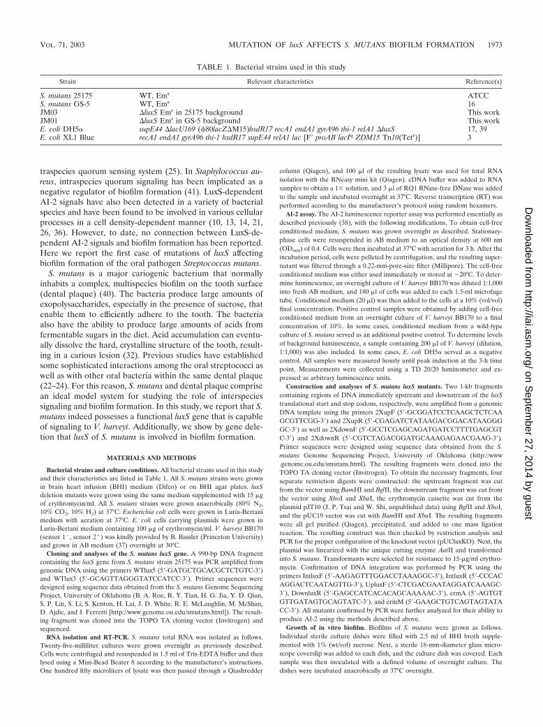
TABLE 1. Bacterial strains used in this study
Strain Relevant characteristics Reference(s)
S. mutans 25175 WT, Ems ATCCS. mutans GS-5 WT, Ems 16JM03 �luxS Emr in 25175 background This workJM01 �luxS Emr in GS-5 background This workE. coli DH5� supE44 �lacU169 (�80lacZ�M15)hsdR17 recA1 endA1 gyrA96 thi-1 relA1 �luxS 17, 39E. coli XL1 Blue recA1 endA1 gyrA96 thi-1 hsdR17 supE44 relA1 lac [F� proAB lacIq ZDM15 Tn10(Tetr)] 3
VOL. 71, 2003 MUTATION OF luxS AFFECTS S. MUTANS BIOFILM FORMATION 1973
on Septem
ber 27, 2014 by guesthttp://iai.asm
.org/D
ownloaded from
Microscopic analyses of in vitro-grown biofilm. Glass coverslips containingattached biofilm were removed from overnight cultures and rinsed briefly withwater. These could be directly viewed using phase-contrast and dark-field mi-croscopy. For fluorescently labeled samples, the biofilms were first removed fromthe culture dish and placed into another dry culture dish. Next, 20 �l of anti-S.mutans mouse monoclonal antibody solution (SWLA1) was added to the at-tached biofilm in each culture dish and incubated at room temperature for 30min (35). After the incubation period, 5 �l of fluorescein isothiocyanate-conju-gated goat anti-mouse antibody was added to the biofilms and incubated at roomtemperature for an additional 10 min. Finally, the coverslip was briefly rinsedwith water to remove excess antibodies and unattached cells, and the sample wasimmediately imaged using fluorescence microscopy.
Treatment of in vitro biofilm with sodium dodecyl sulfate (SDS) and antibi-otics. Biofilms were grown overnight using previously described conditions. Fortreatment with SDS, coverslips were removed from overnight incubation andrinsed briefly. These were then placed into a fresh culture dish, and 2 ml ofsterile, 1% (wt/vol) SDS solution was added to each dish. The biofilms wereplaced on a standard shaker set at 150 rpm for 1 h at room temperature. Next,supernatant samples were checked at magnification �40 to confirm that cellswere fully individualized and not connected in chains. These samples weremeasured for their OD600 values. For treatment with antibiotics, biofilms wereagain grown under standard conditions overnight. The following day, the spentmedium was changed and replaced with BHI supplemented with 1% (wt/vol)sucrose solution. Ampicillin was added at a concentration of either 50 or 500�g/ml. These biofilms were allowed to grow overnight under anaerobic condi-tions at 37°C. The following day, biofilms were removed from incubation andsonicated until the cells were fully dispersed. A 106-fold dilution of each samplewas plated on nonselective BHI agar plates and incubated anaerobically over-night at 37°C.
RESULTS
Identification and isolation of the S. mutans luxS gene. Todetermine whether S. mutans may possess an interspecies quo-rum system, it was first necessary to identify a candidate or-tholog of luxS, the enzyme required for AI-2 production. Usingthe luxS gene from V. harveyi, we performed a BLAST searchof the University of Oklahoma S. mutans genome sequencedatabase. A candidate open reading frame (ORF) was identi-fied. The ORF appears to be an isolated gene (no other con-vincing ORFs nearby) and encodes a protein of 160 aminoacids, which is similar in size to other reported LuxS proteins.It is homologous to the V. harveyi LuxS protein, with 38%amino acid identity and 57% similarity. Using this sequencedata, primers were designed to amplify a region about 500 bp
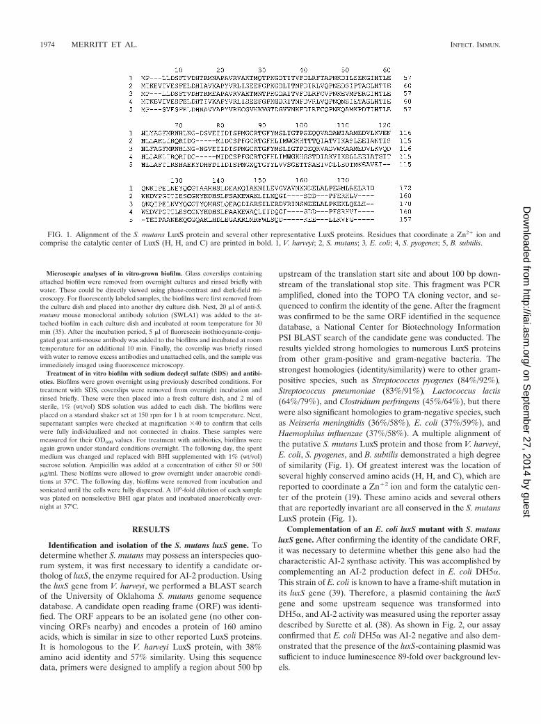
upstream of the translation start site and about 100 bp down-stream of the translational stop site. This fragment was PCRamplified, cloned into the TOPO TA cloning vector, and se-quenced to confirm the identity of the gene. After the fragmentwas confirmed to be the same ORF identified in the sequencedatabase, a National Center for Biotechnology InformationPSI BLAST search of the candidate gene was conducted. Theresults yielded strong homologies to numerous LuxS proteinsfrom other gram-positive and gram-negative bacteria. Thestrongest homologies (identity/similarity) were to other gram-positive species, such as Streptococcus pyogenes (84%/92%),Streptococcus pneumoniae (83%/91%), Lactococcus lactis(64%/79%), and Clostridium perfringens (45%/64%), but therewere also significant homologies to gram-negative species, suchas Neisseria meningitidis (36%/58%), E. coli (37%/59%), andHaemophilus influenzae (37%/58%). A multiple alignment ofthe putative S. mutans LuxS protein and those from V. harveyi,E. coli, S. pyogenes, and B. subtilis demonstrated a high degreeof similarity (Fig. 1). Of greatest interest was the location ofseveral highly conserved amino acids (H, H, and C), which arereported to coordinate a Zn�2 ion and form the catalytic cen-ter of the protein (19). These amino acids and several othersthat are reportedly invariant are all conserved in the S. mutansLuxS protein (Fig. 1).
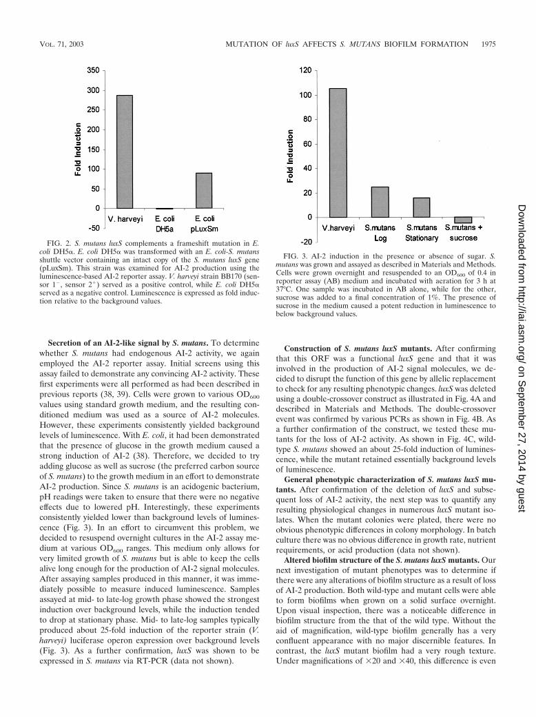
Complementation of an E. coli luxS mutant with S. mutansluxS gene. After confirming the identity of the candidate ORF,it was necessary to determine whether this gene also had thecharacteristic AI-2 synthase activity. This was accomplished bycomplementing an AI-2 production defect in E. coli DH5�.This strain of E. coli is known to have a frame-shift mutation inits luxS gene (39). Therefore, a plasmid containing the luxSgene and some upstream sequence was transformed intoDH5�, and AI-2 activity was measured using the reporter assaydescribed by Surette et al. (38). As shown in Fig. 2, our assayconfirmed that E. coli DH5� was AI-2 negative and also dem-onstrated that the presence of the luxS-containing plasmid wassufficient to induce luminescence 89-fold over background lev-els.
FIG. 1. Alignment of the S. mutans LuxS protein and several other representative LuxS proteins. Residues that coordinate a Zn2� ion andcomprise the catalytic center of LuxS (H, H, and C) are printed in bold. 1, V. harveyi; 2, S. mutans; 3, E. coli; 4, S. pyogenes; 5, B. subtilis.
1974 MERRITT ET AL. INFECT. IMMUN.
on Septem
ber 27, 2014 by guesthttp://iai.asm
.org/D
ownloaded from
Secretion of an AI-2-like signal by S. mutans. To determinewhether S. mutans had endogenous AI-2 activity, we againemployed the AI-2 reporter assay. Initial screens using thisassay failed to demonstrate any convincing AI-2 activity. Thesefirst experiments were all performed as had been described inprevious reports (38, 39). Cells were grown to various OD600
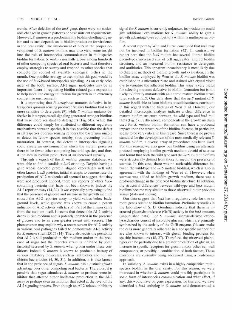
values using standard growth medium, and the resulting con-ditioned medium was used as a source of AI-2 molecules.However, these experiments consistently yielded backgroundlevels of luminescence. With E. coli, it had been demonstratedthat the presence of glucose in the growth medium caused astrong induction of AI-2 (38). Therefore, we decided to tryadding glucose as well as sucrose (the preferred carbon sourceof S. mutans) to the growth medium in an effort to demonstrateAI-2 production. Since S. mutans is an acidogenic bacterium,pH readings were taken to ensure that there were no negativeeffects due to lowered pH. Interestingly, these experimentsconsistently yielded lower than background levels of lumines-cence (Fig. 3). In an effort to circumvent this problem, wedecided to resuspend overnight cultures in the AI-2 assay me-dium at various OD600 ranges. This medium only allows forvery limited growth of S. mutans but is able to keep the cellsalive long enough for the production of AI-2 signal molecules.After assaying samples produced in this manner, it was imme-diately possible to measure induced luminescence. Samplesassayed at mid- to late-log growth phase showed the strongestinduction over background levels, while the induction tendedto drop at stationary phase. Mid- to late-log samples typicallyproduced about 25-fold induction of the reporter strain (V.harveyi) luciferase operon expression over background levels(Fig. 3). As a further confirmation, luxS was shown to beexpressed in S. mutans via RT-PCR (data not shown).
Construction of S. mutans luxS mutants. After confirmingthat this ORF was a functional luxS gene and that it wasinvolved in the production of AI-2 signal molecules, we de-cided to disrupt the function of this gene by allelic replacementto check for any resulting phenotypic changes. luxS was deletedusing a double-crossover construct as illustrated in Fig. 4A anddescribed in Materials and Methods. The double-crossoverevent was confirmed by various PCRs as shown in Fig. 4B. Asa further confirmation of the construct, we tested these mu-tants for the loss of AI-2 activity. As shown in Fig. 4C, wild-type S. mutans showed an about 25-fold induction of lumines-cence, while the mutant retained essentially background levelsof luminescence.
General phenotypic characterization of S. mutans luxS mu-tants. After confirmation of the deletion of luxS and subse-quent loss of AI-2 activity, the next step was to quantify anyresulting physiological changes in numerous luxS mutant iso-lates. When the mutant colonies were plated, there were noobvious phenotypic differences in colony morphology. In batchculture there was no obvious difference in growth rate, nutrientrequirements, or acid production (data not shown).
Altered biofilm structure of the S. mutans luxS mutants. Ournext investigation of mutant phenotypes was to determine ifthere were any alterations of biofilm structure as a result of lossof AI-2 production. Both wild-type and mutant cells were ableto form biofilms when grown on a solid surface overnight.Upon visual inspection, there was a noticeable difference inbiofilm structure from the that of the wild type. Without theaid of magnification, wild-type biofilm generally has a veryconfluent appearance with no major discernible features. Incontrast, the luxS mutant biofilm had a very rough texture.Under magnifications of �20 and �40, this difference is even
FIG. 2. S. mutans luxS complements a frameshift mutation in E.coli DH5�. E. coli DH5� was transformed with an E. coli-S. mutansshuttle vector containing an intact copy of the S. mutans luxS gene(pLuxSm). This strain was examined for AI-2 production using theluminescence-based AI-2 reporter assay. V. harveyi strain BB170 (sen-sor 1�, sensor 2�) served as a positive control, while E. coli DH5�served as a negative control. Luminescence is expressed as fold induc-tion relative to the background values.
FIG. 3. AI-2 induction in the presence or absence of sugar. S.mutans was grown and assayed as described in Materials and Methods.Cells were grown overnight and resuspended to an OD600 of 0.4 inreporter assay (AB) medium and incubated with aeration for 3 h at37°C. One sample was incubated in AB alone, while for the other,sucrose was added to a final concentration of 1%. The presence ofsucrose in the medium caused a potent reduction in luminescence tobelow background values.
VOL. 71, 2003 MUTATION OF luxS AFFECTS S. MUTANS BIOFILM FORMATION 1975
on Septem
ber 27, 2014 by guesthttp://iai.asm
.org/D
ownloaded from

more apparent (Fig. 5A). Wild-type biofilms are very uniform,with complete coverage of the attached surface. They also tendto have relatively small aggregates spread fairly evenlythroughout the biofilm matrix. luxS mutant biofilms are quitedifferent. Their organization seems much more heterogeneous.There were noticeable large gaps in the biofilm matrix, and thecell aggregates appeared much larger. Using fluorescence im-aging, there is a clear indication that the sizes of mutant ag-gregates tended to be much larger than those of the wild type(data not shown).
Since biofilms are known to be very resistant to detergentsand biocides, we were also interested to determine if this prop-erty was influenced by the luxS mutation. Overnight biofilms ofthe wild type and the luxS mutant were incubated in a 1%solution of SDS and shaken at 150 rpm for 1 h. These super-natants were then checked by microscopy to ensure that cellswere not clumped, and subsequently, the OD600 was measured.It was found that the wild-type supernatants had an OD600 thataveraged about sixfold higher than that of the mutant (Fig.5B), which suggested that luxS mutant biofilms were moreresistant to detergent treatment. We also treated biofilms ofthe wild type and the luxS mutant with the antibiotic ampicillinat a concentration of 50 or 500 �g/ml for 16 h. We found thatvery few (1%), if any, wild-type cells survived treatment with50 �g of ampicillin/ml and that none survived the treatmentwith 500 �g/ml. In contrast, numerous cells (10%) within theluxS mutant biofilms survived the treatment with 50 �g ofampicillin/ml and even with 500 �g of ampicillin/ml.
DISCUSSION
In this paper we report the identification of the luxS gene forS. mutans. This gene is recognized as the enzyme primarilyresponsible for the production of autoinducer-2 (AI-2) inter-species quorum signals for V. harveyi. We demonstrated thatthe S. mutans LuxS protein exhibited high degrees of similarityto other LuxS proteins in the National Center for Biotechnol-ogy Information database and was able to complement a LuxSdefect in E. coli. Furthermore, conditioned medium from S.mutans was capable of inducing the luciferase operon expres-sion in V. harveyi, and deletion of this gene abolished thisability. Finally, loss of AI-2 activity was associated with prom-inent changes in biofilm structure.
Within the past couple of years, there has been a plethora ofdata describing various physiological functions from bothgram-positive and gram-negative species which are subject toregulation by luxS (6, 10, 12, 26, 36). Much of the reported datahave supported the hypothesis that luxS is somehow involved inregulating virulence factor expression. Furthermore, there hasnot been a reported scenario in which mutating this gene hasled to severe impairment of growth, which suggests that theproduction of AI-2 is not a requirement for basic metabolicprocesses. Our present findings are consistent with these same
FIG. 4. luxS knockout in S. mutans. (A) Illustration of the knockoutprocedure. Plasmids containing cloned fragments of S. mutans DNA aswell as the erythromycin cassette were cut using the indicated restric-tion sites and then ligated into a linearized pUC19 backbone. Theresulting construct was linearized with the unique AatII site and trans-formed into S. mutans for double crossover. (B) Confirmation of cross-over event. In lanes 1 to 4, wild-type DNA was amplified with primers:internal to luxS, the erythromycin cassette, upstream external luxS plus
erythromycin, or downstream external luxS plus erythromycin. In lanes5 to 8, luxS mutant DNA was amplified using the same primer com-binations. External luxS primers bind to sites that were not subject tocrossover. (C) AI-2 production was assayed to confirm that activity waslost in the mutant.
1976 MERRITT ET AL. INFECT. IMMUN.
on Septem
ber 27, 2014 by guesthttp://iai.asm
.org/D
ownloaded from

FIG. 5. Mutation of luxS causes an alteration in S. mutans biofilm. (A) Two images of in vitro biofilms representing both the wild type and themutant using dark-field microscopy at magnifications �20 and �40. Cells were grown on glass coverslips as described in Materials and Methods.Similar results were obtained for more than 100 independent wild-type and luxS mutant biofilms examined. (B) Biofilms were tested for their abilityto resist detergent treatment with SDS. Three samples of wild-type and mutant biofilms grown on glass coverslips were shaken at 150 rpm for 1 h.The OD600s of the resulting supernatants were compared.
VOL. 71, 2003 MUTATION OF luxS AFFECTS S. MUTANS BIOFILM FORMATION 1977
on Septem
ber 27, 2014 by guesthttp://iai.asm
.org/D
ownloaded from
trends. After deletion of the luxS gene, there were no notice-able changes in growth patterns or basic nutrient requirements.However, S. mutans is a predominantly biofilm-dwelling organ-ism and as such depends on its biofilm production for virulencein the oral cavity. The involvement of luxS in the proper de-velopment of S. mutans biofilms may also yield some insightinto the role of interspecies communication in multispeciesbiofilm formation. S. mutans normally grows among hundredsof other competing species of oral bacteria and must thereforeemploy strategies to survey and respond to other species thatcompete for control of available ecological niches in themouth. One possible strategy to accomplish this goal would bethe use of luxS-based interspecies signaling. As an early colo-nizer of the tooth surface, AI-2 signal molecules may be animportant factor in regulating biofilm-related gene expressionto help modulate energy utilization for growth in an extremelycompetitive environment.
It is interesting that P. aeruginosa mutants defective in in-traspecies quorum sensing produced weaker biofilms that weremore sensitive to detergents (9), while S. mutans mutants de-fective in interspecies cell signaling generated stronger biofilmsthat were more resistant to detergents (Fig. 5B). While thisdifference could simply be attributed to different regulatorymechanisms between species, it is also possible that the defectin intraspecies quorum sensing renders the bacterium unableto detect its fellow species nearby, thus preventing biofilmmaturation. In contrast, the defect in interspecies signalingcould create an environment in which the mutant perceivesthere to be fewer other competing bacterial species, and thus,it initiates its biofilm production at an increased capacity.
Through a search of the S. mutans genome database, wewere able to find a candidate luxS ortholog. Despite having agene whose encoded protein aligned exceedingly well withother known LuxS proteins, initial attempts to demonstrate theproduction of AI-2 molecules all seemed to suggest that theywere not produced. Indeed, there are reports of other luxS-containing bacteria that have not been shown to induce theAI-2 reporter assay (14, 39). It was especially perplexing to findthat the presence of glucose and sucrose in the growth mediumcaused the AI-2 reporter assay to yield values below back-ground levels, while glucose was known to cause a potentinduction of AI-2 activity with E. coli. Part of the answer camefrom the medium itself. It seems that detectable AI-2 activitydrops in rich medium and is potently inhibited in the presenceof glucose and to an even greater extent with sucrose. Thisphenomenon may explain why a recent screen for AI-2 activityin various oral pathogens failed to demonstrate AI-2 activityfor S. mutans strain 25175 (14). There also exists the possibilitythat AI-2 is still produced in rich medium and/or in the pres-ence of sugar but the reporter strain is inhibited by somefactor(s) secreted by S. mutans when grown under these con-ditions. Indeed, S. mutans is known to produce a battery ofvarious inhibitory molecules, such as lantibiotics and nonlan-tibiotic bacteriocins (4, 30, 31). In addition, it is also knownthat in the presence of sugars, S. mutans has a distinct growthadvantage over other competing oral bacteria. Therefore, it ispossible that sugar stimulates S. mutans to produce some in-hibitor that affected either luciferase production in the AI-2assay or perhaps even an inhibitor that acted at the level of theAI-2 signaling process. Even though an AI-2-related inhibitory
signal for S. mutans is currently unknown, its production couldgive additional explanations for S. mutans’ ability to gain agrowth advantage over competitors within its multispecies bio-film.
A recent report by Wen and Burne concluded that luxS maynot be involved in biofilm formation (42). In contrast, wereport here that the luxS mutant has several altered biofilmphenotypes: increased size of cell aggregates, altered biofilmstructure, and an increased biofilm resistance to detergentsand antibiotics. This apparent inconsistency is most likely dueto different methods of biofilm growth and evaluation. In thebiofilm assay employed by Wen et al., S. mutans biofilm wasestablished in a microtiter plate and stained with crystal violetdye to visualize the adherent biofilm. This assay is very usefulfor selecting mutants defective in biofilm formation but is notlikely to identify mutants with an altered mature biofilm struc-ture, such as luxS. Our data show that the luxS mutant of S.mutans is still able to form biofilms on solid surfaces, consistentin this regard with the findings of Wen et al. However, ourdetailed microscopic analyses indicate a clear difference inmature biofilm structure between the wild type and luxS mu-tants (Fig. 5). Furthermore, components in the growth mediumused for S. mutans biofilm formation can have a profoundimpact upon the structure of the biofilm. Sucrose, in particular,seems to be very critical in this regard. Since there is no provenstandard for the development of the most medically relevant S.mutans biofilm, a diverse array of procedures has been used.For this reason, we also grew our biofilms using an alternateprotocol employing biofilm growth medium devoid of sucroseand found that both the wild type and the luxS mutant biofilmswere structurally distinct from those formed in the presence ofsucrose. In this case, there was no noticeable difference be-tween the wild-type and luxS mutant biofilms, which is also inagreement with the findings of Wen et al. However, whensucrose was added to biofilm growth medium, there was aprofound change in the observed biofilm structure. In addition,the structural differences between wild-type and luxS mutantbiofilms became very similar to those observed in our previousassays (data not shown).
Our data suggest that luxS has a regulatory role for one ormore genes related to biofilm formation. Preliminary studies inthe laboratory of S. D. Goodman indicate that there is in-creased glucosyltransferase (GtfB) activity in the luxS mutants(unpublished data). For S. mutans, sucrose-derived exopo-lysaccharides consist of insoluble glucans, which are primarilysynthesized by the activity of the GtfB enzyme. Glucans makethe cells more generally adherent in a nonspecific manner butare also known to interact with glucan binding proteins forspecific interactions (18, 27). Therefore, the observed pheno-types can be partially due to a greater production of glucan, anincrease in specific receptors for glucan and/or other cell wallcomponents, or possibly a combination of both factors. Thesequestions are currently being addressed using a proteomicsapproach.
In summary, S. mutans exists in a highly competitive multi-species biofilm in the oral cavity. For this reason, we wereinterested in whether S. mutans could possibly participate insome form of interspecies communication and what effect, ifany, this would have on gene expression. To this end, we haveidentified a luxS ortholog in S. mutans and demonstrated a
1978 MERRITT ET AL. INFECT. IMMUN.
on Septem
ber 27, 2014 by guesthttp://iai.asm
.org/D
ownloaded from
corresponding AI-2 activity in the AI-2 assay. When luxS wasinactivated, AI-2 activity was abolished with concomitantchanges in biofilm structure and resistance to detergents andantibiotics. Taken together, this study provides evidence forthe first time of the possible involvement of LuxS in bacterialbiofilm formation. It also suggests that AI-2 molecules or itsanalogs could be used to alter biofilm structure in S. mutansand/or make the bacteria within its biofilm more accessible todetergent and/or antibiotic treatments.
ACKNOWLEDGMENTS
We thank Hong Sun for initiating the luxS study with S. mutans andFang Gu for assistance in biofilm analysis. We thank Bonnie Basslerfor providing strains and Renate Lux and Mike Kempf for helpfuldiscussions.
This work was supported by an NIH MPTG training grant, T32-AI07323, to J. Merritt; a BioStar/C3 Scientific Corporation grant andWashington Dental Service grant to W. Shi; and an NIH grant,DE013965, to S. D. Goodman.
REFERENCES
1. Barbeau, J., C. Gauthier, and P. Payment. 1998. Biofilms, infectious agents,and dental unit waterlines: a review. Can. J. Microbiol. 44:1019–1028.
2. Bassler, B. L. 1999. How bacteria talk to each other: regulation of geneexpression by quorum sensing. Curr. Opin. Microbiol. 2:582–587.
3. Bullock, W. O., J. M. Fernandez, and J. M. Short. 1987.J. M. XL1-Blue: ahigh efficiency plasmid transforming recA Escherichia coli strain with beta-galactosidase selection. BioTechniques 5:376–379.
4. Chen, P., F. Qi, J. Novak, and P. W. Caufield. 1999. The specific genes forlantibiotic mutacin II biosynthesis in Streptococcus mutans T8 are clusteredand can be transferred en bloc. Appl. Environ. Microbiol. 65:1356–1360.
5. Chen, X., S. Schauder, N. Potier, A. Van Dorsselaer, I. Pelczer, B. L. Bassler,and F. M. Hughson. 2002. Structural identification of a bacterial quorum-sensing signal containing boron. Nature 415:545–549.
6. Chung, W. O., Y. Park, R. J. Lamont, R. McNab, B. Barbieri, and D. R.Demuth. 2001. Signaling system in Porphyromonas gingivalis based on a LuxSprotein. J. Bacteriol. 183:3903–3909.
7. Costerton, J. W., Z. Lewandowski, D. E. Caldwell, D. R. Korber, and H. M.Lappin-Scott. 1995. Microbial biofilms. Annu. Rev. Microbiol. 49:711–745.
8. Davey, M. E., and G. A. O’Toole. 2000. Microbial biofilms: from ecology tomolecular genetics. Microbiol. Mol. Biol. Rev. 64:847–867.
9. Davies, D. G., M. R. Parsek, J. P. Pearson, B. H. Iglewski, J. W. Costerton,and E. P. Greenberg. 1998. The involvement of cell-to-cell signals in thedevelopment of a bacterial biofilm. Science 280:295–298.
10. Day, W. A., Jr., and A. T. Maurelli. 2001. Shigella flexneri LuxS quorum-sensing system modulates virB expression but is not essential for virulence.Infect. Immun. 69:15–23.
11. Dunny, G. M., and B. A. Leonard. 1997. Cell-cell communication in gram-positive bacteria. Annu. Rev. Microbiol. 51:527–564.
12. Fong, K. P., W. O. Chung, R. J. Lamont, and D. R. Demuth. 2001. Intra- andinterspecies regulation of gene expression by Actinobacillus actinomycetem-comitans LuxS. Infect. Immun. 69:7625–7634.
13. Forsyth, M. H., and T. L. Cover. 2000. Intercellular communication in Hel-icobacter pylori: luxS is essential for the production of an extracellular sig-naling molecule. Infect. Immun. 68:3193–3199.
14. Frias, J., E. Olle, and M. Alsina. 2001. Periodontal pathogens producequorum sensing signal molecules. Infect. Immun. 69:3431–3434.
15. Fuqua, C., and E. P. Greenberg. 1998. Self perception in bacteria: quorumsensing with acylated homoserine lactones. Curr. Opin. Microbiol. 1:183–189.
16. Hamada, S., and H. D. Slade. 1980. Biology, immunology, and cariogenicityof Streptococcus mutans. Microbiol. Rev. 44:331–384.
17. Hanahan, D. 1983. Studies on transformation of Escherichia coli with plas-mids. J. Mol. Biol. 166:557–580.
18. Hazlett, K. R., J. E. Mazurkiewicz, and J. A. Banas. 1999. Inactivation of thegbpA gene of Streptococcus mutans alters structural and functional aspects of
plaque biofilm which are compensated by recombination of the gtfB and gtfCgenes. Infect. Immun. 67:3909–3914.
19. Hilgers, M. T., and M. L. Ludwig. 2001. Crystal structure of the quorum-sensing protein LuxS reveals a catalytic metal site. Proc. Natl. Acad. Sci.USA 98:11169–11174.
20. Huber, B., K. Riedel, M. Hentzer, A. Heydorn, A. Gotschlich, M. Givskov, S.Molin, and L. Eberl. 2001. The cep quorum-sensing system of Burkholderiacepacia H111 controls biofilm formation and swarming motility. Microbiol-ogy 147:2517–2528.
21. Joyce, E. A., B. L. Bassler, and A. Wright. 2000. Evidence for a signalingsystem in Helicobacter pylori: detection of a luxS-encoded autoinducer. J.Bacteriol. 182:3638–3643.
22. Kolenbrander, P. E. 1995. Coaggregations among oral bacteria. MethodsEnzymol. 253:385–397.
23. Kolenbrander, P. E. 2000. Oral microbial communities: biofilms, interac-tions, and genetic systems. Annu. Rev. Microbiol. 54:413–437.
24. Kolenbrander, P. E., K. D. Parrish, R. N. Andersen, and E. P. Greenberg.1995. Intergeneric coaggregation of oral Treponema spp. with Fusobacteriumspp. and intrageneric coaggregation among Fusobacterium spp. Infect. Im-mun. 63:4584–4588.
25. Loo, C. Y., D. A. Corliss, and N. Ganeshkumar. 2000. Streptococcus gordoniibiofilm formation: identification of genes that code for biofilm phenotypes.J. Bacteriol. 182:1374–1382.
26. Lyon, W. R., J. C. Madden, J. C. Levin, J. L. Stein, and M. G. Caparon. 2001.Mutation of luxS affects growth and virulence factor expression in Strepto-coccus pyogenes. Mol. Microbiol. 42:145–157.
27. Mattos-Graner, R. O., S. Jin, W. F. King, T. Chen, D. J. Smith, and M. J.Duncan. 2001. Cloning of the Streptococcus mutans gene encoding glucanbinding protein B and analysis of genetic diversity and protein production inclinical isolates. Infect. Immun. 69:6931–6941.
28. Parsek, M. R., and E. P. Greenberg. 2000. Acyl-homoserine lactone quorumsensing in gram-negative bacteria: a signaling mechanism involved in asso-ciations with higher organisms. Proc. Natl. Acad. Sci. USA 97:8789–8793.
29. Pratt, L. A., and R. Kolter. 1999. Genetic analyses of bacterial biofilmformation. Curr. Opin. Microbiol. 2:598–603.
30. Qi, F., P. Chen, and P. W. Caufield. 2001. The group I strain of Streptococcusmutans, UA140, produces both the lantibiotic mutacin I and a nonlantibioticbacteriocin, mutacin IV. Appl. Environ. Microbiol. 67:15–21.
31. Qi, F., P. Chen, and P. W. Caufield. 1999. Purification of mutacin III fromgroup III Streptococcus mutans UA787 and genetic analyses of mutacin IIIbiosynthesis genes. Appl. Environ. Microbiol. 65:3880–3887.
32. Quivey, R. G., W. L. Kuhnert, and K. Hahn. 2001. Genetics of acid adapta-tion in oral streptococci. Crit. Rev. Oral Biol. Med. 12:301–314.
33. Ruzheinikov, S. N., S. K. Das, S. E. Sedelnikova, A. Hartley, S. J. Foster,M. J. Horsburgh, A. G. Cox, C. W. McCleod, A. Mekhalfia, G. M. Blackburn,D. W. Rice, and P. J. Baker. 2001. The 1.2 A structure of a novel quorum-sensing protein, Bacillus subtilis LuxS. J. Mol. Biol. 313:111–122.
34. Schauder, S., K. Shokat, M. G. Surette, and B. L. Bassler. 2001. The LuxSfamily of bacterial autoinducers: biosynthesis of a novel quorum-sensingsignal molecule. Mol. Microbiol. 41:463–476.
35. Shi, W., A. Jewett, and W. R. Hume. 1998. Rapid and quantitative detectionof Streptococcus mutans with species-specific monoclonal antibodies. Hy-bridoma 17:365–371.
36. Sperandio, V., J. L. Mellies, W. Nguyen, S. Shin, and J. B. Kaper. 1999.Quorum sensing controls expression of the type III secretion gene transcrip-tion and protein secretion in enterohemorrhagic and enteropathogenic Esch-erichia coli. Proc. Natl. Acad. Sci. USA 96:15196–15201.
37. Stickler, D. 1999. Biofilms. Curr. Opin. Microbiol. 2:270–275.38. Surette, M. G., and B. L. Bassler. 1998. Quorum sensing in Escherichia coli
and Salmonella typhimurium. Proc. Natl. Acad. Sci. USA 95:7046–7050.39. Surette, M. G., M. B. Miller, and B. L. Bassler. 1999. Quorum sensing in
Escherichia coli, Salmonella typhimurium, and Vibrio harveyi: a new familyof genes responsible for autoinducer production. Proc. Natl. Acad. Sci. USA96:1639–1644.
40. Tanzer, J. M., J. Livingston, and A. M. Thompson. 2001. The microbiologyof primary dental caries in humans. J. Dent. Educ. 65:1028–1037.
41. Vuong, C., H. L. Saenz, F. Gotz, and M. Otto. 2000. Impact of the agrquorum-sensing system on adherence to polystyrene in Staphylococcus au-reus. J. Infect. Dis. 182:1688–1693.
42. Wen, Z. T., and R. A. Burne. 2002. Functional genomics approach to iden-tifying genes required for biofilm development by Streptococcus mutans.Appl. Environ. Microbiol. 68:1196–1203.
Editor: A. D. O’Brien
VOL. 71, 2003 MUTATION OF luxS AFFECTS S. MUTANS BIOFILM FORMATION 1979
on Septem
ber 27, 2014 by guesthttp://iai.asm
.org/D
ownloaded from