Intracardiac Echocardiographic Guidance During Microwave Catheter Ablation
Upload
independentCategory
view
3download
0
Bacteriophage-Mediated Control of a Two-Species Biofilm Formed byMicroorganisms Causing Catheter-Associated Urinary Tract Infectionsin an In Vitro Urinary Catheter Model
Susan M. Lehman,* Rodney M. Donlan
Division of Healthcare Quality Promotion, Centers for Disease Control and Prevention, Atlanta, Georgia, USA
Microorganisms from a patient or their environment may colonize indwelling urinary catheters, forming biofilm communitieson catheter surfaces and increasing patient morbidity and mortality. This study investigated the effect of pretreating hydrogel-coated silicone catheters with mixtures of Pseudomonas aeruginosa and Proteus mirabilis bacteriophages on the development ofsingle- and two-species biofilms in a multiday continuous-flow in vitro model using artificial urine. Novel phages were purifiedfrom sewage, characterized, and screened for their abilities to reduce biofilm development by clinical isolates of their respectivehosts. Our screening data showed that artificial urine medium (AUM) is a valid substitute for human urine for the purpose ofevaluating uropathogen biofilm control by these bacteriophages. Defined phage cocktails targeting P. aeruginosa and P. mirabi-lis were designed based on the biofilm inhibition screens. Hydrogel-coated catheters were pretreated with one or both cocktailsand challenged with approximately 1 � 103 CFU/ml of the corresponding pathogen(s). The biofilm growth on the catheter sur-faces in AUM was monitored over 72 to 96 h. Phage pretreatment reduced P. aeruginosa biofilm counts by 4 log10 CFU/cm2
(P < 0.01) and P. mirabilis biofilm counts by >2 log10 CFU/cm2 (P < 0.01) over 48 h. The presence of P. mirabilis was alwaysassociated with an increase in lumen pH from 7.5 to 9.5 and with eventual blockage of the reactor lines. The results of this studysuggest that pretreatment of a hydrogel urinary catheter with a phage cocktail can significantly reduce mixed-species biofilmformation by clinically relevant bacteria.
Arecently reported survey of 183 acute care hospitals in 2011found that approximately 9% of the health care-associated
infections were catheter-associated urinary tract infections(CAUTIs) (1). The increases in patient morbidity, hospital stays,and costs of care for patients with CAUTIs are substantial (2, 3).
Microorganisms may colonize indwelling urinary cathetersand form extensive and often multispecies biofilm (4–8). The ex-act role of catheter-associated biofilms in CAUTI pathogenesis ispoorly understood, but there is evidence that such biofilms play animportant role as stable reservoirs of uropathogenic microorgan-isms that are resistant to antimicrobials (9–12) and difficult toeliminate even if the catheter is removed (13–15). Large reduc-tions in CAUTI rates can be achieved by limiting catheterizedpatient days and by implementing good catheter care practices(16), but there is still substantial interest in developing urinarycatheters that are highly resistant to bacterial colonization by vir-tue of an inherent property of the material itself or by impregna-tion or coating of the structural material with an antimicrobial orbiological agent. Proposed strategies have included surface pat-terning (17, 18), novel polymers (19), instillation of catheter re-tention balloons with bactericidal chemicals (20, 21), bacterialinterference (22, 23), and catheter coatings impregnated with an-timicrobial agents (24–34). Of these, only nitrofurazone-impreg-nated catheters and catheters with hydrophilic and/or silver alloy-impregnated coatings have reached the U.S. market; these haveshown mixed results (29–33, 35). However, a recent study by Pick-ard et al. (36) found that neither silver alloy- nor nitrofurazone-coated catheters significantly reduced CAUTIs in a clinical studyof patients with short-term catheterization, and the clinical dataare currently considered insufficient to strongly recommend theiruse in standard practice (16).
Our group previously showed that bacteriophages can be in-
corporated into a hydrogel coating on Foley silicone urinary cath-eters and reduce biofilm formation in an in vitro catheter model(37, 38). Bacteriophages are viruses that specifically infect and killtheir bacterial hosts. They have potential as biofilm control agentsbecause their specificities can be tailored to target certain patho-gens, they are self-replicating in the presence of their host cells andare eliminated by the body in the absence of host cells, they can beused effectively against multidrug-resistant bacteria, and multiplephages can be combined to broaden the effective range of thetreatment (38–40). In the present study, our goal was to evaluatethe effectiveness of a phage cocktail-treated hydrogel silicone uri-nary catheter in mitigating biofilm formation by a mixture of twouropathogens in an in vitro model. Specifically, we chose Pseu-domonas aeruginosa and Proteus mirabilis as the target uropatho-gens. The two species were observed together in urinary catheterbiofilms (5, 41). P. aeruginosa is one of the most frequently iso-
Received 11 July 2014 Returned for modification 21 August 2014Accepted 28 November 2014
Accepted manuscript posted online 8 December 2014
Citation Lehman SM, Donlan RM. 2015. Bacteriophage-mediated control of a two-species biofilm formed by microorganisms causing catheter-associated urinarytract infections in an in vitro urinary catheter model. Antimicrob AgentsChemother 59:1127–1137. doi:10.1128/AAC.03786-14.
Address correspondence to Rodney M. Donlan, [email protected].
* Present address: Susan M. Lehman, AmpliPhi Biosciences Corporation, GlenAllen, Virginia, USA.
Supplemental material for this article may be found at http://dx.doi.org/10.1128/AAC.03786-14.
Copyright © 2015, American Society for Microbiology. All Rights Reserved.
doi:10.1128/AAC.03786-14
February 2015 Volume 59 Number 2 aac.asm.org 1127Antimicrobial Agents and Chemotherapy
on May 6, 2016 by guest
http://aac.asm.org/
Dow
nloaded from

lated species (5, 9, 41) and is associated with serious symptomaticUTIs and CAUTIs that progress to bacteremia (5, 42). Whengrowing in biofilms, it is also known for generating an abundanceof morphological variants with various treatment susceptibilities(38, 43–45). P. mirabilis is a less common CAUTI-associatedpathogen, but it is the primary cause of mineral-encrusted cathe-ters, which increase the risk of complications, such as pyelone-phritis and bloodstream infections (46–49).
MATERIALS AND METHODSMedia, buffers, and growth conditions. Bacterial isolates were grown onTrypticase soy agar (TSA) plates or in tryptic soy broth (TSB). Soft agaroverlays for the phage enumeration assays were composed of 15 g/litergelatin, 8 g/liter agar, 5 g/liter peptone, 3 g/liter sodium chloride, 3 g/literbeef extract, and 0.5 g/liter anhydrous manganous sulfate.
The final chemical composition of artificial urine medium (AUM) wasas described by Brooks and Keevil (50). All reagents except iron sulfate,calcium chloride, and lactic acid were added to a large flask and dissolvedin autoclaved reverse-osmosis (RO) water to approximately 90% of thefinal volume; the remaining reagents were slowly added to the constantlystirring solution (iron sulfate from freshly prepared concentrated stock,and calcium chloride dissolved in some of the remaining water and addedslowly to prevent precipitation); sterile water was added to reach a finalvolume, the pH was adjusted to 6.8, and AUM was vacuum filteredthrough a 0.2-�m surfactant-free cellulose acetate filter and stored atroom temperature. Also critical to preventing salt precipitation duringstorage, all glassware was rinsed with 3 to 6 M HCl and then RO waterprior to sterilization and use.
A human urine sample was collected from one healthy donor over a
24-h period and stored at 4°C during collection. The urine sample wasvacuum filtered through a series of three filters, 0.45 �m, 0.2 �m, and 0.1�m. The aliquots were stored at �20°C until just prior to use.
The bacterial suspensions generally were prepared in Butterfield buf-fer (BB) (42.5 mg/liter KH2PO4 [pH 7.2]). The phage suspensions anddilutions generally were prepared in phage storage buffer (PSB) (5.84g/liter NaCl, 1.06 g/liter Tris-HCl, 0.39 g/liter Tris base, 2.46 g/literMgSO4·7H2O).
All incubations were at 37°C unless indicated otherwise, and all brothcultures were incubated on an orbital shaker at 100 rpm.
Selection of bacterial strains. Thirty-five isolates of P. aeruginosa and39 isolates of P. mirabilis (almost exclusively clinical and urinary tractrelated) were taken from existing lab collections within the Clinical andEnvironmental Microbiology Branch at the CDC. These were screened forbiofilm formation ability, as described below; 12 P. aeruginosa and 10 P.mirabilis isolates were selected for further use and are described in Table 1.Seven additional strains of P. aeruginosa from other sources were also usedfor their respective phages: EAMS2005-A, EAMS2005-B, EAMS2005-C(38), 31, 109, M4, and M6 (70).
Bacteriophage isolation, purification, propagation, and lytic spec-trum. One-liter samples of untreated sewage were collected from theSnapfinger Creek wastewater treatment facility in DeKalb County, GA. P.aeruginosa phages were enriched from each of the four samples as follows:50 ml of AUM in a 250-ml Erlenmeyer flask was spiked with 5 � 107 CFUeach of six P. aeruginosa strains (see Table 1), the flasks were incubated for2 h at 37°C on an orbital shaker at 100 rpm, 50 ml of sewage was added toeach flask, and the mixture was returned to the shaker for overnight in-cubation. Chloroform was added to each flask (2% [vol/vol]), and theflasks were returned to the shaker for 20 min. The purpose of addingchloroform was to lyse the phage-infected bacterial cells, kill the bacterial
TABLE 1 Sources, clinical histories, and biofilm formation data of the bacterial strains used in this study
Strain Isolation history
Use in this study Biofilm formation
Sewageenrichment
Appelmansand host range
Isolation/purificationa
Efficacyscreening Mean OD540 SD (n � 4)
P. aeruginosaPsAer-1 Unknown � � � � 1.1683 0.2197PsAer-2 Unknown � � � � 0.4273 0.0983PsAer-3 Urinary catheter � � � � 0.3798 0.0854PsAer-4 Urine culture � � � � 0.5505 0.1503PsAer-5 Collection site unknown � � � � 0.2003 0.0822PsAer-6 Urine, upstream of catheter � � � � 0.8795 0.0794PsAer-7 Urinary catheter biofilm � � � � 0.2603 0.1648PsAer-8 Urinary catheter biofilm � � � � 0.7095 0.0817PsAer-9 Urinary catheter biofilm � � � � 1.0625 0.2018PsAer-10 Urine, upstream of catheter � � � � 0.4078 0.0904PsAer-11 Urinary catheter biofilm � � � � 1.3830 1.0625PsAer-12 Urine, downstream of catheter � � � � 0.2650 0.0840
P. mirabilisPrMir-1 Stool culture � � � � 0.4528 0.1103PrMir-3 Urinary tract infection � � � � 0.3690 0.1739PrMir-5 Urine or urinary catheter � � � � 0.5795 0.3517PrMir-6 Urine or urinary catheter � � � � 0.2883 0.0921PrMir-7 Urine or urinary catheter � � � � 0.3698 0.3305PrMir-8 Urine or urinary catheter � � � � 0.2943 0.0548PrMir-9 Urine, downstream of catheter � � � � 0.2245 0.0371PrMir-10 Urinary catheter biofilm � � � � 0.2815 0.1348PrMir-11 Urine, upstream of catheter � � � � 0.3230 0.0241PrMir-12 Urinary catheter biofilm � � � � 0.3373 0.1934
a Most strains were used at some point during the process of individual phage purification from mixed lysates, but only the bacterial strains upon which distinct plaquemorphologies ultimately persisted through 3 or 4 rounds of single plaque isolation (and thus becoming the standard “isolation and propagation host” of at least one phage isolate)are indicated here.
Lehman and Donlan
1128 aac.asm.org February 2015 Volume 59 Number 2Antimicrobial Agents and Chemotherapy
on May 6, 2016 by guest
http://aac.asm.org/
Dow
nloaded from

cells that were not infected, and isolate phages. The crude lysate was thencentrifuged at 8,000 � g and 4°C for 20 min. The supernatant was vacuumfiltered through a 0.22-�m pore surfactant-free cellulose acetate filter andstored at 4°C. The P. mirabilis phages were similarly enriched but using 50ml of TSB and 5 � 107 CFU each of 10 P. mirabilis strains (see Table 1).
Appelmans passage was conducted on the mixed lysates in an attemptto increase the likelihood of isolating good biofilm-inhibiting phages thatmight have been present in too low a concentration to be detected after theinitial enrichment. Appelmans passage is a long used but largely unpub-lished technique based on a study by Appelmans (51) that is used toexpand the host range of a phage mixture. Briefly, the phage inoculumcontained mixed lysate from an initial sewage enrichment; this mixturewas serially diluted 10-fold in BB. Six tubes of AUM were each inoculatedwith 2 � 106 CFU/ml of one P. aeruginosa strain. Five of these tubes werethen inoculated with 100 �l of a 10-fold phage dilution, one each for theundiluted phage mixture through the 10�4 dilution. This was done with12 P. aeruginosa strains (see Table 1), and the tubes were incubated on anorbital shaker at 37°C. After approximately 18 h, each tube was checkedfor lysis. All tubes showing visible evidence of at least partial lysis, regard-less of host strain, were combined into one mixed lysate, termed Appel-mans 1. The Appelmans 1 lysate was centrifuged, filtered (0.22 �m), anddiluted for use in a second round. Passage was repeated 3 times, and thefiltered Appelmans 4 lysate was retained. The procedure was repeated forP. mirabilis phages using 25% TSB. The individual phage isolates werethen purified separately from each of the original mixed lysates and fromAppelmans 4. Each of these was diluted 10-fold in BB and plated on eachof seven P. aeruginosa cultures or nine P. mirabilis strain cultures, as ap-propriate (see Table 1). Unique plaque morphologies were purified by atleast three rounds of single plaque isolation. The resulting phage isolatesare shown in Tables S1 and S2 in the supplemental material. Individualphages were sized using pulsed-field gel electrophoresis (PFGE), using thefollowing conditions: briefly, phage stock was mixed 1:1 with 1.4% PFGEagarose to create plugs. The phage capsids were lysed in the plug, using 0.5mg/ml proteinase K (Sigma-Aldrich, St. Louis, MO) in 1% N-lauryl sar-cosine (Sigma-Aldrich) with 0.2% SDS at 55°C. The plugs were washed 4times in Tris-EDTA (TE) buffer (Fisher Scientific, Pittsburgh, PA) andsealed into a 1% PFGE agarose gel. The gels were run for 18 h at 6 V/cm,with a 2-s initial switch time and a 10-s final switch time. The gels werestained with ethidium bromide, and the genomic DNA bands were com-pared to an XbaI-digested Salmonella enterica serovar Braenderup stan-dard to estimate their sizes.
Phage stocks were propagated in liquid culture in AUM (P. aeruginosaphages) or 25% TSB (P. mirabilis phages), using the host on which theywere originally isolated and purified (52). The phage lytic spectrum wasdetermined using the spot plate assay (38).
Biofilm formation assays and method for screening phage for bio-film inhibition. Biofilm formation was quantified using a crystal violet(CV) assay modified from O’Toole and Kolter (53). This assay was used toassess the biofilm formation of the untreated and phage-treated bacterialisolates. For each bacterial isolate being tested, 10 �l of a 16-h culture inTSB was used to inoculate 190 �l of AUM in a 96-well flat-bottom tissueculture plate. A fixed volume of 16-h liquid culture was used to inoculateeach well of the CV assay, since standardizing the initial concentrations of80 isolates at a time was not practical. However, dilution plating of ran-domly selected 16-h cultures showed no noticeable viable count differ-ences among isolates of the same species. The plates were incubated at35°C on a rocker shaker for 10 h. Fifty microliters of 1% Turks stain (10g/liter CV, 3% [vol/vol] glacial acetic acid) was added to each well. After 15min at room temperature (no shaking), all liquid was carefully aspiratedfrom each well. Excess CV was rinsed away by three rounds of carefuladdition and aspiration of 250 �l of sterile distilled water to each well.Biofilm-associated CV was eluted into the well by adding 350 �l of 95%ethanol to each well, which was pipetted up and down three times. Twohundred microliters per well was transferred to a new 96-well plate, andoptical density at 540 nm (OD540) readings were collected using a BioTek
Synergy 2 plate reader (BioTek Instruments, Winooski, VT). Four inde-pendent replicates were run in each experiment.
To screen the phage collection for biofilm control ability, microtiterplates containing 1 � 106 CFU/ml of a log-phase bacterial culture in AUMand 5 � 105 PFU/ml of the test phage were incubated for 18 h. Each of thebacterial strains was tested against each individual phage for that specieswith medium-only negative-control wells and phage-free positive-controlwells. Table 1 indicates the bacterial strains that were used for this efficacyscreening both alone and in strain mixtures.
AUM validation. In order to validate the use of AUM as a urine analogfor phage efficacy screening, a subset of the single-phage and phage cock-tail efficacy screens were run side-by-side in AUM and human urine.Biofilm formation by P. aeruginosa strain PsAer-9 was tested in the pres-ence of each individual phage, and biofilm formation by a mixture of P.aeruginosa strains PsAer-2, PsAer-4, PsAer-9, and PsAer-11 was tested inthe presence of phage cocktails. Biofilm formation in AUM was assessedusing the CV staining protocol described above, except that anhydrousethyl alcohol was used to elute the biofilm-bound CV. Biofilm formationin human urine was assessed using a metabolic activity assay adapted fromSmith and Hunter (54). Following the 16-h biofilm growth incubationperiod, all liquid, including suspended cells, was carefully aspirated fromeach well, and the wells were rinsed once by adding and aspirating phos-phate-buffered saline. Two hundred fifty microliters of 0.5 mg/ml XTT[2,3-bis-(2-methoxy-4-nitro-5-sulfophenyl)-2H-tetrazolium-5-carbox-anilide] (Molecular Probes, Carlsbad, CA) and 50 �M menadione solu-tion, prepared as described by Smith and Hunter (54), was added to eachwell under low-light conditions. The plates were incubated at room tem-perature in the dark for 2 h. The soluble well contents were mixed bypipetting, 200 �l per well was transferred to a new 96-well plate, andOD492 readings were collected using the plate reader, as described above.
Catheter reactors. The biofilm prevention efficacy of the final phagecocktails was determined in a flowing catheter reactor model over a three-to four-day period. The in vitro model system for growing biofilms onhydrogel-coated silicone Foley catheters (16 French [Fr.] Lubri-Sil; C. R.Bard, Covington, GA) was based on the modified drip flow reactors(mDFRs) described previously (38). Four lengths of catheter (approxi-mately 8 in. each) were held in a custom-made square plastic tray, andeach catheter was separately connected to sterile medium, bacterial inoc-ulum, and waste flasks. The feed lines from the sterile medium flask werefitted with flow breaks to prevent upstream migration of bacteria andcontamination of the sterile feed flask. The catheter assembly was steril-ized with ethylene oxide. The flask and tubing assemblies were autoclaved,and the complete system was assembled aseptically. Prewarmed AUM wasadded to the sterile medium flask (6 liters) and the bacterial inoculumflask (0.5 liters).
Three sets of experiments were conducted: (i) anti-Pseudomonasphage cocktail was challenged with P. aeruginosa PsAer-9, (ii) anti-Proteusphage cocktail was challenged with P. mirabilis strain PrMir-12, and (iii)the two phage cocktails together were challenged with a mixture of bothbacteria. The anti-Pseudomonas phage cocktail contained 6 phages,�Paer4, �Paer14, M4, 109, �E2005-A, and �E2005-C, at 1 � 109
PFU/ml each. The anti-Proteus phage cocktail contained 4 phages,�Pmir1, �Pmir32, �Pmir34, and �Pmir37, at 3 � 108 PFU/ml each. Thecocktails were prepared from individual phage stocks that had beenpassed through a 0.22-�m filter and applied to sterile 300-kDa Macrosepdiafiltration columns to replace the culture medium with phage buffer.The bacterial strains used for challenge were grown overnight in AUM andsubcultured a few hours before the start of the experiment. Immediatelyprior to the bacterial inoculum step, the cultures were sonicated in a waterbath and vortexed to evenly disperse the cells, and a 0.5 McFarland stan-dard cell suspension was prepared in prewarmed AUM.
The experimental timeline for each reactor was as follows: (i) eachcatheter segment was filled with the phage cocktail or phage-free controlbuffer (delivered via injection port immediately upstream of the catheter)and incubated for 1 h, (ii) bacteria were added to the stirring inoculum
Phage-Mediated Control of Mixed Catheter Biofilms
February 2015 Volume 59 Number 2 aac.asm.org 1129Antimicrobial Agents and Chemotherapy
on May 6, 2016 by guest
http://aac.asm.org/
Dow
nloaded from

flask to achieve an initial concentration of approximately 1 � 103 CFU/ml, (iii) bacterial inoculum was pumped through the catheters at 1 ml/min for 2 h, and (iii) the bacterial flow was stopped, and sterile mediumwas pumped through the catheters at 0.5 ml/min for up to 4 days. Theexperiments were conducted at 35°C.
Recovery and enumeration of biofilm organisms and phages. Inoc-ulum flask samples were collected at the start and end of the 2-h bacterialinoculation period. The catheter samples were collected 2 h, 24 h, 48 h,and 72 h (96 h for P. aeruginosa reactors) after the initiation of bacterialinoculation. The catheters were aseptically removed from the holdingtrays, and the outer surface was disinfected. The lumen fluid was drainedand stored on ice. Four 1-cm sections were cut from the center of thecatheter. Each section was sliced in half longitudinally, rinsed gently inPSB, and both halves were placed in a 16- by 100-mm screw-cap glass tubecontaining 3 ml of cold PSB and placed on ice. The lumen and cathetersample tubes were processed as previously described: the tubes wereplaced in a water bath sonicator (42-kHz Branson 2510; Branson, Dan-bury, CT) for 10 min, vortexed for 30 s, sonicated for 5 min, vortexed for30 s, sonicated for 30 s, vortexed for 30 s, and returned to ice.
The lumen samples were serially diluted and plated as spread plates forbacterial quantification and on soft agar overlay plates for phage detec-tion. At the 2-h time point, the cell suspensions from the catheter piecesand lumen samples were concentrated by vacuum filtration onto 0.22-�mgridded nitrocellulose filter membranes and the membranes transferredto agar plates. At all other time points, the cell suspensions recovered fromthe catheter pieces were serially diluted and plated for bacterial quantifi-cation. Pseudomonas isolation agar (PIA) (BD Difco, Franklin Lakes, NJ)was used for the P. aeruginosa experiments, CI50 agar (modified fromClayton, Chawla, and Stickler [14] to contain 50 mg/liter colistin) wasused for the P. mirabilis experiments, and all samples were plated on bothmedia for the two-species experiments. The one exception to this was forthe 2-h time point in the two-species experiments, in which only one filtermembrane was prepared per sample and placed on MacConkey II agar(BD Difco). MacConkey II allowed visual differentiation of P. aeruginosaand P. mirabilis when they were present in small and approximately equalnumbers, as was expected at this time point. Lumen pH was measuredusing pH test strips (Ricca Chemical, Arlington, TX).
Statistical analyses. All OD540 data from the 96-well plate biofilmassays were standardized by subtracting the absorbance of the cell-freeblank well from the absorbance of each subject well within that replicate.For the phage efficacy data, the standardized absorbance for each samplewell was then expressed as a percentage of the standardized absorbance forthe phage-free control well. The data were analyzed using the generalizedlinear model procedure in the Statistical Analysis System (SAS) version9.2 software (SAS Institute), with a Dunnett’s test for multiple compari-sons to the untreated (phage-free) control. For the catheter reactor exper-iments, bacterial and phage concentrations (or CFU/cm2 for the biofilmbacteria recovered from catheters) were log10 transformed prior to anal-ysis. For the biofilm bacterial counts, the median value of the 4 subsamplesper catheter was taken as the estimate of the biofilm population size forthat experimental replicate. The data were analyzed separately for eachtime point. The model included the number of bacterial species present,the presence or absence of phage pretreatment, and an interaction be-tween these two factors.
RESULTSSelection of bacterial strains. Twelve P. aeruginosa isolates and 10P. mirabilis isolates (Table 1) were chosen as test strains for useduring subsequent phage enrichment and isolation based on thefollowing criteria: (i) good biofilm-forming ability (high normal-ized mean OD540 in the crystal violet assay), (ii) the repeatability ofbiofilm formation (low standard deviation [SD] of normalizedOD540), (iii) the strains were derived from different patients andstudies, and (iv) collectively, the strains encompassed several dif-ferent, though individually stable, colony morphologies.
Phage isolation and characterization. The phages were en-riched from untreated sewage samples by spiking the sewage sam-ples with equal amounts of �6 strains of the desired host species.The mixed enrichment lysates were then plated on multiple hoststrains to facilitate the detection of as many phages as possible.After �3 rounds of single plaque purification in which a consis-tent plaque morphology was observed, our collection included 34new P. mirabilis phage isolates, 27 new P. aeruginosa phage iso-lates, and 9 P. aeruginosa phages from other studies. Some of thenew isolates were likely multiple isolations of the same phage fromdifferent host strains. We therefore tested the host range of eachisolate using the spot plate assay and estimated each phage ge-nome size by PFGE (see Tables S1 and S2 in the supplementalmaterial). Most of the P. aeruginosa phages infected at least half ofthe clinical P. aeruginosa strains tested (see Table S1). Notableexceptions included the P. aeruginosa strains PsAer-3 andPsAer-5, which were strongly lysed by very few of the phages inour collection. P. mirabilis strains PrMir-5, PrMir-6, PrMir-7,PrMir-8, PrMir-9, PrMir-10, PrMir-11, and PrMir-12 were allinfected by most P. mirabilis phages (see Table S2). Several P.mirabilis and P. aeruginosa phages produced a visible but ex-tremely faint dimpling effect on certain bacterial lawns, possiblyindicating enzymatic activity rather than productive infection.
Validation of AUM and phage efficacy screening. A definedurine analog was used for this study because the large amounts ofurine required for the catheter reactor experiments (�6 literseach) made it impractical to use filtered human urine. In order todetermine whether the conclusions drawn from the AUM-basedexperiments would be similar to the results using human urine, asubset of the phage efficacy screens was run simultaneously inAUM and filtered human urine. Biofilm formation in AUM wasassessed with CV, which stains all adherent biomass attached tothe well surfaces, whereas biofilm formation in human urine wasassessed with XTT, which is based on the ability of viable cells insurface-adherent biofilm to reduce the tetrazolium salt XTT to awater-soluble orange-colored product. Neither assay could beused for both AUM and human urine. In general, human urinesupported more abundant biofilm growth than AUM. The XTTassay was not sensitive enough to use with AUM-grown bacteria,and no tested acetone-ethanol mixtures completely destained thethickest CV-stained biofilms grown in human urine, which pre-vented differentiation among treatment groups even when largedifferences were obvious to the naked eye.
Despite the different underlying biological concepts of the CVand XTT assays, the results of the AUM and human urine exper-iments were almost identical. Of 35 individual phages testedagainst P. aeruginosa PsAer-9, the classification of each as effective(significantly less bacterial accumulation or activity than that inthe phage-free control, P 0.05) or ineffective (no differencefrom that of the phage-free control, P � 0.05) differed for only 3phages: �Paer27 was deemed ineffective in AUM but not in hu-man urine, and �Paer16 and �E2005-C were deemed ineffectivein human urine but not in AUM (data not shown).
The AUM-CV assay was used to determine the efficacies of allof the P. aeruginosa and P. mirabilis phages in our collectionagainst the biofilms of several strains of their target species. Figure1 shows the results of P. aeruginosa phage efficacy screeningagainst biofilms of P. aeruginosa. Three different P. aeruginosastrains were chosen for screening (P. aeruginosa PsAer-2, PsAer-4,and PsAer-9) to reduce bias when selecting phages for down-
Lehman and Donlan
1130 aac.asm.org February 2015 Volume 59 Number 2Antimicrobial Agents and Chemotherapy
on May 6, 2016 by guest
http://aac.asm.org/
Dow
nloaded from

stream catheter reactor experiments. The colony morphologies ofthese chosen bacterial strains (Fig. 1) were stable, and the P.aeruginosa phages exhibited different host range patterns on eachisolate. Eleven of the phages tested significantly reduced theamount of biofilm formed by all three P. aeruginosa strains. Therewas no obvious correlation between colony morphology andphage susceptibility in this assay. Phage efficacy against two otherP. aeruginosa strains (PsAer-6 and PsAer-11) was also determined.Biofilm formation by P. aeruginosa PsAer-6 was unaffected by anyphage under these conditions, even those that produced clearingin the spot plate assays (data not shown). In an effort to find anyphages that could affect biofilm development by this strain, thehighest available concentrations of each phage were tested; M4and �E2005-A were able to reduce biofilm formation by P. aerugi-nosa PsAer-6 to 12% and 31% of that of the control, respectively(both P 0.001), when initially present at approximately 1 � 109
PFU/ml. Phages �E2005-C and 109 were effective against P.aeruginosa PsAer-11 biofilms using the higher phage titer (1 � 109
PFU/ml) (data not shown). Insufficient biofilm development ofthe 6 tested P. mirabilis strains in the AUM-CV assay prevented anevaluation of phage efficacy against P. mirabilis biofilms.
Selection of bacterial isolates and phage cocktail for catheterreactors. Based on the results of the phage screening assays, thephage cocktail used in the catheter model studies contained thefollowing P. aeruginosa phages: �Paer4, �Paer14, M4, 109,�E2005-A, and �E2005-C. P. mirabilis phage selection for thecatheter reactor experiments was based on host range, as deter-mined by the spot plate assay and genome size diversity, as well asthe ease of producing high-titer lysates. P. aeruginosa PsAer-9 wasselected for the catheter model based upon its broad susceptibilityto individual phages (Fig. 1). P. mirabilis PrMir-12 was selected
because it exhibited broad susceptibility to P. mirabilis phages (seeTable S2 in the supplemental material). Both organisms were uri-nary catheter biofilm isolates (Table 1).
Selective quantification of bacteria and phages recoveredfrom catheter reactors. Because of the need to differentiate be-tween P. aeruginosa and P. mirabilis recovered from mixed-cul-ture catheter biofilms, two selective media were used: Pseudomo-nas isolation agar (PIA) for P. aeruginosa and CI50 agar for P.mirabilis. Each medium was selective for its respective target(�100,000-fold reduction in the apparent viable count of thenontarget species) and gave the same viable counts for that targetspecies as did nonselective TSA. Species selectivity was maintainedeven when high initial concentrations of nontarget bacteria wereseeded in TSA-based soft agar overlays. As a result, when samplesfrom mixed-culture catheter reactors were plated in the overlays,we reliably detected phages of one bacterial species at a time sim-ply by seeding the lawn with the appropriate host species. It isimportant to note that CHROMagar Orientation (BD, Sparks,MD) did not give reliable differentiation when colonies of the twospecies grew near each other, and M-PA-C agar (BD, Sparks, MD)did not sufficiently inhibit P. mirabilis grown in AUM. In addi-tion, colistin tolerance in P. mirabilis varied widely among thestrains.
Bacterial and phage populations in catheter reactors overtime. The anti-Pseudomonas cocktail contained six P. aeruginosaphages at 1 � 109 PFU/ml each, chosen based on the results of thescreening assays. Phage 109 appeared to substantially improvebiofilm reduction in the cocktail screens; in the individual screens,only phages 109 and �E2005-C and M4 and �E2005-A signifi-cantly reduced biofilm formation by P. aeruginosa PsAer-11 andPsAer-6, respectively. Phages �Paer4 and �Paer14 both reduced
FIG 1 Efficacy screening of P. aeruginosa phages. (A to C) P. aeruginosa isolates selected for phage efficacy screening, showing colony morphologies on PIA plates.(D) Total biofilm mass accumulation by individual P. aeruginosa strains (blue bars, PsAer-2; red bars, PsAer-4; yellow bars, PsAer-9) following coinoculationwith a single phage. The initial concentrations of bacteria and phage in each well were 1 � 107 CFU/ml and 5 � 105 PFU/ml, respectively. The mean OD540
readings below the horizontal cutoff line indicate a significant (P 0.05, Dunnett’s test) reduction in biofilm mass by that phage-host combination.
Phage-Mediated Control of Mixed Catheter Biofilms
February 2015 Volume 59 Number 2 aac.asm.org 1131Antimicrobial Agents and Chemotherapy
on May 6, 2016 by guest
http://aac.asm.org/
Dow
nloaded from
biofilm formation by all other strains in individual phage screensbut with slightly different host ranges. Together, the six phages inthis cocktail (i.e., 109, �E2005-C, M4, �E2005-A, �Paer4, and�Paer140) infected all 12 P. aeruginosa spp. and 2 Pseudomonasspp. used in the spot tests. The anti-Proteus phage cocktail con-tained four P. mirabilis phages at 3 � 108 PFU/ml each. Together,the four phages in this cocktail (i.e., �Pmir32, �Pmir34, �Pmir1,and �Pmir37) infected all 10 P. mirabilis strains used in the spottests.
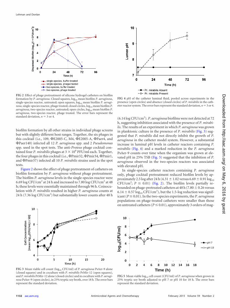
Figure 2 shows the effect of phage pretreatment of catheters onbiofilm formation by P. aeruginosa without phage pretreatment.The biofilm P. aeruginosa levels in the single-species reactor were6.69 log CFU/cm2 at 24 h and increased to 7.80 log CFU/cm2 at 48h; these levels were essentially maintained through 96 h. Coinocu-lation with P. mirabilis resulted in higher P. aeruginosa counts at24 h (7.36 log CFU/cm2) but substantially lower counts after 48 h
(6.14 log CFU/cm2). P. aeruginosa biofilms were not detected at 72h, suggesting inhibition associated with the presence of P. mirabi-lis. The results of an experiment in which P. aeruginosa was grownin planktonic culture in the presence of P. mirabilis (Fig. 3) sug-gested that P. mirabilis did not directly inhibit the growth of P.aeruginosa in the catheter model system. However, a substantialincrease in luminal pH levels in catheter reactors containing P.mirabilis (Fig. 4) and a marked reduction in the P. aeruginosaPsAer-9 counts over time when the organism was grown at ele-vated pH in 25% TSB (Fig. 5) suggested that the inhibition of P.aeruginosa observed in the two-species reactors was associatedwith elevated pH.
In single-species catheter reactors containing P. aeruginosaonly, phage cocktail pretreatment reduced biofilm levels by ap-proximately 2.5 log after 24 h (4.31 1.02 versus 6.69 0.91 log10
CFU/cm2; P � 0.01) (Fig. 2). The biofilm levels partially re-bounded on phage-pretreated catheters at 48 h (7.80 0.26 versus6.14 0.57 log10 CFU/cm2), but the 1.5-log reduction was signif-icant (P � 0.01). In the two-species experiments, the P. aeruginosapopulations on phage-treated catheters were smaller than thoseon untreated catheters (P � 0.01), approximately 3 orders of mag-
FIG 2 Effect of phage pretreatment of silicone hydrogel catheters on biofilmformation by P. aeruginosa. Closed squares, log10 mean biofilm P. aeruginosa,single-species reactor, untreated; open squares, log10 mean biofilm P. aerugi-nosa, single-species reactor, phage treated; closed circles, log10 mean biofilm P.aeruginosa, two-species reactor, untreated; open circles, log10 mean biofilm P.aeruginosa, two-species reactor, phage treated. The error bars represent thestandard deviation, n � 3 or 4.
FIG 3 Mean viable cell count (log10 CFU/ml) of P. aeruginosa PsAer-9 alone(closed squares) and in coculture with P. mirabilis PrMir-12 (open squares),and P. mirabilis PrMir-12 alone (closed circles) and in coculture with P. aerugi-nosa PsAer-9 (open circles), in 25% tryptic soy broth, over 18 h. The error barsrepresent the standard deviation.
FIG 4 pH of the catheter luminal fluid, pooled across experiments in thepresence (open circles) and absence (closed circles) of P. mirabilis in the cath-eter reactor system. The error bars represent the standard deviation, n � 3 or 4.
FIG 5 Mean viable log10 cell count (CFU/ml) of P. aeruginosa when grown in25% tryptic soy broth adjusted to pH 7 or pH 10 for 18 h. The error barsrepresent the standard deviation.
Lehman and Donlan
1132 aac.asm.org February 2015 Volume 59 Number 2Antimicrobial Agents and Chemotherapy
on May 6, 2016 by guest
http://aac.asm.org/
Dow
nloaded from
nitude at 24 h (4.37 0.26 versus 7.36 0.23 log10 CFU/cm2) and4 orders of magnitude at 48 h (2.05 2.31 versus 6.14 1.56 log10
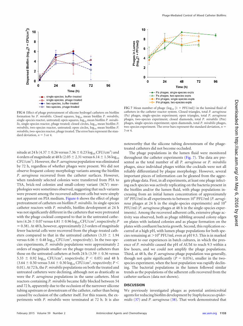
CFU/cm2). However, the P. aeruginosa population was eliminatedby 72 h, regardless of whether phages were present. We did notobserve frequent colony morphology variants among the biofilmP. aeruginosa recovered from the catheter surfaces. However,when randomly selected colonies were transferred from PIA toTSA, brick-red colonies and small-colony variant (SCV) mor-phologies were sometimes observed, suggesting that such variantswere present among the recovered adherent cells but were simplynot apparent on PIA medium. Figure 6 shows the effect of phagepretreatment of catheters on biofilm P. mirabilis. In single-speciescatheter reactors with P. mirabilis, biofilm development at 24 hwas not significantly different in the catheters that were pretreatedwith the phage cocktail compared to that in the untreated cathe-ters (4.26 0.07 versus 4.81 0.96 log10 CFU/cm2, respectively; P� 0.38). At 48 h, however, approximately 2.5 orders of magnitudefewer bacterial cells were recovered from the phage-treated cath-eters compared to that in the untreated catheters (3.35 1.94versus 6.06 0.48 log10 CFU/cm2, respectively). In the two-spe-cies experiments, P. mirabilis populations were approximately 2orders of magnitude smaller on the phage-treated catheters thanthose on the untreated catheters at both 24 h (3.59 0.36 versus5.33 0.92 log10 CFU/cm2, respectively; P 0.05) and 48 h(3.64 0.50 versus 5.64 0.30 log10 CFU/cm2, respectively; P 0.01). At 72 h, the P. mirabilis populations on both the treated anduntreated catheters were declining, although not as drastically aswere the P. aeruginosa populations in the same catheters. Mostreactors containing P. mirabilis became fully blocked between 54and 72 h, apparently due to the occlusion of the narrower siliconetubing upstream or downstream of the catheter, rather than beingcaused by occlusion of the catheter itself. For this reason, the ex-periments with P. mirabilis were terminated at 72 h. It is also
noteworthy that the silicone tubing downstream of the phage-treated catheters did not become occluded.
The phage populations in the lumen fluid were monitoredthroughout the catheter experiments (Fig. 7). The data are pre-sented as the total number of all P. aeruginosa or P. mirabilisphages, since individual phages within the cocktails were not allreliably differentiated by plaque morphology. However, severalimportant pieces of information can be gleaned from the aggre-gate phage data. First, in all experiments, at least one phage infect-ing each species was actively replicating on the bacteria present inthe biofilm and/or the lumen fluid, with phage populations in-creasing from an initial lumen concentration of approximately104 PFU/ml in all experiments to between 106 PFU/ml (P. aerugi-nosa phages at 24 h in the single-species experiments) and 109
PFU/ml (P. aeruginosa phages at 48 h in the single-species exper-iments). Among the recovered adherent cells, extensive phage ac-tivity was observed, both as phage nibbling around colony edgeson plates with isolated colonies and as plaque formation on theplates with confluent bacteria growth. Second, this replication oc-curred at a high pH, with lumen phage populations for both spe-cies remaining at �106 PFU/ml, even at pH 9.5. This is in markedcontrast to our experiences in batch cultures, in which the pres-ence of P. mirabilis caused the pH of AUM to reach 9.5 within afew hours, and we could not amplify the phage populations.Third, at 48 h, the P. aeruginosa phage population was generally,though not quite significantly (P � 0.076), smaller in the two-species experiment, when the host population was rapidly declin-ing. The bacterial populations in the lumen followed similartrends as the populations of the adherent cells recovered from thecatheter surfaces (data not shown).
DISCUSSION
We previously investigated phages as potential antimicrobialagents for reducing biofilm development by Staphylococcus epider-midis (37) and P. aeruginosa (38). That work demonstrated that
FIG 6 Effect of phage pretreatment of silicone hydrogel catheters on biofilmformation by P. mirabilis. Closed squares, log10 mean biofilm P. mirabilis,single-species reactor, untreated; open squares, log10 mean biofilm P. mirabi-lis, single-species reactor, phage treated; closed circles, log10 mean biofilm P.mirabilis, two-species reactor, untreated; open circles, log10 mean biofilm P.mirabilis, two-species reactor, phage treated. The error bars represent the stan-dard deviation, n � 3 or 4.
FIG 7 Mean number of phage (log10 [1 � PFU/ml]) in the luminal fluid ofcatheters in the catheter reactor system. Closed triangles, total P. aeruginosa(Pa) phages, single-species experiment; open triangles, total P. aeruginosaphages, two-species experiment; closed diamonds, total P. mirabilis (Pm)phages, single-species experiment; open diamonds, total P. mirabilis phages,two-species experiment. The error bars represent the standard deviation, n �3 or 4.
Phage-Mediated Control of Mixed Catheter Biofilms
February 2015 Volume 59 Number 2 aac.asm.org 1133Antimicrobial Agents and Chemotherapy
on May 6, 2016 by guest
http://aac.asm.org/
Dow
nloaded from

active phages can be incorporated into a hydrogel coating on cath-eters and reduce biofilm formation in vitro and that phage cock-tails can reduce biofilm formation for longer time periods thansingle phages can. Our current study expanded upon that work bystudying these phage-biofilm interactions against a two-speciesbacterial biofilm using a validated urine analog as the growth me-dium, low initial bacterial inocula, 3- to 4-day experimental time-lines, and phage cocktails against both bacterial species. We choseP. aeruginosa and P. mirabilis as target uropathogens. These twospecies are observed together in urinary catheter biofilms (5, 41).P. aeruginosa is one of the most frequently isolated species (5, 9,41) and is associated with serious symptomatic UTIs and CAUTIsthat progress to bacteremia (5, 42). When grown in biofilms, thisorganism is also known for generating an abundance of morpho-logical variants with various treatment susceptibilities (38, 43–45). P. mirabilis is a less common CAUTI-associated pathogen,but it is the primary cause of mineral-encrusted catheters, whichincrease the risk of complications, such as pyelonephritis andbloodstream infections (46–49).
The AUM described by Brooks and Keevil (50) was chosen forthis work because of the systematic manner in which it was for-mulated, our observations that CAUTI-associated isolates of P.mirabilis growing in AUM caused a pH increase and mineral pre-cipitation similar to that observed in human urine, the explicitinclusion of trace amounts of iron in the medium, the absence ofwhole protein, and the inclusion of the smallest amounts of un-defined “rich” components, such as peptone or yeast extract com-pared to that with other published formulations (55–58). In addi-tion, there is evidence from other work that it supports theexpression of relevant biofilm phenotypes. When Jones et al. (59)compared the structures of P. mirabilis biofilms grown in Brooksand Keevil AUM versus LB Miller broth, they found significantdifferences in surface coverage, biofilm thickness, water channelconformation, and the incidence of the swarmer cell morphologythat is characteristic of P. mirabilis. Because our catheter modelrequired the use of up to 20 liters of urine for a single experiment,and because human urine was difficult to filter sterilize, we choseto use the AUM formulation of Brooks and Keevil (50). The suit-ability of this AUM formulation as a urine analog was confirmedby the results of phage efficacy screening, in which essentially thesame phages were identified as significantly reducing biofilm for-mation in AUM and human urine.
The most important aspect of this study was the use of a two-species biofilm model. We are aware of only three previous studiesinvolving phage interactions with two-species biofilms, all ofwhich involved phage treatment of preformed biofilms separatefrom any specific clinical situation (60–62). In our phage-free re-actors, the total number of biofilm-associated P. mirabilis cellsappears to have been smaller than the number of biofilm-associ-ated P. aeruginosa in both the one- and two-species reactors. Thisis consistent with our biofilm and phage screens in 96-well plates,as well as with a previous report that P. mirabilis biofilm popula-tions were smaller than P. aeruginosa biofilm populations wheneach was grown in the same bladder model system (20). Somestudies have reported synergistic increases in biofilm mass inmixed- versus single-species biofilms (63, 64), but only Macleodand Stickler (41) specifically examined a coculture of P. aeruginosaand P. mirabilis in catheter biofilms, and they found minimal an-tagonism between the two species. Similarly, in our phage-freereactors, the numbers of biofilm-associated P. aeruginosa and P.
mirabilis were not affected by the presence of the other species atthe 2-h, 24-h, and 48-h time points. The elimination of P. aerugi-nosa by 72 h in the two-species catheter reactors, regardless ofphage treatment, was likely driven by the high pH that developedbetween 48 and 72 h due to P. mirabilis urease activity, as evi-denced by P. aeruginosa growth inhibition in medium with pH 10.However, the high pH did not appear to inhibit lytic activities foreither the P. mirabilis or P. aeruginosa phages in our cathetermodel, suggesting that phage application in indwelling urinarycatheters with high pH conditions might be feasible.
Although the application of anti-P. aeruginosa and anti-P. mi-rabilis phages to catheters will be ineffective against other bacterialspecies that may colonize the catheter, both organisms evaluatedin this study play an important role in CAUTIs (5, 42) and othercomplications related to the use of urinary catheters (46–49).
The timeline of phage efficacy is also important for clinicalutility. Previous studies with phage-treated catheters (38) havedemonstrated that phage may delay but not prevent biofilm for-mation, resulting in a rebound effect with prolonged exposure.We also observed a rebound for both P. aeruginosa and P. mirabi-lis. However, the extensive phage nibbling and plaque formationobserved on the plates with recovered adherent bacteria imply thatlarge fractions of the surviving bacterial population were not trulyphage resistant. Alternative explanations are that the survivingbiofilm cells were transiently nonsusceptible to phage infectiondue to metabolic changes (especially cells deeper in the biofilm),did not support phage replication following infection (pseudoly-sogeny, implied in Kay et al. [62]), or existed in “spatial refuges” inwhich nonsusceptible cells physically shield susceptible cells fromphage attack (60, 65). All of these mechanisms would support afairly stable long-term coexistence of phage and biofilm cells, eventhough initial biofilm formation may be slowed. We suggest thatphage pretreatment of hydrogel silicone catheters might mitigatecolonization and biofilm formation by multiple organisms forshort-term exposures. The observation that phages against differ-ent bacterial species when used together do not interfere with thelytic abilities of the other phages in the cocktail suggests the pos-sibility of using combinations of cocktails to target multiple spe-cies in multispecies biofilms. The clinical relevance of this ap-proach, with respect to the potential savings in health care costs,the reduction in costs of antimicrobials or adverse events associ-ated with antimicrobial use, or the prevention of antimicrobialresistance by biofilm mitigation for 48 to 72 h is not clear butmerits further investigation.
It may be fruitful to explore the potential synergistic interac-tions between phage cocktails and other antimicrobial strategies,such as catheters with more conventional antimicrobial sub-stances, biofilm disruptors, or bacterial interference. For example,in studies of bacterial interference, a naturally nonpathogenicEscherichia coli strain later rendered incapable of producing P fim-briae (66) was shown to reduce colonization by Enterococcusfaecalis, Candida albicans, Providencia stuartii, and pathogenic E.coli (67, 68). However, in human trials, both P. aeruginosa and P.mirabilis presented difficulties, with P. aeruginosa tending to over-grow the benign E. coli biofilm, and with the prior presence of P.mirabilis being predictive of poor E. coli persistence (22, 23, 69). Arecent in vitro study showed that pretreating uncoated siliconecatheters with both P. aeruginosa phages and the nonpathogenic E.coli strain had a synergistic effect, reducing and in some casescompletely preventing P. aeruginosa biofilm growth on catheters
Lehman and Donlan
1134 aac.asm.org February 2015 Volume 59 Number 2Antimicrobial Agents and Chemotherapy
on May 6, 2016 by guest
http://aac.asm.org/
Dow
nloaded from

for up to 72 h, when neither phages nor bacterial interferencealone was effective (45). This suggests that the combination ofbacterial interference with both anti-P. aeruginosa and anti-P. mi-rabilis phage cocktails conceivably offers broad protection againsturopathogen colonization without requiring phages that are tar-geted to every uropathogen of concern. It also indicates that thephage-coated catheter principle should be applicable to materialsother than the hydrogel-coated catheters used in our current andprevious related studies.
ACKNOWLEDGMENTS
We thank Silke Talsma and C. R. Bard, Inc., for their generous donation ofthe Lubri-Sil Foley catheters used in this study (no representative of C. R.Bard, Inc., had input in any part of this study, including but not limited toits conception, design, execution, or analysis, and no funding was pro-vided by C. R. Bard, Inc.).
S.M.L. was supported by an American Society for Microbiology/Coordinating Center for Infectious Disease International PostdoctoralFellowship.
We acknowledge Wayne Kirby for constructing the catheter modelsystem components and Jay Ash and the Snapfinger Creek Water QualityLaboratory staff for their assistance in the collection of sewage samples.
The use of trade names and commercial sources is for identificationonly and does not imply endorsement by the Public Health Service or theU.S. Department of Health and Human Services. The findings and con-clusions in this report are those of the authors and do not necessarilyrepresent the official position of the U.S. CDC.
REFERENCES1. Magill SS, Edwards JR, Bamberg W, Beldavs MS, Dumyati G, Kainer
MA, Lynfield R, Maloney M, McAllister-Hollod L, Nadle J, Ray SM,Thompson DL, Wilson LE, Fridkin SK, Emerging Infections ProgramHealthcare-Associated Infections and Antimicrobial Use PrevalenceSurvey Team. 2014. Multistate point-prevalence survey of health care-associated infections. N Engl J Med 370:1198 –1208. http://dx.doi.org/10.1056/NEJMoa1306801.
2. Saint S. 2000. Clinical and economic consequences of nosocomial cathe-ter-related bacteriuria. Am J Infect Control 28:68 –75. http://dx.doi.org/10.1016/S0196-6553(00)90015-4.
3. Tambyah PA, Knasinski V, Maki DG. 2002. The direct costs of nosoco-mial catheter-associated urinary tract infection in the era of managed care.Infect Control Hosp Epidemiol 23:27–31. http://dx.doi.org/10.1086/501964.
4. Garibaldi RA, Mooney BR, Epstein BJ, Britt MR. 1982. An evaluation ofdaily bacteriologic monitoring to identify preventable episodes of cathe-ter-associated urinary tract infection. Infect Control 3:466 – 470.
5. Ganderton L, Chawla J, Winters C, Wimpenny J, Stickler D. 1992.Scanning electron microscopy of bacterial biofilms on indwelling bladdercatheters. Eur J Clin Microbiol Infect Dis 11:789 –796. http://dx.doi.org/10.1007/BF01960877.
6. Frank DN, Wilson SS, St. Amand AL, Pace NR. 2009. Culture-independent microbiological analysis of Foley urinary catheter biofilms.PLoS One 4:e7811. http://dx.doi.org/10.1371/journal.pone.0007811.
7. Daifuku R, Stamm WE. 1984. Association of rectal and urethral coloni-zation with urinary tract infection in patients with indwelling catheters.JAMA 252:2028 –2030. http://dx.doi.org/10.1001/jama.1984.03350150028015.
8. Tambyah PA, Halvorson KT, Maki DG. 1999. A prospective study ofpathogenesis of catheter-associated urinary tract infections. Mayo ClinProc 74:131–136. http://dx.doi.org/10.4065/74.2.131.
9. Hidron AI, Edwards JR, Patel J, Horan TC, Sievert DM, Pollock DA,Fridkin SK, National Healthcare Safety Network Team, ParticipatingNational Healthcare Safety Network Facilities. 2008. NHSN annualupdate: antimicrobial-resistant pathogens associated with healthcare-associated infections: annual summary of data reported to the NationalHealthcare Safety Network at the Centers for Disease Control and Preven-tion, 2006 –2007. Infect Control Hosp Epidemiol 29:996 –1011. http://dx.doi.org/10.1086/591861.
10. Wagenlehner FM, Weidner W, Naber KG. 2005. Emergence of antibioticresistance amongst hospital-acquired urinary tract infections and phar-macokinetic/pharmacodynamic considerations. J Hosp Infect 60:191–200. http://dx.doi.org/10.1016/j.jhin.2004.12.017.
11. Jarvis WR, Martone WJ. 1992. Predominant pathogens in hospital infec-tions. J Antimicrob Chemother 29(Suppl A):19 –24.
12. Gaynes RP, Weinstein RA, Chamberlin W, Kabins SA. 1985. Antibiotic-resistant flora in nursing home patients admitted to the hospital. ArchIntern Med 145:1804 –1807. http://dx.doi.org/10.1001/archinte.1985.00360100064009.
13. Raz R. 2000. Chronic indwelling catheter replacement before antimicro-bial therapy for symptomatic urinary tract infection. J Urol 164:1254 –1258. http://dx.doi.org/10.1016/S0022-5347(05)67150-9.
14. Clayton CL, Chawla JC, Stickler DJ. 1982. Some observations on urinarytract infections in patients undergoing long-term bladder catheterization.J Hosp Infect 3:39 – 47. http://dx.doi.org/10.1016/0195-6701(82)90029-9.
15. Costerton JW, Stewart PS, Greenberg EP. 1999. Bacterial biofilms: acommon cause of persistent infections. Science 284:1318 –1322. http://dx.doi.org/10.1126/science.284.5418.1318.
16. Gould CV, Umscheid CA, Agarwal RK, Kuntz G, Pegues DA, Health-care Infection Control Practices Advisory Committee (HICPAC). 2009.Guideline for prevention of catheter-associated urinary tract infections2009. Healthcare Infection Control Practices Advisory Committee, Cen-ters for Disease Control and Prevention (CDC), Atlanta, GA. http://www.cdc.gov/hicpac/pdf/CAUTI/CAUTIguideline2009final.pdf.
17. Reddy ST, Chung KK, McDaniel CJ, Darouiche RO, Landman J, Bren-nan AB. 2011. Micropatterned surfaces for reducing the risk of catheter-associated urinary tract infection: an in vitro study on the effect of Sharkletmicropatterned surfaces to inhibit bacterial colonization and migration ofuropathogenic Escherichia coli. J Endourol 25:1547–1552. http://dx.doi.org/10.1089/end.2010.0611.
18. Kappell GM, Grover JP, Chrzanowski TH. 2009. Micro-scale surface-patterning influences biofilm formation. Electron J Biotechnol 12:e8. http://dx.doi.org/10.2225/vol12-issue3-fulltext-8.
19. Hook AL, Chang CY, Yang J, Luckett J, Cockayne A, Atkinson S, MeiY, Bayston R, Irvine DJ, Langer R, Anderson DG, Williams P, DaviesMC, Alexander MR. 2012. Combinatorial discovery of polymers resistantto bacterial attachment. Nat Biotechnol 30:868 – 875. http://dx.doi.org/10.1038/nbt.2316.
20. Jones GL, Muller CT, O’Reilly M, Stickler DJ. 2006. Effect of triclosan onthe development of bacterial biofilms by urinary tract pathogens on uri-nary catheters. J Antimicrob Chemother 57:266 –272. http://dx.doi.org/10.1093/jac/dki447.
21. Williams GJ, Stickler DJ. 2007. Some observations on the diffusion ofantimicrobial agents through the retention balloons of Foley catheters. JUrol 178:697–701. http://dx.doi.org/10.1016/j.juro.2007.03.091.
22. Trautner BW, Hull RA, Thornby JI, Darouiche RO. 2007. Coatingurinary catheters with an avirulent strain of Escherichia coli as a means toestablish asymptomatic colonization. Infect Control Hosp Epidemiol 28:92–94. http://dx.doi.org/10.1086/510872.
23. Prasad A, Cevallos ME, Riosa S, Darouiche RO, Trautner BW. 2009. Abacterial interference strategy for prevention of UTI in persons practicingintermittent catheterization. Spinal Cord 47:565–569. http://dx.doi.org/10.1038/sc.2008.166.
24. Cho YH, Lee SJ, Lee JY, Kim SW, Kwon IC, Chung SY, Yoon MS. 2001.Prophylactic efficacy of a new gentamicin-releasing urethral catheter inshort-term catheterized rabbits. BJU Int 87:104 –109. http://dx.doi.org/10.1046/j.1464-410x.2001.00978.x.
25. Hachem R, Reitzel R, Borne A, Jiang Y, Tinkey P, Uthamanthil R,Chandra J, Ghannoum M, Raad I. 2009. Novel antiseptic urinary cath-eters for prevention of urinary tract infections: correlation of in vivo and invitro test results. Antimicrob Agents Chemother 53:5145–5149. http://dx.doi.org/10.1128/AAC.00718-09.
26. Darouiche RO, Mansouri MD, Gawande PV, Madhyastha S. 2008.Efficacy of combination of chlorhexidine and protamine sulphate againstdevice-associated pathogens. J Antimicrob Chemother 61:651– 657. http://dx.doi.org/10.1093/jac/dkn006.
27. Kowalczuk D, Ginalska G, Golus J. 2010. Characterization of the devel-oped antimicrobial urological catheters. Int J Pharm 402:175–183. http://dx.doi.org/10.1016/j.ijpharm.2010.10.014.
28. Regev-Shoshani G, Ko M, Miller C, Av-Gay Y. 2010. Slow release ofnitric oxide from charged catheters and its effect on biofilm formation by
Phage-Mediated Control of Mixed Catheter Biofilms
February 2015 Volume 59 Number 2 aac.asm.org 1135Antimicrobial Agents and Chemotherapy
on May 6, 2016 by guest
http://aac.asm.org/
Dow
nloaded from

Escherichia coli. Antimicrob Agents Chemother 54:273–279. http://dx.doi.org/10.1128/AAC.00511-09.
29. Saint S, Elmore JG, Sullivan SD, Emerson SS, Koepsell TD. 1998. Theefficacy of silver alloy-coated urinary catheters in preventing urinary tractinfection: a meta-analysis. Am J Med 105:236 –241. http://dx.doi.org/10.1016/S0002-9343(98)00240-X.
30. Lai KK, Fontecchio SA. 2002. Use of silver-hydrogel urinary catheters onthe incidence of catheter-associated urinary tract infections in hospital-ized patients. Am J Infect Control 30:221–225. http://dx.doi.org/10.1067/mic.2002.120128.
31. Rupp ME, Fitzgerald T, Marion N, Helget V, Puumala S, Anderson JR,Fey PD. 2004. Effect of silver-coated urinary catheters: efficacy, cost-effectiveness, and antimicrobial resistance. Am J Infect Control 32:445–450. http://dx.doi.org/10.1016/j.ajic.2004.05.002.
32. Stensballe J, Tvede M, Looms D, Lippert FK, Dahl B, Tønnesen E, Ras-mussen LS. 2007. Infection risk with nitrofurazone-impregnated urinarycatheters in trauma patients: a randomized trial. Ann Intern Med 147:285–293. http://dx.doi.org/10.7326/0003-4819-147-5-200709040-00002.
33. Desai DG, Liao KS, Cevallos ME, Trautner BW. 2010. Silver or nitro-furazone impregnation of urinary catheters has a minimal effect on uro-pathogen adherence. J Urol 184:2565–2571. http://dx.doi.org/10.1016/j.juro.2010.07.036.
34. Park JH, Cho YW, Cho YH, Choi JM, Shin HJ, Bae YH, Chung H,Jeong SY, Kwon IC. 2003. Norfloxacin-releasing urethral catheter forlong-term catheterization. J Biomater Sci Polym Ed 14:951–962. http://dx.doi.org/10.1163/156856203322381438.
35. Schumm K, Lam TB. 2008. Types of urethral catheters for manage-ment of short-term voiding problems in hospitalised adults. Co-chrane Database Syst Rev 2008(2):CD004013. http://dx.doi.org/10.1002/14651858.CD004013.pub3.
36. Pickard R, Lam T, MacLennan G, Starr K, Kilonzo M, McPherson G,Gillies K, McDonald A, Walton K, Buckley B, Glazner C, Boachie C,Burr J, Norrie J, Vale L, Grant A, N=Dow J. 2012. Antimicrobialcatheters for reduction of symptomatic urinary tract infection in adultsrequiring short-term catheterisation in hospital: a multicenter ran-domised controlled trial. Lancet 380:1927–1935. http://dx.doi.org/10.1016/S0140-6736(12)61380-4.
37. Curtin JJ, Donlan RM. 2006. Using bacteriophages to reduce formationof catheter-associated biofilms by Staphylococcus epidermidis. AntimicrobAgents Chemother 50:1268 –1275. http://dx.doi.org/10.1128/AAC.50.4.1268-1275.2006.
38. Fu W, Forster T, Mayer O, Curtin JJ, Lehman SM, Donlan RM.2010. Bacteriophage cocktail for the prevention of biofilm formationby Pseudomonas aeruginosa on catheters in an in vitro model system.Antimicrob Agents Chemother 54:397– 404. http://dx.doi.org/10.1128/AAC.00669-09.
39. Biswas B, Adhya S, Washart P, Paul B, Trostel AN, Powell B, CarltonR, Merril CR. 2002. Bacteriophage therapy rescues mice bacteremic froma clinical isolate of vancomycin-resistant Enterococcus faecium. Infect Im-mun 70:204 –210. http://dx.doi.org/10.1128/IAI.70.1.204-210.2002.
40. Chanishvili N, Chanishvili T, Tediashvili M, Barrow PA. 2001. Phagesand their application against drug-resistant bacteria. J Chem Technol Bio-technol 76:689 – 699. http://dx.doi.org/10.1002/jctb.438.
41. Macleod SM, Stickler DJ. 2007. Species interactions in mixed-community crystalline biofilms on urinary catheters. J Med Microbiol56:1549 –1557. http://dx.doi.org/10.1099/jmm.0.47395-0.
42. Tambyah PA, Maki DG. 2000. The relationship between pyuria andinfection in patients with indwelling urinary catheters: a prospective studyof 761 patients. Arch Intern Med 160:673– 677. http://dx.doi.org/10.1001/archinte.160.5.673.
43. Martin C, Ichou MA, Massicot P, Goudeau A, Quentin R. 1995. Geneticdiversity of Pseudomonas aeruginosa strains isolated from patients withcystic fibrosis revealed by restriction fragment length polymorphism ofthe rRNA gene region. J Clin Microbiol 33:1461–1466.
44. Drenkard E, Ausubel FM. 2002. Pseudomonas biofilm formation andantibiotic resistance are linked to phenotypic variation. Nature 416:740 –743. http://dx.doi.org/10.1038/416740a.
45. Liao KS, Lehman SM, Tweardy DJ, Donlan RM, Trautner BW. 2012.Bacteriophages are synergistic with bacterial interference for the preven-tion of Pseudomonas aeruginosa biofilm formation on urinary catheters. JAppl Microbiol 113:1530 –1539. http://dx.doi.org/10.1111/j.1365-2672.2012.05432.x.
46. Warren JW. 1996. Clinical presentations and epidemiology of urinary
tract infections, p 3–27. In Mobley HLT, Warren JW (ed), Urinary tractinfections: molecular pathogenesis and clinical management. ASM Press,Washington, DC.
47. Kunin CM. 1997. Care of the urinary catheter, p 226 –278. Urinary tractinfections: detection, prevention and management. Williams & Wilkins,Baltimore, MD.
48. Stickler DJ, Zimakoff J. 1994. Complications of urinary tract infectionsassociated with devices used for long-term bladder management. J HospInfect 28:177–194. http://dx.doi.org/10.1016/0195-6701(94)90101-5.
49. Jacobsen SM, Stickler DJ, Mobley HL, Shirtliff ME. 2008. Complicatedcatheter-associated urinary tract infections due to Escherichia coli and Pro-teus mirabilis. Clin Microbiol Rev 21:26 –59. http://dx.doi.org/10.1128/CMR.00019-07.
50. Brooks T, Keevil CW. 1997. A simple artificial urine for the growth ofurinary pathogens. Lett Appl Microbiol 24:203–206. http://dx.doi.org/10.1046/j.1472-765X.1997.00378.x.
51. Appelmans R. 1921. De dosage du bactériophage. C R Soc Biol 85:1098.52. Adams M. 1959. Bacteriophages. Interscience Publishers, London, United
Kingdom.53. O’Toole GA, Kolter R. 1998. Initiation of biofilm formation in Pseu-
domonas fluorescens WCS365 proceeds via multiple, convergent signallingpathways: a genetic analysis. Mol Microbiol 28:449 – 461. http://dx.doi.org/10.1046/j.1365-2958.1998.00797.x.
54. Smith K, Hunter IS. 2008. Efficacy of common hospital biocides withbiofilms of multi-drug resistant clinical isolates. J Med Microbiol 57:966 –973. http://dx.doi.org/10.1099/jmm.0.47668-0.
55. Stickler DJ, Morris NS, Winters C. 1999. Simple physical model to studyformation and physiology of biofilms on urethral catheters. Methods En-zymol 310:494 –501. http://dx.doi.org/10.1016/S0076-6879(99)10037-5.
56. Chutipongtanate S, Thongboonkerd V. 2010. Systematic comparisons ofartificial urine formulas for in vitro cellular study. Anal Biochem 402:110 –112. http://dx.doi.org/10.1016/j.ab.2010.03.031.
57. Griffith DP, Musher DM, Itin C. 1976. Urease. The primary cause ofinfection-induced urinary stones. Invest Urol 13:346 –350.
58. Cox AJ, Hukins DW, Davies KE, Irlam JC, Sutton TM. 1987. Anautomated technique for in vitro assessment of the susceptibility of urinarycatheter materials to encrustation. Eng Med 16:37– 41. http://dx.doi.org/10.1243/EMED_JOUR_1987_016_009_02.
59. Jones SM, Yerly J, Hu Y, Ceri H, Martinuzzi R. 2007. Structure ofProteus mirabilis biofilms grown in artificial urine and standard laboratorymedia. FEMS Microbiol Lett 268:16 –21. http://dx.doi.org/10.1111/j.1574-6968.2006.00587.x.
60. Tait K, Skillman LC, Sutherland IW. 2002. The efficacy of bacteriophageas a method of biofilm eradication. Biofouling 18:305–311. http://dx.doi.org/10.1080/0892701021000034418.
61. Sillankorva S, Neubauer P, Azeredo J. 2010. Phage control of dual speciesbiofilms of Pseudomonas fluorescens and Staphylococcus lentus. Biofouling26:567–575. http://dx.doi.org/10.1080/08927014.2010.494251.
62. Kay MK, Erwin TC, McLean RJ, Aron GM. 2011. Bacteriophage ecologyin Escherichia coli and Pseudomonas aeruginosa mixed-biofilm communi-ties. Appl Environ Microbiol 77:821– 829. http://dx.doi.org/10.1128/AEM.01797-10.
63. Burmolle M, Webb JS, Rao D, Hansen LH, Sørensen SJ, Kjelleberg S.2006. Enhanced biofilm formation and increased resistance to antimicro-bial agents and bacterial invasion are caused by synergistic interactions inmultispecies biofilms. Appl Environ Microbiol 72:3916 –3923. http://dx.doi.org/10.1128/AEM.03022-05.
64. Skillman LC, Sutherland IW, Jones MV. 1999. The role of exopolysac-charides in dual species biofilm development. J Appl Microbiol 85(Suppl1):13S–18S.
65. Schrag SJ, Mittler JE. 1996. Host-parasite coexistence: the role of spatialrefuges in stabilizing bacteria-phage interactions. Am Nat 148:348 –377.http://dx.doi.org/10.1086/285929.
66. Hull RA, Donovan WH, Del Terzo M, Stewart C, Rogers M, DarouicheRO. 2002. Role of type 1 fimbria- and P fimbria-specific adherence incolonization of the neurogenic human bladder by Escherichia coli. InfectImmun 70:6481– 6484. http://dx.doi.org/10.1128/IAI.70.11.6481-6484.2002.
67. Trautner BW, Darouiche RO, Hull RA, Hull S, Thornby JI. 2002.Pre-inoculation of urinary catheters with Escherichia coli 83972 inhibits
Lehman and Donlan
1136 aac.asm.org February 2015 Volume 59 Number 2Antimicrobial Agents and Chemotherapy
on May 6, 2016 by guest
http://aac.asm.org/
Dow
nloaded from

catheter colonization by Enterococcus faecalis. J Urol 167:375–379. http://dx.doi.org/10.1016/S0022-5347(05)65471-7.
68. Trautner BW, Hull RA, Darouiche RO. 2003. Escherichia coli 83972inhibits catheter adherence by a broad spectrum of uropathogens. Urol-ogy 61:1059 –1062. http://dx.doi.org/10.1016/S0090-4295(02)02555-4.
69. Darouiche RO, Thornby JI, Cerra-Stewart C, Donovan WH, Hull RA.
2005. Bacterial interference for prevention of urinary tract infection: aprospective, randomized, placebo-controlled, double-blind pilot trial.Clin Infect Dis 41:1531–1534. http://dx.doi.org/10.1086/497272.
70. Lindberg RB, Latta RL. 1974. Phage typing of Pseudomonas aeruginosa:clinical and epidemiologic considerations. J Infect Dis 130:S33–S42. http://dx.doi.org/10.1093/infdis/130.Supplement.S33.
Phage-Mediated Control of Mixed Catheter Biofilms
February 2015 Volume 59 Number 2 aac.asm.org 1137Antimicrobial Agents and Chemotherapy
on May 6, 2016 by guest
http://aac.asm.org/
Dow
nloaded from
Copyright © 2022 FDOKUMEN