AGC Tuning of an Interconnected System after Deregulation Using Genetic Algorithms

1
Poriferan survivin exhibits a conserved regulatory role in the interconnected pathways of cell cycle and apoptosis
B Luthringer1, S Isbert1, WEG Müller1, C Zilberberg1, NL Thakur1, G Wörheide2, RH
Stauber3, M Kelve4 and M Wiens*,1
1 Institute for Physiological Chemistry and Pathobiochemistry, Johannes Gutenberg
University, Medical School, Mainz; Germany.
2 Department of Earth- and Environmental Sciences, Ludwig-Maximilians-Universität,
München; Germany.
3 Department of Otorhinolaryngology, Molecular and Cellular Oncology, Johannes
Gutenberg University, Medical School, Mainz; Germany.
4 Department of Gene Technology, University of Technology, Tallinn; Estonia.
* Corresponding author: M Wiens; Institute for Physiological Chemistry and
Pathobiochemistry, Johannes Gutenberg University, Medical School, Duesbergweg
6, D-55099 Mainz; Germany. Tel: +49-6131-3925961. Fax: +49-6131-3925243. E-
mail: [email protected]
Keywords: survivin; IAP; caspase; sponges; Suberites domuncula; cadmium
Running title: Conserved dual role of survivin
Abbreviations: aa, amino acid; ABTS, 2,2'-azino-bis(3-ethylbenzthiazoline-6-sulfonic
acid); Bcl-2, B-cell lymphoma-2; BIR, baculoviral IAP repeat; CDE, cell cycle
dependent elements; Ced-9/-3, cell death abnormality -9/-3; CHR, cell cycle
homology regions; CPC, chromosomal passenger complex; CRB-1, crumbs homolog
1; CRM1, chromosome region maintenance 1; DIG, digoxigenin; DMF,

2
dimethylformamide; dys-1, dystrophin-1; ELISA, enzyme-linked immunosorbent
assay; IAP, inhibitors of apoptosis protein; LMB, leptomycin B; LSM, laser scanning
microscopy; ML, maximum likelihood; MTT, 3-(4,5-dimethylthiazol-2-yl)-2,5-
diphenyltetrazolium bromide; NES, nuclear export signal; NJ, neighbor-joining; ORF,
open reading frame; PBS, phosphate buffered saline; PFA, paraformaldehyde
The following sequences from S. domuncula have been deposited in the
EMBL/GenBank: SDSURVL (accession number FM210333 and FM210334, cDNA
and genomic sequence respectively) and SDCASL2 (FM210332).

3
Abstract
Survivin orchestrates intracellular pathways during cell division and apoptosis. Its
central function as mitotic regulator and inhibitor of cell death has major implications
for tumor cell proliferation. Analyses in early-branching Metazoa so far propose an
exclusive role of survivin as a chromosomal passenger protein, whereas only later
during evolution a complementary anti-apoptotic function might have arisen,
concurrent with increased organismal complexity. To lift the veil on the ancestral
function(s) of this key regulator, a survivin-like protein (SURVL) of one of the earliest-
branching metazoan taxa was identified and functionally characterized. SURVL of the
sponge Suberites domuncula shares considerable similarities with its metazoan
homologues, ranging from conserved exon/intron structure to presence of protein-
interaction domains. Whereas sponge tissue displays a low steady-state level,
SURVL expression was significantly up-regulated in rapidly proliferating primmorph
cells. In addition, challenge of tissue and primmorphs with heavy metal or lipopeptide
stimulated SURVL expression, concurrent with the expression of a newly discovered
caspase. Complementary functional analyses in transfected HEK-293 cells revealed
that heterologous expression of a SURVL-EFGP fusion not only promotes
proliferation but also enhances resistance to cadmium-induced cell death. Taken
together, these results suggest both a deep evolutionary conserved dual role of
survivin and an equally conserved central position in the interconnected pathways of
cell cycle and apoptosis.

4
Introduction
Apoptosis plays a crucial role not only in countless physiological processes (e.g.,
elimination of redundant cells during vertebrate embryonic development) but also
during pathogenesis. Since apoptosis relies on the clear-cut function of numerous
pro- and anti-apoptotic factors, any malfunction on their part or disruption of their
respective interplay inevitably leads to dysfunctional cellular metabolism. The
ensuing delayed or premature cell death often manifests in severe pathologies,
including carcinogenesis and neurodegenerative diseases.
The groundbreaking discovery of the Caenorhabditis elegans proteins Ced-9,
homologue of human anti-apoptotic Bcl-2, and Ced-3, homologue of human
caspase-1, has demonstrated the molecular conservation of the apoptotic cell death
program along a broad range of Metazoa, from nematodes to vertebrates.1 Since
then, the fundamental role of apoptosis has been increasingly recognized in various
physiological processes. In addition, several invertebrate model organisms revealed
a significant potential for analyses of apoptotic mechanisms, not only under an
evolutionary point of view. Thus, the identification of the human crumbs homologue
(CRB-1) and its causative role in the manifestation of retinitis pigmentosa was based
on the discovery of mutational variants of the Drosophila melanogaster protein
crumbs.2 In addition, loss of function mutations of the C. elegans dys-1 gene
confirmed the role of human dystrophin in the manifestation of the neuromuscular
disease of Duchenne.3 During the last years a novel invertebrate model system has
emerged: sponges (phylum Porifera), which are among the earliest-branching non-
bilaterian animals.4 Recent biomarker analyses date the occurrence of probable
stem-group poriferans back before the end of the Marinoan glaciation (~635 Myr
ago)5 and molecular clock analyses estimated that the main poriferan lineages

5
diverged in the Cryogenian/Ediacaran6, well before the rapid appearance of major
metazoan groups (Cambrian Explosion).
Until today, the model system "Porifera" has revealed several characteristics
that have proven valuable for evolutionary studies within a comparative context. The
major avail is the phylogenetically basal position of Porifera allowing a unique
vantage point on the hypothetical common ancestral taxon, the Urmetazoa.
Moreover, comparative transcriptomics and proteomics divulged several examples of
poriferan molecules, genetic features, protein domains, etc. that remained conserved
throughout evolution, but otherwise have been lost in invertebrate model systems
traditionally used, C. elegans and D. melanogaster.7 Furthermore, the majority of
poriferan genes display a closer sequence similarity to their human homologues than
to the respective molecules of worm and fly (similar to some anthozoans), probably
caused by an accelerated evolution of the latter animal lineages.8,9
The fine-tuned balance between apoptosis and proliferation is upheld via
numerous regulatory molecules that maintain tissue integrity and, ultimately, ensure
survival of the organism. The inhibitors of apoptosis (IAP) family comprises
structurally related proteins (the presence of at least one baculoviral IAP repeat (BIR)
domain is mandatory) that were originally discovered in baculovirus. In addition to
their cell death regulating activities several IAP have a complementary role in cell
division, in particular attributed to survivin. Accordingly, the dual role of human
survivin is based upon its function as (i) a chromosomal passenger protein that
controls segregation of chromatin/regulation of cytokinesis and (ii) a caspase
inhibitor that prevents processing/activation of caspases.10 Since transcription from
its single gene locus is up-regulated in most tumor cell types, survivin is a well-
established analytical marker of neoplasms.11,12 The function of survivin homologues

6
in non-bilaterian Metazoa is much less well-defined. Accordingly, similar to vertebrate
survivin the D. melanogaster homologue deterin deters cells from caspase-induced
apoptosis and some studies propose an additional pro-proliferative effect.13 On the
other hand, the C. elegans survivin homologue BIR-1 appears to be exclusively
involved in cytokinesis, in particular during embryogenesis.14
Here we report the discovery and functional characterization of a survivin-like
protein (SDSURVL) from a member of the ancestral-like clade Porifera, Suberites
domuncula. SDSURVL displays both significant sequence homologies to human
survivin (in particular concerning the BIR domain) and a conserved genetic structure.
Complementary studies in vivo and in transfected cells suggest a conserved dual role
of survivin and, concurrently, indicate a loss of function in certain rapidly evolving and
highly adapted metazoan taxa.

7
Results
Characterization of the poriferan survivin-like protein SDSURVL. The complete
SDSURVL cDNA was isolated from a S. domuncula cDNA library by PCR and
degenerate primers that were directed against a conserved region within the BIR
domain. SDSURVL consists of 620 nt (excluding the poly(A) tail) that encompass an
open reading frame (ORF) of 447 nt (excluding the first stop codon), beginning at
nt43–45 (Metstart). During Northern blot analyses, a DIG-labeled probe detected a
transcript whose size was consistent with that of the cDNA (≈ 650 nt; Figure 4A). The
deduced protein, SDSURVL, comprises 149 aa with an expected size of 17,003 Da.
Analyses of SDSURVL revealed significant sequence similarity to vertebrate survivin
homologues, such as Gallus gallus survivin isoform 1 (with an expect value
[E value]15 of 2 e-20; 32% identical and 52% similar aa) and human survivin (8 e-18;
30%/49%) (Figure 1A). Considerably lower was the similarity to survivin homologues
of more basal Metazoa and fungi, e.g., D. melanogaster deterin (7 e-11; 45%/24%),
placozoan (Trichoplax adhaerens) survivin-like protein (2 e-11; 19%/35%), and
Schizosaccharomyces pombe survivin homolog (5 e-11; 34%/48% with respect to the
BIR domain [aa117-194]). Viral genomes often encode anti-apoptotic or cell cycle
promoting proteins in order to bypass the host defense system. Viral survivin-like
proteins generally display lower sequence homology to their metazoan counterparts
and in particular to S. domuncula survivin, e.g., Epiphyas postvittana
nucleopolyhedrovirus IAP-3 (4 e-07; 17% aa similarities) and Cydia pomonella
granulosis virus ORF17 IAP-3 (3 e-06; 16%).
Since SDSURVL features a single BIR domain, a putative nuclear export
signal (NES), and a coiled-coil region, but is missing a RING domain (characteristic
of non-survivin IAP), the molecule represents a novel member of the survivin

8
subfamily of IAP. The poriferan BIR domain (aa20-93) has an E value of 4.85 e-22 and
is consistent with the PROSITE consensus sequence PS01282 with two exceptions
(marked in bold): [HKEPILVY]-x(2)-R-x(3,8)-[FYW]-x(11,14)-[STAN]-G-[LMF]-x-
[FYHDA]-(x(4)│x(7))-[DESL]-x(2,3)-C-x(2)-C-x(6)-[WA]-x(9)-H-x(4)-[PRSD]-x-C-x(2)-
[LIVMA]. The putative NES (aa95-103) loosely fits a consensus sequence proposed
by 16, L-x(2,3)-[FILVMP]-x(2)-[LI]-x-[LIV]. Furthermore, COILS algorithms indicate the
typical COOH-terminal coiled-coil conformation (aa127-147), usually present in survivin
but missing in other IAP family members. Moreover, the conserved threonine
residue, whose phosphorylation by p34cdc2 mediates human survivin activation,17 is
present in SDSURVL (aa26). Additionally, a ubiquitin ligase complex-binding
destruction motif (APCC-D box; aa136-141) was predicted.
To determine the phylogenetic relationship of survivin-like proteins, molecular
phylogenetic analyses were performed by neighbor-joining, maximum likelihood, and
Bayesian methods, including the so far known sequences of metazoan taxa as well
as related BIR-containing sequences of viruses and yeast. Accordingly, the resulting
composite consensus tree shows the poriferan SDSURVL protein at the base of a
monophyletic metazoan clade, whereas viral sequences form a separate
monophyletic group (Figure 1B).
Characterization of the poriferan survivin-like gene (SDSURVL). The
S. domuncula gene comprises four exons and three introns (Figure 2A). Each
exon/intron border displays typical splicing signals (GT-intron-AG). The phases of
introns vary: the first and third introns are in phase 0, while the second intron is in
phase 2. The human survivin gene shares the same features (Figure 2B) except for
the borders of the third intron (AT-intron-GG). Parts of the first and third exons and

9
the complete second exon are encoding the BIR domain (Figure 2C). Analyses of the
5'-flanking region (Figure 2D) of human (1) and murine (2) survivin genes reveal the
presence of a TATA-less promoter, containing a canonical CpG island and several
transcription factor binding sites.18 However, in silico analyses of the putative
poriferan promoter region (3) predicted the presence of a TATA box (nt-64 to -73) in
addition to other cis-regulatory elements, CAAT box (nt-109 to -113), and ribosomal
binding site (nt-5 to -8). Moreover, similar to the human and murine promoter two
putative Sp1 sites (nt-51, -292) have been predicted, although in contrast to the former
ones no cell cycle dependent elements (CDE) or cell cycle homology regions (CHR)
have been found in the poriferan non-coding region.
Characterization of the poriferan caspase-like protease 2 (SDCASL2). Caspases
(cysteine-dependent aspartyl-specific proteases) are synthesized as zymogens that
become activated by scaffold-mediated transactivation or by cleavage via upstream
proteases.19 Since the anti-apoptotic properties of vertebrate survivin have been
functionally linked to inhibition of caspase-7 and -3, a S. domuncula cDNA library
was screened for homologous molecules. The correct size of the resulting clone,
SDCASL2, then was verified on Northern blots (Figure 4A). SDCASL2 (1,827 nt,
excluding poly(A)) comprises an ORF between nt175–177 (Metstart) and nt1738–1740
(stop), coding for a putative protein of 521 aa that was termed caspase-like
protease 2 (SDCASL2; calculated Mr 58,427). SDCASL2 revealed sequence
homology not only to SDCASL (E value 7 e-31; 33% aa identical and 52% similar aa)
but also to members of the caspase-7 subfamily, e.g., Xenopus laevis and human
representatives (E value 3 e-16 [21%/33%] and 3 e-14 [19%/35%] respectively)
(Figure 3).

10
All caspases contain a CASc domain, which is cleaved into two subunits (p10,
p20) during activation. SDCASL2 represents a putative procaspase, comprising
subunits p10 (aa191-267) and p20 (aa22-154), the latter containing the characteristic Cys
and His active sites (Figure 3). The Cys active site consensus pattern is conserved
(PROSITE pattern PS01122) with two exceptions, marked in bold ([KL]-P-K-[LIVMF]-
[LIVMFY]-[LIVMF](2)-[QPD]-[AF]-C-[RQG]-[GE]), whereas the histidine active site
(PS01121) features three alterations, [HL]-x(2,4)-[SC]-x(1,2)-{A}-x-[LIVMFY](2)-[ST]-
G-H-G.
The absence of a large NH2-terminal prodomain, characteristic of initiator
caspases, suggests a classification to the group of effector caspases. However,
similar to SDCASL, SDCASL2 carries an unusual long COOH-terminal stretch of
254 aa, containing a putative double-stranded RNA-binding motif (aa441-514; PF0035
[Pfam]).
Evolutionary conserved function(s) of poriferan survivin. To elucidate the
controversially discussed ancestral function of survivin (or if indeed its dual role is
conserved through metazoan evolution), functional analyses were performed in
sponge tissue and primmorphs (aggregates of proliferating sponge cells). Moreover,
functional conservation was assessed in a heterologous vertebrate cell model
(SDSURVL-expressing transfected HEK-293 cells) that has been previously used to
explore expression and functional mechanisms of survivin.20,21
Regulation of proliferation. Comparative Northern blot analyses were performed,
showing an increasing level of SDSURVL transcripts during the first two weeks of
primmorph growth, whereas the control (taken immediately after primmorph

11
formation) revealed a considerably lower expression level (Figure 4A). After 14 d,
survivin transcription reached the steady-state. On the other hand, in adult sponge
tissue survivin transcripts were barely detected. Additionally, SDCASL2 expression
was very low throughout the experiments, in both primmorphs and sponge tissue
(Figure 4A). Subsequently, SDSURVL and SDCASL2 expression was investigated in
aging primmorphs (two weeks of age) via complementary in situ hybridization studies
(Figure 4B). For this purpose, primmorph cryosections were incubated with labeled,
single-stranded sense (control) or antisense probes. Both sense probes did not
produce specific signals. In contrast, the antisense SDSURVL probe detected
survivin transcripts abundantly and homogenously throughout the sections, whereas
only few SDCASL2 copies were identified, mainly in the surface cell layer and in cells
lining the aquiferous canal system within the mesohyl (internal part of the sponge)
(Figure 4B).
In parallel, a construct encoding a fusion of SDSURVL and COOH-terminal
EGFP (SDSURVL-EGFP) was introduced into HEK-293 cells. Expression was
confirmed microscopically (Figure 5B) and by immunodetection, using anti-EGFP
antibodies (Figure 8; expected size of 45 kD corresponding to the SDSURVL-EGFP
fusion). The transfection efficiency was determined by the percentage of EGFP-
positive HEK-293 cells via fluorescence microscopy. An average transfection
efficiency of 94.6% was calculated.
Subsequently, the putative regulatory function of SDSURVL in cytokinesis was
explored by monitoring the concentrations of SDSURVL-EGFP transfected (HEK293-
SDSURVL) and mock-transfected HEK-293 cells, 48 h post-transfection (Figure 5A).
Whereas mock-transfection provoked no differences in cell concentration compared
to the non-transfected control, SDSURVL-EGFP expression caused a 2.3-fold

12
increase in number of HEK293-SDSURVL cells (significance level P ≤ 0.05). In
addition, trypan blue staining did not display altered cell viability between control and
SDSURVL-EGFP expressing populations.
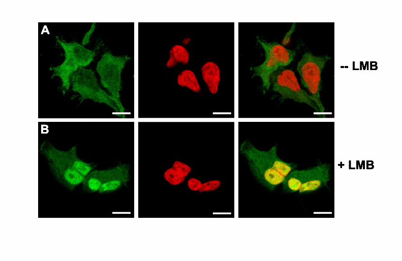
Finally, subcellular localization and nucleo-cytoplasmic trafficking of
SDSURVL-EGFP in transfected cells was analyzed by confocal laser scanning
microscopy (LSM). For this purpose, HEK293-SDSURVL cells were incubated with
the nuclear export inhibitor leptomycin B (LMB) for 30 min or remained untreated
(control). Following counterstaining of dsDNA with the far-red fluorescent dye
DRAQ5, LSM showed that in the controls SDSURVL-EGFP predominantly localizes
in the cytoplasm and, to a lesser degree in the nucleus (Figure 6A). Consequently,
the fluorescence pattern of the merge revealed a barely detectable co-localization of
fusion (green) and nuclei (red). Upon LMB treatment, SDSURVL-EGFP accumulated
in the nucleus. The merged channel clearly depicted a significant co-localization of
nuclei and SDSURVL-EGFP (Figure 6B; yellow), whereas the staining intensity of
cytoplasmic SDSURVL-EGFP is decreased, compared to the untreated control.
Regulation of apoptosis. To explore the putative implementation of SDSURVL as a
pro-survival factor, SDSURVL and SDCASL2 expression was analyzed in sponge
tissue and primmorphs challenged with pro-apoptotic stimulants. Moreover, the
viability of mock-transfected and HEK293-SDSURVL cell lines was assessed
following exposure to the same stimuli. Pam3Cys-Ser-(Lys)4 and cadmium were
employed for their selective ability to trigger extrinsic (the former compound) or
intrinsic (latter) cell death pathways.22,23 Both lipopeptide and heavy metal stimulated
the expression of SDSURVL and SDCASL2 in sponge tissue and primmorphs alike,
already after 6 h (Figure 7). With prolonged incubation time, the expression level of

13
both transcripts further increased; however, this effect was more pronounced in
primmorphs than in tissue samples.
To assess putative effects of SDSURVL expression, mock-transfected
HEK-293 and HEK293-SDSURVL cell lines (95.5% transfection efficiency) were
exposed to Pam3Cys-Ser-(Lys)4 and cadmium. Then, MTT assays, cell death
detection ELISA, and Western blot analyses were performed. MTT assays revealed
that the lipopeptide did not alter cell viabilities, neither in mock-transfected nor in
HEK293-SDSURVL cells, compared to untreated control cells (Figure 8A).
Additionally, Pam3Cys-Ser-(Lys)4-treatment did not change the amount of survivin
(endogenous as well as poriferan). In contrast, both cell lines were sensitive to
cadmium. Thus, MTT assays showed that 46.7% of mock-transfected cells
underwent cell death after 12 h of exposure. Nevertheless, SDSURVL-expression
rescued HEK293-SDSURVL since only 25.6% cells were non-viable (Figure 8B;
significance level P ≤ 0.05). Furthermore, Western blot analyses displayed an
additional effect of cadmium, since increased immunodetection was observed of
both, human survivin and its poriferan homologue after 6-12 h of challenge.
Concurrently, cell death detection ELISA could not reveal altered formation of
mono- and oligonucleosomes, neither in mock-transfected nor HEK293-SDSURVL
cells following Pam3Cys-Ser-(Lys)4-treatment, compared to untreated control cells
(Figure 9A). In contrast, exposure to cadmium significantly induced apoptotic DNA
fragmentation in both cell lines. However, SDSURVL-expression caused a reduced
DNA fragmentation by 51% (Figure 9B; significance level P ≤ 0.05).

14
Discussion
Survivin represents a unique protein within the IAP family, containing a single BIR
domain and a COOH-terminal coiled-coil structure. Thus, it differs from all other
family members that comprise 2-3 BIR domains and a canonical COOH-terminal
RING finger.11 In vertebrates, survivin not only controls cell cycle checkpoints and
correct completion of cytokinesis but also regulates activation of caspases.24
However, the evolutionary conservation of this dual role remains inconclusive since
in Nematoda the survivin homologue appears to be implicated exclusively in
cytokinesis.25 In addition, even though a fungal (S. pombe) survivin-like protein was
proposed to be required for efficient cell division and exhibits anti-apoptotic activity,26
it is considerably larger in size than other survivin proteins.
Key regulators of apoptosis and cell cycle progression have already been
discovered in the phylum Porifera,27 which appears to be the earliest-branching
metazoan taxon.4 However, identification of poriferan survivin provides an important
tool not only to investigate the primordial function(s) of this versatile regulator protein,
but also to elucidate the putative ancient and intimate link between apoptosis,
proliferation, and morphogenesis.
The newly discovered poriferan survivin homologue demonstrates a significant
degree of sequence conservation. A consensus tree resulting of NJ, ML, and
Bayesian analyses places SDSURVL at the base of a metazoan clade, confirming
the common ancestry of metazoan survivin-like proteins, whereas viral sequences
form a well-supported separate group.
SDSURVL features all characteristics of survivins that facilitate protein-
interaction and mediate both anti-apoptotic activity and proper kinetochore
attachment.28 However, it displays several additional putative domains that are

15
known to be involved in cell cycle regulatory functions: (i) APCC-D box, required for
ubiquitination and proteasome-mediated degradation via the anaphase-promoting
ubiquitin ligase complex (APC/C); (ii) LIG_FHA1/2 motifs (aa98-104; aa100-106) that bind
regulators of the G2/M checkpoint; (iii) USP7-binding motif (aa33-37) that is a target of
the UPS7 deubiquitinating enzyme, whose substrates are regulators of cell survival
pathways. So far, no such feature has been described for other survivin molecules.
The gene structure (intron/exon positions, intron phases) is conserved
between poriferan and vertebrate (human, murine) survivin,18 but not within the
D. melanogaster deterin,13 which possesses only two introns. However, some
differences to the vertebrate promoter region are noticeable: the TATA-less
vertebrate promoter includes CpG islands and several Sp1, CDE, and CHR sites. By
analogy with other cell cycle-regulated genes it was postulated that Sp1 regulates
basal transcriptional expression of the survivin gene, further modulated by CDE/CHR
elements, thus imparting cell cycle periodicity of survivin expression.18 In contrast,
the poriferan promoter features a canonical TATA box, two Sp1 sites, and is missing
CDE/CHR elements. Therefore, considering the aforementioned protein interaction
motifs, regulation of the poriferan survivin pool might also occur on a
posttranscriptional/proteasomal level, e.g., via (de)ubiquitination or complex
formation with other regulatory proteins.
Survivin is expressed in many human tumors and in rapidly dividing cells of
fetal tissue during the G2/M phase, but is not detectable in terminally differentiated
cells. It was proposed that survivin exerts its anti-apoptotic function via inhibiting
caspases, particularly caspase-7 and -3,29 though other studies argue against a
direct mechanism of caspase inhibition by survivin, see e.g.,30.

16
To approach the enigmatic role of survivin in non-bilaterian Metazoa,
comparative functional studies of poriferan survivin were performed. Accordingly, the
implementation of SDSURVL in cell proliferation and cell death was studied in
sponges as well as in a human cell model (HEK-293; as SDSURVL-EGFP fusion).
This strategy had already been successfully adopted to assess the anti-apoptotic
potential of deterin and poriferan Bcl-2.31,32 Moreover, survivin-EGFP fusions have
been widely used to study both the bifunctional role of survivin and its dynamic
intracellular localization and appear fully functional.33-35 In contrast to sponge tissue
that displays a very low steady-state expression level, proliferating cells of
primmorphs contained a considerable amount of SDSURVL, indicative of its
involvement in cell division. This assumption was supported by a significantly
increased proliferation rate of HEK-293 cells expressing SDSURVL-EGFP. In
addition, application of two inducers of cell death (cadmium, Pam3Cys-Ser-(Lys)4)
indicated an anti-apoptotic function of SDSURVL since both compounds triggered the
expression of SDSURVL in sponge tissue and primmorphs, possibly a
countermeasure to the induced expression of the caspase-7 homologue SDCASL2.
Concurrently, cadmium induced apoptosis in HEK-293 cells, although concomitant
expression of SDSURVL-EGFP drastically reduced the occurrence of cell death
(≈ 50%). However, Pam3Cys-Ser-(Lys)4 incubation did not alter HEK-293 cell
viability, probably due to the different repertoire of Toll-like receptors (mediating
lipopeptide-induced innate immunity and apoptosis) found within HEK-293 and
sponge cells. Future studies will investigate the effect of further inducers of cell death
on sponge and HEK293-SDSURVL cells.
Cytotoxicity mediated by cadmium is associated with the release of
mitochondrial pro-apoptotic factors into the cytosol.23 These factors cause activation

17
of caspase-3/7,36 which is considered as a biochemical hallmark of the apoptotic
cascade. However, survivin was proposed to interact with various caspases and,
consequently, to inhibit further progression of the apoptotic cascade. The
aforementioned observations in combination with significant similarities of sequence
and structure between SDSURVL and its vertebrate counterparts propose a common
regulatory function in the interconnected pathways of cell cycle and apoptosis. In
HEK-293 cells, poriferan survivin possibly complements the endogenous molecule by
sequestrating pro-apoptotic binding partners, consequently delaying cell death. In
addition, the presence of putative target sites for ubiquitination might explain the
elevated levels of human and poriferan survivin detected in challenged transfected
cells. Accordingly, this effect is not the result of an induced expression but is
indicative of a decreased proteasomal degradation of either protein. Therefore, the
poriferan molecule replenishes the pool of endogenous survivin, concomitantly
preventing degradation of its human counterpart by engaging/silencing elements of
the proteasome.
Furthermore, concurrent with previous observations during which elevated
levels of survivin occur in most human tumor cell types and overexpression of
survivin in transfected HEK-293 increases cell proliferation,20,21 SDSURVL-EGFP
expression stimulates cell division. In addition, LMB treatment of SDSURVL-EGFP
expressing HEK-293 cells causes significant nuclear accumulation of the fusion
protein. This is consistent with previous studies in which CRM1 (chromosome region
maintenance 1)-mediated, NES-dependent nuclear export of human survivin was
blocked by LMB.10,16 Survivin is a subunit of the chromosomal passenger complex
(CPC), consisting of Aurora-B, Borealin and INCENP, and is essential for proper
chromosome segregation and cytokinesis. Since Crm1 is involved in tethering the

18
CPC to the centromere by interacting with survivin's NES,10 SDSURVL might affect
various stages of cell proliferation, from proper kinetochore attachment to spindle
formation.
In conclusion, the identification of a bona fide poriferan survivin homologue
represents a compelling opportunity to investigate the deep evolutionary original
function(s) of survivin, while concurrently avoiding limitations of classical invertebrate
model organisms. The data presented propose an evolutionary ancient dual
regulatory role of survivin, implemented in cell cycle control and programmed cell
death.

19
Materials and Methods
Preparation of S. domuncula tissue and primmorphs. Specimens of the marine
sponge Suberites domuncula (Porifera, Demospongiae, Hadromerida) were collected
near Rovinj (Croatia) in the northern Adriatic Sea and then kept in aquaria in Mainz
(Germany) at 17°C.
Primmorphs were obtained after tissue dissociation into single cells as
described earlier.37 They were cultured in natural sterile-filtered seawater (Sigma-
Aldrich; Taufkirchen, Germany), supplemented with 0.2% [v/v] RPMI-1640 medium
(Biochrom AG; Berlin, Germany) and 60 μM silicate. Cultures were shaken for 5 d to
assist the formation of aggregates, consisting of proliferating cells. Then, two sets of
experiments were performed. For the first set, primmorphs were exposed to cadmium
(CdCl2, Sigma-Aldrich; 50 µM) or the synthetic lipopeptide (S)-[2,3-Bis(palmitoyloxy)-
(2-RS)-propyl]-N-palmitoyl-(R)-Cys-(S)-Ser-(S)-Lys4-OH·3HCl (Pam3Cys-Ser-(Lys)4;
Axxora; Grünberg, Germany; 40 µg/mL) for 6 and 9 h, or they remained untreated as
a control. For the second set of experiments, samples were taken 0 (control), 7, 14,
and 21 d after primmorph formation. All experiments were performed under mild
shaking.
In a similar way, S. domuncula tissue was exposed to cadmium (50 µM) or
Pam3Cys-Ser-(Lys)4 (40 µg/mL) for up to 12 h.
RNA extraction and Northern blotting analyses. Total RNA was isolated through
lysis of homogenized tissue/primmorphs using TRIzol Reagent (Invitrogen;
Karlsruhe, Germany) as described earlier.38 Subsequently, 5 µg of total RNA was
size-separated and blotted on Hybond N1 membranes (Amersham; Freiburg,
Germany). Labeled probes were generated with DIG-11-deoxyuridine triphosphate

20
through the PCR-DIG-Probe Synthesis Kit (according to the manufacturer’s
instructions (Roche Applied Science; Mannheim, Germany)). Hybridization was
performed with a S. domuncula survivin-like probe (SDSURVL, nt1-357) or a caspase-
like protease 2 probe (SDCASL2, nt810-1480). Furthermore, a probe was designed to
detect the expression of the housekeeping gene ß-tubulin (SDTUB, nt83-483; NCBI
accession number AJ550806) as internal reference. Hybridized probes were
detected with anti-DIG Fab fragments (conjugated to alkaline phosphatase) and
ultimately visualized by chemiluminescence, using the 1,2-dioxetane compound CDP
Star (Roche) as substrate.
Cloning of S. domuncula survivin-like protein and caspase-like protease 2.
cDNA coding for the survivin-like protein SDSURVL was isolated from a
S. domuncula cDNA library39 by polymerase chain reaction (PCR) with degenerate
primers that were designed against the conserved BIR domain (forward primer, 5'-
GAT GGC IGA GGC T/CGG CTT-3'; reverse primmer, 5'-CT/CA TGT CCA GTT
TT/CA AG/AA-3'; where I = inosine). PCR was carried out at an initial denaturation at
95°C for 5 min, followed by 35 amplification cycles at 95°C for 30 s, 54°C for 45 s,
and 72°C for 1 min, and terminated with a final extension at 72°C for 10 min. Thus, a
fragment of ≈ 209 bp was isolated and subsequently sequenced using an automatic
DNA sequencer (Li-Cor 4300). Ultimately, the SDSURVL sequence was completed
through primer walking. Subsequently, genomic sequences were extracted from a
S. domuncula genomic DNA library.40
Similarly, SDCASL2 was isolated from the S. domuncula cDNA library:
application of the degenerate forward primer 5'-T/CTI ATT/C/A CAA/G GCI TGT/C
A/T/CG/TI GG-3' (directed against the caspase Cys active site L/F-I-D-A-C-R/L-G)

21
combined with a library-specific primer resulted in a fragment of ≈ 1,200 bp. PCR was
carried out at an initial denaturation at 95°C for 5 min, followed by 35 amplification
cycles at 95°C for 30 s, 54°C for 45 s, and 72°C for 2 min, and terminated with a final
extension at 72°C for 10 min. The complete cDNA was isolated by using a
combination of sequence- and library-specific primers.
Sequence analyses. Putative promoter regions were analyzed with TESS
(http://www.cbil.upenn.edu/cgi-bin/tess/tess?), TFSEARCH
(http://www.cbrc.jp/research/db/TFSEARCH), TATA Signal Prediction in Eukaryotic
Genes (http://zeus2.itb.cnr.it/~webgene/wwwHC_tata.html), and CpGProD
(http://pbil.univ-lyon1.fr/software/cpgprod_query.html). Coiled-coil conformations
were studied via COILS (http://www.ch.embnet.org/software/COILS_form.html).
Homology searches were performed via the servers at the European Bioinformatics
Institute (Hinxton, United Kingdom (http://www.ebi.ac.uk/inc/head.html)) and the
National Center for Biotechnology Information (NCBI; Bethesda, MD
(http://www.ncbi.nlm.nih.gov/BLAST/)). Phylogenetic and molecular evolutionary
analyses were conducted using MEGA version 4 for neighbor-joining analyses or
were obtained through the phylogeny.fr server
(http://www.phylogeny.fr/version2_cgi/programs.cgi) for maximum likelihood analyses
(PhyML) and Bayesian inference analyses (MRBAYES).41,42 For NJ and ML analyses
the degree of support of internal branches was assessed by bootstrapping. For
Bayesian analyses posterior probability values were estimated. Potential subunits,
domains, patterns, and transmembrane regions were predicted after searching
databases of Pfam (http://www.sanger.ac.uk/Software/Pfam/), SMART
(http://smart.embl-heidelberg.de/), and ELM (http://elm.eu.org/).

22
Vector constructs, transfections, and leptomycin B treatment. The forward
primer 5'-GAA TTC ATG GCT AAT ACG TAT GAG A-3' (EcoRI site is underlined)
and the reverse primer 5'-GGA TCC CGA TGT TCG TGT CTT TTC A-3' (BamHI)
were used to amplify the ORF of SDSURVL (nt1–447), including Mstart and excluding
the first stop codon. The cDNA was ligated in frame into the eukaryotic expression
vector pEGFP-N2 (Clontech; Heidelberg, Germany). The recombinant protein, thus,
contained a COOH-terminal EGFP-tag.
Human embryonic kidney (HEK-293; ATCC CRL-1573) cells were obtained
from the American Type Culture Collection (LGC Promochem; Wesel, Germany) and
grown in Dulbecco's Modified Eagle Medium (high glucose and L-glutamine; GIBCO;
Karlsruhe, Germany) with 10% [v/v] fetal bovine serum. Cell counting was performed
using a Neubauer chamber and trypan blue staining, three times for each sample.
On the eve of transfection, cells were seeded to allow for ≈ 60% confluency on
the next day. The transfection technology was provided by IBA (Göttingen, Germany)
and based on the combination of cationic liposomes and magnetic DNA-binding
nanoparticles. Transfections were performed in 12-wells plates with 1.2 µg DNA per
well. For constitutive expression, the cells were split 48 h post-transfection into
selective medium, containing G418 at the appropriate concentration (Carl Roth;
Karlsruhe, Germany; 200 µg/mL). SDSURVL expression was confirmed
microscopically and immunologically, using anti-EGFP antibodies (Miltenyi Biotec
GmbH; Bergisch Gladbach, Germany) and an AHBT3 light microscope with AH3-
RFC reflected light fluorescence attachment (Olympus; Hamburg, Germany).
The nucleo-cytoplasmic trafficking of the SDSURVL fusion protein was
analyzed through treatment of transfected cells with the nuclear export inhibitor

23
leptomycin B (LMB), according to 16. In short, 48 h post-transfection cells were
treated with LMB (6 nM, 30 min), washed in phosphate buffered saline (PBS), and
fixed in 4% [w/v] paraformaldehyde (PFA; 5 min, RT). Following additional washing of
cells with PBS and subsequent counterstaining of nuclei with DRAQ5 (5 µM;
Biostatus Ltd.; Shepshed, UK), images were acquired on a Zeiss 710 confocal LSM
(Zeiss; Göttingen, Germany). The argon laser line of 488 nm was used to excite
EGFP and a 633 nm HeNe laser was employed to excite DRAQ5. For illustration
purposes, green and red channels were extracted from the merged pictures with the
ZEN software (Zeiss).
In situ localization studies. The in situ hybridization method applied was based on
a procedure described by 39. In short, frozen sections (8 µm) of primmorph samples
were fixed in 4% paraformaldehyde, then incubated with proteinase K (1 µg/mL), and
fixed again in paraformaldehyde. Hybridization was performed with single-stranded
SDSURVL DNA probes, overnight at 45°C in 2x SSC (sodium chloride/sodium
citrate, supplemented with 50% formamide). The probes (236 nt each) were generate
during two separate PCR reactions, either with forward primer 5'-TAT GAG AGT TGT
GAC AGA GTA AA-3' (sense/control probe) or with reverse primer 5'-TGT TCA TCA
CGG GGA TTG TCT GA-3' (antisense probe), using SDSURVL cDNA as template.
During the linear amplification the probes were labeled with the DIG-Probe Synthesis
Kit. In a similar way SDCASL2 probes (292 nt) were obtained, using forward primer
5'-GGT GTG TGG CTT ACA AAA CTT TC-3' or reverse primer 5'-GAA CAG ACT
CAG TTG GAG ATG CT-3'. Following hybridization the sections were washed at
50°C at decreasing salt concentrations (1x-0.2x SSC), blocked, and then incubated

24
with anti-DIG Fab fragments, conjugated to alkaline phosphatase. Through addition
of NBT and BCIP hybridized probes were visualized.
Western blot analyses. Mock-transfected (Mock) and SDSURVL-expressing
(HEK293-SDSURVL) cells were exposed to Pam3Cys-Ser-(Lys)4 (40 µg/mL, 6 and
12 h) or cadmium (CdCl2, 50 µM, 6 and 12 h). Cells were then lysed with an
adequate volume of NP40 lysis buffer (50 mM Tris-HCl, 150 mM NaCl, 1% [v/v] NP-
40, pH 8.0; 20 min on ice). After centrifugation, the total protein concentration was
determined in the supernatant with BCA Protein Assay Reagent (Pierce; Rockford,
IL), according to the manufacturer's protocol. 20 µg of total proteins were subjected
to electrophoresis through 14% [v/v] polyacrylamide gels, containing 0.1% [w/v] SDS.
Following electroblotting, PVDF membranes (Invitrogen) were incubated with rabbit
anti-EGFP (1:1,000), conjugated with horseradish peroxidase. Immune complexes
were visualized with Supersignal West Pico Chemiluminescent substrate (Pierce) on
X-ray films. Other antibodies were acquired from Serotec (Raleigh, NC), including
anti-survivin (1:500; directed against aa1-12 of human survivin, a region without
homology to SDSURVL) and anti-tubulin (1:1,000; human).
MTT assays and cell death detection ELISA. In MTT assays the activity of
mitochondrial succinate dehydrogenase is measured as indicator of cellular
metabolic rate and viability. To determine the effect of SDSURVL expression,
transfected and mock-transfected HEK-293 cells were seeded into 96-well plates
(0.5x10⁴ cells/mL) and exposed to Pam3Cys-Ser-(Lys)4 (40 µg/mL, 12 h) or cadmium
(50 µM, 12 h). Afterwards, 10 µL of an MTT solution (Sigma-Aldrich; 5 mg/mL in
PBS) were added to each well for 4 h (37°C). The resulting formazan crystals were

25
dissolved in a stop solution (40% [v/v] DMF, 10% [w/v] SDS). Absorbances were
determined at 570 nm with a Bio-Rad 3550 ELISA plate reader.
Additionally, the apoptotic response was measured in HEK293-SDSURVL and
mock-transfected HEK-293 cells. Since fragmentation of genomic DNA represents a
hallmark of apoptosis, a cell death detection ELISA (enzyme-linked immunosorbent
assay) was applied, based on the immunochemical detection of mono- and
oligonucleosomes in the cytoplasmic fractions of cell lysates. For this purpose,
lysates of cells that had been exposed to Pam3Cys-Ser-(Lys)4 (40 µg/mL, 12 h) or
cadmium (50 µM, 12 h) were transferred to streptavidin-coated 96-well plates and
incubated with biotin-labeled anti-histone and peroxidase-conjugated anti-DNA
antibodies, according to the manufacturer’s instructions (Roche). Following extensive
washing, immobilized antibody-histone complexes were visualized colorimetrically
through incubation with peroxidase substrate ABTS (2,2'-azino-bis(3-
ethylbenzthiazoline-6-sulfonic acid). Finally, absorbances were determined at 405 nm
and 490 nm (reference).
Statistical analyses. Statistical analyses were performed with a Mann-Whitney U
test, a non-parametric statistical test for assessing whether the difference in medians
between two samples is statistically significant. The level of significance was set at
P ≤ 0.05.

26
Acknowledgements
We thank F Natalio for excellent technical assistance and CU Pietrzik for access to
LSM. This work was supported by grants from the Deutsche
Forschungsgemeinschaft (WI 2116/2-2) and the European Commission’s Marie Curie
Research Training Network "BIOCAPITAL".

27
References
1. Hengartner MO. The biochemistry of apoptosis. Nature. 2000; 407: 770–776.
2. den Hollander AI, ten Brink JB, de Kok YJ, van Soest S, van den Born LI, van Driel MA, et al. Mutations in a human homologue of Drosophila crumbs cause retinitis pigmentosa (RP12). Nat Genet. 1999; 23: 217–221.
3. Bessou C, Giugia JB, Franks CJ, Holden-Dye L, Ségalat L. Mutations in the Caenorhabditis elegans dystrophin-like gene dys-1 lead to hyperactivity and suggest a link with cholinergic transmission. Neurogenetics. 1998; 2: 61–72.
4. Philippe H, Derelle R, Lopez P, Pick K, Borchiellini C, Boury-Esnault N et al. Phylogenomics revives traditional views on deep animal relationships. Curr Biol. 2009; 19: 706–701.
5. Love GD, Grosjean E, Stalvies C, Fike DA, Grotzinger JP, Bradley AS et al. Fossil steroids record the appearance of Demospongiae during the Cryogenian period. Nature. 2009; 457: 718–721.
6. Peterson KJ, Cotton JA, Gehling JG, Pisani D. The Ediacaran emergence of bilaterians: congruence between the genetic and the geological fossil records. Philos Trans R Soc Lond B Biol Sci. 2008; 363: 1435–1443.
7. Müller WEG, Schröder HC, Skorokhod A, Bünz C, Müller IM, Grebenjuk VA. Contribution of sponge genes to unravel the genome of the hypothetical ancestor of Metazoa (Urmetazoa). Gene. 2001; 276: 161–173.
8. Gamulin V, Müller IM, Müller WEG. Sponge proteins are more similar to those of Homo sapiens than to Caenorhabditis elegans. Biol J Linn Soc. 2000; 71: 821–828.
9. Kortschak RD, Samuel G, Saint R, Miller DJ. EST analysis of the cnidarian Acropora millepora reveals extensive gene loss and rapid sequence divergence in the model invertebrates. Curr Biol. 2003; 13: 2190–2195.
10. Knauer SK, Mann W, Stauber RH. Survivin's dual role: an export's view. Cell Cycle. 2007; 6: 518–521.
11. Ambrosini G, Adida C, Altieri DC. A novel anti-apoptosis gene, BIRC5, expressed in cancer and lymphoma. Nat Med. 1997; 3: 917–921.
12. Caldas H, Jiang Y, Holloway MP, Fangusaro J, Mahotka C, Conway EM et al. Survivin splice variants regulate the balance between proliferation and cell death. Oncogene. 2005; 24: 1994–2007.
13. Jones G, Jones D, Zhou L, Steller H, Chu Y. Deterin, a new inhibitor of apoptosis from Drosophila melanogaster. J Biol Chem. 2000; 275: 22157–22165.

28
14. Fraser AG, James C, Evan GI, Hengartner MO. Caenorhabditis elegans inhibitor of apoptosis protein (IAP) homologue BIR-1 plays a conserved role in cytokinesis. Curr Biol. 1999; 9: 292–301.
15. Coligan JE, Dunn BM, Ploegh HL, Speicher DW, Wingfield PT. Induction of immune responses. In: Coligan JE (ed). Current Protocols in Protein Science, (New York: John Wiley & Sons); 2000. pp. 2.0.1-2.8.17.
16. Heger P, Lohmaier J, Schneider G, Schweimer K, Stauber RH. Qualitative highly divergent nuclear export signals can regulate export by the competition for transport cofactors in vivo. Traffic. 2001; 2: 544–555.
17. Wall NR, O'Connor DS, Plescia J, Pommier Y, Altieri DC. Suppression of survivin phosphorylation on Thr34 by flavopiridol enhances tumor cell apoptosis. Cancer Res. 2003; 63: 230–235.
18. Li F, Altieri DC. Transcriptional analysis of human survivin gene expression. Biochem J. 1999; 2: 305–311.
19. Yuan J, Horvitz HR. A first insight into the molecular mechanisms of apoptosis. Cell. 2004; 116: 53–59.
20. Tamm I, Wang Y, Sausville E, Scudiero DA, Vigna N, Oltersdorf T et al. IAP-family protein survivin inhibits caspase activity and apoptosis induced by Fas (CD95), Bax, caspases, and anticancer drugs. Cancer Res. 1998; 58: 5315–5320.
21. Torres VA, Tapia JC, Rodríguez DA, Párraga M, Lisboa P, Montoya M et al. Caveolin-1 controls cell proliferation and cell death by suppressing expression of the inhibitor of apoptosis protein survivin. J Cell Sci. 2006; 119: 1812–1823.
22. Aliprantis AO, Yang RB, Weiss DS, Godowski P, Zychlinsky A. The apoptotic signaling pathway activated by toll-like receptor-2. EMBO J. 2000; 19: 3325–3336.
23. Mao WP, Ye JL, Guan ZB, Zhao JM, Zhang C, Zhang NN et al. Cadmium induces apoptosis in human embryonic kidney (HEK) 293 cells by caspase-dependent and -independent pathways acting on mitochondria. Toxicol In Vitro. 2007; 21: 343–354.
24. Zangemeister-Wittke U, Simon HU. An IAP in action: the multiple roles of survivin in differentiation, immunity and malignancy. Cell Cycle. 2004; 9: 1121–1123.
25. Li F. Survivin study: What is the next wave? J Cell Physiol. 2003; 197: 8–29.
26. Uren AG, Beilharz T, O'Connell MJ, Bugg SJ, van Driel R, Vaux DL et al. Role for yeast inhibitor of apoptosis (IAP)-like proteins in cell division. Proc Natl Acad Sci USA. 1999; 96: 10170–10175.
27. Wiens M, Müller WEG. Cell death in Porifera: molecular players in the game of apoptotic cell death in living fossils. Can J Zool. 2006; 84: 307–321.

29
28. Johnson AL, Langer JS, Bridgham JT. Survivin as a cell cycle-related and antiapoptotic protein in granulosa cells. Endocrinology. 2002; 143: 3405–3413.
29. Shin S, Sung BJ, Cho YS, Kim HJ, Ha NC, Hwang JI et al. An anti-apoptotic protein human survivin is a direct inhibitor of caspase-3 and -7. Biochemistry. 2001; 40: 1117–1123.
30. Banks DP, Plescia J, Altieri DC, Chen J, Rosenberg SH, Zhang H et al. Survivin does not inhibit caspase-3 activity. Blood. 2000; 96: 4002–4003.
31. Hawkins CJ, Ekert PG, Uren AG, Holmgreen SP, Vaux DL. Anti-apoptotic potential of insect cellular and viral IAPs in mammalian cells. Cell Death Differ. 1998; 5: 569–576.
32. Wiens M, Diehl-Seifert B, Müller WEG. Sponge Bcl-2 homologous protein (BHP2-GC) confers distinct stress resistance to human HEK-293 cells. Cell Death Differ. 2001; 8: 887–898.
33. Delacour-Larose M, Molla A, Skoufias DA, Margolis RL, Dimitrov S. Distinct dynamics of Aurora B and Survivin during mitosis. Cell Cycle. 2004; 3: 1418–1426.
34. Colnaghi R, Connell CM, Barrett RM, Wheatley SP. Separating the anti-apoptotic and mitotic roles of survivin. J Biol Chem. 2006; 281: 33450–33456.
35. Mahotka C, Liebmann J, Wenzel M, Suschek CV, Schmitt M, Gabbert HE et al. Differential subcellular localization of functionally divergent survivin splice variants. Cell Death Differ. 2002; 9: 1334–1342.
36. Ozben T. Oxidative stress and apoptosis: impact on cancer therapy. J Pharm Sci. 2007; 96: 2181–2196.
37. Custodio MR, Prokić I, Steffen R, Koziol C, Borojevic R, Brümmer F et al. Primmorphs generated from dissociated cells of the sponge Suberites domuncula: a model system for studies of cell proliferation and cell death. Mech Ageing Dev. 1998; 105: 45–59.
38. Grebenjuk VA, Kuusksalu A, Kelve M, Schütze J, Schröder HC, Müller WEG. Induction of (2’-5’)oligoadenylate synthetase in the marine sponges Suberites domuncula and Geodia cydonium by the bacterial endotoxin lipopolysaccharide. Eur J Biochem. 2002; 269: 1382–1392.
39. Wiens M, Korzhev M, Perović-Ottstadt S, Luthringer B, Brandt D, Klein S et al. Toll-like receptors are part of the innate immune defense system of sponges (Demospongiae: Porifera). Mol Biol Evol. 2007; 24: 792–804.
40. Seack J, Perović S, Gamulin V, Schröder HC, Beutelmann P, Müller IM et al. Identification of highly conserved genes: SNZ and SNO in the marine sponge Suberites domuncula: their gene structure and promoter activity in mammalian cells. Biochim Biophys Acta. 2001; 1520: 21–34.

30
41. Yang Z. Maximum likelihood phylogenetic estimation from DNA sequences with variable rates over sites: approximate methods. J Mol Evol. 1994; 39: 306–314.
42. Huelsenbeck JP, Ronquist F. MRBAYES: Bayesian inference of phylogenetic trees. Bioinformatics. 2001; 17: 754–755.

31
Titles and legends to figures
Figure 1 Suberites domuncula survivin-like protein SDSURVL. (A) The deduced
amino acid sequence (SUBDO) was aligned with survivin homologues of
D. melanogaster (DROME, NCBI accession number NP_650608), Anopheles
gambiae (ANOGA, XP_317026), Nematostella vectensis (NEMVE, XP_001624746),
human (HUMAN, CAG46540), G. gallus (CHICK, NP_001012318), Danio rerio
(DANRE, NP_919378), and S. pombe (SCHPO, NP_587866). Conserved residues
(identical or similar with respect to physicochemical properties) in all sequences are
shown in white letters on black; those in 80% (60%) are in white on gray (black on
light gray). The following features are marked, BIR domain, NES, coiled-coil region,
APCC-D box (underlined) as well as the putative phosphorylation site (star). (B)
Composite consensus tree of the phylogenetic analyses, depicting the evolutionary
relationship of S. domuncula SDSURVL to survivin homologues of Metazoa, fungi,
and viruses. The tree was generated after alignment of the aforementioned
sequences, further integrating sequences from: T. adhaerens (TRIAD, EDV24865),
Schistosoma japonicum (SCHJA, AAW27178), Felix catus (FELCA, NP_001009280),
mouse (MOUSE, NP_033819), Xenopus laevis (XENLA, BAD98266), Pan
troglodytes (PANTR, XP_512010), Sus scrofa (PIG, NP999306), Canis lupus
(CANFA, NP_001003348) as well as BIR-containing viral sequences of Helicoverpa
armigera nuclear polyhedrosis virus (NPVHA, NP_075172), Buzura suppressaria
NPV (NPVBS, AAC34373), Epiphyas postvittana NPV (NPVEP, NP_203195),
Choristoneura fumiferana NPV (NPVCF, NP_848342), Amsacta moorei
entomopoxvirus (AMEPV, NP_064803), and Cydia pomonella granulosis virus
(GVCP, NP_148801). Close to each node are the bootstrap values in the order,

32
Neighbor-Joining, Maximum Likelihood, and the posterior probability estimates of the
Bayesian inference.
Figure 2 Suberites domuncula survivin-like gene. (A) Exon/intron architecture. Each
exon/intron border is marked (boxed) as well as introns, start and stop codon
(underlined). (B) Alignment of SDSURVL and human survivin exons with correlating
intron phases. Sizes of introns are indicated. (C) Schematic representation of
SDSURVL mRNA and its translational product, drawn to scale. The BIR domain and
its contributing exons are marked, as well as untranslated regions. (D) Schematic
representation of survivin promoter regions from representative species: murine (1),
human (2), and poriferan (3). The different elements: Sp1 (sequence-specific DNA-
binding protein-1), CDE (cell cycle dependent element), CHR (cell cycle homology
region), CAAT and TATA boxes, and RBS (ribosomal binding site) are drawn to scale
(first base of Metstart, +1 nt). Regions of high CpG island density are also indicated.
The promoter region is depicted up to -400 nt.
Figure 3 Suberites domuncula caspase-like protease 2 SDCASL2. The CASc
domain of the deduced poriferan protein (CASL2_SUBDO; aa22-267) was aligned with
the corresponding domain of S. domuncula caspase-like protein (CASL_SUBDO;
CAL36107; aa17-274); D. melanogaster DCP-1 (DCP1_DROME; AAB58237; aa77-315),
Culex quiquefasciatus (CASP_CULQU; XP_001842236; aa60-304), and human
caspase-7 (CAS7_HUMAN; BAG62964; aa74-311). The putative subunits contained
within (p20 and p10) are marked as well as the Cys and His active sites.

33
Figure 4 Expression of SDSURVL and SDCASL2 in Suberites domuncula
primmorphs and adult tissue. (A) Northern blotting analyses. SDSURVL and
SDCASL2 transcription was monitored in aging primmorphs and in adult sponge
tissue (control, 7, 14, and 21 d). Tubulin transcription was used as an internal control.
(B) In situ hybridization analyses. Cryosections of primmorphs (two weeks old) were
subjected to in situ hybridization analyses, using labeled SDSURVL (1, 2) or
SDCASL2 (3, 4) probes (1/3, sense (negative control) or 2/4, antisense probe).
Hybridized probes were detected through anti-DIG Fab fragments, conjugated to
alkaline phosphatase, and subsequent NBT/BCIP treatment. The sense probes (1, 3)
did not produce any signal, whereas the SDSURVL antisense probe (2) abundantly
and homogenously detected transcripts. The SDCAL2 antisense probe (4) identified
few transcripts in the surface cell layer (S) and in cells lining the aquiferous canal
system (Ca) within the mesohyl, the internal part of the sponge. Sponge spicules
(Sp) are marked. Bar, 1 mm.
Figure 5 SDSURVL expression in HEK-293 cells. (A) Effect of SDSURVL expression
on cell proliferation. Cells were transfected either with empty vector (Mock) or with a
SDSURVL construct (HEK293-SDSURVL) or remained untransfected (HEK293). Cell
concentrations were measured 48 h post-transfection. Proliferation rates of Mock and
HEK293 were comparable, demonstrating that the vector itself had no effect.
However, HEK293-SDSURVL revealed a significantly higher proliferation rate
(P ≤ 0.05). Bars represent mean ± SD (standard deviation) of three independent
experiments, each with n = 5. (B) Histological detection of the SDSURVL-EGFP
fusion protein in transfected HEK-293 cells. Mock-transfected HEK-293 cells (1, 2)
and HEK293-SDSURVL (3, 4) under visible (1/3) or UV light (2/4). Bar, 100 µm.

34
Figure 6 Subcellular localization of SDSURVL-EGFP in transfected HEK-293 cells.
SDSURVL-EGFP expressing cells either remained untreated (A, control) or were
incubated with the nuclear export inhibitor LMB (6 nM, 30 min; B). The localization of
the fusion protein (green) was analyzed by laser scanning microscopy. Nuclei were
DRAQ5-stained (red). The merge pictures revealed a predominantly cytoplasmic
localization of SDSURVL-EGFP in the control, whereas upon LMB treatment the
EGFP-tagged protein accumulated in the nucleus (yellow). The panels show
representative examples of SDSURVL-EGFP expressing cells. Bars, 10 µm.
Figure 7 Gene expression analyses of challenged sponge tissue and primmorphs.
Following exposure of sponge tissue and primmorphs to Pam3Cys-Ser-(Lys)4 (A) or
cadmium (B) for the indicated time, RNA was isolated, size-separated (equal
amounts of RNA were loaded), and blotted. The blots were probed for transcripts of
poriferan survivin-like protein (SDSURVL), caspase-like protease 2 (SDCASL2), and
tubulin (SDTUB). The latter one was used as an internal control.
Figure 8 SDSURVL-EGFP mediated effects on Pam3Cys-Ser-(Lys)4- and cadmium
challenged HEK-293 cells. Viability (MTT assays, left; bars represent mean ± SD of
three independent experiments, each with n = 5) and protein detection (Western blot
analyses, right) of HEK293-SDSURVL and mock-transfected (Mock) cells, following
exposure to Pam3Cys-Ser-(Lys)4 (PAM; A) or cadmium (B). SDSURVL-EGFP
expression was detected with anti-EGFP antibodies while expression of endogenous
survivin was detected with anti-survivin antibodies. Co, untreated cells. Whereas
PAM altered neither cell viability nor survivin/SDSURVL-EGFP concentrations,

35
cadmium provoked significantly decreased cell viability. However, HEK293-
SDSURVL cells were less sensitive to cadmium than mock-transfected controls
(P ≤ 0.05) and demonstrated an increased accumulation of both endogenous survivin
and SDSURVL-EGFP.
Figure 9 Susceptibility of HEK-293 cells to Pam3Cys-Ser-(Lys)4- and cadmium.
Following exposure to Pam3Cys-Ser-(Lys)4 (PAM; A) or cadmium (B), apoptotic
fragmentation of genomic DNA was assessed immunologically in HEK293-SDSURVL
and mock-transfected HEK-293 (Mock) cells. In this cell death detection ELISA,
immobilized cytoplasmic histone-associated-DNA-fragments were detected through
consecutive addition of peroxidase-conjugated anti-DNA antibodies and ABTS and,
then, quantified photometrically. Co, untreated cells. Data represent mean ± SD of
four independent experiments in triplicate. Whereas PAM did not cause
internucleosomal fragmentation of genomic DNA (neither in HEK293-SDSURVL nor
in mock-transfected HEK-293), cadmium induced apoptotic degradation of DNA.
However, expression of SDSURVL-EGFP significantly reduced this effect compared
to mock-transfected controls (P ≤ 0.05).


A
-- LMB
+ LMB
B


Copyright © 2022 FDOKUMEN