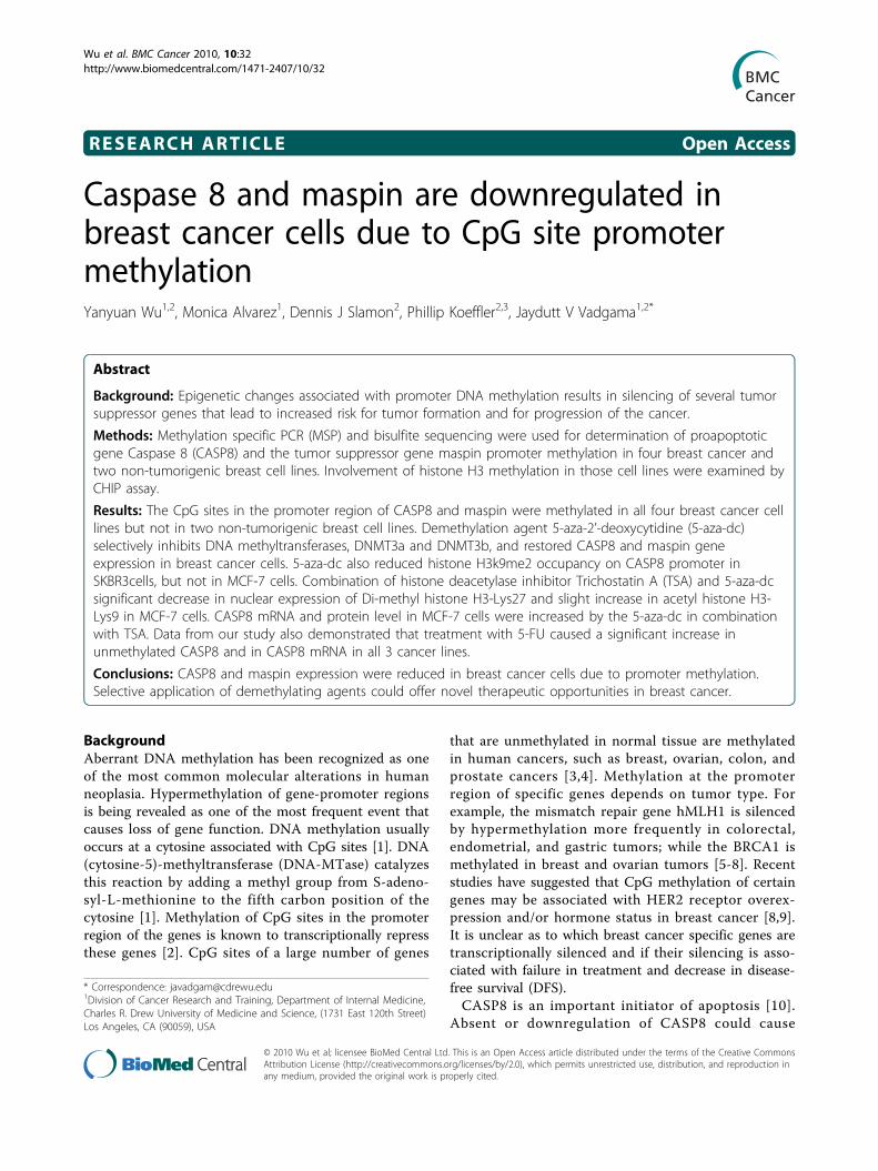
Caspase 8 and maspin are downregulated in breast cancer cells due to CpG site promoter methylation
12
RESEARCH ARTICLE Open Access Caspase 8 and maspin are downregulated in breast cancer cells due to CpG site promoter methylation Yanyuan Wu 1,2 , Monica Alvarez 1 , Dennis J Slamon 2 , Phillip Koeffler 2,3 , Jaydutt V Vadgama 1,2* Abstract Background: Epigenetic changes associated with promoter DNA methylation results in silencing of several tumor suppressor genes that lead to increased risk for tumor formation and for progression of the cancer. Methods: Methylation specific PCR (MSP) and bisulfite sequencing were used for determination of proapoptotic gene Caspase 8 (CASP8) and the tumor suppressor gene maspin promoter methylation in four breast cancer and two non-tumorigenic breast cell lines. Involvement of histone H3 methylation in those cell lines were examined by CHIP assay. Results: The CpG sites in the promoter region of CASP8 and maspin were methylated in all four breast cancer cell lines but not in two non-tumorigenic breast cell lines. Demethylation agent 5-aza-2’-deoxycytidine (5-aza-dc) selectively inhibits DNA methyltransferases, DNMT3a and DNMT3b, and restored CASP8 and maspin gene expression in breast cancer cells. 5-aza-dc also reduced histone H3k9me2 occupancy on CASP8 promoter in SKBR3cells, but not in MCF-7 cells. Combination of histone deacetylase inhibitor Trichostatin A (TSA) and 5-aza-dc significant decrease in nuclear expression of Di-methyl histone H3-Lys27 and slight increase in acetyl histone H3- Lys9 in MCF-7 cells. CASP8 mRNA and protein level in MCF-7 cells were increased by the 5-aza-dc in combination with TSA. Data from our study also demonstrated that treatment with 5-FU caused a significant increase in unmethylated CASP8 and in CASP8 mRNA in all 3 cancer lines. Conclusions: CASP8 and maspin expression were reduced in breast cancer cells due to promoter methylation. Selective application of demethylating agents could offer novel therapeutic opportunities in breast cancer. Background Aberrant DNA methylation has been recognized as one of the most common molecular alterations in human neoplasia. Hypermethylation of gene-promoter regions is being revealed as one of the most frequent event that causes loss of gene function. DNA methylation usually occurs at a cytosine associated with CpG sites [1]. DNA (cytosine-5)-methyltransferase (DNA-MTase) catalyzes this reaction by adding a methyl group from S-adeno- syl-L-methionine to the fifth carbon position of the cytosine [1]. Methylation of CpG sites in the promoter region of the genes is known to transcriptionally repress these genes [2]. CpG sites of a large number of genes that are unmethylated in normal tissue are methylated in human cancers, such as breast, ovarian, colon, and prostate cancers [3,4]. Methylation at the promoter region of specific genes depends on tumor type. For example, the mismatch repair gene hMLH1 is silenced by hypermethylation more frequently in colorectal, endometrial, and gastric tumors; while the BRCA1 is methylated in breast and ovarian tumors [5-8]. Recent studies have suggested that CpG methylation of certain genes may be associated with HER2 receptor overex- pression and/or hormone status in breast cancer [8,9]. It is unclear as to which breast cancer specific genes are transcriptionally silenced and if their silencing is asso- ciated with failure in treatment and decrease in disease- free survival (DFS). CASP8 is an important initiator of apoptosis [10]. Absent or downregulation of CASP8 could cause * Correspondence: [email protected] 1 Division of Cancer Research and Training, Department of Internal Medicine, Charles R. Drew University of Medicine and Science, (1731 East 120th Street) Los Angeles, CA (90059), USA Wu et al. BMC Cancer 2010, 10:32 http://www.biomedcentral.com/1471-2407/10/32 © 2010 Wu et al; licensee BioMed Central Ltd. This is an Open Access article distributed under the terms of the Creative Commons Attribution License (http://creativecommons.org/licenses/by/2.0), which permits unrestricted use, distribution, and reproduction in any medium, provided the original work is properly cited.
-
Upload
independent -
Category
Documents
-
view
3 -
download
0
Transcript of Caspase 8 and maspin are downregulated in breast cancer cells due to CpG site promoter methylation
RESEARCH ARTICLE Open Access
Caspase 8 and maspin are downregulated inbreast cancer cells due to CpG site promotermethylationYanyuan Wu1,2, Monica Alvarez1, Dennis J Slamon2, Phillip Koeffler2,3, Jaydutt V Vadgama1,2*
Abstract
Background: Epigenetic changes associated with promoter DNA methylation results in silencing of several tumorsuppressor genes that lead to increased risk for tumor formation and for progression of the cancer.
Methods: Methylation specific PCR (MSP) and bisulfite sequencing were used for determination of proapoptoticgene Caspase 8 (CASP8) and the tumor suppressor gene maspin promoter methylation in four breast cancer andtwo non-tumorigenic breast cell lines. Involvement of histone H3 methylation in those cell lines were examined byCHIP assay.
Results: The CpG sites in the promoter region of CASP8 and maspin were methylated in all four breast cancer celllines but not in two non-tumorigenic breast cell lines. Demethylation agent 5-aza-2’-deoxycytidine (5-aza-dc)selectively inhibits DNA methyltransferases, DNMT3a and DNMT3b, and restored CASP8 and maspin geneexpression in breast cancer cells. 5-aza-dc also reduced histone H3k9me2 occupancy on CASP8 promoter inSKBR3cells, but not in MCF-7 cells. Combination of histone deacetylase inhibitor Trichostatin A (TSA) and 5-aza-dcsignificant decrease in nuclear expression of Di-methyl histone H3-Lys27 and slight increase in acetyl histone H3-Lys9 in MCF-7 cells. CASP8 mRNA and protein level in MCF-7 cells were increased by the 5-aza-dc in combinationwith TSA. Data from our study also demonstrated that treatment with 5-FU caused a significant increase inunmethylated CASP8 and in CASP8 mRNA in all 3 cancer lines.
Conclusions: CASP8 and maspin expression were reduced in breast cancer cells due to promoter methylation.Selective application of demethylating agents could offer novel therapeutic opportunities in breast cancer.
BackgroundAberrant DNA methylation has been recognized as oneof the most common molecular alterations in humanneoplasia. Hypermethylation of gene-promoter regionsis being revealed as one of the most frequent event thatcauses loss of gene function. DNA methylation usuallyoccurs at a cytosine associated with CpG sites [1]. DNA(cytosine-5)-methyltransferase (DNA-MTase) catalyzesthis reaction by adding a methyl group from S-adeno-syl-L-methionine to the fifth carbon position of thecytosine [1]. Methylation of CpG sites in the promoterregion of the genes is known to transcriptionally repressthese genes [2]. CpG sites of a large number of genes
that are unmethylated in normal tissue are methylatedin human cancers, such as breast, ovarian, colon, andprostate cancers [3,4]. Methylation at the promoterregion of specific genes depends on tumor type. Forexample, the mismatch repair gene hMLH1 is silencedby hypermethylation more frequently in colorectal,endometrial, and gastric tumors; while the BRCA1 ismethylated in breast and ovarian tumors [5-8]. Recentstudies have suggested that CpG methylation of certaingenes may be associated with HER2 receptor overex-pression and/or hormone status in breast cancer [8,9].It is unclear as to which breast cancer specific genes aretranscriptionally silenced and if their silencing is asso-ciated with failure in treatment and decrease in disease-free survival (DFS).CASP8 is an important initiator of apoptosis [10].
Absent or downregulation of CASP8 could cause
* Correspondence: [email protected] of Cancer Research and Training, Department of Internal Medicine,Charles R. Drew University of Medicine and Science, (1731 East 120th Street)Los Angeles, CA (90059), USA
Wu et al. BMC Cancer 2010, 10:32http://www.biomedcentral.com/1471-2407/10/32
© 2010 Wu et al; licensee BioMed Central Ltd. This is an Open Access article distributed under the terms of the Creative CommonsAttribution License (http://creativecommons.org/licenses/by/2.0), which permits unrestricted use, distribution, and reproduction inany medium, provided the original work is properly cited.
resistance to apoptosis and is correlated with unfavor-able disease outcome, such as in childhood medulloblas-toma and neuroblastoma [11,12]. The absence ordownregulation of CASP8 may be due to epigeneticchanges. Studies have also indicated that methylationand demethylation of maspin promoter may regulatemaspin gene expression and that reduced maspinexpression is associated with cancer progression [13].In the current study we used methylation specific PCR
(MSP), and bisulfite sequencing to determine the methy-lation status of these two genes. We examined themechanisms associated with transcriptional silencing ofCASP8 and maspin by promoter methylation using real-time PCR and by restoring the methylated genes back totheir unmethylated status using the demethylating agent,5-aza-2’-deoxycytidine; TSA (Trichostatin A), inhibitorof histone deacetylase; and chemotherapeutic agent 5-Fu(5-Fluorouracil).
MethodsCells and cultureThe breast cancer cells with varying tissue subtypesselected for our methylation studies were: MCF-7 (ERpositive and HER2/neu negative); MDA-MB231 (ERnegative and HER2/neu negative); SKBR3 (ER negativeand HER2/neu positive); HCC1937 (ER negative, HER2/neu negative and BRCA1 mutated); non-tumorigenicbreast epithelial cells (MCF12A), and non-tumorigenicbreast fibroblast cells (MCF10). These cell lines werepurchased from American Type Culture Collection(Rockville, MD), and unless otherwise stated, the cellswere grown and maintained in DMEM/F12 (FisherScientific, CA) containing 10% FCS (Invetrogen), 2 mMglutamine, 50 units/ml penicillin and 50 μg/ml strepto-mycin (Fisher Scientific, CA).5-aza-2’-deoxycytidine (5-aza-dc) and Trichostatin A (TSA)treatmentThe cells were growing in culture medium until 80%confluence; 5 μM 5-aza-dc was added and incubated for3 to 6 days. The medium containing 5-aza-dc wasrefreshed every 2 days. For combination treatment cellswere first treated with 0.3 μM TSA in combination with5 μM 5-aza-dc for 2 days and then TSA was removedfrom culture medium. Treatment with 5 μM 5-aza-dcwas continued for 1 more day.Bisulfite modification and Methylation Specific PCR (MSP)Bisulfite conversion of genomic DNA was carried outusing Zymo EZ DNA Methylation-Gold™ kit (D5005,Zymo Research Corp, Orange, CA) according to themanufacture’s instructions. This process convertsunmethylated cytosine residues to uracil, while methy-lated cytosine residues remain unchanged. Bisulfitemodified DNA was used as a template, and then MSPwas performed to determine the methylation status of
CASP8 and maspin. The primer sequences for MSP areas follows:(a) CASP8, 5’-TGTTGTTTGGGTAACGTATCGA-3’
(methylated forward),
5’-CCCTACTTAACTTAACCCTACTCGAC-3’(methylated reverse), and5’-TTGTTGTTTGGGTAATGTATTGA-3’(unmethylated forward),5’-CAACCCTACTTAACTTAACCCTACTCA-3’(unmethylated reverse);
(b) maspin, 5’-ATTTTATCGAATATTTTATTTTTCGG-3’ (methylated forward),
5’-TAACTCACCTAAACAACACCGCC-3’ (methy-lated reverse), and5’-TTTTATTTTATTGAATATTTTATTTTTTGG-3’ (unmethylated forward),5’-TAACTCACCTAAACAACACCACC-3’(unmethylated reverse).
The primers for MSP were designed using MethPri-mer [14] according to the sequences provided byPanomics (Fremont, CA). The cover regions wereshown in Additional file 1, Figure S1. PCR conditionswere as follows: an initial denaturation at 95°C for 5min was followed by 40 cycles at 94°C for 30 s, 50°C for30 s, and 72°C for 45 s and a final extension at 72°C for7 min.Bisulfite sequencing for CASP8 promoterBisulfite modified DNA (described above) was amplifiedby two primer sets. The primer sequences were as fol-lows: P1-CASP8 (nt -744 to nt -466), 5’-GGTGGTGGGTGTTTGTAGTTTTAGT-3’ (forward) and 5’-CCATCCTTAACCATATTCTCCAATTTA-3’ (reverse);P2-CASP8 (nt -486 to nt +51), 5’-TAGATTTTTTGTAAGAAAGAATGGTAT-3’ (forward) and 5’-ACAAAAAAACAAAATCTAATCTCC-3’ (reverse). PCR condi-tions were as follows: an initial denaturation at 95°C for5 min was followed by 40 cycles of 94°C for 30 s, 52°Cfor 30 s, and 72°C for 45 s and a final extension at 72°Cfor 7 min. The PCR products were purified with QIA-quick PCR purification kit (#28104, QIAGEN), and thensent to Retrogen Inc (San Diego, CA) for sequencing.The primers for bisulfite sequencing were designedusing MethPrimer [14]. The sequence was used todesign the bisulfate sequence primers were as same asthe sequence used to design primers for MSP. The spe-cific cover regions for each primer site have beendemonstrated in Additional file 1.Quantitative real-time PCR (Q-PCR)Total RNA from cultured cells was isolated by usingRNeasy micro kit (#74004, QIAGEN) according to the
Wu et al. BMC Cancer 2010, 10:32http://www.biomedcentral.com/1471-2407/10/32
Page 2 of 12

manufacture’s instructions, and cDNA was synthesizedby reverse transcription (RT) with ThermoScript™ RT-PCR system (Invitrogen) according to the manufacture’sinstructions. Q-PCR analysis was performed with iCycleiQ real-time PCR detection system (Bio-Rad Lab, Her-cules, CA) using SYBR Green Master Mix (#204143,QIAGEN). The primers used were as follows: (a)CASP8, 5’-CCAGAGACTCCAGGAAAAGAGA-3’ (for-ward) and 5’-GATAGAGCATGACCCTGTAGGC-3’(reverse). (b) maspin, 5’-AGATGGCCACTTTGAGAA-CATT-3’ (forward) and 5’-GGTTTGGTGTCTGTCTTGTTGA-3’ (reverse). (c) housekeeping controlgene, 18 s, 5’-GATCCATTGGAGGGCAAGTC-3’ (for-ward) and 5’-TCCCAAGATCCAACTACGAG-3’(reverse). Reactions were characterized during cyclingwhen amplification of the PCR product was firstdetected (Ct). The target message and the housekeepinggene, 18 s, in breast cancer and non-tumorigenic celllines, were quantified by measuring Ct. The relative levelof target messages in cells was normalized on the basisof its 18 s content by taking the difference of thresholdcycles between target gene and 18 s. Using non-tumori-genic breast cancer cells or untreated breast cancer cellsas reference, the level of CASP8 and maspin in eachsample was normalized with the corresponding refer-ence by taking the difference between threshold cycles.Final results were expressed as N-fold difference in tar-get gene expression relative to the reference.Immunobloting and Immunofluorescence (IF)For immunoblot analysis cells were lysed in 1 × lysisbuffer (20 mM Tris pH 7.5; 150 mM NaCl; 1 mMEDTA; 1 mM EGTA; 1% Triton X-100; 2.5 mM sodiumpyrophosphate; 1 mM b-glycerolphosphate; 1 mMNa3VO4; 1 μg/ml leupeptin; 0.1 mM PMSF) and proteinconcentration was measured using BCA dye (Pierce).Total protein (50 μg) from cell lysates was resolved onSDS-PAGE followed by transfer to nitrocellulose mem-brane. The membranes were incubated with antibodiesspecific against Dnmt1 (IMG-261A, IMGENEX), Dnmt2(IMG-281, IMGENEX), Dnmt3a (IMG-268A, IMGE-NEX), Dnmt3b (IMG-184A, IMGENEX), and caspase-8(#9746, Cell Signaling) according to manufacturer’sinstruction. Detection of antigen-bound antibody wasperformed with the enhance chemiluminescencereagent. For immunofluorescence analysis cells (2 × 10⁴/each) were mounted by cytospin on polylysine coatedglass slides and fixed with 4% paraformaldehyde for 15min followed by 100% ice-cold acetone for 10 min at 4°C. To detect caspase-8 protein expression immunofluor-escence analysis was performed with cleaved caspase-8antibody (#9496, Cell Signaling) followed by incubationwith anti-goat IgG-FITC (Santa Cruz) for 30 min indark and mounted with DAPI mounting medium (Vec-tor Labs). The cells with positive staining were counted
in five different areas and adjusted with total number ofcells. To analyze histone H3 methylation and acetylationstatus in MCF-7 cells double immunofluorescence ana-lysis was performed with antibodies against Di-MethylHistone H3 (Lys27) (#9755, Cell Signaling) or AcetylHistone H3 (Lys9) (#9671, Cell Signaling) followed byb-actin antibody (A1975, Sigma-Aldrich). FITC-conju-gated secondary antibodies were used to label Di-MethylHistone H3 (Lys27) and Acetyl Histone H3 (Lys9). Theb-actin was labeled with Tex-red-conjugated secondaryantibody. After mounting, the cells were viewed under afluorescence microscope.CHIP assayCells were fixed with formaldehyde (1% final concentra-tion) to cross-link protein to DNA at room temperaturefor 10 min after treated with or without 5-aza-dc (5 μM,3 days), and then incubated with glycine (0.125 M finalconcentration) for 5 min to stop the cross-linking. Solu-ble chromatin was prepared as described previously[15]. The chromatin was then diluted 1:10 with dilutionbuffer (0.01% SDS, 1.1% Triton X-100, 1.2 mM EDTA,16.7 mM Tris-HCl, 167 mM NaCl and a protease inhi-bitor cocktail set (CalBiochem)), and subjected to immu-noprecipitation with anti-dimethylated histone H3 lysine9 (H3K9me2) antibody (#9753, Cell signaling). Pre-immune serum (Santa Cruz) was also used as a control.The chromatin and antibodies were incubated overnightat 4°C. Chromatin/antibody complexes were recoveredby adding 30 μl of protein A/G Plus-agarose beads(Santa Cruz) and incubating at 4°C for 2 h. The beadswere sequentially washed for 10 min each in 1 ml oflow salt, high salt and LiCl immune complex wash buf-fer. Immunocomplexes were eluted off the beads byincubation with 300 μl of 1% SDS and 50 mMNaHCO3. The eluent was incubated at 65°C for 5 h orovernight to reverse the formaldehyde-induced protein-DNA cross-links. The DNA was extracted with phenoland chloroform. Extracted DNA was resuspended in 100μl of TE buffer and real time PCR was performed byusing Bio-Rad thermal cycler with CASP8 primers: for-ward 5’GGT GCC TGT AGT CCC AGC TAC TC3’and reverse 5’CCT AGA CCC TCC CCT GTT CTGTC3’. For input DNA, the chromatin preparation with-out incubated with antibodies was subjected to real timePCR.
ResultsPromoter methylation on CASP8, and maspin resulted indecrease mRNA and protein expressionFirst, we determined the promoter methylation status infour breast cancer and one non-tumorigenic breast celllines by MSP. We then examined if the promotermethylation would silence or decrease the gene or pro-tein expression for CASP8 and maspin in different
Wu et al. BMC Cancer 2010, 10:32http://www.biomedcentral.com/1471-2407/10/32
Page 3 of 12
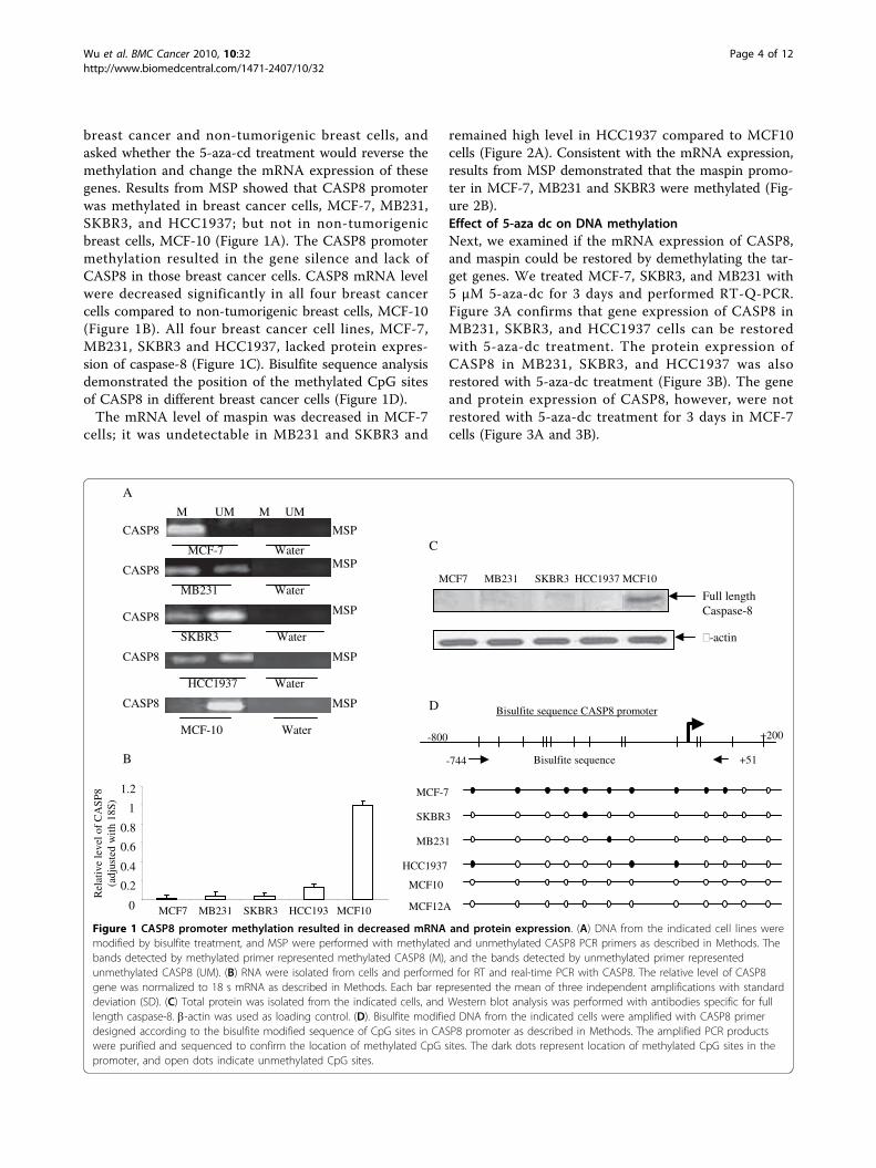
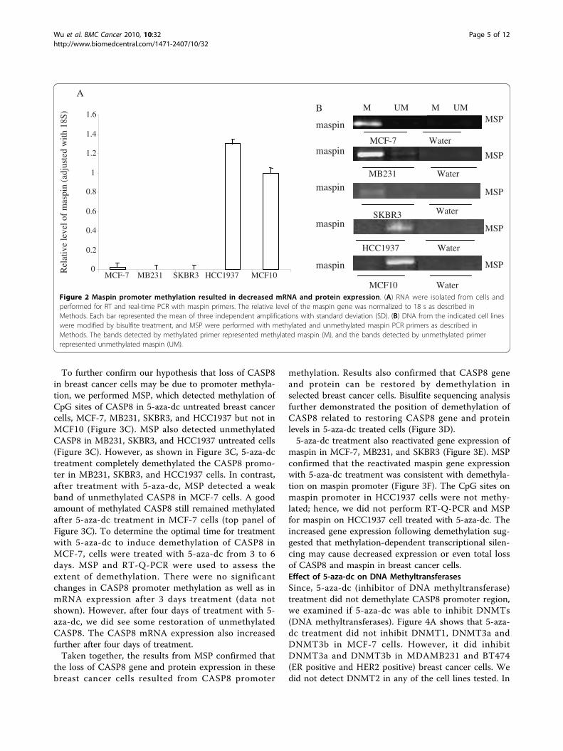
breast cancer and non-tumorigenic breast cells, andasked whether the 5-aza-cd treatment would reverse themethylation and change the mRNA expression of thesegenes. Results from MSP showed that CASP8 promoterwas methylated in breast cancer cells, MCF-7, MB231,SKBR3, and HCC1937; but not in non-tumorigenicbreast cells, MCF-10 (Figure 1A). The CASP8 promotermethylation resulted in the gene silence and lack ofCASP8 in those breast cancer cells. CASP8 mRNA levelwere decreased significantly in all four breast cancercells compared to non-tumorigenic breast cells, MCF-10(Figure 1B). All four breast cancer cell lines, MCF-7,MB231, SKBR3 and HCC1937, lacked protein expres-sion of caspase-8 (Figure 1C). Bisulfite sequence analysisdemonstrated the position of the methylated CpG sitesof CASP8 in different breast cancer cells (Figure 1D).The mRNA level of maspin was decreased in MCF-7
cells; it was undetectable in MB231 and SKBR3 and
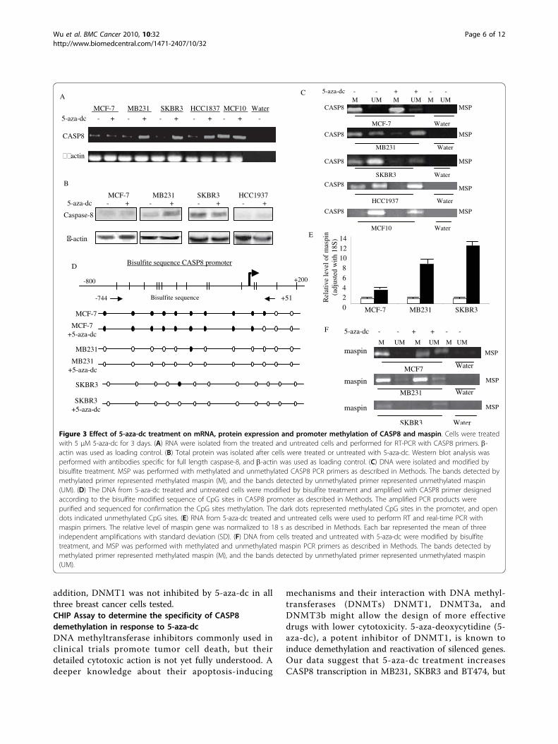
remained high level in HCC1937 compared to MCF10cells (Figure 2A). Consistent with the mRNA expression,results from MSP demonstrated that the maspin promo-ter in MCF-7, MB231 and SKBR3 were methylated (Fig-ure 2B).Effect of 5-aza dc on DNA methylationNext, we examined if the mRNA expression of CASP8,and maspin could be restored by demethylating the tar-get genes. We treated MCF-7, SKBR3, and MB231 with5 μM 5-aza-dc for 3 days and performed RT-Q-PCR.Figure 3A confirms that gene expression of CASP8 inMB231, SKBR3, and HCC1937 cells can be restoredwith 5-aza-dc treatment. The protein expression ofCASP8 in MB231, SKBR3, and HCC1937 was alsorestored with 5-aza-dc treatment (Figure 3B). The geneand protein expression of CASP8, however, were notrestored with 5-aza-dc treatment for 3 days in MCF-7cells (Figure 3A and 3B).
Figure 1 CASP8 promoter methylation resulted in decreased mRNA and protein expression. (A) DNA from the indicated cell lines weremodified by bisulfite treatment, and MSP were performed with methylated and unmethylated CASP8 PCR primers as described in Methods. Thebands detected by methylated primer represented methylated CASP8 (M), and the bands detected by unmethylated primer representedunmethylated CASP8 (UM). (B) RNA were isolated from cells and performed for RT and real-time PCR with CASP8. The relative level of CASP8gene was normalized to 18 s mRNA as described in Methods. Each bar represented the mean of three independent amplifications with standarddeviation (SD). (C) Total protein was isolated from the indicated cells, and Western blot analysis was performed with antibodies specific for fulllength caspase-8. b-actin was used as loading control. (D). Bisulfite modified DNA from the indicated cells were amplified with CASP8 primerdesigned according to the bisulfite modified sequence of CpG sites in CASP8 promoter as described in Methods. The amplified PCR productswere purified and sequenced to confirm the location of methylated CpG sites. The dark dots represent location of methylated CpG sites in thepromoter, and open dots indicate unmethylated CpG sites.
Wu et al. BMC Cancer 2010, 10:32http://www.biomedcentral.com/1471-2407/10/32
Page 4 of 12
To further confirm our hypothesis that loss of CASP8in breast cancer cells may be due to promoter methyla-tion, we performed MSP, which detected methylation ofCpG sites of CASP8 in 5-aza-dc untreated breast cancercells, MCF-7, MB231, SKBR3, and HCC1937 but not inMCF10 (Figure 3C). MSP also detected unmethylatedCASP8 in MB231, SKBR3, and HCC1937 untreated cells(Figure 3C). However, as shown in Figure 3C, 5-aza-dctreatment completely demethylated the CASP8 promo-ter in MB231, SKBR3, and HCC1937 cells. In contrast,after treatment with 5-aza-dc, MSP detected a weakband of unmethylated CASP8 in MCF-7 cells. A goodamount of methylated CASP8 still remained methylatedafter 5-aza-dc treatment in MCF-7 cells (top panel ofFigure 3C). To determine the optimal time for treatmentwith 5-aza-dc to induce demethylation of CASP8 inMCF-7, cells were treated with 5-aza-dc from 3 to 6days. MSP and RT-Q-PCR were used to assess theextent of demethylation. There were no significantchanges in CASP8 promoter methylation as well as inmRNA expression after 3 days treatment (data notshown). However, after four days of treatment with 5-aza-dc, we did see some restoration of unmethylatedCASP8. The CASP8 mRNA expression also increasedfurther after four days of treatment.Taken together, the results from MSP confirmed that
the loss of CASP8 gene and protein expression in thesebreast cancer cells resulted from CASP8 promoter
methylation. Results also confirmed that CASP8 geneand protein can be restored by demethylation inselected breast cancer cells. Bisulfite sequencing analysisfurther demonstrated the position of demethylation ofCASP8 related to restoring CASP8 gene and proteinlevels in 5-aza-dc treated cells (Figure 3D).5-aza-dc treatment also reactivated gene expression of
maspin in MCF-7, MB231, and SKBR3 (Figure 3E). MSPconfirmed that the reactivated maspin gene expressionwith 5-aza-dc treatment was consistent with demethyla-tion on maspin promoter (Figure 3F). The CpG sites onmaspin promoter in HCC1937 cells were not methy-lated; hence, we did not perform RT-Q-PCR and MSPfor maspin on HCC1937 cell treated with 5-aza-dc. Theincreased gene expression following demethylation sug-gested that methylation-dependent transcriptional silen-cing may cause decreased expression or even total lossof CASP8 and maspin in breast cancer cells.Effect of 5-aza-dc on DNA MethyltransferasesSince, 5-aza-dc (inhibitor of DNA methyltransferase)treatment did not demethylate CASP8 promoter region,we examined if 5-aza-dc was able to inhibit DNMTs(DNA methyltransferases). Figure 4A shows that 5-aza-dc treatment did not inhibit DNMT1, DNMT3a andDNMT3b in MCF-7 cells. However, it did inhibitDNMT3a and DNMT3b in MDAMB231 and BT474(ER positive and HER2 positive) breast cancer cells. Wedid not detect DNMT2 in any of the cell lines tested. In
Figure 2 Maspin promoter methylation resulted in decreased mRNA and protein expression. (A) RNA were isolated from cells andperformed for RT and real-time PCR with maspin primers. The relative level of the maspin gene was normalized to 18 s as described inMethods. Each bar represented the mean of three independent amplifications with standard deviation (SD). (B) DNA from the indicated cell lineswere modified by bisulfite treatment, and MSP were performed with methylated and unmethylated maspin PCR primers as described inMethods. The bands detected by methylated primer represented methylated maspin (M), and the bands detected by unmethylated primerrepresented unmethylated maspin (UM).
Wu et al. BMC Cancer 2010, 10:32http://www.biomedcentral.com/1471-2407/10/32
Page 5 of 12
addition, DNMT1 was not inhibited by 5-aza-dc in allthree breast cancer cells tested.CHIP Assay to determine the specificity of CASP8demethylation in response to 5-aza-dcDNA methyltransferase inhibitors commonly used inclinical trials promote tumor cell death, but theirdetailed cytotoxic action is not yet fully understood. Adeeper knowledge about their apoptosis-inducing
mechanisms and their interaction with DNA methyl-transferases (DNMTs) DNMT1, DNMT3a, andDNMT3b might allow the design of more effectivedrugs with lower cytotoxicity. 5-aza-deoxycytidine (5-aza-dc), a potent inhibitor of DNMT1, is known toinduce demethylation and reactivation of silenced genes.Our data suggest that 5-aza-dc treatment increasesCASP8 transcription in MB231, SKBR3 and BT474, but
Figure 3 Effect of 5-aza-dc treatment on mRNA, protein expression and promoter methylation of CASP8 and maspin. Cells were treatedwith 5 μM 5-aza-dc for 3 days. (A) RNA were isolated from the treated and untreated cells and performed for RT-PCR with CASP8 primers. b-actin was used as loading control. (B) Total protein was isolated after cells were treated or untreated with 5-aza-dc. Western blot analysis wasperformed with antibodies specific for full length caspase-8, and b-actin was used as loading control. (C) DNA were isolated and modified bybisulfite treatment. MSP was performed with methylated and unmethylated CASP8 PCR primers as described in Methods. The bands detected bymethylated primer represented methylated maspin (M), and the bands detected by unmethylated primer represented unmethylated maspin(UM). (D) The DNA from 5-aza-dc treated and untreated cells were modified by bisulfite treatment and amplified with CASP8 primer designedaccording to the bisulfite modified sequence of CpG sites in CASP8 promoter as described in Methods. The amplified PCR products werepurified and sequenced for confirmation the CpG sites methylation. The dark dots represented methylated CpG sites in the promoter, and opendots indicated unmethylated CpG sites. (E) RNA from 5-aza-dc treated and untreated cells were used to perform RT and real-time PCR withmaspin primers. The relative level of maspin gene was normalized to 18 s as described in Methods. Each bar represented the mean of threeindependent amplifications with standard deviation (SD). (F) DNA from cells treated and untreated with 5-aza-dc were modified by bisulfitetreatment, and MSP was performed with methylated and unmethylated maspin PCR primers as described in Methods. The bands detected bymethylated primer represented methylated maspin (M), and the bands detected by unmethylated primer represented unmethylated maspin(UM).
Wu et al. BMC Cancer 2010, 10:32http://www.biomedcentral.com/1471-2407/10/32
Page 6 of 12
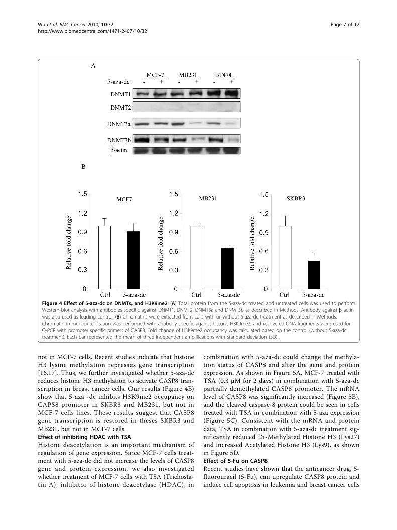
not in MCF-7 cells. Recent studies indicate that histoneH3 lysine methylation represses gene transcription[16,17]. Thus, we further investigated whether 5-aza-dcreduces histone H3 methylation to activate CASP8 tran-scription in breast cancer cells. Our results (Figure 4B)show that 5-aza -dc inhibits H3K9me2 occupancy onCAPS8 promoter in SKBR3 and MB231, but not inMCF-7 cells lines. These results suggest that CASP8gene transcription is restored in theses SKBR3 andMB231, but not in MCF-7 cells.Effect of inhibiting HDAC with TSAHistone deacetylation is an important mechanism ofregulation of gene expression. Since MCF-7 cells treat-ment with 5-aza-dc did not increase the levels of CASP8gene and protein expression, we also investigatedwhether treatment of MCF-7 cells with TSA (Trichosta-tin A), inhibitor of histone deacetylase (HDAC), in
combination with 5-aza-dc could change the methyla-tion status of CASP8 and alter the gene and proteinexpression. As shown in Figure 5A, MCF-7 treated withTSA (0.3 μM for 2 days) in combination with 5-aza-dcpartially demethylated CASP8 promoter. The mRNAlevel of CASP8 was significantly increased (Figure 5B),and the cleaved caspase-8 protein could be seen in cellstreated with TSA in combination with 5-aza expression(Figure 5C). Consistent with the mRNA and proteindata, TSA in combination with 5-aza-dc treatment sig-nificantly reduced Di-Methylated Histone H3 (Lys27)and increased Acetylated Histone H3 (Lys9), as shownin Figure 5D.Effect of 5-Fu on CASP8Recent studies have shown that the anticancer drug, 5-fluorouracil (5-Fu), can upregulate CASP8 protein andinduce cell apoptosis in leukemia and breast cancer cells
Figure 4 Effect of 5-aza-dc on DNMTs, and H3K9me2. (A) Total protein from the 5-aza-dc treated and untreated cells was used to performWestern blot analysis with antibodies specific against DNMT1, DNMT2, DNMT3a and DNMT3b as described in Methods. Antibody against b-actinwas also used as loading control. (B) Chromatins were extracted from cells with or without 5-aza-dc treatment as described in Methods.Chromatin immunoprecipitation was performed with antibody specific against histone H3K9me2, and recovered DNA fragments were used forQ-PCR with promoter specific primers of CASP8. Fold change of H3K9me2 occupancy was calculated based on the control (without 5-aza-dctreatment). Each bar represented the mean of three independent amplifications with standard deviation (SD).
Wu et al. BMC Cancer 2010, 10:32http://www.biomedcentral.com/1471-2407/10/32
Page 7 of 12
[18,19]. 5-Fu has been successfully used for treatment ofbreast cancer in clinical practice. We examined if 5-Futreatment could upregulate CASP8 in breast cancer andif the upregulation involved demethylating CpG sites onCASP8 promoter. As shown earlier, CASP8 mRNA wasundetectable in MCF-7 breast cancer cells due to pro-moter methylation. We then treated MCF-7 cells with5-Fu for 3 days and examined CASP8 mRNA levels. Fig-ure 6A shows almost 4 fold increase in mRNA level inMCF-7 cells treated with 5-Fu compared to non-treatedcells. The mRNA level of CASP8 in MB231 and SKBR3treated with 5-Fu also increased 2.4 fold and 1.8 fold
respectively (Figure 6A). The increases in mRNAexpression are correlated with partial demethylation ofCpG sites on CASP8 promoter (Figure 6B). Bisulfitesequencing confirmed the position (nt -642 to nt -532,located in CASP8 core promoter region) of demethyla-tion of CASP promoter by 5-Fu in these breast cancercells (Figure 6C).
DiscussionHypermethylation of the promoter regions of variousgenes has been recognized as one of the most frequentmechanisms causing loss of gene function. The
Figure 5 Effect of TSA in combination with 5-aza-dc on CASP8. (A) DNA was isolated from the TSA and 5-aza-dc treated and untreated cellsand MSP was performed with methylated and unmethylated CASP8 PCR primers as described in Methods. The bands detected by methylatedprimer represented methylated CASP8 (M), and the bands detected by unmethylated primer represented unmethylated CASP8 (UM). (B) RNAwas isolated from the TSA and 5-aza-dc treated and untreated cells. RT and Q-PCR was performed with CASP8 primers. The relative level ofCASP8 mRNA was normalized to 18 s as described in Methods. Each bar represented the mean of three independent amplifications withstandard deviation (SD), *p < 0.05 between indicated groups. (C) Immunofluorescence analysis was performed with cleaved caspase-8 antibodyfollowed by incubation with anti-goat IgG-FITC and mounted with DAPI mounting medium (left panel). The arrows indicate the FITC labeledcleaved caspase-8 (green in the top panel of left), and the cell nucleus was labeled by DAPI (blue in bottom panel of left). The cells with positivestaining for cleaved caspase-8 were counted in five different areas and adjusted with total number of cells (right panel). Each bar represents themean of number of cells positive for cleaved caspase-8 in the five areas with standard deviation (SD), * p < 0.05 between the indicated groups.(D) Double immunofluorescence analysis was performed with Di-Methyl Histone H3 (Lys27) or Acetyl Histone H3 (Lys9) antibodies followed byactin antibody as described in Methods. The nucleus positive stained with Di-Methyl Histone H3 (Lys27) or Acetyl Histone H3 (Lys9) were labeledwith FITC as green (indicated by white arrows), and the cytoplasm stained with actin was labeled with Tex-red as red (indicated with light bluearrows).
Wu et al. BMC Cancer 2010, 10:32http://www.biomedcentral.com/1471-2407/10/32
Page 8 of 12
association between aberrant DNA methylation and car-cinogenesis has been demonstrated in several studies[20-26]. However, the association between epigeneticchanges with cancer etiology needs to be elucidated.Several cancer-related genes have been reported to be
silenced by aberrant methylation in breast cancer, suchas 14-3-3 s, E-cadherin and tissue inhibitor of metallo-proteinase 3 (TIMP3) genes. Treatment with 5-aza-dcactivated the expression of 14-3-3 s [27] and E-cadheringenes [28] in breast carcinoma cells and of TIMP3 indifferent tumor cell lines [29,30].Hypermethylation of CASP8 has been showed as a fre-
quent feature of relapsed glioblastoma compared withthe corresponding primary tumors [31]. The authorssuggested that epigenetic deregulation of the mitochon-dria-independent apoptosis is a relevant characteristic inrecurrent glioblastoma. The development of targetedtherapies restoring functional extrinsic apoptosis, asrecently shown in vivo with the synergistic combinationof the DNA demethylating agent decitabine and TRAIL[32], may provide a useful tool to overcome the
resistance of glioblastoma to contemporary treatmentmodalities. Methylation of CASP8 gene has also beenreported in some childhood tumors and in neuroendo-crine lung tumors [33]. CASP8 is an important initiatorof apoptosis [10]. Structurally, the promoter region ofCASP8 has binding sites for p53, nuclear factor-�B (NF-�B), AP-1, SP-1, IRF-1, and Ets-like transcription factors[34]. Therefore, CASP8 functions both as a pivotalmolecule for death-receptor-induced apoptosis and as aselective signal transducer, such as for NF-�B activation[35]. Absent or downregulation of CASP8 could causeresistance to apoptosis and is correlated with unfavor-able disease outcome, such as in childhood medulloblas-toma and neuroblastoma [11,12]. Others have alsodemonstrated that absence or downregulation of CASP8may be due to epigenetic changes, such as hypermethy-lation, or mutations [36,37].In current study we have investigated the promoter
methylation of CASP8 and maspin in relation to theirexpression levels as well as the involvement of histonemethyltransferases and histone H3K9me2. Using MSP
Figure 6 Effect of 5-Fu on CASP8. (A) MCF-7, MB231, and SKBR3 cells were treated with or without 5-Fu (10 μM) for 3 days. RNA was isolatedfrom the treated and untreated cells and RT-real-time PCR was performed with CASP8 and 18 s primers. Each bar represented the mean relativelevel of CASP8 and standard deviation (SD) from three measurements and normalized to 18 s. (B) DNA was isolated from the 5-Fu treated anduntreated cells and modified with bisulfite treatment. MSP was performed with methylated and unmethylated CASP8 primers. The bandsdetected with methylated primer indicated methylated CASP8 (M), and the bands from unmethylated primer indicated unmethylated CASP8(UM). (C) Bisulfite modified DNA from the indicated 5-Fu treated, and untreated cells were amplified with CASP8 primer designed according tothe bisulfite modified sequence of CpG sites in CASP8 promoter as described in Methods. The amplified PCR products were purified andsequenced for confirmation of the CpG sites methylation. The dark dots in the bottom panel represented methylated CpG sites in the promoter,and open dots indicated unmethylated CpG sites.
Wu et al. BMC Cancer 2010, 10:32http://www.biomedcentral.com/1471-2407/10/32
Page 9 of 12
and bisulfite sequence analysis, we have established therelationship between aberrant cytosine methylation anddownregulated or loss of CASP8 in breast cancer cells.We confirmed that CpG sites methylation in the promo-ter region of CASP8 is the mechanistic basis for tran-scriptional downregulation or silencing of CASP8 inbreast cancer cells. The methylation status of CASP8can be completely or partially reversed by treatmentwith 5-aza-dc in MB231, SKBR3, and BT474, but not inMCF-7 breast cancer cells. The cells that had fewermethylated CpG sites, such as MB231 and SKBR3 weretotally demethylated by 5-aza-dc. This change indemethylation resulted in a significant increase inCASP8 mRNA and protein expression. In contractSKBR3 cells, most CpG sites of CASP8 were methylatedin MCF-7 cells. Results from MSP showed that methyla-tion was partially reversed by 5-aza-dc in MCF-7 cellsand the mRNA and protein level of CASP8 had no sig-nificant increase. We also examined the effect of histoneacetylation of CASP8 by treating MCF-7 cells with Tri-chostatin A (TSA). The TSA treatment alone did notchange the methylation status and mRNA expression ofCASP8 in MCF-7 cells (data not shown). However, TSAin combination with 5-aza-dc was able to partiallydemethylate CASP8 promoter and increased CASP8mRNA and protein expression in MCF-7 cells. The datasuggested the involvement of histone acetylation in theregulation of CASP8 gene expression in MCF-7 cells.CHIP analysis (Figure 4B) showed that 5-aza-dc treat-ment inhibits H3K9me2 occupancy on CAPS8 promoterin SKBR3 and MDA-MB231, but not in MCF-7 cells.These results suggest that CASP8 gene transcription isrestored in theses SKBR3 and MB231, but not in MCF-7 cells.Several studies have demonstrated a link between
methylation and histone acetylation in which a family ofmethyl-CpG-binding proteins is involved [38]. Whenthese proteins bind to a methylated promoter, theyrecruit HDAC, and the interaction of these two epige-netic events inhibits gene expression by interfering withthe function of transcription factors and the compactionof the chromatin structure [39-41]. Inhibitors of theseepigenetic changes can lead to the reactivation of genesthat suppress tumorigenesis. In accord with this hypoth-esis is the report on the synergistic interaction of 5-aza-dc and the HDAC inhibitor, trichostatin A (TSA), in thereactivation of tumor suppressor genes [42]. This samedrug combination was also reported to induce a syner-gistic reactivation of the estrogen receptor-a in breastcarcinoma cells [43].A recent study in prostate cancer cells indicated that
CASP8 plays a pathway specific role in inhibiting andro-gen receptor signaling [44]. This evidence suggests thatCASP8 may have the role beyond its role as a cell death
protease and may play a role in hormonal receptor cellsignaling.The anticancer drug, fluorouracil (5-Fu), has been
commonly used in clinical practice for first line treat-ment of breast cancer for decades. Up to now, exceptfor interferon-g and azacytidine, the cytotoxic drugs, 5-Fu and methotrexate, have been shown to upregulateCASP8 and induce cell apoptosis in neuroblastoma,medulloblastoma, Ewing sarcoma, glioblastoma, leuke-mia, and breast cancer cells [18,19]. The mechanism bywhich 5-Fu regulates CASP8 protein is more likely toinvolve p53 [19]. However, studies from Zoli, et al. [18]suggest that 5-Fu in combination with doxorubicin andpaclitaxel regulates CASP8 and induces cell apoptosis bya caspase-dependent mechanism independent of hormo-nal, p53, bcl-2 or bax status in breast cancer cells [18].These observations made us to suspect that 5-Fuinduced cell apoptosis may involve a demethylation pro-cess. We then tested the expression CASP8 mRNA, aswell as its methylation status, in MCF-7, MB231 andSKBR3 cells treated with 5-Fu for 3 days. Compared tountreated cells, the cells treated with 5-Fu showed a sig-nificant increase mRNA expression of CASP8 followedby demethylation of its promoter region.Taken together, our results confirm that expression of
CASP8 in breast cancer is epigenetically controlled andthe modification may vary in different types of breastcancer cells.Another gene promoter methylation has been studied
was maspin in current study. Maspin belongs to the ser-pin family [45]. It acts as a tumor suppressor, increasescell adhesion, induces apoptosis, and inhibits tumorgrowth and metastasis [46-48]. Maspin is also involvedin angiogenesis and mammary gland development [49].The expression of maspin is epigenetically controlled bymethylation and/or histone acetylation. Studies havealso indicated that methylation and demethylation ofmaspin promoter may regulate maspin gene expressionand that reduced maspin expression is associated withcancer progression [13]. In our study, we have con-firmed that the expression of maspin in breast cancercells is epigenetically controlled by methylation of theCpG sites. The demethylation agent, 5-aza-dc, treatmentreversed maspin promoter methylation and increasedmaspin gene expression in MCF-7, MB231, and SKBR3cells. An early study from Domann FE et al. [50] alsoreported that normal human mammary epithelial cellsexpressed maspin mRNA and displayed a completelynon-methylated maspin gene promoter. In contrast,most breast cancer cell lines had no detectable maspinexpression and those maspin-negative breast cancer celllines also displayed an aberrant pattern of cytosinemethylation of the maspin promoter. In this study wehave also examined maspin and CASP8 mRNA levels in
Wu et al. BMC Cancer 2010, 10:32http://www.biomedcentral.com/1471-2407/10/32
Page 10 of 12
30 breast cancer tissues and 10 non-breast cancer tis-sues. The mRNA levels of CASP8 and maspin werelower in breast cancer tissue than non-breast cancer tis-sue (data not shown). The decreased mRNA expressinglevels of maspin and CASP8 in patients were associatedwith positive lymph node status, late stage disease, andHER2 overexpression (3+ and/or FISH positive). TheDFS was significant decreased in patients with lowCASP8 or maspin expression (data not shown). Inactiva-tion of apoptotic pathways is often critical for the patho-genesis of tumor cells and for their resistance tochemotherapeutic drug treatment and/or irradiation[51-53]. It needs to be determined if the loss of CASP8and/or maspin expression can be a prognostic markerfor Breast Cancer and if it is associated with amplifica-tion of MYCN (v-myc myelocytomatosis viral relatedoncogene), as frequently observed in neuroblastomas.Limitations of this study are that the most investiga-
tion was used cell lines only. To better understand theclinical relevance of CASP8 and/or maspin promotermethylation in breast tumors and if the decreasedmRNA expression of CASP8 and/or maspin were corre-lated to the aberrant pattern of cytosine methylation ofthe gene promoter further studies with large sample sizeshould be conducted.
ConclusionsIn conclusion, our results show that methylation of CpGsites at the promoter region in certain genes, such asCASP8 and maspin, could result in transcriptionaldownregulation or silencing of genes and protein inbreast cancer cells. The anticancer drug, 5-Fu, is able toupregulate CASP8 gene expression, and the mechanismmay involve demethylation. Screening promoter methy-lation patterns in breast cancer patients could be animportant step to develop treatment protocols that tar-get the methylated gene and improve DFS in breast can-cer patients. Future studies also should examine the useof non-toxic demthylating agents in combination withchemotherapeutic drugs would offer an advantage forthe treatment of breast cancer patients.
Additional file 1: Figure S1. Promoter CpG sites of CASP8 andmaspin and the primers sequences covered regions. A figure toshow the primers sequences used for MSP and bisulfate sequencecovered regions in CASP8 promoter. Figure Legend for Figure S1.Click here for file[ http://www.biomedcentral.com/content/supplementary/1471-2407-10-32-S1.PPT ]
AbbreviationsCASP8: Caspase 8; DNMT: DNA Methyltransferase; MSP: Methylated SpecificPrimer; 5-azadc: 5-aza-2’-deoxycytidine; TSA: Trichostatin A; HDAC: H; 5-Fu: 5-Fluorouracil; ATCC: American Type Culture Collection (Manassas, VA, USA);ER: estrogen receptor; IHC: Immunohistochemistry
AcknowledgementsThis work received financial support from the following sources: NIH/NCIU54 CA14393-01 (JVV); U56 CA101599-01 (JVV), CA15083-25S3 (JVV), NIH/NIDDK R25 DK067015-01 (JVV), Department of Defense (BCRP) BC043180(JVV), and MBRS NIH SO6 GM0685-10-01 (YW).
Author details1Division of Cancer Research and Training, Department of Internal Medicine,Charles R. Drew University of Medicine and Science, (1731 East 120th Street)Los Angeles, CA (90059), USA. 2Division of Hematology/Oncology,Department of Internal Medicine, David Geffen UCLA School of Medicine,CA (5535 Macdonald Research Laboratories Building, 675 Charles E. YoungDrive South), Los Angeles, CA (90095), USA. 3Division of Hematology/Oncology, Department of Internal Medicine, Cedars-Sinai Medical Center,(8700 Beverly Blvd., Suite 6215), Los Angeles, CA (90048), USA.
Authors’ contributionsYW, MA and JVV were responsible for data collection, analysis, manuscriptpreparation, and editing. HPK and DS critical review and were involved instudy design. All authors read and approved the final manuscript.
Competing interestsThe authors declare that they have no competing interests.
Received: 1 July 2009Accepted: 4 February 2010 Published: 4 February 2010
References1. Chiang PK, Gordon RK, Tal J, Zeng GC, Doctor BP, Pardhasaradhi K,
McCann PP: S-Adenosylmethionine and methylation. FASEB J 1996,10:471-480.
2. Baylin SB, Herman JG, Graff JR, Vertino PM, Issa JP: Alterations in DNAmethylation: a fundamental aspect of neoplasia. Adv Cancer Res 1998,72:141-196.
3. Yang X, Yan L, Davidson NE: DNA methylation in breast cancer. EndocrRelat Cancer 2001, 8:115-127.
4. Widschwendter M, Jones PA: DNA methylation and breast carcinogenesis.Oncogene 2002, 21:5462-5482.
5. Miyakura Y, Sugano K, Konishi F, Ichikawa A, Maekawa M, Shitoh K,Shitoh K, Igarashi S, Kotake K, Koyama Y, Nagai H: Extensive methylation ofhMLH1 promoter region predominates in proximal colon cancer withmicrosatellite instability. Gastroenterology 2001, 121:1300-1309.
6. Ricciardiello L, Goel A, Mantovani V, Fiorini T, Fossi S, Chang DK, Lunedei V,Pozzato P, Zagari RM, De Luca L, Fuccio L, Martinelli GN, Roda E, Boland CR,Bazzoli F: Frequent loss of hMLH1 by promoter hypermethylation leadsto microsatellite instability in adenomatous polyps of patients with asingle first-degree member affected by colon cancer. Cancer Res 2003,63:787-792.
7. Press JZ, De Luca A, Boyd N, Young S, Troussard A, Ridge Y, Kaurah P,Kalloger SE, Blood KA, Smith M, Spellman PT, Wang Y, Miller DM,Horsman D, Faham M, Gilks CB, Gray J, Huntsman DG: Ovarian carcinomaswith genetic and epigenetic BRCA1 loss have distinct molecularabnormalities. BMC Cancer 2008, 8:17.
8. Wei M, Xu J, Dignam J, Nanda R, Sveen L, Fackenthal J, Grushko TA,Olopade OI: Estrogen receptor alpha, BRCA1, and FANCF promotermethylation occur in distinct subsets of sporadic breast cancers. BreastCancer Res Treat 2008, 111:113-120.
9. Fiegl H, Millinger S, Goebel G, Müller-Holzner E, Marth C, Laird PW,Widschwendter M: Breast cancer DNA methylation profiles in cancer cellsand tumor stroma: association with HER-2/neu status in primary breastcancer. Cancer Res 2006, 66:29-33.
10. Salvesen GS: Caspase-8: igniting the death machine. Structure 1999, 7:R225-229.
11. Pingoud-Meier C, Lang D, Janss AJ, Rorke LB, Phillips PC, Shalaby T,Grotzer MA: Loss of caspase-8 protein expression correlates withunfavorable survival outcome in childhood medulloblastoma. Clin CancerRes 2003, 9:6401-6409.
12. Yang Q, Kiernan CM, Tian Y, Salwen HR, Chlenski A, Brumback BA,London WB, Cohn SL: Methylation of CASP8, DCR2, and HIN-1 inneuroblastoma is associated with poor outcome. Clin Cancer Res 2007,13:3191-3197.
Wu et al. BMC Cancer 2010, 10:32http://www.biomedcentral.com/1471-2407/10/32
Page 11 of 12
13. Khalkhali-Ellis Z: Maspin: the new frontier. Clin Cancer Res 2006,12:7279-7283.
14. Li LC, Dahiya R: MethPrimer: designing primers for methylation PCRs.Bioinformatics 2002, 18:1427-1431.
15. Kuo M-H, Allis CD: In vivo cross-linking and immunoprecipitation forstudying dynamic protein:DNA associations in a chromatin environment.Methods 1999, 19:425-433.
16. Trojer P, Reinberg D: Histone lysine demethylases and their impact onepigenetics. Cell 2006, 125:213-217.
17. Lan F, Bayliss PE, Rinn JL, Whetstine JR, Wang JK, Chen S, Iwase S,Alpatov R, Issaeva I, Canaani E, Roberts TM, Chang HY, Shi Y: A histone H3lysine 27 demethylase regulates animal posterior development. Nature2007, 449:689-694.
18. Zoli W, Ulivi P, Tesei A, Fabbri F, Rosetti M, Maltoni R, Giunchi DC, Ricotti L,Brigliadori G, Vannini I, Amadori D: Addition of 5-fluorouracil todoxorubicin-paclitaxel sequence increases caspase-dependent apoptosisin breast cancer cell lines. Breast Cancer Res 2005, 7:R681-689.
19. Ehrhardt H, Häcker S, Wittmann S, Maurer M, Borkhardt B, Toloczko A,Debatin KM, Fulda S, Jeremias I: Cytotoxic drug-induced, p53-mediatedupregulation of caspase-8 in tumor cells. Oncogene 2008, 27:783-793.
20. Barton CA, Hacker NF, Clark SJ, O’Brien PM: DNA methylation changes inovarian cancer: Implications for early diagnosis, prognosis andtreatment. Gynecol Oncol 2008, 109:129-139.
21. Dobosy JR, Roberts JL, Fu VX, Jarrard DF: The expanding role ofepigenetics in the development, diagnosis and treatment of prostatecancer and benign prostatic hyperplasia. J Urol 2007, 177:822-831.
22. Yuan J, Luo RZ, Fuji S, Wang L, Hu W, Andreeff M, Pan Y, Kadota M,Oshimura M, Sahin AA, Issa JP, Bast RC Jr, Yu Y: Aberrant methylation andsilencing of ARHI, an imprinted tumor suppressor gene in which thefunction is lost in breast cancers. Cancer Res 2003, 63:4174-4180.
23. Chen K, Sawhney R, Khan M, Benninger MS, Hou Z, Sethi S, Stephen JK,Worsham MJ: Methylation of multiple genes as diagnostic andtherapeutic markers in primary head and neck squamous cellcarcinoma. Arch Otolaryngol Head Neck Surg 2007, 133:1131-1138.
24. Ellinger J, El Kassem N, Heukamp LC, Matthews S, Cubukluoz F, Kahl P,Perabo FG, Müller SC, von Ruecker A, Bastian PJ: Hypermethylation of cell-free serum DNA indicates worse outcome in patients with bladdercancer. J Urol 2008, 179:346-352.
25. Park do Y, Sakamoto H, Kirley SD, Ogino S, Kawasaki T, Kwon E, Mino-Kenudson M, Lauwers GY, Chung DC, Rueda BR, Zukerberg LR: The Cablesgene on chromosome 18q is silenced by promoter hypermethylationand allelic loss in human colorectal cancer. Am J Pathol 2007,171:1509-1519.
26. Henken FE, Wilting SM, Overmeer RM, Rietschoten JG, Nygren AO, Errami A,Schouten JP, Meijer CJ, Snijders PJ, Steenbergen RD: Sequential genepromoter methylation during HPV-induced cervical carcinogenesis. Br JCancer 2007, 97:1457-1464.
27. Ferguson AT, Evron E, Umbricht CB, Pandita TK, Chan TA, Hermeking H,Marks JR, Lambers AR, Futreal PA, Stampfer MR, Sukumar S: High frequencyof hypermethylation at the 14-3-3 s locus leads to gene silencing inbreast cancer. Proc Natl Acad Sci USA 2000, 97:6049-6054.
28. Graff JR, Herman JG, Lapidus RL, Chopra H, Xu R, Jarrard DF, Isaacs WB,Pitha PM, Davidson NE, Baylin SB: E-cadherin expression is silenced byDNA hypermethylation in human breast and prostate carcinomas.Cancer Res 1995, 55:5195-5199.
29. Bachman KE, Herman JG, Corn PG, Merlo A, Costello JF, Cavenee WK,Baylin SB, Graff JR: Methylation-associated silencing of the tissue inhibitorof metalloproteinase-3 gene suggest a suppressor role in kidney, brain,and other human cancers. Cancer Res 1999, 59:798-802.
30. Cameron EE, Bachman KE, Myohanen S, Herman JG, Baylin SB: Synergy ofdemethylation and histone deacetylase inhibition in the re-expression ofgenes silenced in cancer. Nat Genet 1999, 21:103-107.
31. Martinez R_, Setien F, Voelter C, Casado S, Quesada MP, Schackert G,Esteller M: CpG island promoter hypermethylation of the pro-apoptoticgene caspase-8 is a common hallmark of relapsed glioblastomamultiforme. Carcinogenesis 2007, 28:1264-1268.
32. Eramo A, Pallini R, Lotti F, Sette G, Patti M, Bartucci M, Ricci-Vitiani L,Signore M, Stassi G, Larocca LM, Crinò L, Peschle C, De Maria R: Inhibitionof DNA Methylation Sensitizes Glioblastoma for Tumor Necrosis Factor-Related Apoptosis-Inducing Ligand-Mediated Destruction. CancerResearch 2005, 65:11469-11477.
33. Harada K, Toyooka S, Shivapurkar N, Maitra A, Reddy JL, Matta H,Miyajima K, Timmons CF, Tomlinson GE, Mastrangelo D, Hay RJ,Chaudhary PM, Gazdar AF: Deregulation of caspase 8 and 10 expressionin pediatric tumors and cell lines. Cancer Res 2002, 62:5897-5901.
34. Liedtke C, Groger N, Manns MP, Trautwein C: The human caspase-8promoter sustains basal activity through SP1 and ETS-like transcriptionfactors and can be upregulated by a p53-dependent mechanism. J BiolChem 2003, 278:27593-27604.
35. Chaudhary PM, Eby MT, Jasmin A, Kumar A, Liu L, Hood L: Activation ofthe NF-kappaB pathway by caspase-8 and its homologs. Oncogene 2000,19:4451-4460.
36. Teitz T, Wei T, Valentine MB, Vanin EF, Grenet J, Valentine VA, Behm FG,Look AT, Lahti JM, Kidd VJ: Caspase-8 is deleted or silenced preferentiallyin childhood neuroblastomas with amplification of MYCN. Nat Med 2000,6:529-535.
37. Fulda S, Debatin KM: IFNgamma sensitizes for apoptosis by upregulatingcaspase-8 expression through the Stat1 pathway. Oncogene 2002,21:2295-2308.
38. Magdinier F, Wolffe AP: Selective association of the methyl-CpG bindingprotein MBD2 with the silent p14/p16 locus in human neoplasia. ProcNatl Acad Sci USA 2001, 98:4990-4995.
39. Nan X, Ng HH, Johnson CA, Laherty CD, Turner BM, Eisenman RN, Bird A:Transcriptional repression by the methyl-CpG-binding protein MeCP2involves a histone deacetylase complex. Nature 1998, 393:386-389.
40. Jones PL, Veenstra GJ, Wade PA, Vermaak D, Kass SU, Landsberger N,Strouboulis J, Wolffe AP: Methylated DNA and MeCP2 recruit histonedeacetylase to repress transcription. Nat Genet 1998, 19:187-191.
41. Jones PA, Baylin SB: The fundamental role of epigenetic events in cancer.Nat Genet 2002, 3:415-428.
42. Cameron EE, Bachman KE, Myohanen S, Herman JG, Baylin SB: Synergy ofdemethylation and histone deacetylase inhibition in the re-expression ofgenes silenced in cancer. Nat Genet 1999, 21:103-107.
43. Yang X, Phillips DL, Ferguson AT, Nelson WG, Herman JG, Davidson NE:Synergistic activation of functional estrogen receptor (ER)-a by DNAmethyltransferase and histone deacetylase inhibition in human ER-anegative breast cancer cells. Cancer Res 2001, 61:7025-7029.
44. Qi W, Wu H, Yang L, Boyd DD, Wang Z: A novel function of caspase-8 inthe regulation of androgen-receptor-driven gene expression. EMBO 2007,26:65-75.
45. Bailey CM, Khalkhali-Ellis Z, Seftor EA, Hendrix MJ: Biological functions ofmaspin. J Cell Physiol 2006, 209:617-624.
46. Zhang W, Shi HY, Zhang M: Maspin overexpression modulates tumor cellapoptosis through the regulation of Bcl-2 family proteins. BMC Cancer2005, 5(50).
47. Sheng S: The promise and challenge toward the clinical application ofmaspin in cancer. Front Biosci 2004, 9:2733-2745.
48. Odero-Marah VA, Khalkhali-Ellis Z, Chunthapong J, Amir S, Seftor RE,Seftor EA, Hendrix MJ: Maspin regulates different signaling pathways formotility and adhesion in aggressive breast cancer cells. Cancer Biol Ther2003, 2:398-403.
49. Zhang M, Volpert O, Shi YH, Bouck N: Maspin is an angiogenesis inhibitor.Nat Med 2000, 6:196-199.
50. Domann FE, Rice JC, Hendrix MJ, Futscher BW: Epigenetic silencing ofmaspin gene expression in human breast cancers. Int J Cancer 2000,85:805-810.
51. Rinkenberger JL, Korsmeyer SJ: Errors of homeostasis and deregulatedapoptosis. Curr Opin Genet Dev 1997, 7:589-596.
52. Reed JC: Mechanisms of apoptosis avoidance in cancer. Curr Opin Oncol1999, 11:58-75.
53. Lowe SW, Lin AW: Apoptosis in cancer. Carcinogenesis 2000, 21:485-495.
Pre-publication historyThe pre-publication history for this paper can be accessed here:http://www.biomedcentral.com/1471-2407/10/32/prepub
doi:10.1186/1471-2407-10-32Cite this article as: Wu et al.: Caspase 8 and maspin are downregulatedin breast cancer cells due to CpG site promoter methylation. BMCCancer 2010 10:32.
Wu et al. BMC Cancer 2010, 10:32http://www.biomedcentral.com/1471-2407/10/32
Page 12 of 12