Interferon-γ priming is involved in the activation of arginase by oligodeoxinucleotides containing...
11
Interferon-c priming is involved in the activation of arginase by oligodeoxinucleotides containing CpG motifs in murine macrophages Introduction It is well known that unmethylated cytosine guanine motifs contained in bacterial DNA or synthetic oligo- nucleotides CpG-DNA are powerful immunostimulatory molecules. Initially, CpG-DNA were believed to be pre- dominantly pro-inflammatory molecules, stimulating a T helper 1 (Th1)-like response dominated by the release of interleukin (IL)-12 and interferon-c (IFN-c). 1 However, in recent years there has been a spate of interest in some anti-inflammatory properties of CpG-DNA. Pulmonary inflammation decreased in response to lipopolysaccharide (LPS) after systemic exposure to CpG-DNA. 2 CpG-DNA were found to stimulate the production of IL-10, 3–5 increase the expression of indoleamine 2,3-dioxygenase (IDO), an enzyme linked to the suppression of T-cell- mediated immunity, 6,7 and enhance the shedding of the tumour necrosis factor-a (TNF-a) receptor in macro- Miriam V. Liscovsky, Romina P. Ranocchia, Carolina V. Gorlino, Diego O. Alignani, Gabriel Moro ´n, Belkys A. Maletto and Marı´a C. Pistoresi-Palencia Departamento de Bioquı ´mica Clı ´nica, CIBICI (CONICET), Facultad de Ciencias Quı ´micas, Universidad Nacional de Co ´rdoba, Co ´rdoba, Argentina doi:10.1111/j.1365-2567.2008.02938.x Received 8 January 2008; revised 28 July 2008; accepted 29 July 2008. M.V.L. and R.P.R. contributed equally to this work. Correspondence: M. C. Pistoresi-Palencia, CIBICI, Facultad de Ciencias Quı ´micas, Universidad Nacional de Co ´ rdoba, Haya de la Torre y Medina Allende, Ciudad Universitaria. (5000) Co ´ rdoba, Argentina. Email: [email protected] Senior author: Marı ´a C. Pistoresi-Palencia Summary Recognition of microbial products by macrophages (M/) stimulates an inflammatory response and plays a critical role in directing the host immune response against infection. In the present work, we showed for the first time that synthetic oligodeoxynucleotides containing unmethylated cytosine guanine motifs (CpG) are able to stimulate, in the presence of interferon-c (IFN-c), both arginase and inducible nitric oxide synthase (iNOS) in murine M/. Unexpectedly, IFN-c, a cytokine believed to be an inhibitor of arginase activity, intervened in the activation of this enzyme. A significant increase in arginase activity was observed upon a short pre- incubation (1 hr) with IFN-c and subsequent CpG stimulation. Therefore, a very interesting observation of this study was that the CpG-mediated arginase activity is dependent on IFN-c priming. The increase in arginase activity as a result of stimulation with CpG plus IFN-c was correlated with augmented expression of the arginase II isoform. The use of pharmaco- logical specific inhibitors revealed that arginase activity was dependent on p38 mitogen-activated protein kinase (MAPK) and extracellular signal- regulated protein kinase (ERK), but independent of c-Jun N-terminal kinase (JNK) activation. This report reveals a singular effect of the combi- nation of CpG and IFN-c, one of the mayor cytokines produced in response to CpG administration in vivo. Keywords: arginase; CpG; interferon-c; macrophages/monocytes; Toll-like receptors Please cite this article in press as: Liscovsky M. V. et al. Interferon-c priming is involved in the activation of arginase by oligodeoxinucleotides containing CpG motifs in murine macrophages, Immunology (2009) doi: 10.1111/j.1365-2567.2008.02938.x Abbreviations: BMM/, bone marrow-derived macrophages; CHX, cycloheximide; CpG, synthetic oligodeoxynucleotides containing unmethylated cytosine guanine motifs; CpG-DNA, unmethylated cytosine guanine motifs contained in bacterial DNA or synthetic oligonucleotides; ECL, enhanced chemiluminescence; ELISA, enzyme-linked immunosorbent assay; ERK, extracellular signal-regulated protein kinase; EU, endotoxin unit; FBS, fetal bovine serum; GpC, non CpG; HRP, horseradish peroxidase; IDO, indoleamine 2,3-dioxygenase; IFN-c, interferon-c; IL, interleukin; iNOS, inducible nitric oxide synthase; ISPF, alpha-isonitrosopropiophenone; JNK, c-Jun N-terminal kinase; LPS, lipopolysaccharide; M/, macrophage; MAPK, mitogen- activated protein kinase; M-CSF, macrophage colony-stimulating factor; NO, nitric oxide; SDS–PAGE, sodium dodecyl sulphate– polyacrylamide gel electrophoresis; SOCS, suppressors of cytokine signaling; Th1, T helper 1; TLR, Toll-like receptor; TNF-a, tumour necrosis factor-a. Ó 2008 Blackwell Publishing Ltd, Immunology, 128, e159–e169 e159 IMMUNOLOGY ORIGINAL ARTICLE
-
Upload
independent -
Category
Documents
-
view
0 -
download
0
Transcript of Interferon-γ priming is involved in the activation of arginase by oligodeoxinucleotides containing...

Interferon-c priming is involved in the activation of arginaseby oligodeoxinucleotides containing CpG motifs in murine
macrophages
Introduction
It is well known that unmethylated cytosine guanine
motifs contained in bacterial DNA or synthetic oligo-
nucleotides CpG-DNA are powerful immunostimulatory
molecules. Initially, CpG-DNA were believed to be pre-
dominantly pro-inflammatory molecules, stimulating a T
helper 1 (Th1)-like response dominated by the release of
interleukin (IL)-12 and interferon-c (IFN-c).1 However,
in recent years there has been a spate of interest in some
anti-inflammatory properties of CpG-DNA. Pulmonary
inflammation decreased in response to lipopolysaccharide
(LPS) after systemic exposure to CpG-DNA.2 CpG-DNA
were found to stimulate the production of IL-10,3–5
increase the expression of indoleamine 2,3-dioxygenase
(IDO), an enzyme linked to the suppression of T-cell-
mediated immunity,6,7 and enhance the shedding of the
tumour necrosis factor-a (TNF-a) receptor in macro-
Miriam V. Liscovsky, Romina
P. Ranocchia, Carolina V. Gorlino,
Diego O. Alignani, Gabriel Moron,
Belkys A. Maletto and Marıa
C. Pistoresi-Palencia
Departamento de Bioquımica Clınica, CIBICI
(CONICET), Facultad de Ciencias Quımicas,
Universidad Nacional de Cordoba, Cordoba,
Argentina
doi:10.1111/j.1365-2567.2008.02938.x
Received 8 January 2008; revised 28 July
2008; accepted 29 July 2008.
M.V.L. and R.P.R. contributed equally to this
work.
Correspondence: M. C. Pistoresi-Palencia,
CIBICI, Facultad de Ciencias Quımicas,
Universidad Nacional de Cordoba, Haya de
la Torre y Medina Allende, Ciudad
Universitaria. (5000) Cordoba, Argentina.
Email: [email protected]
Senior author: Marıa C. Pistoresi-Palencia
Summary
Recognition of microbial products by macrophages (M/) stimulates an
inflammatory response and plays a critical role in directing the host
immune response against infection. In the present work, we showed for
the first time that synthetic oligodeoxynucleotides containing unmethylated
cytosine guanine motifs (CpG) are able to stimulate, in the presence of
interferon-c (IFN-c), both arginase and inducible nitric oxide synthase
(iNOS) in murine M/. Unexpectedly, IFN-c, a cytokine believed to be an
inhibitor of arginase activity, intervened in the activation of this enzyme.
A significant increase in arginase activity was observed upon a short pre-
incubation (1 hr) with IFN-c and subsequent CpG stimulation. Therefore,
a very interesting observation of this study was that the CpG-mediated
arginase activity is dependent on IFN-c priming. The increase in arginase
activity as a result of stimulation with CpG plus IFN-c was correlated with
augmented expression of the arginase II isoform. The use of pharmaco-
logical specific inhibitors revealed that arginase activity was dependent on
p38 mitogen-activated protein kinase (MAPK) and extracellular signal-
regulated protein kinase (ERK), but independent of c-Jun N-terminal
kinase (JNK) activation. This report reveals a singular effect of the combi-
nation of CpG and IFN-c, one of the mayor cytokines produced in
response to CpG administration in vivo.
Keywords: arginase; CpG; interferon-c; macrophages/monocytes; Toll-like
receptors
Please cite this article in press as: Liscovsky M. V. et al. Interferon-c priming is involved in the activation of arginase by oligodeoxinucleotides
containing CpG motifs in murine macrophages, Immunology (2009) doi: 10.1111/j.1365-2567.2008.02938.x
Abbreviations: BMM/, bone marrow-derived macrophages; CHX, cycloheximide; CpG, synthetic oligodeoxynucleotidescontaining unmethylated cytosine guanine motifs; CpG-DNA, unmethylated cytosine guanine motifs contained in bacterial DNAor synthetic oligonucleotides; ECL, enhanced chemiluminescence; ELISA, enzyme-linked immunosorbent assay; ERK,extracellular signal-regulated protein kinase; EU, endotoxin unit; FBS, fetal bovine serum; GpC, non CpG; HRP, horseradishperoxidase; IDO, indoleamine 2,3-dioxygenase; IFN-c, interferon-c; IL, interleukin; iNOS, inducible nitric oxide synthase; ISPF,alpha-isonitrosopropiophenone; JNK, c-Jun N-terminal kinase; LPS, lipopolysaccharide; M/, macrophage; MAPK, mitogen-activated protein kinase; M-CSF, macrophage colony-stimulating factor; NO, nitric oxide; SDS–PAGE, sodium dodecyl sulphate–polyacrylamide gel electrophoresis; SOCS, suppressors of cytokine signaling; Th1, T helper 1; TLR, Toll-like receptor; TNF-a,tumour necrosis factor-a.
� 2008 Blackwell Publishing Ltd, Immunology, 128, e159–e169 e159
I M M U N O L O G Y O R I G I N A L A R T I C L E
phages (M/) as a counterinflammatory effect.8 In addi-
tion, CpG-DNA induced suppressors of cytokine signaling
(SOCS-1 and SOCS-3) to modulate cytokine responses in
antigen-presenting cells.9 Moreover, although the recogni-
tion of CpG-DNA and LPS by M/ share many elements
that mediate the inflammatory response [IL-1, IL-6,
IL-12, TNF-a and nitric oxide (NO)], CpG-DNA seems
to be less toxic in vivo, a characteristic that has been
observed in several human clinical trials.10,11 Therefore,
low toxicity of CpG-DNA is an attractive feature for use
of CpG-DNA as a vaccine adjuvant and in other thera-
peutic strategies. Exactly, how CpG-DNA regulate discrete
anti-inflammatory elements remains uncertain. For
instance, CpG-DNA stimulate NO secretion in macro-
phages, but it is unknown if CpG-DNA are able to modu-
late NO production in order to avoid the production of
toxic levels. Arginase and inducible nitric oxide synthase
(iNOS) share a common substrate, L-arginine, and an
overwhelming body of evidence indicates that arginase
works as a modulator of NO production by siphoning off
substrate.12–14 Therefore, we sought to determine the
capability of synthetic oligodeoxynucleotides containing
unmethylated cytosine guanine motifs (CpG) to modulate
arginase activity in bone marrow-derived macrophages
(BMM/). The data presented in this work established
that, in addition to their capacity to stimulate NO pro-
duction, CpG also effectively induce arginase activity. Sur-
prisingly, we observed that both CpG-mediated arginase
and NO induction occurred only in the presence of IFN-
c. As IFN-c has been considered to be an inhibitor of the
arginase activation by cytokines or LPS,15–17 the data pre-
sented here shed new light on the NO–arginase regulation
complexity. Furthermore, the effect of CpG plus IFN-con arginase activity revealed a singular effect of the com-
bination of CpG and IFN-c, one of the mayor cytokines
produced in response to CpG administration in vivo.
Materials and methods
Mice
Experiments were performed using 8–10-week-old female
BALB/c mice originally obtained from the Bioterio de la
Facultad de Ciencias Veterinarias Universidad Nacional
de la Plata (Argentina). The Institutional Experimentation
Animal Committee (authorization no. 15-07-62010 and
HCD resolution 450/07) approved the animal handling
and experimental procedures.
Generation of BMM/
Bone marrow cells were obtained by flushing the femurs
and tibias of the female BALB/c mice with RPMI-1640
(GIBCO Cell Culture Systems, Rockville, MD). Cells were
cultured in RPMI-1640 containing 10% heat-inactivated
fetal bovine serum (FBS) (Natocor, Carlos Paz, Argen-
tina), 100 lg/ml of penicillin, 100 U/ml of streptomycin
and the supernatant of L929 fibroblasts, at a final concen-
tration of 10% (v/v), as a source of macrophage colony-
stimulating factor (M-CSF) that drives cell differentiation
towards a 95% pure population of BMM/, identified by
flow cytometric analysis as CD11b-positive cells.
Reagents
Mouse recombinant IFN-c was obtained from R&D
Systems (Minneapolis, MN). L-arginine hydrochloride,
protease inhibitors, Triton X-100, alpha-isonitrosopropi-
ophenone (ISPF), chloroquine, polymyxin B, cycloheximide
(CHX), sulphanilamide and naphthylethylene diamine
dihydrochloride were obtained from Sigma-Aldrich (St
Louis, MO). Ultrapure LPS (from Escherichia coli K12) was
obtained from InvivoGen (San Diego, CA). The mitogen-
activated protein kinase (MAPK) inhibitors SB203580 and
PD98059 were purchased from Calbiochem (San Diego,
CA), and SP600125 was obtained from Sigma-Aldrich.
Synthetic oligodeoxynucleotides
The synthetic oligodeoxynucleotides used were: 1826
(CpG), TCCATGACGTTCCTGACGTT; and 1745 non-
CpG (GpC), TCCAATGAGCTTCCTGAGTCT. The CpG
motifs are underlined. All oligodeoxynucleotides were syn-
thesized with a nuclease-resistant phosphorothioate back-
bone and contained no LPS contaminants (Operon
Technologies-Alameda, CA). In addition, we performed a
standard Limulus amebocyte lysate assay (BioWhittaker
Inc., Walkersville, MD) which showed that the endotoxin
content of the oligodeoxynucleotides after reconstitution
was less than 1 endotoxin unit (EU)/ml.
BMM/ culture
BMM/ were cultured in RPMI-1640 in the absence of
phenol red (Sigma-Aldrich) and supplemented with 10%
(v/v) heat-inactivated FBS, 2 mM L-glutamine, 50 lM
2-mercaptoethanol, 100 lg/ml of penicillin and 100 U/ml
of streptomycin at 37� in a moist atmosphere of 5%
CO2 in air. Unless otherwise mentioned, 0�5 ml of
BMM/ suspension, at 1 · 106 cells/ml, was seeded in
48-well tissue-culture plates (GREINER Bio One, Fricken-
hausen, Germany) for 48 hr and stimulus was added to
the culture medium to give the following final concen-
trations: 0�3 lM CpG or GpC, 25 ng/ml of recombinant
IL-4, 1 lg/ml of LPS and 50 IU/ml of recombinant
IFN-c. In some experiments BMM/ were pre-incubated
with inhibitors. In these cases parallel-control experi-
ments were performed by adding the vehicle solution
(dimethyl sulphoxide). Cell viability was assessed by
Trypan Blue exclusion.
e160 � 2008 Blackwell Publishing Ltd, Immunology, 128, e159–e169
M. V. Liscovsky et al.

Arginase enzyme activity and protein expression assays
Arginase activity was measured in cell lysates, as described
by Corraliza et al.,18 with a few modifications. Briefly, cells
were lysed with 0�1% Triton X-100 plus protease inhibitors
for 30 min. Equal volumes of Tris-HCl (25 mM)–MnCl2(10 mM) buffer and lysate were mixed, and the enzyme was
activated by heating for 10 min at 55�. Arginine hydrolysis
was conducted by incubating the cell lysates with L-arginine
(pH 9�7) at 37� for 60 min. The reaction was stopped upon
the addition of 400 ll of H2SO4/H3PO4/H2O (1:3:7, v/v/v).
The urea concentration was measured at 540 nm after the
addition of 25 ll of ISPF (dissolved in 100% ethanol),
followed by heating at 100� for 40 min. One unit of enzyme
activity is defined as the amount of enzyme that catalyzes
the formation of 1 lmol of urea/min.
To detect arginase isoform protein, 6 · 106 BMM/ were
incubated for 48 hr. Then, cells were lysed in 100 ll of buf-
fer containing 150 mM NaCl, 10 mM Tris–HCl (pH 7�5),
0�5% Triton X-100 and protease inhibitors. The protein
contents were determined using the Bradford assay (Bio-
Rad, Hercules, CA). One-hundred micrograms of each
sample (except for liver, for which we used 50 lg) were
resolved by sodium dodecyl sulphate–polyacrylamide gel
electrophoresis (SDS–PAGE) in a 12% gel, and then trans-
ferred to a nitrocellulose membrane (Millipore, Billerica,
MA). Membranes were incubated with rabbit anti-arginase
I or II and followed by incubation with a HRP-conjugate
anti-rabbit IgG. Protein was detected using the enhanced
chemiluminescence (ECL) system (Amersham Biosciences,
Little Chafont, UK). The specificity of the bands was
demonstrated using liver lysate (arginase I-positive control)
and kidney lysate (arginase II-positive control). Antibody
against arginase I and II were a kind gift of Dr Tomomi
Gotoh.19 As a loading control, the blots were reprobed with
antibody against a-tubulin (Sigma-Aldrich).
NO production assay
NO was measured using the Griess reagent. Briefly,
100 ll of culture supernatant was reacted with 200 ll of
reagent (1% sulphanilamide/0�1% naphthylethylene dia-
mine dihydrochloride/2�5% H3PO4) at room temperature
(25�) for 10 min, after which the absorbance at 540 nm
was determined.
Measurement of cytokine levels
The levels of IL-10 and IL-12 in culture supernatants were
measured using capture enzyme-linked immunosorbent
assay (ELISA) according to the manufacturer’s recom-
mendations (BD PharMingen, San Diego, CA). The con-
centrations are expressed in relation to standard curves
constructed by assaying serial dilutions of the respective
standard cytokine.
Phosphorylated p38 and p42/44 MAPK proteinexpression assays
BMM/ were incubated for 24 hr with medium contain-
ing 2% FBS but deprived of the M-CSF factor. Then,
BMM/ were stimulated, harvested and lysed at the indi-
cated time-points. Equal amounts of samples were
resolved by SDS–PAGE in a 10% gel and electrotrans-
ferred to nitrocellulose membranes. The membranes were
incubated with antibody against phospho-p38, phospho-
p42/44 and phospho-p46/54 (Cell Signaling Technology,
Beverly, MA) followed by incubation with a HRP-conju-
gate anti-rabbit IgG. The reaction was visualized using the
ECL system. As a loading control, the membranes were
stripped and reprobed with antibody against p42/44 or p38
and a-tubulin.
Statistical analysis
Data were analyzed using GRAPHPAD PRISM software (Graph-
Pad Software, San Diego, CA). The data were analyzed
using a one-way analysis of variance (ANOVA) followed by
Tukey’s post-test for multiple comparisons and the
Student’s t-test for two groups. All data were considered
statistically significant if P values were < 0�05.
Results
CpG induced arginase activity in BMM/ in thepresence of IFN-c
Some anti-inflammatory properties have been recently
described for CpG-DNA.2–9 Thus, we wondered if CpG
would induce arginase activity. First, we cultured BMM/with medium alone, or with medium containing CpG,
IFN-c or CpG plus IFN-c, for 48 hr. Although CpG alone
did not induce a statistically significant increase in argi-
nase activity, CpG plus IFN-c consistently increased argi-
nase activity (Fig. 1a). In Fig. 1b, arginase activity fold
induction under stimulation with CpG plus IFN-c was
compared graphically with the arginase activity fold
induction in BMM/ stimulated with IL-4 (the most pow-
erful known stimulus for arginase). It is important to
note that IFN-c alone did not activate this enzyme
(Fig. 1a,b). Conversely to CpG plus IFN-c neither GpC
alone nor GpC plus IFN-c increased arginase activity
(Fig. 1c).
Internalization of CpG-DNA into an acidified endoso-
mal compartment is a prerequisite for ligand binding to
Toll-like receptor 9 (TLR9) and signaling transduction.20
Chloroquine, which interferes with endosomal acidificat-
ion, markedly inhibited CpG-mediated arginase activity in
a dose-dependent manner, but the increase in arginase
activity mediated by LPS was unaffected, thus demon-
strating the specificity of this inhibition (Fig. 1d).
� 2008 Blackwell Publishing Ltd, Immunology, 128, e159–e169 e161
CpG plus IFN-c activated arginase in macrophages
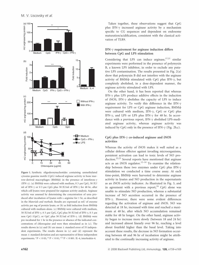
Taken together, these observations suggest that CpG
plus IFN-c increased arginase activity by a mechanism
specific to CG sequences and dependent on endosome
maturation/acidification, consistent with the classical acti-
vation of TLR9.
IFN-c requirement for arginase induction differsbetween CpG and LPS stimulation
Considering that LPS can induce arginase,17,21 similar
experiments were performed in the presence of polymyxin
B, a known LPS inhibitor, in order to exclude any puta-
tive LPS contamination. The results presented in Fig. 2(a)
show that polymyxin B did not interfere with the arginase
activity of BMM/ stimulated with CpG plus IFN-c, but
completely abolished, in a dose-dependent manner, the
arginase activity stimulated with LPS.
On the other hand, it has been reported that whereas
IFN-c plus LPS produce additive effects in the induction
of iNOS, IFN-c abolishes the capacity of LPS to induce
arginase activity. To verify this difference in the IFN-crequirement for LPS or CpG arginase induction, BMM/were cultured with medium, IFN-c, CpG or CpG plus
IFN-c, and LPS or LPS plus IFN-c for 48 hr. In accor-
dance with a previous report, IFN-c abolished LPS-medi-
ated arginase activity, whereas arginase activity was
induced by CpG only in the presence of IFN-c (Fig. 2b,c).
CpG plus IFN-c co-induced arginase and iNOSactivities
Whereas the activity of iNOS makes it well suited as a
cellular defense effector against invading microorganisms,
persistent activation can lead to toxic levels of NO pro-
duction.22,23 Several reports have mentioned that arginase
acts as an iNOS regulator.12–14 To examine the relation-
ship between these two enzymes under CpG plus IFN-cstimulation we conducted a time course assay. At each
time-point, BMM/ were harvested to determine arginase
activity in lysates and NO production in the supernatants
as an iNOS activity indicator. As illustrated in Fig. 3, and
in agreement with a previous report,24 CpG alone was
unable to stimulate NO production, whereas a substantial
increase of NO secretion occurred in the presence of
IFN-c. However, there were some evident differences
regarding the activation of arginase and iNOS. NO was
detected at 18 hr, increased with time and reached a maxi-
mum at 48 hr, after which NO accumulation remained
stable for 48 hr longer. On the other hand, arginase activ-
ity began to increase more slowly (between 18 and 24 hr)
and increased almost linearly over 96 hr, reaching a level
about fourfold higher than the basal level. Taking into
account these results, the decrease in NO formation occur-
ring between 48 and 96 hr of incubation might be attrib-
uted to the continually increasing activity of arginase.
150
100
50
0Medium CpG + IFN-γIFN-γCpG
***
Medium
Medium
CpG + IFN-γIFN-γCpG
0 30Chloroquine (μg/ml)
15 45 60
CpG GpC
LPS
15
10
Fol
d in
duct
ion
Fol
d in
duct
ion
mU
arg
inas
e/m
g pr
otei
n
5
0Medium IL-4
3
2
1
0
200
100
0
100
50
0
CpG + IFN-γMedium
***** Without IFN-γ
With IFN-γ
mU
arg
inas
e/m
g pr
otei
nm
U a
rgin
ase/
mg
prot
ein
***
(a)
(b)
(c)
(d)
Figure 1. Synthetic oligodeoxynucleotides containing unmethylated
cytosine guanine motifs (CpG) induced arginase activity in bone mar-
row-derived macrophages (BMM/) in the presence of interferon-c(IFN-c). (a) BMM/ were cultured with medium, 0�3 lm CpG, 50 IU/
ml of IFN-c or 0�3 lm CpG plus 50 IU/ml of IFN-c for 48 hr, after
which cell lysates were prepared for arginase activity analysis. Arginase
activity was assessed by determining the concentration of urea pro-
duced after incubation of lysates with l-arginine for 1 hr, as described
in the Materials and methods. Results are expressed as mU of enzyme
activity per mg of protein lysate, or (b) as fold induction from BMM/cultured with medium alone. (c) BMM/ were cultured with medium,
50 IU/ml of IFN-c, 0�3 lm CpG, CpG plus 50 IU/ml of IFN-c, 0�3 lm
non CpG (GpC), or GpC plus 50 IU/ml of IFN-c. (d) BMM/ were
pre-incubated for 1 hr in the presence or absence of the indicated con-
centrations of chloroquine and were then stimulated as in (c). The
results shown in (a) and (b) are mean ± standard error of 35 indepen-
dent experiments. The results shown in (c) and (d) represent the
mean ± standard deviation and are representative of three independent
experiments. *P < 0�05; **P < 0�01; ***P < 0�001. IL-4, interleukin-4.
e162 � 2008 Blackwell Publishing Ltd, Immunology, 128, e159–e169
M. V. Liscovsky et al.

CpG plus IFN-c induced up-regulation of arginasetype II protein
In mammals, two distinct arginase isoforms are expressed
in M/: the cytosolic arginase I and the mitochondrial
arginase II, which are identical to the liver-type and
kidney-type arginase, respectively. We examined arginase
I and II protein levels in BMM/ by western blot. CpG
plus IFN-c increased the expression of arginase II but not
of arginase I. In addition, the expression of arginase II
protein was revealed in unstimulated BMM/, consistent
with the basal arginase activity observed in these cells
(Fig. 4a). A densitometry assay demonstrated that the
increased expression of arginase II was consistent with the
observed magnitude of arginase activity induction
(Fig. 4b). Therefore, the CpG plus IFN-c-mediated argi-
nase activity might be primarily caused by an increase in
arginase II expression.
CpG plus IFN-c-mediated arginase activity wasdependent on IFN-c priming but independent of theautocrine IL-10 effect
IFN-c coordinates diverse cellular programmes through
transcriptional regulation and integration of other signal-
ling pathways.25 We found that CpG increased arginase
activity in the presence of IFN-c, whereas other reports
have described the inhibitory effects of IFN-c on arginase
activity.15–17 Therefore, to rule out the possibility that
150
200
100
50
0
mU
arg
inas
e/m
g pr
otei
nm
U a
rgin
ase/
mg
prot
ein
mU
arg
inas
e/m
g pr
otei
n
Without IFN-γ
With IFN-γ
LPS
CpG + IFN-γMedium
100
50
00 1 2
Polymyxin B (mg/ml)3 4 5 6
Medium 0·01 0·03 0·1
CpG (μM)
0·3 1·0 3·0
Without IFN-γ ****
**
With IFN-γ
Medium 0·1LPS (μg/ml)
1·0 10 CpG
250
500
0
(a)
(b)
(c)
Figure 2. Interferon-c (IFN-c) requirement for arginase induction
differs between synthetic oligodeoxynucleotides containing unmethy-
lated cytosine guanine motifs (CpG) and lipopolysaccharide (LPS)
stimulation. (a) Bone marrow-derived macrophages (BMM/) were
pre-incubated for 1 hr in the presence or absence of the indicated
concentrations of polymyxin B, and then stimulated for 48 hr with
medium, 0�3 lm CpG plus 50 IU/ml of IFN-c, or 1 lg/ml of LPS.
(b) BMM/ were stimulated with the indicated concentrations of
CpG in the presence or absence of 50 IU/ml of IFN-c for 48 hr, after
which cell lysates were prepared for arginase activity analysis.
(c) BMM/ were stimulated with the indicated concentrations of LPS
in the presence or absence of 50 IU/ml of IFN-c or with 0�3 lm
CpG plus 50 IU/ml of IFN-c for 48 hr. Data in (a)–(c) represent the
mean ± standard deviation and are representative of three indepen-
dent experiments. **P < 0�01.
150
100
50
0
0
35
NO
2 (μ
M) 25
15
5
24 48 Time (hr)
72 96
0 24 48 Time (hr)
72 96
mU
arg
inas
e/m
g pr
otei
n CpG + IFN-γ
CpG
–
Figure 3. Synthetic oligodeoxynucleotides containing unmethylated
cytosine guanine motifs (CpG) plus interferon-c (IFN-c) co-induced
arginase and inducible nitric oxide synthase (iNOS) activities. Bone
marrow-derived macrophages (BMM/) were incubated with medium,
0�3 lm CpG, or 0�3 lm CpG plus 50 IU/ml of IFN-c for the indicated
period of time. For each well, arginase activity was measured in lysates
and nitric oxide (NO) concentration was measured in supernatants.
Data represent the mean (stimulated minus unstimulated BMM/control) and are representative of three independent experiments.
� 2008 Blackwell Publishing Ltd, Immunology, 128, e159–e169 e163
CpG plus IFN-c activated arginase in macrophages

IFN-c inhibition had actually occurred at higher concen-
trations, we performed an IFN-c dose–response curve. As
depicted in Fig. 5(a), no inhibition of arginase activity
was observed in spite of increasing concentrations of
IFN-c, confirming that arginase activity was induced by
CpG only in the presence of IFN-c.
Previous findings reported that IFN-c primes M/ to
CpG-DNA for some functions.24 Thus, we carried out a
priming assay to evaluate the role of IFN-c in arginase
activation. BMM/ were prestimulated with medium,
CpG or IFN-c alone for 1 hr. Then, BMM/ were care-
fully washed and stimulated with the indicated stimulus
for 48 hr (Fig. 5b). A statistically significant increase in
arginase activity was only observed when BMM/ were
pre-incubated with IFN-c and then stimulated with CpG.
In contrast, BMM/ pre-incubated with CpG alone and
stimulated with IFN-c did not have any positive effect
on arginase activity. These observations demonstrated
that CpG plus IFN-c-mediated arginase activity is depen-
dent on IFN-c priming. This priming phenomenon might
not be a general requisite to mediate the change from
resting BMM/ to pre-activated BMM/, which are highly
receptive to subsequent CpG stimulation, because the
data presented in Fig. 5(c) show that BMM/ were effec-
tively activated by CpG alone and secreted a significant
amount of IL-12 and IL-10. Bearing this in mind, we
attempted to define the role of IFN-c priming in this
arginase activation. For that, BMM/ were cultured in the
presence of the protein synthesis inhibitor CHX. As illus-
trated in Fig. 5(d), CHX blocked the induction of argi-
nase activity that occurred as a result of IFN-c priming.
These results suggest that a rapidly IFN-c-stimulated and
de novo-synthesized protein may trigger arginase induc-
tion in CpG-stimulated BMM/ that could be consistent
with an indirect stimulatory mechanism. Considering that
earlier studies showed that IL-10 increases the arginase
activity in M/,12,26 and that IL-10 secretion is stimulated
in CpG-activated M/ (Fig. 5c and ref.3), we wondered
whether IL-10 has a role in CpG plus IFN-c-mediated
arginase activity. Therefore, we determined the IL-10 con-
centration in the supernatant using ELISA. The results in
Fig. 5(e) clearly show an inverse pattern regarding IL-10
production and arginase activity: CpG alone induced
practically no arginase activity but stimulated IL-10 secre-
tion, whereas CpG plus IFN-c increased arginase activity
but dramatically reduced IL-10 secretion. On the basis of
these results, it seems unlikely that IL-10 participates, at
least directly, in the CpG plus IFN-c-mediated arginase
activity. Therefore, the ability of CpG plus IFN-c to
induce arginase activity might occur by a mechanism
independent of an autocrine IL-10 effect.
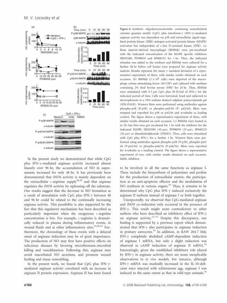
CpG plus IFN-c-mediated arginase activity wasdependent on p38 and ERK MAPK activation, butindependent of JNK
A universal outcome of engaging all TLRs is activation of
the MAPK signaling pathways.27 To investigate whether
arginase activity after stimulation with CpG plus IFN-coccurred through the classical MAPK activation pathways,
we used pharmacological specific inhibitors in culture.
Arginase activity was markedly suppressed by SB203580
and PD98059, p38 MAPK and extracellular signal-related
protein kinase (ERK)-specific inhibitors, respectively, but
not by SP600125, a c-Jun N-terminal kinase (JNK) inhibi-
tor (Fig. 6a), suggesting that p38 and ERK, but not JNK,
signaling pathways are involved in the induction of argi-
nase activity by CpG plus IFN-c.
To corroborate the result obtained from pharmacologi-
cal inhibition, we analyzed the phosphorylation status of
p38 and p44/p42, as an indication of activation, by wes-
tern blot. Consistent with the pharmacological sensitivity
observed, p38 and p42/p44 were substantially phosphory-
lated when stimulated with CpG plus IFN-c (Fig. 6b).
The kinetics of induction of p44/p42 phosphorylation was
Liver
Kidney
Medium
CpG + IFN-γ
Arginase II
α-tubulin
3
2
1
0
Medium
CpG + IFN-γ
Den
sito
met
ry
(arb
itrar
y un
its)
Arginase I
(a)
(b)
Figure 4. Synthetic oligodeoxynucleotides containing unmethylated
cytosine guanine motifs (CpG) plus interferon-c (IFN-c) induced the
up-regulation of arginase type II protein. (a) Bone marrow-derived
macrophages (BMM/) were stimulated or not with 0�3 lm CpG plus
50 IU/ml of IFN-c for 48 hr. Equal amounts of whole-cell lysates
(100 lg/lane) were subjected to electrophoresis on a 12% sodium
dodecyl sulphate polyacrylamide gel (SDS-PAGE). Western blots were
performed using specific antibodies against arginase type I and
arginase type II. To normalize the loaded samples, antibody against
a-tubulin was used as a control that is not affected by the treatment.
Mouse liver (50 lg/lane) and kidney (100 lg/lane) lysates were used as
positive controls for arginase I and arginase II, respectively. (b) Densito-
metric evaluation is expressed as arbitrary units. Data in (a) and (b)
are representative experiments of two performed.
e164 � 2008 Blackwell Publishing Ltd, Immunology, 128, e159–e169
M. V. Liscovsky et al.

rapid, but transient, while p38 phosphorylation was more
sustained.
The capacity of the mentioned drugs to inhibit p38,
ERK (p42/44) and JNK (p46/54) phosphorylation was
verified by western blot analysis, as shown in Fig. 6(c).
Discussion
During the last decade, there has been increasing recogni-
tion of the immunostimulatory properties of CpG-DNA
and its safety profile is an attractive feature for its use in
therapeutic strategies. Initially, CpG-DNA were believed to
be predominantly pro-inflammatory molecules; however,
more recently, some anti-inflammatory properties have
been described.2–9 The overwhelming body of evidence
which indicates that arginase plays a key role as an iNOS
regulator prompted us to investigate the relationship
between these two enzymes in CpG-activated BMM/. Our
data revealed, for the first time, that CpG is able to induce
arginase activation and that this occurred in the presence
of IFN-c, considered to be an inhibitor of the arginase
activation by cytokines or LPS.15–17 Therefore, IFN-cdifferentially affects the responses to LPS and CpG. In the
presence of IFN-c, M/ have been reported to respond to
LPS by secreting great quantities of NO but arginase
production was practically abolished (ref.17 and Fig. 2c),
whereas we demonstrated here that BMM/ in the
presence of IFN-c responded to CpG by activating simul-
taneously iNOS and arginase. Although the effects of LPS
and CpG-DNA on M/ are similar (both induce the
production of NO and the pro-inflammatory cytokines
IL-1, IL-6, IL-12 and TNF-a),10,11 their toxicities differ
greatly.1 In consequence, our observation of the effect of
CpG plus IFN-c on arginase activity demonstrates another
difference between LPS and CpG pathways that could help
to explain the lower toxicity found for CpG.
250 (a) (c)
(b)
(e)
(d)
200 Medium CpG
0 50 100 0 25 50 100 2500
0
50
mU
arg
inas
e/m
g pr
otei
n
100
150
200
250
Medium CpG
** **
**
2
1
0
IL-12 (ng/ml)
250
500 IL-10 IL-12
750
IL-1
0 (p
g/m
l)
IFN-γ (IU/ml)
100
150
50
0
250
** *** 200
150
100
50
0
0 Medium 0·3 3·0
CpG (µM)
250
500
750 ** **
Without IFN-γ With IFN-γ
IL-1
0 (p
g/m
l) Pretreatment
Stimulation
Pretreatment
Stimulation
CHX – – – – – – – –
– – –
– –
– – – –
+ + + +
+ + + +
+
+ + IFN-γIFN-γCpG
IFN-γ – – – –
– –
– – – – – – –
– – – – – – +
+ + + +
+ +
+ + CpG IFN-γ CpG
mU
arg
inas
e/m
g pr
otei
n m
U a
rgin
ase/
mg
prot
ein
Figure 5. Synthetic oligodeoxynucleotides containing unmethylated cytosine guanine motifs (CpG) plus interferon-c (IFN-c)-mediated arginase
activity was dependent on IFN-c priming but independent of the autocrine interleukin (IL)-10 effect. (a) Bone marrow-derived macrophages
(BMM/) were incubated for 48 hr with medium or with 0�3 lm CpG plus the indicated concentrations of IFN-c, after which cell lysates were
prepared for arginase activity analysis. (b) BMM/ were pre-incubated with medium, 0�3 lm CpG, or 50 IU/ml of IFN-c for 1 hr, washed three
times and cultured for a further 48 hr with the indicated stimulus. (c) BMM/ were cultured with medium alone or with 0�3 lm CpG for 48 hr. IL-
10 and IL-12 production were measured in supernatants by enzyme-linked immunosorbent assay (ELISA). (d) Where indicated, BMM/ were pre-
exposed to 10 lg/ml of cycloheximide (CHX) for 30 min. Then, BMM/ were prestimulated in the presence or absence of IFN-c for 1 hr, washed
and stimulated with IFN-c or CpG for a further 48 hr. (e) IL-10 was measured in supernatants of BMM/ incubated with medium, 3�0 lm CpG, or
CpG plus 50 IU/ml of IFN-c for 48 hr. Results shown are the mean ± standard deviation and are representative of three independent experiments.
**P < 0�01; ***P < 0�001.
� 2008 Blackwell Publishing Ltd, Immunology, 128, e159–e169 e165
CpG plus IFN-c activated arginase in macrophages
In the present study we demonstrated that while CpG
plus IFN-c-mediated arginase activity increased almost
linearly over 96 hr, the accumulation of NO in super-
natants increased for only 48 hr. It has previously been
demonstrated that iNOS activity is mainly dependent on
the extracellular L-arginine supply28,29 and that arginase
regulates the iNOS activity by siphoning off the substrate.
Our results suggest that the decrease in NO formation as
a result of stimulation with CpG plus IFN-c between 48
and 96 hr could be related to the continually increasing
arginase activity. This possibility is also supported by the
fact that this regulatory mechanism has been described as
particularly important when the exogenous L-arginine
concentration is low. For example, L-arginine is dramati-
cally reduced in plasma during inflammatory events, in
wound fluids and at other inflammatory sites.14,30,31 Fur-
thermore, the chronology of these events with a delayed
onset of arginase induction may be of great importance.
The production of NO may first have positive effects on
infectious diseases by favoring microbiostasis-microbial
killing and vasodilatation. Following this, arginase may
avoid exacerbated NO secretion, and promote wound
healing and tissue remodeling.
In the present work, we found that CpG plus IFN-c-
mediated arginase activity correlated with an increase in
arginase II protein expression. Arginase II has been found
to be involved in all the same functions as arginase I.
These include the biosynthesis of polyamines and proline
for the production of extracellular matrix, the participa-
tion as an anti-apoptotic effector and the modulation of
NO synthesis in various organs.32 Thus, it remains to be
determined why CpG plus IFN-c induced exclusively the
arginase II isoform instead of arginase I or both together.
Unexpectedly, we observed that CpG-mediated arginase
and iNOS co-induction only occurred in the presence of
IFN-c. This result might seem contradictory to other
authors who have described an inhibitory effect of IFN-con arginase activity.15–17 Despite this discrepancy, our
finding is supported by a previous report which demon-
strated that IFN-c also participates in arginase induction
in primary astrocytes.33 In addition, in RAW 264�7 M/,
IFN-c completely abolished cAMP-dependent induction
of arginase I mRNA, but only a slight reduction was
observed in cAMP induction of arginase II mRNA.34
Interestingly, given the established inhibitory role played
by IFN-c in arginase activity, there are some inexplicable
observations in in vivo models. For instance, although
IFN-c mRNA was markedly increased in the IL-10-defi-
cient mice injected with schistosome egg, arginase I was
induced to the same extent as that in wild-type animals.16
200 Medium
(a)
(c)
(b)Time (min)
P-p38 MAPK
p38 MAPK
α-tubulin
1·5
1·0
0·5
0·00
0 10
3010
30
60
60
90
0 3010 60 90
90
30Time (min)
CpG + IFN-γMedium Inhib DMSO
P-p38 MAPK
α-tubulin
α-tubulin
α-tubulin
P-p42/44 MAPK
P-p46/54 MAPK
Den
sito
met
ry(a
rbitr
ary
units
)
60 90
Time (min)
Time (min)P-p42/44 MAPK
p42/44 MAPK
α-tubulin
3·0
2·0
1·0
0·00 10
Den
sito
met
ry(a
rbitr
ary
units
)
CpG + IFN-γ100
0
200
100
0
200
100
0
0
0
0 5 10 15SP600125 (µM)
20 25 3530
10 20 30 40 50 60PD98059 (µM)
2 4 6SB203580 (µM)
8 10 12mU
arg
inas
e/m
gpr
otei
nm
U a
rgin
ase/
mg
prot
ein
mU
arg
inas
e/m
gpr
otei
nFigure 6. Synthetic oligodeoxynucleotides containing unmethylated
cytosine guanine motifs (CpG) plus interferon-c (IFN-c)-mediated
arginase activity was dependent on p38 and extracellular signal-regu-
lated protein kinase (ERK) mitogen-activated protein kinase (MAPK)
activation but independent of c-Jun N-terminal kinase (JNK). (a)
Bone marrow-derived macrophages (BMM/) were pre-incubated
with the indicated concentration of the MAPK specific inhibitors
SB203580, PD98059 and SP600125 for 1 hr. Then, the indicated
stimulus was added to the medium and BMM/ were cultured for a
further 48 hr before cell lysates were prepared for arginase activity
analysis. Results represent the mean ± standard deviation of a repre-
sentative experiment of three, with similar results obtained on each
occasion. (b) BMM/ (2 · 106 cells) were deprived of the macro-
phage colony-stimulating factor (M-CSF) and cultured with medium
containing 2% fetal bovine serum (FBS) for 24 hr. Then, BMM/were stimulated with 0�3 lm CpG plus 50 IU/ml of IFN-c for the
indicated period of time. Cells were harvested, lysed and subjected to
electrophoresis in a 10% sodium dodecyl sulphate polyacrylamide gel
(SDS-PAGE). Western blots were performed using antibodies against
phospho-p38 (P-p38) or phospho-p42/44 (P- p42/44). Blots were
stripped and reprobed for p38 or p42/44 and a-tubulin as loading
control. The figure shows a representative experiment of three, with
similar results obtained on each occasion. (c) BMM/ were treated as
in (b) but first were pre-incubated for 1 hr with the inhibitor for the
indicated MAPK: SB203580 (10 lm), PD98059 (25 lm), SP600125
(20 lm) or dimethylsulphoxide (DMSO). Then, cells were stimulated
with CpG plus IFN-c for a further 1 hr. Western blots were per-
formed using antibodies against phospho-p38 (P-p38), phospho-p42/
44 (P-p42/44) or phospho-p46/54 (P-p46/54). Blots were reprobed
for a-tubulin as a loading control. The figure shows a representative
experiment of two, with similar results obtained on each occasion.
Inhib, inhibitor.
e166 � 2008 Blackwell Publishing Ltd, Immunology, 128, e159–e169
M. V. Liscovsky et al.
Meanwhile, in the Citrobacter rodentium model of colitis,
associated with a strong mucosal Th1 response, both
iNOS and arginase I were up-regulated in the colon of
infected mice.35 Although the precise reasons for differ-
ences in the role of IFN-c in arginase regulation are not
still clear, they may reflect several variants such as cell
type, cell maturation-associated differentiation and the
exogenous stimuli used.
It is very probable that IFN-c elicited its effect very
early in the pathway, because a short pretreatment of 1 hr
was sufficient to produce an increase in arginase activity
similar to that obtained when IFN-c was added together
with CpG. The efficacy of this brief stimulation with
IFN-c is consistent with the demonstration that the first
wave of IFN-c-induced transcription occurs within 15–
30 min of treatment.36 In addition, we observed that
CHX blocked the induction of arginase activity observed
under IFN-c priming, which would be consistent with an
indirect stimulatory mechanism. Thus, we investigated
the role of IL-10 in CpG plus IFN-c-mediated arginase
activity. Considering the strong inhibition observed of IL-
10 secretion in the presence of CpG plus IFN-c, it seems
unlikely that an autocrine IL-10 effect promotes the CpG
plus IFN-c-mediated arginase activity.
Furthermore, our results are in agreement with previ-
ous studies demonstrating that certain responses to
CpG-DNA, such as arginine uptake and increased iNOS
gene expression, occurred only after IFN-c priming.24
Traditionally, priming with IFN-c was thought to medi-
ate a change from the resting M/ to a pre-activated M/that is highly receptive to a second activating signal.
However, it is now known that M/ integrate multifari-
ous signals to affect an appropriate cellular response in a
more complex way.37 In agreement with this, we
observed that BMM/ were effectively activated by CpG
in the absence of IFN-c and secreted significant amounts
of IL-12 and IL-10. Thus, IFN-c might not only be a
prerequisite for BMM/ activation but also might activate
molecules or transcription factors shared with TLR9,
thereby generating the cross-talk between these pathways
to favour arginase activation.
It is well known that recognition of CpG-DNA by
TLR9 initiates a signaling cascade that begins with the
recruitment of the adaptor protein, MyD88, with the acti-
vation of MAPKs being one of the early biochemical sig-
naling events.27 The results presented here reveal that p38
MAPK and ERK, but not JNK, signaling pathways are
involved in the induction of arginase activity by CpG plus
IFN-c. Interestingly, p38 and ERK have been involved in
other CpG anti-inflammatory functions in M/. A central
negative-feedback role was described for ERK in the
CpG-DNA-mediated Th1-type response by promoting
IL-10 production, whereas p38 has been reported to be
essential for the induction of both IL-10 and IL-12.38 In
addition, the induced expression of SOCS1 and SOCS3
by CpG-DNA were blocked partially when ERK and p38
were inhibited.9 Also, p38 and ERK were involved in the
activation of signaling pathways different from the canoni-
cal Jak-STAT1 for IFN-c. This implies an association
between the IFN-c receptor and MyD88 that results in p38
activation39 and the activation of ERK.40,41 Our observa-
tions lend additional support to the hypothesis of IFN-csignaling integration with pathogen-associated molecular
patterns.
Collectively, the effect of CpG plus IFN-c on arginase
activity reveals an unsuspected complexity in the response
of BMM/ to the combination of CpG and IFN-c, one of
the principal and most relevant cytokines secreted in vivo
in response to CpG-DNA. Furthermore, these data add
support to the growing body of evidence that favour a
dual role for IFN-c activity.42
Finally, comparisons of different adjuvants in mouse
models have demonstrated that CpG-DNA is unsurpassed
at inducing Th1-type responses. What is more, CpG-
DNA can overcome the Th2 bias associated with some
disease states or in both very young and elderly mice.43–46
However, administration of CpG-DNA also generates a
response in order to avoid an exacerbated inflamma-
tion.46,47 Therefore, the possibility that CpG-DNA could
simultaneously or sequentially elicit both a pro-inflamma-
tory and a counter anti-inflammatory response, similar to
our in vitro data, is plausible and very interesting in terms
of immune regulation during infection and in the
improvement of vaccines.
Acknowledgements
We acknowledge Dr Tomomi Gotoh (Graduate School of
Medical Sciences, Kumamoto University, Japan) for the
kind gift of arginase antibodies. We thank Dr Paul Hob-
son, a native English speaker who revised the manuscript.
This work was supported by grants from CONICET-PIP
5750, ANPCyT-PICT 25552 and SeCyT. M.C.P.P. and
G.M. are career members of CONICET. M.V.L, D.O.A,
R.P.R and C.V.G are recipients of graduate fellowships
from CONICET.
References
1 Krieg AM. Therapeutic potential of Toll-like receptor 9 activa-
tion. Nat Rev Drug Discov 2006; 5:471–84.
2 Schwartz DA, Wohlford-Lenane CL, Quinn TJ, Krieg AM. Bacte-
rial DNA or oligonucleotides containing unmethylated CpG
motifs can minimize lipopolysaccharide-induced inflammation
in the lower respiratory tract through an IL-12-dependent path-
way. J Immunol 1999; 163:224–31.
3 Anitescu M, Chace JH, Tuetken R, Yi AK, Berg DJ, Krieg AM,
Cowdery JS. Interleukin-10 functions in vitro and in vivo to
inhibit bacterial DNA-induced secretion of interleukin-12.
J Interferon Cytokine Res 1997; 17:781–8.
� 2008 Blackwell Publishing Ltd, Immunology, 128, e159–e169 e167
CpG plus IFN-c activated arginase in macrophages
4 Redford TW, Yi AK, Ward CT, Krieg AM. Cyclosporin A
enhances IL-12 production by CpG motifs in bacterial DNA
and synthetic oligodeoxynucleotides. J Immunol 1998;
161:3930–5.
5 Samarasinghe R, Tailor P, Tamura T, Kaisho T, Akira S, Ozato
K. Induction of an anti-inflammatory cytokine, IL-10, in
dendritic cells after toll-like receptor signaling. J Interferon Cyto-
kine Res 2006; 26:893–900.
6 Wingender G, Garbi N, Schumak B et al. Systemic application of
CpG-rich DNA suppresses adaptive T cell immunity via induc-
tion of IDO. Eur J Immunol 2006; 36:12–20.
7 Mellor AL, Baban B, Chandler PR, Manlapat A, Kahler DJ,
Munn DH. Cutting edge: CpG oligonucleotides induce splenic
CD19+ dendritic cells to acquire potent indoleamine 2,3-dioxy-
genase-dependent T cell regulatory functions via IFN Type 1 sig-
naling. J Immunol 2005; 175:5601–5.
8 Jin L, Raymond DP, Crabtree TD, Pelletier SJ, Houlgrave CW,
Pruett TL, Sawyer RG. Enhanced murine macrophage TNF
receptor shedding by cytosine–guanine sequences in oligodeoxy-
nucleotides. J Immunol 2000; 165:5153–60.
9 Dalpke AH, Opper S, Zimmermann S, Heeg K. Suppressors of
cytokine signaling (SOCS)-1 and SOCS-3 are induced by CpG-
DNA and modulate cytokine responses in APCs. J Immunol
2001; 166:7082–9.
10 Krieg AM. CpG motifs in bacterial DNA and their immune
effects. Annu Rev Immunol 2002; 20:709–60.
11 Dobrovolskaia MA, Vogel SN. Toll receptors, CD14, and macro-
phage activation and deactivation by LPS. Microbes Infect 2002;
4:903–14.
12 Corraliza IM, Soler G, Eichmann K, Modolell M. Arginase
induction by suppressors of nitric oxide synthesis (IL-4, IL-10
and PGE2) in murine bone-marrow-derived macrophages. Bio-
chem Biophys Res Commun 1995; 206:667–73.
13 Gotoh T, Mori M. Arginase II downregulates nitric oxide (NO)
production and prevents NO-mediated apoptosis in murine
macrophage-derived RAW 264.7 cells. J Cell Biol 1999; 144:
427–34.
14 Chang CI, Liao JC, Kuo L. Arginase modulates nitric oxide pro-
duction in activated macrophages. Am J Physiol 1998; 2:H342–8.
15 Modolell M, Corraliza IM, Link F, Soler G, Eichmann K. Reci-
procal regulation of the nitric oxide synthase/arginase balance in
mouse bone marrow-derived macrophages by TH1 and TH2
cytokines. Eur J Immunol 1995; 25:1101–4.
16 Hesse M, Modolell M, La Flamme AC, Schito M, Fuentes JM,
Cheever AW, Pearce EJ, Wynn TA. Differential regulation of
nitric oxide synthase-2 and arginase-1 by type 1/type 2 cytokines
in vivo: granulomatous pathology is shaped by the pattern of
L-arginine metabolism. J Immunol 2001; 167:6533–44.
17 Wang WW, Jenkinson CP, Griscavage JM, Kern RM, Arabolos
NS, Byrns RE, Cederbaum SD, Ignarro LJ. Co-induction of argi-
nase and nitric oxide synthase in murine macrophages activated
by lipopolysaccharide. Biochem Biophys Res Commun 1995;
210:1009–16.
18 Corraliza IM, Campo ML, Soler G, Modolell M. Determination
of arginase activity in macrophages: a micromethod. J Immunol
Methods 1994; 174:231–5.
19 Ozaki M, Gotoh T, Nagasaki A, Miyanaka K, Takeya M, Fujiy-
ama S, Tomita K, Mori M. Expression of arginase II and related
enzymes in the rat small intestine and kidney. J Biochem (Tokyo)
1999; 125:586–93.
20 Latz E, Schoenemeyer A, Visintin A et al. TLR9 signals after
translocating from the ER to CpG DNA in the lysosome. Nat
Immunol 2004; 5:190–8.
21 Sonoki T, Nagasaki A, Gotoh T, Takiguchi M, Takeya M,
Matsuzaki H, Mori M. Coinduction of nitric-oxide synthase
and arginase I in cultured rat peritoneal macrophages and rat
tissues in vivo by lipopolysaccharide. J Biol Chem 1997;
272:3689–93.
22 Thiemermann C. Nitric oxide and septic shock. Gen Pharmacol
1997; 29:159–66.
23 Bogdan C. Nitric oxide and the immune response. Nat Immunol
2001; 2:907–16.
24 Sweet MJ, Stacey KJ, Kakuda DK, Markovich D, Hume DA.
IFN-gamma primes macrophage responses to bacterial DNA.
J Interferon Cytokine Res 1998; 18:263–71.
25 Schroder K, Hertzog PJ, Ravasi T, Hume DA. Interferon-gamma:
an overview of signals, mechanisms and functions. J Leukoc Biol
2004; 75:163–89.
26 Lang R, Patel D, Morris JJ, Rutschman RL, Murray PJ. Shaping
gene expression in activated and resting primary macrophages
by IL-10. J Immunol 2002; 169:2253–63.
27 Barton GM, Medzhitov R. Toll-like receptor signaling pathways.
Science 2003; 300:1524–5.
28 Nicholson B, Manner CK, Kleeman J, MacLeod CL. Sustained
nitric oxide production in macrophages requires the arginine
transporter CAT2. J Biol Chem 2001; 276:15881–5.
29 Hrabak A, Idei M, Temesi A. Arginine supply for nitric oxide
synthesis and arginase is mainly exogenous in elicited murine
and rat macrophages. Life Sci 1994; 55:797–805.
30 Albina JE, Mills CD, Henry WL Jr, Caldwell MD. Temporal
expression of different pathways of 1-arginine metabolism in
healing wounds. J Immunol 1990; 144:3877–80.
31 Gobert AP, Daulouede S, Lepoivre M et al. L-Arginine availabil-
ity modulates local nitric oxide production and parasite killing
in experimental trypanosomiasis. Infect Immun 2000; 68:4653–7.
32 Cederbaum SD, Yu H, Grody WW, Kern RM, Yoo P, Iyer RK.
Arginases I and II: do their functions overlap? Mol Genet Metab
2004; 81(Suppl 1):S38–44.
33 Lee J, Ryu H, Ferrante RJ, Morris SM Jr, Ratan RR. Transla-
tional control of inducible nitric oxide synthase expression by
arginine can explain the arginine paradox. Proc Natl Acad Sci
USA 2003; 100:4843–8.
34 Morris SM Jr, Kepka-Lenhart D, Chen LC. Differential regula-
tion of arginases and inducible nitric oxide synthase in murine
macrophage cells. Am J Physiol 1998; 1:E740–7.
35 Gobert AP, Cheng Y, Akhtar M et al. Protective role of arginase
in a mouse model of colitis. J Immunol 2004; 173:2109–17.
36 Kerr IM, Stark GR. The control of interferon-inducible gene
expression. FEBS Lett 1991; 285:194–8.
37 Schroder K, Sweet MJ, Hume DA. Signal integration between IFN-
gamma and TLR signalling pathways in macrophages. Immuno-
biology 2006; 211:511–24.
38 Yi AK, Yoon JG, Yeo SJ, Hong SC, English BK, Krieg AM. Role
of mitogen-activated protein kinases in CpG DNA-mediated
IL-10 and IL-12 production: central role of extracellular signal-
regulated kinase in the negative feedback loop of the CpG
DNA-mediated Th1 response. J Immunol 2002; 168:4711–20.
39 Sun D, Ding A. MyD88-mediated stabilization of interferon-
gamma-induced cytokine and chemokine mRNA. Nat Immunol
2006; 7:375–81.
e168 � 2008 Blackwell Publishing Ltd, Immunology, 128, e159–e169
M. V. Liscovsky et al.
40 Ramana CV, Gil MP, Schreiber RD, Stark GR. Stat1-dependent
and -independent pathways in IFN-gamma-dependent signaling.
Trends Immunol 2002; 23:96–101.
41 Valledor AF, Sanchez-Tillo E, Arpa L, Park JM, Caelles C, Llo-
beras J, Celada A. Selective roles of MAPKs during the macro-
phage response to IFN-{gamma}. J Immunol 2008; 180:4523–9.
42 Muhl H, Pfeilschifter J. Anti-inflammatory properties of pro-
inflammatory interferon-gamma. Int Immunopharmacol 2003;
3:1247–55.
43 Martinez X, Li X, Kovarik J, Klein M, Lambert PH, Siegrist CA.
Combining DNA and protein vaccines for early life immuniza-
tion against respiratory syncytial virus in mice. Eur J Immunol
1999; 29:3390–400.
44 Weeratna RD, Brazolot Millan CL, McCluskie MJ, Davis HL.
CpG ODN can re-direct the Th bias of established Th2 immune
responses in adult and young mice. FEMS Immunol Med Micro-
biol 2001; 32:65–71.
45 Alignani D, Maletto B, Liscovsky M, Ropolo A, Moron G, Pisto-
resi-Palencia MC. Orally administered OVA/CpG-ODN induces
specific mucosal and systemic immune response in young and
aged mice. J Leukoc Biol 2005; 77:898–905.
46 Maletto BA, Ropolo AS, Liscovsky MV, Alignani DO, Glocker M,
Pistoresi-Palencia MC. CpG oligodeoxinucleotides functions as
an effective adjuvant in aged BALB/c mice. Clin Immunol 2005;
117:251–61.
47 Kitagaki K, Jain VV, Businga TR, Hussain I, Kline JN. Immuno-
modulatory effects of CpG oligodeoxynucleotides on established
th2 responses. Clin Diagn Lab Immunol 2002; 9:1260–9.
� 2008 Blackwell Publishing Ltd, Immunology, 128, e159–e169 e169
CpG plus IFN-c activated arginase in macrophages