Expression, purification, assay, and crystal structure of perdeuterated human arginase I
15
Expression, Purification, Assay, and Crystal Structure of Perdeuterated Human Arginase I, ⋆,⋆⋆ Luigi Di Costanzo a , Martine Moulin b , Michael Haertlein b , Flora Meilleur c,d , and David W. Christianson a,* a Roy and Diana Vagelos Laboratories, Department of Chemistry, University of Pennsylvania, Philadelphia, Pennsylvania 19104-6323, USA b Institut Laue-Langevin, 6 Rue Jules Horowitz, BP 156, 38042 Grenoble, France c Department of Molecular and Structural Biochemistry, North Carolina State University, Raleigh, North Carolina 2769, USA d Spallation Neutron Source, Oak Ridge National Laboratory, Oak Ridge, Tennessee 37831, USA Abstract Arginase is a manganese metalloenzyme that catalyzes the hydrolysis of L-arginine to yield L- ornithine and urea. In order to establish a foundation for future neutron diffraction studies that will provide conclusive structural information regarding proton/deuteron positions in enzyme-inhibitor complexes, we have expressed, purified, assayed, and determined the X-ray crystal structure of perdeuterated (i.e., fully deuterated) human arginase I complexed with 2-amino-6-boronohexanoic acid (ABH) at 1.90 Å resolution. Prior to the neutron diffraction experiment, it is important to establish that perdeutaration does not cause any unanticipated structural or functional changes. Accordingly, we find that perdeuterated human arginase I exhibits catalytic activity essentially identical to that of the unlabeled enzyme. Additionally, the structure of the perdeuterated human arginase I-ABH complex is identical to that of the corresponding complex with the unlabeled enzyme. Therefore, we conclude that crystals of the perdeuterated human arginase I-ABH complex are suitable for neutron crystallographic study. Keywords protein crystallography; enzyme-inhibitor complex; isotopic labeling Arginase catalyzes the hydrolysis of L-arginine to yield L-ornithine and urea [1]. In humans there are two isozymes, arginase I and arginase II, that share ~60% sequence identity. Ornithine is the biosynthetic precursor of polyamines that facilitate cellular proliferation and tumor growth [2]. Additionally, arginase I is implicated in tumoral immune evasion, and arginase inhibitors therefore have significant chemotherapeutic potential [2]. ⋆ This work was supported by NIH grant GM49758. ⋆⋆ Atomic coordinates of perdeuterated human arginase I complexed with 2(S)-amino-6-boronohexanoic acid have been deposited in the Research Collaboratory for Structural Bioinformatics (http://www.rcsb.org/pdb) with the following accession code: 2PLL. *Corresponding author. Fax: +1 215-573-2201. E-mail address: [email protected]. Publisher's Disclaimer: This is a PDF file of an unedited manuscript that has been accepted for publication. As a service to our customers we are providing this early version of the manuscript. The manuscript will undergo copyediting, typesetting, and review of the resulting proof before it is published in its final citable form. Please note that during the production process errors may be discovered which could affect the content, and all legal disclaimers that apply to the journal pertain. NIH Public Access Author Manuscript Arch Biochem Biophys. Author manuscript; available in PMC 2008 September 1. Published in final edited form as: Arch Biochem Biophys. 2007 September 1; 465(1): 82–89. NIH-PA Author Manuscript NIH-PA Author Manuscript NIH-PA Author Manuscript
-
Upload
independent -
Category
Documents
-
view
3 -
download
0
Transcript of Expression, purification, assay, and crystal structure of perdeuterated human arginase I
Expression, Purification, Assay, and Crystal Structure ofPerdeuterated Human Arginase I, ⋆,⋆⋆
Luigi Di Costanzoa, Martine Moulinb, Michael Haertleinb, Flora Meilleurc,d, and David W.Christiansona,*a Roy and Diana Vagelos Laboratories, Department of Chemistry, University of Pennsylvania, Philadelphia,Pennsylvania 19104-6323, USA
b Institut Laue-Langevin, 6 Rue Jules Horowitz, BP 156, 38042 Grenoble, France
c Department of Molecular and Structural Biochemistry, North Carolina State University, Raleigh, NorthCarolina 2769, USA
d Spallation Neutron Source, Oak Ridge National Laboratory, Oak Ridge, Tennessee 37831, USA
AbstractArginase is a manganese metalloenzyme that catalyzes the hydrolysis of L-arginine to yield L-ornithine and urea. In order to establish a foundation for future neutron diffraction studies that willprovide conclusive structural information regarding proton/deuteron positions in enzyme-inhibitorcomplexes, we have expressed, purified, assayed, and determined the X-ray crystal structure ofperdeuterated (i.e., fully deuterated) human arginase I complexed with 2-amino-6-boronohexanoicacid (ABH) at 1.90 Å resolution. Prior to the neutron diffraction experiment, it is important toestablish that perdeutaration does not cause any unanticipated structural or functional changes.Accordingly, we find that perdeuterated human arginase I exhibits catalytic activity essentiallyidentical to that of the unlabeled enzyme. Additionally, the structure of the perdeuterated humanarginase I-ABH complex is identical to that of the corresponding complex with the unlabeled enzyme.Therefore, we conclude that crystals of the perdeuterated human arginase I-ABH complex are suitablefor neutron crystallographic study.
Keywordsprotein crystallography; enzyme-inhibitor complex; isotopic labeling
Arginase catalyzes the hydrolysis of L-arginine to yield L-ornithine and urea [1]. In humansthere are two isozymes, arginase I and arginase II, that share ~60% sequence identity. Ornithineis the biosynthetic precursor of polyamines that facilitate cellular proliferation and tumorgrowth [2]. Additionally, arginase I is implicated in tumoral immune evasion, and arginaseinhibitors therefore have significant chemotherapeutic potential [2].
⋆This work was supported by NIH grant GM49758.⋆⋆Atomic coordinates of perdeuterated human arginase I complexed with 2(S)-amino-6-boronohexanoic acid have been deposited inthe Research Collaboratory for Structural Bioinformatics (http://www.rcsb.org/pdb) with the following accession code: 2PLL.*Corresponding author. Fax: +1 215-573-2201. E-mail address: [email protected]'s Disclaimer: This is a PDF file of an unedited manuscript that has been accepted for publication. As a service to our customerswe are providing this early version of the manuscript. The manuscript will undergo copyediting, typesetting, and review of the resultingproof before it is published in its final citable form. Please note that during the production process errors may be discovered which couldaffect the content, and all legal disclaimers that apply to the journal pertain.
NIH Public AccessAuthor ManuscriptArch Biochem Biophys. Author manuscript; available in PMC 2008 September 1.
Published in final edited form as:Arch Biochem Biophys. 2007 September 1; 465(1): 82–89.
NIH
-PA Author Manuscript
NIH
-PA Author Manuscript
NIH
-PA Author Manuscript
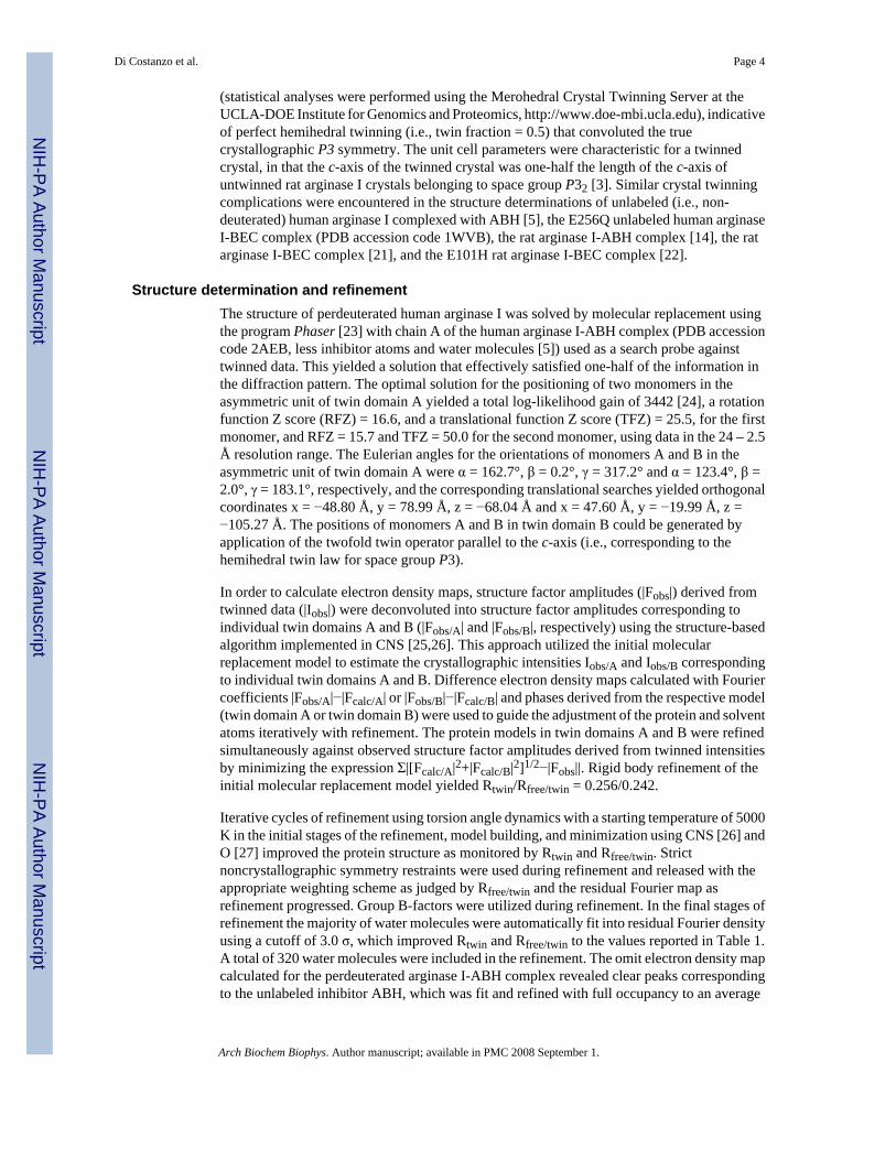
The crystal structure of unlabeled arginase I from rat liver (Rattus norvegicus) was solved 10years ago [3] at 2.1 Å resolution, and since then more than 40 additional arginase structureshave been determined and deposited in the Protein Data Bank (http://www.rcsb.org; [4]). Thesestructures include those of the human isozymes arginase I [5] and arginase II [6], arginase fromBacillus caldovelox [7], arginase from Thermus thermophilus (PDB accession codes 2EF4,2EF5, and 2EIV), and several complexes of rat arginase I with different ligands and inhibitors,as well as various mutants.
Arginase adopts an α/β fold consisting of a central parallel β-sheet flanked on both sides byseveral α-helices [3]. Surprisingly, the subsequent structure determination of histonedeacetylase revealed a fold topologically identical to that of arginase [8–10], which has recentlybeen classified as “Rossmannoid” [11–13]. All mammalian arginases are heterologous trimerswith C3 symmetry. However, the bacterial arginase is a hexamer in which two trimers associatein face-to-face fashion with overall D3 symmetry.
The structure of unlabeled rat arginase I reveals that the metal cluster required for catalysis islocated at the bottom of a ~15 Å-deep cavity in each monomer, and a metal-bridging hydroxideion functions as a nucleophile in catalysis [3]. The metal-bridging hydroxide is also requiredfor the binding of 2(S)-amino-6-boronohexanoic acid (ABH), a reactive substrate analoguethat undergoes nucleophilic attack by this hydroxide ion to yield a tetrahedral boronate anionthat mimics the tetrahedral intermediate in catalysis [14]. Recently, we demonstrated that ABHand the related analogue S-(2-boronoethyl)-L-cysteine (BEC) are highly potent inhibitors ofhuman arginase I with Kd values of 5 and 270 nM, respectively [5]. Analysis of the 1.29 Åresolution structure of the human arginase I-ABH complex reveals that nanomolar affinity isa consequence of strong metal-coordination and hydrogen bond interactions [5]. Furthermore,this structure reveals new mechanistic inferences on the catalytic function of H141, whichappears to be stabilized as the positively charged imidazolium group. This residue mayaccordingly serve as a general acid to facilitate collapse of the tetrahedral intermediate incatalysis.
The structure of the human arginase I-ABH complex is the highest resolution structure of anarginase ever determined. Intriguingly, the positions of some putative ordered hydrogen atomsare evident in electron density maps, including the acidic proton of the H141 imidazoli umgroup [5]. However, the limiting resolution of the X-ray crystal structure determination (1.29Å) is near the threshold of reliability for the determination of hydrogen atom positions due tothe weak electron density of the hydrogen atom. Therefore, we are now preparing for theneutron diffraction study of the human arginase I-ABH complex in order to locate active sitehydrogen/deuterium atoms important for inhibitor binding and catalysis. In order to enhancethe signal-to-noise ratio of measurable neutron diffraction in our forthcoming experiments, wehave prepared perdeuterated (i. e., fully deuterated) human arginase I and we have determinedthe X-ray crystal structure of its complex with ABH at 1.90 Å resolution from hemihedrallytwinned crystals. The structure of perdeuterated human arginase I complexed with ABH is verysimilar to the structure of the unlabeled human arginase I-ABH complex, including thepositions of solvent molecules interacting with ABH. Activity measurements demonstrate thatperdeuteration does not significantly affect the catalytic function of human arginase I. Thiscontrasts with activity measurements of perdeuterated glutathione S-transferase [15] andalkaline phosphatase [16], in which catalytic activity increases 1.4-fold and decreases 1.8-foldrespectively, upon perdeuteration.
Di Costanzo et al. Page 2
Arch Biochem Biophys. Author manuscript; available in PMC 2008 September 1.
NIH
-PA Author Manuscript
NIH
-PA Author Manuscript
NIH
-PA Author Manuscript

Materials and methodsExpression and purification of perdeuterated human arginase I
For protein expression in perdeuterated media, the full length cDNA of human arginase I wassubcloned in a pET-24a (Novagen) vector that confers kanamycin resistance. Perdeuteratedhuman arginase I was obtained by heterologous expression in Escherichia coli BL21(DE3) atthe ILL-EMBL Deuteration Facility in Grenoble, France. Cells were grown in minimalmedium: 6.86 g L−1 (NH4)2SO4, 1.56 g L−1 KH2PO4, 6.48 g L−1 Na2HPO4·2H2O, 0.49 gL−1 diammonium hydrogen citrate, 0.25 g L−1 MgSO4·7H2O, 1.0 ml L−1 (0.5 g L−1
CaCl2·2H2O, 16.7 g L−1 FeCl3·6H2O, 0.18 g L−1 ZnSO4·7H2O, 0.16 g L−1 CuSO4·5H2O, 0.15g L−1 MnSO4·4H2O, 0.18 g L−1 CoCl2·6H2O, 20.1 g L1 EDTA), 5 g L−1 glycerol, 40 mgL−1 kanamycin [17,18]. For preparation of fully deuterated medium, mineral salts were driedout in a rotary evaporator (Heidolph) at 333K and labile protons were exchanged for deuteronsby dissolving in a minimal volume of D2O and re-dried. Perdeuterated d8-glycerol (Euriso-Top, France) was used as a carbon source. Adaptation of BL21(DE3) cells to deuteratedminimal medium was achieved by a multi-stage adaptation process [18]. Typically,1.5 L ofdeuterated medium was inoculated with 100 mL preculture of adapted cells in a 3 L fermenter(Labfors, Infors). During the batch and fed-batch phases the pH was adjusted to 6.9 (by additionof NaOD) and the temperature was adjusted to 303 K. The gas-flow rate of sterile filtered airwas 0.5 L min−1. Stirring was adjusted to ensure a dissolved oxygen tension (DOT) of 30%.The fed-batch phase was initiated when the optical density at 600 nm reached 6.0. D8-glycerolwas added to the culture to keep the growth rate stable during fermentation. When OD600reached 12, arginase overexpression was induced by the addition of 1 mM IPTG and incubationcontinued for 24 h. Cells were then harvested, washed with 10 mM HEPES (pH 6.4), and storedat 193 K. Perdeuterated human arginase I was purified as described for the unlabeled enzyme[19] and all buffers used during purification were made with H2O. Protein purity was assessedby SDS-PAGE and the molecular weight of perdeuterated human arginase I was determinedby MALDI mass spectrometry.
Crystallization and X-ray diffraction data collectionThe complex between perdeuterated human arginase I and ABH was crystallized by the sittingdrop vapor diffusion method at 294 K. An initial Index Screen (Hampton Research) identifiedfourteen different conditions under which crystals appeared overnight. In particular, becauseof the formation of fewer nuclei and bigger crystals, one condition was most suitable forgrowing larger crystals. Drops containing 4.0 μL of enzyme solution [3.5 mg/mL perdeuteratedhuman arginase I, 0.1 mM MnCl2, 1.4 mM ABH, 50.0 mM bicine (pH 8.5)] and 4.0 μL ofprecipitant buffer [0.1 M Bis-Tris-HCl (pH 5.5), 10–20% (wt/vol) PEG-3350] wereequilibrated against a 1 mL reservoir of precipitant buffer. Rod-like crystals appeared inapproximately 1–2 days and grew to typical dimensions of 0.2 mm × 0.2 mm × 0.5 mm in oneweek. These crystallization conditions differed slightly from those initially employed in thecrystallization of the unlabeled enzyme in terms of protein concentrations, pH, and the varietyof PEG used as a precipitant [5].
Prior to X-ray diffraction data collection, exchangeable protons were substituted withdeuterons by equilibrating crystals in precipitant buffer prepared with D2O and 100 mM Bis-Tris-DCl (pD 6.6) for three days. Crystals were cryoprotected in mother liquor containing 28%glycerol-d8 prior to flash-cooling and yielded diffraction data to 1.90 Å resolution at theBrookhaven National Laboratory (Upton, NY) on beamline X29A (λ=1.000 Å, 100 K) usingan ADSC Quantum 315 detector. Data reduction was achieved with Mosflm [20]. X-raydiffraction intensities measured from these crystals exhibited symmetry consistent with theapparent space group P6 (unit cell parameters a = b = 90.8 Å, c = 69.5 Å). The analysis ofmeasured intensities revealed deviations from ideal Wilson statistics with <I2>/<I>2 ≈ 1.5
Di Costanzo et al. Page 3
Arch Biochem Biophys. Author manuscript; available in PMC 2008 September 1.
NIH
-PA Author Manuscript
NIH
-PA Author Manuscript
NIH
-PA Author Manuscript
(statistical analyses were performed using the Merohedral Crystal Twinning Server at theUCLA-DOE Institute for Genomics and Proteomics, http://www.doe-mbi.ucla.edu), indicativeof perfect hemihedral twinning (i.e., twin fraction = 0.5) that convoluted the truecrystallographic P3 symmetry. The unit cell parameters were characteristic for a twinnedcrystal, in that the c-axis of the twinned crystal was one-half the length of the c-axis ofuntwinned rat arginase I crystals belonging to space group P32 [3]. Similar crystal twinningcomplications were encountered in the structure determinations of unlabeled (i.e., non-deuterated) human arginase I complexed with ABH [5], the E256Q unlabeled human arginaseI-BEC complex (PDB accession code 1WVB), the rat arginase I-ABH complex [14], the ratarginase I-BEC complex [21], and the E101H rat arginase I-BEC complex [22].
Structure determination and refinementThe structure of perdeuterated human arginase I was solved by molecular replacement usingthe program Phaser [23] with chain A of the human arginase I-ABH complex (PDB accessioncode 2AEB, less inhibitor atoms and water molecules [5]) used as a search probe againsttwinned data. This yielded a solution that effectively satisfied one-half of the information inthe diffraction pattern. The optimal solution for the positioning of two monomers in theasymmetric unit of twin domain A yielded a total log-likelihood gain of 3442 [24], a rotationfunction Z score (RFZ) = 16.6, and a translational function Z score (TFZ) = 25.5, for the firstmonomer, and RFZ = 15.7 and TFZ = 50.0 for the second monomer, using data in the 24 – 2.5Å resolution range. The Eulerian angles for the orientations of monomers A and B in theasymmetric unit of twin domain A were α = 162.7°, β = 0.2°, γ = 317.2° and α = 123.4°, β =2.0°, γ = 183.1°, respectively, and the corresponding translational searches yielded orthogonalcoordinates x = −48.80 Å, y = 78.99 Å, z = −68.04 Å and x = 47.60 Å, y = −19.99 Å, z =−105.27 Å. The positions of monomers A and B in twin domain B could be generated byapplication of the twofold twin operator parallel to the c-axis (i.e., corresponding to thehemihedral twin law for space group P3).
In order to calculate electron density maps, structure factor amplitudes (|Fobs|) derived fromtwinned data (|Iobs|) were deconvoluted into structure factor amplitudes corresponding toindividual twin domains A and B (|Fobs/A| and |Fobs/B|, respectively) using the structure-basedalgorithm implemented in CNS [25,26]. This approach utilized the initial molecularreplacement model to estimate the crystallographic intensities Iobs/A and Iobs/B correspondingto individual twin domains A and B. Difference electron density maps calculated with Fouriercoefficients |Fobs/A|−|Fcalc/A| or |Fobs/B|−|Fcalc/B| and phases derived from the respective model(twin domain A or twin domain B) were used to guide the adjustment of the protein and solventatoms iteratively with refinement. The protein models in twin domains A and B were refinedsimultaneously against observed structure factor amplitudes derived from twinned intensitiesby minimizing the expression Σ|[Fcalc/A|2+|Fcalc/B|2]1/2−|Fobs||. Rigid body refinement of theinitial molecular replacement model yielded Rtwin/Rfree/twin = 0.256/0.242.
Iterative cycles of refinement using torsion angle dynamics with a starting temperature of 5000K in the initial stages of the refinement, model building, and minimization using CNS [26] andO [27] improved the protein structure as monitored by Rtwin and Rfree/twin. Strictnoncrystallographic symmetry restraints were used during refinement and released with theappropriate weighting scheme as judged by Rfree/twin and the residual Fourier map asrefinement progressed. Group B-factors were utilized during refinement. In the final stages ofrefinement the majority of water molecules were automatically fit into residual Fourier densityusing a cutoff of 3.0 σ, which improved Rtwin and Rfree/twin to the values reported in Table 1.A total of 320 water molecules were included in the refinement. The omit electron density mapcalculated for the perdeuterated arginase I-ABH complex revealed clear peaks correspondingto the unlabeled inhibitor ABH, which was fit and refined with full occupancy to an average
Di Costanzo et al. Page 4
Arch Biochem Biophys. Author manuscript; available in PMC 2008 September 1.
NIH
-PA Author Manuscript
NIH
-PA Author Manuscript
NIH
-PA Author Manuscript

B-factor of 20 Å2, consistent with the average B-factor of 16 Å2 calculated for the entire protein.Disordered segments at the N- and C-termini (M1-K4 and N319-K322) were absent in theexperimental electron density and were omitted from the final model.
The quality of the final model was checked using the software MolProbity (http://molprobity.biochem.duke.edu) and Verify3d [28]; the root-mean-square (r.m.s.) and meandistance of models were calculated using the program Top3d, [29]. Finally, figures were madeusing the program Pymol [30].
Enzyme assayPerdeuterated human arginase I was assayed for arginase activity using a modified version ofthe radioactive assay reported by Rüegg and Russell [31]. A typical reaction mixture in H2Ocontained 50 mM bicine (pH 8.5), 100 μM MnCl2, and 0.05 μM L-[guanidino-14C]arginine ina total volume of 40 μL per each reaction tube. The L-[guanidino-14C]arginine (specific activity1.9 GBq mmol−1) was purchased from PerkinElmer Life Sciences. Typically, 5 μL ofincreasing concentrations of unlabeled L-arginine were added to each 40 μL reaction mixturesuch that the final L-arginine concentrations were 0.5, 1.0, 2.0, 3.0, 5.0, 10.0, 20.0, 30.0 and40.0 mM. Reactions were initiated by the addition of 5 μL of a solution containing 0.47 μMarginase to each reaction mixture. After 5 min, reactions were stopped by the addition of 400μL of stop solution (0.25 M acetic acid (pH 4.5), 7 M urea, and 1:1 (v/v) slurry of Dowex 50W-X8 in water) and then vortexed. Reaction samples were then gently mixed for 10 min andcentrifuged at 6000 rpm for 4 min. A total volume of 3 mL of Ecoscint solution was added to200 μL from each supernatant for liquid scintillation counting using a Beckman modelLS5000E counter. The counts-per-minute were measured three times for each reaction mixtureand averaged. As a control, the same assay was conducted with unlabeled human arginase I.The KM and Vmax values were determined from Lineweaver-Burke plots. A similar experimentwas performed for the activity measurement of the perdeuterated protein in D2O with minormodifications: perdeuterated human arginase I was dialyzed against a buffer solutioncontaining 50 mM bicine (pD = 8.5), 100 μM MnCl2 and the L-arginine solutions were preparedin D2O.
Mass spectra determinationThe extent of perdeuteration in human arginase I was determined by matrix-assisted laserdesorption/ionization time-of-flight mass spectrometry (MALDI-TOF-MS) using an AppliedBiosystems Voyager DE PRO MALDI TOF Mass Spectrometer with an accelerating voltageof 25000 V.
ResultsPerdeuterated human arginase I is easily expressed in E. coli, yielding approximately 5 mg ofprotein from 2 g of cell paste. A similar yield is obtained for the expression of unlabeled humanarginase I. The purity of the protein preparation is ~98% as determined by SDS-PAGE analysis(data not shown). MALDI mass spectra indicate that the molecular weight of the perdeuteratedprotein is 37,026 when prepared in D2O buffer (Figure 1). For reference, the theoreticalmolecular weight of unlabeled human arginase I is 34,735 Da and that of perdeuterated humanarginase I is 37,236 Da, so the extent of perdeuteration is calculated to be [(37,026−34,735)/(37,236−34735)]×100% = 92%. The molecular weight of the perdeuterated protein is 36,801Da when transferred to H2O buffer (Figure 1), and its theoretical molecular weight is 36,701Da with all exchangeable deuterons substituted by protons. From these data, the extent ofperdeuteration is calculated to be [(36,081−34,735)/(36,701−34,735)]×100% = 105%,indicating that not all exchangeable deuterons are substituted by protons in the protein sampletransferred to H2O buffer. For example, some exchangeable deuterons are deeply embedded
Di Costanzo et al. Page 5
Arch Biochem Biophys. Author manuscript; available in PMC 2008 September 1.
NIH
-PA Author Manuscript
NIH
-PA Author Manuscript
NIH
-PA Author Manuscript
in the structure and not easily accessible to solvent. Regardless, we conclude that perdeuteratedhuman arginase I is almost completely deuterium-labeled and therefore suitable for neutrondiffraction experiments.
Perdeuterated human arginase I exhibits a KM value of 7.6 mM at pH 8.5 for L-argininehydrolysis in H2O buffer and 7.7 mM in D2O buffer (the unlabeled enzyme exhibits a KMvalue of 8.3 mM). Perdeuterated human arginase exhibits Vmax = 1.5 × 10⁻³ μmol·s−1, in bothH2O buffer and D2O buffer, which is not significantly different from the value of 1.9 × 10⁻³
μmol·s−1 measured for the unlabeled enzyme. Thus, perdeuterated human arginase I does notexhibit a solvent isotope effect. We conclude that the perdeuteration of the protein has anegligible effect on catalytic activity.
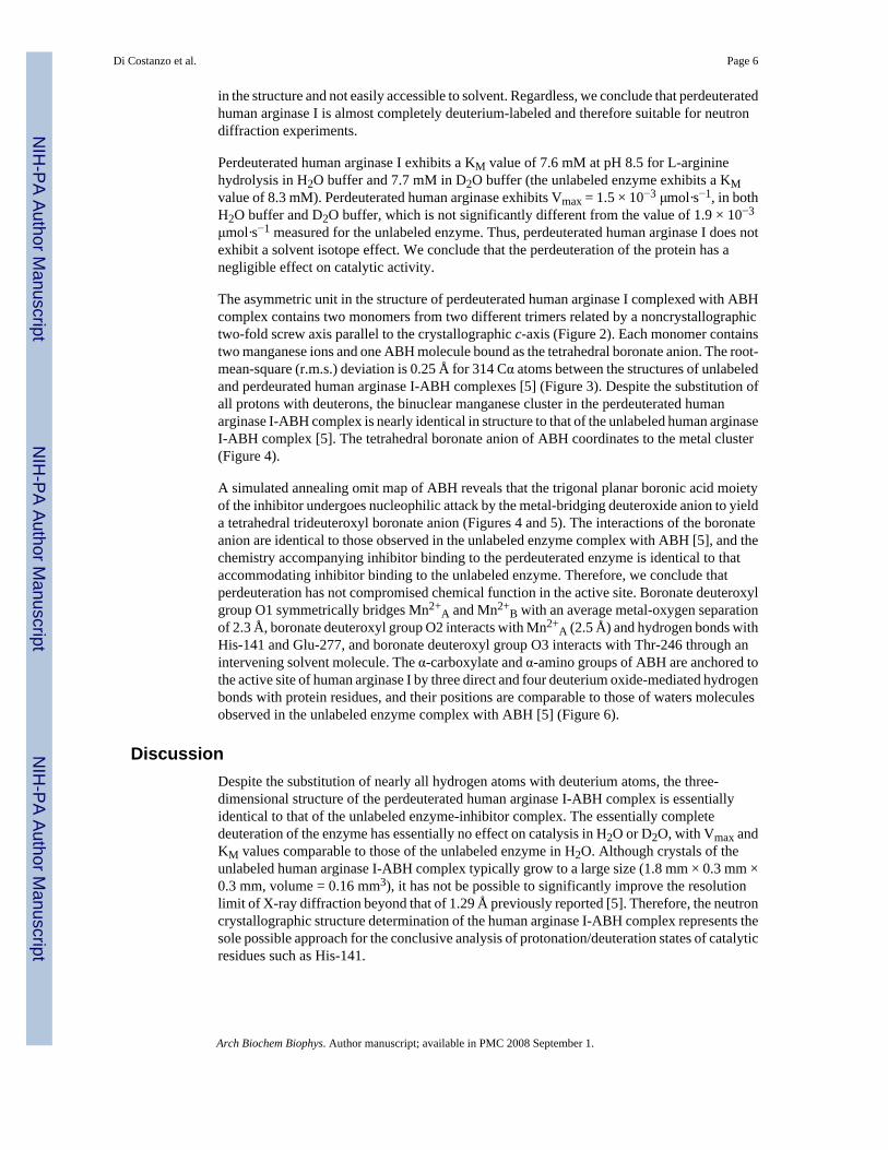
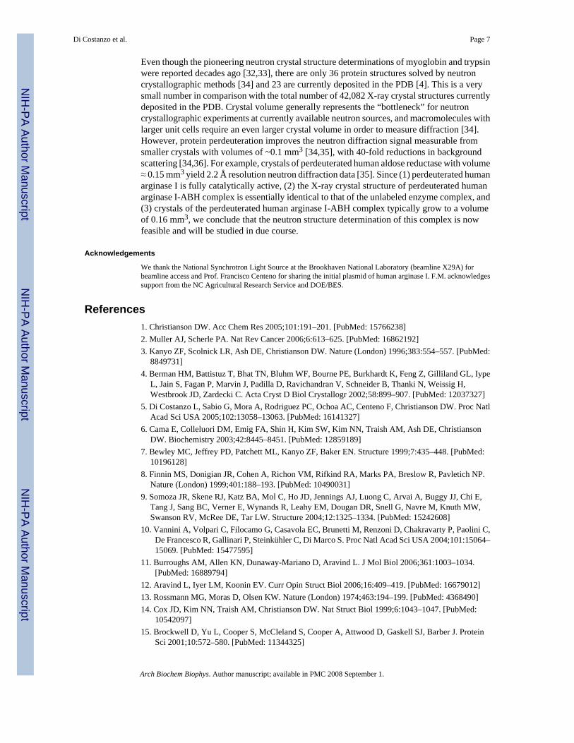
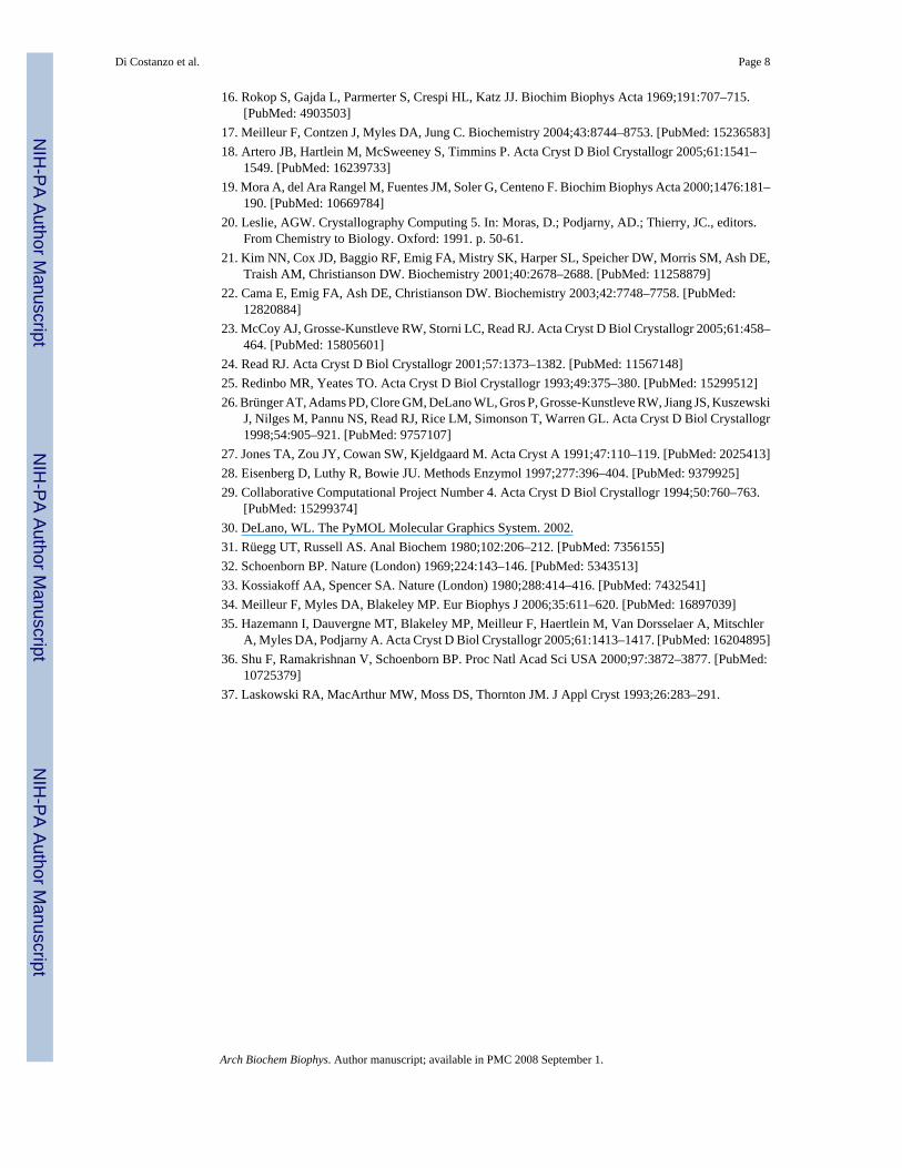
The asymmetric unit in the structure of perdeuterated human arginase I complexed with ABHcomplex contains two monomers from two different trimers related by a noncrystallographictwo-fold screw axis parallel to the crystallographic c-axis (Figure 2). Each monomer containstwo manganese ions and one ABH molecule bound as the tetrahedral boronate anion. The root-mean-square (r.m.s.) deviation is 0.25 Å for 314 Cα atoms between the structures of unlabeledand perdeurated human arginase I-ABH complexes [5] (Figure 3). Despite the substitution ofall protons with deuterons, the binuclear manganese cluster in the perdeuterated humanarginase I-ABH complex is nearly identical in structure to that of the unlabeled human arginaseI-ABH complex [5]. The tetrahedral boronate anion of ABH coordinates to the metal cluster(Figure 4).
A simulated annealing omit map of ABH reveals that the trigonal planar boronic acid moietyof the inhibitor undergoes nucleophilic attack by the metal-bridging deuteroxide anion to yielda tetrahedral trideuteroxyl boronate anion (Figures 4 and 5). The interactions of the boronateanion are identical to those observed in the unlabeled enzyme complex with ABH [5], and thechemistry accompanying inhibitor binding to the perdeuterated enzyme is identical to thataccommodating inhibitor binding to the unlabeled enzyme. Therefore, we conclude thatperdeuteration has not compromised chemical function in the active site. Boronate deuteroxylgroup O1 symmetrically bridges Mn2+
A and Mn2+B with an average metal-oxygen separation
of 2.3 Å, boronate deuteroxyl group O2 interacts with Mn2+A (2.5 Å) and hydrogen bonds with
His-141 and Glu-277, and boronate deuteroxyl group O3 interacts with Thr-246 through anintervening solvent molecule. The α-carboxylate and α-amino groups of ABH are anchored tothe active site of human arginase I by three direct and four deuterium oxide-mediated hydrogenbonds with protein residues, and their positions are comparable to those of waters moleculesobserved in the unlabeled enzyme complex with ABH [5] (Figure 6).
DiscussionDespite the substitution of nearly all hydrogen atoms with deuterium atoms, the three-dimensional structure of the perdeuterated human arginase I-ABH complex is essentiallyidentical to that of the unlabeled enzyme-inhibitor complex. The essentially completedeuteration of the enzyme has essentially no effect on catalysis in H2O or D2O, with Vmax andKM values comparable to those of the unlabeled enzyme in H2O. Although crystals of theunlabeled human arginase I-ABH complex typically grow to a large size (1.8 mm × 0.3 mm ×0.3 mm, volume = 0.16 mm3), it has not be possible to significantly improve the resolutionlimit of X-ray diffraction beyond that of 1.29 Å previously reported [5]. Therefore, the neutroncrystallographic structure determination of the human arginase I-ABH complex represents thesole possible approach for the conclusive analysis of protonation/deuteration states of catalyticresidues such as His-141.
Di Costanzo et al. Page 6
Arch Biochem Biophys. Author manuscript; available in PMC 2008 September 1.
NIH
-PA Author Manuscript
NIH
-PA Author Manuscript
NIH
-PA Author Manuscript
Even though the pioneering neutron crystal structure determinations of myoglobin and trypsinwere reported decades ago [32,33], there are only 36 protein structures solved by neutroncrystallographic methods [34] and 23 are currently deposited in the PDB [4]. This is a verysmall number in comparison with the total number of 42,082 X-ray crystal structures currentlydeposited in the PDB. Crystal volume generally represents the “bottleneck” for neutroncrystallographic experiments at currently available neutron sources, and macromolecules withlarger unit cells require an even larger crystal volume in order to measure diffraction [34].However, protein perdeuteration improves the neutron diffraction signal measurable fromsmaller crystals with volumes of ~0.1 mm3 [34,35], with 40-fold reductions in backgroundscattering [34,36]. For example, crystals of perdeuterated human aldose reductase with volume≈ 0.15 mm3 yield 2.2 Å resolution neutron diffraction data [35]. Since (1) perdeuterated humanarginase I is fully catalytically active, (2) the X-ray crystal structure of perdeuterated humanarginase I-ABH complex is essentially identical to that of the unlabeled enzyme complex, and(3) crystals of the perdeuterated human arginase I-ABH complex typically grow to a volumeof 0.16 mm3, we conclude that the neutron structure determination of this complex is nowfeasible and will be studied in due course.
Acknowledgements
We thank the National Synchrotron Light Source at the Brookhaven National Laboratory (beamline X29A) forbeamline access and Prof. Francisco Centeno for sharing the initial plasmid of human arginase I. F.M. acknowledgessupport from the NC Agricultural Research Service and DOE/BES.
References1. Christianson DW. Acc Chem Res 2005;101:191–201. [PubMed: 15766238]2. Muller AJ, Scherle PA. Nat Rev Cancer 2006;6:613–625. [PubMed: 16862192]3. Kanyo ZF, Scolnick LR, Ash DE, Christianson DW. Nature (London) 1996;383:554–557. [PubMed:
8849731]4. Berman HM, Battistuz T, Bhat TN, Bluhm WF, Bourne PE, Burkhardt K, Feng Z, Gilliland GL, Iype
L, Jain S, Fagan P, Marvin J, Padilla D, Ravichandran V, Schneider B, Thanki N, Weissig H,Westbrook JD, Zardecki C. Acta Cryst D Biol Crystallogr 2002;58:899–907. [PubMed: 12037327]
5. Di Costanzo L, Sabio G, Mora A, Rodriguez PC, Ochoa AC, Centeno F, Christianson DW. Proc NatlAcad Sci USA 2005;102:13058–13063. [PubMed: 16141327]
6. Cama E, Colleluori DM, Emig FA, Shin H, Kim SW, Kim NN, Traish AM, Ash DE, ChristiansonDW. Biochemistry 2003;42:8445–8451. [PubMed: 12859189]
7. Bewley MC, Jeffrey PD, Patchett ML, Kanyo ZF, Baker EN. Structure 1999;7:435–448. [PubMed:10196128]
8. Finnin MS, Donigian JR, Cohen A, Richon VM, Rifkind RA, Marks PA, Breslow R, Pavletich NP.Nature (London) 1999;401:188–193. [PubMed: 10490031]
9. Somoza JR, Skene RJ, Katz BA, Mol C, Ho JD, Jennings AJ, Luong C, Arvai A, Buggy JJ, Chi E,Tang J, Sang BC, Verner E, Wynands R, Leahy EM, Dougan DR, Snell G, Navre M, Knuth MW,Swanson RV, McRee DE, Tar LW. Structure 2004;12:1325–1334. [PubMed: 15242608]
10. Vannini A, Volpari C, Filocamo G, Casavola EC, Brunetti M, Renzoni D, Chakravarty P, Paolini C,De Francesco R, Gallinari P, Steinkühler C, Di Marco S. Proc Natl Acad Sci USA 2004;101:15064–15069. [PubMed: 15477595]
11. Burroughs AM, Allen KN, Dunaway-Mariano D, Aravind L. J Mol Biol 2006;361:1003–1034.[PubMed: 16889794]
12. Aravind L, Iyer LM, Koonin EV. Curr Opin Struct Biol 2006;16:409–419. [PubMed: 16679012]13. Rossmann MG, Moras D, Olsen KW. Nature (London) 1974;463:194–199. [PubMed: 4368490]14. Cox JD, Kim NN, Traish AM, Christianson DW. Nat Struct Biol 1999;6:1043–1047. [PubMed:
10542097]15. Brockwell D, Yu L, Cooper S, McCleland S, Cooper A, Attwood D, Gaskell SJ, Barber J. Protein
Sci 2001;10:572–580. [PubMed: 11344325]
Di Costanzo et al. Page 7
Arch Biochem Biophys. Author manuscript; available in PMC 2008 September 1.
NIH
-PA Author Manuscript
NIH
-PA Author Manuscript
NIH
-PA Author Manuscript
16. Rokop S, Gajda L, Parmerter S, Crespi HL, Katz JJ. Biochim Biophys Acta 1969;191:707–715.[PubMed: 4903503]
17. Meilleur F, Contzen J, Myles DA, Jung C. Biochemistry 2004;43:8744–8753. [PubMed: 15236583]18. Artero JB, Hartlein M, McSweeney S, Timmins P. Acta Cryst D Biol Crystallogr 2005;61:1541–
1549. [PubMed: 16239733]19. Mora A, del Ara Rangel M, Fuentes JM, Soler G, Centeno F. Biochim Biophys Acta 2000;1476:181–
190. [PubMed: 10669784]20. Leslie, AGW. Crystallography Computing 5. In: Moras, D.; Podjarny, AD.; Thierry, JC., editors.
From Chemistry to Biology. Oxford: 1991. p. 50-61.21. Kim NN, Cox JD, Baggio RF, Emig FA, Mistry SK, Harper SL, Speicher DW, Morris SM, Ash DE,
Traish AM, Christianson DW. Biochemistry 2001;40:2678–2688. [PubMed: 11258879]22. Cama E, Emig FA, Ash DE, Christianson DW. Biochemistry 2003;42:7748–7758. [PubMed:
12820884]23. McCoy AJ, Grosse-Kunstleve RW, Storni LC, Read RJ. Acta Cryst D Biol Crystallogr 2005;61:458–
464. [PubMed: 15805601]24. Read RJ. Acta Cryst D Biol Crystallogr 2001;57:1373–1382. [PubMed: 11567148]25. Redinbo MR, Yeates TO. Acta Cryst D Biol Crystallogr 1993;49:375–380. [PubMed: 15299512]26. Brünger AT, Adams PD, Clore GM, DeLano WL, Gros P, Grosse-Kunstleve RW, Jiang JS, Kuszewski
J, Nilges M, Pannu NS, Read RJ, Rice LM, Simonson T, Warren GL. Acta Cryst D Biol Crystallogr1998;54:905–921. [PubMed: 9757107]
27. Jones TA, Zou JY, Cowan SW, Kjeldgaard M. Acta Cryst A 1991;47:110–119. [PubMed: 2025413]28. Eisenberg D, Luthy R, Bowie JU. Methods Enzymol 1997;277:396–404. [PubMed: 9379925]29. Collaborative Computational Project Number 4. Acta Cryst D Biol Crystallogr 1994;50:760–763.
[PubMed: 15299374]30. DeLano, WL. The PyMOL Molecular Graphics System. 2002.31. Rüegg UT, Russell AS. Anal Biochem 1980;102:206–212. [PubMed: 7356155]32. Schoenborn BP. Nature (London) 1969;224:143–146. [PubMed: 5343513]33. Kossiakoff AA, Spencer SA. Nature (London) 1980;288:414–416. [PubMed: 7432541]34. Meilleur F, Myles DA, Blakeley MP. Eur Biophys J 2006;35:611–620. [PubMed: 16897039]35. Hazemann I, Dauvergne MT, Blakeley MP, Meilleur F, Haertlein M, Van Dorsselaer A, Mitschler
A, Myles DA, Podjarny A. Acta Cryst D Biol Crystallogr 2005;61:1413–1417. [PubMed: 16204895]36. Shu F, Ramakrishnan V, Schoenborn BP. Proc Natl Acad Sci USA 2000;97:3872–3877. [PubMed:
10725379]37. Laskowski RA, MacArthur MW, Moss DS, Thornton JM. J Appl Cryst 1993;26:283–291.
Di Costanzo et al. Page 8
Arch Biochem Biophys. Author manuscript; available in PMC 2008 September 1.
NIH
-PA Author Manuscript
NIH
-PA Author Manuscript
NIH
-PA Author Manuscript

Figure 1.MALDI mass spectra of perdeuterated human arginase I prepared in D2O (right) and H2O(left); ionized species with charges 1+ and 2+ are evident in the spectra of samples preparedin H2O.
Di Costanzo et al. Page 9
Arch Biochem Biophys. Author manuscript; available in PMC 2008 September 1.
NIH
-PA Author Manuscript
NIH
-PA Author Manuscript
NIH
-PA Author Manuscript

Figure 2.The asymmetric unit of twin domain A of hemihedrally twinned perdeuterated arginase I-ABHcontains two monomers (red) related by a twofold screw axis parallel to the crystallographicc-axis. Each monomer completes a separate trimer by application of the crystallographicthreefold-axis corresponding to space group P3 (blue monomers). Twin domain B is generatedby the application of the twofold twin operator parallel to the c-axis.
Di Costanzo et al. Page 10
Arch Biochem Biophys. Author manuscript; available in PMC 2008 September 1.
NIH
-PA Author Manuscript
NIH
-PA Author Manuscript
NIH
-PA Author Manuscript

Figure 3.Least-squares superposition of monomer Cα traces of the perdeuterated human arginase I-ABHcomplex (blue) and the unlabeled (i. e., non-deuterated) human arginase I-ABH complex (red)(pdb accession code: 2AEB). The active site Mn2+ ions are represented as spheres and ABHis shown as a color-coded stick figure. The atoms of ABH are color-coded as follows: carbon(yellow), oxygen (red), nitrogen (blue), and boron (green).
Di Costanzo et al. Page 11
Arch Biochem Biophys. Author manuscript; available in PMC 2008 September 1.
NIH
-PA Author Manuscript
NIH
-PA Author Manuscript
NIH
-PA Author Manuscript
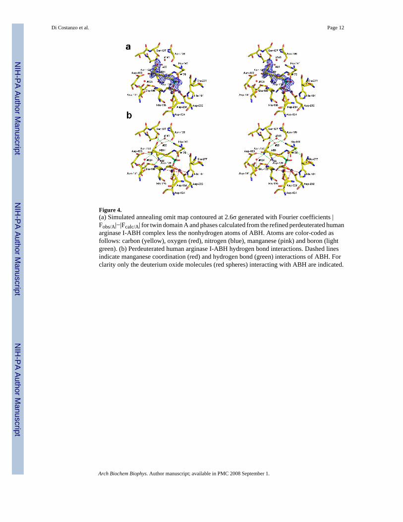
Figure 4.(a) Simulated annealing omit map contoured at 2.6σ generated with Fourier coefficients |Fobs/A|−|Fcalc/A| for twin domain A and phases calculated from the refined perdeuterated humanarginase I-ABH complex less the nonhydrogen atoms of ABH. Atoms are color-coded asfollows: carbon (yellow), oxygen (red), nitrogen (blue), manganese (pink) and boron (lightgreen). (b) Perdeuterated human arginase I-ABH hydrogen bond interactions. Dashed linesindicate manganese coordination (red) and hydrogen bond (green) interactions of ABH. Forclarity only the deuterium oxide molecules (red spheres) interacting with ABH are indicated.
Di Costanzo et al. Page 12
Arch Biochem Biophys. Author manuscript; available in PMC 2008 September 1.
NIH
-PA Author Manuscript
NIH
-PA Author Manuscript
NIH
-PA Author Manuscript

Figure 5.Scheme summarizing intermolecular interactions in the perdeuterated human arginase I-ABHcomplex. Manganese coordination interactions are indicated by green dashed lines, andhydrogen bonds are indicated by black dashed lines. The dideuteroxyl boronic acid moiety ofABH is proposed to undergo nucleophilic attack by the metal-bridging deuteroxide ion to yielda tetrahedral trideuteroxyl boronate anion.
Di Costanzo et al. Page 13
Arch Biochem Biophys. Author manuscript; available in PMC 2008 September 1.
NIH
-PA Author Manuscript
NIH
-PA Author Manuscript
NIH
-PA Author Manuscript
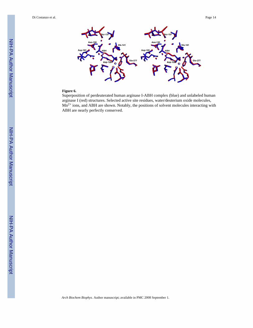
Figure 6.Superposition of perdeuterated human arginase I-ABH complex (blue) and unlabeled humanarginase I (red) structures. Selected active site residues, water/deuterium oxide molecules,Mn2+ ions, and ABH are shown. Notably, the positions of solvent molecules interacting withABH are nearly perfectly conserved.
Di Costanzo et al. Page 14
Arch Biochem Biophys. Author manuscript; available in PMC 2008 September 1.
NIH
-PA Author Manuscript
NIH
-PA Author Manuscript
NIH
-PA Author Manuscript
NIH
-PA Author Manuscript
NIH
-PA Author Manuscript
NIH
-PA Author Manuscript
Di Costanzo et al. Page 15
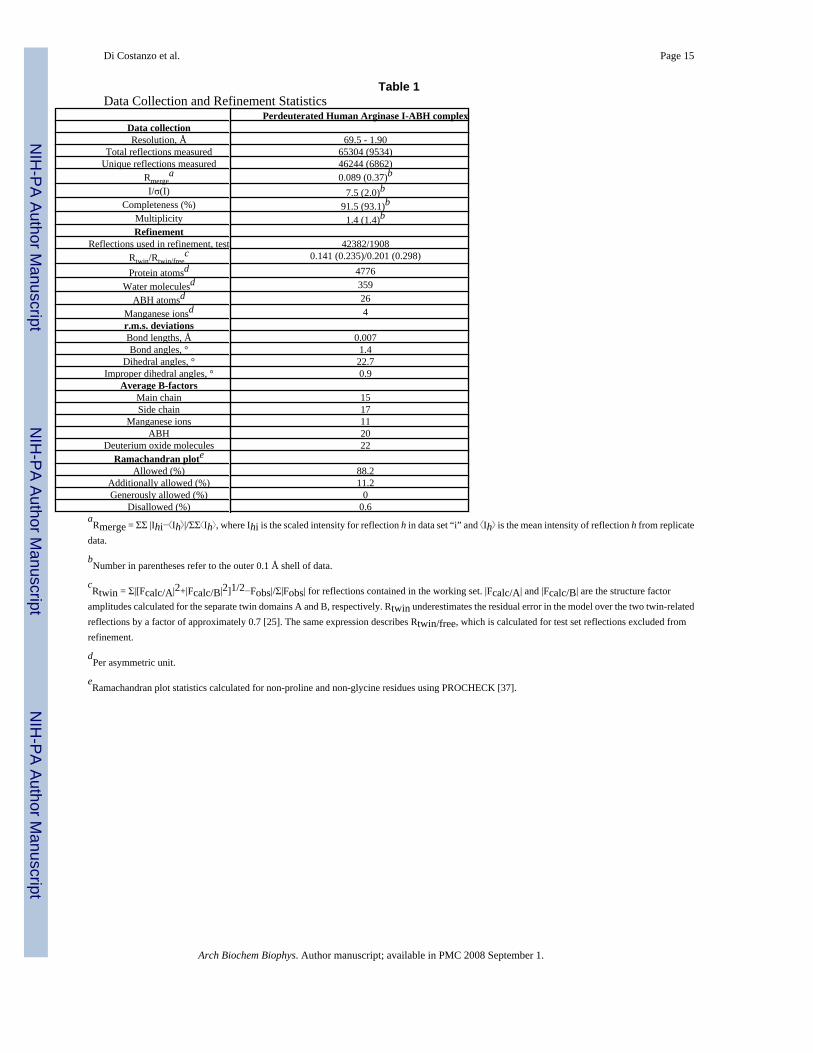
Table 1Data Collection and Refinement Statistics
Perdeuterated Human Arginase I-ABH complexData collectionResolution, Å 69.5 - 1.90
Total reflections measured 65304 (9534)Unique reflections measured 46244 (6862)
Rmergea 0.089 (0.37)b
I/σ(I) 7.5 (2.0)bCompleteness (%) 91.5 (93.1)b
Multiplicity 1.4 (1.4)bRefinement
Reflections used in refinement, test 42382/1908Rtwin/Rtwin/free
c 0.141 (0.235)/0.201 (0.298)
Protein atomsd 4776Water moleculesd 359
ABH atomsd 26Manganese ionsd 4r.m.s. deviationsBond lengths, Å 0.007Bond angles, ° 1.4
Dihedral angles, ° 22.7Improper dihedral angles, ° 0.9
Average B-factorsMain chain 15Side chain 17
Manganese ions 11ABH 20
Deuterium oxide molecules 22Ramachandran plote
Allowed (%) 88.2Additionally allowed (%) 11.2Generously allowed (%) 0
Disallowed (%) 0.6aRmerge = ΣΣ |Ihi− Ih |/ΣΣ Ih , where Ihi is the scaled intensity for reflection h in data set “i” and Ih is the mean intensity of reflection h from replicate
data.
bNumber in parentheses refer to the outer 0.1 Å shell of data.
cRtwin = Σ|[Fcalc/A|2+|Fcalc/B|2]1/2−Fobs|/Σ|Fobs| for reflections contained in the working set. |Fcalc/A| and |Fcalc/B| are the structure factor
amplitudes calculated for the separate twin domains A and B, respectively. Rtwin underestimates the residual error in the model over the two twin-relatedreflections by a factor of approximately 0.7 [25]. The same expression describes Rtwin/free, which is calculated for test set reflections excluded fromrefinement.
dPer asymmetric unit.
eRamachandran plot statistics calculated for non-proline and non-glycine residues using PROCHECK [37].
Arch Biochem Biophys. Author manuscript; available in PMC 2008 September 1.