Extravascular lung water after pneumonectomy followed by ventilator-induced lung injury: A-277
Upload
independentCategory
view
1download
0
BioMed CentralBMC Immunology
ss
Open AcceResearch articleBone marrow cell derived arginase I is the major source of allergen-induced lung arginase but is not required for airway hyperresponsiveness, remodeling and lung inflammatory responses in miceKathryn A Niese1, Ann R Collier1, Amanda R Hajek1, Stephen D Cederbaum2, William E O'Brien3, Marsha Wills-Karp4, Marc E Rothenberg1 and Nives Zimmermann*1Address: 1Division of Allergy and Immunology, Cincinnati Children's Hospital Medical Center and the University of Cincinnati College of Medicine, Cincinnati, Ohio, USA, 2Division of Genetics, Department of Pediatrics, University of California Los Angeles Medical Center, Los Angeles, California, USA, 3Department of Molecular and Human Genetics, Baylor College of Medicine, Houston, Texas, USA and 4Division of Immunobiology, Cincinnati Children's Hospital Medical Center and the University of Cincinnati College of Medicine, Cincinnati, Ohio, USA
Email: Kathryn A Niese - [email protected]; Ann R Collier - [email protected]; Amanda R Hajek - [email protected]; Stephen D Cederbaum - [email protected]; William E O'Brien - [email protected]; Marsha Wills-Karp - [email protected]; Marc E Rothenberg - [email protected]; Nives Zimmermann* - [email protected]
* Corresponding author
AbstractBackground: Arginase is significantly upregulated in the lungs in murine models of asthma, as wellas in human asthma, but its role in allergic airway inflammation has not been fully elucidated in mice.
Results: In order to test the hypothesis that arginase has a role in allergic airway inflammation wegenerated arginase I-deficient bone marrow (BM) chimeric mice. Following transfer of arginase I-deficient BM into irradiated recipient mice, arginase I expression was not required forhematopoietic reconstitution and baseline immunity. Arginase I deficiency in bone marrow-derivedcells decreased allergen-induced lung arginase by 85.8 ± 5.6%. In contrast, arginase II-deficient micehad increased lung arginase activity following allergen challenge to a similar level to wild type mice.BM-derived arginase I was not required for allergen-elicited sensitization, recruitment ofinflammatory cells in the lung, and proliferation of cells. Furthermore, allergen-induced airwayhyperresponsiveness and collagen deposition were similar in arginase-deficient and wild type mice.Additionally, arginase II-deficient mice respond similarly to their control wild type mice withallergen-induced inflammation, airway hyperresponsiveness, proliferation and collagen deposition.
Conclusion: Bone marrow cell derived arginase I is the predominant source of allergen-inducedlung arginase but is not required for allergen-induced inflammation, airway hyperresponsiveness orcollagen deposition.
Published: 1 June 2009
BMC Immunology 2009, 10:33 doi:10.1186/1471-2172-10-33
Received: 30 January 2009Accepted: 1 June 2009
This article is available from: http://www.biomedcentral.com/1471-2172/10/33
© 2009 Niese et al; licensee BioMed Central Ltd. This is an Open Access article distributed under the terms of the Creative Commons Attribution License (http://creativecommons.org/licenses/by/2.0), which permits unrestricted use, distribution, and reproduction in any medium, provided the original work is properly cited.
Page 1 of 11(page number not for citation purposes)

BMC Immunology 2009, 10:33 http://www.biomedcentral.com/1471-2172/10/33
BackgroundAsthma is a serious, chronic inflammatory disorder that isresponsible for one in six pediatric emergency room visits,is the 3rd leading cause of hospitalization among childrenand is one of the leading causes of school absenteeism. Inthe United States, nearly 30% of the population suffersfrom allergies with 5–10% inflicted with asthma. Despiteintense ongoing asthma research, there is currently an epi-demic of this disease in the western world and the inci-dence is on the rise [1,2]. The pathophysiology of asthmais characterized by eosinophil-rich inflammatory cellinfiltrates, increased mucus production, airway hyperreac-tivity, and reversible airway obstruction [3-5]. Experimen-tation in the asthma field has largely focused on analysisof the cellular and molecular events induced by allergenexposure in sensitized animals (primarily mice) andhumans. While these studies have provided the rationalefor the development of multiple therapeutic agents thatinterfere with specific inflammatory pathways [6], thedevelopment of the asthma phenotype is likely to berelated to the complex interplay of a large number of addi-tional genes, and their polymorphic variants.
Accordingly, in an effort to identify new genes involved inthe pathogenesis of asthma, we reported a group of genesthat was induced in the lungs in two phenotypically simi-lar models of experimental asthma triggered by independ-ent regimes [7,8]. Among these asthma signature genes,we found overexpression of genes encoding for enzymesand transporters involved in arginine metabolism, specif-ically arginase I, arginase II and CAT2 [7]. We chose tofocus on these genes because intracellular arginine is a reg-ulator of diverse pathways including production of nitricoxide, polyamines, and proline; these molecules regulatecritical processes associated with asthma including airwaytone, cell hyperplasia and collagen deposition, respec-tively [9,10]. Furthermore, recent studies have shown arole for arginase in several parasitic models [11-16], com-monly associated with Th2/M2 inflammation. Finally,recent studies with arginase inhibitors suggested an effecton outcomes of allergic airway inflammation in mice andguinea pigs [17-19]. However, the results of these studieswere contradictory with one study suggesting a protectiveand the other two a detrimental role for arginase in aller-gen-induced inflammation and airway hyperresponsive-ness. Altogether, we tested the hypothesis that arginaseexpression has a role in allergic airway inflammation bysubjecting arginase I-deficient bone marrow chimericmice and arginase II-deficient mice to allergen challenge-induced airway inflammation. We demonstrate that argi-nase I expression does not affect bone marrow reconstitu-tion following transfer into lethally irradiated recipientsand that arginase is not required for baseline immunity.We also demonstrate that BM-derived arginase I is themain source of allergen-induced lung arginase. However,our studies demonstrate that arginase is not required for
allergen-induced airway inflammation, hyperresponsive-ness or collagen deposition.
MethodsGeneration of arginase I bone marrow (BM) chimerasAll animal studies were approved by the CCHMC IACUCcommittee. Arginase I heterozygous mice [20] were bredand pups were genotyped 7–9 days after birth. Bone mar-row was collected from arginase I -/- pups and hetero-zygous or wild type (majority of experiments) pups(postnatal day 9–12). No difference was observed inexperiments where +/- versus +/+ mice were used as con-trol. In early experiments we transferred 1 × 10⁶ total bonemarrow cells, and in later ones 2 × 10⁵ low density bonemarrow cells were used. No difference in engraftment wasobserved with the two methods. Recipient mice (CD45.1congenic mice) were irradiated [2 doses of 137Cs (700 and475 rads) 3 hours apart] and bone marrow injected i.v.Engraftment was checked by CD45.1 (recipient)/CD45.2(donor) on peripheral blood by flow cytometry (antibod-ies from BD Pharmingen specific for CD45.1 and CD45.2are clones A20 and 104, respectively)) and allergen chal-lenges started 8–14 weeks post-irradiation. In some exper-iment, C57Bl/6 mice were used as recipients and thuschimerism was not checked prior to the allergen chal-lenge. However, in all experiments we verified that argin-ase activity was not induced in the lung of allergen-challenged arginase I BM chimeric mice (see results). Asan additional control, in some experiments we used micethat were not irradiated and bone marrow transferred(non-BMT). Finally, we backcrossed arginase I-hetero-zygous mice for 6 generations on the BALB/c backgroundand verified findings obtained with C57Bl/6 mice (specif-ically, the expression of arginase I in the lung by Northernblot analysis, airway hyperresponsiveness and BALF cellu-larity). Since congenic mice were not used to checkengraftment with BALB/c mice, we verified lack of argin-ase I expression in the lung by Northern blot analysis.
Induction of allergic airway inflammationArginase I BM chimeras and Arginase II-deficient mice[21] were allergen challenged using two models, asdescribed previously [7,8,22]. Briefly, in the first model,mice were sensitized i.p. with ovalbumin (OVA, 100 μg)in alum (1 mg) and challenged intranasally (i.n.) with 50μg OVA or saline. Aspergillus fumigatus antigen-associatedasthma was induced by challenging mice intranasallythree times a week for three weeks, as described [23-25].In brief, mice were lightly-anesthetized with isofluoraneinhalation and 100 μg (50 μl) of Aspergillus fumigatusextract (Bayer Pharmaceuticals, Spokane, WA) or 50 μl ofnormal saline alone was applied to the nasal cavity usinga micropipette with the mouse held in the supine posi-tion. After instillation, mice were held upright until alert.Mice were sacrificed 18–24 hours following the last chal-lenge.
Page 2 of 11(page number not for citation purposes)

BMC Immunology 2009, 10:33 http://www.biomedcentral.com/1471-2172/10/33
Arginase activityArginase activity was measured using the blood urea nitro-gen reagent (Sigma Chemical Company, St. Louis, MO)according to established techniques [26-28].
In situ hybridization of mouse lungIn situ hybridization was performed as described [7]. Inbrief, murine arginase I cDNA in plasmid pCMV-SPORT6(Incyte Genomics, St. Louis, MO) was linearized by EcoRIor Not I digestion, and anti-sense and sense RNA probes,respectively, were generated by T7 and SP6 RNA polymer-ase (Riboprobe Gemini Core System II transcription kit;Promega, Madison, WI). The radiolabeled [αS35-UTP]probes were hybridized and washed under high-strin-gency conditions.
Northern blot analysisRNA was extracted using the Trizol reagent as per the man-ufacturer's instructions. The cDNA probes, generated byPCR or from commercially available vectors [Image Con-sortium obtained from American Tissue Culture Collec-tion, Rockville, MD or Incyte Genomics, Palo Alto, CA],were sequence confirmed, radiolabelled with 32P, andhybridized using standard conditions, as described previ-ously [7].
Measurement of airway hyperreactivityAllergen-induced AHR was determined as described previ-ously [29,30]. Briefly, mice were anesthetized, intubatedand ventilated at a rate of 120 breaths per minute with aconstant tidal volume of air (0.2 ml), and paralyzed withdecamethonium bromide (25 mg/kg). After establishing astable airway pressure, 25 μg/kg weight of acetylcholinewas injected i.v. and dynamic airway pressure (airwaypressure time index [APTI] in cm H2O/sec) was followedfor 5 minutes.
Measurement of collagen accumulationCollagen accumulation was determined as described pre-viously [31]. Briefly, the upper, left lobe of lung washomogenized in 0.5 M acetic acid. Following the additionof pepsin (1 mg/10 mg tissue, Sigma), the lung homoge-nates were vigorously shaken overnight at 4°C. Collagencontent was determined biochemically by quantifyingtotal soluble collagen using the Sircol collagen assay kit(Biocolor Ltd, Newtownabbey, Northern Ireland) accord-ing to the manufacturer's instructions. The data areexpressed as the collagen content normalized per mouseweight. In other experiments, we measured collagen accu-mulation by measuring the content of hydroxyproline, aspreviously described [32].
Antigen-specific antibody measurementPlasma OVA-specific IgG1 was measured after coating thewells with OVA (100 μg/ml). Blocking was done with10% FBS in PBS, and all washes were performed with
0.1% Tween-20 in PBS. Plasma samples were diluted1:1000 and then serially diluted 1:4. After 2 hours of incu-bation, plates were washed and HRP-conjugated anti-mouse IgG1 (1:1,500) (X56; BD PharMingen) was added.The OD was read at 450 nm within 10 minutes.
Ki67 staining and quantificationTissues were fixed in 10% neutral buffered formalin andparaffin embedded. Immunohistochemistry analysis wasperformed on deparaffinized 5 μm sections after blockingof endogenous peroxidase activity, antigen retrieval (in cit-rate buffer (pH6) in microwave oven for 7 min) and block-ing with normal goat serum. The primary antibody [anti-Ki67 1:50 (clone B56, BD PharMingen)] was diluted in0.1% bovine serum albumin in PBS, applied to tissue sec-tions, and incubated overnight at 4°C. Antibody stainingwas detected with biotinylated anti-mouse IgG secondaryantibody and Vectastain ABC, MOM Immunodetection,and DAB Substrate kits (Vector Laboratories, Inc.). Sectionswere counterstained with nuclear fast red. Counts representevaluation of an average number of cells per mm2 deter-mined by an observer blinded to treatment and genotypeusing Metamorph software.
Statistical analysisThe significance of differences between groups were ana-lyzed using two-way (disease and genotype) ANOVAusing Prism software. Values are reported as the mean ±standard deviation. Differences are considered significantif P < 0.05. Pairwise comparisons were performed by Stu-dent's t-test and P values are shown in the figures.
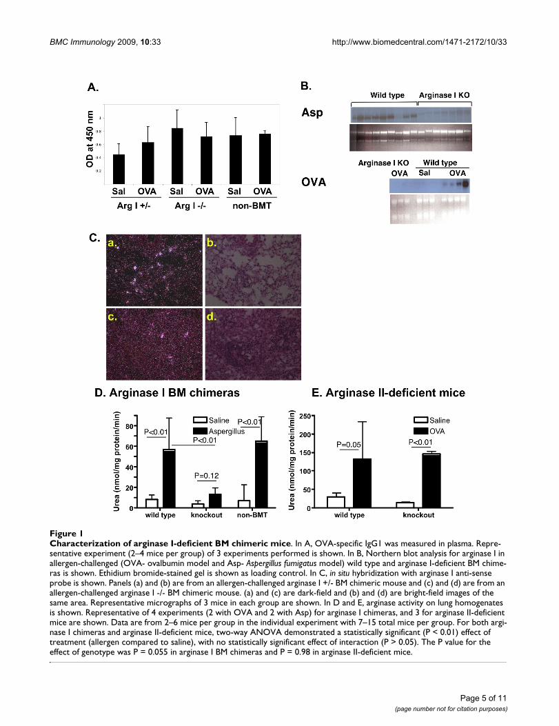
ResultsDevelopment of arginase I-deficient bone marrow chimerasArginase I-deficient mice [20] develop severe hyperammon-emia and die between postnatal days 10 and 14. Since wehave previously demonstrated that arginase I expression inallergic airway inflammation is located in macrophages [7],we developed bone marrow chimeras with arginase I-defi-cient bone marrow derived from arginase I-deficient pups. Inexperiments where congenic CD45.1 mice were used asrecipients, we evaluated donor (CD45.2) chimerism in theperipheral blood at monthly intervals prior to commencingallergen challenges. Importantly, there was no difference inengraftment of arginase I-deficient and wild type bone mar-row (for instance, in a representative experiment there were92.14 ± 1.46 and 91.13 ± 2.64% donor-derived cells, respec-tively; n = 7–8 mice, P = 0.38) demonstrating that arginase Iexpression did not affect bone marrow reconstitution fol-lowing bone marrow transfer.
Bone marrow-derived arginase I is not required for basal immunitySince previous studies have demonstrated that arginasetransgenic mice have defective B cell maturation and
Page 3 of 11(page number not for citation purposes)

BMC Immunology 2009, 10:33 http://www.biomedcentral.com/1471-2172/10/33
impaired development of Payer's patches [33,34], we ana-lyzed chimeric mice for basic hematological and immu-nological parameters. We found that mouse body weight,spleen weight, complete blood counts, engraftment ofindividual cell types (CD4, CD8 and B220-positive cells)were not dependent on arginase expression in BM-derivedcells (data not shown). Peyer's patches were not detecta-ble macroscopically following irradiation irrespective ofarginase expression. In summary, we demonstrate thatbone marrow-derived arginase I is not required for base-line immune cell development.
Bone marrow-derived arginase I is not required for adaptive immunity7–14 weeks post-irradiation, mice were challenged withallergen. We used two models of allergic airway inflam-mation, as we have shown arginase I and II are increasedin both models and are part of the "asthma signaturegenome" [7,8]. Importantly, while these two independentmodels use different antigens and routes of sensitization,the phenotype in mice is similar [7,8]. In the first model,mice are sensitized intraperitoneally (i.p.) with OVA andadjuvant alum and then challenged intranasally with OVAor saline for control. In the second model, mice were sen-sitized and challenged mucosally (by the intranasal route)with Aspergillus fumigatus extract. First, we verified thatsensitization is not affected by arginase I deficiency. Wemeasured OVA-specific IgG1 in OVA-sensitized mice anddetermined that deficiency of arginase I in bone marrow-derived cells did not affect systemic sensitization (Figure1A), which is consistent with our finding that the hemato-logical and immunological parameters are grossly normalin these mice. Similarly, OVA-specific IgE was not affectedby arginase I expression in bone marrow derived cells(data not shown).
Bone marrow arginase I is the main source of allergen-induced lung arginaseWe hypothesized that bone marrow-derived cells arerequired for arginase expression in the lung of allergen-challenged mice. As seen in Figure 1B, Northern blot anal-ysis for arginase I indeed shows decreased expression ofarginase I in arginase I chimeric mice (wild type micetransplanted with arginase I-deficient bone marrow). Sim-ilarly, in situ hybridization of allergen-challenged arginaseI-deficient BM chimeric mice demonstrated a significantdecrease in the number of arginase I-expressing cells com-pared to mice that received arginase I +/- bone marrowcells. (Figure 1C). In order to verify that the arginase I BMchimeras indeed are deficient in lung arginase, we meas-ured arginase activity in allergen-challenged mice. Asshown in Figure 1D, Aspergillus-challenged non-BMTmice and arginase +/+ BM chimeras induce arginase activ-ity in the lung. However, arginase I-deficient BM chimerasdid not show increased arginase activity with allergen
challenge. Similar results were seen in the OVA model(data not shown). We have shown that both arginase Iand arginase II expression are increased in allergen chal-lenged mice [7]. In order to test the hypothesis that argin-ase I is the main contributor to the observed increase inarginase activity in allergen-challenged mice, we meas-ured arginase activity in allergen-challenged arginase II-deficient mice. As seen in Figure 1E, arginase activity wasincreased in the lungs of arginase II-deficient mice to alevel similar to wild type mice. In summary, these datademonstrate that arginase I is primarily induced in bonemarrow-derived cells and that arginase I expression pre-dominantly contributes to allergen-elicited arginase activ-ity in the allergic lung.
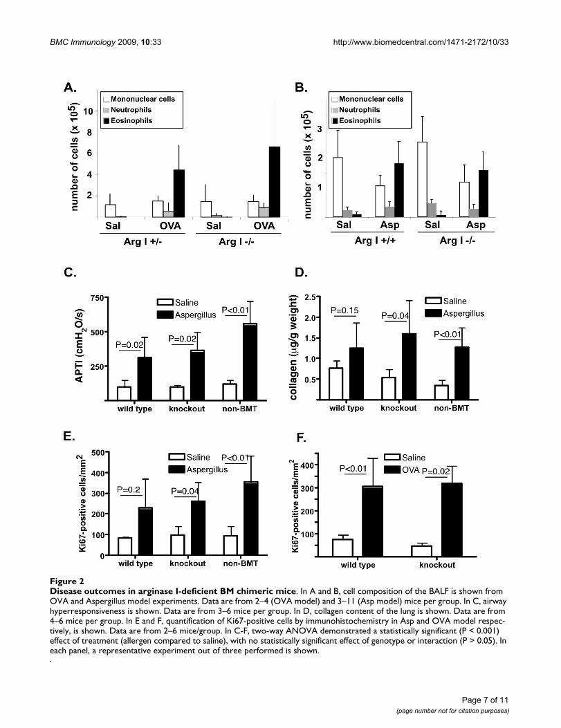
Disease outcomes in allergic airway inflammationWe were first interested if arginase I has a role in inflam-mation and cell recruitment in allergic airway inflamma-tion. Cells in the BALF were differentiated based onmorphology and we demonstrate that arginase I BM chi-meras had similar allergen-elicited recruitment of inflam-matory cells (Figure 2A–B). Similar results were obtainedin arginase I-deficient BM chimeras on the BALB/c back-ground with OVA and Aspergillus-induced allergicinflammation. Furthermore, we assessed the expression ofcytokines and chemokines known to be induced by aller-gen challenge. Northern blot analysis demonstratedinduction of eotaxin-2, TARC, 15-lypoxygenase and smallproline rich protein 2 (SPRR2) by OVA irrespective of argi-nase I expression in the lungs (data not shown). Further-more, the protein level of eotaxin-1, MIG, RANTES, IL-4,IL-5 and leukemia inhibitory factor (LIF) were induced byAspergillus in the lungs of mice irrespective of arginase I(data not shown). These data demonstrate that arginase Iis not required for the development of inflammation inallergic lung inflammation.
Arginase can compete with NOS for substrate, arginine,and can thus lead to decreased production of NO. As NOhas been shown to regulate airway tone, we tested thehypothesis that arginase I has a role in allergen-inducedairway hyperreactivity (AHR). For these studies, we usedonly the Aspergillus model which consistently inducedAHR in allergen-challenged C57Bl/6 mice. As seen in Fig-ure 2C, Aspergillus-challenged non-BMT and arginase I +/+ BM chimeras demonstrate increased responsiveness toacetylcholine, compared to their respective saline-chal-lenged controls. Interestingly, even in the absence of argi-nase I activity in the lung, mice displayed AHR. Similarresults were obtained with arginase I-deficient BM chime-ras on the BALB/c background. These data demonstratethat arginase I is not required for allergen-induced AHR.
Arginase metabolizes arginine to urea and ornithinewhich can further be metabolized by ornithine ami-
Page 4 of 11(page number not for citation purposes)
BMC Immunology 2009, 10:33 http://www.biomedcentral.com/1471-2172/10/33
Page 5 of 11(page number not for citation purposes)
Characterization of arginase I-deficient BM chimeric miceFigure 1Characterization of arginase I-deficient BM chimeric mice. In A, OVA-specific IgG1 was measured in plasma. Repre-sentative experiment (2–4 mice per group) of 3 experiments performed is shown. In B, Northern blot analysis for arginase I in allergen-challenged (OVA- ovalbumin model and Asp- Aspergillus fumigatus model) wild type and arginase I-deficient BM chime-ras is shown. Ethidium bromide-stained gel is shown as loading control. In C, in situ hybridization with arginase I anti-sense probe is shown. Panels (a) and (b) are from an allergen-challenged arginase I +/- BM chimeric mouse and (c) and (d) are from an allergen-challenged arginase I -/- BM chimeric mouse. (a) and (c) are dark-field and (b) and (d) are bright-field images of the same area. Representative micrographs of 3 mice in each group are shown. In D and E, arginase activity on lung homogenates is shown. Representative of 4 experiments (2 with OVA and 2 with Asp) for arginase I chimeras, and 3 for arginase II-deficient mice are shown. Data are from 2–6 mice per group in the individual experiment with 7–15 total mice per group. For both argi-nase I chimeras and arginase II-deficient mice, two-way ANOVA demonstrated a statistically significant (P < 0.01) effect of treatment (allergen compared to saline), with no statistically significant effect of interaction (P > 0.05). The P value for the effect of genotype was P = 0.055 in arginase I BM chimeras and P = 0.98 in arginase II-deficient mice.
BMC Immunology 2009, 10:33 http://www.biomedcentral.com/1471-2172/10/33
notransferase to proline, an amino acid that is often therate-limiting substrate for collagen synthesis [35-37].Thus, we tested the hypothesis that arginase I has a role inallergen-induced collagen deposition. For these studies,we used only the Aspergillus model which demonstratessignificant allergen-induced collagen deposition. As seenin Figure 2D, we observed increased collagen depositionin non-BMT mice, as well as in arginase I-deficient andsufficient BM chimeras. On average, there was a 39.2 ± 5%and 58.8 ± 9% allergen-induced increase in wild type andarginase I-chimeric mice, respectively (P = 0.47, n = 10–16mice per group). These data demonstrate that arginase I isnot required for allergen-induced collagen accumulation.
Ornithine, the metabolite of arginase activity, can serve assubstrate for ornithine decarboxylase, leading to produc-tion of polyamines, which plan an important role in pro-liferation of cells. Thus, we tested the hypothesis thatarginase I has a role in allergen-induced cell proliferation.As seen in Figure 2E–F, we observed increased numbers ofKi67-positive cells in allergen-challenged mice. These lev-els were comparable irrespective of arginase expression.Thus, these data demonstrate that arginase I is notrequired for allergen-induced proliferation.
Arginase II- deficient miceIt remained possible that arginase II is the critical arginaseisoform in outcomes of allergic lung inflammation. Thesubcellular localization and presumed function of the twoisoforms is different with arginase II expressed in themitochondria (while arginase I is cytoplasmic) and pre-dominantly implicated in biosynthetic pathways (whilearginase I is predominantly thought to be involved in theurea cycle). Thus, we allergen challenged arginase II-defi-cient mice [21] and analyzed for outcomes of allergic air-way inflammation. As seen in Figure 3A, arginase II-deficient mice had allergen-induced BALF cell accumula-tion comparable to arginase II-wild type mice. Similarly,the arginase II-deficient mice had levels of allergen-induced airway hyperreactivity similar to wild type mice(Figure 3B). Furthermore, collagen deposition wasinduced in arginase II-deficient mice to levels comparableto those of arginase II-wild type mice (Figure 3C). Finally,proliferation of cells was not affected by the presence ofarginase II (Figure 3D). Importantly, arginase activity inthe lung was induced to similar levels in arginase II-defi-cient and wild type mice (Figure 1E). Thus, these datademonstrate that arginase II is not required for allergen-induced airway inflammation, AHR, collagen depositionand proliferation.
DiscussionThe alternative pathway of macrophage activation, via IL-4, has been recently appreciated in many Th2-associatedresponses, such as parasitic infections and allergic inflam-
mation. However, the role of these cells and their effectorsin the broad spectrum of Th2-associated pathologicalprocesses is poorly understood. We investigated the roleof the enzyme arginase, which is a marker of alternativelyactivated macrophages and highly upregulated in allergicand parasitic infection. While the role of arginase in theurea cycle is well understood, its role in inflammation isless clear. Based on known downstream mediators, argin-ase has been speculated to play a role in NO productionand subsequent inflammation and regulation of airwaytone, as well as proliferation and collagen synthesis viaornithine production. We used arginase I-deficient bonemarrow chimeric mice in models of allergic airwayinflammation. Use of arginase I BM chimeric micerevealed several novel findings. First, we demonstrate thatarginase I expression does not affect bone marrow recon-stitution following transfer into lethally irradiated recipi-ents and that arginase is not required for baselineimmunity. Second, we demonstrate that BM-derived argi-nase I is the main source of allergen-induced lung argin-ase. Third, we found that lung arginase is dispensable forregulation of inflammation, airway tone, fibrosis and cellproliferation during allergic airway inflammation.
Studies using arginase transgenic mice have demonstratedthat overexpression of arginase (specifically in gut epithe-lial cells) leads to interrupted development of B cells andimpaired Peyer's patch architecture [33,34]. Mechanisticanalysis suggested that the hypoargininemia was respon-sible for the phenotype as arginine supplementationreversed the changes. Thus, it was important to assure thatextrahepatic arginase deficiency does not perturb bonemarrow engraftment and baseline immune parameters.We show here that genetic deficiency of arginase in bonemarrow-derived cells does not affect BM engraftment,baseline and adaptive immunity. Thus, we speculate thatexcessive and/or systemic expression of arginase, such asin transgenic mice overexpressing arginase may affect theimmune system via decreasing systemic arginine levels;however, lack of extrahepatic arginase does not appear togrossly affect the immune system.
Previous studies have suggested that macrophages are themain source of arginase in the allergen challenged mouselung. However, other cell types, including respiratory epi-thelial cells have been implicated as potential sources ofarginase [7,38-40]. Furthermore, we and others haveshown that both arginase I and II expression is increasedin the lungs following allergen challenge [7]. Thus, itremained possible that arginase II will contribute to lungarginase activity in allergen challenged arginase I BM chi-meric mice. Our studies measuring arginase activity inboth arginase I BM chimeric and arginase II-deficient micedemonstrated that arginase I, expressed in bone marrowcells, is the main contributor to arginase activity in allergic
Page 6 of 11(page number not for citation purposes)
BMC Immunology 2009, 10:33 http://www.biomedcentral.com/1471-2172/10/33
Page 7 of 11(page number not for citation purposes)
Disease outcomes in arginase I-deficient BM chimeric miceFigure 2Disease outcomes in arginase I-deficient BM chimeric mice. In A and B, cell composition of the BALF is shown from OVA and Aspergillus model experiments. Data are from 2–4 (OVA model) and 3–11 (Asp model) mice per group. In C, airway hyperresponsiveness is shown. Data are from 3–6 mice per group. In D, collagen content of the lung is shown. Data are from 4–6 mice per group. In E and F, quantification of Ki67-positive cells by immunohistochemistry in Asp and OVA model respec-tively, is shown. Data are from 2–6 mice/group. In C-F, two-way ANOVA demonstrated a statistically significant (P < 0.001) effect of treatment (allergen compared to saline), with no statistically significant effect of genotype or interaction (P > 0.05). In each panel, a representative experiment out of three performed is shown.

BMC Immunology 2009, 10:33 http://www.biomedcentral.com/1471-2172/10/33
lung. These data support the notion that bone marrow-derived cells, such as infiltrating inflammatory macro-phages, are the main source of the arginase I enzyme, andarginase activity in the allergic lung.
We originally hypothesized that arginase induction has arole in the pathophysiology of allergic airway inflammation.This was based on several findings. First, we and others haveshown that arginase expression and function is increased inthe lungs of allergen challenged mice and in the lungs andserum of humans with asthma [7,41]. However, these stud-ies did not address the specific role of arginase in pathophys-iology of the disease. Second, downstream pathways ofarginase have been implicated in the regulation of inflam-mation, collagen deposition, proliferation and airway hyper-
responsiveness [35,42,43]; these are all hallmark outcomesin asthma. Specifically, Meurs et al have elegantly demon-strated a role for arginase in contractility of allergen-chal-lenged guinea pig tracheas ex vivo. They have alsodemonstrated that the mechanism for this effect includesarginase-mediated decrease of NO production [44,45].These results, obtained in guinea pigs and in reductionist exvivo approaches, differ from our data that were obtained inthe complex in vivo environment of allergen challengedmice. We speculate that the difference lies either in themodel system (guinea pigs versus mice) or that parallel path-ways are invoked in vivo during allergen challenge, thus mak-ing the role of arginase redundant. Third, mouse models ofparasite infestation, including Schistosome mansoni, Helig-mosomoides polygyrus, Leishmania sp, Toxoplasma gondii
Disease outcomes in arginase II-deficient miceFigure 3Disease outcomes in arginase II-deficient mice. In A, cell composition of the BALF is shown. Data are from 3–4 mice per group. Representative experiment of 5 (2 with Aspergillus and 3 with OVA model) is shown. In B, airway hyperresponsiveness is shown. Data are from 4–6 mice per group. In C, collagen content of the lung is shown. Data are mean ± SD of 4–6 mice per group. In D, quantification of Ki67-positive cells by immunohistochemistry is shown. Data are from 2–5 mice/group. In B-D, two-way ANOVA demonstrated a statistically significant (P < 0.001) effect of treatment (allergen compared to saline), with no statistically significant effect of genotype or interaction (P > 0.05). In B-D, a representative experiment of two performed is shown.
Page 8 of 11(page number not for citation purposes)
BMC Immunology 2009, 10:33 http://www.biomedcentral.com/1471-2172/10/33
and Nipostrongylus brasiliensis [11-16] demonstrated a rolefor arginase expression in host defense. Interestingly, in atleast one of those publications the role of arginase was con-fined to models of infection by intracellular pathogens; how-ever, arginase did not play a role in a systemic disease despiteinduction of arginase in both models [16]. Finally, we haveshown a role for arginase in IL-13 induced airway hyperre-sponsiveness [46] and others have recently suggested a rolefor arginase in mouse and guinea pig asthma models usingarginase inhibitors [17-19]. Specifically, we have shown thatdelivery of arginase I siRNA to mice that receive IL-13 i.t.leads to decreased arginase I expression and airway respon-siveness. It is important to note that this effect was observedat early time points (12–48 hours following single i.t. instal-lation of IL-13). Thus, we speculate that arginase may play arole in airway responsiveness to a single cytokine at earlytime points when collateral pathways have not been able todevelop; in contrast, in allergen challenge multiple pathwaysare activated that may contribute to airway responsivenessthus making arginase redundant. The study by Maarsingh etal [18], demonstrated that inhalation of ABH, an inhibitor ofarginase, protects against allergen-induced airway obstruc-tion, hyperresponsiveness and inflammation. We speculatethat our results differ from the ones in this study eitherbecause of the different species used (mice versus guineapigs) or because of the different approach (inhibitors versusgenetic ablation of arginase). It is possible that the inhibitorhas non-specific off-target effects that may be responsible forthe observed effects. It is important to note that arginaseplays an important role in the urea cycle in the liver; muta-tions in arginase leads to severe metabolic consequences inhumans and mice, including hyperargininemia, hyperam-monemia and premature death [20,47]. In an effort to con-firm our results, we used an inhibitor of arginase, BEC, andfound that it leads to systemic effects, including hyperar-gininemia (data not shown). Since plasma arginine levelscan have profound effects on the immune system [33,34,48-54], we elected not to pursue studies with inhibitors as webelieved we could not distinguish the effects of arginase inhi-bition locally in the lungs from indirect effects from arginaseinhibition in the liver. In contrast to the study by Maarsinghet al where arginase inhibitor decreased inflammation andairway hyperresponsiveness, Ckless et al [17], found thatinhibitor of arginase led to increased inflammation and air-way hyperresponsiveness in mouse models of allergicinflammation. Together, these data caution against use ofarginase inhibitors in allergic airway inflammation until theeffects (specific and off-target) and mechanisms are fully elu-cidated.
In summary, our data suggest divergent role for arginase Iin allergic inflammation compared to parasitic responses.Since arginase is a prominent product of alternatively acti-vated macrophages, which are induced by IL-4 in bothallergic and parasitic responses, our data suggests alterna-
tively activated macrophages evolved to combat parasiticinfections and are either bystanders in allergic inflamma-tion or have developed other effector molecules for aller-gic Th2-associated responses.
ConclusionBone marrow cell derived arginase I is the predominantsource of allergen-induced lung arginase but is notrequired for allergen-induced inflammation, airwayhyperresponsiveness or collagen deposition.
AbbreviationsAHR: airway hyperresponsiveness; BALF: bronchoalveolarlavage fluid; BM: bone marrow; BMT: bone marrow trans-fer; OVA: ovalbumin.
Authors' contributionsKAN, ARC, ARH and NZ performed the research. SDC andWEO provided critical reagents (arginase I and II-deficientmice, respectively). MWK provided the AHR measure-ments. MER participated in the conception and design ofthe study, and helped draft the manuscript. NZ partici-pated in the conception, design and coordination of thestudy, analyzed the data and drafted the manuscript. Allauthors read and approved the final manuscript.
Authors' informationARC is currently at Oak Brook Allergists in Oak Brook, Illi-nois. ARH is currently enrolled in the School of VeterinaryMedicine, University of Illinois at Champaign-Urbana.
AcknowledgementsThe authors wish to thank Laura Koch for technical assistance, Dr. David Williams, Dr. Jose Cancelas-Perez, Victoria Summey and Jeff Bailey from the CCHMC Comprehensive Mouse and Cancer Core facility for expertise and assistance with bone marrow transplantation, Nicole Mason-Richie for assistance with Ki67 staining, Keith F. Stringer and the CCHMC pathology core for assistance with in situ hybridization, and Jennifer Clark for assist-ance with APTI measurement. We thank Drs. DeBroski Herbert and Fred Finkelman for helpful discussions and critical reading of the manuscript.
Funding provided by NIH R01 AI53479-01 (to MER and NZ) and March of Dimes grant 6-FY06-1303 (to NZ).
References1. Holgate ST: The epidemic of allergy and asthma. Nature 1999,
402:B2-4.2. Umetsu DT, McIntire JJ, Akbari O, Macaubas C, DeKruyff RH:
Asthma: an epidemic of dysregulated immunity. Nat Immunol2002, 3:715-720.
3. Busse WW, Lemanske RF Jr: Asthma. N Engl J Med 2001,344:350-362.
4. Hamelmann E, Gelfand EW: IL-5-induced airway eosinophilia –the key to asthma? Immunol Rev 2001, 179:182-191.
5. Lee NA, Gelfand EW, Lee JJ: Pulmonary T cells and eosinophils:coconspirators or independent triggers of allergic respira-tory pathology? J Allergy Clin Immunol 2001, 107:945-957.
6. Barnes PJ: New directions in allergic diseases: mechanism-based anti-inflammatory therapies. J Allergy Clin Immunol 2000,106:5-16.
Page 9 of 11(page number not for citation purposes)
BMC Immunology 2009, 10:33 http://www.biomedcentral.com/1471-2172/10/33
7. Zimmermann N, King NE, Laporte J, Yang M, Mishra A, Pope SM,Muntel EE, Witte DP, Pegg AA, Foster PS, et al.: Dissection ofexperimental asthma with DNA microarray analysis identi-fies arginase in asthma pathogenesis. J Clin Invest 2003,111:1863-1874.
8. Zimmermann N, Mishra A, King NE, Fulkerson PC, Doepker MP,Nikolaidis NM, Kindinger LE, Moulton EA, Aronow BJ, RothenbergME: Transcript Signatures in Experimental Asthma: Identifi-cation of STAT6-Dependent and -Independent Pathways. JImmunol 2004, 172:1815-1824.
9. Morris SM Jr: Regulation of enzymes of the urea cycle andarginine metabolism. Annual Review of Nutrition 2002, 22:87-105.
10. Mills CD: Macrophage arginine metabolism to ornithine/ureaor nitric oxide/citrulline: a life or death issue. Crit Rev Immunol2001, 21:399-425.
11. Anthony RM, Urban JF Jr, Alem F, Hamed HA, Rozo CT, Boucher JL,Van Rooijen N, Gause WC: Memory T(H)2 cells induce alterna-tively activated macrophages to mediate protection againstnematode parasites. Nat Med 2006, 12:955-960.
12. Iniesta V, Gomez-Nieto LC, Corraliza I: The inhibition of arginaseby N(omega)-hydroxy-l-arginine controls the growth ofLeishmania inside macrophages. J Exp Med 2001, 193:777-784.
13. Zhao A, Urban JF Jr, Anthony RM, Sun R, Stiltz J, van Rooijen N, WynnTA, Gause WC, Shea-Donohue T: Th2 cytokine-induced altera-tions in intestinal smooth muscle function depend on alter-natively activated macrophages. Gastroenterology 2008,135:217-225. e211.
14. Hesse M, Modolell M, La Flamme AC, Schito M, Fuentes JM, CheeverAW, Pearce EJ, Wynn TA: Differential regulation of nitric oxidesynthase-2 and arginase-1 by type 1/type 2 cytokines in vivo:granulomatous pathology is shaped by the pattern of L-arginine metabolism. J Immunol 2001, 167:6533-6544.
15. Rutschman R, Lang R, Hesse M, Ihle JN, Wynn TA, Murray PJ: Cut-ting edge: Stat6-dependent substrate depletion regulatesnitric oxide production. J Immunol 2001, 166:2173-2177.
16. El Kasmi KC, Qualls JE, Pesce JT, Smith AM, Thompson RW, Henao-Tamayo M, Basaraba RJ, Konig T, Schleicher U, Koo MS, et al.: Toll-like receptor-induced arginase 1 in macrophages thwartseffective immunity against intracellular pathogens. Nat Immu-nol 2008, 9:1399-1406.
17. Ckless K, Lampert A, Reiss J, Kasahara D, Poynter ME, Irvin CG, Lun-dblad LK, Norton R, Vliet A van der, Janssen-Heininger YM: Inhibi-tion of arginase activity enhances inflammation in mice withallergic airway disease, in association with increases in pro-tein S-nitrosylation and tyrosine nitration. J Immunol 2008,181:4255-4264.
18. Maarsingh H, Zuidhof AB, Bos IS, van Duin M, Boucher JL, Zaagsma J,Meurs H: Arginase inhibition protects against allergen-induced airway obstruction, hyperresponsiveness, andinflammation. Am J Respir Crit Care Med 2008, 178:565-573.
19. Bratt JM, Franzi LM, Linderholm AL, Last MS, Kenyon NJ, Last JA:Arginase enzymes in isolated airways from normal and nitricoxide synthase 2-knockout mice exposed to ovalbumin. Tox-icol Appl Pharmacol 2009, 234:273-80.
20. Iyer RK, Yoo PK, Kern RM, Rozengurt N, Tsoa R, O'Brien WE, Yu H,Grody WW, Cederbaum SD: Mouse model for human arginasedeficiency. Mol Cell Biol 2002, 22:4491-4498.
21. Shi O, Morris SM Jr, Zoghbi H, Porter CW, O'Brien WE: Genera-tion of a mouse model for arginase II deficiency by targeteddisruption of the arginase II gene. Mol Cell Biol 2001, 21:811-813.
22. Zimmermann N, Hogan SP, Mishra A, Brandt EB, Bodette TR, PopeSM, Finkelman FD, Rothenberg ME: Murine eotaxin-2: a constitu-tive eosinophil chemokine induced by allergen challenge andIL-4 overexpression. J Immunol 2000, 165:5839-5846.
23. Mishra A, Weaver TE, Beck DC, Rothenberg ME: Interleukin-5-mediated allergic airway inflammation inhibits the humansurfactant protein C promoter in transgenic mice. J Biol Chem2001, 276:8453-8459.
24. Mehlhop PD, Rijn M van de, Goldberg AB, Brewer JP, Kurup VP, Mar-tin TR, Oettgen HC: Allergen-induced bronchial hyperreactiv-ity and eosinophilic inflammation occur in the absence of IgEin a mouse model of asthma. Proceedings of the National Academyof Sciences of the United States of America 1997, 94:1344-1349.
25. Kurup VP, Seymour BW, Choi H, Coffman RL: ParticulateAspergillus fumigatus antigens elicit a TH2 response in
BALB/c mice. Journal of Allergy & Clinical Immunology 1994,93:1013-1020.
26. Wei LH, Jacobs AT, Morris SM Jr, Ignarro LJ: IL-4 and IL-13 upreg-ulate arginase I expression by cAMP and JAK/STAT6 path-ways in vascular smooth muscle cells. Am J Physiol Cell Physiol2000, 279:C248-256.
27. Wei LH, Wu G, Morris SM Jr, Ignarro LJ: Elevated arginase Iexpression in rat aortic smooth muscle cells increases cellproliferation. Proc Natl Acad Sci USA 2001, 98:9260-9264.
28. Li H, Meininger CJ, Kelly KA, Hawker JR Jr, Morris SM Jr, Wu G:Activities of arginase I and II are limiting for endothelial cellproliferation. Am J Physiol Regul Integr Comp Physiol 2002,282:R64-R69.
29. Kohl J, Baelder R, Lewkowich IP, Pandey MK, Hawlisch H, Wang L,Best J, Herman NS, Sproles AA, Zwirner J, et al.: A regulatory rolefor the C5a anaphylatoxin in type 2 immunity in asthma. JClin Invest 2006, 116:783-796.
30. Wills-Karp M, Luyimbazi J, Xu X, Schofield B, Neben TY, Karp CL,Donaldson DD: Interleukin-13: central mediator of allergicasthma. Science 1998, 282:2258-2261.
31. Fulkerson PC, Fischetti CA, Hassman LM, Nikolaidis NM, RothenbergME: Persistent effects induced by IL-13 in the lung. Am J RespirCell Mol Biol 2006, 35:337-346.
32. Rothenberg ME, Doepker MP, Lewkowich IP, Chiaramonte MG,Stringer KF, Finkelman FD, MacLeod CL, Ellies LG, Zimmermann N:Cationic amino acid transporter 2 regulates inflammatoryhomeostasis in the lung. Proc Natl Acad Sci USA 2006,103:14895-14900.
33. de Jonge WJ, Hallemeesch MM, Kwikkers KL, Ruijter JM, de Gier-deVries C, van Roon MA, Meijer AJ, Marescau B, de Deyn PP, Deutz NE,Lamers WH: Overexpression of arginase I in enterocytes oftransgenic mice elicits a selective arginine deficiency andaffects skin, muscle, and lymphoid development. Am J ClinNutr 2002, 76:128-140.
34. de Jonge WJ, Kwikkers KL, te Velde AA, van Deventer SJ, Nolte MA,Mebius RE, Ruijter JM, Lamers MC, Lamers WH: Arginine defi-ciency affects early B cell maturation and lymphoid organdevelopment in transgenic mice. J Clin Invest 2002,110:1539-1548.
35. Wu G, Morris SM Jr: Arginine metabolism: nitric oxide andbeyond. Biochem J 1998, 336(Pt 1):1-17.
36. Albina JE, Abate JA, Mastrofrancesco B: Role of ornithine as a pro-line precursor in healing wounds. J Surg Res 1993, 55:97-102.
37. Kershenobich D, Fierro FJ, Rojkind M: The relationship betweenthe free pool of proline and collagen content in human livercirrhosis. J Clin Invest 1970, 49:2246-2249.
38. Lindemann D, Racke K: Glucocorticoid inhibition of interleukin-4 (IL-4) and interleukin-13 (IL-13) induced up-regulation ofarginase in rat airway fibroblasts. Naunyn Schmiedebergs ArchPharmacol 2003, 368:546-550.
39. Que LG, Kantrow SP, Jenkinson CP, Piantadosi CA, Huang YC:Induction of arginase isoforms in the lung during hyperoxia.Am J Physiol 1998, 275:L96-102.
40. Takemoto K, Ogino K, Shibamori M, Gondo T, Hitomi Y, TakigawaT, Wang DH, Takaki J, Ichimura H, Fujikura Y, Ishiyama H: Tran-siently, paralleled upregulation of arginase and nitric oxidesynthase and the effect of both enzymes on the pathology ofasthma. Am J Physiol Lung Cell Mol Physiol 2007, 293:L1419-1426.
41. Morris CR, Poljakovic M, Lavrisha L, Machado L, Kuypers FA, MorrisSM Jr: Decreased arginine bioavailability and increased serumarginase activity in asthma. Am J Respir Crit Care Med 2004,170:148-153.
42. Meurs H, Maarsingh H, Zaagsma J: Arginase and asthma: novelinsights into nitric oxide homeostasis and airway hyperre-sponsiveness. Trends Pharmacol Sci 2003, 24:450-455.
43. King NE, Rothenberg ME, Zimmermann N: Arginine in asthmaand lung inflammation. J Nutr 2004, 134:2830S-2836S. discussion2853S.
44. Meurs H, Hamer MA, Pethe S, Vadon-Le Goff S, Boucher JL, ZaagsmaJ: Modulation of cholinergic airway reactivity and nitric oxideproduction by endogenous arginase activity. Br J Pharmacol2000, 130:1793-1798.
45. Meurs H, McKay S, Maarsingh H, Hamer MA, Macic L, Molendijk N,Zaagsma J: Increased arginase activity underlies allergen-induced deficiency of cNOS-derived nitric oxide and airwayhyperresponsiveness. Br J Pharmacol 2002, 136:391-398.
Page 10 of 11(page number not for citation purposes)

BMC Immunology 2009, 10:33 http://www.biomedcentral.com/1471-2172/10/33
Publish with BioMed Central and every scientist can read your work free of charge
"BioMed Central will be the most significant development for disseminating the results of biomedical research in our lifetime."
Sir Paul Nurse, Cancer Research UK
Your research papers will be:
available free of charge to the entire biomedical community
peer reviewed and published immediately upon acceptance
cited in PubMed and archived on PubMed Central
yours — you keep the copyright
Submit your manuscript here:http://www.biomedcentral.com/info/publishing_adv.asp
BioMedcentral
46. Yang M, Rangasamy D, Matthaei KI, Frew AJ, Zimmermann N, Mahal-ingam S, Webb DC, Tremethick DJ, Thompson PJ, Hogan SP, et al.:Inhibition of Arginase I Activity by RNA Interference Atten-uates IL-13-Induced Airways Hyperresponsiveness. J Immunol2006, 177:5595-5603.
47. Iyer R, Jenkinson CP, Vockley JG, Kern RM, Grody WW, CederbaumS: The human arginases and arginase deficiency. J Inherit MetabDis 1998, 21(Suppl 1):86-100.
48. Luiking YC, Poeze M, Ramsay G, Deutz NE: The role of arginine ininfection and sepsis. JPEN J Parenter Enteral Nutr 2005, 29:S70-74.
49. Popovic PJ, Zeh HJ 3rd, Ochoa JB: Arginine and immunity. J Nutr2007, 137:1681S-1686S.
50. Rodriguez PC, Zea AH, Culotta KS, Zabaleta J, Ochoa JB, Ochoa AC:Regulation of T cell receptor CD3zeta chain expression by L-arginine. J Biol Chem 2002, 277:21123-21129.
51. Rodriguez PC, Zea AH, DeSalvo J, Culotta KS, Zabaleta J, QuicenoDG, Ochoa JB, Ochoa AC: L-arginine consumption by macro-phages modulates the expression of CD3zeta chain in T lym-phocytes. J Immunol 2003, 171:1232-1239.
52. Kobayashi T, Yamamoto M, Hiroi T, McGhee J, Takeshita Y, KiyonoH: Arginine enhances induction of T helper 1 and T helper 2cytokine synthesis by Peyer's patch alpha beta T cells andantigen-specific mucosal immune response. Biosci BiotechnolBiochem 1998, 62:2334-2340.
53. Takano H, Lim HB, Miyabara Y, Ichinose T, Yoshikawa T, Sagai M:Oral administration of L-arginine potentiates allergen-induced airway inflammation and expression of interleukin-5 in mice. J Pharmacol Exp Ther 1998, 286:767-771.
54. Sapienza MA, Kharitonov SA, Horvath I, Chung KF, Barnes PJ: Effectof inhaled L-arginine on exhaled nitric oxide in normal andasthmatic subjects. Thorax 1998, 53:172-175.
Page 11 of 11(page number not for citation purposes)
Copyright © 2022 FDOKUMEN