Modulating role of estradiol on arginase II expression in hyperlipidemic rabbits as an...
6
Modulating role of estradiol on arginase II expression in hyperlipidemic rabbits as an atheroprotective mechanism Toshio Hayashi* † , Teiji Esaki* ‡ , Daigo Sumi § , Tapan Mukherjee § , Akihisa Iguchi*, and Gautam Chaudhuri § *Department of Geriatrics, Nagoya University Graduate School of Medicine, 65 Tsuruma-cho, Showa-ku, Nagoya 466-8550, Japan; ‡ Department of Geriatrics, Kainan Hospital Aichi Prefectural Welfare Federation of Agricultural Cooperatives, 396 Yatomi-town, Ama-gun, Aichi Prefecture 498-8502, Japan; and § Department of Obstetrics and Gynecology and Department of Molecular and Medical Pharmacology, David Geffen School of Medicine, 650 Charles E. Young Drive South, University of California, Los Angeles, CA 90095-1735 Communicated by C. Kumar N. Patel, Pranalytica, Inc., Santa Monica, CA, May 18, 2006 (received for review December 1, 2005) We evaluated the effects of a 0.5% cholesterol-enriched diet (HCD) on nitric-oxide synthase (NOS) and arginase expression and the modulating role of 17-estradiol (E 2 ) on this phenomenon. Thirty oopherectomized rabbits were divided into three groups and treated for 15 weeks. Group I received normal chow; group II, HCD; and group III, HCD plus E2 pellets. Animals in group II showed an increase in plasma lipids, and they demonstrated atheromatous lesions as well as expression of arginase I and II accompanied by a significant number of BrdU-positive cells in endothelial cells and intimal muscle cells, suggestive of an increase in cellular prolifer- ation. There was significant expression of inducible NOS and increased staining of nitrotyrosine-positive areas. These were not observed in group I animals. In both groups, E2 levels were low. In group III animals, E 2 supplementation led to a decrease in ather- omatous lesions and BrdU-positive cells and reduced expression of both inducible NOS and arginase I and II accompanied by a decrease in nitrotyrosine staining. E 2 levels were increased. Our results suggest that E 2 was responsible for these effects, despite the animals being hyperlipidemic, similar to those in group II. Because arginase is responsible for cell proliferation by converting L-argi- nine to polyamines, our results indicate that expression of arginase may play an important role in cellular proliferation in atheroscle- rosis, and inhibition of arginase expression by E 2 may be another potential mechanism in attenuating atherogenesis. arteriosclerosis L-arginine nitric oxide endothelium nitric-oxide synthase E strogens retard the development of atherosclerosis by attenu- ating the adhesion of circulating monocytes to endothelial cells and their subsequent migration to the subendothelial layer (1). Decreased expression of vascular cell adhesion molecule 1 (VCAM-1) and monocyte chemoattractant protein 1 (MCP-1) by estrogens may at least in part account for this effect (1–3). We (4, 5) and others (6) have demonstrated that estrogens increase nitric oxide (NO) production by endothelial cells, and it is now known that NO, either on its own (7) or produced after stimulation by estrogens (8), can attenuate the cytokine-induced expression of VCAM-1 (1) as well as MCP-1 (9, 10). Furthermore, the inhibition of NO production in animals administered an inhibitor of nitric- oxide synthase (NOS) results in potentiation of atherosclerotic lesions in high-cholesterol diet (HCD)-induced atherosclerosis (11) and also causes atherosclerotic coronary lesions, especially at microvascular levels in experimental animals (12). L-Arginine is a substrate for NOS, which catalyzes formation of N -hydroxy-L- arginine as an intermediate that subsequently forms NO (13). L-Arginine can be a substrate for NOS, which catalyzes its breakdown to release NO in endothelial cells. L-Arginine is also converted by arginase to ornithine, the only source of synthesis in mammalian cells of the polyamines putrescine, spermidine, and spermine, which are essential for cell prolif- eration and regulation of the cell cycle (14, 15) and, which are, therefore, proatherosclerotic. The exact mechanism by which polyamines increase cell proliferation is not known. In verte- brates there are two isoforms of arginase, both of which catalyze the conversion of arginine to ornithine and urea. They differ with regard to subcellular localization, tissue distribu- tion, and certain enzymatic properties, ref lecting the fact that different genes encode them (14, 15). Arginase I is expressed almost exclusively in the cytosol of liver cells, whereas arginase II is located within the mitochondrial matrix and is expressed at low levels in many tissues (15). Furthermore, citrulline, the end product of the NOS-mediated reaction, is converted to L-arginine by arginosuccinate synthetase and arginosuccinate lyase (16). The present work was, therefore, undertaken to assess whether arginase expression is increased in atheroscle- rotic lesions and to determine the modulating role of estrogen, if any, on this phenomenon. Results Blood Chemistry. All of the rabbits appeared to be healthy through- out the study. No significant differences in serum high-density lipoprotein (HDL)-cholesterol, total serum protein, or body weight existed among the three groups over the course of the study. In animals fed normal chow (group I), there was no difference in total cholesterol levels compared with basal values. The addition of 0.5% cholesterol to the diet (groups II and III) increased the total cholesterol levels significantly compared with the baseline value (Table 1).The treatment with E 2 did not significantly affect the plasma lipid levels in this study; however, it increased the plasma E 2 concentration up to physiological levels similar to that observed in ovary-intact rabbits (Table 1), as reported in ref. 2. Histological Examination of Atherosclerosis. In animals fed normal chow (group I), no atheromatous lesions were observed. On the other hand, histological examination of the thoracic aortas of animals fed a HCD and who received a placebo pellet (group II) revealed more atheromatous lesions, as indicated by the mean percentage of luminal encroachment and the mean lesion area in the hypercholesterolemic than in the animals fed a HCD but who received E 2 pellets (group III). The area of atherosclerosis in the thoracic aorta was reduced by 70% in the E 2 -treated group (group III) compared with the HCD group receiving placebo pellets (group II) (Fig. 1 Left and Center). The intima:media (I:M) ratios also decreased after E 2 treatment (group III vs. group II) (Fig. 1 Right). Conflict of interest statement: No conflicts declared. Abbreviations: E2, 17-estradiol; EDNO, endothelium-derived nitric oxide; eNOS, endothe- lial nitric-oxide synthase; HCD, high-cholesterol diet; I:M ratio, intima:media ratio; iNOS, inducible nitric-oxide synthase; MCP-1, monocyte chemoattractant protein 1; NOS, nitric- oxide synthase; NZW, New Zealand White; VCAM-1, vascular cell adhesion molecule 1. † To whom correspondence should be addressed. E-mail: [email protected]. © 2006 by The National Academy of Sciences of the USA www.pnas.orgcgidoi10.1073pnas.0603918103 PNAS July 5, 2006 vol. 103 no. 27 10485–10490 PHARMACOLOGY
-
Upload
mmumullana -
Category
Documents
-
view
0 -
download
0
Transcript of Modulating role of estradiol on arginase II expression in hyperlipidemic rabbits as an...
Modulating role of estradiol on arginase IIexpression in hyperlipidemic rabbitsas an atheroprotective mechanismToshio Hayashi*†, Teiji Esaki*‡, Daigo Sumi§, Tapan Mukherjee§, Akihisa Iguchi*, and Gautam Chaudhuri§
*Department of Geriatrics, Nagoya University Graduate School of Medicine, 65 Tsuruma-cho, Showa-ku, Nagoya 466-8550, Japan; ‡Department of Geriatrics,Kainan Hospital Aichi Prefectural Welfare Federation of Agricultural Cooperatives, 396 Yatomi-town, Ama-gun, Aichi Prefecture 498-8502, Japan; and§Department of Obstetrics and Gynecology and Department of Molecular and Medical Pharmacology, David Geffen School of Medicine, 650 Charles E.Young Drive South, University of California, Los Angeles, CA 90095-1735
Communicated by C. Kumar N. Patel, Pranalytica, Inc., Santa Monica, CA, May 18, 2006 (received for review December 1, 2005)
We evaluated the effects of a 0.5% cholesterol-enriched diet (HCD)on nitric-oxide synthase (NOS) and arginase expression and themodulating role of 17�-estradiol (E2) on this phenomenon. Thirtyoopherectomized rabbits were divided into three groups andtreated for 15 weeks. Group I received normal chow; group II, HCD;and group III, HCD plus E2 pellets. Animals in group II showed anincrease in plasma lipids, and they demonstrated atheromatouslesions as well as expression of arginase I and II accompanied by asignificant number of BrdU-positive cells in endothelial cells andintimal muscle cells, suggestive of an increase in cellular prolifer-ation. There was significant expression of inducible NOS andincreased staining of nitrotyrosine-positive areas. These were notobserved in group I animals. In both groups, E2 levels were low. Ingroup III animals, E2 supplementation led to a decrease in ather-omatous lesions and BrdU-positive cells and reduced expression ofboth inducible NOS and arginase I and II accompanied by a decreasein nitrotyrosine staining. E2 levels were increased. Our resultssuggest that E2 was responsible for these effects, despite theanimals being hyperlipidemic, similar to those in group II. Becausearginase is responsible for cell proliferation by converting L-argi-nine to polyamines, our results indicate that expression of arginasemay play an important role in cellular proliferation in atheroscle-rosis, and inhibition of arginase expression by E2 may be anotherpotential mechanism in attenuating atherogenesis.
arteriosclerosis � L-arginine � nitric oxide � endothelium � nitric-oxidesynthase
Estrogens retard the development of atherosclerosis by attenu-ating the adhesion of circulating monocytes to endothelial cells
and their subsequent migration to the subendothelial layer (1).Decreased expression of vascular cell adhesion molecule 1(VCAM-1) and monocyte chemoattractant protein 1 (MCP-1) byestrogens may at least in part account for this effect (1–3). We (4,5) and others (6) have demonstrated that estrogens increase nitricoxide (NO) production by endothelial cells, and it is now knownthat NO, either on its own (7) or produced after stimulation byestrogens (8), can attenuate the cytokine-induced expression ofVCAM-1 (1) as well as MCP-1 (9, 10). Furthermore, the inhibitionof NO production in animals administered an inhibitor of nitric-oxide synthase (NOS) results in potentiation of atheroscleroticlesions in high-cholesterol diet (HCD)-induced atherosclerosis (11)and also causes atherosclerotic coronary lesions, especially atmicrovascular levels in experimental animals (12). L-Arginine is asubstrate for NOS, which catalyzes formation of N�-hydroxy-L-arginine as an intermediate that subsequently forms NO (13).
L-Arginine can be a substrate for NOS, which catalyzes itsbreakdown to release NO in endothelial cells. L-Arginine isalso converted by arginase to ornithine, the only source ofsynthesis in mammalian cells of the polyamines putrescine,spermidine, and spermine, which are essential for cell prolif-eration and regulation of the cell cycle (14, 15) and, which are,
therefore, proatherosclerotic. The exact mechanism by whichpolyamines increase cell proliferation is not known. In verte-brates there are two isoforms of arginase, both of whichcatalyze the conversion of arginine to ornithine and urea. Theydiffer with regard to subcellular localization, tissue distribu-tion, and certain enzymatic properties, ref lecting the fact thatdifferent genes encode them (14, 15). Arginase I is expressedalmost exclusively in the cytosol of liver cells, whereas arginaseII is located within the mitochondrial matrix and is expressedat low levels in many tissues (15). Furthermore, citrulline, theend product of the NOS-mediated reaction, is converted toL-arginine by arginosuccinate synthetase and arginosuccinatelyase (16). The present work was, therefore, undertaken toassess whether arginase expression is increased in atheroscle-rotic lesions and to determine the modulating role of estrogen,if any, on this phenomenon.
ResultsBlood Chemistry. All of the rabbits appeared to be healthy through-out the study. No significant differences in serum high-densitylipoprotein (HDL)-cholesterol, total serum protein, or body weightexisted among the three groups over the course of the study. Inanimals fed normal chow (group I), there was no difference in totalcholesterol levels compared with basal values. The addition of0.5% cholesterol to the diet (groups II and III) increased the totalcholesterol levels significantly compared with the baseline value(Table 1).The treatment with E2 did not significantly affect theplasma lipid levels in this study; however, it increased the plasma E2concentration up to physiological levels similar to that observed inovary-intact rabbits (Table 1), as reported in ref. 2.
Histological Examination of Atherosclerosis. In animals fed normalchow (group I), no atheromatous lesions were observed. On theother hand, histological examination of the thoracic aortas ofanimals fed a HCD and who received a placebo pellet (group II)revealed more atheromatous lesions, as indicated by the meanpercentage of luminal encroachment and the mean lesion area inthe hypercholesterolemic than in the animals fed a HCD but whoreceived E2 pellets (group III). The area of atherosclerosis in thethoracic aorta was reduced by 70% in the E2-treated group (groupIII) compared with the HCD group receiving placebo pellets(group II) (Fig. 1 Left and Center). The intima:media (I:M) ratiosalso decreased after E2 treatment (group III vs. group II) (Fig. 1Right).
Conflict of interest statement: No conflicts declared.
Abbreviations: E2, 17�-estradiol; EDNO, endothelium-derived nitric oxide; eNOS, endothe-lial nitric-oxide synthase; HCD, high-cholesterol diet; I:M ratio, intima:media ratio; iNOS,inducible nitric-oxide synthase; MCP-1, monocyte chemoattractant protein 1; NOS, nitric-oxide synthase; NZW, New Zealand White; VCAM-1, vascular cell adhesion molecule 1.
†To whom correspondence should be addressed. E-mail: [email protected].
© 2006 by The National Academy of Sciences of the USA
www.pnas.org�cgi�doi�10.1073�pnas.0603918103 PNAS � July 5, 2006 � vol. 103 � no. 27 � 10485–10490
PHA
RMA
COLO
GY

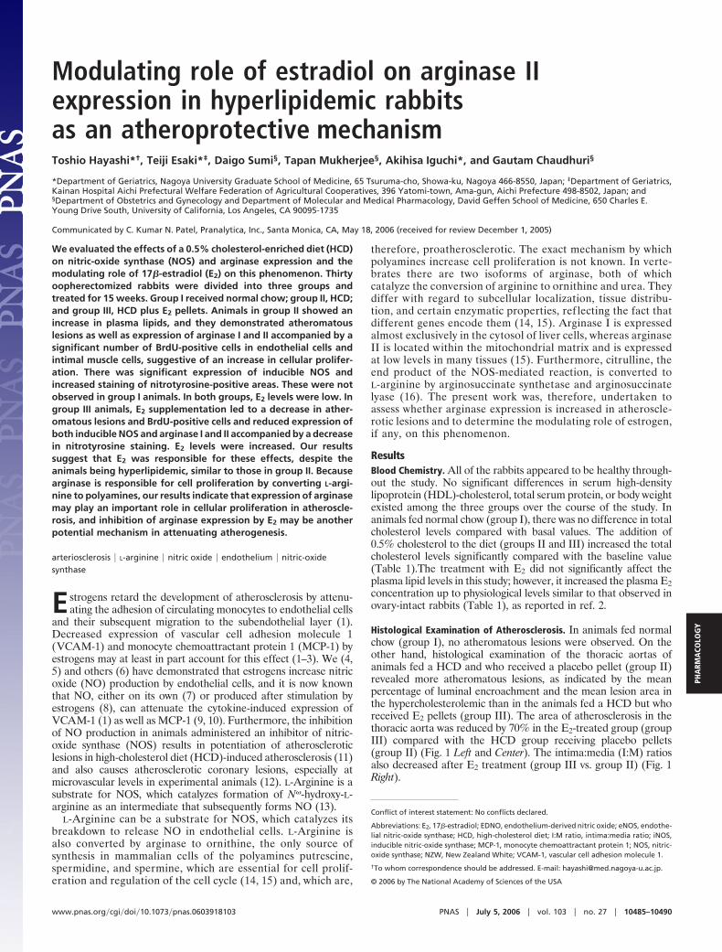
BrdU-Positive Neointima Lesion. BrdU-positive cells, mainly com-posed of endothelial cells and intimal smooth muscle cells, were sig-nificantly decreased in group III compared with group II (Fig. 2).
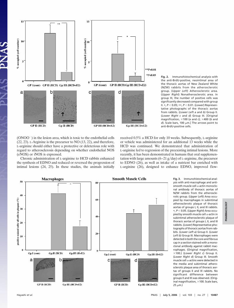
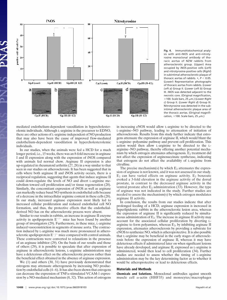
Immunocytochemical Analysis. Smooth muscle cell �-actin, monocytes�macrophages, inducible NOS (iNOS), and nitrotyrosine (one of the reactionproducts of ONOO�). The atheroma in the aorta was composed ofmany macrophages derived from foam cells and intimal smoothmuscle cell proliferation (Fig. 3). A significant reduction in theatherosclerotic area, as well as a decrease in the relative number ofmacrophages, was observed in animals in the E2-treated group inthis study (Fig. 3 Left). The number of smooth muscle cell �-actin-positive cells was not changed between groups II and III (Fig. 3Right). The iNOS and nitrotyrosine-positive areas were decreasedin the E2-treated group (group III) compared with the placebogroup (group II) (Fig. 4). We observed iNOS expression in the Tcells and macrophages in the advanced atherosclerotic plaque of thethoracic aortas of group II, consistent with previous data (17).Arginase I, arginase II, and arginosuccinate synthetase. The atheroma inthe aorta expressed a large amount of arginase I and II, as well as
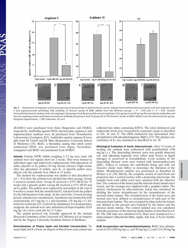
arginosuccinate synthase in animals in group II; however, theexpression of arginase I and II, but not arginosuccinate synthase,decreased in the aortas from group III (Fig. 5).
DiscussionEndothelial dysfunction leading to expression of adhesion mole-cules plays an integral part in the initiation of atherosclerosis.Expression of adhesion molecules on the endothelial surface leadsto adhesion of monocytes to endothelial cells, which is one of theearly steps in the development of atherosclerosis (18). Endothelialdysfunction is also characterized by impaired endothelium-derivednitric oxide (EDNO) production and impaired endothelium-dependent vasodilation (19). Only low concentrations of NO arenormally produced by the endothelial cells physiologically, whichprotect the endothelial cell and, therefore, play an important rolein attenuating the onset of atherosclerosis (20, 21). On the otherhand, once atherosclerosis develops, activated macrophages arepresent in the lesion area, and these macrophages express iNOS(17). This process leads to relatively high concentrations of NO aswell as superoxide (O2
�), leading to formation of peroxynitrite
Table 1. Plasma lipid (total cholesterol, triglycerides, and HDL-cholesterol), total protein,and E2 concentration in rabbits fed a standard diet (group I), HCD (group II), and HCDplus E2 (group III)
GroupTotal cholesterol,
mg/dlTriglycerides,
mg/dlHDL-cholesterol,
mg/dlTotal protein,
g/dl E2, pg/ml
I 80.2 � 8.2 36.9 � 5.1 30.4 � 4.1 8.1 � 1.0 15.8 � 4.1II 1,451.5 � 120.5* 82.4 � 15.6* 30.9 � 5.2 7.8 � 1.1 12.9 � 2.5III 1,298.8 � 140.9* 74.5 � 11.5* 35.2 � 8.2 8.2 � 1.1 40.0 � 2†
Rabbits were treated with each condition for 15 weeks. Results are the mean � SEM of 10 rabbits. P wasmeasured by an unpaired Student t test. *, P � 0.05 versus group I (control); †, P � 0.05 versus group I (control)and group II (HCD).
Fig. 1. Histological evaluation of the atherosclerotic area of thoracic aortas as indicated by the surface involvement, mean lesion area (percent of area occupied bylesion), and I:M ratio (Upper) and representative photographs (Lower). (Upper Left) Surface involvement of the atherosclerotic area of thoracic aortas from rabbits(group I, normal chow; group II, 0.5% HCD and placebo pellet; group III, 0.5% HCD and E2 pellet). ***, P � 0.001. (Upper Center) Area occupied by the atheroscleroticarea of thoracic aortas from the three groups of rabbits. ***, P � 0.001. (Upper Right) I:M ratio of thoracic aortas from three groups of rabbits. ***, P � 0.001. (Lower)Representative photographs of thoracic aortas from rabbits. (Lower Left) Group I. (Lower Center) Group II. (Lower Right) Group III. (Original magnification, �40.)
10486 � www.pnas.org�cgi�doi�10.1073�pnas.0603918103 Hayashi et al.
(ONOO�) in the lesion area, which is toxic to the endothelial cells(22, 23). L-Arginine is the precursor to NO (13, 22), and therefore,L-arginine should either have a protective or deleterious role withregard to atherosclerosis depending on whether endothelial NOS(eNOS) or iNOS is expressed.
Chronic administration of L-arginine to HCD rabbits enhancedthe synthesis of EDNO and reduced or reversed the progression ofintimal lesions (24, 25). In these studies, the animals initially
received 0.5% a HCD for only 10 weeks. Subsequently, L-arginineor vehicle was administered for an additional 13 weeks while theHCD was continued. We demonstrated that administration ofL-arginine led to regression of the preexisting intimal lesions. Morerecently, it has been demonstrated in humans that oral supplemen-tation with large amounts (6–21 g�day) of L-arginine, the precursorto EDNO (26), as well as intake of a nutrient bar enriched withL-arginine (26), designed to enhance EDNO, improved flow-
Fig. 2. Immunohistochemical analysis withthe anti-BrdU-positive, neointimal area ofthe thoracic aortas of New Zealand White(NZW) rabbits from the atheroscleroticgroup. (Upper Left) Atherosclerotic area.(Upper Right) Nonatherosclerotic area. Ingroup III, the number of positive cells wassignificantly decreased compared with groupII. *, P � 0.05; **, P � 0.01. (Lower) Represen-tative photographs of the thoracic aortasfrom rabbits. (Lower Left a and b) Group II.(Lower Right c and d) Group III. [Originalmagnification, �100 (a and c); �400 (b andd). Scale bars, 100 �m.] The arrows point toanti-BrdU-positive cells.
Fig. 3. Immunohistochemical anal-ysis with anti-macrophage and anti-smooth muscle cell �-actin monoclo-nal antibody of thoracic aortas ofNZW rabbits from the atheroscle-rotic group. (Upper Left) Area occu-pied by macrophages in subintimalatherosclerotic plaque of thoracicaortas of groups I, II, and III rabbits.
*, P � 0.05. (Upper Right) Area occu-pied by smooth muscle cell �-actin insubintimal atherosclerotic plaque ofthoracic aortas of groups I, II, and IIIrabbits. (Lower) Representative pho-tographs of thoracic aortas from rab-bits. (Lower Left a) Group II. (LowerLeft b) Group III. Macrophages weredetected inboththecoreandfibrouscap in a section stained with a mono-clonal antibody against rabbit mac-rophages. (Original magnification,�100.) (Lower Right c) Group II.(Lower Right d) Group III. Smoothmuscle cell �-actins were detected inthe media and subintimal athero-sclerotic plaque area of thoracic aor-tas of groups II and III rabbits. Nosignificant difference betweengroups II and III was observed. (Orig-inal magnification, �100. Scale bars,25 �m.)
Hayashi et al. PNAS � July 5, 2006 � vol. 103 � no. 27 � 10487
PHA
RMA
COLO
GY
mediated endothelium-dependent vasodilation in hypercholester-olemic individuals. Although L-arginine is the precursor to EDNO,there are other actions of L-arginine independent of NO productionthat may also have been the cause of improved flow-mediatedendothelium-dependent vasodilation in hypercholesterolemicindividuals.
In our studies, when the animals were fed a HCD for a muchlonger period, i.e., 15 weeks, there was an 8-fold increase in arginaseI and II expression along with the expression of iNOS comparedwith animals fed normal chow. Arginase II expression is alsoup-regulated in rheumatoid arthritis (27, 28) in a way similar to thatseen in our studies on atherosclerosis. It has been suggested that incells where both arginase II and iNOS activity occurs, there is areciprocal regulation, suggesting that agents that induce arginase IIcould down-regulate the levels of NO and divert L-arginine me-tabolism toward cell proliferation and�or tissue regeneration (28).Similarly, the concomitant expression of iNOS as well as arginasecan markedly reduce basal NO synthesis in endothelial cells becauseof a decrease in the intracellular arginine content in these cells (28).In our study, increased arginase expression most likely led toincreased cellular proliferation and reduced endothelial cell NOformation, and thus, the protective effects that the endothelial-derived NO has on the atherosclerotic process were absent.
Similar to our results in rabbits, an increase in arginase II enzymeactivity in apolipoprotein E�/� mice has been found by anothergroup of investigators (29). Furthermore, in these mice, L-arginineinduced vasoconstriction in segments of mouse aorta. The contrac-tion induced by L-arginine was much more pronounced in athero-sclerotic apolipoprotein E�/� mice compared with control animals.These contractions were converted to relaxations in the presenceof an arginase inhibitor (29). On the basis of our results and thoseof others (29), it is possible to speculate that after expression ofarginase in atherosclerotic lesions, L-arginine administration mayhave a deleterious effect on the atherosclerotic process rather thanthe beneficial effect obtained in the absence of arginase expression.
We (1) and others (30, 31) have previously demonstrated thatestrogens may attenuate atherogenesis by increasing NO produc-tion by endothelial cells (4–6). It has also been shown that estrogenscan decrease the expression of TNF�-stimulated VCAM-1 expres-sion by a NO-mediated mechanism (8, 32). This action of estrogens
in increasing eNOS would allow L-arginine to be directed to theL-arginine–NO pathway, leading to attenuation of initiation ofatherosclerosis. Results from this study further indicate that estro-gens attenuate the expression of arginase II, which would blunt theL-arginine–polyamine pathway and prevent cell proliferation. Thisaction would then allow L-arginine to be directed to the L-arginine–NO pathway, thereby offering another potential mecha-nism by which estrogen attenuates atherogenesis. E2 treatment didnot affect the expression of arginosuccinate synthetase, indicatingthat estrogens do not affect the availability of L-arginine fromcitrulline.
The precise mechanism(s) by which E2 attenuates the expres-sion of arginase is not known, and it was not assessed in our study.E2 can have varied effects on arginase activity. E2 benzoateevoked a 3-fold elevation in the arginase activity of the dorsalprostate, in contrast to the decreased arginase activity in theventral prostate after E2 administration (33). However, the typeof arginase was not indicated in the study. Further studies areneeded to assess the mechanism(s) by which estrogen modulatesarginase II activity.
In conclusion, the results from our studies indicate that afterprolonged feeding of a HCD, arginase expression is increased inhyperlipidemic rabbits in the atherosclerotic lesion area, whereasthe expression of arginase II is significantly reduced by simulta-neous administration of E2. The increase in arginase II activity mayaccount for the associated cellular proliferation by diverting L-arginine to form polyamines, whereas E2, by inhibiting arginase IIexpression, attenuates atherosclerosis by providing a substrate foreNOS to synthesize NO, which is atheroprotective. It is also possiblethat L-arginine may be beneficial in the early stages of atheroscle-rosis before the expression of arginase II, whereas it may havedeleterious effects if administered later on when significant lesionshave already developed, and arginase II, expressed as L-arginine isadministered, would then lead to cell proliferation (34). Furtherstudies are needed to assess whether the timing of L-arginineadministration may be the key determining factor as to whether itwould be atheroprotective or lead to deleterious effects.
Materials and MethodsChemicals and Solutions. Monoclonal antibodies against smoothmuscle cell �-actin (HHF35) and monocytes�macrophages
Fig. 4. Immunohistochemical analy-sis with anti-iNOS and anti-nitroty-rosine monoclonal antibody of tho-racic aortas of NZW rabbits fromatherosclerotic group. (Upper) Areaoccupied by iNOS-positive cells (Left)and nitrotyrosine-positive cells (Right)in subintimal atherosclerotic plaque ofthoracic aortas of rabbits. *, P � 0.05.(Lower) Representative photographsof thoracic aortas from rabbits. (LowerLeft a) Group II. (Lower Left b) GroupIII. iNOS was detected adjacent to thenecrotic core. (Original magnification,�100. Scale bars, 25 �m.) (Lower Rightc) Group II. (Lower Right d) Group III.Nitrotyrosine was detected in the sub-intimal atherosclerotic plaque area ofthe thoracic aortas. (Original magnifi-cation, �100. Scale bars, 25 �m.)
10488 � www.pnas.org�cgi�doi�10.1073�pnas.0603918103 Hayashi et al.
(RAM11) were purchased from Enzo Diagnostics and DAKO,respectively. Antibodies against iNOS, nitrotyrosine, arginase I, andarginosuccinate synthase were all purchased from TransductionLaboratories (Lexington, KY). Antibodies against arginase II weregifts from M. Gotoh and M. Mori (Kumamoto University Schoolof Medicine) (35). BrdU, a thymidine analog that labels newlysynthesized DNA, was purchased from Sigma. Peroxidase-conjugated anti-BrdU was purchased from DAKO.
Animals. Female NZW rabbits weighing 3–3.5 kg were used. Allanimals were fed regular chow for 2 weeks. They were housed inindividual cages and underwent oopherectomy with placement ofeither placebo or E2 pellets (10 mg, 60-day release). Eight weeksafter the placement of pellets, new E2 or placebo pellets wereplaced, and the animals were killed at 15 weeks.
The method for oopherectomy was similar to that described inref. 1. For diets, the animals were divided into three groups. GroupI received normal chow. Group II received a 0.5% HCD for 15weeks and a placebo pellet. Group III received a 0.5% HCD andan E2 pellet. The pellets were replaced by new pellets at the end of8 weeks to ensure that the animals had E2 released from the pelletsfor the full duration of the study. At the end of the 15-week feedingperiod, animals were anesthetized. Anesthesia was initiated by withacepromazine (0.3 mg�kg i.v.) and ketamine (10 mg�kg i.v.) fol-lowed by isoflurane [2% (vol/vol) by inhalation]. For postoperativeanalgesia, the animals were also administered buprenorphine (0.03mg�kg i.m.) twice each day for 2–3 days.
The animal protocol was formally approved by the AnimalResearch Committee of the University of California at Los Angelesand by the Nagoya University Graduate School of Medicine.
Determinations of Plasma Lipids and Estradiol Concentration. Toassess lipid and E2 levels, an aliquot of blood from each animal was
collected into tubes containing EDTA. The total cholesterol andtriglyceride levels were measured by enzymatic assays as describedin refs. 36 and 37. The HDL-cholesterol was determined afterprecipitation with phosphotungstate�MgCl2 (37). The plasma con-centration of E2 was examined as described in ref. 38.
Histological Evaluation of Aortic Atherosclerosis. After 15 weeks offeeding, the animals were euthanized with pentobarbital (100mg�kg i.v.). The descending thoracic aorta was quickly dissectedout, and adjoining segments were either snap frozen in liquidnitrogen or preserved in formaldehyde. Cross sections of thedescending thoracic aorta were stained with hematoxylin�eosin(SRL, Tokyo) to examine the endothelial lining and with vanGieson’s elastic stain (SRL) to determine the thickness of theintima. Morphometric analysis was performed as described byWeiner et al. (39). Briefly, the complete section of each block wasprojected onto a vertical surface with a projecting microscope. Sixsamples from each rabbit aorta were analyzed with the objectivelens. The contours of the lumen and the internal elastic lamina weretraced, and the tracings were digitized with a graphics tablet. Thesurface involvement by atherosclerotic lesion was calculated bydividing the lesion circumference by the circumference of theinternal elastic lamina. The circumferences of the lesion area andnormal area were defined as circumferences of each part of theinternal elastic lamina. The area occupied by atherosclerotic lesionswas defined as the percent area bounded by the lumen and theinternal elastic lamina. The control luminal area was calculatedfrom the perimeter of the internal elastic lamina as described in ref.40. The I:M ratio was calculated (41). Data were transferred to aminicomputer (Macintosh iMac; Apple, San Jose, CA) for furtheranalysis.
BrdU Incorporation and Immunohistochemistry. BrdU was adminis-tered at 18 h (100 mg�kg s.c. and 30 mg�kg i.v.) and 12 h (30 mg�kg
Fig. 5. Distribution of arginase I and II and arginosuccinate synthase in atherosclerotic aortas. (Upper) Immunohistochemical analysis with anti-arginase I andII and arginosuccinate synthetase (AS) antibody of thoracic aortas of NZW rabbits from the different groups. *, P � 0.05 and **, P � 0.01. (Lower)Immunohistochemical analysis with anti-arginase I [(a) group II and (b) group III)] and anti-arginase II [(c) group II and (d) group III] monoclonal antibodies andthe anti-arginosuccinate synthetase monoclonal antibody [(e) group II and ( f) group III] of the thoracic aortas of NZW rabbits from the atherosclerotic group.(Original magnification, �100. Scale bars, 25 �m.)
Hayashi et al. PNAS � July 5, 2006 � vol. 103 � no. 27 � 10489
PHA
RMA
COLO
GY
i.v.) before harvest. BrdU labeling was carried out on 5-�m frozensections (42). Background staining was blocked by incubation with5% normal goat serum for 30 min, and then the sections wereincubated with a monoclonal antibody to BrdU (1:200; DAKO) at4°C overnight followed by an alkaline phosphatase-conjugated goatanti-mouse IgG (1:200; Jackson ImmunoResearch) at room tem-perature for 1 h (42). The BrdU-labeled endothelial and smoothmuscle cell nuclei, identified as elongated oval regions of immu-noreactivity, were counted in five sequential sections from thethoracic artery of each rabbit. The percentage of BrdU-labeledendothelial cells was expressed as the ratio of vessels havingBrdU-labeled endothelial and intimal smooth muscle cells to thetotal number of endothelial cells and intimal smooth muscle cellprofiles per cross section (43).
Immunohistochemical Analysis. Cross sections of the descendingthoracic aorta were deparaffinized with xylene and dehydrated withgraded alcohol (17). The specimens were preincubated for 30 minwith methanol containing 0.3% hydrogen peroxide and washed for10 min with PBS. The specimens were permeabilized with 0.1%Triton X-100 in PBS for 20 min and washed with PBS. They werethen blocked with normal horse serum for 1 h and incubated withprimary monoclonal antibody (for smooth muscle cell �-actin,monocytes�macrophages, iNOS, nitrotyrosine, arginase I, arginaseII, and arginosuccinate synthetase) diluted in PBS for 60 min, andwashed again with PBS. Negative controls included substitution ofirrelevant antibodies for the primary antiserum�antibody. A bio-tinylated rabbit anti-mouse IgG (1:500 dilution) was incubated for30 min and washed with PBS followed by avidin–biotin peroxidase
complex reagent (ABC kit; Vector Laboratories) incubation for 30min. The result was a brown peroxidase reaction product ofdiaminobenzidine. The cell nuclei were counterstained with methylgreen (17). In the negative controls, either PBS or irrelevantantibodies replaced the primary antiserum. Each field was scoredfor the number of positive stained cells against each antibody inplaques on slides, and all cells in the plaques were calculated andanalyzed statistically as described in ref. 17. From each section, fivedigital images were obtained with a 3CCD color camera (JVC;Victor Company of Japan, Tokyo) and Leitz microscope. Theintensity and distribution patterns of the staining reaction wereevaluated by two blinded, independent observers (T.E. and T.M.)using a semiquantitative staining score (graded as 0 � none, 1 �weak, 2 � moderate, and 3 � strong staining). The cells whosemean scores were higher than 2 were recognized as positive stainingcells.
Data Analysis. The results were expressed as the mean � SEM. TheSPSS�PC 6.01 software package (SPSS, Chicago) was used for collec-tion, processing, and statistical analysis of all data. Statisticalanalysis was performed with the nonparametrical Wilcoxon signed-rank test for comparison of the means. The Spearman � coefficientwas used to assess any significant correlations between the analyzedsubstances within the distinct groups. P � 0.05 was consideredstatistically significant.
This work was supported in part by Grant AG-15857 from the NationalInstitute on Aging, National Institutes of Health (to G.C.) and by Grant-in-Aid 16406001 from the Ministry of Education, Science, and Culture ofJapan (to T.H.).
1. Nathan, L., Pervin, S., Singh, R., Rosenfeld, M. & Chaudhuri, G. (1999) Circ. Res.85, 377–385.
2. Pervin, S., Singh, R., Rosenfeld, M., Chaudhuri, G. & Nathan, L. (1998) Arterioscler.Thromb. Vasc. Biol. 18, 1575–1582.
3. Simoncini, T., Maffei, S., Basta, G., Barsacchi, G., Genazzani, A. R., Liao, J. K. &De Caterina, R. (2000) Circ. Res. 87, 19–25.
4. Hayashi, T., Fukuto, J. M., Ignarro, L. J. & Chaudhuri, G. (1992) Proc. Natl. Acad.Sci. USA 89, 11259–11263.
5. Hayashi, T., Yamada, K., Esaki, T., Ishikawa, T. & Iguchi, A. (1995) Biochem.Biophys. Res. Commun. 214, 847–855.
6. Weiner, C. P., Lizasoain, I., Baylis, S. A., Knowles, R. G., Charles, I. G. & Moncada,S. (1994) Proc. Natl. Acad. Sci. USA 91, 5212–5216.
7. De Caterina, R., Libby, P., Peng, H. B., Thannickal, V. J., Rajavashisth, T. B.,Gimbrone, M. A., Jr., Shin, W. S. & Liao, J. K. (1995) J. Clin. Invest. 96, 60–68.
8. Mukherjee, T. K., Nathan, L., Dinh, H., Reddy, S. T. & Chaudhuri, G. (2003) J. Biol.Chem. 278, 11746–11752.
9. Zeiher, A. M., Fisslthaler, B., Schray-Utz, B. & Busse, R. (1995) Circ. Res. 76,980–986.
10. Fleming, I. & Busse, R. (1995) Adv. Pharmacol. 34, 187–206.11. Naruse, K., Shimizu, K., Muramatsu, M., Toki, Y., Miyazaki, Y., Okumura, K.,
Hashimoto, H. & Ito, T. (1994) Arterioscler. Thromb. 14, 746–752.12. Quyyumi, A. A., Dakak, N., Andrews, N. P., Husain, S., Arora, S., Gilligan, D. M.,
Panza, J. A. & Cannon, R. O., III (1995) J. Clin. Invest. 95, 1747–1755.13. Fukuto, J. M. & Chaudhuri, G. (1995) Annu. Rev. Pharmacol. Toxicol. 35, 165–194.14. Jenkinson, C. P., Grody, W. W. & Cederbaum, S. D. (1996) Comp. Biochem. Physiol.
B Biochem. Mol. Biol. 114, 107–132.15. Pegg, A. E. & McCann, P. P. (1982) Am. J. Physiol. 243, C212–C221.16. Hecker, M., Sessa, W. C., Harris, H. J., Anggard, E. E. & Vane, J. R. (1990) Proc.
Natl. Acad. Sci. USA 87, 8612–8616.17. Esaki, T., Hayashi, T., Yamada, K. & Iguchi, A. (1997) Atherosclerosis (Shannon, Irel.)
128, 39–46.18. Ross, R. (1999) N. Engl. J. Med. 340, 115–126.19. Chester, A. H., O’Neil, G. S., Moncada, S., Tadjkarimi, S. & Yacoub, M. H. (1990)
Lancet 336, 897–900.20. Khan, B. V., Harrison, D. G., Olbrych, M. T., Alexander, R. W. & Medford, R. M.
(1996) Proc. Natl. Acad. Sci. USA 93, 9114–9119.21. Koh, K. K., Son, J. W., Ahn, J. Y., Lee, S. K., Hwang, H. Y., Kim, D. S., Jin, D. K.,
Ahn, T. H. & Shin, E. K. (2001) Int. J. Cardiol. 81, 43–50.
22. Moncada, S. & Higgs, A. (1993) N. Engl. J. Med. 329, 2002–2012.23. Beckman, J. S. & Koppenol, W. H. (1996) Am. J. Physiol. 271, C1424–C1437.24. Cooke, J. P., Singer, A. H., Tsao, P., Zera, P., Rowan, R. A. & Billingham, M. E.
(1992) J. Clin. Invest. 90, 1168–1172.25. Candipan, R. C., Wang, B. Y., Buitrago, R., Tsao, P. S. & Cooke, J. P. (1996)
Arterioscler. Thromb. Vasc. Biol. 16, 44–50.26. Maxwell, A. J., Anderson, B., Zapien, M. P. & Cooke, J. P. (2000) Cardiovasc. Drugs
Ther. 14, 309–316.27. Huang, L. W., Chang, K. L., Chen, C. J. & Liu, H. W. (2001) Kaohsiung J. Med. Sci.
17, 358–363.28. Corraliza, I. & Moncada, S. (2002) J. Rheumatol. 29, 2261–2265.29. Ming, X. F., Barandier, C., Viswambharan, H., Kwak, B. R., Mach, F., Mazzolai, L.,
Hayoz, D., Ruffieux, J., Rusconi, S., Montani, J. P. & Yang, Z. (2004) Circulation 110,3708–3714.
30. Clarkson, T. B., Hughes, C. L. & Klein, K. P. (1995) Prog. Cardiovasc. Dis. 38,189–198.
31. Alexandersen, P., Haarbo, J., Zandberg, P., Jespersen, J., Skouby, S. O. & Chris-tiansen, C. (2003) Hum. Reprod. 18, 1395–1403.
32. Hayashi, T., Esaki, T., Muto, E., Sumi, D., Thakur, N. K., Jayachandran, M. & Iguchi,A. (2000) Arterioscler. Thromb. Vasc. Biol. 20, 1613–1621.
33. Yamanaka, H., Shimazaki, J., Imai, K., Sugiyama, Y. & Shida, K. (1975) Endocrinol.Jpn. 22, 297–302.
34. Li, H., Meininger, C. J., Kelly, K. A., Hawker, J. R., Jr., Morris, S. M., Jr., & Wu, G.(2002) Am. J. Physiol. 282, R64–R69.
35. Gotoh, M. & Mori, M. (1999) J. Cell Biol. 144, 427–434.36. Allain, C. C., Poon, I. S. & Chen, C. S. G. (1974) Clin. Chem. 20, 470–473.37. Lipid Research Clinics (1982) Manual of Laboratory Operations (Natl. Inst. Health,
Bethesda), DHEW Publ. No. (NIH) 76-628, 2nd Ed., Vol. 1.38. Gaskell, S. J., Finlay, E. M. & Pike, A. W. (1980) Biomed. Mass Spectrom. 7, 500–504.39. Weiner, B. H., Ockene, I. S. & Hoogasian, J. J. (1986) N. Engl. J. Med. 315, 841–845.40. Hayashi, T., Fukuto, J. M., Ignarro, L. J. & Chaudhuri, G. (1995) J. Cardiovasc.
Pharmacol. 26, 792–802.41. Kown, M. H., Yamaguchi, A., Jahncke, C. L., Miniati, D., Murata, S., Grunenfelder,
J., Koransky, M. L., Rothbard, J. B. & Robbins, R. C. (2001) J. Thorac. Cardiovasc.Surg. 121, 971–980.
42. Ehsan, A., Mann, M. J., Acqua, G. D., Tamura, K., Braun-Dullaeus, E. & Dzau, V. J.(2002) Circulation 105, 1686–1692.
43. Virag, J. I. & Murry, C. E. (2003) Am. J. Pathol. 163, 2433–2440.
10490 � www.pnas.org�cgi�doi�10.1073�pnas.0603918103 Hayashi et al.