Advances in corneal stem-cell transplantation in rabbits with severe ocular alkali burns
8
Advances in corneal stem-cell transplantation in rabbits with severe ocular alkali burns Federico Luengo Gimeno, MD, Victoria Lavigne, Silvia Gatto, J. Oscar Croxatto, MD, Laura Correa, Juan E. Gallo, MD, PhD PURPOSE: To evaluate the efficacy of autologous corneal epithelial sheet implantation in restoring transparency of rabbit corneas severely injured by alkaline and the effect of photocoagulation in arresting corneal neovessel ingrowth. SETTING: Ophthalmology Department, School of Biomedical Sciences, Universidad Austral, Buenos Aires, Argentina. METHODS: Limbal stem-cell deficiency (LSCD) was induced in 14 rabbits by alkali burns. A limbal cell biopsy was done in the contralateral eye, and the cells were cultured on a fibroblast feeder layer grown on autologous clotted platelet-poor plasma or commercial fibrin for 21 days. Anterior kera- tectomy was followed by suturing corneal cell sheets over the stroma. If regrowth of vessels occurred, argon laser photocoagulation was applied to them. Rabbits were killed at 30, 60, 90, 180, and 360 days and the corneas processed for histopathology and inmunohistochemistry. RESULTS: A small (2.5 mm 2 ) limbal biopsy achieved stem-cell replication in vitro. Corneal clarity and epithelial defects evolved with a trend toward improvement. There was a significant reduction in corneal neovascularization. Histology showed a multilayered stratified epithelium including sev- eral epithelial-like cells with clear cytoplasm in the deepest part. There were no signs of intraepithe- lial mucin cells on the implanted corneas. Immunohistochemical results showed expression of cytokeratins 3 and 12 in the central corneal epithelium and an absence of cytokeratin 19. CONCLUSIONS: Autologous limbal epithelial cell transplantation improved the corneal surface in eyes with LSCD. Photocoagulation of neovessel ingrowth was effective over the 1-year follow- up. Results may facilitate the application of this technique in patients. J Cataract Refract Surg 2007; 33:1958–1965 Q 2007 ASCRS and ESCRS Corneal stem cell diseases are a major problem in oph- thalmology, and many scientists have been trying to develop an ideal treatment for this condition. 1–8 Cor- neal epithelium is derived from limbal epithelial stem cells; these slow-cycling cells are believed to be located in the basal epithelial layer of the palisades of Vogt, at the limbus. 9 They are thought to maintain their population through a combination of inherent cellular characteristics and the presence of a propitious microenvironment at the limbus. 10,11 In the simplest definition, stem cells are capable of ‘‘unlimited’’ self- renewal and upon division, give rise to transient amplifying cells (TACs) that have limited renewal capability. 12 Although stem cells divide relatively in- frequently in mature nontraumatic tissues, they have a high proliferative potential 13 and can be induced to divide more frequently after wounding or under in vi- tro culture conditions. 14 Conversely, TACs divide more frequently than stem cells and have limited growing potential; upon depletion of their prolifera- tive capacity, they undergo terminal differentiation to constitute the superficial cells of the corneal epithe- lium. 15 Disease or destruction of the limbus is associ- ated with events that eventually lead to visual impairment or blindness. Conjunctivalization and vas- cularization of the corneal surface and persistent or re- curring epithelial defects are hallmarks of limbal deficiency. 16 The number of people with corneal blindness is not as small as one may think. 17–19 It is estimated that 1.5 million people develop corneal neovascularization in the United States annually, leading to corneal opacifi- cation. 20 Among the most frequent causes of total lim- bal stem-cell deficiency (LSCD) are ocular alkali burns, Steven-Johnson syndrome, and ocular pemphigoid. Management of these disorders is complicated. Although many efforts have been made to improve the prognosis of LSCD, the final outcome is often Q 2007 ASCRS and ESCRS 0886-3350/07/$dsee front matter Published by Elsevier Inc. doi:10.1016/j.jcrs.2007.07.020 1958 LABORATORY SCIENCE
-
Upload
independent -
Category
Documents
-
view
3 -
download
0
Transcript of Advances in corneal stem-cell transplantation in rabbits with severe ocular alkali burns

Advances in corneal stem-cell transplantationin rabbits with severe ocular alkali burns
Federico Luengo Gimeno, MD, Victoria Lavigne, Silvia Gatto,J. Oscar Croxatto, MD, Laura Correa, Juan E. Gallo, MD, PhD
PURPOSE: To evaluate the efficacy of autologous corneal epithelial sheet implantation in restoringtransparency of rabbit corneas severely injured by alkaline and the effect of photocoagulation inarresting corneal neovessel ingrowth.
SETTING: Ophthalmology Department, School of Biomedical Sciences, Universidad Austral, BuenosAires, Argentina.
METHODS: Limbal stem-cell deficiency (LSCD) was induced in 14 rabbits by alkali burns. A limbalcell biopsy was done in the contralateral eye, and the cells were cultured on a fibroblast feeder layergrown on autologous clotted platelet-poor plasma or commercial fibrin for 21 days. Anterior kera-tectomy was followed by suturing corneal cell sheets over the stroma. If regrowth of vesselsoccurred, argon laser photocoagulation was applied to them. Rabbits were killed at 30, 60, 90,180, and 360 days and the corneas processed for histopathology and inmunohistochemistry.
RESULTS: A small (2.5 mm2) limbal biopsy achieved stem-cell replication in vitro. Corneal clarityand epithelial defects evolved with a trend toward improvement. There was a significant reductionin corneal neovascularization. Histology showed a multilayered stratified epithelium including sev-eral epithelial-like cells with clear cytoplasm in the deepest part. There were no signs of intraepithe-lial mucin cells on the implanted corneas. Immunohistochemical results showed expression ofcytokeratins 3 and 12 in the central corneal epithelium and an absence of cytokeratin 19.
CONCLUSIONS: Autologous limbal epithelial cell transplantation improved the corneal surface ineyes with LSCD. Photocoagulation of neovessel ingrowth was effective over the 1-year follow-up. Results may facilitate the application of this technique in patients.
J Cataract Refract Surg 2007; 33:1958–1965 Q 2007 ASCRS and ESCRS
LABORATORY SCIENCE
Corneal stem cell diseases are a major problem in oph-thalmology, and many scientists have been trying todevelop an ideal treatment for this condition.1–8 Cor-neal epithelium is derived from limbal epithelialstem cells; these slow-cycling cells are believed to belocated in the basal epithelial layer of the palisadesof Vogt, at the limbus.9 They are thought to maintaintheir population through a combination of inherentcellular characteristics and the presence of a propitiousmicroenvironment at the limbus.10,11 In the simplestdefinition, stem cells are capable of ‘‘unlimited’’ self-renewal and upon division, give rise to transientamplifying cells (TACs) that have limited renewalcapability.12 Although stem cells divide relatively in-frequently in mature nontraumatic tissues, they havea high proliferative potential13 and can be induced todivide more frequently after wounding or under in vi-tro culture conditions.14 Conversely, TACs dividemore frequently than stem cells and have limited
Q 2007 ASCRS and ESCRS
Published by Elsevier Inc.
1958
growing potential; upon depletion of their prolifera-tive capacity, they undergo terminal differentiationto constitute the superficial cells of the corneal epithe-lium.15 Disease or destruction of the limbus is associ-ated with events that eventually lead to visualimpairment or blindness. Conjunctivalization and vas-cularization of the corneal surface and persistent or re-curring epithelial defects are hallmarks of limbaldeficiency.16
The number of people with corneal blindness is notas small as one may think.17–19 It is estimated that 1.5million people develop corneal neovascularization inthe United States annually, leading to corneal opacifi-cation.20 Among the most frequent causes of total lim-bal stem-cell deficiency (LSCD) are ocular alkali burns,Steven-Johnson syndrome, and ocular pemphigoid.Management of these disorders is complicated.Although many efforts have been made to improvethe prognosis of LSCD, the final outcome is often
0886-3350/07/$dsee front matter
doi:10.1016/j.jcrs.2007.07.020

1959LABORATORY SCIENCE: CORNEAL STEM-CELL TRANSPLANTATION IN ALKALI BURNS
unsuccessful. Damage to the cornea from burns canrange from mild to severe, requiring clinicians to rap-idly assess the level of injury and take appropriatesteps to minimize loss of visual function. Ocular alkaliburns are considered one of the most serious work-related accidents because the alkaline substances canrapidly penetrate cell membranes.21,22 Such burnshave a significant socioeconomic impact because ofthe high cost of treatment and the severe visualimpairment, which often ends in disability.
As a result of advances in tissue engineering and celltherapy, there is new hope for patients with alkaliburns.6 The success of corneal stem-cell transplantationrelies on the achievement of a clear cornea or a betterocular surface that allows a later penetrating cornealgraft (2-stepprocedure).23 Transplantationof epithelialstem cells has been used in animals.24,25 In most cases,cell sheets were grown on amniotic membranes orcommercial fibrin gels. Clinical reports included fewerpatients and differed in results.3,23,26–29 The main com-plication in these reports is ingrowth of bloodvessels inthe cornea, which causes a reduction in transparencythat is associated with decreased and even loss of vi-sion; this in turn leads to graft failure.20,30
Platelet-poor plasma (PPP) is an autologous sub-stance able to hold tissues or materials in a requiredconfiguration. Its biocompatible and biodegradableproperties prevent the PPP from inducing foreign-body reactions, tissue necrosis, and extensive fibro-sis.31 Its use in cell cultivation varies.32
The use of argon laser photocoagulation for cornealneovascularization has been described.33,34 The re-gression of neovessels has been achieved in some
Accepted for publication July 9, 2007.
From the Ophthalmology Department (Luengo Gimeno, Gallo),School of Biomedical Sciences, and Transfusional Service Depart-ment (Gatto), Hospital Universitario Austral, Universidad Austral,Pilar, and the Tissue Engineering Department (Lavigne, Correa),Laboratorios Craveri, and the Ocular Pathology Department(Croxatto), Fundacion Oftalmologica Argentina Jorge Malbran,Buenos Aires, Argentina.
No author has a financial or proprietary interest in any material ormethod mentioned.
Presented at the XXVII International Congress of Eye Research,Buenos Aires, Argentina, November 2006.
Supported by Universidad Austral grant 05v001, Buenos Aires,Argentina.
Corresponding author: Juan E. Gallo, MD, PhD, Decanato, Facultadde Ciencias Biomedicas, Avenida Peron 1500, Pilar (B1629AHJ),Buenos Aires, Argentina. E-mail: [email protected],[email protected].
J CATARACT REFRACT SURG
cases30; however, this therapy has not been used forcorneal neovascularization after limbal epithelial celltransplantation. It is not known whether it can havea harmful effect on the grafted stem cells.
We evaluated the efficacy of autologous corneal ep-ithelial sheet implantation in restoring the transpar-ency of rabbit corneas severely injured by alkalineand the potential benefit of photocoagulation in arrest-ing corneal neovessel ingrowth.
MATERIALS AND METHODS
Fourteen New Zealand White rabbits weighing approxi-mately 3000 g eachwere used. The rabbits were treated in ac-cordance with the guidelines of the ARVO Statement for theUse of Animals in Ophthalmic and Vision Research.35 Allsurgical procedures were performed using general anesthe-sia comprising a combination of intramuscular 1 mg/kgmidazolam and 70 mg/kg ketamine and topical anesthesiacomprising sub-Tenon’s capsule injection of 1 mL of lido-caine 4% and proparacaine drops.
Animal Model of Total Limbal Stem-Cell Deficiency
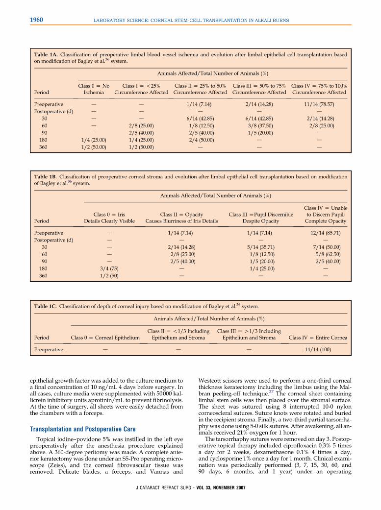
A solution of 1 M sodium hydroxide was used on the cor-neal surface of the left eye under general and topical anesthe-sia. After 1 minute, the eye was gently washed with saline.Clinical signs of LSCD were evaluated during the following2 months and immediately before transplantation. Alkaliinjuries were recorded according to the International LifeSciences Institute classification described by Bagley et al.,36
which was slightly modified (Tables 1A, 1B, and 1C).
Biopsy, Culture, and Expansion of Corneal LimbalEpithelial Cells
A small biopsy of corneal limbal epithelial cells was ob-tained from the superior temporal limbus of the contralateralhealthy eye using a 1.8 mm diameter trephine and a delicateforceps. This was preceded by gentle debridement of theconjunctiva and rinsing of the zone with saline to diminishcell contamination in the culture. The sample was immedi-ately placed in a tube with Ham’s F-12 containing 100 U/mLpenicillin, 100 mg/mL streptomycin, 2.5 mg/mL amphoteri-cin B, and 20 mg/mL gentamicin and then transported tothe laboratory.
The sample was washed 3 times in antibiotic–antimycoticsolutions. A fourth wash removed undesirable antimicro-bials. The sample was enzymatically processed with trypsinto obtain isolated cells. The rabbit limbal epithelial cells wereseeded on an irradiated murine 3T3 fibroblast feeder layerand cultured in an epithelial medium composed of a 3:1 mix-ture of Dulbecco’s modified Eagle medium and Ham’s F-12supplemented with 10% fetal bovine serum, 0.4 mg/mL hy-drocortisone, 5 mg/mL transferrin, 5 mg/mL insulin, 10�10 Mcholera toxin A, 1.8 � 10�4 M adenine, 2 � 10�11 M triio-dothyronine, 100 U/mL penicillin, and 100 mg/mL strep-tomycin and then placed in a 37�C 5% carbon dioxideincubator.
Autologous PPP or commercial fibrin glue (Tissucol) wasclotted in circular culture chambers. Approximately 4 � 104
rabbit limbal epithelial cells/cm2were seeded on a fibroblastfeeder layer grown in those chambers, and cells formeda monolayer after a few days. To achieve cell stratification,
- VOL 33, NOVEMBER 2007
1960 LABORATORY SCIENCE: CORNEAL STEM-CELL TRANSPLANTATION IN ALKALI BURNS
Table 1A. Classification of preoperative limbal blood vessel ischemia and evolution after limbal epithelial cell transplantation basedon modification of Bagley et al.36 system.
Animals Affected/Total Number of Animals (%)
PeriodClass 0 Z No
IschemiaClass I Z !25%
Circumference AffectedClass II Z 25% to 50%Circumference Affected
Class III Z 50% to 75%Circumference Affected
Class IV Z 75% to 100%Circumference Affected
Preoperative d d 1/14 (7.14) 2/14 (14.28) 11/14 (78.57)Postoperative (d) d d d d d
30 d d 6/14 (42.85) 6/14 (42.85) 2/14 (14.28)60 d 2/8 (25.00) 1/8 (12.50) 3/8 (37.50) 2/8 (25.00)90 d 2/5 (40.00) 2/5 (40.00) 1/5 (20.00) d
180 1/4 (25.00) 1/4 (25.00) 2/4 (50.00) d d
360 1/2 (50.00) 1/2 (50.00) d d d
Table 1B. Classification of preoperative corneal stroma and evolution after limbal epithelial cell transplantation based on modificationof Bagley et al.36 system.
Animals Affected/Total Number of Animals (%)
PeriodClass 0 Z Iris
Details Clearly VisibleClass II Z Opacity
Causes Blurriness of Iris DetailsClass III ZPupil Discernible
Despite Opacity
Class IV Z Unableto Discern Pupil;Complete Opacity
Preoperative d 1/14 (7.14) 1/14 (7.14) 12/14 (85.71)Postoperative (d) d d d d
30 d 2/14 (14.28) 5/14 (35.71) 7/14 (50.00)60 d 2/8 (25.00) 1/8 (12.50) 5/8 (62.50)90 d 2/5 (40.00) 1/5 (20.00) 2/5 (40.00)180 3/4 (75) d 1/4 (25.00) d
360 1/2 (50) d d d
Table 1C. Classification of depth of corneal injury based on modification of Bagley et al.36 system.
Animals Affected/Total Number of Animals (%)
Period Class 0 Z Corneal EpitheliumClass II Z !1/3 IncludingEpithelium and Stroma
Class III Z O1/3 IncludingEpithelium and Stroma Class IV Z Entire Cornea
Preoperative d d d 14/14 (100)
epithelial growth factor was added to the culture medium toa final concentration of 10 ng/mL 4 days before surgery. Inall cases, culture media were supplemented with 50000 kal-licrein inhibitory units aprotinin/mL to prevent fibrinolysis.At the time of surgery, all sheets were easily detached fromthe chambers with a forceps.
Transplantation and Postoperative Care
Topical iodine–povidone 5% was instilled in the left eyepreoperatively after the anesthesia procedure explainedabove. A 360-degree peritomy was made. A complete ante-rior keratectomywas done under an S5-Pro operatingmicro-scope (Zeiss), and the corneal fibrovascular tissue wasremoved. Delicate blades, a forceps, and Vannas and
J CATARACT REFRACT SURG
Westcott scissors were used to perform a one-third cornealthickness keratectomy including the limbus using the Mal-bran peeling-off technique.37 The corneal sheet containinglimbal stem cells was then placed over the stromal surface.The sheet was sutured using 8 interrupted 10-0 nyloncorneoscleral sutures. Suture knots were rotated and buriedin the recipient stroma. Finally, a two-third partial tarsorrha-phywas done using 5-0 silk sutures. After awakening, all an-imals received 21% oxygen for 1 hour.
The tarsorrhaphy sutures were removed on day 3. Postop-erative topical therapy included ciprofloxacin 0.3% 5 timesa day for 2 weeks, dexamethasone 0.1% 4 times a day,and cyclosporine 1% once a day for 1 month. Clinical exami-nation was periodically performed (3, 7, 15, 30, 60, and90 days, 6 months, and 1 year) under an operating
- VOL 33, NOVEMBER 2007

1961LABORATORY SCIENCE: CORNEAL STEM-CELL TRANSPLANTATION IN ALKALI BURNS
microscope and photographically documented with a Cool-pix 5700 digital camera (Nikon).
Argon laser photocoagulation connected to an indirectbinocular ophthalmoscope (Ophthalas Eye Lite, Alcon Labo-ratories) and a 20.0 diopter lens were used to occlude theneovessels invading the cornea postoperatively. Laser shotsof 550 mV and 0.3-second exposure were applied on thescleral part of the blood vessels near the limbus, with noshots on the cornea. The photocoagulation session was re-peated if regrowth of vessels occurred. No more than 150shots were used per session.
Histological Examination
Animals were killed for histological and immunohisto-chemical examination at 30, 60, 90, 180, and 360 days. Theglobe was enucleated and placed in 10% paraformaldehydefor 24 hours. The cornea was extracted from the globe usinga delicate forceps and scissors. Histological specimens werestained with hematoxylin–eosin (H&E) and examined underan Eclipse E800 microscope (Nikon).
Immunohistochemical Analysis
Primary monoclonal antibodies against cytokeratins K3and K12 (K3/12) (Mab AE5, Chemicon) and cytokeratin
J CATARACT REFRACT SURG
K19 (Mab 1675,Chemicon)wereused to identify the presenceof different cytokeratins in the corneal epithelium. The sameimmunohistochemical analysiswas performed in the rabbit’scontralateral eye,which had not had surgery (control group).
Corneal samples were cut at 10 mm thickness in a cryostatand processed for immunohistochemistry technique. Briefly,sections were incubated with the first monoclonal antibody(to K3/12 or to K19) and then with biotinylated goat anti-mouse immunoglobulin (Vector Laboratories) followed byavidin–biotin–peroxidase (Vectastain, Vector Laboratories).Finally, they were revealed with diaminobenzidine tetrahy-drochloride enhanced by ammonium nickel sulfate.
RESULTS
A limbal biopsy of 1.8 mm diameter was sufficient toachieve in vitro stem-cell replication. Overall, it took3 weeks to develop a corneal sheet ready to be grafted.Matrices of PPP showed very good elasticity and werehighly transparent. In contrast, commercial gels wereopaque and considerably stiff (Figures 1 and 2). Bythe first week, there was total reepithelialization ofthe small ulcer produced by the biopsy and no side ef-fects were seen on the biopsied eye.
Figure 1. Macroscopic features ofcultured corneal sheets. A and C:Corneal sheet using Tissucol. Band D: Corneal sheet using autolo-gous PPP. The Tissucol sheets areopaque, while the PPP sheets aretransparent.
- VOL 33, NOVEMBER 2007

1962 LABORATORY SCIENCE: CORNEAL STEM-CELL TRANSPLANTATION IN ALKALI BURNS
Figure 2. Histological characteris-tics of corneal sheets. A: Cornealsheet using autologous PPP. B: Cor-neal sheet using Tissucol. Both im-ages show a stratified epitheliumconsisting of 4 layers. Inferior cellsare polygonal and have a prominentnucleolus. The middle layers showcells with irregular morphology,and the upper layer is formed byflattened cells with compact ovoidnucleus ofhomogeneous chromatin.
All alkali injuries resulted in preoperative clinicalsigns of total LSCD (corneal neovascularization, sur-face irregularities, and opacity). Figure 3 shows theclinical progress of the rabbits that had been followedfor 1 year. Corneal opacities evolved differently be-tween rabbits; however, there was a general tendencytoward improvement over time (Table 1B). A clear re-duction in epithelial defects was seen in most cases,even in those with neovessel ingrowth. The cornealgrafts did not develop neovascularization during thefirst postoperative month. From the second month on-ward, argon laser photocoagulation was performed in9 animals according to the onset of neovessels.
Histological corneal sections showed a multilayerstratified epithelium at all times. Epithelial-like cellswith clear cytoplasm were found in the deepest partof the epithelium at day 30 and remained visible 1year postoperatively (Figure 4). There were no signsof vascular pannus or avascular intraepithelial mucincells on the treated corneas (Figure 4). Several eyeshad posterior corneal surface damagewith Descemet’smembrane replication due to the alkaline burn (Fig-ure 4). Corneal damage caused by laser photocoagula-tion was not seen.
J CATARACT REFRACT SURG
Immunohistochemical results showed expression ofK3/12 in the central part of the corneal epithelium. Ex-pression of K19 was not found. The results are similarto those seen on normal corneas (Figure 5).
Five animals developed corneal ectasia and/or en-dophthalmitis 1 month after the alkali burn procedureand did not have limbal stem-cell transplantation.They were excluded from the study and replaced byother animals.
DISCUSSION
In our study, autologous limbal epithelial cell trans-plantation improved the corneal surface of most eyeswith total LSCD, as documented by the absence of orreduction in recurrent epithelial defects, symblephar-on, and total corneal neovascularization. Cases that de-veloped partial neovessel ingrowth after somemonthsof transplantationwere treatedwith argon laser photo-coagulation, resulting in different degrees of regres-sion. Photocoagulation was well tolerated by graftedstem cells. The phenotype of the corneal epithelium us-ing antibodies against cytokeratins was in accordancewith previous studies.23,38–41 Transplantation of
Figure 3. Clinical evolution during1-year follow-up.A: Presence of cor-neal opacification and vasculariza-tion before surgery. B: Applicationof autologous corneal sheet.C: Clin-ical appearance 30 days after trans-plantation. D: Presence of cornealneovascularization 90 days aftergraft, later treated with argon laserphotocoagulation. E: Corneal clar-ity and absence of signs of LSCD12 months after transplantation.
- VOL 33, NOVEMBER 2007

1963LABORATORY SCIENCE: CORNEAL STEM-CELL TRANSPLANTATION IN ALKALI BURNS
Figure 4. Histopathological changes at the 1-year follow-up (H&E stain). A: Rabbit cornea with alkali burn and transplantation of autologouscorneal epithelial cells expanded ex vivo 30 days after surgery shows postinflammatory evidence of cicatricial ectasia with regenerated endo-thelium. The corneal surface has a non-keratinizated–stratified epithelium with normal morphology and configuration. Epithelial-like cellswith clear cytoplasm are seen in the deepest part of the epithelium. B: Rabbit cornea with alkali burn and transplantation of autologous cornealepithelial cells expanded ex vivo 1 year after surgery. The cornea had normal thickness and a loss of keratocytes. The original Descemet’s mem-brane is covered with fibrocollagenous tissue proliferation that shows endothelial cells and a new Descemet’s membrane toward the anteriorchamber. The 5-layer stratified epithelium has normal morphology.
autologous limbal corneal epithelial cells expanded exvivo was an effective procedure for treating LSCD.Most eyes had no vascularization in at least 6 clockhours of the cornea. Corneal tissue opacification wasassociated with stromal fibrosis or Descemet’s replica-tion. Nevertheless, all cases presented a normal cor-neal surface. The preoperative eye situation could becompared to legal blindness in humans.36 Undoubt-edly, in the clinical situation, the lesions may involvenot only the globe but also the conjunctiva and eye-lids; however, the prognosis depends on the initial le-sions, which can be variable. In our model, there was
J CATARACT REFRACT SURG
total preoperative corneal opacity, chronic conjuncti-val inflammation, and symblepharon, which may sug-gest abnormal tear-film integrity and resistance toinfection. These situations were not seen aftertreatment.
After grafting, the eyes made good progress and thedegree of injury diminished. It seems reasonable tothink these cases would benefit from a full-thicknesscorneal transplantation later.
The limbal biopsy is a safe procedure without com-plications.23,25,26 The stem cell–containing limbal epi-thelium is in proximity with conjunctiva, a highly
Figure 5. Immunohistochemicalanalysis of cytokeratins at 12months. A and C: Immunoreactiv-ity of K19 (A) and K3/12 (C) ina normal rabbit cornea. B and D:Immunoreactivity of K19 (B) andK3/12 (D) in a rabbit cornea withalkali burn and subsequent trans-plantation of autologous limbal ep-ithelial cells expanded ex vivo. Inboth corneas, there is negative im-munoreactivity to K19 and positiveimmunoreactivity to K3/12. Theexpression of the K3 and K12 pairin the implanted cornea shows dif-ferent degrees of intensity, whereasthe normal cornea shows homogen-eous staining.
- VOL 33, NOVEMBER 2007

1964 LABORATORY SCIENCE: CORNEAL STEM-CELL TRANSPLANTATION IN ALKALI BURNS
vascularized tissue, so preventive measures aimed atreducing contamination of stem-cell cultures fromerythrocytes or conjunctival cells are of paramount im-portance.42 We were able to perform this proceduresuccessfully in our study. We believe that the use ofPPP, an autologous substance, is a good alternativefor cell support. In this way, the corneal sheet can beeasily handled during in vitro manipulation or sur-gery, facilitating the formation of noncontinuous su-tures. Also, the transparency of the PPP allowstechnicians to follow the process of epithelial strat-ification. In addition, immunological reactions are un-common using autologous substances. This is anadvantage over the commercially available fibrin orthe human amniotic membrane.
Argon laser therapy emerged as a promising treat-ment for corneal neovascularization after corneal lim-bal cell transplantation, reducing the risk for graftfailure. There was a marked tendency toward neoves-sel reduction over time. Rabbits that had several ses-sions of photocoagulation at 1 year of follow-up stillachieved a substantially clear cornea, which suggeststhat the argon laser applied on vessels over the scleracaused no side effects to the corneas.
Results in this experimental studymay facilitate thede-velopment and application of the technique in patients.
REFERENCES1. Ang LPK, Tan DTH. Ocular surface stem cells and disease:
current concepts and clinical applications. Ann Acad Med Singa-
pore 2004; 33:576–580; Available at: http://www.annals.edu.
sg/pdf200411/V33N5p576.pdf. Accessed July 30, 2006
2. Reinhard T, Spelsberg H, Henke L, et al. Long-term results of
allogeneic penetrating limbo-keratoplasty in total limbal stem
cell deficiency. Ophthalmology 2004; 111:775–782
3. Tsai RJ-F, Li L-M, Chen J-K. Reconstruction of damaged cor-
neas by transplantation of autologous limbal epithelial cells.
N Engl J Med 2000; 343:86–93
4. Ramaesh K, Dhillon B. Ex vivo expansion of corneal limbal epithe-
lial/stem cells for corneal surface reconstruction. Eur J Ophthalmol
2003; 13:515–724; Available at: http://www.eur-j-ophthalmol.com/
ejo/content/132/1160/4693.pdf. Accessed July 30, 2006
5. Fernandes M, Sangwan VS, Rao SK, et al. Limbal stem cell
transplantation. Indian J Ophthalmol 2004; 52:5–22; Available
at: http://www.ijo.in/temp/IndianJOphthalmol5215_090054.pdf.
Accessed July 30, 2006
6. Schwab IR, Reyes M, Isseroff RR. Successful transplantation of
bioengineered tissue replacements in patients with ocular sur-
face disease. Cornea 2000; 19:421–426
7. Inatomi T, Nakamura T, Kojyo M, et al. Ocular surface recon-
struction with combination of cultivated autologous oral mucosal
epithelial transplantation and penetrating keratoplasty. Am J
Ophthalmol 2006; 142:757–764
8. Dua HS, Azuara-Blanco A. Autologous limbal transplantation in
patients with unilateral corneal stem cell deficiency. Br J Oph-
thalmol 2000; 84:273–278
9. Townsend WM. The limbal palisades of Vogt. Trans Am Oph-
thalmol Soc 1991; 89:721–756; Available at: http://www.
pubmedcentral.nih.gov/tocrender.fcgi?iidZ124639. Accessed
July 30, 2006
J CATARACT REFRACT SURG
10. Dua HS, Azuara-Blanco A. Limbal stem cells of the corneal
epithelium. Surv Ophthalmol 2000; 44:415–425
11. Sun T-T, Lavker RM. Corneal epithelial stem cells: past, present,
and future. J Invest Dermatol Symp Proc 2004; 9:202–207
12. Pellegrini G, Golisano O, Paterna P, et al. Location and clonal
analysis of stem cells and their differentiated progeny in the
human ocular surface. J Cell Biol 1999; 145:769–782
13. Potten CS. Cell replacement in epidermis (keratopoiesis) via dis-
crete units of proliferation. Int Rev Cytol 1981; 69:271–318
14. Lavker RM, Sun T-T. Epidermal stem cells: properties, markers,
and location. Proc Nat Acad Sci USA 2000; 97:13473–13475
15. Wolosin JM. Cell markers and the side population phenotype in
ocular surface epithelial stem cell characterization and isolation.
Ocular Surf 2006; 4:10–23
16. Dua HS, Joseph A, Shanmuganathan VA, Jones RE. Stem cell
differentiation and the effects of deficiency. Eye 2003; 17:877–885
17. Garg P, Krishna PV, Stratis AK, Gopinathan U. The value of corneal
transplantation in reducing blindness. Eye 2005; 19:1106–1114
18. Whitcher JP, Srinivasan M, Upadhyay MP. Prevention of cor-
neal ulceration in the developing world. Int Ophthalmol Clin
2002; 42(1):71–77
19. Whitcher JP, Srinivasan M, Upadhyay MP. Corneal blindness:
a global perspective. Bull World Health Org 2001; 79:214–221;
Available at: http://www.who.int/bulletin/archives/79(3)214.pdf.
Accessed July 30, 2006
20. Lee P, Wang CC, Adamis AP. Ocular neovascularization: an ep-
idemiologic review. Surv Ophthalmol 1998; 43:245–269
21. Brodovsky SC, McCarty CA, Snibson G, et al. Management of
alkali burns; an 11-year retrospective review. Ophthalmology
2000; 107:1829–1835
22. Dua HS, King AJ, Joseph A. A new classification of ocular sur-
face burns. Br J Ophthalmol 2001; 85:1379–1383
23. Koizumi N, Inatomi T, Suzuki T, et al. Cultivated corneal epithe-
lial stem cell transplantation in ocular surface disorders. Oph-
thalmology 2001; 108:1569–1574
24. Nakamura T, Endo K-I, Cooper LJ, et al. The successful culture
and autologous transplantation of rabbit oral mucosal epithelial
cells on amniotic membrane. Invest Ophthalmol Vis Sci 2003;
44:106–116
25. Talbot M, Carrier P, Giasson CJ, et al. Autologous transplanta-
tion of rabbit limbal epithelia cultured on fibrin gels for ocular sur-
face reconstruction. Mol Vis 2006; 12:65–75; Available at: http://
www.molvis.org/molvis/v12/a7/. Accessed July 30, 2006
26. Pellegrini G, Traverso CE, Franzi AT, et al. Long-term restora-
tion of damaged corneal surfaces with autologous cultivated cor-
neal epithelium. Lancet 1997; 349:990–993
27. Rama P, Bonini S, Lambiase A, et al. Autologous fibrin-cultured
limbal stem cells permanently restore the corneal surface of pa-
tients with total limbal stem cell deficiency. Transplantation
2001; 72:1478–1485
28. Shimazaki J, Aiba M, Goto E, et al. Transplantation of human
limbal epithelium cultivated on amniotic membrane for the treat-
ment of severe ocular surface disorders. Ophthalmology 2002;
109:1285–1290
29. Gomes JAP, dos Santos MS, Cunha MC, et al. Amniotic mem-
brane transplantation for partial and total limbal stem cell deficiency
secondary to chemical burn. Ophthalmology 2003; 110:466–473
30. Chang J-H, Gabison EE, Kato T, Azar DT. Corneal neovascula-
rization. Curr Opin Ophthalmol 2001; 12:242–249
31. Luengo Gimeno F, Gatto S, Ferro J, et al. Preparation of platelet-
rich plasma as a tissue adhesive for experimental transplanta-
tion in rabbits. Thromb J 2006; 4:18; Available at: http://
www.thrombosisjournal.com/content/pdf/1477-9560-4-18.pdf.
Accessed July 30, 2007
32. Nakamura T, Inatomi T, Sotozono C, et al. Transplantation of au-
tologous serum-derived cultivated corneal epithelial equivalents
- VOL 33, NOVEMBER 2007
1965LABORATORY SCIENCE: CORNEAL STEM-CELL TRANSPLANTATION IN ALKALI BURNS
for the treatment of severe ocular surface disease. Ophthalmol-
ogy 2006; 113:1765–1772
33. Krasnick NM, Spigelman AV. Comparison of yellow dye, continu-
ous wave Nd:YAG, and argon green laser on experimentally in-
duced corneal neovascularization. J Refract Surg 1995; 11:45–49
34. Lim KJ, Wee WR, Lee JH. Treatment of corneal neovasculariza-
tion with argon laser. Korean J Ophthalmol 1993; 7:25–27; Avail-
able at: http://pdf.medrang.co.kr/paper/pdf/Kjo/Kjo007-01-05.pdf.
Accessed July 30, 2007
35. Association for Research in Vision and Ophthalmology. Statement
for the use of animals in ophthalmic and visual research. Available
at: http://www.arvo.org/eweb/dynamicpage.aspx?siteZarvo2&
webcodeZAnimalsResearch. Accessed July 30, 2007
36. Bagley DM, Casterton PL, Dressler WE, et al. Proposed new
classification scheme for chemical injury to the human eye. Reg-
ul Toxicol Pharmacol 2006; 45:206–213
37. Polack FM. Lamellar keratoplasty Malbran’s ‘‘peeling off’’ tech-
nique. Arch Ophthalmol 1971; 86:293–295
38. Kurpakus MA, Stock EL, Jones JCR. The role of the basement
membrane in differential expression of keratin proteins in epithe-
lial cells. Dev Biol 1992; 150:243–255
J CATARACT REFRACT SURG
39. Chen WYW, Mui M-M, Kao WW-Y, et al. Conjunctival epithelial
cells do not transdifferentiate in organotypic cultures: expression
of K12 keratin is restricted to corneal epithelium. Curr Eye Res
1994; 13:765–778
40. Zhu G, Ishizaki M, Haseba T, et al. Expression of K12 keratin in
alkali-burned rabbit corneas. Curr Eye Res 1992; 11:875–887
41. Kiritoshi A, SundarRaj N, Thoft RA. Differentiation in cultured
limbal epithelium as defined by keratin expression. Invest Oph-
thalmol Vis Sci 1991; 32:3073–3077
42. Kruse FE, Tseng SCG. Serum differentially modulates the clonal
growth and differentiation of cultured limbal and corneal epithe-
lium. Invest Ophthalmol Vis Sci 1993; 34:2976–2989
First author:Federico Luengo Gimeno, MD
Ophthalmology Department, School ofBiomedical Sciences, Universidad Austral,Pilar, Argentina
- VOL 33, NOVEMBER 2007